microenvironmental modulation of decorin and lumican in temozolomide-resistant glioblastoma and...
TRANSCRIPT

RESEARCH ARTICLE
Microenvironmental Modulation of Decorinand Lumican in Temozolomide-ResistantGlioblastoma and Neuroblastoma CancerStem-Like CellsCristiano Farace1☯‡*, Jaime Antonio Oliver2☯‡, Consolacion Melguizo2,3,4,Pablo Alvarez2,4, Pasquale Bandiera1, Ana Rosa Rama2,4,5, Giulia Malaguarnera6,Raul Ortiz2,4,5, Roberto Madeddu1,7‡*, Jose Prados2,3,4‡
1 Department of Biomedical Sciences, University of Sassari, Sassari, Italy, 2 Institute of Biopathology andRegenerative Medicine (IBIMER), Granada, Spain, 3 Department of Anatomy and Embryology, University ofGranada, Granada, Spain, 4 Biosanitary Institute of Granada (ibs.Granada), SAS-University of Granada,Granada, Spain, 5 Department of Health Science, University of Jaén, Jaén, Spain, 6 Research Center "TheGreat Senescence", University of Catania, Catania, Italy, 7 National Institute of Biostructures and Biosystem(INBB), Rome, Italy
☯ These authors contributed equally to this work.‡RM and JP also contributed equally to this work and are listed alphabetically. CF and JAO are also listedalphabetically.* [email protected] (CF); [email protected] (RM)
AbstractThe presence of cancer stem cells (CSCs) or tumor-initiating cells can lead to cancer recur-
rence in a permissive cell–microenvironment interplay, promoting invasion in glioblastoma
(GBM) and neuroblastoma (NB). Extracellular matrix (ECM) small leucine-rich proteogly-
cans (SLRPs) play multiple roles in tissue homeostasis by remodeling the extracellular
matrix (ECM) components and modulating intracellular signaling pathways. Due to their
pan-inhibitory properties against receptor tyrosine kinases (RTKs), SLRPs are reported to
exert anticancer effects in vitro and in vivo. However, their roles seem to be tissue-specific
and they are also involved in cancer cell migration and drug resistance, paving the way to
complex different scenarios. The aim of this study was to determine whether the SLRPs
decorin (DCN) and lumican (LUM) are recruited in cell plasticity and microenvironmental
adaptation of differentiated cancer cells induced towards stem-like phenotype. Floating
neurospheres were generated by applying CSC enrichment medium (neural stem cell
serum-free medium, NSC SFM) to the established SF-268 and SK-N-SH cancer cell lines,
cellular models of GBM and NB, respectively. In both models, the time-dependent synergis-
tic activation of DCN and LUM was observed. The highest DCN and LUMmRNA/protein
expression was detected after cell exposure to NSC SFM for 8/12 days, considering these
cells as SLRP-expressing (SLRP+) CSC-like. Ultrastructural imaging showed the cellular
heterogeneity of both the GBM and NB neurospheres and identified the inner living cells.
Parental cell lines of both GBM and NB grew only in soft agar + NSC SFM, whereas the sec-
ondary neurospheres (originated from SLRP+ t8 CSC-like) showed lower proliferation rates
PLOSONE | DOI:10.1371/journal.pone.0134111 July 31, 2015 1 / 18
a11111
OPEN ACCESS
Citation: Farace C, Oliver JA, Melguizo C, Alvarez P,Bandiera P, Rama AR, et al. (2015)Microenvironmental Modulation of Decorin andLumican in Temozolomide-Resistant Glioblastomaand Neuroblastoma Cancer Stem-Like Cells. PLoSONE 10(7): e0134111. doi:10.1371/journal.pone.0134111
Editor: Dragana Nikitovic-Tzanakaki, University ofCrete, GREECE
Received: April 28, 2015
Accepted: July 6, 2015
Published: July 31, 2015
Copyright: © 2015 Farace et al. This is an openaccess article distributed under the terms of theCreative Commons Attribution License, which permitsunrestricted use, distribution, and reproduction in anymedium, provided the original author and source arecredited.
Data Availability Statement: All relevant data arewithin the paper.
Funding: This study was supported by Fundació laMarató TV3, Project n° 111431.
Competing Interests: The authors have declaredthat no competing interests exist.

than primary neurospheres. Interestingly, the SLRP+ CSC-like from the GBM and NB neuro-
spheres were resistant to temozolomide (TMZ) at concentrations >750 μM. Our results sug-
gest that GBM and NB CSC-like promote the activation of huge quantities of SLRP in
response to CSC enrichment, simultaneously acquiring TMZ resistance, cellular heteroge-
neity, and a quiescent phenotype, suggesting a novel pivotal role for SLRP in drug resis-
tance and cell plasticity of CSC-like, allowing cell survival and ECM/niche modulation
potential.
BackgroundGlioblastoma (GBM) and neuroblastoma (NB) are the most common and lethal nervous sys-tem malignant cancers in adult and pediatric patients, respectively. The World Health Organi-zation considers GBM the most aggressive astrocytoma, and it can develop into secondaryGBM from low-grade gliomas, or de novo with rapid progression to death [1]. In contrast, NBmainly arises from neural crest cells as a neuroendocrine cancer of the sympathetic nervoussystem, and 60% of pediatric patients show metastatic disease at diagnosis [2]. Although temo-zolomide (TMZ)–an alkylating agent which induces cell death by whole DNA alkylation/meth-ylation in guanine residues–in combination with other drugs or radiotherapy represent a first-line treatment increasing the overall survival (OS) of patients with GBM or NB [3, 4], drugresistance and cancer progression are common. Because GBM is highly invasive in the brainand NB tends to invade other organs, patient OS remains poor (< 1.5 years in GBM patientsand 4 years in NB patients) [5, 6].
Treatment failure in cancer patients has previously been related to cancer stem cell (CSC)subpopulations, which ensure the maintenance of cancer heterogeneity, and these CSC sub-populations are more resistant to selective drugs through multiple concerted steps of self-renewal and differentiation [7–9]. Metastasis and cancer recurrence are also linked to thebehavior of CSCs, including their quiescent phenotype, migratory ability, and evasion of theimmune system [10]. Abundant research suggests that cells stem-like cells are equipped withinnate machinery that protects them from radio/chemotherapy [11, 12]. This includes stem-related mechanisms, such as protective cell niches and changes in the expression of genesinvolved in the regulation of the cell cycle, DNA repair, drug metabolism, and drug efflux [13].The drug resistance and cellular invasion potential of CSCs also increase at the reversible epi-thelial-to-mesenchymal phenotypic transition (EMT) [14, 15], which recapitulates the EMT innormal organogenesis and development [16, 17].
Several microenvironmental signals, including the reorganization of the extracellular matrix(ECM), hypoxia, and autocrine/paracrine factors, can determine stem and cancer cell fates[18–25], and trigger or inhibit EMT processes [26, 27]. Therefore, ECM glycoproteins and pro-teoglycans that are capable of modifying both the ECM environment and intracellular signalingpathways are of utmost importance in the cancer microenvironment [28–30]. The smallleucine-rich proteoglycans (SLRPs), sharing strategically conserved domains, represent a clearexample of the abovementioned concept. The leucine-rich protein core (40–50 kDa) bind to anumber of growth factors (GF) and membrane receptors, whereas ramification of glycosami-noglycanic side chains are involved in ECM–collagen assembly and also in membrane receptorbinding. Interestingly, in spite of their pan-inhibitory properties against receptor tyrosinekinases (RTKs) and cancer growth pathways, the “guardian from the matrix” decorin (DCN)and lumican (LUM) SLRPs could exert anticancer effects in vivo and in vitro [31–33].
SLRPs and Drug-Resistant Cancer Stem-Like Cells
PLOS ONE | DOI:10.1371/journal.pone.0134111 July 31, 2015 2 / 18

However, recent studies have shed light on newly identified tissue-specific properties of bothDCN and LUM in normal tissues and in the malignant cancer microenvironment. As reportedby other authors, the partial glioma inhibition by DCN in gene therapy experiments in ratsbrings with it a marked reduction of microglial cells infiltration [34], which could affects cancerinhibition in vivo. DCN also enhances the evasion of the immune system and muscle invasionin prostate cancer in vivo [35], and exerts unexpected protective and antiapoptotic effects inglioma cell lines under hypoxic conditions [36]. In oral malignant squamous cell carcinomacells, the nuclear localization of DCN seems to enhance cellular invasion via the nuclear epider-mal growth factor receptor (EGFR) pathway [37, 38], whereas in osteosarcoma cells, DCN-mediated growth arrest is avoided via the protracted activation of membrane EGFR [39]. Clini-cally, DCN has been proposed as regulator of chemoresistant mechanism in oral cancer [40]and related to drug resistance and reduced survival in GBM patients [41]. Similarly to DCN,LUM is reported to mediate tumor suppression. However, LUM is expressed in high-gradepancreatic cancers with a low degree of differentiation [42] and in GBM patients, as well. LUMalso inhibits cell adhesion and promotes the migration of osteosarcoma cells by regulating thetransforming growth factor β2 (TGF-β2)/SMAD2 pathway [43], and a 70-kDa LUM proteogly-can seems to enhance cancer cell proliferation and inhibits the migration of pancreatic cancercells. Moreover, together to DCN, LUM was upregulated in cisplatin-resistant head and neckcancer cells [44].
It is noteworthy that SLRPs are expressed in stem cell niches in the chick embryo [45], incerebral endothelial cells [46], in progenitors of various cell types [47], and in a NB cell subpop-ulation unresponsive to nerve-growth-factor-mediated neurite growth [48]. DCN derived fromastrocytes also inhibits neural stem cell/progenitor cell differentiation towards a neuron-likecell structure [49]. Altering the mechanical characteristics of three-dimensional (3D) collagenmatrices, SLRPs are recruited during the ontogenic (developmental) EMT [50], cell precursormigration and differentiation [51], and wound healing/tissue repair in response to central ner-vous system injury and inflammation [52]. In this context, it is conceivable that the small DCNand LUM proteoglycans play a role in the biology of CSCs of nervous system origin. To thisend, we investigated the involvement of DCN and LUM in GBM and NB CSC-like models,simulating the phenomena of anchorage loss and the detachment of differentiated tumor cellsthat underlie the EMT process, and their relationship to CSC-like behavior and the cellresponse to TMZ. In this study, we report for the first time the massive synergistic expressionof DCN and LUM SLRPs in GBM and NB cell lines subjected to floating 3D neurosphere-based CSC-like enrichment. Neurosphere micrographs highlight the stem-like heterogeneityand cell polarization of the 3D NB and GBMmodels. Moreover, SLRP+ NB and GBM CSC-likeisolated from the neurospheres showed lower proliferation rates, less apoptosis, and greaterdrug resistance than the parental cell lines, suggesting pivotal and synergistic roles for DCNand LUM in the TMZ resistance, survival, and maintenance of quiescent, slow-cycling, CSC-like subpopulations.
Methods
Cell lines and CSC enrichmentIn this study, two established GBM and NB cell lines were enrolled. The SK-N-SH cell line is acommercial epithelial cell line originally derived from bone marrow metastasis of a 4-years-old Caucasian female suffering with NB, and it was previously enriched in CSC-like. In con-trast, SF-268 is a nonepithelial cell line derived from a high-grade anaplastic astrocytoma,which has never been used for CSC-like enrichment. The established SK-N-SH (from theAmerican Type Culture Collection) and SF-268 cancer cell lines (kindly provided by the
SLRPs and Drug-Resistant Cancer Stem-Like Cells
PLOS ONE | DOI:10.1371/journal.pone.0134111 July 31, 2015 3 / 18

Instrumentation Scientific Center, Granada University) were routinely maintained as adher-ent cultures (monolayers) in Dulbecco’s modified Eagle’s medium (DMEM) supplementedwith 10% fetal bovine serum and 1% penicillin/streptomycin, in a humidified atmosphere at37°C with 5% CO2. Confluent cells were detached in 5 ml of phosphate-buffered saline (PBS)–ethylenediaminetetraacetic acid (EDTA) at 37°C for 10 min, washed twice in PBS, and subcul-tured as monolayers. CSC enrichment of the cancer cell lines was performed by generatingneurospheres in neural stem cell serum-free medium (NSC SFM) [53, 54], containing Knock-Out DMEM/F12 plus 20 μg/ml basic fibroblast growth factor (bFGF), 20 μg/ml epidermalgrowth factor, 1× StemPro Neural Supplement (Invitrogen, Paisley, UK), 1% L-glutamine, and1% penicillin/streptomycin. NSC SFM was replaced every 2 days after mild centrifugation ofthe neurospheres.
RNA extraction and quantitative reverse transcription (qRT)–PCRThe expression of DCN and LUMmRNAs was assessed in t0 cells and neurospheres after 4(t4), 8 (t8), and 12 days (t12) of CSC enrichment. The parental cell lines in DMEM were used asthe control (t0). RNA was extracted with the RNeasy Mini Kit (Qiagen, MD, USA) and quanti-fied with a Nanodrop spectrophotometer (Thermo Scientific, DE, USA). The RNA (1 μg) fromeach sample was reversed transcribed with M-MLV reverse transcriptase (Sigma, Italy),according to manufacturer’s instructions, and SYBR Green-based amplification (Applied Bio-systems, Foster City, CA) was performed with the CFX96 Real-Time PCR Detection System(Bio-Rad, Italy), as previously reported [55]. The PCR cycling program was: 50°C (2 min),95°C (2 min), 42 cycles of: denaturation at 95°C (30 s), annealing at 56°C (30 s), and extensionat 72°C (40 s), followed by a melting curve analysis (range 56–95°C) with increments of 0.5°C/5 s to assess the primer specificity. The primer sequences were: forward 50-GGA CCG TTT CAACAG AGA GG-30, reverse 50-GAC CAC TCG AAG ATG GCA TT-30 (DCN); forward 50-TGGAGG TCA ATC AAC TTG AGA A-30, reverse 50-CAA ACG CAA ATG CTT GAT CTT-30 (LUM);forward 50-CAA GGA GTA AGA CCC CTG GAC-30, reverse 50-TCT ACA TGG CAA CTG TGAGGA G-30 (glyceraldehyde 3-phosphate dehydrogenase, GAPDH); forward 50-GGC ATC CTCACC CTG AAT GA-30, reverse 50-AGG TGT GGT GCC AGA TTT TC-30 (β-actin, ACTB). Thetarget transcripts were independently normalized to GAPDH and ACTB (housekeepinggenes), and the RNA of the t0 cells was used as the calibration control. The results wereexpressed on a logarithmic scale as fold changes (FCs), with the 2–ΔΔCt method.
Western blottingWhole protein extracts were obtained with the pulsed sonication of cellular pellets in 200 μl ofextraction buffer containing 50 mM Trizma-base, 0,25 mM sucrose, 5 mM EDTA (pH 7.4),0.5% Triton-X 100 and 1% protease inhibitor cocktail. The protein concentrations were deter-mined with the Bradford method. The proteins (50 μg) were mixed 1:1 with 2 × Laemmlibuffer, heated at 95°C for 5 min, and loaded onto a 12% denaturing SDS-PAGE gel with theKaleidoscope prestained standards. The proteins were separated electrophoretically at a con-stant voltage (90 V) for 90 min and blotted onto a 0.45 μm nitrocellulose membrane undersemidry conditions with the Trans-Blot SD Semi-Dry Electrophoretic Transfer Cell (Bio-Rad,Spain) for 25 min at 200 mA. The membranes were blocked with 5% skim milk in PBS, incu-bated at 4°C overnight with rabbit polyclonal anti-LUM antibody (diluted 1:100; sc-33785,Santa Cruz Biotechnology, Santa Cruz, CA) or mouse monoclonal anti-DCN antibody (1:50;sc-73896, Santa Cruz Biotechnology) in blocking buffer, washed 4 times in washing solution(0.1% Tween in PBS), incubated at room temperature for 1 h with the appropriate monoclonalhorseradish-peroxidase-conjugated secondary antibody (1:5000; Sigma Aldrich), and washed
SLRPs and Drug-Resistant Cancer Stem-Like Cells
PLOS ONE | DOI:10.1371/journal.pone.0134111 July 31, 2015 4 / 18

again in washing solution. The blots were detected with ECLWestern Blotting DetectionReagents (Amersham; UK). The Molecular Imager VersaDoc MP 4000 system (Bio-Rad, Her-cules, CA) was used for chemiluminescence visualization. The blots were stripped and incu-bated with mouse monoclonal anti-β-actin antibody (1:30000; Sigma Aldrich) as the loadingcontrol.
Soft agar culturesThe cell lines were cultured in soft agar to assess their colony or neurosphere formation underanchorage-independent conditions, in DMEM or NSC SFM. To explore the proliferation ofthe SLRP+ CSC-like after CSC enrichment for 8 days (t8 CSC-like), secondary neurosphereswere generated from t8 CSC-like. Briefly, StemPro Accutase Cell Dissociation Reagent (Invitro-gen) was used to dissociate the t8 neurospheres. A Trypan blue exclusion test was performedand 5 × 10³/ml cells were collected in 2 × DMEM or NSC SFM. The cells were mixed 1:1 with aprewarmed solution containing 0.6% ultrapure agarose in PBS (0.3% final agar concentration),seeded in triplicate in six-well plates on 2 ml of solidified 0.6% bottom agar, and incubated in ahumidified atmosphere at 37°C with 5% CO2. The soft agar cultures were photographed dailyunder an inverted phase-contrast microscope (Nikon Eclipse TE 2000-S).
Transmission and scanning electron microscopic imagingEight-day (t8) neurospheres were collected, washed in PBS, and fixed in 2.5% glutaraldehydein 0.1 M PBS (pH 7.4) for 2 h at 4°C. The fixed neurospheres were carefully washed four timesin PBS, postfixed in 1% osmium tetroxide (OsO4) in 0.1 M PBS for 1 h at 4°C, and stored inPBS at 4°C until embedding. The samples used for transmission electron microscopy (TEM)were dehydrated in series of increasing acetone concentrations and embedded in epoxy resinfor ultrathin sectioning at 60°C overnight. The ultrathin slices cut with an 8800 Ultratome(LKB, Bromma, Sweden) were stained with 4% uranyl acetate and lead citrate and viewed on aZeiss EM 109–902 transmission electron microscope (Zeiss, Oberchochen Germany). Forscanning electron microscopy (SEM), the postfixed neurospheres were incubated in pure hex-amethyldisilazane for 1 h at 4°C, dried in a critical point dryer (Polaron, Watford, UK), andmetalized in an S150A Sputter Coater (Edwards, Crawley, UK) for scanning in Quanta 200(FEI, Eindhoven, The Netherlands) or DSM 962 SEM instruments (Zeiss, Oberchochen,Germany).
TMZ treatment and MTT assaySingle cells from the monolayers and t8 neurospheres of both cell lines were collected asdescribed above. Cell viability was tested with Trypan blue exclusion and 5 × 10³ cells/wellwere seeded in 96-well microtiter plates, in DMEM or NSC SFM. The next day, the mediumwas replaced with fresh medium without or with different concentrations of TMZ (25–1500 μg/ml; Sigma, Madrid, Spain). DMSO was included as the vehicle control. Each conditionwas tested in six wells. Three days later, the medium was replaced with fresh medium to pre-serve TMZ activity. The end-point of the TMZ treatment was established on day 6. Finally, a 3-(4,5-dimethylthiazol-2-yl)-2,5-diphenyltetrazolium bromide (MTT) colorimetric assay wasperformed (Sigma). Briefly, 20 μl of MTT/well was added and the plates were incubated at37°C. After 4 h, the medium was carefully removed and DMSO was added to dissolve the for-mazan salts. The reactions were measured with a Multiskan EX microplate photometer(Thermo Scientific, Madrid, Spain) at 570 nm. The TMZ dose–response was expressed as thepercentage (%) inhibition of the cell metabolic activity relative to that of untreated cells and
SLRPs and Drug-Resistant Cancer Stem-Like Cells
PLOS ONE | DOI:10.1371/journal.pone.0134111 July 31, 2015 5 / 18

adjusted to the vehicle control. The inhibition of cell growth by TMZ was evaluated in quadru-plicate experiments.
Statistical analysisStudent’s two-tailed t test was used to determine the statistical significance of the differences inthe TMZ treatment results for the SLRP+ CSC-like and the parental cell lines. The differencesin qPCR gene expression were evaluated with one-way analysis of variance (ANOVA). Statisti-cal significance was set at p<0.05.
Results
DCN and LUM expression during neurosphere-based CSC enrichmentNeurospheres of both SF-268 and SK-N-SH cell lines acquired regular 3D conformationsresulting from the sustained radial proliferation of most cells, reaching>50 μm after 4 days(Fig 1A). A qRT–PCR analysis showed that DCN and LUMmRNA expression, with the excep-tion of LUM in SF-268 t12, increased under CSC enrichment conditions in both cell lines,showing the highest DCN and LUMmRNA expression values after 8 days (t8). Compared withthe parental cell lines (t0), and considering ACTB as housekeeping gene, GBM CSC-like weremore enriched in LUMmRNA (FCLum = t0: 1; t4: 11.00; t8: 21.70; t12: 9.51) than in DCNmRNA (FCDcn = t0: 1; t4: 6.32; t8: 17.87; t12: 11.08) (p< 0.01). Similarly, the NB CSC-likeshowed higher LUMmRNA levels (FCLum = t0: 1; t4: 29.04; t8: 105.78; t12: 60.12) than DCNmRNA levels (FCDcn = t0: 1; t4: 23.02; t8: 80.44; t12: 40.22) (p< 0.01; Fig 1B).
The protein analysis showed a clear increase in LUM protein (70-kDa) during CSC enrich-ment. The highest LUM expression was detected in both SF-268 and SK-N-SH t8 neurospheres(Fig 1C). By contrast, 40-kDa LUM protein was detected with much less expression that70-kDa LUM in both SF-268 and SK-N-SH, and especially in t12 (Fig 1C). On the other hand,we detected a significant increase in DCN (>150-kDa) protein expression in both SF-268 andSK-N-SH t12 neurospheres while 40-kDa DCN protein was detected very weakly, with a diffi-cult expression evaluation. According to mRNA results, 8-days SLRP+ CSC-like (t8 CSC-like)derived from both GBM and NB cell lines were enrolled in further analysis.
Soft agar cultures of SLRP+ CSC-like and parental cell linesTo assess the differences in cell growth between the CSC-like and parental cell lines, the sec-ondary neurospheres from SLRP+ t8 CSC-like were generated in soft agar and compared withthe parental-cell-derived primary neurospheres. The anchorage-independent soft agar culturesshowed neurosphere colonies in NSC SFM, whereas no or low colonies of parental cells hadgrown in the DMEM-based soft agar assay after 22 days (Fig 2A). The primary SK-N-SH neu-rospheres were larger than the SF-268 neurospheres, reaching> 200 μm after 3 weeks, and hadshown a clearly visible dark core within the 3D structure. The soft agar cultures of t8 GBM andNB CSC-like (SLRP+) grew as small secondary neurospheres in NSC SFM, and curiously alsoas small colonies in DMEM after 30 days (Fig 2B). The secondary neurospheres were smallerthan the primary neurospheres and intrasphere stressed cells (dark cores) were only detectablein the secondary SF-268 neurospheres after 2 weeks, whereas the secondary SK-N-SH neuro-spheres maintained a translucent appearance.
Heterogeneic ultrastructures of GBM and NB neurospheresUltrastructural imaging with TEM and SEM showed broad cellular heterogeneity in the t8 neu-rospheres of both cell lines. SEM imaging of the neurosphere surfaces showed more packaged
SLRPs and Drug-Resistant Cancer Stem-Like Cells
PLOS ONE | DOI:10.1371/journal.pone.0134111 July 31, 2015 6 / 18

cells in the NB neurospheres than in the GBM neurospheres, whereas the peripheral cells of theGBM neurospheres had more thin membrane extroflections than the SK-N-SH neurospheres(Figs 3A, 3B, 4A and 4B). Adjacent electron-dense and electron-lucent cells and sporadic apo-ptotic cells were observed at the GBM neurosphere peripheries (Fig 3C–3E). The peripheralcells of the NB neurospheres were polarized, with more OsO4 staining in the cytoplasm thanthe inner cells, and contained dense granules, endocytic vesicles, and few membrane extroflec-tions (Fig 4C–4E). Wide areas of cell death, characterized by membrane blebbing, cell wrin-kling, apoptotic bodies, and wide nuclear compacted heterochromatin, were observed in the
Fig 1. DCN and LUMmRNA/protein expression in GBM and NB CSC-like enrichments. (A) Neurosphere generation from SF-268 and SK-N-SH celllines. Bar, 50 μm. (B) Expression of DCN and LUMmRNAs in CSC enrichment cultures. qRT–PCR results were normalized to ACTB and graphed as relativefold changes (FCs) between neurospheres at different time points (t4, t8, and t12) and adherent t0 cells with the 2–ΔΔCt method. All DCN and LUM FC valuesfor the neurospheres were statistically significant (*p < 0.01). (C) Western blotting analysis of DCN and LUM. β-Actin was used as the loading control. TotalDCN and LUM levels were higher in the neurospheres than in the adherent cells of both cell lines. A 70-kDa LUM isoform was upregulated in theneurospheres of both cell lines, with the highest expression in the t8 neurospheres. Adherent SF-268 cells showed basal expression of the 37-kDa LUM coreprotein.
doi:10.1371/journal.pone.0134111.g001
SLRPs and Drug-Resistant Cancer Stem-Like Cells
PLOS ONE | DOI:10.1371/journal.pone.0134111 July 31, 2015 7 / 18

Fig 2. Soft agar cultures of SLRP+ CSCs and parental cancer cell lines. (A) Primary soft agar neurospheres from the parental cell lines. Absence ofcolonies formation in semisolid DMEM (22 days) and neurosphere formation in semisolid NSC SFM (14 and 22 days). (B) Secondary soft agar neurospheresfrom SLRP+ CSC-like isolated from t8 neurospheres. Neurospheres were generated in both semisolid DMEM and semisolid NSC SFM (14 and 22 days),suggesting the slow cycling behavior of the CSC-like and residual death evasion activity in the DMEM-grown secondary neurospheres. Bar, 50 μm.
doi:10.1371/journal.pone.0134111.g002
SLRPs and Drug-Resistant Cancer Stem-Like Cells
PLOS ONE | DOI:10.1371/journal.pone.0134111 July 31, 2015 8 / 18

Fig 3. Representative images of GBM t8 neurospheres. (A) SEM image of a whole SF-268 neurosphere (1000×). (B) SEM image of the neurospheresurface (2200×). (C–J), TEM images of inner and peripheral neurospheres. Details of thin membrane extroflection (C–D), interactions between electron-dense and electron-lucent cells (E), details of cell–cell adhesion (F), suffering sites in the inner spheres, with necrotic (G) and apoptotic (H) cells, living cells,and details of the mitochondrial apparatus in the inner spheres (I, J). Note the apoptotic cells at the periphery of a neurosphere (D) and the living cells close tothe suffering sites (I). Bar: (C–E) and (G–I), 5 μm; (F and J), 1 μm.
doi:10.1371/journal.pone.0134111.g003
SLRPs and Drug-Resistant Cancer Stem-Like Cells
PLOS ONE | DOI:10.1371/journal.pone.0134111 July 31, 2015 9 / 18

middle of the neurospheres of both cell lines (Figs 3G, 3H, 4G and 4H). However, active mito-sis and living cells close to the inner stressed sites were also observed, in both the GBM and NBneurospheres (Figs 3G, 3I, 3J, 4F and 4J). Interestingly, these microenvironment-resistant cellsin the hypoxic neurosphere core showed indented nuclei, large amounts of euchromatin andclearly visible mitochondrial apparatus.
Fig 4. Representative images of NB t8 neurospheres. (A) SEM image of whole SK-N-SH neurosphere(950×). (B) SEM imaging of neurosphere surface (3000×). (C–J) TEM images of inner and peripheralneurosphere. Details of the cell vesicles, cell polarization, and detachment (C–E), living cells in the innerneurospheres and details of the mitochondrial apparatus (F), suffering sites in the inner spheres, with necrotic(G) and apoptotic cells (H and I), and an intrasphere mitotic event (J). Bar: (C–J), 5 μm.
doi:10.1371/journal.pone.0134111.g004
SLRPs and Drug-Resistant Cancer Stem-Like Cells
PLOS ONE | DOI:10.1371/journal.pone.0134111 July 31, 2015 10 / 18

TMZ treatment and MTT assay of SRLP+ CSC-like and parental celllinesLow-range TMZ concentrations (0–500 μM) did not induce significant differences in the meta-bolic activities of the SF-268 parental cells and SRLP+ t8 CSC-like. As shown in Fig 5A, the met-abolic activity of the parental cells ranged from 76.14% to 100% (94.63% ± 7.10%), whereasmetabolic activity of SRLP+ t8 CSC-like ranged from 80.71% to 100% (88.22% ± 5.43%). Higherconcentrations of TMZ (750–1500 μM) induced significant differences between the SLRP+ t8CSC-like and the parental cell line. At these concentrations, the metabolic activity of the SF-268 parental cell line ranged from 38.99% to 58.86% (51.80% ± 7.59%), whereas that of SF-268SRLP+ t8 CSC-like ranged from 79.09% to 79.64% (78.94% ± 0.45%) (p<0.05). The SF-268parental IC50 was around 1250 μM, and did not reach the IC50 of the SF-268 SRLP
+ t8 CSC-like.
At low and high concentrations, TMZ induced significant differences in the metabolic activ-ities of the SK-N-SH parental cells and the SRLP+ t8 CSC-like. As shown in Fig 5B, the meta-bolic activity of cells treated with 0–500 μMTMZ ranged from 75.08% to 100% (88.41% ±6.70%) for parental cells and from 66.86% to 100% (74.70% ± 10.84%) for SRLP+ t8 CSC-like(p<0.05). However, in cells treated with 750–1500 μMTMZ, the metabolic activity switchedand ranged from 42.91% to 68.56% (54.96% ± 9.61%) for parental cells and from 63.92% to70.61% (66.64% ± 2.50%) for SRLP+ t8 CSC-like (p<0.05). The IC50 in SK-N-SH parental cellswas around 1250 μM, and did not reach the IC50 of the SK-N-SH t8 SRLP
+ CSC-like.
DiscussionThe stem cell theory of cancer is a new understanding of cancer development that considersoncogenesis to be aberrant organogenesis [56]. Because CSC-like are present in cancer astumor-initiating cells and circulating tumor cells, they might interact with and react to the CSCniche, which may modulate the cell fates, contributing to cancer tissue heterogeneity and drugresistance [57]. This plasticity facilitates anchorage loss and cell motility, generating circulatingtumor cells via the EMT in an instructive microenvironment. Different CSC subpopulationshave been found within the same tumor [58], dispelling all doubt about the roles of CSCs incancer heterogeneity and microenvironment modulation, and consequently in drug resistance[59, 60]. Several previous studies reported SLRP proteins in breast [61], pancreatic [62], colo-rectal [63], uterine cervical [64], prostate [30], and lung cancers [65], among others, highlight-ing the controversial roles of DCN and LUM in cancer biology. Our results provide the firstevidence of the relevance of DCN and LUM to CSC biology, demonstrating significantlyincreased mRNA and protein levels of both SLRPs in GBM and NB CSC-like. However, whileLUMmRNA and protein increased in t8 neurospheres, DCN mRNA increased at t8 and DCNprotein at t12. This fact could be explained by differences between DCN and LUM in intra-sphere trapping and maybe by CSC specific post-transcriptional regulation mechanisms whichare still unknown. In addition, the influence of growth factors and their receptors, such thoseof EGF and TGF-β1, in DCNmodulation and trafficking has been showed in adult cells [66,67], suggesting putative crosstalk of DCN and growth factors pathways in CSC, which deservefurther mechanistic investigations.
The 3D tumorsphere cultures in conditioned serum-free media, which constitute a method-ological evolution of the neurosphere cultures used in neural stem and progenitor cell research[68–70], are considered representative in vitro cancer models useful in the CSC-like enrich-ment of primary [71–75] and established cancer cell lines [76, 77]. Here, this model was usedto achieve neurosphere-based serum-free CSC enrichment of human GBM and NB cancer celllines, enhancing cell plasticity through cell dedifferentiation towards a stem-like phenotype.
SLRPs and Drug-Resistant Cancer Stem-Like Cells
PLOS ONE | DOI:10.1371/journal.pone.0134111 July 31, 2015 11 / 18
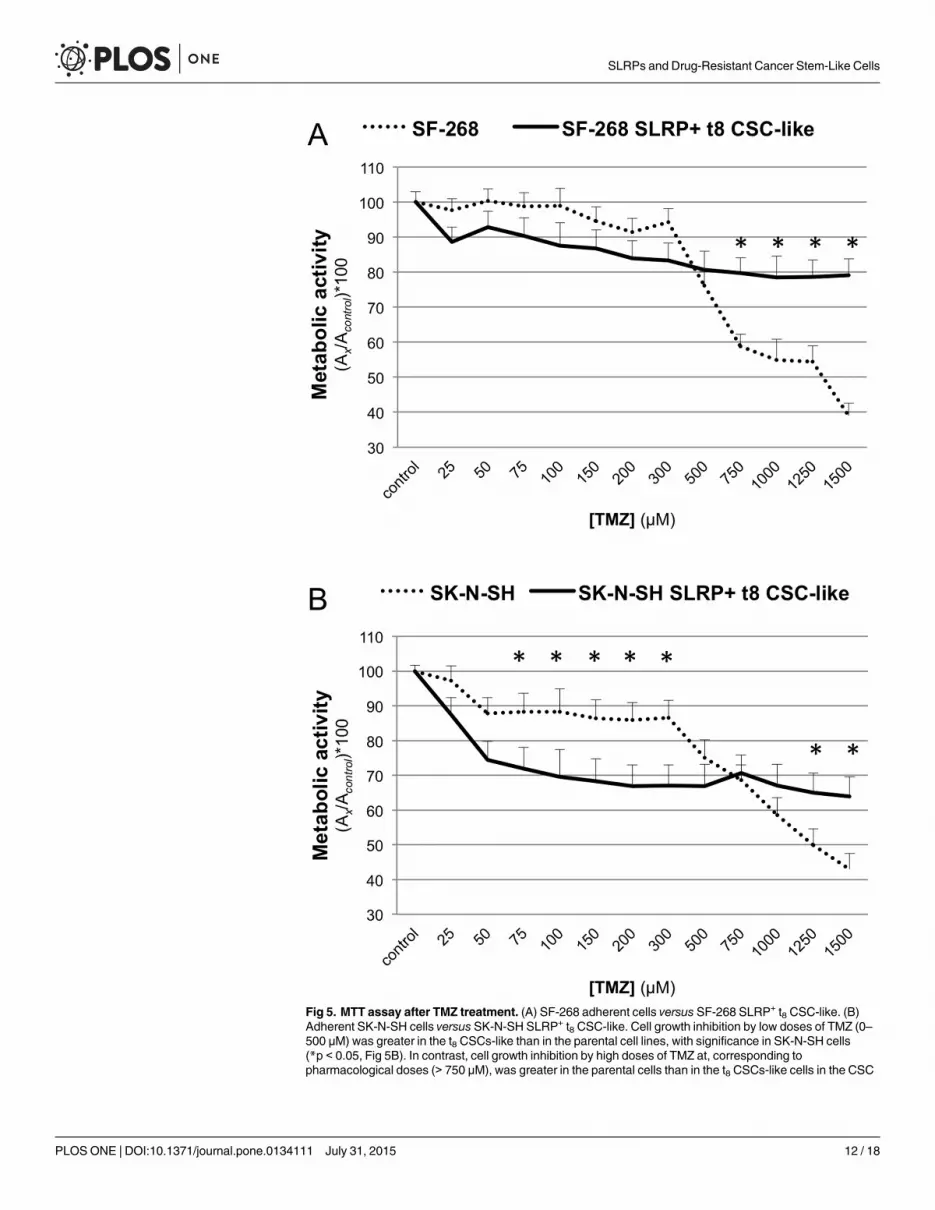
Fig 5. MTT assay after TMZ treatment. (A) SF-268 adherent cells versus SF-268 SLRP+ t8 CSC-like. (B)Adherent SK-N-SH cells versus SK-N-SH SLRP+ t8 CSC-like. Cell growth inhibition by low doses of TMZ (0–500 μM) was greater in the t8 CSCs-like than in the parental cell lines, with significance in SK-N-SH cells(*p < 0.05, Fig 5B). In contrast, cell growth inhibition by high doses of TMZ at, corresponding topharmacological doses (> 750 μM), was greater in the parental cells than in the t8 CSCs-like cells in the CSC
SLRPs and Drug-Resistant Cancer Stem-Like Cells
PLOS ONE | DOI:10.1371/journal.pone.0134111 July 31, 2015 12 / 18

Neurospheres of both cell lines showed heterogeneity in their cell size, ultrastructure, and pro-liferation, confirming the relevance of CSC-like in the maintenance of cell heterogeneity. Ultra-structural imaging of the neurospheres revealed both electron-dense and electron-lucent cells,which implies the presence of cells at different stages of differentiation, with peripheral cellsenriched in endocytic vesicles, probably for vesicle-mediated internalization of DCN by RTK[66], and cell-specific signs of differentiation, particularly dense granules in NB cells and thinmembrane extroflections in GBM cells. The presence of living inner-neurosphere cells afterCSC enrichment for 8 days suggest that only a fraction of the inner cells evade apoptosis ornecrosis in the hypoxic microenvironment of the neurosphere core. Interestingly, CSC-likewith the highest levels of DCN and LUMmRNA, isolated from heterogenic t8 neurospheres,switched towards a quiescent phenotype in the soft-agar-grown secondary neurospheres,showing reduced cell proliferation and apoptosis. The SLRP+ CSC-like still partly grew as loosecell aggregates in the DMEM soft agar-grown secondary cultures, suggesting residual CSC-likeevasion of cell death. Moreover, the SLRP+ CSC-like acquired TMZ resistance, as shown in anMTT-based assay. Despite the higher proliferation rate of the parental cell lines vis-à-vis theslow cycling of the CSC-like at low doses of TMZ, significant resistance to high-dose TMZ wasobserved in the SLRP+ CSC-like of both cell lines, which could be a microenvironment-relatedphenomenon and/or attributable to the overexpression of multidrug-resistance or DNA repairgenes in the CSC-like [78, 79]. On the other hand, the lower cell viability of CSC-like thanparental cells at low TMZ concentrations might be result of underestimation due to the func-tion of the ABC transporters in pumping out of the cells the MTT molecules.
Interestingly, quiescent CSC-like seem to be involved in the EMT and the mesenchymal–epithelial transition (MET) and in cancer dormancy, which have been closely associated withdrug resistance [80]. Our data, showing the anchorage-independent growth, resistance to highconcentrations of TMZ, and lower cell proliferation of t8 CSC-like relative to those of theparental cell lines, suggest that GBM and NB cells can acquire a quiescent stem-like phenotype.These results indicate a putative relation between DCN and cell quiescence which has beenobserved in other cellular models [81], and that SLRPs members, in particular the LUM pro-teoglycan, could play a role in the CSC microenvironment and in cancer dormancy [82]. How-ever, further studies of the pathophysiologic role of DCN and LUM in CSC will be necessary.
The tumor mass is mainly composed of differentiated tumor cells. In metastatic cancer, theMET program fosters epithelial-like cell proliferation of homed circulating tumor cells. Previ-ous studies have shown that DCN inhibits glioma growth and cell differentiation. In contrast,DCN has been reported to play protective and antiapoptotic roles in glioma cell lines exposedto hypoxic microenvironments. Inherent to this study, the formation of 3D neurospheres nec-essarily generates a hypoxic microenvironment in the inner regions, which is why some cellsinside the neurospheres undergo apoptosis/necrosis. It is well known that hypoxia plays a criti-cal role in CSCs and niche maintenance, promoting hypoxia-inducible factor (HIF)-dependentreprogramming of the differentiated tumor cells towards a CSC-like phenotype [83]. Interest-ingly, HIF downstream effectors inhibit NB cell differentiation and were reported to co-localizewith neural crest and stem cell markers in the perivascular niche in NB biopsy samples [84].Here, we report the presence of both suffering and living cells in the hypoxic microenviron-ment of the inner neurosphere, and that the living inner neurosphere cells are resistant toTMZ. Hence, according to the studies abovementioned and the proof-of-concept of the known
enrichments of both cell lines (*p < 0.05, Fig 5A and 5B). DMSO background was subtracted from samplesand control values, and data shown as mean (SD) of [(Asamples—ADMSO) / (Acontrol—ADMSO)]*100 of fourindependent experiments.
doi:10.1371/journal.pone.0134111.g005
SLRPs and Drug-Resistant Cancer Stem-Like Cells
PLOS ONE | DOI:10.1371/journal.pone.0134111 July 31, 2015 13 / 18

SLRP anticancer activity, DCN and LUM could play dual microenvironment-dependent rolesin the maintenance of CSCs, inhibiting the growth of epithelial-like proliferative cells, but con-comitantly promoting the survival and stem-like properties of residual CSC-like, includingTMZ resistance, quiescence, and the maintenance of heterogenic cancer cell phenotypes.Therefore, our data support microenvironment-dependent protective roles for SLRPs in bothGBM and NB CSC-like.
In addition to the known ECM remodeling and soluble factors (TGF-β, tumor necrosis fac-tor α, FGF) and membrane receptors (RTKs, Toll-like receptors 2–4) binding activities, we pro-pose a pivotal role for the SLRP proteoglycans in neurosphere generation, CSC nicheregulation, and the maintenance of a quiescent stem-cell-like phenotype, and consequently incell fate and drug resistance of CSC-like. The SLRP expression patterns in NB CSCs may alsoindicate that the developmental and oncogenic EMT programs are actively cross-linked.
Further functional and clinical studies should clarify the roles of SLRPs in CSC biology andin cancer maintenance. More accurate evaluations of the SLRPs in GBM and NB biopsy speci-mens in terms of the CSC niche are required to determine the clinical potential of SRLPs,which may inspire niche-targeted cancer therapies in the fight against undifferentiated SRLP+
malignant cancers.
AcknowledgmentsThis study was supported by Fundació la Marató TV3, Project n° 111431.
Author ContributionsConceived and designed the experiments: CF JAO RM JP. Performed the experiments: CF JAOPB RO. Analyzed the data: CF JAO CM. Contributed reagents/materials/analysis tools: PAARR GM. Wrote the paper: CF JAO RM JP.
References1. Omuro A, DeAngelis LM. Glioblastoma and other malignant gliomas: a clinical review. JAMA: the jour-
nal of the American Medical Association. 2013; 310(17):1842–50. doi: 10.1001/jama.2013.280319PMID: 24193082.
2. Cheung NK, Dyer MA. Neuroblastoma: developmental biology, cancer genomics and immunotherapy.Nature reviews Cancer. 2013; 13(6):397–411. doi: 10.1038/nrc3526 PMID: 23702928.
3. Gutenberg A, Bock HC, Reifenberger G, BruckW, Giese A. Toxicity and survival in primary glioblas-toma patients treated with concomitant plus adjuvant temozolomide versus adjuvant temozolomide:results of a single-institution, retrospective, matched-pair analysis. Acta neurochirurgica. 2013; 155(3):429–35. doi: 10.1007/s00701-012-1583-y PMID: 23254891.
4. Stupp R, MasonWP, van den Bent MJ, Weller M, Fisher B, Taphoorn MJ, et al. Radiotherapy plus con-comitant and adjuvant temozolomide for glioblastoma. The New England journal of medicine. 2005;352(10):987–96. doi: 10.1056/NEJMoa043330 PMID: 15758009.
5. Oike T, Suzuki Y, Sugawara KI, Shirai K, Noda SE, Tamaki T, et al. Radiotherapy plus ConcomitantAdjuvant Temozolomide for Glioblastoma: Japanese Mono-Institutional Results. PloS one. 2013;8(11):e78943. doi: 10.1371/journal.pone.0078943 PMID: 24265731.
6. Maris JM, Hogarty MD, Bagatell R, Cohn SL. Neuroblastoma. Lancet. 2007; 369(9579):2106–20. doi:10.1016/S0140-6736(07)60983-0 PMID: 17586306.
7. Sottoriva A, Verhoeff JJ, Borovski T, McWeeney SK, Naumov L, Medema JP, et al. Cancer stem celltumor model reveals invasive morphology and increased phenotypical heterogeneity. Cancer research.2010; 70(1):46–56. doi: 10.1158/0008-5472.CAN-09-3663 PMID: 20048071.
8. Bonavia R, Inda MM, CaveneeWK, Furnari FB. Heterogeneity maintenance in glioblastoma: a socialnetwork. Cancer research. 2011; 71(12):4055–60. doi: 10.1158/0008-5472.CAN-11-0153 PMID:21628493; PubMed Central PMCID: PMC3117065.
SLRPs and Drug-Resistant Cancer Stem-Like Cells
PLOS ONE | DOI:10.1371/journal.pone.0134111 July 31, 2015 14 / 18

9. Patel AP, Tirosh I, Trombetta JJ, Shalek AK, Gillespie SM, Wakimoto H, et al. Single-cell RNA-seqhighlights intratumoral heterogeneity in primary glioblastoma. Science. 2014; 344(6190):1396–401.doi: 10.1126/science.1254257 PMID: 24925914.
10. Moore N, Lyle S. Quiescent, Slow-Cycling Stem Cell Populations in Cancer: A Review of the Evidenceand Discussion of Significance. Journal of oncology. 2011; 2011. doi: 10.1155/2011/396076
11. Hua Y, Gorshkov K, Yang Y, WangW, Zhang N, Hughes DP. Slow down to stay alive: HER4 protectsagainst cellular stress and confers chemoresistance in neuroblastoma. Cancer. 2012; 118(20):5140–54. doi: 10.1002/cncr.27496 PMID: 22415601; PubMed Central PMCID: PMC3414637.
12. Schatton T, Frank MH. Antitumor immunity and cancer stem cells. Annals of the New York Academy ofSciences. 2009; 1176:154–69. doi: 10.1111/j.1749-6632.2009.04568.x PMID: 19796244; PubMedCentral PMCID: PMC2893543.
13. Beier D, Schulz JB, Beier CP. Chemoresistance of glioblastoma cancer stem cells—much more com-plex than expected. Molecular cancer. 2011; 10:128. doi: 10.1186/1476-4598-10-128 PMID:21988793; PubMed Central PMCID: PMC3207925.
14. Tsai JH, Donaher JL, Murphy DA, Chau S, Yang J. Spatiotemporal regulation of epithelial-mesenchy-mal transition is essential for squamous cell carcinomametastasis. Cancer cell. 2012; 22(6):725–36.doi: 10.1016/j.ccr.2012.09.022 PMID: 23201165; PubMed Central PMCID: PMC3522773.
15. Chaffer CL, Weinberg RA. A perspective on cancer cell metastasis. Science. 2011; 331(6024):1559–64.doi: 10.1126/science.1203543 PMID: 21436443.
16. Kang Y, Massague J. Epithelial-mesenchymal transitions: twist in development and metastasis. Cell.2004; 118(3):277–9. doi: 10.1016/j.cell.2004.07.011 PMID: 15294153.
17. Hugo H, Ackland ML, Blick T, LawrenceMG, Clements JA, Williams ED, et al. Epithelial—mesenchymalandmesenchymal—epithelial transitions in carcinoma progression. Journal of cellular physiology. 2007;213(2):374–83. doi: 10.1002/jcp.21223 PMID: 17680632.
18. Brizzi MF, Tarone G, Defilippi P. Extracellular matrix, integrins, and growth factors as tailors of the stemcell niche. Current opinion in cell biology. 2012; 24(5):645–51. doi: 10.1016/j.ceb.2012.07.001 PMID:22898530.
19. Watt FM, HuckWT. Role of the extracellular matrix in regulating stem cell fate. Nature reviews Molecu-lar cell biology. 2013; 14(8):467–73. doi: 10.1038/nrm3620 PMID: 23839578.
20. Sailer MH, Gerber A, Tostado C, Hutter G, Cordier D, Mariani L, et al. Non-invasive neural stem cellsbecome invasive in vitro by combined FGF2 and BMP4 signaling. Journal of cell science. 2013; 126(Pt16):3533–40. doi: 10.1242/jcs.125757 PMID: 23788430; PubMed Central PMCID: PMC3744023.
21. Scheel C, Eaton EN, Li SH, Chaffer CL, Reinhardt F, Kah KJ, et al. Paracrine and autocrine signalsinduce and maintain mesenchymal and stem cell states in the breast. Cell. 2011; 145(6):926–40. doi:10.1016/j.cell.2011.04.029 PMID: 21663795; PubMed Central PMCID: PMC3930331.
22. Lu P, Weaver VM, Werb Z. The extracellular matrix: a dynamic niche in cancer progression. The Jour-nal of cell biology. 2012; 196(4):395–406. doi: 10.1083/jcb.201102147 PMID: 22351925; PubMed Cen-tral PMCID: PMC3283993.
23. Morris JPt, Wang SC, Hebrok M. KRAS, Hedgehog, Wnt and the twisted developmental biology of pan-creatic ductal adenocarcinoma. Nature reviews Cancer. 2010; 10(10):683–95. doi: 10.1038/nrc2899PMID: 20814421; PubMed Central PMCID: PMC4085546.
24. Chong HC, Tan CK, Huang RL, Tan NS. Matricellular proteins: a sticky affair with cancers. Journal ofoncology. 2012; 2012:351089. doi: 10.1155/2012/351089 PMID: 22481923; PubMed Central PMCID:PMC3306981.
25. Jogi A, Vallon-Christersson J, Holmquist L, Axelson H, Borg A, Pahlman S. Human neuroblastomacells exposed to hypoxia: induction of genes associated with growth, survival, and aggressive behavior.Experimental cell research. 2004; 295(2):469–87. doi: 10.1016/j.yexcr.2004.01.013 PMID: 15093745.
26. Kajiyama H, Shibata K, Terauchi M, Yamashita M, Ino K, Nawa A, et al. Chemoresistance to paclitaxelinduces epithelial-mesenchymal transition and enhances metastatic potential for epithelial ovarian car-cinoma cells. Int J Oncol. 2007; 31(2):277–83. PMID: 17611683.
27. Prindull G, Zipori D. Environmental guidance of normal and tumor cell plasticity: epithelial mesenchy-mal transitions as a paradigm. Blood. 2004; 103(8):2892–9. doi: 10.1182/blood-2003-08-2807 PMID:15070660.
28. Iozzo RV, Sanderson RD. Proteoglycans in cancer biology, tumour microenvironment and angiogene-sis. Journal of cellular and molecular medicine. 2011; 15(5):1013–31. doi: 10.1111/j.1582-4934.2010.01236.x PMID: 21155971; PubMed Central PMCID: PMC3633488.
29. Theocharis AD, Skandalis SS, Tzanakakis GN, Karamanos NK. Proteoglycans in health and disease:novel roles for proteoglycans in malignancy and their pharmacological targeting. The FEBS journal.2010; 277(19):3904–23. doi: 10.1111/j.1742-4658.2010.07800.x PMID: 20840587.
SLRPs and Drug-Resistant Cancer Stem-Like Cells
PLOS ONE | DOI:10.1371/journal.pone.0134111 July 31, 2015 15 / 18

30. Edwards IJ. Proteoglycans in prostate cancer. Nature reviews Urology. 2012; 9(4):196–206. doi: 10.1038/nrurol.2012.19 PMID: 22349653.
31. Biglari A, Bataille D, Naumann U, Weller M, Zirger J, Castro MG, et al. Effects of ectopic decorin in mod-ulating intracranial glioma progression in vivo, in a rat syngeneic model. Cancer gene therapy. 2004;11(11):721–32. doi: 10.1038/sj.cgt.7700783 PMID: 15475879; PubMed Central PMCID: PMC2902255.
32. Neill T, Schaefer L, Iozzo RV. Decorin: a guardian from the matrix. The American journal of pathology.2012; 181(2):380–7. doi: 10.1016/j.ajpath.2012.04.029 PMID: 22735579; PubMed Central PMCID:PMC3409438.
33. Sofeu Feugaing DD, Gotte M, Viola M. More than matrix: the multifaceted role of decorin in cancer.European journal of cell biology. 2013; 92(1):1–11. doi: 10.1016/j.ejcb.2012.08.004 PMID: 23058688.
34. Engel S, Isenmann S, Stander M, Rieger J, Bahr M, Weller M. Inhibition of experimental rat gliomagrowth by decorin gene transfer is associated with decreased microglial infiltration. Journal of neuroim-munology. 1999; 99(1):13–8. PMID: 10496172.
35. El Behi M, Krumeich S, Lodillinsky C, Kamoun A, Tibaldi L, Sugano G, et al. An essential role for dec-orin in bladder cancer invasiveness. EMBOmolecular medicine. 2013; 5(12):1835–51. doi: 10.1002/emmm.201302655 PMID: 24142880; PubMed Central PMCID: PMC3914526.
36. Santra M, Katakowski M, Zhang RL, Zhang ZG, Meng H, Jiang F, et al. Protection of adult mouse pro-genitor cells and human glioma cells by de novo decorin expression in an oxygen- and glucose-deprived cell culture model system. Journal of cerebral blood flow and metabolism: official journal of theInternational Society of Cerebral Blood Flow and Metabolism. 2006; 26(10):1311–22. doi: 10.1038/sj.jcbfm.9600285 PMID: 16467781.
37. Dil N, Banerjee AG. A role for aberrantly expressed nuclear localized decorin in migration and invasionof dysplastic and malignant oral epithelial cells. Head & neck oncology. 2011; 3:44. doi: 10.1186/1758-3284-3-44 PMID: 21958730; PubMed Central PMCID: PMC3198745.
38. Dil N, Banerjee AG. Knockdown of aberrantly expressed nuclear localized decorin attenuates tumourangiogenesis related mediators in oral cancer progression model in vitro. Head & neck oncology. 2012;4:11. doi: 10.1186/1758-3284-4-11 PMID: 22507529; PubMed Central PMCID: PMC3370992.
39. Zafiropoulos A, Nikitovic D, Katonis P, Tsatsakis A, Karamanos NK, Tzanakakis GN. Decorin-inducedgrowth inhibition is overcome through protracted expression and activation of epidermal growth factorreceptors in osteosarcoma cells. Molecular cancer research: MCR. 2008; 6(5):785–94. doi: 10.1158/1541-7786.MCR-07-0165 PMID: 18505923.
40. Kasamatsu A, Uzawa K, Minakawa Y, Ishige S, Kasama H, Endo-Sakamoto Y, et al. Decorin in humanoral cancer: A promising predictive biomarker of S-1 neoadjuvant chemosensitivity. Biochemical andbiophysical research communications. 2015; 457(1):71–6. doi: 10.1016/j.bbrc.2014.12.093 PMID:25550184.
41. PopeWB, Mirsadraei L, Lai A, Eskin A, Qiao J, Kim HJ, et al. Differential gene expression in glioblas-toma defined by ADC histogram analysis: relationship to extracellular matrix molecules and survival.AJNR American journal of neuroradiology. 2012; 33(6):1059–64. doi: 10.3174/ajnr.A2917 PMID:22268080.
42. Yang ZX, Lu CY, Yang YL, Dou KF, Tao KS. Lumican Expression in Pancreatic Ductal Adenocarci-noma. Hepato-gastroenterology. 2012; 60(122):349–53. doi: 10.5754/hge12642 PMID: 22951524.
43. Nikitovic D, Chalkiadaki G, Berdiaki A, Aggelidakis J, Katonis P, Karamanos NK, et al. Lumican regu-lates osteosarcoma cell adhesion by modulating TGFbeta2 activity. The international journal of bio-chemistry & cell biology. 2011; 43(6):928–35. doi: 10.1016/j.biocel.2011.03.008 PMID: 21421073.
44. Yamano Y, Uzawa K, Saito K, Nakashima D, Kasamatsu A, Koike H, et al. Identification of cisplatin-resistance related genes in head and neck squamous cell carcinoma. International journal of cancerJournal international du cancer. 2010; 126(2):437–49. doi: 10.1002/ijc.24704 PMID: 19569180.
45. Zagris N, Gilipathi K, Soulintzi N, Konstantopoulos K. Decorin developmental expression and functionin the early avian embryo. The International journal of developmental biology. 2011; 55(6):633–9. doi:10.1387/ijdb.113321nz PMID: 21948712.
46. Kallmann BA, Wagner S, Hummel V, Buttmann M, Bayas A, Tonn JC, et al. Characteristic gene expres-sion profile of primary human cerebral endothelial cells. FASEB journal: official publication of the Feder-ation of American Societies for Experimental Biology. 2002; 16(6):589–91. PMID: 11919163.
47. Daquinag AC, Zhang Y, Amaya-Manzanares F, Simmons PJ, Kolonin MG. An isoform of decorin is aresistin receptor on the surface of adipose progenitor cells. Cell stem cell. 2011; 9(1):74–86. doi: 10.1016/j.stem.2011.05.017 PMID: 21683670.
48. Oe T, Sasayama T, Nagashima T, Muramoto M, Yamazaki T, Morikawa N, et al. Differences in geneexpression profile among SH-SY5Y neuroblastoma subclones with different neurite outgrowthresponses to nerve growth factor. Journal of neurochemistry. 2005; 94(5):1264–76. doi: 10.1111/j.1471-4159.2005.03273.x PMID: 15992370.
SLRPs and Drug-Resistant Cancer Stem-Like Cells
PLOS ONE | DOI:10.1371/journal.pone.0134111 July 31, 2015 16 / 18

49. Barkho BZ, Song H, Aimone JB, Smrt RD, Kuwabara T, Nakashima K, et al. Identification of astrocyte-expressed factors that modulate neural stem/progenitor cell differentiation. Stem cells and develop-ment. 2006; 15(3):407–21. doi: 10.1089/scd.2006.15.407 PMID: 16846377; PubMed Central PMCID:PMC2777811.
50. Scholzen T, Solursh M, Suzuki S, Reiter R, Morgan JL, Buchberg AM, et al. The murine decorin. Com-plete cDNA cloning, genomic organization, chromosomal assignment, and expression during organo-genesis and tissue differentiation. The Journal of biological chemistry. 1994; 269(45):28270–81. PMID:7961765.
51. Iozzo RV, Schaefer L. Proteoglycans in health and disease: novel regulatory signaling mechanismsevoked by the small leucine-rich proteoglycans. The FEBS journal. 2010; 277(19):3864–75. doi: 10.1111/j.1742-4658.2010.07797.x PMID: 20840584; PubMed Central PMCID: PMC3000440.
52. Stichel CC, Kappler J, Junghans U, Koops A, Kresse H, Muller HW. Differential expression of the smallchondroitin/dermatan sulfate proteoglycans decorin and biglycan after injury of the adult rat brain. Brainresearch. 1995; 704(2):263–74. PMID: 8788923.
53. Chaichana K, Zamora-Berridi G, Camara-Quintana J, Quinones-Hinojosa A. Neurosphere assays:growth factors and hormone differences in tumor and nontumor studies. Stem cells. 2006; 24(12):2851–7. doi: 10.1634/stemcells.2006-0399 PMID: 16945995.
54. Galli R. The neurosphere assay applied to neural stem cells and cancer stem cells. Methods in molecu-lar biology. 2013; 986:267–77. doi: 10.1007/978-1-62703-311-4_17 PMID: 23436418.
55. Asara Y, Marchal JA, Carrasco E, Boulaiz H, Solinas G, Bandiera P, et al. Cadmiummodifies the cellcycle and apoptotic profiles of human breast cancer cells treated with 5-fluorouracil. International jour-nal of molecular sciences. 2013; 14(8):16600–16. doi: 10.3390/ijms140816600 PMID: 23941782;PubMed Central PMCID: PMC3759927.
56. Puglisi MA, Tesori V, Lattanzi W, Gasbarrini GB, Gasbarrini A. Colon cancer stem cells: controversiesand perspectives. World journal of gastroenterology: WJG. 2013; 19(20):2997–3006. doi: 10.3748/wjg.v19.i20.2997 PMID: 23716979; PubMed Central PMCID: PMC3662939.
57. Gammon L, Biddle A, Heywood HK, Johannessen AC, Mackenzie IC. Sub-sets of cancer stem cells dif-fer intrinsically in their patterns of oxygen metabolism. PloS one. 2013; 8(4):e62493. doi: 10.1371/journal.pone.0062493 PMID: 23638097; PubMed Central PMCID: PMC3640080.
58. Walton JD, Kattan DR, Thomas SK, Spengler BA, Guo HF, Biedler JL, et al. Characteristics of stemcells from human neuroblastoma cell lines and in tumors. Neoplasia. 2004; 6(6):838–45. doi: 10.1593/neo.04310 PMID: 15720811; PubMed Central PMCID: PMC1531688.
59. Mazzoleni S, Politi LS, Pala M, Cominelli M, Franzin A, Sergi Sergi L, et al. Epidermal growth factorreceptor expression identifies functionally and molecularly distinct tumor-initiating cells in human glio-blastomamultiforme and is required for gliomagenesis. Cancer research. 2010; 70(19):7500–13. doi:10.1158/0008-5472.CAN-10-2353 PMID: 20858720.
60. Junttila MR, de Sauvage FJ. Influence of tumour micro-environment heterogeneity on therapeuticresponse. Nature. 2013; 501(7467):346–54. doi: 10.1038/nature12626 PMID: 24048067.
61. Leygue E, Snell L, Dotzlaw H, Hole K, Hiller-Hitchcock T, Roughley PJ, et al. Expression of lumican inhuman breast carcinoma. Cancer research. 1998; 58(7):1348–52. PMID: 9537227.
62. Yamamoto T, Matsuda Y, Kawahara K, Ishiwata T, Naito Z. Secreted 70kDa lumican stimulates growthand inhibits invasion of human pancreatic cancer. Cancer letters. 2012; 320(1):31–9. doi: 10.1016/j.canlet.2012.01.023 PMID: 22266188.
63. Seya T, Tanaka N, Shinji S, Yokoi K, Koizumi M, Teranishi N, et al. Lumican expression in advancedcolorectal cancer with nodal metastasis correlates with poor prognosis. Oncology reports. 2006; 16(6):1225–30. PMID: 17089042.
64. Naito Z, Ishiwata T, Kurban G, Teduka K, Kawamoto Y, Kawahara K, et al. Expression and accumula-tion of lumican protein in uterine cervical cancer cells at the periphery of cancer nests. Int J Oncol.2002; 20(5):943–8. PMID: 11956587.
65. Matsuda Y, Yamamoto T, Kudo M, Kawahara K, Kawamoto M, Nakajima Y, et al. Expression and rolesof lumican in lung adenocarcinoma and squamous cell carcinoma. Int J Oncol. 2008; 33(6):1177–85.PMID: 19020750.
66. Feugaing DD, Tammi R, Echtermeyer FG, Stenmark H, Kresse H, Smollich M, et al. Endocytosis of thedermatan sulfate proteoglycan decorin utilizes multiple pathways and is modulated by epidermalgrowth factor receptor signaling. Biochimie. 2007; 89(5):637–57. doi: 10.1016/j.biochi.2006.12.012PMID: 17335953.
67. Van Bockstal M, Lambein K, Van Gele M, De Vlieghere E, Limame R, Braems G, et al. Differential regu-lation of extracellular matrix protein expression in carcinoma-associated fibroblasts by TGF-beta1 regu-lates cancer cell spreading but not adhesion. Oncoscience. 2014; 1(10):634–48. PMID: 25593993;PubMed Central PMCID: PMC4278277.
SLRPs and Drug-Resistant Cancer Stem-Like Cells
PLOS ONE | DOI:10.1371/journal.pone.0134111 July 31, 2015 17 / 18

68. Reynolds BA, Weiss S. Clonal and population analyses demonstrate that an EGF-responsive mamma-lian embryonic CNS precursor is a stem cell. Developmental biology. 1996; 175(1):1–13. doi: 10.1006/dbio.1996.0090 PMID: 8608856.
69. Reynolds BA, Weiss S. Generation of neurons and astrocytes from isolated cells of the adult mamma-lian central nervous system. Science. 1992; 255(5052):1707–10. PMID: 1553558.
70. Reynolds BA, Tetzlaff W, Weiss S. A multipotent EGF-responsive striatal embryonic progenitor cell pro-duces neurons and astrocytes. The Journal of neuroscience: the official journal of the Society for Neu-roscience. 1992; 12(11):4565–74. PMID: 1432110.
71. Lee J, Kotliarova S, Kotliarov Y, Li A, Su Q, Donin NM, et al. Tumor stem cells derived from glioblasto-mas cultured in bFGF and EGFmore closely mirror the phenotype and genotype of primary tumorsthan do serum-cultured cell lines. Cancer cell. 2006; 9(5):391–403. doi: 10.1016/j.ccr.2006.03.030PMID: 16697959.
72. Hemmati HD, Nakano I, Lazareff JA, Masterman-Smith M, Geschwind DH, Bronner-Fraser M, et al.Cancerous stem cells can arise from pediatric brain tumors. Proceedings of the National Academy ofSciences of the United States of America. 2003; 100(25):15178–83. doi: 10.1073/pnas.2036535100PMID: 14645703; PubMed Central PMCID: PMC299944.
73. Galli R, Binda E, Orfanelli U, Cipelletti B, Gritti A, De Vitis S, et al. Isolation and characterization oftumorigenic, stem-like neural precursors from human glioblastoma. Cancer research. 2004; 64(19):7011–21. doi: 10.1158/0008-5472.CAN-04-1364 PMID: 15466194.
74. Singh SK, Hawkins C, Clarke ID, Squire JA, Bayani J, Hide T, et al. Identification of human brain tumourinitiating cells. Nature. 2004; 432(7015):396–401. doi: 10.1038/nature03128 PMID: 15549107.
75. Singh SK, Clarke ID, Terasaki M, Bonn VE, Hawkins C, Squire J, et al. Identification of a cancer stemcell in human brain tumors. Cancer research. 2003; 63(18):5821–8. PMID: 14522905.
76. Qiang L, Yang Y, Ma YJ, Chen FH, Zhang LB, Liu W, et al. Isolation and characterization of cancerstem like cells in human glioblastoma cell lines. Cancer letters. 2009; 279(1):13–21. doi: 10.1016/j.canlet.2009.01.016 PMID: 19232461.
77. Shi MF, Jiao J, Lu WG, Ye F, Ma D, Dong QG, et al. Identification of cancer stem cell-like cells fromhuman epithelial ovarian carcinoma cell line. Cellular and molecular life sciences: CMLS. 2010; 67(22):3915–25. doi: 10.1007/s00018-010-0420-9 PMID: 20549538.
78. Persano L, Pistollato F, Rampazzo E, Della Puppa A, Abbadi S, Frasson C, et al. BMP2 sensitizes glio-blastoma stem-like cells to Temozolomide by affecting HIF-1alpha stability and MGMT expression. Celldeath & disease. 2012; 3:e412. doi: 10.1038/cddis.2012.153 PMID: 23076220; PubMed CentralPMCID: PMC3481140.
79. Qiu ZK, Shen D, Chen YS, Yang QY, Guo CC, Feng BH, et al. Enhanced MGMT expression contributesto temozolomide resistance in glioma stem-like cells. Chinese journal of cancer. 2014; 33(2):115–22.doi: 10.5732/cjc.012.10236 PMID: 23958055; PubMed Central PMCID: PMC3935013.
80. Shekhani MT, Jayanthy AS, Maddodi N, Setaluri V. Cancer stem cells and tumor transdifferentiation:implications for novel therapeutic strategies. American journal of stem cells. 2013; 2(1):52–61. PMID:23671816; PubMed Central PMCID: PMC3636725.
81. Mauviel A, Santra M, Chen YQ, Uitto J, Iozzo RV. Transcriptional regulation of decorin gene expres-sion. Induction by quiescence and repression by tumor necrosis factor-alpha. The Journal of biologicalchemistry. 1995; 270(19):11692–700. PMID: 7744809.
82. Bleau AM, Agliano A, Larzabal L, de Aberasturi AL, Calvo A. Metastatic dormancy: a complex networkbetween cancer stem cells and their microenvironment. Histology and histopathology. 2014; 29(12):1499–510. PMID: 24887025.
83. Heddleston JM, Li Z, McLendon RE, Hjelmeland AB, Rich JN. The hypoxic microenvironment main-tains glioblastoma stem cells and promotes reprogramming towards a cancer stem cell phenotype. Cellcycle. 2009; 8(20):3274–84. PMID: 19770585; PubMed Central PMCID: PMC2825672.
84. Pietras A, Gisselsson D, Ora I, Noguera R, Beckman S, Navarro S, et al. High levels of HIF-2alphahighlight an immature neural crest-like neuroblastoma cell cohort located in a perivascular niche. TheJournal of pathology. 2008; 214(4):482–8. doi: 10.1002/path.2304 PMID: 18189331.
SLRPs and Drug-Resistant Cancer Stem-Like Cells
PLOS ONE | DOI:10.1371/journal.pone.0134111 July 31, 2015 18 / 18