expression of decorin, biglycan, and collagen type i in human renal fibrosing disease
TRANSCRIPT
Kidney International, Vol. 57 (2000), pp. 487–498
Expression of decorin, biglycan, and collagen type I inhuman renal fibrosing disease
MICHAEL B. STOKES, SUSANN HOLLER, YAN CUI, KELLY L. HUDKINS, FRANK EITNER,AGNES FOGO, and CHARLES E. ALPERS
Department of Pathology, University of Washington Medical Center, Seattle, Washington, and Department of Pathology,Vanderbilt University Medical Center, Nashville, Tennessee, USA
Expression of decorin, biglycan, and collagen type I in human Extracellular matrix accumulation in mesangial areasrenal fibrosing disease. may contribute to impaired glomerular function in diverse
Background. The extracellular matrix proteoglycans de- human renal diseases, including amyloidosis, diabetic ne-corin and biglycan may have a pathogenic role in renal fibrosingphropathy, light-chain deposition disease (LCDD), fi-disease via regulation of the activity of growth factors, such asbrillary glomerulonephritis, immunotactoid glomerulo-transforming growth factor-b, and effects on collagen type I
fibrillogenesis. The expression of decorin and biglycan in hu- pathy, and idiopathic mesangial sclerosis. Proteoglycansman glomerular diseases characterized by mesangial sclerosis are major components of the extracellular matrix thatis unknown. have diverse biologic functions, including binding andMethods. Decorin, biglycan, and collagen type I were local-
inactivation of growth factors, including basic fibroblastized immunohistochemically in human renal biopsy cases ofamyloidosis (N 5 18), diabetic nephropathy (N 5 11), fibrillary growth factor (FGF-2) and transforming growth factor-bglomerulonephritis (N 5 5), immunotactoid glomerulopathy (TGF-b), and regulation of collagen fibrillogenesis [1, 2].(N 5 5), light-chain deposition disease (N 5 4), idiopathic Decorin and biglycan are members of the family of small,mesangial sclerosis (N 5 4), and nephrosclerosis (N 5 6),
leucine-rich proteoglycans that may be particularly rele-and in morphologically normal tissues obtained from tumorvant to the pathogenesis of renal fibrosing injury [3]. Innephrectomies (N 5 8). Decorin and biglycan mRNA synthesis
was evaluated by in situ hybridization. vitro studies have indicated that the synthesis of decorinResults. Decorin and biglycan protein were not identified in and biglycan by fibroblasts, mesangial cells, and smooth
normal glomeruli. Decorin accumulated in amyloid deposits, muscle cells may be regulated by TGF-b and that decorinbut not in deposits of fibrillary glomerulonephritis or immuno-may bind and inactivate TGF-b [1, 2]. Further supporttactoid glomerulopathy. Biglycan weakly accumulated in amy-
loid deposits, and both decorin and biglycan weakly stained for a role for decorin as a negative regulator of TGF-bmesangial nodules in cases of morphologically advanced light- activity comes from in vivo observations in the anti-Thychain deposition disease and diabetic nephropathy. In all ana- 1 rat model of mesangioproliferative glomerulonephritis,lyzed cases, irrespective of the underlying disease, decorin and
in which the administration of exogenous decorin [4], orbiglycan accumulated in glomeruli in areas of fibrous organiza-decorin gene therapy [5], prevented the extracellulartion of the urinary space and in areas of tubulointerstitial fibro-
sis. Biglycan, but not decorin, accumulated in the neointima matrix accumulation that has been attributed to the ac-of arteriosclerotic blood vessels. Decorin and biglycan mRNA tion of TGF-b. Border and Noble have proposed that asynthesis was detected at sites of proteoglycan accumulation relative “deficiency” of decorin may contribute to TGF-in glomeruli, interstitium, and neointima. Collagen type I colo-
b–mediated renal injury [6]. The functions of biglycancalized with decorin and biglycan deposits.Conclusions. Differences in extracellular matrix proteogly- are largely unknown. Besides possible roles in regulating
can composition may be diagnostically useful in distinguishing TGF-b activity and collagen fibril formation, it may altermorphologically similar diseases. Distinct patterns of proteo- the bioavailability of decorin by competing for a sharedglycan expression may be related to modulation of specific
re-uptake receptor [7]. Determining the expression ofgrowth factor activity in different glomerular diseases.decorin and biglycan in human renal disease could pro-vide insights into pathogenesis and could possibly lead
Key words: mesangial sclerosis, proteoglycans, amyloidosis, growthto the design of novel therapeutic strategies.factor, extracellular matrix, glomerulosclerosis, arteriosclerosis, tubu-
lointerstitial fibrosis. In experimental renal injury, the expression of decorinand biglycan has been localized to glomerulosclerosisReceived for publication May 10, 1999lesions and tubulointerstitial fibrosis [7–9]. Decorin, bi-and in revised form August 9, 1999
Accepted for publication September 9, 1999 glycan, and their shared endocytosis receptor have beendetected in glomerulosclerosis lesions in diseased rat kid- 2000 by the International Society of Nephrology
487
Stokes et al: Proteoglycans in human renal disease488
ney [7]. In human kidney, decorin has been immunolocal- Antibodiesized to areas of interstitial fibrosis, but not in glomeruli Polyclonal rabbit antisera LF-51, LF-30/136, and LF-67[10–12]. Biglycan has been localized in glomeruli and (to human decorin, human biglycan, and human collagencollecting duct epithelial cells in developing human kid- type I, respectively) were kind gifts of Dr. Larry Fisherney [10]. However, the distribution of biglycan in adult (National Institute of Dental Research, Bethesda, MD,human kidney has not previously been determined. A USA). LF-30/136 was generated against a synthetic pep-key role for decorin has been postulated in regulating tide corresponding to amino acids 5 to 17 of the corecollagen fibrillogenesis, as demonstrated by the finding protein of human PGII/decorin, conjugated to keyholeof abnormal collagen fibrils in decorin-deficient mice limpet hemocyanin [19]. LF-51 was generated against a[13]. Both decorin and biglycan interact with collagen synthetic peptide corresponding to amino acids 11 totype I in vitro, and codeposition of these molecules has 25 of the core protein of the secreted form of humanbeen described in various models of fibrosing injury bone PGI/biglycan, conjugated to bovine serum albumin[14, 15]. (BSA) [19]. LF-67 was generated against 26 amino acids
In this study, we examined the expression of decorin, of a synthetic C telopeptide antigen of the human colla-biglycan, and collagen type I in human renal tissues dem- gen a I (I) chain [20]. The specificities of these antiseraonstrating a spectrum of glomerulopathies and tubuloin- have been previously confirmed in Western blot andterstitial fibrosis. Glomerular diseases characterized by immunoprecipitation experiments [15, 19–21].accumulations of extracellular matrix (mesangial sclero-sis) were selected to test the hypotheses (a) that decorin Immunohistochemistryand biglycan are up-regulated in glomerular sclerosing Sections were deparaffinized in xylene and rehydratedinjury, and (b) that differences in extracellular matrix pro- in a descending ethanol series. Endogenous peroxidaseteoglycan composition can be used to distinguish mor- was quenched with 3% H2O2 for 10 minutes. For LF-phologically similar diseases. 30/136 and LF-51 staining, sections were digested with
chondroitinase ABC lyase (ICN Biomedicals, CostaMesa, CA, USA) at 250 mU/mL in 0.1 mol/L Tris, 0.01%METHODSBSA, for 60 minutes at 378C, followed by blocking withTissue selection and pathologic examinationnormal goat serum for 10 minutes. For collagen type IArchived paraffin-embedded diagnostic renal biopsystaining, sections were digested in proteinase K (5 mg/mLtissues from cases of amyloidosis (N 5 18), diabetic ne-at 378C for 15 min; Sigma, St. Louis, MO, USA). Sectionsphropathy (N 5 11), fibrillary glomerulonephritis (N 5 5),were incubated with primary antibody and diluted in 1%immunotactoid glomerulopathy (N 5 5), LCDD (N 5 4),BSA/phosphate-buffered saline (PBS) for one hour atnephrosclerosis (N 5 6), and idiopathic mesangial scle-room temperature (LF-36/130, 1:250; LF-51 and LF-67,rosis (N 5 4) were selected for study. All cases were1:500), followed by a biotinylated goat antirabbit anti-characterized by light microscopy, immunofluorescencebody (Vector, Burlingame, CA, USA), ABC-Elite re-microscopy, and electron microscopy, using standardagent (Vector). The reaction product was visualized withtechniques [16]. Clinical and pathologic data pertaining3,39 diaminobenzidine (Sigma) and nickel chloride en-to the cases of immuntactoid glomerulopathy have beenhancement. The slides were counterstained with methyldescribed in detail elsewhere [17]. Amyloid depositsgreen. Negative controls for immunohistochemistry in-were identified by green/red birefringent Congo Redcluded substitution of the primary antibody with equalstaining under polarized light. Immunohistochemicalamounts of an irrelevant rabbit IgG (Dako).staining of Congo Red material with a monoclonal anti-
body to amyloid A (AA) protein (Dako, Carpinteria,Molecular probesCA, USA) were positive in one biopsy. All other cases
Two pBluescript SK plasmids containing either humanof amyloidosis were derived from k or l light chainsbone decorin cDNA (plasmid P2) or human bone bigly-(AL), as demonstrated by routine immunofluorescencecan cDNA (plasmid P16) were generous gifts of Dr.microscopic studies. Idiopathic mesangial sclerosis wasLarry Fisher (National Institute of Dental Research).diagnosed by exclusion of the other causes of mesangialPlasmid P16 contains a 1658 bp insert with the completesclerosis listed previously in this article [18]. Macroscopi-protein encoding sequence of the human biglycan genecally normal-appearing renal tissues were obtained from[21]. Plamid P2 contains a 1.6 kb insert with the proteinnephrectomies (N 5 8) for localized renal carcinoma atencoding sequence of the human decorin gene [21]. Thesites away from the tumor. Renal biopsy tissues wereplasmids were linearized with Xba I and Kpn I (P16)fixed in 10% neutral-buffered formalin. Portions of ne-or BamH I and Kpn I (P2) and transcribed into bothphrectomy tissues were fixed in both 10% neutral-buf-antisense and sense (negative control) riboprobes in T3-fered formalin and in methyl Carnoy’s fixative (60%
methanol, 30% chloroform, and 10% acetic acid). or T7-primed reactions [21], using reagents from Pro-
Stokes et al: Proteoglycans in human renal disease 489
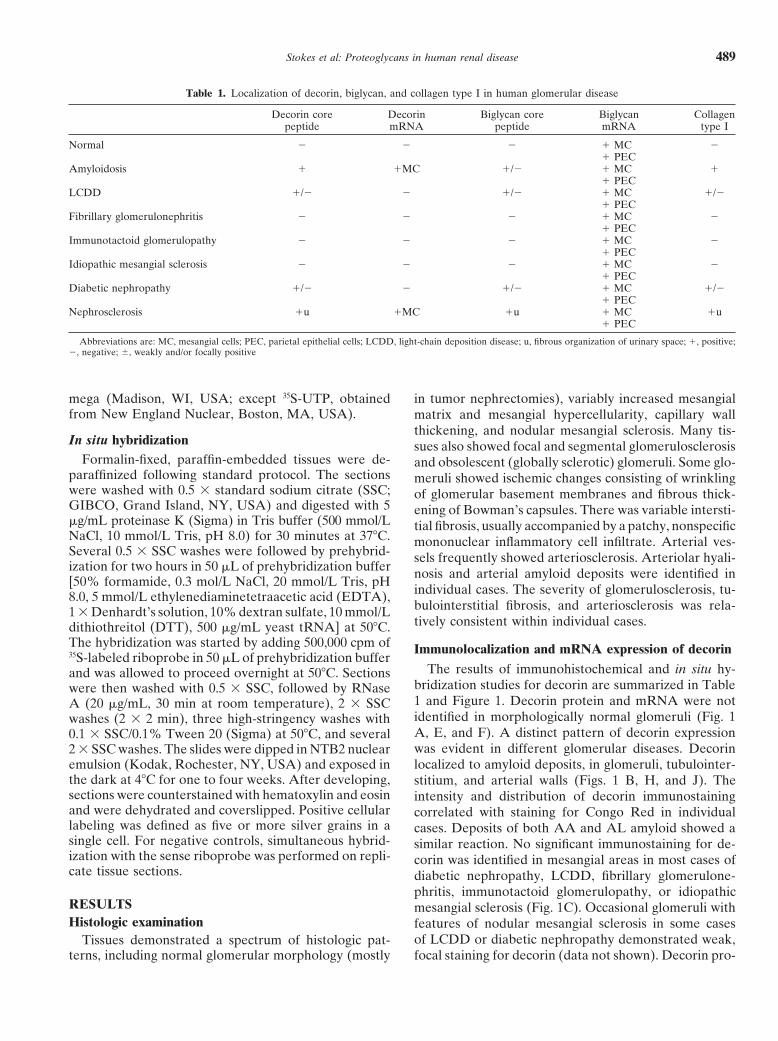
Table 1. Localization of decorin, biglycan, and collagen type I in human glomerular disease
Decorin core Decorin Biglycan core Biglycan Collagenpeptide mRNA peptide mRNA type I
Normal 2 2 2 1 MC 21 PEC
Amyloidosis 1 1MC 1/2 1 MC 11 PEC
LCDD 1/2 2 1/2 1 MC 1/21 PEC
Fibrillary glomerulonephritis 2 2 2 1 MC 21 PEC
Immunotactoid glomerulopathy 2 2 2 1 MC 21 PEC
Idiopathic mesangial sclerosis 2 2 2 1 MC 21 PEC
Diabetic nephropathy 1/2 2 1/2 1 MC 1/21 PEC
Nephrosclerosis 1u 1MC 1u 1 MC 1u1 PEC
Abbreviations are: MC, mesangial cells; PEC, parietal epithelial cells; LCDD, light-chain deposition disease; u, fibrous organization of urinary space; 1, positive;2, negative; 6, weakly and/or focally positive
mega (Madison, WI, USA; except 35S-UTP, obtained in tumor nephrectomies), variably increased mesangialfrom New England Nuclear, Boston, MA, USA). matrix and mesangial hypercellularity, capillary wall
thickening, and nodular mesangial sclerosis. Many tis-In situ hybridization sues also showed focal and segmental glomerulosclerosis
Formalin-fixed, paraffin-embedded tissues were de- and obsolescent (globally sclerotic) glomeruli. Some glo-paraffinized following standard protocol. The sections meruli showed ischemic changes consisting of wrinklingwere washed with 0.5 3 standard sodium citrate (SSC; of glomerular basement membranes and fibrous thick-GIBCO, Grand Island, NY, USA) and digested with 5 ening of Bowman’s capsules. There was variable intersti-mg/mL proteinase K (Sigma) in Tris buffer (500 mmol/L tial fibrosis, usually accompanied by a patchy, nonspecificNaCl, 10 mmol/L Tris, pH 8.0) for 30 minutes at 378C. mononuclear inflammatory cell infiltrate. Arterial ves-Several 0.5 3 SSC washes were followed by prehybrid- sels frequently showed arteriosclerosis. Arteriolar hyali-ization for two hours in 50 mL of prehybridization buffer nosis and arterial amyloid deposits were identified in[50% formamide, 0.3 mol/L NaCl, 20 mmol/L Tris, pH
individual cases. The severity of glomerulosclerosis, tu-8.0, 5 mmol/L ethylenediaminetetraacetic acid (EDTA),bulointerstitial fibrosis, and arteriosclerosis was rela-1 3 Denhardt’s solution, 10% dextran sulfate, 10 mmol/Ltively consistent within individual cases.dithiothreitol (DTT), 500 mg/mL yeast tRNA] at 508C.
The hybridization was started by adding 500,000 cpm of Immunolocalization and mRNA expression of decorin35S-labeled riboprobe in 50 mL of prehybridization bufferThe results of immunohistochemical and in situ hy-and was allowed to proceed overnight at 508C. Sections
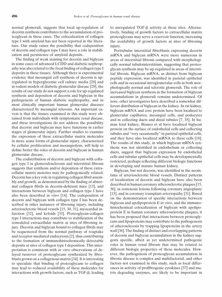
bridization studies for decorin are summarized in Tablewere then washed with 0.5 3 SSC, followed by RNase1 and Figure 1. Decorin protein and mRNA were notA (20 mg/mL, 30 min at room temperature), 2 3 SSCidentified in morphologically normal glomeruli (Fig. 1washes (2 3 2 min), three high-stringency washes withA, E, and F). A distinct pattern of decorin expression0.1 3 SSC/0.1% Tween 20 (Sigma) at 508C, and severalwas evident in different glomerular diseases. Decorin2 3 SSC washes. The slides were dipped in NTB2 nuclear
emulsion (Kodak, Rochester, NY, USA) and exposed in localized to amyloid deposits, in glomeruli, tubulointer-the dark at 48C for one to four weeks. After developing, stitium, and arterial walls (Figs. 1 B, H, and J). Thesections were counterstained with hematoxylin and eosin intensity and distribution of decorin immunostainingand were dehydrated and coverslipped. Positive cellular correlated with staining for Congo Red in individuallabeling was defined as five or more silver grains in a cases. Deposits of both AA and AL amyloid showed asingle cell. For negative controls, simultaneous hybrid- similar reaction. No significant immunostaining for de-ization with the sense riboprobe was performed on repli- corin was identified in mesangial areas in most cases ofcate tissue sections. diabetic nephropathy, LCDD, fibrillary glomerulone-
phritis, immunotactoid glomerulopathy, or idiopathicRESULTS mesangial sclerosis (Fig. 1C). Occasional glomeruli withHistologic examination features of nodular mesangial sclerosis in some cases
of LCDD or diabetic nephropathy demonstrated weak,Tissues demonstrated a spectrum of histologic pat-terns, including normal glomerular morphology (mostly focal staining for decorin (data not shown). Decorin pro-
Stokes et al: Proteoglycans in human renal disease490
Stokes et al: Proteoglycans in human renal disease 491
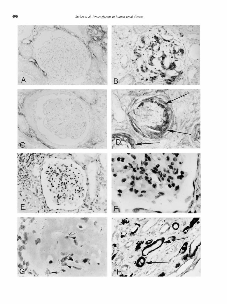
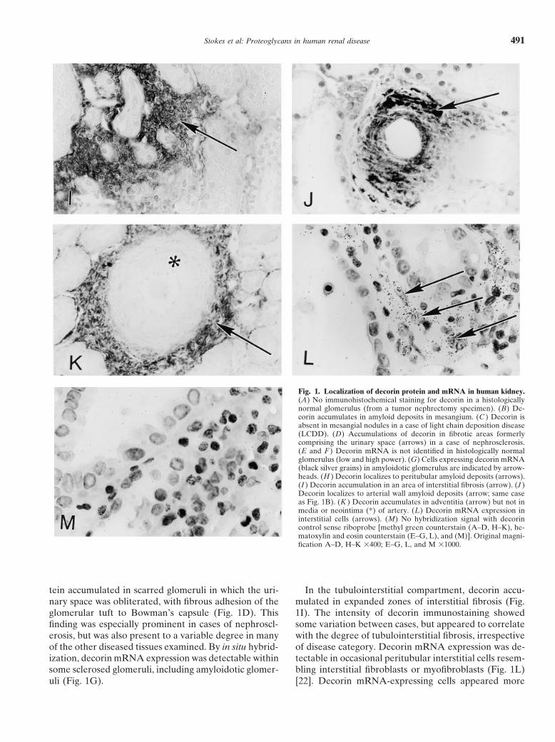
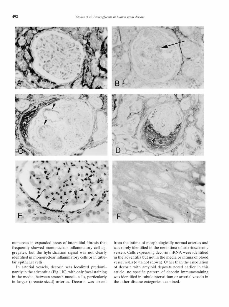
Fig. 1. Localization of decorin protein and mRNA in human kidney.(A) No immunohistochemical staining for decorin in a histologicallynormal glomerulus (from a tumor nephrectomy specimen). (B) De-corin accumulates in amyloid deposits in mesangium. (C ) Decorin isabsent in mesangial nodules in a case of light chain deposition disease(LCDD). (D) Accumulations of decorin in fibrotic areas formerlycomprising the urinary space (arrows) in a case of nephrosclerosis.(E and F ) Decorin mRNA is not identified in histologically normalglomerulus (low and high power). (G) Cells expressing decorin mRNA(black silver grains) in amyloidotic glomerulus are indicated by arrow-heads. (H ) Decorin localizes to peritubular amyloid deposits (arrows).(I ) Decorin accumulation in an area of interstitial fibrosis (arrow). (J )Decorin localizes to arterial wall amyloid deposits (arrow; same caseas Fig. 1B). (K ) Decorin accumulates in adventitia (arrow) but not inmedia or neointima (*) of artery. (L) Decorin mRNA expression ininterstitial cells (arrows). (M) No hybridization signal with decorincontrol sense riboprobe [methyl green counterstain (A–D, H–K), he-matoxylin and eosin counterstain (E–G, L), and (M)]. Original magni-fication A–D, H–K 3400; E–G, L, and M 31000.
tein accumulated in scarred glomeruli in which the uri- In the tubulointerstitial compartment, decorin accu-mulated in expanded zones of interstitial fibrosis (Fig.nary space was obliterated, with fibrous adhesion of the
glomerular tuft to Bowman’s capsule (Fig. 1D). This 1I). The intensity of decorin immunostaining showedsome variation between cases, but appeared to correlatefinding was especially prominent in cases of nephroscl-
erosis, but was also present to a variable degree in many with the degree of tubulointerstitial fibrosis, irrespectiveof disease category. Decorin mRNA expression was de-of the other diseased tissues examined. By in situ hybrid-
ization, decorin mRNA expression was detectable within tectable in occasional peritubular interstitial cells resem-bling interstitial fibroblasts or myofibroblasts (Fig. 1L)some sclerosed glomeruli, including amyloidotic glomer-
uli (Fig. 1G). [22]. Decorin mRNA-expressing cells appeared more
Stokes et al: Proteoglycans in human renal disease492
numerous in expanded areas of interstitial fibrosis that from the intima of morphologically normal arteries andwas rarely identified in the neointima of arterioscleroticfrequently showed mononuclear inflammatory cell ag-
gregates, but the hybridization signal was not clearly vessels. Cells expressing decorin mRNA were identifiedin the adventitia but not in the media or intima of bloodidentified in mononuclear inflammatory cells or in tubu-
lar epithelial cells. vessel walls (data not shown). Other than the associationof decorin with amyloid deposits noted earlier in thisIn arterial vessels, decorin was localized predomi-
nantly in the adventitia (Fig. 1K), with only focal staining article, no specific pattern of decorin immunostainingwas identified in tubulointerstitium or arterial vessels inin the media, between smooth muscle cells, particularly
in larger (arcuate-sized) arteries. Decorin was absent the other disease categories examined.
Stokes et al: Proteoglycans in human renal disease 493
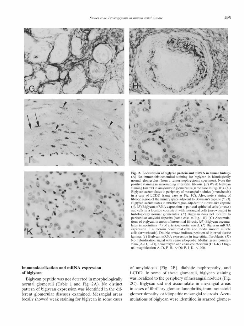
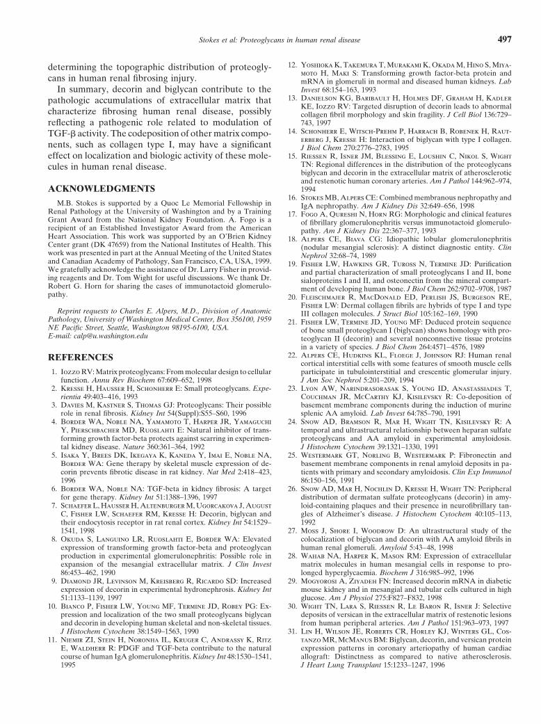
Fig. 2. Localization of biglycan protein and mRNA in human kidney.(A) No immunohistochemical staining for biglycan in histologicallynormal glomerulus (from a tumor nephrectomy specimen). Note thepositive staining in surrounding interstitial fibrosis. (B) Weak biglycanstaining (arrow) in amyloidotic glomerulus (same case as Fig. 1B). (C )Biglycan accumulates at periphery of mesangial nodules (arrowheads)in a case of LCDD (same case as Fig. 1C). Also, note staining offibrotic region of the urinary space adjacent to Bowman’s capsule (*; D).Biglycan accumulates in fibrotic region adjacent to Bowman’s capsule(*). (E) Biglycan mRNA expression in parietal epithelial cells (arrows)and cells in a location consistent with mesangial cells (arrowheads) inhistologically normal glomerulus. (F ) Biglycan does not localize toperitubular amyloid deposits (same case as Fig. 1H). (G) Accumula-tions of biglycan in areas of interstitial fibrosis. (H ) Biglycan accumu-lates in neointima (*) of arteriosclerotic vessel. (I ) Biglycan mRNAexpression in numerous neointimal cells and media smooth musclecells (arrowheads). Double arrows indicate position of internal elasticlamina. (J ) Biglycan mRNA expression in interstitial fibroblasts. (K )No hybridization signal with sense riboprobe. Methyl green counter-stain (A–D, F–H), hematoxylin and eosin counterstain (E, I–K). Origi-nal magnification A–D, F–H, 3400; E, I–K, 31000.
Immunolocalization and mRNA expression of amyloidosis (Fig. 2B), diabetic nephropathy, andof biglycan LCDD. In some of these glomeruli, biglycan staining
was localized to the periphery of mesangial nodules (Fig.Biglycan peptide was not detected in morphologically2C). Biglycan did not accumulate in mesangial areasnormal glomeruli (Table 1 and Fig. 2A). No distinctin cases of fibrillary glomerulonephritis, immunotactoidpattern of biglycan expression was identified in the dif-glomerulopathy, or idiopathic mesangial sclerosis. Accu-ferent glomerular diseases examined. Mesangial areas
focally showed weak staining for biglycan in some cases mulations of biglycan were identified in scarred glomer-
Stokes et al: Proteoglycans in human renal disease494
Stokes et al: Proteoglycans in human renal disease 495
uli in which the urinary space was obliterated, with fi- In morphologically normal kidneys, collagen type Ilocalized to the interstitium, particularly in the medullabrous adhesion of the glomerular tuft to Bowman’s
capsule, in a similar distribution to that observed for (data not shown). Collagen type I accumulated in ex-panded zones of interstitial fibrosis (Fig. 3F). Collagendecorin (compare Fig. 1D and Fig. 2D). By in situ hybrid-
ization, biglycan mRNA localized to parietal epithelial type I localized to the adventitia and, focally, to thecells and, focally, to intraglomerular cells (Fig. 2E) in media of morphologically normal arteries, but was notboth morphologically normal and sclerosed glomeruli. detected in normal intima. In the neointima of arterio-
Biglycan did not localize to peritubular or interstitial sclerotic vessels, diffuse accumulations of collagen typeamyloid deposits (Fig. 2F). The distribution of biglycan I were observed in a parallel distribution to that observedin the tubulointerstitium overlapped with that of decorin for biglycan (compare Fig. 2H and Fig. 3H).(compare Fig. 1H and Fig. 2G), with accentuation invascular adventitia and diffuse staining in expanded ar-
DISCUSSIONeas of interstitial fibrosis. Biglycan mRNA expressionThe present study is the first to our knowledge towas seen in peritubular interstitial fibroblasts or myofi-
describe the synthesis and deposition of the proteogly-broblast-like cells, which were more numerous in areascans decorin and biglycan in human glomerular diseasesof interstitial fibrosis (Fig. 2J). No hybridization signalcharacterized by accumulations of extracellular matrix.for biglycan was identified in tubular epithelial cells orThe demonstration of decorin and biglycan expression atin infiltrating mononuclear inflammatory cells.sites of glomerular fibrosing injury and tubulointerstitialBiglycan peptide localized to the adventitia and, fo-fibrosis supports a pathogenic role for these proteogly-cally, to the media of morphologically normal arteries,cans in progressive human renal disease. Differences inbut was not detected in normal intima (data not shown).proteoglycan composition may be diagnostically usefulAccumulations of biglycan were diffusely present in thein distinguishing morphologically similar glomerular dis-neointima of arteriosclerotic vessels (Fig. 2H). Biglycaneases. The finding that decorin colocalizes with amyloidmRNA was identified in occasional smooth muscle cellsdeposits, but not in diabetic nephropathy, LCDD, fibril-in the media and in neointimal cells (Fig. 2I), but not inlary glomerulonephritis, and immunotactoid glomerulo-endothelial cells.pathy, supports the existence of different pathways of
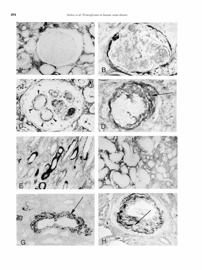
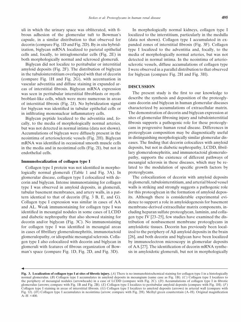
Immunolocalization of collagen type I mesangial sclerosis in these diseases, which may be re-lated to the modulation of specific growth factors byCollagen type I protein was not identified in morpho-proteoglycans.logically normal glomeruli (Table 1 and Fig. 3A). In
The colocalization of decorin with amyloid depositsglomerular disease, collagen type I colocalized with de-in glomeruli, tubulointerstitium, and arterial blood vesselcorin and biglycan. Intense immunostaining for collagenwalls is striking and strongly suggests a pathogenic roletype I was observed in amyloid deposits, in glomeruli,for this proteoglycan in the formation of amyloid depos-tubular basement membranes, and artery walls, in a pat-its. Although there is considerable experimental evi-tern identical to that of decorin (Fig. 3 B, E, and G).
Collagen type I expression was similar in cases of AA dence to support a role in amyloidogenesis for basementmembrane-derived extracellular matrix components, in-and AL. Weak immunostaining for collagen type I was
identified in mesangial nodules in some cases of LCDD cluding heparan sulfate proteoglycan, laminin, and colla-gen type IV [23–25], few studies have examined the dis-and diabetic nephropathy that also showed staining for
decorin and/or biglycan (Fig. 3C). No immunostaining tribution of nonbasement membrane proteoglycans inamyloidotic tissues. Decorin has previously been local-for collagen type I was identified in mesangial areas
in cases of fibrillary glomerulonephritis, immunotactoid ized to the periphery of Ab amyloid deposits in the brain[26], and both decorin and biglycan have been localizedglomerulopathy, or idiopathic mesangial sclerosis. Colla-
gen type I also colocalized with decorin and biglycan in by immunoelectron microscopy in glomerular depositsof AA [27]. The identification of decorin mRNA synthe-glomeruli with features of fibrous organization of Bow-
man’s space (compare Fig. 1D, Fig. 2D, and Fig. 3D). sis in amyloidotic glomeruli, but not in morphologically
b
Fig. 3. Localization of collagen type I at sites of fibrotic injury. (A) There is no immunohistochemical staining for collagen type I in a histologicallynormal glomerulus. (B) Collagen type I accumulates in amyloid deposits in mesangium (same case as Fig. 1B). (C ) Collagen type I localizes tothe periphery of mesangial nodules (arrowheads) in a case of LCDD (compare with Fig. 2C). (D) Accumulations of collagen type I in fibroticglomerulus (arrows; compare with Fig. 1B and Fig. 2B). (E) Collagen type I localizes to peritubular amyloid deposits (compare with Fig. 1H). (F )Collagen type I staining in areas of interstitial fibrosis. (G) Collagen type I localizes to amyloid deposits (arrows) in arterial wall (compare withFig. 1J). (H ) Collagen type I accumulates in neointima (arrow; compare with Fig. 2H). Methyl green counterstain (A–H). Original magnificationA–H 3400.
Stokes et al: Proteoglycans in human renal disease496
normal glomeruli, suggests that local up-regulation of to unregulated TGF-b activity at these sites. Alterna-decorin synthesis contributes to the accumulation of pro- tively, binding of growth factors to extracellular matrixteoglycan in these cases. The colocalization of collagen proteoglycans may serve a reservoir function, increasingtype I with amyloid has not been reported in the litera- the availability of growth factors at sites of fibrosingture. Our study raises the possibility that codeposition injury.of decorin and collagen type I may have a role in stabili- Peritubular interstitial fibroblasts expressing decorinzation and persistence of amyloid deposits. mRNA and biglycan mRNA were more numerous in
The finding of weak staining for decorin and biglycan areas of interstitial fibrosis compared with morphologi-in some cases of advanced LCDD and diabetic nephrop- cally normal tubulointerstitium, suggesting that proteo-athy was also related to the focal finding of collagen type I glycan synthesis may be up-regulated in areas of intersti-deposits in these tissues. Although there is experimental tial fibrosis. Biglycan mRNA, as distinct from biglycanevidence that mesangial cell synthesis of decorin is up- peptide expression, was identified in parietal epithelialregulated in hyperglycemic cell culture media [28] and cells and in occasional intraglomerular cells in both mor-in rodent models of diabetic glomerular disease [29], the phologically normal and sclerotic glomeruli. The role ofresults of our study do not support a role for up-regulated increased biglycan synthesis in the formation of biglycansynthesis and deposition of decorin and biglycan in the accumulations in glomeruli remains undetermined. Ofpathogenesis of human diabetic nephropathy, and in note, other investigators have described a somewhat dif-most clinically important human glomerular diseases ferent distribution of biglycan in the kidney. In rat kidney,characterized by mesangial sclerosis. An important ca- biglycan mRNA and core protein have been localized toveat is that the tissues examined in this study were ob- glomerular capillaries, mesangial cells, and podocytestained from individuals with symptomatic renal disease, and in collecting ducts and distal tubules [7, 35]. In hu-and these investigations do not address the possibility man fetal kidney, Bianco et al have localized biglycanthat decorin and biglycan may have functions in earlier protein on the surface of endothelial cells and collectingstages of glomerular injury. Further studies to examine tubules and “very occasionally” in parietal epithelial cells,the expression of these extracellular matrix molecules and they have also localized mRNA to these cells [10].in more acute forms of glomerular injury, characterized The results of this study, in which biglycan mRNA syn-by cellular proliferation and mesangiolysis, will help to thesis was not identified in endothelium or collectingdefine better the roles of decorin and biglycan in human ducts, suggest that biglycan expression by endothelialglomerular disease. cells and tubular epithelial cells may be developmentally
The codistribution of decorin and biglycan with colla- restricted, perhaps reflecting different biologic functionsgen type I in glomerulosclerosis and interstitial fibrosis
in developing and mature human kidney.suggests that synthesis and/or deposition of these extra-
Biglycan, but not decorin, was identified in the neoin-cellular matrix moieties may be pathogenically related.tima of arteriosclerotic blood vessels. Distinct patternsDecorin has a key role in regulating collagen fibril assem-of localization of decorin and biglycan have also beenbly and growth, as demonstrated by the finding of abnor-described in human coronary atherosclerotic plaques [15,mal collagen fibrils in decorin-deficient mice [13], and36], in restenosis lesions following coronary angioplastyinteractions between biglycan and collagen type I have[15], and in coronary transplant arteriopathy [31]. Basedalso been described in vitro [14]. The codeposition ofon the demonstration of specific interactions betweendecorin and biglycan with collagen type I has been de-biglycan and apolipoprotein E in vitro, and the immuno-scribed in other instances of fibrosing injury, includinghistochemical colocalization of biglycan with apolipo-arteriosclerotic blood vessels [15, 30, 31], myocardial in-protein E in human coronary atherosclerotic plaques, itfarction [32], and keloids [33]. Proteoglycan-collagenhas been proposed that interactions between proteogly-type I interactions may contribute to stabilization of thecans and lipoproteins may contribute to the pathogenesisremodeled extracellular matrix at sites of fibrosing in-of atherosclerosis by trapping lipoproteins in the arteryjury. Decorin and biglycan bound to collagen fibrils maywall [36]. The finding of distinct and overlapping patternsbe sequestered from the normal pathway of reuptakeof decorin and biglycan accumulation in the kidney sug-and receptor-mediated endocytosis [2], thus contributinggests specific, albeit as yet undetermined pathogenicto the formation of immunohistochemically detectableroles in human renal fibrosis that may be related todeposits at sites of collagen type I deposition. This inter-different biologic properties of these molecules. How-pretation is consistent with in vitro observations of de-ever, the pathogenesis of proteoglycan accumulation inlayed turnover of proteoglycans synthesized by fibro-fibrotic disease is complex and multifactorial, and otherblasts grown on a collagenous matrix [34]. It is interestingfactors not examined in this study, such as local differ-to speculate that binding of proteoglycans to collagenences in activity of profibrogenic cytokines [37] and ma-may lead to reduced availability of these molecules for
interactions with growth factors, such as TGF-b, leading trix degrading enzymes, are likely to be important in
Stokes et al: Proteoglycans in human renal disease 497
12. Yoshioka K, Takemura T, Murakami K, Okada M, Hino S, Miya-determining the topographic distribution of proteogly-moto H, Maki S: Transforming growth factor-beta protein and
cans in human renal fibrosing injury. mRNA in glomeruli in normal and diseased human kidneys. LabInvest 68:154–163, 1993In summary, decorin and biglycan contribute to the
13. Danielson KG, Baribault H, Holmes DF, Graham H, Kadlerpathologic accumulations of extracellular matrix thatKE, Iozzo RV: Targeted disruption of decorin leads to abnormal
characterize fibrosing human renal disease, possibly collagen fibril morphology and skin fragility. J Cell Biol 136:729–743, 1997reflecting a pathogenic role related to modulation of
14. Schonherr E, Witsch-Prehm P, Harrach B, Robenek H, Raut-TGF-b activity. The codeposition of other matrix compo-erberg J, Kresse H: Interaction of biglycan with type I collagen.
nents, such as collagen type I, may have a significant J Biol Chem 270:2776–2783, 199515. Riessen R, Isner JM, Blessing E, Loushin C, Nikol S, Wighteffect on localization and biologic activity of these mole-
TN: Regional differences in the distribution of the proteoglycanscules in human renal disease.biglycan and decorin in the extracellular matrix of atheroscleroticand restenotic human coronary arteries. Am J Pathol 144:962–974,1994ACKNOWLEDGMENTS
16. Stokes MB, Alpers CE: Combined membranous nephropathy andM.B. Stokes is supported by a Quoc Le Memorial Fellowship in IgA nephropathy. Am J Kidney Dis 32:649–656, 1998
Renal Pathology at the University of Washington and by a Training 17. Fogo A, Qureshi N, Horn RG: Morphologic and clinical featuresGrant Award from the National Kidney Foundation. A. Fogo is a of fibrillary glomerulonephritis versus immunotactoid glomerulo-recipient of an Established Investigator Award from the American pathy. Am J Kidney Dis 22:367–377, 1993Heart Association. This work was supported by an O’Brien Kidney 18. Alpers CE, Biava CG: Idiopathic lobular glomerulonephritisCenter grant (DK 47659) from the National Institutes of Health. This (nodular mesangial sclerosis): A distinct diagnostic entity. Clinwork was presented in part at the Annual Meeting of the United States Nephrol 32:68–74, 1989and Canadian Academy of Pathology, San Francisco, CA, USA, 1999. 19. Fisher LW, Hawkins GR, Tuross N, Termine JD: PurificationWe gratefully acknowledge the assistance of Dr. Larry Fisher in provid- and partial characterization of small proteoglycans I and II, boneing reagents and Dr. Tom Wight for useful discussions. We thank Dr. sialoproteins I and II, and osteonectin from the mineral compart-Robert G. Horn for sharing the cases of immunotactoid glomerulo- ment of developing human bone. J Biol Chem 262:9702–9708, 1987pathy. 20. Fleischmajer R, MacDonald ED, Perlish JS, Burgeson RE,
Fisher LW: Dermal collagen fibrils are hybrids of type I and typeReprint requests to Charles E. Alpers, M.D., Division of Anatomic III collagen molecules. J Struct Biol 105:162–169, 1990
Pathology, University of Washington Medical Center, Box 356100, 1959 21. Fisher LW, Termine JD, Young MF: Deduced protein sequenceNE Pacific Street, Seattle, Washington 98195-6100, USA. of bone small proteoglycan I (biglycan) shows homology with pro-E-mail: [email protected] teoglycan II (decorin) and several nonconnective tissue proteins
in a variety of species. J Biol Chem 264:4571–4576, 198922. Alpers CE, Hudkins KL, Floege J, Johnson RJ: Human renalREFERENCES
cortical interstitial cells with some features of smooth muscle cellsparticipate in tubulointerstitial and crescentic glomerular injury.1. Iozzo RV: Matrix proteoglycans: From molecular design to cellular
function. Annu Rev Biochem 67:609–652, 1998 J Am Soc Nephrol 5:201–209, 199423. Lyon AW, Narindrasorasak S, Young ID, Anastassiades T,2. Kresse H, Hausser H, Schonherr E: Small proteoglycans. Expe-
rientia 49:403–416, 1993 Couchman JR, McCarthy KJ, Kisilevsky R: Co-deposition ofbasement membrane components during the induction of murine3. Davies M, Kastner S, Thomas GJ: Proteoglycans: Their possible
role in renal fibrosis. Kidney Int 54(Suppl):S55–S60, 1996 splenic AA amyloid. Lab Invest 64:785–790, 199124. Snow AD, Bramson R, Mar H, Wight TN, Kisilevsky R: A4. Border WA, Noble NA, Yamamoto T, Harper JR, Yamaguchi
Y, Pierschbacher MD, Ruoslahti E: Natural inhibitor of trans- temporal and ultrastructural relationship between heparan sulfateproteoglycans and AA amyloid in experimental amyloidosis.forming growth factor-beta protects against scarring in experimen-
tal kidney disease. Nature 360:361–364, 1992 J Histochem Cytochem 39:1321–1330, 199125. Westermark GT, Norling B, Westermark P: Fibronectin and5. Isaka Y, Brees DK, Ikegaya K, Kaneda Y, Imai E, Noble NA,
Border WA: Gene therapy by skeletal muscle expression of de- basement membrane components in renal amyloid deposits in pa-tients with primary and secondary amyloidosis. Clin Exp Immunolcorin prevents fibrotic disease in rat kidney. Nat Med 2:418–423,
1996 86:150–156, 199126. Snow AD, Mar H, Nochlin D, Kresse H, Wight TN: Peripheral6. Border WA, Noble NA: TGF-beta in kidney fibrosis: A target
for gene therapy. Kidney Int 51:1388–1396, 1997 distribution of dermatan sulfate proteoglycans (decorin) in amy-loid-containing plaques and their presence in neurofibrillary tan-7. Schaefer L, Hausser H, Altenburger M, Ugorcakova J, August
C, Fisher LW, Schaefer RM, Kresse H: Decorin, biglycan and gles of Alzheimer’s disease. J Histochem Cytochem 40:105–113,1992their endocytosis receptor in rat renal cortex. Kidney Int 54:1529–
1541, 1998 27. Moss J, Shore I, Woodrow D: An ultrastructural study of thecolocalization of biglycan and decorin with AA amyloid fibrils in8. Okuda S, Languino LR, Ruoslahti E, Border WA: Elevated
expression of transforming growth factor-beta and proteoglycan human renal glomeruli. Amyloid 5:43–48, 199828. Wahab NA, Harper K, Mason RM: Expression of extracellularproduction in experimental glomerulonephritis: Possible role in
expansion of the mesangial extracellular matrix. J Clin Invest matrix molecules in human mesangial cells in response to pro-longed hyperglycaemia. Biochem J 316:985–992, 199686:453–462, 1990
9. Diamond JR, Levinson M, Kreisberg R, Ricardo SD: Increased 29. Mogyorosi A, Ziyadeh FN: Increased decorin mRNA in diabeticmouse kidney and in mesangial and tubular cells cultured in highexpression of decorin in experimental hydronephrosis. Kidney Int
51:1133–1139, 1997 glucose. Am J Physiol 275:F827–F832, 199830. Wight TN, Lara S, Riessen R, Le Baron R, Isner J: Selective10. Bianco P, Fisher LW, Young MF, Termine JD, Robey PG: Ex-
pression and localization of the two small proteoglycans biglycan deposits of versican in the extracellular matrix of restenotic lesionsfrom human peripheral arteries. Am J Pathol 151:963–973, 1997and decorin in developing human skeletal and non-skeletal tissues.
J Histochem Cytochem 38:1549–1563, 1990 31. Lin H, Wilson JE, Roberts CR, Horley KJ, Winters GL, Cos-tanzo MR, McManus BM: Biglycan, decorin, and versican protein11. Niemir ZI, Stein H, Noronha IL, Kruger C, Andrassy K, Ritz
E, Waldherr R: PDGF and TGF-beta contribute to the natural expression patterns in coronary arteriopathy of human cardiacallograft: Distinctness as compared to native atherosclerosis.course of human IgA glomerulonephritis. Kidney Int 48:1530–1541,
1995 J Heart Lung Transplant 15:1233–1247, 1996
Stokes et al: Proteoglycans in human renal disease498
32. Yamamoto K, Kusachi S, Ninomiya Y, Murakami M, Doi M, 35. Pyke C, Kristensen P, Ostergaard PB, Oturai PS, Romer J:Proteoglycan expression in the normal rat kidney. Nephron 77:461–Takeda K, Shinji T, Higashi T, Koide N, Tsuji T: Increase in the
expression of biglycan mRNA expression co-localized closely with 470, 199736. O’Brien KD, Olin KL, Alpers CE, Chiu W, Ferguson M, Hud-that of type I collagen mRNA in the infarct zone after experimen-
tally-induced myocardial infarction in rats. J Mol Cell Cardiol kins K, Wight TN, Chait A: Comparison of apolipoprotein andproteoglycan deposits in human coronary atherosclerotic plaques:30:1749–1756, 1998
33. Hunzelmann N, Anders S, Sollberg S, Schonherr E, Krieg T: Colocalization of biglycan with apolipoproteins. Circulation 98:519–527, 1998Co-ordinate induction of collagen type I and biglycan expression
in keloids. Br J Dermatol 135:394–399, 1996 37. Evanko SP, Raines EW, Ross R, Gold LI, Wight TN: Proteogly-can distribution in lesions of atherosclerosis depends on lesion34. Greve H, Blumberg P, Schmidt G, Schlumberger W, Rauterberg
J, Kresse H: Influence of collagen lattice on the metabolism of severity, structural characteristics, and the proximity of platelet-derived growth factor and transforming growth factor-beta. Amsmall proteoglycan II by cultured fibroblasts. Biochem J 269:149–
155, 1990 J Pathol 152:533–546, 1998