the internal region leucine-rich repeat 6 of decorin interacts with low density lipoprotein...
TRANSCRIPT
The Internal Region Leucine-rich Repeat 6 of DecorinInteracts with Low Density Lipoprotein Receptor-relatedProtein-1, Modulates Transforming Growth Factor (TGF)-�-dependent Signaling, and Inhibits TGF-�-dependent FibroticResponse in Skeletal Muscles*
Received for publication, October 11, 2011, and in revised form, December 14, 2011 Published, JBC Papers in Press, December 27, 2011, DOI 10.1074/jbc.M111.312488
Claudio Cabello-Verrugio‡§, Cristian Santander‡, Catalina Cofré‡, Maria José Acuña‡, Francisco Melo¶�,and Enrique Brandan‡1
From the ‡Centro de Regulación Celular y Patología (CRCP), Centro de Regeneración y Envejecimiento (CARE), Departamento deBiología Celular y Molecular, Pontificia Universidad Católica de Chile, Santiago, the ¶Millennium Institute on Immunology andImmunotherapy and �Departamento de Genética Molecular y Microbiología, Facultad de Ciencias Biológicas, PontificiaUniversidad Católica de Chile, Santiago, and the §Centro de Genética Humana (CGH), Facultad de Medicina, Clínica Alemana-Universidad del Desarrollo, Santiago, Chile
Background: Transforming growth factor (TGF)-� signaling depends on decorin and LRP-1.Results: Decorin internal regions 5 and 6 interact with LRP-1 to modulate TGF-� mediated signaling.Conclusion: A specific decorin region regulates TGF-�-dependent signaling.Significance: Identification of specific sites in decorin that are involved in TGF-� signaling might have potential therapeuticapplications for treatment of fibrotic diseases.
Decorin is a small proteoglycan, composed of 12 leucine-richrepeats (LRRs) that modulates the activity of transforminggrowth factor type � (TGF-�) and other growth factors, andthereby influences proliferation and differentiation in a widearray of physiological and pathological processes, such as fibro-sis, in several tissues and organs. Previously we described twonovel modulators of the TGF-�-dependent signaling pathway:LDL receptor-related protein (LRP-1) and decorin. Here wehave determined the regions in decorin that are responsible forinteraction with LRP-1 and are involved in TGF-�-dependentbinding and signaling. Specifically, we used decorin deletionmutants, as well as peptides derived from internal LRR regions,to determine the LRRs responsible for these decorin functions.Our results indicate that LRR6 and LRR5 participate in theinteraction with LRP-1 and TGF-� as well as in its dependentsignaling. Furthermore, the internal region (LRR6i), composedof 11 amino acids, is responsible for decorin binding to LRP-1and subsequent TGF-�-dependent signaling. Furthermore,using an in vivo approach, we also demonstrate that the LRR6region of decorin can inhibit TGF-� mediated action inresponse to skeletal muscle injury.
Transforming growth factor-� (TGF-�) is a multifunctionalcytokine involved in development, cell proliferation, and extra-
cellular matrix (ECM)2 deposition (1). This growth factor con-tains nine conserved cysteines and belongs to a superfamily ofstructurally related proteins, known as the transforminggrowth factor � superfamily, which includes activins, inhibins,and bone morphogenic proteins (2). TGF-� regulates cellularprocesses in the nucleus by binding to cell surface transmem-brane receptors with serine/threonine kinase activity and cyto-plasmic effectors, including Smad proteins and non-Smadsproteins (3, 4).Previously we described two novel modulators of this TGF-
�-dependent signaling pathway: LDL receptor-related protein(LRP-1) (5) and decorin (6). LRP-1 is a giant receptor, belongingto the low-density lipoprotein receptor family, which bind andinternalize extracellular ligands for degradation (7, 8). Weshowed that decorin is endocytosed and degraded bymyoblastsand that both processes are blocked by siRNA suppression ofLRP-1 expression (5). This observation supports the hypothesisthat LRP-1 is a receptor for decorin (5, 9). These findings alsopoint to LRP-1-mediated catabolism as a new control pathwayfor the biological activities of decorin, specifically for its abilityto influence ECM signaling.Following the discovery of LRP-1 as a cell surface receptor for
decorin, we attempted to determine its role in decorin TGF-�-dependent signaling. Decorin-deficient myoblasts show thatthe decreased TGF-� response can be fully restored by decorinre-expression (6, 10). Importantly, this reactivation occurswithout changes in the ability of TGF-� to bind its receptors or* This work was supported by FONDAP-Biomedicine Grants 13980001, CARE
PFB12/2007, FONDECYT 11080212, FONDECYT 1110400, ICM P09-016-F,and MDA 89419 and Fundación Chilena para Biología Celular ProyectoMF-100.
1 To whom correspondence should be addressed: Dept. de Biología Celular yMolecular, Facultad de Ciencias Biológicas, P. Universidad Católica deChile, Casilla 114-D, Santiago, Chile. Fax: 56-2-635-5395; E-mail: [email protected].
2 The abbreviations used are: ECM, extracellular matrix; CTGF, connective tis-sue growth factor; EGFR, epidermal growth factor receptor; LRR6i, LRR6internal region; LRR6e, LRR6 external region; LRRs, leucine-rich repeats;LRP-1, low density lipoprotein receptor-related protein; TGF-�, transform-ing growth factor �.
THE JOURNAL OF BIOLOGICAL CHEMISTRY VOL. 287, NO. 9, pp. 6773–6787, February 24, 2012© 2012 by The American Society for Biochemistry and Molecular Biology, Inc. Published in the U.S.A.
FEBRUARY 24, 2012 • VOLUME 287 • NUMBER 9 JOURNAL OF BIOLOGICAL CHEMISTRY 6773
at PO
NT
IFIC
IA U
NIV
ER
SID
AD
, on February 27, 2012
ww
w.jbc.org
Dow
nloaded from

in intracellular signalingmediators such as Smad protein phos-phorylation or Smad-4 nuclear translocation (3, 11). In myo-blasts, inhibition of decorin binding to LRP-1, or depletion ofLRP-1, decreased TGF-� response to levels observed indecorin-deficient myoblasts. Interestingly, re-expression ofdecorin in decorin-deficient myoblasts did not restore theTGF-� response when either the Smad pathway or PI3K activ-ity was inhibited, suggesting that this LRP-1-decorin modula-tory pathway requires activation of the Smad pathway byTGF-� and involves PI3K activity (6). These results reveal a newregulatory mechanism for TGF-� signaling, involving decorinand LRP-1 at the myoblast surface (5).Decorin is one of the most well studied members of the fam-
ily of small leucine-rich proteoglycans (12). Decorin is a pro-teoglycan formed by a core protein with 12 leucine-rich repeats(LRRs) and a chondroitin/dermatan sulfate glycosaminoglycanchain (12–15). Decorin can be localized in the ECM or at thecell plasma membrane, where it interacts with cell surfacereceptors. This differential localization can explain the differ-ent and sometimes contradictory functions of this proteoglycan(9). Decorin in the ECM is believed to regulate ECM structure(16, 17) and modulate the bioavailability of several growth fac-tors, including TGF-� (18). In addition, decorin has also beenreported to be associated with cell surface receptors. Further-more, decorin has an intrinsic ability to interact with severalcytokines (19). It has been shown that decorin binds to insulingrowth factor-1 and the insulin growth factor-1 receptor (20),resulting in insulin growth factor-1 receptor phosphorylationand activation, followed by receptor down-regulation (21).Decorin has also been reported to cause rapid phosphorylationof the epidermal growth factor receptor (EGFR) and activationof themitogen-activated protein kinase signaling pathway (22).In addition, decorin has been reported to modulate two pro-fibrotic molecules, TGF-� (6, 9, 23, 24) and connective tissuegrowth factor (CTGF) (25). Interestingly, decorin can also bindto the cMet receptor, the receptor for hepatocyte growth factor,inducing transient activation of the receptor and inhibiting cellmigration and growth (26). These novel results are in agree-ment with our previous observations that decorin-deficientmyoblasts exhibit exacerbatedmigration capacity after graftingin vivo (27).Fibrotic disorders are the end point ofmany chronic diseases
in different tissues such as kidney, skin, and skeletal muscle(28–30). Fibrosis development is caused by the action ofgrowth factors and cytokines, which are overexpressed infibrotic tissues. For years, TGF-� has been described as theprincipal inducer of fibrosis in different tissues (31). Duchennemuscular dystrophy is a disease caused by the absence of theprotein dystrophin, which producesmuscle weakness and leadsto cycles of degeneration and regeneration of muscle fibers,resulting in a decrease in muscle mass and an increase in fibro-sis, concomitant with augmented TGF-� levels in biopsies ofDuchenne muscular dystrophy patients (32–34).Elucidation of the precise binding sites of modulator mole-
cules such as decorin is an important task with important ther-apeutic implications. The specific decorin-binding site onLRP-1 has not been determined. In addition, the role of thisdecorin-binding site on LRP-1 in TGF-�-dependent signaling
remains unknown. In this study, we use decorin-deletionmutants, as well as peptides from internal LRR regions, todetermine the LRRs responsible for these decorin functions.We found that LRR6 and LRR5 are responsible for LRP-1 bind-ing and subsequent TGF-�-dependent signaling. Also weobtained evidence that the LRR6 internal region (LRR6i) par-ticipates in interaction with LRP-1- and TGF-�-dependent sig-naling. Furthermore, using an in vivo approach, we demon-strate that this LRR6 region can inhibit TGF-�-mediated actionin response to skeletal muscle injury.
EXPERIMENTAL PROCEDURES
Animals and Experimental Muscle Injury—12-Week-oldC57BL/10 ScSn male mice were used for this study. Animalswere kept at room temperature with a 24-h night-day cycle andfed with pellets and water ad libitum. Injury of normal muscletissue was performed by barium-chloride injection (35, 36) inmice under ketamine/xylazine anesthesia (80/12 mg/kg bodyweight, intraperitoneally). Briefly, 60 �l of an aqueous 1.2%mass/volume (m/w) BaCl2 solution was injected along thewhole length of the left tibialis anterior muscle. ContralateralPBS-injected muscles were used as controls. After 3 days ofBaCl2 injections, an intramuscular injection of 20 ng of TGF-�1in the presence or absence of 10 �g of the LRR6 region wasperformed. 4 days post-injury, tibialis anteriors were dissectedand removed under anesthesia, and animals were sacrificed.Tissues were rapidly frozen and stored at�80 °C until process-ing. All protocols were conducted in strict accordance andwiththe formal approval of the Animal Ethics Committee of the P.Universidad Católica de Chile.Peptides Derived from LRR Regions—The following decorin
LRR regionswere used: LRR4, KELPEKMPKTLQELRAHENEI;LRR5, TKVRKVTFNGLNQMIVIELGTNPL; LRR6, KSSGIEN-GAFQGMKKLSYIRIADTNI; LRR6 external (LRR6e), KSS-GIENGAFQGMKK; LRR6 internal (LRR6i), LSYIRIADTNI;LRR12, QYWEIQPSTFRCVYVRSAIQLGNYK; and Scramblepeptide, SITNLDRYAII. All peptides were obtained from Pep-tide 2.0 Inc.Cell Culture—The mouse skeletal muscle cell line, C2C12
(ATCC) (18), was grown and induced to differentiate asdescribed (38). Decorin-deficient myoblasts have been previ-ously described and characterized (6, 10).MutantDecorinGeneration—All decorins generated have an
HA epitope in their C termini. Wild type decorin and decorinmutants that lack different LRRwere generated by overlap PCRstarting from human decorin (NM_133593) cloned in pcDNA3.1 (kindly donated by Elke Schönherr, Institute of Physiologi-cal Chemistry and Pathobiochemistry, Münster, Germany).Generated constructs were sequenced and then stably trans-fected into CHO-K1 cells using medium supplemented withG418 (400 �g/ml). After 4 weeks, different clones were isolated(25). Decorin purification was conducted using conditionedmedium from the CHO-K1 clones, and a DEAE-Sephacel col-umn (Bio-Rad) as previously described (39).Labeling of Cultures—CHO-K1 cells, stably transfected with
different deletion mutant constructs of decorin, were labeledfor 18 h in sulfate and serum-free DMEM/F-12 with 100�Ci/ml of H2[35S]SO4 (PerkinElmer Life Sciences, 25 mCi/ml)
Decorin of LRR6 Interacts with LRP-1 Modulating TGF-� Signal
6774 JOURNAL OF BIOLOGICAL CHEMISTRY VOLUME 287 • NUMBER 9 • FEBRUARY 24, 2012
at PO
NT
IFIC
IA U
NIV
ER
SID
AD
, on February 27, 2012
ww
w.jbc.org
Dow
nloaded from
(39). This conditioned medium was concentrated using aDEAE-Sephacel column pre-equilibrated in 10 mM Tris-HCl,pH 7.4, 0.2 M NaCl, and 0.1% Triton X-100. The sample boundto the column was incubated with heparitinase in an appropri-ate buffer for 4 h at 37 °C. Samples were then eluted with 1 M
NaCl and dialyzed against PBS (39).Decorin Endocytosis Assays—Endocytosis of 35S-decorin by
myoblasts in culture was determined as previously described(5).Transient Plasmid Transfection—Cells were plated in
24-well plates until they reached 60% confluence. Cells werethen incubated in Opti-MEM I containing 1 �g of TGF-�responsive plasmid p3TP-Lux (6), 0.02 �g of pRL-SV40, 2 �l ofPLUS reagent, and 1 �l of Lipofectamine. After 6 h, FBS wasadded to themedium, and cells were cultured for a further 12 h.Medium was changed to fresh growth medium and the follow-ing reagents were added: TGF-�1 dissolved in 0.5% FBS; anddifferent decorin mutants or decorin LRR regions in PBS. Dualluciferase activity assays (Promega) were performed after 6 h,depending on the experiment.Short Interfering RNA (siRNA) Transfection—Annealed
siRNA specific for LRP-1 and control siRNA have been previ-ously described (5, 6). Briefly, for experiments with p3TP-Luxplasmid reporters, cells were co-transfected with siRNA. Fol-lowing transfection, FBS was added to the medium, and cellswere cultured for a further 24 h. Dual luciferase activity assays(Promega) were performed after 6 h depending on theexperiment.Immunoprecipitation and Immunoblot Analysis—For
immunoprecipitation assays, myoblasts were lysed in 50 mM
Tris-HCl, pH 7.4, 0.1 M NaCl, 0.5% Triton X-100 buffer, con-taining a mixture of protease inhibitors and 1 mM phenylmeth-ylsulfonyl fluoride. Equal amounts of protein (150 �g) frompre-cleared extracts were immunoprecipitated overnight at4 °C with 5 �g of rabbit anti-LRP-1, as previously described (5),followed by incubation for 2 h at 4 °C with 20 �l of proteinA-agarose beads (Pierce). Equal volumes of immunoprecipi-tated protein were subjected to SDS-PAGE. For immunoblotanalyses, cell extracts obtained from myoblasts were preparedin 50mMTris-HCl, pH 7.4, 0.1 MNaCl, 0.5% Triton X-100 withamixture of protease inhibitors and 1mMPMSF. Aliquots weresubjected to SDS-gel electrophoresis on 5 or 10% polyacryl-amide gels, electrophoretically transferred onto nitrocellulosemembranes (Schleicher and Schuell), and probed with each ofthe following antibodies: rabbit anti-LRP-1 extracellulardomain (1:1000) (Calbiochem) as previously described (5); rab-bit anti-fibronectin (1:5000) andmouse anti-�-tubulin (1:5000)(Sigma); goat anti-CTGF (1:500) and rabbit anti-HA (1/2000)(Santa Cruz Biotechnology); and mouse anti-GAPDH (1:2000)(Chemicon). All immunoblots were visualized by enhancedchemiluminescence (Pierce).RNA Isolation, Reverse Transcription and Quantitative Real
Time PCR—Myoblasts were serum-starved for 18 h and thenincubated for several times with the indicated decorin deletionmutants. Total RNA was isolated from cell cultures at the indi-cated times in the figures using TRIzol (Invitrogen) accordingto the manufacturer’s instructions. Total RNA (1 �g) wasreverse-transcribed to cDNA using random hexamers and
SuperScript reverse transcriptase (Invitrogen). TaqMan quan-titative real-time PCR were performed in duplicate on a Strat-agene MX 3005 Termocyler (Agilent Technology), using pre-designed primer sets for mouse CTGF and the housekeepinggene GAPDH (TaqMan Assays-on-Demand, Applied Biosys-tems). mRNA expression was quantified using the comparative�Ctmethod (2���Ct), usingGAPDHas the reference gene. ThemRNA levels are expressed relative to the mean expression inthe control group. Values correspond to the mean of the �Ctvalue � S.D. of three independent experiments.Protein Determination—Protein levels were determined
from cell extract aliquots using a bicinchoninic acid proteinassay kit (Pierce) with BSA as a protein standard.Statistics—The statistical significance of the differences
between the means of the experimental groups was evaluatedusing one-way analysis of variance with a post hoc Bonferronimultiple comparison test (Sigma Stat 3.5 Software). A differ-ence was considered statistically significant at a p value � 0.05.
RESULTS
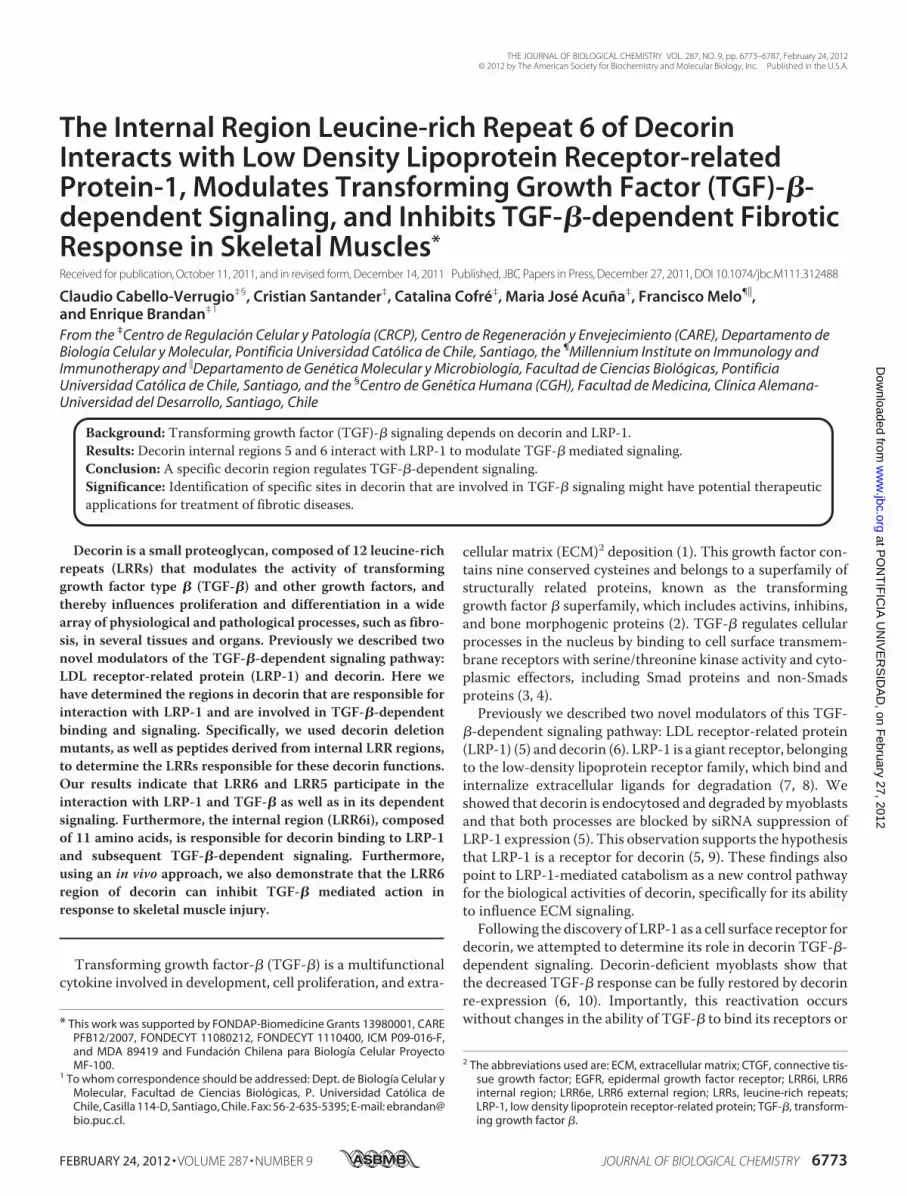
The LRR4–6 Region of Decorin Is Necessary for Binding toLRP-1 and Subsequent Endocytosis—We have previously pro-posed a new regulatory mechanism for TGF-� signaling medi-ated by decorin and LRP-1 (6). One of the key features of thismechanism is decorin binding to LRP-1, followed by decorininternalization by endocytosis (5). The core protein of decorincontains 12 LRRs that interact with various growth factors, andregulate their action (9, 20, 41, 42). To analyze which decorinLRR region is important for interaction with LRP-1, we gener-ated four decorin deletion mutants that lack different LRRregions (Fig. 1A). These mutant decorins were expressed inCHO cells and purified by DEAE. Fig. 1B shows aWestern blotagainst the HA epitope, with or without chondroitinase ABCtreatment, which degrades chondroitin/dermatan sulfate gly-cosaminoglycans. All of the glycanated forms, as well as theircorresponding cores protein (which migrated at the expectedmolecular weights), are indicated. In some core protein dele-tion mutants, a doublet is observed that likely corresponds tothe presence or absence of N-linked carbohydrates (43). Toevaluate which regions of the decorin core protein are impor-tant for interaction with LRP-1, C2C12 myoblasts were co-in-cubated with different 35S-decorin constructs at 4 °C, to avoidendocytosis, and cells were lysed and immunoprecipitated withantibodies against the extracellular domain of LRP-1. After sep-aration by SDS-PAGE, the immunoblot revealed that decorinco-immunoprecipitates with the LRP-1 antibody complex (Fig.1C). In particular, the figure shows that both full-length decorinand decorin lacking LRR10–12 co-immunoprecipitates withthe LRP-1 antibody complex. In contrast, less co-immunopre-cipitation was observed for decorin lacking LRR4–6, LRR4–5,or LRR5–6. The characteristic decorin smear is observed afterautoradiography.We have shown previously that decorin is endocytosed upon
binding to LRP-1 (5). Therefore, in this study, we determinedthe extent of endocytosis of different decorin deletionmutants.Fig. 1D shows that, among the different decorinmutants tested,decorins lacking LRR4–6, LRR4–5, and LRR5–6 displayed adiminished rate of clearance from the incubationmediumcom-
Decorin of LRR6 Interacts with LRP-1 Modulating TGF-� Signal
FEBRUARY 24, 2012 • VOLUME 287 • NUMBER 9 JOURNAL OF BIOLOGICAL CHEMISTRY 6775
at PO
NT
IFIC
IA U
NIV
ER
SID
AD
, on February 27, 2012
ww
w.jbc.org
Dow
nloaded from
pared with full-length decorin. Interestingly, in contrast,decorin lacking LRR10–12 showed an enhanced rate of endo-cytosis. Importantly, the clearance rate of all decorin forms wasdirectly mediated by LRP-1, because the presence of RAP, aninhibitor of both decorin binding and endocytosis of decorin byLRP-1, completely prevented decorin clearance from the incu-
bation medium (Fig. 1D) (6). Altogether these results suggestthat the region of decorin containing LRR4–6 is important forboth binding of decorin to LRP-1 and subsequent endocytosis.The LRR4–6 Region of Decorin Is Required for Its Effect on
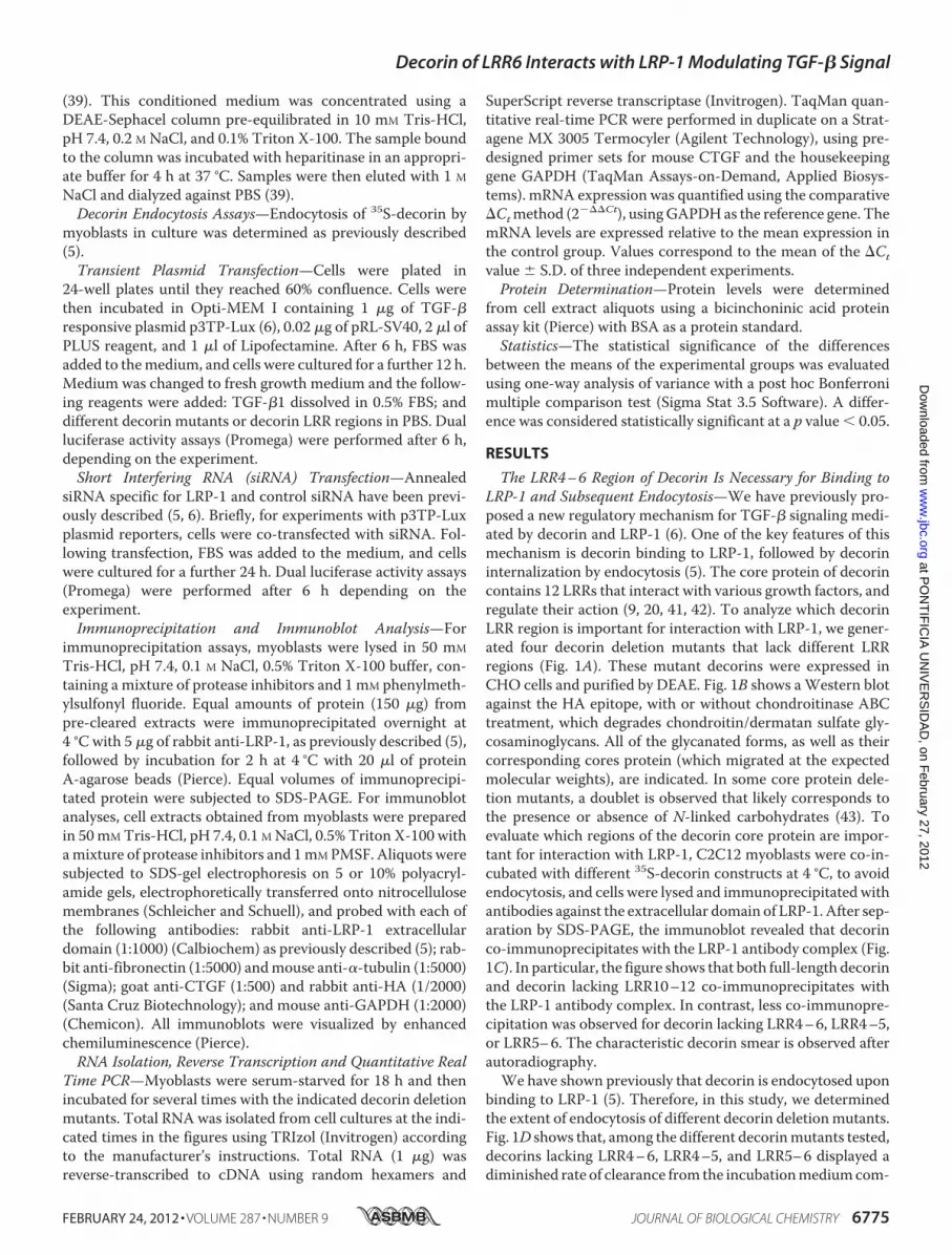
TGF-� Signaling Mediated by LRP-1—We have previouslyshown that decorin-deficient myoblasts display a decreased
FIGURE 1. The LRR4 – 6 decorin region is necessary for LRP-1 binding and LRP-1-mediated decorin endocytosis. A, schematic showing decorin deletionmutants. B, deletion mutants generated in CHO-K1, which lack LRR4 and 5 (�4 –5); 4, 5, and 6 (�4 – 6); 5 and 6 (�5– 6); or 10, 11, and 12 (�10 –12); were purifiedby DEAE-Sephacel and separated in a 4 –12% SDS-PAGE gradient for detection of glycanated forms (upper) or protein-core after treatment with chondroitinaseABC (lower). Decorins were visualized by Western blot using anti-HA antibodies. C, C2C12 cells were incubated with 35S-labeled decorin mutants at 4 °C for 3 h.Extracts were immunoprecipitated with anti-LRP-1 antibodies and the presence of decorin deletion mutants in the immunoprecipitate (IP) was evaluated byautoradiography (18). Protein levels were detected in the immunoprecipitate with an anti-LRP-1 antibody by Western blot. D, C2C12 myoblasts were incubated with35S-labeled decorin mutants at 37 °C for 3 h in the absence (control) or presence of 1 �M receptor-associated protein (RAP), then cells were analyzed to determine thelevel of endocytosis as described before (5). Values correspond to the mean � S.D. from three independent experiments (*, #, and &, p � 0.001; †, p � 0.05).
Decorin of LRR6 Interacts with LRP-1 Modulating TGF-� Signal
6776 JOURNAL OF BIOLOGICAL CHEMISTRY VOLUME 287 • NUMBER 9 • FEBRUARY 24, 2012
at PO
NT
IFIC
IA U
NIV
ER
SID
AD
, on February 27, 2012
ww
w.jbc.org
Dow
nloaded from
response to TGF-�1 compared with wild type myoblasts, andthat this effect can be reverted upon exogenous addition ofdecorin core protein, a process that requires the presence ofLRP-1 (6). Therefore, in this study, we evaluated which regionof decorin is responsible for rescuing TGF-�-dependent activ-ity by measuring p3TP-Lux reporter activity in response toTGF-�. Fig. 2A shows that decorin mutants lacking LRR4–6,LRR4–5, or LRR5–6 were unable to rescue p3TP-Lux reporteractivity. In contrast, both full-length decorin and decorin
mutant LRR10–12 were able to rescue TGF-�-dependent sig-naling. The inability of decorin mutants lacking LRR4–6,LRR4–5, or LRR5–6 to restore TGF-�-mediated activity wasconfirmed by measuring induction of CTGF mRNA inresponse to TGF-�. Fig. 2B shows that CTGF mRNA levelsdetermined by quantitative RT-PCR in decorin-deficient myo-blasts increase when full-length decorin, or decorin mutantlacking LRR10–12, were added to the myoblast incubationmedium. In contrast, decorin mutants lacking LRR4–6,
FIGURE 2. Decorin-mediated LRP-1-dependent TGF-� signaling requires the LRR4 – 6 region of decorin (Dcn). A, wild type and decorin-deficient myo-blasts were transiently transfected with plasmids containing p3TP-lux and pRL-SV40 sequences and incubated with 75 nM or without complete (Full) humandecorin (hDcnHA) or deletion mutants lacking LRR (�4 –5), (�4 – 6), (�5– 6), or (�10 –12) as described in the legend to Fig. 1. After 6 h of TGF-�1 (1.0 ng/ml)treatment, cells were lysed and reporter activities were determined. Values correspond to the mean � S.D. from three independent experiments (*, #, &, and##, p � 0.05). B, decorin-deficient myoblasts were incubated with or without TGF-�1 (5.0 ng/ml) in the absence or presence of 75 nM hDcnHA or deletionmutants as described for A. Total RNA was isolated and CTGF and GAPDH expression was determined by qRT-PCR according to “Experimental Procedures.”Values correspond to the mean of dCT value � S.D. of three independent experiments (* and #, p � 0.05). C, wild type and decorin-deficient myoblasts weretransfected with control siRNA or LRP-1 siRNA, together with the TGF-� responding p3TP-lux reporter, and pRL-SV40 as a control for transfection efficiency (6).Cells were incubated with TGF-�1 in the absence or presence of 75 nM hDcnHA or deletion mutants as described in A. After 6 h, luciferase activity wasdetermined. Values for wild type cells treated with Lipofectamine (control) and TGF-�1 (1.0 ng/ml) correspond to 100%. Values correspond to the mean � S.D.from three independent experiments (*, p � 0.001).
Decorin of LRR6 Interacts with LRP-1 Modulating TGF-� Signal
FEBRUARY 24, 2012 • VOLUME 287 • NUMBER 9 JOURNAL OF BIOLOGICAL CHEMISTRY 6777
at PO
NT
IFIC
IA U
NIV
ER
SID
AD
, on February 27, 2012
ww
w.jbc.org
Dow
nloaded from

LRR4–5, or LRR5–6 did not rescue TGF-�-mediated activityas determined by CTGF expression (Fig. 2B). To confirm thatthis differential ability of decorin mutants to rescue TGF-�activity was dependent on LRP-1, C2C12myoblasts were trans-fected with a specific siRNA for LRP-1, as we previouslydescribed (6), andTGF-� activitywas determinedbymeasuringp3TP-lux reporter activity. Under these experimental condi-tions, recovery of TGF-� activity was dependent on expressionof LRP-1. Fig. 2D shows that the recovery of TGF-� activitymediated by full-length decorin or decorin lacking LRR10–12is completely lost in the absence of LRP-1. Altogether, theseresults strongly suggest that LRR4–6, LRR4–5, and LRR5–6are required for the decorin-mediated TGF-� response, andthat this response is dependent on LRP-1.
Because the results from the interaction of decorinmutants with LRP-1 as well as TGF-� activity signalingexperiments suggest that LRR4–6 is required, we generateddecorin deletion mutants that lack LRR5 and LRR6 regions.These mutant decorins were expressed in CHO cells andpurified by DEAE as described in the legend Fig. 1A. Fig. 3Ashows a Western blot against the HA epitope, with chon-droitinase ABC treatment for mutants lacking LRR5 andLRR6. As shown in Fig. 3B, decorin lacking LRR5 or LRR6was unable to recover TGF-�-dependent activity. Theinability of decorin mutants lacking LRR4–6, LRR4–5,LRR5, or LRR6 to restore TGF-�-mediated activity was con-firmed by measuring induction of CTGF mRNA in responseto TGF-� (Fig. 3C).
FIGURE 3. Two regions contained in decorin LRR4 – 6 are directly involved in decorin (Dcn) endocytosis and TGF-�-mediated signaling mediated byLRP-1. A, protein cores after treatment with chondroitinase ABC, corresponding to hDcnHA or deletion mutants generated in CHO-K1, which lack LRR4 and -5(�4 –5), LRR4, -5, and 6 (�4 – 6), LRR5 and -6 (�5– 6), LRR5 (�5), LRR6 (�6) or LRR10, -11, and -12 (�10 –12). Decorins were visualized by Western blot usinganti-HA antibodies. B, wild type and decorin-deficient myoblasts were transfected with control siRNA or LRP-1 siRNA, together with the TGF-� respondingp3TP-lux reporter, and pRL-SV40 as a control for transfection efficiency (6). Cells were incubated with TGF-�1 in the absence or presence of decorins or deletionmutants as described in the legend of Fig. 2A. After 6 h, luciferase activity was determined. Values for wild type cells treated with Lipofectamine (control) andTGF-�1 (1.0 ng/ml) correspond to 100%. Values correspond to the mean � S.D. from three independent experiments (*, p � 0.001). C, decorin-deficientmyoblasts were incubated with or without TGF-�1 (5.0 ng/ml) in the absence or presence of 75 nM hDcnHA or deletion mutants as described for B. CTGF andGAPDH expression was determined by qRT-PCR as described in the legend to “Experimental Procedures.” Values correspond to the mean of dCt value � S.D.of three independent experiments. (* and #, p � 0.05).
Decorin of LRR6 Interacts with LRP-1 Modulating TGF-� Signal
6778 JOURNAL OF BIOLOGICAL CHEMISTRY VOLUME 287 • NUMBER 9 • FEBRUARY 24, 2012
at PO
NT
IFIC
IA U
NIV
ER
SID
AD
, on February 27, 2012
ww
w.jbc.org
Dow
nloaded from
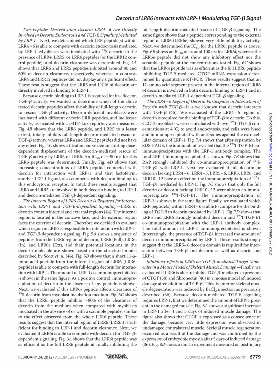
Two Peptides Derived from Decorin LRR4–6 Are DirectlyInvolved inDecorin Endocytosis andTGF-� SignalingMediatedby LRP-1—Next, we determined which LRR peptide(s) withinLRR4–6 is able to compete with decorin endocytosis mediatedby LRP-1. Myoblasts were incubated with 35S-decorin in thepresence of LRR4, LRR5, or LRR6 peptides (or the LRR12 con-trol peptide), and decorin clearance was determined. Fig. 4Ashows that LRR6 and LRR5 peptides inhibited around 80 and60% of decorin clearance, respectively, whereas, in contrast,LRR4 and LRR12 peptides did not display any significant effect.These results suggest that the LRR5 and LRR6 of decorin aredirectly involved in binding to LRP-1.Because decorin binding to LRP-1 is required for its effect on
TGF-� activity, we wanted to determine which of the abovetested decorin peptides affect the ability of full-length decorinto rescue TGF-� activity. Decorin-deficient myoblasts wereincubated with different decorin LRR peptides, and luciferaseactivity, associated with a p3TP-Lux reporter, was measured.Fig. 4B shows that the LRR6 peptide, and LRR5 to a lesserextent, totally inhibits full-length decorin-mediated rescue ofTGF-� activity, whereas LRR4 andLRR12peptides did not haveany effect. Fig. 4C shows a titration curve demonstrating dose-dependent displacement of the decorin-mediated rescue ofTGF-� activity by LRR5 or LRR6. An IC50 of �90 nM for thisLRR6 peptide was determined. Finally, Fig. 4D shows thatincreasing concentrations of LRR6 peptide competed withdecorin for interaction with LRP-1, and that lactoferrin,another LRP-1 ligand, also competes with decorin binding tothis endocytocic receptor. In total, these results suggest thatLRR6 and LRR5 are involved in both decorin binding to LRP-1and decorin-mediated TGF-�-dependent signaling.The Internal Region of LRR6 Decorin Is Required for Interac-
tion with LRP-1 and TGF-�-dependent Signaling—LRRs indecorin contain internal and external regions (44). The internalregion is located in the concave face, and the exterior regionfaces the exterior of decorin. Therefore we decided to evaluatewhich region in LRR6 is responsible for interactionwith LRP-1-and TGF-�-dependent signaling. Fig. 5A shows a sequence ofpeptides from the LRR6 region of decorin, LRR6 (Full), LRR6i(In), and LRR6e (Ext), and their potential locations in thedecorin molecule are shown based on the structural modeldescribed by Scott et al. (44). Fig. 5B shows that a short 11-a-mino acid peptide from the internal region of LRR6 (LRR6ipeptide) is able to compete with full-length decorin for interac-tionwith LRP-1. The amount of LRP-1 co-immunoprecipitatedis shown in the same figure. As a positive control, immunopre-cipitation of decorin in the absence of any peptide is shown.Next, we evaluated if this LRR6i peptide affects clearance of35S-decorin from myoblast incubation medium. Fig. 5C showsthat the LRR6i peptide inhibits �80% of the clearance ofdecorin from the medium when compared with myoblastsincubated in the absence of or with a scramble peptide, similarto the effect observed from the whole LRR6 peptide. Theseresults suggest that the internal region of LRR6 (LRR6i) is suf-ficient for binding to LRP-1 and decorin clearance. Next, weevaluated if LRR6i is able to compete with decorin for TGF-�-dependent signaling. Fig. 6A shows that the LRR6i peptide wasas efficient as the full LRR6 peptide at totally inhibiting the
full-length decorin-mediated rescue of TGF-� signaling. Thesame figure shows that a peptide corresponding to the externalregion of LRR6 (LRR6e) showed very little inhibitory activity.Next, we determined the IC50 for the LRR6i peptide as above.Fig. 6B shows an IC50 of around 100 nM for LRR6i, whereas theLRR6e peptide did not show any inhibitory effect nor thescramble peptide at the concentrations tested. Fig. 6C showsthat the LRR6i peptide was as efficient as the full LRR6 peptideinhibiting TGF-�-mediated CTGF mRNA expression deter-mined by quantitative RT-PCR. These results suggest that an11-amino acid segment present in the internal region of LRR6of decorin is involved in both decorin binding to LRP-1 and indecorin-mediated LRP-1-dependent TGF-� signaling.The LRR4–6 Region of Decorin Participates in Interaction of
Decorin with TGF-�—It is well known that decorin interactswith TGF-� (45). We evaluated if the LRR4–6 region ofdecorin is required for the binding ofTGF-� to decorin. To this,C2C12myoblasts were co-incubatedwith two 125I-TGF-� con-centrations at 4 °C, to avoid endocytosis, and cells were lysedand immunoprecipitated with antibodies against the extracel-lular domain of LRP-1. Fig. 7A shows that after separation bySDS-PAGE, the immunoblot revealed that the 125I-TGF-�1 co-immunoprecipitates with the LRP-1 antibody complex. Thetotal LRP-1 immunoprecipitated is shown. Fig. 7B shows thatRAP strongly inhibited the co-immunoprecipitation of 125I-TGF-�1 with LRP-1. Next, we evaluated if full decorin ordecorin lacking LRR4–6, LRR4–5, LRR5–6, LRR5, LRR6, andLRR10–12 have an effect on the immunoprecipitation of 125I-TGF-�1 mediated by LRP-1. Fig. 7C shows that only the fulldecorin or decorin lacking LRR10–12 were able to co-immu-noprecipitate 125I-TGF-�1. The immunoprecipitation ofLRP-1 is shown in the same figure. Finally, we evaluated whichLRR peptide(s) within LRR4–6 is able to compete for the bind-ing of TGF-� to decorinmediated by LRP-1. Fig. 7D shows thatLRR5 and LRR6 strongly inhibited decorin and 125I-TGF-�1co-immunoprecipitation with the LRP-1 antibody complex.The total amount of LRP-1 immunoprecipitated is shown.Interestingly, the presence of TGF-�1 increased the amount ofdecorin immunoprecipitated by LRP-1. These results stronglysuggest that the LRR5–6 decorin domain is required for inter-action between TGF-� and decorin as well as decorin andLRP-1.Inhibitory Effects of LRR6 on TGF-�-mediated Target Mole-
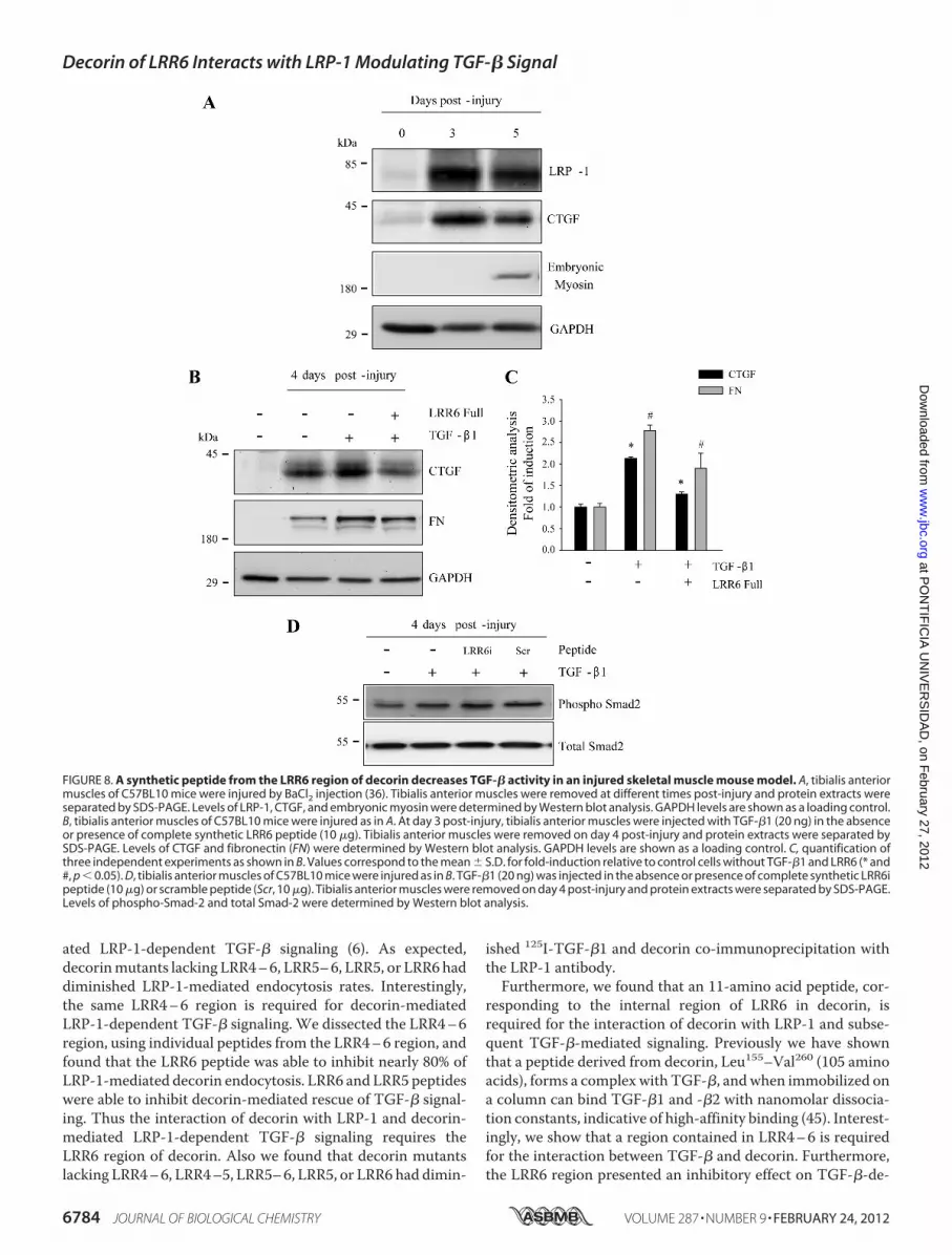
cules in aMouseModel of SkeletalMuscleDamage—Finally, weevaluated if LRR6 is able to inhibit TGF-�-mediated expressionof CTGF (38) and fibronectin (46) in amousemodel of induceddamage after addition of TGF-�. Tibialis anterior skeletal mus-cle degeneration was induced by BaCl2 injection as previouslydescribed (36). Because decorin-mediated TGF-� signalingrequires LRP-1, first we determined the amount of LRP-1 pres-ent in the damagedmuscle. Fig. 8A shows a significant increasein LRP-1 after 3 and 5 days of induced muscle damage. Thefigure also shows that CTGF is expressed as a consequence ofthe damage, because very little expression was observed inundamaged contralateral muscle. Skeletal muscle regenerationoccurred as a result of the damage and was confirmed by theexpression of embryonicmyosin after 5 days of induced damage(36). Fig. 8B shows a similar experimentmeasured on post-injury
Decorin of LRR6 Interacts with LRP-1 Modulating TGF-� Signal
FEBRUARY 24, 2012 • VOLUME 287 • NUMBER 9 JOURNAL OF BIOLOGICAL CHEMISTRY 6779
at PO
NT
IFIC
IA U
NIV
ER
SID
AD
, on February 27, 2012
ww
w.jbc.org
Dow
nloaded from
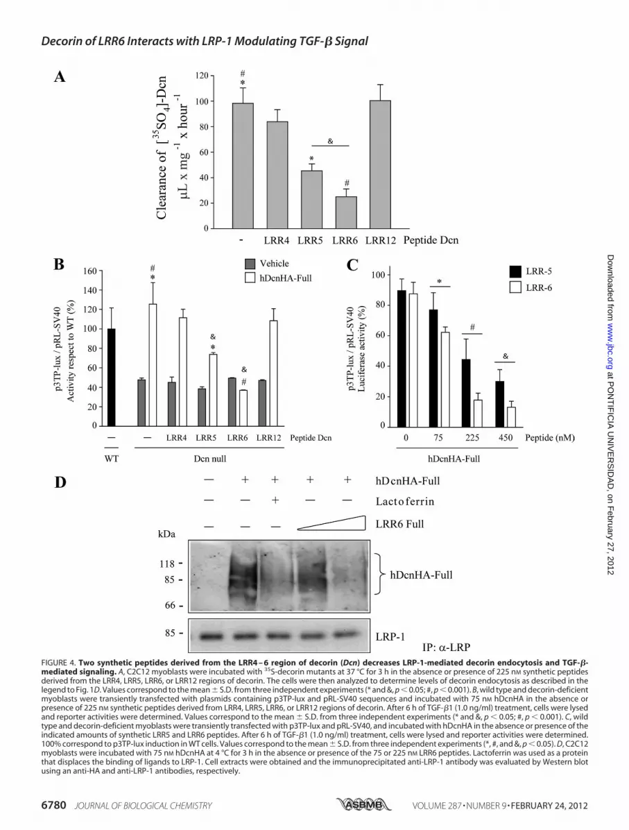
FIGURE 4. Two synthetic peptides derived from the LRR4 – 6 region of decorin (Dcn) decreases LRP-1-mediated decorin endocytosis and TGF-�-mediated signaling. A, C2C12 myoblasts were incubated with 35S-decorin mutants at 37 °C for 3 h in the absence or presence of 225 nM synthetic peptidesderived from the LRR4, LRR5, LRR6, or LRR12 regions of decorin. The cells were then analyzed to determine levels of decorin endocytosis as described in thelegend to Fig. 1D. Values correspond to the mean � S.D. from three independent experiments (* and &, p � 0.05; #, p � 0.001). B, wild type and decorin-deficientmyoblasts were transiently transfected with plasmids containing p3TP-lux and pRL-SV40 sequences and incubated with 75 nM hDcnHA in the absence orpresence of 225 nM synthetic peptides derived from LRR4, LRR5, LRR6, or LRR12 regions of decorin. After 6 h of TGF-�1 (1.0 ng/ml) treatment, cells were lysedand reporter activities were determined. Values correspond to the mean � S.D. from three independent experiments (* and &, p � 0.05; #, p � 0.001). C, wildtype and decorin-deficient myoblasts were transiently transfected with p3TP-lux and pRL-SV40, and incubated with hDcnHA in the absence or presence of theindicated amounts of synthetic LRR5 and LRR6 peptides. After 6 h of TGF-�1 (1.0 ng/ml) treatment, cells were lysed and reporter activities were determined.100% correspond to p3TP-lux induction in WT cells. Values correspond to the mean � S.D. from three independent experiments (*, #, and &, p � 0.05). D, C2C12myoblasts were incubated with 75 nM hDcnHA at 4 °C for 3 h in the absence or presence of the 75 or 225 nM LRR6 peptides. Lactoferrin was used as a proteinthat displaces the binding of ligands to LRP-1. Cell extracts were obtained and the immunoprecipitated anti-LRP-1 antibody was evaluated by Western blotusing an anti-HA and anti-LRP-1 antibodies, respectively.
Decorin of LRR6 Interacts with LRP-1 Modulating TGF-� Signal
6780 JOURNAL OF BIOLOGICAL CHEMISTRY VOLUME 287 • NUMBER 9 • FEBRUARY 24, 2012
at PO
NT
IFIC
IA U
NIV
ER
SID
AD
, on February 27, 2012
ww
w.jbc.org
Dow
nloaded from

day 4, where TGF-�1 was injected in the regenerating muscle onday3afterdamage induction.The figure shows thatCTGFexpres-sion increases in response to TGF-�. However, when LRR6 pep-tide was co-injected with TGF-�1 on day 3 after damage induc-tion, inhibition of CTGF induction was observed. In addition,LRR6peptide co-injection resulted in adecrease inTGF-�-depen-dent inductionof fibronectin. Fig. 8C showsaquantificationof this
experiment. Finally, we found that the level of phospho-Smad2 inresponse to TGF-�1 was not affected by LRR6i. As control a nulleffect of a scramble peptide on phospho-Smad2 is shown. Thisresult is in concordance with our previous observation that sug-gests that levels ofphospho-Smad2arenot affectedwhen theLRP-1-decorin-dependent TGF-�-mediated signaling pathway is acti-vated (6). This in vivo experiment strongly suggests that LRR6
FIGURE 5. A peptide containing the internal region of LRR6 decorin competes with full-length decorin for binding to LRP-1 and reducesLRP-1-mediated decorin endocytosis. A, sequence of peptides from the LRR6 region of decorin, LRR6 (Full), LRR6i (In), and LRR6e (Ext), are depictedand their potential locations in the three-dimensional decorin molecule are shown based on the structural model described by Scott et al. (44). B, C2C12myoblasts were incubated with 75 nM hDcnHA at 4 °C for 3 h in the absence or presence of 225 nM of the synthetic peptides derived from LRR6 asdescribed in A, cell extracts were immunoprecipitated with anti-LRP-1 antibody and the presence of hDcnHA and LRP-1 in the immunoprecipitate wereevaluated by Western blot using anti-HA and anti-LRP-1 antibodies, respectively. C, C2C12 myoblasts were incubated with 35S-decorin mutants at 37 °Cfor 3 h in the absence or presence of synthetic peptides from the LRR6 region of decorin as described in A and B, or scramble peptide. The cells were thenanalyzed to determinate decorin endocytosis levels as described in the legend to Fig. 1C. Values correspond to the mean � S.D. from three independentexperiments (* and #, p � 0.001).
Decorin of LRR6 Interacts with LRP-1 Modulating TGF-� Signal
FEBRUARY 24, 2012 • VOLUME 287 • NUMBER 9 JOURNAL OF BIOLOGICAL CHEMISTRY 6781
at PO
NT
IFIC
IA U
NIV
ER
SID
AD
, on February 27, 2012
ww
w.jbc.org
Dow
nloaded from
inhibits the induction of fibronectin and CTGF, mediated byTGF-�1 signaling, in damagedmuscle.
DISCUSSION
In this article, we show that the LRR6 and LRR5 regions ofdecorin are vital for binding to LRP-1 and TGF-�-mediated
signaling. Furthermore, we determined that the internal regionof LRR6, an 11-amino acid peptide on the concave face ofdecorin, is critical for the two functions described above. Usingdifferent deletions mutants we found that LRR4–6 is essentialfor interaction with LRP-1, the endocytic receptor for decorin(5), interacting with TGF-�, and induction of decorin-medi-
FIGURE 6. TGF-� activity is decreased by a synthetic peptide derived from the LRR6 internal region of decorin (Dcn). A, wild type and decorin-deficientmyoblasts were transiently transfected with plasmids containing p3TP-lux and pRL-SV40 sequences and incubated with 75 nM hDcnHA in the absence orpresence of 225 nM synthetic peptides from the LRR6 region of decorin, LRR6 (Full), LRR6i (In), or LRR6e (Ext). After 6 h of TGF-�1 (1.0 ng/ml) treatment, cells werelysed and reporter activities were determined. Values correspond to the mean � S.D. from three independent experiments (*, #, and &, p � 0.05). B, wild typeand decorin-deficient myoblasts were transiently transfected with p3TP-lux and pRL-SV40 plasmids and incubated with 75 nM hDcnHA-Full in the absence orpresence of different amounts of LRR6 (Full, In, or Ext) or scramble peptide (Scramble). Treatment with TGF-�1 (1.0 ng/ml) and reporter activities weredetermined as in A. Values correspond to the mean � S.D. from three independent experiments (*, p � 0.05; # and &, p � 0.001). C, decorin-deficient myoblastswere incubated with TGF-�1 (5.0 ng/ml) plus 75 nM hDcnHA in the absence or presence of 225 nM synthetic peptides LRR6: Full, In, or Ext, or scramble (Scr)peptide. Total RNA was isolated and CTGF and GAPDH expression was determined by qRT-PCR according to “Experimental Procedures.” Values correspond tothe mean of dCT value � S.D. of three independent experiments (* and #, p � 0.05).
Decorin of LRR6 Interacts with LRP-1 Modulating TGF-� Signal
6782 JOURNAL OF BIOLOGICAL CHEMISTRY VOLUME 287 • NUMBER 9 • FEBRUARY 24, 2012
at PO
NT
IFIC
IA U
NIV
ER
SID
AD
, on February 27, 2012
ww
w.jbc.org
Dow
nloaded from
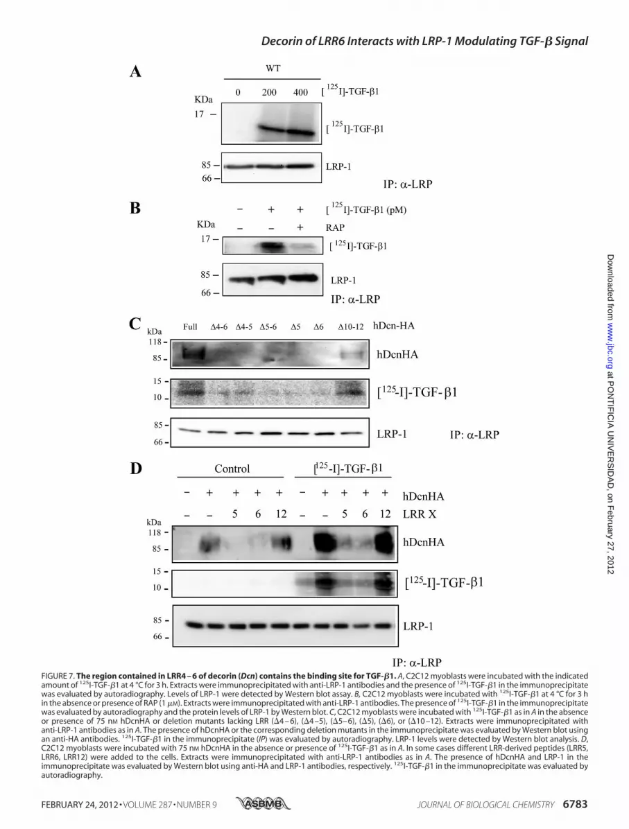
FIGURE 7. The region contained in LRR4 – 6 of decorin (Dcn) contains the binding site for TGF-�1. A, C2C12 myoblasts were incubated with the indicatedamount of 125I-TGF-�1 at 4 °C for 3 h. Extracts were immunoprecipitated with anti-LRP-1 antibodies and the presence of 125I-TGF-�1 in the immunoprecipitatewas evaluated by autoradiography. Levels of LRP-1 were detected by Western blot assay. B, C2C12 myoblasts were incubated with 125I-TGF-�1 at 4 °C for 3 hin the absence or presence of RAP (1 �M). Extracts were immunoprecipitated with anti-LRP-1 antibodies. The presence of 125I-TGF-�1 in the immunoprecipitatewas evaluated by autoradiography and the protein levels of LRP-1 by Western blot. C, C2C12 myoblasts were incubated with 125I-TGF-�1 as in A in the absenceor presence of 75 nM hDcnHA or deletion mutants lacking LRR (�4 – 6), (�4 –5), (�5– 6), (�5), (�6), or (�10 –12). Extracts were immunoprecipitated withanti-LRP-1 antibodies as in A. The presence of hDcnHA or the corresponding deletion mutants in the immunoprecipitate was evaluated by Western blot usingan anti-HA antibodies. 125I-TGF-�1 in the immunoprecipitate (IP) was evaluated by autoradiography. LRP-1 levels were detected by Western blot analysis. D,C2C12 myoblasts were incubated with 75 nM hDcnHA in the absence or presence of 125I-TGF-�1 as in A. In some cases different LRR-derived peptides (LRR5,LRR6, LRR12) were added to the cells. Extracts were immunoprecipitated with anti-LRP-1 antibodies as in A. The presence of hDcnHA and LRP-1 in theimmunoprecipitate was evaluated by Western blot using anti-HA and LRP-1 antibodies, respectively. 125I-TGF-�1 in the immunoprecipitate was evaluated byautoradiography.
Decorin of LRR6 Interacts with LRP-1 Modulating TGF-� Signal
FEBRUARY 24, 2012 • VOLUME 287 • NUMBER 9 JOURNAL OF BIOLOGICAL CHEMISTRY 6783
at PO
NT
IFIC
IA U
NIV
ER
SID
AD
, on February 27, 2012
ww
w.jbc.org
Dow
nloaded from
ated LRP-1-dependent TGF-� signaling (6). As expected,decorinmutants lacking LRR4–6, LRR5–6, LRR5, or LRR6 haddiminished LRP-1-mediated endocytosis rates. Interestingly,the same LRR4–6 region is required for decorin-mediatedLRP-1-dependent TGF-� signaling. We dissected the LRR4–6region, using individual peptides from the LRR4–6 region, andfound that the LRR6 peptide was able to inhibit nearly 80% ofLRP-1-mediated decorin endocytosis. LRR6 and LRR5 peptideswere able to inhibit decorin-mediated rescue of TGF-� signal-ing. Thus the interaction of decorin with LRP-1 and decorin-mediated LRP-1-dependent TGF-� signaling requires theLRR6 region of decorin. Also we found that decorin mutantslacking LRR4–6, LRR4–5, LRR5–6, LRR5, or LRR6 had dimin-
ished 125I-TGF-�1 and decorin co-immunoprecipitation withthe LRP-1 antibody.Furthermore, we found that an 11-amino acid peptide, cor-
responding to the internal region of LRR6 in decorin, isrequired for the interaction of decorin with LRP-1 and subse-quent TGF-�-mediated signaling. Previously we have shownthat a peptide derived from decorin, Leu155–Val260 (105 aminoacids), forms a complex with TGF-�, andwhen immobilized ona column can bind TGF-�1 and -�2 with nanomolar dissocia-tion constants, indicative of high-affinity binding (45). Interest-ingly, we show that a region contained in LRR4–6 is requiredfor the interaction between TGF-� and decorin. Furthermore,the LRR6 region presented an inhibitory effect on TGF-�-de-
FIGURE 8. A synthetic peptide from the LRR6 region of decorin decreases TGF-� activity in an injured skeletal muscle mouse model. A, tibialis anteriormuscles of C57BL10 mice were injured by BaCl2 injection (36). Tibialis anterior muscles were removed at different times post-injury and protein extracts wereseparated by SDS-PAGE. Levels of LRP-1, CTGF, and embryonic myosin were determined by Western blot analysis. GAPDH levels are shown as a loading control.B, tibialis anterior muscles of C57BL10 mice were injured as in A. At day 3 post-injury, tibialis anterior muscles were injected with TGF-�1 (20 ng) in the absenceor presence of complete synthetic LRR6 peptide (10 �g). Tibialis anterior muscles were removed on day 4 post-injury and protein extracts were separated bySDS-PAGE. Levels of CTGF and fibronectin (FN) were determined by Western blot analysis. GAPDH levels are shown as a loading control. C, quantification ofthree independent experiments as shown in B. Values correspond to the mean � S.D. for fold-induction relative to control cells without TGF-�1 and LRR6 (* and#, p � 0.05). D, tibialis anterior muscles of C57BL10 mice were injured as in B. TGF-�1 (20 ng) was injected in the absence or presence of complete synthetic LRR6ipeptide (10 �g) or scramble peptide (Scr, 10 �g). Tibialis anterior muscles were removed on day 4 post-injury and protein extracts were separated by SDS-PAGE.Levels of phospho-Smad-2 and total Smad-2 were determined by Western blot analysis.
Decorin of LRR6 Interacts with LRP-1 Modulating TGF-� Signal
6784 JOURNAL OF BIOLOGICAL CHEMISTRY VOLUME 287 • NUMBER 9 • FEBRUARY 24, 2012
at PO
NT
IFIC
IA U
NIV
ER
SID
AD
, on February 27, 2012
ww
w.jbc.org
Dow
nloaded from
pendent signaling in the nanomolar range. However, whetherthe same decorin sequence is responsible for TGF-� binding,interaction with LRP-1, and TGF-�-mediated signaling (47)requires further investigation. We have previously demon-strated that decorin, among other proteoglycans, is able to bindand sequester TGF-� from its transducing receptors, thusmodulating the bioavailability of this growth factor (18). Itwould be interesting to evaluate if the LRR6 region can competewith decorin for TGF-� binding.
A sequence similar to LRR6i has been reported to bind col-lagen type I (48). Furthermore, as indicated in the Introduction,decorin interacts with EGFR (14, 50). Santra et al. (51) investi-gated the structural requirements of the decorin/EGFR inter-action, and their results suggest that the LRR6 region is alsorequired for proper interaction of decorin and EGFR. It is strik-ing that several of the different biological functions attributedto decorin, such as receptor or ligand binding, reside in thesame structural region. Some small LRR proteoglycans, such asdecorin, have been shown to dimerize with high affinity (13, 44,52). Interestingly, the crystal structure of decorin indicates thatdecorin dimerizes through the concave surfaces of its LRRdomains, some of which correspond to the sequence describedto be responsible for its protein-ligand interactions (44). It hasbeen argued that the sequence of decorin that binds to collagenI (which is similar to the sequence described in this paper forLRP-1 binding and TGF-� signaling) is located on the concaveface of LRR6, a position that would be less accessible for triplehelical collagen (14). It has also been argued that, given theoverall dimensions of the decorin core protein, a dimericdecorin would not fit in the EGFR receptor groove where EGFbinds (14). In contrast, Scott et al. (44) argue, based on theircrystal structure studies, that decorin has a more open struc-ture that seems incompatible with a tight interaction with asingle collagen triple helix. Furthermore, the concave surface ofdecorin has been postulated to be involved in a high-affinitydimer interaction, and is thus unlikely to be available for ligandbinding (44). However, we have experimental evidence fromchemical cross-linking assays that the LRR4, LRR5, and LRR6regions of decorin do not affect decorin dimer formation (25).Clearly the role of the dimer-monomer transition and its func-tion in the binding of decorin to specific ligands and receptorsrequires further investigation. Interestingly, we have recentlydemonstrated that decorin interacts with CTGF, this interac-tion is mediated by LRR12 (25). The fact that decorin is able tointeract with two different pro-fibrotic molecules, TGF-� andCTGF, and these molecules also interact with LRP-1 (25, 53),open interesting possibilities of fine tune regulation mediatedby decorin and these growth factors. Furthermore, an increaseinTGF-� activitymediated byCTGFhas been described (54). Ifdecorin modulates then this augmented activity requires fur-ther investigation.Fibrotic disorders are characterized by excessive connective
tissue and ECM deposition that preclude the normal healing ofdifferent tissues (55). Marked overexpression of TGF-� andCTGF, both profibrotic cytokines, is strongly linked to thepathogenesis of these diseases. CTGF is selectively induced byTGF-�1 (38, 40, 56), and TGF-� and CTGF are coordinatelyexpressed at sites of tissue repair and fibrosis (57, 58). In addi-
tion, production of TGF-� is altered in many pathologic condi-tions. For example, TGF-� is overproduced as in pulmonaryfibrosis, cirrhosis, glomerulosclerosis, cardiomyopathy, Crohndisease, scleroderma and chronic graft versus host disease (55,59). Several studies have indicated that TGF-� is a potentinducer of the myofibroblast phenotype (60, 61), which corre-lates with induction of elevated collagen synthesis but is pre-vented by induction of cell proliferation. Thus, although it isapparent that targeting TGF-� for therapy is of major clinicalinterest, to date no therapeutic treatment for fibrosis exist (30).We present experimental evidence that indicates that LRR6
inhibits (at least in part) expression of TGF-� target proteins invivo in response to skeletal muscle damage. We evaluated amouse model where the expression of TGF-� is elevated (62)concomitant with the expression of CTGF and fibronectin: aninduced skeletal muscle damage model (36). Thus, inducedskeletal muscle damage (63) was associated with reducedexpression of CTGF and fibronectin if the LRR6 region wasco-injected with TGF-�. Because the effect of LRR6 on TGF-�-mediated activity requires the presence of LRP-1, we deter-mined levels of this endocytic receptor protein.Muscle damagestrongly induced LRP-1 expression in skeletal muscles. In thisexperimentalmodel it has been shown that decorin is also pres-ent (63). Furthermore, there is some evidence in the literaturethat LRP-1 is required for fibrotic responses mediated byTGF-�1 (64). It has also been demonstrated that TGF-� recep-tor-V is identical to thr LRP-1/�2-macrogloblin receptor (53).Thus, the CTGF response would bemediated by LRP-1 expres-sion (49). Results3 from our laboratory show elevated levels ofLRP-1 in dystrophic skeletal muscles compared with wild typemice. Therefore the use of LRR6 peptides, or a modified LRR6molecule with higher biological activity, to modulate decorin-mediated LRP-1-dependent TGF-� signaling, opens the possi-bility of designing pharmaceutical strategies for use in the fightagainst fibrosis, a severe consequence ofmany chronic diseases.Excess connective tissue in dystrophic skeletal muscles, as
well as in other skeletalmuscular dystrophies, could be a barrierfor successful cell therapy approaches, as exemplified by the useof tendon fibroblasts expressing a metalloproteinase (matrixmetalloproteinase-9), which restored the vascular network andsignificantly reduced collagen deposition, allowing for efficientcell therapy in aged dystrophic mice (37). Thus, the fact thatthis short LRR6 peptide derived from decorin is directlyinvolved in TGF-� signaling is an important finding withpotential therapeutic implications.
REFERENCES1. Massagué, J. (1998) TGF-� signal transduction. Annu. Rev. Biochem. 67,
753–7912. Gray, P. C., Bilezikjian, L. M., and Vale, W. (2002) Antagonism of activin
by inhibin and inhibin receptors. A functional role for betaglycan. Mol.Cell. Endocrinol. 188, 254–260
3. Massagué, J., andGomis, R. R. (2006) The logic of TGF-�a signaling. FEBSLett. 580, 2811–2820
4. Kang, J. S., Liu, C., and Derynck, R. (2009) New regulatory mechanisms ofTGF-� receptor function. Trends Cell Biol. 19, 385–394
3 C. Cabello-Verrugio, C. Santander, C. Cofré, M. J. Acuña, F. Melo, and E. Bran-dan, unpublished results.
Decorin of LRR6 Interacts with LRP-1 Modulating TGF-� Signal
FEBRUARY 24, 2012 • VOLUME 287 • NUMBER 9 JOURNAL OF BIOLOGICAL CHEMISTRY 6785
at PO
NT
IFIC
IA U
NIV
ER
SID
AD
, on February 27, 2012
ww
w.jbc.org
Dow
nloaded from
5. Brandan, E., Retamal, C., Cabello-Verrugio, C., andMarzolo, M. P. (2006)The low density lipoprotein receptor-related protein functions as an en-docytic receptor for decorin. J. Biol. Chem. 281, 31562–31571
6. Cabello-Verrugio, C., and Brandan, E. (2007) A novel modulatory mech-anism of transforming growth factor-� signaling through decorin andLRP-1. J. Biol. Chem. 282, 18842–18850
7. Herz, J., and Strickland, D. K. (2001) LRP, amultifunctional scavenger andsignaling receptor. J. Clin. Invest. 108, 779–784
8. Gonias, S. L., Wu, L., and Salicioni, A. M. (2004) Low density lipoproteinreceptor-related protein. Regulation of the plasma membrane proteome.Thromb. Haemost. 91, 1056–1064
9. Brandan, E., Cabello-Verrugio, C., and Vial, C. (2008) Novel regulatorymechanisms for the proteoglycans decorin and biglycan during muscleformation and muscular dystrophy.Matrix Biol. 27, 700–708
10. Riquelme, C., Larrain, J., Schonherr, E., Henriquez, J. P., Kresse, H., andBrandan, E. (2001) Antisense inhibition of decorin expression in myo-blasts decreases cell responsiveness to transforming growth factor betaand accelerates skeletal muscle differentiation. J. Biol. Chem. 276,3589–3596
11. Derynck, R., and Zhang, Y. E. (2003) Smad-dependent and Smad-inde-pendent pathways in TGF-� family signaling. Nature 425, 577–584
12. Iozzo, R. V. (1999) The biology of the small leucine-rich proteoglycans.Functional network of interactive proteins. J. Biol. Chem. 274,18843–18846
13. McEwan, P. A., Scott, P. G., Bishop, P. N., and Bella, J. (2006) Structuralcorrelations in the family of small leucine-rich repeat proteins and pro-teoglycans. J. Struct. Biol. 155, 294–305
14. Schaefer, L., and Iozzo, R. V. (2008) Biological functions of the small leu-cine-rich proteoglycans. From genetics to signal transduction. J. Biol.Chem. 283, 21305–21309
15. Kobe, B., and Kajava, A. V. (2001) The leucine-rich repeat as a proteinrecognition motif. Curr. Opin. Struct. Biol. 11, 725–732
16. Cáceres, S., Cuellar, C., Casar, J. C., Garrido, J., Schaefer, L., Kresse, H., andBrandan, E. (2000) Synthesis of proteoglycans is augmented in dystrophicmdx mouse skeletal muscle. Eur. J. Cell Biol. 79, 173–181
17. Fadic, R., Mezzano, V., Alvarez, K., Cabrera, D., Holmgren, J., and Bran-dan, E. (2006) Increase in decorin and biglycan in Duchenne musculardystrophy. Role of fibroblasts as cell source of these proteoglycans in thedisease. J. Cell. Mol. Med. 10, 758–769
18. Droguett, R., Cabello-Verrugio, C., Riquelme, C., and Brandan, E. (2006)Extracellular proteoglycans modify TGF-� bioavailability attenuating itssignaling during skeletal muscle differentiation.Matrix Biol. 25, 332–341
19. Iozzo, R. V., and Schaefer, L. (2010) Proteoglycans in health and disease.Novel regulatory signaling mechanisms evoked by the small leucine-richproteoglycans. FEBS J. 277, 3864–3875
20. Schönherr, E., Sunderkötter, C., Iozzo, R. V., and Schaefer, L. (2005)Decorin, a novel player in the insulin-like growth factor system. J. Biol.Chem. 280, 15767–15772
21. Schaefer, L., Tsalastra, W., Babelova, A., Baliova, M., Minnerup, J., So-rokin, L., Gröne, H. J., Reinhardt, D. P., Pfeilschifter, J., Iozzo, R. V., andSchaefer, R. M. (2007) Decorin-mediated regulation of fibrillin-1 in thekidney involves the insulin-like growth factor-I receptor and mammaliantarget of rapamycin. Am. J. Pathol. 170, 301–315
22. Moscatello, D. K., Santra, M., Mann, D. M., McQuillan, D. J., Wong, A. J.,and Iozzo, R. V. (1998)Decorin suppresses tumor cell growth by activatingthe epidermal growth factor receptor. J. Clin. Invest. 101, 406–412
23. Takeuchi, Y., Kodama, Y., andMatsumoto, T. (1994) Bonematrix decorinbinds transforming growth factor-� and enhances its bioactivity. J. Biol.Chem. 269, 32634–32638
24. Yamaguchi, Y., Mann, D. M., and Ruoslahti, E. (1990) Negative regulationof transforming growth factor-� by the proteoglycan decorin.Nature 346,281–284
25. Vial, C., Gutiérrez, J., Santander, C., Cabrera, D., and Brandan, E. (2011)Decorin interacts with connective tissue growth factor (CTGF)/CCN2 byLRR12 inhibiting its biological activity. J. Biol. Chem. 286, 24242–24252
26. Goldoni, S., Humphries, A., Nyström, A., Sattar, S., Owens, R. T.,McQuil-lan, D. J., Ireton, K., and Iozzo, R. V. (2009) Decorin is a novel antagonisticligand of the Met receptor. J. Cell Biol. 185, 743–754
27. Olguin,H.C., Santander, C., andBrandan, E. (2003) Inhibition ofmyoblastmigration via decorin expression is critical for normal skeletal muscledifferentiation. Dev. Biol. 259, 209–224
28. Bolster, M. B., and Silver, R. M. (1993) Lung disease in systemic sclerosis(scleroderma). Baillieres Clin. Rheumatol. 7, 79–97
29. Ziyadeh, F. N. (1993) The extracellular matrix in diabetic nephropathy.Am. J. Kidney. Dis. 22, 736–744
30. Wynn, T. A. (2008) Cellular and molecular mechanisms of fibrosis.J. Pathol. 214, 199–210
31. Denton, C. P., and Abraham, D. J. (2001) Transforming growth factor-�and connective tissue growth factor. Key cytokines in scleroderma patho-genesis. Curr. Opin. Rheumatol. 13, 505–511
32. Bernasconi, P., Di Blasi, C., Mora, M., Morandi, L., Galbiati, S., Confaloni-eri, P., Cornelio, F., and Mantegazza, R. (1999) Transforming growth fac-tor-�1 and fibrosis in congenital muscular dystrophies. Neuromusc. Dis-ord. 9, 28–33
33. Alvarez, K., Fadic, R., and Brandan, E. (2002) Augmented synthesis anddifferential localization of heparan sulfate proteoglycans in Duchennemuscular dystrophy. J. Cell. Biochem. 85, 703–713
34. Schiaffino, S., and Partridge, T. (2008) Advances in Muscle Research, Vol.3, pp. XIV-380, Springer, The Netherlands
35. Caldwell, C. J.,Mattey, D. L., andWeller, R.O. (1990) Role of the basementmembrane in the regeneration of skeletal muscle. Neuropathol. Appl.Neurobiol. 16, 225–238
36. Casar, J. C., Cabello-Verrugio, C., Olguin, H., Aldunate, R., Inestrosa,N. C., and Brandan, E. (2004) Heparan sulfate proteoglycans are increasedduring skeletal muscle regeneration. Requirement of syndecan-3 for suc-cessful fiber formation. J. Cell Sci. 117, 73–84
37. Gargioli, C., Coletta, M., De Grandis, F., Cannata, S. M., and Cossu, G.(2008) PlGF-MMP-9-expressing cells restore microcirculation and effi-cacy of cell therapy in aged dystrophic muscle. Nat. Med. 14, 973–978
38. Vial, C., Zúñiga, L. M., Cabello-Verrugio, C., Cañón, P., Fadic, R., andBrandan, E. (2008) Skeletal muscle cells express the profibrotic cytokineconnective tissue growth factor (CTGF/CCN2), which induces their ded-ifferentiation. J. Cell. Physiol. 215, 410–421
39. Brandan, E., Carey, D. J., Larraín, J., Melo, F., and Campos, A. (1996)Synthesis and processing of glypican during differentiation of skeletalmuscle cells. Eur. J. Cell Biol. 71, 170–176
40. Cabello-Verrugio, C., Córdova, G., Vial, C., Zúñiga, L.M., and Brandan, E.(2011) Connective tissue growth factor induction by lysophosphatidicacid requires transactivation of transforming growth factor type � recep-tors and the JNK pathway. Cell. Signal. 23, 449–457
41. Kresse, H., and Schönherr, E. (2001) Proteoglycans of the extracellularmatrix and growth control. J. Cell. Physiol. 189, 266–274
42. Giri, S. N. (2003) Novel pharmacological approaches to manage intersti-tial lung fibrosis in the twenty-first century. Annu. Rev. Pharmacol. Toxi-col. 43, 73–95
43. Krusius, T., and Ruoslahti, E. (1986) Primary structure of an extracellularmatrix proteoglycan core protein deduced from cloned cDNA. Proc. Natl.Acad. Sci. U.S.A. 83, 7683–7687
44. Scott, P. G., McEwan, P. A., Dodd, C. M., Bergmann, E. M., Bishop, P. N.,andBella, J. (2004)Crystal structure of the dimeric protein core of decorin,the archetypal small leucine-rich repeat proteoglycan. Proc. Natl. Acad.Sci. U.S.A. 101, 15633–15638
45. Schönherr, E., Broszat,M., Brandan, E., Bruckner, P., andKresse,H. (1998)Decorin core protein fragment Leu155–Val260 interacts with TGF-� butdoes not compete for decorin binding to type I collagen. Arch. Biochem.Biophys. 355, 241–248
46. Varga, J., Rosenbloom, J., and Jimenez, S. A. (1987) Transforming growthfactor � (TGF-�) causes a persistent increase in steady-state amounts oftype I and type III collagen and fibronectin mRNAs in normal humandermal fibroblasts. Biochem. J. 247, 597–604
47. Hildebrand, A., Romarís, M., Rasmussen, L. M., Heinegård, D., Twardzik,D. R., Border, W. A., and Ruoslahti, E. (1994) Interaction of the smallinterstitial proteoglycans biglycan, decorin, and fibromodulin with trans-forming growth factor �. Biochem. J. 302, 527–534
48. Kalamajski, S., Aspberg, A., and Oldberg, A. (2007) The decorin sequenceSYIRIADTNIT binds collagen type I. J. Biol. Chem. 282, 16062–16067
Decorin of LRR6 Interacts with LRP-1 Modulating TGF-� Signal
6786 JOURNAL OF BIOLOGICAL CHEMISTRY VOLUME 287 • NUMBER 9 • FEBRUARY 24, 2012
at PO
NT
IFIC
IA U
NIV
ER
SID
AD
, on February 27, 2012
ww
w.jbc.org
Dow
nloaded from

49. Segarini, P. R., Nesbitt, J. E., Li, D., Hays, L. G., Yates, J. R., 3rd, andCarmichael, D. F. (2001) The low density lipoprotein receptor-relatedprotein/�2-macroglobulin receptor is a receptor for connective tissuegrowth factor. J. Biol. Chem. 276, 40659–40667
50. Iozzo, R. V., and Karamanos, N. (2010) Proteoglycans in health and dis-ease. Emerging concepts and future directions. FEBS J. 277, 3863
51. Santra, M., Reed, C. C., and Iozzo, R. V. (2002) Decorin binds to a narrowregion of the epidermal growth factor (EGF) receptor, partially overlap-ping but distinct from the EGF-binding epitope. J. Biol. Chem. 277,35671–35681
52. Orgel, J. P., Eid, A., Antipova, O., Bella, J., and Scott, J. E. (2009) Decorincore protein (decoron) shape complements collagen fibril surface struc-ture and mediates its binding. PloS One 4, e7028
53. Huang, S. S., Ling, T. Y., Tseng, W. F., Huang, Y. H., Tang, F. M., Leal,S. M., and Huang, J. S. (2003) Cellular growth inhibition by IGFBP-3 andTGF-�1 requires LRP-1. FASEB J. 17, 2068–2081
54. Abreu, J. G., Ketpura, N. I., Reversade, B., and De Robertis, E. M. (2002)Connective tissue growth factor (CTGF) modulates cell signaling by BMPand TGF-�. Nat. Cell Biol. 4, 599–604
55. Prud’homme,G. J. (2007) Pathobiology of transforming growth factor� incancer, fibrosis, and immunologic disease, and therapeutic consider-ations. Lab. Invest. 87, 1077–1091
56. Duncan, M. R., Frazier, K. S., Abramson, S., Williams, S., Klapper, H.,Huang, X., and Grotendorst, G. R. (1999) Connective tissue growth factormediates transforming growth factor �-induced collagen synthesis.Down-regulation by cAMP. FASEB J. 13, 1774–1786
57. Frazier, K.,Williams, S., Kothapalli, D., Klapper,H., andGrotendorst, G. R.(1996) Stimulation of fibroblast cell growth, matrix production, and gran-ulation tissue formation by connective tissue growth factor. J. Invest. Der-
matol. 107, 404–41158. Shinozaki, M., Kawara, S., Hayashi, N., Kakinuma, T., Igarashi, A., and
Takehara, K. (1997) Induction of subcutaneous tissue fibrosis in newbornmice by transforming growth factor �. Simultaneous application with ba-sic fibroblast growth factor causes persistent fibrosis. Biochem. Biophys.Res. Commun. 240, 292–297
59. Pohlers, D., Brenmoehl, J., Löffler, I., Müller, C. K., Leipner, C., Schultze-Mosgau, S., Stallmach, A., Kinne, R. W., and Wolf, G. (2009) TGF-� andfibrosis in different organs. Molecular pathway imprints. Biochim. Bio-phys. Acta 1792, 746–756
60. Desmoulière, A., Geinoz, A., Gabbiani, F., and Gabbiani, G. (1993) Trans-forming growth factor-� 1 induces �-smooth muscle actin expression ingranulation tissue myofibroblasts and in quiescent and growing culturedfibroblasts. J. Cell Biol. 122, 103–111
61. Folger, P. A., Zekaria, D., Grotendorst, G., and Masur, S. K. (2001) Trans-forming growth factor-�-stimulated connective tissue growth factor ex-pression during cornealmyofibroblast differentiation. Invest. Ophthalmol.Vis. Sci. 42, 2534–2541
62. McLennan, I. S., and Koishi, K. (1997) Cellular localization of transform-ing growth factor-�2 and -�3 (TGF-�2, TGF-�3) in damaged and regen-erating skeletal muscles. Dev. Dyn. 208, 278–289
63. Casar, J. C., McKechnie, B. A., Fallon, J. R., Young, M. F., and Brandan, E.(2004) Transient up-regulation of biglycan during skeletal muscle regen-eration. Delayed fiber growth along with decorin increase in biglycan-deficient mice. Dev. Biol. 268, 358–371
64. Hu, K., Wu, C., Mars, W. M., and Liu, Y. (2007) Tissue-type plasminogenactivator promotes murine myofibroblast activation through LDL recep-tor-related protein 1-mediated integrin signaling. J. Clin. Invest. 117,3821–3832
Decorin of LRR6 Interacts with LRP-1 Modulating TGF-� Signal
FEBRUARY 24, 2012 • VOLUME 287 • NUMBER 9 JOURNAL OF BIOLOGICAL CHEMISTRY 6787
at PO
NT
IFIC
IA U
NIV
ER
SID
AD
, on February 27, 2012
ww
w.jbc.org
Dow
nloaded from