extracellular matrix in multiple sclerosis lesions: fibrillar coliagens, biglycan and decorin are...
TRANSCRIPT

R E S E A R C H A R T I C L E bpa_399 966..975
Extracellular Matrix in Multiple Sclerosis Lesions: FibrillarCollagens, Biglycan and Decorin are Upregulated andAssociated with Infiltrating Immune CellsHema Mohan1,2; Markus Krumbholz1,2; Rakhi Sharma3; Sylvia Eisele1,2; Andreas Junker1,2; Michael Sixt4;Jia Newcombe5; Hartmut Wekerle2; Reinhard Hohlfeld1,2; Hans Lassmann3; Edgar Meinl1,2
1 Institute for Clinical Neuroimmunology, Ludwig Maximilians University, Munich, Germany.2 Department of Neuroimmunology, Max-Planck-Institute of Neurobiology, Martinsried, Germany.4 Department of Molecular Medicine, Max-Planck-Institute of Biochemistry, Martinsried, Germany.3 Center for Brain Research, Medical University of Vienna, Vienna, Austria.5 NeuroResource, U.C.L Institute of Neurology, London, UK.
AbstractExtracellular matrix (ECM) proteins can modify immune reactions, e.g. by sequestering ordisplaying growth factors and by interacting with immune and glial cells. Here we quanti-fied by quantitative polymerase chain reaction (qPCR) expression of 50 ECM componentsand 34 ECM degrading enzymes in multiple sclerosis (MS) active and inactive white matterlesions. COL1A1, COL3A1, COL5A1 and COL5A2 chains were induced strongly in activelesions and even more in inactive lesions. These chains interact to form collagen types I, IIIand V, which are fibrillar collagens. Biglycan and decorin, which can decorate fibrillarcollagens, were also induced strongly. The fibrillar collagens, biglycan and decorin werelargely found between the endothelium and astrocytic glia limitans in the perivascular spacewhere they formed a meshwork which was closely associated with infiltrating immune cells.In active lesions collagen V was also seen in the heavily infiltrated parenchyma. Fibrillarcollagens I and III inhibited in vitro human monocyte production of CCL2 (MCP-1), aninflammatory chemokine involved in recruitment of immune cells. Together, ECM changesin lesions with different activities were quantified and proteins forming a perivascularfibrosis were identified. Induced fibrillar collagens may contribute to limiting enlargementof MS lesions by inhibiting the production of CCL2 by monocytes.
Keywords
extracellular matrix, inflammation, multiplesclerosis, neuroimmunology.
Corresponding author:
Dr. Edgar Meinl, MD, Department ofNeuroimmunology, Max-Planck-Institute ofNeurobiology, Am Klopferspitz 18, 82152Martinsried, Germany (E-mail:[email protected])
Received 3 February 2010; accepted 16 March2010.
Grant support: DeutscheForschungsgemeinschaft (SFB 571), Hermannand Lilly Schilling Foundation, Verein zurTherapieforschung für MultipleSklerose-Kranke, BMBF (krankheitsbezogenesKompetenznetz Multiple Sklerose), ExcellencyInitiative of the Ludwig Maximilians UniversityMunich.
doi:10.1111/j.1750-3639.2010.00399.x
INTRODUCTIONThe pathological hallmarks of active multiple sclerosis (MS)lesions are blood–brain barrier (BBB) disruption, inflammationand demyelination with axonal damage. The interactions betweeninfiltrating immune cells and the central nervous system (CNS)environment, made up of both cellular surfaces and the extracellu-lar matrix (ECM), contributes to control the progression of MSlesions.
ECM, the ground substance found in the interstitial spaces ofall organs, provides support to cells. The ECM makes up aboutone-fifth of the normal brain (7, 38). Under normal conditions,the ECM, has a unique composition in the CNS as it containsrelatively small amounts of fibrous proteins (collagens, laminins
and fibronectin), and high amounts of linear polysaccharides[glycosaminoglycans (GAGs) such as hyaluronan, chondroitinsulfate and heparan sulfate] (32, 36, 53). Endothelial cells, astro-cytes, neurons, microglia and other CNS resident cells can syn-thesize and secrete ECM proteins (47, 48). The ECM has tradi-tionally been considered to play predominantly a structural role,but recently additional features of the CNS ECM have emerged.During development the ECM is involved in migration, matura-tion, differentiation and survival of neurons (34). In adultsthe ECM not only provides physical support for CNS residentcells, but also regulates ionic and nutritional homeostasis (5, 36,57). Furthermore, the ECM binds both growth promoting andgrowth inhibitory factors and acts as their reservoir (11, 12, 21,22, 40).
Brain Pathology ISSN 1015-6305
966 Brain Pathology 20 (2010) 966–975
© 2010 The Authors; Journal Compilation © 2010 International Society of Neuropathology
Recent studies showed a complex alteration in the CNS ECMduring the course of MS, including altered expression of bothparenchymal and basement membrane related ECM proteins (50,52). In particular, expression of chondroitin and dermatan sulphateproteoglycans in active MS lesions is changed and foamy macroph-ages accumulate these proteoglycans together with myelin break-down products (43). Loss of tenascin-C and -R immunoreactivity isseen in acute MS lesions (15). The basement membrane proteinvitronectin is enhanced in the blood vessel walls of active MSlesions, at the border of chronic active lesions and on some hyper-trophic astrocytes (45). Finally, altered profiles of different lamininisoforms in the basement membrane of inflamed blood vessels andincreased immunoreactivity for fibronectin, agrin and collagen IVhave been observed around blood vessels (14, 44, 50).
Experimental autoimmune encephalomyelitis (EAE) studieshave indicated decisive roles for basement membranes and theirlaminins for immune cell entry into the CNS (1, 35, 54). In addi-tion, the ECM may cause the failure of MS lesions to remyelinateas hyaluronan, a major component of the ECM in demyelinatedlesions, interferes with oligodendrocyte maturation (4, 42). Furtherunderstanding of the ECM changes in MS lesions and the impact ofthese changes on infiltrating immune cells and CNS resident cellsis of importance in understanding the dynamics of MS lesiondevelopment. Therefore, we dissected active and inactive lesions aswell as unaffected white matter from control brain and determinedthe expression of 50 ECM molecules and 34 ECM modifyingenzymes by quantitative polymerase chain reaction (qPCR). Fibril-lar collagens were the ECM molecules most strikingly induced inMS lesions. They localized to the perivascular space where theywere closely associated with infiltrating immune cells. In vitroexperiments revealed decreased production of monocytic CCL2(MCP-1) in the presence of fibrillar collagens, which may inhibitfurther immune cell recruitment to MS lesions.
MATERIALS AND METHODS
Tissue samples
A total of 46 tissue blocks from 25 MS patients and six controlswithout clinical or histological evidence of CNS disease was ana-lyzed (Supporting Information Table S1). The tissue blockscomprised 27 frozen specimens and 19 formalin-fixed, paraffin-embedded (FFPE) samples. Autopsy samples were obtained fromthe BrainNet Europe, the UK MS Brain Bank, the NeuroResourcetissue bank at the UCL Institute of Neurology in London and theCenter for Brain Research, Vienna. Some tissue samples were pro-vided by the Netherlands Brain Bank (NBB), Netherlands Institutefor Neuroscience, Amsterdam; all material has been collected fromdonors from whom a written informed consent for brain autopsyand the use of the material and clinical information for researchpurposes had been obtained by the NBB. The study was approvedbe the ethical committee of the Medical Faculty of Ludwig Maxi-milians University, Munich, Germany.
MS lesions were classified according to defined criteria: Activedemyelinating lesions contained abundant macrophages withdegraded myelin products [Luxol fast blue (LFB) or oil red Opositive] either throughout the lesion (acute plaques) or as a broadrim around the lesion edge (chronic active plaques). Inactive demy-elinated lesions were sharply demarcated from the normal appear-
ing white matter (NAWM) and without LFB or oil red O positivemacrophages and a rim of microglial activation. Slowly expandinglesions revealed mild to moderate microglia activation at the lesionedge with few macrophages containing myelin debris.
Dissection of MS lesions
Seven demyelinated inactive and four demyelinated active whitematter lesions were macrodissected manually. Cryosections(20 mm) from the tissue samples were mounted on PEN slides(P.A.L.M. Microlaser, Bernried, Germany). Every sixth section(30 mm) was stained with LFB to identify demyelinated areas andthe unstained sections were superimposed on stained LFB sections.The lesion area was marked and manually macrodissected. In total,200–300 mm of each block was used. Macrodissected sectionswere then stained with LFB to check the dissected area. In addition,four blocks containing actively demyelinating lesions were usedwithout macro dissection. Control tissue samples used for qPCRcontained exclusively white matter.
RNA extraction, cDNA synthesis and qPCR
We used seven demyelinated inactive and eight demyelinated activelesions and six control white matter blocks. RNA was extractedtwice with Trizol (TRI® Reagent, SIGMA, Munich, Germany),and cDNA was synthesized using random hexamers (High Capac-ity cDNA Reverse Transcription kit from Applied Biosystems(ABI; Darmstadt, Germany). qPCR was performed for 84 ECM-related genes (50 ECM components and 34 ECM modifyingenzymes) using custom-made low-density arrays (LDA) (ABI).These genes are shown in Table 1 and Table 2. Data analysis wascarried out using RQ Manager 1.2 software (ABI), taking GAPDH,bactin and PPIA as housekeeping genes.
RNA from cultured cells was isolated by lysing cells with Trizoland subsequently using RNeasy columns with DNase digestionstep (Qiagen, Hilden, Germany). cDNA was synthesized usingrandom hexamers (High Capacity cDNA Reverse Transcription kit,ABI). Genes selected for LDA for monocyte gene expressionanalysis included CD80, PDL1-B7-H1, PDL2, CD69, CD200R,SIRPa, CD206, ADORA2A, IL-1R2, SLAM, IL-1, IL-6, IL-10,TNF, TGFb, IL-12p40, CXCL10, CCL18, MPO, MMP9, CCR2,CX3CR1, CD62L, CD204, MT-2A, BDNF, NGF, NT-3, NT-4,NRTN, LIF, IGF-1, IL-4, IL-5, IFNg, CD36, CD163, MFGE8,MERTK, CXCL8, CXCL2, CCL2, CCL5, CCL3, CCL4, BAFF,GAPDH, PPIA.
Immunohistochemistry
Cryosections were fixed in 4% paraformaldehyde (PFA) and ana-lyzed by LFB, H&E (hematoxylin and eosin), oil red O, andalso CD68 immunohistochemistry (DAKO, Hamburg, Germany).Immunohistochemistry was performed using mouse peroxidaseanti-peroxidase (PAP) or rabbit PAP system (DAKO). Cryosectionswere fixed with 4% PFA and endogenous peroxidase activity wasblocked using 1.5% methanolic hydrogen peroxide. Primary anti-bodies were directed against collagen I (mAb, Abcam, Cambridge,UK), collagen III (mAb, Abcam), collagen V (pAb; AbD SeroTec,Düsseldorf, Germany), biglycan (rabbit serum, a kind gift fromProf Larry Fisher, NIH, USA) and decorin (mAb: R&D and rabbit
Mohan et al Extracellular Matrix in MS Lesions
967Brain Pathology 20 (2010) 966–975
© 2010 The Authors; Journal Compilation © 2010 International Society of Neuropathology
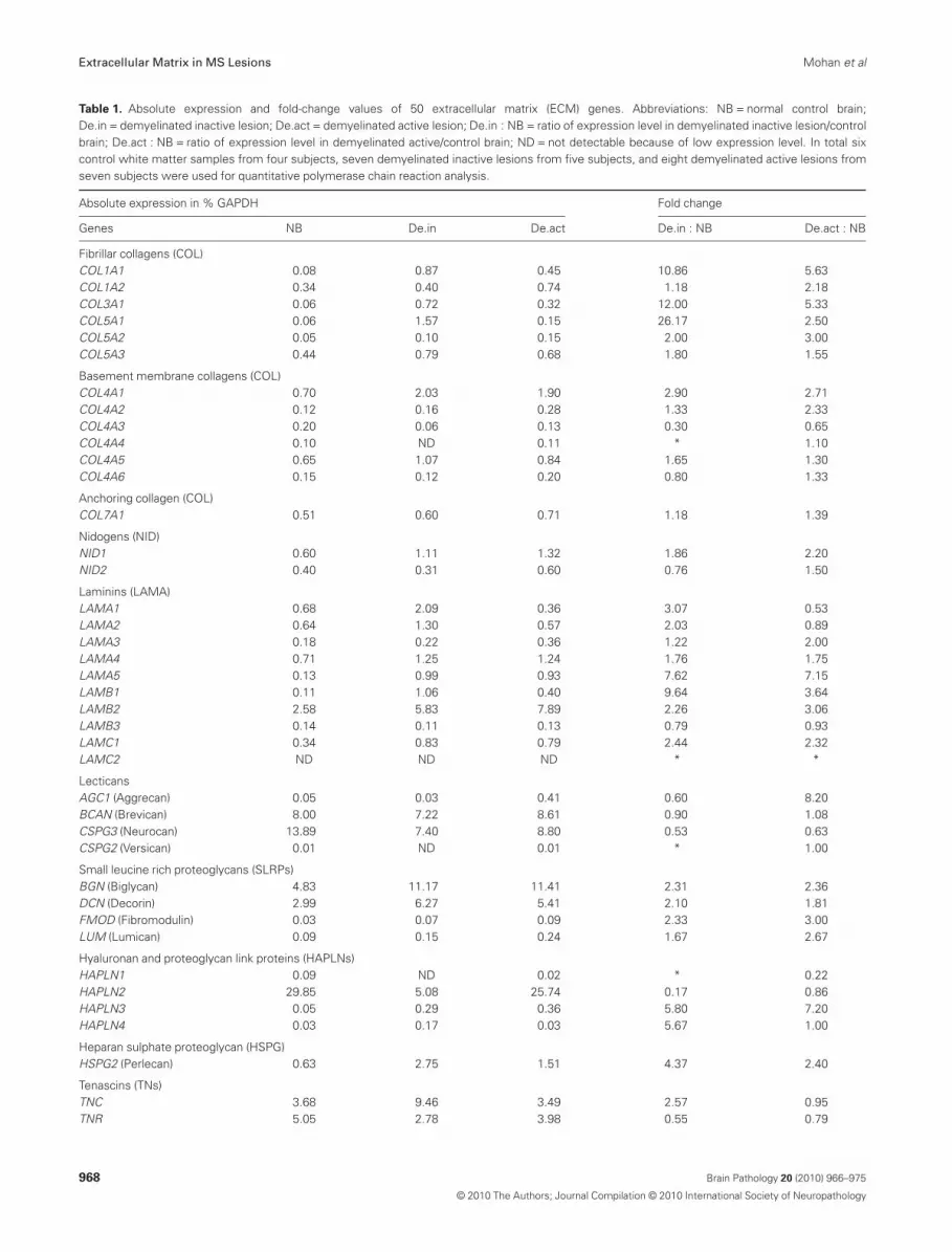
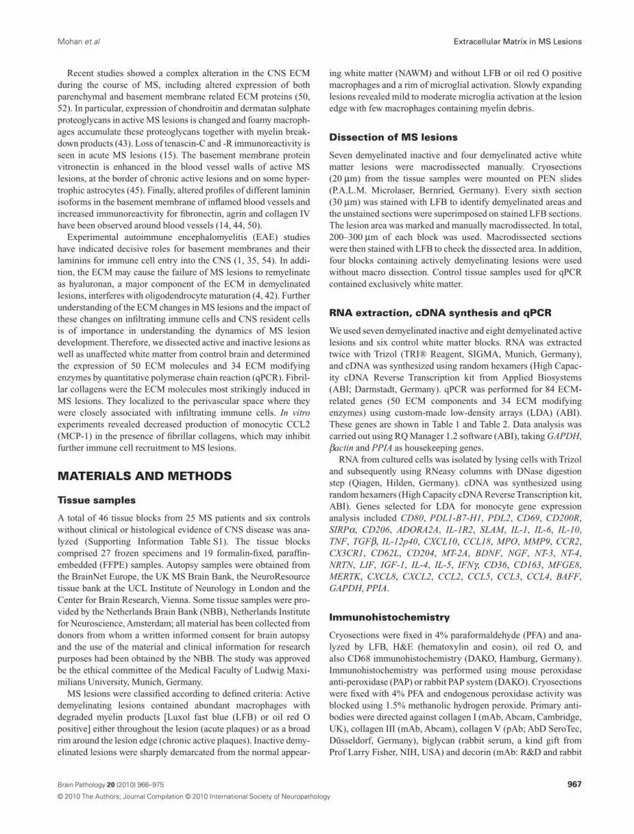
Table 1. Absolute expression and fold-change values of 50 extracellular matrix (ECM) genes. Abbreviations: NB = normal control brain;De.in = demyelinated inactive lesion; De.act = demyelinated active lesion; De.in : NB = ratio of expression level in demyelinated inactive lesion/controlbrain; De.act : NB = ratio of expression level in demyelinated active/control brain; ND = not detectable because of low expression level. In total sixcontrol white matter samples from four subjects, seven demyelinated inactive lesions from five subjects, and eight demyelinated active lesions fromseven subjects were used for quantitative polymerase chain reaction analysis.
Absolute expression in % GAPDH Fold change
Genes NB De.in De.act De.in : NB De.act : NB
Fibrillar collagens (COL)COL1A1 0.08 0.87 0.45 10.86 5.63COL1A2 0.34 0.40 0.74 1.18 2.18COL3A1 0.06 0.72 0.32 12.00 5.33COL5A1 0.06 1.57 0.15 26.17 2.50COL5A2 0.05 0.10 0.15 2.00 3.00COL5A3 0.44 0.79 0.68 1.80 1.55
Basement membrane collagens (COL)COL4A1 0.70 2.03 1.90 2.90 2.71COL4A2 0.12 0.16 0.28 1.33 2.33COL4A3 0.20 0.06 0.13 0.30 0.65COL4A4 0.10 ND 0.11 * 1.10COL4A5 0.65 1.07 0.84 1.65 1.30COL4A6 0.15 0.12 0.20 0.80 1.33
Anchoring collagen (COL)COL7A1 0.51 0.60 0.71 1.18 1.39
Nidogens (NID)NID1 0.60 1.11 1.32 1.86 2.20NID2 0.40 0.31 0.60 0.76 1.50
Laminins (LAMA)LAMA1 0.68 2.09 0.36 3.07 0.53LAMA2 0.64 1.30 0.57 2.03 0.89LAMA3 0.18 0.22 0.36 1.22 2.00LAMA4 0.71 1.25 1.24 1.76 1.75LAMA5 0.13 0.99 0.93 7.62 7.15LAMB1 0.11 1.06 0.40 9.64 3.64LAMB2 2.58 5.83 7.89 2.26 3.06LAMB3 0.14 0.11 0.13 0.79 0.93LAMC1 0.34 0.83 0.79 2.44 2.32LAMC2 ND ND ND * *
LecticansAGC1 (Aggrecan) 0.05 0.03 0.41 0.60 8.20BCAN (Brevican) 8.00 7.22 8.61 0.90 1.08CSPG3 (Neurocan) 13.89 7.40 8.80 0.53 0.63CSPG2 (Versican) 0.01 ND 0.01 * 1.00
Small leucine rich proteoglycans (SLRPs)BGN (Biglycan) 4.83 11.17 11.41 2.31 2.36DCN (Decorin) 2.99 6.27 5.41 2.10 1.81FMOD (Fibromodulin) 0.03 0.07 0.09 2.33 3.00LUM (Lumican) 0.09 0.15 0.24 1.67 2.67
Hyaluronan and proteoglycan link proteins (HAPLNs)HAPLN1 0.09 ND 0.02 * 0.22HAPLN2 29.85 5.08 25.74 0.17 0.86HAPLN3 0.05 0.29 0.36 5.80 7.20HAPLN4 0.03 0.17 0.03 5.67 1.00
Heparan sulphate proteoglycan (HSPG)HSPG2 (Perlecan) 0.63 2.75 1.51 4.37 2.40
Tenascins (TNs)TNC 3.68 9.46 3.49 2.57 0.95TNR 5.05 2.78 3.98 0.55 0.79
Extracellular Matrix in MS Lesions Mohan et al
968 Brain Pathology 20 (2010) 966–975
© 2010 The Authors; Journal Compilation © 2010 International Society of Neuropathology
serum, also a kind gift from Prof Larry Fisher). Sections wereincubated O/N at 4°C followed by secondary polyclonal rabbitanti-mouse (DAKO) or polyclonal swine anti-rabbit Ig (DAKO),for 1 h at room temperature and tertiary mouse PAP (mAb, DAKO)or rabbit PAP (pAb, DAKO), for 30 minutes at room temperature.Bound antibodies were detected with diaminobenzidine and sec-tions were counterstained with hematoxylin. For immunofluores-cence primary antibodies recognizing collagen V [pAb (rabbit),AbD SeroTec], CD31 (pAb, R&D, Wiesbaden, Germany), glialfibrillary acidic protein (GFAP; mAb, Molecular Probes,Karlsruhe, Germany, directly labeled with Alexa 488) were used.As secondary antibodies donkey anti-mouse Alexa 488, donkeyanti-sheep Alexa 488 and donkey anti-rabbit Alexa 594 (all fromMolecular Probes) were used. Confocal images were taken fromLeica SP2UV microscope. Negative controls included omission ofprimary antibody. For FFPE tissue, antigen retrieval was performedby treatment with citrate buffer pH6 in a steaming water bath.
Cell culture
Peripheral blood mononuclear cells (PBMCs) were isolated fromthe blood of healthy donors by density gradient centrifugation.Monocytes were isolated by positive selection with immunomag-netic beads (CD14 MicroBeads, Miltenyi Biotech, Bergisch Glad-bach, Germany) and were grown in the presence and absence ofECM proteins (coated culture plates) for 24 h.
Tissue culture plates were coated with collagen I at 2 mg/cm2
(BD Biosciences, Heidelberg, Germany); collagen III, 2 mg/cm2
(BD Biosciences); collagen V, 2 mg/cm2 (BD Biosciences); bigly-can, 10 mg/mL (R&D); decorin, 10 mg/mL (R&D). All ECM pro-teins were diluted to final concentration with Ca2+, Mg2+ free PBS(Gibco, Karlsruhe, Germany) and cell culture plates were incu-bated at RT for 2 h, and then washed with distilled water (Gibco)and air dried. All the ECM proteins were tested negative for pres-ence of LPS (Biowhittaker™ LAL kit, Walkersville, MD, USA).
The myelin basic protein (MBP) specific T cell clone ES-BP8(30) was stimulated with HLA-DR compatible PBMC and20 mg/mL MBP (Biogenesis, Berlin, Germany) or 10 mg/mL MBP
29-48 peptide for 2 days. On the third day cultures were pulsed with1 mCi/well of 3H-thymidine (Amersham Biosciences, Freiburg,Germany) for 24 h.
Enzyme-linked immunosorbent assays (ELISAs)
To detect CCL2, CCL4, IL10, IL1b in cell culture supernatants theDuoset ELISA system (R&D) was used. Assays were performedaccording to the manufacturer’s instructions.
RESULTS
Altered expression of ECM components inMS lesions
In this study we quantified the expression of 50 genes coding forproteins forming the ECM (Table 1) and for 34 enzymes modifyingthe ECM (Table 2) in control brain, active and inactiveMS lesions. Of the 50 ECM genes tested, 22 were upregulatedmore than two-fold in active lesions and 21 in inactive lesions(Table 1). Fifteen ECM components were induced in both activeand chronic inactive lesions. Twenty-three genes in inactive and 25in active lesions were considered unchanged, that is, they had achange between 0.51- and 1.99-fold.
Upregulated ECM components included fibrillar collagens,basement membrane collagen, laminins, SLRPs, hyaluronan linkproteins, thrombospondins and perlecan (details in Table 1).
Fibrillar collagens and SLRPs form aperivascular fibrosis in MS lesions and are inclose interaction with the infiltrating immunecells in the perivascular space
Our expression profiling identified a total of 22 componentsupregulated in active MS lesions and 21 in inactive MS lesions.Considering the relative induction in MS lesions and the absoluteexpression level of the ECM genes, our interest was directed to thefibrillar collagens and the SLRPs. We noted a strong induction of
Table 1. Continued
Absolute expression in % GAPDH Fold change
Genes NB De.in De.act De.in : NB De.act : NB
Thrombospondins (THBSs)THBS1 0.12 0.98 0.41 8.17 3.42THBS2 2.75 4.22 4.05 1.53 1.47THBS3 0.01 0.02 0.03 2.00 3.00THBS4 0.83 1.08 1.09 1.30 1.31
Fibrillins (FBNs)FBN1 2.59 3.12 1.46 1.20 0.56FBN2 0.04 0.03 0.09 0.75 2.25FBN3 0.11 0.10 0.13 0.91 1.18
OthersFN1 (Fibronectin) 2.60 4.86 3.94 1.87 1.52RELN (Reelin) 0.08 0.63 0.04 7.86 0.50VTN (Vitronectin) 0.03 0.03 0.03 1.00 1.00
*Calculation was not possible because at least one value was under the detection limit.
Mohan et al Extracellular Matrix in MS Lesions
969Brain Pathology 20 (2010) 966–975
© 2010 The Authors; Journal Compilation © 2010 International Society of Neuropathology
COL1A1, COL3A1, COL5A1, COL5A2 chains, both in active andinactive demyelinated lesions (Table 1). These collagens interact toform collagen types I, III and V, which are grouped as fibrillarcollagens, known to act as structural proteins (20). Biglycan anddecorin, both classified as SLRPs, were also strongly induced inactive and inactive demyelinated lesions (Table 1).
The ECM molecules identified by the transcript analysis werethen localized by immunostaining. In control brain tissue we sawfaint staining around blood vessels with antibodies to the three
fibrillar collagens, decorin and biglycan (Figure 2E, H and data notshown). Similar stainings were observed in the NAWM (Figure 1C,and data not shown). In contrast, in both active and inactive MSlesions staining of fibrillar collagens, decorin, and biglycan wasmore intense (Figure 2A–D, F, G) and localized around small,medium and large blood vessels (Figure 1A).
In the larger blood vessels in MS lesions the extended perivascu-lar (Virchow Robin) space was filled by a meshwork of fibrillarcollagen, biglycan and decorin (Figure 2). In chronic active and
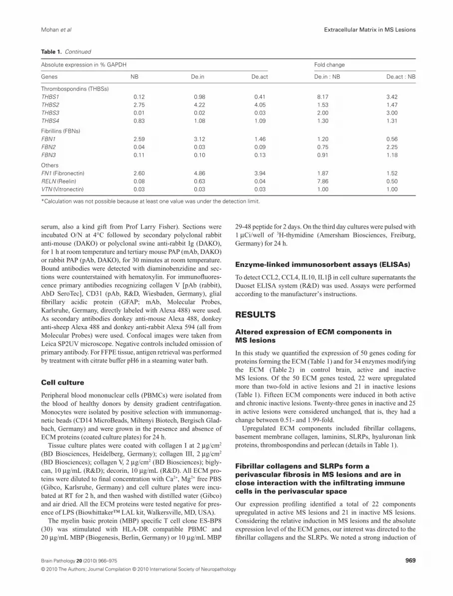
Table 2. Absolute expression and fold-change values of 34 ECM modifying enzymes. Abbreviations: NB = normal control brain; De.in = demyelinatedinactive lesion; De.act = demyelinated active lesion; De.in : NB = ratio of expression level in demyelinated inactive lesion/control brain;De.act : NB = ratio of expression level in demyelinated active/control brain; ND = not detectable because of low expression level. In total six controlwhite matter samples from four subjects, seven demyelinated inactive lesions from five subjects, and eight demyelinated active lesions from sevensubjects were used for quantitative polymerase chain reaction analysis.
Absolute expression in % GAPDH Fold change
Genes NB De.in De.act De.in : NB De.act : NB
A disintegrin and metalloproteinase domains (ADAMs)ADAM8 0.11 0.05 0.31 0.45 2.82ADAM10 16.40 13.58 13.23 0.83 0.81ADAM12 0.30 0.29 0.40 0.97 1.33ADAM17 1.29 0.85 1.32 0.66 1.02
A disintegrin like and metalloproteinase with thrombospondin type 1 motifs (ADAMTs)ADAMTS1 7.38 7.88 3.92 1.07 0.53ADAMTS4 16.04 3.24 15.15 0.20 0.94ADAMTS5 0.04 0.17 0.06 4.25 1.50
Matrix metalloproteinases (MMPs)MMP1 ND ND ND * *MMP2 0.61 1.09 3.36 1.79 5.50MMP3 ND ND ND * *MMP7 ND ND 0.03 * *MMP8 ND ND ND * *MMP9 0.01 0.05 0.13 5.00 13.00MMP10 ND ND ND * *MMP11 0.05 0.30 0.30 6.00 6.00MMP12 ND ND ND * *MMP13 ND ND ND * *MMP14 1.00 5.71 4.61 5.71 4.61MMP15 0.71 0.83 0.36 1.17 0.51MMP16 0.75 0.97 1.03 1.29 1.37MMP17 0.39 5.05 0.36 12.95 0.92MMP19 0.01 0.04 0.08 4.00 8.00MMP20 ND ND ND * *MMP21 0.07 ND 0.07 * 1.00MMP23 0.02 0.01 0.01 0.50 0.50MMP24 0.37 1.21 0.34 3.27 0.92MMP25 ND ND 0.01 * *MMP26 ND ND ND * *MMP27 ND ND ND * *MMP28 0.24 0.75 0.08 3.13 0.33
Tissue inhibitor of metalloproteinases (TIMPs)TIMP1 0.97 5.96 7.00 6.14 7.22TIMP2 13.78 9.56 13.82 0.69 1.00TIMP3 9.21 22.98 15.51 2.50 1.68TIMP4 0.88 0.69 0.98 0.78 1.11
*Calculation was not possible because the values were under the detection limit.
Extracellular Matrix in MS Lesions Mohan et al
970 Brain Pathology 20 (2010) 966–975
© 2010 The Authors; Journal Compilation © 2010 International Society of Neuropathology
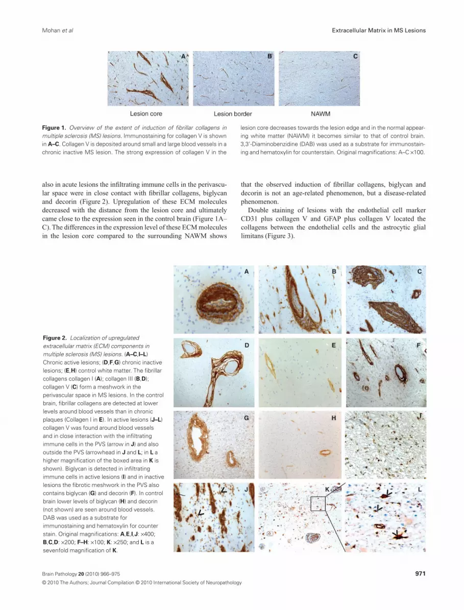
also in acute lesions the infiltrating immune cells in the perivascu-lar space were in close contact with fibrillar collagens, biglycanand decorin (Figure 2). Upregulation of these ECM moleculesdecreased with the distance from the lesion core and ultimatelycame close to the expression seen in the control brain (Figure 1A–C). The differences in the expression level of these ECM moleculesin the lesion core compared to the surrounding NAWM shows
that the observed induction of fibrillar collagens, biglycan anddecorin is not an age-related phenomenon, but a disease-relatedphenomenon.
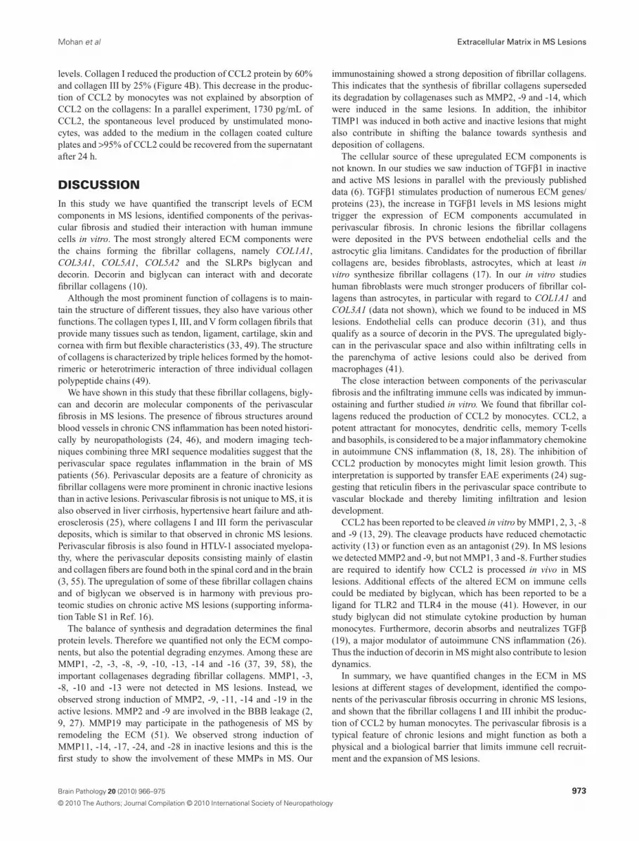
Double staining of lesions with the endothelial cell markerCD31 plus collagen V and GFAP plus collagen V located thecollagens between the endothelial cells and the astrocytic gliallimitans (Figure 3).
Figure 2. Localization of upregulatedextracellular matrix (ECM) components inmultiple sclerosis (MS) lesions. (A–C,I–L)Chronic active lesions; (D,F,G) chronic inactivelesions; (E,H) control white matter. The fibrillarcollagens collagen I (A); collagen III (B,D);collagen V (C) form a meshwork in theperivascular space in MS lesions. In the controlbrain, fibrillar collagens are detected at lowerlevels around blood vessels than in chronicplaques (Collagen I in E). In active lesions (J–L)collagen V was found around blood vesselsand in close interaction with the infiltratingimmune cells in the PVS (arrow in J) and alsooutside the PVS (arrowhead in J and L; in L ahigher magnification of the boxed area in K isshown). Biglycan is detected in infiltratingimmune cells in active lesions (I) and in inactivelesions the fibrotic meshwork in the PVS alsocontains biglycan (G) and decorin (F). In controlbrain lower levels of biglycan (H) and decorin(not shown) are seen around blood vessels.DAB was used as a substrate forimmunostaining and hematoxylin for counterstain. Original magnifications: A,E,I,J: ¥400;B,C,D: ¥200; F–H: ¥100; K: ¥250; and L is asevenfold magnification of K.
Figure 1. Overview of the extent of induction of fibrillar collagens inmultiple sclerosis (MS) lesions. Immunostaining for collagen V is shownin A–C. Collagen V is deposited around small and large blood vessels in achronic inactive MS lesion. The strong expression of collagen V in the
lesion core decreases towards the lesion edge and in the normal appear-ing white matter (NAWM) it becomes similar to that of control brain.3,3’-Diaminobenzidine (DAB) was used as a substrate for immunostain-ing and hematoxylin for counterstain. Original magnifications: A–C ¥100.
Mohan et al Extracellular Matrix in MS Lesions
971Brain Pathology 20 (2010) 966–975
© 2010 The Authors; Journal Compilation © 2010 International Society of Neuropathology
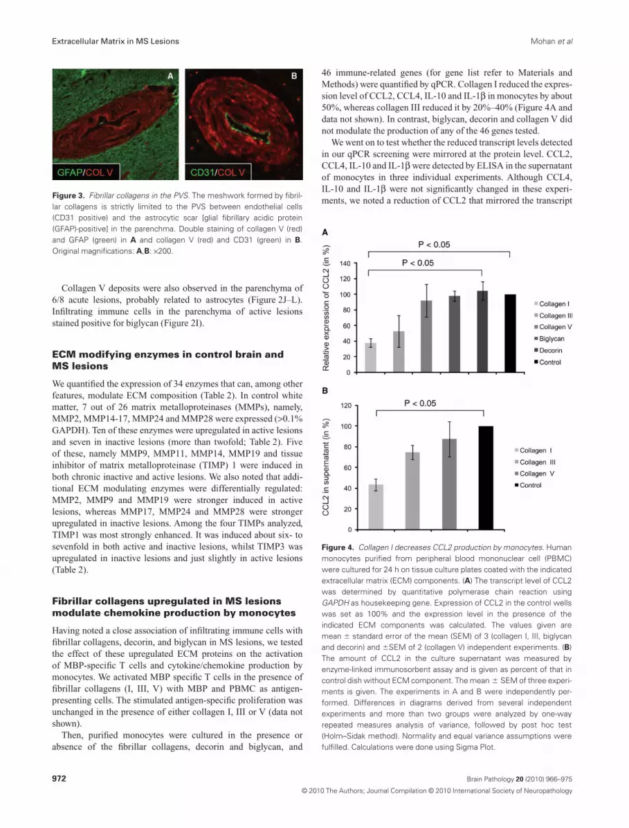
Collagen V deposits were also observed in the parenchyma of6/8 acute lesions, probably related to astrocytes (Figure 2J–L).Infiltrating immune cells in the parenchyma of active lesionsstained positive for biglycan (Figure 2I).
ECM modifying enzymes in control brain andMS lesions
We quantified the expression of 34 enzymes that can, among otherfeatures, modulate ECM composition (Table 2). In control whitematter, 7 out of 26 matrix metalloproteinases (MMPs), namely,MMP2, MMP14-17, MMP24 and MMP28 were expressed (>0.1%GAPDH). Ten of these enzymes were upregulated in active lesionsand seven in inactive lesions (more than twofold; Table 2). Fiveof these, namely MMP9, MMP11, MMP14, MMP19 and tissueinhibitor of matrix metalloproteinase (TIMP) 1 were induced inboth chronic inactive and active lesions. We also noted that addi-tional ECM modulating enzymes were differentially regulated:MMP2, MMP9 and MMP19 were stronger induced in activelesions, whereas MMP17, MMP24 and MMP28 were strongerupregulated in inactive lesions. Among the four TIMPs analyzed,TIMP1 was most strongly enhanced. It was induced about six- tosevenfold in both active and inactive lesions, whilst TIMP3 wasupregulated in inactive lesions and just slightly in active lesions(Table 2).
Fibrillar collagens upregulated in MS lesionsmodulate chemokine production by monocytes
Having noted a close association of infiltrating immune cells withfibrillar collagens, decorin, and biglycan in MS lesions, we testedthe effect of these upregulated ECM proteins on the activationof MBP-specific T cells and cytokine/chemokine production bymonocytes. We activated MBP specific T cells in the presence offibrillar collagens (I, III, V) with MBP and PBMC as antigen-presenting cells. The stimulated antigen-specific proliferation wasunchanged in the presence of either collagen I, III or V (data notshown).
Then, purified monocytes were cultured in the presence orabsence of the fibrillar collagens, decorin and biglycan, and
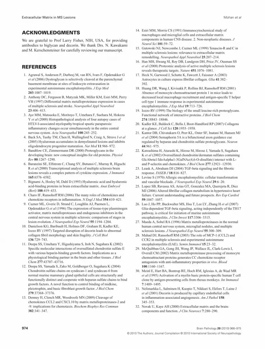
46 immune-related genes (for gene list refer to Materials andMethods) were quantified by qPCR. Collagen I reduced the expres-sion level of CCL2, CCL4, IL-10 and IL-1b in monocytes by about50%, whereas collagen III reduced it by 20%–40% (Figure 4A anddata not shown). In contrast, biglycan, decorin and collagen V didnot modulate the production of any of the 46 genes tested.
We went on to test whether the reduced transcript levels detectedin our qPCR screening were mirrored at the protein level. CCL2,CCL4, IL-10 and IL-1b were detected by ELISA in the supernatantof monocytes in three individual experiments. Although CCL4,IL-10 and IL-1b were not significantly changed in these experi-ments, we noted a reduction of CCL2 that mirrored the transcript
Figure 3. Fibrillar collagens in the PVS. The meshwork formed by fibril-lar collagens is strictly limited to the PVS between endothelial cells(CD31 positive) and the astrocytic scar [glial fibrillary acidic protein(GFAP)-positive] in the parenchma. Double staining of collagen V (red)and GFAP (green) in A and collagen V (red) and CD31 (green) in B.Original magnifications: A,B: ¥200.
Figure 4. Collagen I decreases CCL2 production by monocytes. Humanmonocytes purified from peripheral blood mononuclear cell (PBMC)were cultured for 24 h on tissue culture plates coated with the indicatedextracellular matrix (ECM) components. (A) The transcript level of CCL2was determined by quantitative polymerase chain reaction usingGAPDH as housekeeping gene. Expression of CCL2 in the control wellswas set as 100% and the expression level in the presence of theindicated ECM components was calculated. The values given aremean � standard error of the mean (SEM) of 3 (collagen I, III, biglycanand decorin) and �SEM of 2 (collagen V) independent experiments. (B)The amount of CCL2 in the culture supernatant was measured byenzyme-linked immunosorbent assay and is given as percent of that incontrol dish without ECM component. The mean � SEM of three experi-ments is given. The experiments in A and B were independently per-formed. Differences in diagrams derived from several independentexperiments and more than two groups were analyzed by one-wayrepeated measures analysis of variance, followed by post hoc test(Holm–Sidak method). Normality and equal variance assumptions werefulfilled. Calculations were done using Sigma Plot.
Extracellular Matrix in MS Lesions Mohan et al
972 Brain Pathology 20 (2010) 966–975
© 2010 The Authors; Journal Compilation © 2010 International Society of Neuropathology
levels. Collagen I reduced the production of CCL2 protein by 60%and collagen III by 25% (Figure 4B). This decrease in the produc-tion of CCL2 by monocytes was not explained by absorption ofCCL2 on the collagens: In a parallel experiment, 1730 pg/mL ofCCL2, the spontaneous level produced by unstimulated mono-cytes, was added to the medium in the collagen coated cultureplates and >95% of CCL2 could be recovered from the supernatantafter 24 h.
DISCUSSIONIn this study we have quantified the transcript levels of ECMcomponents in MS lesions, identified components of the perivas-cular fibrosis and studied their interaction with human immunecells in vitro. The most strongly altered ECM components werethe chains forming the fibrillar collagens, namely COL1A1,COL3A1, COL5A1, COL5A2 and the SLRPs biglycan anddecorin. Decorin and biglycan can interact with and decoratefibrillar collagens (10).
Although the most prominent function of collagens is to main-tain the structure of different tissues, they also have various otherfunctions. The collagen types I, III, and V form collagen fibrils thatprovide many tissues such as tendon, ligament, cartilage, skin andcornea with firm but flexible characteristics (33, 49). The structureof collagens is characterized by triple helices formed by the homot-rimeric or heterotrimeric interaction of three individual collagenpolypeptide chains (49).
We have shown in this study that these fibrillar collagens, bigly-can and decorin are molecular components of the perivascularfibrosis in MS lesions. The presence of fibrous structures aroundblood vessels in chronic CNS inflammation has been noted histori-cally by neuropathologists (24, 46), and modern imaging tech-niques combining three MRI sequence modalities suggest that theperivascular space regulates inflammation in the brain of MSpatients (56). Perivascular deposits are a feature of chronicity asfibrillar collagens were more prominent in chronic inactive lesionsthan in active lesions. Perivascular fibrosis is not unique to MS, it isalso observed in liver cirrhosis, hypertensive heart failure and ath-erosclerosis (25), where collagens I and III form the perivasculardeposits, which is similar to that observed in chronic MS lesions.Perivascular fibrosis is also found in HTLV-1 associated myelopa-thy, where the perivascular deposits consisting mainly of elastinand collagen fibers are found both in the spinal cord and in the brain(3, 55). The upregulation of some of these fibrillar collagen chainsand of biglycan we observed is in harmony with previous pro-teomic studies on chronic active MS lesions (supporting informa-tion Table S1 in Ref. 16).
The balance of synthesis and degradation determines the finalprotein levels. Therefore we quantified not only the ECM compo-nents, but also the potential degrading enzymes. Among these areMMP1, -2, -3, -8, -9, -10, -13, -14 and -16 (37, 39, 58), theimportant collagenases degrading fibrillar collagens. MMP1, -3,-8, -10 and -13 were not detected in MS lesions. Instead, weobserved strong induction of MMP2, -9, -11, -14 and -19 in theactive lesions. MMP2 and -9 are involved in the BBB leakage (2,9, 27). MMP19 may participate in the pathogenesis of MS byremodeling the ECM (51). We observed strong induction ofMMP11, -14, -17, -24, and -28 in inactive lesions and this is thefirst study to show the involvement of these MMPs in MS. Our
immunostaining showed a strong deposition of fibrillar collagens.This indicates that the synthesis of fibrillar collagens supersededits degradation by collagenases such as MMP2, -9 and -14, whichwere induced in the same lesions. In addition, the inhibitorTIMP1 was induced in both active and inactive lesions that mightalso contribute in shifting the balance towards synthesis anddeposition of collagens.
The cellular source of these upregulated ECM components isnot known. In our studies we saw induction of TGFb1 in inactiveand active MS lesions in parallel with the previously publisheddata (6). TGFb1 stimulates production of numerous ECM genes/proteins (23), the increase in TGFb1 levels in MS lesions mighttrigger the expression of ECM components accumulated inperivascular fibrosis. In chronic lesions the fibrillar collagenswere deposited in the PVS between endothelial cells and theastrocytic glia limitans. Candidates for the production of fibrillarcollagens are, besides fibroblasts, astrocytes, which at least invitro synthesize fibrillar collagens (17). In our in vitro studieshuman fibroblasts were much stronger producers of fibrillar col-lagens than astrocytes, in particular with regard to COL1A1 andCOL3A1 (data not shown), which we found to be induced in MSlesions. Endothelial cells can produce decorin (31), and thusqualify as a source of decorin in the PVS. The upregulated bigly-can in the perivascular space and also within infiltrating cells inthe parenchyma of active lesions could also be derived frommacrophages (41).
The close interaction between components of the perivascularfibrosis and the infiltrating immune cells was indicated by immun-ostaining and further studied in vitro. We found that fibrillar col-lagens reduced the production of CCL2 by monocytes. CCL2, apotent attractant for monocytes, dendritic cells, memory T-cellsand basophils, is considered to be a major inflammatory chemokinein autoimmune CNS inflammation (8, 18, 28). The inhibition ofCCL2 production by monocytes might limit lesion growth. Thisinterpretation is supported by transfer EAE experiments (24) sug-gesting that reticulin fibers in the perivascular space contribute tovascular blockade and thereby limiting infiltration and lesiondevelopment.
CCL2 has been reported to be cleaved in vitro by MMP1, 2, 3, -8and -9 (13, 29). The cleavage products have reduced chemotacticactivity (13) or function even as an antagonist (29). In MS lesionswe detected MMP2 and -9, but not MMP1, 3 and -8. Further studiesare required to identify how CCL2 is processed in vivo in MSlesions. Additional effects of the altered ECM on immune cellscould be mediated by biglycan, which has been reported to be aligand for TLR2 and TLR4 in the mouse (41). However, in ourstudy biglycan did not stimulate cytokine production by humanmonocytes. Furthermore, decorin absorbs and neutralizes TGFb(19), a major modulator of autoimmune CNS inflammation (26).Thus the induction of decorin in MS might also contribute to lesiondynamics.
In summary, we have quantified changes in the ECM in MSlesions at different stages of development, identified the compo-nents of the perivascular fibrosis occurring in chronic MS lesions,and shown that the fibrillar collagens I and III inhibit the produc-tion of CCL2 by human monocytes. The perivascular fibrosis is atypical feature of chronic lesions and might function as both aphysical and a biological barrier that limits immune cell recruit-ment and the expansion of MS lesions.
Mohan et al Extracellular Matrix in MS Lesions
973Brain Pathology 20 (2010) 966–975
© 2010 The Authors; Journal Compilation © 2010 International Society of Neuropathology
ACKNOWLEDGMENTSWe are grateful to Prof Larry Fisher, NIH, USA, for providingantibodies to biglycan and decorin. We thank Drs. N. Kawakamiand M. Kerschensteiner for carefully reviewing our manuscript.
REFERENCES1. Agrawal S, Anderson P, Durbeej M, van RN, Ivars F, Opdenakker G
et al (2006) Dystroglycan is selectively cleaved at the parenchymalbasement membrane at sites of leukocyte extravasation inexperimental autoimmune encephalomyelitis. J Exp Med203:1007–1019.
2. Anthony DC, Ferguson B, Matyzak MK, Miller KM, Esiri MM, PerryVH (1997) Differential matrix metalloproteinase expression in casesof multiple sclerosis and stroke. Neuropathol Appl Neurobiol23:406–415.
3. Aye MM, Matsuoka E, Moritoyo T, Umehara F, Suehara M, HokezuY et al (2000) Histopathological analysis of four autopsy cases ofHTLV-I-associated myelopathy/tropical spastic paraparesis:inflammatory changes occur simultaneously in the entire centralnervous system. Acta Neuropathol 100:245–252.
4. Back SA, Tuohy TM, Chen H, Wallingford N, Craig A, Struve J et al(2005) Hyaluronan accumulates in demyelinated lesions and inhibitsoligodendrocyte progenitor maturation. Nat Med 11:966–972.
5. Bandtlow CE, Zimmermann DR (2000) Proteoglycans in thedeveloping brain: new conceptual insights for old proteins. PhysiolRev 80:1267–1290.
6. Baranzini SE, Elfstrom C, Chang SY, Butunoi C, Murray R, HiguchiR et al (2000) Transcriptional analysis of multiple sclerosis brainlesions reveals a complex pattern of cytokine expression. J Immunol165:6576–6582.
7. Bignami A, Hosley M, Dahl D (1993) Hyaluronic acid and hyaluronicacid-binding proteins in brain extracellular matrix. Anat Embryol(Berl) 188:419–433.
8. Charo IF, Ransohoff RM (2006) The many roles of chemokines andchemokine receptors in inflammation. N Engl J Med 354:610–621.
9. Cuzner ML, Gveric D, Strand C, Loughlin AJ, Paemen L,Opdenakker G et al (1996) The expression of tissue-type plasminogenactivator, matrix metalloproteases and endogenous inhibitors in thecentral nervous system in multiple sclerosis: comparison of stages inlesion evolution. J Neuropathol Exp Neurol 55:1194–1204.
10. Danielson KG, Baribault H, Holmes DF, Graham H, Kadler KE,Iozzo RV (1997) Targeted disruption of decorin leads to abnormalcollagen fibril morphology and skin fragility. J Cell Biol136:729–743.
11. Deepa SS, Umehara Y, Higashiyama S, Itoh N, Sugahara K (2002)Specific molecular interactions of oversulfated chondroitin sulfate Ewith various heparin-binding growth factors. Implications as aphysiological binding partner in the brain and other tissues. J BiolChem 277:43707–43716.
12. Deepa SS, Yamada S, Zako M, Goldberger O, Sugahara K (2004)Chondroitin sulfate chains on syndecan-1 and syndecan-4 fromnormal murine mammary gland epithelial cells are structurally andfunctionally distinct and cooperate with heparan sulfate chains to bindgrowth factors. A novel function to control binding of midkine,pleiotrophin, and basic fibroblast growth factor. J Biol Chem279:37368–37376.
13. Denney H, Clench MR, Woodroofe MN (2009) Cleavage ofchemokines CCL2 and CXCL10 by matrix metalloproteinases-2 and-9: implications for chemotaxis. Biochem Biophys Res Commun382:341–347.
14. Esiri MM, Morris CS (1991) Immunocytochemical study ofmacrophages and microglial cells and extracellular matrixcomponents in human CNS disease. 2. Non-neoplastic diseases. JNeurol Sci 101:59–72.
15. Gutowski NJ, Newcombe J, Cuzner ML (1999) Tenascin-R and C inmultiple sclerosis lesions: relevance to extracellular matrixremodelling. Neuropathol Appl Neurobiol 25:207–214.
16. Han MH, Hwang SI, Roy DB, Lundgren DH, Price JV, Ousman SSet al (2008) Proteomic analysis of active multiple sclerosis lesionsreveals therapeutic targets. Nature 451:1076–1081.
17. Heck N, Garwood J, Schutte K, Fawcett J, Faissner A (2003)Astrocytes in culture express fibrillar collagen. Glia 41:382–392.
18. Huang DR, Wang J, Kivisakk P, Rollins BJ, Ransohoff RM (2001)Absence of monocyte chemoattractant protein 1 in mice leads todecreased local macrophage recruitment and antigen-specific T helpercell type 1 immune response in experimental autoimmuneencephalomyelitis. J Exp Med 19:713–726.
19. Iozzo RV (1999) The biology of the small leucine-rich proteoglycans.Functional network of interactive proteins. J Biol Chem274:18843–18846.
20. Kadler KE, Baldock C, Bella J, Boot-Handford RP (2007) Collagensat a glance. J Cell Sci 120:1955–1958.
21. Kantor DB, Chivatakarn O, Peer KL, Oster SF, Inatani M, Hansen MJet al (2004) Semaphorin 5A is a bifunctional axon guidance cueregulated by heparan and chondroitin sulfate proteoglycans. Neuron44:961–975.
22. Kawashima H, Atarashi K, Hirose M, Hirose J, Yamada S, SugaharaK et al (2002) Oversulfated chondroitin/dermatan sulfates containingGlcAbeta1/IdoAalpha1-3GalNAc(4,6-O-disulfate) interact with L-and P-selectin and chemokines. J Biol Chem 277:12921–12930.
23. Leask A, Abraham DJ (2004) TGF-beta signaling and the fibroticresponse. FASEB J 18:816–827.
24. Levine S (1970) Allergic encephalomyelitis: cellular transformationand vascular blockade. J Neuropathol Exp Neurol 29:6–20.
25. Lopez SB, Ravassa AS, Arias GT, Gonzalez MA, Querejeta R, DiezMJ (2006) Altered fibrillar collagen metabolism in hypertensive heartfailure. Current understanding and future prospects. Rev Esp Cardiol59:1047–1057.
26. Luo J, Ho PP, Buckwalter MS, Hsu T, Lee LY, Zhang H et al (2007)Glia-dependent TGF-beta signaling, acting independently of the TH17pathway, is critical for initiation of murine autoimmuneencephalomyelitis. J Clin Invest 117:3306–3315.
27. Maeda A, Sobel RA (1996) Matrix metalloproteinases in the normalhuman central nervous system, microglial nodules, and multiplesclerosis lesions. J Neuropathol Exp Neurol 55:300–309.
28. Mahad DJ, Ransohoff RM (2003) The role of MCP-1 (CCL2) andCCR2 in multiple sclerosis and experimental autoimmuneencephalomyelitis (EAE). Semin Immunol 15:23–32.
29. McQuibban GA, Gong JH, Wong JP, Wallace JL, Clark-Lewis I,Overall CM (2002) Matrix metalloproteinase processing of monocytechemoattractant proteins generates CC chemokine receptorantagonists with anti-inflammatory properties in vivo. Blood100:1160–1167.
30. Meinl E, Hart BA, Bontrop RE, Hoch RM, Iglesias A, de Waal MRet al (1995) Activation of a myelin basic protein-specific human T cellclone by antigen-presenting cells from rhesus monkeys. Int Immunol7:1489–1495.
31. Nelimarkka L, Salminen H, Kuopio T, Nikkari S, Ekfors T, Laine Jet al (2001) Decorin is produced by capillary endothelial cellsin inflammation-associated angiogenesis. Am J Pathol 158:345–353.
32. Novak U, Kaye AH (2000) Extracellular matrix and the brain:components and function. J Clin Neurosci 7:280–290.
Extracellular Matrix in MS Lesions Mohan et al
974 Brain Pathology 20 (2010) 966–975
© 2010 The Authors; Journal Compilation © 2010 International Society of Neuropathology
33. Okada M, Miyamoto O, Shibuya S, Zhang X, Yamamoto T, Itano T(2007) Expression and role of type I collagen in a rat spinal cordcontusion injury model. Neurosci Res 58:371–377.
34. Oohira A, Matsui F, Tokita Y, Yamauchi S, Aono S (2000) Molecularinteractions of neural chondroitin sulfate proteoglycans in the braindevelopment. Arch Biochem Biophys 374:24–34.
35. Owens T, Bechmann I, Engelhardt B (2008) Perivascular spaces andthe two steps to neuroinflammation. J Neuropathol Exp Neurol67:1113–1121.
36. Rauch U (2004) Extracellular matrix components associated withremodeling processes in brain. Cell Mol Life Sci 61:2031–2045.
37. Rowe RG, Weiss SJ (2008) Breaching the basement membrane: who,when and how? Trends Cell Biol 18:560–574.
38. Rutka JT, Apodaca G, Stern R, Rosenblum M (1988) Theextracellular matrix of the central and peripheral nervous systems:structure and function. J Neurosurg 69:155–170.
39. Sabeh F, Li XY, Saunders TL, Rowe RG, Weiss SJ (2009) Secretedversus membrane-anchored collagenases: relative roles infibroblast-dependent collagenolysis and invasion. J Biol Chem284:23001–23011.
40. Saksela O, Rifkin DB (1990) Release of basic fibroblast growthfactor-heparan sulfate complexes from endothelial cells byplasminogen activator-mediated proteolytic activity. J Cell Biol110:767–775.
41. Schaefer L, Babelova A, Kiss E, Hausser HJ, Baliova M, KrzyzankovaM et al (2005) The matrix component biglycan is proinflammatoryand signals through Toll-like receptors 4 and 2 in macrophages. J ClinInvest 115:2223–2233.
42. Sherman LS, Back SA (2008) A “GAG” reflex prevents repair of thedamaged CNS. Trends Neurosci 31:44–52.
43. Sobel RA, Ahmed AS (2001) White matter extracellular matrixchondroitin sulfate/dermatan sulfate proteoglycans in multiplesclerosis. J Neuropathol Exp Neurol 60:1198–1207.
44. Sobel RA, Mitchell ME (1989) Fibronectin in multiple sclerosislesions. Am J Pathol 135:161–168.
45. Sobel RA, Chen M, Maeda A, Hinojoza JR (1995) Vitronectin andintegrin vitronectin receptor localization in multiple sclerosis lesions.J Neuropathol Exp Neurol 54:202–213.
46. Spielmeyer W (1922) Histopathologie des Nervensystems. Verlag vonJulius Springer: Berlin.
47. Tilling T, Engelbertz C, Decker S, Korte D, Huwel S, Galla HJ (2002)Expression and adhesive properties of basement membrane proteinsin cerebral capillary endothelial cell cultures. Cell Tissue Res310:19–29.
48. Van der Laan LJ, De Groot CJ, Elices MJ, Dijkstra CD (1997)Extracellular matrix proteins expressed by human adult astrocytes invivo and in vitro: an astrocyte surface protein containing the CS1
domain contributes to binding of lymphoblasts. J Neurosci Res50:539–548.
49. Van der Rest M, Garrone R (1991) Collagen family of proteins.FASEB J 5:2814–2823.
50. Van Horssen J, Bo L, Vos CM, Virtanen I, de Vries HE (2005)Basement membrane proteins in multiple sclerosis-associatedinflammatory cuffs: potential role in influx and transport ofleukocytes. J Neuropathol Exp Neurol 64:722–729.
51. Van Horssen J, Vos CM, Admiraal L, van Haastert ES, Montagne L,van d V et al (2006) Matrix metalloproteinase-19 is highly expressedin active multiple sclerosis lesions. Neuropathol Appl Neurobiol32:585–593.
52. Van Horssen J, Dijkstra CD, de Vries HE (2007) The extracellularmatrix in multiple sclerosis pathology. J Neurochem 103:1293–1301.
53. Viapiano MS, Matthews RT (2006) From barriers to bridges:chondroitin sulfate proteoglycans in neuropathology. Trends Mol Med12:488–496.
54. Wu C, Ivars F, Anderson P, Hallmann R, Vestweber D, Nilsson P et al(2009) Endothelial basement membrane laminin alpha5 selectivelyinhibits T lymphocyte extravasation into the brain. Nat Med15:519–527.
55. Wu E, Dickson DW, Jacobson S, Raine CS (1993) Neuroaxonaldystrophy in HTLV-1-associated myelopathy/tropical spasticparaparesis: neuropathologic and neuroimmunologic correlations.Acta Neuropathol 86:224–235.
56. Wuerfel J, Haertle M, Waiczies H, Tysiak E, Bechmann I, WerneckeKD et al (2008) Perivascular spaces—MRI marker of inflammatoryactivity in the brain? Brain 131:2332–2340.
57. Yamaguchi Y (2000) Lecticans: organizers of the brain extracellularmatrix. Cell Mol Life Sci 57:276–289.
58. Yong VW, Power C, Forsyth P, Edwards DR (2001)Metalloproteinases in biology and pathology of the nervous system.Nat Rev Neurosci 2:502–511.
SUPPORTING INFORMATIONAdditional Supporting Information may be found in the onlineversion of this article:
Table S1. Tissue samples of patients and controls.
Please note: Wiley-Blackwell are not responsible for the content orfunctionality of any supporting materials supplied by the authors.Any queries (other than missing material) should be directed to thecorresponding author for the article.
Mohan et al Extracellular Matrix in MS Lesions
975Brain Pathology 20 (2010) 966–975
© 2010 The Authors; Journal Compilation © 2010 International Society of Neuropathology