microelectrical sensors as emerging platforms for protein biomarker detection in point-of-care...
TRANSCRIPT
Microelectrical sensors as emerging platforms for proteinbiomarker detection in point-of-care diagnostics
David L Arruda,Wentworth Institute of Technology, 550 Huntington Avenue, Boston, MA 02115, USA, Tel.: +1 508269 2031, Fax: +1 617 989 4591
William C Wilson,Wentworth Institute of Technology, 550 Huntington Avenue, Boston, MA 02115, USA, Tel.: +1 857284 3015, Fax: +1 617 989 4591
Crystal Nguyen,Wentworth Institute of Technology, 550 Huntington Avenue, Boston, MA 02115, USA, Tel.: +1 860306 4391, Fax: +1 617 989 4591
Qi W Yao,Wentworth Institute of Technology, 550 Huntington Avenue, Boston, MA 02115, USA, Tel.: +1 617319 0551, Fax: +1 617 989 4591
Robert J Caiazzo Jr,Molecular Urology Laboratory, Brigham and Women’s Hospital, Harvard Medical, School, 221Longwood Avenue, LMRC-610, Boston, MA 02115, USA, Tel.: +1 617 732 4804, Fax: +1 617 5826191
Ilie Talpasanu, PhD, Dr Eng,Assistant Professor, Department of Electronics and Mechanical, Wentworth, Institute of Technology,550 Huntington Avenue, Boston, MA 02115, USA, Tel.: +1 617 989 4226, Fax: +1 617 989 4591
Douglas E Dow, PhD, andAssistant Professor, Department of Electronics and Mechanical, Wentworth Institute of Technology,550 Huntington Avenue, Boston, MA 02115, USA, Tel.: +1 617 989 4134 Fax: +1 617 989 4591
Brian C-S Liu, PhD†Director, Translational Research. Molecular Urology Laboratory, Brigham and Women’s Hospital,Harvard Medical School, 221 Longwood Avenue, LMRC-610, Boston, MA 02115, USA, Tel.: +1 617732 4973, Fax: +1 617 582 6191David L Arruda: [email protected]; William C Wilson: [email protected]; Crystal Nguyen:[email protected]; Qi W Yao: [email protected]; Robert J Caiazzo: [email protected]; Ilie Talpasanu:[email protected]; Douglas E Dow: [email protected]; Brian C-S Liu: [email protected]
Abstract
†Author for correspondence: Molecular Urology Laboratory, Division of Urology, Brigham and Women’s Hospital, Harvard MedicalSchool, 221 Longwood Avenue, LMRC-610, Boston, MA 02115, USA, Tel.: +1 617 732 4973, Fax: +1 617 582 6191, [email protected] & competing interests disclosureThis work was supported in part by grants DK063665, DK066020, DK075566 from the NIH to Brian C-S Liu. Additional funding wassupported by the Interstitial Cystitis Association and the Fishbein Family Foundation. Robert J Caiazzo Jr serves as a consultant forInanovate, Inc. Brian C-S Liu serves on the Board of Scientific Advisors for Inanovate, Inc. The authors have no other relevant affiliationsor financial involvement with any organization or entity with a financial interest in or financial conflict with the subject matter or materialsdiscussed in the manuscript apart from those disclosed.No writing assistance was utilized in the production of this manuscript.
NIH Public AccessAuthor ManuscriptExpert Rev Mol Diagn. Author manuscript; available in PMC 2010 August 1.
Published in final edited form as:Expert Rev Mol Diagn. 2009 October ; 9(7): 749–755. doi:10.1586/erm.09.47.
NIH
-PA Author Manuscript
NIH
-PA Author Manuscript
NIH
-PA Author Manuscript
Current methods used to measure protein expression on microarrays, such as labeled fluorescentimaging, are not well suited for real-time, diagnostic measurements at the point of care. Studies haveshown that microelectrical sensors utilizing silica nanowire, impedimetric, surface acoustic wave,magnetic nanoparticle and microantenna technologies have the potential to impact disease diagnosisby offering sensing characteristics that rival conventional sensing techniques. Their ability totransduce protein binding events into electrical signals may prove essential for the development ofnext-generation point-of-care devices for molecular diagnostics, where they could be easilyintegrated with microarray, microfluidic and telemetry technologies. However, common limitationsassociated with the microelectrical sensors, including problems with sensor fabrication andsensitivity, must first be resolved. This review describes governing technical concepts and providesexamples demonstrating the use of various microelectrical sensors in the diagnosis of disease viaprotein biomarkers.
Keywordsbiomarker; impedimetric; magnetic nanoparticle; microantenna; point of care; surface acoustic wave;telemedicine
Recent advancements in biomarker identification provide the possibility for the clinicaldiagnosis of many diseases at the point of care (POC). Biomarker technology has been appliedfor the diagnosis of a wide variety of conditions, including cancers, cardiac diseases,autoimmune diseases, and acute events, such as stroke, cardiac ischemia, head injury andpathogen detection [1–4]. Molecular diagnostic techniques utilizing biomarkers may besuitable as detection platforms for POC diagnostic devices when coupled with microarray andmicrofluidic technologies [5]. Clinical benefits of POC testing would include faster turnaroundtime, reduced dependence on central laboratory facilities and the availability of results at thetime of physician consultation [6]. This would enable more timely treatment and decreasedhospital length of stay, leading to lower overall costs and increased patient satisfaction [7].
A major challenge with biomarker technology at the POC is the limitation associated with thecurrent techniques used to measure protein expression. Currently, many molecular methodslabel the target protein, and utilize fluorescent imaging techniques to determine expression.Such techniques typically require advanced optical imaging systems, including lasers, whichcan be bulky and expensive. These systems function well in central diagnostic laboratories,but would be more difficult to miniaturize and make portable for applications at POC.
A system involving microelectrical sensors could be used to measure the physicalcharacteristics of the proteins or an attached label. The measuring process may eventually allowfor real-time analysis, assuming that techniques for sample preparation improve. Themeasurements could be digitized and transmitted for the purpose of telemedicine. As a resultof these properties, microelectrical sensors may permit biomarker detection in POC devices.
This paper will review promising reports and potential further developments of silica nanowire(SiNW), impedimetric, surface acoustic wave (SAW), magnetic nanoparticle andmicroantenna technologies. These approaches involve protein binding events affecting apropagating electrical signal, which may be translated into the amount of protein interactions.
NanowiresAdvancements in nanotechnology have enabled the fabrication of electrochemical sensors thatare comparable in size to those of the biological and chemical species being sensed [8,9]. Suchsensors could function as primary transducers that interface with macroscopic instruments[8]. A variety of biomolecules can be used in microelectrical sensors, including proteins
Arruda et al. Page 2
Expert Rev Mol Diagn. Author manuscript; available in PMC 2010 August 1.
NIH
-PA Author Manuscript
NIH
-PA Author Manuscript
NIH
-PA Author Manuscript
(receptor proteins as well as antibodies), bacteriophages and aptamers. For the purpose of thisreport, the focus will be on the use of proteins or antibodies in microelectrical sensors [10–12]. One type of nanoscale biosensor is constructed with semiconducting SiNWs, which havethe ability to bind analytes on their surface [8]. These SiNWs may be configured as field-effecttransistors (FETs). The binding of a charged antigen to an antibody immobilized on a SiNWcan act as a field-effect gate upon each individual SiNW, thereby changing the conductancethrough the wire [13]. This change in conductance of the SiNW can be measured electronicallyand correlated to the amount of molecular activities that have occurred on the sensing surface.In principle, SiNWs could be fabricated onto a relatively small sensing area usingphotolithography and metal deposition to form a microarray, allowing for simultaneousdetection of different biomarkers in a given sample [9].
Cui and colleagues demonstrated the use of SiNW devices configured to function as a FET forthe real-time detection of protein interactions [14]. In the experiment, biotin was immobilizedto the oxide surface of the SiNWs and used as a binding receptor. The conductance of theSiNWs increased when solutions of streptavidin were delivered to the nanowire sensor devices.Similarly, Zheng and colleagues utilized SiNW devices functionalized with distinct surfacereceptors that were incorporated in an array pattern for the purpose of multiplexed electricaldetection of cancer markers [15]. Simultaneous conductance measurements from multipleSiNWs were recorded as different protein solutions were sequentially delivered.Concentration-dependent conductance changes were only observed for the individual SiNWwith the corresponding surface receptors for the specific protein solution [15].
ImpedimetricsImpedance, the ratio of voltage to current, reflects the amount of hindrance to current flow thatexists between two electrodes. Impedance has real and imaginary components known asresistance and capacitance [16]. The impedance of a charged aqueous solution can bedetermined by measuring the current caused by a sinusoidal voltage applied across theelectrodes. The binding of bacteria or proteins to the electrodes of an impedimetric sensorshould result in a measurable change in impedance, and may thus be a suitable strategy formeasuring biomarker levels at the POC. Studies have shown that these binding events do affectthe measured impedance of a sensor [17–20].
An impedimetric sensor for biomarker detection would require an immobilized analyte, suchas antibodies, on a dielectric substrate, which can specifically bind to target molecules presentin a sample solution; for simplicity, we will describe an impedimetric sensor that utilizesantibodies as the immobilized analyte (Figure 1). A sample solution containing the targetmolecule(s), in this case proteins, would then be incubated on the sensor. Here, the targetproteins will be captured by their corresponding antibodies. The unbound antigens would thenbe washed away, and the electrodes covered by a charged aqueous solution, such as phosphate-buffered saline. Ideally, an impedimetric sensor would utilize an interdigitated transducer(IDT) because it increases surface area, resulting in an amplification of the signal. The use ofan IDT may also increase the sensitivity of an impedimetric sensor [16]. In addition to usingan IDT, an impedimetric sensor would also utilize two sensing lines that work in parallel. Thesesensing lines would consist of a reference line containing only the immobilized analyte, in thiscase antibodies, and the experimental line, which may have antigens bound to the immobilizedantibodies. These two lines can be compared with each other to determine whether there areprotein interactions in the experimental line. The addition of molecules such as proteins orbacteria would influence both the resistance and capacitance of the active surfaces of the sensor[18]. A change in capacitance would reflect a change in the effective surface area of theelectrodes because of the bound antigens [16].
Arruda et al. Page 3
Expert Rev Mol Diagn. Author manuscript; available in PMC 2010 August 1.
NIH
-PA Author Manuscript
NIH
-PA Author Manuscript
NIH
-PA Author Manuscript
This approach has been used to detect 103 cfu/ml salmonella in which anti-salmonellaantibodies were immobilized on the electrode surfaces [20]. Escherichia coli bacteria (106 cfu/ml) have also been detected by measuring changes in conductance, which is related toimpedance due to the binding of E. coli to the electrode surface [17]. Growth-based detectionof bacteria captured on the active surface of impedimetric sensors has been demonstrated bymultiple investigators [11,21]. In addition to the detection of bacteria, several groups havereported on the use of impedimetric sensors for the detection of protein–protein (antibody–antigen) interactions [18,19,22]. Specifically, researchers have demonstrated the ability todetect cardiac markers by measuring the change in the impedance of a sensor whenantimyoglobin antibodies (100 ng/ml) bound to myoglobin proteins immobilized on a sensingsurface [19]. Similarly, researchers were able to detect the stroke marker neuron-specificenolase (NSE) by measuring the change in impedance when electrodes containing immobilizedantiNSE antibodies were incubated with solutions containing various concentrations of theprotein NSE. These researchers were able to detect NSE as low as 0.5 pg/ml and calculatedthat nonspecific binding accounted for approximately 10% of the sensor response [22]. Thesefindings demonstrate the potential for the development of impedimetric sensors for thediagnosis of disease.
Surface acoustic wave sensorsSurface acoustic waves are mechanical vibrations that propagate just below the surface ofpiezoelectric solids when excited by an electrical signal at the resonant frequency. The velocityof a SAW is sensitive to changes in the mass applied to the active area, the viscosity of thematerial applied to the active area and the temperature of the surface [23]. A typical SAWdelay-line consists of two IDTs that are an electrode pair fabricated on a piezoelectric substratevia photolithography. A sinusoidal voltage applied to the input IDT is translated into oscillatingmechanical strain, forming a SAW that propagates along the surface of the piezoelectricmaterial. The SAW is then converted back into a sinusoidal voltage of different frequency(phase) and amplitude at the output IDT. These differences are related to changes in the velocityof the SAW and can be correlated to changes in the mass loading, viscosity and temperatureon the surface of the substrate. The effect of temperature on the substrate can be accounted forby using a dual delay-line configuration with both lines at the same temperature.
Figure 2 displays a typical SAW sensor in the ‘dual-delay-line’ configuration. Measurementscomparing the experimental line with the reference line should minimize the effect oftemperature [24,25].
The addition of bound antigens to immobilized antibodies on the surface of the piezoelectricsubstrate may result in a detectable mass loading [23,26]. Welsch and colleagues demonstratedthe ability of a SAW delay line that utilized a shear, horizontally polarized propagation mode,to detect antigen–antibody binding, reporting a sensitivity of approximately 6 pg/mm2 [27].Dahint and colleagues developed a SAW sensor utilizing a different piezoelectric substratewith improved sensitivity of approximately 0.5 pg/mm2 corresponding to a concentration ofapproximately 0.5 μg/ml [28]. However, these reported sensitivities have to be improved fora SAW delay line to detect low-abundance proteins (pg/ml to ng/ml in patient serum), whichwould be useful biomarkers for many diseases.
One subcategory of SAW, the Love propagation mode, appears especially promising forchemical and biosensing applications due to high sensitivity to mass loading and the ability tomake measurements in liquid environments with minimal propagation losses [23]. A Lovemode SAW sensor (Figure 3) contains the addition of a waveguide layer and makes use ofspecial piezoelectric substrates, namely AT-cut quartz and 36°-Y-cut-X-propagating LiTaO3[29,30]. The waveguide functions to trap the SAW along the surface of the piezoelectric
Arruda et al. Page 4
Expert Rev Mol Diagn. Author manuscript; available in PMC 2010 August 1.
NIH
-PA Author Manuscript
NIH
-PA Author Manuscript
NIH
-PA Author Manuscript
substrate to minimize energy losses and to protect the IDTs from corrosion in a liquid-sensingenvironment [24,26]. The surface of the waveguide in contact with the sensing layer is oftenmodified with a gold layer (~50 nm thick), which provides better adherence of the sensinglayer (antibodies) and prevents the nonspecific binding of proteins to the active area.Nonspecific binding represents a major hurdle for implementation of microelectrical sensorsfor protein detection in general.
A Love wave SAW sensor with a sensing layer of anti-Bacillus antibodies was used to detectlow levels of Bacillus thuringiensis in aqueous conditions [31]. Tests using bovine serumalbumin (BSA) in place of B. thuringiensis spores indicated a detection limit of 0.187 ng BSA[31]. E. coli bacteria were also detected using Love mode SAW sensors [32]. The low-leveldetection of bacteria spores by Love mode SAW sensors is promising in regard to future proteinbiomarker detection using SAW technologies.
Magnetic labelingMagnetic nanobeads are magnetic nanoparticles, usually iron oxide, coated with a materialsuch as a biocompatible polymer [33]. The nanoparticles can be synthesized, using severaltechniques, to be smaller than hundreds of nanometers, a size comparable with cells, viruses,genes and proteins [34,35]. The surface of these nanoparticles can be functionalized withcertain ligands to selectively bind to target proteins [33,36]. They have also been used asimmunoassay labels [37]. In a microelectrical sensor application, magnetic nanoparticles couldbe used to label antigens or antibodies, and have been used to detect CA-125, an ovarian cancerbiomarker; specifically, magnetic nanoparticles carrying anti-CA-125 antibodies were able todetect the protein at low concentrations (1–10 fmol) [38].
Magnetic nanoparticles are stable in that they are not affected by reagent chemistry, and theycan be detected with minimal noise in a biological environment [33]. In the presence of anexternal magnetic field, the nanoparticles poles align [36]; when the field is removed, arelaxation occurs as a result of Brownian and Neel relaxations [33]. Magnetorelaxometry is amethod that can be used to detect this relaxation time, which can be correlated to the presenceof nanoparticles, such as those labeling antigens or antibodies [35,36]. This decaying magneticfield has been measured by several instruments, including hall sensors, giant magnetoresistive(GMR) sensors, anisotropic magnetoresistive sensors (AMR), and superconducting quantuminterference detectors (SQUIDs) [33,37].
A magneto-impedance-based sensor could also be used to detect the presence of magneticnanoparticle labels attached to antibodies immobilized on a substrate. An external magneticfield must still be applied to cause the nanoparticles to polarize. The impedance of the substratewould be affected by the resulting magnetic field of the nanoparticles [33].
Furthermore, there are techniques that utilize magnetic nanoparticles for sorting biologicalentities using microfluidics [39,40]. Osterfeld and colleagues believe that through the use ofmicrofluidics, it should be possible to reduce magnetic nanoparticle label-based assay timesto 30 min [41]. These results are promising for further development of POC devices utilizingthis technology.
MicroantennasA theoretical approach to detecting protein–protein interactions (antigen–antibodyinteractions) is the use of planar microantennas. Antennas are devices that are used to transmitor receive electromagnetic waves, where the transmission path length between a pair ofantennas affects the strength of a radiofrequency (RF) signal [42]. Biological microantennascould be constructed by printing a conductive material using photolithography, and
Arruda et al. Page 5
Expert Rev Mol Diagn. Author manuscript; available in PMC 2010 August 1.
NIH
-PA Author Manuscript
NIH
-PA Author Manuscript
NIH
-PA Author Manuscript
subsequently attaching antibodies to this conductive layer (Figure 4) [43]. When an RF signalis applied to the transmitter microantenna, a measurable signal should be received by thereceiver antenna; the binding of antigens by the antibodies printed on the microantennas shouldresult in a change in the signal, relative to a reference signal. In this manner, the changed signalmay be able to detect protein–protein interactions. In addition, the design of a microantennasensor may also require the use of a shielded trace for better signal transmission betweenantennas [44]. Similarly, the additional presence of the antibody–antigen complex may increasethe effective antenna height and reduce the transmission path length, thus resulting in a strongersignal. If the antigens alone fail to produce a noticeable increase in the effective height of theantenna, additional magnetic nanoparticle labels may have a pronounced effect.
Expert commentaryStudies have shown that the discussed microelectrical sensors have the potential to impactdisease diagnosis by offering sensing characteristics that rival conventional sensing techniques.The ability to convert chemical and biological binding events into electronic and digital signalssuggests the potential for an interface between these sensors and microprocessors, which wouldfurther the development of next-generation POC devices for molecular diagnostics. However,common limitations associated with the microelectrical sensing techniques, includingproblems with sensor fabrication and sensitivity, need to be resolved [8,15,18,19,27,28].
The manufacture of microelectrical sensors requires technologies that have not yet maturedsufficiently for widespread application. For example, photolithography, the techniquecommonly used to construct microcircuits on substrates, is currently limited to a line-widthresolution [45]. The sensitivity of impedimetric and SAW-based sensors are limited by thisline-width resolution [45]. Higher resolution allows for the construction of IDTs with improvedsignal amplification and higher operating frequencies. This would allow for higher sensitivityof the impedimetric and SAW techniques, respectively [20,23].
The biochemical nature of protein interactions presents challenges that affect measurementsensitivity. The sensitivity of nanowire-based sensors depends on the ionic strength of theanalyte. As is the case with blood serum samples, diagnostics will require a standard desaltingstep before analysis to achieve the highest sensitivity [15]. The sensitivity of magneticnanoparticle-based sensors is limited by dissimilarity in the size of the particles. Sensitivitycould be improved if future technologies allow for the synthesis of homogenous particles[37]. Nonspecific binding of proteins to the sensing platform also represents a major hurdlefor implementation of microelectrical sensors for protein detection in general. Finally, thesignal-to-noise ratio of microelectrical sensors must be further improved in order to betterdifferentiate between actual antigen-binding events and sample noise. This can be achievedthrough the development of more advanced sample-preparation strategies and optimization ofthe chemistry occurring at the active area of the sensor.
An additional strategy would improve the sensitivity of protein detection in sensing devicesthrough electrochemical means. Current electrochemical immunosensors (EIS) utilize a varietyof strategies for biomarker detection, including but not limited to: amperometric devicesutilizing chemical reactions to generate a current on the sensing electrode, sandwich EISdevices that use magnetic gold nanoparticles to enhance enzyme labeling, and macrocantileverdevices that produce measurably sharp changes in electrical impedance [46–48]. To date, EISdevices have been used for the multiplex measurement of cancer biomarkers (including AFP,ferritin, CEA, hCG-β, CA15-3, CA125 and CA19-9), for the quantification of levels of thecolorectal cancer biomarker carcinoembryonic antigen in solution, and for the quantificationof the prostate cancer biomarker AMCAR from patient urine samples [46–48]. For more detailon these devices and detection strategies, we suggest the following references [46–49]. EIS
Arruda et al. Page 6
Expert Rev Mol Diagn. Author manuscript; available in PMC 2010 August 1.
NIH
-PA Author Manuscript
NIH
-PA Author Manuscript
NIH
-PA Author Manuscript

devices are well-suited for incorporation in lab-on-a-chip devices due to the fact that they canbe economically mass produced and miniaturized, and have excellent detection limitsnecessary to find the low-abundance biomarkers present in small volume samples [48].
Upon resolution of these limitations, microelectrical sensors would be suitable for applicationin hand-held POC diagnostic devices, where their small physical size and high sensitivity wouldbe useful in the multiplexed, simultaneous detection of an array of disease biomarkers.
Five-year viewHealthcare reform requires a paradigm shift from clinic-based to in-home care in order torelieve the stresses of soaring health-care costs. POC diagnostics will play a critical role indecreasing healthcare costs by providing quicker diagnoses, resulting in more efficienttreatment and reduced time spent by patients in the clinic. Microelectrical sensors may be usedin conjunction with micro-fluidic and telemetry technologies to aid the development of POCdiagnostic devices to be implemented in the reformed healthcare system. These developmentscould be supported with both public and private investments, including the multibillion dollarallocation from the American Recovery and Reinvestment Act of 2009 for health informationtechnology, including telemedicine [50].
Key issues• Current methods used to measure protein expression on microarrays are not well
suited for making real-time, diagnostic measurements at the point of care.
• Microelectrical sensors have been shown to be capable of measuring protein-bindingevents.
• Studies indicate that silica nanowire biosensors are capable of highly sensitive andselective real-time detection of protein interactions.
• Impedimetric sensors utilizing interdigitated transducer electrodes have been shownto be capable of detecting protein interactions.
• Investigations into the performance of Love Mode surface acoustic-wave sensorsindicate that they may be promising for use in biosensing applications.
• Magnetic nanoparticle labels are used for biomarker detection and, together withmicrofluidics, may be developed for point-of-care devices.
• Common limitations associated with the microelectrical sensing techniques includeproblems with sensor fabrication and sensitivity.
• Microelectrical sensors may be integrated with microarray, microfluidic andtelemetry technologies to aid in the development of point-of-care diagnostic devices.
AcknowledgmentsThe authors thank Ryan T Noonan for his assistance with copyediting the manuscript.
ReferencesPapers of special note have been highlighted as:
• of interest
•• of considerable interest
Arruda et al. Page 7
Expert Rev Mol Diagn. Author manuscript; available in PMC 2010 August 1.
NIH
-PA Author Manuscript
NIH
-PA Author Manuscript
NIH
-PA Author Manuscript
1. Anderson KS, LaBaer J. The sentinel within: exploiting the immune system for cancer biomarkers. JProteome Res 2005;4(4):1123–1133. [PubMed: 16083262]
2. Stein EA, Kaplan LA. Serum enzymes, isoenzymes, myoglobin, and contractile proteins in acutemyocardial infarction. Cardiovasc Clin 1983;13(3):355–369. [PubMed: 6349809]
3. Notkins AL. New predictors of disease. Sci Am 2007;296(3):72–79. [PubMed: 17348162]4. Reynolds MA, Kirchick HJ, Dahlen JR, et al. Early biomarkers of stroke. Clin Chem 2003;49(10):
1733–1739. [PubMed: 14500614]5. Hall DA, Ptacek J, Snyder M. Protein microarray technology. Mech Ageing Dev 2007;128(1):161–
167. [PubMed: 17126887]6. St-Louis P. Status of point-of-care testing: promise, realities, and possibilities. Clin Biochem 2000;33
(6):427–440. [PubMed: 11074234]7. Price CP. Point of care testing. BMJ 2001;322(7297):1285–1288. [PubMed: 11375233]8••. Patolsky F, Zheng G, Lieber CM. Nanowire-based biosensors. Anal Chem 2006;78(13):4260–4269.
Describes the use of silicon nanowire devices for the detection and quantification of biological andchemical species such as proteins and viruses. [PubMed: 16856252]
9. Ferrari M. Cancer nanotechnology: opportunities and challenges. Nat Rev Cancer 2005;5(3):161–171.[PubMed: 15738981]
10. Xu D, Yu X, Liu Z, He W, Ma Z. Label-free electrochemical detection for aptamer-based arrayelectrodes. Anal Chem 2005;77(16):5107–5113. [PubMed: 16097746]
11. Shabani A, Zourob M, Allain B, et al. Bacteriophage-modified microarrays for the direct impedimetricdetection of bacteria. Anal Chem 2008;80(24):9475–9482. [PubMed: 19072262]
12. Koo OK, Liu Y, Shuaib S, et al. Targeted capture of pathogenic bacteria using a mammalian cellreceptor coupled with dielectrophoresis on a biochip. Anal Chem 2009;81(8):3094–3101. [PubMed:19317455]
13. Johnson CJ, Zhukovsky N, Cass AE, Nagy JM. Proteomics, nanotechnology and moleculardiagnostics. Proteomics 2008;8(4):715–730. [PubMed: 18297650]
14. Cui Y, Wei Q, Park H, Lieber CM. Nanowire nanosensors for highly sensitive and selective detectionof biological and chemical species. Science 2001;293(5533):1289–1292. [PubMed: 11509722]
15•. Zheng G, Patolsky F, Cui Y, Wang WU, Lieber CM. Multiplexed electrical detection of cancermarkers with nanowire sensor arrays. Nat Biotechnol 2005;23(10):1294–1301. Describes highlysensitive, label-free, multiplexed electrical detection of cancer markers using silicon nanowiredevices configured in an array format. [PubMed: 16170313]
16•. Nilsson, JW.; Riedel, SA. Electric Circuits. Pearson/Prentice Hall; NJ, USA: 2005. Explainsimpedance and other general circuit theory concepts.
17. Suehiro J, Noutomi D, Shutou M, Hara M. Selective detection of specific bacteria usingdielectrophoretic impedance measurement method combined with an antigen–antibody reaction. JElectrostatics 2003;58(3–4):229–246.
18. Yu X, Lv R, Ma Z, et al. An impedance array biosensor for detection of multiple antibody–antigeninteractions. Analyst 2006;131(6):745–750. [PubMed: 16732363]
19. Tweedie M, Subramanian R, Lemoine P, et al. Fabrication of impedimetric sensors for label-freepoint-of-care immunoassay cardiac marker systems, with passive microfluidic delivery. Conf ProcIEEE Eng Med Biol Soc 2006;1:4610–4614. [PubMed: 17947103]
20••. Kim G, Morgan M, Hahm BK, et al. Interdigitated microelectrode based impedance biosensor fordetection of Salmonella enteritidis in food samples. J Physics: Conference Series 2008;100(5):052044. Describes impedimetric sensors in biological applications with promising results.
21. Oliver LM, Dunlop PS, Byrne JA, et al. An impedimetric sensor for monitoring the growth ofStaphylococcus epidermidis. Conf Proc IEEE Eng Med Biol Soc 2006;1:535–538. [PubMed:17946403]
22. Barton AC, Davis F, Higson SP. Labeless immunosensor assay for the stroke marker protein neuronspecific enolase based upon an alternating current impedance protocol. Anal Chem 2008;80:9411–9416. [PubMed: 19007247]
Arruda et al. Page 8
Expert Rev Mol Diagn. Author manuscript; available in PMC 2010 August 1.
NIH
-PA Author Manuscript
NIH
-PA Author Manuscript
NIH
-PA Author Manuscript

23••. Ballantine, DS. Acoustic Wave Sensors: Theory, Design, and Physico-Chemical Applications.Academic Press; CA, USA: 1997. Describes the theory and design of surface acoustic wave sensorsin detail and offers insight into many of their practical applications, including that as biosensors.
24. Hermann, F.; Weihnacht, M. Sensors based on shear-horizontal surface acoustic waves in layeredquartz/SiO2 and LiTaO3/SiO2 structures. Presented at: IEEE Ultrasonics Symposium; Lake Tahoe,CA, USA. 17–20 October 1999;
25. Gizeli E, Bender F, Rasmusson A, et al. Sensitivity of the acoustic waveguide biosensor to proteinbinding as a function of the waveguide properties. Biosens Bioelectron 2003;18(11):1399–1406.[PubMed: 12896842]
26•. Lange K, Rapp BE, Rapp M. Surface acoustic wave biosensors: a review. Anal Bioanal Chem2008;391(5):1509–1519. Provides a review of the current state of surface acoustic wave biosensorswith emphasis on Love mode technologies. [PubMed: 18265962]
27. Welsch W, Klein C, von Schickfus M, Hunklinger S. Development of a surface acoustic waveimmunosensor. Anal Chem 1996;68(13):2000–2004. [PubMed: 9027218]
28. Dahint R, Bender F. A concentration dependent study of acoustic plate mode immunosensor responseusing antigen/antibody systems with different binding ability. IEEE Trans Ultrason Ferroelectr FreqControl 1998;45(5):1216–1220. [PubMed: 18244282]
29. Nakamura, K.; Kazumi, M.; Shimizu, H. SH-type and Rayleigh-type surface waves on rotated Y-cutLiTaO3; Presented at: IEEE Ultrasonics Symposium; Phoenix, AZ, USA. 26–28 October 1977;
30. Bender F, Cernosek RW, Josse F. Love-wave biosensors using cross-linked polymer substrates.Electron waveguides on LiTaO3. Lett 2000;36(19):1672–1673.
31. Branch DW, Brozik SM. Low-level detection of a Bacillus anthracis simulant using Love-wavebiosensors on 36 degrees YX. 19(8):849–859. LiTaO3Biosens. Bioelectron.
32. Moll N, Pascal E, Dinh DH, et al. A Love wave immunosensor for whole E. coli bacteria detectionusing an innovative two-step immobilisation approach. Biosens Bioelectron 2007;22(9–10):2145–2150. [PubMed: 17097870]
33••. Varadan, VK. Nanomedicine: Design and Applications of Magnetic Nanomaterials, –Nanosensors,and Nanosystems. Wiley; West Sussex, UK: Hoboken; NJ, USA: 2008. Explains current use ofmagnetic nanoparticles and several different applications for microfluidics.
34. Somasundaran, P. Encyclopedia of Surface and Colloid Science. Taylor & Francis; NY, USA: 2006.35. Wnek, GE.; Bowlin, GL. Encyclopedia of Biomaterials and Biomedical Engineering. Informa
Healthcare USA; New York, USA: 2008.36. Schwarz, JA.; Contescu, CI.; Putyera, K. Dekker Encyclopedia of Nanoscience and Nanotechnology.
M Dekker; NY, USA: 2004.37. Clarke, J.; Braginski, AI. The SQUID Handbook. Wiley-VCH; Weinheim, Germany: 2004.38. Bulte, JWM.; Modo, MMJJ. Nanoparticles in Biomedical Imaging: Emerging Technologies and
Applications. Springer; NY, USA: 2008.39. Hardt, S.; Schönfeld, F. Microfluidic Technologies for Miniaturized Analysis Systems. Springer; NY,
USA: 2007.40. Gomez, FA. Biological Applications of Microfluidics. John Wiley; Hoboken, NJ, USA: 2008.41•. Osterfeld SJ, Yu H, Gaster RS, et al. Multiplex protein assays based on real-time magnetic nanotag
sensing. Proc Natl Acad Sci USA 2008;105(52):20637–20640. Promising application of magneticnanoparticle labeling for biomarker detection including multiplexing. [PubMed: 19074273]
42. American Radio Relay League. The ARRL Antenna Book; The American Radio Relay League, Inc;West Hartford, CT, USA. 2007.
43. Pozar, DM.; Schaubert, D. Microstrip antennas: the Analysis and Design of Microstrip Antennas andArrays. Institute of Electrical and Electronics Engineers; NY, USA: 1995. IEEE Antennas andPropagation Society.
44. Tong, XC. Advanced Materials and Design for Electromagnetic Interference Shielding. CRC Press;Boca Raton, FL, USA: 2009.
45. Nonogaki, S.; Ueno, T.; Ito, T. Microlithography Fundamentals in Semiconductor Devices andFabrication Technology. Marcel Dekker; NY, USA: 1998.
Arruda et al. Page 9
Expert Rev Mol Diagn. Author manuscript; available in PMC 2010 August 1.
NIH
-PA Author Manuscript
NIH
-PA Author Manuscript
NIH
-PA Author Manuscript
46. Tang D, Yuan R, Chai Y. Ultrasensitive electrochemical immunosensor for clinical immunoassayusing thionine-doped magnetic gold nanospheres as labels and horseradish peroxidase as enhancer.Anal Chem 2008;80(5):1582–1588. [PubMed: 18220412]
47. Maraldo D, Garcia FU, Mutharasan R. Method for quantification of a prostate cancer biomarker inurine without sample preparation. Anal Chem 2007;79(20):7683–7690. [PubMed: 17867650]
48. Wilson MS, Nie W. Multiplex measurement of seven tumor markers using an electrochemical proteinchip. Anal Chem 2006;78(18):6476–6483. [PubMed: 16970323]
49. Stoeva SI, Lee JS, Smith JE, Rosen ST, Mirkin CA. Multiplexed detection of protein cancer markerswith biobarcoded nanoparticle probes. J Am Chem Soc 2006;128(26):8378–8379. [PubMed:16802785]
50. Bernert, EJ.; Ferris, AM. CCH Tax Law Editors. American Recovery and Reinvestment Act of 2009:Conference Report CCH. Baker Hostetler; CCH Inc; IL, USA: 2009.
Arruda et al. Page 10
Expert Rev Mol Diagn. Author manuscript; available in PMC 2010 August 1.
NIH
-PA Author Manuscript
NIH
-PA Author Manuscript
NIH
-PA Author Manuscript
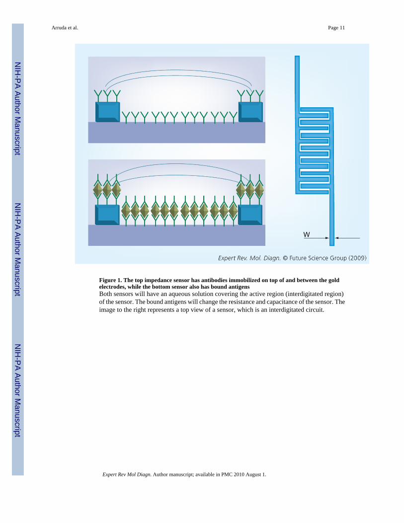
Figure 1. The top impedance sensor has antibodies immobilized on top of and between the goldelectrodes, while the bottom sensor also has bound antigensBoth sensors will have an aqueous solution covering the active region (interdigitated region)of the sensor. The bound antigens will change the resistance and capacitance of the sensor. Theimage to the right represents a top view of a sensor, which is an interdigitated circuit.
Arruda et al. Page 11
Expert Rev Mol Diagn. Author manuscript; available in PMC 2010 August 1.
NIH
-PA Author Manuscript
NIH
-PA Author Manuscript
NIH
-PA Author Manuscript
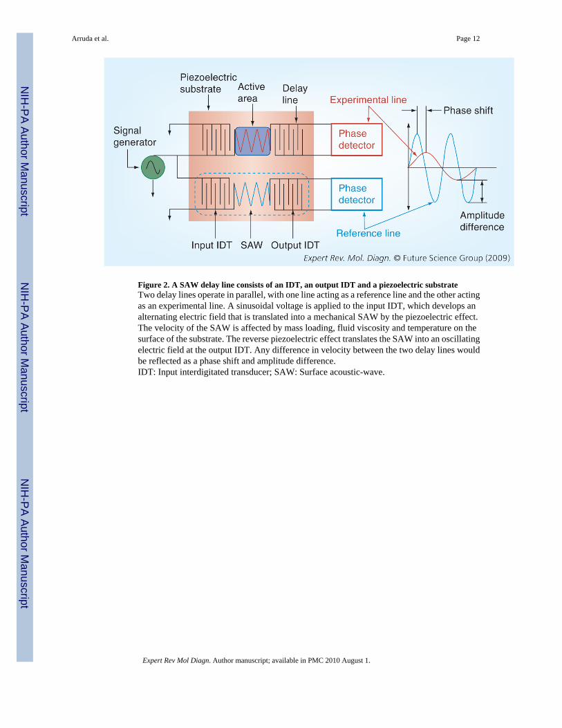
Figure 2. A SAW delay line consists of an IDT, an output IDT and a piezoelectric substrateTwo delay lines operate in parallel, with one line acting as a reference line and the other actingas an experimental line. A sinusoidal voltage is applied to the input IDT, which develops analternating electric field that is translated into a mechanical SAW by the piezoelectric effect.The velocity of the SAW is affected by mass loading, fluid viscosity and temperature on thesurface of the substrate. The reverse piezoelectric effect translates the SAW into an oscillatingelectric field at the output IDT. Any difference in velocity between the two delay lines wouldbe reflected as a phase shift and amplitude difference.IDT: Input interdigitated transducer; SAW: Surface acoustic-wave.
Arruda et al. Page 12
Expert Rev Mol Diagn. Author manuscript; available in PMC 2010 August 1.
NIH
-PA Author Manuscript
NIH
-PA Author Manuscript
NIH
-PA Author Manuscript
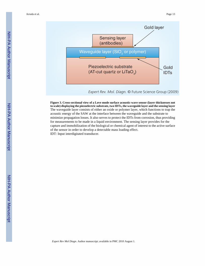
Figure 3. Cross-sectional view of a Love mode surface acoustic-wave sensor (layer thicknesses notto scale) displaying the piezoelectric substrate, two IDTs, the waveguide layer and the sensing layerThe waveguide layer consists of either an oxide or polymer layer, which functions to trap theacoustic energy of the SAW at the interface between the waveguide and the substrate tominimize propagation losses. It also serves to protect the IDTs from corrosion, thus providingfor measurements to be made in a liquid environment. The sensing layer provides for thecapture and immobilization of the biological or chemical agent of interest to the active surfaceof the sensor in order to develop a detectable mass loading effect.IDT: Input interdigitated transducer.
Arruda et al. Page 13
Expert Rev Mol Diagn. Author manuscript; available in PMC 2010 August 1.
NIH
-PA Author Manuscript
NIH
-PA Author Manuscript
NIH
-PA Author Manuscript
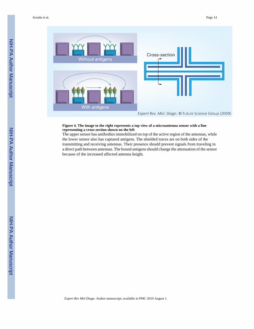
Figure 4. The image to the right represents a top view of a microantenna sensor with a linerepresenting a cross-section shown on the leftThe upper sensor has antibodies immobilized on top of the active region of the antennas, whilethe lower sensor also has captured antigens. The shielded traces are on both sides of thetransmitting and receiving antennas. Their presence should prevent signals from traveling ina direct path between antennas. The bound antigens should change the attenuation of the sensorbecause of the increased affected antenna height.
Arruda et al. Page 14
Expert Rev Mol Diagn. Author manuscript; available in PMC 2010 August 1.
NIH
-PA Author Manuscript
NIH
-PA Author Manuscript
NIH
-PA Author Manuscript