melanin-concentrating hormone: a new sleep factor?
TRANSCRIPT

REVIEW ARTICLEpublished: 18 March 2011
doi: 10.3389/fneur.2011.00014
Melanin-concentrating hormone: a new sleep factor?PabloTorterolo1, Patricia Lagos1 and Jaime M. Monti 2*
1 Department of Physiology, School of Medicine, University of the Republic, Montevideo, Uruguay2 Department of Pharmacology and Therapeutics, School of Medicine, Clinics Hospital, Montevideo, Uruguay
Edited by:
Michael J. Thorpy, Albert EinsteinCollege of Medicine of YeshivaUniversity, USA
Reviewed by:
John J. Renger, Merck ResearchLaboratories, USAPriyattam J. Shiromani, HarvardMedical School, USA
*Correspondence:
Jaime M. Monti, Department ofPharmacology and Therapeutics,School of Medicine, Clinics Hospital,Montevideo 11600, Uruguay.e-mail: [email protected]
Neurons containing the neuropeptide melanin-concentrating hormone (MCH) are mainlylocated in the lateral hypothalamus and the incerto-hypothalamic area, and have widespreadprojections throughout the brain. While the biological functions of this neuropeptide areexerted in humans through two metabotropic receptors, the MCHR1 and MCHR2, onlythe MCHR1 is present in rodents. Recently, it has been shown that the MCHergic sys-tem is involved in the control of sleep. We can summarize the experimental findings asfollows: (1) The areas related to the control of sleep and wakefulness have a high densityof MCHergic fibers and receptors. (2) MCHergic neurons are active during sleep, espe-cially during rapid eye movement (REM) sleep. (3) MCH knockout mice have less REMsleep, notably under conditions of negative energy balance. Animals with genetically inac-tivated MCHR1 also exhibit altered vigilance state architecture and sleep homeostasis. (4)Systemically administered MCHR1 antagonists reduce sleep. (5) Intraventricular microin-jection of MCH increases both slow wave sleep (SWS) and REM sleep; however, theincrement in REM sleep is more pronounced. (6) Microinjection of MCH into the dorsalraphe nucleus increases REM sleep time. REM seep is inhibited by immunoneutraliza-tion of MCH within this nucleus. (7) Microinjection of MCH in the nucleus pontis oralisof the cat enhances REM sleep time and reduces REM sleep latency. All these datastrongly suggest that MCH has a potent role in the promotion of sleep. Although both SWSand REM sleep are facilitated by MCH, REM sleep seems to be more sensitive to MCHmodulation.
Keywords: hypothalamus, depression, raphe nuclei, orexin, peptides, REM sleep
INTRODUCTIONSleep occupies approximately one-third of human’s life. Sleep isnot a homogeneous process. In most mammals two sleep states arerecognized: slow wave sleep (SWS) and the rapid eye movements(REM) sleep. Wakefulness, SWS, and REM sleep are differenti-ated by the electroencephalographic (EEG) activity (synchronizedduring SWS, desynchronized during wakefulness, and REM sleep),cognition (consciousness during wakefulness and dreams duringREM sleep), muscle activity (atonia during REM sleep), and othersphysiological parameters (McCarley, 2007; Torterolo and Vanini,2010).
In the twentieth century experimental and clinical researchbegan to unravel the mystery of sleep. Nowadays, the main neu-ronal networks that are involved in the generation of wakefulness,SWS, and REM sleep are known (Siegel, 2005; McCarley, 2007;Szymusiak and McGinty, 2008; Torterolo and Vanini, 2010). Thethalamo-cortical network is the pacemaker for the EEG rhythmsof wakefulness and sleep, and is the scaffold for the cognitivefunctions. This network is regulated by the activating (waking-related) systems located in the basal forebrain, postero-lateralhypothalamus, and mesopontine reticular formation. While thepreoptic area is critical for SWS generation, the mesopontineregion contains the “necessary and sufficient” neuronal networksfor REM sleep generation.
Several neurotransmitters and neuromodulators such as acetyl-choline (ACh), noradrenaline (NA), dopamine, serotonin, his-tamine, adenosine, and hypocretins, have been involved in thecontrol of sleep and wakefulness (Jones, 2005; McCarley, 2007;Torterolo and Vanini, 2010). Do melanin-concentrating hormone(MCH), and MCHergic system play a role in the control of sleep?Is MCH a sleep factor? So far, about 1000 papers related to MCHhave been published. Notwithstanding this, only a relatively smallnumber of publications associate MCH with sleep. In this reviewarticle we evaluate the data on MCH and sleep, and give our pointof view of how the MCHergic system might regulate behavioralstates.
MELANIN-CONCENTRATING HORMONEIn mammals, MCH is a cyclic neuropeptide with 19 aminoacidsthat has been found predominantly in neurons localized in thelateral hypothalamus and incerto-hypothalamic area (Kawauchiet al., 1983; Skofitsch et al., 1985; Bittencourt et al., 1992).
Originally, MCH was described as a circulating hormone iso-lated from salmon pituitaries, where it induces the aggregation ofmelanin granules in melanophores; this action results in a paleskin color (Kawauchi et al., 1983). This mechanism lets the ani-mal to camouflage in response to the background color of theenvironment or in response to stress. This effect is opposite to
www.frontiersin.org March 2011 | Volume 2 | Article 14 | 1
Torterolo et al. MCH and sleep
the pigment-dispersing effect of alpha-melanocyte stimulatinghormone (α-MSH). Subsequently,MCH was identified in rat brainusing immunohistochemical techniques (Skofitsch et al., 1985;Bittencourt et al., 1992).
MCH structure is identical in all mammals analyzed to date,including mice, rats, rabbits, and humans (Saito and Nagasaki,2008). MCH is generated by the cleavage of a precursor of165 amino acids, the prepro-MCH (ppMCH). ppMCH con-tains other peptides in addition to MCH, designated as neu-ropeptide EI (NEI) and neuropeptide GE (NGE; Bittencourt andCelis, 2008). In addition, the precursor may potentially give riseto an alternative splice variant termed MCH-gene-overprinted-polypeptide (MGOP), as well as a portion of the antisense-RNA-overlapping-MCH (AROM; Toumaniantz et al., 1996; Borsu et al.,2000).
MCH RECEPTORS AND SIGNALING MECHANISMSSeveral research groups almost simultaneously discovered the firstMCH receptor in 1999. It was found to be an orphan G-proteincoupled receptor, named SLC-1 (Bachner et al., 1999; Cham-bers et al., 1999; Lembo et al., 1999; Shimomura et al., 1999).Because MCH was the only ligand from rat brain extract thatactivated rat or human SLC-1 receptor, it was named MCHR1.In 2001, another MCH receptor was identified. This receptorwas termed MCHR2 and has a 38% sequence homology withMCHR1 (Hill et al., 2001; Mori et al., 2001; Sailer et al., 2001).The binding of MCH to MCHR1 activates diverse intracellularsignaling pathways by coupling to Gi, Gq, and Go proteins, whileMCHR2 is known to couple to the Gq protein (Hawes et al., 2000;Sailer et al., 2001).
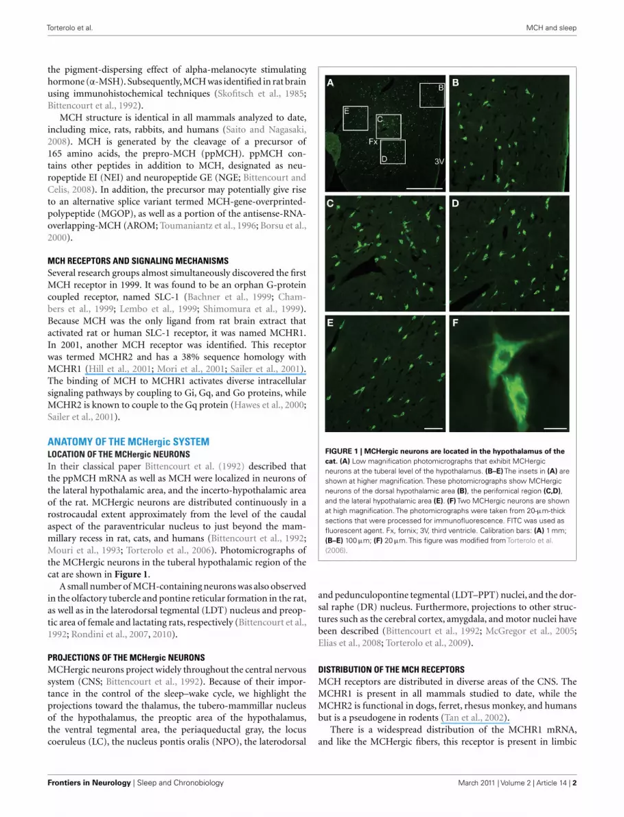
ANATOMY OF THE MCHergic SYSTEMLOCATION OF THE MCHergic NEURONSIn their classical paper Bittencourt et al. (1992) described thatthe ppMCH mRNA as well as MCH were localized in neurons ofthe lateral hypothalamic area, and the incerto-hypothalamic areaof the rat. MCHergic neurons are distributed continuously in arostrocaudal extent approximately from the level of the caudalaspect of the paraventricular nucleus to just beyond the mam-millary recess in rat, cats, and humans (Bittencourt et al., 1992;Mouri et al., 1993; Torterolo et al., 2006). Photomicrographs ofthe MCHergic neurons in the tuberal hypothalamic region of thecat are shown in Figure 1.
A small number of MCH-containing neurons was also observedin the olfactory tubercle and pontine reticular formation in the rat,as well as in the laterodorsal tegmental (LDT) nucleus and preop-tic area of female and lactating rats, respectively (Bittencourt et al.,1992; Rondini et al., 2007, 2010).
PROJECTIONS OF THE MCHergic NEURONSMCHergic neurons project widely throughout the central nervoussystem (CNS; Bittencourt et al., 1992). Because of their impor-tance in the control of the sleep–wake cycle, we highlight theprojections toward the thalamus, the tubero-mammillar nucleusof the hypothalamus, the preoptic area of the hypothalamus,the ventral tegmental area, the periaqueductal gray, the locuscoeruleus (LC), the nucleus pontis oralis (NPO), the laterodorsal
FIGURE 1 | MCHergic neurons are located in the hypothalamus of the
cat. (A) Low magnification photomicrographs that exhibit MCHergicneurons at the tuberal level of the hypothalamus. (B–E) The insets in (A) areshown at higher magnification. These photomicrographs show MCHergicneurons of the dorsal hypothalamic area (B), the perifornical region (C,D),and the lateral hypothalamic area (E). (F) Two MCHergic neurons are shownat high magnification. The photomicrographs were taken from 20-μm-thicksections that were processed for immunofluorescence. FITC was used asfluorescent agent. Fx, fornix; 3V, third ventricle. Calibration bars: (A) 1 mm;(B–E) 100 μm; (F) 20 μm. This figure was modified from Torterolo et al.(2006).
and pedunculopontine tegmental (LDT–PPT) nuclei, and the dor-sal raphe (DR) nucleus. Furthermore, projections to other struc-tures such as the cerebral cortex, amygdala, and motor nuclei havebeen described (Bittencourt et al., 1992; McGregor et al., 2005;Elias et al., 2008; Torterolo et al., 2009).
DISTRIBUTION OF THE MCH RECEPTORSMCH receptors are distributed in diverse areas of the CNS. TheMCHR1 is present in all mammals studied to date, while theMCHR2 is functional in dogs, ferret, rhesus monkey, and humansbut is a pseudogene in rodents (Tan et al., 2002).
There is a widespread distribution of the MCHR1 mRNA,and like the MCHergic fibers, this receptor is present in limbic
Frontiers in Neurology | Sleep and Chronobiology March 2011 | Volume 2 | Article 14 | 2
Torterolo et al. MCH and sleep
structures and in areas related to the control of sleep andwakefulness. MCHR1 have been detected also in several peripheralorgans (Chung et al., 2009).
The distribution of MCHR2 nearly overlaps with that ofMCH1R, but the latter shows higher relative levels of density and awider distribution pattern (Hill et al., 2001; Mori et al., 2001). Thefunction of MCHR2 remains unknown due to the lack of availableanimal models.
MCH: VOLUME CONDUCTION THROUGH THE CEREBROSPINAL FLUID(CSF)?As it is the case with the central actions of oxytocin (Veeninget al., 2010), it is likely that some of its biological effects are pro-duced by volume conduction, through a neuro-humoral pathwayvia the CSF (Torterolo et al., 2008). In a recent report, Veeningand Barendregt (2010) stated that the CSF system, in addition toprovide nutrients to and remove waste products from the brain,contains message molecules in the form of actively released neu-roactive substances. The authors suggested that this special form ofvolume transmission is a way to broadcast coordinated messagesto a variety of brain areas and may underlay changes in behavioralstates (Veening and Barendregt, 2010).
MCHergic fibers are lying close the ventricle walls of the cat (P.Torterolo, unpublished observation) and MCH is present in theCSF (Quintela et al., 2010). At the level of the DR, we demonstratedthat tanycytes lining the floor of the fourth ventricle show positiveimmunoreactivity for MCH, and that their long basal processesare closely related to serotonergic neurons of the DR (Torteroloet al., 2008). The tanycytes are specialized ependymal cells thatuptake substances from the CSF and release them to the underly-ing neuropil (Rodriguez et al., 2005). Since serotonergic neuronsof the DR have a fundamental role in the control of REM sleep(Monti, 2010a, 2010b), MCH could modulate the serotonergicactivity and REM sleep through this type of volume conduction(see below).
MCHergic SYSTEM. FUNCTIONAL CONSIDERATIONSNEUROTRANSMITTERS THAT CO-LOCALIZE WITH MCHIn addition to the neuropeptides derived from the ppMCH, otherneurotransmitter and neuromodulators have been described toco-localize with MCH. Elias et al. (2001) have demonstrated thatthe anorexinergic cocaine- and amphetamine-regulated transcript(CART), is co-expressed in 95% of the MCHergic neurons of thezona incerta and in 70% of MCH-positive neurons of the lat-eral hypothalamus of the rat. MCHergic neurons that co-localizewith CART send ascending projections toward the septum andhippocampus, whereas the non-CART MCHergic neurons senddescending projections toward the brainstem and spinal cord(Hanriot et al., 2007). In addition, all the MCHergic neuronsexpress the novel hypothalamic molecule nesfatin-1 that alsosuppresses feeding (Fort et al., 2008).
Recently, utilizing MCH immunohistochemistry and in situhybridization to detect GAD67 mRNA, Sapin et al. (2010) demon-strated that 85% of the MCHergic neurons of the tuberal region ofthe hypothalamus are also GABAergic. Besides, MCH and GABAco-localize in the LDT and medial preoptic area (Rondini et al.,2007, 2010).
ELECTROPHYSIOLOGICAL CHARACTERISTIC OF THE MCHergicNEURONSIn vitro recordings have shown that the MCHergic neurons presenta resting potential ranging from −62 to −50 mV, slightly morehyperpolarized than nearby hypocretinergic neurons (Guyonet al., 2009). MCHergic neurons also showed a relatively lowlevel of spontaneous activity compared with hypocretinergic andGABAergic neurons of the region.
Glutamate and GABA regulate MCHergic neuronal activity.Accordingly, both AMPA and NMDA depolarize these neurons(van den Pol et al., 2004). The activation of group I of metabotropicglutamate receptors also excite MCHergic neurons (Huang andvan den Pol, 2007). The GABA agonist muscimol hyperpolarizesMCHergic neurons while there are no data regarding the effect ofGABAB agents Guyon et al. (2009).
SYNAPTIC EFFECTS OF MCHStudies in lateral hypothalamic neurons in vitro have shown apredominantly inhibitory effect of MCH both at pre- and postsy-naptic levels (Gao and van den Pol, 2001). Hence, MCH inhibitedthe synaptic activity of both glutamatergic and GABAergic inputsto lateral hypothalamic neurons. In addition, MCH reduced theamplitude of glutamate-evoked currents and the amplitude ofminiature excitatory currents, indicating an inhibitory modu-lation of postsynaptic glutamate receptors. Furthermore, MCHattenuated L-, N-, and P/Q-type calcium of calcium voltage-activated currents (Gao and van den Pol, 2002).
MCHergic neurons express MCHR1 autoreceptors; by thismean the neuropeptide inhibits MCHergic neurons via a depres-sion of voltage-dependent calcium channels without affectingthe resting potential (Huang et al., 2007). Therefore, MCHergicneurons would exert a negative feedback on their own activity.
ROLE OF MCH IN THE CONTROL OF ENERGY HOMEOSTASISAND MOODThe high conservation of MCH structure along phylogeny sug-gests that this neuropeptide is involved in important physiologicalprocesses. The lateral hypothalamic area, where the MCHergicneurons are mainly located, is an integrative region par excellence.This region has been implicated in a wide variety of functions,such as energy homeostasis, wakefulness and sleep, regulation ofmotor activity, motivation, and reward (Bernardis and Bellinger,1996). MCHergic neurons seem to be implicated in some of thesefunctions.
Due to the strong relationship among sleep, energy homeosta-sis and major depression we will briefly review the role of MCHin these processes and condition.
There are strong experimental evidences that MCH increasesfood intake and reduces energy expenditure such as:
1. Acute infusion of MCH into the lateral ventricles inducesfeeding in rodents. Fasting induces an up-regulation of MCHmRNA expression (Qu et al., 1996).
2. MCH mRNA levels are also elevated in the obese leptin-deficient ob/ob mice, suggesting that leptin negatively regulatesMCH expression (Qu et al., 1996). In fact, by means of doubleknockout mice for MCH and leptin, it was demonstrated that
www.frontiersin.org March 2011 | Volume 2 | Article 14 | 3
Torterolo et al. MCH and sleep
MCH is a critical mediator of the leptin-deficient phenotype(Segal-Lieberman et al., 2003).
3. The stimulating effect of food intake has been confirmed intransgenic mice, in which an over-expression of MCH causesobesity (Ludwig et al., 2001), while prepro-MCH knockoutmice (MCH-KO) are hypophagic and lean (Shimada et al.,1998). Indeed, MCH is the only known hypothalamic peptidewhose genetic deletion results in leanness. In these MCH-KOmice, an increase in temperature, heart rate, and metabolic rateoccurs along with an increase in energy expenditure (Astrandet al., 2004). A toxin-mediated genetic cell ablation strategyusing a truncated ataxin-3 has been applied to induce apopto-sis of MCHergic neurons; these mice developed a late onsetsyndrome characterized also by leanness, hypophagia, andincreased energy expenditure (Alon and Friedman, 2006).
4. Mice knockout for the MCHR1 are lean, although in contrastto MCH-KO mice, they are hyperactive, hyperphagic, and havealtered metabolism (Marsh et al., 2002). In addition, block-ade of MCHR1 with specific antagonists has anorectic effects(Borowsky et al., 2002).
5. The MCHergic neurons are under the direct control of NPY-containing neurons of the arcuate nucleus; these neurons arepart of the neuronal network that governs feeding and energyexpenditure (van den Pol et al., 2004). Furthermore, periph-eral signals of energy abundance such as glucose, enhanceMCHergic activity in vitro (Burdakov et al., 2005).
The presence of MCHergic fibers and receptors in the lim-bic system tends to suggest that the MCHergic system plays arole in the control of mood and emotion. Borowsky et al. (2002)have showed that systemic administration of an MCHR1 antag-onist produces antidepressant effects (Borowsky et al., 2002). Wehave also shown that the microinjection of a low dose of MCH(50 ng) into the DR of the rat evokes a depressive-like behavioras shown by a significant increase in the immobility time as wellas a decrease in climbing behavior in the forced swimming test(Lagos et al., 2011b). The depressive-like effect induced by MCHwas prevented by systemic pre-treatment with fluoxetine, a selec-tive serotonin reuptake inhibitor. Consistent with these results, theimmunoneutralization of MCH by microinjection of antibodiesanti-MCH into the DR has antidepressant effects; anti-MCH anti-bodies decreased the immobility time in the forced swimmingtest without modifying the motor activity measured in the openfield test.
The above-mentioned data suggest that MCH modulates theactivity of serotonergic DR neurons, and could be involved in thecontrol of mood. Furthermore, antagonism of the MCHR1 pro-duces also a decrease in anxiety in animals models (Borowsky et al.,2002; Chaki et al., 2005).
Additional functions of MCH via central or peripheral MCHR1such as the control of reproduction or the immune response havebeen also described (Lakaye et al., 2009; Wu et al., 2009).
ROLE OF MCH IN THE CONTROL OF SLEEP ANDWAKEFULNESSFrom 2003 on, several investigators have examined the roleof MCH in sleep, and the evidence indicates that it pro-
motes sleep (Torterolo et al., 2003a; Verret et al., 2003;Willie et al., 2003).
ANATOMICAL CONSIDERATIONSWe mentioned above that MCHergic fibers and receptors arepresent in regions related to the control of sleep and wakeful-ness. In addition, MCHergic neurons have been identified in keyareas responsible for the generation of wakefulness and REM sleepsuch as the LDT (only in female rats), and the pontine reticularformation (Bittencourt et al., 1992; Rondini et al., 2007). Last butnot least, MCH is present in the tanycytes of the DR of the cat,where serotonergic REM “OFF” neurons are present (Torteroloet al., 2008).
RELATIONSHIP WITH HYPOCRETINERGIC NEURONSDue to the importance of the hypocretinergic system in sleepphysiology and pathology, the interaction between the MCHer-gic, and the hypocretinergic system is worth to be considered. Thehypocretinergic neurons degenerate in narcolepsy, a paradigmaticsleep disorder (Nishino et al., 2000; Peyron et al., 2000; Thannickalet al., 2000). The activity of the hypocretinergic neurons increasesduring active wakefulness, and they are believed to be central inthe maintenance of this state (Torterolo et al., 2001, 2003b; Leeet al., 2005; Mileykovskiy et al., 2005).
A strong relationship exists between hypocretinergic andMCHergic neurons in the hypothalamus. The MCHergic neuronsare intermingled with hypocretin (also called orexin)-containingneurons in the lateral hypothalamus, mainly at the tuberal andtubero-mammillar levels (Torterolo et al., 2006). MCHergic fibersare in close relationship with hypocretinergic neurons and viceversa, which tends to indicate the existence of reciprocal synapticcontacts between both types of cells (Guan et al., 2002; Torteroloet al., 2006). This fact as well as the presence of hypocretinergicreceptors on MCHergic neurons suggests an important functionalinteraction between both systems (Backberg et al., 2002). In fact,hypocretin increases MCH mRNA expression in hypothalamicneurons, directly excites MCHergic neurons and increases glu-tamate release onto them (Bayer et al., 2002; van den Pol et al.,2004). On the other hand, MCH modulates hypocretin/orexin-mediated effects on behavioral state and synaptic transmissionin the lateral hypothalamus (Rao et al., 2008). Rao et al. (2008)demonstrated that the efficacy of glutamatergic synapses onhypocretinergic neurons is enhanced in MCHR1 knockout mice,and hypocretin-1-induced firing is facilitated. On the contrary,in wild-type mice, MCH significantly attenuated the hypocretin-1-induced enhancement of spike frequency in hypocretinergicneurons, but not its basal activity. Furthermore, in these neu-rons, MCH attenuated hypocretin-1-induced enhancement of thefrequency of miniature excitatory postsynaptic currents (EPSCs).These effects imply that MCH exerts a unique inhibitory influenceon hypocretinergic signaling as a way to fine-tune the output ofthese neurons.
Interestingly, hypocretinergic and MCHergic neurons respondin a different way to most homeostatic signals such as glucose(Burdakov et al., 2005), or to waking-related neurotransmitterssuch as NA (Bayer et al., 2005). In this regard, it is of note thatwhile hypocretinergic neurons express the α1 adrenergic receptors,
Frontiers in Neurology | Sleep and Chronobiology March 2011 | Volume 2 | Article 14 | 4

Torterolo et al. MCH and sleep
MCHergic neurons express the α2 adrenergic receptors, whichare related to activation or inhibition of its target, respectively(Modirrousta et al., 2005).
WAKING AND SLEEP-RELATED MECHANISMS NEUROTRANSMITTERSREGULATE MCHergic ACTIVITYSerotonergic neurons of the dorsal and median raphe nucleus, aswell as noradrenergic neurons of the LC are active during wake-fulness (Monti and Jantos, 2008; Monti, 2010b; Torterolo andVanini, 2010). Midbrain dopaminergic neurons are also involvedin the promotion of wakefulness (Monti and Jantos, 2008). Onthe other hand, cholinergic neurons are active both during wake-fulness and REM sleep (Torterolo and Vanini, 2010). All theseneurotransmitters regulate the activity of the MCHergic neurons.
Noradrenaline hyperpolarizes the MCHergic neurons by directsynaptic actions mediated by the α2 adrenergic receptor (vanden Pol et al., 2004). Furthermore NA has presynaptic effects;it decreases the frequency of EPSCs and increases the frequencyof miniature inhibitory postsynaptic currents (IPSCs; van den Polet al., 2004).
Serotonin hyperpolarizes MCHergic neurons in the presenceof TTX indicating a direct postsynaptic effect (van den Pol et al.,2004).
Dopamine decreases the excitability of MCHergic neurons bydecreasing the membrane resistance without modifying the restingpotential (Guyon et al., 2009).
Acetylcholine hyperpolarizes MCHergic neurons through mus-carinic receptors and increases GABA release onto these neuronsthrough nicotinic mechanisms (van den Pol et al., 2004).
Cannabinoids are sleep-promoting neuromodulators (Murillo-Rodriguez,2008), and they increase the firing rate of the MCHergicneurons. Acting through the CB1 receptor, these substances inducedepolarization of MCHergic neurons and decrease the activity ofnearby GABAergic neurons (Huang et al., 2007).
In summary, extrahypothalamic waking-related neurotrans-mitters (NA, serotonin, ACh) inhibit MCHergic neurons, thussuggesting that in order to generate or maintain wakefulness, theseneurons should be inhibited. Notwithstanding this, it is intrigu-ing that ACh promotes REM sleep and simultaneously inhibitsMCHergic neurons (see below).
MCHergic NEURONAL ACTIVITY DURING WAKEFULNESS AND SLEEPHarthoorn et al. (2005) have demonstrated an up-regulation ofthe content of MCH in hypothalamic neurons during the restingphase of the light–dark cycle of the rat, when this animal sleepsmore.
Modirrousta et al. (2005), studied the c-fos expression (as anindex of neuronal activity) of the MCHergic neurons after a sleeprebound that followed a short protocol (3 h) of total sleep depri-vation in the rat. Although c-fos expression was observed in avery small proportion of MCHergic neurons (≈3%), the authorsdemonstrated that MCHergic neurons were activated during thesleep rebound (that consisted mainly in SWS) in comparison towakefulness. In contrast, almost no MCHergic neurons expressedc-fos during sleep (mainly SWS) induced by an adenosine receptoragonist in rats (Satoh et al., 2006). In the cat, we did not observeFos immunoreactivity in MCHergic neurons either during a SWS
rebound that followed a short protocol of total sleep deprivation,or during REM sleep induced by microinjections of carbachol intothe NPO (Torterolo et al., 2006). However, it should be consideredthat the occurrence of a false negative is one of the inconveniencesof this technique.
Verret et al. (2003) also studied the c-fos expression in theMCHergic neurons of the rat during a REM sleep rebound thatfollowed a prolonged protocol (72 h) of REM sleep deprivation.The authors observed that in comparison to the waking period,during the REM sleep rebound a large number of Fos+ cellswere immunoreactive for MCH and almost 60% of the MCH-containing neurons were Fos positive. The MCHergic neurons thatexpressed c-fos either did or did not co-localize CART (Hanriotet al., 2007).
In accordance with the studies mentioned above, MCHergicneurons did not express c-fos in wakefulness induced by applica-tion of GABAA antagonists in the lateral hypothalamus (Goutagnyet al., 2005).
Hassani et al. (2009) made use of in vivo extracellular record-ings of identified MCHergic neurons of head restrained rats todescribe their activity during the sleep–wakefulness cycle. Theseneurons were silent during wakefulness and began to dischargeduring the SWS transition; their firing rate slightly increased dur-ing SWS and reached a maximum during REM sleep. During thisperiod of time they fired in doublets or groups of spikes, and oftenin relation to twitches of the whiskers and muzzle. Furthermore,the MCHergic neurons discharged in a reciprocal manner to thehypocretinergic neurons. Although the sleep “ON” pattern of dis-charge of the neurons that were analyzed was clear, it remains toknow if this is the general pattern of activity of all the hypothal-amic and extrahypothalamic MCHergic neurons. Besides, due tothe strong relationship of the MCHergic neurons with the con-trol of energy homeostasis, it would be important to know if inconditions of positive energy balance there are MCHergic neuronsactive during wakefulness, as suggested by the in vitro studies ofBurdakov et al. (2005).
STUDIES ON GENETICALLY MODIFIED MICEStudies in knockout mice for both MCHR1 and MCH, show thatthese animals have an altered sleep architecture (Adamantidis et al.,2008; Willie et al., 2008).
MCH-KO mice have a reduction in SWS and an increase ofwakefulness during baseline conditions (Willie et al., 2008). Inresponse to fasting these animals exhibit a marked hyperactivity,accelerated weight loss, and an exaggerated decrease in REM sleepcompared to the wild-type littermates. The decrease in REM sleepresults from a reduced number of REM sleep bouts. Following 6 hof sleep deprivation, these animals have a normal sleep rebound.This data support a role for MCH in vigilance state regulation,particularly in response to a challenge in energy homeostasis.
Adamantidis et al. (2008) studied the spontaneous sleep andits homeostatic regulation in mice with deletion of the MCHR1gene. In contrast to previous data showing that MCH promotessleep, the authors described that the lack of functional MCHR1results in a hypersomnia-like phenotype. In comparison with wildlittermates, these mice exhibit higher amounts of REM sleep dur-ing the active (dark) phase. Furthermore MCHR-1 knockout mice
www.frontiersin.org March 2011 | Volume 2 | Article 14 | 5
Torterolo et al. MCH and sleep
showed a more pronounced rebound of SWS in response to sleepdeprivation. In addition, the authors found that modafinil, a stim-ulating drug, induced less wakefulness in MCHR1 knockout micethan in wild-type littermates. Then, these MCHR1 knockout ani-mals exhibit altered vigilance and sleep homeostasis, but the sign ofthe alteration seems the opposite to previous results. The authorsdiscussed that these unexpected results could be related to thedevelopment of compensatory mechanisms; the resulting adap-tive changes would oppose those caused by the blockade of theendogenous peptide.
EFFECT OF MCH ADMINISTRATION ON SLEEP VARIABLESIntraventricular microinjections of MCHIntraventricular microinjections of MCH into the active (dark)phase of the rat, produced a marked increase in REM sleep (up to200% above control values) and a moderate increase in SWS (upto 70% of control values; Verret et al., 2003). The increment inREM sleep time was due to an increase in the frequency of REMsleep episodes, whereas their duration remained unchanged.
Intracerebral microinjections of MCHUsing cholera toxin subunit-b as a retrograde tracer in cats, wehave demonstrated that ≈15% of the lateral hypothalamic neu-rons that project to the NPO, contained MCH (Torterolo et al.,2009). The NPO of the cat (also called medial pontine reticularformation or perilocus coeruleus α) or its corresponding nucleusin the rat, which is called the sub-laterodorsal nucleus, is an inte-gral part of different models of REM sleep, and is considered toexert executive control over the initiation and maintenance of thisbehavioral state (Reinoso-Suarez et al., 2001; Xi et al., 2001; Chaseand Morales, 2005; Siegel, 2005; Fuller et al., 2007; Luppi et al.,2007; McCarley, 2007). A single injection of a cholinergic agonist,such as carbachol, generates REM sleep with a very short latency(down to 30 s) and a duration that can exceed 2 h (Baghdoyanet al., 1987). Microinjections of MCH within this region produceda significant decrease in REM sleep latency with an increase inthe time the animal spent in this behavioral state. The incrementin REM sleep time was due to a increase of REM sleep episodesduration and frequency (Torterolo et al., 2009).
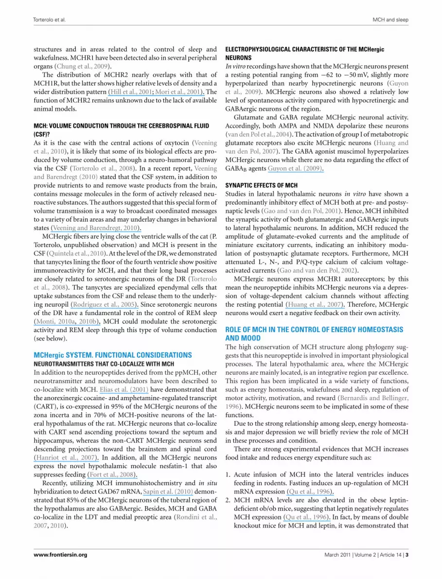
As mentioned earlier, the DR has a dense network of MCH-immunoreactive fibers and tanycytes (Torterolo et al., 2008). Inthis respect, we have studied the effect of MCH administrationinto the DR on sleep during the light period of the light–darkcycle of the rat. MCH induced a marked and dose-dependentincrease of the time the animal spent in REM sleep, and also amoderate increase of SWS; on the other hand, wakefulness wasreduced (Lagos et al., 2009). The increase of REM sleep time wasdue to an increment in the frequency of REM sleep bouts, whilethe duration of the episodes was not modified. Figure 2 shows rep-resentative hypnograms of an animal microinjected with MCH orvehicle (control) into the DR.
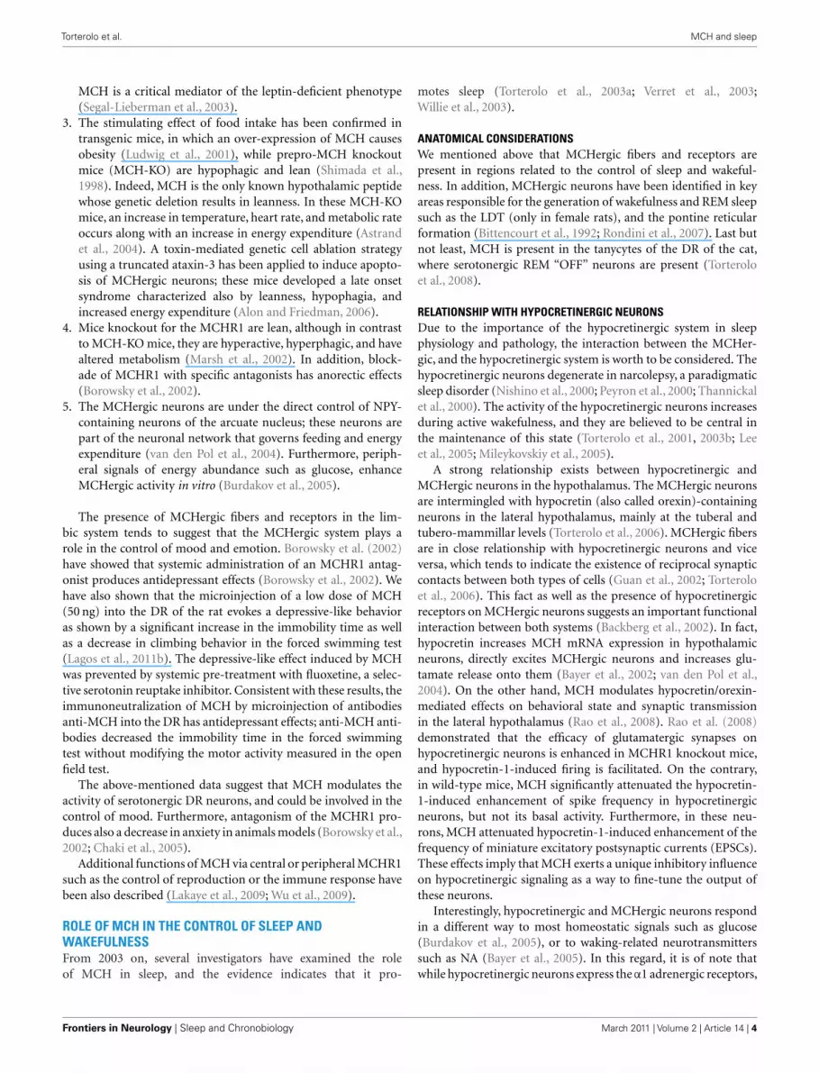
The blockade of physiologically released-MCH in the DR byimmunoneutralization (microinjections of antibodies anti-MCH)increased wakefulness and suppressed REM sleep, while SWStended to remain unchanged (Lagos et al., 2011a). The decre-ment in REM sleep time was due to a decrease in the frequency of
FIGURE 2 | Representative hypnograms illustrating the occurrence of
wakefulness and sleep following microinjection of MCH into the
dorsal raphe nucleus. The effects of the vehicle (A), and 100 ng of MCH(B) are presented. Comparing to control, the hypnogram correspondingMCH microinjection depicts a substantial increase in the number of REMsleep episodes. This figure was modified from Lagos et al. (2009).
FIGURE 3 | Representative hypnograms illustrating the occurrence of
wakefulness and sleep following microinjection of anti-MCH
antibodies (immunoneutralization) into the dorsal raphe nucleus. Theeffects of the vehicle (A) and 1/100 dilution dose of anti-MCH (B) arepresented. Comparing to control, the hypnogram corresponding toanti-MCH microinjection depicts a substantial decrease in the number ofREM sleep episodes. This figure was modified from Lagos et al. (2011a).
REM sleep episodes whereas the duration of the episodes was notmodified (Figure 3).
Systemic administration of MCHR1 antagonistsSystemic administration of MCHR1 antagonists in rats decreasedSWS and REM sleep time. This treatment also increased
Frontiers in Neurology | Sleep and Chronobiology March 2011 | Volume 2 | Article 14 | 6

Torterolo et al. MCH and sleep
wakefulness as well as SWS and REM sleep latencies (Ahnaouet al., 2008). The reduction of REM sleep was related to a smallernumber of REM sleep bouts, while the duration of the REM sleepepisodes was unchanged.
MCH: PROMOTION OF SLEEP AND SUPPRESSION OF WAKEFULNESSMost of the preceding data strongly suggest that the MCHergic sys-tem promotes sleep, but especially REM sleep. Indeed, reversibleinactivation by microinjection of a GABAA agonist into the hypo-thalamic area where the MCHergic neurons are located, stronglysuppresses REM sleep (Lin et al., 1989).
Hypocretinergic neuronsMCH inhibits waking-related hypocretinergic neurons (Rao et al.,2008), and by this means the MCHergic system may facilitateSWS and REM sleep. In fact, sleep attacks, sleep onset REM, andthe intrusion of REM sleep-like atonia during wakefulness (cata-plexy) characterize narcolepsy, where there is a suppression of thehypocretinergic activity due to a degeneration of the hypocre-tinergic neurons (Guilleminault and Fromherz, 2005; Mignot,2005).
Serotonergic neuronsSerotonergic neurons of the DR are active during wakefulness,reduce their discharge during SWS, and are essentially silent dur-ing REM sleep (REM-off neurons; Jacobs and Azmitia, 1992).Furthermore, serotonin release decreases during REM sleep indiverse brain areas (Portas et al., 2000). Application of 5-HT1A
receptor agonists into the DR facilitates REM sleep occurrence, aneffect mediated by the activation of inhibitory somatodendritic5-HT1A autoreceptors (Portas et al., 1996; Monti et al., 2002).The inhibition of serotonergic neurons of the DR following themicroinjection of the GABAA agonist muscimol also increasesREM sleep time (Nitz and Siegel, 1997). This outcome is sim-ilar to the one obtained following intra-DR microinjections ofMCH (Lagos et al., 2009). In preliminary studies, microinjectionof MCH into the DR of the cat produced similar effects (Deveraet al., 2007).
Based upon the wake-related discharge pattern of serotonergicneurons it has been suggested that these neurons promote alsowakefulness; in fact, there is strong evidence that the serotonergicneurons of the DR have a key role in the activation of the EEG(Dringenberg and Vanderwolf, 1998).
Therefore, it is likely that the microinjection of MCH into theDR inhibits the serotonergic neurons and by doing so, reduceswakefulness and increases REM sleep.
GABAergic neurons of the NPODue to the postulated inhibitory actions of MCH,we believe that inorder to facilitate the generation of REM sleep, MCH must inhibitthe GABAergic “wake-ON” neurons in the NPO, thus allow-ing the activation of “REM sleep-ON” neurons (Xi et al., 1999;Luppi et al., 2007; Brown et al., 2008).
Other waking-related neuronsMCHergic neurons project also to other neural structures involvedin the control of sleep and wakefulness including the basal fore-brain, the tubero-mammillar nucleus of the hypothalamus, the
LDT–PPT, the LC, and the ventrolateral periaqueductal gray(vlPAG). Interestingly, the bilateral inhibition or lesions of thevlPAG and nearby areas generates an important increase in REMsleep amount (Sastre et al., 1996; Vanini et al., 2007; Kaur et al.,2009); therefore it is expected that MCH would promote REMsleep when applied onto this region (Peyron et al., 2009). Webelieve that MCH promotes sleep (both SWS and REM) actingnot only on the vlPAG, but acting in tandem onto several wakingand sleep-related regions.
MCHergic neurons innervate also the specific and non-specificnuclei of the thalamus, including the reticular nucleus (Bittencourtet al., 1992). By actions onto these nuclei as well as onto the neo-cortex, the MCHergic neurons could influence the EEG rhythmsand behavioral states (Steriade, 2005).
PHYSIOLOGICAL CONSIDERATIONSEnergy conservation and restoration is one of the probable func-tions of sleep (Benington and Heller, 1995). Thus MCH wouldhave an integrative role in energy conservation by promoting foodintake, decreasing metabolism, and inducing sleep.
MCH decreases temperature, heart rate, and metabolic rateby central actions that enhance the parasympathetic/sympathetictone ratio and decrease the release of thyroid hormones. Somato-motor activity is also reduced by MCH (Saito and Nagasaki,2008). Given the profile of the activity of the MCHergic neu-rons it is possible that these actions are maximally expressedduring sleep.
In order to conserve energy, MCHergic neurons could beinvolved also in the promotion of sleep in accordance with meta-bolic demands. It is likely that in conditions of energy abundancesuch as post-feeding time, MCH would first decrease the level ofwakefulness, and then induce sleep. Furthermore, in special caseswhen energy conservation is imperative, MCHergic system mayplay a critical role. One case is hibernation, where the whole bodymetabolism falls to 1–2% of the basal value, and the body tem-perature could reach the freezing point of water (Heller and Ruby,2004). Interestingly, the entrance to this state is through SWS.
Another paradigmatic physiological state is lactation, where wehypothesize that the activity of the MCHergic system would bevery high. In fact, during this condition MCH is expressed in neu-rons of the medial preoptic area, a critical region for reproductivecontrol and maternal behavior (Rondini et al., 2010). During thelactating period in the rat, the animals appear somnolent and theEEG is synchronized before milk ejection (Lincoln et al., 1980).Lactation is also associated with an increase in SWS in women(Blyton et al., 2002). Furthermore, it has been reported an increasein the amount of REM sleep after delivery (Petre-Quadens and DeLee, 1974). These authors also showed a gradual decrease in REMsleep in bottle-feeding mothers compared to breastfeeding moth-ers where REM sleep values remain high. Therefore, it would beimportant to investigate if the cause of this hypersomnia duringlactation is promoted by MCH.
On the other hand, depression of the MCHergic neuronal activ-ity may mediate the effect of long-term sleep deprivation. Thiscondition leads to a marked increase in energy expenditure andbody weight loss (Rechtschaffen and Bergmann, 2002). In fact, thissyndrome partly recalls the effect of the deletion of the MCH gene.
www.frontiersin.org March 2011 | Volume 2 | Article 14 | 7

Torterolo et al. MCH and sleep
FIGURE 4 |The schematic shows a simple structural model that
proposes a tentative mechanism by which MCH promotes sleep.
During wakefulness MCHergic neurons may be activated during specialconditions such as energy abundance. Inhibiting and facilitating theactivating and somonogenic (SWS and REM sleep neuronal networks)respectively, MCH would facilitate and maintain sleep.
MCH IN SLEEP: WORKING HYPOTHESISRecently, Peyron et al. (2009) suggested that the MCHergicneurons are inhibited during wakefulness by extrahypothala-mic waking-related neurons, and during SWS are inhibited byGABAergic neurons of the sleep-promoting preoptic region. Onthe other hand, glutamatergic REM sleep“ON”neurons of the sub-laterodorsal nucleus would drive the MCHergic neurons. Thus,according to this proposal the MCHergic neurons are inhibited ordriven by the classical activating or sleep-promoting system.
A complementary view is to consider that MCHergic neuronsplay a more active role in the induction of sleep; in other words,these neurons would drive the sleep-promoting regions insteadof being driven by them. In addition, MCHergic neurons wouldinhibit the activating systems and promote sleep. A simple modelis presented in Figure 4.
THE MCHergic SYSTEM IN PATHOLOGYIn spite of the large number of preclinical studies, there are almostno data regarding the pathophysiology of the MCHergic system.
Preclinical studies have demonstrated that MCH is related toenergy homeostasis and it has been suggested that a dysfunc-tion of this system could lead to obesity. In fact, there is onestudy that reports a trend toward association of several MCHR1single-nucleotide polymorphisms with an obese phenotype inindependent study groups of obese German children and ado-lescents (Wermter et al., 2005). Furthermore, MCHR1 antagonistscould be a therapeutic option for treating obesity (Rivera et al.,2008).
Kokkotou et al. (2008) demonstrated that mice geneticallydeficient in MCH have substantially reduced local inflammatoryresponses in a model of experimental colitis. Likewise, mice receiv-ing treatments with an anti-MCH antibody developed attenuatedcolonic inflammation and survived longer. Furthermore, thereis an increased colonic expression of MCH and its receptor in
patients with inflammatory bowel disease, suggesting a role ofMCH in inflammatory processes in the intestine (Kokkotou et al.,2008).
We have mentioned earlier that there is preclinical evidence tosupport an increase in MCHergic activity in depression and thatthe antagonism of this system has antidepressant effects (Borowskyet al., 2002; Lagos et al., 2011b). In addition to pro-depressiveeffects, MCH promotes the occurrence of REM sleep in labora-tory animals. Interestingly, in patients with a diagnosis of majordepression (where there would be an increase in MCHergic tone),REM sleep occurrence is increased (Adrien, 2002). Accordingly, itcould be of interest to determine the role of MCH in patients withdepressive disorder.
Postpartum emotional distress or “blues” occurs in about 75–80% of new mothers about 3–5 days after birth (Lee, 1998). This“blues” is typically confined to the 1st week postpartum. Moreserious forms of maternal depression and postpartum psychosisoccur in less than 10% of new mothers. Regarding the probableincrement in activity of the MCHergic neurons during postpar-tum and lactation, it would be important to investigate if MCH isrelated to these emotional distresses.
As above-mentioned, there is a close anatomical and functionalrelationship between hypocretinergic and MCHergic neurons.Therefore, it is imperative to know how MCHergic neurons func-tion in narcolepsy, where there is a degeneration of the hypocre-tinergic neurons; in fact, the malfunction of MCHergic neuronscould play a role in the pathophysiology of this disease.
Parkinson’s disease (PD) but not Huntington’s disease, isaccompanied by decrease in the number of MCHergic neurons(Thannickal et al., 2007; Aziz et al., 2008). The tentative relation-ship between a loss of MCHergic activity and some symptoms ofPD has not been studied yet; however, a decrease in the activityof MCHergic cells could partly explain the high incidence of theREM sleep behavior disorder in this pathology (Thorpy and Adler,2005).
CONCLUSIONS AND FUTURE DIRECTIONSMCHergic neurons constitute a powerful regulatory system withwide and divergent projection. MCH is involved in the centralregulation of food intake, energy balance, and mood. Recent evi-dence strongly suggests that MCH has a regulatory role in thecontrol of sleep, mainly REM sleep. New experimental approachesare needed to unravel the role of MCH in sleep. Clinical research isalso needed in order to determine the involvement of this peptidein a number of neuropsychiatric conditions.
ACKNOWLEDGMENTSWe are very grateful to the reviewers for their comments andsuggestions that have improved the quality of our report.
REFERENCESAdamantidis, A., Salvert, D., Goutagny,
R., Lakaye, B., Gervasoni, D., Grisar,T., Luppi, P. H., and Fort, P. (2008).Sleep architecture of the melanin-concentrating hormone receptor 1-knockout mice. Eur. J. Neurosci. 27,1793–1800.
Adrien, J. (2002). Neurobiological basesfor the relation between sleepand depression. Sleep Med. Rev. 6,341–351.
Ahnaou, A., Drinkenburg, W. H., Bouw-knecht, J. A., Alcazar, J., Steckler, T.,andDautzenberg,F.M.(2008).Block-ingmelanin-concentratinghormone
MCH(1) receptor affects rat sleep-wake architecture. Eur. J. Pharmacol.579, 177–188.
Alon, T., and Friedman, J. M. (2006).Late-onset leanness in mice withtargeted ablation of melanin con-centrating hormone neurons. J.Neurosci. 26, 389–397.
Astrand, A., Bohlooly, Y. M., Larsdot-ter, S., Mahlapuu, M., Andersen,H., Tornell, J., Ohlsson, C., Snaith,M., and Morgan, D. G. (2004).Mice lacking melanin-concentratinghormone receptor 1 demonstrateincreased heart rate associated withaltered autonomic activity. Am. J.
Frontiers in Neurology | Sleep and Chronobiology March 2011 | Volume 2 | Article 14 | 8

Torterolo et al. MCH and sleep
Physiol. Regul. Integr. Comp. Physiol.287, R749–R758.
Aziz, A., Fronczek, R., Maat-Schieman,M., Unmehopa, U., Roelandse, F.,Overeem, S., van Duinen, S., Lam-mers, G. J., Swaab, D., and Roos,R. (2008). Hypocretin and melanin-concentrating hormone in patientswith Huntington disease. BrainPathol. 18, 474–483.
Bachner, D., Kreienkamp, H., Weise, C.,Buck, F., and Richter, D. (1999).Identification of melanin concen-trating hormone (MCH) as thenatural ligand for the orphansomatostatin-like receptor 1 (SLC-1). FEBS Lett. 457, 522–524.
Backberg, M., Hervieu, G., Wilson,S., and Meister, B. (2002). Orexinreceptor-1 (OX-R1) immunoreac-tivity in chemically identified neu-rons of the hypothalamus: focus onorexin targets involved in controlof food and water intake. Eur. J.Neurosci. 15, 315–328.
Baghdoyan, H. A., Rodrigo-Angulo, M.L., McCarley, R. W., and Hobson, J.A. (1987). A neuroanatomical gra-dient in the pontine tegmentumfor the cholinoceptive induction ofdesynchronized sleep signs. BrainRes. 414, 245–261.
Bayer, L., Eggermann, E., Serafin, M.,Grivel, J., Machard, D., Muhlethaler,M., and Jones, B. E. (2005). Oppositeeffects of noradrenaline and acetyl-choline upon hypocretin/orexin ver-sus melanin concentrating hormoneneurons in rat hypothalamic slices.Neuroscience 130, 807–811.
Bayer, L., Mairet-Coello, G., Risold,P. Y., and Griffond, B. (2002).Orexin/hypocretin neurons: chem-ical phenotype and possible inter-actions with melanin-concentratinghormone neurons. Regul. Pept. 104,33–39.
Benington, J. H., and Heller, H. C.(1995). Restoration of brain energymetabolism as the function of sleep.Prog. Neurobiol. 45, 347–360.
Bernardis, L. L., and Bellinger, L. L.(1996). The lateral hypothalamicarea revisited: ingestive behavior.Neurosci. Biobehav. Rev. 20, 189–287.
Bittencourt, J., and Celis, M. E. (2008).Anatomy, function and regulation ofneuropeptide EI (NEI). Peptides 29,1441–1450.
Bittencourt, J. C., Presse, F., Arias, C.,Peto, C., Vaughan, J., Nahon, J.L., Vale, W., and Sawchenko, P. E.(1992). The melanin-concentratinghormone system of the rat brain: animmuno- and hybridization histo-chemical characterization. J. Comp.Neurol. 319, 218–245.
Blyton, D. M., Sullivan, C. E., andEdwards, N. (2002). Lactation is
associated with an increase in slow-wave sleep in women. J. Sleep Res. 11,297–303.
Borowsky, B., Durkin, M. M., Ogoza-lek, K., Marzabadi, M. R., DeLeon,J., Heurich, R., Lichtblau, H., Sha-poshnik, Z., Daniewska, I., Black-burn, T. P., Branchek, T. A., Ger-ald, C., Vaysse, P. J., and Forray,C. (2002). Antidepressant, anxiolyticand anorectic effects of a melanin-concentrating hormone-1 receptorantagonist. Nat. Med. 8, 825–830.
Borsu, L., Presse, F., and Nahon, J. L.(2000). The AROM gene, splicedmRNAs encoding new DNA/RNA-binding proteins are transcribedfrom the opposite strand of themelanin-concentrating hormonegene in mammals. J. Biol. Chem.275, 40576–40587.
Brown, R. E., McKenna, J. T., Win-ston, S., Basheer, R., Yanagawa, Y.,Thakkar, M. M., and McCarley,R. W. (2008). Characterization ofGABAergic neurons in rapid-eye-movement sleep controlling regionsof the brainstem reticular formationin GAD67-green fluorescent proteinknock-in mice. Eur. J. Neurosci. 27,352–363.
Burdakov, D., Gerasimenko, O., andVerkhratsky, A. (2005). Physiologi-cal changes in glucose differentiallymodulate the excitability of hypo-thalamic melanin-concentratinghormone and orexin neurons insitu. J. Neurosci. 25, 2429–2433.
Chaki, S., Funakoshi, T., Hirota-Okuno, S., Nishiguchi, M., Shi-mazaki, T., Iijima, M., Grottick,A. J., Kanuma, K., Omodera,K., Sekiguchi, Y., Okuyama, S.,Tran, T. A., Semple, G., andThomsen, W. (2005). Anxiolytic-and antidepressant-like profile ofATC0065 and ATC0175: nonpep-tidic and orally active melanin-concentrating hormone receptor 1antagonists. J. Pharmacol. Exp. Ther.313, 831–839.
Chambers, J., Ames, R. S., Bergsma, D.,Muir, A., Fitzgerald, L. R., Hervieu,G., Dytko, G. M., Foley, J. J., Martin,J., Liu, W. S., Park, J., Ellis, C.,Ganguly, S., Konchar, S., Cluderay, J.,Leslie, R., Wilson, S., and Sarau, H.M. (1999). Melanin-concentratinghormone is the cognate ligandfor the orphan G-protein-coupledreceptor SLC-1. Nature 400,261–265.
Chase, M., and Morales, F. R. (2005).“Control of motoneurons duringsleep,” in Principles and Practicesof Sleep Medicine, eds M. H.Kryger, T. Roth and W. C. Dement(Philadelphia: Elsevier-Saunders),154–168.
Chung, S., Saito, Y., and Civelli,O. (2009). MCH receptors/genestructure-in vivo expression. Pep-tides 30, 1985–1989.
Devera, A., Lagos, P., Chase, M., andTorterolo, P. (2007). MCH en elnúcleo dorsal del rafe: rol en la vigiliay el sueño. Actas Fisiol. 11, 129.
Dringenberg, H. C., and Vanderwolf,C. H. (1998). Involvement of directand indirect pathways in electro-corticographic activation. Neurosci.Biobehav. Rev. 22, 243–257.
Elias, C. F., Lee, C. E., Kelly, J. F., Ahima,R. S., Kuhar, M., Saper, C. B., andElmquist, J. K. (2001). Characteri-zation of CART neurons in the ratand human hypothalamus. J. Comp.Neurol. 432, 1–19.
Elias, C. F., Sita, L. V., Zambon, B.K., Oliveira, E. R., Vasconcelos, L.A., and Bittencourt, J. C. (2008).Melanin-concentrating hormoneprojections to areas involved insomatomotor responses. J. Chem.Neuroanat. 35, 188–201.
Fort, P., Salvert, D., Hanriot, L., Jego, S.,Shimizu, H., Hashimoto, K., Mori,M., and Luppi, P. H. (2008). Thesatiety molecule nesfatin-1 is co-expressed with melanin concentrat-ing hormone in tuberal hypothala-mic neurons of the rat. Neuroscience155, 174–181.
Fuller, P. M., Saper, C. B., and Lu, J.(2007). The pontine REM switch:past and present. J. Physiol. 584,735–741.
Gao, X. B., and van den Pol, A.N. (2001). Melanin concentratinghormone depresses synaptic activ-ity of glutamate and GABA neu-rons from rat lateral hypothalamus.J. Physiol. 533, 237–252.
Gao, X. B., and van den Pol, A.N. (2002). Melanin-concentratinghormone depresses L-, N-, andP/Q-type voltage-dependent cal-cium channels in rat lateral hypo-thalamic neurons. J. Physiol. 542,273–286.
Goutagny, R., Luppi, P. H., Salvert, D.,Gervasoni, D., and Fort, P. (2005).GABAergic control of hypothalamicmelanin-concentrating hormone-containing neurons across thesleep-waking cycle. Neuroreport 16,1069–1073.
Guan, J. L., Uehara, K., Lu, S., Wang,Q. P., Funahashi, H., Sakurai, T.,Yanagisawa, M., and Shioda, S.(2002). Reciprocal synaptic relation-ship between orexin-and melanin-concentrating hormone-containingneurons in the rat lateral hypo-thalamus: a novel circuit impli-cated in feeding regulation. Int.J. Obes. Relat. Metab. Disord. 26,1523–1532.
Guilleminault, C., and Fromherz, S.(2005). “Narcolepsy: diagnosis andmanagement,” in Principles andPractices of Sleep Medicine, eds M.H. Kryger, T. Roth and W. C.Dement (Philadelphia: Saunders),780–790.
Guyon, A., Conductier, G., Rovere,C., Enfissi, A., and Nahon, J.L. (2009). Melanin-concentratinghormone producing neurons: activ-ities and modulations. Peptides 30,2031–2039.
Hanriot, L., Camargo, N., Courau, A.C., Leger, L., Luppi, P. H., andPeyron, C. (2007). Characteriza-tion of the melanin-concentratinghormone neurons activated dur-ing paradoxical sleep hypersom-nia in rats. J. Comp. Neurol. 505,147–157.
Harthoorn, L. F., Sane, A., Nethe, M.,and Van Heerikhuize, J. J. (2005).Multi-transcriptional profiling ofmelanin-concentrating hormoneand orexin-containing neurons.Cell. Mol. Neurobiol. 25, 1209–1223.
Hassani, O. K., Lee, M. G., and Jones,B. E. (2009). Melanin-concentratinghormone neurons discharge in areciprocal manner to orexin neu-rons across the sleep-wake cycle.Proc. Natl. Acad. Sci. U.S.A. 106,2418–2422.
Hawes, B. E., Kil, E., Green, B., O’Neill,K., Fried, S., and Graziano, M. P.(2000). The melanin-concentratinghormone receptor couples to mul-tiple G proteins to activate diverseintracellular signaling pathways.Endocrinology 141, 4524–4532.
Heller, H. C., and Ruby, N. F. (2004).Sleep and circadian rhythms inmammalian torpor. Annu. Rev.Physiol. 66, 275–289.
Hill, J., Duckworth, M., Murdock,P., Rennie, G., Sabido-David, C.,Ames, R. S., Szekeres, P., Wilson, S.,Bergsma, D. J., Gloger, I. S., Levy, D.S., Chambers, J. K., and Muir, A. I.(2001). Molecular cloning and func-tional characterization of MCH2, anovel human MCH receptor. J. Biol.Chem. 276, 20125–20129.
Huang, H., Acuna-Goycolea, C., Li, Y.,Cheng, H. M., Obrietan, K., andvan den Pol, A. N. (2007). Cannabi-noids excite hypothalamic melanin-concentrating hormone but inhibithypocretin/orexin neurons: implica-tions for cannabinoid actions onfood intake and cognitive arousal. J.Neurosci. 27, 4870–4881.
Huang, H., and van den Pol, A.N. (2007). Rapid direct excita-tion and long-lasting enhancementof NMDA response by group Imetabotropic glutamate receptoractivation of hypothalamic melanin-
www.frontiersin.org March 2011 | Volume 2 | Article 14 | 9
Torterolo et al. MCH and sleep
concentrating hormone neurons. J.Neurosci. 27, 11560–11572.
Jacobs, B. L., and Azmitia, E. C. (1992).Structure and function of the brainserotonin system. Physiol. Rev. 72,165–229.
Jones, B. (2005). “Basic mechanismsof sleep-wake states,” in Principlesand Practices of Sleep Medicine, edsM. H. Kryger, T. Roth and W.C. Dement (Philadelphia: Elsevier-Saunders), 136–153.
Kaur, S., Thankachan, S., Begum, S.,Liu, M., Blanco-Centurion, C., andShiromani, P. J. (2009). Hypocretin-2 saporin lesions of the ventro-lateral periaquaductal gray (vlPAG)increase REM sleep in hypocretinknockout mice. PLoS ONE 4, e6346.doi: 10.1371/journal.pone.0006346
Kawauchi, H., Kawazoe, I., Tsub-okawa, M., Kishida, M., and Baker,B. I. (1983). Characterization ofmelanin-concentrating hormone inchum salmon pituitaries. Nature305, 321–323.
Kokkotou, E., Moss, A. C., Torres,D., Karagiannides, I., Cheifetz, A.,Liu, S., O’Brien, M., Maratos-Flier,E., and Pothoulakis, C. (2008).Melanin-concentrating hormone asa mediator of intestinal inflamma-tion. Proc. Natl. Acad. Sci. U.S.A. 105,10613–10618.
Lagos, P., Torterolo, P., Jantos, H.,Chase, M. H., and Monti, J. M.(2009). Effects on sleep of melanin-concentrating hormone microinjec-tions into the dorsal raphe nucleus.Brain Res. 1265, 103–110.
Lagos, P., Torterolo, P., Jantos, H., Chase,M. H., and Monti, J. M. (2011a).Immunoneutralization of melanin-concentrating hormone (MCH) inthe dorsal raphe nucleus: effects onsleep and wakefulness. Brain Res.1269, 112–118.
Lagos, P., Urbanavicius, J., Scorza,C., Miraballes, R., and Torterolo,P. (2011b). Depressive-like profileinduced by MCH microinjectionsinto the dorsal raphe nucleus eval-uated in the forced swim test. Behav.Brain Res. 218, 259–266.
Lakaye, B., Coumans, B., Harray, S.,and Grisar, T. (2009). Melanin-concentrating hormone andimmune function. Peptides 30,2076–2080.
Lee, K. A. (1998). Alterations in sleepduring pregnancy and postpartum:a review of 30 years of research. SleepMed. Rev. 2, 231–242.
Lee, M. G., Hassani, O. K., and Jones,B. E. (2005). Discharge of identifiedorexin/hypocretin neurons acrossthe sleep-waking cycle. J. Neurosci.25, 6716–6720.
Lembo, P. M., Grazzini, E., Cao,J., Hubatsch, D. A., Pelletier, M.,Hoffert, C., St-Onge, S., Pou,C., Labrecque, J., Groblewski, T.,O’Donnell, D., Payza, K., Ahmad, S.,and Walker, P. (1999). The receptorfor the orexigenic peptide melanin-concentrating hormone is a G-protein-coupled receptor. Nat. CellBiol. 1, 267–271.
Lin, J. S., Sakai, K., Vanni-Mercier, G.,and Jouvet, M. (1989). A critical roleof the posterior hypothalamus in themechanisms of wakefulness deter-mined by microinjection of musci-mol in freely moving cats. Brain Res.479, 225–240.
Lincoln, D. W., Hentzen, K., Hin, T.,van der Schoot, P., Clarke, G., andSummerlee, A. J. (1980). Sleep: aprerequisite for reflex milk ejec-tion in the rat. Exp. Brain Res. 38,151–162.
Ludwig, D. S., Tritos, N. A., Mastaitis,J. W., Kulkarni, R., Kokkotou, E.,Elmquist, J., Lowell, B., Flier, J. S., andMaratos-Flier, E. (2001). Melanin-concentrating hormone overexpres-sion in transgenic mice leads to obe-sity and insulin resistance. J. Clin.Invest. 107, 379–386.
Luppi, P. H., Gervasoni, D., Ver-ret, L., Goutagny, R., Peyron,C., Salvert, D., Leger, L., andFort, P. (2007). Paradoxical(REM) sleep genesis: the switchfrom an aminergic-cholinergicto a GABAergic-glutamatergichypothesis. J. Physiol. Paris 100,271–283.
Marsh, D. J., Weingarth, D. T., Novi,D. E., Chen, H. Y., Trumbauer, M.E., Chen, A. S., Guan, X. M., Jiang,M. M., Feng, Y., Camacho, R. E.,Shen, Z., Frazier, E. G., Yu, H.,Metzger, J. M., Kuca, S. J., Shear-man, L. P., Gopal-Truter, S., Mac-Neil, D. J., Strack, A. M., Mac-Intyre, D. E., Van der Ploeg, L.H., and Qian, S. (2002). Melanin-concentrating hormone 1 receptor-deficient mice are lean, hyperactive,and hyperphagic and have alteredmetabolism. Proc. Natl. Acad. Sci.U.S.A. 99, 3240–3245.
McCarley, R. W. (2007). Neurobiologyof REM and NREM sleep. Sleep Med.8, 302–330.
McGregor, R., Damian, A., Fabbiani,G., Torterolo, P., Pose, I., Chase, M.,and Morales, F. R. (2005). Directhypothalamic innervation of thetrigeminal motor nucleus: a retro-grade tracer study. Neuroscience 136,1073–1081.
Mignot, E. (2005). “Narcolepsy:pharmacology, pathophysiologyand genetics,” in Principles and
Practices of Sleep Medicine, edsM. H. Kryger, T. Roth and W. C.Dement (Philadelphia: Saunders),761–799.
Mileykovskiy, B. Y., Kiyashchenko, L. I.,and Siegel, J. M. (2005). Behavioralcorrelates of activity in identifiedhypocretin/orexin neurons. Neuron46, 787–798.
Modirrousta, M., Mainville, L., andJones, B. E. (2005). Orexin andMCH neurons express c-Fos dif-ferently after sleep deprivation vs.recovery and bear different adren-ergic receptors. Eur. J. Neurosci. 21,2807–2816.
Monti, J. M. (2010a). The role of dorsalraphe nucleus serotonergic and non-serotonergic neurons, and of theirreceptors, in regulating waking andrapid eye movement (REM) sleep.Sleep Med. Rev. 14, 319–327.
Monti, J. M. (2010b). The structure ofthe dorsal raphe nucleus and its rel-evance to the regulation of sleepand wakefulness. Sleep Med. Rev. 14,307–317.
Monti, J. M., and Jantos, H. (2008). Theroles of dopamine and serotonin,and of their receptors, in regulatingsleep and waking. Prog. Brain Res.172, 625–646.
Monti, J. M., Jantos, H., and Monti, D.(2002). Increased REM sleep afterintra-dorsal raphe nucleus injec-tion of flesinoxan or 8-OHDPAT:prevention with WAY 100635.Eur. Neuropsychopharmacol. 12,47–55.
Mori, M., Harada, M., Terao, Y., Sugo,T., Watanabe, T., Shimomura,Y., Abe, M., Shintani, Y., Onda,H., Nishimura, O., and Fujino,M. (2001). Cloning of a novel Gprotein-coupled receptor, SLT, a sub-type of the melanin-concentratinghormone receptor. Biochem.Biophys. Res. Commun. 283,1013–1018.
Mouri, T., Takahashi, K., Kawauchi, H.,Sone, M., Totsune, K., Murakami,O., Itoi, K., Ohneda, M., Sasano,H., and Sasano, N. (1993).Melanin-concentrating hormonein the human brain. Peptides 14,643–646.
Murillo-Rodriguez, E. (2008). The roleof the CB1 receptor in the reg-ulation of sleep. Prog. Neuropsy-chopharmacol. Biol. Psychiatry 32,1420–1427.
Nishino, S., Ripley, B., Overeem, S.,Lammers, G. J., and Mignot, E.(2000). Hypocretin (orexin) defi-ciency in human narcolepsy. Lancet355, 39–40.
Nitz, D., and Siegel, J. (1997). GABArelease in the dorsal raphe nucleus:
role in the control of REM sleep. Am.J. Physiol. 273, R451–R455.
Petre-Quadens, O., and De Lee, C.(1974). “Sleep-cycle alterations dur-ing pregnancy, postpartum andthe menstrual cycle,” in Biorhythmsand Human Reproduction, eds M.Ferin, F. Halberg and R. M.Richart (New York: John Wiley),335–351.
Peyron, C., Faraco, J., Rogers, W., Ripley,B., Overeem, S., Charnay, Y., Nevsi-malova, S.,Aldrich, M., Reynolds, D.,Albin, R., Li, R., Hungs, M., Pedraz-zoli, M., Padigaru, M., Kucherlap-ati, M., Fan, J., Maki, R., Lammers,G. J., Bouras, C., Kucherlapati, R.,Nishino, S., and Mignot, E. (2000).A mutation in a case of early onsetnarcolepsy and a generalized absenceof hypocretin peptides in humannarcoleptic brains. Nat. Med. 6,991–997.
Peyron, C., Sapin, E., Leger, L.,Luppi, P. H., and Fort, P. (2009).Role of the melanin-concentratinghormone neuropeptide in sleep reg-ulation. Peptides 30, 2052–2059.
Portas, C. M., Bjorvatn, B., andUrsin, R. (2000). Serotonin and thesleep/wake cycle: special emphasison microdialysis studies. Prog. Neu-robiol. 60, 13–35.
Portas, C. M., Thakkar, M.,Rainnie, D., and McCarley,R. W. (1996). Microdialysisperfusion of 8-hydroxy-2-(di-N -propylamino)tetralin (8-OH-DPAT) in the dorsal raphe nucleusdecreases serotonin release andincreases rapid eye movement sleepin the freely moving cat. J. Neurosci.16, 2820–2828.
Qu, D., Ludwig, D. S., Gammeltoft,S., Piper, M., Pelleymounter,M. A., Cullen, M. J., Mathes,W. F., Przypek, R., Kanarek, R.,and Maratos-Flier, E. (1996). Arole for melanin-concentratinghormone in the central regulationof feeding behaviour. Nature 380,243–247.
Quintela, H., Lagos, P., Alzugaray,S., Torterolo, P., and Unger-feld, R. (2010). “Changes onmelanin-concentration hormonecerebrospinal fluid concentration inanoestrous ewes in response to theram effect,” in Proceedings of the The8th International Ruminant Repro-duction Symposium, Anchorage,AL.
Rao, Y., Lu, M., Ge, F., Marsh,D. J., Qian, S., Wang, A. H.,Picciotto, M. R., and Gao, X.B. (2008). Regulation of synap-tic efficacy in hypocretin/orexin-containing neurons by melanin
Frontiers in Neurology | Sleep and Chronobiology March 2011 | Volume 2 | Article 14 | 10
Torterolo et al. MCH and sleep
concentrating hormone in the lat-eral hypothalamus. J. Neurosci. 28,9101–9110.
Rechtschaffen, A., and Bergmann, B. M.(2002). Sleep deprivation in the rat:an update of the 1989 paper. Sleep25, 18–24.
Reinoso-Suarez, F., de Andres, I.,Rodrigo-Angulo, M. L., and Gar-zon, M. (2001). Brain structures andmechanisms involved in the genera-tion of REM sleep. Sleep Med. Rev. 5,63–77.
Rivera, G., Bocanegra-Garcia, V.,Galiano, S., Cirauqui, N., Ceras, J.,Perez, S., Aldana, I., and Monge,A. (2008). Melanin-concentratinghormone receptor 1 antago-nists: a new perspective for thepharmacologic treatment ofobesity. Curr. Med. Chem. 15,1025–1043.
Rodriguez, E. M., Blazquez, J. L., Pas-tor, F. E., Pelaez, B., Pena, P., Peruzzo,B., and Amat, P. (2005). Hypothal-amic tanycytes: a key componentof brain-endocrine interaction. Int.Rev. Cytol. 247, 89–164.
Rondini, T. A., de Crudis Rodrigues,B., de Oliveira, A. P., Bittencourt, J.C., and Elias, C. F. (2007). Melanin-concentrating hormone is expressedin the laterodorsal tegmental nucleusonly in female rats. Brain Res. Bull.74, 21–28.
Rondini, T. A., Donato, J. Jr., RodriguesBde, C., Bittencourt, J. C., andElias, C. F. (2010). Chemical identityand connections of medial preop-tic area neurons expressing melanin-concentrating hormone during lac-tation. J. Chem. Neuroanat. 39,51–62.
Sailer, A. W., Sano, H., Zeng, Z.,McDonald, T. P., Pan, J., Pong,S. S., Feighner, S. D., Tan, C. P.,Fukami, T., Iwaasa, H., Hreniuk, D.L., Morin, N. R., Sadowski, S. J.,Ito, M., Bansal, A., Ky, B., Figueroa,D. J., Jiang, Q., Austin, C. P., Mac-Neil, D. J., Ishihara, A., Ihara, M.,Kanatani, A., Van der Ploeg, L. H.,Howard, A. D., and Liu, Q. (2001).Identification and characterizationof a second melanin-concentratinghormone receptor, MCH-2R.Proc. Natl. Acad. Sci. U.S.A. 98,7564–7569.
Saito, Y., and Nagasaki, H. (2008). Themelanin-concentrating hormonesystem and its physiological func-tions. Results Probl. Cell Differ. 46,159–179.
Sapin, E., Berod, A., Leger, L., Her-man, P. A., Luppi, P. H., and Peyron,
C. (2010). A very large number ofGABAergic neurons are activatedin the tuberal hypothalamus dur-ing paradoxical (REM) sleep hyper-somnia. PLoS ONE 5, e11766. doi:10.1371/journal.pone.0011766
Sastre, J. P., Buda, C., Kitahama, K.,and Jouvet, M. (1996). Importanceof the ventrolateral region of theperiaqueductal gray and adjacenttegmentum in the control of para-doxical sleep as studied by muscimolmicroinjections in the cat. Neuro-science 74, 415–426.
Satoh, S., Matsumura, H., Kanbayashi,T., Yoshida, Y., Urakami, T., Naka-jima, T., Kimura, N., Nishino, S., andYoneda, H. (2006). Expression pat-tern of FOS in orexin neurons dur-ing sleep induced by an adenosineA2A receptor agonist. Behav. BrainRes. 170, 277–286.
Segal-Lieberman, G., Bradley, R. L.,Kokkotou, E., Carlson, M., Trombly,D. J., Wang, X., Bates, S., Myers, M.G. Jr., Flier, J. S., and Maratos-Flier,E. (2003). Melanin-concentratinghormone is a critical mediator ofthe leptin-deficient phenotype. Proc.Natl. Acad. Sci. U.S.A. 100, 10085–10090.
Shimada, M., Tritos, N. A., Lowell, B.B., Flier, J. S., and Maratos-Flier, E.(1998). Mice lacking melanin-con-centrating hormone are hypophagicand lean. Nature 396, 670–674.
Shimomura, Y., Mori, M., Sugo, T.,Ishibashi, Y., Abe, M., Kurokawa,T., Onda, H., Nishimura, O., Sum-ino, Y., and Fujino, M. (1999).Isolation and identification ofmelanin-concentrating hormoneas the endogenous ligand of theSLC-1 receptor. Biochem. Biophys.Res. Commun. 261, 622–626.
Siegel, J. M. (2005). “REM sleep,” inPrinciples and Practices of Sleep Med-icine, eds M. H. Kryger, T. Rothand W. C. Dement (Philadelphia:Elsevier-Saunders), 120–135.
Skofitsch, G., Jacobowitz, D. M., andZamir, N. (1985). Immunohisto-chemical localization of a melaninconcentrating hormone-like peptidein the rat brain. Brain Res. Bull. 15,635–649.
Steriade, M. (2005). “Brain electricalactivity and sensory processing dur-ing waking and sleep states,” in Prin-ciples and Practices of Sleep Med-icine, eds M. H. Kryger, T. Rothand W. C. Dement (Philadelphia:Saunders), 101–119.
Szymusiak, R., and McGinty, D. (2008).Hypothalamic regulation of sleep
and arousal. Ann. N. Y. Acad. Sci.1129, 275–286.
Tan, C. P., Sano, H., Iwaasa, H., Pan, J.,Sailer, A. W., Hreniuk, D. L., Feigh-ner, S. D., Palyha, O. C., Pong, S. S.,Figueroa, D. J., Austin, C. P., Jiang,M. M., Yu, H., Ito, J., Ito, M., Guan,X. M., MacNeil, D. J., Kanatani, A.,Van der Ploeg, L. H., and Howard,A. D. (2002). Melanin-concentratinghormone receptor subtypes 1 and2: species-specific gene expression.Genomics 79, 785–792.
Thannickal, T. C., Lai, Y. Y., and Siegel,J. M. (2007). Hypocretin (orexin)cell loss in Parkinson’s disease. Brain130, 1586–1595.
Thannickal, T. C., Moore, R. Y., Nien-huis, R., Ramanathan, L., Gulyani,S., Aldrich, M., Cornford, M.,and Siegel, J. M. (2000). Reducednumber of hypocretin neuronsin human narcolepsy. Neuron 27,469–474.
Thorpy, M. J., and Adler, C. H. (2005).Parkinson’s disease and sleep. Neu-rol. Clin. 23, 1187–1208.
Torterolo, P., Lagos, P., Sampogna, S.,and Chase, M. H. (2008). Melanin-concentrating hormone (MCH)immunoreactivity in non-neuronalcells within the raphe nuclei andsubventricular region of the brain-stem of the cat. Brain Res. 1210,163–178.
Torterolo, P., Rojas, M., Sampogna, S.,Morales, F. R., and Chase, M. H.(2003a). MCH-containing neuronsand the control of sleep and wake-fulness. Sleep 26, A20.
Torterolo, P., Yamuy, J., Sampogna, S.,Morales, F. R., and Chase, M. H.(2003b). Hypocretinergic neuronsare primarily involved in activationof the somatomotor system. Sleep 1,25–28.
Torterolo, P., Sampogna, S., and Chase,M. H. (2009). MCHergic projectionsto the nucleus pontis oralis partici-pate in the control of active (REM)sleep. Brain Res. 1268, 76–87.
Torterolo, P., Sampogna, S., Morales,F. R., and Chase, M. H. (2006).MCH-containing neurons in thehypothalamus of the cat: searchingfor a role in the control of sleepand wakefulness. Brain Res. 1119,101–114.
Torterolo, P., and Vanini, G. (2010).Nuevos conceptos sobre la gen-eración y el mantenimientode la vigilia. Rev. Neurol. 50,747–758.
Torterolo, P., Yamuy, J., Sampogna, S.,Morales, F. R., and Chase, M. H.
(2001). Hypothalamic neurons thatcontain hypocretin (orexin) expressc-fos during active wakefulnessand carbachol-induced active sleep.Sleep Res. Online 4, 25–32. Avail-able at: http://www.sro.org/2001/Torterolo/2025
Toumaniantz, G., Bittencourt, J. C.,and Nahon, J. L. (1996). Therat melanin-concentrating hormonegene encodes an additional putativeprotein in a different reading frame.Endocrinology 137, 4518–4521.
van den Pol, A. N., Acuna-Goycolea, C.,Clark, K. R., and Ghosh, P. K. (2004).Physiological properties of hypo-thalamic MCH neurons identifiedwith selective expression of reportergene after recombinant virus infec-tion. Neuron 42, 635–652.
Vanini, G., Torterolo, P., McGregor,R., Chase, M. H., and Morales, F.R. (2007). GABAergic processesin the mesencephalic tegmentummodulate the occurrence of active(rapid eye movement) sleep inguinea pigs. Neuroscience 145,1157–1167.
Veening, J. G., and Barendregt, H.P. (2010). The regulation of brainstates by neuroactive substances dis-tributed via the cerebrospinal fluid;a review. Cerebrospinal Fluid Res.7, 1.
Veening, J. G., de Jong, T., andBarendregt, H. P. (2010). Oxytocin-messages via the cerebrospinal fluid:behavioral effects; a review. Physiol.Behav. 101, 193–210.
Verret, L., Goutagny, R., Fort, P.,Cagnon, L., Salvert, D., Leger,L., Boissard, R., Salin, P., Pey-ron, C., and Luppi, P. H. (2003).A role of melanin-concentratinghormone producing neurons in thecentral regulation of paradoxicalsleep. BMC Neurosci. 4, 19. doi:10.1186/1471-2202-4-19
Wermter, A. K., Reichwald, K., Buch,T., Geller, F., Platzer, C., Huse, K.,Hess, C., Remschmidt, H., Guder-mann, T., Preibisch, G., Siegfried, W.,Goldschmidt, H. P., Li, W. D., Price,R. A., Biebermann, H., Krude, H.,Vollmert, C., Wichmann, H. E., Illig,T., Sorensen, T. I., Astrup, A., Larsen,L. H., Pedersen, O., Eberle, D.,Clement, K., Blundell, J., Wabitsch,M., Schafer, H., Platzer, M., Hinney,A., and Hebebrand, J. (2005). Muta-tion analysis of the MCHR1 genein human obesity. Eur. J. Endocrinol.152, 851–862.
Willie, J. T., Sinton, C. M., Maratos-Flier, E., and Yanagisawa, M. (2008).
www.frontiersin.org March 2011 | Volume 2 | Article 14 | 11
Torterolo et al. MCH and sleep
Abnormal response of melanin-concentrating hormone deficientmice to fasting: hyperactivity andrapid eye movement sleep suppres-sion. Neuroscience 156, 819–829.
Willie, J. T., Sinton, C. M., Mieda,M., Maratos-Flier, E., and Yanag-isawa, M. (2003). Orexin andmelanin-concentrating hormone(MCH) double knockout mice:compensatroy role for MCHin narcolepsy-cataplexy. Sleep26, A50.
Wu, M., Dumalska, I., Morozova, E., vanden Pol, A., and Alreja, M. (2009).
Melanin-concentrating hormonedirectly inhibits GnRH neurons andblocks kisspeptin activation, linkingenergy balance to reproduction.Proc. Natl. Acad. Sci. U.S.A. 106,17217–17222.
Xi, M. C., Morales, F. R., and Chase,M. H. (1999). Evidence that wake-fulness and REM sleep are con-trolled by a GABAergic pontinemechanism. J. Neurophysiol. 82,2015–2019.
Xi, M. C., Morales, F. R., and Chase, M.H. (2001). Induction of wakefulnessand inhibition of active (REM)
sleep by GABAergic processes in thenucleus pontis oralis. Arch. Ital. Biol.139, 125–145.
Conflict of Interest Statement: Theauthors declare that the research wasconducted in the absence of any com-mercial or financial relationships thatcould be construed as a potential con-flict of interest.
Received: 21 December 2010; accepted: 02March 2011; published online: 18 March2011.
Citation: Torterolo P, Lagos P andMonti JM (2011) Melanin-concentratinghormone: a new sleep factor? Front. Neur.2:14. doi: 10.3389/fneur.2011.00014This article was submitted to Frontiers inSleep and Chronobiology, a specialty ofFrontiers in Neurology.Copyright © 2011 Torterolo, Lagos andMonti. This is an open-access article sub-ject to an exclusive license agreementbetween the authors and Frontiers MediaSA, which permits unrestricted use, distri-bution, and reproduction in any medium,provided the original authors and sourceare credited.
Frontiers in Neurology | Sleep and Chronobiology March 2011 | Volume 2 | Article 14 | 12