lecture notes in computer science 5116
TRANSCRIPT
Lecture Notes in Computer Science 5116Commenced Publication in 1973Founding and Former Series Editors:Gerhard Goos, Juris Hartmanis, and Jan van Leeuwen
Editorial Board
David HutchisonLancaster University, UK
Takeo KanadeCarnegie Mellon University, Pittsburgh, PA, USA
Josef KittlerUniversity of Surrey, Guildford, UK
Jon M. KleinbergCornell University, Ithaca, NY, USA
Alfred KobsaUniversity of California, Irvine, CA, USA
Friedemann MatternETH Zurich, Switzerland
John C. MitchellStanford University, CA, USA
Moni NaorWeizmann Institute of Science, Rehovot, Israel
Oscar NierstraszUniversity of Bern, Switzerland
C. Pandu RanganIndian Institute of Technology, Madras, India
Bernhard SteffenUniversity of Dortmund, Germany
Madhu SudanMassachusetts Institute of Technology, MA, USA
Demetri TerzopoulosUniversity of California, Los Angeles, CA, USA
Doug TygarUniversity of California, Berkeley, CA, USA
Gerhard WeikumMax-Planck Institute of Computer Science, Saarbruecken, Germany

Elizabeth A. Krupinski (Ed.)
DigitalMammography
9th International Workshop, IWDM 2008Tucson, AZ, USA, July 20-23, 2008Proceedings
13

Volume Editor
Elizabeth A. KrupinskiDepartment of Radiology ResearchUniversity of ArizonaTucson, AZ, USAE-mail: [email protected]
Library of Congress Control Number: 2008930118
CR Subject Classification (1998): I.4, I.5-6, H.3, J.3
LNCS Sublibrary: SL 6 – Image Processing, Computer Vision, Pattern Recognition,and Graphics
ISSN 0302-9743ISBN-10 3-540-70537-6 Springer Berlin Heidelberg New YorkISBN-13 978-3-540-70537-6 Springer Berlin Heidelberg New York
This work is subject to copyright. All rights are reserved, whether the whole or part of the material isconcerned, specifically the rights of translation, reprinting, re-use of illustrations, recitation, broadcasting,reproduction on microfilms or in any other way, and storage in data banks. Duplication of this publicationor parts thereof is permitted only under the provisions of the German Copyright Law of September 9, 1965,in its current version, and permission for use must always be obtained from Springer. Violations are liableto prosecution under the German Copyright Law.
Springer is a part of Springer Science+Business Media
springer.com
© Springer-Verlag Berlin Heidelberg 2008Printed in Germany
Typesetting: Camera-ready by author, data conversion by Scientific Publishing Services, Chennai, IndiaPrinted on acid-free paper SPIN: 12438426 06/3180 5 4 3 2 1 0

Preface
This volume (5116) of Springer’s Lecture Notes in Computer Science contains the proceedings of the 9th International Workshop on Digital Mammography (IWDM) which was held July 20 – 23, 2008 in Tucson, AZ in the USA. The IWDM meetings traditionally bring together a diverse set of researchers (physicists, mathematicians, computer scientists, engineers), clinicians (radiologists, surgeons) and representatives of industry, who are jointly committed to developing technologies to support clinicians in the early detection and subsequent patient management of breast cancer. The IWDM conference series was initiated at a 1993 meeting of the SPIE Medical Imaging Symposium in San Jose, CA, with subsequent meetings hosted every two years at sites around the world. Previous meetings were held in York, England; Chicago, IL USA; Nijmegen, Netherlands; Toronto, Canada; Bremen, Germany; Durham, NC USA and Manchester, UK.
The 9th IWDM meeting was attended by a very international group of participants, and during the two and one-half days of scientific sessions there were 70 oral presentations, 34 posters and 3 keynote addresses. The three keynote speakers discussed some of the “hot” topics in breast imaging today. Karen Lindfors spoke on “Dedicated Breast CT: Initial Clinical Experiences.” Elizabeth Rafferty asked the question is “Breast Tomosynthesis: Ready for Prime Time?” Finally, Martin Tornai discussed “3D Multi-Modality Molecular Breast Imaging.” All three talks reflected the very strong influence that imaging modalities capable of providing 3D rather than 2D information, as with traditional mammography, are having on breast imaging today. Although these keynote addresses and some of the scientific presentations hinted at the clinical promise of these new technologies, it is still unknown whether there is going to be a true impact on earlier detection and hence treatment of breast cancer. Perhaps the answer will be more clearly provided at the 10th IWDM meeting!
Full-field digital mammography (FFDM) has been an important topic at earlier IWDM meetings and it was still the focus of a number of talks this year. Perhaps this is due to the fact that although it has been around for a few years now, it is still being deployed rather slowly in many parts of the world, possibly due to cost-related issues. Even in the USA the conversion from film to digital is far less than complete and this may be a reflection of cost as it relates to declining reimbursement rates for mammography and fewer insurance companies paying for regular exams in younger women and those with no family history of breast cancer. Clearly this is a problem not only for mammography but also for women in general. Hopefully scientific evidence, such as that presented at the IWDM meeting, will prevail and women will continue to benefit from the advances in technology being made by dedicated breast cancer researchers and clinicians.
The 2008 IWDM program reflected many of the current trends, advances and efforts being made to further improve digital mammography for the early detection of breast cancer and improved management. As in previous years, a number of papers dealt with the challenges of developing tools to analyze breast density and texture in order to better predict a woman’s breast cancer risk. Included this year, however, were studies that

VI Preface
now employed volumetric assessments using FFDM images and digital tomosynthesis images. In general, there were significantly more papers at this meeting on volumetric imaging, digital breast tomosynthesis and breast CT. Although most presentations dealt with the more technical aspects of these volumetric imaging modalities, there were a few more clinically based studies that hinted at the significant potential in diagnostic accuracy that may be gained. These new techniques will certainly raise even more questions as they continue to develop, including their impact on the clinical reading environment and reading efficiency. Will the potential diagnostic benefits of volumetric and multiple-slice imaging data be outweighed by the increased time it takes to interpret the images? We don’t know the answer today, but as these images become integrated into the clinical routine on a more frequent basis it may be necessary to find the answer and concentrate research efforts on optimizing presentation modes.
As in previous years computer-aided image analysis techniques were discussed in great detail, but there were some new and interesting trends. Although computer-aided detection was still a focus, many groups have moved away from FFDM images and traditional mammography to ultrasound, MRI, CT and tomosynthesis. The focus seems to be more on an integrated approach to computer-aided decision tools that combine information from different modalities in order to improve not only lesion detection but also lesion discrimination. Temporal comparisons and registration of multi-modality images were also discussed as ways to improve computer-aided decision tools, with some very promising results.
In general, it seems that breast imaging is on the cusp of some very significant changes in the ways that images are acquired, analyzed and integrated with other types of patient information. More studies need to be done to fully evaluate how these new technologies and analysis tools actually impact both diagnostic accuracy and diagnostic efficiency. On a broader level, it may also be necessary to conduct more cost–benefit analyses in order to convince regulatory and reimbursing agencies to approve and pay for these amazing advances in imaging and patient care. The benefit to society often seems clear to those working so closely in the development and evaluation of new technologies, but convincing the rest of society that the benefits are real seems to take longer.
As with any scientific meeting, many people put in many long hours prior to the meeting to make it look effortless, and such was the case with the 9th IWDM meeting. Following the precedent set by the organizers of the 8th IWDM, presenters were required to submit a 4-page abstract for consideration by the Scientific Committee. The abstracts were reviewed by at least two members of the Scientific Committee and feedback was provided to the submitters. The rejection rate was about 20% this year, reflecting the high quality of abstracts that were accepted after diligent review by the Scientific Committee. The 104 final 8-page papers included in these proceedings represent the work of some of the finest and most dedicated researchers in breast imaging today. Many thanks and deep appreciation go to the Scientific Committee for the time taken from their busy schedules to review the abstracts and provide feedback to the authors for the final papers.
The 9th IWDM also had the generous support from its industrial partners who both exhibited at the meeting and provided sponsorship of various conference events. Their participation in both the exhibit hall and the scientific meeting added considerably to the quality and success of the meeting. Many thanks go to June Stavem, who worked

Preface VII
many hours to recruit the industrial partners and help them throughout the meeting. Many thanks are also extended to Michel Rogulski, who provided significant technical advice throughout the planning phases of the meeting as well as on-site support.
July 2008 Elizabeth Krupinski

Scientific Meeting Preliminary Program (Subject to Change)
Monday July 21, 2008
8:00 Welcome: Elizabeth Krupinski
8:15 Keynote Address: Karen Lindfors, MD "Dedicated Breast CT: Initial Clinical"
Session 1: Breast Density, Texture & Risk I Chair: Michael Brady
8:45 Serghei Malkov "Improvements to Single Energy Absorptiometry Method for Digital Mammography to Quantify Breast Tissue Density"
9:00 Arnau Oliver Breast Density Segmentation: a Comparison Between Clustering and Region Based Techniques"
9:15 Wenda He "Mammographic Segmentation Based on Texture Modeling of Tabar Mammographic Building Blocks"
9:30 Michael Barnathan "Analyzing Tree-Like Structures in Biomedical Images Based on Texture and Branching: an Application to Breast Imaging"
9:45 Keith Hartman "Volumetric Assessment of Breast Tissue Composition from FFDM Images"
10:00 Coffee Break with Exhibitors & Poster Viewing
Session 2: Clinical Experiences Chair: Etta Pisano
10:30 Sue Astley "Effect of Image Quality on Recall Rates 10:45 Takako Morita "A Comparison Between Film-Screen Mammography
and Full-Field Digital Mammography Utilizing Phase Contrast Technology in Breast Cancer Screening Programs"
11:00 Niamh Hambly "Impact of Digital Mammography in Breast Cancer Screening: Initial Experience in a National Breast Screening Program"
11:15 Andrew Smith "Clinical Performance of Breast Tomosynthesis as a Function of radiologist Experience Level"
11:30 Anders Tingberg "BIRADS Classification in Breast Tomosynthesis Compared to Mammography and Ultrasonography"
11:45 David Getty "Stereoscopic Digital Mammography: Improved Accuracy of Lesion Detection in Breast Cancer Screening"
12:00 Abass Alavi "Potential Role of FDG-PET Imaging in Defining Biology of Primary Breast Lesions"
X Scientific Meeting
12:15 Lunch Break
Session 3: Breast Imaging Physics Chair: Martin Yaffe
1:15 Katsuhiro Ichikawa "Clinical Usefulness of Super High-Resolution Liquid Crystal Displays Using Independent Sub-Pixel Driving Technology"
1:30 Baorui Ren "The Effect of Tomosynthesis X-Ray Pulse Width on Measured Beam Quality"
1:45 Mark Williams "Tomographic Dual Modality Breast Scanner" 2:00 Aurelie Laidevant "Dual-Energy X-Ray Absorptiometry Method Using a
Full Field Digital Mammography System" 2:15 Ann-Katherine Carton "Optimization of a Dual-Energy Contrast-
Enhanced Technique for a Photon Counting Digital Breast Tomosynthesis System"
2:30 Shih-Ying Huang "Simulation and Phantom Studies of Contrast-Enhanced Dual Energy Mammography (CEDEM)"
2:45 Satoru Matsuo "Evaluation of a Phase Contrast Imaging with Digital Mammography"
3:00 Coffee Break with Exhibitors & Poster Presentation Session #1 Chair: Hiroshi Fujita
Sue Astley "Prompting in Mammography: Reproducibility" Michael Berks "Synthesising Abnormal Structures in Mammograms Using
Pyramid Decomposition" Sylvain Bernard "Computer-Aided Microcalcification Detection on Digital
Breast Tomosynthesis data: a preliminary Evaluation" Murk Bottema "Temporal Analysis of Mammograms Based on Graph
Matching: Ann-Katherine Carton "Temporal Subtraction versus Dual-Energy Contrast-
Enhanced Digital Breast Tomosynthesis: a Pilot Study" Yang Gong "Texture-Based Simultaneous Registration and Segmentation of
Breast DCE-MRI" Eduardo Guibelalde "A CDMAM Image Phantom Software Improvement for
Human Observer Assessment" Yuji Ikedo "Computerized Classification of Whole Breast Ultrasound Images
Based on Mammary Gland Patterns" Tetsuro Kusunoki "Comparison Between Physical Image Quality as Measured
by a Newly Developed Phantom Dedicated for Digital Mammography QC and that by European Guidelines Methods"
Anthony Maeder "Assuring Authenticity of Digital Mammograms by Image Watermarking"
Claudia Mello-Thoms "Different Search Patterns and Similar Decision Outcomes: How Can Experts Agree in the Decisions They Make When Reading Digital Mammograms?"
Thomas Mertelmeier "Optimization of Tomosynthesis Acquisition Parameters: Angular Range and Number of Projections"

Scientific Meeting XI
Takako Morita "Subtle Abnormalities in Highly Dense Breasts Detected by use of a Digital Phase Contrast Mammography System: a Report of three Invasive Cancer Cases in the Early Stage"
Nadia Oberhofer "Image Quality Assessment and Equipment Optimisation with Automated Phantom Evaluation in Full Field Digital Mammography (FFDM)"
Benjamin Pollard "Effect of Increased Ambient Lighting on Mass Detection in Mammograms"
Hans Roehrig Reducing Noise of Medical Grade Liquid Crystal Displays (LCD) and its Relation to the Detection of Micro-Calcifications
Shanghua Sun "An Ontology to Support Adaptive Training for Breast Radiologists"
Session 4: Image Analysis and CAD I Chair: Maryellen Giger
4:00 Dave Tahmoush "A Web Database for Computer-Aided Detection and Diagnosis of Medical Images"
4:15 Nico Karssemeijer "An Interactive Computer Aided Decision Support System for Detection of Masses in Mammograms"
4:30 Heang-Ping Chan "Detection of Masses in Digital Tomosynthesis Mammography: Effects of the Number of Projection Views and Dose"
4:45 Swatee Singh "Effect of Similarity Metrics and ROI Sizes in Featureless Computer Aided Detection of Breast Masses in Tomosynthesis"
5:00 Georgia Tourassi "Knowledge Transfer Across Breast Cancer Screening Modalities: a pilot Study Using an Information-Theoretic CADe System for Mass Detection"
5:15 Fengmei Zou "Gradient Vector Flow Fields and Spiculated Mass Detection in Digital Mammography Images"
5:30 Da Qi "The Evaluation of Effects on Breast Cancer Diagnosis Using the Mammographic Semantic Information"
5:45 Shinsuke Katsuhara "Computerized SCheme for Focal Asymmetric Densities on Mammograms by use of Geometric and Texture Analysis"
6:00 Yading Yuan "Identifying Corresponding Lesions from CC and MLO Views via Correlative Feature Analysis"
7:00 Dinner
Tuesday July 22, 2008
Session 5: Image Analysis and CAD II Chair: Nico Karssemeijer
8:00 Piotr Habas "Case-Specific Reliability Assessment for Improved False Positive Reduction with an Information-Theoretic CAD System"
8:15 Lubomir Hadjiiski "Computerized Detection and Classification of Malignant and Benign Microcalcifications on Full Field Digital Mammograms"
8:30 Michiel Kallenberg "The Effect of Training Sample Size on Performance of Mass Detection"
XII Scientific Meeting
8:45 Balaji Krishnapuram "Multiple-Instance Learning Improves CAD Detection of Masses in Digital Mammography"
9:00 Isaac Leichter "Optimizing the CAD Process for Detecting Mammographic Lesions by a New Generation Algorithm Based on linear Classifiers and a Gradient Based Method"
9:15 Maciej Mazurowski "Reliability Assessment of Ensemble Classifiers: Application in Mammography"
9:30 Berkman Sahiner "Breast Mass Classification on Full-Field Digital Mammography and Screen-Film Mammography"
9:45 Guido Van Schie "Detection of Microcalcifications Using a Nonuniform Noise Model"
10:00 Coffee Break with Exhibitors & Poster Viewing
Session 6: Modeling and Simulation Chair: Ulrich Bick
10:30 Vijay Rajagopal "The Biomechanical Breast Reference State for Multi-Modal Image Analysis"
10:45 Craig Abbey "An Ideal Observer for a Model of X-Ray Imaging in Breast Parenchymal Tissue"
11:00 Michael Berks "Statistical Appearance Models of Mammographic Masses"
11:15 Mary Yip "Validation of a Digital Mammography Image Simulation Chain with Automated Scoring of CDMAM Images"
11:30 Michael O'Connor "Comparison of Two Methods to Develop Breast Models for Simulation of Breast Tomosynthesis and CT"
11:45 Christine Tanner "Statistical Deformation Models of Breast Compressions from Biomechanical Simulations"
12:00 Lunch Break
Session 7: Image Analysis and CAD III Chair: Sue Astley
1:15 Gobert Lee "Classification of Benign and Malignant Masses in Ultrasound Breast Image Based on Geometric and Echo Features"
1:30 Gina Clarke "Validation of Tumor Burden Measurements Using Three-Dimensional Histopathology"
1:45 Akira Hasegawa "A Tool for Temporal Comparison of Mammograms: Image Toggling and Dense-Tissue-Preserving Registration"
2:00 Takeshi Hara "Development of Whole Breast Ultrasound Viewer and Automated Mass Detection System"
2:15 Mitchell Goodsitt "Automated Registration of Volumes of Interest for a Combined X-Ray and Ultrasound Breast Imaging System"

Scientific Meeting XIII
2:30 Keynote Address: Betty Rafferty, MD "Breast Tomosynthesis: Ready for Prime Time?"
3:00 Coffee Break with Exhibitors & Poster Presentation Session #2 Chair: Elizabeth Krupinski
Jennifer Diffey "Estimating Individual Cancer Risk in the UK Screening Programme: a Feasibility study"
Gisella Gennaro "Clinical Performance of Digital Breast Tomosynthesis versus Full-Field Digital Mammography: Preliminary Results:
Edward Hadley "Analysis of Using Anatomical Linear Structure Information in Mammographic Risk Assessment"
Despina Kontos "Evaluating the Effect of Tomosynthesis Acquisition Parameters on Image Texture: a Study Based on an Anthropomorphic Breast Tissue Software Model"
Yoshifumi Kuroki "Computer Aided Detection (CAD) for Digital Mammography: What Kind of reader Group Does CAD Have an Effect in?"
Isaac Leichter "Does a Mammography CAD Algorithm with Varying Filtering Levels of Detection Marks, Used to Reduce the False Mark Rate, Adversely Affect the Detection of Small Masses?"
Hui Li "Performance of CADx on a Large Clinical Database of FFDM Images"
Ana Maria Lopez "Expedited Breast Care: a New Model in Breast Health" Jennifer Oduko "Effect of Tungsten-Anode X-Ray Tubes on Dose and Image
Quality in Full-Field Digital Mammography" Christina Shafer "Assessment of Low Energies and Slice Depth in the
Quantification of Breast Tomosynthesis" Joerg Teubl "Comparison of Multiple View Strategies to Reduce False
Positives in Breast Imaging" Meritxell Tortajada "Image Correction and Reconstruction for Breast Biopsy" Chris Tromans "Progress Toward a Quantitative Scale for Describing
Radiodensity in Mammographic Images" Dominique Van de Sompel "Systematic Performance Analysis of SART as
Applied to Digital Breast Tomosynthesis" Mari Varjonen "Optimizing the target-Filter Combination in Digital
Mammography in the Sense of Image Quality and Average Glandular Dose" Moi Hoon Yap "Generic Infrastructure for Medical Informatics (GIMI): the
Development of a Mammographic Training System" Cuiping Zhang "Evaluation of 3D Breast Reconstruction Accuracy Using
Non-Contact Scanner Images: a Phantom Study"
Digital Breast Tomosynthesis
Investigation of Different PV Distributions in Digital Tomosynthesis Mammography (DTM) Y Zhang, HP Chan, MM Goodsitt, A Schmitz, JW Eberhard, BEH Claus
XIV Scientific Meeting
Characterization of Projection Ordering in Iterative Reconstruction for Breast Tomosynthesis G Wu, J Mainprize, M Yaffe
Effect of Scan Angle and reconstruction Algorithm on Model Observer Performance in Tomosynthesis I Reiser, BA Lau, RM Nishikawa
A Novel Approach for Filtered Backprojection in Tomosynthesis Based on Filter Kernels Determined by Iterative Reconstruction Techniques
J Ludwig, T Mertelmeier, H Kunze, W Härer
3D Digital Breast Tomosynthesis Using Total Variation Regularization I Kastanis, S Arridge, A Stewart, S Gunn, C Ullberg, T Francke
Image Artifact in Digital Breast Tomosynthesis and its Dependence on System and Reconstruction Parameters YH Hu, W Zhao, T Mertelmeier, J Ludwig
Multi-Projection Correlation Imaging as a New Diagnostic Tool for Improved Breast Cancer Detection AS Chawla, E Samei, JY Lo, T Mertelmeier
Sensitivity of Contrast-Enhanced Digital Breast Tomosynthesis to Changes in Iodine Concentration During Acquisition ML Hill, JG Mainprize, MJ Yaffe
Breast Density, Texture & Risk II
Quantifying Breast Thickness for Density Measurement J Diffey, A Hufton, C Beeston, J Smith, T Marchant, S Astley
Effect of Tissue Thickness Variation in Volumetric Breast Density Estimation Alonzo-Proulx, Ah Tyson, J Mainprize, Ge Mawdsley, Mj Yaffe
Breast Abnormality Detection Incorporating Breast Density Information Based on Independent Components Analysis S Petroudi, N Nicolaou, J Georgiou, M Brady
Comparison of Breast Percent Density Estimated from Digital Mammograms and Central Reconstructed Tomosynthesis Slice Images PR Bakic, D Kontos, AB Troxel, A Maidment
Digital Breast Tomosynthesis Parenchymal Texture Analysis for Breast Cancer Risk Estimation: a Preliminary study D Kontos, PR Bakic, AB Troxel, EF Conant, ADA Maidment
Texture Based Segmentation of Breast DCE-MRI YC Gong, M Brady

Scientific Meeting XV
Physics, Image Quality & Quality Assurance
Image Quality Measurements in Breast Tomosynthesis R van Engen, R Bouwman, R van der Burght, B Lazzari, DR Dance, P Heid, M Aslund, KC Young
One Year of Experience with remote Quality Assurance of Digital Mammography System in the Flemish Breast Cancer Screening Program
J Jacobs, K Lemmens, J Nens, K Michielsen, G Marchal, H Bosmans
Automatic Exposure Control in Digital Mammography: Contrast-to-Noise Ratio versus Average Glandular Dose
G Gennaro, P Golinelli, E Bellan, P Colombo, L D'Ercole, A Di Nallo, L Gallo, C Giordano,G Meliadò, B Morri, E Nassivera, N Oberhofer, D Origgi, M Paolucci, N Paruccini, M Piergentili, E Rizzi, R Rossi
Effect of Anode Target/Filter Combination on the Dose and Image Quality of a Digital Mammography System Based on an Amorphous Selenium Detector P Baldelli, N Phelan, G Egan
Comparative Technical Study of Two Generations of CR Plates for Digital Mammography
H Bosmans, K Lemmens, J Jacobs, B Verbrugge, K Michielsen, F Zanca, J Nens, C Van Ongeval, G Marchal
Comparing the Performance of Digital Mammography Systems KC Young, JM Oduko, O Gundogdu, A Alsager
Cross-Calibration of Hologic Selenia Full-Field Digital Mammography Systems for Volumetric Breast Density Measurements
J Wang, S Malkov, B Fan, J Shepherd
Classification of artifacts in Clinical Digital Mammography C Van Ongeval, J Jacobs, A Van Steen, F Zanca, H Bosmans, G Marchal
Contrast Sensitivity in Mammographic Softcopy Reading - Determination with Psychophysical Procedures D Apelt, HO Peitgen
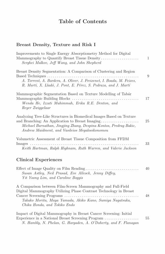
Table of Contents
Breast Density, Texture and Risk I
Improvements to Single Energy Absorptiometry Method for DigitalMammography to Quantify Breast Tissue Density . . . . . . . . . . . . . . . . . . . . 1
Serghei Malkov, Jeff Wang, and John Shepherd
Breast Density Segmentation: A Comparison of Clustering and RegionBased Techniques . . . . . . . . . . . . . . . . . . . . . . . . . . . . . . . . . . . . . . . . . . . . . . . . 9
A. Torrent, A. Bardera, A. Oliver, J. Freixenet, I. Boada, M. Feixes,R. Martı, X. Llado, J. Pont, E. Perez, S. Pedraza, and J. Martı
Mammographic Segmentation Based on Texture Modelling of TabarMammographic Building Blocks . . . . . . . . . . . . . . . . . . . . . . . . . . . . . . . . . . . 17
Wenda He, Izzati Muhimmah, Erika R.E. Denton, andReyer Zwiggelaar
Analyzing Tree-Like Structures in Biomedical Images Based on Textureand Branching: An Application to Breast Imaging . . . . . . . . . . . . . . . . . . . . 25
Michael Barnathan, Jingjing Zhang, Despina Kontos, Predrag Bakic,Andrew Maidment, and Vasileios Megalooikonomou
Volumetric Assessment of Breast Tissue Composition from FFDMImages . . . . . . . . . . . . . . . . . . . . . . . . . . . . . . . . . . . . . . . . . . . . . . . . . . . . . . . . . 33
Keith Hartman, Ralph Highnam, Ruth Warren, and Valerie Jackson
Clinical Experiences
Effect of Image Quality on Film Reading . . . . . . . . . . . . . . . . . . . . . . . . . . . . 40Susan Astley, Neil Prasad, Eve Allcock, Jenny Diffey,Yit Yoong Lim, and Caroline Boggis
A Comparison between Film-Screen Mammography and Full-FieldDigital Mammography Utilizing Phase Contrast Technology in BreastCancer Screening Programs . . . . . . . . . . . . . . . . . . . . . . . . . . . . . . . . . . . . . . . 48
Takako Morita, Maya Yamada, Akiko Kano, Sumiya Nagatsuka,Chika Honda, and Tokiko Endo
Impact of Digital Mammography in Breast Cancer Screening: InitialExperience in a National Breast Screening Program . . . . . . . . . . . . . . . . . . 55
N. Hambly, N. Phelan, G. Hargaden, A. O’Doherty, and F. Flanagan

XVIII Table of Contents
Clinical Performance of Breast Tomosynthesis as a Function ofRadiologist Experience Level . . . . . . . . . . . . . . . . . . . . . . . . . . . . . . . . . . . . . . 61
Andrew P. Smith, Elizabeth A. Rafferty, and Loren Niklason
BIRADS Classification in Breast Tomosynthesis Compared toMammography and Ultrasonography . . . . . . . . . . . . . . . . . . . . . . . . . . . . . . . 67
Anders Tingberg, Ingvar Andersson, Debra M. Ikeda, Mark Ruschin,Tony Svahn, and Pontus Timberg
Stereoscopic Digital Mammography: Improved Accuracy of LesionDetection in Breast Cancer Screening . . . . . . . . . . . . . . . . . . . . . . . . . . . . . . . 74
David J. Getty, Carl J. D’Orsi, and Ronald M. Pickett
Potential Role of FDG-PET Imaging in Defining Biology of PrimaryBreast Lesions . . . . . . . . . . . . . . . . . . . . . . . . . . . . . . . . . . . . . . . . . . . . . . . . . . . 80
S. Basu, A. Mavi, T. Cermik, R. Kumar, and A. Alavi
Breast Imaging Physics
Clinical Usefulness of Super High-Resolution Liquid Crystal DisplaysUsing Independent Sub-pixel Driving Technology . . . . . . . . . . . . . . . . . . . . 84
Katsuhiro Ichikawa, Hiroko Kawashima, Naohiro Kimura, andMikio Hasegawa
The Effect of Tomosynthesis X-Ray Pulse Width on Measured BeamQuality . . . . . . . . . . . . . . . . . . . . . . . . . . . . . . . . . . . . . . . . . . . . . . . . . . . . . . . . . 91
Baorui Ren, Andrew Smith, David Aizer, Don Kennedy,Jeffrey Yorker, and Zhenxue Jing
Tomographic Dual Modality Breast Scanner . . . . . . . . . . . . . . . . . . . . . . . . . 99Mark B. Williams, Patricia G. Judy, Mitali J. More,Jennifer A. Harvey, Stan Majewski, James Proffitt, John McKisson,Alexander Stolin, Brian Kross, Alexander Stewart, Edward Bullard,Manish Kankaria, and Roman Janer
Dual-Energy X-Ray Absorptiometry Method Using a Full Field DigitalMammography System . . . . . . . . . . . . . . . . . . . . . . . . . . . . . . . . . . . . . . . . . . . 108
Aurelie Laidevant, Serghei Malkov, Alfred Au, and John Shepherd
Optimization of a Dual-Energy Contrast-Enhanced Technique for aPhoton Counting Digital Breast Tomosynthesis System . . . . . . . . . . . . . . . 116
Ann-Katherine Carton, Christer Ullberg, Karin Lindman,Tom Francke, and Andrew Maidment
Simulation and Phantom Studies of Contrast-Enhanced Dual EnergyMammography (CEDEM) . . . . . . . . . . . . . . . . . . . . . . . . . . . . . . . . . . . . . . . . . 124
Shih-Ying Huang, John M. Boone, Dandan Zheng, Kai Yang,Nathan J. Packard, and George Burkett Jr.
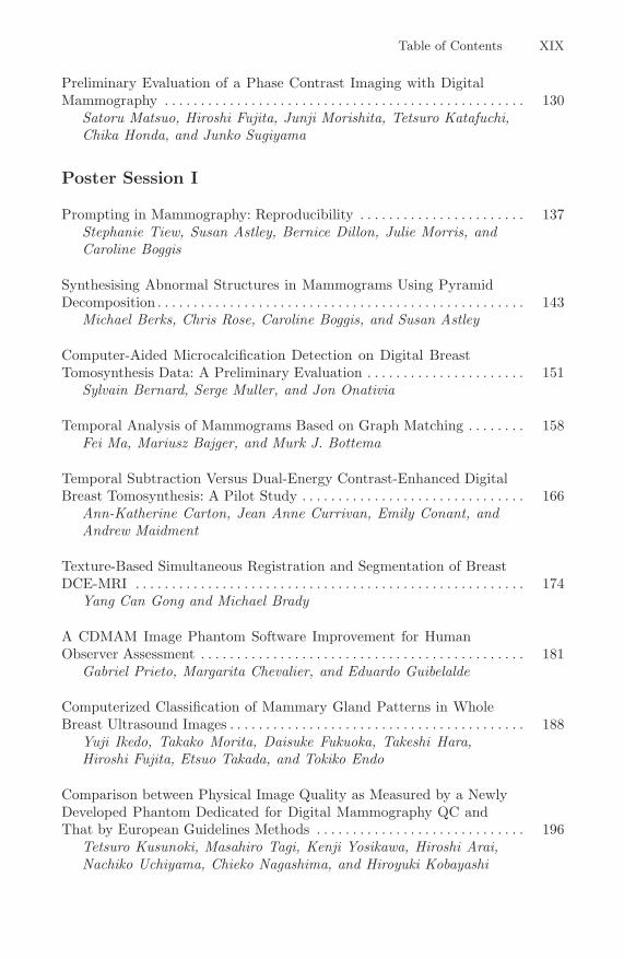
Table of Contents XIX
Preliminary Evaluation of a Phase Contrast Imaging with DigitalMammography . . . . . . . . . . . . . . . . . . . . . . . . . . . . . . . . . . . . . . . . . . . . . . . . . . 130
Satoru Matsuo, Hiroshi Fujita, Junji Morishita, Tetsuro Katafuchi,Chika Honda, and Junko Sugiyama
Poster Session I
Prompting in Mammography: Reproducibility . . . . . . . . . . . . . . . . . . . . . . . 137Stephanie Tiew, Susan Astley, Bernice Dillon, Julie Morris, andCaroline Boggis
Synthesising Abnormal Structures in Mammograms Using PyramidDecomposition . . . . . . . . . . . . . . . . . . . . . . . . . . . . . . . . . . . . . . . . . . . . . . . . . . . 143
Michael Berks, Chris Rose, Caroline Boggis, and Susan Astley
Computer-Aided Microcalcification Detection on Digital BreastTomosynthesis Data: A Preliminary Evaluation . . . . . . . . . . . . . . . . . . . . . . 151
Sylvain Bernard, Serge Muller, and Jon Onativia
Temporal Analysis of Mammograms Based on Graph Matching . . . . . . . . 158Fei Ma, Mariusz Bajger, and Murk J. Bottema
Temporal Subtraction Versus Dual-Energy Contrast-Enhanced DigitalBreast Tomosynthesis: A Pilot Study . . . . . . . . . . . . . . . . . . . . . . . . . . . . . . . 166
Ann-Katherine Carton, Jean Anne Currivan, Emily Conant, andAndrew Maidment
Texture-Based Simultaneous Registration and Segmentation of BreastDCE-MRI . . . . . . . . . . . . . . . . . . . . . . . . . . . . . . . . . . . . . . . . . . . . . . . . . . . . . . 174
Yang Can Gong and Michael Brady
A CDMAM Image Phantom Software Improvement for HumanObserver Assessment . . . . . . . . . . . . . . . . . . . . . . . . . . . . . . . . . . . . . . . . . . . . . 181
Gabriel Prieto, Margarita Chevalier, and Eduardo Guibelalde
Computerized Classification of Mammary Gland Patterns in WholeBreast Ultrasound Images . . . . . . . . . . . . . . . . . . . . . . . . . . . . . . . . . . . . . . . . . 188
Yuji Ikedo, Takako Morita, Daisuke Fukuoka, Takeshi Hara,Hiroshi Fujita, Etsuo Takada, and Tokiko Endo
Comparison between Physical Image Quality as Measured by a NewlyDeveloped Phantom Dedicated for Digital Mammography QC andThat by European Guidelines Methods . . . . . . . . . . . . . . . . . . . . . . . . . . . . . 196
Tetsuro Kusunoki, Masahiro Tagi, Kenji Yosikawa, Hiroshi Arai,Nachiko Uchiyama, Chieko Nagashima, and Hiroyuki Kobayashi

XX Table of Contents
Assuring Authenticity of Digital Mammograms by ImageWatermarking . . . . . . . . . . . . . . . . . . . . . . . . . . . . . . . . . . . . . . . . . . . . . . . . . . . 204
Anthony Maeder, Jason Dowling, Anthony Nguyen,Emma Brunton, and Phuong Nguyen
Different Search Patterns and Similar Decision Outcomes: How CanExperts Agree in the Decisions They Make When Reading DigitalMammograms? . . . . . . . . . . . . . . . . . . . . . . . . . . . . . . . . . . . . . . . . . . . . . . . . . . 212
Claudia Mello-Thoms, Marie Ganott, Jules Sumkin,Christiane Hakim, Cynthia Britton, Luisa Wallace, andLara Hardesty
Optimization of Tomosynthesis Acquisition Parameters: Angular Rangeand Number of Projections . . . . . . . . . . . . . . . . . . . . . . . . . . . . . . . . . . . . . . . . 220
Thomas Mertelmeier, Jasmina Ludwig, Bo Zhao, and Wei Zhao
Subtle Abnormalities in Highly Dense Breasts Detected by Use of aDigital Phase Contrast Mammography System: A Report of ThreeInvasive Cancer Cases in the Early Stage . . . . . . . . . . . . . . . . . . . . . . . . . . . . 228
Takako Morita, Maya Yamada, Akiko Kano, Sumiya Nagatsuka,Chika Honda, and Tokiko Endo
Image Quality Assessment and Equipment Optimisation withAutomated Phantom Evaluation in Full Field Digital Mammography(FFDM) . . . . . . . . . . . . . . . . . . . . . . . . . . . . . . . . . . . . . . . . . . . . . . . . . . . . . . . . 235
Nadia Oberhofer, Nicoletta Paruccini, and Ehrenfried Moroder
Breast Mass Detection under Increased Ambient Lighting . . . . . . . . . . . . . 243Benjamin J. Pollard, Amarpreet S. Chawla,Noriyuki Hashimoto, and Ehsan Samei
Reducing Noise of Medical Grade Liquid Crystal Displays (LCD) andIts Relation to the Detection of Micro-calcifications . . . . . . . . . . . . . . . . . . 249
Hans Roehrig, Jiahua Fan, William J. Dallas,Elizabeth A. Krupinski, and Jeffrey Johnson
An Ontology to Support Adaptive Training for Breast Radiologists . . . . . 257Shanghua Sun, Paul Taylor, Louise Wilkinson, and Lisanne Khoo
Image Analysis and CAD I
A Web Database for Computer-Aided Detection and Diagnosis ofMedical Images . . . . . . . . . . . . . . . . . . . . . . . . . . . . . . . . . . . . . . . . . . . . . . . . . . 265
Dave Tahmoush and Hanan Samet
An Interactive Computer Aided Decision Support System for Detectionof Masses in Mammograms . . . . . . . . . . . . . . . . . . . . . . . . . . . . . . . . . . . . . . . . 273
Nico Karssemeijer, Andrea Hupse, Maurice Samulski,Michiel Kallenberg, Carla Boetes, and Gerard den Heeten

Table of Contents XXI
Detection of Masses in Digital Breast Tomosynthesis Mammography:Effects of the Number of Projection Views and Dose . . . . . . . . . . . . . . . . . . 279
Heang-Ping Chan, Jun Wei, Yiheng Zhang, Berkman Sahiner,Lubomir Hadjiiski, and Mark A. Helvie
Effect of Similarity Metrics and ROI Sizes in Featureless ComputerAided Detection of Breast Masses in Tomosynthesis . . . . . . . . . . . . . . . . . . 286
Swatee Singh, Georgia D. Tourassi, and Joseph Y. Lo
Knowledge Transfer across Breast Cancer Screening Modalities: APilot Study Using an Information-Theoretic CADe System for MassDetection . . . . . . . . . . . . . . . . . . . . . . . . . . . . . . . . . . . . . . . . . . . . . . . . . . . . . . . 292
Georgia D. Tourassi, Amy C. Sharma, Swatee Singh,Robert S. Saunders, Joseph Y. Lo, Ehsan Samei, andBrian P. Harrawood
Gradient Vector Flow Fields and Spiculated Mass Detection in DigitalMammography Images . . . . . . . . . . . . . . . . . . . . . . . . . . . . . . . . . . . . . . . . . . . . 299
Fengmei Zou, Yufeng Zheng, Zhengdong Zhou, andKwabena Agyepong
The Evaluation of Effects on Breast Cancer Diagnoses When UsingMammographic Semantic Information . . . . . . . . . . . . . . . . . . . . . . . . . . . . . . 307
Da Qi, Erika R.E. Denton, Joanna M.E. Leason,Diaa Othman, and Reyer Zwiggelaar
Computerized Scheme for Focal Asymmetric Densities on Mammogramsby Use of Geometric and Texture Analyses . . . . . . . . . . . . . . . . . . . . . . . . . . 315
Shinsuke Katsuhara, Hitoshi Futamura, Satoshi Kasai,Takako Morita, and Tokiko Endo
Identifying Corresponding Lesions from CC and MLO Views ViaCorrelative Feature Analysis . . . . . . . . . . . . . . . . . . . . . . . . . . . . . . . . . . . . . . . 323
Yading Yuan, Maryellen Giger, Hui Li, Li Lan, and Charlene Sennett
Image Analysis and CAD II
Case-Specific Reliability Assessment for Improved False PositiveReduction with an Information-Theoretic CAD System . . . . . . . . . . . . . . . 329
Piotr A. Habas, Jacek M. Zurada, and Georgia D. Tourassi
Computerized Detection and Classification of Malignant and BenignMicrocalcifications on Full Field Digital Mammograms . . . . . . . . . . . . . . . . 336
Lubomir Hadjiiski, Peter Filev, Heang-Ping Chan, Jun Ge,Berkman Sahiner, Mark A. Helvie, and Marilyn A. Roubidoux
The Effect of Training Sample Size on Performance of MassDetection . . . . . . . . . . . . . . . . . . . . . . . . . . . . . . . . . . . . . . . . . . . . . . . . . . . . . . . 343
Michiel Kallenberg and Nico Karssemeijer

XXII Table of Contents
Multiple-Instance Learning Improves CAD Detection of Masses inDigital Mammography . . . . . . . . . . . . . . . . . . . . . . . . . . . . . . . . . . . . . . . . . . . . 350
Balaji Krishnapuram, Jonathan Stoeckel, Vikas Raykar, Bharat Rao,Philippe Bamberger, Eli Ratner, Nicolas Merlet, Inna Stainvas,Menahem Abramov, and Alexandra Manevitch
Optimizing the CAD Process for Detecting Mammographic Lesions bya New Generation Algorithm Using Linear Classifiers and a GradientBased Approach . . . . . . . . . . . . . . . . . . . . . . . . . . . . . . . . . . . . . . . . . . . . . . . . . 358
Philippe Bamberger, Isaac Leichter, Nicolas Merlet, Eli Ratner,Glenn Fung, and Richard Lederman
Reliability Assessment of Ensemble Classifiers: Application inMammography . . . . . . . . . . . . . . . . . . . . . . . . . . . . . . . . . . . . . . . . . . . . . . . . . . 366
Maciej A. Mazurowski, Jacek M. Zurada, and Georgia D. Tourassi
Breast Mass Classification on Full-Field Digital Mammography andScreen-Film Mammography . . . . . . . . . . . . . . . . . . . . . . . . . . . . . . . . . . . . . . . 371
Jiazheng Shi, Berkman Sahiner, Heang-Ping Chan,Lubomir M. Hadjiiski, Jun Ge, and Jun Wei
Detection of Microcalcifications Using a Nonuniform Noise Model . . . . . . 378Guido van Schie and Nico Karssemeijer
Modeling and Simulation
The Breast Biomechanics Reference State for Multi-modal ImageAnalysis . . . . . . . . . . . . . . . . . . . . . . . . . . . . . . . . . . . . . . . . . . . . . . . . . . . . . . . . 385
Vijay Rajagopal, Martyn P. Nash, Ralph P. Highnam, andPoul M.F. Nielsen
An Ideal Observer for a Model of X-Ray Imaging in Breast ParenchymalTissue . . . . . . . . . . . . . . . . . . . . . . . . . . . . . . . . . . . . . . . . . . . . . . . . . . . . . . . . . . 393
Craig K. Abbey and John M. Boone
Statistical Appearance Models of Mammographic Masses . . . . . . . . . . . . . 401Michael Berks, Steven Caulkin, Rumana Rahim,Caroline Boggis, and Susan Astley
Validation of a Digital Mammography Image Simulation Chain withAutomated Scoring of CDMAM Images . . . . . . . . . . . . . . . . . . . . . . . . . . . . . 409
Mary Yip, Abdulaziz Alsager, Emma Lewis, Kevin Wells, andKenneth C. Young
Comparison of Two Methods to Develop Breast Models for Simulationof Breast Tomosynthesis and CT . . . . . . . . . . . . . . . . . . . . . . . . . . . . . . . . . . . 417
J. Michael O’Connor, Mini Das, Clay Didier, Mufeed Mah’D, andStephen J. Glick
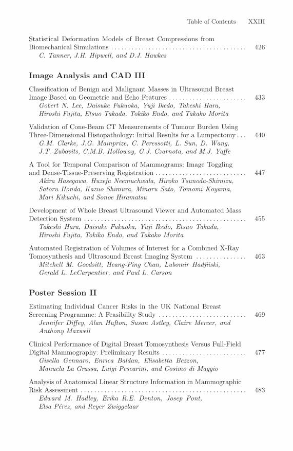
Table of Contents XXIII
Statistical Deformation Models of Breast Compressions fromBiomechanical Simulations . . . . . . . . . . . . . . . . . . . . . . . . . . . . . . . . . . . . . . . . 426
C. Tanner, J.H. Hipwell, and D.J. Hawkes
Image Analysis and CAD III
Classification of Benign and Malignant Masses in Ultrasound BreastImage Based on Geometric and Echo Features . . . . . . . . . . . . . . . . . . . . . . . 433
Gobert N. Lee, Daisuke Fukuoka, Yuji Ikedo, Takeshi Hara,Hiroshi Fujita, Etsuo Takada, Tokiko Endo, and Takako Morita
Validation of Cone-Beam CT Measurements of Tumour Burden UsingThree-Dimensional Histopathology: Initial Results for a Lumpectomy . . . 440
G.M. Clarke, J.G. Mainprize, C. Peressotti, L. Sun, D. Wang,J.T. Zubovits, C.M.B. Holloway, G.J. Czarnota, and M.J. Yaffe
A Tool for Temporal Comparison of Mammograms: Image Togglingand Dense-Tissue-Preserving Registration . . . . . . . . . . . . . . . . . . . . . . . . . . . 447
Akira Hasegawa, Huzefa Neemuchwala, Hiroko Tsunoda-Shimizu,Satoru Honda, Kazuo Shimura, Minoru Sato, Tomomi Koyama,Mari Kikuchi, and Sonoe Hiramatsu
Development of Whole Breast Ultrasound Viewer and Automated MassDetection System . . . . . . . . . . . . . . . . . . . . . . . . . . . . . . . . . . . . . . . . . . . . . . . . 455
Takeshi Hara, Daisuke Fukuoka, Yuji Ikedo, Etsuo Takada,Hiroshi Fujita, Tokiko Endo, and Takako Morita
Automated Registration of Volumes of Interest for a Combined X-RayTomosynthesis and Ultrasound Breast Imaging System . . . . . . . . . . . . . . . 463
Mitchell M. Goodsitt, Heang-Ping Chan, Lubomir Hadjiiski,Gerald L. LeCarpentier, and Paul L. Carson
Poster Session II
Estimating Individual Cancer Risks in the UK National BreastScreening Programme: A Feasibility Study . . . . . . . . . . . . . . . . . . . . . . . . . . 469
Jennifer Diffey, Alan Hufton, Susan Astley, Claire Mercer, andAnthony Maxwell
Clinical Performance of Digital Breast Tomosynthesis Versus Full-FieldDigital Mammography: Preliminary Results . . . . . . . . . . . . . . . . . . . . . . . . . 477
Gisella Gennaro, Enrica Baldan, Elisabetta Bezzon,Manuela La Grassa, Luigi Pescarini, and Cosimo di Maggio
Analysis of Anatomical Linear Structure Information in MammographicRisk Assessment . . . . . . . . . . . . . . . . . . . . . . . . . . . . . . . . . . . . . . . . . . . . . . . . . 483
Edward M. Hadley, Erika R.E. Denton, Josep Pont,Elsa Perez, and Reyer Zwiggelaar
XXIV Table of Contents
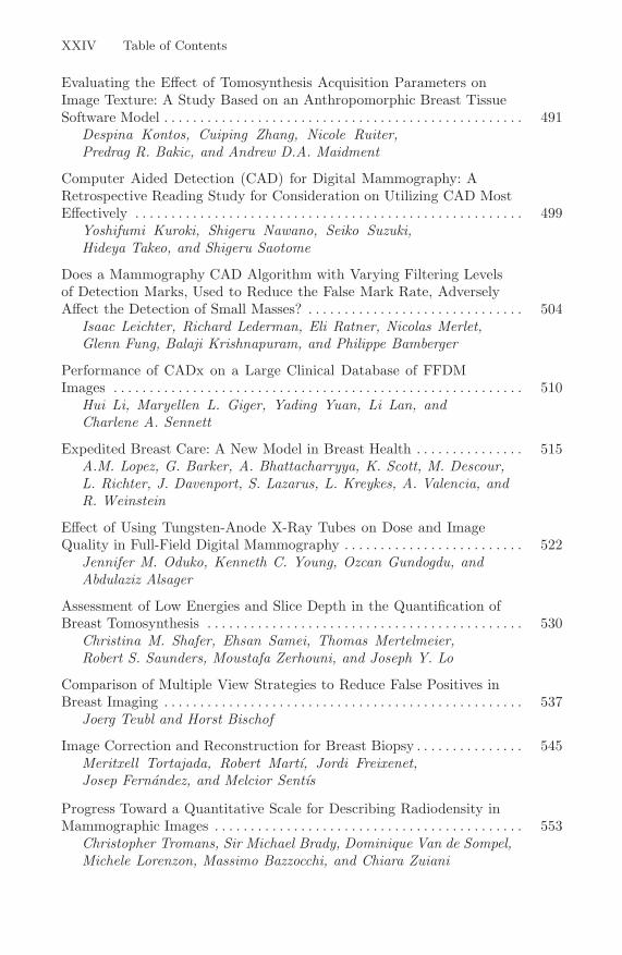
Evaluating the Effect of Tomosynthesis Acquisition Parameters onImage Texture: A Study Based on an Anthropomorphic Breast TissueSoftware Model . . . . . . . . . . . . . . . . . . . . . . . . . . . . . . . . . . . . . . . . . . . . . . . . . . 491
Despina Kontos, Cuiping Zhang, Nicole Ruiter,Predrag R. Bakic, and Andrew D.A. Maidment
Computer Aided Detection (CAD) for Digital Mammography: ARetrospective Reading Study for Consideration on Utilizing CAD MostEffectively . . . . . . . . . . . . . . . . . . . . . . . . . . . . . . . . . . . . . . . . . . . . . . . . . . . . . . 499
Yoshifumi Kuroki, Shigeru Nawano, Seiko Suzuki,Hideya Takeo, and Shigeru Saotome
Does a Mammography CAD Algorithm with Varying Filtering Levelsof Detection Marks, Used to Reduce the False Mark Rate, AdverselyAffect the Detection of Small Masses? . . . . . . . . . . . . . . . . . . . . . . . . . . . . . . 504
Isaac Leichter, Richard Lederman, Eli Ratner, Nicolas Merlet,Glenn Fung, Balaji Krishnapuram, and Philippe Bamberger
Performance of CADx on a Large Clinical Database of FFDMImages . . . . . . . . . . . . . . . . . . . . . . . . . . . . . . . . . . . . . . . . . . . . . . . . . . . . . . . . . 510
Hui Li, Maryellen L. Giger, Yading Yuan, Li Lan, andCharlene A. Sennett
Expedited Breast Care: A New Model in Breast Health . . . . . . . . . . . . . . . 515A.M. Lopez, G. Barker, A. Bhattacharryya, K. Scott, M. Descour,L. Richter, J. Davenport, S. Lazarus, L. Kreykes, A. Valencia, andR. Weinstein
Effect of Using Tungsten-Anode X-Ray Tubes on Dose and ImageQuality in Full-Field Digital Mammography . . . . . . . . . . . . . . . . . . . . . . . . . 522
Jennifer M. Oduko, Kenneth C. Young, Ozcan Gundogdu, andAbdulaziz Alsager
Assessment of Low Energies and Slice Depth in the Quantification ofBreast Tomosynthesis . . . . . . . . . . . . . . . . . . . . . . . . . . . . . . . . . . . . . . . . . . . . 530
Christina M. Shafer, Ehsan Samei, Thomas Mertelmeier,Robert S. Saunders, Moustafa Zerhouni, and Joseph Y. Lo
Comparison of Multiple View Strategies to Reduce False Positives inBreast Imaging . . . . . . . . . . . . . . . . . . . . . . . . . . . . . . . . . . . . . . . . . . . . . . . . . . 537
Joerg Teubl and Horst Bischof
Image Correction and Reconstruction for Breast Biopsy . . . . . . . . . . . . . . . 545Meritxell Tortajada, Robert Martı, Jordi Freixenet,Josep Fernandez, and Melcior Sentıs
Progress Toward a Quantitative Scale for Describing Radiodensity inMammographic Images . . . . . . . . . . . . . . . . . . . . . . . . . . . . . . . . . . . . . . . . . . . 553
Christopher Tromans, Sir Michael Brady, Dominique Van de Sompel,Michele Lorenzon, Massimo Bazzocchi, and Chiara Zuiani

Table of Contents XXV
Systematic Performance Analysis of SART as Applied to Digital BreastTomosynthesis . . . . . . . . . . . . . . . . . . . . . . . . . . . . . . . . . . . . . . . . . . . . . . . . . . . 561
Dominique Van de Sompel and Michael Brady
Optimizing the Target-Filter Combination in Digital Mammography inthe Sense of Image Quality and Average Glandular Dose . . . . . . . . . . . . . . 570
Mari Varjonen and Pekka Strommer
Generic Infrastructure for Medical Informatics (GIMI): TheDevelopment of a Mammographic Training System . . . . . . . . . . . . . . . . . . . 577
Moi Hoon Yap, Alastair G. Gale, and Hazel J. Scott
Evaluation of 3D Breast Surface Reconstruction Accuracy UsingNon-contact Scanner Images: A Phantom Study . . . . . . . . . . . . . . . . . . . . . 585
Cuiping Zhang, Predrag R. Bakic, Shugao Xia, Fengshan Liu, andAndrew D.A. Maidment
Digital Breast Tomosynthesis
Investigation of Different PV Distributions in Digital BreastTomosynthesis (DBT) Mammography . . . . . . . . . . . . . . . . . . . . . . . . . . . . . . 593
Yiheng Zhang, Heang-Ping Chan, Mitchell M. Goodsitt,Andrea Schmitz, Jeffrey W. Eberhard, and Bernhard E.H. Claus
Characterization of Projection Ordering in Iterative ReconstructionMethods for Breast Tomosynthesis . . . . . . . . . . . . . . . . . . . . . . . . . . . . . . . . . 601
Gang Wu, James Mainprize, and Martin Yaffe
Effect of Scan Angle and Reconstruction Algorithm on Model ObserverPerformance in Tomosynthesis . . . . . . . . . . . . . . . . . . . . . . . . . . . . . . . . . . . . . 606
I. Reiser, B.A. Lau, and R.M. Nishikawa
A Novel Approach for Filtered Backprojection in Tomosynthesis Basedon Filter Kernels Determined by Iterative Reconstruction Techniques . . . 612
Jasmina Ludwig, Thomas Mertelmeier, Holger Kunze, andWolfgang Harer
3D Digital Breast Tomosynthesis Using Total VariationRegularization . . . . . . . . . . . . . . . . . . . . . . . . . . . . . . . . . . . . . . . . . . . . . . . . . . . 621
Iason Kastanis, Simon Arridge, Alex Stewart, Spencer Gunn,Christer Ullberg, and Tom Francke
Image Artifact in Digital Breast Tomosynthesis and Its Dependence onSystem and Reconstruction Parameters . . . . . . . . . . . . . . . . . . . . . . . . . . . . . 628
Yue-Houng Hu, Wei Zhao, Thomas Mertelmeier, andJasmina Ludwig

XXVI Table of Contents
Multi-projection Correlation Imaging as a New Diagnostic Tool forImproved Breast Cancer Detection . . . . . . . . . . . . . . . . . . . . . . . . . . . . . . . . . 635
Amarpreet S. Chawla, Ehsan Samei, Joseph Y. Lo, andThomas Mertelmeier
Sensitivity of Contrast-Enhanced Digital Breast Tomosynthesis toChanges in Iodine Concentration During Acquisition . . . . . . . . . . . . . . . . . 643
Melissa L. Hill, James G. Mainprize, and Martin J. Yaffe
Breast Density, Texture and Risk II
Quantifying Breast Thickness for Density Measurement . . . . . . . . . . . . . . . 651Jennifer Diffey, Alan Hufton, Christine Beeston, Julia Smith,Tom Marchant, and Susan Astley
Effect of Tissue Thickness Variation in Volumetric Breast DensityEstimation . . . . . . . . . . . . . . . . . . . . . . . . . . . . . . . . . . . . . . . . . . . . . . . . . . . . . . 659
Olivier Alonzo-Proulx, Albert H. Tyson, Gordon E. Mawdsley, andMartin J. Yaffe
Breast Abnormality Detection Incorporating Breast DensityInformation Based on Independent Components Analysis . . . . . . . . . . . . . 667
Styliani Petroudi, Nicoletta Nicolaou, Julius Georgiou, andMichael Brady
Comparison of Breast Percent Density Estimated from DigitalMammograms and Central Reconstructed Tomosynthesis SliceImages . . . . . . . . . . . . . . . . . . . . . . . . . . . . . . . . . . . . . . . . . . . . . . . . . . . . . . . . . 674
Predrag R. Bakic, Despina Kontos, Andrea B. Troxel, andAndrew Maidment
Digital Breast Tomosynthesis Parenchymal Texture Analysis for BreastCancer Risk Estimation: A Preliminary Study . . . . . . . . . . . . . . . . . . . . . . . 681
Despina Kontos, Predrag R. Bakic, Andrea B. Troxel,Emily F. Conant, and Andrew D.A. Maidment
Texture Based Segmentation of Breast DCE-MRI . . . . . . . . . . . . . . . . . . . . 689Yang Can Gong and Michael Brady
Physics, Image Quality and Quality Assurance
Image Quality Measurements in Breast Tomosynthesis . . . . . . . . . . . . . . . . 696Ruben van Engen, Ramona Bouwman, Roeland van der Burght,Barbara Lazzari, David R. Dance, Patrice Heid,Magnus Aslund, and Kenneth C. Young
Table of Contents XXVII
One Year of Experience with Remote Quality Assurance of DigitalMammography Systems in the Flemish Breast Cancer ScreeningProgram . . . . . . . . . . . . . . . . . . . . . . . . . . . . . . . . . . . . . . . . . . . . . . . . . . . . . . . . 703
Jurgen Jacobs, Kim Lemmens, Joris Nens, Koen Michielsen,Guy Marchal, and Hilde Bosmans
Automatic Exposure Control in Digital Mammography:Contrast-to-Noise Ratio Versus Average Glandular Dose . . . . . . . . . . . . . . 711
Gisella Gennaro, Paola Golinelli, Elena Bellan, Paola Colombo,Loredana D’Ercole, Anna Di Nallo, Lara Gallo, Carlo Giordano,Gabriele Meliado, Barbara Morri, Elvina Nassivera,Nadia Oberhofer, Daniela Origgi, Massimiliano Paolucci,Nicoletta Paruccini, Michela Piergentili, Elisa Rizzi, andRaffaella Rossi
Effect of Anode/Filter Combination on the Dose and Image Quality ofa Digital Mammography System Based on an Amorphous SeleniumDetector . . . . . . . . . . . . . . . . . . . . . . . . . . . . . . . . . . . . . . . . . . . . . . . . . . . . . . . . 716
Paola Baldelli, Niall Phelan, and Gillian Egan
Comparative Technical Study of Two Generations of CR Plates forDigital Mammography . . . . . . . . . . . . . . . . . . . . . . . . . . . . . . . . . . . . . . . . . . . . 724
Hilde Bosmans, Kim Lemmens, Jurgen Jacobs,Beatrijs Verbrugge, Koen Michielsen, Federica Zanca, Joris Nens,Chantal Van Ongeval, and Guy Marchal
Comparing the Performance of Digital Mammography Systems . . . . . . . . 732Kenneth C. Young, Jennifer M. Oduko, Ozcan Gundogdu, andAbdulaziz Alsager
Cross-Calibration and Longitudinal Quality Control of Hologic SeleniaFull-Field Digital Mammography Systems for Volumetric BreastDensity Measurements . . . . . . . . . . . . . . . . . . . . . . . . . . . . . . . . . . . . . . . . . . . . 740
Jeff Wang, Serghei Malkov, Bo Fan, and John Shepherd
Classification of Artifacts in Clinical Digital Mammography . . . . . . . . . . . 748Chantal Van Ongeval, Jurgen Jacobs, Andre Van Steen,Federica Zanca, Hilde Bosmans, and Guy Marchal
Contrast Sensitivity in Mammographic SoftcopyReading – Determination with Psychophysical Procedures . . . . . . . . . . . . . 756
Dorte Apelt and Heinz-Otto Peitgen
Author Index . . . . . . . . . . . . . . . . . . . . . . . . . . . . . . . . . . . . . . . . . . . . . . . . . . 765