inhibition of aldehyde dehydrogenase expands hematopoietic stem cells with radioprotective capacity
TRANSCRIPT
TISSUE-SPECIFIC STEM CELLS
Inhibition of Aldehyde Dehydrogenase Expands Hematopoietic Stem
Cells with Radioprotective Capacity
GARRETT G. MURAMOTO,aJ. LAUREN RUSSELL,
aRACHID SAFI,
bALICE B. SALTER,
aHEATHER A. HIMBURG,
a
PAMELA DAHER,a SARAH K. MEADOWS,a PHUONG DOAN,a ROBERT W. STORMS,a NELSON J. CHAO,a,c
DONALD P. MCDONNELL,b JOHN P. CHUTEa,b
aDivision of Cellular Therapy, Department of Medicine, bDepartment of Pharmacology and Cancer Biology, andcDepartment of Immunology, Duke University Medical Center, Durham, North Carolina, USA
Key Words. Adult haematopoietic stem cells • Cord blood • Hematopoietic stem cell transplantation • Retinoic acid • siRNA
ABSTRACT
Hematopoietic stem cells (HSCs) are enriched for aldehydedehydrogenase (ALDH) activity and ALDH is a selectablemarker for human HSCs. However, the function of ALDHin HSC biology is not well understood. We sought todetermine the function of ALDH in regulating HSC fate.Pharmacologic inhibition of ALDH with diethylaminoben-zaldehyde (DEAB) impeded the differentiation of murineCD342c-kit1Sca-11lineage2 (342KSL) HSCs in culture andfacilitated a ninefold expansion of cells capable of radiopro-tecting lethally irradiated mice compared to input 34
2KSL
cells. Treatment of bone marrow (BM) 342KSL cells withDEAB caused a fourfold increase in 4-week competitive
repopulating units, verifying the amplification of short-termHSCs (ST-HSCs) in response to ALDH inhibition. TargetedsiRNA of ALDH1a1 in BM HSCs caused a comparableexpansion of radioprotective progenitor cells in culturecompared to DEAB treatment, confirming that ALDH1a1was the target of DEAB inhibition. The addition of all transretinoic acid blocked DEAB-mediated expansion of ST-HSCs in culture, suggesting that ALDH1a1 regulates HSCdifferentiation via augmentation of retinoid signaling. Phar-macologic inhibition of ALDH has therapeutic potential as ameans to amplify ST-HSCs for transplantation purposes.STEM CELLS 2010;28:523–534
Disclosure of potential conflicts of interest is found at the end of this article.
INTRODUCTION
Hematopoietic stem cell (HSC) transplantation involves theadministration of myeloablative radiotherapy and/or chemo-therapy followed by infusion of CD34þ hematopoietic stem/progenitor cells [1–3]. Despite a sufficient number of long-term HSCs within the donor graft, a 2-4 week period ofpancytopenia typically follows this procedure, during whichtime patients require antibiotics and transfusion support [1–4]. In the setting of cord blood (CB) transplantation, hema-tologic recovery can lag for up to 2 months after trans-plant, putting recipients at risk for life-threatening infectiouscomplications [1–5]. The period of pancytopenia that occursafter transplantation results from the ablative effects of theconditioning on host hematopoiesis coupled with the inher-ent lag time to the production of donor HSC-derived prog-eny [6–8]. Since the prolonged period of pancytopenia afterCB transplantation accounts for substantial morbidity andmortality, it has been proposed that co-transplantation ofcommitted myeloid progenitor cells or short-term HSCs(ST-HSCs) could accelerate hematologic recovery in these
patients and perhaps lessen the mortality of this procedure[6, 7, 9].
Studies in mice have defined the phenotype of long-termHSCs (LT-HSCs) and ST-HSCs [6, 7]. Bone marrow (BM)cells that are depleted of lineage markers, express c-kitþ andSca-1þ and lack CD34 expression (CD34�c-kitþSca-1þlineage� [34�KSL] cells) or express Thy 1.1lo, are enrichedfor LT-HSCs [6, 7], whereas 34þKSL cells are enriched forST-HSCs, which have a more restricted self-renewal capacityand repopulating ability [6, 7, 10, 11]. It has been shown thatthe 34þFlt-3�KSL subset is enriched for virtually all ST-HSCs and these cells give rise to 34þFlt-3þKSL cells, whichare primarily lymphoid repopulating cells [6]. Although LT-HSCs are required for long-term hematopoietic reconstitution,these cells are substantially less efficient than ST-HSCs atproviding radioprotection of lethally irradiated recipients [7,10, 11]. It has also been shown that transplantation of com-mitted megakaryocyte-erythroid progenitors (MEPs) or com-mon myeloid progenitors (CMPs) can provide radioprotectionin lethally irradiated mice in the absence of HSC transplanta-tion [7]. Importantly, transplantation of as few as 500 34þFlt-3�KSL cells was able to radioprotect a fraction of lethally
Author contributions: G.M. and J.L.R.: collection of data, data analysis, manuscript writing, final approval of manuscript; G.M. andJ.L.R.: contributed equally to this work; R.S.: collection of data, data analysis, conception and design; A.S., H.H., P.D., and P.D.:collection of data; S.M.: collection of data, data analysis; R.S. and N.C.: conception and design, data analysis, manuscript writing; D.M.:conception and design, provision of materials, manuscript writing; J.C.: conception and design, financial support, data analysis,manuscript writing, final approval of manuscript.
Correspondence: John P. Chute, MD, Duke University Medical Center, Division of Cellular Therapy, 2400 Pratt Street, Box 3961,Durham, North Carolina 27710, USA. Telephone: 919�668�4706; Fax: 919�668�1091; e-mail: [email protected] ReceivedNovember 3, 2009; accepted for publication December 26, 2009; first published online in STEM CELLS EXPRESS January 6, 2010.VC AlphaMed Press 1066-5099/2009/$30.00/0 doi: 10.1002/stem.299
STEM CELLS 2010;28:523–534 www.StemCells.com
irradiated mice, whereas 50,000 MEPs or CMPs wererequired to achieve a comparable level of radioprotection [6,7]. Taken together, these data suggest that co-transplantationof ST-HSCs could be an efficient method to lessen the mor-bidity and mortality of HSC transplantation.
We recently showed that inhibition of aldehyde dehydro-genase (ALDH) facilitated the expansion of human hemato-poietic cells capable of repopulation in NOD/SCID mice, sug-gesting that ALDH promotes HSC differentiation [12]. In thisstudy, we sought to more precisely determine which cell typeswithin the HSC hierarchy are regulated by ALDH, the ALDHisoform responsible for regulating HSC differentiation and themechanism through which ALDH regulates HSC differentia-tion. Since the NOD/SCID assay is limited in discriminatingshort-term and long-term HSCs, radioprotective cell content,and complete multilineage repopulation [13], we have per-formed extensive studies in congenic mice to measure theeffect of pharmacologic and genetic inhibition of ALDH onHSC content and function. Inhibition of ALDH in the pres-ence of cytokines impeded HSC differentiation in culture andinduced a significant expansion of both radioprotective hema-topoietic progenitor cells and ST-HSCs compared to inputBM 34�KSL cells. Therefore, ALDH plays an important rolein regulating the transition of ST-HSCs to committed progeni-tor cells and inhibition of ALDH has potential as a means toamplify ST-HSCs for therapeutic purposes.
MATERIALS AND METHODS
Isolation of BM HSCs and ALDH Activity Assay
C57BL6 and congenic B6.SJL-Ptprca Pep3b/BoyJ (B6.SJL) mice(The Jackson Laboratory, Bar Harbor, ME, http://www.jax.org)were utilized in experiments approved by the Duke UniversityAnimal Care and Use Committee. Whole BM cells were collectedfrom 8-10 week old animals as previously described [14, 15].BM mononuclear cells (MNC) were isolated via density centrifu-gation and lineage-marker negative (Lin�) cells were thenenriched via magnetic column purification (Miltenyi Biotec,Auburn, CA, Germany, http://www.miltenyibiotec.com), accord-ing to the manufacturer’s guidelines. Multiparameter flow cytom-etry was conducted to isolate purified CD34�c-kitþSca-1þlin�
(34�KSL) or KSL subsets as previously described [14, 15].ALDH activity assays of murine and human cells were per-
formed using the Aldefluor kit (Stem Cell Technologies, Vancou-ver, BC, Canada, http://www.stemcell.com), according to themanufacturer’s recommended procedure, followed by immunophe-notyping with PE-Sca-1, APC-c-kit, and an APC-anti-lineagemarker cocktail. All antibodies were purchased from Becton Dick-inson (BD Biosciences, San Diego, http://www.bdbiosciences.com), unless otherwise noted. Data were acquired using a FACS-calibur or FACScanto II flow cytometer (BD Biosciences).
In Vitro Cultures of BM HSCs
Purified murine BM 34�KSL or KSL cells were seeded at 0.2-1.0� 104 cells per milliliter in culture with Iscove’s modified Dul-becco’s medium þ 10% fetal bovine serum (FBS) þ 1% penicil-lin/streptomycin (pcn/strp), containing 120 ng/ml murine stemcell factor (SCF), 50 ng/ml murine fms-like tyrosine kinase-3(Flt-3) ligand, and 20 ng/ml murine thrombopoietin (‘‘TSF’’;R&D Systems Inc., Minneapolis, http://www.rndsystems.com),with or without 100 lM diethylaminobenzaldehyde (DEAB)(Sigma-Aldrich, St. Louis, http://www.sigmaaldrich.com) for 7days at 37�C in 5% CO2. Retinaldehyde (retinal; Sigma-Aldrich)was added at 1 lM to certain cultures. All trans retinoic acid(ATRA) (Sigma-Aldrich) was added to a subset of cultures ofBM 34�KSL cells with TSF þ DEAB. At day 7, viable cell
counts were obtained and immunophenotypic analysis for KSLsubsets was performed as previously described [14, 15]. Methodsfor immunophenotypic analyses and cell cycle analysis [16] areprovided in the supporting information.
In Vivo Radioprotection and CompetitiveRepopulating Assays
For the lethal radioprotection assay, mice received 950-cGy totalbody irradiation (TBI) and subsequently were transplanted witheither 10, 30, or 100 BM 34�KSL donor cells or their progenyafter culture with TSF alone or TSF þ DEAB. Kaplan-Meieranalysis of the survival data was analyzed using Prism 4 software(GraphPad Software Inc., San Diego, http://www.graphpad.com).
For the competitive repopulating assays (CRU), BM 34�KSLcells from B6.SJL mice (CD45.1þ) were sorted into 96-well U-bottom plates (BD Biosciences). Day 0 34�KSL cells were eitherinjected into recipient animals or placed into cultures containingTSF with or without DEAB, as described above. RecipientC57BL6 animals (CD45.2þ) were irradiated with 950-cGy TBIusing a Cs137 irradiator and then injected, via tail vein, with 10,30, or 100 34�KSL cells or their progeny after culture with TSFalone or TSF þ DEAB. 1 � 105 nonirradiated CD45.2 MNCswere co-injected into recipient mice as competitor cells. Donor-derived hematologic reconstitution was monitored in the periph-eral blood by flow cytometry, as previously described [15, 17], atweeks 4, 8, 12, and 30 after transplant. Animals were consideredto be engrafted if donor CD45.1þ cells were present at �1% forall lineages [15, 17]. Radioprotective cell frequency and CRUcalculations were performed using L-Calc software (Stem CellTechnologies) [15, 17, 18].
siRNA of ALDH1a1 in HSCs
KSL cells, which were sorted via FACS, from C57BL/6J micewere cultured in 96-well U-bottom plates containing AccellMedia (Thermo Fisher Scientific, part of Thermo Fisher Scien-tific, Chicago, http://www.thermo.com) with 120 ng/ml SCF, 50ng/ml Flt-3 ligand, 20 ng/ml thrombopoietin, and 1 lMALDH1a1-specific or nontargeting siRNA construct (ThermoFisher Scientific, Waltham, MA) for 96 hours, according to themanufacturer’s recommended protocol. At 96 hours, 10% FBSwas added and the cultures were carried to day 7. To assessALDH1a1 expression, total RNA from day 0 BM KSL cells ortheir siRNA-treated progeny was isolated, reverse-transcribed,and polymerase chain reaction (PCR) amplification reactionswere performed as previously described [12]. To measure theeffect of siRNA of ALDH1a1 on BM radioprotective cell fre-quency, C57BL6 mice were irradiated with 950-cGy TBI andsubsequently transplanted via tail vein injection with day 0 BMKSL cells or the progeny of BM KSL cells treated withALDH1a1 siRNA or nontargeting construct. Overall survival wasmonitored through day þ40.
Statistical Analysis
Student’s t tests were performed for comparisons of all resultsfrom in vitro cultures. Kaplan-Meier estimates were performed tocompare the overall survival between groups of mice transplantedwith radioprotective cells and siRNA-treated cells [14, 15]. Thelimiting dilution method of Taswell was applied and Poisson sta-tistical analysis was performed to estimate the CRU frequenciesbetween groups of mice [12, 19, 20].
RESULTS
Treatment with DEAB Inhibits ALDH Activity andBlocks Retinoid Signaling in HSCs
DEAB is a competitive antagonist of ALDH [21–24]. To con-firm that DEAB inhibited ALDH activity in BM HSCs, weexamined ALDH activity using the Aldefluor reagent within
524 ALDH Inhibition Expands Radioprotective HSCs
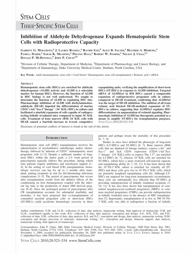
day 0 BM KSL cells versus the progeny of BM KSL cellstreated for 7 days with cytokines (thromobopoietin, SCF, Flt-3 ligand; ‘‘TSF’’) or the progeny of BM KSL cells treated
with TSF þ 100 lM DEAB (Fig. 1A, 1B). We were unableto detect a distinct ALDHþ population within the BM KSLsubset or within the progeny of TSF or TSF þ DEAB
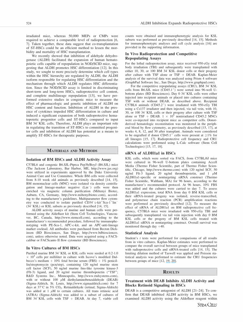
Figure 1. DEAB inhibits ALDH enzyme activity and blocks retinoid signaling in HSCs. (A): FACS-based strategy for isolation of BM CD34�KSLcells is shown. Whole BM cells from adult C57Bl6 mice were lineage-depleted and subsequently stained with antibodies to c-kit, Sca-1, and CD34 andthe CD34�KSL population was collected by FACS. (B): Representative flow cytometric analysis using the Aldefluor reagent was performed on murine BMKSL cells at day 0 and after 7 days of culture with TSF versus TSF þ DEAB (top). Identical analysis was performed on human CB CD34þCD38�lin� cellsunder the same conditions (bottom). (C): Treatment of BM 34�KSL cells with TSF þ DEAB caused a significant reduction in ALDH-positive cells in cul-ture (means6 SD, n ¼ 3; *, p ¼ .02 vs. day 0; ^, p ¼ .01 vs. day 0, p ¼ .02 vs. TSF). (D): The expression of CEPBe and CD38, RAR-dependent genes, isshown in murine BM 34�KSL cells at day 0 and after culture with TSF, TSF þ DEAB, TSF þ retinal, and TSF þ retinal þ DEAB. Treatment with DEABblocked cytokine-induced (TSF, SCF, Flt-3 ligand) and retinal-induced expression of CEBPe (*, p < .001; ^, p < .001) and CD38 (*, p < .001; ^, p < .001)in BM HSCs. Neither retinal nor DEAB affected the expression of the glucocorticoid receptor, a non-RAR-dependent gene (n ¼ 3, means6 SD). Numbersrepresent the fold change relative to expression in the TSF group. Abbreviations: 34�KSL, CD34�c-kitþSca-1þlineage�; ALDH, aldehyde dehydrogenase;BM, bone marrow; CB, cord blood; DEAB, diethylaminobenzaldehyde; Flt-3, fms-like tyrosine kinase-3; HSCs, hematopoietic stem cells; ND, not detected;RAR, retinoic acid receptor; SCF, stem cell factor; TSF, thrombopoietin, stem cell factor, Flt-3 ligand.
Muramoto, Russell, Safi et al. 525
www.StemCells.com
cultures at 24 hours, day 3, or day 7 (Fig. 1B). Therefore, weanalyzed human cord blood (CB) CD34þCD38�lin� HSCs todetermine whether DEAB treatment decreased ALDH activityin human HSCs. The majority (mean 72.9%) of day 0CD34þCD38�lin� cells were ALDH bright, as were 41% ofthe TSF-treated progeny. However, only 2.5% of the progenyof TSF þ DEAB cultures demonstrated ALDH activity, veri-fying that DEAB treatment inhibited ALDH activity in humanHSCs during culture (Fig. 1B, 1C). Interestingly, the expres-sion of ALDH1a1 also decreased >50% in response to TSFþ DEAB compared to TSF alone (supporting informationFig. S1).
Microarray analyses have suggested that several isoformsof ALDH may be expressed by HSCs, including ALDH1a1,ALDH2, ALDH1a7, ALDH3a2, and ALDH9a1 [25–27].Using quantitative reverse transcriptase (qRT)-PCR analysis,we found that ALDH1a1, which catalyzes retinoic acid bio-synthesis, and ALDH2, ALDH1a7, ALDH3a2 and ALDH9a1were expressed by murine BM 34�KSL cells (supporting in-formation Fig. S2). ALDH1a2, ALDH1a3, and ALDH8a1,which also catalyze retinoic acid synthesis, were notexpressed. Since ALDH1a1 has retinaldehyde (retinal) metab-olizing activity and the other expressed isoforms have little(ALDH2) or no retinal metabolizing activity (ALDH1a7,ALDH3a2, and ALDH9a1) [25, 28], we treated BM 34�KSLcells with retinal þ DEAB to determine if ALDH1a1 was aprimary target of DEAB action. We measured expression ofCEBPe and CD38, which are retinoic acid receptor (RAR)-target genes [29–31], in the BM 34�KSL cells since theexpression of CEBPe and CD38 increases in response to reti-noic acid signaling. CEBPe expression increased in responseto TSF and further increased in response to retinal (Fig. 1D).In contrast, when DEAB was added to BM 34�KSL cell cul-
tures with TSF or TSF þ retinal, CEBPe expression was sig-nificantly reduced (Fig. 1D). The expression of CD38 alsoincreased significantly in response to TSF þ retinal. WhenDEAB was added to TSF þ retinal, CD38 expression wasreduced 18-fold (Fig. 1D). Conversely, expression of the glu-cocorticoid receptor, which is not RAR-dependent, did notchange significantly in BM 34�KSL cells in response to reti-nal or retinal þ DEAB. Taken together, these data confirmedthat DEAB treatment antagonized retinoid signaling in HSCsand suggested that ALDH1a1, which has retinal-metabolizingactivity, was a primary target of DEAB action. Of note, theprogeny of HSCs treated with TSF þ DEAB also demon-strated significantly reduced expression of Mac-1 (myeloid)and B220 (B cell) differentiation markers compared to theprogeny of TSF alone (supporting information Fig. S3). Theseresults confirmed the inhibitory effect of DEAB on HSC dif-ferentiation in culture but also raised the possibility that otherdifferentiation pathways independent of ALDH and RARwere antagonized by DEAB.
Inhibition of ALDH Maintains Phenotypic andFunctional Progenitor Cells in Culture
To determine the effect of ALDH inhibition on HSC contentin culture, BM 34�KSL cells were cultured with TSF with orwithout 100 lM DEAB for 7 days. Treatment of 34�KSLcells with TSF caused a significant decline in lineage negativecells at day 7 (mean 42.6%), whereas the progeny of TSF þDEAB contained more than twofold increased percentage oflineage negative cells compared to that of TSF alone (97.1%;Fig. 2A, 2B). Moreover, treatment with TSF þ DEAB main-tained a more than sevenfold higher percentage of KSL cellsin culture as compared to culture with TSF alone (mean
Figure 1. (Continued)
526 ALDH Inhibition Expands Radioprotective HSCs
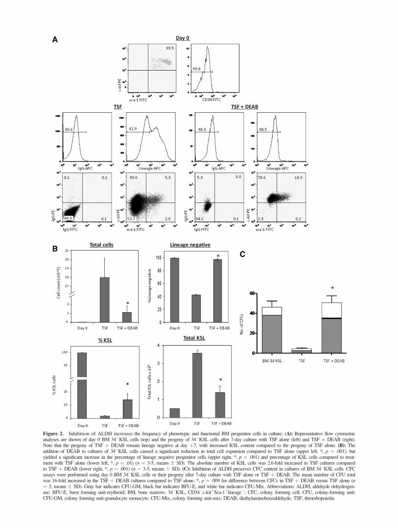
Figure 2. Inhibition of ALDH increases the frequency of phenotypic and functional BM progenitor cells in culture. (A): Representative flow cytometricanalyses are shown of day 0 BM 34�KSL cells (top) and the progeny of 34�KSL cells after 7-day culture with TSF alone (left) and TSF þ DEAB (right).Note that the progeny of TSF þ DEAB remain lineage negative at day þ7, with increased KSL content compared to the progeny of TSF alone. (B): Theaddition of DEAB to cultures of 34�KSL cells caused a significant reduction in total cell expansion compared to TSF alone (upper left, *, p ¼ .001), butyielded a significant increase in the percentage of lineage negative progenitor cells (upper right, *, p < .001) and percentage of KSL cells compared to treat-ment with TSF alone (lower left, *, p ¼ .01) (n ¼ 3-5, means 6 SD). The absolute number of KSL cells was 2.6-fold increased in TSF cultures comparedto TSF þ DEAB (lower right, *, p ¼ .001) (n ¼ 3-5, means6 SD). (C): Inhibition of ALDH preserves CFC content in cultures of BM 34�KSL cells. CFCassays were performed using day 0 BM 34�KSL cells or their progeny after 7-day culture with TSF alone or TSF þ DEAB. The mean number of CFU totalwas 16-fold increased in the TSF þ DEAB cultures compared to TSF alone. *, p ¼ .009 for difference between CFCs in TSF þ DEAB versus TSF alone (n¼ 3, means6 SD). Gray bar indicates CFU-GM, black bar indicates BFU-E, and white bar indicates CFU-Mix. Abbreviations: ALDH, aldehyde dehydrogen-ase; BFU-E, burst forming unit-erythroid; BM, bone marrow; 34�KSL, CD34�c-kitþSca-1þlineage�; CFC, colony forming cell; CFU, colony-forming unit;CFU-GM, colony forming unit-granulocyte monocyte; CFU-Mix, colony forming unit-mix; DEAB, diethylaminobenzaldehyde; TSF, thrombopoietin.
14.2% vs. 2.0%; Fig. 2A, 2B). However, culture with TSFalone caused a 360-fold expansion of total cells whereas cul-ture with TSF þ DEAB supported a 22.6-fold expansion (Fig.2B). Therefore, the total number of KSL cells increased 2.6-fold in TSF cultures compared to TSF þ DEAB cultures atday 7 (Fig. 2B). Of note, the progeny of TSF þ DEAB cul-tures contained 16-fold increased numbers of colony-formingcells as compared to the progeny of TSF alone, demonstratingthat inhibition of ALDH impeded the differentiation of func-tional BM progenitor cells in culture (Fig. 2C).
Inhibition of ALDH Facilitates the Expansion ofRadioprotective Cells
We next sought to determine whether inhibition of ALDHcould increase the number of BM stem/progenitor cells within vivo radioprotective capacity. We transplanted B6.SJL(CD45.1þ) day 0 BM 34�KSL cells (10, 30, or 100 cells) or
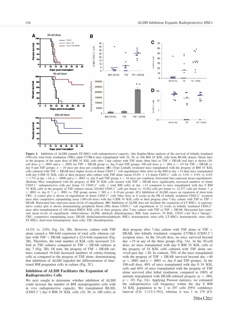
their progeny after 7-day culture with TSF alone or TSF þDEAB, into lethally irradiated, congenic C57BL6 (CD45.2þ)recipient mice. At the 10-cell dose, no mice survived beyondday þ19 in any of the three groups (Fig. 3A). At the 30-celldose, no mice transplanted with day 0 BM 34�KSL cells orthe progeny of 34�KSL cells cultured with TSF alone sur-vived past day þ20. In contrast, 70% of the mice transplantedwith the progeny of TSF þ DEAB survived beyond day þ40(p < .0001 and p < .0001 vs. day 0 and TSF groups). At the100-cell dose, 40% of mice transplanted with day 0 34�KSLcells and 60% of mice transplanted with the progeny of TSFalone survived after lethal irradiation, compared to 100% ofanimals transplanted with DEAB-cultured progeny (p ¼ .004,p ¼ .03; Fig. 3A). Applying Poisson statistics, we estimatedthe radioprotective cell frequency within the day 0 BM34�KSL population to be 1 in 297 cells [95% confidenceinterval (CI): 1:113-1:781], whereas it was 1 in 179 [CI:
Figure 3. Inhibition of ALDH expands ST-HSCs with radioprotective capacity. (A): Kaplan-Meier analysis of the survival of lethally irradiated(950-cGy total body irradiation (TBI)) adult C57BL6 mice transplanted with 10, 30, or 100 BM 34�KSL cells from B6.SJL donors (black line)or the progeny of the same dose of BM 34�KSL cells after 7-day culture with TSF alone (blue line) or TSF þ DEAB (red line) is shown (30-cell dose: p < .0001 and p < .0001 for TSF þ DEAB group vs. day 0 and TSF groups; 100-cell dose: p ¼ .004, p ¼ .03 for TSF þ DEAB vs.day 0 and TSF groups; n ¼ 10 mice per dose per condition). (B): (Top) Lethally irradiated mice transplanted with the progeny of BM 34�KSLcells cultured with TSF þ DEAB have higher levels of donor CD45.1þ cell engraftment (blue dots) in the BM at day þ14 than mice transplantedwith day 0 BM 34�KSL cells or their progeny after culture with TSF alone (mean 25.0% 6 1.9 donor CD45.1þ cells vs. 5.4% 6 4.9% vs. 6.8%6 1.7% at day þ14; p ¼ .0008 and p < .0001 vs. day 0 and TSF group; n ¼ 10 mice per condition; horizontal bars represent mean engraftment).(Bottom) Mice transplanted with the progeny of BM 34�KSL cells treated with TSF þ DEAB have significantly increased numbers of donorCD45.1þ radioprotective cells per femur (% CD45.1þ cells � total BM cells) at day þ14 compared to mice transplanted with day 0 BM34�KSL cells or the progeny of TSF cultures (mean 224,862 CD45.1þ cells per femur vs. 14,562 cells per femur vs. 21,577 cells per femur; *, p< .0001 vs. day 0; ^, p < .0001 vs. TSF group; means 6 SD, n ¼ 8-10 per group). (C): Inhibition of ALDH causes an expansion of short-termCRU. A scatter plot is shown of engraftment of donor CD45.1þ cells (blue dots) at 4 weeks in the PB of lethally irradiated CD45.2þ recipientmice after competitive repopulating assay (100-cell dose) with day 0 BM 34�KSL cells or their progeny after 7-day culture with TSF or TSF þDEAB. Horizontal bars represent mean levels of engraftment. (D): Inhibition of ALDH does not facilitate the expansion of LT-HSCs. A represen-tative scatter plot is shown demonstrating peripheral blood (PB) donor CD45.1þ cell engraftment at 12 weeks in lethally irradiated CD45.2þ
mice after transplantation of 100 donor BM34�KSL cells or their progeny after 7-day culture with TSF or TSF þ DEAB. Horizontal bars repre-sent mean levels of engraftment. Abbreviations: ALDH, aldehyde dehydrogenase; BM, bone marrow; 34�KSL, CD34�c-kitþSca-1þlineage�;CRU, competitive repopulating assay; DEAB, diethylaminobenzaldehyde; HSCs, hematopoietic stem cells; LT-HSCs, hematopoietic stem cells;ST-HSCs, short-term hematopoietic stem cells; TSF, thrombopoietin.
528 ALDH Inhibition Expands Radioprotective HSCs
1:82-1:391] for the progeny of 34�KSL cells cultured withTSF alone. In contrast, the radioprotective cell frequencywithin the progeny of 34�KSL cells cultured with TSF þDEAB was 1 in 33 cells [CI: 1:19-1:57], representing a nine-and sixfold increase in radioprotective cell frequency com-pared to day 0 34�KSL cells and the progeny of cultures withTSF alone (Table 1). Importantly, the progeny of TSF cul-tures contained 15.9-fold more total cells than TSF þ DEAB-cultured progeny. Therefore, when normalized to the total cellnumbers transplanted into recipient mice, the progeny of TSFþ DEAB had a 96-fold (15.9 � 6-fold) increased radioprotec-tive cell frequency compared to the progeny of TSF alone. Ifnormalized to total KSL progenitor cells infused per mouse,the progeny of TSF þ DEAB had a 14-fold increased fre-quency of radioprotective cells compared to TSF alone.
To directly demonstrate the increased radioprotective cellfrequency in the progeny of TSF þ DEAB cultures versus theprogeny of TSF alone, we measured donor CD45.1þ cellengraftment in the BM of lethally irradiated mice at day þ14after transplantation of a limiting dose (100 cells) of BM34�KSL cells or their progeny after culture with TSF aloneor TSF þ DEAB (Fig. 3B). Mice transplanted with the prog-eny of TSF þ DEAB cultures had 4.6-fold increased donorCD45.1þ cell engraftment in the BM at day 14 compared tomice transplanted with day 0 BM 34�KSL cells and 3.8-foldincreased engraftment compared to mice transplanted with theprogeny of TSF alone (mean 25.0% donor CD45.1þ cells vs.5.4% vs. 6.8% at day þ14; p ¼ .0008 and p < .0001 vs. day0 and TSF group; Fig. 3B). Since mice transplanted with theprogeny of TSF þ DEAB had 3.6- and 2.3-fold increasedtotal BM cells at day þ14, this translated into a 15.4-foldincrease in the number of donor CD45.1þ radioprotectivecells per femur in mice transplanted with the progeny of TSFþ DEAB cultures versus mice transplanted with day 0 BM34�KSL cells and a 10.4-fold increase compared to micetransplanted with the progeny of TSF alone (mean 224,862CD45.1þ cells per femur vs. 14,562 cells per femur vs.
21,577 cells per femur; p < .0001 and p < .0001 vs. day 0and TSF group; Fig. 3B).
Inhibition of ALDH Expands ST-HSCs withCompetitive Repopulating Capacity
Since inhibition of ALDH increased the number of BM radio-protective cells in culture, we sought to determine if this wascaused by an amplification of short-term HSCs (ST-HSCs)with competitive repopulating capacity. BM 34�KSL cellsfrom B6.SJL mice (CD45.1þ) or their progeny after culturewith TSF alone or TSF þ DEAB were transplanted at limit-ing doses in a competitive repopulating assay into lethally irra-diated CD45.2þ C57BL6 mice. At 4 weeks after transplant,mice transplanted with the progeny of 100 BM 34�KSL cellscultured with TSF þ DEAB demonstrated significantlyincreased CD45.1þ cell multilineage engraftment (mean 34.0%CD45.1þ) compared to mice transplanted with the same doseof day 0 34�KSL cells (mean 4.4%) or their progeny after cul-ture with TSF alone (mean 16.7%) (Fig. 3C). Transplantationof mice over a range of limiting doses (10-200 cells) coupledwith Poisson statistical analysis demonstrated the frequency of4-week CRU within day 0 BM 34�KSL cells to be 1 in 147cells (95% CI: 1:91-1:238) and 1 in 156 cells for the progenyof TSF cultures (95% CI: 1:95-1:258) (supporting informationTable S1). Conversely, the CRU frequency within the progenyof TSF þ DEAB cultures was 1 in 39 cells (95% CI: 1:25-1:62). Therefore, the progeny of TSF þ DEAB cultures con-tained 3.8- and 4-fold increased numbers of ST-HSCs com-pared to day 0 34�KSL cells and the progeny of 34�KSL cellscultured with TSF alone, respectively.
To measure the effect of ALDH inhibition on long-termHSC (LT-HSC) content in culture, we also performed CRUanalysis at 12 and 30 weeks after transplantation (Fig. 3D). Micetransplanted with day 0 BM 34�KSL cells displayed increasingdonor 45.1þ cell repopulation over time and the CRU estimateswere 1 in 17 and 1 in 20 cells at 12 and 30 weeks, respectively(supporting information Table S2). The CRU estimates withinthe progeny of TSF þ DEAB culture were 1 in 56 and 1 in 67cells at 12 and 30 weeks, respectively. Similarly, the CRU esti-mates for the progeny of TSF alone were 1 in 61 and 1 in 70cells at 12 and 30 weeks, respectively. These data demonstratethat inhibition of ALDH activity does not amplify LT-HSCs inculture compared to day 0 BM 34�KSL cells.
As a complement to competitive repopulating assays in mice,we also examined the effect of DEAB treatment on the short-term engraftment potential of human CB CD34þCD38�lin� cellsin a NOD/SCID transplantation assay. Mice transplanted with 2.5� 103 CB CD34þ38�lin� HSCs demonstrated human CD45þ
cell engraftment at 4 weeks in 3 of 12 mice (25%; mean 0.9%huCD45þ BM cells). Similarly, mice transplanted with the prog-eny of the identical dose of CD34þ38�lin� cells after culturewith TSF demonstrated engraftment in 2 of 10 mice (20%; mean0.7% huCD45þ cells). In contrast, mice transplanted with theprogeny of CB CD34þCD38�lin� cells after culture with TSF þDEAB demonstrated human CD45þ cell engraftment in 5 of 10mice (50%; mean 1.4% huCD45þ cells). These results suggestedthat treatment with DEAB enhanced the short-term repopulatingactivity of human HSCs. This observation is also consistent withour prior observation that inhibition of ALDH caused an expan-sion of human LT-HSCs as measured in primary and secondarytransplanted NOD/SCID mice [12].
Inhibition of ALDH Delays Cell Cycle Progressionof HSCs in Culture
The repopulating capacity of HSCs has been shown to declinein association with transit through cell cycle in vitro [32, 33].
Table 1. Inhibition of ALDH1 increases the radioprotective cellfrequency
Condition Cell dose
No.
surviving
radioprotective
cell (RPC) estimate 95% CI
34�KSL 10 0:10 1 in 297 113-78130 0:10
100 4:10TSF 10 0:10 1 in 179 82-391
30 0:10100 6:10
TSF þ DEAB 10 0:10 1 in 33 19-5730 7:10
100 10:10
FACS-sorted bone marrow 34�KSL cells were transplanted intolethally irradiated C57BI6 mice (950-cGy total body irradiation(TBI)) via tail vein injection of 10, 30, or 100 cells (n ¼ 10mice per dose). The identical numbers of 34�KSL cells wereplaced in culture with TSF, stem cell factor, and Flt-3 ligand orTSF þ 100 lM DEAB � 7 days and their progeny weretransplanted into lethally irradiated mice as described inMaterials and Methods. A limiting dilution analysis demonstratedthat the radioprotective cell content was ninefold increased in theprogeny of TSF þ DEAB cultures compared to input 34�KSLcells and sixfold increased compared to the progeny of culturewith TSF alone.Abbreviations: 34�KSL, CD34�c-kitþSca-1þlineage�; CI,confidence interval; DEAB, diethylaminobenzaldehyde; TSF,thrombopoietin.
Muramoto, Russell, Safi et al. 529
www.StemCells.com
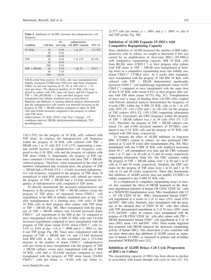
We sought to determine whether inhibition of ALDHincreased BM stem/progenitor cell repopulating capacity viamodulation of cell cycle progression of HSCs in culture. Day0 BM KSL cells were mostly quiescent (50.4% in G0, 26.2%in G1). After 3 days in culture, the progeny of TSF cultureswere primarily in cycle, whereas the progeny of TSF þDEAB showed a modest increase in the percentage of cells inG0 and a decrease in cells in the G2/S/M phase (Fig. 4A; p ¼.004 for G0 comparison, p ¼ .02 for G2/S/M comparison). Af-ter 7 days in culture, the progeny of TSF þ DEAB culturedemonstrated an increase in cell cycling compared to theprogeny of TSF alone (Fig. 4A; p ¼ .01 and p ¼ .02 for G0
and G2/S/M phase, respectively), perhaps reflecting replicativesenescence in TSF-treated cells at day 7 as compared to theprogeny of TSF þ DEAB. The mean frequency of day 0 KSLcells and their progeny after culture with TSF and TSF þDEAB in each phase of the cell cycle is shown in supportinginformation Fig. S5.
Inhibition of ALDH Increases the Frequency ofCD34
1Flt-3
2KSL Cells in Culture and Can Be
Reversed with ATRA
BM 34þFlt-3�KSL cells have been shown to be highlyenriched for ST-HSCs [6]. Since inhibition of ALDH sup-ported an increase in short-term CRUs and radioprotectivecells in culture, we sought to determine if DEAB treatmentcaused an increase in the number of 34þFlt-3�KSL cells inculture compared to cytokines alone. At day 7 of culture ofBM 34�KSL HSCs with TSF alone, only 1.6% 6 0.2 of thecultured progeny remained 34þFlt-3�KSL cells, whereas26.4% 6 3.5 of the progeny after culture with TSF þ DEABwere 34þFlt-3�KSL cells (Fig. 4B, p ¼ .003). These datademonstrate that inhibition of ALDH facilitates the expansionof ST-HSCs in culture. Interestingly, the addition of 1 lMATRA to TSF þ DEAB cultures essentially reversed theeffect of DEAB treatment, yielding >20-fold less 34þFlt-3�KSL cells in culture (mean 1.1% 6 0.2, p ¼ .003). Thisresult suggested that inhibition of ALDH impeded HSC dif-ferentiation via antagonism of retinoid signaling.
Since committed CMPs and MEPs have also been shownto possess radioprotective capacity [7], we compared the fre-quency of IL-7Ra�Lin�Sca-1�c-kitþFccRloCD34þ cells(CMPs) with IL-7Ra�Lin�Sca-1�c-kitþFccRloCD34� cells(MEPs) within the progeny of TSF cultures versus TSF þDEAB cultures [7]. Interestingly, the progeny of TSF þDEAB cultures contained a significantly increased frequencyof CMPs and a significant decrease in MEPs compared to theprogeny of TSF alone (Fig. 4C; p < .001 and p ¼ .01, respec-tively). Taken together, these data demonstrate that inhibitionof ALDH causes a generalized inhibition of myeloid differen-tiation of HSCs in response to cytokines alone.
siRNA of ALDH1a1 Increases BM RadioprotectiveProgenitor Cell Content
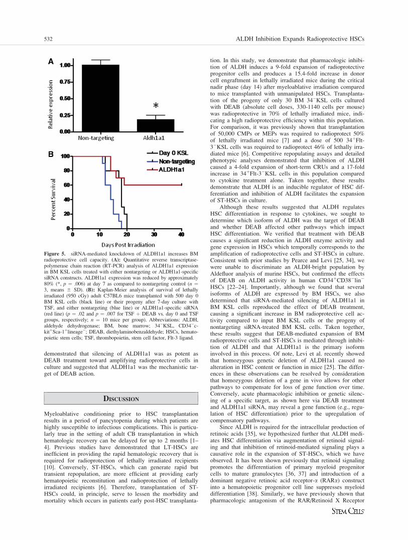
We show here that several isoforms of ALDH are expressedin HSCs, including ALDH1a1, ALDH2, ALDH3a2,ALDH1a7, and ALDH9a1 [25–27]. Since treatment withDEAB blocked retinoid signaling in HSCs and ALDH1a1 hasretinoic acid biosynthetic activity, we hypothesized thatALDH1a1 might be the primary mechanistic target of DEAB.We therefore treated BM KSL cells with an siRNA specifi-cally targeting ALDH1a1 in BM HSCs to determine if thiswould cause comparable expansion of radioprotective ST-HSCs as observed with DEAB. Treatment of BM KSL cellswith ALDH1a1-siRNA caused an 80% decrease in ALDH1a1expression compared to nontargeting siRNA-treated BM KSL
cells at day 7 of culture (Fig. 5; p ¼ .006). Mice transplantedwith the progeny of BM KSL cells treated with ALDH1a1-siRNA demonstrated significantly increased survival at dayþ40 compared to mice transplanted with day 0 BM KSL cells(Fig. 5, 60 % vs. 0%, p ¼ .02) and increased survival com-pared to mice transplanted with BM KSL cells treated withnontargeting siRNA (60% vs. 0%, p ¼ .007). These results
Figure 4. Antagonism of ALDH inhibits cell cycle progression ofHSCs and increases the frequency of 34þFlt-3�KSL cells in culturecompared to cytokines alone. (A): A representative cell cycle analysisof 34�KSL cells and their progeny after 7-day cultures with TSFalone or TSF þ DEAB is shown. The majority (mean 83.5%) of day0 34�KSL cells were in G0/G1. At day þ3, the progeny of TSF þDEAB cultures demonstrated a modest increase in cells in G0 anddecreased numbers in G2/S/M phase, consistent with overall inhibitionof cell cycle progression compared to TSF alone. At day þ7, theprogeny of TSF cultures demonstrated increased numbers of cells inG0 compared to TSF þ DEAB. (B): (i) Representative FACS analy-ses of 34þFlt-3�KSL cells in culture of BM 34�KSL cells with TSF,TSF þ 1 lM ATRA, TSF þ DEAB, or TSF þ DEAB þ ATRA areshown. (ii) Treatment with TSF þ DEAB supported an increase inthe frequency of 34þFlt-3�KSL cells in culture compared to culturewith TSF alone (*, p ¼ .003, n ¼ 3, mean 6 SD). The addition ofATRA reversed the effects of DEAB on ST-HSC expansion in culture(^p ¼ .003, n ¼ 3; mean 6 SD). (C): Representative FACS analysesof CMP and MEP content are shown for BM 34�KSL cells after cul-ture with TSF (top) or TSF þ DEAB (middle). The progeny of TSFþ DEAB cultures contained an increased frequency of CMPs com-pared to the progeny of TSF alone (mean 36.1% vs. 4.8%, **, p <.001) and a decreased frequency of MEPs (mean 0.1% vs. 0.3%,respectively; *, p ¼ .01) (n ¼ 6; means 6 SD) (bottom). Abbrevia-tions: ALDH, aldehyde dehydrogenase; ATRA, All trans retinoicacid; 34�KSL, CD34�c-kitþSca-1þlineage�; CMP, common myeloidprogenitor; DEAB, diethylaminobenzaldehyde; Flt-3, fms-like tyrosinekinase-3; HSCs, hematopoietic stem cells; MEP, megakaryocyte-ery-throid progenitor; TSF, thrombopoietin.
530 ALDH Inhibition Expands Radioprotective HSCs
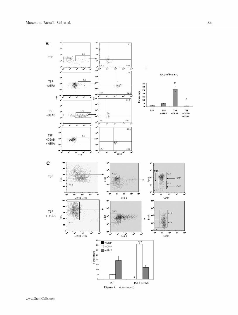
Figure 4. (Continued)
Muramoto, Russell, Safi et al. 531
www.StemCells.com
demonstrated that silencing of ALDH1a1 was as potent asDEAB treatment toward amplifying radioprotective cells inculture and suggested that ALDH1a1 was the mechanistic tar-get of DEAB action.
DISCUSSION
Myeloablative conditioning prior to HSC transplantationresults in a period of pancytopenia during which patients arehighly susceptible to infectious complications. This is particu-larly true in the setting of adult CB transplantation in whichhematologic recovery can be delayed for up to 2 months [1–4]. Previous studies have demonstrated that LT-HSCs areinefficient in providing the rapid hematologic recovery that isrequired for radioprotection of lethally irradiated recipients[10]. Conversely, ST-HSCs, which can generate rapid buttransient repopulation, are more efficient at providing earlyhematopoietic reconstitution and radioprotection of lethallyirradiated recipients [6]. Therefore, transplantation of ST-HSCs could, in principle, serve to lessen the morbidity andmortality which occurs in patients early post-HSC transplanta-
tion. In this study, we demonstrate that pharmacologic inhibi-tion of ALDH induces a 9-fold expansion of radioprotectiveprogenitor cells and produces a 15.4-fold increase in donorcell engraftment in lethally irradiated mice during the criticalnadir phase (day 14) after myeloablative irradiation comparedto mice transplanted with unmanipulated HSCs. Transplanta-tion of the progeny of only 30 BM 34�KSL cells culturedwith DEAB (absolute cell doses, 330-1140 cells per mouse)was radioprotective in 70% of lethally irradiated mice, indi-cating a high radioprotective efficiency within this population.For comparison, it was previously shown that transplantationof 50,000 CMPs or MEPs was required to radioprotect 50%of lethally irradiated mice [7] and a dose of 500 34þFlt-3�KSL cells was required to radioprotect 46% of lethally irra-diated mice [6]. Competitive repopulating assays and detailedphenotypic analyses demonstrated that inhibition of ALDHcaused a 4-fold expansion of short-term CRUs and a 17-foldincrease in 34þFlt-3�KSL cells in this population comparedto cytokine treatment alone. Taken together, these resultsdemonstrate that ALDH is an inducible regulator of HSC dif-ferentiation and inhibition of ALDH facilitates the expansionof ST-HSCs in culture.
Although these results suggested that ALDH regulatesHSC differentiation in response to cytokines, we sought todetermine which isoform of ALDH was the target of DEABand whether DEAB affected other pathways which impactHSC differentiation. We verified that treatment with DEABcauses a significant reduction in ALDH enzyme activity andgene expression in HSCs which temporally corresponds to theamplification of radioprotective cells and ST-HSCs in culture.Consistent with prior studies by Pearce and Levi [25, 34], wewere unable to discriminate an ALDH-bright population byAldefluor analysis of murine HSCs, but confirmed the effectsof DEAB on ALDH activity in human CD34þCD38�lin�
HSCs [22–24]. Importantly, although we found that severalisoforms of ALDH are expressed by BM HSCs, we alsodetermined that siRNA-mediated silencing of ALDH1a1 inBM KSL cells reproduced the effect of DEAB treatment,causing a significant increase in BM radioprotective cell ac-tivity compared to input BM KSL cells or the progeny ofnontargeting siRNA-treated BM KSL cells. Taken together,these results suggest that DEAB-mediated expansion of BMradioprotective cells and ST-HSCs is mediated through inhibi-tion of ALDH and that ALDH1a1 is the primary isoforminvolved in this process. Of note, Levi et al. recently showedthat homozygous genetic deletion of ALDH1a1 caused noalteration in HSC content or function in mice [25]. The differ-ences in these observations can be resolved by considerationthat homozygous deletion of a gene in vivo allows for otherpathways to compensate for loss of gene function over time.Conversely, acute pharmacologic inhibition or genetic silenc-ing of a specific target, as shown here via DEAB treatmentand ALDH1a1 siRNA, may reveal a gene function (e.g., regu-lation of HSC differentiation) prior to the upregulation ofcompensatory pathways.
Since ALDH is required for the intracellular production ofretinoic acids [35], we hypothesized further that ALDH medi-ates HSC differentiation via augmentation of retinoid signal-ing and that inhibition of retinoid-mediated signaling plays acausative role in the expansion of ST-HSCs, which we haveobserved. It has been shown previously that retinoid signalingpromotes the differentiation of primary myeloid progenitorcells to mature granulocytes [36, 37] and introduction of adominant negative retinoic acid receptor-a (RARa) constructinto a hematopoietic progenitor cell line suppresses myeloiddifferentiation [38]. Similarly, we have previously shown thatpharmacologic antagonism of the RAR/Retinoid X Receptor
Figure 5. siRNA-mediated knockdown of ALDH1a1 increases BMradioprotective cell capacity. (A): Quantitative reverse transcriptase-polymerase chain reaction (RT-PCR) analysis of ALDH1a1 expressionin BM KSL cells treated with either nontargeting or ALDH1a1-specificsiRNA constructs. ALDH1a1 expression was reduced by approximately80% (*, p ¼ .006) at day 7 as compared to nontargeting control (n ¼3, means 6 SD). (B): Kaplan-Meier analysis of survival of lethallyirradiated (950 cGy) adult C57BL6 mice transplanted with 500 day 0BM KSL cells (black line) or their progeny after 7-day culture withTSF, and either nontargeting (blue line) or ALDH1a1-specific siRNA(red line) (p ¼ .02 and p ¼ .007 for TSF þ DEAB vs. day 0 and TSFgroups, respectively; n ¼ 10 mice per group). Abbreviations: ALDH,aldehyde dehydrogenase; BM, bone marrow; 34�KSL, CD34�c-kitþSca-1þlineage�; DEAB, diethylaminobenzaldehyde; HSCs, hemato-poietic stem cells; TSF, thrombopoietin, stem cell factor, Flt-3 ligand.
532 ALDH Inhibition Expands Radioprotective HSCs
(RXR) heterodimer promotes the maintenance of humanNOD/SCID repopulating cells in culture [39]. ATRA adminis-tration is also used therapeutically to induce the terminal dif-ferentiation of acute promyelocytic leukemia cells, which beardominant negative activity at RARa [40]. Conversely, it hasbeen shown that the addition of ATRA to cytokine-containingcultures of murine BM KSL cells enhances the maintenanceof pre-CFU-S cells and long-term repopulating cells comparedto cytokine cultures alone [41, 42]. It was subsequentlyshown that these effects of ATRA were mediated throughRARc, rather than RARa, and that the addition of ATRAalso enhanced the serial transplantability of KSL cells com-pared to input KSL cells or their progeny after cytokine treat-ment [43]. Taken together, these studies suggest that theeffects of retinoic acid are dependent upon the developmentalstage of the hematopoietic stem/progenitor cell. Retinoic acidappears to maintain the repopulating activity of primitiveHSCs [41–43], whereas it promotes the myeloid differentia-tion of multipotent progenitor cells (MPPs) [41]. Our resultsare not inconsistent with these prior observations. We showthat inhibition of ALDH decreases RAR-mediated signalingand increases the number of ST-HSCs and radioprotectivecells in culture, which is consistent with antagonism of RAR-mediated differentiation of ST-HSCs and MPPs. Furthermore,we demonstrate that the addition of ATRA to cultures of BM34�KSL cells with DEAB overrides the effect of DEAB to-ward maintaining ST-HSCs in culture and promotes differen-tiation and lineage commitment. Taken together, our resultsimplicate the ALDH/RAR axis in regulating the myeloid dif-ferentiation of ST-HSCs. It remains possible that DEAB treat-ment inhibits HSC differentiation via alternative pathways in-dependent of effects on ALDH or RAR. However, thisappears less likely since siRNA-mediated silencing ofALDH1a1 caused a comparable increase in BM radioprotec-tive cell activity.
It has been previously demonstrated that ex vivo cultureof HSCs with proliferation-inducing cytokines and the transi-tion of HSCs from G1 to G2/S/M phase are associated with aloss of repopulating capacity [32]. Here, we show that phar-macologic inhibition of ALDH with DEAB slowed the G0/G1
transition in HSCs, resulting in a modestly higher frequency
of HSCs in G0 and less cells in G2/S/M phase compared toHSCs treated with TSF alone. This inhibition of cell cycleprogression in HSCs, although modest, may have contributedto the significant increase in repopulating ST-HSCs observedin DEAB-treated BM 34�KSL cells as compared to the prog-eny of cytokines alone. Since several tumor-specific cancerstem cells have high expression of ALDH [44, 45], theseresults also have implications for cancer research. ALDH-bright tumor cells have been shown to have a higher prolifer-ative and repopulating potential than ALDH-negative tumorcells [44, 45]. Our study suggests that pharmacologic inhibi-tion of ALDH should be explored as a strategy to decreasethe growth and proliferation of cancer stem cells.
In summary, we show here that ALDH has a precise func-tion in regulating the differentiation of ST-HSCs, inhibition ofALDH causes significant amplification of ST-HSCs withradioprotective capacity, and these effects are mediated viainhibition of retinoid signaling and HSC cell cycle transition.siRNA-mediated silencing of ALDH1a1 in BM HSCs main-tained radioprotective cells in culture at a level comparable toDEAB treatment, suggesting that ALDH1a1 is the primarytarget of DEAB. ALDH inhibition represents a translatablestrategy to expand ST-HSCs to augment hematopoieticengraftment in patients undergoing stem cell transplantation.
ACKNOWLEDGMENTS
The authors acknowledge the Duke Human Vaccine InstituteFlow Cytometry Core Facility for assistance with cell sorting.This work was supported, in part, by a grant from the NationalInstitute of Allergy and Infectious Diseases No. AI067798(J.P.C.).
DISCLOSURE OF POTENTIAL CONFLICTS
OF INTEREST
The authors indicate no potential conflicts of interest.
REFERENCES
1 Laughlin MJ, Eapen M, Rubinstein P et al. Outcomes after transplan-tation of cord blood or bone marrow from unrelated donors in adultswith leukemia. N Engl J Med 2004;351:2265–2275.
2 Cornelissen JJ, Lowenberg B. Role of allogeneic stem cell transplanta-tion in current treatment of acute myeloid leukemia. Hematology AmSoc Hematol Educ Program 2005:151–155.
3 Alyea EP, Kim HT, Ho V et al. Impact of conditioning regimenintensity on outcome of allogeneic hematopoietic cell trans-plantation for advanced acute myelogenous leukemia and myelody-splastic syndrome. Biol Blood Marrow Transplant 2006;12:1047–1055.
4 Meijer E, Dekker AW, Lokhorst HM et al. Low incidence of infec-tious complications after nonmyeloablative compared with myeloabla-tive allogeneic stem cell transplantation. Transpl Infect Dis 2004;6:171–178.
5 Barker JN. Umbilical Cord Blood (UCB) Transplantation: An Alterna-tive to the Use of Unrelated Volunteer Donors? Hematology Am SocHematol Educ Program2007:55–61.
6 Yang L, Bryder D, Adolfsson J et al. Identification of Lin(�)Sca1(þ)kit(þ)CD34(þ)Flt3- short-term hematopoietic stem cells capa-ble of rapidly reconstituting and rescuing myeloablated transplantrecipients. Blood 2005;105:2717–2723.
7 Na Nakorn T, Traver D, Weissman IL et al. Myeloerythroid-restrictedprogenitors are sufficient to confer radioprotection and provide themajority of day 8 CFU-S. J Clin Invest 2002;109:1579–1585.
8 Plett PA, Frankovitz SM, Orschell-Traycoff CM. In vivo trafficking,cell cycle activity, and engraftment potential of phenotypically definedprimitive hematopoietic cells after transplantation into irradiated ornonirradiated recipients. Blood 2002;100:3545–3552.
9 BitMansour A, Burns SM, Traver D et al. Myeloid progenitors protectagainst invasive aspergillosis and Pseudomonas aeruginosa infectionfollowing hematopoietic stem cell transplantation. Blood 2002;100:4660–4667.
10 Osawa M, Hanada K, Hamada H et al. Long-term lymphohemato-poietic reconstitution by a single CD34-low/negative hematopoieticstem cell. Science 1996;273:242–245.
11 Uchida N, Aguila HL, Fleming WH et al. Rapid and sustained hema-topoietic recovery in lethally irradiated mice transplanted with purifiedThy-1.1lo Lin-Sca-1þ hematopoietic stem cells. Blood 1994;83:3758–3779.
12 Chute JP, Muramoto GG, Whitesides J et al. Inhibition of aldehydedehydrogenase and retinoid signaling induces the expansion of humanhematopoietic stem cells. Proc Natl Acad Sci U S A 2006;103:11707–11712.
13 Horn PA, Thomasson BM, Wood BL et al. Distinct hematopoieticstem/progenitor cell populations are responsible for repopulatingNOD/SCID mice compared with nonhuman primates. Blood 2003;102:4329–4335.
14 Chute J, Muramoto G, Salter A et al. Transplantation of vascular en-dothelial cells mediates the hematopoietic recovery and survival oflethally irradiated mice. Blood 2007;109:2365–2372.
15 Salter A, Meadows S, Muramoto G et al. Endothelial progenitor cellinfusion induces hematopoietic stem cell reconstitution in vivo. Blood2009;113:2104–2107.
www.StemCells.com
Muramoto, Russell, Safi et al. 533
16 Jordan CT, Yamasaki G, Minamoto D. High-resolution cell cycleanalysis of defined phenotypic subsets within primitive human hema-topoietic cell populations. Exp Hematol 1996;24:1347–1355.
17 Zhang CC, Kaba M, Ge G et al. Angiopoietin-like proteins stimulateex vivo expansion of hematopoietic stem cells. Nat Med 2006;12:240–245.
18 Bonnefoix T, Bonnefoix P, Verdiel P et al. Fitting limiting dilutionexperiments with generalized linear models results in a test of the sin-gle-hit Poisson assumption. J Immunol Methods 1996;194:113–119.
19 Chute J, Saini A, Chute D et al. Ex vivo culture with human brain en-dothelial cells increases the SCID-repopulating capacity of adulthuman bone marrow. Blood 2002;100:4433–4439.
20 Chute J, Muramoto G, Fung J et al. Soluble factors elaborated byhuman brain endothelial cells induce the concomitant expansion ofpurified human BM CD34þCD38� cells and SCID repopulating cells.Blood 2005;105:576–583.
21 Jones RJ, Barber JP, Vala MS et al. Assessment of aldehyde dehydro-genase in viable cells. Blood 1995;85:2742–2746.
22 Storms RW, Trujillo AP, Springer JB et al. Isolation of primitivehuman hematopoietic progenitors on the basis of aldehyde dehydro-genase activity. Proc Natl Acad Sci U S A 1999;96:9118–9123.
23 Storms RW, Green PD, Safford KM et al. Distinct hematopoietic pro-genitor compartments are delineated by the expression of aldehyde de-hydrogenase and CD34. Blood 2005;106:95–102.
24 Hess DA, Meyerrose TE, Wirthlin L et al. Functional characterizationof highly purified human hematopoietic repopulating cells isolatedaccording to aldehyde dehydrogenase activity. Blood 2004;104:1648–1655.
25 Levi BP, Yilmaz O, Deuster G et al. Aldehyde dehydrogenase 1a1 isdispensable for stem cell function in the mouse hematopoietic andnervous systems. Blood 2009;113:1670–1680.
26 Forsberg E, Prohaska S, Katzman S et al. Differential expression ofnovel potential regulators in hematopoietic stem cells. PLoS Genet2005;1:e28.
27 Ivanova N, Dimos J, Schaniel C et al. A stem cell moleclar signature.Science 2002;298:601–604.
28 Duester G. Genetic dissection of retinoid dehydrogenases. Chem BiolInteract 2001;130-132:469–480.
29 Parrella E, Gianni M, Cecconi V et al. Phosphodiesterase IV inhibi-tion by piclamilast potentiates the cytodifferentiating action of reti-noids in myeloid leukemia cells. Cross-talk between the camp and theretinoic acid signaling pathways. J Biol Chem 2004;279:42026–42040.
30 Park D, Chumarakov A, Vuong P et al. CCAAT/enhancer bindingprotein epsilon is a potential retinoid target gene in acute promyelo-cytic leukemia treatment. J Clin Invest 1999;103:1399–1408.
31 Walkley CR, Purton LE, Snelling HJ et al. Identification of the molec-ular requirements for an RAR alpha-mediated cell cycle arrest duringgranulocytic differentiation. Blood 2004;103:1286–1295.
32 Glimm H, Oh IH, Eaves CJ. Human hematopoietic stem cells stimu-lated to proliferate in vitro lose engraftment potential during their S/G(2)/M transit and do not reenter G(0). Blood 2000;96:4185–4193.
33 Traycoff CM, Orazi A, Ladd AC et al. Proliferation-induced declineof primitive hematopoietic progenitor cell activity is coupled with anincrease in apoptosis of ex vivo expanded CD34þ cells. Exp Hematol1998;26:53–62.
34 Pearce DJ, Bonnet D. The combined use of Hoechst efflux ability andaldehyde dehydrogenase activity to identify murine and human hema-topoietic stem cells. Exp Hematol 2007;35:1437–1446.
35 Haselbeck RJ, Hoffmann I, Duester G. Distinct functions for Aldh1and Raldh2 in the control of ligand production for embryonic retinoidsignaling pathways. Dev Genet 1999;25:353–364.
36 Collins SJ. The role of retinoids and retinoic acid receptors in normalhematopoiesis. Leukemia 2002;16:1896–1905.
37 Kastner P, Chan S. Function of RARalpha during the maturation ofneutrophils. Oncogene 2001;20:7178–7185.
38 Tsai S, Bartelmez S, Heyman R et al. A mutated retinoic acid recep-tor-alpha exhibiting dominant-negative activity alters the lineage de-velopment of a multipotent hematopoietic cell line. Genes Dev 1992;6:2258–2269.
39 Safi R, Muramoto G, Salter A et al. Pharmacological manipulation ofthe RAR/RXR signaling pathway maintains the repopulating capacityof hematopoietic stem cells in culture. Mol Endocrinol 2009;23:188–201.
40 Tallman MS, Andersen JW, Schiffer CA et al. All-trans-retinoic acidin acute promyelocytic leukemia. N Engl J Med 1997;337:1021–1028.
41 Purton LE, Bernstein ID, Collins SJ. All-trans retinoic acid delays thedifferentiation of primitive hematopoietic precursors (lin-c-kitþSca-1(þ)) while enhancing the terminal maturation of committed granulo-cyte/monocyte progenitors. Blood 1999;94:483–495.
42 Purton LE, Bernstein ID, Collins SJ. All-trans retinoic acid enhancesthe long-term repopulating activity of cultured hematopoietic stemcells. Blood 2000;95:470–477.
43 Purton LE, Dworkin S, Olsen GH et al. RARgamma is critical formaintaining a balance between hematopoietic stem cell self-renewaland differentiation. J Exp Med 2006;203:1283–1293.
44 Ginestier C, Hur M, Charafe-Jauffret E et al. ALDH1 is a marker ofnormal and malignant human mammary stem cells and a predictor ofpoor clinical outcome. Cell Stem Cell 2007;1:555–567.
45 Huang E, Hynes M, Zhang T et al. Aldehyde dehydrogenase 1 is amarker for normal and malignant human colonic stem cells and tracksSC overpopulation during colon tumorigenesis. Cancer Res 2009;69:3382–3389.
See www.StemCells.com for supporting information available online.
534 ALDH Inhibition Expands Radioprotective HSCs