the evaluation of the radioprotective effect of triphala (an ayurvedic rejuvenating drug) in the...
TRANSCRIPT

been found to be superior radioprotectors than the othernon-sulphydryl compounds but the major drawback ofthese compounds has been their high toxicity at the op-timum protective dose (Sweeny, 1979), which preclud-ed their effective use in men. A turning point came withthe observation that s-2-(aminopropylamino) ethyl-phosphorothioic acid (WR-2721) showed substantialand selective protection of normal tissues with little orno protection to the solid tumors (Yuhas, 1980). How-ever, it has also been found to be highly toxic at the op-timum protective dose and the possibility of using it ona daily basis was not feasible (Cairnie, 1983). There-fore, it is desirable to search other materials that areless toxic and can offer high protection.
0944-7113/02/09/02-099 $ 15.00/0
� Introduction
The search for radioprotectors started with the realiza-tion of the need for a safeguard against the military useof atomic weapons. With the recognition that normaltissue protection in radiotherapy is as important as thedestruction of the cancer cells, the focus of protectionresearch became more therapy oriented. The use of cer-tain chemical agents may reduce the ill effects of radia-tion in such conditions. Patt et al. (1949) for the firsttime observed that the pretreatment of rats and micewith cysteine before exposure to radiation protectedthem against the radiation-induced sickness and mor-tality. Subsequently, several chemical compounds weresynthesized and tested for their radioprotective ability(Sweeny, 1979). Only sulphydryl compounds have
Phytomedicine, Vol. 9: 99–108, 2002© Urban & Fischer Verlaghttp://www.urbanfischer.de/journals/phytomed Phytomedicine
The evaluation of the radioprotective effect of Triphala(an ayurvedic rejuvenating drug) in the mice exposedto γ-radiation
G. C. Jagetia1, M. S. Baliga1, K. J. Malagi2 and M. Sethukumar Kamath2
1Department of Radiobiology, Kasturba Medical College, Manipal, India2Department of Ayurveda, Kasturba Hospital, Manipal, India
Summary
The effect of 0, 5, 6.25, 10, 12.5, 20, 25, 40, 50 and 80 mg/kg b. wt. of aqueous extract of triphala (anAyurvedic herbal medicine) administrered intraperitoneally was studied on the radiation-inducedmortality in mice exposed to 10 Gy of γ-radiation. Treatment of mice with different doses of tripha-la consecutively for five days before irradiation delayed the onset of mortality and reduced thesymptoms of radiation sickness when compared with the non-drug treated irradiated controls.The highest protection against GI (gastrointestinal) death was observed for 12.5 mg/kg triphala,where a highest number of survivors were reported up to 10 days post-irradiation. While 10 mg/kgtriphala i.p. provided the best protection as evidenced by the highest number of survivors after 30days post-irradiation in this group when compared with the other doses of triphala. Toxicity studyshowed that triphala was non-toxic up to a dose of 240 mg/kg, where no drug-induced mortalitywas observed. The LD50 dose i.p. of triphala was found to be 280 mg/kg b. wt. Our study demon-strates the ability of triphala as a good radioprotective agent and the optimum protective dose oftriphala was 1/28 of its LD50 dose.
Key words: Triphala, radiation, mice, survival, acute toxicity, radioprotection, Terminalia chebula Retz.,Phyllanthus emblica Linn. or Emblica officinalis Gaertn.and Terminalia bellerica (Gaertn.) Roxb.

(Hashimoto and Nakajima, 1997). Triphala and/or its in-dividual plant constituents have been reported to pos-sess anti-bacterial (Nadkarni, 1976; Mehta et al., 1993;Ahmad et al., 1998; Phadke and Kulkarni, 1989; Mehtaet al., 1993), antimalarial (Valsaraj et al., 1997), antifun-gal activity (Dutta et al., 1998; Valsaraj et al., 1997),anti-cancer (Tokura and Kagawa, 1995), anti-mutagenic(Rani et al., 1994; Niwa et al., 1995) anti-allergic (Taka-gi and Sanashiro, 1996) and anti-viral activities (Val-saraj et al., 1997; Badmaev and Nowakowski, 2000;Yukaka et al., 1996; Kurokawa and Sato, 1995; Hozumiand Oyama, 1997; El-Mekkawey and Meselhy, 1995) indifferent study systems. Triphala is a cardio-tonic andexerts its protective effect by improving the blood circu-lation, reducing the myocardial necrosis (Tariq et al.,1977), serum cholesterol levels and strengthens the cap-illaries (Tariq et al., 1977; Hussain, 1975; Thakur, 1984;Thakur et al., 1988). It is also hepatoprotective and im-proves the liver function (Gulati et al., 1995; Anand andSingh, 1997). The decoction of triphala has been foundto treat leucorrhea in women (Singh and Londhe 1993).It is an effective laxative and improves the gastrointesti-nal motility (Tamhane et al., 1997) thereby curing thediseases of gastrointestinal tract (Nadkarni, 1976; An-tarkar et al., 1980). Triphala has been reported to possessanti-aging properties and improves the mental faculties(Nadkarni, 1976; Antarkar et al., 1980). Triphala hasbeen found to potentiate the adrenergic function therebyenabling the body to recover from stress. In addition, theimmunomodulatory property (Suresh and Vasudevan,1994; Rege et al., 1999) may help in increasing thebody’s defence system resulting in the enhancement ofthe body resistance against the diseases (Nadkarni,1976). The diverse medicinal properties attributed totriphala and its antioxidant properties stimulated us toinvestigate the radioprotective activity of triphala.
The lesson from the experience with radioprotectorsworld wide is that the animal studies with death as theend point is the most confirmatory, because the 30 daystime period after lethal whole body irradiation clearly in-dicates the capacity of the drug, in test to modulate the re-covery and regeneration of the gastrointestinal epitheli-um and the hemopoietic progenitor cells in the bone mar-row, the two most radiosensitive organs that are essentialfor sustaining of the life. The aim of the present study wasto evaluate the radioprotective effect of various doses oftriphala in the mice exposed to 10 Gy of whole-bodygamma radiation taking survival as the end point.
� Materials and methods
The animal care and handling was done according to theguidelines set by the World Health Organization, Gene-va, Switzerland and the INSA (Indian National Science
The herbal drugs offer an alternative to the syntheticcompounds and have been considered either non-toxic orless toxic and this has given impetus to screen for theirradioprotective ability. The mechanism of action ofherbal drugs and their extract preparations differ in manyrespects from that of the synthetic drugs or single sub-stances (Wagner, 1999). It can be characterized as a poly-valent action and interpreted as additive or, in somecases, potentiating. Studies carried out in the past decadeand a half have shown that the herbal preparations likeLiv. 52, protected mice against the radiation-inducedsickness, mortality, dermatitis, spleen injury, liver dam-age, decrease in the peripheral blood cell counts, prenataldevelopment, lipid peroxidation and radiation-inducedchromosome damage (Saini et al., 1984a, b; Saini andSaini, 1985; Saini et al., 1985; Ganapathi and Jagetia,1995; Jagetia and Ganapathi, 1989, 1991). The brah-marasayana, narasimharasayana, ashwagandharasayana,and amrithaprasham, a group of herbal preparations usedto improve the general health, have also been reported toreduce the radiation-induced lipid peroxidation in theliver, and leucopenia in mice (Kumar et al., 1996).Abana, an another herbal preparation, clinically used inIndia as a cardioprotective agent has also been reportedto protect the mice bone marrow against the radiation-in-duced micronuclei formation (Jagetia and Aruna, 1997).
According to the Ayurvedic system of medicine, thebody is composed of tridosha or three humours, vata,translated into wind, corresponds to mind and nervoussystem, the pitta translated into fire or bile and is re-sponsible for all metabolic transformations includingdigestion and assimilation of the food, while kapha istranslated as water or mucus and it is responsible forthe anabolic functions such as development of muscleand bone tissues. Triphala, a compound formulation ofthe herbs, Terminalia chebula, Phyllanthus emblica orEmblica officinalis and Terminalia bellerica has beendescribed in the Ayurveda as a ‘tridoshic rasayan’,having balancing and rejuvenating effects on the threeconstitutional elements that govern human life i.e.vata, pitta and kapha by Charka (1,500B.C.) in theCharaka Samihita (Sharma and Dash, 1998). In theAyurveda, the word rasayana, is a term used for a ther-apy that produces sturdiness of the body, the sense or-gans and the teeth, prevent wrinkles in skin, graying ofhair and promote the immune functions and intellectand render longevity to life (Sharma and Dash, 1998).
Triphala, is one of the important rasayana drugs com-monly used in the Ayurvedic system of medicine. This isan antioxidant rich herbal formulation (Vaidya et al.,1998; Jose and Kuttan, 1995; Naiwu et al., 1992, Takagiand Sanashiro, 1996) that has been reported to treat ane-mia, jaundice, constipation, cough, asthma, fever, eyediseases, chronic ulcers, leucorrhoea, pyorrhea (Nad-karni, 1976) and also assists in the weight loss
100 G. C. Jagetia et al.
Academy, New Delhi, India). Eight to ten week old maleSwiss albino mice weighing 30 to 36 g were selectedfrom an inbred colony maintained under the controlledconditions of temperature (23 ± 2 ºC), humidity (50 ±5%) and light (10 and 14 h of light and dark, respec-tively). The animals were provided with the sterile foodand water ad libitum. Four animals were housed in apolypropylene cage containing sterile paddy husk (pro-cured locally) as bedding throughout the experiment.
Composition of the drug As the name indicates, triphala (tri = three, phala =fruits) is a mixture of fruits of three plants namely Ter-minalia chebula Retz. (Family Combretaceae), Termi-nalia bellerica (Gaertn.) Roxb. (Family Combre-taceae) and Phyllanthus emblica Linn or Emblica offic-inalis Gaertn. (Euphorbiaceae) in powdered form inequal proportions (1:1:1).
Preparation of the extract The aqueous extract of different batches of triphala pow-der was prepared as described in the Ayurvedic text.Briefly, 100 grams of the powder (Zandu Pharmaceuti-cals, India) was boiled in 1000 ml of DDW till the volumewas reduced to one fourth of the original (250 ml). Theextract was cooled, centrifuged using a cold centrifuge(Sorvall RC-5B, USA) and the supernatant was collectedand was concentrated by evaporating its liquid contents.An approximate 26% yield of the extract was obtained.
Preparation of the drug and mode of administrationThe required amount of triphala extract (TE) was dis-solved in sterile double distilled water (DDW) and ad-ministered intraperitoneally.
Determination of acute drug toxicity The acute toxicity of TE was determined according toPrieur et al (1973) and Ghosh (1984). Briefly, the ani-
mals were allowed to fast by withdrawing the food andwater for 18 h. The fasted animals were divided intoseveral groups and each group of animals was injectedwith various doses viz. 200, 220, 240, 260, 280, 300,350, 400, 450, 500, 750 and 1000 mg/kg body weight(b.wt.) of freshly prepared extract of TE intraperi-toneally. Animals were provided with food and waterimmediately after the drug administration. Mortality ofthe animals was observed up to 14 days post-drug treat-ment. Acute LD50 of the extract was calculated using acomputer program for probit analysis.
Effect of TE on the radiation–induced mortality The animals were divided into the following groups:
DDW + irradiation groupThe animals of this group were administered with
0.01 ml/g b.wt. of sterile double distilled water (DDW)intraperitoneally.
TE + irradiation group The animals of this group were injected intraperi-
toneally with 5, 6.25, 10, 12.5, 20, 25, 40, 50 and 80mg/kg b.wt. of TE consecutively for five days (Jagetiaand Aruna 1997).
Irradiation One hour after the last administration of DDW or TEon the 5th day, the prostrate and immobilized animals(achieved by inserting cotton plugs in the restrainer) ofboth the groups were whole-body exposed to 0 (sham-irradiation) or 10 Gy of 60Co gamma radiation (Gam-matron, Siemens, Germany) in a specially designedwell-ventilated acrylic box. A batch of 6 animals wasirradiated each time at a dose rate of 1.33 Gy/min at asource to animal distance (midpoint) of 102 cm. Theanimals were monitored daily up to 30 days post-irradi-ation for the development of symptoms of radiationsickness, and mortality. The statistical significance be-tween the treatments was determined by “Z” test.
The evaluation of the radioprotective effect of Triphala 101
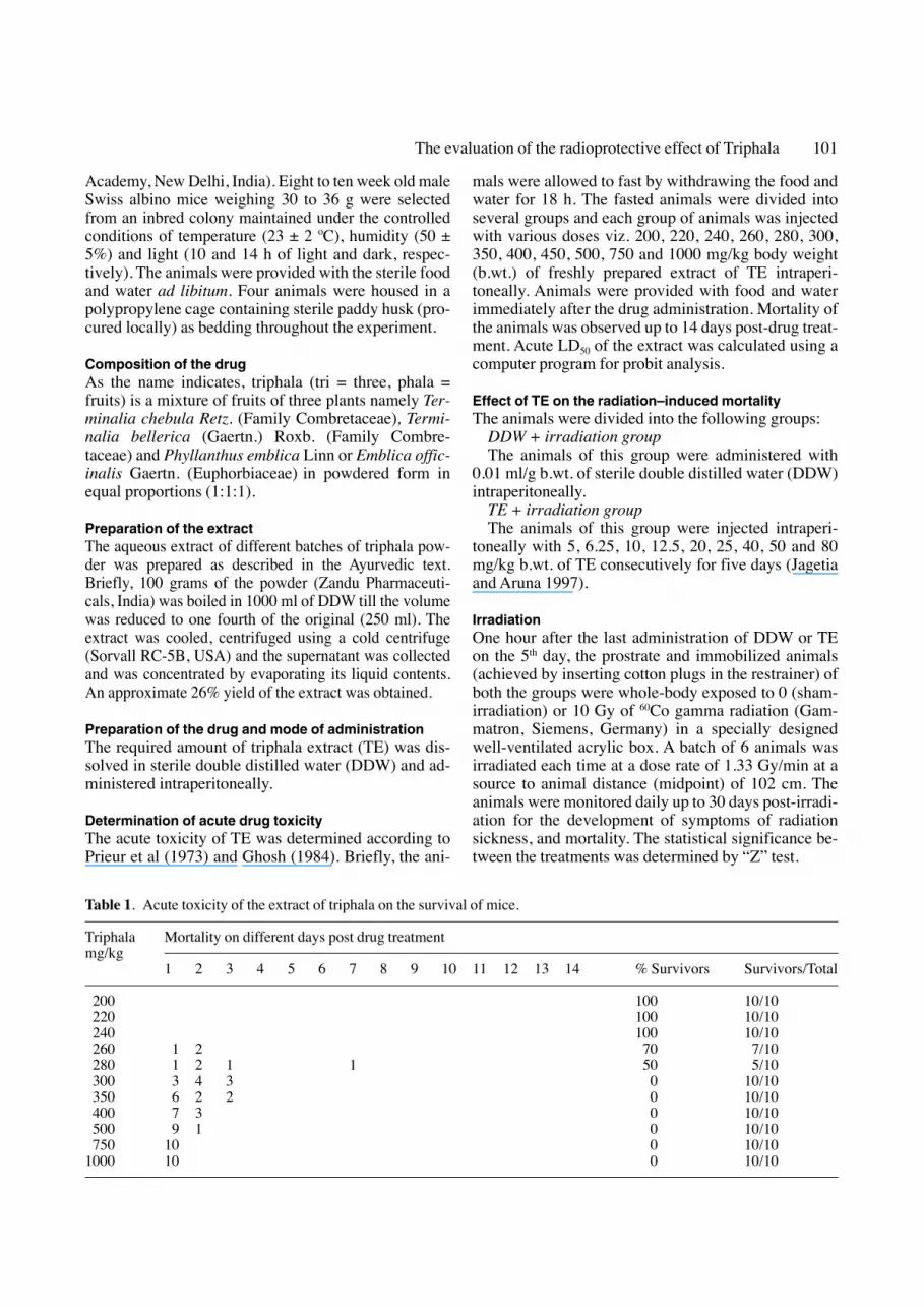
Table 1. Acute toxicity of the extract of triphala on the survival of mice.
Triphala Mortality on different days post drug treatmentmg/kg
1 2 3 4 5 6 7 8 9 10 11 12 13 14 % Survivors Survivors/Total
200 100 10/10220 100 10/10240 100 10/10260 1 2 70 7/10280 1 2 1 1 50 5/10300 3 4 3 0 10/10350 6 2 2 0 10/10400 7 3 0 10/10500 9 1 0 10/10750 10 0 10/10
1000 10 0 10/10
� Results
Acute Toxicity
The administration of different doses of TE viz. 200,220, and 240 mg/kg b.wt. did not induce any mortalityduring the whole observation period. However, a fur-ther increase in the drug dose up to 260 mg/kg b.wt. re-sulted in a 30% reduction in the survival of mice. Anincrease in the dose of TE up to 280 mg/kg b.wt.caused a 50% reduction in the survival of mice. 100%mortality was observed at 300 mg/kg and thereafter upto a dose of 1000 mg/kg b.wt. of TE (Table 1).
102 G. C. Jagetia et al.
Tabl
e 2.
Eff
ect o
f var
ious
dos
es o
f tr
ipha
la o
n th
e su
rviv
al o
f mic
e ex
pose
d to
10
Gy
of γ
-irr
adia
tion.
Tri
phal
aM
orta
lity
on d
iffe
rent
pos
t-ir
radi
atio
n da
ysN
o. o
fTo
tal
(mg/
kg)
Surv
ivor
s1
23
45
67
89
1011
1213
1415
1617
1819
2021
2223
2425
2627
2829
30
0–
––
33
41
22
51
1–
1–
–1
––
––
––
––
––
––
–1
255
––
––
––
–2
22
–1
–1
––
––
––
––
––
––
––
––
4a12
6.25
––
––
––
–1
–1
–1
––
––
–1
2–
––
–1
––
––
–5b
1210
––
––
––
––
2–
––
–1
1–
––
––
––
––
–1
––
––
7c12
12.5
––
––
––
––
1–
1–
1–
––
–1
––
––
––
1–
–1
––
6c12
20–
––
––
––
––
2–
11
––
11
–1
––
––
1–
––
––
–4a
1225
––
––
––
––
22
–1
–1
––
––
––
––
––
––
––
––
4a12
40–
––
––
12
11
11
––
11
––
––
––
––
––
––
––
–3
1250
––
–2
11
22
31
––
––
––
––
––
––
––
––
––
––
012
80–
–5
5–
1–
–1
––
––
––
––
––
––
––
––
––
––
–0
12
– P
< a
= 0
.02;
b =
0.0
1 an
d c
= 0
.001
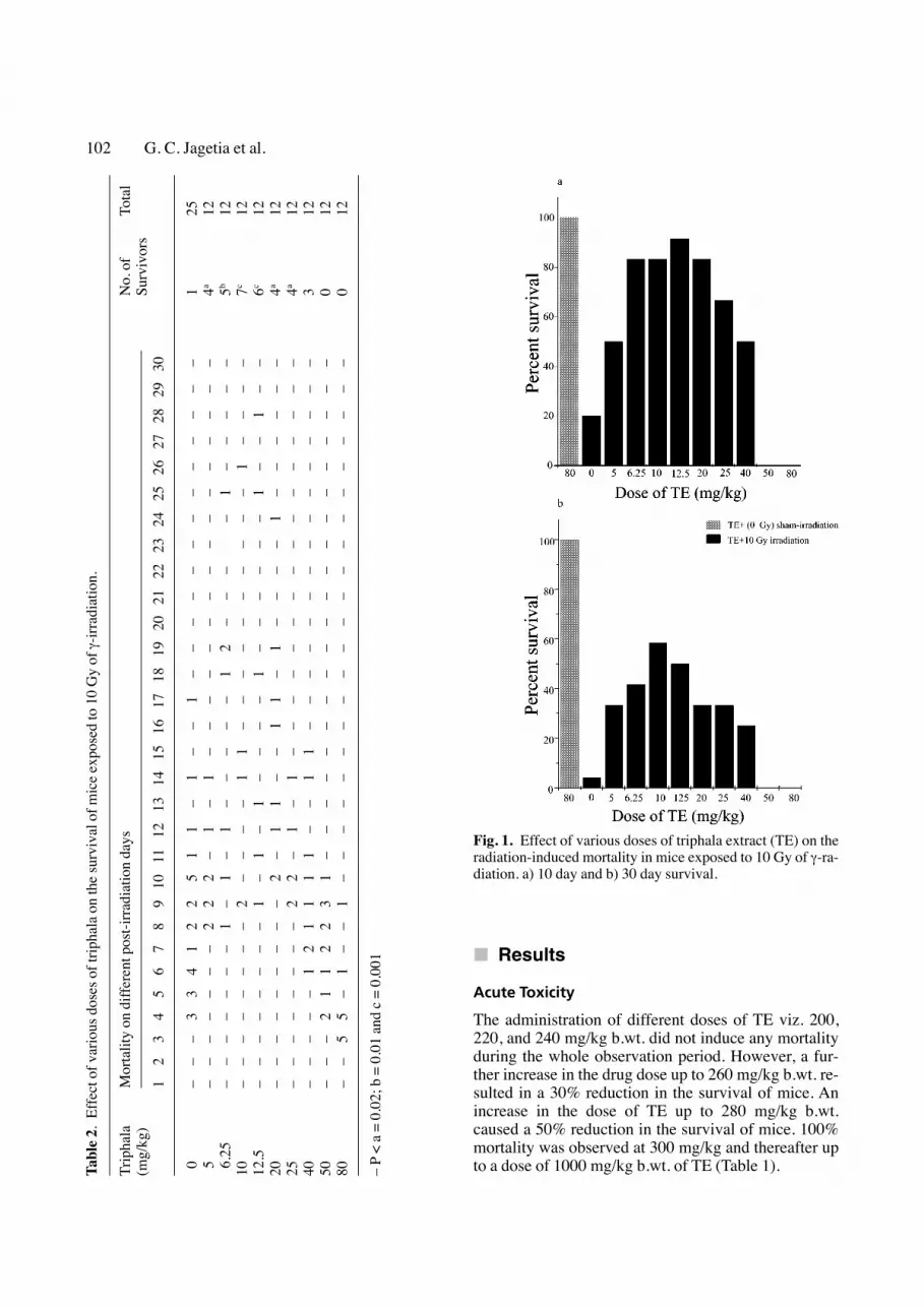
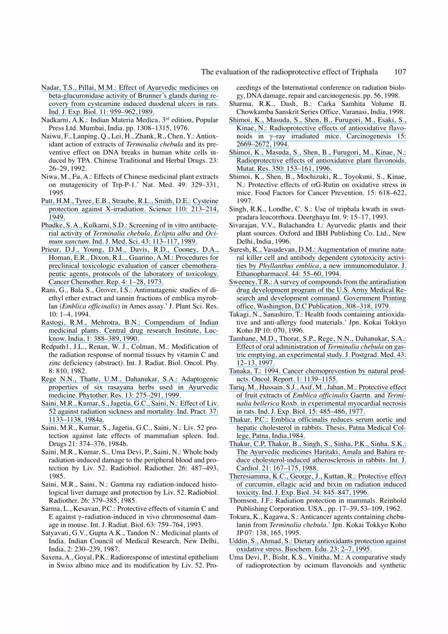
Fig. 1. Effect of various doses of triphala extract (TE) on theradiation-induced mortality in mice exposed to 10 Gy of γ-ra-diation. a) 10 day and b) 30 day survival.
mice against the radiation-induced mortality, however,the differences were statistically non-significant.
Analysis of thirty day survival revealed a drug dosedependent increase in the survival of irradiated mice upto a dose of 10 mg/kg in the TE+irradiation group,where a highest survival of 58.33% was observed ascompared to the DDW+irradiation, where only 4% ani-mals survived at the end of 30 days (Table 2). A furtherincrease in the drug dose to 12.5 mg/kg resulted in8.33% reduction in the survival, when compared withthe 10 mg/kg TE. Increase in TE doses further, resultedin a consistent decline in the animal survival reaching anadir at 50 mg and 80 mg/kg, where no survivors couldbe reported at the end of 30 days (Fig 1). TE adminis-tration before irradiation increased the survival signifi-cantly for 5 to 25 mg/kg (p < 0.02 to 0.001). However,the number of survivors was highest (58.33%) afterpretreatment of mice with 10 mg/kg of TE, and hence itwas considered the optimum dose for radioprotection.The optimum radioprotective dose of 10 mg/kg TE wasfound to be 1/28 of the LD50 dose (280 mg/kg b.wt.),which was far below the LD50 dose.
� Discussion
Ayurveda (in Sanskrit Ayu = life and veda = knowl-edge), the Indian system of medicine, dating back to5000 years has been an integral part of Indian cultureand materia medica. The Ayurveda, extensively uses theplant-derived compound formulations for the treatmentof various ailments after a careful study into the type ofthe disease (Sivarajan and Balachandra, 1996). Oftenthe drugs formulated are such that they have the desiredactivity with the adequate potency and are devoid of un-toward side effects. As it is observed that the desired ac-tivity is rarely present in adequate potency in a singleplant and it may also contain unwanted activities. There-fore, several plants with the common desired activitiesand varied undesirable activities are selected so that thefinal formulation will have a concentrated desired activ-ity and the undesired activities will be absent or diluted.Further, it is also observed that in such formulation, cer-tain other compound may be of help in enhancing of thepotency of the active compounds resulting in an additiveor synergistic positive effect, which may be of immensebenefit to the patient (Kulkarni, 1997). Keeping thisAyurveda philosophy in mind, triphala, an herbalrasayans preparation, credited with diverse beneficialproperties like anti-aging, antimutagenic, anticancer, an-tibacterial, anti-viral, cardioprotective, hepatoprotec-tive, anti-stress, cleanser of colon, gas distentioner, an-tidiabetic, antiparasitic, anti-diarrhea and antianemic(Nadkarni, 1976; Mehta et al., 1993; Ahmad et al., 1998;Phadke and Kulkarni, 1989; Niwa et al., 1995; Valsaraj
Effect of TE on the radiation–induced mortality
The mice were treated with different batches of 0, 5,6.25, 10, 12.5, 20, 25, 40, 50 and 80 mg/kg b.wt. oftriphala extract, consecutively for 5 days before whole-body exposure to 10 Gy gamma radiation or not weremonitored daily up to 30 days post-irradiation for thedevelopment of symptoms of radiation sickness, andmortality. The effect of different doses of TE on the ra-diation-induced mortality is shown in Table 2 and Fig 1and the results are expressed as percent survival.
The animals of DDW+irradiation group exhibitedsigns of radiation sickness within 2–4 days after expo-sure to 10 Gy of γ-radiation. The main symptoms in-cluded reduction in the food and water intake, irritabil-ity, epilation, weight loss, emaciation, lethargy, diar-rhea, and ruffling of hairs. Facial edema was also ob-served in a few animals between one and two weeksafter exposure. During the second week after exposurethere were a few cases of animals, exhibiting paralysisand difficulty in locomotion. The first mortality in thisgroup was observed on day 4 and 80% of the animalsdied within 10 days after irradiation, while 96% of theanimals died within 30 days of irradiation resultingonly in 4% survival by day 30 post-irradiation.
The daily administration of 5, 6.25, 10, 12.5, 20, 25,40, 50 and 80 mg/kg b.wt. TE for five consecutive daysdid not cause any drug-induced mortality. The pretreat-ment of mice with various doses of TE either delayedor reduced the severity of radiation sickness. The onsetof radiation-induced mortality was also delayed inTE+irradiation group when compared with theDDW+10 Gy irradiation group. The longest delay wasobserved for 20 mg/kg TE, where the first mortalitywas observed by day 10 post-irradiation (Table 2),while a shortest delay was observed for 80 mg/kg,where the first mortality occurred on day 3 post-irradi-ation.
Treatment of mice with various doses of TE also hadan ameliorating effect on the gastrointestinal tract asevidenced by an increase in the 10 day survival ofmice, where an increase of 4.58 fold was observed for12.5 mg/kg, 4.16 fold for 6.25, 10, and 20 mg/kg TE,3.33 for 25 mg/kg and 2.5 fold for 5 and 40 mg/kg TE(Fig. 1). Majority of the animals (80%) of DDW+ irra-diation group, died within 10 days after irradiation,while the TE pre-treatment increased the 10-day sur-vival significantly. A lowest mortality was observed inthe animals treated with 12.5 mg/kg TE before irradia-tion and this decline in mortality was significant (p <0.001). The other doses of TE also reduced the mortali-ty, in comparison with DDW+ irradiation, however, asignificant elevation in the 10-day survival was ob-served only for 6.25, 10, 12.5 and 20 mg/kg TE (p <0.001) treatment. 5 and 25 mg/kg of TE also protected
The evaluation of the radioprotective effect of Triphala 103

et al., 1997) has been selected for the evaluation of itsradioprotective activity in mice.
The animals of the irradiated group (DDW + irradia-tion) exhibited signs of radiation sickness within 2–4days after exposure to 10 Gy and the symptoms includ-ed reduction in the food and water intake, irritability,epilation, weight loss, emaciation, lethargy, diarrhea,and ruffling of hairs. The death of 80% of the animalsexposed to 10 Gy of radiation within 10 days is becauseof functional failure of the gastrointestinal tract (Bondet al., 1965; Uma Devi et al., 1999). The remaining 16%animals died within the next 20 days exhibitinghemopoietic syndrome and the charecteristic symptomslike, irritability, epilation, weight loss, emaciation,lethargy and ruffling of hairs (Bond et al., 1965; UmaDevi et al., 1999). It is a well-established fact that ioniz-ing radiation at cellular level can induce damage in thebiologically important macromolecules such as DNA,proteins, lipids and carbohydrates in the various organs.While some damage is expressed early others are ex-pressed over a period of time depending upon the cellkinetics and the radiation tolerance of the tissues. Likein chemotherapy, the effect of whole body irradiation ismainly felt by the highly proliferating germinal epithe-lium, gastrointestinal epithelium and the bone marrowprogenitor cells. Of these the germinal epithelium doesnot have a life supporting function to the exposed indi-vidual, while the gastrointestinal epithelium and thebone marrow progenitor cells are crucial for sustainingof life and any damage to these cells will impair the nor-mal physiological processes drastically. The gastroin-testinal epithelium is less sensitive than the bone mar-row progenitor cells but as the cell transit time is quick,it is expressed earlier than the hemopoetic syndrome(Bond et al., 1965). In mice death within 10 days post-irradiation is due to the gastrointestinal damage (UmaDevi et al., 1999). The bone marrow stem cells are moresensitive to radiation damage than the intestinal cryptbut, the peripheral blood cells have a longer transit timethan the intestinal cells and hence the gastrointestinalsyndrome appears earlier than the bone marrow syn-drome and in mice death due to irradiation from 11 to30 days is due to the hemopoetic damage inflicted byradiation (Bond et al., 1965; Uma Devi et al., 1999).
The pretreatment of mice with different doses of TEresulted in a dose dependent reduction in the radiation-induced mortality up to 10 mg/kg and a further increasein the drug dose resulted in the decline in the animalsurvival when compared with the 10 mg/kg TE. Theearlier studies on radioprotection have shown that anagent in test (for radioprotective action) acts only at aparticular dose range and above which it may not beprotective and some times can even be toxic (Thom-son, 1962; Yuhas and Storrer, 1969). The active princi-ple of Plumbago rosea, the plumbagin at pico to femto
gram range has been reported to stimulate the granulo-cytes in vitro, while at higher doses it had immunosup-pressive activity (Wagner et al., 1988). The reason maybe that after a particular concentration, a compound in-stead of being an anti-oxidant may act like a pro-oxi-dant inducing toxic symptoms resulting in the death.This is the reason that TE has optimum protection at 10mg/kg and the higher doses result in the decline in theprotective action of TE. The TE pretreatment providedprotection against the radiation sickness and mitigatedthe animal sufferings. Reports regarding the use of TEto protect against the radiation damage are unavailable,as this is probably the first report regarding the radio-protective action of TE. However, certain other herbalpreparations like Liv. 52, and abana have been reportedto protect the mice against the radiation-induced sick-ness, mortality, dermatitis, spleen injury (Saini et al.,1984 a, b) and radiation-induced chromosome damage(Jagetia and Ganapathi, 1989, 1991; Jagetia and Aruna,1997). The brahmarasayana, narasimharasayana, ash-wagandha-rasayana, and amrithaprasham, a group ofherbal preparations used to improve the general healthand debility, have been reported to reduce the radia-tion-induced lipid peroxidation in the liver, and leu-copenia in mice (Kumar et al., 1996).
The pattern of survival in TE+irradiation group wassimilar to that of the irradiated control group exceptthat the mortality was delayed. This clearly indicatesthe effectiveness of TE in arresting GI death, where thenumber of survivors for 5, 6.25, 10, 12.5, 20, 25 and 40mg/kg was higher than that of the irradiated control.The reduction in GI death may be due to the protectionof intestinal epithelium, which would have allowedproper absorption of the nutrients. Triphala has beenused as laxative to support the body’s vitality in manand it even stops diarrhea. Our findings support thecontention that triphala may protect the gastrointestinaltract epithelium against the toxic insult of radiation,protecting against the GI death in this study. It has beenreported that, Terminalia chebula, an important con-stituent of Triphala, mitigated the cysteamine-inducedduodenal ulcers in rats by increasing the beta-glu-curonidase activity in the Brunner’s glands (Nadar andPillai, 1989) and protected the epithelial cells againstthe cytopathic effects caused by influenza A virus(Badmaev et al., 2000). Another herbal drug Liv. 52has been reported to protect the intestinal epitheliumagainst the radiation-induced damage (Saxena andGoyal, 1998).
The pretreatment of mice with TE significantly re-duced the bone marrow deaths in the TE+irradiationgroup, especially at a dose of 5 to 25 mg/kg, where asignificant elevation in the survival has been observed.This increase in the 30 day survival may be owing tothe protection afforded by TE to the bone marrow stem
104 G. C. Jagetia et al.

cell compartment, which continued to supply the requi-site number of cells in the survivors. The bone marrowcells have been reported to be protected against the ra-diation-induced damage by various other plant formu-lations (Saini et al., 1984a, b; Jagetia and Ganapathi,1989, 1991; Jagetia and Aruna, 1997; Kumar et al.,1996). Further, triphala, and its constituents are report-ed to possess antimicrobial activity (Mehta et al., 1993;Dutta et al., 1998; Ahmad et al., 1998; Phadke andKulkarni, 1989; Valsaraj et al., 1997), which wouldhave also been responsible for the radioprotective ac-tion of TE. One of the constituents of triphala, the Phyl-lanthus emblica, has been found to be immunomodula-tor (Suresh and Vasudevan, 1994; Rege et al., 1999)and this would have increased the body’s defence sys-tem by increasing the immunity. Further the antimicro-bial action of triphala would have prevented the local-ization of the pathogenic microbes in the GI tract andbacterial infection, resulting in the observed radiopro-tection.
TE is mainly composed of Terminalia chebula, Phyl-lanthus emblica and Terminalia bellerica in equal pro-portions and each plant has been utilized to treat variousailments and diseases in the Ayurvedic system ofmedicine. Terminalia chebula is a commonly advocatedagent in Ayurveda for improving gastrointestinal motil-ity (Tamhane et al., 1997). The water and chloroformextracts of it have been shown to protect against thesodium azide and 4-nitro-o-phenylenediamine inducedmutagenesis (Grover and Bala, 1992). Recently, it hasalso been reported to possess antioxidant activity andprevent the TPA-induced DNA breaks in human whitecells (Naiwu et al., 1992). Similarly, Terminalia belleri-ca has been found to contain anti-HIV-1, antimalarial,and antifungal activity (Valsaraj et al., 1997). The alco-holic extract of this plant reduced the serum GOT, GPTand LDH activity, caused a significant reduction in fattyacid levels, and protected against the myocardial necro-sis (Tariq et al., 1977). Phyllanthus embelica has alsobeen found to be rich in ascorbic acid contents andascorbic acid has already been reported to reduce the ra-diation-induced sickness and mortality (Redpath et al.,1982) and to protect mice bone marrow cells against theradiation-induced chromosome damage (Sarma andKesavan, 1993). In addition to ascorbic acid, the com-ponents of triphala, Phyllanthus emblica, Terminaliachebula and Terminalia bellerica also contain ellagicacid, which has been reported to decrease the bone mar-row micronuclei formation in the mice (Thresiamma etal., 1996). Phyllanthus emblica has also been reportedto contain flavonoids (Jose and Kuttan, 1995), a class ofcompounds reported to be possess antioxidant and freeradical scavenging activities (Tanaka, 1994; Uddin andAhmad, 1995; Korina and Afanasev, 1997; Uma Devi etal., 2000). Certain flavanoids have been found to pro-
tect against the radiation-induced DNA damage (Shi-moi et al., 1994, 1996, 1997; Uma Devi et al., 1998) andmortality (Uma Devi et al., 1999). The aqueous, ace-tone and chloroform extracts of Emblica officinalefound to have antimutagenic effect (Grover and Kaur,1989).
The exact mechanism of action of TE is not known,however, it may scavenge free radicals produced by ra-diation and thus reduce the radiation-induced damageto the cellular DNA. The presence of ascorbic acid andthe flavonoids like quercetin may be responsible forthis action as these compounds are reported to protectthe DNA from radiation-induced micronuclei in mice(Sarma and Kesavan, 1993; Shimoi et al., 1997). Whiletesting NO (nitric oxide) scavenging activity of severalagents, triphala was found to scavenge the nitric oxideproduction in vitro (unpublished data). The aqueousextract of one of the constituents of TE, Phyllanthusemblica has been reported to be a potent inhibitor oflipid peroxidation formation, and scavenger of hydrox-yl and superoxide radicals in vitro (Jose and Kuttan,1995). Photochemical studies have shown that Termi-nalia bellerica contains bellericanin, ellagic acid, gal-lic acid, chebulagic acid, ethyl gallate and β-sitosterol.Terminalia chebula has been found to contain chebu-lin, terchebin, chebulagic acid, chebulinic acid, corila-gin, ellagitannin, ellagic acid, gallic acid, β-D-glco-gallin, and terchebin. The Phyllanthus emblica hasbeen reported to be a rich source of vitamin C and alsocontains terchebin, corilagin, tannins, ellagic acid,phyllembic acid, gallic acid and flavonoids in differentproportions depending on the season, type of climateand the plant processing (Chemexcil, 1992; Satyavatiet al., 1987; Wealth of India 1952, 1976; Rastogi andMehrotra, 1990; Jose and Kuttan, 1995). Most of thesecompounds have been reported to possess antioxidantand free radical scavenging activities (Tanaka, 1994;Uddin and Ahmad, 1995; Korina and Afanasev, 1997)and increase the antioxidant enzymes (Kong Ah-Ng etal., 2000). The presence of various antioxidant com-pounds in triphala might have been responsible for theobserved radioprotection by scavenging of free radi-cals generated by radiation exposure. Alternativelytriphala might have increased the intracellular level ofGSH, and stimulated the immune systems which couldhave provided protection against the radiation-inducedmortality.
� Conclusions
From our study it is clear that TE, a plant based formu-lation provided protection against the radiation-in-duced sickness and mortality and the optimum protec-tive dose of 10 mg/kg i.p. is far below the LD50 (280
The evaluation of the radioprotective effect of Triphala 105
mg/kg) dose. The exact mechanism of action of TE isnot known, however, it may scavenge free radicals pro-duced by radiation and thus inhibit radiation-induceddamage to the cellular DNA. We have observed scav-enging of NO (nitric oxide) radicals in vitro by TE (un-published data) and this testifies to our belief that oneof the mechanisms of radioprotection by triphala maybe owing to the scavenging of free radicals generatedby radiation exposure. Alternatively, it may also in-crease GSH levels and may reduce the radiation-in-duced lipid peroxidation. Since significant protectionis obtained at a very low non-toxic dose the extractmay have an advantage over the known radioprotectorsavailable so far. Studies are planned to explore the ap-plicability of triphala in cancer treatment by lookingfor the preferential protection to the normal tissues andits clinical applicability for cancer cure in the fractiona-tion regime. Since this formulation has been in use inIndia for the last 5000 years, the clinical acceptabilityshall not be a problem.
� Acknowledgements
We thank Prof. M. S. Vidyasagar, and Dr. J. Velumuru-gan, Department of Radiotherapy and Oncology, Kas-turba Medical College, Manipal, India for providingthe necessary irradiation facilities and help in radiationdosimetry.
� References
Ahmad, I., Mehmood, Z., Mohammad, F.: Screening of someIndian medicinal plants for their antimicrobial properties.J. Ethnopharmacol. 62: 183–193, 1998.
Anand, K., Singh, B.: ′3,4,5-trihydroxy benzoic acid (gallicacid), the hepatoprotective principle in the fruits of Termi-nalia belerica- Bioassay guided activity’. Pharmacol. Res.36: 315–321, 1997.
Antarkar, D.S., Vaidya, A.B., Doshi, J.C., Athavale, A.V.,Vinchoo, K.S., Natekar, M.R., Tathed, P.S., Ramesh, V.,Kale, N.: A double blind clinical trial of Arogya-Wardhanian Ayurvedic drug in acute viral hepatitis. Ind. J. Med. Res.72: 588–593, 1980.
Badmaev, V., Nowakowski, M.: Protection of epithelial cellsagainst influenza A virus by a plant derived biological re-sponse modifier Ledretan-96. Phytother. Res. 4: 245–249,2000.
Bond, V. P., Fliedner, T.M., Archambeau, J.O.: Mammalianradiation lethality Academic press New York. pp. 15–49,61–275, 1965.
Cairnie, A.: Adverse effects of radioprotector WR2721. Radi-at. Res. 94: 221–226, 1983.
Chemexcil.: Selected medicinal plants of India. Basic Chem-icals, Pharmaceutical and Cosmetic Export PromotionCouncil, Bombay 400 039, India. pp. 205–207, 1992.
Dutta, B.K, Rahman, I., Das, T.K.: Antifungal activity of In-dian plant extracts. Mycoses 41: 535–536, 1998.
El-Mekkawey, S., Meselhy, M.: ‘Inhibitory effects of Egyptianfolk medicines on human immunodeficiency virus (HIV) re-verse transcriptase’. Chem. Pharm. Bull. 43: 641–648, 1995.
Ganapathi, N.G., Jagetia, G.C.: Liv. .52 pretreatment inhibitsthe radiation-induced lipid peroxidation in mouse liver.Curr. Sci. 68: 601–603, 1995.
Ghosh, M.N.: Toxicity studies. In, Fundamentals of experi-mental pharmacology, edited by Ghosh MN ScientificBook Agency, Calcutta, India. 153–158, 1984.
Grover, I.S., Bala, S.: Antimutagenic activity of Terminaliachebula myroblan. in Salmonella typhimurium. Ind. J.Exptl. Biol. 30: 339–341, 1992.
Grover, I.S., Kaur, S.: Effect of Emblica officinalis Gaertn In-dian gooseberry fruit extract on sodium azide and 4-nitro-o-phenylamine induced mutagenesis in Salmonella ty-phimurium. Ind. J. Exptl. Biol. 27: 207–209,1989.
Gulati, R., Agarwal, S., Agarwal, S.S.: Hepatoprotectivestudies on Phyllanthus emblica and quercitin.’ Indian J.Exp. Biol. 33 (4): 261–268, 1995.
Hashimoto, M., Nakajima, Y.: Antiobesity agents, alpha-amylase inhibitors, lipase inhibitors, foods and beveragescontaining plant extracts’. Jpn. Kokai Tokkyo Koho JP 09:227, 398, 1997.
Hozumi, T., Oyama H.: Crude drugs for treating AIDS’. Jpn.Kokai Tokkyo Koho JP 09: 87, 185, 1997.
Hussain, S.J.: Screening of some unani cardiotonic drugs,D.U.M., Thesis, Aligarh Muslim University, Aligarh,India, 1975.
Jagetia, G.C., Aruna, R.: The herbal preparation abana pro-tects against radiation-induced micronuclei in the mousebone marrow. Mutat. Res. 393: 157–163, 1997.
Jagetia, G.C., Ganapathi, N.G.: Inhibition of clastogenic ef-fect of radiation by Liv. 52 in the bone marrow of mice.Mutat. Res. 224: 507–510, 1989.
Jagetia, G.C., Ganapathi, N.G.: Treatment of mice with aherbal preparation Liv. 52. reduces the frequency of radia-tion induced chromosome damage in bone marrow. Mutat.Res. 253: 123–126, 1991.
Jose, K., Kuttan, R.: Inhibition of oxygen free radicals byEmbelica officinales extract and Chyavanprash. AmalaRes. Bull. 15: 46–52, 1995.
Kong, Ah-Ng. T., Yu, R., Chen, C., Mandlekar, S., Primiano,T.: Signal transduction events elicited by natural products,Role of MAPK and caspase pathways in homeostatic re-sponse and induction of apoptosis. Arch. Pharm. Res. 23:1–16, 2000.
Korina, L.G., Afanasev, I.B.: Antioxidant and chelating prop-erties of flavonoids. Adv. Pharmacol. 38: 151–163, 1997.
Kulkarni R.D.: Principles of Pharmacology in Ayurveda.Ram Sangam Graphics, Mumbai, India, 1997.
Kumar, P.V., Kuttan, R., Kuttan, G.: Radioprotective effectsof Rasayanas. Ind. J. Exptl. Biol. 34: 848–850, 1996.
Kurokawa, M., Sato, H.: Effects of traditional herbalmedicines against herpes simplex virus (HSV) type 2 andacyclovir-resistant HSV type 1 in vitro and in vivo.’WakanIyakugaku Zasshi, 12: 187–194, 1995.
Mehta, B.K., Shitut, S., Wankhade, H.: In vitro antimicrobialefficacy of triphala. Fitoterapia 64: 371–372, 1993.
106 G. C. Jagetia et al.
Nadar, T.S., Pillai, M.M.: Effect of Ayurvedic medicines onbeta-glucuronidase activity of Brunner’s glands during re-covery from cysteamine induced duodenal ulcers in rats.Ind. J. Exp. Biol. 11: 959–962,1989.
Nadkarni, A.K.: Indian Materia Medica, 3rd edition, PopularPress Ltd. Mumbai, India. pp. 1308–1315, 1976.
Naiwu, F., Lanping, Q., Lei, H., Zhank, R., Chen, Y.: Antiox-idant action of extracts of Terminalia chebula and its pre-ventive effect on DNA breaks in human white cells in-duced by TPA. Chinese Traditional and Herbal Drugs. 23:26–29, 1992.
Niwa, M., Fu, A.: Effects of Chinese medicinal plant extractson mutagenicity of Trp-P-1.’ Nat. Med. 49: 329–331,1995.
Patt, H.M., Tyree, E.B., Straube, R.L., Smith, D.E.: Cysteineprotection against X-irradiation. Science 110: 213–214,1949.
Phadke, S. A., Kulkarni, S.D.: Screening of in vitro antibacte-rial activity of Terminalia chebula, Eclipta alba and Oci-mum sanctum. Ind. J. Med. Sci. 43: 113–117, 1989.
Prieur, D.J., Young, D.M., Davis, R.D., Cooney, D.A.,Homan, E.R., Dixon, R.L., Guarino, A.M.: Procedures forpreclinical toxicologic evaluation of cancer chemothera-peutic agents, protocols of the laboratory of toxicology.Cancer Chemother. Rep. 4: 1–28, 1973.
Rani, G., Bala S., Grover, I.S.: Antimutagenic studies of di-ethyl ether extract and tannin fractions of emblica myrob-lan (Emblica officinalis) in Ames assay.’ J. Plant Sci. Res.10: 1–4, 1994.
Rastogi, R.M., Mehrotra, B.N.: Compendium of Indianmedicinal plants. Central drug research Institute, Luc-know, India, 1: 388–389, 1990.
Redpath1, J.L., Renan, W. J., Colman, M.: Modification ofthe radiation response of normal tissues by vitamin C andzinc deficiency (abstract). Int. J. Radiat. Biol. Oncol. Phy.8: 810, 1982.
Rege N.N., Thatte, U.M., Dahanukar, S.A.: Adaptogenicproperties of six rasayana herbs used in Ayurvedicmedicine. Phytother. Res. 13: 275–291, 1999.
Saini, M.R., Kumar, S., Jagetia, G.C., Saini, N.: Effect of Liv.52 against radiation sickness and mortality. Ind. Pract. 37:1133–1138, 1984a.
Saini, M.R., Kumar, S., Jagetia, G.C., Saini, N.: Liv. 52 pro-tection against late effects of mammalian spleen. Ind.Drugs 21: 374–376, 1984b.
Saini, M.R., Kumar, S., Uma Devi, P., Saini, N.: Whole bodyradiation-induced damage to the peripheral blood and pro-tection by Liv. 52. Radiobiol. Radiother. 26: 487–493,1985.
Saini, M.R., Saini, N.: Gamma ray radiation-induced histo-logical liver damage and protection by Liv. 52. Radiobiol.Radiother. 26: 379–385, 1985.
Sarma, L., Kesavan, P.C.: Protective effects of vitamin C andE against γ–radiation-induced in vivo chromosomal dam-age in mouse. Int. J. Radiat. Biol. 63: 759–764, 1993.
Satyavati, G.V., Gupta A.K., Tandon N.: Medicinal plants ofIndia. Indian Council of Medical Research, New Delhi,India, 2: 230–239, 1987.
Saxena, A., Goyal, P.K.: Radioresponse of intestinal epitheliumin Swiss albino mice and its modification by Liv. 52. Pro-
ceedings of the International conference on radiation biolo-gy, DNA damage, repair and carcinogenesis. pp. 56, 1998.
Sharma, R.K., Dash, B.: Carka Samhita Volume II.Chowkamba Sanskrit Series Office, Varanasi, India, 1998.
Shimoi, K., Masuda, S., Shen, B., Furugori, M., Esaki, S.,Kinae, N.: Radioprotective effects of antioxidative flavo-noids in γ–ray irradiated mice. Carcinogenesis 15:2669–2672, 1994.
Shimoi, K., Masuda, S., Shen, B., Furugori, M., Kinae, N.:Radioprotective effects of antioxidative plant flavonoids.Mutat. Res. 350: 153–161, 1996.
Shimoi, K., Shen, B., Mochizuki, R., Toyokuni, S., Kinae,N.: Protective effects of αG-Rutin on oxidative stress inmice. Food Factors for Cancer Prevention. 15: 618–622,1997.
Singh, R.K., Londhe, C. S.: Use of triphala kwath in swet-pradara leucorrhoea. Deerghayu Int. 9: 15–17, 1993.
Sivarajan, V.V., Balachandra I.: Ayurvedic plants and theirplant sources. Oxford and IBH Publishing Co. Ltd., NewDelhi, India, 1996.
Suresh, K., Vasudevan, D.M.: Augmentation of murine natu-ral killer cell and antibody dependent cytotoxicity activi-ties by Phyllanthus emblica, a new immunomodulator. J.Ethanopharmacol. 44: 55–60, 1994.
Sweeney, T.R.: A survey of compounds from the antiradiationdrug development program of the U.S. Army Medical Re-search and development command. Government Printingoffice, Washington, D.C Publication, 308–318, 1979.
Takagi, N., Sanashiro, T.: Health foods containing antioxida-tive and anti-allergy food materials.’ Jpn. Kokai TokkyoKoho JP 10: 070, 1996.
Tamhane, M.D., Thorat, S.P., Rege, N.N., Dahanukar, S.A.:Effect of oral administration of Terminalia chebula on gas-tric emptying, an experimental study. J. Postgrad. Med. 43:12–13, 1997.
Tanaka, T.: 1994. Cancer chemoprevention by natural prod-ucts. Oncol. Report. 1: 1139–1155.
Tariq, M., Hussain, S.J., Asif, M., Jahan, M.: Protective effectof fruit extracts of Emblica officinalis Gaertn. and Termi-nalia bellerica Roxb. in experimental myocardial necrosisin rats. Ind. J. Exp. Biol. 15: 485–486, 1977.
Thakur, P.C.: Emblica officinalis reduces serum aortic andhepatic cholesterol in rabbits. Thesis, Patna Medical Col-lege, Patna, India,1984.
Thakur, C.P, Thakur, B., Singh, S., Sinha, P.K., Sinha, S.K.:The Ayurvedic medicines Haritaki, Amala and Bahira re-duce cholesterol-induced atherosclerosis in rabbits. Int. J.Cardiol. 21: 167–175, 1988.
Theresiamma, K.C., George, J., Kuttan, R.: Protective effectof curcumin, ellagic acid and bixin on radiation inducedtoxicity. Ind. J. Exp. Biol. 34: 845–847, 1996.
Thomson, J.F.: Radiation protection in mammals. ReinholdPublishing Corporation. USA., pp. 17–39, 53–109, 1962.
Tokura, K., Kagawa, S.: Anticancer agents containing chebu-lanin from Terminalia chebula.’ Jpn. Kokai Tokkyo KohoJP 07: 138, 165, 1995.
Uddin, S., Ahmad, S.: Dietary antioxidants protection againstoxidative stress. Biochem. Edu. 23: 2–7, 1995.
Uma Devi, P., Bisht, K.S., Vinitha, M.: A comparative studyof radioprotection by ocimum flavonoids and synthetic
The evaluation of the radioprotective effect of Triphala 107
aminothiol protectors in the mouse. Br. J. Radiol. 71:782–784, 1998.
Uma Devi, P., Ganasoundari, A., Rao, B.S.S., Srinivasan, K.K.: In vivo radioprotection by Ocimum flavonoids, sur-vival of mice. Radiat. Res. 151: 74–78, 1999.
Uma Devi, P., Ganasoundari, A., Vrinda, B., Srinivasan,K.K., Unnikrishnan, M.K.: Radiation protection by theocimum flavonoids orientin and vicenin: mechanisms ofaction. Radiat. Res.154: 455–460, 2000.
Vaidya, A.D.B., Pillai, D.M., Ramachandran, R., Ghaisis S,Pandita, N., Bhide, S.V.: Antioxidants in context of medic-inal plants. IN, Current perspectives on food antioxidantsin health, Krishnaswamy, P. R.: (ed.) Proceedings of Scien-tific meeting organized by the Brooke Bond and Health In-formation Centre, Bangalore, India, 15–21, 1998.
Valsaraj, R., Pushpangadan, P., Smitt, U.W., Adsersen, A.,Christensen, S.B., Sittie, A., Nyman, U., Nielsen, C.,Olsen, C.E.: New anti-HIV-1, antimalarial, and antifungalcompounds from Terminalia bellerica. J. Nat. Prod. 60:739–742, 1997.
Wagner, H., Kreher, B., Jurcic, K.: In vitro stimulation ofgranulocytes and lymphocytes by pico and fentogramquantities of cytostatic agents Arzneimittel Forschung.Drug Res. 38: 273–275, 1988.
Wagner, H.: Phytomedicine research in Germany. Environ.Health Perspect. 107: 779–781, 1999.
Wealth of India.: Raw materials. Council of Scientific and In-dustrial Research, New Delhi, India 3: 168–170, 1952.
Wealth of India.: Raw materials. Council of Scientific and In-dustrial Research, New Delhi, India 10: 164–178, 1976.
Yuhas, J. M.: Active and passive absorption kinetics as thebasis for selective protection of normal tissue by s-2-aminopropylamino ethylphosphorothioic acid. CancerRes. 40: 1519–1524, 1980.
Yuhas, J.M., Storer, J.B.: Chemoprotection against threemodes of radiation death in the mouse. Int. J. Radiat. Biol.15: 233–237, 1969.
Yukaka, T., Kurokawa, M., Sato, H., Yoshida, Y.: Treatmentof cytomegalovirus infections with traditional herbs.’ An-tiviral Res. 32 (2): 63–70, 1996.
� Address
Dr. Ganesh Chandra Jagetia, Department of Radiobiol-ogy, Kasturba Medical College, Manipal 576 119,India.Tel.: ++091-8252-71201 to 71300 ext. 22814;Fax: ++091-8252-70062/71927;e-mail: [email protected]
108 G. C. Jagetia et al.