role and structural characterization of plant aldehyde dehydrogenases from family 2 and family 7
TRANSCRIPT
Biochem. J. (2015) 468, 109–123 doi:10.1042/BJ20150009 109
Role and structural characterization of plant aldehyde dehydrogenases fromfamily 2 and family 7Radka Koncitıkova*†, Armelle Vigouroux‡, Martina Kopecna*, Tomas Andree†, Jan Bartos§, Marek Sebela*, Solange Morera‡1
and David Kopecny*1
*Department of Protein Biochemistry and Proteomics, Centre of the Region Hana for Biotechnological and Agricultural Research, Faculty of Science, Palacky University, Slechtitelu 11,Olomouc CZ-783 71, Czech Republic†Department of Biochemistry, Faculty of Science, Palacky University, Slechtitelu 11, Olomouc CZ-783 71, Czech Republic‡Laboratoire d’Enzymologie et Biochimie Structurales, CNRS, Avenue de la Terrasse, Gif-sur-Yvette 91198, France§Centre of Plant Structural and Functional Genomics, Centre of the Region Hana for Biotechnological and Agricultural Research, Institute of Experimental Botany, Slechtitelu 31,Olomouc CZ-78371, Czech Republic
Aldehyde dehydrogenases (ALDHs) are responsible for oxidationof biogenic aldehyde intermediates as well as for celldetoxification of aldehydes generated during lipid peroxidation.So far, 13 ALDH families have been described in plants.In the present study, we provide a detailed biochemicalcharacterization of plant ALDH2 and ALDH7 families byanalysing maize and pea ALDH7 (ZmALDH7 and PsALDH7)and four maize cytosolic ALDH(cALDH)2 isoforms RF2C,RF2D, RF2E and RF2F [the first maize ALDH2 was discoveredas a fertility restorer (RF2A)]. We report the crystal structuresof ZmALDH7, RF2C and RF2F at high resolution. TheZmALDH7 structure shows that the three conserved residuesGlu120, Arg300 and Thr302 in the ALDH7 family are located inthe substrate-binding site and are specific to this family. Ourkinetic analysis demonstrates that α-aminoadipic semialdehyde,a lysine catabolism intermediate, is the preferred substratefor plant ALDH7. In contrast, aromatic aldehydes including
benzaldehyde, anisaldehyde, cinnamaldehyde, coniferaldehydeand sinapaldehyde are the best substrates for cALDH2. In linewith these results, the crystal structures of RF2C and RF2F revealthat their substrate-binding sites are similar and are formed by anaromatic cluster mainly composed of phenylalanine residues andseveral nonpolar residues. Gene expression studies indicate thatthe RF2C gene, which is strongly expressed in all organs, appearsessential, suggesting that the crucial role of the enzyme wouldcertainly be linked to the cell wall formation using aldehydes fromphenylpropanoid pathway as substrates. Finally, plant ALDH7may significantly contribute to osmoprotection because it oxidizesseveral aminoaldehydes leading to products known as osmolytes.
Key words: aldehyde dehydrogenase 2 (ALDH2), aldehydedehydrogenase 7 (ALDH7), benzaldehyde, coniferaldehyde,cytokinin, fertility restorer.
INTRODUCTION
Aldehyde dehydrogenases (ALDHs) constitute a superfamilyof NAD(P)+ -dependent enzymes that catalyse irreversibleoxidation of aldehydes to the corresponding carboxylic acids.Aldehydes, which are highly reactive molecules, are toxic athigh concentrations. Therefore, ALDHs play a crucial role indetoxifying aldehydes produced by various metabolic pathways.They also play a role during adaptation to various stressconditions such as salinity, drought, heat and cold [1]. ALDHsare classified according to their sequence identity. Thosesharing more than 40 % sequence identity belong to the samefamily, whereas those with more than 60% form a subfamily
[2]. The superfamily of plant ALDHs currently contains 13distinct families: ALDH2, ALDH3, ALDH5, ALDH6, ALDH7,ALDH10, ALDH11, ALDH12, ALDH18, ALDH21, ALDH22,ALDH23 and ALDH24 [3]. Only ALDH2, 3, 5, 6, 7 and 18families possess mammalian orthologues.
The plant ALDH2 family (EC 1.2.1.-) comprises mitochondrial(mtALDH) and cytosolic (cALDH) isoforms split into ALDH2Band ALDH2C subfamilies respectively. They share ∼54%–63% amino-acid identity with human ALDH2 (hALDH2), whichbelongs to the ALDH2A subfamily. hALDH2 plays a role inethanol metabolism by catalysing the oxidation of ethanol-derived acetaldehyde to acetate [4–6]. Its crystal structure isknown (PDB 1CW3) [6]. The first maize (Zea mays) ADLH2
Abbreviations: ABAL, 4-aminobutyraldehyde; ALDH, aldehyde dehydrogenase; AMADH, aminoaldehyde dehydrogenase; APAL, 3-aminopropionaldehyde; AASAL, α-aminoadipate-semialdehyde; BAL, betaine aldehyde; cALDH, cytosolic aldehyde dehydrogenase; DAP, days afterpollination; GABA, γ-aminobutyric acid; GBAL, 4-guanidinobutyraldehyde; hALDH2, human ALDH2; LKR, lysine-ketoglutarate reductase; MASAL, adipicsemialdehyde methyl ester; mtALDH, mitochondrial aldehyde dehydrogenase; P6C, Δ1-piperideine-6-carboxylate; PCAL, pyridine carboxaldehyde;PsALDH, ALDH from Pisum sativum (pea); RF, fertility restorer; TMABAL, 4-(trimethylamino)butyraldehyde; SDH, saccharopine dehydrogenase; TMAPAL,3-(trimethylamino)propionaldehyde; ZmALDH, ALDH from Zea mays (maize).
1 Correspondence may be addressed to either of these authors (email [email protected] or [email protected]).Sequence data can be found in the EMBL/GenBank data libraries under accession numbers KJ004509 for ZmALDH7, KJ004510 for RF2A, KJ004511
for RF2B, KJ004512 for RF2C, KM225857 for RF2D, KM225858 for RF2E and KJ004513 for RF2F.The structure for ZmALDH2-3 (RF2C) complexed with NAD+ , ZmALDH2-6 (RF2F) complexed with NAD+ and ZmALDH7 complexed with NAD+ will
appear in the PDB under accession code 4PXL, 4PZ2 and 4PXN respectively.
c© The Authors Journal compilation c© 2015 Biochemical Society
Bio
chem
ical
Jo
urn
al
ww
w.b
ioch
emj.o
rgAut
hor C
opy
110 R. Koncitıkova and others
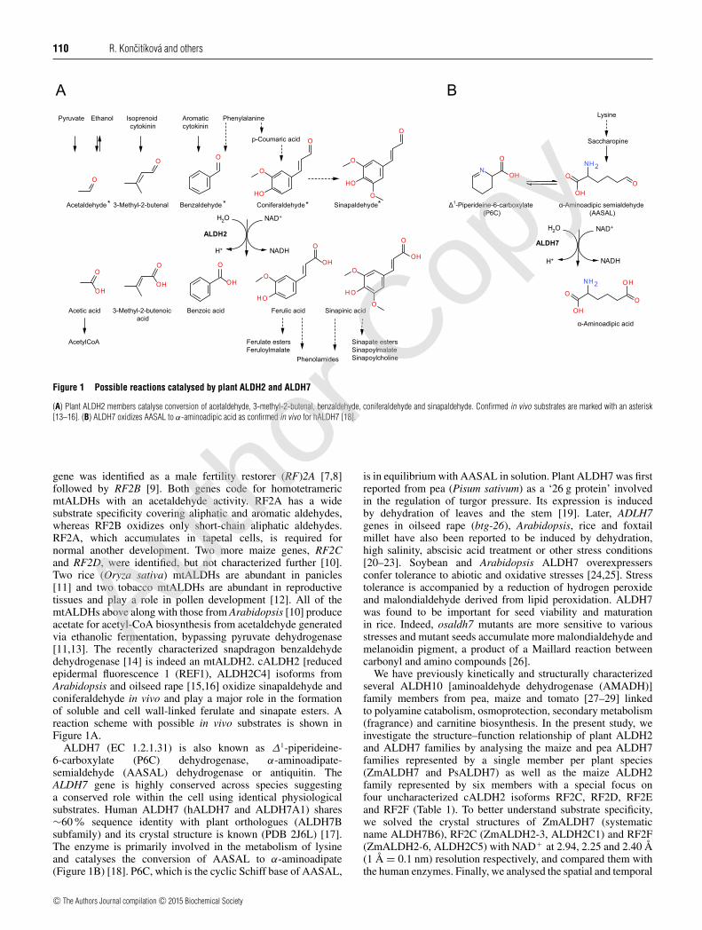
Figure 1 Possible reactions catalysed by plant ALDH2 and ALDH7
(A) Plant ALDH2 members catalyse conversion of acetaldehyde, 3-methyl-2-butenal, benzaldehyde, coniferaldehyde and sinapaldehyde. Confirmed in vivo substrates are marked with an asterisk[13–16]. (B) ALDH7 oxidizes AASAL to α-aminoadipic acid as confirmed in vivo for hALDH7 [18].
gene was identified as a male fertility restorer (RF)2A [7,8]followed by RF2B [9]. Both genes code for homotetramericmtALDHs with an acetaldehyde activity. RF2A has a widesubstrate specificity covering aliphatic and aromatic aldehydes,whereas RF2B oxidizes only short-chain aliphatic aldehydes.RF2A, which accumulates in tapetal cells, is required fornormal another development. Two more maize genes, RF2Cand RF2D, were identified, but not characterized further [10].Two rice (Oryza sativa) mtALDHs are abundant in panicles[11] and two tobacco mtALDHs are abundant in reproductivetissues and play a role in pollen development [12]. All of themtALDHs above along with those from Arabidopsis [10] produceacetate for acetyl-CoA biosynthesis from acetaldehyde generatedvia ethanolic fermentation, bypassing pyruvate dehydrogenase[11,13]. The recently characterized snapdragon benzaldehydedehydrogenase [14] is indeed an mtALDH2. cALDH2 [reducedepidermal fluorescence 1 (REF1), ALDH2C4] isoforms fromArabidopsis and oilseed rape [15,16] oxidize sinapaldehyde andconiferaldehyde in vivo and play a major role in the formationof soluble and cell wall-linked ferulate and sinapate esters. Areaction scheme with possible in vivo substrates is shown inFigure 1A.
ALDH7 (EC 1.2.1.31) is also known as Δ1-piperideine-6-carboxylate (P6C) dehydrogenase, α-aminoadipate-semialdehyde (AASAL) dehydrogenase or antiquitin. TheALDH7 gene is highly conserved across species suggestinga conserved role within the cell using identical physiologicalsubstrates. Human ALDH7 (hALDH7 and ALDH7A1) shares∼60% sequence identity with plant orthologues (ALDH7Bsubfamily) and its crystal structure is known (PDB 2J6L) [17].The enzyme is primarily involved in the metabolism of lysineand catalyses the conversion of AASAL to α-aminoadipate(Figure 1B) [18]. P6C, which is the cyclic Schiff base of AASAL,
is in equilibrium with AASAL in solution. Plant ALDH7 was firstreported from pea (Pisum sativum) as a ‘26 g protein’ involvedin the regulation of turgor pressure. Its expression is inducedby dehydration of leaves and the stem [19]. Later, ADLH7genes in oilseed rape (btg-26), Arabidopsis, rice and foxtailmillet have also been reported to be induced by dehydration,high salinity, abscisic acid treatment or other stress conditions[20–23]. Soybean and Arabidopsis ALDH7 overexpressersconfer tolerance to abiotic and oxidative stresses [24,25]. Stresstolerance is accompanied by a reduction of hydrogen peroxideand malondialdehyde derived from lipid peroxidation. ALDH7was found to be important for seed viability and maturationin rice. Indeed, osaldh7 mutants are more sensitive to variousstresses and mutant seeds accumulate more malondialdehyde andmelanoidin pigment, a product of a Maillard reaction betweencarbonyl and amino compounds [26].
We have previously kinetically and structurally characterizedseveral ALDH10 [aminoaldehyde dehydrogenase (AMADH)]family members from pea, maize and tomato [27–29] linkedto polyamine catabolism, osmoprotection, secondary metabolism(fragrance) and carnitine biosynthesis. In the present study, weinvestigate the structure–function relationship of plant ALDH2and ALDH7 families by analysing the maize and pea ALDH7families represented by a single member per plant species(ZmALDH7 and PsALDH7) as well as the maize ALDH2family represented by six members with a special focus onfour uncharacterized cALDH2 isoforms RF2C, RF2D, RF2Eand RF2F (Table 1). To better understand substrate specificity,we solved the crystal structures of ZmALDH7 (systematicname ALDH7B6), RF2C (ZmALDH2-3, ALDH2C1) and RF2F(ZmALDH2-6, ALDH2C5) with NAD+ at 2.94, 2.25 and 2.40 Å(1 Å = 0.1 nm) resolution respectively, and compared them withthe human enzymes. Finally, we analysed the spatial and temporal
c© The Authors Journal compilation c© 2015 Biochemical Society
Autho
r Cop
y
Plant ALDH2 and ALDH7 families 111
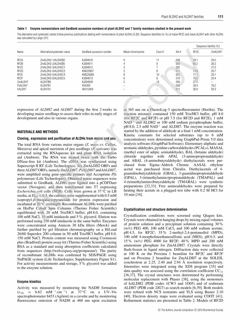
Table 1 Enzyme nomenclature and GenBank accession numbers of plant ALDH2 and 7 family members studied in the present work
The alternative and systematic names follow previous publications dealing with nomenclature of plant ALDHs [3,30]. Sequence identities (in %) of maize RF2C and maize ALDH7 with other ALDHswas calculated by Lalign [31].
Sequence identity (%)
Name Alternative/systematic name GenBank accession number Maize chromosome Exon # AA # RF2C ZmALDH7
RF2A ZmALDH2-1/ALDH2B2 KJ004510 9 11 549 59.1 29.5RF2B ZmALDH2-2/ALDH2B5 KJ004511 4 9 550 60.2 28.2RF2C ZmALDH2-3/ALDH2C1 KJ004512 3 7 502 – 27.5RF2D ZmALDH2-4/ALDH2C2 KM225857 3 8 511 73.8 29.7RF2E ZmALDH2-5/ALDH2C4 KM225858 8 10 501 71.1 29.1RF2F ZmALDH2-6/ALDH2C5 KJ004513 6 8 519 70.2 29.4ZmALDH7 ALDH7B6 KJ004509 2 14 509 27.5 –PsALDH7 ALDH7B1 X54359 – – 508 28.4 78.2hALDH7 ALDH7A1 AK312459 – – 511 30.0 59.3
expression of ALDH2 and ALDH7 during the first 2 weeks indeveloping maize seedlings to assess their roles in early stages ofdevelopment and also in various organs.
MATERIALS AND METHODS
Cloning, expression and purification of ALDHs from maize and pea
The total RNA from various maize organs (Z. mays cv. Cellux,Morseva) and apical meristem of pea seedlings (P. sativum) wasextracted using the RNAqueous kit and plant RNA isolationaid (Ambion). The RNA was treated twice with the TurboDNase-free kit (Ambion). The cDNA was synthesized usingSuperscript II RT (Life Technologies). Six ZmALDH2 ORFs andthree ALDH7 ORFs, namely ZmALDH7, PsALDH7 and hALDH7,were amplified using gene-specific primers and Accuprime Pfxpolymerase (Life Technologies). Obtained maize sequences weresubmitted to GenBank. ALDHs were ligated into a pCDFDuetvector (Novagen), and then transformed into T7 expressingEscherichia coli cells (NEB). Cells were grown at 37 C in LBmedia, at D600 = 0.5, the cultures were supplemented with 0.5 mMisopropyl-β-thiogalactopyranoside for protein expression andincubated at 20 C overnight. Recombinant ALDHs were purifiedon HisPur Cobalt Spin Columns (Thermo Fisher Scientific)equilibrated with 20 mM Tris/HCl buffer, pH 8.0, containing100 mM NaCl, 10 mM imidazole and 5% glycerol. Elution wasperformed using 250 mM imidazole in the same buffer. Enzymeswere concentrated using Amicon 30 kDa filters (Merck) andfurther purified by gel filtration chromatography on a HiLoad26/60 Superdex 200 column in 50 mM Tris/HCl buffer, pH 8.0,150 mM NaCl. Protein content was measured using Coomassieplus (Bradford) protein assay kit (Thermo Fisher Scientific) usingBSA as a standard and using absorption coefficients calculatedfrom sequences (http://web.expasy.org/protparam/). The purityof recombinant ALDHs was confirmed by SDS/PAGE usingNuPAGE system (Life Technologies; Supplementary Figure S1).For activity measurements and storage, 5% glycerol was addedto the enzyme solution.
Enzyme kinetics
Activity was measured by monitoring the NADH formation(ε340 = 6.62 mM− 1·cm− 1) at 37 C on a UV–Visspectrophotometer 8453 (Agilent) in a cuvette and by monitoringfluorescence emission of NADH at 460 nm upon excitation
at 365 nm on a FluoroLog-3 spectrofluorometer (Horiba). Thereaction mixtures contained 150 mM Tris/HCl buffer, pH 8.0(for RF2C and RF2F) or pH 7.5 (for RF2D and RF2E), 1 mMNAD+ and ALDH2 or 100 mM sodium pyrophosphate buffer,pH 8.0, 2.5 mM NAD+ and ALDH7. The enzyme reaction wasstarted by the addition of aldehyde at a final 1 mM concentration.Kinetic constants for selected substrates (up to 6 mMconcentration) were determined using GraphPad Prism 5.0 dataanalysis software (GraphPad Software). Elementary aliphatic andaromatic aldehydes, pyridine carboxaldehydes (PCALs), MASAL(methyl ester of adipic semialdehyde), BAL (betaine aldehyde)chloride together with APAL (3-aminopropionaldehyde)and ABAL (4-aminobutyraldehyde) diethylacetals were pur-chased from Sigma–Aldrich Chemie. AASAL ethyleneacetal was purchased from Chiralix. Diethylacetals of 4-guanidinobutyraldehyde (GBAL), 3-guanidinopropionaldehyde(GPAL), 3-(trimethylamino)propionaldehyde (TMAPAL) and4-(trimethylamino)butyraldehyde (TMABAL) were syntheticpreparations [32,33]. Free aminoaldehydes were prepared byheating their acetals in a plugged test tube with 0.2 M HCl for10 min [34].
Crystallization and structure determination
Crystallization conditions were screened using Qiagen kits.Crystals were obtained in hanging drops by mixing equal volumesof protein solution and a precipitant solution containing 30%(w/v) PEG 400, 100 mM CaCl2 and 100 mM sodium acetate,pH 4.5, for RF2C; 35% 2-methyl-2,4-pentanediol (MPD),100 mM 4-morpholineethanesulfonic acid (MES), pH 6.5, and15% (w/v) PEG 4000 for RF2F; 40 % MPD and 200 mMammonium phosphate for ZmALDH7. Crystals were directlyflash-frozen in liquid nitrogen. Diffraction data were collectedat 100 K on the Proxima 1 beamline for RF2C and RF2Fand on Proxima 2 beamline for ZmALDH7 at the SOLEILsynchrotron at 2.25, 2.40 and 2.94 Å resolution respectively.Intensities were integrated using the XDS program [35] anddata quality was assessed using the correlation coefficient CC1/2
[36,37]. The crystal structures were determined by performingmolecular replacement with Phaser [38], using the monomersof hALDH2 (PDB codes 1CW3 and 1O05) and of seabreamALDH7 (PDB code 2JG7) as search models [6,39]. Both modelswere refined with NCS restraints and TLS using Buster 2.10[40]. Electron density maps were evaluated using COOT [41].Refinement statistics are presented in Table 2. Models of RF2D
c© The Authors Journal compilation c© 2015 Biochemical Society
Autho
r Cop
y
112 R. Koncitıkova and others
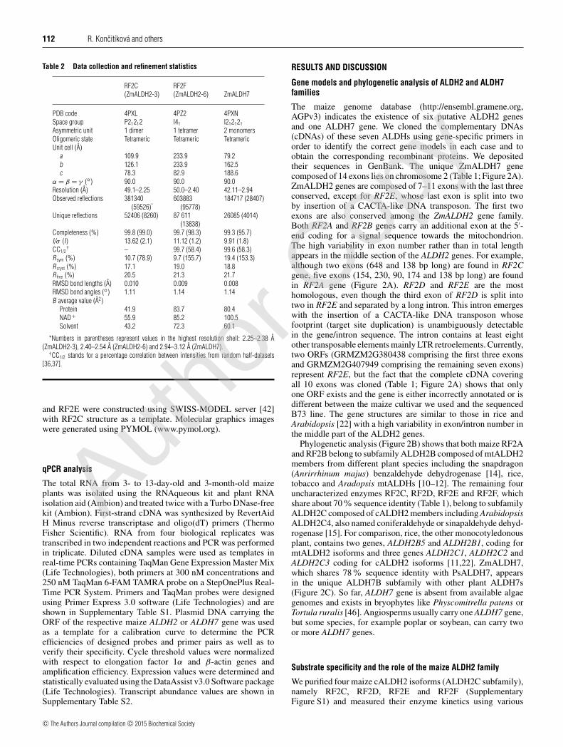
Table 2 Data collection and refinement statistics
RF2C(ZmALDH2-3)
RF2F(ZmALDH2-6) ZmALDH7
PDB code 4PXL 4PZ2 4PXNSpace group P21212 I41 I212121
Asymmetric unit 1 dimer 1 tetramer 2 monomersOligomeric state Tetrameric Tetrameric TetramericUnit cell (A)
a 109.9 233.9 79.2b 126.1 233.9 162.5c 78.3 82.9 188.6
α = β = γ () 90.0 90.0 90.0Resolution (A) 49.1–2.25 50.0–2.40 42.11–2.94Observed reflections 381340
(59526)*603883
(95778)184717 (28407)
Unique reflections 52406 (8260) 87 611(13838)
26085 (4014)
Completeness (%) 99.8 (99.0) 99.7 (98.3) 99.3 (95.7)I/σ (I) 13.62 (2.1) 11.12 (1.2) 9.91 (1.8)CC1/2
† – 99.7 (58.4) 99.6 (58.3)Rsym (%) 10.7 (78.9) 9.7 (155.7) 19.4 (153.3)Rcryst (%) 17.1 19.0 18.8Rfree (%) 20.5 21.3 21.7RMSD bond lengths (A) 0.010 0.009 0.008RMSD bond angles () 1.11 1.14 1.14B average value (A2)
Protein 41.9 83.7 80.4NAD + 55.9 85.2 100.5Solvent 43.2 72.3 60.1
*Numbers in parentheses represent values in the highest resolution shell: 2.25–2.38 A(ZmALDH2-3), 2.40–2.54 A (ZmALDH2-6) and 2.94–3.12 A (ZmALDH7).
†CC1/2 stands for a percentage correlation between intensities from random half-datasets[36,37].
and RF2E were constructed using SWISS-MODEL server [42]with RF2C structure as a template. Molecular graphics imageswere generated using PYMOL (www.pymol.org).
qPCR analysis
The total RNA from 3- to 13-day-old and 3-month-old maizeplants was isolated using the RNAqueous kit and plant RNAisolation aid (Ambion) and treated twice with a Turbo DNase-freekit (Ambion). First-strand cDNA was synthesized by RevertAidH Minus reverse transcriptase and oligo(dT) primers (ThermoFisher Scientific). RNA from four biological replicates wastranscribed in two independent reactions and PCR was performedin triplicate. Diluted cDNA samples were used as templates inreal-time PCRs containing TaqMan Gene Expression Master Mix(Life Technologies), both primers at 300 nM concentrations and250 nM TaqMan 6-FAM TAMRA probe on a StepOnePlus Real-Time PCR System. Primers and TaqMan probes were designedusing Primer Express 3.0 software (Life Technologies) and areshown in Supplementary Table S1. Plasmid DNA carrying theORF of the respective maize ALDH2 or ALDH7 gene was usedas a template for a calibration curve to determine the PCRefficiencies of designed probes and primer pairs as well as toverify their specificity. Cycle threshold values were normalizedwith respect to elongation factor 1α and β-actin genes andamplification efficiency. Expression values were determined andstatistically evaluated using the DataAssist v3.0 Software package(Life Technologies). Transcript abundance values are shown inSupplementary Table S2.
RESULTS AND DISCUSSION
Gene models and phylogenetic analysis of ALDH2 and ALDH7families
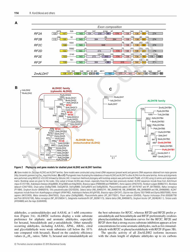
The maize genome database (http://ensembl.gramene.org,AGPv3) indicates the existence of six putative ALDH2 genesand one ALDH7 gene. We cloned the complementary DNAs(cDNAs) of these seven ALDHs using gene-specific primers inorder to identify the correct gene models in each case and toobtain the corresponding recombinant proteins. We depositedtheir sequences in GenBank. The unique ZmALDH7 genecomposed of 14 exons lies on chromosome 2 (Table 1; Figure 2A).ZmALDH2 genes are composed of 7–11 exons with the last threeconserved, except for RF2E, whose last exon is split into twoby insertion of a CACTA-like DNA transposon. The first twoexons are also conserved among the ZmALDH2 gene family.Both RF2A and RF2B genes carry an additional exon at the 5′-end coding for a signal sequence towards the mitochondrion.The high variability in exon number rather than in total lengthappears in the middle section of the ALDH2 genes. For example,although two exons (648 and 138 bp long) are found in RF2Cgene, five exons (154, 230, 90, 174 and 138 bp long) are foundin RF2A gene (Figure 2A). RF2D and RF2E are the mosthomologous, even though the third exon of RF2D is split intotwo in RF2E and separated by a long intron. This intron emergeswith the insertion of a CACTA-like DNA transposon whosefootprint (target site duplication) is unambiguously detectablein the gene/intron sequence. The intron contains at least eightother transposable elements mainly LTR retroelements. Currently,two ORFs (GRMZM2G380438 comprising the first three exonsand GRMZM2G407949 comprising the remaining seven exons)represent RF2E, but the fact that the complete cDNA coveringall 10 exons was cloned (Table 1; Figure 2A) shows that onlyone ORF exists and the gene is either incorrectly annotated or isdifferent between the maize cultivar we used and the sequencedB73 line. The gene structures are similar to those in rice andArabidopsis [22] with a high variability in exon/intron number inthe middle part of the ALDH2 genes.
Phylogenetic analysis (Figure 2B) shows that both maize RF2Aand RF2B belong to subfamily ALDH2B composed of mtALDH2members from different plant species including the snapdragon(Anrirrhinum majus) benzaldehyde dehydrogenase [14], rice,tobacco and Aradopsis mtALDHs [10–12]. The remaining fouruncharacterized enzymes RF2C, RF2D, RF2E and RF2F, whichshare about 70% sequence identity (Table 1), belong to subfamilyALDH2C composed of cALDH2 members including ArabidopsisALDH2C4, also named coniferaldehyde or sinapaldehyde dehyd-rogenase [15]. For comparison, rice, the other monocotyledonousplant, contains two genes, ALDH2B5 and ALDH2B1, coding formtALDH2 isoforms and three genes ALDH2C1, ALDH2C2 andALDH2C3 coding for cALDH2 isoforms [11,22]. ZmALDH7,which shares 78% sequence identity with PsALDH7, appearsin the unique ALDH7B subfamily with other plant ALDH7s(Figure 2C). So far, ALDH7 gene is absent from available algaegenomes and exists in bryophytes like Physcomitrella patens orTortula ruralis [46]. Angiosperms usually carry one ALDH7 gene,but some species, for example poplar or soybean, can carry twoor more ALDH7 genes.
Substrate specificity and the role of the maize ALDH2 family
We purified four maize cALDH2 isoforms (ALDH2C subfamily),namely RF2C, RF2D, RF2E and RF2F (SupplementaryFigure S1) and measured their enzyme kinetics using various
c© The Authors Journal compilation c© 2015 Biochemical Society
Autho
r Cop
y
PlantALDH2and
ALDH7fam
ilies113
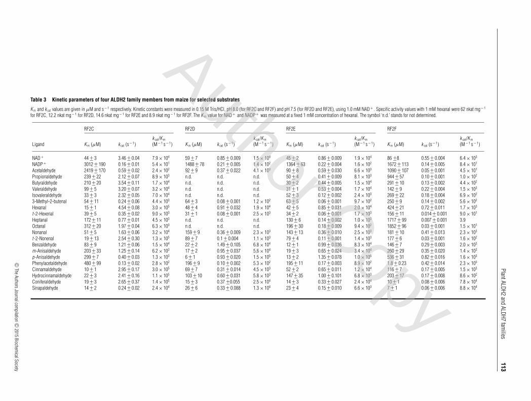
Table 3 Kinetic parameters of four ALDH2 family members from maize for selected substrates
K m and k cat values are given in μM and s− 1 respectively. Kinetic constants were measured in 0.15 M Tris/HCl, pH 8.0 (for RF2C and RF2F) and pH 7.5 (for RF2D and RF2E), using 1.0 mM NAD+ . Specific activity values with 1 mM hexanal were 62 nkat mg− 1
for RF2C, 12.2 nkat mg− 1 for RF2D, 14.6 nkat mg− 1 for RF2E and 8.9 nkat mg− 1 for RF2F. The K m value for NAD+ and NADP+ was measured at a fixed 1 mM concentration of hexanal. The symbol ‘n.d.’ stands for not determined.
RF2C RF2D RF2E RF2F
Ligand K m (μM) k cat (s− 1)k cat/K m
(M− 1 s− 1) K m (μM) k cat (s− 1)k cat/K m
(M− 1 s− 1) K m (μM) k cat (s− 1)k cat/K m
(M− 1 s− 1) K m (μM) k cat (s− 1)k cat/K m
(M− 1 s− 1)
NAD + 44 +− 3 3.46 +− 0.04 7.9 × 10⁴ 59 +− 7 0.85 +− 0.009 1.5 × 10⁴ 45 +− 2 0.86 +− 0.009 1.9 × 10⁴ 86 +−8 0.55 +− 0.004 6.4 × 10³
NADP+ 3012 +− 190 0.16 +− 0.01 5.4 × 101 1488 +− 78 0.21 +− 0.005 1.4 × 10² 1364 +− 63 0.22 +− 0.004 1.6 × 10² 1672 +− 113 0.14 +− 0.005 8.4 × 101
Acetaldehyde 2419 +− 170 0.59 +− 0.02 2.4 × 10² 92 +− 9 0.37 +− 0.022 4.1 × 10³ 90 +− 8 0.59 +− 0.030 6.6 × 10³ 1090 +− 107 0.05 +− 0.001 4.5 × 101
Propionaldehyde 239 +− 22 2.12 +− 0.07 8.9 × 10³ n.d. n.d. n.d. 50 +− 4 0.41 +− 0.009 8.1 × 10³ 944 +− 57 0.10 +− 0.001 1.0 × 10²
Butyraldehyde 210 +− 20 3.54 +− 0.11 1.7 × 10⁴ n.d. n.d. n.d. 30 +− 2 0.44 +− 0.005 1.5 × 10⁴ 291 +− 10 0.13 +− 0.002 4.4 × 10²
Valeraldehyde 99 +− 5 3.20 +− 0.07 3.2 × 10⁴ n.d. n.d. n.d. 31 +− 1 0.53 +− 0.004 1.7 × 10⁴ 142 +− 9 0.22 +− 0.004 1.5 × 10³
Isovaleraldehyde 33 +− 3 2.32 +− 0.05 7.0 × 10⁴ n.d. n.d. n.d. 52 +− 3 0.12 +− 0.002 2.4 × 10³ 269 +− 22 0.18 +− 0.004 6.9 × 10²
3-Methyl-2-butenal 54 +− 11 0.24 +− 0.06 4.4 × 10³ 64 +− 3 0.08 +− 0.001 1.2 × 10² 63 +− 5 0.06 +− 0.001 9.7 × 10² 250 +− 9 0.14 +− 0.002 5.6 × 10²
Hexanal 15 +− 1 4.54 +− 0.08 3.0 × 10⁵ 48 +− 4 0.91 +− 0.032 1.9 × 10⁴ 42 +− 5 0.85 +− 0.031 2.0 × 10⁴ 424 +− 21 0.72 +− 0.011 1.7 × 10³
t -2-Hexenal 39 +− 5 0.35 +− 0.02 9.0 × 10³ 31 +− 1 0.08 +− 0.001 2.5 × 10³ 34 +− 2 0.06 +− 0.001 1.7 × 10³ 156 +− 11 0.014 +− 0.001 9.0 × 101
Heptanal 172 +− 11 0.77 +− 0.01 4.5 × 10³ n.d. n.d. n.d. 130 +− 6 0.14 +− 0.002 1.0 × 10³ 1717 +− 99 0.007 +− 0.001 3.9Octanal 312 +− 20 1.97 +− 0.04 6.3 × 10³ n.d. n.d. n.d. 196 +− 30 0.18 +− 0.009 9.4 × 10² 1852 +− 96 0.03 +− 0.001 1.5 × 101
Nonanal 51 +− 5 1.63 +− 0.06 3.2 × 10⁴ 159 +− 9 0.36 +− 0.009 2.3 × 10³ 143 +− 13 0.36 +− 0.010 2.5 × 10³ 181 +− 10 0.41 +− 0.013 2.3 × 10³
t -2-Nonenal 19 +− 13 2.54 +− 0.30 1.3 × 10⁵ 89 +− 7 0.1 +− 0.004 1.1 × 10³ 79 +− 4 0.11 +− 0.001 1.4 × 10³ 177 +− 6 0.03 +− 0.001 1.6 × 10²
Benzaldehyde 83 +− 9 1.21 +− 0.06 1.5 × 10⁴ 22 +− 2 1.49 +− 0.105 6.8 × 10⁴ 12 +− 1 0.99 +− 0.036 8.3 × 10⁴ 146 +− 7 0.29 +− 0.003 2.0 × 10³
m-Anisaldehyde 203 +− 33 1.25 +− 0.14 6.2 × 10³ 17 +− 2 0.95 +− 0.037 5,6 × 10⁴ 19 +− 3 0.65 +− 0.024 3.4 × 10⁴ 260 +− 29 0.35 +− 0.020 1.4 × 10³
p-Anisaldehyde 299 +− 7 0.40 +− 0.03 1.3 × 10³ 6 +− 1 0.93 +− 0.020 1.5 × 10⁵ 13 +− 2 1.35 +− 0.078 1.0 × 10⁵ 536 +− 31 0.82 +− 0.016 1.6 × 10³
Phenylacetaldehyde 480 +− 99 0.13 +− 0.02 2.8 × 10² 196 +− 9 0.10 +− 0.002 5.3 × 10² 195 +− 11 0.17 +− 0.003 8.9 × 10² 1.8 +− 0.23 0.42 +− 0.014 2.3 × 10⁵
Cinnamaldehyde 10 +− 1 2.95 +− 0.17 3.0 × 10⁵ 69 +− 7 0.31 +− 0.014 4.5 × 10³ 52 +− 2 0.65 +− 0.011 1.2 × 10⁴ 116 +− 7 0.17 +− 0.005 1.5 × 10³
Hydrocinnamaldehyde 22 +− 3 2.41 +− 0.16 1.1 × 10⁵ 103 +− 10 0.60 +− 0.031 5.8 × 10³ 147 +− 35 1.00 +− 0.101 6.8 × 10³ 203 +− 17 0.17 +− 0.008 8.6 × 10²
Coniferaldehyde 19 +− 3 2.65 +− 0.37 1.4 × 10⁵ 15 +− 3 0.37 +−0.055 2.5 × 10⁴ 14 +− 3 0.33 +− 0.027 2.4 × 10⁴ 10 +− 1 0.08 +− 0.006 7.8 × 10⁴
Sinapaldehyde 14 +− 2 0.24 +− 0.02 2.4 × 10⁴ 26 +− 6 0.33 +− 0.088 1.3 × 10⁴ 23 +− 4 0.15 +− 0.010 6.6 × 10³ 7 +− 1 0.06 +− 0.006 8.8 × 10⁴
c ©TheAuthorsJournalcom
pilationc ©
2015Biochem
icalSociety
Author Copy
114 R. Koncitıkova and others
Figure 2 Phylogeny and gene models for studied plant ALDH2 and ALDH7 families
(A) Gene models for Zea mays ALDH2 and ALDH7 families. Gene models were constructed using cloned cDNA sequences (present work) and genomic DNA sequences obtained from maize genome(http://ensembl.gramene.org/Zea_mays/Info/Index). (B and C) Phylogenetic trees illustrating the relatedness of maize ALDH2 and ALDH7 to other ALDHs from the same families. Amino acid alignmentswere performed using MUSCLE v3.8 [43] followed by Gblocks [44]. A maximum likelihood phylogeny with bootstrap analysis was performed with PhyML v3.0 [45] using LG amino acid replacementmatrix. Bootstrap values are given for the nodes. Only subset of known ALDHs was chosen, especially those that have been previously studied. ALDH2 sequences include those from Antirrhinummajus (FJ151199), Arabidopsis thaliana (At3g48000, At1g23800 and At3g24503), Brassica napus (FN995990 and FN995991), Homo sapiens (AY621070), Hordeum vulgare (BAB62757), Nicotianatabacum (CAA71003), Oryza sativa (Os06g15990, Os02g49720, Os01g40860, Os01g40870 and Os06g39230), Physcomitrella patens (XP_001767457 and XP_001785650), Rattus norvegicus(P11884), Sorghum bicolor (BAB92019), Vitis pseudoreticulata (DQ150256), Setaria italica (XM_004953741, XM_004965148, XM_004965940, XM_004968994 and XM_004968990). ALDH7sequences include those from Acanthopagrus schlegelii (AY847462), Arabidopsis thaliana (At1g54100), Brassica napus (Q41247), Glycine max (Glyma.15G178400 and Glyma.09G070300), Homosapiens (AK312459), Malus domestica (BAA75633), Oryza sativa (Os09g26880), Physcomitrella patens (XP_001778351), Pisum sativum (X54359), Populus trichocarpa (Potri.003G067700and Potri.001G167100), Rattus norvegicus (NP_001258034.1), Selaginella moellendorffii (XP_002961173), Setaria italica (XM_004956875), Sorghum bicolor (XP_002462451.1), Tortula ruralis(AY034889) and Zea mays (KJ004509).
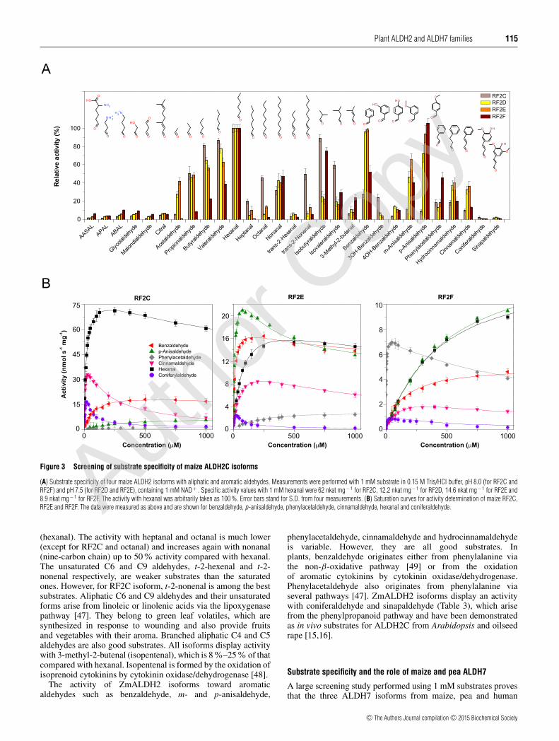
aldehydes, ω-aminoaldehydes and AASAL at 1 mM concentra-tion (Figure 3A). ALDH2C isoforms display a wide substratepreference for aliphatic and aromatic aldehydes, especiallyfor hexanal, benzaldehyde and p-anisaldehyde. Other naturallyoccurring aldehydes, including AASAL, APAL, ABAL, citraland glycolaldehyde were weak substrates (all below the 10%rate compared with hexanal). Based on the catalytic efficiencyvalues (kcat/Km ratios; Table 3), hexanal and cinnamaldehyde are
the best substrates for RF2C, whereas RF2D and RF2E prefer p-anisaldehyde and benzaldehyde and RF2F preferentially oxidizesphenylacetaldehyde. Saturation curves for the RF2C, RF2E andRF2F show that a strong excess substrate inhibition appears at lowconcentrations for some aromatic aldehydes, such as for cinnamal-dehyde with RF2C or phenylacetaldehyde with RF2F (Figure 3B).
The specific activity of all ZmALDH2 isoforms increaseswith the chain length of aliphatic aldehydes up to six carbons
c© The Authors Journal compilation c© 2015 Biochemical Society
Autho
r Cop
y
Plant ALDH2 and ALDH7 families 115
Figure 3 Screening of substrate specificity of maize ALDH2C isoforms
(A) Substrate specificity of four maize ALDH2 isoforms with aliphatic and aromatic aldehydes. Measurements were performed with 1 mM substrate in 0.15 M Tris/HCl buffer, pH 8.0 (for RF2C andRF2F) and pH 7.5 (for RF2D and RF2E), containing 1 mM NAD+ . Specific activity values with 1 mM hexanal were 62 nkat mg− 1 for RF2C, 12.2 nkat mg− 1 for RF2D, 14.6 nkat mg− 1 for RF2E and8.9 nkat mg − 1 for RF2F. The activity with hexanal was arbitrarily taken as 100 %. Error bars stand for S.D. from four measurements. (B) Saturation curves for activity determination of maize RF2C,RF2E and RF2F. The data were measured as above and are shown for benzaldehyde, p-anisaldehyde, phenylacetaldehyde, cinnamaldehyde, hexanal and coniferaldehyde.
(hexanal). The activity with heptanal and octanal is much lower(except for RF2C and octanal) and increases again with nonanal(nine-carbon chain) up to 50 % activity compared with hexanal.The unsaturated C6 and C9 aldehydes, t-2-hexenal and t-2-nonenal respectively, are weaker substrates than the saturatedones. However, for RF2C isoform, t-2-nonenal is among the bestsubstrates. Aliphatic C6 and C9 aldehydes and their unsaturatedforms arise from linoleic or linolenic acids via the lipoxygenasepathway [47]. They belong to green leaf volatiles, which aresynthesized in response to wounding and also provide fruitsand vegetables with their aroma. Branched aliphatic C4 and C5aldehydes are also good substrates. All isoforms display activitywith 3-methyl-2-butenal (isopentenal), which is 8 %–25% of thatcompared with hexanal. Isopentenal is formed by the oxidation ofisoprenoid cytokinins by cytokinin oxidase/dehydrogenase [48].
The activity of ZmALDH2 isoforms toward aromaticaldehydes such as benzaldehyde, m- and p-anisaldehyde,
phenylacetaldehyde, cinnamaldehyde and hydrocinnamaldehydeis variable. However, they are all good substrates. Inplants, benzaldehyde originates either from phenylalanine viathe non-β-oxidative pathway [49] or from the oxidationof aromatic cytokinins by cytokinin oxidase/dehydrogenase.Phenylacetaldehyde also originates from phenylalanine viaseveral pathways [47]. ZmALDH2 isoforms display an activitywith coniferaldehyde and sinapaldehyde (Table 3), which arisefrom the phenylpropanoid pathway and have been demonstratedas in vivo substrates for ALDH2C from Arabidopsis and oilseedrape [15,16].
Substrate specificity and the role of maize and pea ALDH7
A large screening study performed using 1 mM substrates provesthat the three ALDH7 isoforms from maize, pea and human
c© The Authors Journal compilation c© 2015 Biochemical Society
Autho
r Cop
y
116 R. Koncitıkova and others
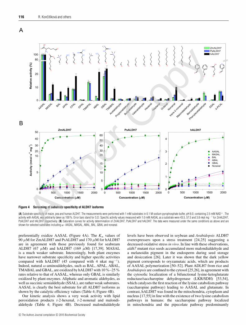
Figure 4 Screening of substrate specificity of ALDH7 isoforms
(A) Substrate specificity of maize, pea and human ALDH7. The measurements were performed with 1 mM substrates in 0.1 M sodium pyrophosphate buffer, pH 8.0, containing 2.5 mM NAD+ . Theactivity with AASAL was arbitrarily taken as 100 %. Error bars stand for S.D. Specific activity values measured with 1.0 mM AASAL as a substrate were 43.5, 57.5 and 3.8 nkat mg− 1 for ZmALDH7,PsALDH7 and hALDH7 respectively. (B) Saturation curves for activity determination of ZmALDH7, PsALDH7 and hALDH7. The data were measured under the same conditions as above and areshown for selected substrates including α- AASAL, MASAL, ABAL, BAL, GBAL and nonanal.
preferentially oxidize AASAL (Figure 4A). The Km values of90 μM for ZmALDH7 and PsALDH7 and 170 μM for hALDH7are in agreement with those previously found for seabreamALDH7 (67 μM) and hALDH7 (169 μM) [17,39]. MASALis a much weaker substrate. Interestingly, both plant enzymeshave narrower substrate specificity and higher specific activitiescompared with hALDH7 (45 compared with 4 nkat mg− 1).Indeed, natural ω-aminoaldehydes, such as BAL, APAL, ABAL,TMABAL and GBAL, are oxidized by hALDH7 with 10%–25%rates relative to that of AASAL, whereas only GBAL is similarlyoxidized by plant enzymes. Aliphatic and aromatic aldehydes, aswell as succinic semialdehyde (SSAL), are rather weak substrates.AASAL is clearly the best substrate for all ALDH7 isoforms asshown by the catalytic efficiency values (Table 4; Figure 4B).
Our kinetic analysis shows a very weak activity with lipidperoxidation products t-2-hexenal, t-2-nonenal and malondi-aldehyde (Table 4; Figure 4B). Decreased malondialdehyde
levels have been observed in soybean and Arabidopsis ALDH7overexpressers upon a stress treatment [24,25] suggesting adecreased oxidative stress in vivo. In line with these observations,aldh7 mutant rice seeds accumulated more malondialdehyde anda melanoidin pigment in the endosperm during seed storageand desiccation [26]. Later it was shown that the dark yellowpigment corresponds to oryzamutaic acids, which are productsof AASAL polymerization [50–52]. Plant ADLH7 from rice andArabidopsis are confined to the cytosol [25,26], in agreement withthe cytosolic localization of a bifunctional lysine-ketoglutaratereductase/saccharopine dehydrogenase (LKR/SDH) [53,54],which catalyses the first reaction of the lysine catabolism pathway(saccharopine pathway) leading to AASAL and glutamate. Incontrast, hALDH7 was found in the mitochondria, cytoplasm andnucleus [17,55] in line with the existence of two lysine catabolismpathways in humans: the saccharopine pathway localizedin mitochondria and the pipecolate pathway predominantly
c© The Authors Journal compilation c© 2015 Biochemical Society
Autho
r Cop
y
Plant ALDH2 and ALDH7 families 117
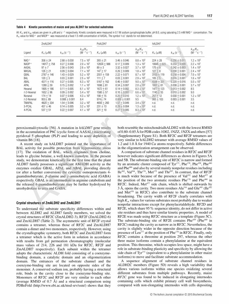
Table 4 Kinetic parameters of maize and pea ALDH7 for selected substrates
All K m and k cat values are given in μM and s− 1 respectively. Kinetic constants were measured in 0.1 M sodium pyrophosphate buffer, pH 8.0, using saturating 2.5 mM NAD+ concentration. TheK m value for NAD+ and NADP+ was measured at a fixed 1.5 mM concentration of AASAL. The symbol ‘n.d.’ stands for not determined.
ZmALDH7 PsALDH7 hALDH7
Ligand K m (μM) k cat (s− 1)k cat/K m
(M− 1 s− 1) K m (μM) k cat (s− 1)k cat/K m
(M− 1 s− 1) K m (μM) k cat (s− 1)k cat/K m
(M− 1 s− 1)
NAD + 358 +− 24 2.68 +− 0.030 7.5 × 10³ 393 +− 21 3.46 +− 0.046 8.8 × 10³ 224 +− 28 0.256 +− 0.011 1.2 × 10³
NADP+ 14017 +− 758 0.27 +− 0.008 2.0 × 101 12834 +− 988 0.17 +− 0.009 1.3 × 101 10645 +− 623 0.233 +− 0.009 2.2 × 101
AASAL 97 +− 7 2.56 +− 0.043 2.6 × 10⁴ 89 +− 3 3.32 +− 0.027 3.7 × 10⁴ 170 +− 9 0.242 +− 0.003 1.4 × 10³
MASAL 126 +− 21 0.09 +− 0.004 7.1 × 10² 147 +− 17 0.26 +− 0.009 1.8 × 10³ 127 +− 8 0.030 +− 0.001 2.4 × 10²
GBAL 2767 +− 146 1.43 +− 0.029 5.2 × 10² 2551 +− 159 2.22 +− 0.071 8.7 × 10² 3103 +− 119 0.218 +− 0.004 7.0 × 101
BAL 120 +− 3 0.03 +− 0.001 2.5 × 10² 111 +− 7 0.03 +− 0.001 2.9 × 10² 105 +− 5 0.016 +− 0.001 1.6 × 10²
ABAL 4371 +− 116 0.27 +− 0.005 6.3 × 101 5167 +− 162 0.46 +− 0.007 9.0 × 101 4339 +− 331 0.220 +− 0.016 5.0 × 101
APAL 1280 +− 59 0.15 +− 0.002 1.1 × 10² 1698 +− 81 0.34 +− 0.007 2.0 × 10² 1007 +− 81 0.038 +− 0.001 3.7 × 101
Hexanal 1805 +− 186 0.11 +− 0.005 6.1 × 101 1672 +− 61 0.14 +− 0.002 8.3 × 101 1477 +− 123 0.013 +− 0.002 8.5t -2-Hexenal 1652 +− 96 0.09 +− 0.002 5.4 × 101 1580 +− 57 0.10 +− 0.002 6.6 × 101 1145 +− 55 0.010 +− 0.002 9.0Nonanal 174 +− 14 0.07 +− 0.006 4.3 × 10² 198 +− 15 0.25 +− 0.010 1.2 × 10³ 257 +− 12 0.030 +− 0.001 1.2 × 10²
t -2-Nonenal 953+− 89 0.008 +− 0.001 8.4 974+− 74 0.009 +− 0.001 9.5 993 +− 103 0.003 +− 0.001 3.5TMAPAL 4820 +− 334 1.54 +− 0.096 3.2 × 10² 4656 +− 260 1.57 +− 0.049 3.4 × 10² n.d. n.d. n.d.3-PCAL 437 +− 46 0.14 +− 0.005 3.2 × 10² 231 +− 13 0.29 +− 0.004 1.3 × 10³ n.d. n.d. n.d.4-PCAL 537 +− 51 0.22 +− 0.007 4.1 × 10² 321 +− 10 0.27 +− 0.005 8.3 × 10² n.d. n.d. n.d.
peroxisomal/cytosolic [56]. A mutation in hALDH7 gene resultsin the accumulation of P6C (cyclic form of AASAL) inactivatingpyridoxal 5′-phosphate (PLP) and leading to acute depletion ofvitamin B6 [18].
A recent study on hALDH7 pointed out the importance ofBAL activity for possible protection from hyperosmotic stress[17]. The oxidation of BAL, which originates from choline,leads to glycine betaine, a well-known osmolyte. In the presentstudy, we demonstrate kinetically for the first time that the plantALDH7 family possesses a significant AMADH activity as theenzymes oxidize GBAL, APAL and ABAL providing directly(or after a further conversion) the cytosolic osmoprotectants 4-guanidinobutyrate, β-alanine and γ -aminobutyric acid (GABA)respectively. GBAL is an intermediate in arginine catabolism andthe released 4-guanidinobutyrate may be further hydrolysed byureohydrolase to urea and GABA.
Crystal structures of ZmALDH2 and ZmALDH7
To understand the substrate specificity differences within andbetween ALDH2 and ALDH7 family members, we solved thecrystal structures of RF2C (ZmALDH2-3), RF2F (ZmALDH2-6)and ZmALDH7 (Table 2). The asymmetric unit of RF2F crystalcontains a tetramer, whereas those of RF2C and ZmALDH7contain a dimer and two monomers, respectively. However, usingthe crystallographic symmetry, both RF2C and ZmALDH7 forma tetramer which is the active form in solution in accordancewith results from gel permeation chromatography (molecularmass values of 214, 226 and 181 kDa for RF2C, RF2F andZmALDH7 respectively). Each subunit within the tetrameradopts the characteristic ALDH fold consisting of a coenzyme-binding domain, a catalytic domain and an oligomerizationdomain. The entrances of the substrate channel and thecoenzyme-binding site are located on opposite sides of themonomer. A conserved sodium ion, probably having a structuralrole, binds in the cavity close to the coenzyme-binding site.Monomers of RF2C and RF2F are very similar to each other(average RMSD of 0.7 Å) and a structural comparison usingPDBeFold (http://www.ebi.ac.uk/msd-srv/ssm/) shows that they
both resemble the mitochondrialhALDH2 with the lowest RMSDof 0.80–0.85 Å for PDB codes 1O02, 1NZZ, 1NZX and others [57](Supplementary Figure S1). Both RF2C and RF2F tetramers arevery similar to hALDH2 tetramer with average RMSD values of1.2 and 1.0 Å for 1940 Cα atoms respectively. Subtle differencesin the oligomerization arrangement can be observed.
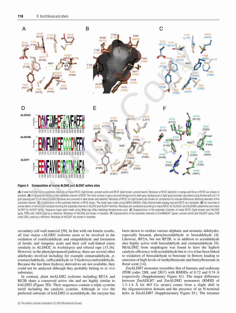
A comparison of substrate channel residues in RF2C and RF2Fhowever indicates significant differences as shown in Figures 5Aand 5B. The substrate-binding site of RF2C is narrow and formedby an aromatic cluster composed of Tyr171, Phe178, Phe458, Phe460
and Phe466 and also by several mainly nonpolar residues includingIle302, Val304, Thr174, Met175 and Thr297. In contrast, that of RF2Fis much wider because of the presence of Val192 and Met477 atthe position of the two aromatic residues Phe178 and Phe460 inRF2C. Indeed, Met477 side chain, which is shifted outwards by3 Å, opens the cavity. Two more residues Ala316 and Glu495 (Ile302
and Met478 in RF2C) also contribute to the substrate channelbroadening. The cavity width of RF2F clearly correlates withhigh Km values for various substrates most probably due to weakernonpolar interactions except for phenylacetaldehyde. RF2D andRF2E, which share 95% sequence identity, do not differ in activesite residues and thus have similar kinetic properties. A model ofRF2E was made using RF2C structure as a template (Figure 5C).The substrate-binding site of RF2E contains Phe459 (Phe460 inRF2C) making the cavity as narrow as in RF2C. Nevertheless, thecavity is slightly wider in the opposite direction because of thepresence of Leu177 at the position of Phe178 in RF2C. Finally, onlyRF2C contains a threonine at position 297, whereas the otherthree maize isoforms contain a phenylalanine at the equivalentposition. This threonine, which occupies less space, might have arole in substrate-binding plasticity and specificity by allowing theside chain of Tyr171 (equivalent to a phenylalanine in other maizeisoforms) to move and facilitate substrate accommodation.
A sequence alignment of substrate channel residues inALDH2C members (Figure 5D) reveals a high variability thatallows various isoforms within one species oxidizing severaldifferent substrates from multiple pathways. Recently, maizeRF2C gene was found to be induced in elongating internodescontaining cells which exhibit primary cell wall biosynthesis,compared with non-elongating internodes with cells depositing
c© The Authors Journal compilation c© 2015 Biochemical Society
Autho
r Cop
y
118 R. Koncitıkova and others
Figure 5 Composition of maize ALDH2 and ALDH7 active sites
(A) A view from the top on substrate channels of maize RF2C (light brown, present work) and RF2F (dark brown, present work). Residues of RF2C labelled in orange and those of RF2F are shown inbrackets. (B) A transversal section of the substrate channel of RF2F. The inner surface is grey coloured (foreground is dark grey, background is light grey) and was calculated using Hollow with a 0.7 Agrid spacing and 1.2 A interior probe. Residues are coloured in dark brown and labelled. Residues of RF2C (in light brown) are shown for comparison to indicate differences affecting diameter of thesubstrate channel. (C) Composition of the substrate channel in RF2E (blue). The model was made using SWISS-MODEL (http://swissmodel.expasy.org) and RF2C as a template. (D) An overview ofconservation of amino acid residues forming the substrate channel in ALDH2 and ALDH7 families. Residues are numbered according to maize RF2C for ALDH2C and ALDH2B subfamilies and maizeALDH7 for ALDH7 family. Sequence logos were made using WebLogo (http://weblogo.threeplusone.com). (E) Superposition of the substrate channels of maize RF2C (light brown) and hALDH2(grey, PDB code 1NZX) used as a reference. Residues of hALDH2 are shown in brackets. (F) Superposition of the substrate channels of ZmAMADH7 (green, present work) and hALDH7 (grey, PDBcode 2J6L) used as a reference. Residues of hALDH7 are shown in brackets.
secondary cell wall material [58]. In line with our kinetic results,all four maize cALDH2 isoforms seem to be involved in theoxidation of coniferaldehyde and sinapaldehyde and formationof ferulic and sinapinic acids and their cell wall-linked esterssimilarly to ALDH2C in Arabidopsis and oilseed rape [15,16].However, in the phenylpropanoid pathway, there are several otheraldehydes involved including for example cinnamaldehyde, p-coumaryladehyde, caffeyaldehyde or 5-hydroxyconiferaldehyde.Because the last three hydroxy derivatives are not available, theycould not be analysed although they probably belong to in vivosubstrates.
In contrast, plant mtALDH2 isoforms including RF2A andRF2B share a conserved active site and are highly similar tohALDH2 (Figure 5D). Their sequences contain a triple cysteinemotif including the catalytic cysteine. Although in vivo thepreferred substrate of hALDH2 is acetaldehyde, the enzyme has
been shown to oxidize various aliphatic and aromatic aldehydes,especially hexanal, phenylacetaldehyde or benzaldehyde [4].Likewise, RF2A, but not RF2B, is in addition to acetaldehydealso highly active with benzaldehyde and cinnamaldehyde [9].MtALDH2 from snapdragon was found to have the highestcatalytic efficiency with acetaldehyde but in vivo it has been linkedto oxidation of benzaldehyde to benzoate in flowers leading toemission of high levels of methylbenzoate and benzylbenzoate infloral scent [14].
ZmALDH7 monomer resembles that of humans and seabream(PDB codes 2J6L and 2JG7) with RMSDs of 0.72 and 0.74 Årespectively (Supplementary Figure S1). The major differencebetween ZmADLH7 and ZmALDH2 monomers (RMSD of1.3–1.4 Å for 465 Cα atoms) comes from a slight shift inthe oligomerization domain and the presence of an N-terminalhelix in ZmALDH7 (Supplementary Figure S1). The tetramer
c© The Authors Journal compilation c© 2015 Biochemical Society
Autho
r Cop
y
Plant ALDH2 and ALDH7 families 119
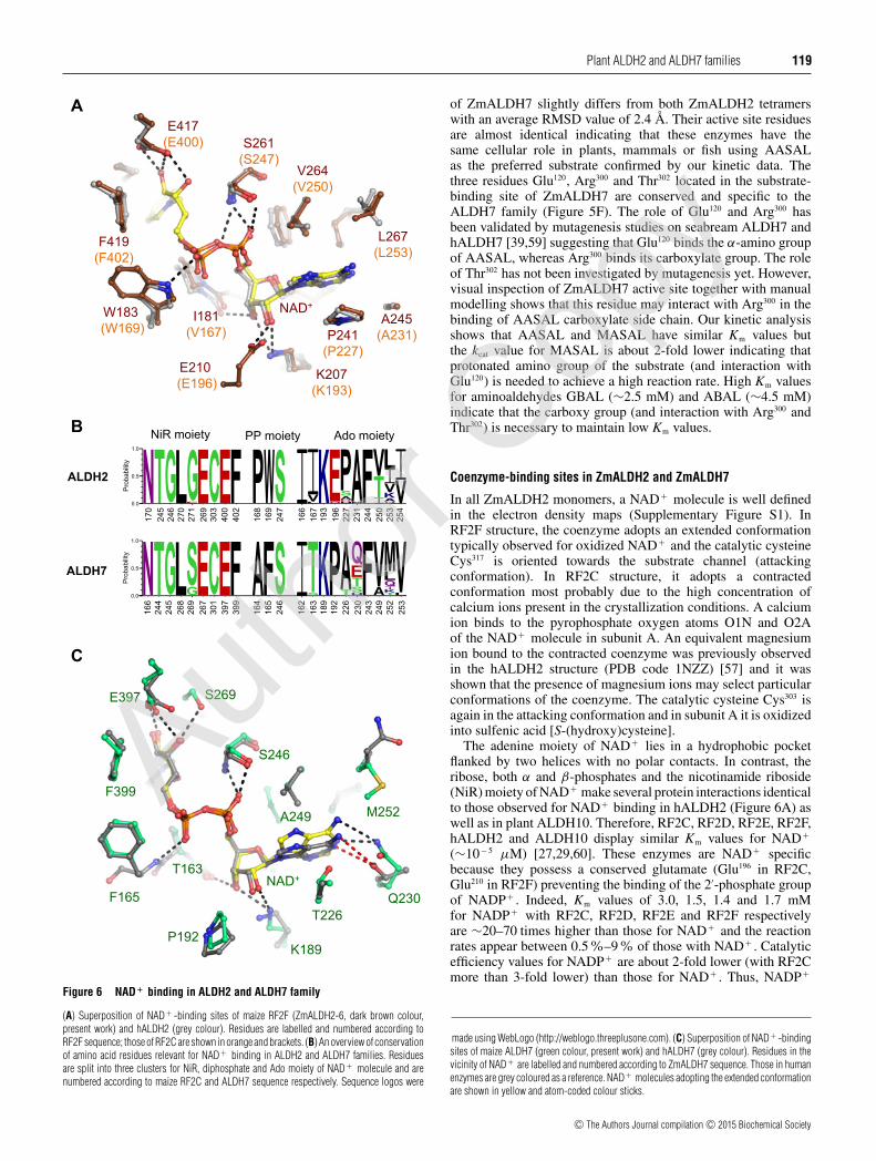
Figure 6 NAD+ binding in ALDH2 and ALDH7 family
(A) Superposition of NAD + -binding sites of maize RF2F (ZmALDH2-6, dark brown colour,present work) and hALDH2 (grey colour). Residues are labelled and numbered according toRF2F sequence; those of RF2C are shown in orange and brackets. (B) An overview of conservationof amino acid residues relevant for NAD+ binding in ALDH2 and ALDH7 families. Residuesare split into three clusters for NiR, diphosphate and Ado moiety of NAD + molecule and arenumbered according to maize RF2C and ALDH7 sequence respectively. Sequence logos were
of ZmALDH7 slightly differs from both ZmALDH2 tetramerswith an average RMSD value of 2.4 Å. Their active site residuesare almost identical indicating that these enzymes have thesame cellular role in plants, mammals or fish using AASALas the preferred substrate confirmed by our kinetic data. Thethree residues Glu120, Arg300 and Thr302 located in the substrate-binding site of ZmALDH7 are conserved and specific to theALDH7 family (Figure 5F). The role of Glu120 and Arg300 hasbeen validated by mutagenesis studies on seabream ALDH7 andhALDH7 [39,59] suggesting that Glu120 binds the α-amino groupof AASAL, whereas Arg300 binds its carboxylate group. The roleof Thr302 has not been investigated by mutagenesis yet. However,visual inspection of ZmALDH7 active site together with manualmodelling shows that this residue may interact with Arg300 in thebinding of AASAL carboxylate side chain. Our kinetic analysisshows that AASAL and MASAL have similar Km values butthe kcat value for MASAL is about 2-fold lower indicating thatprotonated amino group of the substrate (and interaction withGlu120) is needed to achieve a high reaction rate. High Km valuesfor aminoaldehydes GBAL (∼2.5 mM) and ABAL (∼4.5 mM)indicate that the carboxy group (and interaction with Arg300 andThr302) is necessary to maintain low Km values.
Coenzyme-binding sites in ZmALDH2 and ZmALDH7
In all ZmALDH2 monomers, a NAD+ molecule is well definedin the electron density maps (Supplementary Figure S1). InRF2F structure, the coenzyme adopts an extended conformationtypically observed for oxidized NAD+ and the catalytic cysteineCys317 is oriented towards the substrate channel (attackingconformation). In RF2C structure, it adopts a contractedconformation most probably due to the high concentration ofcalcium ions present in the crystallization conditions. A calciumion binds to the pyrophosphate oxygen atoms O1N and O2Aof the NAD+ molecule in subunit A. An equivalent magnesiumion bound to the contracted coenzyme was previously observedin the hALDH2 structure (PDB code 1NZZ) [57] and it wasshown that the presence of magnesium ions may select particularconformations of the coenzyme. The catalytic cysteine Cys303 isagain in the attacking conformation and in subunit A it is oxidizedinto sulfenic acid [S-(hydroxy)cysteine].
The adenine moiety of NAD+ lies in a hydrophobic pocketflanked by two helices with no polar contacts. In contrast, theribose, both α and β-phosphates and the nicotinamide riboside(NiR) moiety of NAD+ make several protein interactions identicalto those observed for NAD+ binding in hALDH2 (Figure 6A) aswell as in plant ALDH10. Therefore, RF2C, RF2D, RF2E, RF2F,hALDH2 and ALDH10 display similar Km values for NAD+
(∼10− 5 μM) [27,29,60]. These enzymes are NAD+ specificbecause they possess a conserved glutamate (Glu196 in RF2C,Glu210 in RF2F) preventing the binding of the 2′-phosphate groupof NADP+ . Indeed, Km values of 3.0, 1.5, 1.4 and 1.7 mMfor NADP+ with RF2C, RF2D, RF2E and RF2F respectivelyare ∼20–70 times higher than those for NAD+ and the reactionrates appear between 0.5 %–9% of those with NAD+ . Catalyticefficiency values for NADP+ are about 2-fold lower (with RF2Cmore than 3-fold lower) than those for NAD+ . Thus, NADP+
made using WebLogo (http://weblogo.threeplusone.com). (C) Superposition of NAD + -bindingsites of maize ALDH7 (green colour, present work) and hALDH7 (grey colour). Residues in thevicinity of NAD + are labelled and numbered according to ZmALDH7 sequence. Those in humanenzymes are grey coloured as a reference. NAD+ molecules adopting the extended conformationare shown in yellow and atom-coded colour sticks.
c© The Authors Journal compilation c© 2015 Biochemical Society
Autho
r Cop
y
120 R. Koncitıkova and others
does not function as an effective coenzyme in the plant ALDH2family. A recent mutagenesis study on this conserved glutamateresidue confirmed its importance for NAD+ specificity in the plantALDH3 family [61]. Figure 6B presents an overview of residuesforming the NAD+ -binding site and their frequency. Notably, theresidues involved in adenosine moiety (Ado) binding of NAD+
are highly variable.Although the coenzyme-binding site in ZmALDH7 slightly
differs in residue composition from that of ALDH2 family andof hALDH7, a NAD+ molecule present in each ZmALDH7monomer adopts the extended conformation with the catalyticCys301 in the attacking conformation and most of the importantprotein interactions conserved. Indeed, Phe165 in ZmALDH7equivalent to Phe167 in hALDH7 is bound to the β-phosphateoxygen atom via its main chain NH atom restoring the directinteraction between the side chain of the tryptophan residue andthe β-phosphate in both ALDH2 and ALDH10 families (Trp169
and Trp183 in RF2C and RF2F; Figure 6C). Ser269 in ZmALDH7(glycine in ZmALDH2) makes an additional interaction with theO3D atom of the NiR moiety whereas hALDH7 and ALDH2 en-zymes possess a glycine at the equivalent position. The conservedglutamate in ALDH2 (Glu196 in RF2C) and ALDH10, whichprevents binding of the 2′-phosphate group of NADP+ , is replacedby a proline in ALDH7 family (Pro192 in ZmALDH7). This prolinealso makes a steric hindrance to 2′-phosphate of NADP+ asreflected in the Km value of 14 mM (40 times higher than that forNAD+ ) and the reaction rate, which is only 0.5% of that measuredwith NAD+ and ZmALDH7. The major difference betweenZmALDH7 and hALDH7 or ALDH2 concerns the adenineposition of NAD+ . Indeed, the adenine adopts a different positionin ZmALDH7 because of the presence of Thr226 side chain (alaninein hALDH7 and proline in ALDH2), which pushes the adeninering up to 1 Å towards Ala249 (Figure 6C). The presence of thecouple Tre226–Ala249 appears only in a small subgroup of monocotsincluding maize, whereas most ALDH7 family members carry aconserved alanine–valine couple (Ala–Val250 in hALDH7) at thecorresponding positions. In ALDH7 family, adenine makes onepolar interaction between its N1 atom and a glutamate, glutamine,serine, threonine or histidine. All the above differences contributeby one order of magnitude to the higher Km values for NAD+
of ZmALDH7 and PsALDH7 compared with those of ALDH2and ALDH10 family members. Consequently, a higher saturatingconcentration of NAD+ was used for kinetic measurements.
Expression of ALDH2 and ALDH7 genes in maize
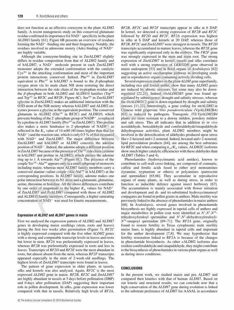
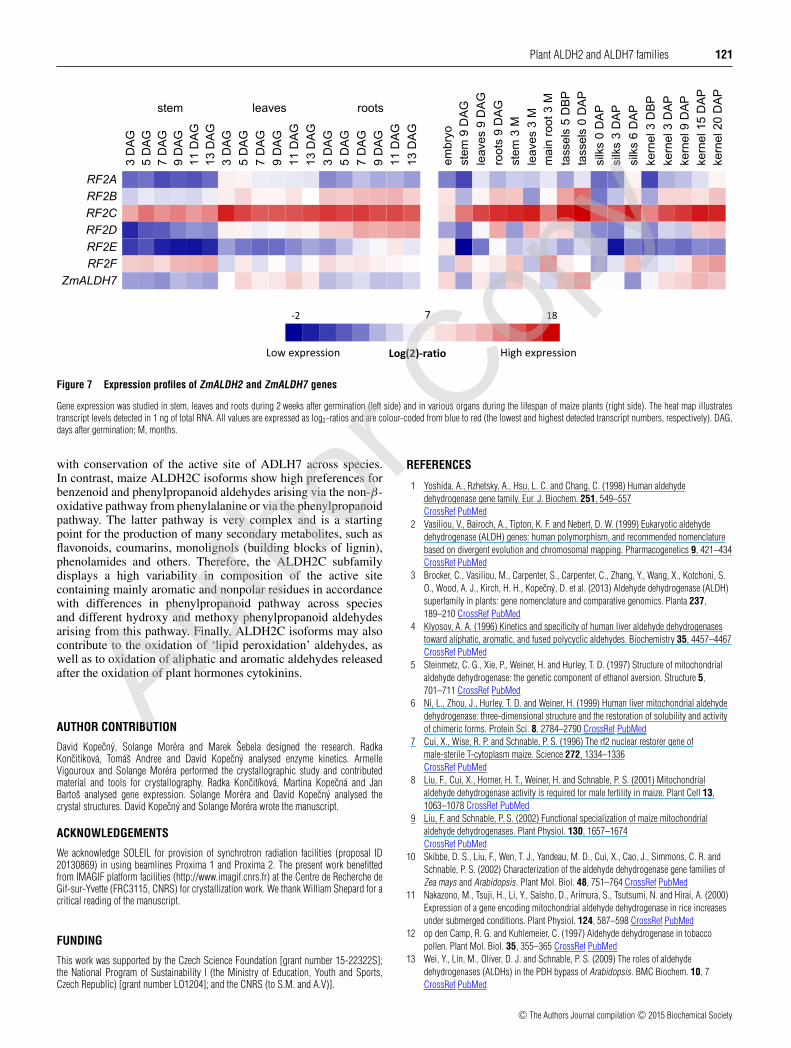
First we analysed the expression pattern of ALDH2 and ALDH7genes in developing maize seedlings (stem, roots and leaves)during the first two weeks after germination (Figure 7). RF2Cis highly expressed compared with the five other ALDH2 geneswith a strong and comparable transcript levels in leaves and rootsbut lower in stem. RF2A was preferentially expressed in leaves,whereas RF2B was preferentially expressed in roots and less inleaves. Transcripts of RF2D and RF2E were the most abundant inroots, but almost absent from the stem, whereas RF2F transcriptsappeared especially in the stem of 2-week-old seedlings. Thehighest levels of ZmALDH7 transcripts were found in leaves.
The pattern of gene expression in older plants, in tassels,silks and kernels was also analysed. Again, RF2C is the mostexpressed ALDH2 gene in maize. RF2B, RF2C and ZmALDH7are highly abundant in tassels at 5 days before pollination (DBP)and 0 days after pollination (DAP) suggesting their importantrole in pollen development. In silks, gene expression was lowercompared with that in tassels. Relatively high levels of RF2A,
RF2B, RF2C and RF2F transcripts appear in silks at 6 DAP.In kernel, we detected a strong expression of RF2B and RF2Cfollowed by RF2D and RF2F. RF2A expression was highestin silks at 6 DAP and kernels at 20 DAP, whereas those ofRF2B, RF2C and ZmALDH7 were strongest in tassels. The RF2Dtranscripts accumulated in mature leaves, whereas the RF2E genewas significantly expressed only in the embryo. The FR2F genewas strongly expressed in the stem and main root. The strongexpression of ZmALDH7 in kernel, tassels and silks correlateswell with a strong expression of LKR/SDH gene observed inmaize endosperm [53] and in floral organs of Arabidopsis [62]suggesting an active saccharopine pathway in developing seedsand in reproductive organs containing actively dividing cells.
Several expression studies on the plant ALDH gene superfamily,including rice and foxtail millet, show that many ALDH2 genesare induced by abiotic stressors, but some may also be down-regulated [22,23]. Indeed, OsALDH2B5 gene was found up-regulated by submergence, drought and salinity stresses, whereasthe OsALDH2C2 gene is down-regulated by drought and salinitystresses [11,22]. Interestingly, a gene coding for mtALDH2 inChinese wild grapevine Vitis pseudoreticulata (VpALDH2B4)[63] is induced by pathogens. Transgenic 35S:VpALDH2B4plants are more resistant to a downy mildew, powdery mildewand salt stress. This all indicates that in addition to in vivoacetaldehyde, benzaldehyde and coniferaldehyde/sinapaldehydedehydrogenase activities, plant ALDH2 members might beinvolved in the detoxification of aldehydes produced upon stresslike t-2-hexenal and t-2-nonenal. Indeed, hexanal and t-2-nonenal,lipid peroxidation products [64], are among the best substratesfor RF2C and when comparing kcat/Km values, ALDH2C isoformsshow much higher catalytic efficiencies with these aldehydes thanALDH7 (Tables 3 and 4).
Phenolamides (hydroxycinnamic acid amides), known tocontribute to cell-wall cross-linking, are composed of coumaric,caffeic and ferulic acids fused to either aryl monoamines(tyramine, tryptamine or others) or polyamines (putrescineand spermidine) [65,66]. They accumulate in reproductiveorgans of many plants, as well as during stress, in order tofunction as inducible defence against insect herbivory [67].The accumulation is mainly associated with flower initiationand development and di- and tri-substituted hydroxycinnamoylconjugates are found in pollen grains in anthers. Male sterility waspreviously linked to the absence of phenolamides in maize anthers[68]. In Arabidopsis, several genes involved in phenolamidebiosynthesis are highly expressed in tapetal cells of anthers andmajor metabolites in pollen coat were identified as N1,N5,N10-trihydroxyferuloyl spermidine and N1,N5-di(hydroxyferuloyl)-N10-sinapoyl spermidine [69–71]. The RF2A gene, originallyfound to restore fertility to Texas cytoplasmic male sterilitymaize lines, is highly abundant in tapetal cells and importantfor the anther development [7,8]. We may hypothesize thatfertility restoration linked to RF2A is because of the changesin phenolamide biosynthesis. As other cALDH2 isoforms alsooxidize coniferaldehyde and sinapaldehyde, they might contributeto the biosynthesis of phenolamides in various plant organs as wellas during stress conditions.
CONCLUSIONS
In the present work, we studied maize and pea ALDH7 andcompared their kinetics with that of human ALDH7. Based onour kinetic and structural results, we can conclude now that ahigh conservation of the ALDH7 gene during evolution is linkedto the substrate preference of ALDH7 for AASAL in accordance
c© The Authors Journal compilation c© 2015 Biochemical Society
Autho
r Cop
y
Plant ALDH2 and ALDH7 families 121
Figure 7 Expression profiles of ZmALDH2 and ZmALDH7 genes
Gene expression was studied in stem, leaves and roots during 2 weeks after germination (left side) and in various organs during the lifespan of maize plants (right side). The heat map illustratestranscript levels detected in 1 ng of total RNA. All values are expressed as log2-ratios and are colour-coded from blue to red (the lowest and highest detected transcript numbers, respectively). DAG,days after germination; M, months.
with conservation of the active site of ADLH7 across species.In contrast, maize ALDH2C isoforms show high preferences forbenzenoid and phenylpropanoid aldehydes arising via the non-β-oxidative pathway from phenylalanine or via the phenylpropanoidpathway. The latter pathway is very complex and is a startingpoint for the production of many secondary metabolites, such asflavonoids, coumarins, monolignols (building blocks of lignin),phenolamides and others. Therefore, the ALDH2C subfamilydisplays a high variability in composition of the active sitecontaining mainly aromatic and nonpolar residues in accordancewith differences in phenylpropanoid pathway across speciesand different hydroxy and methoxy phenylpropanoid aldehydesarising from this pathway. Finally, ALDH2C isoforms may alsocontribute to the oxidation of ‘lipid peroxidation’ aldehydes, aswell as to oxidation of aliphatic and aromatic aldehydes releasedafter the oxidation of plant hormones cytokinins.
AUTHOR CONTRIBUTION
David Kopecny, Solange Morera and Marek Sebela designed the research. RadkaKoncitıkova, Tomas Andree and David Kopecny analysed enzyme kinetics. ArmelleVigouroux and Solange Morera performed the crystallographic study and contributedmaterial and tools for crystallography. Radka Koncitıkova, Martina Kopecna and JanBartos analysed gene expression. Solange Morera and David Kopecny analysed thecrystal structures. David Kopecny and Solange Morera wrote the manuscript.
ACKNOWLEDGEMENTS
We acknowledge SOLEIL for provision of synchrotron radiation facilities (proposal ID20130869) in using beamlines Proxima 1 and Proxima 2. The present work benefittedfrom IMAGIF platform facilities (http://www.imagif.cnrs.fr) at the Centre de Recherche deGif-sur-Yvette (FRC3115, CNRS) for crystallization work. We thank William Shepard for acritical reading of the manuscript.
FUNDING
This work was supported by the Czech Science Foundation [grant number 15-22322S];the National Program of Sustainability I (the Ministry of Education, Youth and Sports,Czech Republic) [grant number LO1204]; and the CNRS (to S.M. and A.V)].
REFERENCES
1 Yoshida, A., Rzhetsky, A., Hsu, L. C. and Chang, C. (1998) Human aldehydedehydrogenase gene family. Eur. J. Biochem. 251, 549–557CrossRef PubMed
2 Vasiliou, V., Bairoch, A., Tipton, K. F. and Nebert, D. W. (1999) Eukaryotic aldehydedehydrogenase (ALDH) genes: human polymorphism, and recommended nomenclaturebased on divergent evolution and chromosomal mapping. Pharmacogenetics 9, 421–434CrossRef PubMed
3 Brocker, C., Vasiliou, M., Carpenter, S., Carpenter, C., Zhang, Y., Wang, X., Kotchoni, S.O., Wood, A. J., Kirch, H. H., Kopecny, D. et al. (2013) Aldehyde dehydrogenase (ALDH)superfamily in plants: gene nomenclature and comparative genomics. Planta 237,189–210 CrossRef PubMed
4 Klyosov, A. A. (1996) Kinetics and specificity of human liver aldehyde dehydrogenasestoward aliphatic, aromatic, and fused polycyclic aldehydes. Biochemistry 35, 4457–4467CrossRef PubMed
5 Steinmetz, C. G., Xie, P., Weiner, H. and Hurley, T. D. (1997) Structure of mitochondrialaldehyde dehydrogenase: the genetic component of ethanol aversion. Structure 5,701–711 CrossRef PubMed
6 Ni, L., Zhou, J., Hurley, T. D. and Weiner, H. (1999) Human liver mitochondrial aldehydedehydrogenase: three-dimensional structure and the restoration of solubility and activityof chimeric forms. Protein Sci. 8, 2784–2790 CrossRef PubMed
7 Cui, X., Wise, R. P. and Schnable, P. S. (1996) The rf2 nuclear restorer gene ofmale-sterile T-cytoplasm maize. Science 272, 1334–1336CrossRef PubMed
8 Liu, F., Cui, X., Horner, H. T., Weiner, H. and Schnable, P. S. (2001) Mitochondrialaldehyde dehydrogenase activity is required for male fertility in maize. Plant Cell 13,1063–1078 CrossRef PubMed
9 Liu, F. and Schnable, P. S. (2002) Functional specialization of maize mitochondrialaldehyde dehydrogenases. Plant Physiol. 130, 1657–1674CrossRef PubMed
10 Skibbe, D. S., Liu, F., Wen, T. J., Yandeau, M. D., Cui, X., Cao, J., Simmons, C. R. andSchnable, P. S. (2002) Characterization of the aldehyde dehydrogenase gene families ofZea mays and Arabidopsis. Plant Mol. Biol. 48, 751–764 CrossRef PubMed
11 Nakazono, M., Tsuji, H., Li, Y., Saisho, D., Arimura, S., Tsutsumi, N. and Hirai, A. (2000)Expression of a gene encoding mitochondrial aldehyde dehydrogenase in rice increasesunder submerged conditions. Plant Physiol. 124, 587–598 CrossRef PubMed
12 op den Camp, R. G. and Kuhlemeier, C. (1997) Aldehyde dehydrogenase in tobaccopollen. Plant Mol. Biol. 35, 355–365 CrossRef PubMed
13 Wei, Y., Lin, M., Oliver, D. J. and Schnable, P. S. (2009) The roles of aldehydedehydrogenases (ALDHs) in the PDH bypass of Arabidopsis. BMC Biochem. 10, 7CrossRef PubMed
c© The Authors Journal compilation c© 2015 Biochemical Society
Autho
r Cop
y
122 R. Koncitıkova and others
14 Long, M. C., Nagegowda, D. A., Kaminaga, Y., Ho, K. K., Kish, C. M., Schnepp, J.,Sherman, D., Weiner, H., Rhodes, D. and Dudareva, N. (2009) Involvement of snapdragonbenzaldehyde dehydrogenase in benzoic acid biosynthesis. Plant J. 59, 256–265CrossRef PubMed
15 Nair, R. B., Bastress, K. L., Ruegger, M. O., Denault, J. W. and Chapple, C. (2004) TheArabidopsis thaliana REDUCED EPIDERMAL FLUORESCENCE1 gene encodes analdehyde dehydrogenase involved in ferulic acid and sinapic acid biosynthesis. Plant Cell16, 544–554 CrossRef PubMed
16 Mittasch, J., Bottcher, C., Frolov, A., Strack, D. and Milkowski, C. (2013) Reprogrammingthe phenylpropanoid metabolism in seeds of oilseed rape by suppressing the orthologs ofreduced epidermal fluorescence1. Plant Physiol. 161, 1656–1669 CrossRef PubMed
17 Brocker, C., Lassen, N., Estey, T., Pappa, A., Cantore, M., Orlova, V., Chavakis, T.,Kavanagh, K. L., Oppermann, U. and Vasiliou, V. (2010) Aldehyde dehydrogenase 7A1(ALDH7A1) is a novel enzyme involved in cellular defense against hyperosmotic stress. J.Biol. Chem. 285, 18452–18463 PubMed
18 Mills, P. B., Struys, E., Jakobs, C., Plecko, B., Baxter, P., Baumgartner, M., Willemsen, M.A., Omran, H., Tacke, U., Uhlenberg, B., Weschke, B. and Clayton, P. T. (2006) Mutationsin antiquitin in individuals with pyridoxine-dependent seizures. Nat. Med. 12, 307–309CrossRef PubMed
19 Guerrero, F. D., Jones, J. T. and Mullet, J. E. (1990) Turgor-responsive gene transcriptionand RNA levels increase rapidly when pea shoots are wilted. Sequence and expression ofthree inducible genes. Plant Mol. Biol. 15, 11–26 CrossRef PubMed
20 Stroeher, V. L., Boothe, J. G. and Good, A. G. (1995) Molecular cloning and expression ofa turgor-responsive gene in Brassica napus. Plant Mol. Biol. 27, 541–551CrossRef PubMed
21 Kirch, H. H., Schlingensiepen, S., Kotchoni, S., Sunkar, R. and Bartels, D. (2005) Detailedexpression analysis of selected genes of the aldehyde dehydrogenase (ALDH) genesuperfamily in Arabidopsis thaliana. Plant Mol. Biol. 57, 315–332 CrossRef PubMed
22 Gao, C. and Han, B. (2009) Evolutionary and expression study of the aldehydedehydrogenase (ALDH) gene superfamily in rice (Oryza sativa). Gene 431, 86–94CrossRef PubMed
23 Zhu, C., Ming, C., Zhao-Shi, X., Lian-Cheng, L., Xue-Ping, C. and You-Zhi, M. (2014)Characteristics and expression patterns of the aldehyde dehydrogenase (ALDH) genesuperfamily of foxtail millet (Setaria italica L.). PLoS One 9, e101136 CrossRef PubMed
24 Rodrigues, S. M., Andrade, M. O., Gomes, A. P., Damatta, F. M., Baracat-Pereira, M. C.and Fontes, E. P. (2006) Arabidopsis and tobacco plants ectopically expressing thesoybean antiquitin-like ALDH7 gene display enhanced tolerance to drought, salinity, andoxidative stress. J. Exp. Bot. 57, 1909–1918 CrossRef PubMed
25 Kotchoni, S. O., Kuhns, C., Ditzer, A., Kirch, H. H. and Bartels, D. (2006) Over-expressionof different aldehyde dehydrogenase genes in Arabidopsis thaliana confers tolerance toabiotic stress and protects plants against lipid peroxidation and oxidative stress. PlantCell Environ. 29, 1033–1048 CrossRef PubMed
26 Shin, J. H., Kim, S. R. and An, G. (2009) Rice aldehyde dehydrogenase 7 is needed forseed maturation and viability. Plant Physiol. 149, 905–915 CrossRef PubMed
27 Tylichova, M., Kopecny, D., Morera, S., Briozzo, P., Lenobel, R., Snegaroff, J. and Sebela,M. (2010) Structural and functional characterization of plant aminoaldehydedehydrogenase from Pisum sativum with a broad specificity for natural and syntheticaminoaldehydes. J. Mol. Biol. 396, 870–882 CrossRef PubMed
28 Kopecny, D., Tylichova, M., Snegaroff, J., Popelkova, H. and Sebela, M. (2011)Carboxylate and aromatic active-site residues are determinants of high-affinity binding ofω-aminoaldehydes to plant aminoaldehyde dehydrogenases. FEBS J. 278, 3130–3139CrossRef PubMed
29 Kopecny, D., Koncitıkova, R., Tylichova, M., Vigouroux, A., Moskalıkova, H., Soural, M.,Sebela, M. and Morera, S. (2013) Plant ALDH10 family: identifying critical residues forsubstrate specificity and trapping a thiohemiacetal intermediate. J. Biol. Chem. 288,9491–9507 CrossRef PubMed
30 Sophos, N. A., Pappa, A., Ziegler, T. L. and Vasiliou, V. (2001) Aldehyde dehydrogenasegene superfamily: the 2000 update. Chem.-Biol. Interact. 130–132, 323–337 CrossRef
31 Huang, X. and Miller, W. (1991) A time-efficient linear-space local similarity algorithm.Adv. Appl. Math. 12, 337–357 CrossRef
32 Sebela, M., Brauner, F., Radova, A., Jacobsen, S., Havlis, J., Galuszka, P. and Pec, P.(2000) Characterisation of a homogeneous plant aminoaldehyde dehydrogenase.Biochim. Biophys. Acta 1480, 329–341 PubMed
33 Vaz, F. M., Fouchier, S. W., Ofman, R., Sommer, M. and Wanders, R. J. (2000) Molecularand biochemical characterization of rat γ -trimethylaminobutyraldehyde dehydrogenaseand evidence for the involvement of human aldehyde dehydrogenase 9 in carnitinebiosynthesis. J. Biol. Chem. 275, 7390–7394 CrossRef PubMed
34 Trossat, C., Rathinasabapathi, B. and Hanson, A. D. (1997) Transgenically expressedbetaine aldehyde dehydrogenase efficiently catalyzes oxidation ofdimethylsulfoniopropionaldehyde and -aminoaldehydes. Plant Physiol. 113, 1457–1461PubMed
35 Kabsch, W. (2010) XDS. Acta Crystallogr. D Biol. Crystallogr. 66, 125–132 CrossRef
36 Karplus, P. A. and Diederichs, K. (2012) Linking crystallographic model and data quality.Science 336, 1030–1033 CrossRef PubMed
37 Diederichs, K. and Karplus, P. A. (2013) Better models by discarding data? ActaCrystallogr. D Biol. Crystallogr. 69, 1215–1222 CrossRef
38 Storoni, L. C., McCoy, A. J. and Read, R. J. (2004) Likelihood-enhanced fast rotationfunctions. Acta Crystallogr. D Biol. Crystallogr. 60, 432–438 CrossRef
39 Tang, W. K., Wong, K. B., Lam, Y. M., Cha, S. S., Cheng, C. H. and Fong, W. P. (2008) Thecrystal structure of seabream antiquitin reveals the structural basis of its substratespecificity. FEBS Lett. 582, 3090–3096 CrossRef PubMed
40 Bricogne, G., Blanc, E., Brandl, M., Flensburg, C., Keller, P., Paciorek, W., Roversi, P,Sharff, A., Smart, O. S., Vonrhein, C. and Womack, T. O. (2011), In BUSTER version 2.1.0.Global Phasing Ltd., Cambridge, United Kingdom
41 Emsley, P. and Cowtan, K. (2004) Coot: model-building tools for molecular graphics. ActaCrystallogr. D Biol. Crystallogr. 60, 2126–2132 CrossRef PubMed
42 Arnold, K., Bordoli, L., Kopp, J. and Schwede, T. (2006) The SWISS-MODEL Workspace:a web-based environment for protein structure homology modelling. Bioinformatics 22,195–201 CrossRef PubMed
43 Edgar, R. C. (2004) MUSCLE: multiple sequence alignment with high accuracy and highthroughput. Nucleic Acids Res. 32, 1792–1797 CrossRef PubMed
44 Castresana, J. (2000) Selection of conserved blocks from multiple alignments for theiruse in phylogenetic analysis. Mol. Biol. Evol. 17, 540–552 CrossRef PubMed
45 Guindon, S. and Gascuel, O. (2003) A simple, fast, and accurate algorithm to estimatelarge phylogenies by maximum likelihood. Syst. Biol. 52, 696–704 CrossRef PubMed
46 Chen, X., Zeng, Q. and Wood, A. J. (2002) Aldh7B6 encodes a turgor-responsivealdehyde dehydrogenase homologue that is constitutively expressed in Tortula ruralisgametophytes. Bryologist 105, 177–184 CrossRef
47 Dudareva, N., Klempien, A., Muhlemann, J. K. and Kaplan, I. (2013) Biosynthesis,function and metabolic engineering of plant volatile organic compounds. New Phytol.198, 16–32 CrossRef PubMed
48 Kopecny, D., Pethe, C., Sebela, M., Houba-Herin, N., Madzak, C., Majira, A. and Laloue,M. (2005) High-level expression and characterization of Zea mays cytokininoxidase/dehydrogenase in Yarrowia lipolytica. Biochimie 87, 1011–1022CrossRef PubMed
49 Boatright, J., Negre, F., Chen, X., Kish, C. M., Wood, B., Peel, G., Orlova, I., Gang, D.,Rhodes, D. and Dudareva, N. (2004) Understanding in vivo benzenoid metabolism inpetunia petal tissue. Plant Physiol. 135, 1993–2011 CrossRef PubMed
50 Shen, Y., Zhang, Y., Yang, C., Lan, Y., Liu, L., Liu, S., Chen, Z., Ren, G. and Wan, J. (2012)Mutation of OsALDH7 causes a yellow-colored endosperm associated with accumulationof oryzamutaic acid A in rice. Planta 235, 433–441 CrossRef PubMed
51 Nakano, H., Kosemura, S., Suzuki, T., Hirose, K., Kaji, R. and Sakai, M. (2009)Oryzamutaic acid A, a novel yellow pigment from an Oryza sativa mutant with yellowendosperm. Tetrahedron Lett. 50, 2003–2005 CrossRef
52 Nakano, H., Kosemura, S., Yoshida, M., Suzuki, T., Iwaura, R., Kaji, R., Sakai, M. andHirose, K. (2010) Oryzamutaic acids B-G, new alkaloids from an Oryza sativa mutant withyellow endosperm. Tetrahedron Lett. 51, 49–53 CrossRef
53 Kemper, E. L., Neto, G. C., Papes, F., Moraes, K. C., Leite, A. and Arruda, P. (1999) Therole of opaque2 in the control of lysine-degrading activities in developing maizeendosperm. Plant Cell 11, 1981–1994 CrossRef PubMed
54 Zhu, X., Tang, G. and Galili, G. (2000) Characterization of the two saccharopinedehydrogenase isozymes of lysine catabolism encoded by the single composite AtLKR=AtLKR/SDH locus of Arabidopsis. Plant Physiol. 124, 1363–1372 CrossRef PubMed
55 Wong, J. W., Chan, C. L., Tang, W. K., Cheng, C. H. and Fong, W. P. (2010) Is antiquitin amitochondrial enzyme? J. Cell. Biochem. 109, 74–81 PubMed
56 Hallen, A., Jamie, J. F. and Cooper, A. J. (2013) Lysine metabolism in mammalian brain:an update on the importance of recent discoveries. Amino Acids. 45, 1249–1272CrossRef PubMed
57 Perez-Miller, S. J. and Hurley, T. D. (2003) Coenzyme isomerization is integral to catalysisin aldehyde dehydrogenase. Biochemistry 42, 7100–7109 CrossRef PubMed
58 Bosch, M., Mayer, C. D., Cookson, A. and Donnison, I. S. (2011) Identification of genesinvolved in cell wall biogenesis in grasses by differential gene expression profiling ofelongating and non-elongating maize internodes. J. Exp. Bot. 62, 3545–3561CrossRef PubMed
59 Chan, C. L., Wong, J. W., Wong, C. P., Chan, M. K. and Fong, W. P. (2011) Humanantiquitin: structural and functional studies. Chem. Biol. Interact. 191, 165–170CrossRef PubMed
60 Farres, J., Wang, X., Takahashi, K., Cunningham, S. J., Wang, T. T. and Weiner, H. (1994)Effects of changing glutamate 487 to lysine in rat and human liver mitochondrial aldehydedehydrogenase. A model to study human (Oriental type) class 2 aldehyde dehydrogenase.J. Biol. Chem. 269, 13854–13860 PubMed
61 Stiti, N., Podgorska, K. and Bartels, D. (2014) Aldehyde dehydrogenase enzymeALDH3H1 from Arabidopsis thaliana: Identification of amino acid residues critical forcofactor specificity. Biochim. Biophys. Acta 1844, 681–693 CrossRef PubMed
c© The Authors Journal compilation c© 2015 Biochemical Society
Autho
r Cop
y
Plant ALDH2 and ALDH7 families 123
62 Tang, G., Miron, D., Zhu-Shimoni, J. X. and Galili, G. (1997) Regulation of lysinecatabolism through lysine-ketoglutarate reductase and saccharopine dehydrogenase inArabidopsis. Plant Cell 9, 1305–1316 PubMed
63 Wen, Y., Wang, X., Xiao, S. and Wang, Y. (2012) Ectopic expression of VpALDH2B4, anovel aldehyde dehydrogenase gene from Chinese wild grapevine (Vitispseudoreticulata), enhances resistance to mildew pathogens and salt stress inArabidopsis. Planta 236, 525–539CrossRef PubMed
64 Esterbauer, H. and Zollner, H. (1989) Methods for determination of aldehydic lipidperoxidation products. Free Radic. Biol. Med. 7, 197–203CrossRef PubMed
65 Bassard, J. E., Ullmann, P., Bernier, F. and Werck-Reichhart, D. (2010) Phenolamides:bridging polyamines to the phenolic metabolism. Phytochemistry 71, 1808–1824CrossRef PubMed
66 Gaquerel, E., Gulati, J. and Baldwin, I. T. (2014) Revealing insect herbivory-inducedphenolamide metabolism: from single genes to metabolic network plasticity analysis.Plant J. 79, 679–692 CrossRef PubMed
67 Onkokesung, N., Gaquerel, E., Kotkar, H., Kaur, H., Baldwin, I. T. and Galis, I. (2012)MYB8 controls inducible phenolamide levels by activating three novel hydroxycinnamoylcoenzyme A: polyamine transferases in Nicotiana attenuata. Plant Physiol. 158, 389–407CrossRef PubMed
68 Martin-Tanguy, J., Perdrizet, E., Prevost, J. and Martin, C. (1982) Hydroxycinnamic acidamides in fertile and cytoplasmic male sterile lines of maize. Phytochemistry 21,1939–1945 CrossRef
69 Grienenberger, E., Besseau, S., Geoffroy, P., Debayle, D., Heintz, D., Lapierre, C., Pollet,B., Heitz, T. and Legrand, M. (2009) A BAHD acyltransferase is expressed in the tapetumof Arabidopsis anthers and is involved in the synthesis of hydroxycinnamoyl spermidines.Plant J. 58, 246–259 CrossRef PubMed
70 Fellenberg, C., Milkowski, C., Hause, B., Lange, P. R., Bottcher, C., Schmidt, J. and Vogt,T. (2008) Tapetum-specific location of a cation-dependent O-methyltransferase inArabidopsis thaliana. Plant J. 56, 132–145 CrossRef PubMed
71 Fellenberg, C., Bottcher, C. and Vogt, T. (2009) Phenylpropanoid polyamine conjugatebiosynthesis in Arabidopsis thaliana flower buds. Phytochemistry 70, 1392–1400CrossRef PubMed
Received 5 January 2015/23 February 2015; accepted 3 March 2015Published as BJ Immediate Publication 3 March 2015, doi:10.1042/BJ20150009
c© The Authors Journal compilation c© 2015 Biochemical Society
Autho
r Cop
y