increased striatal dopamine release and hyperdopaminergic-like behaviour in mice lacking both...
TRANSCRIPT

Increased striatal dopamine release and hyperdopaminergic-likebehaviour in mice lacking both alpha-synuclein and gamma-synuclein
Steven L. Senior1, Natalia Ninkina4, Robert Deacon3, David Bannerman3, Vladimir L.Buchman4, Stephanie J. Cragg2, and Richard Wade-Martins1
1Wellcome Trust Centre For Human Genetics, University of Oxford, Roosevelt Drive, Oxford. OX37BN2Department of Physiology, Anatomy & Genetics, University of Oxford, Sherrington Building,South Parks Road. OX1 3PT3Department of Experimental Psychology, University of Oxford, Parks Road, Oxford. OX1 3UD4School of Biosciences, Cardiff University, Museum Avenue, Cardiff. CF10 3US
AbstractAlpha-synuclein is intimately involved in the pathogenesis of Parkinson’s disease, and has beenimplicated in the regulation of synthesis, release and reuptake of dopamine. However, micelacking members of the synuclein family have been reported to display no overt behaviouralphenotype. This may be a result of compensatory upregulation of other synucleins duringdevelopment. Here we report on behaviour and dopamine synapse function of alpha-synucleinnull, gamma-synuclein null and alpha-gamma-synuclein double-null knockout mice. Double-nullmice were hyperactive in a novel environment and alternated at a lower rate in a T-mazespontaneous alternation task, a phenotype reminiscent of mice expressing reduced levels of thedopamine transporter. To investigate a possible hyperdopaminergic phenotype in alpha-gamma-synuclein double-null mice, we used fast-scan cyclic voltammetry at carbon-fiber microelectrodesto assess dopamine release and reuptake in striatal slices from wild-type, alpha-null, gamma-nulland double-null mice in real time. Double-null mice were found to have a two-fold increase in theextracellular concentration of dopamine detected after discrete electrical stimuli in the striatum.By measuring the rate of reuptake of dopamine and tissue dopamine content in these animals, weshowed that the observed increase in size of striatal dopamine transients was not attributable to adecrease in reuptake of dopamine via the dopamine transporter, and can not be attributed to anincrease in tissue dopamine levels in the striatum. Rather, we propose that loss of both alpha andgamma-synuclein causes an increase in release probability from dopaminergic synapses.
Keywordscocaine; dopamine; knockout mice; Parkinson’s disease; synuclein; voltammetry
Corresponding author:[email protected]; [email protected] address for SLS and RW-M: Department of Physiology, Anatomy & Genetics, University of Oxford, Le Gros ClarkBuilding, South Parks Road. OX1 3QX
Europe PMC Funders GroupAuthor ManuscriptEur J Neurosci. Author manuscript; available in PMC 2011 July 28.
Published in final edited form as:Eur J Neurosci. 2008 February ; 27(4): 947–957. doi:10.1111/j.1460-9568.2008.06055.x.
Europe PM
C Funders A
uthor Manuscripts
Europe PM
C Funders A
uthor Manuscripts

IntroductionThe synucleins are a family of small proteins of unknown function, which localise tosynaptic terminals (Nakajo et al., 1993; Totterdell et al., 2004; Totterdell and Meredith,2005). The three members, alpha, beta and gamma-synuclein share considerable N-terminalamino acid sequence homology (Nakajo et al., 1993; Jakes et al., 1994), and are eachcapable of binding to lipid vesicles (Fortin et al., 2004; Fortin et al., 2005; Kubo et al.,2005). To date, mice with targeted deletions of alpha-synuclein, beta-synuclein and gamma-synuclein, as well as mice lacking both alpha and beta-synuclein and mice lacking bothalpha and gamma-synuclein have been reported to present no overt behavioural phenotypein the absence of a drug challenge (Abeliovich et al., 2000; Cabin et al., 2002; Ninkina et al.,2003; Chandra et al., 2004; Robertson et al., 2004). Recently, Kuhn et al (Kuhn et al., 2007)demonstrated that similar changes in gene regulation occur in response to targeted deletionof alpha-synuclein or gamma-synuclein, suggesting a degree of functional overlap.Furthermore, beta-synuclein is up-regulated in the midbrain of mice lacking alpha-synuclein, gamma-synuclein or both alpha and gamma-synuclein (Robertson et al., 2004).Similarly, gamma-synuclein is up-regulated in mice lacking both alpha and beta-synuclein(Chandra et al., 2004). These apparently compensatory changes suggest that there is adegree of functional redundancy in the synuclein gene family.
Alpha-synuclein has been shown to be intimately involved in the pathogenesis ofParkinson’s Disease (PD) (Polymeropoulos et al., 1997; Spillantini et al., 1998; Kruger etal., 2001; Singleton et al., 2003; Chartier-Harlin et al., 2004; Zarranz et al., 2004). This hasled several groups to suggest that alpha-synuclein is involved in regulating dopamine (DA)homeostasis. In tissue culture models, alpha-synuclein has been shown to inhibit DAsynthesis by regulating the activity of tyrosine hydroxylase, protein phosphotase 2A, andaromatic amino acid decarboxylase (Perez et al., 2002; Peng et al., 2005; Tehranian et al.,2006). Other groups have shown that alpha-synuclein regulates the trafficking of thedopamine transporter (DAT) to the cell surface (Lee et al., 2001; Wersinger et al., 2003;Wersinger and Sidhu, 2003; Fountaine and Wade-Martins, 2007) and thus regulates the rateof reuptake of DA. Furthermore, mice lacking alpha-synuclein, gamma-synuclein or alphaand gamma-synuclein are resistant to the Parkinsonian neurotoxin MPTP, the active form ofwhich (MPP+) gains entry into dopaminergic cells via the DAT (Abeliovich et al., 2000;Dauer et al., 2002; Robertson et al., 2004). It has also been shown that alpha and gamma-synuclein expression is altered by chronic exposure to the DAT antagonist cocaine (BrenzVerca et al., 2003; Mash et al., 2003; Qin et al., 2005). Other evidence suggests that alpha-synuclein may be involved in synaptic vesicle fusion or regulation at dopaminergicsynapses. Mice lacking alpha-synuclein have been shown to have increased recovery frompaired-pulse depression (PPD) at DA synapses in the striatum (Abeliovich et al., 2000), andto have an increased rate of filling of the DA vesicle pool (Yavich et al., 2004). Miceoverexpressing mutated alpha-synuclein exhibit reduced DA release without acorresponding loss in DA neurons (Yavich et al., 2005). Likewise, over-expression of alpha-synuclein inhibited vesicle fusion in rat pheochromocytoma (PC12) cells (Larsen et al.,2006). Therefore, alpha-synuclein may be a negative regulator of DA release.
Given that alpha-synuclein seems to inhibit DA synthesis, as well as facilitating the reuptakeof DA from the synapse by the DAT, and that recovery from synaptic depression is morerapid in alpha-synuclein-null mice, we hypothesised that mice lacking synucleins mayexhibit increased extracellular levels of DA. However, this effect may be obscured if betaand gamma-synuclein are able to compensate for the absence of alpha-synuclein in thenigrostriatal DA system, which is possible given their homology with alpha-synuclein, andincrease in expression in the absence of alpha-synuclein. Here, we undertook experiments to
Senior et al. Page 2
Eur J Neurosci. Author manuscript; available in PMC 2011 July 28.
Europe PM
C Funders A
uthor Manuscripts
Europe PM
C Funders A
uthor Manuscripts

investigate the effect of deleting one or both of alpha and gamma-synuclein on behaviourand dopamine neurotransmission in mice.
Materials and MethodsSubjects
Female mice lacking alpha-synuclein (alpha-null), gamma-synuclein (gamma-null) or bothalpha-synuclein and gamma-synuclein (double-null) were derived from homozygousbreeding colonies described previously (Robertson et al., 2004). Wild-type controls weregenerated from a C57BL/6 breeding colony derived from the original intercross thatproduced the double-null mice. All mice were 12-20 weeks old. The same cohort of micewas used for all behavioural tests (wild-type n = 10, alpha-null n = 10, gamma-null n = 10and double-null n = 15). All procedures were carried out in accordance with UK HomeOffice and institutional guidelines.
Rotating rod testPerformance on the rotating rod test was measured as described elsewhere, (Guenther et al.,2001). Briefly, mice were tested on the accelerating rotating rod every 80 minutes, fourtimes a day for three consecutive days. The rod was set to rotate at 4 rpm and the mouse wasplaced onto the rod nose first. After 10 seconds, the rod accelerated at 20rpm−2, to amaximum speed of 40 rpm. The speed at which the mouse fell was recorded and averagedfor each day; if the mouse did not fall then the speed was recorded as 40 rpm. Performancewas compared by 2-way ANOVA, comparing performance across days.
Multiple static rodsPerformance on the multiple static rods test was measured as described elsewhere (Deaconet al., 2002a). Briefly, mice were placed on a rod suspended approximately 30 cm above acushioned surface, facing the end of the rod. The time taken for the mouse to turn through180° was recorded, as was the time taken to traverse the rod and step onto a platform at theend. The experiment was carried out using three rods of decreasing diameter (rod 1 - 35 mm,rod 2 - 22 mm, and rod 3 - 9 mm). The data were analysed by one-way ANOVA.
Glucose consumptionGlucose-induced polydipsia was measured as described previously (Guenther et al., 2001).Mice were housed individually overnight with free access to food and either water orglucose solution at 4%, 5%, 8%, or 12% (wt./vol.). Water bottles were weighed to thenearest 0.1 g before the start of the test and again the next day. The amount of water/glucoseconsumed was calculated from the difference between start and end weight. The data wereanalysed by two-way ANOVA comparing weight of glucose solution drunk as a proportionof body weight by concentration of glucose.
Open field testThe non-anxiogenic open field test was performed as described previously (Deacon et al.,2002b). A mouse was placed in one corner of a grey open field measuring 50 cm by 30 cm,divided into 10 cm squares, and behaviour was observed for 3 minutes. The time taken forthe mouse to leave the corner square and the time taken to the first rear were recorded. Thetotal number of squares entered with all four paws was recorded, as was the total number ofrears.
Senior et al. Page 3
Eur J Neurosci. Author manuscript; available in PMC 2011 July 28.
Europe PM
C Funders A
uthor Manuscripts
Europe PM
C Funders A
uthor Manuscripts

Home cage activityMice were housed individually overnight with free access to food and water. Activity wasmonitored using pressure sensors over a 15-hour period from 5:30 pm until 8:30 am usingthe Threshold activity monitoring system (Med Associates, Georgia, Vermont). The numberof times a pressure threshold was crossed was used as an index of activity. Total number ofthreshold crossings was analysed.
Spontaneous alternation in a T-mazeSpontaneous alternation in a T-maze is typically viewed as a measure of spatial workingmemory. When given a choice between two goal arms, one of which has been exploredbefore, wild-type mice will prefer to explore the novel arm. (Deacon and Rawlins, 2006).Rates of alternation in wild-type mice are typically 70-90% depending on strain.Spontaneous alternation in a T-maze is sensitive to hippocampal lesions, genetic ablation ofglutamate receptor subunits (Deacon et al., 2002b; Bannerman et al., 2004; Sanderson et al.,2007), as well as dopamine agonists (Kokkinidis and Anisman, 1976a, 1976b; Kokkinidis,1987). Spontaneous alternation in a T-maze was assessed as described previously (Deaconand Rawlins, 2006). Briefly, a mouse was placed in the start-arm of the T-maze and allowedto explore. Once the mouse entered a goal-arm a sliding door was closed preventing themouse leaving the goal arm. The arm entered (left or right) was recorded and the mouse wasallowed to explore that goal-arm for 25 seconds. The mouse was then returned to the start-arm, with all doors open again, and allowed to explore again. The goal-arm entered on thesecond run was recorded and the mouse was returned to its home cage. Each mouse wastested twice each day for a total of 20 trials. The percentage of trials on which the mouseentered a different goal arm on the second run (i.e. alternated) was calculated.
Fast-scan cyclic voltammetryMice were killed by an overdose of halothane by inhalation, decapitated, and their brainsrapidly removed on to ice. Coronal striatal slices, 350 μm thick, were prepared from thebrains of mice in ice-cold, HEPES-buffered physiological saline saturated with 95% O2/5%CO2 and maintained in a bicarbonate-buffered artificial cerebrospinal fluid (aCSF,containing 2.4 mM Ca2+) as described previously (Cragg, 2003; Rice and Cragg, 2004).Extracellular dopamine concentration ([DA]o) was monitored and quantified in dorsalstriatum at 32 °C using fast-scan cyclic voltammetry (FCV) as described previously (Cragg,2003) with 10 μm-diameter carbon-fiber microelectrodes (exposed tip length, ~ 100 μm;WPI, UK) and a Millar Voltammeter (PD Systems, UK). The applied voltage was atriangular waveform, with a voltage range of −0.7 V to 1.3 V to −0.7 V vs. Ag/AgCl at ascan rate of 800 V/s. The sampling frequency was 8 Hz. Data were acquired and analyzedusing Strathclyde Whole Cell Program (University of Strathclyde, UK). Electrodes werepositioned in striatal slices to a minimum depth of 100 μm. The substance monitored inevery evoked release signal was identified as DA by comparison of the potentials for peakoxidation and reduction currents in the signal voltammogram with those of DA in calibrationmedia (typically +500-600 and −200 mV vs. Ag/AgCl respectively). Current sampled at theoxidation peak potential was measured from the baseline of each voltammogram to provideaccurate profiles of [DA]o versus time. This procedure minimizes inclusion of contributionsfrom other electroactive and non-electroactive species to the DA oxidation current.Electrodes were calibrated in 2 μM DA in experimental media.
Electrical stimulation was applied using a surface, concentric bipolar Pt/Ir electrode (25 μmdiameter, FHC, USA) as described previously (Rice and Cragg, 2004) at stimulationcurrents previously found to be peri-maximal for release by a single pulse in the mousedorsal striatum (650 μA, 200 μsec pulse duration).Where stimulus protocols were repeated
Senior et al. Page 4
Eur J Neurosci. Author manuscript; available in PMC 2011 July 28.
Europe PM
C Funders A
uthor Manuscripts
Europe PM
C Funders A
uthor Manuscripts

at a given recording site, a minimum of 3-minutes were allowed to ensure consistent release,as previously (Rice and Cragg, 2004)
DA release from synapses in the dorsal striatum typically exhibit pronounced depressionfollowing exocytosis (Abeliovich et al., 2000; Cragg, 2003). To investigate the recoveryfrom such paired-pulse depression (PPD) we applied two stimulus pulses at fixed intervalsof 1, 2, 3, 5, 7 or 15 seconds. Recovery from PPD is given by the ratio of peak [DA]ofollowing the second stimulus to that following the first stimulus. Stimuli pairs consisted ofeither two single pulses, a single pulse followed by a burst (four pulses at 100 Hz), or a pairof two bursts.
DA uptake was assessed using FCV data in two ways. Firstly, the overall decay phases ofDA transients (evoked by 1 pulse) were compared between genotypes. DA uptake by theDAT is the main factor governing DA decay in these evoked transients (Giros et al., 1996).The rate of DA uptake by the DAT obeys Michaelis-Menten kinetics and therefore isproportional to Vmax and varies with [DA]o; by comparing the decay phase of DA transientsmatched for similar peak [DA]o, large differences in uptake rate due to differences in [DA]oare eliminated and prevailing differences in Vmax should be apparent (Cragg et al., 2000).DA transients were concentration-matched by sub-selecting a sample of two records eachwith a peak [DA]o of 1.0 ± 0.05 μM from each of four animals for each genotype (n = 8 foreach genotype).
Secondly, experiments with FCV were also used to estimate Vmax of DA uptake asdescribed previously (Cragg et al., 2000). By applying a high-intensity stimulation, [DA]ocan become sufficiently high (>>Km of the DAT, Km ≈ 200 nM, Ross, 1991) that DATsbecome saturated i.e. the Michaelis-Menten clearance rate, V, for DA approaches a constant,Vmax (when uptake kinetics become zero order, independent of [DA]o and constant withtime). In turn, at these high [DA]o (> 2 μM), the decay phase of extracellular DA transientsbecomes linear, with a slope that approaches Vmax. Other factors such as electrode responsetime may also contribute to this absolute decay rate, which is therefore only anapproximation or a minimum estimate of Vmax. In the experiments conducted here, possiblecontributions to this clearance slope of variable electrode response times were standardizedby using the same electrode in paired wild-type and null striata. High [DA]o were evoked bya train of 50 pulses at 100 Hz, in aCSF with elevated Ca2+ (4.8 mM) and containing 1 μMdihydro-beta-erythroidine (DHβE) to block nicotinic acetylcholine receptors in order toovercome the pronounced synaptic depression typically observed in the dorsal striatum(Cragg et al., 2002; Cragg, 2003; Rice and Cragg, 2004).
HPLC analysis of tissue DA contentFollowing FCV experiments, the caudate putamen (CPu) was dissected from two coronalslices per animal and stored at −20°C in 0.5 ml of 0.1 M HClO4. On the day of analysis, thesamples were thawed and homogenized and centrifuged at 16,000 g for 15 min at 4°C. Thesupernatant was analysed for DA and 3,4-dihydroxyphenylacetic acid (DOPAC) usingHPLC with electrochemical detection. Analytes were separated on a 100mm Microsorb C18reverse-phase column and detected using a LC-4B electrochemical detector and a carbonworking electrode held at + 0.7 V vs a Ag/AgCl reference electrode. The mobile phaseconsisted of 14.5% methanol, 0.1 M NaH2PO4, 0.8 mM EDTA, and 3.2 mM sodium octanesulfonate, pH 3.35, and flow rate was fixed at 1 ml/min. The size of the tissue sample wasestimated by protein quantification using a BCA protein quantification kit and analytemeasurements were normalised to protein measurements for each sample.
Senior et al. Page 5
Eur J Neurosci. Author manuscript; available in PMC 2011 July 28.
Europe PM
C Funders A
uthor Manuscripts
Europe PM
C Funders A
uthor Manuscripts

Statistics and data analysisAll data are expressed as mean ± SEM. Statistical analysis was performed using SPSS 13(SPSS Inc., Chicago, Illinois) and GraphPad (GraphPad Software, San Diego, California).
ResultsMice lacking both alpha-synuclein and gamma-synuclein exhibit a hyperdopaminergic-likebehavioural phenotype
In order to assess the effect of deleting one or both of alpha-synuclein and gamma-synuclein, we performed a broad behavioural screen using mice deleted for alpha-synuclein(alpha-null), gamma-synuclein (gamma-null) or both alpha-synuclein and gamma-synuclein(double-null), as well as wild-type control mice. We assessed performance on the rotatingrod, multiple static rods, activity in a non-anxiogenic open field, home cage activity, glucoseconsumption and performance on a T-maze test of spontaneous alternation. One-wayanalysis of variance (ANOVA) revealed a significant main effect of genotype on activity inthe non-anxiogenic open field (Fig. 1A, wild-type n = 10, alpha-null n = 10, gamma-null n =10 and double-null n = 15, F3,41, = 3.56, P = 0.022). Post-hoc comparison showed thatdouble-null mice were significantly hyperactive compared to wild-type mice (Bonferronipost-tests, *P = 0.039). There were no statistically significant differences between any otherlines (Bonferroni post-tests, P > 0.05 in all cases). Although it appeared that double-nullmice reared more often in the open field (Fig. 1B) this result was not statistically significant(wild-type n = 10, alpha-null n = 10, gamma-null n = 10 and double-null n = 15, one-wayANOVA, F3,41 = 1.42 P = 0.251). Deletion of synuclein genes was found to have no effecton activity in the home cage (Fig. 1C, one-way ANOVA, wild-type n = 10, alpha-null n =10, gamma-null n = 10 and double-null n = 15, F3,41 = 0.026, P = 0.994).
Double-null, but not alpha-null or gamma-null mice, performed significantly worse thanwild-type mice on a test of spontaneous alternation (Fig. 2, one-way ANOVA, wild-type n =10, alpha-null n = 10, gamma-null n = 10 and double-null n = 15 F3,41 = 5.716, P = 0.002,Bonferroni post-tests **P = 0.002). No statistically significant differences were foundbetween any of the lines of mice on the rotating rod test, the multiple static rods test orglucose consumption (data not shown).
Elevated levels of electrically evoked DA in the caudate putamen of mice lacking bothalpha-synuclein and gamma-synuclein
It has been previously shown that hyperdopaminergic mice also exhibit hyperactivity in anovel environment but not in the home cage, and show a reduced preference for novelty in aspontaneous alternation paradigm (Zhuang et al., 2001). Furthermore, mice renderedhyperdopaminergic by treatment with amphetamine also exhibit impaired spontaneousalternation. (Kokkinidis and Anisman, 1976a, 1976b; Kokkinidis, 1987). In both cases, thesemice have elevated levels of extracellular DA. Therefore an increase in DA release mayunderlie the behavioural phenotype that we have observed in mice lacking both alpha andgamma-synuclein.
We therefore undertook experiments using fast-cyclic voltammetry (FCV) at a carbon fibremicroelectrode to examine whether dopaminergic neurotransmission is affected in thecaudate putamen in wild-type and knockout mice. We firstly explored fundamentalcharacteristics of DA release in striatum, in particular, DA transients released by singlepulses (200 μs) as well as by burst stimuli (4 pulses at 100 Hz). Typically, dorsal striatal DAsynapses in vitro readily show short-term synaptic depression after prior release by a singlepulse that limits re-release during a burst (Cragg, 2003; Rice and Cragg, 2004). Notably,peak [DA]o evoked by a single pulse were approximately two-fold greater in double-null
Senior et al. Page 6
Eur J Neurosci. Author manuscript; available in PMC 2011 July 28.
Europe PM
C Funders A
uthor Manuscripts
Europe PM
C Funders A
uthor Manuscripts

mice than wild-type control mice (Fig. 3A, wild-type n = 16, alpha-null n = 22, gamma-null= 37, double-null n = 24 (from four animals in each genotype), one-way ANOVA, F3,95, =21.19, P < 0.001, Bonferroni post-tests ***P < 0.001) whereas [DA]o evoked in alpha-nulland gamma-null mice were not different from wild-type mice (Bonferroni post-tests P >0.05 in all cases). Peak [DA]o evoked by burst stimuli were similarly modified withgenotype (Fig. 3B, one-way ANOVA, wild-type n = 21, alpha-null n = 24, gamma-null =28, double-null n = 23 (representing four animals in each genotype), F3,92 = 18.73, P <0.001, Bonferroni post-tests ***P<0.001) and therefore the relative release probability ofDA by bursts versus non-burst stimuli was not significantly different between genotypes(Fig. 3C, one-way ANOVA, n values as for 3B and C, F3,95 = 1.6, P = 0.194).
Previous studies have reported an increase in recovery from short-term paired pulsedepression (PPD) at DA synapses in the caudate putamen in mice lacking alpha-synuclein(Abeliovich et al., 2000). We did not resolve an increase in recovery from PPD in any of thethree knockout mouse lines compared with wild-type mice (Fig. 3D, two-way ANOVA, n =6-9 for each time point, representing 4 animals from each genotype. Main effect of genotypeF3,155 = 0.068, P = 0.977, genotype × time interaction F15,155 = 0.48, P = 0.947).
Loss of alpha-synuclein or gamma-synuclein or both alpha-synuclein and gamma-synuclein does not affect dopamine content in the striatum
We next investigated whether the observed increase in electrically evoked [DA]o in double-null mice was due to an increase in DA content of the striatum of double-null mice bymeasuring DA content of the caudate putamen using HPLC. We found no significantdifference in DA content between genotypes (Fig. 4, one-way ANOVA each group n = 16 (4samples from 4 animals), F3,62 = 1.89, P = 0.141), which suggests that the observed increasein size of the evoked DA transients can not be attributed to an increase in tissue DA levels.These data do not exclude the possibility that the density of DA release sites or axons hasincreased with concomitant proportional decrease in DA content per release site.
Uptake of DA in synuclein knockout mice is not significantly different from control miceSeveral groups have reported functional interactions between alpha-synuclein and the DAT(Lee et al., 2001; Wersinger et al., 2003; Wersinger and Sidhu, 2003; Fountaine and Wade-Martins, 2007). We investigated whether the increase in evoked [DA]o detected in thedouble-null mice was caused by a reduction in DA uptake using two approaches.Comparison of transients from each of the four strains of mice matched for peak [DA]o (1.0± 0.05 μM) allowed ready comparison of the decay phase of the extracellular DA transientwhich is primarily governed by uptake with this method (Giros et al., 1996). However, theoverall rate of decay was similar across genotypes (Fig. 5A, two-way ANOVA, n = 8 foreach genotype, F117,1120 = 0.76, P = 0.9706) suggesting no difference between any of themouse lines in the rate of reuptake of DA. As a second method of assessing uptake of DA,we used a stimulus train of 50 pulses at 100 Hz to force release of DA to peak [DA]o thatwere at least 10-fold the Km of the DAT (Km of the DAT ≈ 200 nM, (Ross, 1991)). Thesubsequent decay of [DA]o includes an initial decay phase that is linear (Fig. 5B) with agradient proportional to Vmax (gradient equals a minimum estimate for Vmax, see Methods).Using this approach, we were able to estimate Vmax for DA uptake in wild-type and double-null mice. Average Vmax values were 3.13 μMsec−1 ± 0.25 in wild-types and 3.23 μMsec−1
± 0.19 in double-null mice which were not significantly different (Fig. 5C, two-sample t-test, wild-type n = 18, double-null n = 25, P = 0.49), nor was the relationship between Vmaxand the peak [DA]o released at contributing recording sites different between genotypes(Fig. 5D, type II linear regression, F1,39 = 1.164, P < 0.0001). This suggests that therelationship between the relative densities of DA-releasing fibres and corresponding DATsis similar in wild-type and synuclein-knockout striatum.
Senior et al. Page 7
Eur J Neurosci. Author manuscript; available in PMC 2011 July 28.
Europe PM
C Funders A
uthor Manuscripts
Europe PM
C Funders A
uthor Manuscripts

We also assessed pharmacologically whether the different apparent properties of DA releaseseen in wild-type and double-null mice (Fig. 3) could nonetheless be due to differences inDA uptake that could be reversed by blocking DA uptake with cocaine (5 μM). Addition ofcocaine increased peak [DA]o evoked by a single pulse (Fig. 6A, B, [DA]o values arenormalized to peak [DA]o in the absence of cocaine) and extended consequent extracellularlifetimes of DA in both genotypes consistent with uptake inhibition (wild-type, Fig. 6A, onesample t-test, t = 4.393 df = 2, P = 0.0481; double-null, Fig. 6B, one sample t-test, t = 8.015df = 2, P = 0.0041) but did not eliminate the difference in evoked DA release (mean [DA]oin the presence of 5 μM cocaine = 1.55 μM +/− 0.08 for wild-type mice and 2.26 μM +/−0.15 for double-null mice) or significantly affect the difference in evoked DA transientsbetween wild-type and double-null mice (Fig. 6C, two-sample t-test, t = 0.4215 df = 5, P =0.6909).
DiscussionWe have described the results of a broad behavioural screen using mice lacking alpha-synuclein, gamma-synuclein or both alpha and gamma-synuclein. The mice lacking bothalpha and gamma-synuclein (double-null) were found to be significantly hyperactive in anovel environment, but not in the home cage, and to alternate at a lower rate in aspontaneous alternation paradigm. Most significantly, double-null mice were shown to havea two-fold increase in electrically evoked DA release in the striatum. Consistent withprevious reports showing that targeted deletion of alpha-synuclein does not cause deficits inmotor performance (Abeliovich et al. 2000), no differences between any of the genotypeswere observed in tests of motor performance. We also included a test of glucose-inducedpolydipsia, as sugar-induced polydipsia correlates with alcohol preference in both humansand mice (Overstreet et al., 1993; Kampov-Polevoy et al., 1995; Kampov-Polevoy et al.,1999), and alpha-synuclein has been linked to craving in alcoholic patients (Bonsch et al.,2004; Bonsch et al., 2005b; Bonsch et al., 2005a). However, there was no significantdifference between any of the genotypes in the quantity of glucose consumed.
The lack of either a behavioural phenotype or an increase in evoked DA transients in micelacking either alpha-synuclein (alpha-null) or gamma-synuclein (gamma-null) alonesuggests that there is considerable functional redundancy in the synuclein gene family, andthat functional complementation may occur between members of the synuclein family in thenigrostriatal DA system. This is consistent with reports that when one or more synucleinsare deleted, the remaining members of the family are up-regulated in some brain regions(Chandra et al., 2004; Robertson et al., 2004), and that there is considerable overlap in thepools of genes whose expression is changed in the absence of alpha-synuclein or gamma-synuclein (Kuhn et al., 2007).
The behavioural phenotype observed in the double-null mice is broadly similar to thatobserved in hyperdopaminergic mice expressing reduced levels of the DAT (Zhuang et al.,2001) suggesting changes in the DA system may also have occurred in mice lacking bothalpha and gamma-synuclein. The hypothesis that mice lacking synucleins may havefunctional dysregulation of the DA system is corroborated by previous studies which haveshown alpha-synuclein knockout mice to exhibit a decrease in locomotor response toamphetamine (Abeliovich et al., 2000) and an increase in acquisition of an intracranial self-stimulation reward (Oksman et al., 2006). Furthermore, there is a growing literaturesuggesting that alpha-synuclein is involved in reward and addiction (Li et al., 2002; BrenzVerca et al., 2003; Mash et al., 2003; Bonsch et al., 2004; Bonsch et al., 2005b; Bonsch etal., 2005a; Qin et al., 2005), which implies a role for alpha-synuclein in DA regulation.
Senior et al. Page 8
Eur J Neurosci. Author manuscript; available in PMC 2011 July 28.
Europe PM
C Funders A
uthor Manuscripts
Europe PM
C Funders A
uthor Manuscripts

A previous report showed that mice lacking alpha-synuclein display an increased rate ofrecovery from paired-pulse depression (PPD) in the striatum (Abeliovich et al., 2000). Wemeasured recovery from PPD in the dorsal striatum, but were unable to detect an effect ofthe deletion of alpha-synuclein, gamma-synuclein, or both alpha and gamma-synuclein onthe rate of recovery from PPD at dopaminergic synapses in the dorsal striatum. Thisapparent discrepancy may be attributed to several factors. First, the genetic background ofthe mice is different in our experiments (pure C57BL6) than in the Abeliovich et al study(F1 C57BL6 × 129SV/j hybrid), and it has been shown that the effect of targeted deletion ofgenes involved in regulation of DA varies depending on strain background (Morice et al.,2004). Second, the mice used in our study were older (approximately 6 months old) thanthose used in the Abeliovich et al study (6-8 weeks). It is possible that over time, thenigrostriatal DA system is able to compensate for the loss of alpha-synuclein on recoveryfrom PPD. Third, our experiments were carried out at 32°C, while those of Abeliovich et alwere carried out at 36°C. While subphysiological temperatures are commonly used inelectrophysiological experiments to limit ex vivo decline in slice viability, temperaturedifferences may expose different limiting factors in release probability.
Importantly, we have shown that DA release evoked by single action potentials as well asshort burst stimuli in the striatum is significantly greater in double-null mice than in wild-type controls or in alpha-null or gamma-null mice. This supports the hypothesis that double-null mice, but not alpha-null or gamma-null mice, are hyperdopaminergic. Three separateapproaches used here to explore the regulation of extracellular DA transients by DA uptakesuggest that the increase size in evoked DA transients is not readily attributable to a decreasein the rate of reuptake of DA via the DAT: Firstly, concentration-matched DA transients hadidentical decay phases. Secondly, estimates of Vmax were similar in wild-types and double-null mice; and thirdly, inhibition of DA uptake did not eliminate differences in extracellularDA behaviour between wild-types and double-null mice. Thus we were not able to detect aneffect of inactivation of both alpha and gamma-synuclein on reuptake of DA. This result isconsistent with previous measurements of DA uptake in synuclein-null mice (Robertson etal., 2004; Yavich et al., 2004), but inconsistent with data obtained in tissue culture systemssuggesting that alpha-synuclein acts to facilitate or inhibit movement of DAT to the cellsurface (Lee et al., 2001; Wersinger et al., 2003; Fountaine and Wade-Martins, 2007). WhileMash et al (2003) and Qin et al (2005) showed that alpha-synuclein and DAT areupregulated in response to chronic cocaine use, it is not clear from these studies whether theincrease in alpha-synuclein is tightly linked to an increase in DAT at the cell surface, orwhether the change in alpha-synuclein levels is part of a wider set of synaptic alterations.We can only speculate that there may be differences in the way that DAT is handled in micein vivo compared to in a cell culture system that make DAT trafficking less sensitive to theloss of synucleins.
Based on our observations of increased DA release with no change in DAT activity and nochange in tissue dopamine content, we propose that mice lacking both alpha and gamma-synuclein, but not mice lacking only alpha or gamma-synuclein, have an increase in theprobability of release of DA from synapses in response to a given stimulation. By measuringDA release and content in the same slices we have been able to show that the increase inevoked DA release is not caused by any simple increase in the average density ofdopaminergic synapses in the striatum, or in the DA content of each synapse. However, wecannot rule out an increase in terminal density coupled to a corresponding decrease insynaptic DA content, or vice versa. That being said, the changes in terminal density thatwould be required for these effects are not supported by the histological or ultrastructuralstudies to date of axon terminal density or vesicular pools which do not report detectablechanges after alpha-synuclein single and double (alpha/beta or alpha/gamma) deletion(Abeliovich et al., 2000; Chandra et al., 2004; Robertson et al., 2004).
Senior et al. Page 9
Eur J Neurosci. Author manuscript; available in PMC 2011 July 28.
Europe PM
C Funders A
uthor Manuscripts
Europe PM
C Funders A
uthor Manuscripts

Previous studies have examined the effect of deleting two members of the synuclein familyon behaviour and/or DA release in mice (Chandra et al. 2004; Robertson et al. 2004). Ourresults are broadly consistent with these reports, in that mice lacking two members of thesynuclein family do not display severe behavioural phenotypic changes, and that micelacking both alpha and gamma-synuclein showed no change in striatal tissue DA levels(Robertson et al., 2004). While Chandra et al. observed some decrease in striatal DA contentin mice lacking both alpha and beta synuclein, neither we, nor Robertson et al. (2004) haveobserved this in mice lacking both alpha and gamma-synuclein. This discrepancy may beattributable to the different roles of beta and gamma-synuclein, and their relative functionalhomology to alpha-synuclein. In mice lacking both alpha and beta-synuclein, Chandra et al.did not find any change in the fraction of DA content that can be released but did not,however, report absolute DA release. In that study, the release of radio-labelled DA wasrecorded from synaptosomes after a 30-s pulse of high K+ (25 mM) to directly open voltage-gated Ca2+-channels and subsequent hypertonic sucrose (30-s pulse, 0.5 M), that reportedlyinduces Ca2+-independent exocytosis of docked vesicles from the readily releasable pool(Rosenmund and Stevens, 1996). DA release evoked by these approaches is stronglyinfluenced by the size of the total releasable pool of dopamine and may be insensitive tomany discrete factors that could regulate neurotransmitter release probability duringphysiological action potentials. Such previous studies of dopamine release usedmethodologies lacking the temporal and spatial resolution of the electrochemical techniquesused here. The approach we employ in the current study to detect release of endogenous DAusing FCV after discrete sub-millisecond electrical stimuli is highly sensitive to factors thatcontrol release probability rather than just DA content (Cragg 2003). Furthermore, Chandraet al. detect release over 1-minute sample windows, whereas we report measurements of[DA]o sampled at 8 Hz. Overall, it is unlikely that the stimulation protocol used by Chandraet al. is able to model release of DA in response to discrete, physiological action potentialswithin the setting of the intact striatum.
Our results form part of an emerging body of evidence which suggests the synucleins play arole in synaptic vesicle regulation at dopaminergic synapses. Previous studies in micelacking alpha-synuclein have reported alterations in the regulation of synaptic vesicle poolsin hippocampal slices (Cabin et al., 2002; Yavich et al., 2004). Cabin et al (2002)demonstrated a marked reduction in the number of vesicles in the resting pool, but not thereadily releasable pool. Yavich et al (2004) showed that mice lacking alpha-synuclein havean increased rate of refilling of the readily releasable pool, and this observation is consistentwith the increase in recovery from PPD in alpha-synuclein-null mice observed byAbeliovich et al (2000). Taken together, these data suggest that the absence of alpha-synuclein promotes the transport of vesicles from the reserve storage pool to the readilyreleasable pools within the synapse. It may be that the increase in electrically evoked DArelease in mice lacking both alpha and gamma-synuclein that we report here is attributableto an increase in the size of the readily releasable pool. The data of Cabin et al (2002)suggests that this increase is likely to be matched by a corresponding decrease in the size ofthe reserve pool, and hence the synaptic content is unchanged.
More recently, Yavich and colleagues (Yavich et al., 2005) have demonstrated that over-expression of human A30P mutant alpha-synuclein in mice results in a decrease in DArelease in response to medial forebrain stimulation. This decrease was not caused by achange in the uptake of DA via the DAT or a reduction in the level of DA or its metabolites.Furthermore, it has been shown that overexpression of alpha-synuclein causes a decrease inthe rate of release of DA from chromaffin cells, possibly by interfering with a late step inexocytosis (Larsen et al., 2006). Thus it appears that alpha-synuclein may be a negativeregulator of neurotransmitter release, by regulating both the rate of transfer of vesicles to thereadily releasable pool, and the probability of vesicle fusion at a given synaptic stimulation.
Senior et al. Page 10
Eur J Neurosci. Author manuscript; available in PMC 2011 July 28.
Europe PM
C Funders A
uthor Manuscripts
Europe PM
C Funders A
uthor Manuscripts
Our data are consistent with these results, and provide indirect support for the hypothesisthat the synucleins are involved in synaptic vesicle fusion. We suggest that deletion of alphaand gamma-synuclein results in an increase in the probability of DA release, possibly byaltering the ability of synaptic vesicles to fuse with the cell membrane in response to astimulus, but deletion of alpha or gamma-synuclein alone can be compensated for by theremaining synucleins.
AcknowledgmentsThe authors would like to thank Dr T. Sharp and Dr K. Jennings for their kind assistance with HPLC measurementsof DA content in striatal tissue samples. This work was supported by a Wellcome Trust 4-Year D.Phil. Studentshipin Neurosciences (SLS), a Wellcome Trust Research Career Development Fellowship (RW-M) and a WellcomeTrust Programme Grant (VLB).
ReferencesAbeliovich A, Schmitz Y, Farinas I, Choi-Lundberg D, Ho WH, Castillo PE, Shinsky N, Verdugo JM,
Armanini M, Ryan A, Hynes M, Phillips H, Sulzer D, Rosenthal A. Mice lacking alpha-synucleindisplay functional deficits in the nigrostriatal dopamine system. Neuron. 2000; 25:239–252.[PubMed: 10707987]
Bannerman DM, Deacon RM, Brady S, Bruce A, Sprengel R, Seeburg PH, Rawlins JN. A comparisonof GluR-A-deficient and wild-type mice on a test battery assessing sensorimotor, affective, andcognitive behaviors. Behav Neurosci. 2004; 118:643–647. [PubMed: 15174943]
Bonsch D, Reulbach U, Bayerlein K, Hillemacher T, Kornhuber J, Bleich S. Elevated alpha synucleinmRNA levels are associated with craving in patients with alcoholism. Biol Psychiatry. 2004;56:984–986. [PubMed: 15601610]
Bonsch D, Lederer T, Reulbach U, Hothorn T, Kornhuber J, Bleich S. Joint analysis of the NACP-REP1 marker within the alpha synuclein gene concludes association with alcohol dependence. HumMol Genet. 2005a; 14:967–971. [PubMed: 15731118]
Bonsch D, Greifenberg V, Bayerlein K, Biermann T, Reulbach U, Hillemacher T, Kornhuber J, BleichS. Alpha-synuclein protein levels are increased in alcoholic patients and are linked to craving.Alcohol Clin Exp Res. 2005b; 29:763–765. [PubMed: 15897720]
Brenz Verca MS, Bahi A, Boyer F, Wagner GC, Dreyer JL. Distribution of alpha- and gamma-synucleins in the adult rat brain and their modification by high-dose cocaine treatment. Eur JNeurosci. 2003; 18:1923–1938. [PubMed: 14622225]
Cabin DE, Shimazu K, Murphy D, Cole NB, Gottschalk W, McIlwain KL, Orrison B, Chen A, EllisCE, Paylor R, Lu B, Nussbaum RL. Synaptic vesicle depletion correlates with attenuated synapticresponses to prolonged repetitive stimulation in mice lacking alpha-synuclein. J Neurosci. 2002;22:8797–8807. [PubMed: 12388586]
Chandra S, Fornai F, Kwon HB, Yazdani U, Atasoy D, Liu X, Hammer RE, Battaglia G, German DC,Castillo PE, Sudhof TC. Double-knockout mice for alpha- and beta-synucleins: effect on synapticfunctions. Proc Natl Acad Sci U S A. 2004; 101:14966–14971. [PubMed: 15465911]
Chartier-Harlin MC, Kachergus J, Roumier C, Mouroux V, Douay X, Lincoln S, Levecque C, LarvorL, Andrieux J, Hulihan M, Waucquier N, Defebvre L, Amouyel P, Farrer M, Destee A. Alpha-synuclein locus duplication as a cause of familial Parkinson’s disease. Lancet. 2004; 364:1167–1169. [PubMed: 15451224]
Cragg SJ. Variable dopamine release probability and short-term plasticity between functional domainsof the primate striatum. J Neurosci. 2003; 23:4378–4385. [PubMed: 12764127]
Cragg SJ, Hille CJ, Greenfield SA. Dopamine release and uptake dynamics within nonhuman primatestriatum in vitro. J Neurosci. 2000; 20:8209–8217. [PubMed: 11050144]
Cragg SJ, Hille CJ, Greenfield SA. Functional domains in dorsal striatum of the nonhuman primate aredefined by the dynamic behavior of dopamine. J Neurosci. 2002; 22:5705–5712. [PubMed:12097522]
Dauer W, Kholodilov N, Vila M, Trillat AC, Goodchild R, Larsen KE, Staal R, Tieu K, Schmitz Y,Yuan CA, Rocha M, Jackson-Lewis V, Hersch S, Sulzer D, Przedborski S, Burke R, Hen R.
Senior et al. Page 11
Eur J Neurosci. Author manuscript; available in PMC 2011 July 28.
Europe PM
C Funders A
uthor Manuscripts
Europe PM
C Funders A
uthor Manuscripts

Resistance of alpha -synuclein null mice to the parkinsonian neurotoxin MPTP. Proc Natl AcadSci U S A. 2002; 99:14524–14529. [PubMed: 12376616]
Deacon RM, Rawlins JN. T-maze alternation in the rodent. Nat Protoc. 2006; 1:7–12. [PubMed:17406205]
Deacon RM, Croucher A, Rawlins JN. Hippocampal cytotoxic lesion effects on species-typicalbehaviours in mice. Behav Brain Res. 2002a; 132:203–213. [PubMed: 11997150]
Deacon RM, Bannerman DM, Kirby BP, Croucher A, Rawlins JN. Effects of cytotoxic hippocampallesions in mice on a cognitive test battery. Behav Brain Res. 2002b; 133:57–68. [PubMed:12048174]
Fortin DL, Troyer MD, Nakamura K, Kubo S, Anthony MD, Edwards RH. Lipid rafts mediate thesynaptic localization of alpha-synuclein. J Neurosci. 2004; 24:6715–6723. [PubMed: 15282274]
Fortin DL, Nemani VM, Voglmaier SM, Anthony MD, Ryan TA, Edwards RH. Neural activitycontrols the synaptic accumulation of alpha-synuclein. J Neurosci. 2005; 25:10913–10921.[PubMed: 16306404]
Fountaine TM, Wade-Martins R. RNA interference-mediated knockdown of alpha-synuclein protectshuman dopaminergic neuroblastoma cells from MPP(+) toxicity and reduces dopamine transport. JNeurosci Res. 2007; 85:351–363. [PubMed: 17131421]
Giros B, Jaber M, Jones SR, Wightman RM, Caron MG. Hyperlocomotion and indifference to cocaineand amphetamine in mice lacking the dopamine transporter. Nature. 1996; 379:606–612.[PubMed: 8628395]
Guenther K, Deacon RM, Perry VH, Rawlins JN. Early behavioural changes in scrapie-affected miceand the influence of dapsone. Eur J Neurosci. 2001; 14:401–409. [PubMed: 11553290]
Jakes R, Spillantini MG, Goedert M. Identification of two distinct synucleins from human brain. FEBSLett. 1994; 345:27–32. [PubMed: 8194594]
Kampov-Polevoy AB, Garbutt JC, Janowsky DS. Association between preference for sweets andexcessive alcohol intake: a review of animal and human studies. Alcohol Alcohol. 1999; 34:386–395. [PubMed: 10414615]
Kampov-Polevoy AB, Overstreet DH, Rezvani AH, Janowsky DS. Saccharin-induced increase in dailyfluid intake as a predictor of voluntary alcohol intake in alcohol-preferring rats. Physiol Behav.1995; 57:791–795. [PubMed: 7777619]
Kokkinidis L. Amphetamine-elicited perseverative and rotational behavior: evaluation of directionalpreference. Pharmacol Biochem Behav. 1987; 26:527–532. [PubMed: 3575367]
Kokkinidis L, Anisman H. Dissociation of the effects of scopolamine and d-amphetamine on aspontaneous alternation task. Pharmacol Biochem Behav. 1976a; 5:293–297. [PubMed: 996062]
Kokkinidis L, Anisman H. Interaction between cholinergic and catecholaminergic agents in aspontaneous alternation task. Psychopharmacology (Berl). 1976b; 48:261–270. [PubMed: 823581]
Kruger R, Kuhn W, Leenders KL, Sprengelmeyer R, Muller T, Woitalla D, Portman AT, Maguire RP,Veenma L, Schroder U, Schols L, Epplen JT, Riess O, Przuntek H. Familial parkinsonism withsynuclein pathology: clinical and PET studies of A30P mutation carriers. Neurology. 2001;56:1355–1362. [PubMed: 11376188]
Kubo S, Nemani VM, Chalkley RJ, Anthony MD, Hattori N, Mizuno Y, Edwards RH, Fortin DL. Acombinatorial code for the interaction of alpha-synuclein with membranes. J Biol Chem. 2005;280:31664–31672. [PubMed: 16020543]
Kuhn M, Haebig K, Bonin M, Ninkina N, Buchman VL, Poths S, Riess O. Whole genome expressionanalyses of single- and double-knock-out mice implicate partially overlapping functions of alpha-and gamma-synuclein. Neurogenetics. 2007; 8:71–81. [PubMed: 17318638]
Larsen KE, Schmitz Y, Troyer MD, Mosharov E, Dietrich P, Quazi AZ, Savalle M, Nemani V,Chaudhry FA, Edwards RH, Stefanis L, Sulzer D. Alpha-synuclein overexpression in PC12 andchromaffin cells impairs catecholamine release by interfering with a late step in exocytosis. JNeurosci. 2006; 26:11915–11922. [PubMed: 17108165]
Lee FJ, Liu F, Pristupa ZB, Niznik HB. Direct binding and functional coupling of alpha-synuclein tothe dopamine transporters accelerate dopamine-induced apoptosis. Faseb J. 2001; 15:916–926.[PubMed: 11292651]
Senior et al. Page 12
Eur J Neurosci. Author manuscript; available in PMC 2011 July 28.
Europe PM
C Funders A
uthor Manuscripts
Europe PM
C Funders A
uthor Manuscripts

Li JY, Jensen P Henning, Dahlstrom A. Differential localization of alpha-, beta- and gamma-synucleins in the rat CNS. Neuroscience. 2002; 113:463–478. [PubMed: 12127102]
Mash DC, Ouyang Q, Pablo J, Basile M, Izenwasser S, Lieberman A, Perrin RJ. Cocaine abusers havean overexpression of alpha-synuclein in dopamine neurons. J Neurosci. 2003; 23:2564–2571.[PubMed: 12684441]
Morice E, Denis C, Giros B, Nosten-Bertrand M. Phenotypic expression of the targeted null-mutationin the dopamine transporter gene varies as a function of the genetic background. Eur J Neurosci.2004; 20:120–126. [PubMed: 15245485]
Nakajo S, Tsukada K, Omata K, Nakamura Y, Nakaya K. A new brain-specific 14-kDa protein is aphosphoprotein. Its complete amino acid sequence and evidence for phosphorylation. Eur JBiochem. 1993; 217:1057–1063. [PubMed: 8223629]
Ninkina N, Papachroni K, Robertson DC, Schmidt O, Delaney L, O’Neill F, Court F, Rosenthal A,Fleetwood-Walker SM, Davies AM, Buchman VL. Neurons expressing the highest levels ofgamma-synuclein are unaffected by targeted inactivation of the gene. Mol Cell Biol. 2003;23:8233–8245. [PubMed: 14585981]
Oksman M, Tanila H, Yavich L. Brain reward in the absence of alpha-synuclein. Neuroreport. 2006;17:1191–1194. [PubMed: 16837852]
Overstreet DH, Kampov-Polevoy AB, Rezvani AH, Murrelle L, Halikas JA, Janowsky DS. Saccharinintake predicts ethanol intake in genetically heterogeneous rats as well as different rat strains.Alcohol Clin Exp Res. 1993; 17:366–369. [PubMed: 8488981]
Peng X, Tehranian R, Dietrich P, Stefanis L, Perez RG. Alpha-synuclein activation of proteinphosphatase 2A reduces tyrosine hydroxylase phosphorylation in dopaminergic cells. J Cell Sci.2005; 118:3523–3530. [PubMed: 16030137]
Perez RG, Waymire JC, Lin E, Liu JJ, Guo F, Zigmond MJ. A role for alpha-synuclein in theregulation of dopamine biosynthesis. J Neurosci. 2002; 22:3090–3099. [PubMed: 11943812]
Polymeropoulos MH, Lavedan C, Leroy E, Ide SE, Dehejia A, Dutra A, Pike B, Root H, Rubenstein J,Boyer R, Stenroos ES, Chandrasekharappa S, Athanassiadou A, Papapetropoulos T, Johnson WG,Lazzarini AM, Duvoisin RC, Di Iorio G, Golbe LI, Nussbaum RL. Mutation in the alpha-synuclein gene identified in families with Parkinson’s disease. Science. 1997; 276:2045–2047.[PubMed: 9197268]
Qin Y, Ouyang Q, Pablo J, Mash DC. Cocaine abuse elevates alpha-synuclein and dopaminetransporter levels in the human striatum. Neuroreport. 2005; 16:1489–1493. [PubMed: 16110277]
Rice ME, Cragg SJ. Nicotine amplifies reward-related dopamine signals in striatum. Nat Neurosci.2004; 7:583–584. [PubMed: 15146188]
Robertson DC, Schmidt O, Ninkina N, Jones PA, Sharkey J, Buchman VL. Developmental loss andresistance to MPTP toxicity of dopaminergic neurones in substantia nigra pars compacta ofgamma-synuclein, alpha-synuclein and double alpha/gamma-synuclein null mutant mice. JNeurochem. 2004; 89:1126–1136. [PubMed: 15147505]
Rosenmund C, Stevens CF. Definition of the readily releasable pool of vesicles at hippocampalsynapses. Neuron. 1996; 16:1197–1207. [PubMed: 8663996]
Ross SB. Synaptic concentration of dopamine in the mouse striatum in relationship to the kineticproperties of the dopamine receptors and uptake mechanism. J Neurochem. 1991; 56:22–29.[PubMed: 1824780]
Sanderson DJ, Gray A, Simon A, Taylor AM, Deacon RM, Seeburg PH, Sprengel R, Good MA,Rawlins JN, Bannerman DM. Deletion of glutamate receptor-A (GluR-A) AMPA receptorsubunits impairs one-trial spatial memory. Behav Neurosci. 2007; 121:559–569. [PubMed:17592947]
Singleton AB, Farrer M, Johnson J, Singleton A, Hague S, Kachergus J, Hulihan M, Peuralinna T,Dutra A, Nussbaum R, Lincoln S, Crawley A, Hanson M, Maraganore D, Adler C, Cookson MR,Muenter M, Baptista M, Miller D, Blancato J, Hardy J, Gwinn-Hardy K. alpha-Synuclein locustriplication causes Parkinson’s disease. Science. 2003; 302:841. [PubMed: 14593171]
Spillantini MG, Crowther RA, Jakes R, Hasegawa M, Goedert M. alpha-Synuclein in filamentousinclusions of Lewy bodies from Parkinson’s disease and dementia with lewy bodies. Proc NatlAcad Sci U S A. 1998; 95:6469–6473. [PubMed: 9600990]
Senior et al. Page 13
Eur J Neurosci. Author manuscript; available in PMC 2011 July 28.
Europe PM
C Funders A
uthor Manuscripts
Europe PM
C Funders A
uthor Manuscripts

Tehranian R, Montoya SE, Van Laar AD, Hastings TG, Perez RG. Alpha-synuclein inhibits aromaticamino acid decarboxylase activity in dopaminergic cells. J Neurochem. 2006; 99:1188–1196.[PubMed: 16981894]
Totterdell S, Meredith GE. Localization of alpha-synuclein to identified fibers and synapses in thenormal mouse brain. Neuroscience. 2005; 135:907–913. [PubMed: 16112475]
Totterdell S, Hanger D, Meredith GE. The ultrastructural distribution of alpha-synuclein-like protein innormal mouse brain. Brain Res. 2004; 1004:61–72. [PubMed: 15033420]
Wersinger C, Sidhu A. Attenuation of dopamine transporter activity by alpha-synuclein. Neurosci Lett.2003; 340:189–192. [PubMed: 12672538]
Wersinger C, Prou D, Vernier P, Sidhu A. Modulation of dopamine transporter function by alpha-synuclein is altered by impairment of cell adhesion and by induction of oxidative stress. Faseb J.2003; 17:2151–2153. [PubMed: 12958153]
Yavich L, Tanila H, Vepsalainen S, Jakala P. Role of alpha-synuclein in presynaptic dopaminerecruitment. J Neurosci. 2004; 24:11165–11170. [PubMed: 15590933]
Yavich L, Oksman M, Tanila H, Kerokoski P, Hiltunen M, van Groen T, Puolivali J, Mannisto PT,Garcia-Horsman A, MacDonald E, Beyreuther K, Hartmann T, Jakala P. Locomotor activity andevoked dopamine release are reduced in mice overexpressing A30P-mutated human alpha-synuclein. Neurobiol Dis. 2005; 20:303–313. [PubMed: 16242637]
Zarranz JJ, Alegre J, Gomez-Esteban JC, Lezcano E, Ros R, Ampuero I, Vidal L, Hoenicka J,Rodriguez O, Atares B, Llorens V, Tortosa E Gomez, del Ser T, Munoz DG, de Yebenes JG. Thenew mutation, E46K, of alpha-synuclein causes Parkinson and Lewy body dementia. Ann Neurol.2004; 55:164–173. [PubMed: 14755719]
Zhuang X, Oosting RS, Jones SR, Gainetdinov RR, Miller GW, Caron MG, Hen R. Hyperactivity andimpaired response habituation in hyperdopaminergic mice. Proc Natl Acad Sci U S A. 2001;98:1982–1987. [PubMed: 11172062]
Senior et al. Page 14
Eur J Neurosci. Author manuscript; available in PMC 2011 July 28.
Europe PM
C Funders A
uthor Manuscripts
Europe PM
C Funders A
uthor Manuscripts

Fig. 1.Activity of wild-type, alpha-null, gamma-null and double-null mice in the non-anxiogenicopen field and home cage. (A) Double-null mice were significantly hyperactive in the non-anxiogenic open field compared to wild-type mice (one-way ANOVA, wild-type n = 10,alpha-null n = 10, gamma-null n = 10 and double-null n = 15 F3,41, = 3.56, P = 0.022;Bonferroni post-tests *P = 0.039 compared to wild-type). (B) There was no significant effectof genotype on the rate of rearing in the non-anxiogenic open field (one-way ANOVA, wild-type n = 10, alpha-null n = 10, gamma-null n = 10 and double-null n = 15 F3,41 = 1.42 P =0.251). (C) There were no significant differences between any of the lines of mice in activityin the home-cage (one-way ANOVA, wild-type n = 10, alpha-null n = 10, gamma-null n =10 and double-null n = 15 F3,41 = 0.026, P = 0.994).
Senior et al. Page 15
Eur J Neurosci. Author manuscript; available in PMC 2011 July 28.
Europe PM
C Funders A
uthor Manuscripts
Europe PM
C Funders A
uthor Manuscripts

Fig. 2.Performance of wild-type, alpha-null, gamma-null and double-null mice on spontaneousalternation in a T-maze. Double-null mice alternated at a significantly lower rate in a test ofspontaneous alternation (one-way ANOVA, wild-type n = 10, alpha-null n = 10, gamma-null n = 10 and double-null n = 15, F3,41 = 5.716, P = 0.002, Bonferroni post-tests **P =0.002 compared with wild-type).
Senior et al. Page 16
Eur J Neurosci. Author manuscript; available in PMC 2011 July 28.
Europe PM
C Funders A
uthor Manuscripts
Europe PM
C Funders A
uthor Manuscripts

Fig. 3.Electrically evoked DA transients in wild-type, alpha-null, gamma-null and double-nullmice measured by fast-scan cyclic voltammetry. (A) Mean profiles of [DA]o versus time(mean ± s.e.m.) after a single stimulus pulse (0.2 μs, arrow). Mice lacking both alpha-synuclein and gamma-synuclein, but not mice lacking only one of the synucleins, showincreased levels of electrically evoked DA transients at synapses in the CPu relative to wild-type mice (one-way ANOVA, wild-type n = 16, alpha-null n = 22, gamma-null = 37,double-null n = 24 (from four animals in each genotype), F3,95, = 21.19, P < 0.001,Bonferroni post-tests ***P < 0.001). (B) DA transients evoked by 4 pulses at 100 Hz aresimilarly modified by genotype (one-way ANOVA, wild-type n = 21, alpha-null n = 24,gamma-null = 28, double-null n = 23 (from four animals in each genotype), F3,92 = 18.73, P< 0.001, Bonferroni post-tests ***P < 0.001). The low ratios of DA evoked by a burststimuli (4 pulses, 100 Hz) compared to a single pulse (4p:1p ratio) show that the limitedrelease by a burst after prior release by a single pulse that is typical of dorsal striatal DAsynapses (Cragg, 2003) occurs in all genotypes in a manner that did not differ betweengenotypes (C, one-way ANOVA, n values as for 3A and B, F3,95 = 1.6, P = 0.194). (D)Recovery from paired-pulse depression in the striatum was not significantly differentbetween strains (two-way ANOVA, n = 6-9 for each time point, representing 4 animals fromeach genotype. main effect of genotype F3,155 = 0.068, P = 0.977, genotype × timeinteraction F15,155 = 0.48, P = 0.947).
Senior et al. Page 17
Eur J Neurosci. Author manuscript; available in PMC 2011 July 28.
Europe PM
C Funders A
uthor Manuscripts
Europe PM
C Funders A
uthor Manuscripts
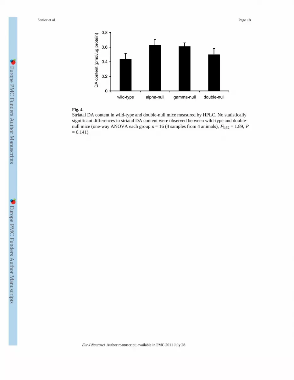
Fig. 4.Striatal DA content in wild-type and double-null mice measured by HPLC. No statisticallysignificant differences in striatal DA content were observed between wild-type and double-null mice (one-way ANOVA each group n = 16 (4 samples from 4 animals), F3,62 = 1.89, P= 0.141).
Senior et al. Page 18
Eur J Neurosci. Author manuscript; available in PMC 2011 July 28.
Europe PM
C Funders A
uthor Manuscripts
Europe PM
C Funders A
uthor Manuscripts

Fig. 5.Comparison between genotypes of rates of uptake of evoked DA transients as measured byFCV. (A) Mean profiles of [DA]o versus time (mean ± s.e.m.) after a single stimulus pulse(0.2 μs, arrow). Comparison of the rates of decay of concentration-matched DA transientssuggest that DA uptake rates are not different between the four genotypes (two-wayANOVA, n = 8 for each genotype, F117,1120 = 0.76, P = 0.9706). (B) A sample profile of[DA]o versus time after a 50 pulse, 100 Hz stimulus (solid bar). This stimulus permitssynapses to release [DA]o reaching concentrations reaching at least 10-fold the Km of theDAT, when DA uptake rate becomes independent of [DA]o i.e. approaches Vmax. Thisproduces an initial period of linear decay (dashed line), the gradient of which is a minimumestimate of Vmax at that site. (C) Mean Vmax estimates were not significantly differentbetween genotypes. (two-sample t-test, wild-type n = 18, double-null n = 25, P = 0.489). (D)Observed Vmax calculations co-varied with peak [DA]o. (type II linear regression, wild-typen = 18, F1,16 = 178.4, P < 0.0001; double-null n = 25, F 1,23 = 68.76, P < 0.0001) in keepingwith locally variable packing density of dopaminergic terminals. To control for possibledifferences in packing density at subpopulation of recording sites sampled andcorresponding co-variation in Vmax, we used the gradient of the regression line of Vmaxagainst peak [DA]o in panel (D) as a modified measure of Vmax. There is no differencebetween this modified Vmax constant in wild-type and double-null mice (F1,39 = 1.164, P =0.287).
Senior et al. Page 19
Eur J Neurosci. Author manuscript; available in PMC 2011 July 28.
Europe PM
C Funders A
uthor Manuscripts
Europe PM
C Funders A
uthor Manuscripts

Fig. 6.Effect of DA uptake inhibition by cocaine on extracellular DA transients in wild-type anddouble-null mice. Mean profiles of [DA]o versus time after a single stimulus pulse beforeand after addition of cocaine (5 μM) in (A) wild type and (B) double-null mice (normalisedto peak [DA]o in the absence of cocaine for each genotype). Cocaine, by inhibiting DAuptake, increased peak [DA]o and extended extracellular lifetimes of DA in both wild-typeand double-null mice. (C) Evoked [DA]o transients were enhanced significantly by cocainein both wild-type (one sample t-test, t = 4.393 df = 2, P = 0.0481) and double-null mice (onesample t-test, t = 8.015 df = 2, P = 0.0041), however the relative increase in evoked [DA]odid not differ between genotypes.
Senior et al. Page 20
Eur J Neurosci. Author manuscript; available in PMC 2011 July 28.
Europe PM
C Funders A
uthor Manuscripts
Europe PM
C Funders A
uthor Manuscripts