impaired neurovascular coupling in the appxps1 mouse model of alzheimer’s disease
TRANSCRIPT

Send Orders of Reprints at [email protected]
Current Alzheimer Research, 2012, 9, 1229-1238 1229
1567-2050/12 $58.00+.00 © 2012 Bentham Science Publishers
Impaired Neurovascular Coupling in the APPxPS1 Mouse Model of Alzheimer’s Disease
Armelle Rancillac*, Hélène Geoffroy and Jean Rossier
Laboratoire de Neurobiologie, CNRS UMR 7637, ESPCI ParisTech, Paris, France
Abstract: The tight coupling between neuronal activity and the local increase of blood flow termed neurovascular
coupling is essential for normal brain function. This mechanism of regulation is compromised in Alzheimer’s Disease
(AD). In order to determine whether a purely vascular dysfunction or a neuronal alteration of blood vessels diameter con-
trol could be responsible for the impaired neurovascular coupling observed in AD, blood vessels reactivity in response to
different pharmacological stimulations was examined in double transgenic APPxPS1 mice model of AD.
Blood vessels movements were monitored using infrared videomicroscopy ex vivo, in cortical slices of 8 month-old
APPxPS1 and wild type (WT) mice. We quantified vasomotor responses induced either by direct blood vessel stimulation
with a thromboxane A2 analogue, the U46619 (9,11-dideoxy-11a,9a-epoxymethanoprostaglandin F2 ) or via the stimula-
tion of interneurons with the nicotinic acetylcholine receptor (nAChRs) agonist DMPP (1,1-Dimethyl-4-
phenylpiperazinium iodide). Using both types of stimulation, no significant differences were detected for the amplitude of
blood vessel diameter changes between the transgenic APPxPS1 mice model of AD and WT mice, although the kinetics
of recovery were slower in APPxPS1 mice. We find that activation of neocortical interneurons with DMPP induced both
vasodilation via Nitric Oxide (NO) release and constriction via Neuropeptide Y (NPY) release. However, we observed a
smaller proportion of reactive blood vessels following a neuronal activation in transgenic mice compared with WT mice.
Altogether, these results suggest that in this mouse model of AD, deficiency in the cortical neurovascular coupling essen-
tially results from a neuronal rather than a vascular dysfunction.
Keywords: Blood vessels, somatosensory cortex, DMPP, Nitric Oxide, interneurons, U46619, Neuropeptide Y, nicotinic cho-linergic receptors.
INTRODUCTION
Alzheimer’s Disease (AD) is a multifactorial and pro-gressive neurodegenerative pathology associated with vari-ous vascular dysfunctions. During AD development, levels of beta amyloid (A ) peptides increase and form depositions in neuritic plaques and in cerebral blood vessels, causing cerebral amyloid angiopathy (CAA). Disturbances in neu-ronal circuits, such as loss of neurons and synapses [1-3] as well as progressive vascular dysregulation [4] are well known features of this pathology.
Neurons and astrocytes are known to function in a close metabolic and structural interdependence with blood vessels forming the so called neurovascular unit [5, 6]. The tight coupling between increased neuronal activity and local con-trol of cerebral blood vessels diameter known as neurovascu-lar coupling or functional hyperemia is precociously altered in AD patients [7-10] and in various AD transgenic mouse models [4, 11-13]. In AD, ultrastructural changes affecting diverse properties of the neurovascular unit have been ob-served, such as capillary distortions, blood-brain-barrier (BBB) dysfunction, losses of endothelial mitochondria,
*Address correspondence to this author at the Laboratoire de Neurobiologie, CNRS UMR 7637, ESPCI ParisTech, 10 rue Vauquelin, 75005 Paris,
France; Tel.: +33 (0)1 40 79 51 83; Fax: +33 (0)1 40 79 47 57; E-mail: [email protected]
thickening of the basement membrane, endothelial degenera-tion, increase in degererative pericytes, perivascular neuroin-flammation [14]. Moreover, early in the physiopathological development of the disease, the AD brain is characterized by an altered perfusion to activated areas [15]. These ultrastruc-tural changes affecting the neurovascular unit result in dis-turbed regulatory mechanisms and contribute to cerebral damage.
This neurovascular dysfunction observed in AD could result from purely vascular alterations or from a neurodegen-erative process [16-18] and a progressive loss of the neuronal control of blood vessels tonus.
Using pharmacological stimulation of blood vessels within cortical slices, the objective of this study was to de-termine ex vivo, the vascular or neuronal origin of the im-paired neurovascular coupling occurring during AD. To this aim, here we used 8 month-old double transgenic mice over-expressing human mutated genes of both human amyloid precursor protein (APP751 with the Swedish and London mutations; SL) and humanpresenilin 1 (PS1M146L) [19]. This APPxPS1 mouse model of AD develops early-onset brain amyloidosis (3-4 months), undergoes substantial cell loss and CAA could first be detected from about 20 weeks of age. We performed direct stimulation of blood vessels using a thromboxane A2 analogue, the U46619 (9,11-dideoxy-11a,9a-epoxymethanoprostaglandin F2 ) acting on G-protein

1230 Current Alzheimer Research, 2012, Vol. 9, No. 10 Rancillac et al.
coupled thromboxane receptor located on endothelial cells of blood vessels [20], or indirect stimulation via neuronal acti-vation using the nicotinic acetylcholine receptor (nAchRs) agonist DMPP (1,1-Dimethyl-4-phenylpiperazinium iodide). Indeed, in the neocortex, the neurovascular coupling is in part mediated by a direct interaction with interneurons [5, 21, 22] via the release of vasoactive messengers. As functional postsynaptic nicotinic acetylcholine receptors (nAchRs) are only expressed by non Fast Spiking (FS) neocortical inhibi-tory interneurons [23-25], DMPP allows a specific neuronal activation.
Here, we observed that for both direct and indirect stimu-lation protocols, the amplitudes of evoked blood vessel movements were not significantly different in APPxPS1 ver-sus WT mouse. We find that activation of neocortical in-terneurons with DMPP could dilate blood vessels via Nitric Oxide (NO) or constrict blood vessels via Neuropepide Y (NPY) release. However, this neuronal stimulation using DMPP induced less vasomotor responses in APPxPS1 com-pared to WT mice. These findings suggest that a loss of neu-ronal control rather than a purely vascular impairment under-lie the neurovascular alteration occurring during the devel-opment of AD.
MATERIAL AND METHODS
Animals
We used nine wild type C57Bl6J (WT) and six APPxPS1 transgenic 8 month-old male mice, as previously described [19]. Transgenic mice (Tg) were generated by crossing ho-mozygous PS1 (carrying presenilin 1 M146L mutation under the control of the HMG-CoA reductase promoter) with hemizygous APPSL mice (hAPP751 transgene with the Swedish K670N/M671L and London V717I mutations, un-der the control of the Thy1 promoter) to obtain APPxPS1 transgenics.
All animals were housed in a temperature-controlled (21–25°C) room under daylight conditions. They arrived in the laboratory 2 weeks before initiating experiments to acclimate to their new environment. Experimental procedures were conducted in strict compliance with approved institutional protocols and in accordance with the provisions for animal care and use described in the European Communities Coun-cil directive of 24 November 1986 (86-16-09/EEC). Experi-ments were performed in compliance and following approval of the Sanofi-Aventis Animal Care and use Committee.
Slice Preparation
Mice were anesthetized using halothane and decapitated. Brains were quickly removed and placed into cold (~4 °C) cutting solution containing (in mM): 110 choline chloride, 11.6 Na-ascorbate, 7 MgCl2, 2.5 KCl, 1.25 NaH2PO4, and 0.5 CaCl2, continuously bubbled with 95% O2 – 5% CO2. Coronal brain slices (300 m thick) comprising the somatosensory cortex were cut with a vibratome (VT1200S; Leica, Nussloch, Germany), and transferred to a holding chamber filled with artificial cerebrospinal fluid (ACSF), containing (in mM): 126 NaCl, 2.5 KCl, 1.25 NaH2PO4, 2 CaCl2, 1 MgCl2, 26 NaHCO3, 20 glucose, and 1 kynurenic acid (a nonspecific glutamate receptor antagonist, Sigma, France), constantly oxygenated (95% O2 – 5% CO2) and held
constantly oxygenated (95% O2 – 5% CO2) and held at room temperature. Individual slices were next placed in a sub-merged recording chamber kept at 32°C and perfused (1.5 ml/min) with oxygenated ACSF (in the absence of kynurenic acid). Blood vessels were visualized in the slice using infra-red videomicroscopy with Dodt gradient contrast optics.
Drugs
Thrombaxane A2 receptors and nicotinic acetylcholine receptors agonists (9,11-dideoxy-11a,9a-epoxymethano-prostaglandin F2 , U46619, 50 nM and 1,1-Dimethyl-4-phenylpiperazinium iodide, DMPP, 100 M, respectively, Sigma, France) were added in the ACSF perfusion of the cortical slices for 6 min. To block the NOS, slices were treated for at least 1 h with an irreversible inhibitor of consti-tutive nitric oxide synthase (nNOS) and a reversible inhibitor of inducible nitric oxide synthase (iNOS), N -Nitro-L-arginine (L-NNA; 100 M; Sigma, France). NPY Y1 recep-tors were blocked with the selective antagonist N2-(Diphenylacetyl)-N-[(4-hydroxyphenyl)methyl]-D-arginine amide (BIBP 3226; 100 M; Sigma, France).
Vascular Reactivity
Blood vessel movements were monitored at 32°C by in-frared videomicroscopy on cortical slices. Blood vessels re-maining in focal plane for more than 50 m, exhibiting a well-defined luminal diameter (10-20 m) and located in supragranular layers of the somatosensory cortex were se-lected for vascular reactivity. Images were acquired every 15 s using Image Pro Plus 7.0 (MediaCybernetics, San Diego, CA) and control baselines of blood vessel diameters were determined for 5 min. Blood vessels with unstable baseline were discarded from the analysis. U46619 or DMPP were applied during 6 minutes after stable baseline period. Lumi-nal diameters were quantified off-line at different locations along the blood vessel using custom written routines running within IgorPro (Wavemetrics) to determine the most reactive part of the vessel. Maximal vasomotor responses were then expressed as percentages of the mean baseline diameter dur-ing the control period.
Statistical Analyses
To determine the statistical significance of the vasomotor responses, changes in diameter were compared using a paired t-test at the time before the onset of the drug applica-tion where the mean diameter change is closest to 0% and at the time where the maximal response was observed. Vascu-lar reactivities in control WT versus APPxPS1 mice were compared using t-test. To compare the proportions of reac-tive blood vessels in WT versus APPxPS1, we used a z test. The Area Under the Curve (AUC) for the vascular response of each blood vessel was calculated using the trapezoidal rule. For U46619 study, the AUC was calculated as the in-cremental or decremental values from the baseline obtained during the 27 minutes (from time 3 to 30 minutes after U46619 onset application). For DMPP study, the AUC was calculated as the incremental or decremental values from the baseline obtained during the 12 minutes (from time 0 to 12 minutes after DMPP onset application). Comparisons of

Impaired Neurovascular Coupling in AD Current Alzheimer Research, 2012, Vol. 9, No. 10 1231
AUC among genotype groups were performed by Mann Whitney U test. The AUC was expressed in arbitrary units.
Histology and Immumohistochemistry
Following the recordings of blood vessel reactivity, slices were fixed over night. Amyloid deposits were labeled by standard Congo red staining [26], and amyloid loads were manually delineated and quantified with Photoshop CS3. A total of 2-3 measures were performed and averaged for every mouse. Evaluation of amyloid loads was performed in the barrel somatosensory cortex and in the CA1 region of the hippocampus. Blood vessels in transgenic versus WT mice were assessed by immunostaining for basement membrane protein collagen-IV (goat anti-collagen type IV; 1: 400; Chemicon). Blood vessels density was measured using com-puter-based thresholding methods in Photoshop CS3. A total of 2-3 measures were performed and averaged for every mouse in the barrel somatosensory cortex.
RESULTS
Amyloid Deposition and Vascular Density in 8 Month-
Old APPxPS1 Mice
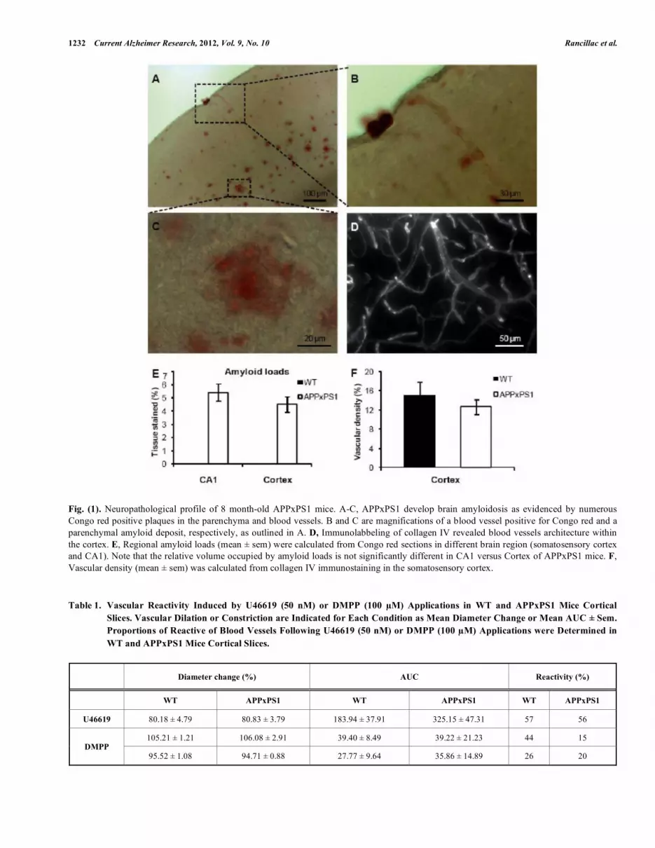
In order to examine the vascular reactivity in the somato-sensory cortex of 8 months APPxPS1 versus WT mice, we first looked at the amyloid depositions and the vascular den-sity in this region of interest. Neuropathological analyses revealed in APPxPS1 the presence of numerous extracellular aggregated (Congo red positive) A deposits (Fig. 1A-C) whereas none could be observed in WT mice. Interestingly, we observed that A deposits in the cortex were more nu-merous and bigger in deep layers, compared to supragranular layers (Fig. 1A and C). Some Congo red positive blood ves-sels were observed (Fig. 1B), indicating a cerebral amyloid angiopathy at 8 months in this APPxPS1 mice. We quanti-fied Congo red positive aggregates in the cortex and the CA1 field of the hippocampus. Amyloid loads represented 4.53 ± 0.57% of the parenchymal tissue in the somatosensory cor-tex, and 5.41 ± 0.66% in the CA1 region (no significant dif-ference between these regions) (Fig. 1E).
In the cortex, immunostaining of collagen-IV to label blood vessels revealed no difference in the mean vascular density in APPxPS1 (12.67 ± 1.48%) compared to the WT mice (15.15 ± 2.61%) (Fig. 1 D and F).
Direct Stimulation of Blood Vessels Using U46619
To determine if purely vascular dysfunctions or a loss of neural control underlay the neurovascular coupling impair-ment observed in AD, we examined blood vessels reactivity in 8 months APPxPS1 versus WT mice. Using infrared videomicroscopy on cortical slice preparations, we first in-vestigated a potential vascular dysfunction by using direct pharmacological stimulation of blood vessels. We applied a thromboxane A2 stable analogue, the U46619 (50 nM) for 6 minutes on acute WT or APPxPS1 cortical slices. We fo-cused our study on penetrating arterioles that are known to be of prime importance in feeding deeply located microves-sels and neurons [27]. Well defined arterioles of supragranu-lar layers were selected for quantitative analyses. This direct vascular stimulation induced strong, significant and reversi-
ble vasoconstrictions in both WT (80.82 ± 6.59%, n = 8; p<0.05) and APPxPS1 mice (80.83 ± 3.79%, n = 10; p<0.001) (Fig. 2, Table 1). The diameter change measured either at 14 minutes after the onset of U46619 application, where the maximum of the response was reached, or at 30 minutes after the onset of U46619 application, did not differ significantly between APPxPS1 and WT mice. However, when the response of each arteriole to the U46619 applica-tion was expressed as the area under the response curve (AUC), the percent AUC for the U46619–induced vasocon-striction was significantly highest at 30 minutes in APPxPS1 mice compared to WT mice (325.15 ± 47.31 and 183.94 ± 37.91 respectively, P<0.05, Table 1). Finally, the proportion of blood vessels that constrict in response to U46619 in APPxPS1 mice (10 out of 18) compared to WT mice (8 out of 14) did not differ significantly (p>0.68, Table 1). These results suggest that, at 8 months of age, blood vessels recov-ery following a direct stimulation is slower but reactivity remains functional in these transgenic mice.
Indirect Stimulation of Blood Vessels Via Neuronal Acti-
vation Using DMPP
Next, in order to investigate whether neuronal release of vasoactive compounds was altered in APPxPS1 mice, we used the nicotinic receptor agonist DMPP (100 M) to stimulate interneurons. DMPP induced, both in WT or APPxPS1 mice, either vasodilatations (105.21 ± 1.21%, n = 12 and 106.08 ± 2.91%, n = 3, respectively), or vasoconstric-tions (95.52 ± 1.08%, n = 7 and 93.42 ± 4.77%, n = 4, re-spectively; Table 1, Fig. 3). Here again, no significant differ-ences (p>0.56) were observed for the mean vasodilatation or vasoconstriction amplitudes between both lines of mice. However, the proportion of blood vessels reactive to DMPP was smaller in APPxPS1 (7 out of 20) compared to WT (19 out of 27) mice (p<0.05, z-test). In particular, vasodilation were less frequently induced by DMPP in transgenic mice (3 out of 20; 15%) compared to WT mice (12 out of 27; 44%; Table 1). Following DMPP stimulation, proportion of vaso-constriction in transgenic mice (7 out of 27; 26%) was not significantly different compared to WT mice (4 out of 19, 20%; z-test, P>0.8; Table 1). However, no significant differ-ence of AUC for DMPP-induced vasodilation or vasocon-striction were observed in APPxPS1 mice compared with WT mice (P>0.64, Table 1).
These results suggest that the impairment of the func-tional hyperemia observed in AD is primarily due to a defect in the neuronal control of blood vessels rather than to a pure vascular dysfunction.
Vasodilations Induced by DMPP Application are Medi-
ated by NO Whereas Vasoconstrictions are due to NPY
Release
The stimulation of GABAergic interneurons can induce vasomotricity via the production of vasoactive substances [5, 21, 22]. DMPP depolarizes nicotinic cholinergic receptors (nAChR)-expressing interneurons that were shown to be also responsive to serotonergic via the serotonin 5-hydroxy-tryptamine 3A (5-HT3A) receptor [28]. As the pharmacologi-cal stimulation of this receptor was recently reported to bi-directionally control arterioles diameter by the release of NO
1232 Current Alzheimer Research, 2012, Vol. 9, No. 10 Rancillac et al.
Fig. (1). Neuropathological profile of 8 month-old APPxPS1 mice. A-C, APPxPS1 develop brain amyloidosis as evidenced by numerous
Congo red positive plaques in the parenchyma and blood vessels. B and C are magnifications of a blood vessel positive for Congo red and a
parenchymal amyloid deposit, respectively, as outlined in A. D, Immunolabbeling of collagen IV revealed blood vessels architecture within
the cortex. E, Regional amyloid loads (mean ± sem) were calculated from Congo red sections in different brain region (somatosensory cortex
and CA1). Note that the relative volume occupied by amyloid loads is not significantly different in CA1 versus Cortex of APPxPS1 mice. F,
Vascular density (mean ± sem) was calculated from collagen IV immunostaining in the somatosensory cortex.
Table 1. Vascular Reactivity Induced by U46619 (50 nM) or DMPP (100 M) Applications in WT and APPxPS1 Mice Cortical
Slices. Vascular Dilation or Constriction are Indicated for Each Condition as Mean Diameter Change or Mean AUC ± Sem.
Proportions of Reactive of Blood Vessels Following U46619 (50 nM) or DMPP (100 M) Applications were Determined in
WT and APPxPS1 Mice Cortical Slices.
Diameter change (%) AUC Reactivity (%)
WT APPxPS1 WT APPxPS1 WT APPxPS1
U46619 80.18 ± 4.79 80.83 ± 3.79 183.94 ± 37.91 325.15 ± 47.31 57 56
105.21 ± 1.21 106.08 ± 2.91 39.40 ± 8.49 39.22 ± 21.23 44 15 DMPP
95.52 ± 1.08 94.71 ± 0.88 27.77 ± 9.64 35.86 ± 14.89 26 20

Impaired Neurovascular Coupling in AD Current Alzheimer Research, 2012, Vol. 9, No. 10 1233
Fig. (2). Vasoconstrictions induced by direct stimulation of blood vessels using U46619 were not significantly different in APPxPS1 com-
pared to WT mice.
A, Mean vascular constrictions ± sem induced by U46619 (50 nM) in 8 month-old APPxPS1 (n = 10, open circles) and WT (n = 8, filled loz-
enges) mice. B, Infrared images of an intracortical blood vessel that reversibly constricted in response to bath application of U46619 (50 nM)
in an APPxPS1 mouse. C, At 14 min (T14), vasoconstrictions shown in (A) were significant in WT (p<0.05) as in APPxPS1 (p<0.001), com-
pared to values at T0. No significant differences were detected at 14 min, or at 30 min, between these two lines of mice (p>0.3).
to dilate, or NPY to constrict [29], we hypothesized that DMPP induced vasomotor changes could also be due to NO or NPY release.
Therefore, in order to determine the molecular events underlying vasomotor changes following DMPP stimulation, we successively blocked different possible mechanisms. Lowering basal NO levels by treatment with the constitutive nNOS inhibitor L-NNA (100 M) prevent all vasodilations
in response to DMPP applications. Only vasoconstrictions were recorded (94.65 ± 2.41 of baseline diameter; n = 7/16, P<0.01) (Fig. 4A). These results suggest that DMPP-induced vasodilations are mediated by NO release.
Then, to determine the molecular pathway underlying vasoconstrictions, treatment of DMPP was reproduced in the presence of NPY Y1 receptor antagonist (BIBP 3226, 1 M). Indeed, vasoconstrictions mediated by NPY are known to be

1234 Current Alzheimer Research, 2012, Vol. 9, No. 10 Rancillac et al.
mediated by smooth muscle NPY Y1 vascular receptor [30]. Under BIBP 3226, constrictions where blocked and only dilations (106.24 ± 2.02%; n = 5/13; P<0.01) could be re-corded (Fig. 4B). Amplitudes of vasomotor responses under these different conditions were not statistically different from control condition. Altogether, these data strongly suggest that pharmacological stimulations of nAchRs-expressing interneurons induce vasodilations through NO release, whereas they induce vasoconstriction through NPY release and activation of its Y1 receptor.
DISCUSSION
The alteration of the neurovascular coupling occurring during AD, could result either from a purely vascular disor-der or from a neurodegenerative process leading to a loss of blood vessels control by neuronal activity. Indeed, increasing evidence suggests a causal role for vascular disorder in the development of AD. Vascular risk factors including hyper-tension, diabetes, and hypercholesterolemia increase the risk of incident AD dementia [31] and regional cerebral hypoper-
Fig. (3). Vasodilatations and vasoconstrictions induced by indirect blood vessels stimulation, via DMPP induced interneurons activation,
were not significantly different in APPxPS1 compared to WT mice. A, Mean vascular dilatations (n = 12) and constrictions (n = 7) ± sem
induced by DMPP (100 M) in 8 month-old WT mice. B, Mean vascular dilatations (n = 3) and constrictions (n = 4) ± sem induced by
DMPP (100 M) in 8 month-old transgenic APPxPS1 mice. C, Mean maximal or minimal responses for vasodilatation and vasoconstriction,
respectively, were compared to initial values (at T0) for WT and APPxPS1 mice and between WT and APPxPS1 mice.

Impaired Neurovascular Coupling in AD Current Alzheimer Research, 2012, Vol. 9, No. 10 1235
fusion is one of the earliest pathological features of AD. On the other hand, various modes of neurodegeneration can co-exist in a same mouse model and lead to reduced blood ves-sels innervation [32]. Previous findings already shown that A accumulates in neurons [33-36], as it was also observed at 2 months in this model [37], which might cause a synaptic degeneration [1] and lead to neuronal death [38]. Extracellu-lar amyolid- induces significant entorhinal neuronal loss of principal and SOM/NPY neurons in these APPxPS1 mice [39]. Moreover, autophagy is induced but impaired in af-fected neurons in the AD brain [40, 41], causing autophagic vacuoles to accumulate profusely in dystrophic neuritis. This axonopathy disturbs neuronal trafficking [42] and could per-turb vasomotor peptidergic release.
Vascular Dysfunction
In the present study, to determine if purely vascular or neuronal dysfunction were responsible for the decreased neurovascular coupling occurring during AD, we used either direct blood vessels stimulation, or neuronal activation, while recording vasomotor changes. Here, no differences could be observed in the peak amplitude of evoked blood vessels movements between WT and APPxPS1 mice using either type of stimulation. However, the AUC for the U46619-induced vasoconstriction was significantly highest in APPxPS1 mice compared to WT mice, accordingly to in vivo studies that reported in other models of AD that vaso-constrictions are usually exaggerated [43-45]. This suggests reduced vessel elasticity, which could be due to CAA. In-deed, CAA was first detected from about 20 weeks of age and developed beyond the age of 40 weeks in this mouse line, with a high prevalenceof the A 40 isoform forming amyloid deposits in the vascular system [46]. The blood a vessel inelasticity could compromise mental function by preventing neural regions from reacting to new activity by adapting blood supply [47].
Neuronal Dysfunction
Here, we observed that following a neuronal stimulation , reactive vessels were less numerous in transgenic mice com-pared to WT mice. Therefore, it is likely that neurodegenera-
tion rather than a purely vascular impairment could be re-sponsible for the alteration of neurovascular coupling in AD.
Indeed, a neuronal loss in the entorhinal cortex [39] and an extensive, selective and early neurodegeneration of the dendritic inhibitory interneurons of the hippocampus have been observed in this model. At 6 months of age, a diminu-tion of 50-60% of SOM-immunopositive neurons was noted [48], in accordance with the loss of SOM and/or NPY neu-rons frequently reported in AD patients and the linear corre-lation between SOM and/or NPY deficiency and A content which have been reported [48]. As extracellular A deposits are observed in the hippocampus and in the neocortex (Fig. 1) and [37], this suggest that a neurodegeneration of cortical interneurons could be responsible for the decreased propor-tion of reactive blood vessels following a neuronal stimula-tion observed in this study.
Alternatively, Alzheimer's disease is characterized by large losses of nicotinic cholinergic receptors [49, 50], that could also contribute to the lack of reactivity following DMPP application.
Dual Role of nAChR-Expressing Interneurons
Our results indicate that activation of nAChR-expressing interneurons induces a complex vascular response. Indeed the selective nAChR agonist DMPP induced either constric-tions (37%) or dilations (63%) of penetrating arterioles within supragranular layers. All vasoconstrictions were abol-ished in the presence of the NPY receptor antagonist (BIBP 3226), suggesting that they were elicited by NPY release. This result confirm and extend prior studies implicating NPY in vasoconstrictions [21, 51, 52].
Conversely, DMPP-induced dilations were blocked in the presence of a nNOS inhibitor suggesting that NO is the pre-dominant messenger inducing the vasodilation, in line previ-ous studies performed in the cerebellum [22, 53, 54] and in the cortex [21, 55-59]. This result confirm the previous ob-servation that nAChRs activation increased levels of the me-tabolites of NO, and therefore presumably of NO [60]. To-gether, our observations that in the somatosensory cortex a
Fig. (4). DMPP induced vasodilations are mediated by NO while constrictions are mediated by NPY in 8 month-old WT mice. A, Mean
vasoconstriction (n = 7) ± sem induced by DMPP (100 M) in the presence of nNOS inhibitor L-NNA (100 M). B, Mean vascular dilation
(n = 5) ± sem induced by DMPP (100 M) in the presence of the NPY Y1 receptor antagonist BIBP 3226 (1 M).

1236 Current Alzheimer Research, 2012, Vol. 9, No. 10 Rancillac et al.
selective nAChR agonist induce NO mediated vasodilations confirms and extends prior studies reinforcing the central role of NO in the neurovascular coupling.
Role of Astrocytes in the Vascular Response to DMPP
Astrocytes are known as cellular intermediaries that cou-ple neuronal activity to local blood flow changes through the phospholipase A2 (PLA2)-mediated synthesis of arachidonic acid, which leads to production of prostaglandins and ep-oxyeicosatrienoic acids [61].
In the present study, we demonstrated that following DMPP stimulation, vasoconstrictions were mediated by NPY release. This neuropeptide is released by interneurons and directly activates NPY Y1 receptors located on vascular smooth muscle cells. Otherwise, we demonstrated that fol-lowing DMPPP stimulation, vasodilations were mediated by NO release. NO is known to either directly acts on vascular smooth muscle cells through cGMP-PKG [62, 63] or indi-rectly interacts with numerous signaling pathways also in-volved in the vascular control [64-66]. In particular, NO could induce astrocytic cytosolic Ca
2+ increased [67]. How-
ever, in vivo data strongly suggest that the involvement of astrocytes is likely to occur in the late phase of the neurovas-cular coupling [68, 69]. Even if further experiments would be necessary to fully characterize the NO mediated vasodila-tions following DMPP application, it appears that in our stimulation conditions, no other components possibly re-leased by astrocytes are involved in the neurovascular cou-pling.
CONCLUSION
In this study, we observed by infrared videomicroscopy, the cortical vascular response induced by either a vascular activator, the thomboxane A2 analogue, or by a neuronal activator, the nicotinic agonist DMPP in an APPxPS1 trans-genic mouse model of AD. No differences were observed in the amplitude of vascular responses between WT and trans-genic mice following both types of activators. Therefore, in this mouse model of AD a neuronal alteration rather than vascular changes seems responsible for the impaired neurovascular coupling.
CONFLICT OF INTEREST
The author(s) confirm that this article content has no con-flicts of interest.
ACKNOWLEDGEMENTS
We thank the Sanofi-Aventis Neurodegenerative Disease Group for the generous gift of the animals involved in this
study and Marcel Leopoldie for animal husbandry. We are grateful to Dr. Isabelle Férézou for helpful comments read-ing the manuscript and to Marion Daenens and Thierry Dendele for technical support. This work was supported by the French National Research Agency (ANR-06-NEURO-033-01grant).
REFERENCES
[1] Selkoe DJ. Alzheimer's disease is a synaptic failure. Science
298(5594): 789-791 (2002).
[2] Morrison JH, Hof PR. Selective vulnerability of corticocortical and
hippocampal circuits in aging and Alzheimer's disease. Prog Brain
Res 136467-486 (2002).
[3] Palop JJ, Chin J, Roberson ED, Wang J, Thwin MT, Bien-Ly N, et
al. Aberrant excitatory neuronal activity and compensatory remod-
eling of inhibitory hippocampal circuits in mouse models of Alz-
heimer's disease. Neuron 55(5): 697-711 (2007).
[4] Iadecola C. Neurovascular regulation in the normal brain and in
Alzheimer's disease. Nat Rev Neurosci 5(5): 347-360 (2004).
[5] Cauli B, Hamel E. Revisiting the role of neurons in neurovascular
coupling. Front Neuroenergetics 29- (2010).
[6] Lecrux C, Hamel E. The neurovascular unit in brain function and
disease. Acta Physiol (Oxf) 203(1): 47-59 (2011).
[7] Hock C, Villringer K, Muller-Spahn F, Wenzel R, Heekeren H,
Schuh-Hofer S, et al. Decrease in parietal cerebral hemoglobin
oxygenation during performance of a verbal fluency task in patients
with Alzheimer's disease monitored by means of near-infrared
spectroscopy (NIRS)--correlation with simultaneous rCBF-PET
measurements. Brain Res 755(2): 293-303 (1997).
[8] Mentis MJ, Alexander GE, Krasuski J, Pietrini P, Furey ML,
Schapiro MB, et al. Increasing required neural response to expose
abnormal brain function in mild versus moderate or severe Alz-
heimer's disease: PET study using parametric visual stimulation.
Am J Psychiatry 155(6): 785-794 (1998).
[9] Rombouts SA, Barkhof F, Veltman DJ, Machielsen WC, Witter
MP, Bierlaagh MA, et al. Functional MR imaging in Alzheimer's
disease during memory encoding. AJNR Am J Neuroradiol 21(10):
1869-1875 (2000).
[10] Zlokovic BV. Neurovascular pathways to neurodegeneration in
Alzheimer's disease and other disorders. Nat Rev Neurosci 12(12):
723-738 (2011).
[11] Hamel E, Nicolakakis N, Aboulkassim T, Ongali B, Tong XK.
Oxidative stress and cerebrovascular dysfunction in mouse models
of Alzheimer's disease. Exp Physiol 93(1): 116-120 (2008).
[12] Iadecola C, Zhang F, Niwa K, Eckman C, Turner SK, Fischer E, et
al. SOD1 rescues cerebral endothelial dysfunction in mice overex-
pressing amyloid precursor protein. Nat Neurosci 2(2): 157-161
(1999).
[13] Nicolakakis N, Hamel E. Neurovascular function in Alzheimer's
disease patients and experimental models. J Cereb Blood Flow Me-
tab (2011).
[14] Shi J, Perry G, Smith MA, Friedland RP. Vascular abnormalities:
the insidious pathogenesis of Alzheimer's disease. Neurobiol Aging
21(2): 357-361 (2000).
[15] Brown WR, Thore CR. Review: Cerebral microvascular pathology
in aging and neurodegeneration. Neuropathol Appl Neurobiol
(2010).
Table 2. Vascular Reactivity Induced by DMPP Applications in WT Mice Cortical Slices in the Presence of L-NNA or BIBP 3226.
Vascular Dilation (White Line) or Constriction (Grey Lines) are Indicated for Each Condition as Mean Diameter Change
or Mean AUC ± Sem. Proportions of Reactive of Blood Vessels Following DMPP (100 M) Applications were Determined
in WT Mice.
Diameter change (%) AUC Reactivity (%)
DMPP + BIBP 3226 106.24 ± 2.02 39.75 ± 6.21 38
DMPP + L-NNA 94.65 ± 2.41 26.76 ± 6.00 44

Impaired Neurovascular Coupling in AD Current Alzheimer Research, 2012, Vol. 9, No. 10 1237
[16] de la Torre JC. Vascular basis of Alzheimer's pathogenesis. Ann N
Y Acad Sci 977196-215 (2002).
[17] Zlokovic BV. Neurovascular mechanisms of Alzheimer's neurode-
generation. Trends Neurosci 28(4): 202-208 (2005).
[18] Benarroch E. Neurovascular unit dysfunction: A vascular compo-
nent of Alzheimer disease? Neurology 68(May 15): 1730-1732
(2007).
[19] Blanchard V, Moussaoui S, Czech C, Touchet N, Bonici B,
Planche M, et al. Time sequence of maturation of dystrophic neu-
rites associated with Abeta deposits in APP/PS1 transgenic mice.
Exp Neurol 184(1): 247-263 (2003).
[20] Narumiya S, Hirata N, Namba T, Hayashi Y, Ushikubi F, Sugimoto
Y, et al. Structure and function of prostanoid receptors. J Lipid
Mediat 6(1-3): 155-161 (1993).
[21] Cauli B, Tong XK, Rancillac A, Serluca N, Lambolez B, Rossier J,
et al. Cortical GABA interneurons in neurovascular coupling: re-
lays for subcortical vasoactive pathways. J Neurosci 24(41): 8940-
8949 (2004).
[22] Rancillac A, Rossier J, Guille M, Tong XK, Geoffroy H, Amatore
C, et al. Glutamatergic Control of Microvascular Tone by Distinct
GABA Neurons in the Cerebellum. J Neurosci 26(26): 6997-7006
(2006).
[23] Porter JT, Cauli B, Tsuzuki K Lambolez B, Rossier J, Audinat E.
Selective excitation of subtypes of neocortical interneurons by
nicotinic receptors. J Neurosci 19(13): 5228-5235 (1999).
[24] Xiang Z, Huguenard JR, Prince DA. Cholinergic switching within
neocortical inhibitory networks. Science 281(5379): 985-988
(1998).
[25] Gulledge AT, Park SB, Kawaguchi Y, Stuart GJ. Heterogeneity of
phasic cholinergic signaling in neocortical neurons. J Neurophysiol
97(3): 2215-2229 (2007).
[26] Wilcock DM, Gordon MN, Morgan D. Quantification of cerebral
amyloid angiopathy and parenchymal amyloid plaques with Congo
red histochemical stain. Nat Protoc 1(3): 1591-1595 (2006).
[27] Nishimura N, Schaffer CB, Friedman B, Lyden PD, Kleinfeld D.
Penetrating arterioles are a bottleneck in the perfusion of neocortex.
Proc Natl Acad Sci U S A 104(1): 365-370 (2007).
[28] Lee S, Hjerling-Leffler J, Zagha E, Fishell G, Rudy B. The largest
group of superficial neocortical GABAergic interneurons expresses
ionotropic serotonin receptors. J Neurosci 30(50): 16796-16808
(2010).
[29] Perrenoud Q, Rossier J, Férézou I, Geoffroy H, Gallopin T, Vitalis
T, Rancillac A.Activation of cortical 5-HT(3) receptor-expressing
interneurons induces NO mediated vasodilatations and NPY medi-
ated vasoconstrictions. <http://www.ncbi.nlm.nih.gov/ pub-
med/22907992> Front Neural Circuits. 2012;6:50. Epub 2012 Aug
10.
[30] Abounader R, Elhusseiny A, Cohen Z, Olivier A, Stanimirovic D,
Quirion R, et al. Expression of neuropeptide Y receptors mRNA
and protein in human brain vessels and cerebromicrovascular cells
in culture. J Cereb Blood Flow Metab 19(2): 155-163 (1999).
[31] Li J, Wang YJ, Zhang M, Gao CY, Fang CQ, Yan JC, et al. Vas-
cular risk factors promote conversion from mild cognitive impair-
ment to Alzheimer disease. Neurology 76(17): 1485-1491 (2011).
[32] Yang DS, Kumar A, Stavrides P, Peterson J, Peterhoff CM, Pawlik
M, et al. Neuronal apoptosis and autophagy cross talk in aging
PS/APP mice, a model of Alzheimer's disease. Am J Pathol 173(3):
665-681 (2008).
[33] Cook DG, Forman MS, Sung JC, Leight S, Kolson DL, Iwatsubo
T, et al. Alzheimer's A beta(1-42) is generated in the endoplasmic
reticulum/intermediate compartment of NT2N cells. Nat Med 3(9):
1021-1023 (1997).
[34] Hartmann T, Bieger SC, Bruhl B, Tienari PJ, Ida N, Allsop D, et al.
Distinct sites of intracellular production for Alzheimer's disease A
beta40/42 amyloid peptides. Nat Med 3(9): 1016-1020 (1997).
[35] Xu H, Sweeney D, Wang R, Thinakaran G, Lo AC, Sisodia SS, et
al. Generation of Alzheimer beta-amyloid protein in the trans-
Golgi network in the apparent absence of vesicle formation. Proc
Natl Acad Sci USA 94(8): 3748-3752 (1997).
[36] Greenfield JP, Tsai J, Gouras GK, Hai B, Thinakaran G, Checler F,
et al. Endoplasmic reticulum and trans-Golgi network generate dis-
tinct populations of Alzheimer beta-amyloid peptides. Proc Natl
Acad Sci USA 96(2): 742-747 (1999).
[37] Langui D, Girardot N, El Hachimi KH and others. Subcellular
topography of neuronal Abeta peptide in APPxPS1 transgenic
mice. Am J Pathol 165(5): 1465-1477 (2004).
[38] Schmitz C, Rutten BP, Pielen A and others. Hippocampal neuron
loss exceeds amyloid plaque load in a transgenic mouse model of
Alzheimer's disease. Am J Pathol 164(4): 1495-1502 (2004).
[39] Moreno-Gonzalez I, Baglietto-Vargas D, Sanchez-Varo R and
others. Extracellular amyloid-beta and cytotoxic glial activation in-
duce significant entorhinal neuron loss in young
PS1(M146L)/APP(751SL) mice. J Alzheimers Dis 18(4): 755-776
(2009).
[40] Nixon RA, Wegiel J, Kumar A and others. Extensive involvement
of autophagy in Alzheimer disease: an immuno-electron micros-
copy study. J Neuropathol Exp Neurol 64(2): 113-122 (2005).
[41] Yu WH, Cuervo AM, Kumar A and others. Macroautophagy--a
novel Beta-amyloid peptide-generating pathway activated in Alz-
heimer's disease. J Cell Biol 171(1): 87-98 (2005).
[42] Wirths O, Weis J, Szczygielski J, Multhaup G, Bayer TA. Ax-
onopathy in an APP/PS1 transgenic mouse model of Alzheimer's
disease. Acta Neuropathol 111(4): 312-319 (2006).
[43] Niwa K, Younkin L, Ebeling C and others. Abeta 1-40-related
reduction in functional hyperemia in mouse neocortex during so-
matosensory activation. Proc Natl Acad Sci U S A 97(17): 9735-
9740 (2000).
[44] Christie R, Yamada M, Moskowitz M, Hyman B. Structural and
functional disruption of vascular smooth muscle cells in a trans-
genic mouse model of amyloid angiopathy. Am J Pathol 158(3):
1065-1071 (2001).
[45] Niwa K, Kazama K, Younkin SG, Carlson GA, Iadecola C. Altera-
tions in cerebral blood flow and glucose utilization in mice overex-
pressing the amyloid precursor protein. Neurobiol Dis 9(1): 61-68
(2002).
[46] El Tayara NT, Delatour B, Volk A, Dhenain M. Detection of vas-
cular alterations by in vivo magnetic resonance angiography and
histology in APP/PS1 mouse model of Alzheimer's disease.
MAGMA 23(1): 53-64 (2010).
[47] de la Torre JC, Mussivand T. Can disturbed brain microcirculation
cause Alzheimer's disease? Neurol Res 15(3): 146-153 (1993).
[48] Ramos B, Baglietto-Vargas D, del Rio JC and others. Early neuro-
pathology of somatostatin/NPY GABAergic cells in the hippocam-
pus of a PS1xAPP transgenic model of Alzheimer's disease. Neu-
robiol Aging 27(11): 1658-1672 (2006).
[49] Whitehouse PJ, Au KS. Cholinergic receptors in aging and Alz-
heimer's disease. Prog Neuropsychopharmacol Biol Psychiatry
10(3-5): 665-676 (1986).
[50] James JR, Nordberg A. Genetic and environmental aspects of the
role of nicotinic receptors in neurodegenerative disorders: emphasis
on Alzheimer's disease and Parkinson's disease. Behav Genet
25(2): 149-159 (1995).
[51] Abounader R, Villemure JG, Hamel E. Characterization of neu-
ropeptide Y (NPY) receptors in human cerebral arteries with selec-
tive agonists and the new Y1 antagonist BIBP 3226. Br J Pharma-
col 116(4): 2245-2250 (1995).
[52] Dacey RG, Jr., Bassett JE, Takayasu M. Vasomotor responses of
rat intracerebral arterioles to vasoactive intestinal peptide, sub-
stance P, neuropeptide Y, and bradykinin. J Cereb Blood Flow Me-
tab 8(2): 254-261 (1988).
[53] Yang G, Chen G, Ebner TJ, Iadecola C. Nitric oxide is the pre-
dominant mediator of cerebellar hyperemia during somatosensory
activation in rats. Am J Physiol 277(6 Pt 2): R1760-R1770 (1999).
[54] Yang G, Huard JM, Beitz AJ, Ross ME, Iadecola C. Stellate neu-
rons mediate functional hyperemia in the cerebellar molecular
layer. J Neurosci 20(18): 6968-6973 (2000).
[55] Brown LA, Key BJ, Lovick TA. Fluorescent imaging of nitric
oxide production in neuronal varicosities associated with intrapar-
enchymal arterioles in rat hippocampal slices. Neurosci Lett
294(1): 9-12 (2000).
[56] Lovick TA, Brown LA, Key BJ. Neurovascular relationships in
hippocampal slices: physiological and anatomical studies of
mechanisms underlying flow-metabolism coupling in intraparen-
chymal microvessels. Neuroscience 92(1): 47-60 (1999).
[57] Estrada C, DeFelipe J. Nitric oxide-producing neurons in the neo-
cortex: morphological and functional relationship with intraparen-
chymal microvasculature. Cereb Cortex 8(3): 193-203 (1998).

1238 Current Alzheimer Research, 2012, Vol. 9, No. 10 Rancillac et al.
[58] Tong XK, Hamel E. Basal forebrain nitric oxide synthase (NOS)-
containing neurons project to microvessels and NOS neurons in the
rat neocortex: cellular basis for cortical blood flow regulation. Eur
J Neurosci 12(8): 2769-2780 (2000).
[59] Liu X, Li C, Falck JR and others. Interaction of nitric oxide, 20-
HETE, and EETs during functional hyperemia in whisker barrel
cortex. Am J Physiol Heart Circ Physiol 295(2): H619-H631
(2008).
[60] Pogun S, Demirgoren S, Taskiran D and others. Nicotine modulates
nitric oxide in rat brain. Eur Neuropsychopharmacol 10(6): 463-
472 (2000).
[61] Petzold GC, Murthy VN. Role of astrocytes in neurovascular cou-
pling. Neuron 71(5): 782-797 (2011).
[62] Garthwaite J, Boulton CL. Nitric oxide signaling in the central
nervous system. Annu Rev Physiol 57683-706 (1995).
[63] Jaffrey SR, Snyder SH. Nitric oxide: a neural messenger. Annu
Rev Cell Dev Biol 11417-440 (1995).
[64] Fleming I. Cytochrome p450 and vascular homeostasis. Circ Res
89(9): 753-762 (2001).
[65] Fujimoto Y, Uno E, Sakuma S. Effects of reactive oxygen and
nitrogen species on cyclooxygenase-1 and -2 activities. Prosta-
glandins Leukot Essent Fatty Acids 71(5): 335-340 (2004).
[66] Salvemini D. Regulation of cyclooxygenase enzymes by nitric
oxide. Cell Mol Life Sci 53(7): 576-582 (1997).
[67] Willmott NJ, Wong K, Strong AJ. A fundamental role for the nitric
oxide-G-kinase signaling pathway in mediating intercellular Ca(2+)
waves in glia. J Neurosci 20(5): 1767-1779 (2000).
[68] Calcinaghi N, Jolivet R, Wyss MT and others. Metabotropic gluta-
mate receptor mGluR5 is not involved in the early hemodynamic
response. J Cereb Blood Flow Metab 31(9): e1-10 (2011).
[69] Wang X, Lou N, Xu Q and others. Astrocytic Ca2+ signaling
evoked by sensory stimulation in vivo. Nat Neurosci 9(6): 816-823
(2006).
Received: ??????????????? Revised: ??????????????? Accepted: ???????????????