ifn-γ-mediated efficacy of allergen-free immunotherapy using mycobacterial antigens and cpg-odn
TRANSCRIPT
ORIGINAL ARTICLE
IFN-c-mediated efficacy of allergen-freeimmunotherapy using mycobacterial antigensand CpG-ODN
Denise M Fonseca1, Marina O Paula1, Pryscilla F Wowk1, Lıvia W Campos1, Ana F Gembre1, WalterM Turato1, Simone G Ramos2, Marcelo Dias-Baruffi3, Renato Barboza4, Eliane Gomes4, Cynthia Horn5,Gilles Marchal6, Luisa K Arruda7, Momtchilo Russo4 and Vania LD Bonato1
Epidemiological and experimental evidence supports the notion that microbial infections that are known to induce Th1-type
immune responses can suppress Th2 immune responses, which are characteristics of allergic disorders. However, live microbial
immunization might not be feasible for human immunotherapy. Here, we evaluated whether induction of Th1 immunity by the
immunostimulatory sequences of CpG-oligodeoxynucleotides (CpG-ODN), with or without culture filtrate proteins (CFP), from
Mycobacterium tuberculosis would suppress ongoing allergic lung disease. Presensitized and ovalbumin (OVA)-challenged mice
were treated subcutaneously with CpG, or CpG in combination with CFP (CpG/CFP). After 15 days of treatment, airway
inflammation and specific T- and B-cell responses were determined. Cell transfer experiments were also performed. CpG
treatment attenuated airway allergic disease; however, the combination CpG/CFP treatment was significantly more effective in
decreasing airway hyperresponsiveness, eosinophilia and Th2 response. When an additional intranasal dose of CFP was given,
allergy was even more attenuated. The CpG/CFP therapy also reduced allergen-specific IgG1 and IgE antibodies and increased
IgG2a. Transfer of spleen cells from mice immunized with CpG/CFP also reduced allergic lung inflammation. CpG/CFP treatment
induced CFP-specific production of IFN-c and IL-10 by spleen cells and increased production of IFN-c in response to OVA. The
essential role of IFN-c for the therapeutic effect of CpG/CFP was evidenced in IFN-c knockout mice. These results show that
CpG/CFP treatment reverses established Th2 allergic responses by an IFN-c-dependent mechanism that seems to act both
locally in the lung and systemically to decrease allergen-specific Th2 responses.
Immunology and Cell Biology advance online publication, 15 March 2011; doi:10.1038/icb.2011.9
Keywords: experimental asthma; M. tuberculosis; culture filtrate proteins; CpG-oligodeoxynucleotides; allergen free immunotherapy;IFN-gamma
Allergic asthma is a chronic inflammatory lung disease characterizedby a prominent allergen-specific Th2 immune response, which pro-motes IgE synthesis, eosinophilic inflammation and airway hyperre-sponsiveness (AHR).1,2 Allergen-specific immunotherapy has beenshown to decrease symptoms and medication usage in patients withasthma.3 However, currently available modalities of immunotherapyhave limitations, including lack of efficacy in some patients, long timeto induce the effects and risk of side-effects, which may present as life-threatening systemic allergic reactions.3 Therefore, improved forms ofimmunotherapy are necessary to downmodulate allergic responsesand prevent new hypersensitivity reactions.
It has been suggested that allergen-free strategies of immunotherapycould be used to treat asthma.4,5 Infection or immunization withmicroorganisms, including mycobacterial species, have been shown tomodulate allergic responses.6–10 The bacillus Calmette–Guerin (BCG)has been widely studied as a strategy to downmodulate airwayexperimental allergy. In addition to live BCG, it has been describedthat BCG killed by extensive freeze-drying reduced eosinophilia, lunginflammation and AHR in murine and guinea pig models of asthma.11
On the other hand, BCG administration, in conjunction with allergen-specific immunotherapy has not resulted in increased efficacy inhuman patients.12
Received 21 October 2010; revised 20 December 2010; accepted 18 January 2011
1Department of Biochemistry and Immunology, School of Medicine of Ribeirao Preto, University of Sao Paulo, Riberao Preto, Brazil; 2Department of Pathology, School of Medicineof Ribeirao Preto, University of Sao Paulo, Riberao Preto, Brazil; 3Department of Clinical, Toxicological, Bromatological Analyses, School of Pharmaceutical Sciences of RibeiraoPreto, University of Sao Paulo, Riberao Preto, Brazil; 4Department of Immunology, Institute of Biomedical Sciences, University of Sao Paulo, Sao Paulo, Brazil; 5Evandro ChagasResearch Institute, Osvaldo Cruz Foundation, Rio de Janeiro, Brazil; 6Immunotherapix Bio Top, Institute Pasteur, Paris, France and 7Department of Medicine, Division of ClinicalImmunology, School of Medicine of Ribeirao Preto, University of Sao Paulo, Riberao Preto, BrazilCorrespondence: Professor VLD Bonato, Department of Biochemistry and Immunology, School of Medicine of Riberao Preto, University of Sao Paulo, Av. Bandeirantes, 3900,Riberao Preto, Sao Paulo 14049-900, Brazil.E-mail: [email protected]
Immunology and Cell Biology (2011), 1–9& 2011 Australasian Society for Immunology Inc. All rights reserved 0818-9641/11
www.nature.com/icb
The use of toll-like receptor (TLR) agonists represents a promisingadditional strategy of allergen-free therapy, because TLR-inducedcytokines, such as IL-12, cooperate in induction of Th1 immuneresponses.3–5,13 In experimental models, studies demonstrated thatCpG-oligodeoxynucleotides (CpG-ODN), a TLR9 agonist, effectivelycontrolled allergic acute inflammation not only in IFN-g-dependent,but also in IFN-g-independent ways.14–17 Studies in human subjectshave shown promising results with the use of CpG-ODNs or CpG-ODN plus allergens.18–20 These treatments induced a Th1 immuneresponse that downmodulated allergic response. However, in somecases, the use of CpG-ODN did not show a significant efficacy againstsevere cases of asthma.21
We have previously described that immunization of mice withM. tuberculosis culture filtrate proteins (CFP), in the presence of CpG-ODN, induced a Th1-biased specific immune response.22 The CFP areimmunodominant antigens, actively secreted by M. tuberculosis duringbacterial growth. These proteins are highly immunogenic and havebeen intensively studied for the development of tuberculosisvaccines.23,24
Considering the data on modulation of allergic responses bymycobacterial infection/immunization, or by TLR9 agonist (CpG),and the IFN-g-inducing ability of CFP antigens, we proposed anallergen-free immunotherapy, using CpG-ODN in conjunction withCFP antigens (CpG/CFP). Avoiding the use of whole mycobacteriawould eliminate the risk of developing an active infection in immuno-compromised patients, such as those exposed to long-term corticos-teroid treatment. In addition, immunotherapy with CpG/CFP could
have advantages over CpG alone, because although the TLR agoniststimulates mainly the innate system, the mycobacterial antigenscould induce a specific immune response driven by the TLR agonist,which could be sustained for a long time and elicited at any time bymemory cells. In the present study, we observed that subcutaneousCpG/CFP treatment downmodulated eosinophil accumulation, lunginflammation and AHR. In contrast to CpG alone, CpG/CFP co-administration induced a Th1-adaptive immune response. This anti-gen-specific immune response was boosted locally by intranasaladministration of CFP, and this treatment was associated with anaccentuated downmodulation of allergy. Finally, it was also observedthat the IFN-g-induced immune response by CFP antigens was crucialfor the modulation of allergy in this model of allergen-free immu-notherapy.
RESULTS
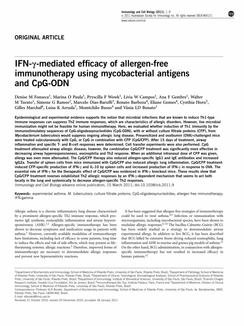
CpG/CFP treatment modulated local allergic Th2 responseAllergic mice treated subcutaneously with CpG/CFP, but not withCpG only, exhibited a significant reduction in eosinophil counts andincrease in lymphocyte number in bronchoalveolar lavage fluid(BALF), as compared with allergic-untreated mice (Figure 1b).Levels of IL-4, IL-5 and IL-13 were reduced in BALF after treatmentwith CpG/CFP or CpG, as compared with allergic-untreatedgroup; however, reduction of IL-13 and IL-5 was more pronouncedwith CpG/CFP treatment (Figure 1c). On the other hand, decreasein levels of TSLP, eotaxin and IL-17, and increased secretionof IFN-g were found only in CpG/CFP-treated mice (Figure 1c).
Figure 1 Cell profile and cytokine production in BALF, following treatment with CpG/CFP. OVA-sensitized and challenged BALB/c mice were treated with CpG
or CpG/CFP sc, according to the study protocol (a). At 72 h after the second OVA-challenge, cells (b) and cytokine secretion (c) were quantified in BALF.
*Po0.05. Representative data of two experiments (n¼8). sc, subcutaneous; ip, intraperitoneal; in, intranasal.
Mycobacterial antigens in allergen-free immunotherapyDM Fonseca et al
2
Immunology and Cell Biology
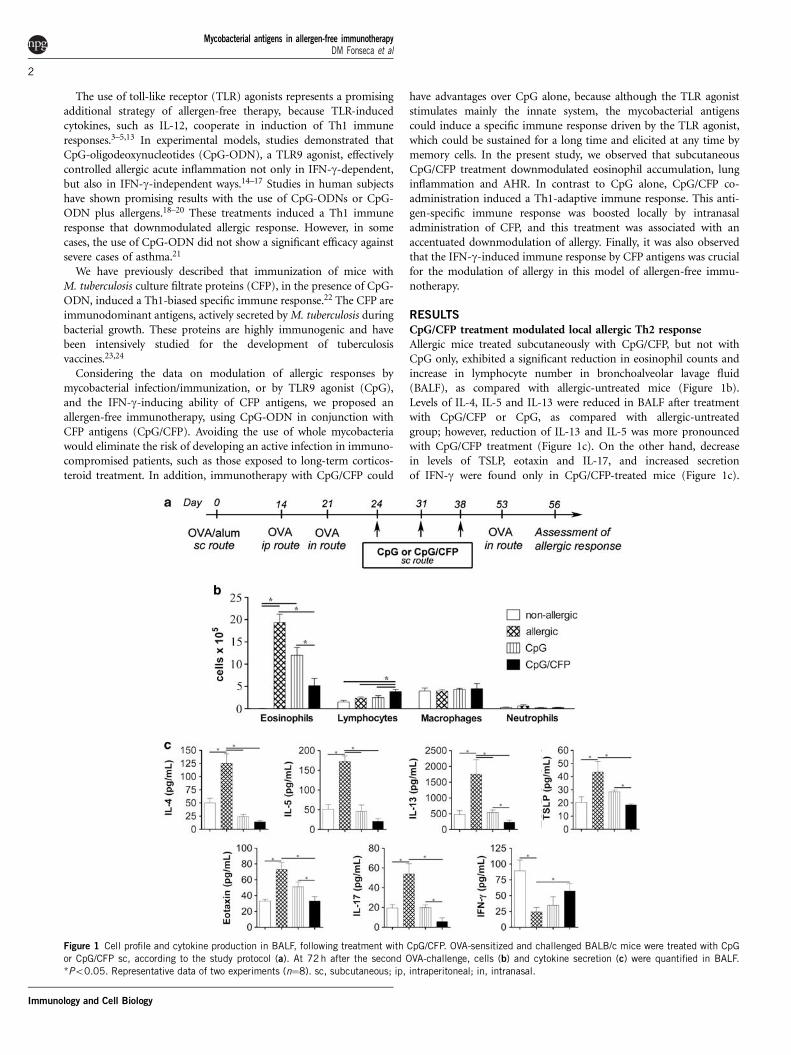
No significant differences were seen for IL-10 production (data notshown). Immune modulation by CpG/CFP treatment was accompa-nied by decrease of inflammation scores and mucus staining in lungtissue (Figure 2).
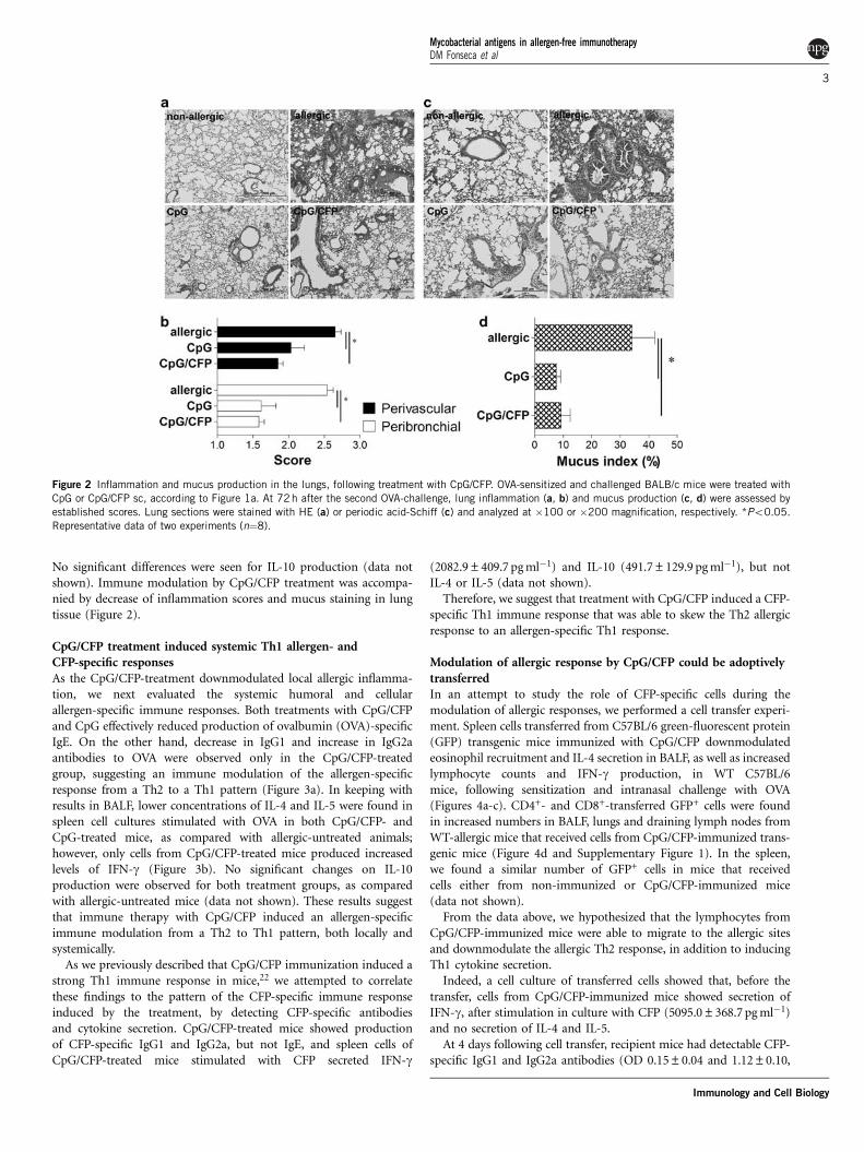
CpG/CFP treatment induced systemic Th1 allergen- andCFP-specific responsesAs the CpG/CFP-treatment downmodulated local allergic inflamma-tion, we next evaluated the systemic humoral and cellularallergen-specific immune responses. Both treatments with CpG/CFPand CpG effectively reduced production of ovalbumin (OVA)-specificIgE. On the other hand, decrease in IgG1 and increase in IgG2aantibodies to OVA were observed only in the CpG/CFP-treatedgroup, suggesting an immune modulation of the allergen-specificresponse from a Th2 to a Th1 pattern (Figure 3a). In keeping withresults in BALF, lower concentrations of IL-4 and IL-5 were found inspleen cell cultures stimulated with OVA in both CpG/CFP- andCpG-treated mice, as compared with allergic-untreated animals;however, only cells from CpG/CFP-treated mice produced increasedlevels of IFN-g (Figure 3b). No significant changes on IL-10production were observed for both treatment groups, as comparedwith allergic-untreated mice (data not shown). These results suggestthat immune therapy with CpG/CFP induced an allergen-specificimmune modulation from a Th2 to Th1 pattern, both locally andsystemically.
As we previously described that CpG/CFP immunization induced astrong Th1 immune response in mice,22 we attempted to correlatethese findings to the pattern of the CFP-specific immune responseinduced by the treatment, by detecting CFP-specific antibodiesand cytokine secretion. CpG/CFP-treated mice showed productionof CFP-specific IgG1 and IgG2a, but not IgE, and spleen cells ofCpG/CFP-treated mice stimulated with CFP secreted IFN-g
(2082.9±409.7 pg ml�1) and IL-10 (491.7±129.9 pg ml�1), but notIL-4 or IL-5 (data not shown).
Therefore, we suggest that treatment with CpG/CFP induced a CFP-specific Th1 immune response that was able to skew the Th2 allergicresponse to an allergen-specific Th1 response.
Modulation of allergic response by CpG/CFP could be adoptivelytransferredIn an attempt to study the role of CFP-specific cells during themodulation of allergic responses, we performed a cell transfer experi-ment. Spleen cells transferred from C57BL/6 green-fluorescent protein(GFP) transgenic mice immunized with CpG/CFP downmodulatedeosinophil recruitment and IL-4 secretion in BALF, as well as increasedlymphocyte counts and IFN-g production, in WT C57BL/6mice, following sensitization and intranasal challenge with OVA(Figures 4a-c). CD4+- and CD8+-transferred GFP+ cells were foundin increased numbers in BALF, lungs and draining lymph nodes fromWT-allergic mice that received cells from CpG/CFP-immunized trans-genic mice (Figure 4d and Supplementary Figure 1). In the spleen,we found a similar number of GFP+ cells in mice that receivedcells either from non-immunized or CpG/CFP-immunized mice(data not shown).
From the data above, we hypothesized that the lymphocytes fromCpG/CFP-immunized mice were able to migrate to the allergic sitesand downmodulate the allergic Th2 response, in addition to inducingTh1 cytokine secretion.
Indeed, a cell culture of transferred cells showed that, before thetransfer, cells from CpG/CFP-immunized mice showed secretion ofIFN-g, after stimulation in culture with CFP (5095.0±368.7 pg ml�1)and no secretion of IL-4 and IL-5.
At 4 days following cell transfer, recipient mice had detectable CFP-specific IgG1 and IgG2a antibodies (OD 0.15±0.04 and 1.12±0.10,
Figure 2 Inflammation and mucus production in the lungs, following treatment with CpG/CFP. OVA-sensitized and challenged BALB/c mice were treated with
CpG or CpG/CFP sc, according to Figure 1a. At 72h after the second OVA-challenge, lung inflammation (a, b) and mucus production (c, d) were assessed by
established scores. Lung sections were stained with HE (a) or periodic acid-Schiff (c) and analyzed at �100 or �200 magnification, respectively. *Po0.05.
Representative data of two experiments (n¼8).
Mycobacterial antigens in allergen-free immunotherapyDM Fonseca et al
3
Immunology and Cell Biology
respectively), and showed production of IFN-g by spleen cells incultures stimulated with CFP (631.7±151.5 pg ml�1), indicating thatthe transferred cells were viable and presented a Th1 profile.
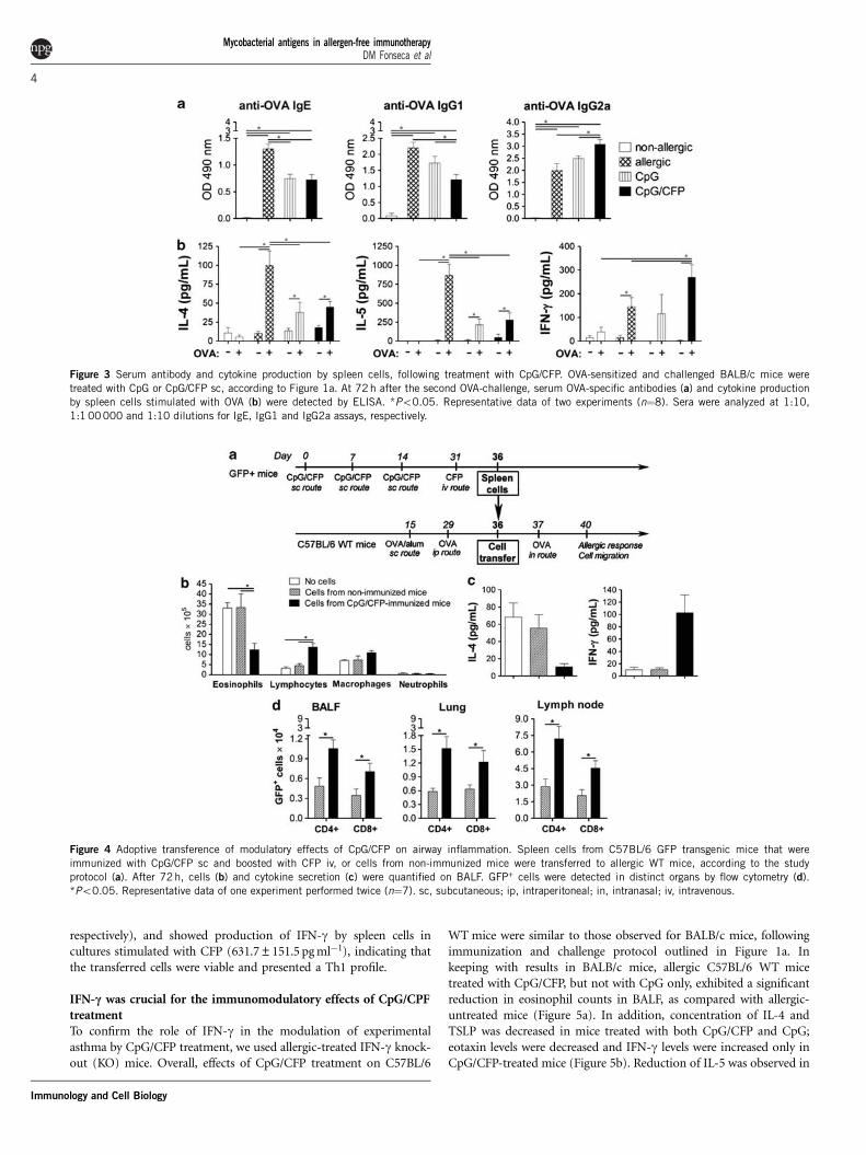
IFN-c was crucial for the immunomodulatory effects of CpG/CPFtreatmentTo confirm the role of IFN-g in the modulation of experimentalasthma by CpG/CFP treatment, we used allergic-treated IFN-g knock-out (KO) mice. Overall, effects of CpG/CFP treatment on C57BL/6
WT mice were similar to those observed for BALB/c mice, followingimmunization and challenge protocol outlined in Figure 1a. Inkeeping with results in BALB/c mice, allergic C57BL/6 WT micetreated with CpG/CFP, but not with CpG only, exhibited a significantreduction in eosinophil counts in BALF, as compared with allergic-untreated mice (Figure 5a). In addition, concentration of IL-4 andTSLP was decreased in mice treated with both CpG/CFP and CpG;eotaxin levels were decreased and IFN-g levels were increased only inCpG/CFP-treated mice (Figure 5b). Reduction of IL-5 was observed in
Figure 3 Serum antibody and cytokine production by spleen cells, following treatment with CpG/CFP. OVA-sensitized and challenged BALB/c mice were
treated with CpG or CpG/CFP sc, according to Figure 1a. At 72 h after the second OVA-challenge, serum OVA-specific antibodies (a) and cytokine production
by spleen cells stimulated with OVA (b) were detected by ELISA. *Po0.05. Representative data of two experiments (n¼8). Sera were analyzed at 1:10,
1:1 00000 and 1:10 dilutions for IgE, IgG1 and IgG2a assays, respectively.
Figure 4 Adoptive transference of modulatory effects of CpG/CFP on airway inflammation. Spleen cells from C57BL/6 GFP transgenic mice that were
immunized with CpG/CFP sc and boosted with CFP iv, or cells from non-immunized mice were transferred to allergic WT mice, according to the study
protocol (a). After 72h, cells (b) and cytokine secretion (c) were quantified on BALF. GFP+ cells were detected in distinct organs by flow cytometry (d).
*Po0.05. Representative data of one experiment performed twice (n¼7). sc, subcutaneous; ip, intraperitoneal; in, intranasal; iv, intravenous.
Mycobacterial antigens in allergen-free immunotherapyDM Fonseca et al
4
Immunology and Cell Biology
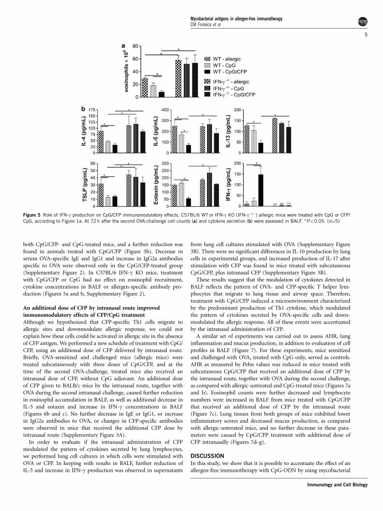
both CpG/CFP- and CpG-treated mice, and a further reduction wasfound in animals treated with CpG/CFP (Figure 5b). Decrease inserum OVA-specific IgE and IgG1 and increase in IgG2a antibodiesspecific to OVA were observed only in the CpG/CFP-treated group(Supplementary Figure 2). In C57BL/6 IFN-g KO mice, treatmentwith CpG/CFP or CpG had no effect on eosinophil recruitment,cytokine concentrations in BALF or allergen-specific antibody pro-duction (Figures 5a and b, Supplementary Figure 2).
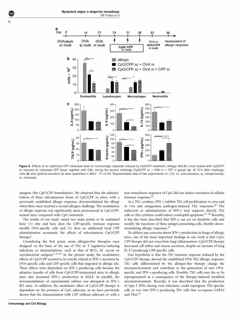
An additional dose of CFP by intranasal route improvedimmunomodulatory effects of CFP/CpG treatmentAlthough we hypothesized that CFP-specific Th1 cells migrate toallergic sites and downmodulate allergic response, we could notexplain how these cells could be activated in allergic site in the absenceof CFP antigen. We performed a new schedule of treatment with CpG/CFP, using an additional dose of CFP delivered by intranasal route.Briefly, OVA-sensitized and challenged mice (allergic mice) weretreated subcutaneously with three doses of CpG/CFP, and at thetime of the second OVA-challenge, treated mice also received anintranasal dose of CFP, without CpG adjuvant. An additional doseof CFP given to BALB/c mice by the intranasal route, together withOVA during the second intranasal challenge, caused further reductionin eosinophil accumulation in BALF, as well as additional decrease inIL-5 and eotaxin and increase in IFN-g concentrations in BALF(Figures 6b and c). No further decrease in IgE or IgG1, or increasein IgG2a antibodies to OVA, or changes in CFP-specific antibodieswere observed in mice that received the additional CFP dose byintranasal route (Supplementary Figure 3A).
In order to evaluate if the intranasal administration of CFPmodulated the pattern of cytokines secreted by lung lymphocytes,we performed lung cell cultures in which cells were stimulated withOVA or CFP. In keeping with results in BALF, further reduction ofIL-5 and increase in IFN-g production was observed in supernatants
from lung cell cultures stimulated with OVA (Supplementary Figure3B). There were no significant differences in IL-10 production by lungcells in experimental groups, and increased production of IL-17 afterstimulation with CFP was found in mice treated with subcutaneousCpG/CFP, plus intranasal CFP (Supplementary Figure 3B).
These results suggest that the modulation of cytokines detected inBALF reflects the pattern of OVA- and CFP-specific T helper lym-phocytes that migrate to lung tissue and airway space. Therefore,treatment with CpG/CFP induced a microenvironment characterizedby the predominant production of Th1 cytokine, which modulatedthe pattern of cytokines secreted by OVA-specific cells and down-modulated the allergic response. All of these events were accentuatedby the intranasal administration of CFP.
A similar set of experiments was carried out to assess AHR, lunginflammation and mucus production, in addition to evaluation of cellprofiles in BALF (Figure 7). For these experiments, mice sensitizedand challenged with OVA, treated with CpG only, served as controls.AHR as measured by Pehn values was reduced in mice treated withsubcutaneous CpG/CFP that received an additional dose of CFP bythe intranasal route, together with OVA during the second challenge,as compared with allergic-untreated and CpG-treated mice (Figures 7aand b). Eosinophil counts were further decreased and lymphocytenumbers were increased in BALF from mice treated with CpG/CFPthat received an additional dose of CFP by the intranasal route(Figure 7c). Lung tissues from both groups of mice exhibited lowerinflammatory scores and decreased mucus production, as comparedwith allergic-untreated mice, and no further decrease in these para-meters were caused by CpG/CFP treatment with additional dose ofCFP intranasally (Figures 7d–g).
DISCUSSION
In this study, we show that it is possible to accentuate the effect of anallergen-free immunotherapy with CpG-ODN by using mycobacterial
Figure 5 Role of IFN-g production on CpG/CFP immunomodulatory effects. C57BL/6 WT or IFN-g KO (IFN-g�/�) allergic mice were treated with CpG or CFP/
CpG, according to Figure 1a. At 72 h after the second OVA-challenge cell counts (a) and cytokine secretion (b) were assessed in BALF. *Po0.05. (n¼5)
Mycobacterial antigens in allergen-free immunotherapyDM Fonseca et al
5
Immunology and Cell Biology
antigens (the CpG/CFP formulation). We observed that the adminis-tration of three subcutaneous doses of CpG/CFP to mice, with apreviously established allergic response, downmodulated the allergywhen these mice received a second allergen-challenge. The modulationof allergic response was significantly more pronounced in CpG/CFP-treated mice compared with CpG treatment.
The results of our study raised two main points to be explainedhere: (1) why and how does the CFP-specific immune responsemodify OVA-specific cells and (2) does an additional local CFPadministration accentuate the effects of subcutaneous CpG/CFPtherapy?
Considering the first point, some allergen-free therapies weredesigned on the basis of the use of Th1 or T regulatory-inducinginfections or immunizations, such as that of BCG, M. vaccae ormycobacterial antigens.6–11,25 In the present study, the modulatoryeffects of CpG/CFP seemed to be strictly related to IFN-g secretion byOVA-specific cells and CFP-specific cells that migrated to allergic site.These effects were dependent on IFN-g-producing cells because theadoptive transfer of cells from CpG/CFP-immunized mice to allergicmice also increased IFN-g production in BALF. In parallel, thedownmodulation of experimental asthma was abrogated in IFN-gKO mice. In addition, the modulator effect of CpG/CFP therapy isdependent on the presence of CpG adjuvant, as we have previouslyshown that the immunization with CFP without adjuvant or with a
non-stimulatory sequence of CpG did not induce activation of cellularimmune response.22
As a Th1 cytokine, IFN-g inhibits Th2 cell proliferation in vitro andin vivo and antagonizes pathogen-induced Th2 responses.26 Theinduction or administration of IFN-g may suppress directly Th2cells or this cytokine could induce eosinophil apoptosis.27–30 Recently,it has also been described that IFN-g can act on dendritic cells andmodify the functions of these antigen presenting cells, thereby down-modulating allergic responses.31
To address any concerns about IFN-g production in lungs of allergicmice, one of the most important findings in our work is that CpG/CFP therapy did not exacerbate lung inflammation. CpG/CFP therapydecreased cell influx and mucus secretion, despite an increase of lungIL-17-producing CFP-specific cells.
Our hypothesis is that the Th1 immune response induced by theCpG/CFP therapy skewed the established OVA-Th2 allergic response.Th1 cells differentiated by the allergen-free therapy change themicroenvironment and contribute to the generation of new OVA-specific and IFN-g-producing cells. Possibly, Th2 cells may die or bereprogrammed as a consequence of the therapy-induced modifiedmicroenvironment. Recently, it was described that the productionof type I IFNs during viral infections could reprogram Th2-specificcells in vivo into IFN-g-producing Th1 cells that co-express GATA3and Tbet.32
Figure 6 Effects of an additional CFP intranasal dose on immunologic response induced by CpG/CFP treatment. Allergic BALB/c mice treated with CpG/CFP
sc received an intranasal CFP boost, together with OVA, during the second challenge (CpG/CFP sc + OVA in + CFP in group) (a). At 72 h after challenge,
cells (b) and cytokine secretion (c) were quantified in BALF. *Po0.05. Representative data of two experiments (n¼10). sc, subcutaneous; ip, intraperitoneal;
in, intranasal.
Mycobacterial antigens in allergen-free immunotherapyDM Fonseca et al
6
Immunology and Cell Biology
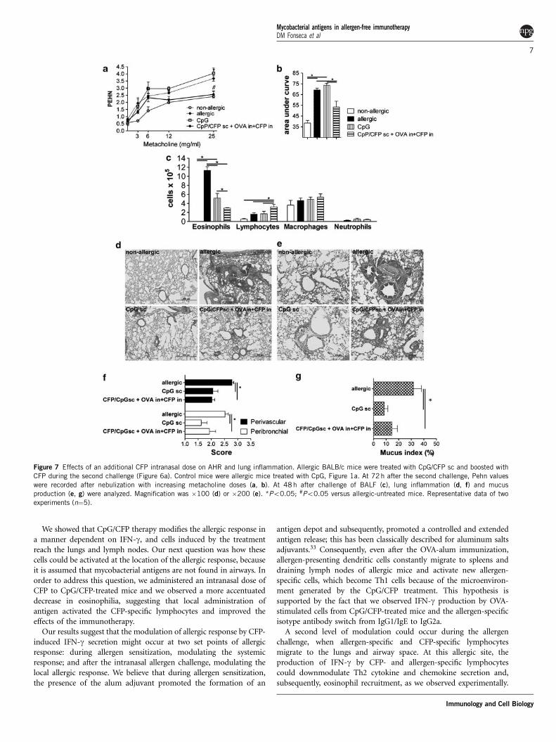
We showed that CpG/CFP therapy modifies the allergic response ina manner dependent on IFN-g, and cells induced by the treatmentreach the lungs and lymph nodes. Our next question was how thesecells could be activated at the location of the allergic response, becauseit is assumed that mycobacterial antigens are not found in airways. Inorder to address this question, we administered an intranasal dose ofCFP to CpG/CFP-treated mice and we observed a more accentuateddecrease in eosinophilia, suggesting that local administration ofantigen activated the CFP-specific lymphocytes and improved theeffects of the immunotherapy.
Our results suggest that the modulation of allergic response by CFP-induced IFN-g secretion might occur at two set points of allergicresponse: during allergen sensitization, modulating the systemicresponse; and after the intranasal allergen challenge, modulating thelocal allergic response. We believe that during allergen sensitization,the presence of the alum adjuvant promoted the formation of an
antigen depot and subsequently, promoted a controlled and extendedantigen release; this has been classically described for aluminum saltsadjuvants.33 Consequently, even after the OVA-alum immunization,allergen-presenting dendritic cells constantly migrate to spleens anddraining lymph nodes of allergic mice and activate new allergen-specific cells, which become Th1 cells because of the microenviron-ment generated by the CpG/CFP treatment. This hypothesis issupported by the fact that we observed IFN-g production by OVA-stimulated cells from CpG/CFP-treated mice and the allergen-specificisotype antibody switch from IgG1/IgE to IgG2a.
A second level of modulation could occur during the allergenchallenge, when allergen-specific and CFP-specific lymphocytesmigrate to the lungs and airway space. At this allergic site, theproduction of IFN-g by CFP- and allergen-specific lymphocytescould downmodulate Th2 cytokine and chemokine secretion and,subsequently, eosinophil recruitment, as we observed experimentally.
Figure 7 Effects of an additional CFP intranasal dose on AHR and lung inflammation. Allergic BALB/c mice were treated with CpG/CFP sc and boosted with
CFP during the second challenge (Figure 6a). Control mice were allergic mice treated with CpG, Figure 1a. At 72h after the second challenge, Pehn values
were recorded after nebulization with increasing metacholine doses (a, b). At 48 h after challenge of BALF (c), lung inflammation (d, f) and mucus
production (e, g) were analyzed. Magnification was �100 (d) or �200 (e). *Po0.05; #Po0.05 versus allergic-untreated mice. Representative data of two
experiments (n¼5).
Mycobacterial antigens in allergen-free immunotherapyDM Fonseca et al
7
Immunology and Cell Biology
It is possible that, even in the absence of the CFP in the challenge, theTh1 CFP-specific cells are activated through bystander processes. Inaddition, when we administered an additional dose of CFP by theintranasal route, the local response by CFP-specific cells increased anddownmodulated eosinophilia, IL-5 and eotaxin. Because we identifiedIFN-g as the main mediator involved in the modulation of experi-mental asthma, and because the additional dose of CFP by intranasalroute downmodulated eosinophilia IL-5 and eotaxin, but not otherTh2 cytokines, we suggest that therapy with CpG/CFP mostly affectsthe immune mechanisms associated with recruitment and/or number(viability) of eosinophils. It is possible that, besides the induction ofIFN-g, the intranasal CFP administration also induced eosinophilFas/FasL-mediated apoptosis, as previously described by otherIFN-g-inducing formulations.30 This hypothesis is under investigationby our group.
In conclusion, immunization with mycobacterial antigens, plusadjuvant CpG/CFP, may represent a promising and safe allergen-freeimmune therapy. This formulation was able to downmodulate allergicimmune responses and AHR through an IFN-g-dependent mechan-ism, without stimulating an excessive and harmful inflammatoryresponse.
METHODS
MiceSpecific pathogen-free female BALB/c, C57BL/6 WT and C57BL/6 GFP
transgenic mice, 6–8 weeks old, were obtained from local breeding facility of
the School of Medicine of Ribeirao Preto, Ribeirao Preto, Brazil. IFN-g KO
were kindly provided by Dr Joao S Silva (School of Medicine of Ribeirao Preto,
Ribeirao Preto, Brazil). Mice were housed under barrier conditions and
provided with food and sterile water. Experiments were approved by the local
ethical guidelines (protocol 071/2006).
M. tuberculosis culture filtrate proteins, CpG-oligodeoxynucleotidesand ovalbuminM. tuberculosis CFP antigens were obtained using an M. tuberculosis 14-day
culture, as previously described.22 CpG-ODN were synthesized using custom
primers (Invitrogen, San Diego, CA, USA). Chicken OVA was obtained from
commercial source (OVA—grade V; Sigma, St Louis, MO, USA). CFP antigens
and CpG-ODN had no detectable endotoxin, whereas OVA contained 0.2 EU
endotoxin/mg of protein, as determined by Limulus amebocyte-lysate assay
(Cambrex Bio Science Walkersville, Walkersville, MD, USA).
Induction of allergic inflammation and immunotherapyBALB/c mice were sensitized with two doses of OVA. The first dose of 100mg
was administered subcutaneously with 1.6 mg alum, and the second dose of
50mg was injected intraperitoneally, 14 days after the first injection. At 7 days
after sensitization, mice were challenged with OVA 100mg in saline, intranasally.
At 72 h after the first OVA challenge, mice were treated with three subcutaneous
injections of 50mg CpG-ODN, plus 50mg CFP (CpG/CFP) at 7-day intervals.
Control groups received saline (allergic-untreated mice) or three doses of 50mg
CpG-ODN (CpG-treated mice). At15 days after the end of treatment, mice
received a second intranasal OVA challenge. AHR was evaluated 24 h following
the second OVA challenge, and other parameters were assessed 72 h after
challenge.
A group of CpG/CFP-treated BALB/c mice received 50mg CFP by intranasal
route, together with the second OVA challenge (CpG/CFP subcutaneouslyc—
CFP intranasally). Allergic-untreated, allergic CpG-treated and allergic-
untreated mice that received CFP by intranasal route, served as controls.
IFN-g KO or C57BL/6 WT mice were OVA-sensitized, challenged and treated
subcutaneously with CpG/CFP, as described above.
Cell transfer experimentGFP transgenic mice were immunized with three doses of CpG/CFP. At 15 days
after the third immunization, mice received one intravenous dose of 50mg CFP.
After 5 days, 5�106 spleen cells from CpG/CFP-immunized mice or from non-
immunized mice were transferred intravenously to OVA-sensitized C57BL/6
WT mice. The WT mice were challenged with OVA by intranasal route, 1 day
after cell transfer. At 72 h after OVA-challenge, BALF, lung, draining lymph
nodes and spleen from WT-recipient mice were collected and analyzed by flow
cytometry to detect GFP+ cell migration. Cells from these sites were stained
with anti-CD4, CD8 or CD19 mAb (BD Biosciences-PharMingen, San Diego,
CA, USA) and analyzed by flow cytometry.
Determination of AHRAHR was assessed by using a single-chamber, whole-body plethysmograph
(Buxco Electronics, Wilmington, NC, USA) after nebulization with methacho-
line, as previously described.34
Spleen and lung cell culturesAt 72 h after the second allergen challenge, spleens were collected and homo-
genized; 5�106 cells ml�1 were cultured in complete RPMI-1640 medium (with
10% bovine serum, gentamicin, penicillin/streptomycin and polymyxin B) at
37 1C in 5% CO2. Cells were stimulated or not with OVA (100mg ml�1) or CFP
(10mg ml�1). After 48 h, supernatants were harvested for cytokine detection.
Lung cells were isolated, as previously described,35 and cultured and stimulated
as described above.
Cell counts in BALF and detection of cytokines and antibodiesBALF was obtained following five consecutive 1 ml injections of RPMI-1640
medium into the lungs. Total volume of 5 ml was centrifuged (400�g, 10 min),
supernatants were stored at �20 1C and cells were counted in a Neubauer
chamber; 40 000 cells were centrifuged onto microscope slides and stained with
panoptic (Clozarp-Hemogram, Inc., Curitiba, Brazil). Percentages of eosino-
phils, lymphocytes, macrophages and neutrophils were multiplied by the total
number of cells from each sample.
Concentrations of IL-4, IL-5, IL-13, TSLP, eotaxin, IFN-g, IL-17, IL-10 and
TNF-a in BALF, and in spleen and lung cell-culture supernatants were
determined by ELISA, according to manufacturer’s instructions and as pre-
viously described.22,35 Cytokine detection limits were as follows: 19 pg ml�1 for
IL-5 and IL-10; 9 pg ml�1 for IL-4, IL-17, IFN-g and TNF-a; and 5 pg ml�1 for
IL-13, TSLP and eotaxin.
CFP- and OVA-specific antibodies were assayed by sandwich ELISA,
according to manufacturer’s instructions (BD Biosciences-PharMingen, San
Diego, CA, USA), using biotin-conjugated anti-mouse IgE (R35-118), IgG1
(A85-1) and IgG2a (R19-15).
Histological analysisSections of 5mm of left lung lobes were stained with hematoxylin-eosin.
Perivascular and peribronchial inflammation was scored in a scale of 0–3 of
all vessels and bronchi in each section, as previously described.36
Lung sections were also stained with periodic acid-Schiff (PAS)/hematoxylin.
The mucus-occupying ratio (mucus index) was calculated using ImageJ software
(NIHImage, Bethesda, MD, USA), by analyzing all bronchi micro-photographed
with a magnification of �200. Results were expressed as the ratio of total bronchi
area to the periodic acid-Schiff stained area of each lung section from each mouse.
Statistical analysisAll values were expressed as means ±s.e.m. Data were compared using analysis
of variance and Prisma software (Graph Pad software, Inc., San Diego, CA,
USA). When the values indicated the presence of a significant difference,
the Tukey test was used. Values of Po0.05 were considered as significant.
CONFLICT OF INTERESTThe authors declare no conflict of interest.
ACKNOWLEDGEMENTSWe thank Elaine Medeiros Floriano for the general technical assistance
provided. Grant support: This study received financial support from the
‘Fundacao de Amparo a Pesquisa do Estado de Sao Paulo’ (FAPESP: Founda-
tion for the Support of Research in the State of Sao Paulo; grant no. 05/59198-8
Mycobacterial antigens in allergen-free immunotherapyDM Fonseca et al
8
Immunology and Cell Biology

and 05/01995-0) and the ‘Conselho Nacional de Desenvolvimento Cientıfico
e Tecnologico’ (CNPq, Council for Scientific and Technological Development).
1 Holgate ST. Pathogenesis of asthma. Clin Exp Allergy 2008; 38: 872–897.2 Barnes PJ. Immunology of asthma and chronic obstructive pulmonary disease. Nat Rev
Immunol 2008; 8: 183–192.3 Akdis M, Akdis CA. Therapeutic manipulation of immune tolerance in allergic disease.
Nat Rev Drug Discov 2009; 8: 645–660.4 Nagata M, Nakagome K. Allergen immunotherapy in asthma: current status and future
perspectives. Allergol Int 2010; 59: 15–19.5 Thomas WR. Innovation in immunotherapy. Clin Exp Allergy 2009; 39: 450–454.6 Zuany-Amorim C, Sawicka E, Manlius C, Le Moine A, Brunet LR, Kemeny DM et al.
Suppression of airway eosinophilia by killed Mycobacterium vaccae-induced allergen-specific regulatory T-cells. Nat Med 2002; 8: 625–629.
7 Erb KJ, Holloway JW, Sobeck A, Moll H, Le Gros G. Infection of mice with Mycobacter-ium bovis-bacillus Calmette-Guerin (BCG) suppresses allergen-induced airway eosino-philia. J Exp Med 1998; 187: 561–569.
8 Zuany-Amorim C, Manlius C, Trifilieff A, Brunet LR, Rook G, Bowen G et al. Long-termprotective and antigen-specific effect of heat-killed Mycobacterium vaccae in a murinemodel of allergic pulmonary inflammation. J Immunol 2002; 169: 1492–1499.
9 Obihara CC, Kimpen JL, Gie RP, Lill SW, Hoekstra MO, Marais BJ et al. Mycobacteriumtuberculosis infection may protect against allergy in a tuberculosis endemic area. ClinExp Allergy 2006; 36: 70–76.
10 Bager P, Rostgaard K, Nielsen NM, Melbye M, Westergaard T. Age at bacilleCalmette-Guerin vaccination and risk of allergy and asthma. Clin Exp Allergy 2003;33: 1512–1517.
11 Lagranderie M, Abolhassani M, Vanoirbeek J, Lefort J, Nahori MA, Lapa E et al.Mycobacterium bovis BCG killed by extended freeze-drying reduces airway hyperre-sponsiveness in 2 animal models. J Allergy Clin Immunol 2008; 121: 471–478.
12 Cohon A, Arruda LK, Martins MA, Guilherme L, Kalil J. Evaluation of BCG administra-tion as an adjuvant to specific immunotherapy in asthmatic children with mite allergy.J Allergy Clin Immunol 2007; 120: 210–213.
13 Fonseca DE, Kline JN. Use of CpG oligonucleotides in treatment of asthma and allergicdisease. Adv Drug Deliv Rev 2009; 61: 256–262.
14 Sur S, Wild JS, Choudhury BK, Sur N, Alam R, Klinman DM. Long term prevention ofallergic lung inflammation in a mouse model of asthma by CpG-oligodeoxynucleotides.J Immunol 1999; 162: 6284–6293.
15 Broide D. Immunostimulatory DNA sequences inhibit IL-5, eosinophilic inflammationand airway hyperresponsivess in mice. J Immunol 1998; 161: 7054–7062.
16 Xu W, Tamura T, Takatsu K. CpG ODN mediated prevention from ovalbumin-inducedanaphylaxis in mouse through B cell pathway. Int Pharmacol 2008; 8: 351–361.
17 Ashino S, Wakita D, Zhang Y, Chamoto K, Kitamura H, Nishimura T. CpG-ODN inhibitsairway inflammation at effector phase through down-regulation of antigen-specific Th2-cell migration into lung. Int Immunol 2007; 20: 259–266.
18 Creticos PS, Schroeder JT, Hamilton RG, Balcer-Whaley SL, Khattignavong AP,Lindblad R et al. Immunotherapy with a ragweed-toll-like receptor 9 agonist vaccinefor allergic rhinitis. N Engl J Med 2006; 355: 1445–1455.
19 Simons FE, Shikishima Y, Van Nest G, Eiden JJ, Hayglass KT. Selective immuneredirection in humans with ragweed allergy by injecting Amb a 1 linked to immunos-timulatory DNA. J Allergy Clin Immunol 2004; 113: 1144–1151.
20 Senti G, Johansen P, Haug S, Bull C, Gottschaller C, Muller P et al. Use of A-type CpGoligodeoxynucleotides as an adjuvant in allergen-specific immunotherapy in humans: aphase I/IIa clinical trial. Clin Exp Allergy 2009; 39: 562–570.
21 Gauvreau GM, Hessel EM, Boulet LP, Coffman RL, O’Byrne PM. Immunostimulatorysequences regulate interferon-inducible genes but not allergic airway responses. Am JRespir Crit Care Med 2006; 174: 15–20.
22 Fonseca DM, Silva CL, Paula MO, Soares EG, Marchal G, Horn C et al. Increased levelsof IFN-g primed by CFP antigen and CpG-ODN immunization do not confer significantprotection against Mycobacterium tuberculosis infection. Immunology 2007; 121:508–517.
23 Weldingh K, Andersen P. Immunological evaluation of novel Mycobacterium tubercu-losis culture filtrate proteins. FEMS 1999; 23: 159–164.
24 Andersen P. Effective vaccination of mice against Mycobacterium tuberculosis infec-tion with a soluble mixture of secreted mycobacterial proteins. Infect Immun 1994; 62:2536–2544.
25 Trujillo-Vargas CM, Mayer KD, Bickert T, Palmetshofer A, Grunewald S, Ramirez-PinedaJR et al. Vaccinations with T-helper type 1 directing adjuvants have different suppres-sive effects on the development of allergen-induced T-helper type 2 responses. Clin ExpAllergy 2005; 35: 1003–1013.
26 Fernandez-Botran R, Sanders VM, Mosmann TR, Vitetta ES. Lymphokine-mediatedregulation of the proliferative response of clones of T helper 1 and T helper 2 cells.J Exp Med 1988; 168: 543–548.
27 Hofstra CL, Van Ark I, Hofman G, Nijkamp FP, Jardieu PM, Van Oosterhout AJ.Differential effects of endogenous and exogenous interferon-gamma on immunoglobu-lin E, cellular infiltration, and airway responsiveness in a murine model of allergicasthma. Am J Respir Cell Mol Biol 1998; 19: 826–835.
28 Fujiwara M, Hirose K, Kagami S, Takatori H, Wakashin H, Tamachi Tet al. T-bet inhibitsboth Th2 cell-mediated eosinophil recruitment and Th17 cell-mediated netrophilrecruitment into airways. J Allergy Clin Immunol 2007; 119: 662–670.
29 Li XM, Chopra RK, Chou TY, Schofield BH, Wills-Karp M, Huang SK. Mucosal IFN-ggene transfer inhibits pulmonary allergy responses in mice. J Immunol 1996; 157:3216–3219.
30 Tong J, Bandulwala HS, Clay BS, Anders RA, Shilling RA, Balachandran DD et al. Fas-positive T cells regulate the resolution of airway inflammation in a murine model ofasthma. J Exp Med 2006; 203: 1173–1184.
31 Nakagome K, Okunishi K, Imamura M, Harada H, Matsumoto T, Tanaka R et al. IFN-gamma attenuates antigen-induced overall immune response in the airway as a Th1-type immune regulatory cytokine. J Immunol 2009; 183: 209–220.
32 Hegazy AN, Peine M, Helmstetter C, Panse I, Frohlich A, Bergthaler A et al. Interferonsdirect Th2 cell reprogramming to generate a stable GATA-3(+)T-bet(+) cell subset withcombined Th2 and Th1 cell functions. Immunity 2010; 32: 116–128.
33 Lambrecht BN, Kool M, Willart MA, Hammad H. Mechanism of action of clinicallyapproved adjuvants. Curr Opin Immunol 2009; 21: 23–29.
34 Bortolatto J, Borducchi E, Rodriguez D, Keller AC, Faquim-Mauro E, Bortoluci KR et al.Toll-like receptor 4 agonists adsorbed to aluminium hydroxide adjuvant attenuateovalbumin-specific allergic airway disease: role of MyD88 adaptor molecule andinterleukin-12/interferon-gamma axis. Clin Exp Allergy 2008; 38: 1668–1679.
35 Fonseca DM, Silva CL, Wowk PF, Paula MO, Ramos SG, Horn C et al. Mycobacteriumtuberculosis culture filtrate proteins plus CpG oligodeoxynucleotides confer protectionto Mycobacterium bovis BCG-primed mice by inhibiting interleukin-4 secretion. InfectImmun 2009; 77: 5311–5321.
36 Curtis JL, Warnock ML, Arraj SM, Kaltreider HB. Histological analysis of an immuneresponse in the lung parenchyma of mice. Am J Pathol 1990; 137: 689–699.
Supplementary Information accompanies the paper on Immunology and Cell Biology website (http://www.nature.com/icb)
Mycobacterial antigens in allergen-free immunotherapyDM Fonseca et al
9
Immunology and Cell Biology