adjuvant effect of synthetic oligodeoxyribonucleotides (cpg-odn) from the paracoccidioides...
TRANSCRIPT

Adjuvant Effect of Synthetic Oligodeoxyribonucleotides(CpG-ODN) From the Paracoccidioides brasiliensisgp43 gene on the Th2–Th1 Immunomodulation ofExperimental Paracoccidioidomycosis
C. C. Amaral*, I. P. Garcia*, G. F. Fernandes*, S. R. Almeidaz, Z. P. Camargo* & M. C. Souzaz
*Discipline of Cellular Biology, Department of
Microbiology, Immunology and Parasitology,
Federal University of Sao Paulo; yDepartment ofClinical and Toxicological Analysis, University of
Sao Paulo, Sao Paulo; and zMethodist
University of Sao Paulo, Sao Bernardo do
Campo, SP, Brazil
Received 19 May 2005; Accepted in revisedform 25 August 2005
Correspondence to: Dr M. C. Souza, Universidade
Metodista de Sao Paulo (UMESP), 09895-400,
Rua Dom Jaime de Barros Camara, 1000,
Planalto, Sao Bernardo do Campo, SP, Brazil.
E-mail: [email protected]; Dr Z. P. Carmargo.
E-mail: [email protected]
Abstract
Paracoccidioidomycosis (PCM) is caused by the dimorphic fungusParacoccidioides brasiliensis. Immunostimulatory effects of P. brasiliensis DNAand CpG-oligodeoxyribonucleotides (CpG-ODN) have shown a Th2–Th1immunomodulation of the isogenic murine model of susceptibility, whichdevelops a progressive and disseminating disease. In this study, we investigatedthe optimum time interval and doses of CpG-ODN which are able to induceTh2–Th1 immunomodulation. The optimum concentrations for the inductionof a decrease in antibody production were 0.5 and 1 mg. Mice immunized twicewith CpG-ODN and gp43 (5 and 7 days before the challenge) showed a 60%higher chance of survival compared with the control group (nonimmunized),and an increase in Th1 isotype (IgG2a) was also observed. In vitro assays of naiveand preimmunized mice showed discrete cellular proliferation when stimulatedby suitable concentrations of CpG-ODN. Type 1 cytokines interleukin-12 (IL-12)and interferon-g were increased in cell culture supernatants, but no significantdifference was found in Th2 IL-4 cytokines in stimulated or nonstimulated cellcultures. Concerning the Th2–Th1 kinetics in experimental PCM models byadjuvant effect of CpG-ODN, there are still many questions to be answered andclarified. However, the gathering of data obtained in this investigation has led us tosuggest that the modulation of Th2–Th1 in experimental PCM depends on timeand CpG-ODN concentration.
Introduction
Paracoccidioidomycosis (PCM), caused by the thermallydimorphic fungus Paracoccidioides brasiliensis, is the mostfrequent systemic mycosis in Latin America. This infectionis acquired by inhalation of airborne propagules found innature, which reach the lungs and are then converted toyeast forms [1]. These yeast forms can be either eliminatedby immune-competent cells or disseminated into tissuesthrough lymphatic or haematogenous routes. PCM ischaracterized by suppurative granulomatous inflammation,suppression of cellular immunity and high antibody titres[2]. The disease may develop into multiple forms, rangingfrom benign and localized to severe and disseminatedforms, depending on the extension of the cellular immun-ity depression [3, 4].
In the acute form of PCM, there is an involvement ofthe reticuloendothelial system, hypergammaglobulinaemia,a depressed cellular immune response, a diffuse inflamma-tory granulomatous response and concomitant fungus dis-semination. The chronic form of PCM presents a broadspectrum of clinical manifestations with frequent damageto the lung and oropharyngeal mucosa and a deep impair-ment of the immune response [4, 5] with preserved cellu-lar immunity and moderate specific antibody response. Acorrelation between PCM clinical forms and the pattern ofimmune response has been investigated in human andmouse models [5].
Cellular immune response is considered the maindefence mechanism against P. brasiliensis infection, andalthough large amounts of specific antibodies are
doi: 10.1111/j.1365-3083.2005.01680.x............................................................................................................................................................................................................
# 2005 Blackwell Publishing Ltd. Scandinavian Journal of Immunology 62, 325–333 325

produced, they are not able to confer protection [6].Severe PCM forms are characterized by strong T-cell dys-function, impaired in vivo delayed-type hypersensitivity,alterations in T-cell subpopulation ratios, an unbalancedlevel of Th1 cytokine production and suppressive cellularproliferation [7, 8].
An isogenic murine model of PCM showing a divergingimmune response, in which polar forms of the disease arereproduced, was previously characterized. A/Sn and B10Amouse strains are known as models of resistance andsusceptibility to experimental PCM, respectively [3, 9,10]. On the other hand, various studies have demonstratedthat the major challenge to innate immune cells is thediscrimination between foreign pathogens and self. Therole of lymphocytes in the development and maintenanceof an adaptive immune response has been extensivelydemonstrated [11].
Many of the cellular and molecular events that regulatethe induction of humoral and cell-mediated immunity havebeen well characterized [11]. It has become evident that theinduction and regulation of adaptive immunity is greatlyinfluenced by innate immune system response, which occursduring the infection or inflammation process [11, 12].Innate immune cells, such as antigen-presenting cells, havegermline-encoded pattern-recognition receptors (PRR)which can recognize evolutionary conserved molecules andbe triggered by them (essential to pathogen function, but notpresent in the host). These pathogen-associated molecularpatterns (PAMP) are widely spread and include cell wallcomponents such as mannans in the yeast cell wall, lipopo-lysaccharide in gram-negative bacteria, lipoproteins, pepti-doglycans and DNA containing unmethylated CpG motifs(these can be identified as hexamers which are composed ofcentral unmethylated cytosine CpG dinucleotides) [13].
There is some evidence that the innate immune systemutilizes a variety of receptors, such as PRR, to recognizePAMP which are unique in microorganisms [11]. CpGmotifs within the DNA of microorganisms act as PAMPand can activate a variety of cells involved in the inductionof an adaptive immune response.
CpG motifs present in pathogen DNA and syntheticoligodeoxyribonucleotides (CpG-ODN) can enhanceB-cell survival, influence dendritic cell differentiation andinduce cytokine secretion by B cells, monocytes, naturalkiller cells (NK) and dendritic cells [14–17]. Treating micewith ODN containing CpG motifs can protect these ani-mals against a variety of pathogens [18–22]. All theseobservations imply that CpG motifs are potent PAMP,which effectively activate the innate immune system andmodulate the adaptive immune response [23].
In our previous study [24], we showed that immuno-stimulatory sequences (ISS) found in the DNA ofP. brasiliensis have the capacity to immunomodulate theTh2–Th1 response in a PCM murine model. We have alsoobserved that DNA from P. brasiliensis and synthetic
ODN from P. brasiliensis gp43 genes work as Th2–Th1stimulators [24]. However, the mechanisms of this modu-lation are not yet clear. In the present study, we exploredthe mechanisms underlying the adjuvant kinetics of ODNin Th2–Th1 immunomodulation of susceptible mice dur-ing PCM infection.
Materials and methods
Mice and infection. B10.A isogenic mice aged 8–12 weekswere obtained from the animal facilities of the FederalUniversity of Sao Paulo and infected intraperitoneallywith 106 viable yeast cells (determined by Trypan Bluedye) of virulent P. brasiliensis strain (isolate 1924) sus-pended in phosphate-buffered saline (PBS).
Oligodeoxynucleotides. ODN (ODN 3234: 50-ATGTAG ACG TTT CTT GT-30) were purchased by theBiophysics Department of the Federal University of SaoPaulo. The ODN was purified by precipitation with 3 M
sodium acetate and absolute ethanol, air-dried and sus-pended in sterilized PBS until use.
Immunization schedules to find the optimum ODNconcentration. Five mice from each group were immunizedin the tail base by the subcutaneous route, with CpG-ODN at different concentrations (0.1, 0.5, 1, 5, 10 and20 mg) in sterilized PBS, and they were simultaneouslyinfected with 106 P. brasiliensis yeast cells. The mice wereobserved for 8 weeks.
Immunization schedules to find the optimum timeinterval. Mice were divided into three groups of 10 ani-mals each. Before the infection with 106 P. brasiliensisyeast cells, they were preimmunized (T0) with 1 mg ofCpG-ODN and 50 mg of gp43. Five mice from eachgroup were infected on the 3rd, the 5th or the 7th dayafter the first immunization. The other 15 mice received asecond dose of immunization (CpG-ODNþ gp43) on the7th day (T7), so they were infected on the 3rd, the 5th orthe 7th day after this second immunization. In all experi-ments, control mice (only infected) were used in parallel.The mice were observed for 8 weeks.
ELISA assays for the detection of specific humoralresponse: Mice were infected as described above; serawere obtained in the 2nd, 4th, 6th and 8th weeks post-infection and tested for the detection of specific IgG anti-gp43. Polystyrene plates containing 96 wells (Costar,Corning Inc., Cambridge, MA, USA) were coated withpurified gp43 at 250 ng/well in 0.1 M carbonate–bicarbo-nate buffer, pH 9.6. They were incubated for 2 h at 37 �Cand left overnight at 4 �C. The plates were washed fivetimes with PBS containing 0.1% Tween-20 (PBS-T), andthe remaining sites were blocked with 5% nonfat milk inPBS-T at 37 �C for 2 h. After five washings with PBS-T[dilution 1:50 in PBS-T containing 0.25% gelatine (PBS-T-G)],sera were added to the wells in duplicates; the plates wereincubated for 1 h at 37 �C and washed as described above.
326 Adjuvant Effect of CpG-ODN on Experimental Paracoccidioidomycosis C. C. Amaral et al.............................................................................................................................................................................................................
# 2005 Blackwell Publishing Ltd. Scandinavian Journal of Immunology 62, 325–333
Anti-mouse IgG (y-chain specific) peroxidase conjugated(Sigma Chemical Co., St Louis, MO, USA) was added toeach well (dilution 1:1000 in PBS-T-G). The plates wereincubated for 1 h at 37 �C and washed five times withPBS-T, and then 100 ml of substrate solution (5mg ofo-phenylenediamine in 10ml of 0.1 M citrate phosphatebuffer, pH 4.5, plus 10 ml of 30% H2O2) was added. Aftercolour development, the reaction was interrupted by theaddition of 50 ml of 4N H2SO4. The optical densities wereread by an ELISA reader (Titertek Multiskan MCC/340,Labsystem, Helsinki, Finland) at 492 nm. For the isotyp-ing of antibodies, an ELISA isotyping panel was usedaccording to the manufacturer’s instructions (Bio-RadLaboratories, Richmond, CA, USA).
Proliferation assay. Inguinal and popliteal lymph nodes(LN) from naive mice were removed, and the cells were dis-persed manually and centrifuged twice at 1500� g (Harrier18/80, Sanyo, Loughborough, UK) for 5min. The pellet wassuspended in 10mM 4-(2-Hydroxymethylpiperazine-1-etha-nesulfonic acid) (HEPES), 100U of penicillin/ml, 100mg ofstreptomycin/ml (Sigma), 2mM L-glutamine (Sigma), 50mM
b-mercaptoethanol (Sigma), 5mM sodium pyruvate (Sigma)and 100mM nonessential amino acids (Life Technologies,Washington DC, WA, USA) with 1% fetal bovine serum(FBS) (High Clone, Gibco-BRL, Bethesda, MD, USA). Thecells were counted in a 1:1 dilution in 0.1% Trypan Blue dye.Viable cells were cultured at a density of 4� 105 cells/well in96-well plates (Costar). Different concentrations of CpG-ODN (0.1, 0.5 and 1.0mg) were added and incubated at37 �C and 5% CO2 for 144h. Controls were run with com-plete culture medium and 2mg of concanavalin A. Then,cultures were pulsed with [H3] thymidine (AmershamPharmacia Biotech, Uppsala, Sweden) (0.5mCi/well), har-vested 18–24h later, on the 4th day of culture. The incorpo-rated radioactivity was determined by a liquid scintillationcounter. Supernatants were collected, centrifuged at 350� gfor 5min and stored at �70 �C, before use.
Cytokine quantification. The supernatants from the cellcultures were collected at 24, 48 and 72 h and assayed byELISA. The antibodies used to coat the 96-well plates wereanti-interleukin-4 (IL-4), anti-IL-12 and anti-interferon-g(anti-IFN-g) (PharMingen, San Diego, CA, USA), accord-ing to the manufacturer’s instructions. Concentrationswere determined by plotting curves with serial dilutionsof recombinant mouse cytokines.
Histological analysis. Fragments of the spleen and theliver from infected mice were excised, fixed in 10% buf-fered formalin and embedded in paraffin for sectioning.The sections were stained with hematoxylin–eosin andexamined microscopically (Optiphot-2, Nikon, Tokio,Japan). Granulomatous formations accompanied by thepresence of P. brasiliensis cells were observed in each organ.
Fungal loads in organs of immunized and infectedanimals. In the 12th week, the groups of infected animalswere killed and the fungal loads in their organs (the lungs,
the liver and the spleen) were determined by colony-forming units (CFU). The organs were rapidly collected,weighed and disrupted using a tissue grinder. Cellularsuspensions were plated on BHI agar and antibiotics, 5%FBS and growth factors; according to Singer-Vermes et al.(1992) [23, 25]. The colonies were counted from the 7thday, until no increase in colony numbers was observed.The results were expressed in logarithmic values of CFUcounts/g of tissue.
Statistical comparisons. Statistical data were obtained fromthe analysis of variance (ANOVA) and from the Turkey-Kramer test. All values were reported as the mean� theSEM with significance in the range of P< 0.005.
Results
Dose-dependent effect of CpG-ODN during the course of
experimental P. brasiliensis infection
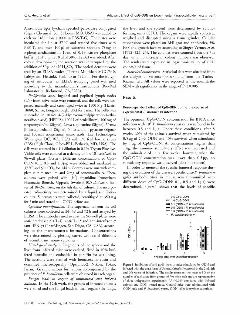
The optimum CpG-ODN concentration for B10.A miceinfection with 106 P. brasiliensis yeast cells was found to bebetween 0.5 and 1 mg. Under these conditions, after 8weeks, 60% of the animals survived when stimulated by0.5 mg of CpG-ODN and 40% survived when stimulatedby 1 mg of CpG-ODN. At concentrations higher than1 mg, the immune stimulatory effect was increased andthe animals died in a few weeks; however, when theCpG-ODN concentration was lower than 0.5 mg, nostimulatory response was observed (data not shown).
In order to monitor the specific humoral response dur-ing the evolution of the disease, specific anti-P. brasiliensisgp43 antibody titres in mouse sera (immunized withdifferent doses of CpG-ODN: 0.1, 0.5 and 1 mg) weredetermined. Figure 1 shows that the levels of specific
P. brasiliensis only
00
1
2
OD
492
nm
2
3
4
4 6
*
*
0.5 CpG-ODN1.0 CpG-ODN0.1 (ODN + P. brasiliensis)0.5 (ODN + P. brasiliensis)1.0 (ODN + P. brasiliensis)
Weeks after immunization/infection
Figure 1 Inhibition of anti-gp43 titres in mice stimulated by ODN and
infected with the yeast form of Paracoccidioides brasiliensis in the 2nd, 4th
and 6th weeks of infection. The results represent the mean� SD of the
number of each assay from groups of five mice each and are representative
of three independent experiments; *P� 0.005 compared with infected
animals and ODN-treated mice. Control mice were administered with
ODN only and P. brasiliensis yeasts. ODN, oligodeoxyribonucleotides.
C. C. Amaral et al. Adjuvant Effect of CpG-ODN on Experimental Paracoccidioidomycosis 327............................................................................................................................................................................................................
# 2005 Blackwell Publishing Ltd. Scandinavian Journal of Immunology 62, 325–333

antibodies decreased in immunized mice in the 4th and6th weeks in relation to the nonimmunized rodents butinduced by P. brasiliensis alone (infected only). LN cellsfrom B10.A naive mice showed a discrete increase incellular proliferation in relation to the nonstimulatedcells when stimulated by 0.5- and 1-mg CpG-ODN dosesin vitro (Fig. 2).
In vivo assays can be observed in Fig. 3(A–D), whichshows the histopathological analysis of the liver and spleensections of CpG-ODN-immunized and nonimmunizedmice using 1 mg of CpG-ODN. In immunized mice, adecrease in fungal load was discovered in the liver and thespleen, which exhibited a limited number of well-organized granulomas in relation to the nonimmunizedanimals, which display numerous nonorganized inflammatorylesions with viable yeast cells. This decrease in fungal load in
12,500
Cell proliferation
10,000
7500
CP
M
5000
2500
0
Unstim
ulate
d
Unstimulated
+ Con
trol
+Control
gp43
gp43
1.0
*
*
1.0 µg ODN0.5 µg ODN0.1 µg ODN
0.5 0.1
Figure 2 Induction of proliferation by ODN in lymph node (LN) cells
from naive mice. The results represent the mean� SD of the number of
each assay from groups of five mice and are representative of three
independent experiments; *P� 0.005 compared with the nonstimulated
animals and ODN-treated mice. ODN, oligodeoxyribonucleotides.
Infected
ODN-immunized
ODN + gp43-immunized
Lungs
6
5
4
3
Log 1
0 C
FU
(g/
tissu
e)
2
1
0Liver Spleen
40× 40×
40×
40×
10× 10×
10×10×
A B
C
E
D
Figure 3 (A) Section of the spleen of infected
mice only (106 Paracoccidioides brasiliensisyeast cells) in 40� and 10�. (B) Section of
the spleen from ODN-treated (1 mg) mice and
infected later. (C) Section of the liver infected
only at 40� and 10�. (D) ODN-treated
(1 mg) mice liver and infected later at 40�and 10�. (E) Representation of colony-form-
ing units (CFU) from ODN, ODNþ gp43-
treated and nontreated mice. ODN,
oligodeoxyribonucleotides.
328 Adjuvant Effect of CpG-ODN on Experimental Paracoccidioidomycosis C. C. Amaral et al.............................................................................................................................................................................................................
# 2005 Blackwell Publishing Ltd. Scandinavian Journal of Immunology 62, 325–333

these organs can be observed in the CFU assays presented inFig. 3(E).
LN cells were cultured in 24, 48 and 72 h, and cytokineconcentrations in supernatants of LN cells stimulated by0.5 and 1 mg of CpG-ODN were realized at 24, 48 and72 h by ELISA. The levels of Th1 cytokines IL-12(Fig. 4A) and IFN-g (Fig. 4B) increased in relation to thecontrol, and no difference was found in Th2 IL-4 cytokineconcentration in stimulated or nonstimulated LN cells(data not shown).
Time-dependent effect of CpG-ODN plus gp43 on the survival
of P. brasiliensis and anti-gp43-specific antibodies on the
course of P. brasiliensis infection
Attempts to verify the optimum time interval for immun-ization with CpG-ODN were assayed, but no significanteffect was observed (data not shown). Before infectionwith 106 P. brasiliensis yeast cells, the combination CpG-ODN (1 mg) plus gp43 (50 mg) was tested in relation totime. The animals were then immunized following theadopted schedule (see the section entitled Materials andmethods), monitored for survival for 8 weeks, and theanti-gp43-specific antibody level was determined. In ourobservations, the best scheme of immunization in relation
to survival was when mice were immunized twice (at T0and T7) and infected on the 5th (Fig. 5A) and 7th days(Fig. 5B) before the last immunization. A decrease in spe-cific anti-gp43 antibody production was observed whenmice were immunized twice (at time 0 and on the 7th day)and then infected on the 5th day after this second dose ofimmunization.
Mice immunized with CpG-ODNþ gp43 before the5th and 7th days of infection presented higher amounts ofIgG2a (Th1 isotype) than the control group (onlyinfected) (Fig. 6A,B). IgG2b levels were superior in non-immunized mice compared with those in mice immunized5 and 7 days before infection. IgG2a and IgG2b were notdetected in either group of immunized and nonimmunizedmice on the 3rd day before infection (data not shown).
Discussion
For many years, experimental studies have shown abun-dant evidence that cell-mediated immunity is critical forhost defence in PCM [25–28]. The innate immune systemof vertebrates recognizes conserved structures and allowsimmediate host immune response to limit invasion bymicrobes [13]. On the other hand, synthetic ODN con-taining CpG motifs also show similar immunostimulatoryactivity [29]. Other studies have demonstrated the adju-vant effects of CpG-ODN when co-administrated withantigens. In such studies, ISS work as potent adjuvantswhich are able to promote Th1 response when they areinoculated together with antigens of pathogenic agents[30]. Studies have also shown that when mice are pre-treated with CpG-ODN, they are protected against chal-lenges with Listeria monocytogenes [18, 21] or Leishmaniamajor [22].
In several infectious diseases [31–37], the Th1 or Th2differentiation of the antigen-specific immune responsehas significant implications in therapy. In murine leishma-niasis, susceptible mice can become resistant by the admin-istration of IL-12 [38] or ODN [22]. This implies thatCpG-ODN motifs may be useful as adjuvants in order toinduce protective Th1 response.
Currently, there is little information about protectiveTh1 immune response in PCM, and in this study, CpG-ODN was used as an immune enhancer in order to protectsusceptible B10.A mice against P. brasiliensis infection.During the course of our experiments, important observa-tions were made which may contribute to the understand-ing of this immune activator (CpG-ODN) in the immuneresponse against PCM, in the experimental murine modelof susceptibility.
Our first observation concerns the optimum CpG-ODN concentration for use in immunization and, forthis purpose, we observed that doses varying from 0.5 to1 mg were ideal for protecting susceptible mice.
1000
pg/m
lpg
/ml
IL-12
IFN-γ
750
500
A
B
250
024 h
*
**
*
*
48 h 72 h
24 h 48 h 72 h
Unstimulated
Stimulated
Unstimulated
Stimulated
300
200
100
0
Figure 4 (A) Determination of IL-12 secretion from cells stimulated by
the use of ODN in supernatants of proliferative in vitro assays.
*P< 0.005 compared with the nonstimulated mice. (B) Increase in
IFN-g secretion by cells stimulated by the use of ODN concentration
in naive mice cells in vitro. *P< 0.005 compared with nonstimulated mice.
IFN-g, interferon-g; IL-12, interleukin-12; ODN, oligodeoxyribonucleotides.
C. C. Amaral et al. Adjuvant Effect of CpG-ODN on Experimental Paracoccidioidomycosis 329............................................................................................................................................................................................................
# 2005 Blackwell Publishing Ltd. Scandinavian Journal of Immunology 62, 325–333

It has been proposed that the balance between Th1 andTh2 responses regulates the clinical outcome in manyinfectious diseases, such as leishmaniasis, bacterial infec-tions, mycobacterial infections, fungal infections and hel-minth infections [31]. In the present study, it has beenshown that the administration of CpG-ODN could pro-mote the activation of mouse immune system throughTh1 cytokine secretion and stimulation of immune cellsbefore challenge with P. brasiliensis. Thus, our mainhypothesis was based on the fact that the innate immunesystem could be activated and this response could bemaintained during the challenge with the fungus.
Discrete cellular proliferation in naive cells stimulated ‘invitro’ with CpG-ODN was observed. Some studies haveshown that both human and murine PCM are predomin-antly characterized by an antigen-specific hyporesponsive-ness. Thus, the poor proliferation shown by stimulated cellscould be indicative of a change in immune response [39].
When the supernatants of these cells were analysed forcytokines, there was an increase in IFN-g and IL-12 secre-tion accompanied by an increase in cell proliferation,whereas Th2 cytokine IL-4 showed no difference in
stimulated and nonstimulated cultures. This could reflecta tendency for Th1 immune response.
The production of cytokines, such as IFN-g by Th1cells, can activate macrophages and increase NO produc-tion, which is directly involved in the death of intracellularyeast [40]. It has been established by many groups thatIL-12 plays a pivotal role in controlling cell-mediatedimmunity and confers immunoprotection to differentintracellular pathogens [41, 42]. IL-12 is necessary forthe production of IFN-g by NK and T cells [43, 44]. Inaddition, IL-12 plays a key role in the initiation anddetermination of the type of T-helper response at variousstages of the antigenic challenge. Our results are in agree-ment with these previous studies, as mice immunized withCpG-ODN presented increased IL-12 production and thiscytokine is very important in Th2–Th1 modulation.
Experimental data indicate that resistance to P. brasi-liensis infection relies on T-cell macrophages and B-cellactivities, which are related to IFN-g and other Th1-typecytokines [25]. On the other hand, susceptibility is linkedto the preferential production of cytokines of the Th2type, i.e. IL-4, IL-5, IL-13 and IL-10 [26].
ODN only (1×)
ODN only (2×)
0.6
A
0.5
0.4
OD
492
nm
OD
492
nm
Per
cent
age
of S
urvi
val
0.3
0.2
0.1
0.0
Group 4Paracoccidioidesbrasiliensis (control)
Group 3
gp43
00
1 2 3 4 5 6 7 8
Weeks
Weeks after infection
Group 3
*
*
*
Group 4
gp43-treated
Untreated
Untreated
IgG anti-gp435 days
0 2nd 4th 6th 8th
100
75
50
25
ODN only (1×)
ODN (1×)
0 1 2 3 4 5 6 7 8
Weeks
Weeks after infection
ODN only (2×)
Group 5
Group 5
Group 6
Group 6
gp43
gp43-treated
Untreated
Untreated
Untreated
IgG anti-gp437 days1.00
B
0.75
0.50
0.25
0.00
100
75
50
Per
cent
age
of S
urvi
val
25
0
0 2nd 4th 6th
*
*
*
8th
Figure 5 (A) Anti-gp43 antibody levels and survival curve of mice ODN-immunized once or twice before 5 days of infection. Mice from group 3 were
immunized once, and mice from group 4 received two immunizations before infection. The results represent the mean� SD of the number of each assay
obtained with five mice per group and are representative of three independent experiments. Note: In the 4th, 6th and 8th weeks, there are no bars because
mice from group 3 died. (B) Anti-gp43 antibody levels and survival curve obtained from mice immunized once or twice with ODN before 7 days of
infection. Group 5 represents one immunization, while group 6 received two immunizations before infection. The results represent the mean� SD of the
number of each assay obtained with five mice per group and are representative of three independent experiments. Note: In the 8th week, there are no bars
because mice died. ODN, oligodeoxyribonucleotides.
330 Adjuvant Effect of CpG-ODN on Experimental Paracoccidioidomycosis C. C. Amaral et al.............................................................................................................................................................................................................
# 2005 Blackwell Publishing Ltd. Scandinavian Journal of Immunology 62, 325–333

LN cells from susceptible mice presented sustained pro-duction of type 1 cytokines. The high and early IFN-g andIL-12 production by LN cells was not accompanied byequivalent levels of IL-4 (data not shown). IFN-g appearedin decreasing levels after 72 h of culture in these cells. Thisfact suggests that different cell populations are involved inthe secretion of these cytokines or, alternatively, indicatesthe existence of an asynchronous activation pattern of genecoding for IL-12 and IFN-g [45]. It should be mentionedthat immunized mice produced larger amounts of IgG2aat week 8 after infection, whereas nonimmunized micepresented larger amounts of IgG2b secretion in the sameweek.
Considering that the Ig isotypes are determined by thecytokine patterns, the detection of a particular isotypecould be considered as an indicator in accordance withthe pattern of inductive cytokines, Th1 or Th2 [46].IgG2a, an isotype positively regulated by IFN-g, appearsin larger amounts in immunized mice in relation to thosenonimmunized, which showed an increase in IgG2b [45].
In our previous study, we showed that ISS sequencesfound in P. brasiliensis DNA diminish nitric oxide produc-tion in macrophages, and at the same time, there is anincrease in IFN-g secretion by other cells of the innate
immune system [24]. CpG-ODN represents a class ofcompounds that mimics the presence of pathogen-associated DNA, and based on the type of DNA sequence,the immune system may follow different immunologicalpathways [47].
Firstly, the immunostimulatory effect of CpG-ODNalone was found to provoke a decrease in anti-gp43 anti-body response and this effect reflects on the survival ofmice, as compared to those nonimmunized. However, thisprotective effect is dose-dependent, where 0.5 or 1 mg isthe most protective dose.
Human and animal immune defences against P. brasi-liensis infection depend on effective cellular immuneresponse at the initial infection site: the lungs [48, 49].As PCM is immunologically characterized by a chronicinflammatory granulomatous reaction, the formation ofgranuloma was analysed in CpG-ODN-immunized andnonimmunized mice. The intensity of pulmonarylesions in control mice showed an intense inflammatoryinvolvement of the lungs with fungi spreading to thespleen and the liver, whereas in pulmonary lesions ofCpG-ODN-immunized mice, an intense inflammatoryinvolvement of the lungs was also found, but with thefungi spreading to the spleen and the liver showing areduction in the histological analysis. In assays fromrecovered fungi in immunized mouse organs, a decreasein colony formation was found when compared to organsof untreated mice.
Studies have demonstrated that macrophage activationand granuloma formation protect the host against extra-pulmonary dissemination of fungi in PCM infection, andthat this is characterized by an inflammatory responseinduced by the fungus [50]. In this study, in vivo resultsobtained from mice infected after CpG-ODN immuniza-tion showed that these rodents presented little extrapul-monary dissemination. This observation led us tosuggesting an inflammatory response from innate immuneresponse in immunized mice.
This study suggests that CpG-ODN demonstrates aprotective effect on mouse P. brasiliensis infection, andthis protection is improved when the mice are immunizedtwice before infection with CpG-ODNþ gp43 (at T0 andT7) and infected on the 5th and 7th days. Concomitantly,in this scheme, the specific anti-gp43 antibody responsedecreases, and there is an increase in mouse survival. Onthe other hand, no protective effect was observed whenmice were immunized only with CpG-ODN (withoutgp43) before infection (data not shown).
The activation of the innate immune system cells maybe the result of partial protection given by CpG-ODNwhich could limit fungal dissemination. Interestingly, inthe experiments in which CpG-ODN was given beforeinfection, no effect on antibody response or mice survivalwas observed – except when CpG-ODN was giventogether with P. brasiliensis antigen (gp43), in which case
1.00
A
B
0.75
0.50
OD
492
nm
OD
492
nm
0.25
0.00
0.4*
*
*
*
0.3
0.2
0.1
0.0
Normal sera Immunized
Isotyping 7 days
Isotyping 5 days
Untreated
Normal sera Immunized Untreated
IgG2a
IgG2b
IgG2a
IgG2b
Figure 6 (A) Isotyping panel of anti-Paracoccidioides brasiliensis-specificantibodies obtained from mice which were ODN-immunized 5 days before
infection; *P� 0.005 compared with untreated mice. (B) Isotyping panel
of anti-Paracoccidioides brasiliensis-specific antibodies obtained from mice
which were ODN-immunized 7 days before infection, *P� 0.005 com-
pared with untreated mice. ODN, oligodeoxyribonucleotides.
C. C. Amaral et al. Adjuvant Effect of CpG-ODN on Experimental Paracoccidioidomycosis 331............................................................................................................................................................................................................
# 2005 Blackwell Publishing Ltd. Scandinavian Journal of Immunology 62, 325–333

the response of anti-gp43-specific antibodies was moreelevated, especially when the combination was giventwice: 5 and 7 days before infection. The latter finding isin agreement with previous reports, demonstrating thatrepeated administration of CpG-ODN together withpathogen antigens may extend the time of protectionagainst certain agents [51, 52].
Many questions about Th2–Th1 kinetics in murineexperimental PCM by CpG-ODN adjuvant effect remainunclear at present; however, gathering the data obtained inthis investigation, it is possible to suggest that the modu-lation of Th2–Th1 is dependent on time and CpG-ODNconcentration.
Acknowledgment
This work was supported by the Fundacao de Amparo aPesquisa do Estado de Sao Paulo (FAPESP) Proc No. 02/04269-0.
References
1 McEwen J, Bedoa V, Patino MM, Salazar ME, Restrepo AM.
Experimental murine paracoccidioidomycosis induced by inhalation
of conidia. J Med Vet Mycol 1987;25:165–7.
2 San Blas G. Paracoccidioidomycosis and its etiological agent
Paracoccidioides brasiliensis. J Med Vet Mycol 1993;31:99–113.
3 Almeida SR, Moraes JZ, Camargo ZP, Gesztesi JL, Mariano M,
Lopes JD. Pattern of immune response to gp43 form
Paracoccidioides brasiliensis in susceptible and resistant mice is influ-
enced by antigen-presenting cells. Cell Immunol 1998;190:68–76.
4 Franco M, Montenegro MR, Mendes RP, Marques AS, Dillon NL,
Mota NS. Paracoccidioidomycosis: a recently proposed classification
of its clinical forms. Rev Soc Bras Med Trop 1987;20:129–32.
5 Arango M, Yarzabal L. T-cell dysfunction and hyperimmuno-
globulinemia E in paracoccidioidomycosis. Mycopathologia
1982;79:115–23.
6 Franco MF, Mendes RP, Moscardi-Bacchi M, Rezkallah-Iwasso
MT, Montenegro MR. Paracoccidioidomycosis. Bailliere’s Clin
Trop Med Commun Dis 1989;4:185–200.
7 Mota NGS, Peracoli MTS, Mendes RP et al. Mononuclear subsets
in patients with different clinical forms of paracoccidioidomycosis.
J Med Vet Mycol 1988;26:105–11.
8 Benard G, Mendes-Giannini MJS, Juvenale M, Miranda ET,
Duarte AJS. Immunosuppression in paracoccidioidomycosis: T cell
hyporesponsiveness to two Paracoccidioides brasiliensis glycoproteinsthat elicit strong humoral response. J Infect Dis 1997;175:1263–7.
9 Calich VLG, Singer-Vermes LM, Siqueira AM, Burger E.
Susceptibility and resistance of inbred mice to Paracoccidioides bra-siliensis. Br J Exp Pathol 1985;66:585–94.
10 Singer-Vermes LM, Caldeira CB, Burger E et al. Experimental
Paracoccidioidomycosis, relationship among the dissemination of
the infection, humoral and cellular immune responses. Clin Exp
Immunol 1993;94:75–9.
11 Uwiera RRE, Gerdts V, Pontarollo RA, Babiuk LA, Middleton DM,
Griebel PJ. Plamid DNA induces increased lymphocyte trafficking: a
specific role for CpG motifs. Cell Immunol 2001;214:155–64.
12 Medzhitov R, Janeway CA Jr. Innate immunity: the virtues of a
nonclonal system of recognition. Cell 1997;91:295–8.
13 Wagner H. Interactions between bacterial CpG-DNA and TLR9
bridge innate and adaptive immunity. Curr Opin Microbiol
2002;5:62–9.
14 Yi AK, Krieg AM. Rapid induction of mitogen-activated protein kinases
by immune stimulatory CpG DNA. J Immunol 1998;161:4493–7.
15 Krieg AM, Yi AK, Matson S et al. CpG motifs in bacterial DNA
trigger direct B cell activation. Nature 1995;374:546–69.
16 Ballas ZK, Rasmussen WL, Krieg AM. Induction of NK activity in
murine and human cells by CpG motifs in oligodeoxynucleotides
and bacterial DNA. J Immunol 1996;157:1840–5.
17 Sato Y, Roman M, Tighe H, Lee D, Nguyen MD, Silverman GJ.
Immunostimulatory DNA sequences necessary for effective intrader-
mal gene immunization. Science 1996;273:352–4.
18 Klinman DM, Barnhart KM, Conover J. CpG motifs as immune
adjuvants. Vaccine 1999;17:19–25.
19 Oxenius A, Martinic MMA, Hengartner H, Klenerman P. CpG
containing oligonucleotides are efficient adjuvants for induction of
protective antiviral immune responses with T-cell peptide vaccines.
J Virol 1999;73:4120–6.
20 Elkins KL, Rhinehart-Jones TR, Stibitz S, Conover JS, Klinman
DM. Bacterial DNA containing CpG motifs stimulates lymphocyte-
dependent protection of mice against lethal infection with intracel-
lular bacteria. J Immunol 1999;162:2291–8.
21 Krieg AM, Love-Homan L, Yi A, Harty JT. CpG DNA induces
sustained IL-12 expression in vivo and resistance to Listeriamonocytogenes challenge. J Immunol 1998;16:2428–34.
22 Zimmermann S, Egeter O, Hausmann S, Lipford GB, Rocken M,
Wagner H. CpG oligodeoxynucleotides trigger protective and cura-
tive Th1 responses in lethal murine leishmaniasis. J Immunol
1998;15:3627–30.
23 Singer-Vermes LM, Ciavaglia MC, Kashino SS, Burguer E, Calich VLG.
The source of the growth-promoting factor(s) affects the plating efficiency
of Paracoccidioides brasiliensis. J Med Vet Mycol 1992;30:261–4.
24 Souza MC, Correa M, Almeida SR, Lopes JD, Camargo ZP.
Immunostimulatory DNA from Paracoccidioides brasiliensis acts asT-helper 1 promoter in susceptible mice. Scand J Immunol
2001;54:348–56.
25 Calich VL, Kashino SS. Cytokines produced by susceptible and
resistant mice in the course of Paracoccidioides brasiliensis infection.Braz J Med Biol Res 1998;31:615–23.
26 Cano LE, Kashino SS, Arruda C et al. Protective role of gamma
interferon in experimental pulmonary paracoccidioidomycosis.
Infect Immun 1998;66:800–6.
27 Baida H, Biselli PJ, Juvenale M et al. Differential antibody isotype
expression to the major Paracoccidioides brasiliensis antigen in juve-
nile and adult form of paracoccidioidomycosis. Microbes Infect
1999;1:273–8.
28 Hostetler JS, Brummer E, Coffman RL. Stevens DA. Effect of anti-
IL4, interferon-gamma and an antifungal triazole (SCH42427) in
paracoccidioidomycosis: correlation of IgE levels with the outcome.
Clin Exp Immunol 1993;94:11–6.
29 Yamamoto S, Yamamoto T, Kataoka T, Kuramoto E, Yano O,
Tokunaga T. Unique palindromic sequences in synthetic oligonu-
cleotides are required to induce IFN and augment IFN-mediated
natural killer activity. J Immunol 1992;148:4072–6.
30 Lipford GB, Bauer M, Blank C, Reiter R, Wagner H, Heeg K. CpG
containing synthetic oligonucleotides promote B and cytotoxic T
cell responses to protein antigen: a new class of vaccine adjuvants.
Eur J Immunol 1997;27:2340–4.
31 Spellberg B, Edwards JE. Type 1/ Typ2 immunity in infectious
diseases. Clin Infect Dis 2001;32:76–102.
32 Smith JM, Griffin JF. Strategies for the development of a vaccine
against ringworm. J Med Vet Mycol 1995;33:87–91.
33 Sadick MD, Lockslwy RM, Tubbs C, Raff HV. Murine cutaneous
leishmaniasis: resistance correlates with the capacity to generate IFN-
332 Adjuvant Effect of CpG-ODN on Experimental Paracoccidioidomycosis C. C. Amaral et al.............................................................................................................................................................................................................
# 2005 Blackwell Publishing Ltd. Scandinavian Journal of Immunology 62, 325–333

g in response to Leishmania antigens ‘in vitro’. J Immunol
1986;136:655–61.
34 Heinzel FP, Sadick MD, Holaday BJ, Coffman RL, Locksley RM.
Reciprocal expression of interferon gamma or interleukin 4 during
the resolution of progression of murine leishmaniasis. Evidence
for expansion of distinct helper T cell subsets. J ExpMed 1989;169:59–72.
35 Rossi-Bergmann B, Muller I, Godinho EB. Th1 and Th2 T-cell
subsets are differentially activated by macrophages and B cells in
murine leishmaniasis. Infect Immun 1993;61:2266–9.
36 Reiner SL, Locksley RM. The regulation of immunity to Leishmaniamajor. Annu Rev Immunol 1995;13:151–77.
37 Walker KB, Butler R, Colstons MJ. Role of Th-1 lymphocytes in
the development of protective immunity against Mycobacteriumleprae. J Immunol 1992;148:1885–9.
38 Afonso LC, ScharTon TM, Vieira LQ, Wsocka M, Trinchieri SP.
The adjuvant effect of interleukin-12 in a vaccine against Leishmaniamajor. Science 1994;263:235–7.
39 Romano CC, Mendes-Giannini MJS, Duarte AJS, Benard G. IL-12
and neutralization of endogenous IL�10 revert the in vitro antigen-specific cellular immunosuppression of paracoccidioidomycosis
patients. Cytokine 2002;18 (3):149–57.
40 Nascimento FR, Calich VL, Roriguez D, Russo M. Dual role for
nitric oxide in paracoccidioidomycosis: essential for resistance,
but overproduction associated with susceptibility. J Immunol
2002;168:4593–600.
41 Orange JS, Wolf SF, Biron CA. Effects of IL-12 on the response and
susceptibility to viral infections. J Immunol 1994;152:1253–64.
42 Zhou P, Sieve MC, Bennet J, Kwon-Chung KJ, Tewari RP,
Gazzinelli RT. IL-12 prevents mortality in mice infected with
Histoplasma capsulatum through the induction of IFN-gamma.
J Immunol 1995;155:785–95.
43 Hunter CA, Candolfi E, Subauste C, Van Cleave V, Remington JS.
Studies on the role of interleukin-12 in acute murine toxoplasmosis.
Immunology 1995;84:16–20.
44 Kashino SS, Fazioli RA, Moscardi-Bacchi M, Franco M, Singer-
Vermes LM, Burger E. Effect of macrophage blockade on resistance
mechanisms of inbred mice to Paracoccidioides brasiliensis infection.Mycopathologia 1995;130:131–40.
45 Kashino SS, Faziolli RA, Cafalli-Favati C et al. Resistance to
Paracoccidioides brasiliensis infection is linked to a preferential Th1
immune response, whereas susceptibility is associated with to
absence of interferon-gamma production. J Interferon Cytokine
Res 2000;20:89–97.
46 Romagnani S. T-cell subsets. (Th1 Versus Th2). Immunol Today
1997;18:263–6.
47 Krug A, Rothenfusser S, Selinger S, Boch C, Kerkmann M, Battiany
J. CpG-A oligonucleotides induce a monocyte-derived dendritic cell-
like phenotype that preferentially activates CD8 T cells. J Immunol
2003;170:3468–77.
48 Miyaji M, Nishimura K. Granuloma formation and killing funct-
ions of granuloma congenitally athymic nude mice infected with
Blastomyces dermatitidis and Paracoccidioides brasiliensis.Mycopathologia 1983;82:129–41.
49 Burger E, Vaz CCA, Sano A, Calich VLG, Singer-Vermes LM,
Xidieh CF. Paracoccidioides brasiliensis infection in nude mice: stu-
dies with isolates differing in virulence and definition of their T-Cell
dependent and T-Cell independent components. Am J Trop Med
Hyg 1996;55:391–8.
50 De Brito T, Franco MF. Granulomatous inflammation. Rev Inst
Med Trop Sao Paulo 1994;36:185–92.
51 Klinman DM, Conover J, Coban C. Repeated administration of
synthetic oligodeoxynucleotides expressing CpG motifs provides
long-term protection against bacterial infection. Infect Immun
1999;67:5658–63.
52 Gramzinski RA, Doolan DL, Sedegah M, Davis HL, Krieg AM,
Hoffman SL. Interleukin-12 and gamma interferon-dependent pro-
tection against malaria conferred by CpG oligodeoxynucleotide in
mice. Infect Immun 2001;69:1643–9.
C. C. Amaral et al. Adjuvant Effect of CpG-ODN on Experimental Paracoccidioidomycosis 333............................................................................................................................................................................................................
# 2005 Blackwell Publishing Ltd. Scandinavian Journal of Immunology 62, 325–333