cpg-odn and mpla prevent mortality in a murine model of post-hemorrhage-staphyloccocus aureus...
TRANSCRIPT

CpG-ODN and MPLA Prevent Mortality in a Murine Modelof Post-Hemorrhage-Staphyloccocus aureus PneumoniaAntoine Roquilly1,2, Laetitia Gautreau3, Jean Pierre Segain4, Pierre de Coppet4, Veronique Sebille5,
Cedric Jacqueline1, Jocelyne Caillon1, Gilles Potel1, Corinne Lejus2, Regis Josien3,6,7, Karim
Asehnoune1,2*
1 Laboratoire UPRES EA 3826 «Therapeutiques cliniques et experimentales des infections», Faculte de medecine, Universite de Nantes, Nantes, France, 2 Centre Hospitalier
Universitaire de Nantes, Service anesthesie reanimation chirurgicale, Hotel Dieu-HME, Nantes, France, 3 Unite Mixte de Recherche 643, Institut National de la Sante et de la
Recherche Medicale, Nantes, France, 4 Unite Mixte de Recherche 1280 ‘‘Physiologie des Adaptations Nutritionnelles’’, Institut National de Recherche Agronomique,
Universite de Nantes, Nantes, France, 5 Cellule de Biostatistique – Cellule de promotion a la recherche clinique & EA 4275, Universite de Nantes, Faculte de Pharmacie,
Nantes, France, 6 Centre Hospitalier Universitaire de Nantes, Laboratoire d’Immunologie, Nantes, France, 7 Institut de Transplantation –Urologie – Nephrologie (ITUN),
Nantes, France
Abstract
Infections are the most frequent cause of complications in trauma patients. Post-traumatic immune suppression (IS) exposespatients to pneumonia (PN). The main pathogen involved in PN is Methicillin Susceptible Staphylococcus aureus (MSSA).Dendritic cells () may be centrally involved in the IS. We assessed the consequences of hemorrhage on pneumoniaoutcomes and investigated its consequences on DCs functions. A murine model of hemorrhagic shock with a subsequentMSSA pneumonia was used. Hemorrhage decreased the survival rate of infected mice, increased systemic dissemination ofsepsis and worsened inflammatory lung lesions. The mRNA expression of Tumor Necrosis Factor-alpha (TNF-a), Interferon-beta (IFN-b) and Interleukin (IL)-12p40 were mitigated for hemorrhaged-mice. The effects of hemorrhage on subsequent PNwere apparent on the pDCs phenotype (reduced MHC class II, CD80, and CD86 molecule membrane expression). In addition,hemorrhage dramatically decreased CD8+ cDCs- and CD8- cDCs-induced allogeneic T-cell proliferation during PN comparedwith mice that did not undergo hemorrhage. In conclusion, hemorrhage increased morbidity and mortality associated withPN; induced severe phenotypic disturbances of the pDCs subset and functional alterations of the cDCs subset. Afterhemorrhage, a preventive treatment with CpG-ODN or Monophosphoryl Lipid A increased transcriptional activity in DCs(TNF-a, IFN-b and IL-12p40) and decreased mortality of post-hemorrhage MSSA pneumonia.
Citation: Roquilly A, Gautreau L, Segain JP, de Coppet P, Sebille V, et al. (2010) CpG-ODN and MPLA Prevent Mortality in a Murine Model of Post-Hemorrhage-Staphyloccocus aureus Pneumonia. PLoS ONE 5(10): e13228. doi:10.1371/journal.pone.0013228
Editor: Olivier Neyrolles, Institut de Pharmacologie et de Biologie Structurale, France
Received March 11, 2010; Accepted September 11, 2010; Published October 7, 2010
Copyright: � 2010 Roquilly et al. This is an open-access article distributed under the terms of the Creative Commons Attribution License, which permitsunrestricted use, distribution, and reproduction in any medium, provided the original author and source are credited.
Funding: This work was supported by a grant from the Societe Francaise d’Anesthesie Reanimation (SFAR) and institutional sources. K Asehnoune was arecipient of funding from SFAR. L. Gautreau was recipient of funding from the Centaure Foundation, Fondation pour la Recherche Medicale and ProgreffeFoundation. The funders had no role in study design, data collection and analysis, decision to publish, or preparation of the manuscript.
Competing Interests: The authors have declared that no competing interests exist.
* E-mail: [email protected]
Introduction
In developed countries, severe trauma remains the leading cause
of death, particularly among individuals younger than 30 years old
[1,2]. Despite the development of new antibiotics and significant
advances in rescue and intensive care medicine, infections are the
most frequent cause of complications and death in severely injured
patients [3,4]. The average cost of these infections in intensive care
units remains very high despite the use of prevention strategies [5].
Among infections, pneumonia (PN) is a major cause of
morbimortality [6,7]. We [8] and others [9] have reported that
methicillin-susceptible Staphylococcus aureus (MSSA) is the main
pathogen involved in post-traumatic PN. A marked depression of
cell-mediated immune function, known as post-traumatic immune
suppression (IS), plays a role in sepsis after severe trauma [10].
The major features of post-trauma IS include 1) decreased ex
vivo production of lipopolysaccharide (LPS)-induced proinflamma-
tory cytokines [11,12] and 2) decreased human leucocyte antigen
(HLA)-DR expression (antigen presentation capacity) on antigen-
presenting cells (APCs) [13]. Major surgery, multiple injuries, and
severe sepsis lead to decreased monocyte HLA-DR expression
[13–15]. Decreased monocyte HLA-DR expression is the only IS
marker that correlates with infection and clinical outcomes in
severe trauma patients [14,16].
Dendritic cells (DCs) are the most potent antigen-presenting
cells and are endowed with the unique capacity to activate naıve T
cells [17]. DCs are thus central in the initiation of adaptive
immunity. They are also able to detect pathogen-associated
molecular patterns (PAMPs) through large numbers of pattern
recognition receptors (PRRs) including Toll-like receptors (TLRs).
Stimulation of immature DCs by several TLR agonists (via TLR4
and TLR9) triggers DCs maturation. Several subsets of DCs have
been described in the mouse spleen: a main population called
conventional DCs (cDCs) that can be separated into CD8+ and
CD82 subsets, and a population of plasmacytoid DCs (pDCs). The
pDCs are specialized in the production of type I interferon (IFN),
whereas cDCs produce large amounts of interleukin (IL)-12.
The goals of the present study were 1) to determine the
consequences of hemorrhage on subsequent MSSA PN, 2) to
investigate the effect of hemorrhage on splenic DCs functions, and
PLoS ONE | www.plosone.org 1 October 2010 | Volume 5 | Issue 10 | e13228

3) to evaluate the ability of TLR agonists to reverse mortality of
post-hemorrhage pneumonia. Our results demonstrate that
hemorrhage decreased survival of mice challenged with MSSA
PN, increased systemic dissemination of the infection, and
worsened lung damage associated with PN. Hemorrhaged mice
developed severe phenotypic disturbances of the pDCs subset and
functional alterations of the cDCs subset. Interestingly, CpG-
ODN and MPLA increased the transcription of cytokines in DCs
and prevented mortality associated with post-hemorrhage PN.
Results
Pilot studyTo determine the effects of hemorrhage on survival in mice, PN
was induced with MSSA (76104, 76105, or 76106 colony forming
units [CFUs]) 24 hours after hemorrhagic shock (HP group) and
compared with mice in which PN was induced without
hemorrhagic shock (P group). As shown in Figure S2A–C, survival
was decreased when PN was preceded by hemorrhagic shock with
the lowest inoculum level (76104 CFU; 100% versus 89% for P and
HP groups, respectively; P,0.05) and with the intermediate
inoculum (76105 CFU; 72% versus 51%, P,0.05), whereas all
animals died before hour 60 with the highest inoculum tested
(76106 CFU). Post-hemorrhagic susceptibility to sepsis is a
dynamic process; therefore, hemorrhage-induced mortality was
evaluated over time (Figure 1). PN was induced 2, 4, 8, 24, 48, or
96 hours after hemorrhagic shock (HP-H2, -H4, -H8, -H24, -H48
and -H96 groups, respectively). Survival was not significantly
lower in mice that underwent hemorrhagic shock 2 or 4 hours
before PN induction (group P, 73%; HP-H2, 69%; HP-H4, 64%;
P.0.05 versus P group), whereas survival was significantly
decreased in mice that underwent hemorrhagic shock 8 or
24 hours before PN (HP-H8, 58%; HP-H24, 51%; P,0.05 versus
P group). However, longer intervals between hemorrhagic shock
and PN induction did not decrease survival (HP-H48, 67%; H-
H96, 71%) (Figure 1).
Main studyBased on the results of the pilot study, PN was induced with
76105 CFU MSSA (P group) 24 hours after hemorrhage (HP
group) (Figure S1).
Hemorrhagic shock increased weight loss and other
biological consequences of PN. The effects of hemorrhage
on PN outcomes were assessed. Mice in the HP group lost more
weight in the first 24 hours (Figure 2) and experienced more severe
metabolic acidosis and smaller drop in blood glucose (Table 1)
compared with mice in group P. Weight and blood gas appeared
unchanged in H group compared with group S except for
bicarbonate and base excess levels (Table 1).
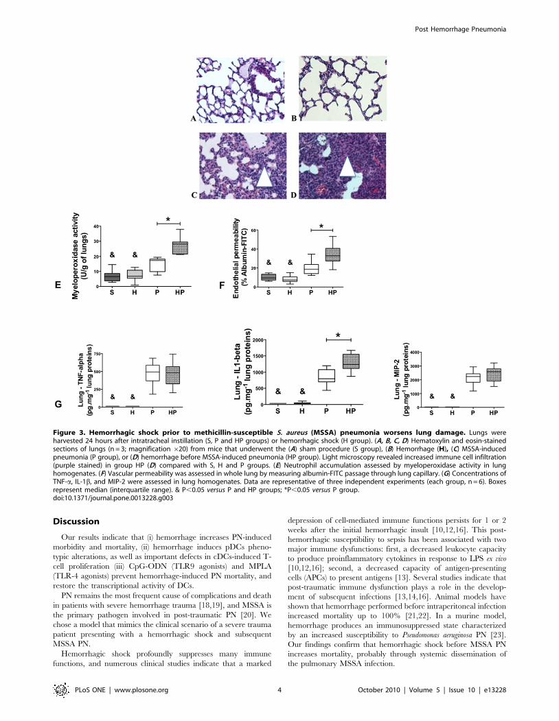
Hemorrhagic shock aggravated lung lesions and slowed
recovery after PN. Because hemorrhagic shock worsens PN
outcomes, we assessed the lungs histology. In both sham-treated
group (S group) and H group, lung tissue was characterized by thin-
walled air spaces with a single pneumocyte layer (Figure 3A, B). In
contrast, immune cell infiltrates (macrophages and neutrophils)
were detected and alveolar layers were thicker in the P group
24 hours after MSSA injection (Figure 3C). Histological recovery
began at day 4 with increasing aeration of the lung; lungs were
almost normal at day 7 (Figure S3C, E, G). Histological lesions
appeared sooner (as soon as 12 hours vs. 24 hours) and for a longer
duration (7 days vs. 4 days) in the HP group compared with the P
group (Figure 3D and Figure S3D, F, H).
Twenty-four hours after MSSA injection, neutrophil accumu-
lation, as assessed by myeloperoxidase activity (Figure 3D), and
endothelial lesions, as assessed by endothelial permeability to
albumin-FITC (Figure 3E), were increased in the HP group
compared with the P group. Lung concentrations of tumor
necrosis factor (TNF)-a, IL-1b, and macrophage inflammatory
protein (MIP)-2 were all elevated in the P and HP groups
compared with the S and H groups (Figure 3F). In addition, lung
production of IL-1b was higher in the HP group compared with P
group (Figure 3F).
Hemorrhagic shock increased systemic dissemination of
MSSA. Because PN lung lesions were increased after
hemorrhagic shock, local and systemic bacterial burden were
evaluated 12, 24, and 48 hours after PN. MSSA was not detected
in the S and H groups; however, lung bacterial load were not
different in groups HP and P (Figure 4A). Spleen cultures showed
that systemic dissemination of infection (MSSA CFUs) was higher
Figure 1. Effects of hemorrhage on timing-based mortality.Three groups of mice were studied: H group (hemorrhaged animals); Pgroup (methicillin-susceptible S. aureus (MSSA)-induced pneumoniaonly; n = 15) and HP group (animals hemorrhaged before MSSA-inducedpneumonia; n = 15). Pneumonia was induced in mice with MSSA(76105 CFU) 2, 4, 8, 24, 48, or 96 hours after hemorrhage (HP-H2, -H4, -H8, -H24, -H48, and -H96 groups, respectively) and survival wascompared with mice with P group and H group. Survival rates areexpressed as percentage and are representative of three independentexperiments. & P,0.05 versus all others; *P,0.05 versus P group.doi:10.1371/journal.pone.0013228.g001
Figure 2. Hemorrhagic shock increases weight loss aftermethicillin-susceptible S. aureus (MSSA) pneumonia. Micereceived sham treatment (S; cardiac puncture without blood collection),Hemorrhage (H), MSSA–induced pneumonia alone (P), or hemorrhagebefore MSSA–induced pneumonia (HP), and were weighed daily for 5days. Data are representative of three independent experiments (eachgroup, n = 6). Data are expressed as mean 6 SEM. & P,0.05 versus Sand H groups, *P,0.05 versus P group.doi:10.1371/journal.pone.0013228.g002
Post Hemorrhage Pneumonia
PLoS ONE | www.plosone.org 2 October 2010 | Volume 5 | Issue 10 | e13228

at 24 and 48 hours after MSSA injection in group HP compared
with group P (Figure 4B). At each time point, the percentage of
mice exhibiting systemic dissemination was higher in group HP
than in group P (Figure 4C).
Hemorrhagic shock aggravated blood hyporeactivity to
LPS observed in PN. As previously described [10], we
measured cytokine production in whole blood cells cultures
stimulated by LPS. TNF-a and IL-1b production was
significantly lower in the HP group compared with all other
groups (Figure 5A, B), whereas MIP-2 production was decreased
in both HP and P groups compared with groups S and H
(Figure 5C).
DCs link innate immunity to adaptive immunity and may be
critically involved in hemorrhage-induced PN mortality. We
therefore sought to determine whether hemorrhagic shock
preceding PN could affect spleen DCs numbers, phenotypes,
and function.
Hemorrhagic shock downregulates inflammatory
cytokine mRNA expression in total spleen DCs. We
assessed time-dependent mRNA expression of inflammatory
cytokines by real-time RT-PCR in total spleen DCs. Cytokine
mRNA levels peaked 6 hours after MSSA injection (see Figure
S4). TNF-a, IFN-b, and IL-12 mRNA levels were significantly
increased in group P compared with group S (Figure 6A–C and
Figure S4), indicating that DCs are involved in the innate immune
response to MSSA PN. Mice that underwent hemorrhagic shock
prior to MSSA-induced PN (group HP) demonstrated markedly
decreased TNF-a, IFN-b, and IL-12p40 mRNA expression
compared with P group (Figure 6A–C). Hemorrhage alone did
not affect mRNA expression compared with S group.
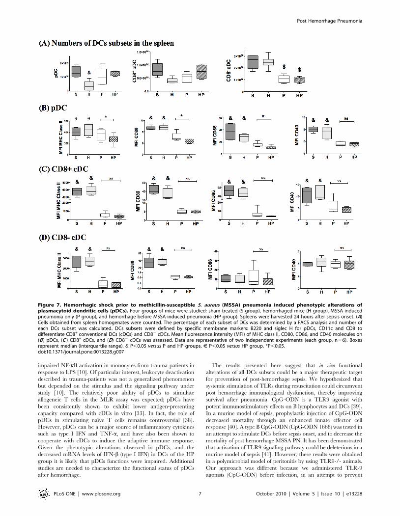
Hemorrhagic shock performed before PN altered the
phenotype of pDCs but not cDCs. To further evaluate
potential alterations of DCs, we determined the numbers and
phenotypes of individual spleen DCs subsets. The number of
CD82 cDCs was decreased in PN-infected groups, whereas
number of pDCs was decreased in H group (Figure 7A). The
pDCs from HP mice exhibited a significant decrease in MHC class
II, CD80, and CD86 molecule levels compared with the S, H and
P groups (Figure 7B), whereas CD40 expression was decreased in
both HP and P groups compared with the S and H groups.
Regarding CD8+ cDCs, MHC-class II and costimulatory
molecules were downregulated in the HP and P groups
compared with the S and H groups (Figure 7C). Finally, MHC
class II and CD80 molecules, but not CD86, were downregulated
similarly in CD8- cDCs of groups HP and P compared with S and
H groups (Figure 7D). Therefore, hemorrhage did not significantly
affect the numbers of DCs subsets following PN infection;
however, hemorrhage induced phenotypic alterations in pDCs,
but not cDCs.
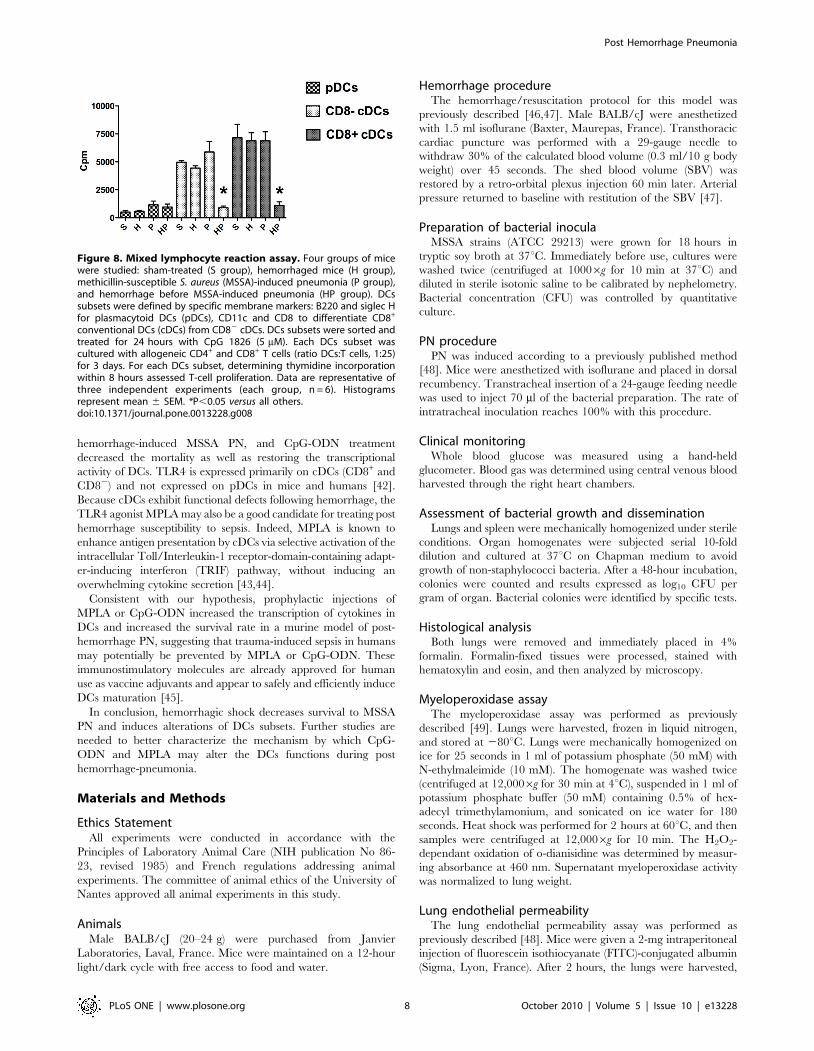
Hemorrhagic shock before PN decreased the ability of
cDCs to induce T-cell proliferation. To further characterize
potential functional abnormalities of DCs after hemorrhage, the
abilities of each DCs subset (pDCs, CD8+ cDCs, and CD82 cDCs)
to induce T-cell proliferation were tested using an allogeneic
mixed lymphocyte reaction (MLR) assay. As previously described
[18], DCs subsets were separated by fluorescence-activated cell
sorting (FACS) 24 hours after MSSA injection and stimulated
overnight with TLR9 ligand to induce maturation before the
MLR assay. T-cell proliferation induced by CD8+ and CD82
cDCs did not differ between the S, H and P groups. In contrast,
the ability of both cDCs subsets to induce T-cell proliferation was
dramatically impaired in the HP group (Figure 8). Taken together,
these data indicate that hemorrhagic shock potently decreased the
antigen-presenting function of mature cDCs. As previously
reported [19], pDCs are relatively poor stimulators of T cells
and no significant differences in pDCs were observed among the
four treatment groups.
CpG-ODN 1668 and MPLA restored transcriptional
activity in DCs and reversed excess PN mortality caused
by hemorrhagic shock. Because hemorrhage affected DCs
subsets, the ability of TLR9 agonist CpG-ODN 1668 (64 mg/
mouse; group HP-CpG) and TLR4 agonist monophosphoryl lipid
A (MPLA, 50 mg/mouse; group HP-MPLA) to prevent the
increased post-hemorrhage PN mortality was evaluated. To
mimic a clinical scenario, TLR agonists were intravenously
injected immediately after the resuscitation of hemorrhage and
before PN induction. The mRNA levels of TNF-a, IFN-b and IL-
12p40 were increased in DCs from HP-CpG group compared
with HP group (Figure 9A, B, C). The mRNA levels of IL-12p40
were increased in HP-MPLA group compared with HP group
(Figure 9C). As shown in Figure 9D, the survival rate was
significantly increased in mice that received MPLA (93% in HP-
MPLA vs. 60% in group HP; P,0.01) or CpG-ODN (97% in HP-
CpG; P,0.01 compared with 60% in group HP). Injection of the
control CpG-ODN (64 mg/mouse, group HP-CpG control) did
not improve survival compared with the HP group.
Table 1. Hemorrhagic shock worsens biological consequences of methicillin-susceptible S. aureus (MSSA) pneumonia.
Sham (S) group Hemorrhage (H) group Pneumonia (P) groupHemorrhage-Pneumonia(HP) group
Lactate (mmol/l) 3.560.5 4.261.3 4.661.3& 6.061.5&*
pH 7.3460.03 7.3260.04 7.2660.03& 7.2260.01&*
HCO3- (mmol/l) 28.561.1 24.562.1& 26.861.6 24.563.0&*
BE (mmol/l) +0.460.2 21.460.2& 20.960.8 22.860.7&*
PcO2 (kPa) 6.461.1 5.861.6 3.461.2& 3.861.3&
PcCO2 (kPa) 7.360.9 6.860.4 8.560.7 9.161.3
SvO2 (%) 59612 41612& 29618& 35619&
Blood glucose (mmol/l) 1.8760.29 1.8860.45 0.8160.3& 1.3660.23&*
BE: Base Excess, PcO2: Central venous Pression in O2, PcCO2: Central venous Pression in CO2, SvO2: Venous saturation in oxygen. Blood samples were collected via cardiacroute (right atria) 24 hours after hemorrhage (H group) or MSSA pneumonia onset (P and HP groups).&P#0.05 versus S group,*P#0.05 versus P group.doi:10.1371/journal.pone.0013228.t001
Post Hemorrhage Pneumonia
PLoS ONE | www.plosone.org 3 October 2010 | Volume 5 | Issue 10 | e13228
Discussion
Our results indicate that (i) hemorrhage increases PN-induced
morbidity and mortality, (ii) hemorrhage induces pDCs pheno-
typic alterations, as well as important defects in cDCs-induced T-
cell proliferation (iii) CpG-ODN (TLR9 agonists) and MPLA
(TLR-4 agonists) prevent hemorrhage-induced PN mortality, and
restore the transcriptional activity of DCs.
PN remains the most frequent cause of complications and death
in patients with severe hemorrhage trauma [18,19], and MSSA is
the primary pathogen involved in post-traumatic PN [20]. We
chose a model that mimics the clinical scenario of a severe trauma
patient presenting with a hemorrhagic shock and subsequent
MSSA PN.
Hemorrhagic shock profoundly suppresses many immune
functions, and numerous clinical studies indicate that a marked
depression of cell-mediated immune functions persists for 1 or 2
weeks after the initial hemorrhagic insult [10,12,16]. This post-
hemorrhagic susceptibility to sepsis has been associated with two
major immune dysfunctions: first, a decreased leukocyte capacity
to produce proinflammatory cytokines in response to LPS ex vivo
[10,12,16]; second, a decreased capacity of antigen-presenting
cells (APCs) to present antigens [13]. Several studies indicate that
post-traumatic immune dysfunction plays a role in the develop-
ment of subsequent infections [13,14,16]. Animal models have
shown that hemorrhage performed before intraperitoneal infection
increased mortality up to 100% [21,22]. In a murine model,
hemorrhage produces an immunosuppressed state characterized
by an increased susceptibility to Pseudomonas aeruginosa PN [23].
Our findings confirm that hemorrhagic shock before MSSA PN
increases mortality, probably through systemic dissemination of
the pulmonary MSSA infection.
Figure 3. Hemorrhagic shock prior to methicillin-susceptible S. aureus (MSSA) pneumonia worsens lung damage. Lungs wereharvested 24 hours after intratracheal instillation (S, P and HP groups) or hemorrhagic shock (H group). (A, B, C, D) Hematoxylin and eosin-stainedsections of lungs (n = 3; magnification 620) from mice that underwent the (A) sham procedure (S group), (B) Hemorrhage (H), (C) MSSA-inducedpneumonia (P group), or (D) hemorrhage before MSSA-induced pneumonia (HP group). Light microscopy revealed increased immune cell infiltration(purple stained) in group HP (D) compared with S, H and P groups. (E) Neutrophil accumulation assessed by myeloperoxidase activity in lunghomogenates. (F) Vascular permeability was assessed in whole lung by measuring albumin-FITC passage through lung capillary. (G) Concentrations ofTNF-a, IL-1b, and MIP-2 were assessed in lung homogenates. Data are representative of three independent experiments (each group, n = 6). Boxesrepresent median (interquartile range). & P,0.05 versus P and HP groups; *P,0.05 versus P group.doi:10.1371/journal.pone.0013228.g003
Post Hemorrhage Pneumonia
PLoS ONE | www.plosone.org 4 October 2010 | Volume 5 | Issue 10 | e13228

Neutrophils have been shown to play a central role in the innate
immune response to hemorrhage because they are primed for
increased lung sequestration and cytotoxic activity [24]. Consistent
with these data, our results demonstrate that hemorrhage increases
pulmonary neutrophil infiltration induced by MSSA PN. Other
surrogate markers of lung damage, such as lung proinflammatory
cytokines (mainly IL-1b) and vascular permeability, were also
exacerbated by hemorrhage in the present study. Despite this
apparently overwhelming inflammatory response, as in another
murine model of sepsis-induced immune dysfunction [25],
bacterial clearance in the lungs was not affected by hemorrhage
whereas systemic bacterial dissemination was constant.
Studies [10,12,26] have demonstrated that blood leukocytes
from trauma or septic patients produced lower levels of
proinflammatory cytokines in whole blood cultures stimulated ex
vivo with LPS compared with controls. In the present study,
Figure 4. Hemorrhagic shock prior to methicillin-susceptible S. aureus (MSSA) pneumonia induces a bacteriemia. Four groups of micewere studied: sham-treated (S group), Hemorrhaged mice (H group), MSSA-induced pneumonia (P group), and hemorrhage before MSSA-inducedpneumonia (HP group). Mice were sacrificed 12, 24, or 48 hours after pneumonia onset. MSSA counts in (A) lungs and (B, C) spleen homogenateswere performed after culture on specific media. Detection threshold was 0.7 colony forming unit (CFU) per gram of tissue. MSSA counts were alwaysbelow the threshold in the S and H groups. Data are representative of three independent experiments (each group, n = 6). Data are presented asmean 6 SEM (A, B) or percentage 6 SEM (C). & P,0.05 versus P and HP groups; *P,0.05 versus group P.doi:10.1371/journal.pone.0013228.g004
Figure 5. Whole Blood cells cultures. Hemorrhagic shock prior to methicillin-susceptible S. aureus (MSSA) pneumonia worsens peripherical bloodreactivity after ex vivo LPS stimulation. Four groups of mice were studied: sham-treated (S group), hemorrhaged mice (H group); MSSA-inducedpneumonia (P group), and hemorrhage before MSSA-induced pneumonia (HP group). Whole blood was exposed to LPS from E.coli O111 B4 for24 hours and the following cytokines were assessed in the cell culture medium: (A) TNF-a, (B) IL-1b, and (C) MIP-2. Cytokine concentrations in theabsence of LPS stimulation were always below the detection threshold (30 pg/ml). Data are representative of three independent experiments (eachgroup, n = 6). Boxes represent median (interquartile range). J P,0.05 versus all other groups; & P,0.05 versus P and HP groups.doi:10.1371/journal.pone.0013228.g005
Post Hemorrhage Pneumonia
PLoS ONE | www.plosone.org 5 October 2010 | Volume 5 | Issue 10 | e13228

hemorrhagic shock performed before MSSA PN also resulted in a
marked decrease of blood reactivity to LPS. The association of an
overwhelming inflammation into the lungs with a blood
hyporeactivity to LPS in ex vivo whole blood cell cultures has been
described in human trauma patients [27] (compartmentalization
of the inflammatory response between lung and systemic
circulation). Moreover, lung production of cytokines depends on
several cellular types (neutrophils, epithelial cells, macrophages)
[24] whereas such a production in whole blood cultures depends
mainly on monocytes [28].
Antigen presentation to T cells requires both signal I (MHC
class II-peptide complex) and signal II (co-stimulatory molecules
such as CD80 and CD86), which are both provided by
professional APCs [29]. Interestingly, an early decrease in HLA-
DR and CD86 membrane expression on circulating monocytes
predict subsequent infections after trauma [16]. DCs are the most
potent APCs and have the unique ability to activate naıve T cells
[30]. In a murine model of hemorrhage, Kawasaki et al. [31]
reported that decreased expression of MHC class II and CD83
molecules on splenic DCs following hemorrhage was associated
with a defect in DCs-induced T-cell proliferation. To the best of
our knowledge, the effects of hemorrhage followed by a
subsequent infection have not been reported to date.
Several DCs subsets have been described in rodents and
humans [17,32]. PDCs are known to be important in the antiviral
response. These cells secrete large amounts of type I IFN upon
viral stimulation, exhibit a restricted TLR repertoire specialized in
nucleic acid recognition (TLR7 and TLR9) and have a weak
capacity for antigen presentation [33,34]. In mice, cDCs express a
large panel of TLRs including TLR9, and are critically involved in
bacterial control. Two subsets of cDCs have been described:
CD8+ cDCs produce considerable amounts of IL-12, induce
strong T helper cell (Th)1 responses, and efficiently cross-present
antigens to CD8+ T cells; CD8- cDCs produce IL-10 and drive
the Th2 response. The innate immune response relies on close
collaboration among the three DCs subsets [17]. Alterations of
total DCs after hemorrhage have been reported, whereas specific
subset alterations remain poorly studied. A decreased number of
cDCs was reported in trauma patients [35], as well as in patients
with severe sepsis [15]. Accordingly, in a model of peritonitis, both
cDCs subsets were markedly decreased in the spleens of infected
mice [36]. In the present study, the effects of hemorrhage on
subsequent PN (HP group) were apparent on the pDCs phenotype
(reduced MHC class II, CD80, and CD86 molecule membrane
expression) with no significant cDCs phenotypic alterations. In
addition, our findings demonstrate that post-hemorrhage IS
dramatically decreased CD8+ cDCs- and CD82 cDCs-induced
allogeneic T-cell proliferation during PN compared with mice that
did not undergo hemorrhage. These results could be explained
either by the reported decreased cytokines mRNA production or
by a resistance to TLR-induced maturation in cDCs during post
hemorrhage pneumonia. We would like to point out here that the
phenotype of DCs was assessed directly ex vivo on spleen cells
whereas MLR were performed after stimulating DCs in vitro with
TLR ligands to induce maturation [37]. Therefore, these
experiments cannot be clearly correlated.
It is important to note that although human cDCs do not
express TLR9, mouse pDCs and cDCs express and respond to
TLR9. Our data suggest that hemorrhagic shock induces ex vivo
unresponsiveness to TLR9 agonists in cDCs by an unknown
mechanism, which was apparent during the subsequent sepsis.
Lack of response to TLR4 ligands was observed as the decreased
production of inflammatory cytokines in whole blood cells cultures
stimulated with LPS ex vivo. This is consistent with studies showing
Figure 6. Hemorrhagic shock prior to methicillin-susceptible S. aureus (MSSA) pneumonia markedly decreases cytokine mRNAlevels in total dendritic cells (DCs). Four groups of mice were studied: sham-treated (S group), hemorrhaged mice (H group), MSSA-inducedpneumonia (P group), and hemorrhage before MSSA-induced pneumonia (HP group). Mice were sacrificed 6 hours after MSSA injection. Real-timeRT-PCR analysis of (A) TNF-a, (B) IFN-b, (C) IL-12p40 was performed. In each group, mRNA was extracted from CD11c cells positively selected from cellsuspension obtained from enzymatic spleen digestion. Data are representative of three independent experiments (each group, n = 6). Boxesrepresent median (interquartile range). & P,0.05 versus all others, $ P,0.05 versus S and H groups.doi:10.1371/journal.pone.0013228.g006
Post Hemorrhage Pneumonia
PLoS ONE | www.plosone.org 6 October 2010 | Volume 5 | Issue 10 | e13228
impaired NF-kB activation in monocytes from trauma patients in
response to LPS [10]. Of particular interest, leukocyte deactivation
described in trauma-patients was not a generalized phenomenon
but depended on the stimulus and the signaling pathway under
study [10]. The relatively poor ability of pDCs to stimulate
allogeneic T cells in the MLR assay was expected; pDCs have
been consistently shown to exhibit lower antigen-presenting
capacity compared with cDCs in vitro [33]. In fact, the role of
pDCs in stimulating naıve T cells remains controversial [38].
However, pDCs can be a major source of inflammatory cytokines
such as type I IFN and TNF-a, and have also been shown to
cooperate with cDCs to induce the adaptive immune response.
Given the phenotypic alterations observed in pDCs, and the
decreased mRNA levels of IFN-b (type I IFN) in DCs of the HP
group it is likely that pDCs functions were impaired. Additional
studies are needed to characterize the functional status of pDCs
after hemorrhage.
The results presented here suggest that in vivo functional
alterations of all DCs subsets could be a major therapeutic target
for prevention of post-hemorrhage sepsis. We hypothesized that
systemic stimulation of TLRs during resuscitation could circumvent
post hemorrhage immunological dysfunction, thereby improving
survival after pneumonia. CpG-ODN is a TLR9 agonist with
potent immunostimulatory effects on B lymphocytes and DCs [39].
In a murine model of sepsis, prophylactic injection of CpG-ODN
decreased mortality through an enhanced innate effector cell
response [40]. A type B CpG-ODN (CpG-ODN 1668) was tested in
an attempt to stimulate DCs before sepsis onset, and to decrease the
mortality of post hemorrhage MSSA PN. It has been demonstrated
that activation of TLR9 signaling pathway could be deleterious in a
murine model of sepsis [41]. However, these results were obtained
in a polymicrobial model of peritonitis by using TLR9-/- animals.
Our approach was different because we administered TLR-9
agonists (CpG-ODN) before infection, in an attempt to prevent
Figure 7. Hemorrhagic shock prior to methicillin-susceptible S. aureus (MSSA) pneumonia induced phenotypic alterations ofplasmacytoid dendritic cells (pDCs). Four groups of mice were studied: sham-treated (S group), hemorrhaged mice (H group), MSSA-inducedpneumonia only (P group), and hemorrhage before MSSA-induced pneumonia (HP group). Spleens were harvested 24 hours after sepsis onset. (A)Cells obtained from spleen homogenates were counted. The percentage of each subset of DCs was determined by a FACS analysis and number ofeach DCs subset was calculated. DCs subsets were defined by specific membrane markers: B220 and siglec H for pDCs, CD11c and CD8 todifferentiate CD8+ conventional DCs (cDCs) and CD82 cDCs. Mean fluorescence intensity (MFI) of MHC class II, CD80, CD86, and CD40 molecules on(B ) pDCs, (C ) CD8+ cDCs, and (D) CD82 cDCs was assessed. Data are representative of two independent experiments (each group, n = 6). Boxesrepresent median (interquartile range). & P,0.05 versus P and HP groups, J P,0.05 versus HP group, *P,0.05.doi:10.1371/journal.pone.0013228.g007
Post Hemorrhage Pneumonia
PLoS ONE | www.plosone.org 7 October 2010 | Volume 5 | Issue 10 | e13228
hemorrhage-induced MSSA PN, and CpG-ODN treatment
decreased the mortality as well as restoring the transcriptional
activity of DCs. TLR4 is expressed primarily on cDCs (CD8+ and
CD82) and not expressed on pDCs in mice and humans [42].
Because cDCs exhibit functional defects following hemorrhage, the
TLR4 agonist MPLA may also be a good candidate for treating post
hemorrhage susceptibility to sepsis. Indeed, MPLA is known to
enhance antigen presentation by cDCs via selective activation of the
intracellular Toll/Interleukin-1 receptor-domain-containing adapt-
er-inducing interferon (TRIF) pathway, without inducing an
overwhelming cytokine secretion [43,44].
Consistent with our hypothesis, prophylactic injections of
MPLA or CpG-ODN increased the transcription of cytokines in
DCs and increased the survival rate in a murine model of post-
hemorrhage PN, suggesting that trauma-induced sepsis in humans
may potentially be prevented by MPLA or CpG-ODN. These
immunostimulatory molecules are already approved for human
use as vaccine adjuvants and appear to safely and efficiently induce
DCs maturation [45].
In conclusion, hemorrhagic shock decreases survival to MSSA
PN and induces alterations of DCs subsets. Further studies are
needed to better characterize the mechanism by which CpG-
ODN and MPLA may alter the DCs functions during post
hemorrhage-pneumonia.
Materials and Methods
Ethics StatementAll experiments were conducted in accordance with the
Principles of Laboratory Animal Care (NIH publication No 86-
23, revised 1985) and French regulations addressing animal
experiments. The committee of animal ethics of the University of
Nantes approved all animal experiments in this study.
AnimalsMale BALB/cJ (20–24 g) were purchased from Janvier
Laboratories, Laval, France. Mice were maintained on a 12-hour
light/dark cycle with free access to food and water.
Hemorrhage procedureThe hemorrhage/resuscitation protocol for this model was
previously described [46,47]. Male BALB/cJ were anesthetized
with 1.5 ml isoflurane (Baxter, Maurepas, France). Transthoracic
cardiac puncture was performed with a 29-gauge needle to
withdraw 30% of the calculated blood volume (0.3 ml/10 g body
weight) over 45 seconds. The shed blood volume (SBV) was
restored by a retro-orbital plexus injection 60 min later. Arterial
pressure returned to baseline with restitution of the SBV [47].
Preparation of bacterial inoculaMSSA strains (ATCC 29213) were grown for 18 hours in
tryptic soy broth at 37uC. Immediately before use, cultures were
washed twice (centrifuged at 10006g for 10 min at 37uC) and
diluted in sterile isotonic saline to be calibrated by nephelometry.
Bacterial concentration (CFU) was controlled by quantitative
culture.
PN procedurePN was induced according to a previously published method
[48]. Mice were anesthetized with isoflurane and placed in dorsal
recumbency. Transtracheal insertion of a 24-gauge feeding needle
was used to inject 70 ml of the bacterial preparation. The rate of
intratracheal inoculation reaches 100% with this procedure.
Clinical monitoringWhole blood glucose was measured using a hand-held
glucometer. Blood gas was determined using central venous blood
harvested through the right heart chambers.
Assessment of bacterial growth and disseminationLungs and spleen were mechanically homogenized under sterile
conditions. Organ homogenates were subjected serial 10-fold
dilution and cultured at 37uC on Chapman medium to avoid
growth of non-staphylococci bacteria. After a 48-hour incubation,
colonies were counted and results expressed as log10 CFU per
gram of organ. Bacterial colonies were identified by specific tests.
Histological analysisBoth lungs were removed and immediately placed in 4%
formalin. Formalin-fixed tissues were processed, stained with
hematoxylin and eosin, and then analyzed by microscopy.
Myeloperoxidase assayThe myeloperoxidase assay was performed as previously
described [49]. Lungs were harvested, frozen in liquid nitrogen,
and stored at 280uC. Lungs were mechanically homogenized on
ice for 25 seconds in 1 ml of potassium phosphate (50 mM) with
N-ethylmaleimide (10 mM). The homogenate was washed twice
(centrifuged at 12,0006g for 30 min at 4uC), suspended in 1 ml of
potassium phosphate buffer (50 mM) containing 0.5% of hex-
adecyl trimethylamonium, and sonicated on ice water for 180
seconds. Heat shock was performed for 2 hours at 60uC, and then
samples were centrifuged at 12,0006g for 10 min. The H2O2-
dependant oxidation of o-dianisidine was determined by measur-
ing absorbance at 460 nm. Supernatant myeloperoxidase activity
was normalized to lung weight.
Lung endothelial permeabilityThe lung endothelial permeability assay was performed as
previously described [48]. Mice were given a 2-mg intraperitoneal
injection of fluorescein isothiocyanate (FITC)-conjugated albumin
(Sigma, Lyon, France). After 2 hours, the lungs were harvested,
Figure 8. Mixed lymphocyte reaction assay. Four groups of micewere studied: sham-treated (S group), hemorrhaged mice (H group),methicillin-susceptible S. aureus (MSSA)-induced pneumonia (P group),and hemorrhage before MSSA-induced pneumonia (HP group). DCssubsets were defined by specific membrane markers: B220 and siglec Hfor plasmacytoid DCs (pDCs), CD11c and CD8 to differentiate CD8+
conventional DCs (cDCs) from CD82 cDCs. DCs subsets were sorted andtreated for 24 hours with CpG 1826 (5 mM). Each DCs subset wascultured with allogeneic CD4+ and CD8+ T cells (ratio DCs:T cells, 1:25)for 3 days. For each DCs subset, determining thymidine incorporationwithin 8 hours assessed T-cell proliferation. Data are representative ofthree independent experiments (each group, n = 6). Histogramsrepresent mean 6 SEM. *P,0.05 versus all others.doi:10.1371/journal.pone.0013228.g008
Post Hemorrhage Pneumonia
PLoS ONE | www.plosone.org 8 October 2010 | Volume 5 | Issue 10 | e13228

mechanically homogenized in 1 ml of isotonic saline, and then
centrifuged at 40006g for 10 min. Blood was collected via right
ventricular puncture and centrifuged at 4,0006g for 10 min.
FITC-albumin was measured in 100-ml supernatant aliquots
obtained from lung homogenates and blood by fluorometry at
480 nm. Lung endothelial permeability was calculated according
to the validated equation: Perm-FITC %ð Þ~ FLHS{FLNð Þ|ððWHÞ{QFBÞ= FLBS{FLBNð Þ|We|0:07| 1{Hteð Þð Þ (see
Table S1).
Preparation of lung homogenate for enzyme-linkedimmunosorbent assay
Enzyme-linked immunosorbent assay (ELISA) analysis of lung
homogenates was performed as previously described [49]. Immedi-
ately after removal, weighed lung samples were mechanically
homogenized in cold lysis buffer (16 phosphate buffered saline
[PBS, pH 7.4], 0.1% Triton X-100) containing 1 mM protease
inhibitor cocktail (Sigma). Homogenates were centrifuged at
12,0006g for 20 min at 4uC. Supernatant was then collected and
stored at 280uC until analysis. Protein concentration in each sample
was determined using the BCATM protein assay kit, according to
manufacturer’s instructions (Pierce, Rockford, IL. United States).
Cell cultures for LPS reactivity assessmentLPS reactivity of cell cultures was assessed as previously
described [46,50]. Monocyte cytokine secretion was induced in
whole blood culture by LPS. Briefly, blood samples were diluted
1:5 in RPMI-1640 medium (Laboratoire de Biotechnologies,
Reims, France) supplemented with 100 U ml21 penicillin
(Panpharma, Fougeres, France) and 100 mg ml21 streptomycin
(Sigma). The diluted blood was cultured in 24-well plates (500 ml
per well) with or without LPS (E. coli O111:B4, 10 mg/ml; Sigma)
in a 5% CO2 incubator for 24 hours at 37uC. Supernatants were
collected by centrifugation at 12,0006g for 2 min and stored at
280uC before cytokine determination by ELISA.
Determination of cytokine levels in samplesTNF-a, IL-1b, and MIP-2 concentrations were quantified with
ELISA kits according to manufacturer’s instructions (R&D
Systems, Lille, France).
Figure 9. Effect of CpG-ODN or MPLA on cytokine mRNA levels in dendritic cells (DCs). Six groups of mice were studied: sham-treated (Sgroup), hemorrhagic shock (H group), methicillin-susceptible S. aureus (MSSA)-induced pneumonia (P group), hemorrhage before MSSA-inducedpneumonia (HP group), injections of CpG-ODN 1668 or of MPLA were performed immediately after resuscitation of hemorrhage: HP-CpG and MPLAgroups respectively. (A, B, C) Mice were sacrificed 6 hours after MSSA injection. Real-time RT-PCR analysis of (A) TNF-a, (B) IFN-b, (C) IL-12p40 wasperformed. In each group, mRNA was extracted from CD11c cells positively selected from cell suspension obtained from enzymatic spleen digestion.Boxes represent median (interquartile range). & P,0.05 versus S, H, HP and HP-MPLA groups; $ P,0.05 versus S, H and HP groups. (D) The survival ratein each group was monitored twice a day for 7 days (168 hours). Survival rates are presented as percentages. * P,0.01 versus HP group. Data arerepresentative of three independent experiments (each group, n = 5 to 6 mice for A, B and C; n = 8 mice for D).doi:10.1371/journal.pone.0013228.g009
Post Hemorrhage Pneumonia
PLoS ONE | www.plosone.org 9 October 2010 | Volume 5 | Issue 10 | e13228

Spleen cell suspensionSpleens were minced and digested in 2 mg/ml collagenase D
(Roche Diagnostics, Meylan, France) in RPMI 1640 supplemented
with 1% fetal calf serum (FCS) for 25 min at 37uC. EDTA
(10 mM) was added for the last 5 min of digestion. The cell
suspension was then filtered through a 80-mm filter and washed in
PBS (centrifuged at 12,0006g for 10 min at 4uC).
Real-Time Reverse Transcription Polymerase ChainReaction
Real-time reverse transcription polymerase chain reaction (RT-
PCR) was performed as previously described [51]. Spleen cells
were incubated with anti-mouse CD11c-coated magnetic beads
for 15 min at 4uC. After washing, CD11c+ cells were purified by
positive selection using magnetic affinity cell sorting (MACS)
separation columns (Miltenyi Biotec, Paris, France). This proce-
dure routinely yielded cell populations with purity up to 90%.
Total RNA was isolated from purified spleen CD11c+ cells with
TRIzol reagent (Invitrogen, Cergy Pontoise, France) and treated
for 45 min at 37uC with 2 U of RQ1 DNase (Promega, Lyon,
France). RNA (1 mg) was reverse-transcribed with Superscript III
Reverse Transcriptase (Invitrogen). The cDNA (1 ml) was
subjected to real-time RT-PCR in a Bio-Rad iCycler iQ system
using the QuantiTect SYBR Green PCR kit (Qiagen, Courta-
boeuf, France). Thermal cycling conditions consisted of 45 cycles
of 30 seconds at 95uC followed by 30 seconds at 60uC. Mice
primer sequences for TNFa, IFN-b, IL-12, IL-10 and glyceralde-
hyde-3-phosphate dehydrogenase (GAPDH) were designed using
‘‘Primer-BLAST’’ primer design software on the National Center
for Biotechnology Information (NCBI) website (see Table S2 for
primer sequences). GAPDH was used to normalize gene
expression. Relative gene expression was calculated by the 22DD
Ct method [52] using samples from the sham group as calibrator
samples.
Antibody, flow cytometry, and cell sortingFlow cytometry and cell sorting were performed as previously
described [37] (see Figure S5 for example of gating). Monoclonal
antibodies (mAb) used for cytometry and/or cell sorting were
obtained from BD Biosciences (United States, Franklin Lakes, NJ,
United States): anti-CD3 (1452C11), anti-CD8a (53.6-7), anti-
CD11c (HL3), anti-CD19 (1D3), anti-CD40 (3123), anti-CD45R
(B220, RA3-6B2), anti-CD80 (16-10A1), anti-CD86 (GL1), anti-
IAd (class II MHC, AMS-32.1), anti-NK11 (PK136), anti-Ter 119
(Ter119), anti-T-cell receptor (TCR)b (H57.597). The anti-siglec
H was obtained from eBiosciences (San Diego, CA. United States).
All mAbs were conjugated to FITC, phycoerythrin (PE), PECy7,
peridinin-chlorophyll-protein-complex (PerCP)-Cy5.5, allophyco-
cyanin (APC), or biotin (detected with APC-Cy-labeled streptavi-
din) (BD Biosciences). Flow cytometry was performed on a FACS
LSR II (BD Biosciences) and cell sorting was performed on a
FACS Aria (BD Biosciences).
For phenotypic analysis, spleen cells were labeled with
antibodies against CD8, CD11c, B220, Siglec H, and lineage
(Lin) antigens (CD3, CD19, TCRb, NK1.1, Ter119). DCs
activation was assessed by the following biotinylated mAbs:
CD80, CD86, CD40 or MHC-class II (IAd). A total of 46106
cells were analyzed for each biotinylated Ab. Dead cells were
excluded by 4’,6-diamidino-2-phenylindole (DAPI) staining. Data
were analyzed using FlowJo software (Treestar, United States) (see
Figure S5 for an example).
For DCs cell sorting, after collagenase digestion and Ficoll
gradient centrifugation, spleen cells were first incubated with
unconjugated CD3 (17A2, BD Biosciences) and CD19 (1D3, BD
Biosciences) rat mAbs, which are specific for T and B cells,
respectively. Positive cells were magnetically depleted with sheep
anti-rat IgG-conjugated beads (Dynabeads, Invitrogen), and
sheep/goat a-mouse IgG conjugated beads. The remaining cells
were labeled with antibodies against B220, CD19, CD11c, and
CD8a. This procedure routinely yielded populations with levels of
purity up to 98%.
For the MLR assay, T cells were recovered from the lymph
nodes of C57BL/6 mice. Cell suspensions were depleted of
erythrocytes, myeloid, natural killer, CD8+, and B cells with the
Pan T-cell Isolation kit (Miltenyi Biotech, Paris, France) according
to manufacturer’s instructions. Sorted DCs subsets were stimulated
with CpG-ODN 1826 (5 mM) for 24 hours and then cultured with
allogeneic T cells (DCs:T-cell ratio of 1:25) in round-bottom 96-
well plates with RPMI 1640 (10% Fetal Calf Serum, 100 mg/mL
streptomycin, 100 IU/mL penicillin). Cells were cultured for 3
days at 37uC in 5% CO2, and then pulsed for the last 8 hours with
0.5 mCi of [3H]thymidine (GE Healthcare) per well. The cells were
then harvested onto glass fiber filters and [3H]thymidine
incorporation was measured using standard scintillation proce-
dures (Packard Institute).
Toll Like Receptor AgonistsCpG-ODN 1668, CpG-ODN 1168 control and MPLA were
purchased from Invivogen (Toulouse, France).
Statistical analysisSAS 9.1 (Evry-Gregy sur Yerres, France) and GraphPad prism
(La jolla, CA. Uinited States) software were used for statistical
analysis. For the pilot study, potential experimental effects were
tested using Cox proportional-hazards regression models by
introducing the corresponding covariate into the model. In case
of a non-significant effect, the corresponding covariate was not
included in subsequent analyses. Survival rates were compared
using the log-rank test or exact log-rank test as appropriate.
Normally distributed data were expressed as mean 6 standard
error of the mean (SEM) and analyzed using analysis of variance
(ANOVA) and Student’s t-test. Continuous non-parametric
variables were expressed as median (interquartile range) and were
compared using the Kruskal Wallis test for multiple comparisons.
In case of significance, the Mann-Whitney test was used for inter-
group comparison. P,0.05 was considered to be statistically
significant.
Supporting Information
Figure S1 Diagrammatic representation of the six experimental
groups in the main study. In the sham group (S), cardiac puncture
was performed without blood collection or resuscitation. Volume-
controlled hemorrhage was performed by cardiac puncture
(0.3 ml/10 g body weight) and resuscitation with shed blood was
performed after 60 min (groups Hemorrhage [H] and Hemor-
rhage-Pneumonia [HP]). After 24 hours, mice underwent intra-
tracheal instillation of 76105 CFU (70 ml) of methicillin-suscepti-
ble S. aureus (groups Pneumonia [P] and HP) or sterile PBS (group
S). Intravenous infusion of CpG-ODN (64 mg/mouse, HP-CpG
group) or MPLA (50 mg/mouse, HP-MPLA group) were per-
formed immediately after resuscitation. Twenty-four hours* after
intratracheal instillation, mice were euthanized and specimens
were collected. *unless otherwise stated.
Found at: doi:10.1371/journal.pone.0013228.s001 (1.02 MB
PDF)
Post Hemorrhage Pneumonia
PLoS ONE | www.plosone.org 10 October 2010 | Volume 5 | Issue 10 | e13228

Figure S2 Effects of hemorrhage on inoculum-based mortality.
Two groups of mice were studied: HP group (animals hemor-
rhaged before methicillin-susceptible S. aureus (MSSA)-induced
pneumonia; n = 15) and P group (MSSA-induced pneumonia only;
n = 15). Survival rates are expressed as percentage and are
representative of three independent experiments. Twenty-four
hours after hemorrhage for HP group, pneumonia was induced
with (A) 76104 CFU, (B) 76105 CFU, or (C) 76106 CFU of
MSSA. Survival was monitored twice a day for 7 days. *P,0.05
versus P group.
Found at: doi:10.1371/journal.pone.0013228.s002 (3.00 MB TIF)
Figure S3 Evolution of histological findings following sepsis
onset. Four groups of mice were studied (each group, n = 3): naive,
sham-treated (S), methicillin-susceptible S. aureus (MSSA)-induced
pneumonia only (P), and hemorrhage before MSSA-induced
pneumonia (HP). Formalin-fixed tissues were processed, stained
with hematoxylin and eosin, and analyzed by microscopy
(magnification,620). Representative lung histology for (A) normal
lung (naive), (B) lung at 24 hours post sterile instillation (S group).
The parenchyma is shown along with a series of images obtained
12, 96, and 168 hours after pneumonia onset in (C, E, G) for
group P, respectively, and (D, F, H) for group HP. Aggregates of
purple-stained immune cells were observed as early as 12 hours
postinfection (arrow) and were more numerous in group HP
compared with group P at all time points. These data established a
murine model of MSSA pneumonia that closely mimics the
clinical and histological findings for human patients.
Found at: doi:10.1371/journal.pone.0013228.s003 (3.00 MB TIF)
Figure S4 Time-dependent cytokine mRNA expression in
spleen dendritic cells (DC) following sepsis onset. Mice in which
pneumonia was induced by methicillin-susceptible S. aureus (P
group). Mice were sacrificed 1, 6, or 12 hours after Meticillin
Susceptible Staphylococcus aureus injection. Then mRNA was
extracted from CD11c+ cells positively selected in spleen cells
suspension. Data are representative of two independent experi-
ments (n = 8). Boxes represent median (interquartile range).
*P,0.05.
Found at: doi:10.1371/journal.pone.0013228.s004 (3.00 MB TIF)
Figure S5 Phenotypic characterization of mouse spleen dendrit-
ic cell (DC) subsets. Spleen cells from sham-treated mice (S group),
methicillin-susceptible S. aureus (MSSA)-infected mice (P group),
hemorrhage-shocked and MSSA-infected mice (HP group) were
labelled with antibodies against lineage antigens (CD3e, CD19,
TCRb, Ter119, NK1.1) after DAPI staining. Conventional
dendritic cell (cDC) and plasmacytoid cell (pDC) subsets were
identified within the lineage-negative population as CD11chigh,
CD8+, or CD82 cells and B220- siglec H+ cells respectively.
Expression of CD80, CD86, CD40, and MHC class II (as shown
here) molecules was determined on the surface of DC subsets.
Numbers indicate percentage of cells within the gates. Blue curve
(HP group, n = 6), yellow curve (P group, n = 6) and pink curve (S
group, n = 6).
Found at: doi:10.1371/journal.pone.0013228.s005 (3.00 MB TIF)
Table S1 Equation used to calculate endothelial permeability to
albumin FITC.
Found at: doi:10.1371/journal.pone.0013228.s006 (0.03 MB
DOC)
Table S2 Primers for quantitative reverse-transcription poly-
merase chain reaction. TNF, tumor necrosis factor-a; IFN,
interferon; IL, interleukin; GAPDH, glyceraldehyde-3-phosphate
dehydrogenase.
Found at: doi:10.1371/journal.pone.0013228.s007 (0.07 MB
DOC)
Acknowledgments
We thank Ms. Auger, Ms Miegeville, Dr. Masson, and Pr. Laboisse for
their technical assistance.
Author Contributions
Conceived and designed the experiments: AR LG JPS PDC CJ JC GP CL
JR KA. Performed the experiments: AR LG JPS PDC CJ JC JR KA.
Analyzed the data: AR LG JPS PDC VS CJ JC GP CL JR KA.
Contributed reagents/materials/analysis tools: AR LG JPS PDC CJ JC GP
CL JR KA. Wrote the paper: AR LG JPS PDC VS CJ JC GP CL JR KA.
References
1. Mathers CD, Loncar D (2006) Projections of global mortality and burden of
disease from 2002 to 2030. PLoS Med 3: e442.
2. Patton GC, Coffey C, Sawyer SM, Viner RM, Haller DM, et al. (2009) Globalpatterns of mortality in young people: a systematic analysis of population health
data. Lancet 374: 881–892.
3. Bandiera GW, Hillers TK, White F (1999) Evaluating programs to prevent
unintentional trauma in Canada: challenges and directions. J Trauma 47:
932–936.
4. Magnotti LJ, Croce MA, Fabian TC (2004) Is ventilator-associated pneumoniain trauma patients an epiphenomenon or a cause of death? Surg Infect (Larchmt)
5: 237–242.
5. Safdar N, Dezfulian C, Collard HR, Saint S (2005) Clinical and economic
consequences of ventilator-associated pneumonia: a systematic review. Crit Care
Med 33: 2184–2193.
6. Papia G, McLellan BA, El-Helou P, Louie M, Rachlis A, et al. (1999) Infectionin hospitalized trauma patients: incidence, risk factors, and complications.
J Trauma 47: 923–927.
7. Rincon-Ferrari MD, Flores-Cordero JM, Leal-Noval SR, Murillo-Cabezas F,
Cayuelas A, et al. (2004) Impact of ventilator-associated pneumonia in patientswith severe head injury. J Trauma 57: 1234–1240.
8. Lepelletier D, Roquilly A, Demeure dit latte D, Mahe PJ, Loutrel O, et al. (2010)Retrospective analysis of the risk factors and pathogens associated with early-
onset ventilator-associated pneumonia in surgical-ICU head-trauma patients.
J Neurosurg Anesthesiol 22: 32–37.
9. Bronchard R, Albaladejo P, Brezac G, Geffroy A, Seince PF, et al. (2004) Earlyonset pneumonia: risk factors and consequences in head trauma patients.
Anesthesiology 100: 234–239.
10. Adib-Conquy M, Moine P, Asehnoune K, Edouard A, Espevik T, et al. (2003)
Toll-like receptor-mediated tumor necrosis factor and interleukin-10 production
differ during systemic inflammation. Am J Respir Crit Care Med 168: 158–164.
11. Keel M, Ecknauer E, Stocker R, Ungethum U, Steckholzer U, et al. (1996)
Different pattern of local and systemic release of proinflammatory and anti-inflammatory mediators in severely injured patients with chest trauma. J Trauma
40: 907–12; discussion 912–4.
12. Spolarics Z, Siddiqi M, Siegel JH, Garcia ZC, Stein DS, et al. (2003) Depressed
interleukin-12-producing activity by monocytes correlates with adverse clinicalcourse and a shift toward Th2-type lymphocyte pattern in severely injured male
trauma patients. Crit Care Med 31: 1722–1729.
13. Cheadle WG, Hershman MJ, Wellhausen SR, Polk HC, Jr. (1991) HLA-DR
antigen expression on peripheral blood monocytes correlates with surgicalinfection. Am J Surg 161: 639–645.
14. Hershman MJ, Cheadle WG, Wellhausen SR, Davidson PF, Polk HC, Jr. (1990)Monocyte HLA-DR antigen expression characterizes clinical outcome in the
trauma patient. Br J Surg 77: 204–207.
15. Poehlmann H, Schefold JC, Zuckermann-Becker H, Volk HD, Meisel C (2009)
Phenotype changes and impaired function of dendritic cell subsets in patientswith sepsis: a prospective observational analysis. Crit Care 13: R119.
16. Ditschkowski M, Kreuzfelder E, Rebmann V, Ferencik S, Majetschak M, et al.(1999) HLA-DR expression and soluble HLA-DR levels in septic patients after
trauma. Ann Surg 229: 246–254.
17. Merad M, Manz MG (2009) Dendritic cell homeostasis. Blood 113: 3418–3427.
18. Czaja AS, Rivara FP, Wang J, Koepsell T, Nathens AB, et al. (2009) Late
Outcomes of Trauma Patients With Infections During Index Hospitalization.
J Trauma 67: 805–814.
19. Osborn TM, Tracy JK, Dunne JR, Pasquale M, Napolitano LM (2004)Epidemiology of sepsis in patients with traumatic injury. Crit Care Med 32:
2234–2240.
20. Agbaht K, Lisboa T, Pobo A, Rodriguez A, Sandiumenge A, et al. (2007)
Management of ventilator-associated pneumonia in a multidisciplinary intensive
care unit: does trauma make a difference? Intensive Care Med 33: 1387–1395.
Post Hemorrhage Pneumonia
PLoS ONE | www.plosone.org 11 October 2010 | Volume 5 | Issue 10 | e13228

21. Esrig BC, Frazee L, Stephenson SF, Polk HC, Jr., Fulton RL, et al. (1977) The
predisposition to infection following hemorrhagic shock. Surg Gynecol Obstet
144: 915–917.
22. Stephan RN, Kupper TS, Geha AS, Baue AE, Chaudry IH (1987) Hemorrhage
without tissue trauma produces immunosuppression and enhances susceptibility
to sepsis. Arch Surg 122: 62–68.
23. Robinson A, Abraham E (1991) Effects of hemorrhage and resuscitation on
bacterial antigen-specific pulmonary plasma cell function. Crit Care Med 19:
1285–1293.
24. Abraham E, Carmody A, Shenkar R, Arcaroli J (2000) Neutrophils as early
immunologic effectors in hemorrhage- or endotoxemia-induced acute lung
injury. Am J Physiol Lung Cell Mol Physiol 279: L1137–45.
25. Pene F, Zuber B, Courtine E, Rousseau C, Ouaaz F, et al. (2008) Dendritic cells
modulate lung response to Pseudomonas aeruginosa in a murine model of sepsis-
induced immune dysfunction. J Immunol 181: 8513–8520.
26. Marie C, Muret J, Fitting C, Losser MR, Payen D, et al. (1998) Reduced ex vivo
interleukin-8 production by neutrophils in septic and nonseptic systemic
inflammatory response syndrome. Blood 91: 3439–3446.
27. Muehlstedt SG, Richardson CJ, Lyte M, Rodriguez JL (2002) Systemic and
pulmonary effector cell function after injury. Crit Care Med 30: 1322–1326.
28. Damsgaard CT, Lauritzen L, Calder PC, Kjaer TM, Frokiaer H (2009) Whole-
blood culture is a valid low-cost method to measure monocytic cytokines - a
comparison of cytokine production in cultures of human whole-blood,
mononuclear cells and monocytes. J Immunol Methods 340: 95–101.
29. Medzhitov R, Janeway C, Jr. (2000) Innate immunity. N Engl J Med 343:
338–344.
30. Banchereau J, Steinman RM (1998) Dendritic cells and the control of immunity.
Nature 392: 245–252.
31. Kawasaki T, Fujimi S, Lederer JA, Hubbard WJ, Choudhry MA, et al. (2006)
Trauma-hemorrhage induces depressed splenic dendritic cell functions in mice.
J Immunol 177: 4514–4520.
32. Pulendran B, Tang H, Denning TL (2008) Division of labor, plasticity, and
crosstalk between dendritic cell subsets. Curr Opin Immunol 20: 61–67.
33. Liu YJ (2005) IPC: professional type 1 interferon-producing cells and
plasmacytoid dendritic cell precursors. Annu Rev Immunol 23: 275–306.
34. Salio M, Palmowski MJ, Atzberger A, Hermans IF, Cerundolo V (2004) CpG-
matured murine plasmacytoid dendritic cells are capable of in vivo priming of
functional CD8 T cell responses to endogenous but not exogenous antigens.
J Exp Med 199: 567–579.
35. Henrich D, Maier M, Relja B, Trendafilov P, Schiessling S, et al. (2009)
Significant decline of peripheral myeloid dendritic cells following multiple
trauma. J Surg Res 154: 239–245.
36. Flohe SB, Agrawal H, Schmitz D, Gertz M, Flohe S, et al. (2006) Dendritic cells
during polymicrobial sepsis rapidly mature but fail to initiate a protective Th1-
type immune response. J Leukoc Biol 79: 473–481.
37. Ouabed A, Hubert FX, Chabannes D, Gautreau L, Heslan M, et al. (2008)
Differential control of T regulatory cell proliferation and suppressive activity bymature plasmacytoid versus conventional spleen dendritic cells. J Immunol 180:
5862–5870.
38. Megjugorac NJ, Gallagher GE, Gallagher G (2009) Modulation of humanplasmacytoid DC function by IFN-{lambda}1 (IL-29). J Leukoc Biol.
39. Klinman DM (2004) Immunotherapeutic uses of CpG oligodeoxynucleotides.Nat Rev Immunol 4: 249–258.
40. Weighardt H, Feterowski C, Veit M, Rump M, Wagner H, et al. (2000)
Increased resistance against acute polymicrobial sepsis in mice challenged withimmunostimulatory CpG oligodeoxynucleotides is related to an enhanced innate
effector cell response. J Immunol 165: 4537–4543.41. Plitas G, Burt BM, Nguyen HM, Bamboat ZM, Dematteo RP (2008) Toll-like
receptor 9 inhibition reduces mortality in polymicrobial sepsis. J Exp Med 205:177–83.
42. Ueno H, Klechevsky E, Morita R, Aspord C, Cao T, et al. (2007) Dendritic cell
subsets in health and disease. Immunol Rev 219: 118–142.43. Fitzgerald KA, Golenbock DT (2007) Immunology. The shape of things to
come. Science 316: 1574–1576.44. Mata-Haro V, Cekic C, Martin M, Chilton PM, Casella CR, et al. (2007) The
vaccine adjuvant monophosphoryl lipid A as a TRIF-biased agonist of TLR4.
Science 316: 1628–1632.45. Kanzler H, Barrat FJ, Hessel EM, Coffman RL (2007) Therapeutic targeting of
innate immunity with Toll-like receptor agonists and antagonists. Nat Med 13:552–559.
46. Asehnoune K, Fitting C, Edouard AR, Cosson C, Benhamou D, et al. (2006)Influence of resuscitation volume on blood cells TNF production in a murine
model of haemorrhage. Resuscitation 68: 127–133.
47. Asehnoune K, Fitting C, Edouard AR, Minville V, Benhamou D, et al. (2006)beta(2)-Adrenoceptor blockade partially restores ex vivo TNF production
following hemorrhagic shock. Cytokine 34: 212–218.48. Boutoille D, Marechal X, Pichenot M, Chemani C, Guery B, et al. (2009) FITC-
albumin as a marker for assessment of endothelial permeability in mice:
comparison with 125I-albumin. Exp Lung Res 35: 263–271.49. Kim JY, Park JS, Strassheim D, Douglas I, Diaz del Valle F, et al. (2005)
HMGB1 contributes to the development of acute lung injury after hemorrhage.Am J Physiol Lung Cell Mol Physiol 288: L958–65.
50. Adib-Conquy M, Asehnoune K, Moine P, Cavaillon JM (2001) Long-term-impaired expression of nuclear factor-kappa B and I kappa B alpha in peripheral
blood mononuclear cells of trauma patients. J Leukoc Biol 70: 30–38.
51. Thibault R, De Coppet P, Daly K, Bourreille A, Cuff M, et al. (2007) Down-regulation of the monocarboxylate transporter 1 is involved in butyrate
deficiency during intestinal inflammation. Gastroenterology 133: 1916–1927.52. Livak KJ, Schmittgen TD (2001) Analysis of relative gene expression data using
real-time quantitative PCR and the 2(-Delta Delta C(T)) Method. Methods 25:
402–408.
Post Hemorrhage Pneumonia
PLoS ONE | www.plosone.org 12 October 2010 | Volume 5 | Issue 10 | e13228