a metastatic hepatocellular carcinoma manifested as cerebellar hemorrhage
TRANSCRIPT
58
IINNTTRROODDUUCCTTIIOONNThe primary cause of cerebellar
hemorrhage is hypertension. The initial symptomsare sudden onset of headache, dizziness, andvomiting. Other causes include medications (anti-coagulants, and sympathomimetic drugs),arteriovenous malformation and tumor-relatedhemorrhage. The most common primary originsof brain metastases are lung, breast carcinoma,and melanoma [1]. Approximately three-quartersof metastases to the brain occur in the cerebralhemisphere, and one-quarter of those occur in thecerebellum. Most cerebellar tumors are metastaticin origin. Approximately 64% of hepatocellularcarcinomas (HCC) metastasize to other sites.
Hepatocellular carcinoma commonly metastasizesto the lung, regional lymph nodes and adrenalglands, but rarely to the brain. Shuangshoti et al [2] and Qureshi et al [3] reported that hepatic tumor metastases to the brain account for1.3% to 2.9% of all metastatic brain tumors.Metastatic HCC to the cerebellum manifesting ashemorrhage is rare; therefore, we present thisreport to alert physicians to the possibility ofmetastatic tumors in the cerebellum in patientswith HCC.
CCAASSEE RREEPPOORRTTA 52-year-old man presented to the
emergency department with sudden onset ofsevere nausea and vomiting and persistentoccipital headache. The patient appeared acutelyill but was alert and well oriented. He walkedwith a broad-based gait with trunk swaying to the right side and demonstrated poor sitting
Spontaneous cerebellar hemorrhage is an uncommon manifestation of metastatichepatocellular carcinoma (HCC) in the central nervous system. A 52-year-old man with a 45-month history of HCC presented with sudden onset of severe vomiting, headache and difficultywalking. A preliminary diagnosis of hypertensive intracerebellar hemorrhage was made whenhe was admitted to the intensive care unit. The patient underwent a suboccipital craniotomy toevacuate the intracranial cerebellar hematoma. After a period of treatment, he was dischargedfrom hospital. However, the patient’s condition began to worsen six months after the operation.Magnetic resonance imaging of the brain documented a mass in the right hemisphere of thecerebellum. The tumor was radically resected and histology was consistent with metastatic HCC.After one year of regular rehabilitation, the patient could walk with a walker but requiredsupervision and was partially dependent on family members for daily activities. ( Mid Taiwan J
Med 2008;13:58-63 )
KKeeyy wwoorrddss
Cerebellar hemorrhage, Hepatocellular carcinoma, Metastatic
Received : 2 October 2007. Revised : 13 November 2007.Accepted : 6 December 2007.Address reprint requests to : Dr. Li-Wei Chou, Department ofPhysical Medicine and Rehabilitation, China Medical UniversityHospital, 2 Yuh-Der Road, Taichung 404, Taiwan.
A Metastatic Hepatocellular CarcinomaManifested as Cerebellar Hemorrhage
Chien-Chung Lin1, Nai-Hsin Meng
1,3, Sui-Foon Lo
1,4, Der-Yang Cho
2, Li-Wei Chou
1,4
1Department of Physical Medicine and Rehabilitation, and
2Department of Neurosurgery, China Medical University Hospital, Taichung, Taiwan, R.O.C.
3College of Medicine, and
4College of Chinese Medicine, China Medical University, Taichung, Taiwan, R.O.C.
CASE REPORT
Chien-Chung Lin, et al. 59
balance. He had no history of diabetes mellitus,hypertension, family history of hepatocellularcarcinoma, or smoking. He rarely drank alcoholand did not abuse intravenous drugs. He was ahepatitis B surface antigen (HbsAg) carrier andhad been well until approximately 68 monthsprior to this admission when one nodule wasnoted on Chest X-ray. He refused to undergo anyinvasive procedures at that time. The nodule wasmonitored at regular follow-ups and a diagnosisof HCC was made 45 months prior to thisadmission. Computed tomography of theabdomen showed a 7-cm well-enhanced mass inthe right lower lobe of liver. He underwentsegmentectomy of the liver and transcatheterhepatic artery embolization followed by pureethanol injection in another hospital 45 monthsprior to this admission. The patient was regularlyfollowed and his liver biochemical tests werenormal.
When he presented to the emergencydepartment, his temperature was 36.5 C, bloodpressure was 143/87 mmHg, pulse was 58 beatsper minute and oxygen saturation was 100percent. On physical examination, there were no signs of trauma to the face or scalp. Anexamination of the chest, heart, and abdomendisclosed no abnormalities. The patient’s armsand legs were well perfused with normal pulses.
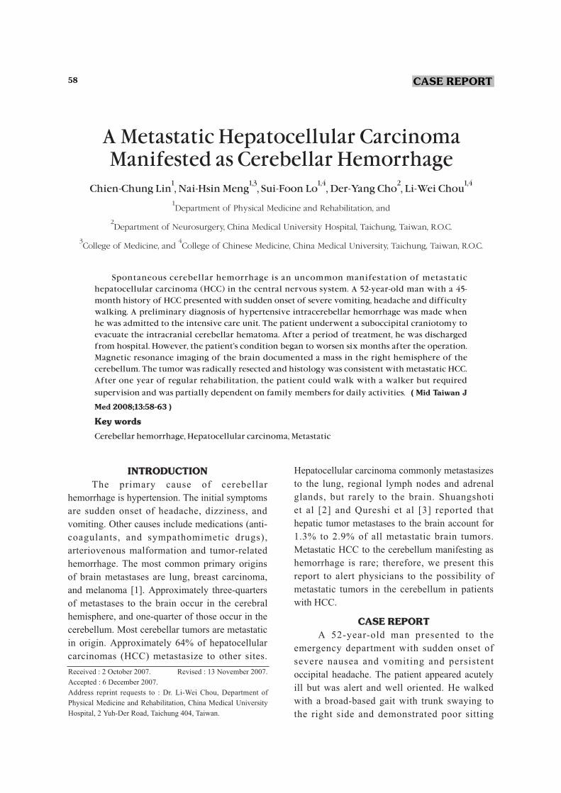
On neurologic examination, the pupils were 2.5mm in diameter, equal, round, and reactive tolight. Bilateral corneal reflexes were present, andhis face was symmetric. Muscle strength wasgrade 4 in the right upper and lower limbs. Deeptendon reflexes were normal, as was sensation.Laboratory studies revealed alanine transaminase(ALT): 23 IU/L; aspartate transaminase (AST): 36IU/L. Chest radiography showed multiple finereticular nodules in both lungs and one nodularlesion in the lower lobe of the left lung.Computed tomography (CT) of brain revealed ahighly dense lesion in the right cerebellarhemisphere with rupture to the 4th ventricle butno mass effect (Fig. 1A). The preliminarydiagnosis was cerebellar hemorrhage; he wasadmitted for further treatment on the same day.
The patient underwent a suboccititalcraniotomy to evacuate the intracranial cerebellarhematoma on the admission day; however, nospecimen was sent to the pathologist for furtherexamination at that time. Laboratory studies attwo-month follow-up revealed ALT: 48 IU/L;AST: 34 IU/L; α-fetoprotein (AFP) (enzymeimmunoassay, EIA): 0.735 ng/m L. After threemonths of rehabilitation, he could walk with awalker under supervision. He was regularlyfollowed at his neurosurgeon’s clinic.
The patient’s headaches, dizziness and
Fig. 1. Brain CT scan before surgery (A) reveals a high density lesion in the right cerebellar hemisphere. The residual hematoma inthe right cerebellar hemisphere (B) appears iso- to hypodense on brain CT at 6-mo follow-up.
A B
Metastatic HCC Manifested as Cerebellar Hemorrhage60
difficulty walking progressively worsened duringthe 6 months of postoperative follow-up. Physicalexamination at 6-month follow-up demonstrated awide-based ataxic gait but no focal or bilateralweakness or coma; the patient had intactcognition, strength, deep tendon reflexes, andsensation in all four extremities.
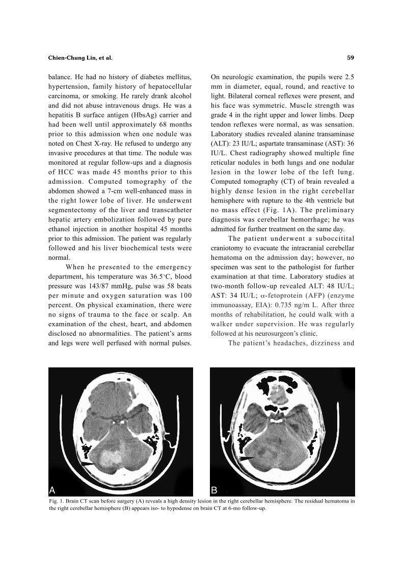
Computed tomography (CT) of brain at 6-month postoperative follow-up showed that theresidual hematoma in the cerebellum vermis wasisodense to hypodense with compression of the4th ventricle and obstructive hydrocephalus (Fig.1B). Magnetic resonance imaging (MRI) of thebrain demonstrated a mass in the right cerebellarhemisphere (Fig. 2). Laboratory studies revealedAFP: 1.17 ng/mL, carcinoembryonic antigen(CEA): 0.57 ng/mL, cancer antigen 15-3 (CA15-3): 14.2 U/mL, prostatic acid phosphatase (PSA):0.863 ng/mL, ferritin: 133 ng/mL, cancer antigen-125 (CA-125): 5.65 U/mL, carbohydrate antigen19-9 (CA19-9): 16.1 U/mL, squamous cellcarcinoma associated antigen (SCC): 0.5 ng/mL,hepatitis B surface antigen (HbsAg): negative,antibody to hepatitis B surface antigen (Anti-HBs): 1.7, IgG antibody subclass of antibody tohepatitis B core antigen (Anti-HBc-IgG):positive, and antibody to hepatitis C virus (HCVAb): negative. Abdominal sonography revealedno focal solid organic lesions and no recurrenttumors in the liver. Chest radiography and CT ofchest revealed multiple nodules in both lungs.
The largest two were in the upper lobes of theright lung and lower lobe of the left lung; imagesalso demonstrated enlarged lymph nodes in theright axilla. No definite hypodense lesions werenoted in the liver.
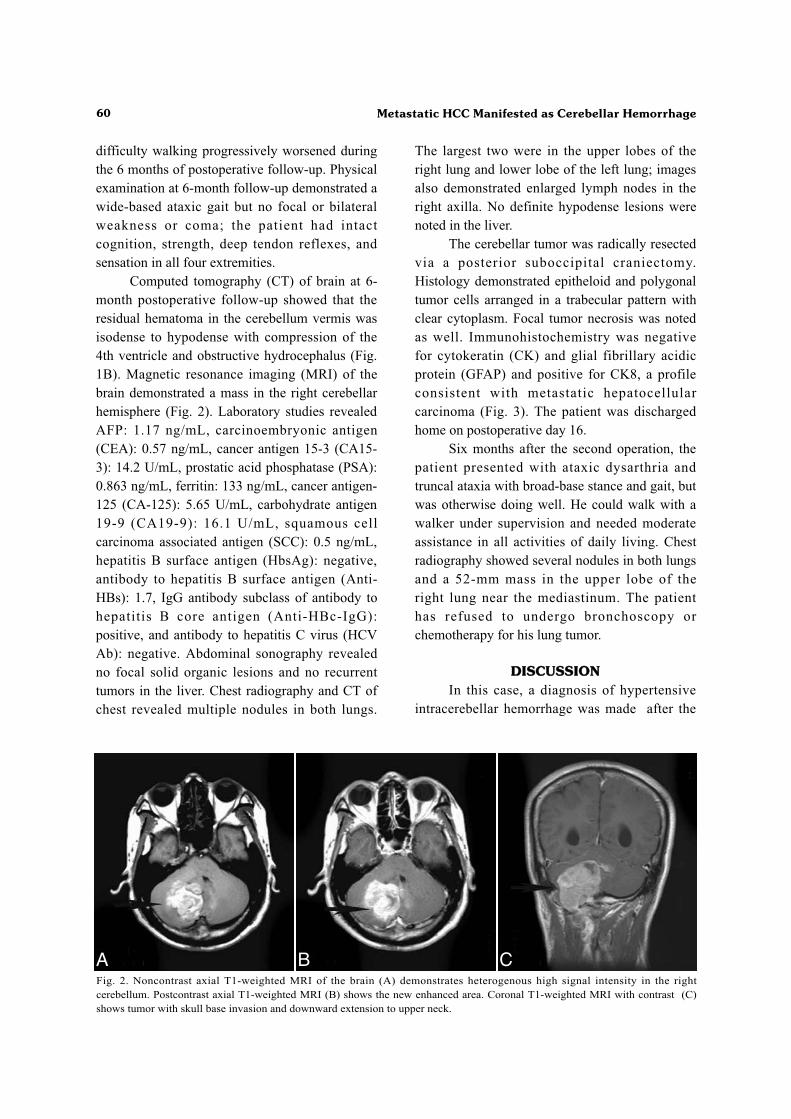
The cerebellar tumor was radically resectedvia a posterior suboccipital craniectomy.Histology demonstrated epitheloid and polygonaltumor cells arranged in a trabecular pattern withclear cytoplasm. Focal tumor necrosis was notedas well. Immunohistochemistry was negative for cytokeratin (CK) and glial fibrillary acidicprotein (GFAP) and positive for CK8, a profileconsistent with metastatic hepatocellularcarcinoma (Fig. 3). The patient was dischargedhome on postoperative day 16.
Six months after the second operation, thepatient presented with ataxic dysarthria andtruncal ataxia with broad-base stance and gait, butwas otherwise doing well. He could walk with awalker under supervision and needed moderateassistance in all activities of daily living. Chestradiography showed several nodules in both lungsand a 52-mm mass in the upper lobe of the right lung near the mediastinum. The patient has refused to undergo bronchoscopy orchemotherapy for his lung tumor.
DDIISSCCUUSSSSIIOONNIn this case, a diagnosis of hypertensive
intracerebellar hemorrhage was made after the
Fig. 2. Noncontrast axial T1-weighted MRI of the brain (A) demonstrates heterogenous high signal intensity in the rightcerebellum. Postcontrast axial T1-weighted MRI (B) shows the new enhanced area. Coronal T1-weighted MRI with contrast (C)shows tumor with skull base invasion and downward extension to upper neck.
A B C
Chien-Chung Lin, et al. 61
cerebellar hematoma had been removed. Nospecimen was sent to the pathologist for furtherexamination. After participating in regular andcomprehensive rehabilitation programs, thepatient still required minimal assistance in allactivities of daily living after he was dischargedhome. Six months after the operation, the patientpresented with progressively worsening ability towalk. Recurrent cerebrovascular disease waspreliminarily diagnosed. Computed tomographyof the brain showed that the residual hematoma inthe cerebellar vermis was isodense to hypodensewith compression of the 4th ventricle, whichinduced obstructive hydrocephalus. This was anunexpected finding because the hematoma shouldhave been completely reabsorbed, and shouldhave presented as a linear low density lesion withperipheral brain tissue atrophy. Since there was atumor-like mass noted in the chest at theemergency department, an underlying causeincluding primary or metastatic tumor shouldhave been considered. Magnetic resonanceimaging of the brain confirmed a mass in the rightcerebellum seven days later. The tumor wasresected and the finding supported thehepatocellular origin of the tumor.
Intracerebral hemorrhage (ICH) is classifiedas primary or secondary, depending on theunderlying cause of hemorrhage. Primary ICHrefers to a hemorrhage that originates fromspontaneous rupture of small arteries or arterioles
that have been damaged by chronic hypertensionor amyloid angiopathy. Secondary ICH refers tohemorrhage resulting from trauma, arteriovenousmalformation, intracranial aneurysm,coagulopathy, haemorrhagic conversion ofcerebral infarct, dural sinus thrombosis,intracranial neoplasm, cavernous angioma, duralarteriovenous fistula, venous angioma, cocaine orsympathomimetic drug exposure or CNSvasculitis. [3] Hypertension is the most importantand prevalent risk factor, directly accounting forabout 60% to 70% of cases [4,5]. The secondmost common cause of primary ICH is cerebralamyloid angiopathy, which accounts for about15% of cases [3]. The most common sites ofprimary ICH are the basal ganglia, deepcerebellum and pons. When hemorrhages occur inother brain areas or in nonhypertensive patients,greater consideration should be given to headinjury, bleeding disorders, anticoagulant therapy,neoplasms, and vascular malformations. For theseverely hypertensive patient with a wellcircumscribed and homogeneous hematomalocated in a typical location for hypertensive ICH,the clinician can regard hypertensive hemorrhageas the first differential diagnosis. Evaluation for ableeding disorder should be performed in everypatient with an ICH. Hemorrhage is morecommon in metastatic than in primary braintumors, and tumor-related hemorrhage is the mostcommon cause of brain hemorrhage in patients
Fig. 3. Photomicrographs demonstrate the histological features of cerebellar lesion. Panel A shows focal tumor necrosis (H & E,original magnification 40). Panel B shows epitheloid and polygonal tumor cells with clear cytoplasm, arranged in a trabecularpattern (H & E, original magnification 200).
A B
62 Metastatic HCC Manifested as Cerebellar Hemorrhage
with systemic solid tumors [6]. Brain metastasesare the most common cerebral tumors. The mostcommon primary site of origin is lung; skin,kidney, and breast account for much of theremainder [1]. Most metastases occur at thejunction between gray and white matter in thecerebral hemispheres, but deeper supratentoriallocations are also common; fewer occur in thecerebellum or brain stem [7].
Hepatocellular carcinoma is the fifth mostcommon cancer and the third leading cause ofcancer-related mortality worldwide [8]. Thehigher incidence of HCC in Asia is linked to theincreased prevalence of chronic viral hepatitis.HCC metastasizes by hematogenous andlymphatic routes [9]. Shee-Chan Lin et al [10]reported that manipulation of tumor withtranscatheter arterial embolization will increasethe risk of hematogenous metastasis due to anincrease in activity of serum type IV collagen-degrading enzyme, or a decrease in activity of thetumor invasion-inhibitor factor [11]. HCCcommonly metastasizes to the lung, regionallymph nodes and adrenal glands, but rarely tobrain. In HCC and other carcinomas, seeding ofthe brain, the meninges, or the cranium is usuallyin the distribution of the middle cerebral artery[9].
If there is no known cancer andintratumoral hemorrhage is suspected, resectionor biopsy of the hematoma should be performed.Careful neuropathologic examination is required,because the tumor can be microscopic. Factorsaffecting the prognosis of patients with metastaticbrain tumor include age, primary tumor site,pretreatment performance status, presence ofextracranial disease, interval between treatment ofthe primary lesion and of brain metastasis,number and location of intracranial metastases aswell as surgical excision of brain metastasis [12].Hypertension is the most important and prevalentrisk factor, directly accounting for about 60% to70% of intracerebral hemorrhage [4,5]. It is easyfor the clinician to ignore tumor-relatedhemorrhage. Brain metastases, but especially
cerebellar metastases, from hepatocellularcarcinoma are rare. In conclusion, this reportshould alert the physician to the possibility ofcerebellar metastasis in patients with HCC.
RREEFFEERREENNCCEESS1. Barnholtz-Sloan JS, Sloan AE, Davis FG. Incidence
proportions of brain metastases in patients diagnosed
(1973 to 2001) in the Metropolitan Detroit Cancer
Surveillance System. J Clin Oncol 2004;22:2865-72.
2. Shuangshoti S, Rungruxsirivorn S, Panyathanya R.
Intracranial metastatic of hepatic carcinoma: a study
within 28 years. J Med Assoc Thai 1989;72:307-13.
3. Qureshi AI, Tuhrim S, Broderick JP, et al. Spontaneous
intracerebral hemorrhage. N Engl J Med 2001;344:
1450-60.
4. Brott T, Thalinger K, Hertzberg V. Hypertension as a
risk factor for spontaneous intracerebral hemorrhage.
Stroke 1986;17:1078-83.
5. Thrift AG, McNeil JJ, Forbes A, et al. Three important
subgroups of hypertensive persons at greater risk of
intracerebral hemorrhage. Hypertension 1998;31:
1223-29.
6. Lieu AS, Hwang SL, Howng SL, et al. Brain tumors
with hemorrhage. J Formos Med Assoc 1999;98:365-
7.
7. Delattre JY, Krol G, Thaler HT. Distribution of brain
metastases. Arch Neurol 1988;45:741-44.
8. Parkin DM, Bray F, Ferlay J. Estimating the world
cancer burden Globocan 2000. Int J Cancer 2001;
94:153-56.
9. Yang WT, Yeo W, Leung SF, et al. MRI and CT of
metastatic hepatocellular carcinoma causing spinal
cord compression. Clin Radiol 1997;52:755-60.
10. Lin SC, Shih SC, Kao CR, et al. Transcatheter arterial
embolization treatment in patients with hepatocellular
carcinoma and risk of pulmonary metastasis. World J
Gastroenterol 2003;9:1208-11.
11. Feridman HD. Hepatocellular carcinoma with central
nervous system metastasis: A case report and literature
review. Med Pediatr Oncol 1991;19:139-44.
12. Nakagawa H, Miyawaki Y, Fujita T, et al. Surgical
treatment of brain metastastases of lung cancer:
retrospective analyses of 89 cases. J Neurol Neurosurg
Psychiatry 1994;57:950-6.

63
1 1,3 1,4 2 1,4
1 2
3 4
( )
64%
52 B
2008;13:58-63
404 2
2007 10 2 2007 11 32007 12 6