high efficiency opsonin-independent phagocytosis of candida parapsilosis by human neutrophils
TRANSCRIPT

High Efficiency Opsonin-Independent Phagocytosis of Candidaparapsilosis by Human Neutrophils
Jennifer R. Linden1, Matthew A. Maccani1, Sonia S. Laforce-Nesbitt2, and Joseph M.Bliss1,2,*1Graduate Program in Pathobiology, Brown University, Providence, RI2Department of Pediatrics, Women & Infants Hospital of Rhode Island, Warren Alpert MedicalSchool of Brown University, Providence, RI
AbstractCandida species are associated with invasive fungal infections, and C. parapsilosis has becomeincreasingly prevalent. As key antifungal effector cells, the function of human neutrophilsconfronting C. parapsilosis was investigated. We hypothesized that interaction between neutrophilsand Candida species may not be uniform. Opsonins were omitted from these studies to understandthe antifungal mechanisms at their most basic level. Human neutrophils underwent phagocytosis ofC. parapsilosis with much higher efficiency than C. albicans. Immunofluorescence assays with β-glucan specific antibody detected more surface exposed β-glucan on C. parapsilosis than C.albicans. However, blockade of the β-glucan receptor, Dectin-1, reduced phagocytosis of C.albicans but not C. parapsilosis. Inclusion of excess β-glucan, mannan, or chitin also had no effecton phagocytosis of C. parapsilosis. Consistent with the difference in phagocytosis, neutrophilsmediated damage to C. parapsilosis but not C. albicans in assays of residual metabolic activity. C.parapsilosis was more sensitive to oxidative stress, and inclusion of antioxidant in toxicity assaysdecreased neutrophil mediated damage, suggesting that generation of reactive oxygen speciescontributes to the mechanism of toxicity. These data suggest that the interaction between neutrophilsand Candida species is not uniform and may partially account for differences observed in theepidemiology and natural history of infections caused by these species.
KeywordsNeutrophil; Phagocytosis; Candida; Dectin-1; β-glucan
INTRODUCTIONCandida species are a prevalent cause of invasive fungal infections, currently the fourth leadingorganism in hospital acquired bloodstream infections in the United States [1]. Candidaalbicans is associated with the majority of these infections and possesses a number of specificvirulence attributes that have been the subject of intensive study [2]. Candida parapsilosis hasbeen considered a less virulent species and historically has been associated with a smallproportion of bloodstream infections. However, this organism has increased in prevalence inrecent years. C. parapsilosis is a common cause of bloodstream infection in neonates [3-5],and has overtaken C. albicans in frequency of hospital-acquired infection in some hospitals
*Corresponding Author: Department of Pediatrics, Women & Infants Hospital of Rhode Island, 101 Dudley St., Providence, RI 02905Phone: (401) 274-1122 ext. 1384 Fax: (401) 453-7571 [email protected] .DISCLOSURE OF CONFLICT OF INTEREST None.
NIH Public AccessAuthor ManuscriptMed Mycol. Author manuscript; available in PMC 2011 February 1.
Published in final edited form as:Med Mycol. ; : 1–10. doi:10.1080/13693780903164566.
NIH
-PA Author Manuscript
NIH
-PA Author Manuscript
NIH
-PA Author Manuscript

worldwide [4,6,7]. Recent studies have identified the importance of secreted lipase in thevirulence of this organism and have documented its ability to damage tissue in vitro [8,9].
The neutrophil is well-recognized for its important role in host defense against invasive fungalinfections, and various neutrophil functions have been studied using C. albicans and otherfungi as targets [10,11]. The interaction between neutrophils and C. parapsilosis has receivedconsiderably less attention. Because of the increasing prevalence of C. parapsilosis in invasivecandidiasis and the central role of neutrophils in host defense, we sought to more clearlyunderstand the specific attributes of human neutrophils confronting this species. In this report,we focus on phagocytosis as an important initial step in this interaction and on the mechanismsof neutrophil-induced toxicity to each species. Opsonization of pathogenic organisms withspecific antibody or complement plays a key role in this process in vivo, and is well known toalter the dynamics of phagocytosis under in vitro conditions [12-14]. In an effort to understandthe interaction between neutrophils and yeast with the least number of confounding variables,we conducted these studies in the absence of opsonins. We also investigated the role ofcarbohydrates present in the fungal cell wall in the phagocytosis process.
MATERIALS AND METHODSOrganisms and media
C. albicans strains used in this study include a laboratory strain, Ca3153A [15,16], and a clinicalisolate from an infant with urinary candidiasis, Ca-4 [17]. C. parapsilosis strains included threeindependent clinical isolates colonizing premature infants in a previously reported study, Ro18,Ro29 and Ro75 [18]. Starter cultures for phagocytosis and toxicity assays were grown 16 h at37°C with vigorous agitation in YEPD medium (1% yeast extract, 2% peptone, 2% dextrose).Cultures were predominantly (>99%) yeast forms following this incubation.
Preparation and pretreatment of neutrophilsFollowing review and approval by the Institutional Review Board, human neutrophils wereisolated by density gradient centrifugation from peripheral blood of healthy adult volunteers.Briefly, leukocytes were separated from whole blood on Histopaque-1077 (Sigma) by densitygradient centrifugation. Neutrophils were further purified by dextran sedimentation andhypotonic lysis of contaminating erythrocytes. Cells were adjusted to 5 × 10⁶ cells/mL in HBSS+ Ca/Mg, and were >95% neutrophils as determined by Wright-Giemsa stain. In selectedexperiments, neutrophils were pretreated with various reagents to study the specifics of theyeast-neutrophil interaction. To evaluate the requirement for actin polymerization, neutrophilswere preincubated with cytochalasin D (10 μg/mL) at 4°C for 30 min with constant mixing.The role of β-glucan was studied by pretreating neutrophils with excess β-glucan or withblocking monoclonal antibodies to Dectin- 1. A β-glucan solution (10 mg/ml, Sigma, frombarley) was prepared using described methods to maximize solubility [19]. Briefly, β-glucanwas dissolved in 1N NaOH by heating to 60°C, diluted to 0.5 mg/ml in HBSS, and correctedto neutral pH with HCl. A vehicle control was used for comparison in these experiments,prepared identically to the β-glucan solution but omitting the β-glucan. Neutrophils wereincubated with β-glucan or vehicle control for 30 min at 4°C with constant mixing prior toinclusion in the phagocytosis assay. Dectin-1 receptor was blocked with specific antibody(GE2, generously provided by Gordon Brown) [20] by incubation with 10 μg/mL of antibodyfor 20 min at 4°C. To investigate the role of other carbohydrate receptors in phagocytosis,neutrophils were pretreated with mannan (Sigma, from Saccharomyces cerevisiae) or chitin(Sigma, from crab shells). Mannan was solubilized in HBSS and preincubated with neutrophils(10 mg/ml or 1 mg/ml) for 30 min at 4°C with constant mixing. Solubility of chitin is limitedin aqueous solution, so solutions were prepared according to the described method [21]. Briefly,chitin was suspended in HBSS with 8% NaOH and 4% Urea (w/v) and sonicated for 30 min.
Linden et al. Page 2
Med Mycol. Author manuscript; available in PMC 2011 February 1.
NIH
-PA Author Manuscript
NIH
-PA Author Manuscript
NIH
-PA Author Manuscript
The solution was incubated at 4°C overnight with rotation and incubated at −20°C for 1 hour.A 1:100 dilution was performed into HBSS to achieve 0.1 mg/ml, and the pH was adjustedusing HCl. A vehicle control was used for comparison in these experiments, preparedidentically to the chitin solution but omitting the chitin. Neutrophils were incubated with chitinor vehicle control for 30 min at 4°C with constant mixing prior to inclusion in the phagocytosisassay.
Phagocytosis assayCandida species were washed in Hank’s Balanced Salt Solution (HBSS), enumerated on ahemacytometer, and adjusted to a final concentration appropriate to provide the desiredmultiplicity of infection (MOI). In selected experiments, yeast were heat killed at 65°C for 90min. Yeast were labeled with FITC at 10 μg/mL in the dark with rotation for 30 minutes at 37°C followed by extensive washes with HBSS. Once neutrophils were prepared and pretreatedwhen indicated, equal volumes of Candida yeast and neutrophils were combined at ratiosappropriate to provide a MOI ranging from 10 to 100 (10 to 100 yeast per neutrophil). Cellswere pelleted at 500 × g for 2 minutes, incubated on ice for 30 min to give a pool of cells withsurface-bound Candida, then at 37°C for 30, 60 or 90 min to allow phagocytosis. Cells werepelleted at 500 × g for 2 min and resuspended in 20 μL HBSS. 2.5 μL of ethidium bromide(100 μg/mL) was mixed with 2.5 μL of the sample on the surface of a microscope slide andexamined by fluorescence microscopy. Intracellular Candida were differentiated fromextracellular by retention of green fluorescence. A minimum of 100 neutrophils were counted,and the percent that had undergone phagocytosis of yeast was calculated. In some experiments,the number of intracellular yeast within neutrophils undergoing phagocytosis was calculatedand reported as 1, 2, 3, or 4+ yeast per cell.
Indirect immunofluorescence assay and flow cytometry for β-glucanC. albicans and C. parapsilosis yeast were harvested from overnight culture and washed withPBS. Yeast cells were blocked with 3% bovine serum albumin (BSA) at 22°C for 30 min, thenincubated with 15 μg/ml BF-Div, a mouse IgM monoclonal antibody specific for both β-(1-3)-and β-(1-6)-linked glucan [22] for 30 min at 22°C. Cells were washed with PBS and incubatedwith an appropriate FITC-conjugated secondary antibody for 30 min at 22°C. Controls includedyeast cells incubated in 3% BSA only or with secondary antibody only. Cells were washed andexamined by fluorescence microscopy or by flow cytometry with a BD BiosciencesFACSCanto instrument.
Neutrophil-induced toxicity assayCandida species were washed in HBSS + Ca/Mg with 0.1% (w/v) glucose. Human adultneutrophils were prepared as described above. In selected experiments, neutrophils werepreincubated with cytochalasin D (10 μg/mL) at 4°C for 30 min with constant mixing. Cellswere enumerated on a hemacytometer and combined at varied effector to target (E: T) ratiosin a 96 well dish in 60 μl total volume with appropriate controls. After a 60 min incubation at37°C, plates were centrifuged at 1600 × g and the supernatant was carefully aspirated from thewells. Neutrophils were lysed by agitation in 0.02% (v/v) Tween-20 in water, pH 11.0 [23]and incubated at 37°C for 15 min. The centrifugation and lysis steps were repeated, and lysisconfirmed by light microscopy. Aspirated supernatants were plated on YEPD, confirming thatonly trivial numbers of yeast cells (< 2% of inoculum) were lost in this procedure. Toxicity toCandida was measured by residual metabolic activity, using (2,3)-bis-(2-methoxy-4-nitro-5-sulphenyl)-(2H)-tetrazolium-5-carboxanilide (XTT, Sigma) assay as described [24]. Briefly,XTT was freshly prepared in Dulbecco’s PBS, heated at 60°C for 30 min and filtered.Coenzyme Q (Sigma) was added, and the solution was added to cells at concentrations of 0.5mg/ml XTT and 40 mg/ml Coenzyme Q. Plates were incubated at 37°C for 1 h and the intensity
Linden et al. Page 3
Med Mycol. Author manuscript; available in PMC 2011 February 1.
NIH
-PA Author Manuscript
NIH
-PA Author Manuscript
NIH
-PA Author Manuscript

of the colorimetric reaction, reflecting metabolic activity, was measured by optical density at450 nm (OD450) using an automated plate reader. Individual experiments were performed intriplicate at minimum, and a minimum of 5 individual neutrophil donors were studied inindependent experiments. Percent residual XTT activity was calculated as Mean OD450 (Yeast+ neutrophils) / Mean OD450 (Yeast alone). To study the contribution of reactive oxygenspecies (ROS) in neutrophil-mediated toxicity to Candida yeast, toxicity assays wereconducted in the presence of the ROS scavenger, N-acetylcysteine, at concentrations of 1, 2,and 4 mM in HBSS. Percent residual XTT activity was calculated as above for eachconcentration of N-acetylcysteine.
Hydrogen peroxide (H2O2) toxicity assayCandida species were washed 3 times in HBSS and enumerated on a hemacytometer. 1×10⁶
cells/well were inoculated into 96 well plates containing HBSS+ 0.1% glucose with andwithout 1mM H202 in triplicate. Plates were incubated at 37°C for 1 hour. Hydrogen peroxideinduced toxicity was measured by residual XTT activity as described above. Percent residualXTT activity was calculated as Mean OD450 (Yeast + H202) / Mean OD450 (Yeast alone).
Statistical analysisComparisons of phagocytosis and induced toxicity among different Candida species and strainsand among various neutrophil treatments were made by analysis of variance (ANOVA).Between group comparisons were made by Newman-Keuls test, and p values < 0.05 wereconsidered significant.
RESULTSPhagocytosis of Candida species by human neutrophils
To compare the relative phagocytosis efficiency of C. albicans and C. parapsilosis yeast formsby human neutrophils, phagocytosis assays were conducted using FITC-labeled C. albicansand C. parapsilosis yeast and neutrophils isolated from healthy adult volunteers in the absenceof opsonins. Heat killed organisms gave more uniform and reliable labeling with FITC andthus were used preferentially in these experiments. Live organisms behaved similarly (seebelow). To determine the optimal incubation time to allow phagocytosis to occur, yeast andneutrophils were coincubated for 30, 60, and 90 minutes. No increase in phagocytosis of anystrain was detected at incubations longer than 30 minutes (data not shown), so the 30 minutetime point was used in all subsequent experiments. Phagocytosis assays were conducted atmultiplicity of infection (MOI) ranging from 10 to 100. Two strains of C. albicans (laboratorystrain, Ca3153A and a clinical isolate, Ca-4) and 3 strains of C. parapsilosis (clinical isolates)were studied. In all cases and at all MOIs, phagocytosis was much more efficient for neutrophilsincubated with C. parapsilosis than C. albicans (Fig. 1A, p < 0.001). Increasing MOI above10 increased phagocytosis efficiency for C. albicans strain Ca3153A (p < 0.02) but not forCa-4 (p > 0.16). Increasing MOI above 10 increased phagocytosis efficiency for C.parapsilosis strains Ro18 (p < 0.006) and Ro29 (p < 0.002) but not Ro75 (p > 0.46).
To confirm that the difference in phagocytosis efficiency between C. albicans and C.parapsilosis was not an artifact related to the heat-killing process, phagocytosis assays wereconducted with live C. albicans and C. parapsilosis yeast at an MOI of 100. Again, significantlyhigher rates of phagocytosis were seen with all C. parapsilosis strains relative to C. albicans(Fig. 1B). C. albicans strain Ca3153A showed somewhat higher phagocytosis efficiency whenusing heat-killed vs. live yeast (p=0.001), whereas phagocytosis rates between heat-killed andlive yeast of strain Ca-4 were no different (p=0.99). Heat-killed yeast also improvedphagocytosis rates for C. parapsilosis strains Ro18 (p=0.006) and Ro29 (p=0.005), but notRo75 (p=0.39). Although some minor differences in phagocytosis efficiency between live and
Linden et al. Page 4
Med Mycol. Author manuscript; available in PMC 2011 February 1.
NIH
-PA Author Manuscript
NIH
-PA Author Manuscript
NIH
-PA Author Manuscript

heat-killed yeast were observed in some strains, in all cases C. parapsilosis maintained higherphagocytosis rates with both live and heat-killed yeast than C. albicans (p < 0.001).
In addition to the higher rates of phagocytosis noted with C. parapsilosis, neutrophils withintracellular yeast were qualitatively noted to have a higher yeast burden with C.parapsilosis than with C. albicans. To quantify this observation, the number of yeast perneutrophil undergoing phagocytosis was counted for each strain. A single, representative strainof C. albicans (Ca3153a) and C. parapsilosis (Ro18) is depicted in Fig. 1C. The percentageof neutrophils undergoing phagocytosis that had 4 or more yeast per cell was significantlyhigher for C. parapsilosis than for C. albicans at a MOI of 100 or 50 (p=0.0001 for eachcomparison). There was a trend toward higher yeast burden with C. parapsilosis at a MOI of10 as well (p=0.056).
Finally, because actin polymerization is known to be required for phagocytosis, we wished todemonstrate this requirement in our assays. Neutrophils were preincubated with cytochalasinD prior to exposure to yeast. Cytochalasin D treatment reduced phagocytosis to 8% or less inall strains (Fig. 1D).
Role of β-glucan/Dectin-1, mannan and chitin in phagocytosis of C. albicans and C.parapsilosis
The β-glucan receptor, Dectin-1, has recently been shown to be expressed on neutrophils andto have a role in phagocytosis of C. albicans [20]. To assess whether this receptor was involvedin the enhanced phagocytosis activity of C. parapsilosis, exposure of β-glucan on the cellsurface of C. albicans and C. parapsilosis yeast was assessed by indirect immunofluorescenceassay (IFA). Additionally, Dectin-1 blocking antibody and soluble glucan were used todetermine the role of this receptor in phagocytosis. The β-glucan specific monoclonal antibody,BFDiv [22], was used to label β-glucan in live C. albicans and C. parapsilosis yeast cells byIFA (Fig. 2). This antibody binds to both β-(1-3)- and β-(1-6)-linked glucan. Exposure of β-glucan as detected by antibody binding was not uniform for C. albicans. Only occasional yeastshowed detectable fluorescence, and in the majority of cells where fluorescence was detected,it was faint and localized to punctate regions of the cell (arrows in Fig. 2A). Labeling of C.parapsilosis yeast with the antibody was much more consistent and brighter, however bindingwas also not uniform, with some regions of the cell surface brighter than others (Fig. 2B). Toquantify the differential binding of β-glucan specific antibody, yeast cells stained in an identicalfashion were analyzed by flow cytometry (Fig. 2C). Two C. parapsilosis (Ro18 and Ro29)strains were analyzed. The third strain (Ro75) had a propensity to clump into cellular aggregatesand was therefore less amenable to flow cytometric analysis. Consistent with the IFA results,C. parapsilosis strains exhibited much increased fluorescence relative to C. albicans.
To determine whether the apparently increased surface exposure of β-glucan in C.parapsilosis relative to C. albicans contributed to the increased phagocytosis efficiency of theformer, phagocytosis assays were conducted in the presence of excess β-glucan (Fig. 3A).Although β-glucan reduced phagocytosis of C. albicans (p = 0.04), excess β-glucan had noeffect on C. parapsilosis phagocytosis. To investigate the role of the β-glucan receptor,Dectin-1, blocking antibody GE2 was coincubated with neutrophils prior to phagocytosisassays. Consistent with previous studies [20], blocking of Dectin-1 decreased the phagocytosisof C. albicans by 57% (p< 0.001) (Fig. 3B). In contrast, treatment of neutrophils with thisantibody resulted in a small, but statistically significant increase in phagocytosis of C.parapsilosis (p= 0.02).
To investigate the possible contribution of other fungal polysaccharides to the difference inphagocytosis efficiency between C. albicans and C. parapsilosis, assays were conducted in
Linden et al. Page 5
Med Mycol. Author manuscript; available in PMC 2011 February 1.
NIH
-PA Author Manuscript
NIH
-PA Author Manuscript
NIH
-PA Author Manuscript
the presence of excess mannan (Fig. 3C) or chitin (Fig. 3D). Neither of these polysaccharidesresulted in any detectable inhibition of phagocytosis in either species.
Toxicity to Candida species mediated by human neutrophilsTo assess the capacity of human neutrophils to damage unopsonized Candida yeast, metabolicactivity of the yeast following coincubation was measured using the colorimetric XTTmetabolism assay. Traditional, plate-based methods to measure fungal killing by neutrophilswere found to provide inconsistent results and to be unduly affected by the tendency of yeastcells to adhere to plastic or each other (data not shown). This inaccuracy has also been notedby others [25]. To circumvent these issues, we opted to use metabolic activity as a surrogatefor toxicity to the yeast cells. In these assays, neutrophils and Candida yeast were coincubated,the neutrophils lysed, and residual metabolic activity of the yeast was measured by XTT assay.After preliminary experiments using varied effector: target (E:T) ratios, a ratio of 2 neutrophilsto 1 yeast was selected for subsequent analyses, as it allowed maximal discrimination betweenspecies and strains. Mean residual metabolic activity of each strain, expressed as a percent oftotal metabolic activity of the same strain of Candida incubated in the absence of neutrophils,is shown in Fig. 4A. All strains of C. parapsilosis were more susceptible to neutrophil-inducedtoxicity than C. albicans (p ≤ 0.001), and no statistically significant differences among strainsof the same species were observed.
The efficient phagocytosis of C. parapsilosis described above required actin polymerization,as pretreatment of neutrophils with cytochalasin D disrupted phagocytosis activity. Todetermine the requirement for actin polymerization in neutrophil-induced toxicity to C.parapsilosis, cytochalasin D treated neutrophils were used in the XTT assay. The capacity ofneutrophils to induce toxicity to C. parapsilosis was significantly inhibited by cytochalasin D(Fig. 4B), suggesting that the induced toxicity was an active process that likely requiresphagocytosis.
Neutrophils can generate a number of antimicrobial effects, including reactive oxygen species(ROS) as well as non-oxidative mechanisms. The observation that C. albicans is more resistantto neutrophil-induced toxicity may reflect this species’ resistance to phagocytosis. However,there may also be differences between these species in their intrinsic resistance to oxidativestress. To test this hypothesis, yeast forms of both species were treated with 1 mM hydrogenperoxide (H2O2), followed by XTT assay to determine residual metabolic activity (Fig. 5A).C. albicans yeast were more resistant than C. parapsilosis yeast to this form of oxidative stress(p < 0.01 for all strains). To investigate the contribution of oxidative mechanisms to thesusceptibility of C. parapsilosis to neutrophil-mediated toxicity, assays were conducted in thepresence of the ROS scavenger, N-acetylcysteine (Fig 5B). Although rates of phagocytosiswere unaffected (data not shown), N-acetylcysteine decreased the toxicity of neutrophils in anapparently dose-dependent manner, achieving statistical significance in the case of C.albicans strain Ca3153A (p = 0.009) and C. parapsilosis strain Ro18 (p = 0.02). Similar trendswere seen with the other strains, although statistical significance was not achieved. These datasuggest that ROS play a role in neutrophil mediated toxicity, and the extent to which ROScontribute may vary among strains. Taken together, these data suggest that the resistance ofC. albicans to neutrophil-mediated toxicity may be a combination of resistance to phagocytosisand resistance to oxidative stress, while C. parapsilosis is more susceptible to both. Generationof ROS is likely to contribute to neutrophil-mediated toxicity of C. parapsilosis.
DISCUSSIONThe importance of opsonins in efficient phagocytosis of C. albicans yeast has long beenrecognized. Lehrer and Cline reported greater than 90% phagocytosis of C. albicans yeast byneutrophils with human serum present, but when the serum was replaced by albumin or
Linden et al. Page 6
Med Mycol. Author manuscript; available in PMC 2011 February 1.
NIH
-PA Author Manuscript
NIH
-PA Author Manuscript
NIH
-PA Author Manuscript

buffered salt solution, phagocytosis was “virtually absent” [26]. Likewise, Diamond et al.observed 5-8% ingestion of C. albicans by neutrophils when serum was absent [13]. Similartrends were observed more recently with the human monocytic cell line, THP-1 [12,14].Another study comparing phagocytosis rates among several fungal pathogens found similarrates of phagocytosis between C. albicans and C. parapsilosis, but all samples were opsonizedwith pooled human serum [27]. The two main sources of opsonins, antibody and complement,have the potential to create variability in assay systems. Antibody properties such as isotype,antigen specificity and binding affinity, as well as properties of the yeast cell target includingabundance of cognate antigens and variability in their expression, may all impact the nature ofphagocytosis. Likewise, the extent and efficiency of complement deposition on the yeast cellsurface and the binding to complement receptors may not be uniform. Because opsonizationinherently introduces additional complexities to the interaction between yeast and phagocyte,we conducted these experiments in the absence of opsonins. These conditions may also berelevant for premature infants, a group in which C. parapsilosis is particularly prevalent [3],and in whom low concentrations of antibody and key complement components have beenreported [28]. Surprisingly, we detected a previously undescribed efficiency of phagocytosiswith C. parapsilosis that is far greater than C. albicans under these conditions, and wasassociated with increased neutrophil-induced toxicity. Generation of ROS by the neutrophilsis likely an important mechanism for the antifugal effect. The differences observed betweenthese species may contribute to the increased virulence of C. albicans over C. parapsilosis indisseminated candidiasis.
We found ample, but nonuniform binding of the β-glucan antibody, BF-Div, to C.parapsilosis, but considerably less binding to yeast forms of C. albicans. These patterns areconsistent with previously reported surface exposure of β-glucan in C. albicans as detected bya soluble form of the β-glucan receptor, Dectin-1, as probe [29]. The contribution of β-glucanand its receptor, Dectin-1, in interaction between human neutrophils and C. albicans has beenthe subject of recent study [20]. These authors demonstrated that treatment of neutrophils withthe Dectin-1 blocking antibody, GE2, reduces phagocytosis of C. albicans yeast. Our data areconsistent with this observation; however, despite apparently increased exposure of β-glucanon C. parapsilosis, receptor blockade with GE2 caused no reduction in phagocytosis nor didpretreatment with excess β-glucan. These data suggest that Dectin-1 is not required for theincreased phagocytosis efficiency of C. parapsilosis. The binding of fungi by phagocytes iscomplex and is mediated primarily by a host of pattern-recognition receptors (PRRs), whichrecognize characteristic pathogen-associated molecular patterns (PAMPs) found on a widerange of microorganisms but absent in the host [11]. Individual fungal species can berecognized by several PRRs, and the outcomes of these interactions vary considerably [30].The nature of these specific interactions likely explains the differences in phagocytosis betweenthese two species. The receptor(s) important for the enhanced phagocytosis of C.parapsilosis is the subject of ongoing investigation.
Our finding of enhanced neutrophil-induced toxicity of C. parapsilosis relative to C.albicans is in agreement with others that found C. parapsilosis more susceptible to neutrophilkilling by human [31] or murine [32] neutrophils. The latter study found that killing (based oncolony forming unit counts) correlated with increases in toxicity, based on 51Cr release fromlabeled fungi. In contrast, Roilides et al. reported that C. parapsilosis was less susceptible tohuman neutrophil induced damage than C. albicans [33]. However, the neutrophils in this studywere maintained in the absence of calcium and magnesium, which others have shown to beimportant for interaction of neutrophils with C. albicans [13]. In a study of murinemacrophages, C. parapsilosis was again more efficiently killed than C. albicans [34]. Theauthors attributed the increased killing of C. parapsilosis to a relative decrease in stimulationof oxygen metabolism in the macrophages by C. albicans. Similar differences in macrophagekilling of these two species were also reported by Brummer and Stevens [35]. Interestingly,
Linden et al. Page 7
Med Mycol. Author manuscript; available in PMC 2011 February 1.
NIH
-PA Author Manuscript
NIH
-PA Author Manuscript
NIH
-PA Author Manuscript

macrophages were efficient at phagocytosis of both species, with C. parapsilosis rates onlyslightly higher than C. albicans. Phagocytosis of the two species was only modestly differentwhether serum was present or absent. This finding implies that the mechanism of phagocytosisby murine macrophages is intrinsically different from that of human neutrophils, perhapsthrough the use of differing receptors. Alternatively, murine macrophages may behavedifferently than human cells. Unlike the murine studies, the human monocytic cell line, THP-1,did not phagocytose C. albicans yeast efficiently in the absence of opsonins [12,14].
The interaction between human neutrophils and C. parapsilosis is strikingly different than theinteraction with C. albicans. Understanding these differences may help to explain thedifference in virulence between these two species and provide insight in the molecularmechanisms of neutrophils which are varied and tailored toward the specific microbe faced.As Candida infections with non-albicans species increase, insights into the specifics at thehost-fungus interface may aid in the design of novel therapeutic strategies in patients at risk.
AcknowledgmentsWe thank Jonathan Reichner and Liz Lavigne for providing mAb BF-Div and for helpful discussions. We thank GordonBrown for providing mAb GE2, and Sunil Shaw for assistance with FACS analysis.
This work was supported in part by Basil O’Connor Starter Scholar Research Award Grant No. 5-FY05-1211 fromthe March of Dimes Foundation, a National Institute of Health grant (K08 AI064919), and a NIH COBRE grant (P20RR018728).
REFERENCES1. Wisplinghoff H, Bischoff T, Tallent SM, et al. Nosocomial bloodstream infections in US hospitals:
analysis of 24,179 cases from a prospective nationwide surveillance study. Clin Infect Dis2004;39:309–317. [PubMed: 15306996]
2. Calderone, RA., editor. Candida and Candidiasis. ASM Press; Washington D.C.: 2002.3. Benjamin DK Jr. Stoll BJ, Fanaroff AA, et al. Neonatal candidiasis among extremely low birth weight
infants: risk factors, mortality rates, and neurodevelopmental outcomes at 18 to 22 months. Pediatrics2006;117:84–92. [PubMed: 16396864]
4. Fridkin SK, Kaufman D, Edwards JR, Shetty S, Horan T. Changing incidence of Candida bloodstreaminfections among NICU patients in the United States: 1995-2004. Pediatrics 2006;117:1680–1687.[PubMed: 16651324]
5. Krcmery V, Fric M, Pisarcikova M, et al. Fungemia in neonates: report of 80 cases from seven universityhospitals. Pediatrics 2000;105:913–914. [PubMed: 10819665]
6. Pagano L, Antinori A, Ammassari A, et al. Retrospective study of candidemia in patients withhematological malignancies. Clinical features, risk factors and outcome of 76 episodes. Eur J Haematol1999;63:77–85. [PubMed: 10480286]
7. Pfaller MA, Diekema DJ, Jones RN, et al. International surveillance of bloodstream infections due toCandida species: frequency of occurrence and in vitro susceptibilities to fluconazole, ravuconazole,and voriconazole of isolates collected from 1997 through 1999 in the SENTRY antimicrobialsurveillance program. J Clin Microbiol 2001;39:3254–3259. [PubMed: 11526159]
8. Gacser A, Schafer W, Nosanchuk JS, Salomon S, Nosanchuk JD. Virulence of Candida parapsilosis,Candida orthopsilosis, and Candida metapsilosis in reconstituted human tissue models. Fungal GenetBiol 2007;44:1336–1341. [PubMed: 17391997]
9. Gacser A, Trofa D, Schafer W, Nosanchuk JD. Targeted gene deletion in Candida parapsilosisdemonstrates the role of secreted lipase in virulence. J Clin Invest 2007;117:3049–3058. [PubMed:17853941]
10. Mansour MK, Levitz SM. Interactions of fungi with phagocytes. Curr Opin Microbiol 2002;5:359–365. [PubMed: 12160853]
11. Nicola AM, Casadevall A, Goldman DL. Fungal killing by mammalian phagocytic cells. Curr OpinMicrobiol 2008;11:313–317. [PubMed: 18573683]
Linden et al. Page 8
Med Mycol. Author manuscript; available in PMC 2011 February 1.
NIH
-PA Author Manuscript
NIH
-PA Author Manuscript
NIH
-PA Author Manuscript

12. Wellington M, Bliss JM, Haidaris CG. Enhanced phagocytosis of Candida species mediated byopsonization with a recombinant human antibody single-chain variable fragment. Infect Immun2003;71:7228–7231. [PubMed: 14638823]
13. Diamond RD, Krzesicki R, Jao W. Damage to pseudohyphal forms of Candida albicans by neutrophilsin the absence of serum in vitro. J Clin Invest 1978;61:349–359. [PubMed: 340470]
14. Wellington M, Dolan K, Haidaris CG. Monocyte responses to Candida albicans are enhanced byantibody in cooperation with antibody-independent pathogen recognition. FEMS Immunol MedMicrobiol 2007;51:70–83. [PubMed: 17610517]
15. Morrow B, Ramsey H, Soll DR. Regulation of phase-specific genes in the more general switchingsystem of Candida albicans strain 3153A. J Med Vet Mycol 1994;32:287–294. [PubMed: 7983573]
16. Vargas K, Wertz PW, Drake D, Morrow B, Soll DR. Differences in adhesion of Candida albicans3153A cells exhibiting switch phenotypes to buccal epithelium and stratum corneum. Infect Immun1994;62:1328–1335. [PubMed: 8132340]
17. Bliss JM, Sullivan MA, Malone J, Haidaris CG. Differentiation of Candida albicans and Candidadubliniensis by using recombinant human antibody single-chain variable fragments specific forhyphae. J Clin Microbiol 2003;41:1152–1160. [PubMed: 12624045]
18. Bliss JM, Basavegowda KP, Watson WJ, Sheikh AU, Ryan RM. Vertical and horizontal transmissionof Candida albicans in very low birth weight infants using DNA fingerprinting techniques. PediatrInfect Dis J 2008;27:231–235. [PubMed: 18277930]
19. Bhatty RS. Laboratory and pilot plant extraction and purification of β-glucans from hullless barleyand oat brans. J Cereal Sci 1995;22:163–170.
20. Kennedy AD, Willment JA, Dorward DW, et al. Dectin-1 promotes fungicidal activity of humanneutrophils. Eur J Immunol 2007;37:467–478. [PubMed: 17230442]
21. Hu X, Du Y, Tang Y, et al. Solubility and property of chitin in NaOH/urea aqueous solution.Carbohydr Polym 2007;70:451–458.
22. Lavigne LM, Albina JE, Reichner JS. Beta-glucan is a fungal determinant for adhesion-dependenthuman neutrophil functions. J Immunol 2006;177:8667–8675. [PubMed: 17142767]
23. Decleva E, Menegazzi R, Busetto S, Patriarca P, Dri P. Common methodology is inadequate forstudies on the microbicidal activity of neutrophils. J Leukoc Biol 2006;79:87–94. [PubMed:16244110]
24. Meshulam T, Levitz SM, Christin L, Diamond RD. A simplified new assay for assessment of fungalcell damage with the tetrazolium dye, (2,3)-bis-(2-methoxy-4-nitro-5-sulphenyl)-(2H)-tetrazolium-5-carboxanilide (XTT). J Infect Dis 1995;172:1153–1156. [PubMed: 7561202]
25. Vonk AG, Wieland CW, Netea MG, Kullberg BJ. Phagocytosis and intracellular killing of Candidaalbicans blastoconidia by neutrophils and macrophages: a comparison of different microbiologicaltest systems. J Microbiol Methods 2002;49:55–62. [PubMed: 11777582]
26. Lehrer RI, Cline MJ. Interaction of Candida albicans with human leukocytes and serum. J Bacteriol1969;98:996–1004. [PubMed: 4182532]
27. Lyman CA, Walsh TJ. Phagocytosis of medically important yeasts by polymorphonuclear leukocytes.Infect Immun 1994;62:1489–1493. [PubMed: 8132358]
28. Carr R. Neutrophil production and function in newborn infants. Br J Haematol 2000;110:18–28.[PubMed: 10930976]
29. Gantner BN, Simmons RM, Underhill DM. Dectin-1 mediates macrophage recognition of Candidaalbicans yeast but not filaments. EMBO J 2005;24:1277–1286. [PubMed: 15729357]
30. Netea MG, Brown GD, Kullberg BJ, Gow NA. An integrated model of the recognition of Candidaalbicans by the innate immune system. Nat Rev Microbiol 2008;6:67–78. [PubMed: 18079743]
31. Borg-von Zepelin M, Schuff-Werner P. Chemiluminescence of polymorphonuclear granulocytes inthe presence of selected Candida species. Mycoses 1992;35:121–129. [PubMed: 1474983]
32. Vecchiarelli A, Bistoni F, Cenci E, Perito S, Cassone A. In-vitro killing of Candida species by murineimmunoeffectors and its relationship to the experimental pathogenicity. Sabouraudia 1985;23:377–387. [PubMed: 3906948]
33. Roilides E, Holmes A, Blake C, Pizzo PA, Walsh TJ. Effects of granulocyte colony-stimulating factorand interferon-gamma on antifungal activity of human polymorphonuclear neutrophils against
Linden et al. Page 9
Med Mycol. Author manuscript; available in PMC 2011 February 1.
NIH
-PA Author Manuscript
NIH
-PA Author Manuscript
NIH
-PA Author Manuscript

pseudohyphae of different medically important Candida species. J Leukoc Biol 1995;57:651–656.[PubMed: 7536791]
34. Sasada M, Johnston RB Jr. Macrophage microbicidal activity. Correlation between phagocytosis-associated oxidative metabolism and the killing of Candida by macrophages. J Exp Med1980;152:85–98. [PubMed: 7400757]
35. Brummer E, Stevens DA. Candidacidal mechanisms of peritoneal macrophages activated withlymphokines or gamma-interferon. J Med Microbiol 1989;28:173–181. [PubMed: 2494342]
Linden et al. Page 10
Med Mycol. Author manuscript; available in PMC 2011 February 1.
NIH
-PA Author Manuscript
NIH
-PA Author Manuscript
NIH
-PA Author Manuscript

Fig. 1. Phagocytosis of C. albicans and C. parapsilosis by human neutrophilsC. albicans and C. parapsilosis yeast were labeled with FITC and combined with humanneutrophils at the indicated MOI. After allowing phagocytosis to occur, cells werecounterstained with ethidium bromide and examined by fluorescence microscopy. Neutrophilscontaining intracellular (green) yeast were scored as a percentage of total neutrophils. Errorbars represent standard deviation. Each experiment was performed in triplicate at minimum,and data represent a minimum of three individual neutrophil donors. (A) Phagocytosis of heat-killed yeast at MOI = 10, 50, or 100. Phagocytosis of C. parapsilosis was significantly higherthan C. albicans for all strains and at all MOIs (p < 0.001). (B) Phagocytosis of live vs. heat-killed (HK) yeast at MOI = 100. Phagocytosis of live C. parapsilosis was significantly higherthan live C. albicans for all strains (p < 0.001). (C) Percentage of neutrophils undergoingphagocytosis that internalized 1, 2, 3, or 4+ yeast per cell. A single, representative strain of C.albicans (Ca3153a) and C. parapsilosis (Ro18) is depicted in the figure. The percentage ofneutrophils that had phagocytosed 4+ yeast/cell was significantly higher for C. parapsilosisthan C. albicans at MOI = 50 or 100 (p = 0.0001), and trended toward a higher percentage atMOI = 10 (p = 0.056). (D) Effect of cytochalasin D on phagocytosis. Neutrophils werepretreated with cytochalasin D and phagocytosis assays were conducted at MOI = 100 tomaximize phagocytosis efficiency. As expected, cytochalsin D inhibits phagocytosis (p <0.03). Because of the low baseline phagocytosis rate of Ca-4, statistical significance was notachieved.
Linden et al. Page 11
Med Mycol. Author manuscript; available in PMC 2011 February 1.
NIH
-PA Author Manuscript
NIH
-PA Author Manuscript
NIH
-PA Author Manuscript

Fig. 2. Indirect immunofluorescence assay and flow cytometric analysis of surface exposed β-glucanC. albicans (Ca3153a, Panel A) and C. parapsilosis (Ro18, Panel B) yeast incubated with theβ-glucan specific monoclonal antibody, BF-Div. Antibody binding was detected with anappropriate FITC-labeled secondary antibody and viewed by fluorescence microscopy. Allstrains were tested and showed similar patterns. Representative strains are included in thefigure. C. albicans yeast were not labeled uniformly by the antibody, with the majority of cellsbeing either entirely negative or showing faint, localized fluorescence (arrows). In contrast,C. parapsilosis yeast were labeled more consistently and intensely by the antibody, but alsowith a nonuniform distribution. A phase contrast photomicrograph of the same microscopicfield is included. To quantitate antibody binding, C. albicans (Ca3153a, Ca-4) and C.
Linden et al. Page 12
Med Mycol. Author manuscript; available in PMC 2011 February 1.
NIH
-PA Author Manuscript
NIH
-PA Author Manuscript
NIH
-PA Author Manuscript

parapsilosis (Ro18, Ro29) yeast cells were labeled with β-glucan specific monoclonal antibodyas described above and analyzed by flow cytometry (Panel C). The green line representsstaining with BF-Div and the black line represents control yeast incubated with the secondaryantibody only. Consistent with the IFA results, increased fluorescence intensity was seen withC. parapsilosis relative to C. albicans.
Linden et al. Page 13
Med Mycol. Author manuscript; available in PMC 2011 February 1.
NIH
-PA Author Manuscript
NIH
-PA Author Manuscript
NIH
-PA Author Manuscript
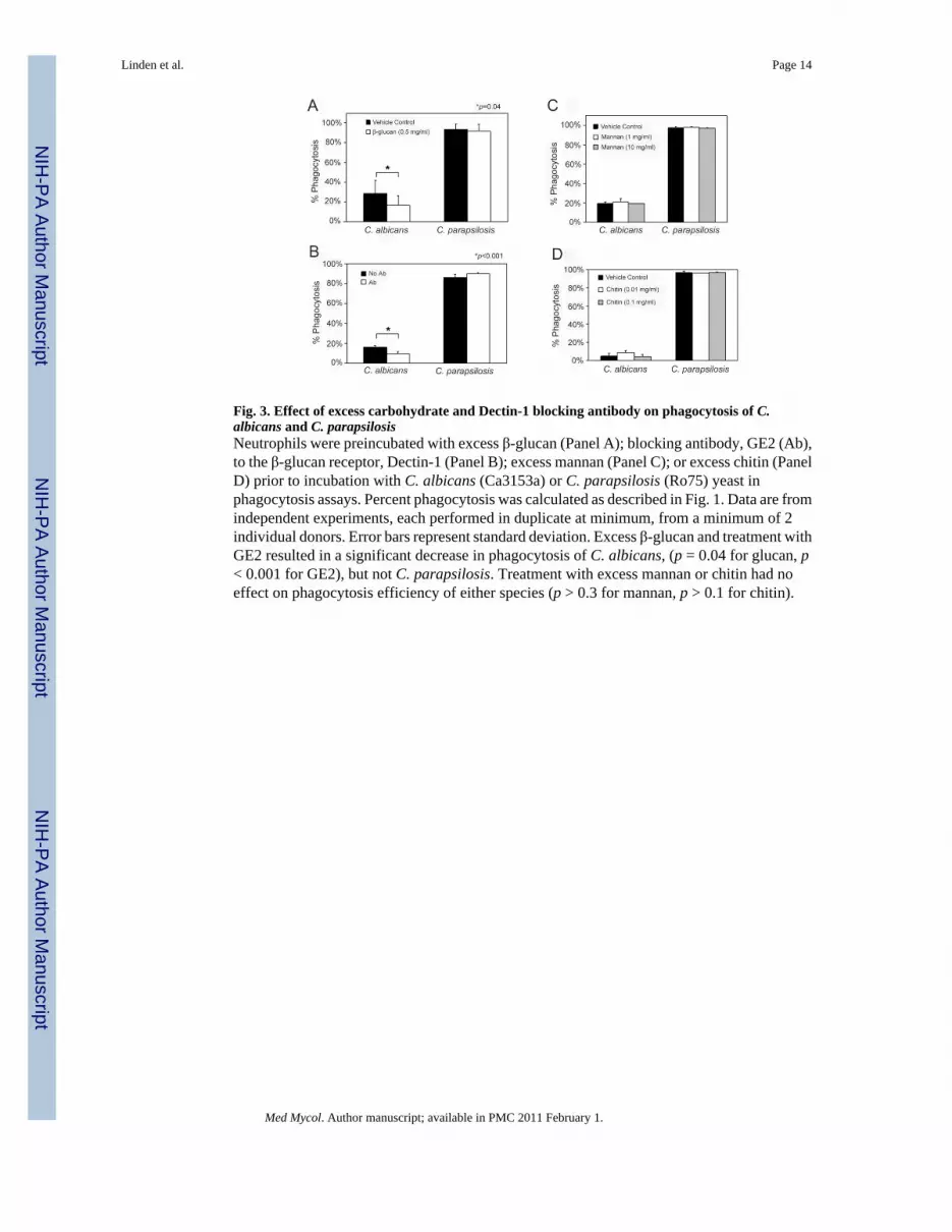
Fig. 3. Effect of excess carbohydrate and Dectin-1 blocking antibody on phagocytosis of C.albicans and C. parapsilosisNeutrophils were preincubated with excess β-glucan (Panel A); blocking antibody, GE2 (Ab),to the β-glucan receptor, Dectin-1 (Panel B); excess mannan (Panel C); or excess chitin (PanelD) prior to incubation with C. albicans (Ca3153a) or C. parapsilosis (Ro75) yeast inphagocytosis assays. Percent phagocytosis was calculated as described in Fig. 1. Data are fromindependent experiments, each performed in duplicate at minimum, from a minimum of 2individual donors. Error bars represent standard deviation. Excess β-glucan and treatment withGE2 resulted in a significant decrease in phagocytosis of C. albicans, (p = 0.04 for glucan, p< 0.001 for GE2), but not C. parapsilosis. Treatment with excess mannan or chitin had noeffect on phagocytosis efficiency of either species (p > 0.3 for mannan, p > 0.1 for chitin).
Linden et al. Page 14
Med Mycol. Author manuscript; available in PMC 2011 February 1.
NIH
-PA Author Manuscript
NIH
-PA Author Manuscript
NIH
-PA Author Manuscript

Fig. 4. Toxicity to C. albicans and C. parapsilosis mediated by neutrophils and effect of cytochalasinD(A) C. albicans and C. parapsilosis yeast were combined with neutrophils. After an incubationperiod, neutrophils were lysed, and residual metabolic activity of the yeast was measured byXTT. Data are expressed as the percentage of XTT activity of yeast of the same strain incubatedin the absence of neutrophils. Data are from 5 independent experiments with 5 individualdonors, and each was performed in triplicate. Error bars represent standard deviation.Neutrophils induced significant toxicity to each C. parapsilosis strain but no detectable toxicityto C. albicans strains (p ≤ 0.001 comparing each C. parapsilosis strain to each C. albicansstrain). There were no significant differences among strains of the same species. (B)
Linden et al. Page 15
Med Mycol. Author manuscript; available in PMC 2011 February 1.
NIH
-PA Author Manuscript
NIH
-PA Author Manuscript
NIH
-PA Author Manuscript

Neutrophils were pretreated with either cytochalsin D (Cyt-D) or buffer alone (Control) priorto inclusion in the toxicity assay with the 3 strains of C. parapsilosis. Data are expressed asthe percentage of XTT activity of yeast of the same strain incubated in the absence ofneutrophils and are from triplicate experiments. Error bars represent standard deviation. Ineach case, pretreatment of neutrophils with cytochalasin D resulted in significantly less toxicitythan control (p < 0.02 for each strain).
Linden et al. Page 16
Med Mycol. Author manuscript; available in PMC 2011 February 1.
NIH
-PA Author Manuscript
NIH
-PA Author Manuscript
NIH
-PA Author Manuscript

Fig. 5. Contribution of oxidative mechanisms to neutrophil-mediated toxicity(A) To determine the inherent sensitivity of each species to oxidative stress, all strains wereincubated in the presence of 1 mM H2O2, followed by XTT assay. Data are expressed as thepercentage of XTT activity of yeast of the same strain incubated in the absence of H2O2. Errorbars represent standard deviation. C. parapsilosis strains were significantly more sensitive toH2O2 than C. albicans (p < 0.01 for all strains). (B) Toxicity assays were conducted as describedin Fig. 4 in the presence of varied concentrations of the ROS scavenger, N-acetylcysteine. Dataare expressed as the percentage of XTT activity of yeast of the same strain incubated in theabsence of neutrophils. Data are from 5 individual donors in a minimum of 4 individualexperiments, and each was performed in triplicate. Error bars represent standard deviation.This agent led to a dose-dependent inhibition of toxicity, and statistical significance wasachieved for C. albicans strain Ca3153A and C. parapsilosis strain Ro75 (* p = 0.009; ** p =0.02). Because the quantity of neutrophils that could be obtained from an individual donor waslimited, only two strains of each species were tested.
Linden et al. Page 17
Med Mycol. Author manuscript; available in PMC 2011 February 1.
NIH
-PA Author Manuscript
NIH
-PA Author Manuscript
NIH
-PA Author Manuscript