entamoeba histolytica phagocytosis of human erythrocytes
TRANSCRIPT

Entamoeba histolytica Phagocytosis of HumanErythrocytes Involves PATMK, a Memberof the Transmembrane Kinase FamilyDouglas R. Boettner
1, Christopher D. Huston
2,3, Alicia S. Linford
1, Sarah N. Buss
1, Eric Houpt
4, Nicholas E. Sherman
1,
William A. Petri, Jr.1,4,5*
1 Department of Microbiology, University of Virginia, Charlottesville, Virginia, United States of America, 2 Department of Medicine, University of Vermont, Burlington,
Vermont, United States of America, 3 Department of Microbiology, University of Vermont, Burlington, Vermont, United States of America, 4 Department of Medicine,
University of Virginia, Charlottesville, Virginia, United States of America, 5 Department of Pathology, University of Virginia, Charlottesville, Virginia, United States of America
Entamoeba histolytica is the cause of amebic colitis and liver abscess. This parasite induces apoptosis in host cells andutilizes exposed ligands such as phosphatidylserine to ingest the apoptotic corpses and invade deeper into host tissue.The purpose of this work was to identify amebic proteins involved in the recognition and ingestion of dead cells. Amember of the transmembrane kinase family, phagosome-associated TMK96 (PATMK), was identified in a proteomicscreen for early phagosomal proteins. Anti-peptide affinity-purified antibody produced against PATMK demonstratedthat it was a type I integral membrane protein that was expressed on the trophozoite surface, and that co-localizedwith human erythrocytes at the site of contact. The role of PATMK in erythrophagocytosis in vitro was demonstratedby: (i) incubation of ameba with anti-PATMK antibodies; (ii) PATMK mRNA knock-down using a novel shRNA expressionsystem; and (iii) expression of a carboxy-truncation of PATMK (PATMKD932). Expression of the carboxy-truncation ofPATMKD932 also caused a specific reduction in the ability of E. histolytica to establish infection in the intestinal model ofamebiasis, however these amebae retained the ability to cause hepatic abscesses when directly injected in the liver. Inconclusion, PATMK was identified as a member of the TMK family that participates in erythrophagocytosis and isuniquely required for intestinal infection.
Citation: Boettner DR, Huston CD, Linford AS, Buss SN, Houpt E, et al. (2008) Entamoeba histolytica phagocytosis of human erythrocytes involves PATMK, a member of thetransmembrane kinase family. PLoS Pathog 4(1): e8. doi:10.1371/journal.ppat.0040008
Introduction
Entamoeba histolytica, the causative agent of amebiasis, isestimated to be the second leading cause of morbidity andmortality among protozoan parasites worldwide [1]. Phagocy-tosis has been one of the most recognized behaviors of E.histolytica. Erythrophagocytosis has even been used as adiagnostic indicator of invasive E. histolytica infection bymicroscopy [2]. Still, little is known concerning why host cellsare ingested and/orwhat affect this has on the course of disease.
Invasive infection by E. histolytica leads to dramatic tissuedestruction [3–6], including hallmarks of both apoptotic andnecrotic host cell death [7–9]. Previous work has demon-strated that following contact by E. histolytica, host cellsdisplay many features of apoptosis including DNA laddering,caspase 3 activation and phosphatidylserine (PS) exposure.Apoptotic host cells are subsequently ingested by the ameba[10], an interaction which has been shown to involve exposedphosphatidylserine (PS) on the host cell surface [10,11]. Invitro, calcium treatment of erythrocytes causes external-ization of phosphatidylserine and an increase in amebicuptake, providing a convenient and physiological model foranalysis of this process [11]. Although little is knownconcerning the role of this behavior in disease, phagocytosishas been suggested as a virulence determining factor [12].Amebic clones [13], and engineered mutants by eitherexpression of dominant negative constructs [14], or bychemical mutagenesis [15] which display defective in vitrophagocytosis are less virulent in vivo. In addition, use of pan-
caspase inhibitors to interfere with apoptotic induction invivo has also reduced infection by this parasite [16]. Giventhese results we hypothesized that the identification ofproteins which participate in the ingestion of the apoptoticcorpse would be key to understanding virulence.Many individual groups have used the process of ingestion
of beads to identify essential proteins required for phag-ocytosis in organisms ranging from amebae to man [17–19].Criticisms concerning the physiological relevance of beadingestion have recently been dispelled by data demonstratingthat bead ingestion is sensitive to inhibition by Annexin V,similar to uptake of apoptotic cells [20]. Although large scaleproteomic analysis has revealed many interesting proteins,
Editor: L. David Sibley, Washington University School of Medicine, United States ofAmerica
Received February 7, 2007; Accepted December 10, 2007; Published January 18,2008
Copyright: � 2008 Boettner et al. This is an open-access article distributed underthe terms of the Creative Commons Attribution License, which permits unrestricteduse, distribution, and reproduction in any medium, provided the original authorand source are credited.
Abbreviations: BSA, Bovine Serum Albumin; CFSE, 5-carboxyfluorescein diacetatesuccinimidyl ester; CMTMR, 5–4-chloromethyl- benzoylaminotetramethylrhod-amine; EDTA, Ethylene Diaminetetraacetic Acid; E. histolytica, Entamoeba histolytica;FLAG, epitope tag: DYKDDDDK; FITC, Fluorescein; Gal/GalNAc lectin, Galactose/N-acetyl galactosamine binding lectin; HEPES, 4–2-hydroxyethyl-1-piperazineethane-sulfonic acid; IgG, Immunoglobin G; MER, Mer tyrosine kinase; PATMK, PhagosomeAssociated Transmembrane Kinase; PS, Phosphatidylserine; shRNA, short hairpinRNA; TIGR, The Institute for Genomic Research
* To whom correspondence should be addressed. E-mail: [email protected]
PLoS Pathogens | www.plospathogens.org January 2008 | Volume 4 | Issue 1 | e80122
there appears to be much more left to discover. Controversiesconcerning both the PS receptor [21] as well as the role of theendoplasmic reticulum in phagocytosis [22,23] indicate thatthese efforts have not been exhaustive.
Two independent groups have published work using latexbeads that were either carboxylated or opsonized with IgG toidentify the constituents of the E. histolytica phagosome [24–28]. These proteomic screens taken together with the E.histolytica genome [26] have identified homologues of phag-osome maturation proteins seen in metazoans. Rab7, Rab11,Rap2, PI3K, Rac1 and Rho all appear, consistent with othersystems. However, some metazoan proteins including EEA1,RIN1, and LAMPs do not have discernable homologues in theE. histolytica genome. These recent screens have identified onlya small number of surface proteins that could act early in thephagocytic pathway, including Hgl, Igl, ABC transporter, p-glycoprotein-2 and 6, and M17. Of these, only Hgl and Iglhave been confirmed as constituents of phagosomes [24].
Amebic adherence to apoptotic host cells has been shownto require receptors in addition to the amebic Gal/GalNAcadherence lectin that mediates adherence to and killing ofthe live cell [11]. Our previous work identified the exposureof PS on the host cell surface as one of the recognized ligandsleading to ingestion. The goals of these studies were toidentify possible candidate amebic surface proteins with arole in the process of host cell ingestion.
Results
Sequencing of Magnetic Bead Preparations RevealedProteins with Conserved Function in Phagocytosis
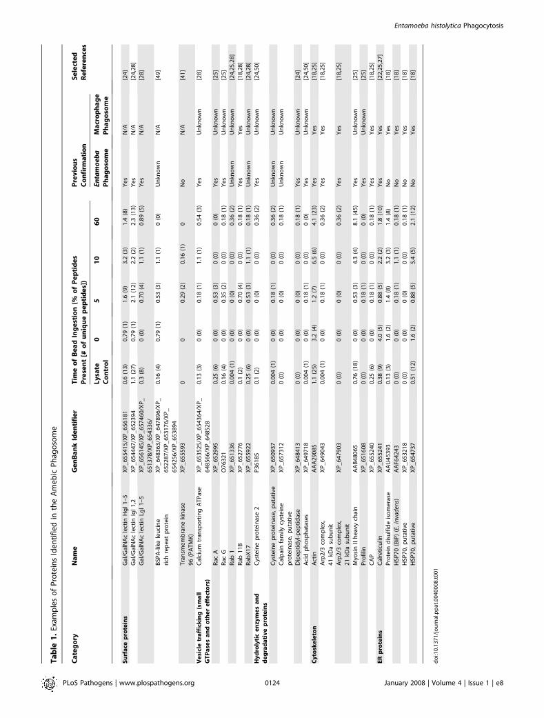
Ingested carboxylated magnetic beads within intact phag-osomes were separated from amebic lysate and subjected todelipidation and mass spectrometry sequencing. Phagosomepreparations were performed at 0, 5, 10, and 60 minutesfollowing centrifugation of the beads into contact withtrophozoites. In addition lysed ameba were incubated withbeads and subjected to the same steps to account forbackground binding to the carboxylated beads. This resultantproteome identified many proteins previously associated withphagocytosis in the literature, including: the galactose bind-ing lectin, small GTPases, hydrolytic proteins, cytoskeletonproteins, and endoplasmic reticulum proteins (Tables 1 and
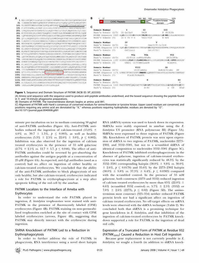
S1–S4). Our prediction was that molecules important forregulating ingestion of the host cell would appear in earlyphagosomes, but would not be present in late preparations.Therefore, the data were sorted for proteins that appeared at0, 5 and/or 10 minutes. A member of the transmembranekinase family stood out in this analysis, which we namedPATMK (Phagosome-associated transmembrane kinase). Thisputative receptor kinase appeared at 5 and 10 minutes but inno other preparations.The amino acid sequence of PATMK (Figure 1A) predicted
a 146 kDa protein, containing a 21 amino acid signal peptidesequence, an ectodomain containing 25 CXXC repeats, a 22amino acid membrane spanning domain and an intracellulardomain with the catalytic residues of a kinase (Figure 1B and1C) [29]. The kinase specificity for PATMK could not bepredicted given that its sequence contained the necessaryresidues for both serine/threonine and tyrosine kinase familymembers. Attempts to biochemically define the in vitrokinase activity by expressing the kinase domain in E. coli or byimmunoprecipitating PATMK from trophozoites were un-successful (data not shown). Whether PATMK is a pseudoki-nase, as the lack of a conserved ATP-orienting glycine-richmotif suggests (domain I, Figure 1C), will require additionalstudies. We concluded that the presence of PATMK in theearly phagosome proteome was consistent with it having arole in phagocytosis of the apoptotic corpse.
Antibodies against the Ectodomain of PATMK Reveal aSurface Protein on E. histolyticaRabbit anti-serum against a peptide specific for the
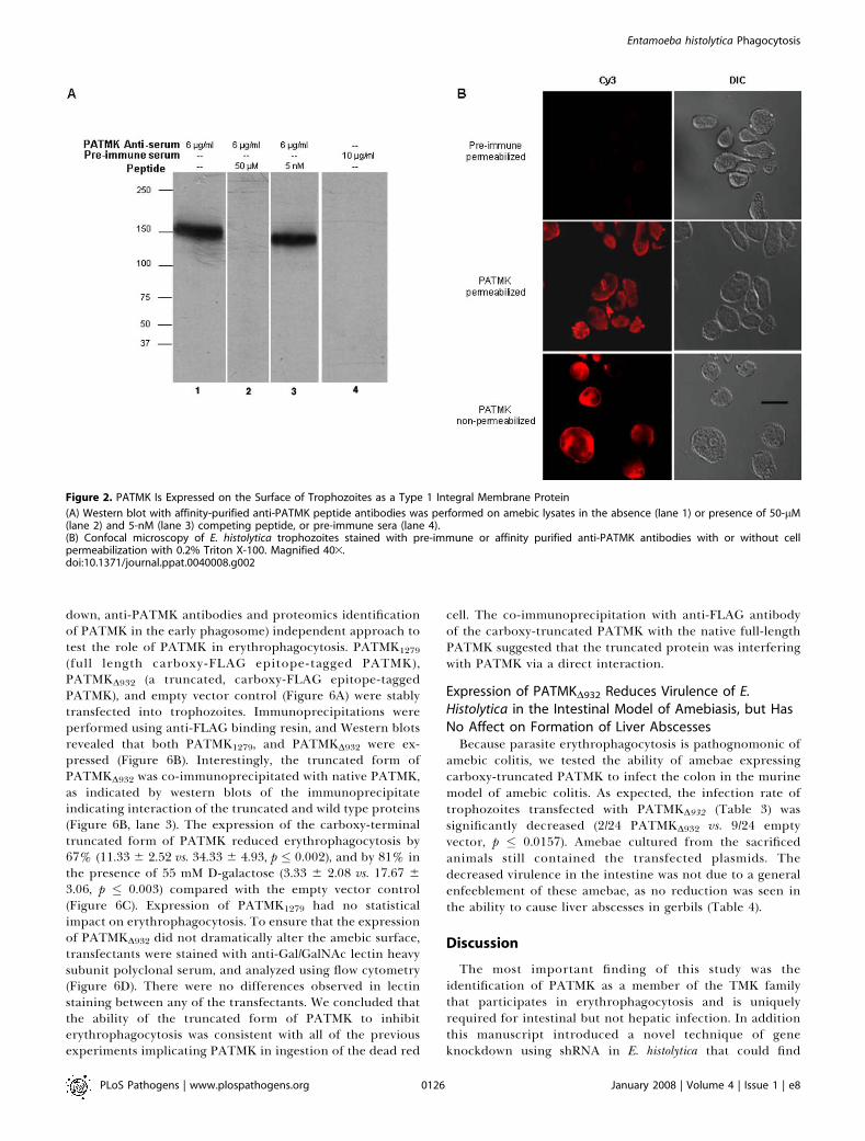
ectodomain of PATMK (EIQKQNPISTSLKISKISSD) (under-lined in Figure 1A) revealed a single band at ;140 kDa. Thisband disappeared when antibody was pre-absorbed against 50lM but not 5 nM of the antigen peptide (Figure 2A). Affinity-purified anti-PATMK antibodies stained the surface of bothpermeabilized and non-permeabilized trophozoites, whereaspre-immune serum yielded little staining (Figure 2B). Weconcluded that PATMK was expressed in E. histolyticatrophozoites as a plasma membrane protein of the expectedmass and with the amino-terminus extra-cellular.
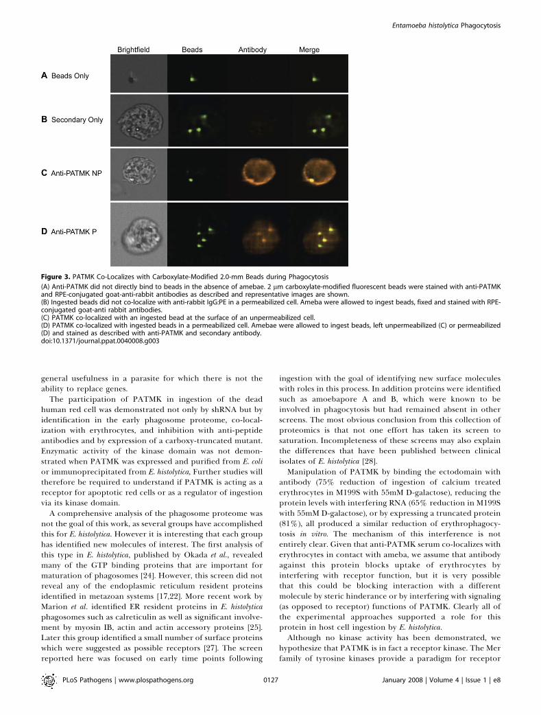
PATMK Co-Localizes with Ingested BeadsIn order to understand the role PATMK played in
ingestion, E. histolytica trophozoites were stained with anti-PATMK after ingestion of 2 lm carboxylate-modifiedfluorescent beads. To ensure that the PATMK antibody didnot directly bind beads, we used identical methods to stainbeads and bead containing cells and imaged the cells usingthe Amnis Imagestream imaging cytometer (Amnis Corpo-ration; Seattle, WA). There was no evidence of non-specificbinding of anti-PATMK to the carboxylate-modified beads(Figure 3A). Additionally, no staining was observed withsecondary antibody alone (Figure 3B). However, the locationof PATMK aggregates in both permeabilized and non-permeabilized trophozoites did correlate with that of theingested bead (Figure 3C and 3D), suggesting that PATMKmay directly interact with cargo during ingestion.
Anti-PATMK Pre-Incubation of Ameba Blocks Ingestion ofHuman ErythrocytesAmebae were tested for their ability to ingest healthy or
calcium-treated (apoptotic) erythrocytes following a 20
PLoS Pathogens | www.plospathogens.org January 2008 | Volume 4 | Issue 1 | e80123
Entamoeba histolytica Phagocytosis
Author Summary
There is a highly ordered process by which the parasite Entamoebahistolytica interacts with human cells. Adherence via a parasite lectinis followed in seconds by killing, with only the corpse and not aliving cell ingested by the ameba. This process is so central topathogenesis that clinicians use the presence of ingested eryth-rocytes to identify E. histolytica and distinguish it from harmlesscommensal amebae of the gut. We hypothesized that identificationof molecules involved in the ingestion of the corpse might provideinsight into how amebae cause colitis. We identified a member ofthe transmembrane kinase family as an early component of thephagosome. Inhibition of this kinase blocked red cell ingestion andprevented amebae from colonizing and invading the gut. There wasno impact on dominant-negative parasites to cause liver abscess,suggesting the pathogenesis program differs between anatomicsites. Future studies of the transmembrane kinanse in erythropha-gocytosis may provide insight into how amebae colonize and invadethe gut, with the ultimate goal of preventing disease.
Ta
ble
1.
Exam
ple
so
fP
rote
ins
Ide
nti
fie
din
the
Am
eb
icP
hag
oso
me
Ca
teg
ory
Na
me
Ge
nB
an
kId
en
tifi
er
Tim
eo
fB
ea
dIn
ge
stio
n(%
of
Pe
pti
de
s
Pre
sen
t[#
of
un
iqu
ep
ep
tid
es]
)
Pre
vio
us
Co
nfi
rma
tio
n
Se
lect
ed
Re
fere
nce
s
Ly
sate
Co
ntr
ol
05
10
60
En
tam
oe
ba
Ph
ag
oso
me
Ma
cro
ph
ag
e
Ph
ag
oso
me
Su
rfa
cep
rote
ins
Gal
/Gal
NA
cle
ctin
Hg
l1
–5
XP
_6
55
41
5/X
P_
65
61
81
0.6
(13
)0
.79
(1)
1.6
(9)
3.2
(3)
1.4
(8)
Ye
sN
/A[2
4]
Gal
/Gal
NA
cle
ctin
Igl
1,2
XP
_6
54
44
7/X
P_
65
23
94
1.1
(27
)0
.79
(1)
2.1
(12
)2
.2(2
)2
.3(1
3)
Ye
sN
/A[2
4,2
8]
Gal
/Gal
NA
cle
ctin
Lgl
1–
5X
P_
65
61
45
/XP
_6
57
46
0/X
P_
65
13
78
/XP
_6
54
33
6/
0.3
(8)
0(0
)0
.70
(4)
1.1
(1)
0.8
9(5
)Y
es
N/A
[28
]
BSP
A-l
ike
leu
cin
e
rich
rep
eat
pro
tein
XP
_6
48
36
3/X
P_
64
78
96
/XP
_
65
22
87
/XP
_6
53
17
6/X
P_
65
42
56
/XP
_6
53
89
4
0.1
6(4
)0
.79
(1)
0.5
3(3
)1
.1(1
)0
(0)
Un
kno
wn
N/A
[49
]
Tra
nsm
em
bra
ne
kin
ase
96
(PA
TM
K)
XP
_6
55
59
30
00
.29
(2)
0.1
6(1
)0
No
N/A
[41
]
Ve
sicl
etr
aff
ick
ing
(sm
all
GT
Pa
ses
an
do
the
re
ffe
cto
rs)
Cal
ciu
mtr
ansp
ort
ing
AT
Pas
eX
P_
65
35
25
/XP
_6
54
36
4/X
P_
64
85
66
/XP
_6
48
52
8
0.1
3(3
)0
(0)
0.1
8(1
)1
.1(1
)0
.54
(3)
Ye
sU
nkn
ow
n[2
8]
Rac
AX
P_
65
29
95
0.2
5(6
)0
(0)
0.5
3(3
)0
(0)
0(0
)Y
es
Un
kno
wn
[25
]
Rac
GO
76
32
10
.16
(4)
0(0
)0
.35
(2)
0(0
)0
.18
(1)
Ye
sU
nkn
ow
n[2
5]
Rab
1X
P_
65
13
36
0.0
04
(1)
0(0
)0
(0)
0(0
)0
.36
(2)
Un
kno
wn
Un
kno
wn
[24
,25
,28
]
Rab
11
BX
P_
65
27
76
0.1
(2)
0(0
)0
.70
(4)
0(0
)0
.18
(1)
Ye
sY
es
[18
,28
]
Rab
X1
7X
P_
65
59
22
0.2
5(6
)0
(0)
0.5
3(3
)1
.1(1
)0
.18
(1)
Un
kno
wn
Un
kno
wn
[24
,28
]
Hy
dro
lyti
ce
nzy
me
sa
nd
de
gra
da
tiv
ep
rote
ins
Cys
tein
ep
rote
inas
e2
P3
61
85
0.1
(2)
0(0
)0
(0)
0(0
)0
.36
(2)
Ye
sU
nkn
ow
n[2
4,5
0]
Cys
tein
ep
rote
inas
e,
pu
tati
veX
P_
65
09
37
0.0
04
(1)
0(0
)0
.18
(1)
0(0
)0
.36
(2)
Un
kno
wn
Un
kno
wn
Cal
pai
nfa
mily
cyst
ein
e
pro
tein
ase
,p
uta
tive
XP
_6
57
31
20
(0)
0(0
)0
(0)
0(0
)0
.18
(1)
Un
kno
wn
Un
kno
wn
Dip
ep
tid
yl-p
ep
tid
ase
XP
_6
48
41
30
(0)
0(0
)0
(0)
0(0
)0
.18
(1)
Ye
sU
nkn
ow
n[2
4]
Aci
dp
ho
sph
atas
es
XP
_6
49
71
80
.00
4(1
)0
(0)
0.1
8(1
)0
(0)
0(0
)Y
es
Un
kno
wn
[24
,50
]
Cy
tosk
ele
ton
Act
inA
AA
29
08
51
.1(2
5)
3.2
(4)
1.2
(7)
6.5
(6)
4.1
(23
)Y
es
Ye
s[1
8,2
5]
Arp
2/3
com
ple
x,
41
kDa
sub
un
it
XP
_6
49
04
30
.00
4(1
)0
(0)
0.1
8(1
)0
(0)
0.3
6(2
)Y
es
Ye
s[1
8,2
5]
Arp
2/3
com
ple
x,
21
kDa
sub
un
it
XP
_6
47
90
30
(0)
0(0
)0
(0)
0(0
)0
.36
(2)
Ye
sY
es
[18
,25
]
Myo
sin
IIh
eav
ych
ain
AA
B4
80
65
0.7
6(1
8)
0(0
)0
.53
(3)
4.3
(4)
8.1
(45
)Y
es
Un
kno
wn
[25
]
Pro
filin
XP
_6
51
60
80
(0)
0(0
)0
.18
(1)
0(0
)0
(0)
Ye
sU
nkn
ow
n[2
5]
CA
PX
P_
65
52
40
0.2
5(6
)0
(0)
0.1
8(1
)0
(0)
0.1
8(1
)Y
es
Ye
s[1
8,2
5]
ER
pro
tein
sC
alre
ticu
linX
P_
65
52
41
0.3
8(9
)4
.0(5
)0
.88
(5)
2.2
(2)
1.8
(10
)Y
es
Ye
s[2
2,2
5,2
7]
Pro
tein
dis
ulf
ide
iso
me
rase
AA
U4
53
93
0.1
3(3
)1
.6(2
)1
.4(8
)3
.2(3
)1
.4(8
)N
oY
es
[18
]
HSP
70
(BiP
)(E
.in
vad
ens)
AA
F64
24
30
(0)
0(0
)0
.18
(1)
1.1
(1)
0.1
8(1
)N
oY
es
[18
]
HSP
70
,p
uta
tive
XP
_6
53
21
80
(0)
0(0
)0
(0)
0(0
)0
.18
(1)
No
Ye
s[1
8]
HSP
70
,p
uta
tive
XP
_6
54
73
70
.51
(12
)1
.6(2
)0
.88
(5)
5.4
(5)
2.1
(12
)N
oY
es
[18
]
do
i:10
.13
71
/jo
urn
al.p
pat
.00
40
00
8.t
00
1
PLoS Pathogens | www.plospathogens.org January 2008 | Volume 4 | Issue 1 | e80124
Entamoeba histolytica Phagocytosis
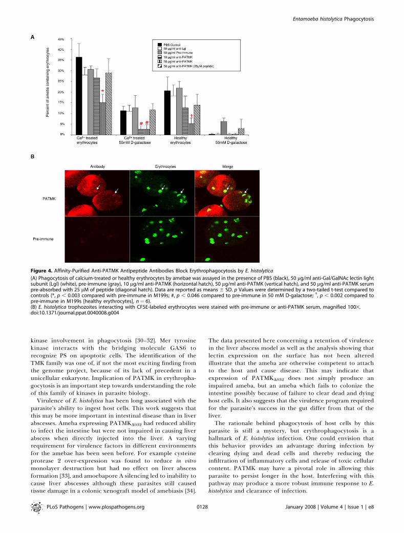
minute pre-incubation on ice in medium containing 50 lg/mlof anti-PATMK antibodies (Figure 4A). Anti-PATMK anti-bodies reduced the ingestion of calcium-treated (15.0% 6
4.0% vs. 30.7 6 1.5%, p � 0.003), as well as healthyerythrocytes (5.3% 6 2.5% vs. 22.0% 6 3.0%, p � 0.002).Inhibition was also observed for the ingestion of calciumtreated erythrocytes in the presence of 55 mM galactose(2.7% 6 2.1% vs. 12.7 6 5.7, p � 0.046). The effect of anti-PATMK antibodies could be reversed by pre-absorbing theantibodies against the antigen peptide at a concentration of25 lM (Figure 4A). As expected, anti-Lgl antibodies (used as acontrol) had no affect on ingestion of either healthy orcalcium-treated erythrocytes. We concluded that the abilityof the anti-PATMK antibodies to block phagocytosis of notonly healthy, but also calcium-treated, erythrocytes indicateda role for PATMK in erythrophagocytosis at a step afterapoptotic killing of the red cell by the amebae.
PATMK Localizes to the Interface of Ameba withErythrocytes
In order to understand the role PATMK played iningestion, E. histolytica trophozoites were stained with anti-PATMK in the presence of fluorescently labeled (CFSE)erythrocytes (Figure 4B). PATMK staining on non-permeabi-lized trophozoites enriched at the site of contact with CFSElabeled erythrocytes (arrows, Figure 4B), suggesting thatPATMK may directly interact with the erythrocyte duringingestion.
ShRNA Knockdown of PATMK Led to a Reduction inErythrophagocytosis
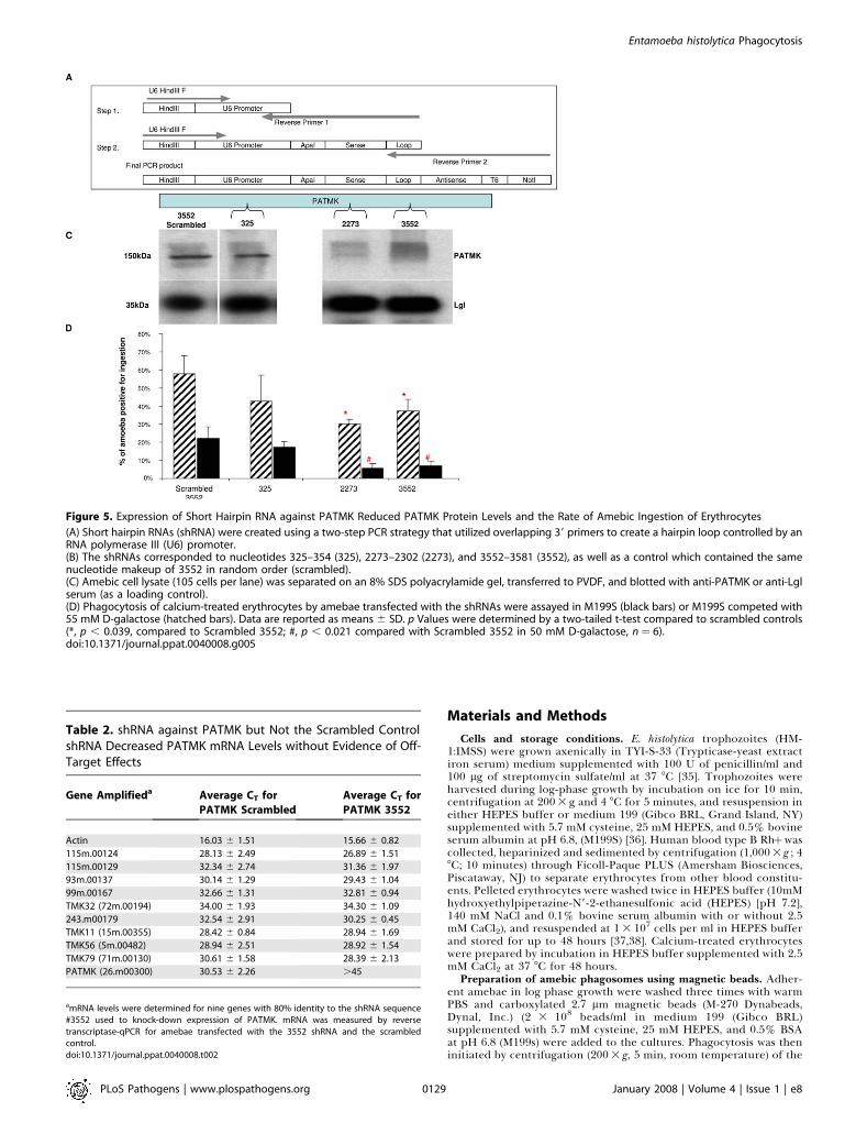
In order to further address the role of PATMK inphagocytosis, RNA interference using a novel short hairpin
RNA (shRNA) system was used to knock down its expression.ShRNAs were stably expressed in amebae using the E.histolytica U6 promoter (RNA polymerase III) (Figure 5A).ShRNAs were expressed to three regions of PATMK (Figure5B). Knockdown of PATMK protein was seen upon expres-sion of shRNA to two regions of PATMK, nucleotides 2273–2302, and 3552–3581, but not to a scrambled shRNA ofidentical composition to nucleotides 3552–3581 (Figure 5C).Knockdown of PATMK inhibited erythrophagocytosis: in theabsence of galactose, ingestion of calcium-treated erythro-cytes was statistically significantly reduced by 48.3% by the3552–3581 corresponding hairpin (58.0% 6 9.8% vs. 30.0%6 2.6%, p � 0.0176) and 35.6% by the 2273–2302 hairpin(58.0% 6 9.8% vs. 37.3% 6 6.4%, p � 0.0383) comparedwith the scrambled control. In the presence of 55 mMgalactose, both constructs (2273 and 3552) reduced ingestionof calcium treated erythrocytes by more than 65% (22.0% 6
6.6% (scrambled 3552 control) vs. 5.7% 6 2.5% (3552) or7.0% 6 2.6% (2273), p � 0.02) (Figure 5D). The amino-terminus most construct (325–354) neither reduced PATMKprotein levels nor had a significant affect on ingestion ofcalcium treated erythrocytes. No off-target effects on mRNAlevels were observed with the shRNA technique (Table 2). Weconcluded both that shRNA is a promising technique forgene knockdown in E. histolytica, and that inhibition of theingestion of calcium-treated erythrocytes by PATMK knock-down supported a role for PATMK in the ingestion of deadred cells.
Expression of a Truncated Form of PATMK at Residue 932(PATMKD932) Caused a Reduction in Host Cell IngestionBecause gene replacement is not currently possible in E.
histolytica, we sought a fourth (in addition to shRNA knock-
Figure 1. Sequence and Domain Structure of PATMK (NCBI ID: XP_655593)
(A) Amino acid sequence with the sequence used to produce anti-peptide antibodies underlined, and the boxed sequence showing the peptide foundin 5- and 10-minute phagosome preparations.(B) Domains of PATMK. The transmembrane domain begins at amino acid 841.(C) Alignment of PATMK with Hank’s consensus of conserved residues for serine/threonine or tyrosine kinase. Upper cased residues are conserved, andpositions requiring any amino acid are denotated by ‘‘X,’’ whereas positions requiring hydrophobic residues are denoted by ‘‘O’’.doi:10.1371/journal.ppat.0040008.g001
PLoS Pathogens | www.plospathogens.org January 2008 | Volume 4 | Issue 1 | e80125
Entamoeba histolytica Phagocytosis
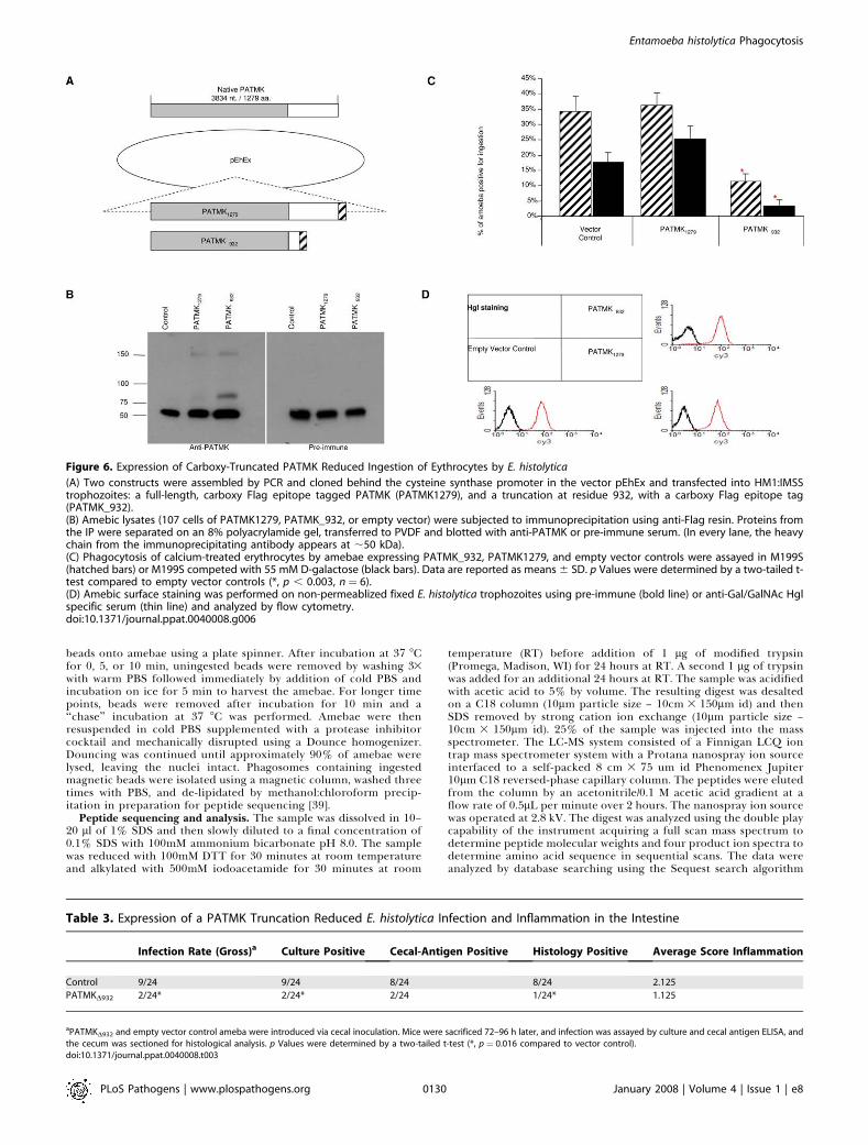
down, anti-PATMK antibodies and proteomics identificationof PATMK in the early phagosome) independent approach totest the role of PATMK in erythrophagocytosis. PATMK1279
(full length carboxy-FLAG epitope-tagged PATMK),PATMKD932 (a truncated, carboxy-FLAG epitope-taggedPATMK), and empty vector control (Figure 6A) were stablytransfected into trophozoites. Immunoprecipitations wereperformed using anti-FLAG binding resin, and Western blotsrevealed that both PATMK1279, and PATMKD932 were ex-pressed (Figure 6B). Interestingly, the truncated form ofPATMKD932 was co-immunoprecipitated with native PATMK,as indicated by western blots of the immunoprecipitateindicating interaction of the truncated and wild type proteins(Figure 6B, lane 3). The expression of the carboxy-terminaltruncated form of PATMK reduced erythrophagocytosis by67% (11.33 6 2.52 vs. 34.33 6 4.93, p � 0.002), and by 81% inthe presence of 55 mM D-galactose (3.33 6 2.08 vs. 17.67 6
3.06, p � 0.003) compared with the empty vector control(Figure 6C). Expression of PATMK1279 had no statisticalimpact on erythrophagocytosis. To ensure that the expressionof PATMKD932 did not dramatically alter the amebic surface,transfectants were stained with anti-Gal/GalNAc lectin heavysubunit polyclonal serum, and analyzed using flow cytometry(Figure 6D). There were no differences observed in lectinstaining between any of the transfectants. We concluded thatthe ability of the truncated form of PATMK to inhibiterythrophagocytosis was consistent with all of the previousexperiments implicating PATMK in ingestion of the dead red
cell. The co-immunoprecipitation with anti-FLAG antibodyof the carboxy-truncated PATMK with the native full-lengthPATMK suggested that the truncated protein was interferingwith PATMK via a direct interaction.
Expression of PATMKD932 Reduces Virulence of E.Histolytica in the Intestinal Model of Amebiasis, but HasNo Affect on Formation of Liver AbscessesBecause parasite erythrophagocytosis is pathognomonic of
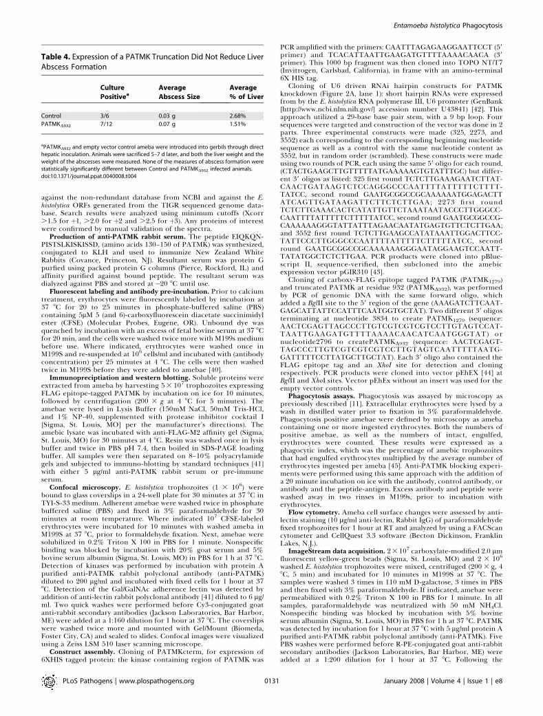
amebic colitis, we tested the ability of amebae expressingcarboxy-truncated PATMK to infect the colon in the murinemodel of amebic colitis. As expected, the infection rate oftrophozoites transfected with PATMKD932 (Table 3) wassignificantly decreased (2/24 PATMKD932 vs. 9/24 emptyvector, p � 0.0157). Amebae cultured from the sacrificedanimals still contained the transfected plasmids. Thedecreased virulence in the intestine was not due to a generalenfeeblement of these amebae, as no reduction was seen inthe ability to cause liver abscesses in gerbils (Table 4).
Discussion
The most important finding of this study was theidentification of PATMK as a member of the TMK familythat participates in erythrophagocytosis and is uniquelyrequired for intestinal but not hepatic infection. In additionthis manuscript introduced a novel technique of geneknockdown using shRNA in E. histolytica that could find
Figure 2. PATMK Is Expressed on the Surface of Trophozoites as a Type 1 Integral Membrane Protein
(A) Western blot with affinity-purified anti-PATMK peptide antibodies was performed on amebic lysates in the absence (lane 1) or presence of 50-lM(lane 2) and 5-nM (lane 3) competing peptide, or pre-immune sera (lane 4).(B) Confocal microscopy of E. histolytica trophozoites stained with pre-immune or affinity purified anti-PATMK antibodies with or without cellpermeabilization with 0.2% Triton X-100. Magnified 403.doi:10.1371/journal.ppat.0040008.g002
PLoS Pathogens | www.plospathogens.org January 2008 | Volume 4 | Issue 1 | e80126
Entamoeba histolytica Phagocytosis
general usefulness in a parasite for which there is not theability to replace genes.
The participation of PATMK in ingestion of the deadhuman red cell was demonstrated not only by shRNA but byidentification in the early phagosome proteome, co-local-ization with erythrocytes, and inhibition with anti-peptideantibodies and by expression of a carboxy-truncated mutant.Enzymatic activity of the kinase domain was not demon-strated when PATMK was expressed and purified from E. colior immunoprecipitated from E. histolytica, Further studies willtherefore be required to understand if PATMK is acting as areceptor for apoptotic red cells or as a regulator of ingestionvia its kinase domain.
A comprehensive analysis of the phagosome proteome wasnot the goal of this work, as several groups have accomplishedthis for E. histolytica. However it is interesting that each grouphas identified new molecules of interest. The first analysis ofthis type in E. histolytica, published by Okada et al., revealedmany of the GTP binding proteins that are important formaturation of phagosomes [24]. However, this screen did notreveal any of the endoplasmic reticulum resident proteinsidentified in metazoan systems [17,22]. More recent work byMarion et al. identified ER resident proteins in E. histolyticaphagosomes such as calreticulin as well as significant involve-ment by myosin IB, actin and actin accessory proteins [25].Later this group identified a small number of surface proteinswhich were suggested as possible receptors [27]. The screenreported here was focused on early time points following
ingestion with the goal of identifying new surface moleculeswith roles in this process. In addition proteins were identifiedsuch as amoebapore A and B, which were known to beinvolved in phagocytosis but had remained absent in otherscreens. The most obvious conclusion from this collection ofproteomics is that not one effort has taken its screen tosaturation. Incompleteness of these screens may also explainthe differences that have been published between clinicalisolates of E. histolytica [28].Manipulation of PATMK by binding the ectodomain with
antibody (75% reduction of ingestion of calcium treatederythrocytes in M199S with 55mM D-galactose), reducing theprotein levels with interfering RNA (65% reduction in M199Swith 55mM D-galactose), or by expressing a truncated protein(81%), all produced a similar reduction of erythrophagocy-tosis in vitro. The mechanism of this interference is notentirely clear. Given that anti-PATMK serum co-localizes witherythrocytes in contact with ameba, we assume that antibodyagainst this protein blocks uptake of erythrocytes byinterfering with receptor function, but it is very possiblethat this could be blocking interaction with a differentmolecule by steric hinderance or by interfering with signaling(as opposed to receptor) functions of PATMK. Clearly all ofthe experimental approaches supported a role for thisprotein in host cell ingestion by E. histolytica.Although no kinase activity has been demonstrated, we
hypothesize that PATMK is in fact a receptor kinase. The Merfamily of tyrosine kinases provide a paradigm for receptor
Figure 3. PATMK Co-Localizes with Carboxylate-Modified 2.0-mm Beads during Phagocytosis
(A) Anti-PATMK did not directly bind to beads in the absence of amebae. 2 lm carboxylate-modified fluorescent beads were stained with anti-PATMKand RPE-conjugated goat-anti-rabbit antibodies as described and representative images are shown.(B) Ingested beads did not co-localize with anti-rabbit IgG:PE in a permeabilized cell. Ameba were allowed to ingest beads, fixed and stained with RPE-conjugated goat-anti rabbit antibodies.(C) PATMK co-localized with an ingested bead at the surface of an unpermeabilized cell.(D) PATMK co-localized with ingested beads in a permeabilized cell. Amebae were allowed to ingest beads, left unpermeabilized (C) or permeabilized(D) and stained as described with anti-PATMK and secondary antibody.doi:10.1371/journal.ppat.0040008.g003
PLoS Pathogens | www.plospathogens.org January 2008 | Volume 4 | Issue 1 | e80127
Entamoeba histolytica Phagocytosis
kinase involvement in phagocytosis [30–32]. Mer tyrosinekinase interacts with the bridging molecule GAS6 torecognize PS on apoptotic cells. The identification of theTMK family was one of, if not the most exciting finding fromthe genome project, because of its lack of precedent in aunicellular eukaryote. Implication of PATMK in erythropha-gocytosis is an important step towards understanding the roleof this family of kinases in parasite biology.
Virulence of E. histolytica has been long associated with theparasite’s ability to ingest host cells. This work suggests thatthis may be more important in intestinal disease than in liverabscesses. Ameba expressing PATMKD932 had reduced abilityto infect the intestine but were not impaired in causing liverabscess when directly injected into the liver. A varyingrequirement for virulence factors in different environmentsfor the amebae has been seen before. For example cysteineprotease 2 over-expression was found to reduce in vitromonolayer destruction but had no effect on liver abscessformation [33], and amoebapore A silencing led to inability tocause liver abscesses although these parasites still causedtissue damage in a colonic xenograft model of amebiasis [34].
The data presented here concerning a retention of virulencein the liver abscess model as well as the analysis showing thatlectin expression on the surface has not been alteredillustrate that the ameba are otherwise competent to attachto the host and cause disease. This may indicate thatexpression of PATMKD932 does not simply produce animpaired ameba, but an ameba which fails to colonize theintestine possibly because of failure to clear dead and dyinghost cells. It also suggests that the virulence program requiredfor the parasite’s success in the gut differ from that of theliver.The rationale behind phagocytosis of host cells by this
parasite is still a mystery, but erythrophagocytosis is ahallmark of E. histolytica infection. One could envision thatthis behavior provides an advantage during infection byclearing dying and dead cells and thereby reducing theinfiltration of inflammatory cells and release of toxic cellularcontent. PATMK may have a pivotal role in allowing thisparasite to persist longer in the host. Interfering with thispathway may produce a more robust immune response to E.histolytica and clearance of infection.
Figure 4. Affinity-Purified Anti-PATMK Antipeptide Antibodies Block Erythrophagocytosis by E. histolytica
(A) Phagocytosis of calcium-treated or healthy erythrocytes by amebae was assayed in the presence of PBS (black), 50 lg/ml anti-Gal/GalNAc lectin lightsubunit (Lgl) (white), pre-immune (gray), 10 lg/ml anti-PATMK (horizontal hatch), 50 lg/ml anti-PATMK (vertical hatch), and 50 lg/ml anti-PATMK serumpre-absorbed with 25 lM of peptide (diagonal hatch). Data are reported as means 6 SD. p Values were determined by a two-tailed t-test compared tocontrols (*, p , 0.003 compared with pre-immune in M199s; #, p , 0.046 compared to pre-immune in 50 mM D-galactose; �, p , 0.002 compared topre-immune in M199s [healthy erythrocytes], n¼ 6).(B) E. histolytica trophozoites interacting with CFSE-labeled erythrocytes were stained with pre-immune or anti-PATMK serum, magnified 1003.doi:10.1371/journal.ppat.0040008.g004
PLoS Pathogens | www.plospathogens.org January 2008 | Volume 4 | Issue 1 | e80128
Entamoeba histolytica Phagocytosis
Materials and Methods
Cells and storage conditions. E. histolytica trophozoites (HM-1:IMSS) were grown axenically in TYI-S-33 (Trypticase-yeast extractiron serum) medium supplemented with 100 U of penicillin/ml and100 lg of streptomycin sulfate/ml at 37 8C [35]. Trophozoites wereharvested during log-phase growth by incubation on ice for 10 min,centrifugation at 2003 g and 4 8C for 5 minutes, and resuspension ineither HEPES buffer or medium 199 (Gibco BRL, Grand Island, NY)supplemented with 5.7 mM cysteine, 25 mM HEPES, and 0.5% bovineserum albumin at pH 6.8, (M199S) [36]. Human blood type B Rhþwascollected, heparinized and sedimented by centrifugation (1,0003 g ; 48C; 10 minutes) through Ficoll-Paque PLUS (Amersham Biosciences,Piscataway, NJ) to separate erythrocytes from other blood constitu-ents. Pelleted erythrocytes were washed twice in HEPES buffer (10mMhydroxyethylpiperazine-N9-2-ethanesulfonic acid (HEPES) [pH 7.2],140 mM NaCl and 0.1% bovine serum albumin with or without 2.5mM CaCl2), and resuspended at 1 3 107 cells per ml in HEPES bufferand stored for up to 48 hours [37,38]. Calcium-treated erythrocyteswere prepared by incubation in HEPES buffer supplemented with 2.5mM CaCl2 at 37 8C for 48 hours.
Preparation of amebic phagosomes using magnetic beads. Adher-ent amebae in log phase growth were washed three times with warmPBS and carboxylated 2.7 lm magnetic beads (M-270 Dynabeads,Dynal, Inc.) (2 3 108 beads/ml in medium 199 (Gibco BRL)supplemented with 5.7 mM cysteine, 25 mM HEPES, and 0.5% BSAat pH 6.8 (M199s) were added to the cultures. Phagocytosis was theninitiated by centrifugation (2003 g, 5 min, room temperature) of the
Figure 5. Expression of Short Hairpin RNA against PATMK Reduced PATMK Protein Levels and the Rate of Amebic Ingestion of Erythrocytes
(A) Short hairpin RNAs (shRNA) were created using a two-step PCR strategy that utilized overlapping 39 primers to create a hairpin loop controlled by anRNA polymerase III (U6) promoter.(B) The shRNAs corresponded to nucleotides 325–354 (325), 2273–2302 (2273), and 3552–3581 (3552), as well as a control which contained the samenucleotide makeup of 3552 in random order (scrambled).(C) Amebic cell lysate (105 cells per lane) was separated on an 8% SDS polyacrylamide gel, transferred to PVDF, and blotted with anti-PATMK or anti-Lglserum (as a loading control).(D) Phagocytosis of calcium-treated erythrocytes by amebae transfected with the shRNAs were assayed in M199S (black bars) or M199S competed with55 mM D-galactose (hatched bars). Data are reported as means 6 SD. p Values were determined by a two-tailed t-test compared to scrambled controls(*, p , 0.039, compared to Scrambled 3552; #, p , 0.021 compared with Scrambled 3552 in 50 mM D-galactose, n¼ 6).doi:10.1371/journal.ppat.0040008.g005
Table 2. shRNA against PATMK but Not the Scrambled ControlshRNA Decreased PATMK mRNA Levels without Evidence of Off-Target Effects
Gene Amplifieda Average CT for
PATMK Scrambled
Average CT for
PATMK 3552
Actin 16.03 6 1.51 15.66 6 0.82
115m.00124 28.13 6 2.49 26.89 6 1.51
115m.00129 32.34 6 2.74 31.36 6 1.97
93m.00137 30.14 6 1.29 29.43 6 1.04
99m.00167 32.66 6 1.31 32.81 6 0.94
TMK32 (72m.00194) 34.00 6 1.93 34.30 6 1.09
243.m00179 32.54 6 2.91 30.25 6 0.45
TMK11 (15m.00355) 28.42 6 0.84 28.94 6 1.69
TMK56 (5m.00482) 28.94 6 2.51 28.92 6 1.54
TMK79 (71m.00130) 30.61 6 1.58 28.39 6 2.13
PATMK (26.m00300) 30.53 6 2.26 .45
amRNA levels were determined for nine genes with 80% identity to the shRNA sequence#3552 used to knock-down expression of PATMK. mRNA was measured by reversetranscriptase-qPCR for amebae transfected with the 3552 shRNA and the scrambledcontrol.doi:10.1371/journal.ppat.0040008.t002
PLoS Pathogens | www.plospathogens.org January 2008 | Volume 4 | Issue 1 | e80129
Entamoeba histolytica Phagocytosis
beads onto amebae using a plate spinner. After incubation at 37 8Cfor 0, 5, or 10 min, uningested beads were removed by washing 33with warm PBS followed immediately by addition of cold PBS andincubation on ice for 5 min to harvest the amebae. For longer timepoints, beads were removed after incubation for 10 min and a‘‘chase’’ incubation at 37 8C was performed. Amebae were thenresuspended in cold PBS supplemented with a protease inhibitorcocktail and mechanically disrupted using a Dounce homogenizer.Douncing was continued until approximately 90% of amebae werelysed, leaving the nuclei intact. Phagosomes containing ingestedmagnetic beads were isolated using a magnetic column, washed threetimes with PBS, and de-lipidated by methanol:chloroform precip-itation in preparation for peptide sequencing [39].
Peptide sequencing and analysis. The sample was dissolved in 10–20 ll of 1% SDS and then slowly diluted to a final concentration of0.1% SDS with 100mM ammonium bicarbonate pH 8.0. The samplewas reduced with 100mM DTT for 30 minutes at room temperatureand alkylated with 500mM iodoacetamide for 30 minutes at room
temperature (RT) before addition of 1 lg of modified trypsin(Promega, Madison, WI) for 24 hours at RT. A second 1 lg of trypsinwas added for an additional 24 hours at RT. The sample was acidifiedwith acetic acid to 5% by volume. The resulting digest was desaltedon a C18 column (10lm particle size – 10cm 3 150lm id) and thenSDS removed by strong cation ion exchange (10lm particle size –10cm 3 150lm id). 25% of the sample was injected into the massspectrometer. The LC-MS system consisted of a Finnigan LCQ iontrap mass spectrometer system with a Protana nanospray ion sourceinterfaced to a self-packed 8 cm 3 75 um id Phenomenex Jupiter10lm C18 reversed-phase capillary column. The peptides were elutedfrom the column by an acetonitrile/0.1 M acetic acid gradient at aflow rate of 0.5lL per minute over 2 hours. The nanospray ion sourcewas operated at 2.8 kV. The digest was analyzed using the double playcapability of the instrument acquiring a full scan mass spectrum todetermine peptide molecular weights and four product ion spectra todetermine amino acid sequence in sequential scans. The data wereanalyzed by database searching using the Sequest search algorithm
Figure 6. Expression of Carboxy-Truncated PATMK Reduced Ingestion of Eythrocytes by E. histolytica
(A) Two constructs were assembled by PCR and cloned behind the cysteine synthase promoter in the vector pEhEx and transfected into HM1:IMSStrophozoites: a full-length, carboxy Flag epitope tagged PATMK (PATMK1279), and a truncation at residue 932, with a carboxy Flag epitope tag(PATMK_932).(B) Amebic lysates (107 cells of PATMK1279, PATMK_932, or empty vector) were subjected to immunoprecipitation using anti-Flag resin. Proteins fromthe IP were separated on an 8% polyacrylamide gel, transferred to PVDF and blotted with anti-PATMK or pre-immune serum. (In every lane, the heavychain from the immunoprecipitating antibody appears at ;50 kDa).(C) Phagocytosis of calcium-treated erythrocytes by amebae expressing PATMK_932, PATMK1279, and empty vector controls were assayed in M199S(hatched bars) or M199S competed with 55 mM D-galactose (black bars). Data are reported as means 6 SD. p Values were determined by a two-tailed t-test compared to empty vector controls (*, p , 0.003, n¼ 6).(D) Amebic surface staining was performed on non-permeablized fixed E. histolytica trophozoites using pre-immune (bold line) or anti-Gal/GalNAc Hglspecific serum (thin line) and analyzed by flow cytometry.doi:10.1371/journal.ppat.0040008.g006
Table 3. Expression of a PATMK Truncation Reduced E. histolytica Infection and Inflammation in the Intestine
Infection Rate (Gross)a Culture Positive Cecal-Antigen Positive Histology Positive Average Score Inflammation
Control 9/24 9/24 8/24 8/24 2.125
PATMKD932 2/24* 2/24* 2/24 1/24* 1.125
aPATMKD932 and empty vector control ameba were introduced via cecal inoculation. Mice were sacrificed 72–96 h later, and infection was assayed by culture and cecal antigen ELISA, andthe cecum was sectioned for histological analysis. p Values were determined by a two-tailed t-test (*, p¼ 0.016 compared to vector control).doi:10.1371/journal.ppat.0040008.t003
PLoS Pathogens | www.plospathogens.org January 2008 | Volume 4 | Issue 1 | e80130
Entamoeba histolytica Phagocytosis
against the non-redundant database from NCBI and against the E.histolytica ORFs generated from the TIGR sequenced genome data-base. Search results were analyzed using minimum cutoffs (Xcorr.1.5 for þ1, .2.0 for þ2 and .2.5 for þ3). Any proteins of interestwere confirmed by manual validation of the spectra.
Production of anti-PATMK rabbit serum. The peptide EIQKQN-PISTSLKISKISSD, (amino acids 130–150 of PATMK) was synthesized,conjugated to KLH and used to immunize New Zealand WhiteRabbits (Covance, Princeton, NJ). Resultant serum was protein Gpurfied using packed protein G columns (Pierce, Rockford, IL) andaffinity purified against bound peptide. The resultant serum wasdialyzed against PBS and stored at �20 8C until use.
Fluorescent labeling and antibody pre-incubation. Prior to calciumtreatment, erythrocytes were fluorescently labeled by incubation at37 8C for 20 to 25 minutes in phosphate-buffered saline (PBS)containing 5lM 5 (and 6)-carboxyfluorescein diacetate succinimidylester (CFSE) (Molecular Probes, Eugene, OR). Unbound dye wasquenched by incubation with an excess of fetal bovine serum at 37 8Cfor 20 min, and the cells were washed twice more with M199s mediumbefore use. Where indicated, erythrocytes were washed once inM199S and re-suspended at 106 cells/ml and incubated with (antibodyconcentration) per 25 minutes at 4 8C. The cells were then washedtwice in M199S before they were added to amebae [40].
Immunopreciptiation and western blotting. Soluble proteins wereextracted from ameba by harvesting 53 107 trophozoites expressingFLAG epitope-tagged PATMK by incubation on ice for 10 minutes,followed by centrifugation (200 3 g at 4 8C for 5 minutes). Theamebae were lysed in Lysis Buffer (150mM NaCl, 50mM Tris-HCl,and 1% NP-40, supplemented with protease inhibitor cocktail I[Sigma, St. Louis, MO] per the manufacturer’s directions). Theamebic lysate was incubated with anti-FLAG-M2 affinity gel (Sigma,St. Louis, MO) for 30 minutes at 4 8C. Resin was washed once in lysisbuffer and twice in PBS pH 7.4, then boiled in SDS-PAGE loadingbuffer. All samples were then separated on 8–10% polyacrylamidegels and subjected to immuno-blotting by standard techniques [41]with either 5 lg/ml anti-PATMK rabbit serum or pre-immuneserum.
Confocal microscopy. E. histolytica trophozoites (1 3 106) werebound to glass coverslips in a 24-well plate for 30 minutes at 37 8C inTYI-S-33 medium. Adherent amebae were washed twice in phosphatebuffered saline (PBS) and fixed in 3% paraformaldehyde for 30minutes at room temperature. Where indicated 107 CFSE-labelederythrocytes were incubated for 10 minutes with washed ameba inM199S at 37 8C, prior to formaldehyde fixation. Next, amebae weresolubilized in 0.2% Triton X 100 in PBS for 1 minute. Nonspecificbinding was blocked by incubation with 20% goat serum and 5%bovine serum albumin (Sigma, St. Louis, MO) in PBS for 1 h at 37 8C.Detection of kinases was performed by incubation with protein Apurified anti-PATMK rabbit polyclonal antibody (anti-PATMK)diluted to 200 lg/ml and incubated with fixed cells for 1 hour at 378C. Detection of the Gal/GalNAc adherence lectin was detected byaddition of anti-lectin rabbit polyclonal antibody [41] diluted to 6 lg/ml. Two quick washes were performed before Cy3-conjugated goatanti-rabbit secondary antibodies (Jackson Laboratories, Bar Harbor,ME) were added at a 1:160 dilution for 1 hour at 37 8C. The coverslipswere washed twice more and mounted with Gel/Mount (Biomeda,Foster City, CA) and sealed to slides. Confocal images were visualizedusing a Zeiss LSM 510 laser scanning microscope.
Construct assembly. Cloning of PATMKcterm, for expression of6XHIS tagged protein: the kinase containing region of PATMK was
PCR amplified with the primers: CAATTTAGAGAAGGAATTCCT (59primer) and TCACATTAATTGAAGATGTTTTAAAACAACA (39primer). This 1000 bp fragment was then cloned into TOPO NT/T7(Invitrogen, Carlsbad, California), in frame with an amino-terminal6X HIS tag.
Cloning of U6 driven RNAi hairpin constructs for PATMKknockdown (Figure 2A, lane 1): short hairpin RNAs were expressedfrom by the E. histolytica RNA polymerase III, U6 promoter (GenBank[http://www.ncbi.nlm.nih.gov/] accession number U43841) [42]. Thisapproach utilized a 29-base base pair stem, with a 9 bp loop. Foursequences were targeted and construction of the vector was done in 2parts. Three experimental constructs were made (325, 2273, and3552) each corresponding to the corresponding beginning nucleotidesequence as well as a control with the same nucleotide content as3552, but in random order (scrambled). These constructs were madeusing two rounds of PCR, each using the same 59 oligo for each round,(CTACTGAAGCTTGTTTTTATGAAAAAGTGTATTTGC) but differ-ent 39 oligos as listed: 325 first round TCTCTTGAAAGAATCTTAT-CAACTGATAAGTCTCCAGGGCCCAATTTTATTTTTCTTTT-TATCC, second round GAATGCGGCCGCAAAAAATGGAGACTTATCAGTTGATAAGATTCTTCTCTTGAA; 2273 first roundTCTCTTGAAACACTCATATTGTTCTAAATAATACCCTTGGGCC-CAATTTTATTTTTCTTTTTATCC, second round GAATGCGGCCG-CAAAAAAGGGTATTATTTAGAACAATATGAGTGTTCTCTTGAA;and 3552 first round TCTCTTGAAGCCATATAAATTGGACTTCC-TATTCCCTTGGGCCCAATTTTATTTTTCTTTTTATCC, secondround GAATGCGGCCGCAAAAAAGGGAATAGGAAGTCCAATT-TATATGGCTCTCTTGAA. PCR products were cloned into pBlue-script II, sequence-verified, then subcloned into the amebicexpression vector pGIR310 [43].
Cloning of carboxy-FLAG epitope tagged PATMK (PATMK1279)and truncated PATMK at residue 932 (PATMKD932), was performedby PCR of genomic DNA with the same forward oligo, whichadded a BglII site to the 59 region of the gene (AAAGATCTTCAAT-GAGCATTATTCCATTTCAATGGTGCTAT). Two different 39 oligosterminating at nucleotide 3834 to create PATMK1279 (sequence:AACTCGAGTTAGCCCTTGTCGTCGTCGTCCTTGTAGTCCAT-TAATTGAAGATGTTTTAAAACAACATCAATGGGTAT) ornucleotide2796 to createPATMKD932 (sequence: AACTCGAGT-TAGCCCTTGTCGTCGTCGTCCTTGTAGTCAATTTTTAATG-GATTTTTCCTTATGCTTGCTAT). Each 39 oligo also contained theFLAG epitope tag and an XhoI site for detection and cloningrespectively. PCR products were cloned into vector pEhEX [44] atBglII and XhoI sites. Vector pEhEx without an insert was used for theempty vector controls.
Phagocytosis assays. Phagocytosis was assayed by microscopy aspreviously described [11]. Extracellular erythrocytes were lysed by awash in distilled water prior to fixation in 3% paraformaldehyde.Phagocytosis positive amebae were defined by microscopy as amebacontaining one or more ingested erythrocytes. Both the numbers ofpositive amebae, as well as the numbers of intact, engulfed,erythrocytes were counted. These results were expressed as aphagocytic index, which was the percentage of amebic trophozoitesthat had engulfed erythrocytes multiplied by the average number oferythrocytes ingested per ameba [45]. Anti-PATMK blocking experi-ments were performed using this same approach with the addition ofa 20 minute incubation on ice with the antibody, control antibody, orantibody and the peptide-antigen. Excess antibody and peptide werewashed away in two rinses in M199s, prior to incubation witherythrocytes.
Flow cytometry. Ameba cell surface changes were assessed by anti-lectin staining (10 lg/ml anti-lectin, Rabbit IgG) of paraformaldehydefixed trophozoites for 1 hour at RT and analyzed by using a FACScancytometer and CellQuest 3.3 software (Becton Dickinson, FranklinLakes, N.J.).
ImageStream data acquisition. 23107 carboxylate-modified 2.0 lmfluorescent yellow-green beads (Sigma, St. Louis, MO) and 2 3 106
washed E. histolytica trophozoites were mixed, centrifuged (200 3 g, 48C, 5 min) and incubated for 10 minutes in M199S at 37 8C. Thesamples were washed 3 times in 110 mM D-galactose, 3 times in PBSand then fixed with 3% paraformaldehyde. If indicated, amebae werepermeabilized with 0.2% Triton X 100 in PBS for 1 minute. In allsamples, paraformaldehyde was neutralized with 50 mM NH4Cl.Nonspecific binding was blocked by incubation with 5% bovineserum albumin (Sigma, St. Louis, MO) in PBS for 1 h at 37 8C. PATMKwas detected by incubation for 1 hour at 37 8C with 5 lg/ml protein Apurified anti-PATMK rabbit polyclonal antibody (anti-PATMK). FivePBS washes were performed before R-PE-conjugated goat anti-rabbitsecondary antibodies (Jackson Laboratories, Bar Harbor, ME) wereadded at a 1:200 dilution for 1 hour at 37 8C. Following the
Table 4. Expression of a PATMK Truncation Did Not Reduce LiverAbscess Formation
Culture
PositiveaAverage
Abscess Size
Average
% of Liver
Control 3/6 0.03 g 2.68%
PATMKD932 7/12 0.07 g 1.51%
aPATMKD932 and empty vector control ameba were introduced into gerbils through directhepatic inoculation. Animals were sacrificed 5–7 d later, and both the liver weight and theweight of the abscesses were measured. None of the measures of abscess formation werestatistically significantly different between Control and PATMKD932 infected animals.doi:10.1371/journal.ppat.0040008.t004
PLoS Pathogens | www.plospathogens.org January 2008 | Volume 4 | Issue 1 | e80131
Entamoeba histolytica Phagocytosis
incubation, samples were washed with PBS five times, resuspended in200 ll PBS and plunged through a 26 3/8 -gauge needle 5 times. Whenindicated the procedeure was carried out in the absence of ameba(beads only) or in the absence of anti-PATMK antibody (secondayonly). Analysis was performed using the Amnis Imagestream imagingcytometer (Amnis Corporation; Seattle, WA) and ImageStream DataExploration and Analysis Software (IDEAS). Prior to data analysis,spectral compensation was performed using beads and stained cells.At least 5000 images were collected and gating was performed togenerate a population of single, in-focus, bead positive cell images(usually yielding ,500 images).
Animal models. CBA mice were challenged with 2 3 106
trophozoites by cecal inoculation, by previously described methods[46]. Mice were sacrificed 72–96 hours following challenge and thececum removed for culture in TYI-S-33 medium and paraffinembedding for histological scoring as previously described [47].
Gerbils were challenged with 53105 trophozoites by direct hepaticinoculation by previously described methods [48]. Gerbils weresacrificed 5–8 d following challenge and liver abscess weights weredetermined and cultures started in TYI-S-33.
Supporting Information
Table S1. Phagosome Proteome - Time 0
Found at doi:10.1371/journal.ppat.0040008.st001 (129 KB PDF).
Table S2. Phagosome Proteome - 5 Minutes
Found at doi:10.1371/journal.ppat.0040008.st002 (85 KB PDF).
Table S3. Phagosome Proteome - 10 Minutes
Found at doi:10.1371/journal.ppat.0040008.st003 (135 KB PDF).
Table S4. Phagosome Proteome - 30 Minutes
Found at doi:10.1371/journal.ppat.0040008.st004 (44 KB PDF).
Accession Number
PATMK is number: XP_655593.
Acknowledgments
Thanks to Suzanne Stroup for performing the experiments with themurine model of amebiasis, to Lauren Lockhart for the experimentswith the liver abscess model of amebiasis, to Anindya Dutta forsuggesting the shRNA approach, and to Kodi Ravichandran forhelpful discussions.
Author contributions. DRB wrote the manuscript and performedmost of the experiments, CDH and NES performed and analyzed theproteomics, ASL designed the shRNA system, SNB conductedconfocal imagestream analysis and EH the animal models, and WAPconceived the experiments.
Funding. This work was supported by National Institutes of Health(NIH) grant AI26649 to WAP and AI053678 and P20 RR021905–01 toCDH. NIH had no role in the design or conduct of the study.
Competing interests. The authors have declared that no competinginterests exist.
References1. WHO (1997) WHO/PAHO/UNESCO report. A consultation with experts on
amoebiasis. Mexico City, Mexico 28–29 January, 1997. Epidemiol Bull 18:13–14.
2. Gonzalez-Ruiz A, Haque R, Aguirre A, Castanon G, Hall A, et al. (1994)Value of microscopy in the diagnosis of dysentery associated with invasiveEntamoeba histolytica. J Clin Pathol 47: 236–239.
3. Jimenez F (1981) Pathology of amebiasis. Bull N Y Acad Med 57: 217–223.4. Maltz G, Knauer CM (1991) Amebic liver abscess: a 15-year experience. Am
J Gastroenterol 86: 704–710.5. Brandt H, Tamayo RP (1970) Pathology of human amebiasis. Hum Pathol 1:
351–385.6. Aikat BK, Bhusnurmath SR, Pal AK, Chhuttani PN, Datta DV (1979) The
pathology and pathogenesis of fatal hepatic amoebiasis–A study based on79 autopsy cases. Trans R Soc Trop Med Hyg 73: 188–192.
7. Berninghausen O, Leippe M (1997) Necrosis versus apoptosis as themechanism of target cell death induced by Entamoeba histolytica. InfectImmun 65: 3615–3621.
8. Huston CD, Houpt ER, Mann BJ, Hahn CS, Petri WA Jr. (2000) Caspase 3-dependent killing of host cells by the parasite Entamoeba histolytica. CellMicrobiol 2: 617–625.
9. Ragland BD, Ashley LS, Vaux DL, Petri WA Jr. (1994) Entamoebahistolytica: target cells killed by trophozoites undergo DNA fragmentationwhich is not blocked by Bcl-2. Exp Parasitol 79: 460–467.
10. Huston CD, Boettner DR, Miller-Sims V, Petri WA Jr. (2003) Apoptotickilling and phagocytosis of host cells by the parasite Entamoeba histolytica.Infect Immun 71: 964–972.
11. Boettner DR, Huston CD, Sullivan JA, Petri WA Jr. (2005) Entamoebahistolytica and Entamoeba dispar utilize externalized phosphatidylserinefor recognition and phagocytosis of erythrocytes. Infect Immun 73: 3422–3430.
12. Orozco E, Guarneros G, Martinez-Palomo A, Sanchez T (1983) Entamoebahistolytica. Phagocytosis as a virulence factor. J Exp Med 158: 1511–1521.
13. Rodriguez MA, Orozco E (1986) Isolation and characterization ofphagocytosis- and virulence-deficient mutants of Entamoeba histolytica. JInfect Dis 154: 27–32.
14. Katz U, Ankri S, Stolarsky T, Nuchamowitz Y, Mirelman D (2002)Entamoeba histolytica expressing a dominant negative N-truncated lightsubunit of its gal-lectin are less virulent. Mol Biol Cell 13: 4256–4265.
15. Hirata KK, Que X, Melendez-Lopez SG, Debnath A, Myers S, et al. (2006) Aphagocytosis mutant of Entamoeba histolytica is less virulent due todeficient proteinase expression and release. Exp Parasitol 115: 192–199.
16. Yan L, Stanley SL Jr. (2001) Blockade of caspases inhibits amebic liverabscess formation in a mouse model of disease. Infect Immun 69: 7911–7914.
17. Scianimanico S, Pasquali C, Lavoie J, Huber LA, Gorvel JP, et al. (1997)Two-dimensional gel electrophoresis analysis of endovacuolar organelles.Electrophoresis 18: 2566–2572.
18. Garin J, Diez R, Kieffer S, Dermine JF, Duclos S, et al. (2001) Thephagosome proteome: insight into phagosome functions. J Cell Biol 152:165–180.
19. Gotthardt D, Blancheteau V, Bosserhoff A, Ruppert T, Delorenzi M, et al.(2006) Proteomic fingerprinting of phagosome maturation and evidencefor the role of a Galpha during uptake. Mol Cell Proteomics 5: 2228–2243.
20. Kiss RS, Elliott MR, Ma Z, Marcel YL, Ravichandran KS (2006) Apoptoticcells induce a phosphatidylserine-dependent homeostatic response fromphagocytes. Curr Biol 16: 2252–2258.
21. Wolf A, Schmitz C, Bottger A (2007) Changing story of the receptor forphosphatidylserine-dependent clearance of apoptotic cells. EMBO Rep 8:465–469.
22. Gagnon E, Duclos S, Rondeau C, Chevet E, Cameron PH, et al. (2002)Endoplasmic reticulum-mediated phagocytosis is a mechanism of entryinto macrophages. Cell 110: 119–131.
23. Touret N, Paroutis P, Terebiznik M, Harrison RE, Trombetta S, et al. (2005)Quantitative and dynamic assessment of the contribution of the ER tophagosome formation. Cell 123: 157–170.
24. Okada M, Huston CD, Mann BJ, Petri WA Jr., Kita K, et al. (2005) Proteomicanalysis of phagocytosis in the enteric protozoan parasite Entamoebahistolytica. Eukaryot Cell 4: 827–831.
25. Marion S, Laurent C, Guillen N (2005) Signalization and cytoskeletonactivity through myosin IB during the early steps of phagocytosis inEntamoeba histolytica: a proteomic approach. Cell Microbiol 7: 1504–1518.
26. Loftus B, Anderson I, Davies R, Alsmark UC, Samuelson J, et al. (2005) Thegenome of the protist parasite Entamoeba histolytica. Nature 433: 865–868.
27. Marion S, Guillen N (2006) Genomic and proteomic approaches highlightphagocytosis of living and apoptotic human cells by the parasiteEntamoeba histolytica. Int J Parasitol 36: 131–139.
28. Okada M, Huston CD, Oue M, Mann BJ, Petri WA Jr., et al. (2006) Kineticsand strain variation of phagosome proteins of Entamoeba histolytica byproteomic analysis. Mol Biochem Parasitol 145: 171–183.
29. Hanks SK, Hunter T (1995) The eukaryotic protein kinase superfamily:kinase (catalytic) domain structure and classification. FASEB J 9: 576–596.
30. Gumienny TL, Brugnera E, Tosello-Trampont AC, Kinchen JM, Haney LB,et al. (2001) CED-12/ELMO, a novel member of the CrkII/Dock180/Racpathway, is required for phagocytosis and cell migration. Cell 107: 27–41.
31. Scott RS, McMahon EJ, Pop SM, Reap EA, Caricchio R, et al. (2001)Phagocytosis and clearance of apoptotic cells is mediated by MER. Nature411: 207–211.
32. Wu Y, Singh S, Georgescu MM, Birge RB (2005) A role for Mer tyrosinekinase in alphavbeta5 integrin-mediated phagocytosis of apoptotic cells. JCell Sci 118: 539–553.
33. Hellberg A, Nickel R, Lotter H, Tannich E, Bruchhaus I (2001) Over-expression of cysteine proteinase 2 in Entamoeba histolytica or Entamoebadispar increases amoeba-induced monolayer destruction in vitro but doesnot augment amoebic liver abscess formation in gerbils. Cell Microbiol 3:13–20.
34. Zhang X, Zhang Z, Alexander D, Bracha R, Mirelman D, et al. (2004)Expression of amoebapores is required for full expression of Entamoebahistolytica virulence in amebic liver abscess but is not necessary for theinduction of inflammation or tissue damage in amebic colitis. InfectImmun 72: 678–683.
35. Diamond LS, Harlow DR, Cunnick CC (1978) A new medium for the axenic
PLoS Pathogens | www.plospathogens.org January 2008 | Volume 4 | Issue 1 | e80132
Entamoeba histolytica Phagocytosis
cultivation of Entamoeba histolytica and other Entamoeba. Trans R SocTrop Med Hyg 72: 431–432.
36. Ravdin JI, Stanley P, Murphy CF, Petri WA Jr. (1989) Characterization ofcell surface carbohydrate receptors for Entamoeba histolytica adherencelectin. Infect Immun 57: 2179–2186.
37. Bratosin D, Estaquier J, Petit F, Arnoult D, Quatannens B, et al. (2001)Programmed cell death in mature erythrocytes: a model for investigatingdeath effector pathways operating in the absence of mitochondria. CellDeath Differ 8: 1143–1156.
38. Bratosin D, Leszczynski S, Sartiaux C, Fontaine O, Descamps J, et al. (2001)Improved storage of erythrocytes by prior leukodepletion: flow cytometricevaluation of stored erythrocytes. Cytometry 46: 351–356.
39. Wessel D, Flugge UI (1984) A method for the quantitative recovery ofprotein in dilute solution in the presence of detergents and lipids. AnalBiochem 138: 141–143.
40. Callahan MK, Halleck MS, Krahling S, Henderson AJ, Williamson P, et al.(2003) Phosphatidylserine expression and phagocytosis of apoptoticthymocytes during differentiation of monocytic cells. J Leukoc Biol 74:846–856.
41. Beck DL, Boettner DR, Dragulev B, Ready K, Nozaki T, et al. (2005)Identification and gene expression analysis of a large family of trans-membrane kinases related to the Gal/GalNAc lectin in Entamoebahistolytica. Eukaryot Cell 4: 722–732.
42. Gou D, Jin N, Liu L (2003) Gene silencing in mammalian cells by PCR-basedshort hairpin RNA. FEBS Lett 548: 113–118.
43. Ramakrishnan G, Zhao JL, Newton A (1991) The cell cycle-regulated
flagellar gene flbF of Caulobacter crescentus is homologous to a virulencelocus (lcrD) of Yersinia pestis. J Bacteriol 173: 7283–7292.
44. Saito-Nakano Y, Yasuda T, Nakada-Tsukui K, Leippe M, Nozaki T (2004)Rab5-associated vacuoles play a unique role in phagocytosis of the entericprotozoan parasite Entamoeba histolytica. J Biol Chem 279: 49497–49507.
45. Fadok VA, Voelker DR, Campbell PA, Cohen JJ, Bratton DL, et al. (1992)Exposure of phosphatidylserine on the surface of apoptotic lymphocytestriggers specific recognition and removal by macrophages. J Immunol 148:2207–2216.
46. Houpt ER, Glembocki DJ, Obrig TG, Moskaluk CA, Lockhart LA, et al.(2002) The mouse model of amebic colitis reveals mouse strain suscepti-bility to infection and exacerbation of disease by CD4þ T cells. J Immunol169: 4496–4503.
47. Hamano S, Asgharpour A, Stroup SE, Wynn TA, Leiter EH, et al. (2006)Resistance of C57BL/6 mice to amoebiasis is mediated by nonhemopoieticcells but requires hemopoietic IL-10 production. J Immunol 177: 1208–1213.
48. Chadee K, Meerovitch E (1985) Entamoeba histolytica: lymphoreticularchanges in gerbils (Meriones unguiculatus) with experimentally inducedcecal amebiasis. J Parasitol 71: 566–575.
49. Davis PH, Zhang Z, Chen M, Zhang X, Chakraborty S, et al. (2006)Identification of a family of BspA like surface proteins of Entamoebahistolytica with novel leucine rich repeats. Mol Biochem Parasitol 145: 111–116.
50. Temesvari LA, Harris EN, Stanley SL Jr., Cardelli JA (1999) Early and lateendosomal compartments of Entamoeba histolytica are enriched incysteine proteases, acid phosphatase and several Ras-related Rab GTPases.Mol Biochem Parasitol 103: 225–241.
PLoS Pathogens | www.plospathogens.org January 2008 | Volume 4 | Issue 1 | e80133
Entamoeba histolytica Phagocytosis