neutrophils efficiently cross-prime naive t cells in vivo
TRANSCRIPT
doi:10.1182/blood-2006-12-063826Prepublished online June 11, 2007;
Vincenzo Barnaba and Pascale JeanninCeline Beauvillain, Yves Delneste, Mari Scotet, Audrey Peres, Hugues Gascan, Pierre Guermonprez, Neutrophils efficiently cross-prime naive T cells in vivo
(973 articles)Phagocytes � (5019 articles)Immunobiology �
Articles on similar topics can be found in the following Blood collections
http://bloodjournal.hematologylibrary.org/site/misc/rights.xhtml#repub_requestsInformation about reproducing this article in parts or in its entirety may be found online at:
http://bloodjournal.hematologylibrary.org/site/misc/rights.xhtml#reprintsInformation about ordering reprints may be found online at:
http://bloodjournal.hematologylibrary.org/site/subscriptions/index.xhtmlInformation about subscriptions and ASH membership may be found online at:
digital object identifier (DOIs) and date of initial publication. theindexed by PubMed from initial publication. Citations to Advance online articles must include
final publication). Advance online articles are citable and establish publication priority; they areappeared in the paper journal (edited, typeset versions may be posted when available prior to Advance online articles have been peer reviewed and accepted for publication but have not yet
Copyright 2011 by The American Society of Hematology; all rights reserved.20036.the American Society of Hematology, 2021 L St, NW, Suite 900, Washington DC Blood (print ISSN 0006-4971, online ISSN 1528-0020), is published weekly by
For personal use only. by guest on June 3, 2013. bloodjournal.hematologylibrary.orgFrom

1
Neutrophils efficiently cross-prime naive T cells in vivo
Céline Beauvillain1, Yves Delneste2, Mari Scotet2, Audrey Peres3, Hugues Gascan2, Pierre
Guermonprez3, Vincenzo Barnaba4, and Pascale Jeannin1
From the 1INSERM, U564, Angers, F-49933 France; Univ Angers, Angers, F-49933 France; CHU Angers,
Immunology and Allergology Laboratory, Angers, F-49933 France; 2INSERM, U564, Angers, F-49933 France;
Univ Angers, Angers, F-49933 France; 3Institut Curie, INSERM, U653, Paris, F-75005 France; and 4Istituto
Pasteur-Cenci Bolognetti, Università degli Studi di Roma "La Sapienza”, Roma, Italia.
Short title: Neutrophils cross-prime naive T cells
Reprints: Pascale Jeannin, INSERM Unit 564, University Hospital of Angers, 4, rue Larrey,
49933 Angers, France.
e-mail address: [email protected]
Tel: +33 (0) 241 354 732
Fax: +33 (0) 241 731 630
Blood First Edition Paper, prepublished online June 11, 2007; DOI 10.1182/blood-2006-12-063826
Copyright © 2007 American Society of Hematology
For personal use only. by guest on June 3, 2013. bloodjournal.hematologylibrary.orgFrom
2
Abstract
Neutrophils are professional phagocytes that migrate early, in high number, to the infection
sites. Our study has analyzed how neutrophils cross-present antigens and influence CD8+ T-
cell responses. By using highly purified neutrophils from peritoneal exudates and bone
marrow, we have shown that neutrophils cross-present ovalbumin to a CD8+ T-cell hybridoma
and to naive CD8+ T-cells from OT1 transgenic mice. Cross-presentation by neutrophils was
TAP- and proteasome-dependent, and was as efficient as in macrophages. Moreover, it
actually occurred earlier than in professional antigen-presenting cells. Peritoneal exudate
neutrophils from mice injected intraperitoneally with ovalbumin, also cross-presented
ovalbumin, proving that neutrophils take up and present exogenous antigens into MHC-I
molecules in vivo. We then evaluated the in vivo influence of antigen cross-presentation by
neutrophils on CD8+ T-cell response using β2-microglobulin-deficient mice, transferred with
OT1 CD8+ T-cells, and injected with ovalbumin-pulsed neutrophils. Four days after
neutrophil injection, OT1 cells proliferated and expressed effector functions (IFNγ production
and cytolysis). They also responded efficiently to a rechallenge with ovalbumin-pulsed
dendritic cells in CFA. These data are the first demonstration that neutrophils cross-prime
CD8+ T-cells in vivo and suggest that they may constitute, together with professional antigen-
presenting cells, an attractive target to induce cytotoxic T-cells in vaccines.
For personal use only. by guest on June 3, 2013. bloodjournal.hematologylibrary.orgFrom

3
Introduction
Although it was originally thought that peptides presented into MHC-I molecules to CD8+ T
cells derived exclusively from endogenous proteins, it is now admitted that, under certain
circumstances, professional antigen-presenting cells (APCs) (dendritic cells (DCs) and
macrophages) can present some exogenous antigens in the MHC-I molecules.1,2 This process,
called antigen cross-presentation, can occur by two different mechanisms. One is dependent
on the transporter associated with antigen processing (TAP) that includes proteasome-
dependent and -independent pathways, and the other one is TAP-independent.3
Due to their ability (i) to migrate from peripheral tissues to the lymph nodes, where they
encounter recirculating naive T cells, and (ii) to express high levels of costimulatory
molecules, DCs are usually considered as the only APC able to prime naive T cells.4,5 This
property has been recently extended to murine macrophages.6 Activated DCs can present
some exogenous proteins to naive CD8+ T cells and induce their differentiation into effector
cytotoxic T cells (CTL). This process, referred to as cross-priming, allows the in vivo
generation of protective cytotoxic responses against microorganisms that do not infect DCs or
that infect DCs but inhibit their properties.7 Cross-priming by DCs has opened new
perspectives in vaccines based on CTL induction.7,8 In the absence of licensing, DCs stimulate
an abortive response that leads to cross-tolerance, rather than immunity. In this context, upon
ingestion of apoptotic cells, bone-marrow derived APCs present self-derived peptides in
MHC-I molecules to CD8+ T cells and participate in the maintenance of peripheral tolerance.9
Murine liver sinusoidal endothelial cells also cross-present antigens and may contribute, in
some circumstances, to maintain tolerance against soluble antigens in vivo.10 The origin of the
antigen, the nature of the cross-presenting cell, the presence of CD4+ T cell help11 and the
local microenvironment control the outcome of cross-presentation (cross-priming versus
cross-tolerance).3
Innate immune cells participate to the first line of defense against invading microorganisms.
Among them, polymorphonuclear neutrophils rapidly migrate at the infection site (1 to 4 h
after microorganism entry) and accumulate locally more than any other cell type. Their
migration and subsequent activation are controlled by chemokines, such as CXCL8, CCL3 or
CXCL2.12,13 Microorganism-derived TLR agonists also activate neutrophils.14 Neutrophils
exhibit potent phagocytic properties similar to that of macrophages15 and have a unique
arsenal of microbicidal mediators which are rapidly released upon contact with pathogens.
For personal use only. by guest on June 3, 2013. bloodjournal.hematologylibrary.orgFrom
4
Microorganisms are either phagocytosed by neutrophils and destroyed via oxygen-dependent
and -independent mechanisms or sequestred in extracellular traps.16
Although historically regarded as strict innate cells characterized by the ability to kill,
accumulating data suggest that neutrophils may influence adaptive immunity by acting either
indirectly (via APCs) or directly on T cells.17,18 (1) Activated neutrophils modulate DC
maturation18-21 and trafficking.22,23 (2) Infected neutrophils may serve as substrates for antigen
cross-presentation by DCs.24 (3) Activated neutrophils acquire the expression of MHC-II
molecules, costimulatory molecules (such as CD80 in mouse and CD86 and CD80 in
human)25,26 and of the DC maturation marker CD8327 and present in vitro antigens into MHC-
II molecules to memory CD4+ T cells.28-30 (4) Murine neutrophils phagocyte and process
exogenous bacteria in vitro via an alternate MHC-I processing pathway.31 Lastly, in some
circumstances, neutrophils migrate from the periphery to the lymph nodes, where they may
encounter recirculating T cells.18,32
In this study, we evaluated whether neutrophils may cross-present antigens and participate in
vivo to the control of CD8+ T cell responses to exogenous antigens. We provide evidence that
murine and human neutrophils cross-present exogenous antigens in vitro. Using an in vivo
model, in which professional APCs do not express functional MHC-I molecules, we show that
injection of antigen-pulsed neutrophils induce naive CD8+ T cell differentiation into cytotoxic
T cells.
For personal use only. by guest on June 3, 2013. bloodjournal.hematologylibrary.orgFrom
5
Materials and Methods
Animals. C57BL/6 mice and OVA-specific TCR transgenic mice OT1 were from Charles
River laboratories (L’Arbresle, France). B6;129S-TAP1tm1Arp (TAP-/-) mice and β2-
microglobulin-deficient mice (B6.129P2-β2mtm1Unc) were from Institut Curie (Paris, France).
Experiments were conducted according to institutional guidelines.
Ovalbumin. Ovalbumin (Ova) (Affiland, Liège, Belgium) was used after dialysis33 and
endotoxin decontamination (Profos, Regensburg, Germany). Ova was conjugated to iron
oxide beads (Biomag; Perseptive Diagnostics, Cambridge, MA) and the amount of Ova bound
to the beads was calculated as described by the manufacturer. The same concentrations of Ova
either free or bound to beads were used, in parallel, in the experiments.
Murine bone marrow neutrophil (BMN) and peritoneal exudate neutrophils (PEN)
purification. Single-cell bone marrow preparations were placed on top of a Percoll (Sigma, St
Louis, MO) step gradient (52, 65 and 75% Percoll in PBS). An enriched neutrophil
preparation was recovered at the interface between 65 and 75% Percoll. Then, BMN were
purified by positive selection based on Ly-6G expression (using PE-labeled anti-Ly-6G mAb;
BD Pharmingen, Le pont de Claix, France) and anti-PE mAb magnetic microbeads (Miltenyi
Biotech, Bergish Gladbach, Germany). Cell purity, determined by FACS-staining with anti-
Ly-6G and anti-neutrophils (clone7/4) mAbs, were > 99%. BMN were incubated overnight in
RPMI medium supplemented with 2 mM L-glutamine, 50 U/ml penicillin, 50 µg/ml
streptomycin, 10 mM Hepes and 0.1 mM non essential amino acids (cRPMI) (all from Life
technologies, Cergy Pontoise, France) supplemented with 50 µM β-mercaptoethanol (Sigma),
10% FCS (Cambrex, Saint-Beauzire, France) and containing 5 ng/ml of GM-CSF (R&D
Systems, Abingdon, UK) before use.
Peritoneal exudate cells, obtained 6 h after i.p injection with 1 ml 3% thioglycollate
(Biomérieux, Marcy l’étoile, France), were depleted in MHC-II+ cells using anti-MHC-II
mAb-coated magnetic beads (Miltenyi Biotech) before sedimentation on a discontinuous
Percoll gradient, followed by a positive magnetic selection of Ly-6G+ cells. PEN purity,
determined by FACS, was routinely > 99%.
Human neutrophil purification. After Ficoll-Paque centrifugation of peripheral blood from
healthy subjects, neutrophils were separated from erythrocytes by 3% dextran (Amersham
Biosciences, Uppsala, Sweden) density gradient sedimentation. Purity, determined by FACS
analysis on forward scatter/side scatter parameters and using specific mAbs (see below), was
For personal use only. by guest on June 3, 2013. bloodjournal.hematologylibrary.orgFrom

6
> 99%. Human blood samples were obtained with written informed consent in accordance
with the Angers University Hospital ethical committee requirements.
Murine DC and macrophage generation. Murine bone marrow-derived DCs were prepared
as decribed.34 DCs were cultured for 5 days in 10% FCS cRPMI with 5 ng/ml GM-CSF. At
day 5, non adherent immature DCs (that express CD11c and intermediate levels of I-Ab) were
used in in vitro and in vivo experiments. Macrophages were isolated from mice peritoneal
exudates 5 days after thioglycollate injection. DC and macrophage populations contained
more than 95% of CD11c+ and F4/80+ cells, respectively.
Murine naive CD8+ T cell purification. Naive CD8+ T cells from OT1 mice were isolated
from spleen and lymph nodes using the CD8+ T cell isolation kit (Miltenyi Biotec), followed
by two additional purification steps: depletion of DCs by CD11c+ microbeads and then,
positive selection of naive CD62Lhigh cells using CD62L+ microbeads (Miltenyi Biotec). Cell
purity, determined by FACS-staining for CD3, CD8, CD44 and CD62L, was > 99%.
Flow cytometry analysis. Murine cells were phenotyped using FITC-labeled anti-neutrophils
(rat IgG2a; clone 7/4; Serotec, Oxford, UK), -MHC-I H2-Kb, -MHC-II I/Ab, -CD3, -CD11b, -
CD11c, -CD62L, -CD44, -CD80 and -CD86 mAbs, PE-anti-Ly-6G, -CD8α (all from BD
Pharmingen, San Diego, CA) and anti-F4/80 Abs (Dako, Glostrup, Denmark). FITC- and PE-
labeled isotype controls were from Serotec and BD Pharmingen. In some experiments, murine
neutrophils were analyzed with the 25-D1.16 anti-H2-Kb-SIINFEKL mAb (kindly provided by Pr
Claude Perreault, University of Montreal, Canada). In others, murine neutrophils were
incubated for 20 min at 37°C with TexasRed-labeled ovalbumin (Invitrogen, Carlsbad, CA)
before FACS analysis. Human neutrophil purity was assessed using FITC-labeled anti-CD13,
-CD3, -BDCA1-3 and -CD14 mAbs (BD Pharmingen). Isotype control mAbs were from BD
Pharmingen. Cells were analyzed using a FACScalibur cytofluorometer (BD Biosciences,
Erembodegem, Belgium).
In vitro and ex vivo antigen cross-presentation assays. Murine PEN and BMN from wild
type and TAP-/- mice, DCs, macrophages and LB27.4 cells (ATCC, Manassas, VA), used at
2x10⁵ cells/well in 96-well plates (or at the indicated number of cells/well), were pulsed for
different time periods with different concentrations of ovalbumin. In some experiments, cells
were also incubated with 10 µM lactacystin (Sigma). After washing, cells were fixed with
0.01% glutaraldehyde (Sigma) for 3 min at room temperature, washed and incubated with
1x10⁵ ovalbumin-specific CD8+ T-hybridoma cells B3Z or naive OT1 CD8+ T cells for 18 h
and 24 h respectively, in 1% FCS cRPMI. B3Z cell activation was measured using the
fluorogenic substrate methyl-umbelliferyl-D-galactoside (Sigma).35 Results are expressed as
For personal use only. by guest on June 3, 2013. bloodjournal.hematologylibrary.orgFrom
7
Stimulation Index (SI) defined as follows: SI = (A-B)/B where A and B are the levels of
fluorescence obtained in response to cells pulsed or not with Ova, respectively. OT1 T cell
stimulation was assessed by measuring IL-2 production by ELISA (BD Pharmingen). In ex
vivo experiments, C57BL/6 mice were intraperitoneally injected with thioglycollate + 200 µg
ovalbumin. PEN were purified and cultured with OT1 CD8+ cells for 24 h in 96-well plates.
Cross-presentation by human neutrophils was analyzed using the CD8+ T cell clone NS3-1
specific for the HLA-A2 binding peptide 1406-1415 of the HCV NS3 c33c subtype 1a
protein36. Human neutrophils (HLA-A2+), pulsed with different concentrations of HCV NS3
c33c protein subtype 1a (amino acids 1192-1459) (Biodesign, Saco, Maine) or with
ovalbumin (used as negative control) were incubated for 6 h in 1% FCS cRPMI, washed, and
cultured with the NS3 TCC (at a 1 : 1.5 T/neutrophil cell ratio). IFN-γ was quantified by
ELISA in the 24 h-supernatants (Mabtech AB, Stockholm, Sweden).
In vivo antigen cross-presentation. 2x10⁶ PEN and BMN from wild type mice, pulsed with
0.4 mM ovalbumin (for 4 h and 2 h, respectively) in 1% FCS cRPMI, were washed and
intravenously or subcutaneously transferred into β2-microglobulin-/- mice. Naive OT1 CD8+ T
cells (3x10⁶), were stained with 5 µM fluorescent dye CFDA-SE (Molecular Probes,
Carlsbad, CA) before intravenous injection into β2-microglobulin-/- mice at day 1. At day 4,
spleen and lymph node cells were incubated with PE-labeled anti-CD8α+ mAb and analyzed
by flow cytometry for CFDA-SE expression.
In vivo assay for immune tolerance. 3x10⁶ OT1 CD8+ T cells were intravenously injected
into β2-microglobulin-/- mice. After 24 h, mice were intravenously injected with PBS, 200 µg
Ova, 3x10⁶ unpulsed neutrophils (BMN and PEN) or with 3x10⁶ BMN, PEN, LB27.4 or DCs
previously pulsed with 0.4 mM Ova. At day 13, mice were boosted with CFA (Sigma) plus
2x10⁶ DCs either pulsed or not with Ova. 3 days later, spleen and lymph node cells were
restimulated in vitro and IL-2 and IFN-γ were quantified by ELISA (BD Pharmingen).
In vivo cytolysis assay. In vivo CTL assay was performed as described.37 Briefly, a 1:1
mixture of SIINFEKL peptide-pulsed and unpulsed syngeneic splenocytes (3x10⁶ each)
labeled with 10 (CFSEhigh) and 1 µM (CFSElow) CFDA-SE, respectively, were intravenously
injected into β2-microglobulin-/- mice previously intravenously or subcutaneously injected
with 200 µg Ova, 3x10⁶ unpulsed neutrophils (BMN and PEN) or with 3x10⁶ BMN, PEN,
LB27.4 or DCs previously pulsed with 0.4 mM Ova. Three hours later, lymph node and
spleen cells were evaluated by flow cytometry for CFDA-SE expression. Specific killing was
evaluated by the reduction of the CFSEhigh population without any reduction of the CFSElow
population relative to control mice according to the following formula: 100 – [(number of
For personal use only. by guest on June 3, 2013. bloodjournal.hematologylibrary.orgFrom

8
peptide pulsed cells in primed/number of unpulsed cells in primed)/(number of peptide pulsed
cells in unprimed/number of unpulsed cells in unprimed)x100]6.
Statistical analysis. Data are shown as mean ± s.d. and analyzed either by the Mann-Whitney
test or the ANOVA test. P < 0.05 was taken as the level of significance.
For personal use only. by guest on June 3, 2013. bloodjournal.hematologylibrary.orgFrom
9
Results
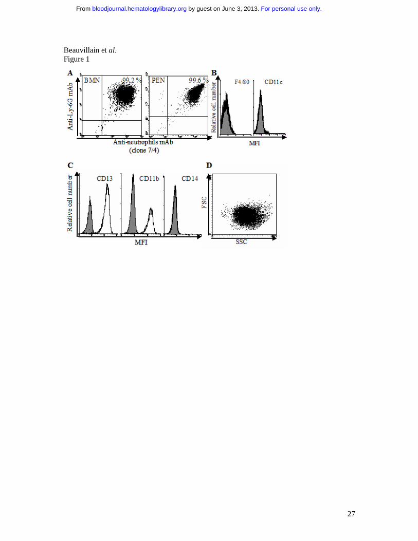
Murine neutrophils cross-present soluble and particulate antigens. In this study, we
analyzed whether murine neutrophils isolated from peritoneal exudates (PEN) or from bone
marrow (BMN) may cross-present soluble ovalbumin (Ova). In order to exclude the
involvement of contaminating APCs in in vitro and in vivo experiments, we used highly
purified populations of neutrophils obtained by 2 (for BMN) or 3 step (for PEN) purification
protocols. Phenotypical analysis showed that neutrophil populations consisted of more than
99% of Ly-6Ghigh- and neutrophils (clone 7/4)-positive (Fig. 1A), as well as of CD11c- and
F4/80-negative cells (Fig.1B). Giemsa staining also showed that more than 99% of cells
exhibited a neutrophil morphology (intracellular granules and segmented nucleus) (data not
shown).
Neutrophils were pulsed with 0.4 mM Ova for 2 to 8 h and used to stimulate the Ova-specific
CD8+ T-cell hybridoma B3Z. Results showed that PEN and BMN efficiently cross-presented
Ova to B3Z cells with a maximum observed when BMN were pulsed for 2 h (stimulation
index (SI) =5.4 ± 0.5; mean ± s.d., n=5) and PEN pulsed for 4 h (SI=4.9 ± 0.8) (Fig. 2A). In
the following experiments, BMN and PEN were pulsed for 2 and 4 h, respectively. Cross-
presentation by thioglycolate-elicited macrophages and bone marrow-derived DCs, used as
positive controls, was maximal when pulsed for 8 h with Ova (SI=6.4 ± 0.5 and 9.3 ± 0.7,
respectively) (Fig. 2A). The LB27.4 cell line, used as a negative control,2 did not cross-
present Ova, whatever the time-point analyzed (Fig. 2A). Antigen uptake is required to endow
a cell with cross-presentation capacity. Supporting our observations, PEN and BMN
efficiently internalized Ova (Fig. 2B left panels), expressed cell surface MHC-I molecules
(Fig. 2B, middle panels), and presented SIINFEKL in Kb molecules after Ova pulse (Fig. 2B,
right panels). In contrast, although LB27.4 cells expressed MHC-I molecules and endocytosed
Ova, they did not express Kb-SIINFEKL after Ova pulse, showing that antigen uptake is not
sufficient for antigen cross-presentation (Fig. 2B).
In additional experiments, neutrophils and professional myeloid APCs (all used at 2x10⁵/ml)
were pulsed with increasing concentrations of Ova and used to stimulate B3Z cells. PEN and
BMN cross-presented Ova in a dose-dependent manner (Fig. 2C); PEN and BMN were as
potent as macrophages, (excepted for the highest concentration of Ova tested (0.4 mM), while
less efficient than DCs (at any concentration of Ova tested) (Fig. 2C). Experiments performed
with graded doses of myeloid cells reinforce this observation and also evidence a direct
relationship between the number of neutrophils and the amplitude of the CD8+ T cell response
(Fig. 2D). Particulate antigens have been reported to be more efficiently cross-presented than
For personal use only. by guest on June 3, 2013. bloodjournal.hematologylibrary.orgFrom
10
soluble antigens by professional APCs.2,38 We then tested the ability of neutrophils to cross-
present Ova coupled to iron beads (Ova-beads). Cross-presentation by BMN required one
hundred lower concentrations of Ova when coupled to iron beads compared to soluble Ova
(Fig. 2E). PEN and macrophages were as potent as BMN in cross-presenting Ova-beads (Fig.
2E, right upper panel). In contrast, DCs were more efficient than macrophages and
neutrophils in cross-presenting soluble Ova and Ova-beads (Fig. 2C and Fig. 2E, right lower
panel). Together, these data demonstrate that neutrophils cross-present soluble and particulate
antigens in vitro and that antigen cross-presentation by neutrophils occurs earlier than in
myeloid APCs and is as efficient as in macrophages.
Antigen cross-presentation by murine neutrophils is proteasome- and TAP-dependent.
One of the major antigen cross-presentation pathway depends on proteasomes and TAP. To
investigate the mechanism(s) involved in antigen cross-presentation by neutrophils, we first
used neutrophils purified from TAP-/- mice. Cross-presentation by neutrophils from TAP-/-
mice was reduced (SI = 1.2 ± 0.4, corresponding to a decrease of 65 ± 10%; mean ± s.d., n=3)
compared to neutrophils from wild-type mice (SI = 4.3 ± 0.4) (Fig. 2F). Moreover, incubation
of wild-type BMN with lactacystin reduced Ova cross-presentation (SI = 0.5 ± 0.8; decrease
of 85 ± 15%) (Fig. 2F). Similar data were observed using PEN (data not shown). These data
show that neutrophils process and cross-present soluble Ova via a proteasome- and TAP-
dependent mechanism.
Human neutrophils cross-present soluble antigens. We then evaluated whether human
peripheral blood neutrophils may also cross-present soluble antigens using the NS3-1 CD8+ T
cell clone (NS3 TCC) which is specific for the HLA-A2-restricted peptide NS31406-1415 of the
HCV-NS3 c33c protein.36 Activation of the NS3-1 TCC was assessed by measuring IFNγ
secretion.39 We excluded the presence of contaminating myeloid APCs in human neutrophils
by FACS analysis: > 99% of the cells were CD13+, CD11b+ (Fig. 1C) and presented forward
and side scatter (FSC/SSC) parameters characteristics of neutrophils (Fig. 1D). No CD14+
(Fig. 1C) and BDCA1-3+ cells (data not shown) were detected. Human neutrophils were
pulsed with NS3 or Ova (as a negative control) and used to stimulate the NS3-1 TCC. Results
showed that human neutrophils pulsed with NS3, but not with Ova, stimulated the NS3-1
TCC, in a manner that was dependent on the concentration of NS3 (Fig. 2G). These data show
that human neutrophils cross-present exogenous antigen.
For personal use only. by guest on June 3, 2013. bloodjournal.hematologylibrary.orgFrom

11
Murine neutrophils cross-prime naive T cells in vitro. We then investigated whether
neutrophils may prime naive CD8+ T cells in vitro. Murine neutrophils were pulsed with Ova
and used to stimulate highly purified naive CD8+ T cells from OT1 transgenic mice (purity >
99%; data not shown). OT1 CD8+ T cell stimulation was assessed by quantifying IL-2
production. Results showed that both BMN and PEN cross-primed OT1 CD8+ T cells in vitro
(IL-2 = 0.28 ± 0.02 and 0.27 ± 0.04 ng/ml, respectively, with 0.4 mM Ova; mean ± s.d., n=3)
(Fig. 3A). Supporting these data, BMN and PEN expressed CD80 (MFI = 50 ± 4 and 52 ± 7,
respectively; mean ± s.d., n=3)25,26 (Fig. 3B) and BMN also weakly expressed CD8625 (MFI =
12 ± 4) (data not shown). Neutrophils were less potent than DCs (IL-2 = 2 ± 0.02 ng/ml with
0.4 mM Ova) (Fig. 3A). Together, these data demonstrate that neutrophils are able to cross-
present soluble antigens to naive CD8+ T cells in vitro.
Murine neutrophils cross-present antigen to naive CD8+ T cells in vivo. To determine
whether cross-presentation by neutrophils also occur in vivo, we analyzed the cross-
presentation capacity of neutrophils isolated from the peritoneal exudates of mice previously
intraperitoneally injected with Ova. Highly purified neutrophils from Ova-injected mice, but
not from PBS-injected mice, stimulated OT1 CD8+ T cells in vitro (IL-2 = 0.27 ± 0.02 ng/ml;
mean ± s.d., n=3) (Fig. 4A), demonstrating that neutrophils efficiently take up and process
Ova into MHC I-bound peptides in vivo.
We further assessed whether Ova-pulsed neutrophils may also induce naive CD8+ T cell
proliferation in vivo. To respond to this question, we used β2-microglobulin-deficient mice. In
these mice, bone marrow-derived APCs, lack MHC-I molecules6 and cannot cross-present
Ova or Ova-pulsed neutrophils.
β2-microglobulin-/- mice were intravenously injected with CFDA-SE-labeled naive (CD44low,
CD62Lhigh) CD8+ T cells from OT1 mice (Fig. 4B); OT1 CD8+ T cell purity, based on CD3
and CD8 expression, was > 99.5%, allowing to exclude a potential contamination by myeloid
cells (data not shown). At day one, mice were intravenously injected with Ova-pulsed BMN
and PEN purified from wild-type (wt) mice. As controls, mice were either injected with PBS,
200 µg Ova, Ova-pulsed LB27.4 (Ova-LB27.4) or Ova-pulsed DCs (Ova-DC) generated from
wt mice (Fig. 4B). Four days later, a proliferation of OT1 CD8+ T cells was observed in the
lymph nodes (Fig. 4C & D, upper panel) and in the spleen (Fig. 4C & D, lower panel) of mice
injected with Ova-pulsed BMN, Ova-pulsed PEN (data not shown), and Ova-pulsed DCs.
PBS, Ova and Ova-pulsed LB27.4 failed to induce CD8+ T cell proliferation in vivo (data not
shown and Fig. 4C & D) allowing to exclude (i) Ova or Ova-pulsed neutrophil cross-
For personal use only. by guest on June 3, 2013. bloodjournal.hematologylibrary.orgFrom

12
presentation by APCs from β2-microglobulin-/- mice and (ii) the presence of contaminating
APCs in neutrophils or CD8+T cell preparations. Finally, unpulsed neutrophils and NS3-
pulsed neutrophils did not induce CD8+ T cell proliferation, showing that Ova-pulsed
neutrophils-induced CD8+ T cell proliferation is antigen-specific (data not shown). These data
demonstrate that Ova-pulsed neutrophils cross-present antigen to naive T cells in vivo.
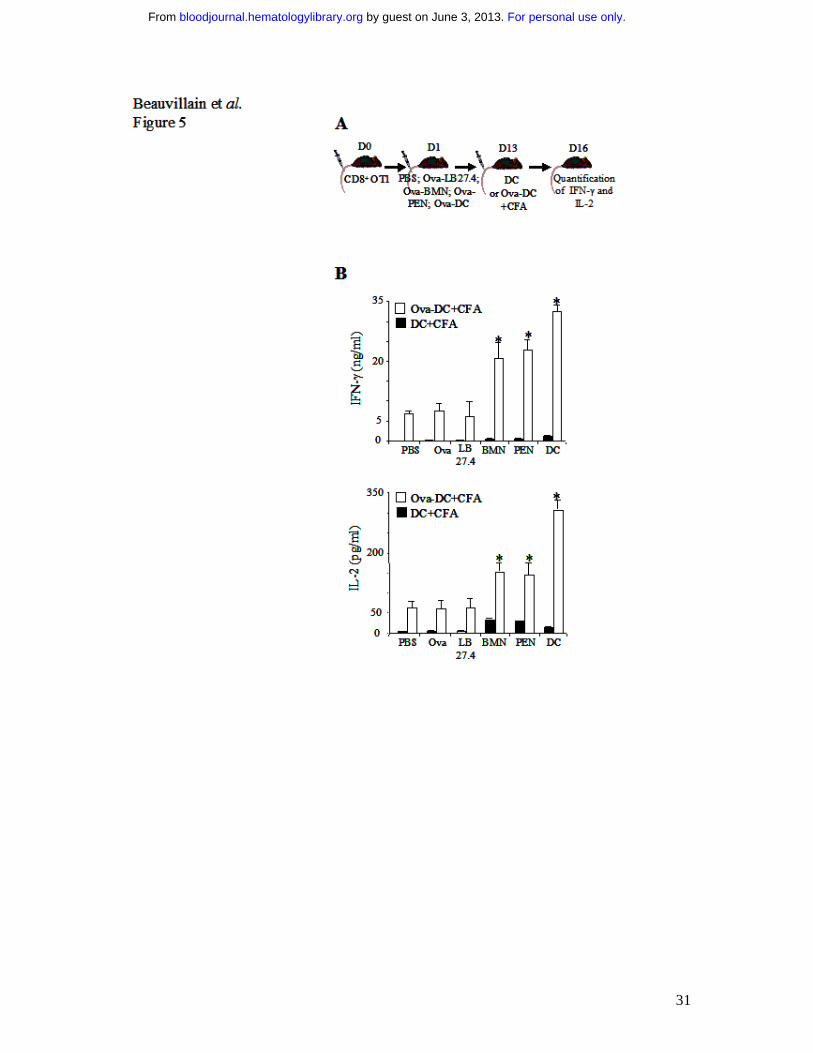
Murine neutrophils do not induce cross-tolerance in vivo. In response to antigen cross-
presentation, the in vivo proliferation of naive CD8+ T cells does not systematically lead to
effective T cell cross-priming. After several rounds of cell division, naive CD8+ T cells can be
deleted, leading to a specific tolerance.40 We therefore had to determine whether Ova-pulsed
neutrophils induce cross-tolerance or cross-priming in vivo. We first tested the cross-tolerance
hypothesis. Peripheral tolerance is the inability to respond to a second antigen challenge
delivered together with a strong adjuvant.40 β2-microglobulin-/- mice, adoptively transferred
with OT1 CD8+ T cells, were injected with Ova-pulsed BMN, Ova-pulsed PEN, or, as
controls, with PBS, Ova, Ova-pulsed LB27.4, or Ova-pulsed DCs. At day 13, mice were
injected either with Ova-pulsed DCs plus CFA or with unpulsed DCs plus CFA (Fig 5A).
Three days after DC injection, we analyzed IL-2 and IFN-γ production by in vitro
restimulated splenic CD8+ T cells. Interestingly, CD8+ T cells from mice injected either with
Ova-pulsed BMN or with Ova-pulsed PEN and restimulated by Ova-pulsed DCs plus CFA,
responded in a similar extent (IFN-γ = 20 ± 3 and 23 ± 2 ng/ml, respectively; IL-2 = 165 ± 18
and 160 ± 22 pg/ml, respectively; mean ± s.d., n=3) and more efficiently than CD8+ T cells
from PBS-injected mice (IFN-γ = 7 ng/ml ± 0.8 and IL-2 = 65 pg/ml ± 15) (Fig 5B). CD8+ T
cell stimulation in mice injected with Ova-pulsed DCs was higher than in Ova-pulsed
neutrophils (Fig. 5B) especially for IL-2 production. Compared to PBS injected mice, the
CD8+ response remained unchanged in Ova and Ova-pulsed LB27.4 injected mice (Fig. 5B).
No specific production of IL-2 and IFN-γ by CD8+ T cells from mice injected with unpulsed
DCs plus CFA, was detected. Altogether, these data show that Ova-pulsed neutrophils do not
render mice tolerant to Ova.
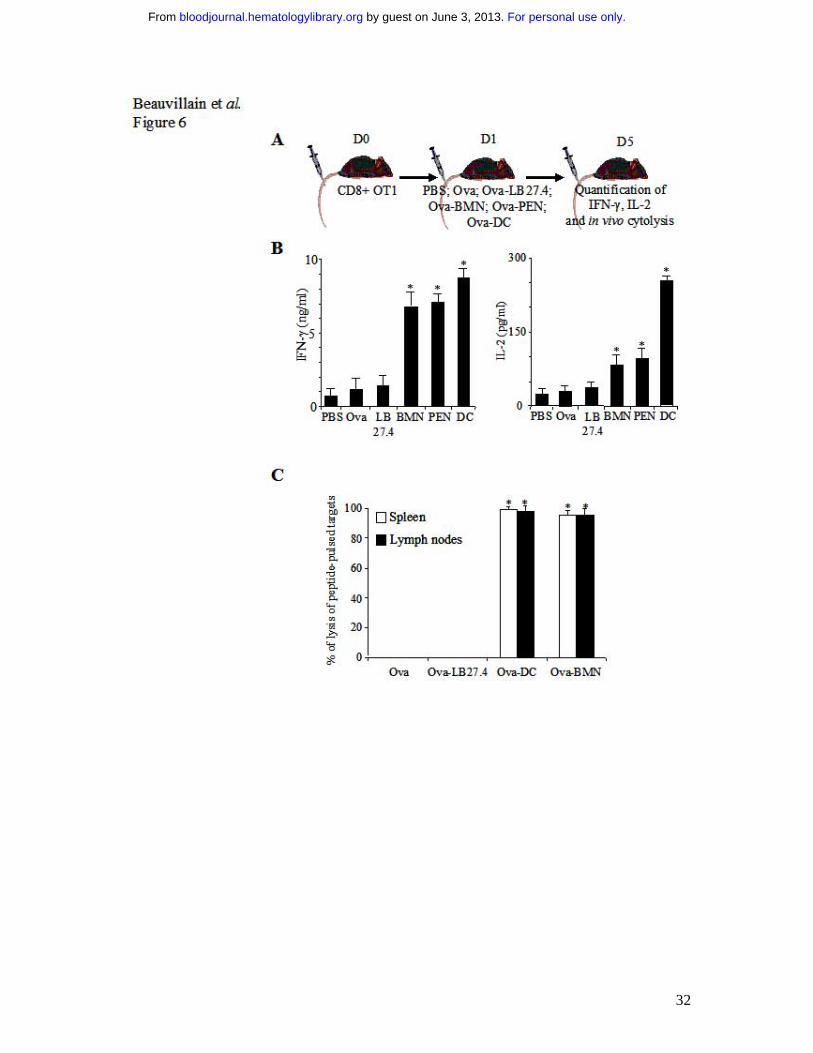
Murine neutrophils cross-prime CD8+ T cells in vivo. We then tested whether Ova-pulsed
neutrophils may cross-prime CD8+ T cell in vivo. The initiation of an effective immune
response is associated with a priming of T cells that is characterized not only by T cell
proliferation but also by the acquisition of effector functions (IFN-γ production and cytolysis).
For personal use only. by guest on June 3, 2013. bloodjournal.hematologylibrary.orgFrom
13
β2-microglobulin-/- mice, adoptively transferred with OT1 CD8+ T cells, were injected with
Ova-pulsed BMN, Ova-pulsed PEN, or, as controls, with PBS, Ova, Ova-pulsed LB27.4, or
Ova-pulsed DCs. After 4 days, CD8+ T cells were analyzed for IFN-γ and IL-2 production in
response to in vitro stimulation (Fig. 6A and 6B) and for the acquisition of cytolytic function,
determined in vivo (Fig. 6C) as previously described.40
CD8+ T cells from Ova-pulsed BMN injected mice secreted higher levels of IFN-γ and IL-2
than CD8+ T cells from mice injected with PBS, Ova or Ova-pulsed LB27.4 (Fig. 6B) and
acquired the capacity to lyse peptide-pulsed targets (Fig. 6C). In parallel, CD8+ T cells from
Ova-pulsed PEN injected mice presented effector functions similar to CD8+ T cells from Ova-
pulsed BMN injected mice (data not shown and Fig. 6B). CD8+ T cells from Ova-pulsed DC
injected mice secreted the highest levels of IFN-γ and IL-2 (Fig. 6B) and also exhibited
cytolytic function (Fig. 6C).
No specific cytolysis was observed in mice injected with PBS, Ova or Ova-pulsed LB27.4
(data not shown and Fig. 6C). These data indicate that, in our experimental conditions, Ova-
pulsed neutrophils have cross-primed naive T cells as they have induced their differentiation
into functional effector cells.
Subcutaneously injected neutrophils cross-prime CD8+ T cells in vivo. We then tested
whether Ova-pulsed neutrophils injected via the subcutaneous route may cross-prime CD8+ T
cell in vivo. β2-microglobulin-/- mice, adoptively transferred with OT1 CD8+ T cells, were
subcutaneously injected with Ova-pulsed BMN + CFA, Ova-pulsed PEN+CFA, or, as
controls, with PBS + CFA, Ova + CFA, Ova-pulsed LB27.4 + CFA, or Ova-pulsed DCs +
(Fig. 7A). At day 5, in Ova-pulsed BMN (Fig. 7B) and Ova-pulsed PEN injected mice (data
not shown), we observed that OT1 CD8+ T cells proliferated in the lymph nodes, secreted
higher levels of IFN-γ and IL-2 than CD8+ T cells from mice injected with PBS, Ova or Ova-
pulsed LB27.4 (Fig. 7C) and acquired the capacity to lyse Ova-peptide-pulsed targets (Fig.
7D and data not shown). Similar results were obtained in Ova-pulsed DC injected mice (Fig.
7B, C & D). In contrast, CD8+ T cells from mice injected s.c. with PBS + CFA, Ova + CFA or
Ova-pulsed LB27.4 + CFA failed to proliferate (Fig. 7B) and to acquire cytolytic function
(Fig. 7D). Lastly, no CD8+ T cell proliferation and activation were detected in mice injected
with PMN in the absence of CFA and in the spleen of mice injected s.c. with unpulsed PMN +
CFA (data not shown). These data indicate that, in our experimental conditions,
subcutaneously injected Ova-pulsed neutrophils cross-prime naive T cells.
For personal use only. by guest on June 3, 2013. bloodjournal.hematologylibrary.orgFrom
14
Discussion
This study provides the first evidence for an in vivo role of neutrophils in antigen cross-
presentation and naive T cell cross-priming. Our findings demonstrate that (i) human and
murine neutrophils cross-present antigens in vitro, (ii) that ovalbumin cross-presentation by
neutrophils is proteasome- and TAP-dependent, and (iii) that antigen-pulsed neutrophils do
not induce cross-tolerance but induce naive CD8+ T cell differenciation into effector cells in
vivo. This property of neutrophils may open new perspectives to boost the efficacy of vaccine
based on CTL induction.
Neutrophils are the first cells to migrate, in high number, at the site of infection. Their main
recognized function is to clear invading microorganisms. To date, the ability of neutrophils to
present exogenous antigens to CD8+ T cells remains poorly documented. One study reported
that neutrophils loaded with CTL peptides stimulate memory T cells in vitro, showing that
MHC-I molecules on neutrophils are functional.41 Potter et al observed that neutrophils that
have phagocytosed Ova-expressing bacteria, processed Ova, in a proteasome-independent
manner, for presentation to memory CD8+ T cells,31 thereby demonstrating that the vacuolar
pathway associated to antigen cross-presentation is functional in neutrophils.31 Using
lactacystin-treated neutrophils and neutrophils from TAP-/-, mice, we report that soluble
antigen cross-presentation by neutrophils depends on TAP machinery and on proteasome.
This pathway is the most extensively described in antigen cross-presentation.3 The
professional APCs, DCs and macrophages, have also been reported to cross-present Ova in a
proteasome- and TAP-dependent manner.38 Supporting our findings, neutrophils have been
reported to express proteasome and TAP.42,43 Altogether, these data demonstrate that the two
main pathways associated to antigen cross-presentation are functional in neutrophils.
Antigen cross-presentation may lead in vivo to immunity or tolerance.3 Upon contact with
bacteria or fungi, neutrophils are the main cells that accumulate early and take up the
microorganisms. Moreover, activated neutrophil infiltration in inflammatory site is associated
to some auto-immune disorders such as vasculitis.44 It therefore appeared crucial to determine
whether neutrophils are involved in cross-tolerance or cross-priming. Our observations that
neutrophils cross-present antigens to naive T cells in vitro and that Ova-pulsed neutrophils
induce naive OT1 CD8+ T cell proliferation in vivo, were not sufficient to speculate about a
role for neutrophils in cross-tolerance versus cross-priming. As examples, endothelial cells
For personal use only. by guest on June 3, 2013. bloodjournal.hematologylibrary.orgFrom

15
cross-present antigen to naive CD8+ T cells in vitro while they induce cross-tolerance in
vivo.45 The proliferation of naive OT1 CD8+ T cell induced by targeting ovalbumin to DEC-
205 on DCs is followed by a rapid T cell death and peripheral CD8+ T cell tolerance.40 We
therefore analyzed the consequences of antigen cross-presentation by neutrophils in an in vivo
model where DCs and macrophages were unable to cross-present antigens, in order to exclude
a re-presentation of neutrophils or neutrophil-derived fragments by bone-marrow-derived
APCs. In this model, Ova-pulsed neutrophils cross-prime naive T cells, as they induce their
differentiation into functional effector cells. These data are the first demonstration of an in
vivo CD8+ T cell cross-priming by neutrophils.
Neutrophils are terminally differentiated cells with a short life-span. Recent data suggest that
some signals (i.e. pro-inflammatory cytokines or TLR agonists) increase their survival.46,47 In
agreement with these observations, our data show that the survival time period of
intravenously or subcutaneously injected Ova-pulsed neutrophils is sufficient to allow
neutrophils to migrate to the spleen and the lymph nodes, to encounter and to stimulate naive
CD8+ T cells. Recent observations also demonstrated that neutrophils migrate from the
periphery to the draining lymph nodes.18,32 Taken together, these data support a potential role
for neutrophils in CD8+ T cell cross-priming.
Our data, added to observations from others showing that neutrophils can vehicle antigens or
microorganisms from the periphery to the draining lymph nodes, suggest that neutrophils
could be useful to cross-prime CD8+ T cells against microorganisms that do not infect DCs or
downregulate their properties.
Cross-tolerance versus cross-priming in vivo depends on parameters such as the levels of
antigen that is cross-presented, the presence of CD4+ T cell help and the degree of
costimulatory signals expressed on the antigen cross-presenting cells. Firstly, in in vitro
experiments, we observed that neutrophils were as potent as macrophages in cross-presenting
antigen. Secondly, in our models, APC from β2-microglobulin-/- mice express functional
MHC-II molecules and can prime Ova specific CD4+ T cells. In some circumstances, an
efficient in vivo cross-priming requires cognate CD4+ T cell help. In this three-cell model, the
antigen is presented both into the MHC-I and the MHC-II by the same APC48-50 and antigen
stimulated-CD4+ T cells contribute to APC activation via CD40L expression. Although the
ability of neutrophils to present antigens in MHC-II molecules in vivo remains to be
demonstrated, stimulated neutrophils express MHC-II25 and are activated by CD40L.51
For personal use only. by guest on June 3, 2013. bloodjournal.hematologylibrary.orgFrom

16
Thirdly, in agreement with recent data showing that neutrophils contain preformed CD80 and
CD86 molecules that are rapidly expressed on the cell surface upon stimulation, we reported
that murine BMN and PEN expressed CD80 (and CD86 for BMN). Together, the ability of
neutrophils to efficiently cross-present Ag and to express costimulatory molecules supports
their ability to cross-prime CD8+ T cells.
We report here that neutrophils cross-prime CD8+ T cells in vivo. The observations that (1)
neutrophils accumulate at the site of infection before DCs and macrophages, and (2) that in
vitro cross-presentation by neutrophils occurs earlier than in professional APCs, suggest that
neutrophils may interact in vivo with naive T CD8+ cells prior to DCs. More precisely, in
response to a pathogen, whether neutrophils and DCs that have cross-presented the antigen
may sequentially interact with CD8+ T cells and the consequences of these sequential
interactions on CD8+ T cell responses should be evaluated. This point will be difficult to
analyze in vivo. In a model where both neutrophils and DCs express functional MHC-I
molecules, live and apoptotic neutrophils that have taken up the antigen can serve as
substrates for cross-presentation by DCs.21,24 Lastly, the observation that neutrophil depletion
reduces the CD8+ T cell response against non secreted bacterial antigen,24 supports our
observation, although the mechanisms used by neutrophils to boost the CD8+ response
remained undetermined. In our in vivo model, we excluded neutrophil cross-presentation by
APCs and demonstrated that antigen cross-priming by neutrophils in vivo enhanced CD8+ T
cell response to a subsequent restimulation by DCs. These data thereby suggest that
neutrophils may act in concert with DCs in vivo to initiate and/or amplify CD8+ T cell
responses to exogenous antigens.
Different observations have suggested a role of activated CD8+ T cells in some autoimmune
disease such as Wegener granulomatosis (WG)52,53 and multiple sclerosis (MS).54 These
pathologies are also associated with an accumulation of neutrophils at the inflammatory site55
or with an increase of IL-8 production,56 respectively. Moreover, in WG, circulating
neutrophils also express MHC-II molecules.57 Based on our data, it seems of interest to
evaluate whether, in these pathologies, neutrophils may cross-present self antigens and
contribute to the generation and/or maintenance of auto-reactive CD8+ T cell responses. This
may lead to define more specific immunotherapies that could be used together with immuno-
suppressive agents.
For personal use only. by guest on June 3, 2013. bloodjournal.hematologylibrary.orgFrom

17
In conclusion, these data show that neutrophils cross-present antigens and, in our
experimental conditions, cross-prime CD8+ T cells in vivo. These findings suggest that it
could be of interest to reanalyze the role of neutrophils in the physiopathology of some auto-
immune disorders. Furthermore, this new property of neutrophils could be exploited to boost
the efficacy of vaccines based on CTL induction, for example by using preferentially
adjuvants that favour neutrophil recruitment and migration into the lymph nodes.
For personal use only. by guest on June 3, 2013. bloodjournal.hematologylibrary.orgFrom

18
Acknowledgments
We are grateful to Dr N. Shastri (University of California, Berkeley, CA) for providing with
the B3Z hybridoma. We thank Lena Manoukian for typing assistance and Patrice Chiron for
animal breeding assistance. This work was supported by INSERM Contrat Avenir and the
Canceropole Grand-Ouest. CB performed research, analyzed data and wrote the paper, YD
designed research and wrote the paper, MS performed research, AP and VB provided KO and
transgenic mice and human T cell clone, HG is the director of the laboratory, VB and PG
provided vital reagents and participated to the design of experiments and PJ supervised the
entire project, designed research and wrote the paper.
For personal use only. by guest on June 3, 2013. bloodjournal.hematologylibrary.orgFrom

19
References
1. Bevan MJ. Cross-priming for a secondary cytotoxic response to minor H antigens with H-2 congenic cells which do not cross-react in the cytotoxic assay. J Exp Med. 1976;143:1283-1288. 2. Kovacsovics-Bankowski M, Clark K, Benacerraf B, Rock KL. Efficient major histocompatibility complex class I presentation of exogenous antigen upon phagocytosis by macrophages. Proc Natl Acad Sci U S A. 1993;90:4942-4946. 3. Rock KL, Shen L. Cross-presentation: underlying mechanisms and role in immune surveillance. Immunol Rev. 2005;207:166-183. 4. Banchereau J, Steinman RM. Dendritic cells and the control of immunity. Nature. 1998;392:245-252. 5. Mellman I, Steinman RM. Dendritic cells: specialized and regulated antigen processing machines. Cell. 2001;106:255-258. 6. Pozzi LA, Maciaszek JW, Rock KL. Both dendritic cells and macrophages can stimulate naive CD8 T cells in vivo to proliferate, develop effector function, and differentiate into memory cells. J Immunol. 2005;175:2071-2081. 7. Shen L, Rock KL. Priming of T cells by exogenous antigen cross-presented on MHC class I molecules. Curr Opin Immunol. 2006;18:85-91. 8. van der Bruggen P, Van den Eynde BJ. Processing and presentation of tumor antigens and vaccination strategies. Curr Opin Immunol. 2006;18:98-104. 9. Heath WR, Kurts C, Miller JF, Carbone FR. Cross-tolerance: a pathway for inducing tolerance to peripheral tissue antigens. J Exp Med. 1998;187:1549-1553. 10. Limmer A, Ohl J, Wingender G, et al. Cross-presentation of oral antigens by liver sinusoidal endothelial cells leads to CD8 T cell tolerance. Eur J Immunol. 2005;35:2970-2981. 11. Albert ML, Jegathesan M, Darnell RB. Dendritic cell maturation is required for the cross-tolerization of CD8+ T cells. Nat Immunol. 2001;2:1010-1017. 12. Ramos CD, Fernandes KS, Canetti C, Teixeira MM, Silva JS, Cunha FQ. Neutrophil recruitment in immunized mice depends on MIP-2 inducing the sequential release of MIP-1alpha, TNF-alpha and LTB(4). Eur J Immunol. 2006;36:2025-2034. 13. Ramos CD, Canetti C, Souto JT, et al. MIP-1alpha[CCL3] acting on the CCR1 receptor mediates neutrophil migration in immune inflammation via sequential release of TNF-alpha and LTB4. J Leukoc Biol. 2005;78:167-177. 14. Parker LC, Whyte MK, Dower SK, Sabroe I. The expression and roles of Toll-like receptors in the biology of the human neutrophil. J Leukoc Biol. 2005;77:886-892. 15. Lee WL, Harrison RE, Grinstein S. Phagocytosis by neutrophils. Microbes Infect. 2003;5:1299-1306. 16. Brinkmann V, Reichard U, Goosmann C, et al. Neutrophil extracellular traps kill bacteria. Science. 2004;303:1532-1535. 17. Nathan C. Neutrophils and immunity: challenges and opportunities. Nat Rev Immunol. 2006;6:173-182. 18. Maletto BA, Ropolo AS, Alignani DO, et al. Presence of neutrophil-bearing antigen in lymphoid organs of immune mice. Blood. 2006;108:3094-3102. 19. van Gisbergen KP, Sanchez-Hernandez M, Geijtenbeek TB, van Kooyk Y. Neutrophils mediate immune modulation of dendritic cells through glycosylation-dependent interactions between Mac-1 and DC-SIGN. J Exp Med. 2005;201:1281-1292. 20. Bennouna S, Denkers EY. Microbial antigen triggers rapid mobilization of TNF-alpha to the surface of mouse neutrophils transforming them into inducers of high-level dendritic cell TNF-alpha production. J Immunol. 2005;174:4845-4851.
For personal use only. by guest on June 3, 2013. bloodjournal.hematologylibrary.orgFrom
20
21. Megiovanni AM, Sanchez F, Robledo-Sarmiento M, Morel C, Gluckman JC, Boudaly S. Polymorphonuclear neutrophils deliver activation signals and antigenic molecules to dendritic cells: a new link between leukocytes upstream of T lymphocytes. J Leukoc Biol. 2006;79:977-988. 22. Bennouna S, Bliss SK, Curiel TJ, Denkers EY. Cross-talk in the innate immune system: neutrophils instruct recruitment and activation of dendritic cells during microbial infection. J Immunol. 2003;171:6052-6058. 23. Evans EW, Harmon BG. A review of antimicrobial peptides: defensins and related cationic peptides. Vet Clin Pathol. 1995;24:109-116. 24. Tvinnereim AR, Hamilton SE, Harty JT. Neutrophil involvement in cross-priming CD8+ T cell responses to bacterial antigens. J Immunol. 2004;173:1994-2002. 25. Sandilands GP, Ahmed Z, Perry N, Davison M, Lupton A, Young B. Cross-linking of neutrophil CD11b results in rapid cell surface expression of molecules required for antigen presentation and T-cell activation. Immunology. 2005;114:354-368. 26. Wagner C, Iking-Konert C, Hug F, et al. Cellular inflammatory response to persistent localized Staphylococcus aureus infection: phenotypical and functional characterization of polymorphonuclear neutrophils (PMN). Clin Exp Immunol. 2006;143:70-77. 27. Iking-Konert C, Wagner C, Denefleh B, et al. Up-regulation of the dendritic cell marker CD83 on polymorphonuclear neutrophils (PMN): divergent expression in acute bacterial infections and chronic inflammatory disease. Clin Exp Immunol. 2002;130:501-508. 28. Okuda K, Tani K, Ishigatsubo Y, Yokota S, David CS. Antigen-pulsed neutrophils bearing Ia antigens can induce T lymphocyte proliferative response to the syngeneic or semisyngeneic antigen-primed T lymphocytes. Transplantation. 1980;30:368-372. 29. Radsak M, Iking-Konert C, Stegmaier S, Andrassy K, Hansch GM. Polymorphonuclear neutrophils as accessory cells for T-cell activation: major histocompatibility complex class II restricted antigen-dependent induction of T-cell proliferation. Immunology. 2000;101:521-530. 30. Ishikawa F, Miyazaki S. New biodefense strategies by neutrophils. Arch Immunol Ther Exp (Warsz). 2005;53:226-233. 31. Potter NS, Harding CV. Neutrophils process exogenous bacteria via an alternate class I MHC processing pathway for presentation of peptides to T lymphocytes. J Immunol. 2001;167:2538-2546. 32. Abadie V, Badell E, Douillard P, et al. Neutrophils rapidly migrate via lymphatics after Mycobacterium bovis BCG intradermal vaccination and shuttle live bacilli to the draining lymph nodes. Blood. 2005;106:1843-1850. 33. Stoitzner P, Tripp CH, Eberhart A, et al. Langerhans cells cross-present antigen derived from skin. Proc Natl Acad Sci U S A. 2006;103:7783-7788. 34. Jeannin P, Magistrelli G, Herbault N, et al. Outer membrane protein A renders dendritic cells and macrophages responsive to CCL21 and triggers dendritic cell migration to secondary lymphoid organs. Eur J Immunol. 2003;33:326-333. 35. Sanderson S, Shastri N. LacZ inducible, antigen/MHC-specific T cell hybrids. Int Immunol. 1994;6:369-376. 36. Accapezzato D, Visco V, Francavilla V, et al. Chloroquine enhances human CD8+ T cell responses against soluble antigens in vivo. J Exp Med. 2005;202:817-828. 37. Hernandez J, Aung S, Redmond WL, Sherman LA. Phenotypic and functional analysis of CD8(+) T cells undergoing peripheral deletion in response to cross-presentation of self-antigen. J Exp Med. 2001;194:707-717.
For personal use only. by guest on June 3, 2013. bloodjournal.hematologylibrary.orgFrom

21
38. Kovacsovics-Bankowski M, Rock KL. A phagosome-to-cytosol pathway for exogenous antigens presented on MHC class I molecules. Science. 1995;267:243-246. 39. Lapenta C, Santini SM, Spada M, et al. IFN-alpha-conditioned dendritic cells are highly efficient in inducing cross-priming CD8(+) T cells against exogenous viral antigens. Eur J Immunol. 2006;36:2046-2060. 40. Bonifaz L, Bonnyay D, Mahnke K, Rivera M, Nussenzweig MC, Steinman RM. Efficient targeting of protein antigen to the dendritic cell receptor DEC-205 in the steady state leads to antigen presentation on major histocompatibility complex class I products and peripheral CD8+ T cell tolerance. J Exp Med. 2002;196:1627-1638. 41. Reali E, Guerrini R, Moretti S, et al. Polymorphonuclear neutrophils pulsed with synthetic peptides efficiently activate memory cytotoxic T lymphocytes. J Leukoc Biol. 1996;60:207-213. 42. El Ouakfaoui S, Heitz D, Paquin R, Beaulieu AD. Granulocyte-macrophage colony-stimulating factor modulates tapasin expression in human neutrophils. J Leukoc Biol. 1999;65:205-210. 43. Derouet M, Thomas L, Cross A, Moots RJ, Edwards SW. Granulocyte macrophage colony-stimulating factor signaling and proteasome inhibition delay neutrophil apoptosis by increasing the stability of Mcl-1. J Biol Chem. 2004;279:26915-26921. 44. Suzuki K. Neutrophil functions of patients with vasculitis related to myeloperoxidase-specific anti-neutrophil antibody. Int J Hematol. 2001;74:134-143. 45. Limmer A, Ohl J, Kurts C, et al. Efficient presentation of exogenous antigen by liver endothelial cells to CD8+ T cells results in antigen-specific T-cell tolerance. Nat Med. 2000;6:1348-1354. 46. Francois S, El Benna J, Dang PM, Pedruzzi E, Gougerot-Pocidalo MA, Elbim C. Inhibition of neutrophil apoptosis by TLR agonists in whole blood: involvement of the phosphoinositide 3-kinase/Akt and NF-kappaB signaling pathways, leading to increased levels of Mcl-1, A1, and phosphorylated Bad. J Immunol. 2005;174:3633-3642. 47. Kobayashi SD, Voyich JM, Whitney AR, DeLeo FR. Spontaneous neutrophil apoptosis and regulation of cell survival by granulocyte macrophage-colony stimulating factor. J Leukoc Biol. 2005;78:1408-1418. 48. Ridge JP, Di Rosa F, Matzinger P. A conditioned dendritic cell can be a temporal bridge between a CD4+ T-helper and a T-killer cell. Nature. 1998;393:474-478. 49. Bennett SR, Carbone FR, Karamalis F, Flavell RA, Miller JF, Heath WR. Help for cytotoxic-T-cell responses is mediated by CD40 signalling. Nature. 1998;393:478-480. 50. Schoenberger SP, Toes RE, van der Voort EI, Offringa R, Melief CJ. T-cell help for cytotoxic T lymphocytes is mediated by CD40-CD40L interactions. Nature. 1998;393:480-483. 51. Khan SY, Kelher MR, Heal JM, et al. Soluble CD40 ligand accumulates in stored blood components, primes neutrophils through CD40, and is a potential cofactor in the development of transfusion-related acute lung injury. Blood. 2006;108:2455-2462. 52. Schlesier M, Kaspar T, Gutfleisch J, Wolff-Vorbeck G, Peter HH. Activated CD4+ and CD8+ T-cell subsets in Wegener's granulomatosis. Rheumatol Int. 1995;14:213-219. 53. Ikeda M, Tsuru S, Watanabe Y, Kitahara S, Inouye T. Reduced CD4-CD8 T cell ratios in patients with Wegener's granulomatosis. J Clin Lab Immunol. 1992;38:103-109. 54. Haegele KF, Stueckle CA, Malin JP, Sindern E. Increase of CD8+ T-effector memory cells in peripheral blood of patients with relapsing-remitting multiple sclerosis compared to healthy controls. J Neuroimmunol. 2006.
For personal use only. by guest on June 3, 2013. bloodjournal.hematologylibrary.orgFrom

22
55. Gross WL, Trabandt A, Csernok E. Pathogenesis of Wegener's granulomatosis. Ann Med Interne (Paris). 1998;149:280-286. 56. Lund BT, Ashikian N, Ta HQ, et al. Increased CXCL8 (IL-8) expression in Multiple Sclerosis. J Neuroimmunol. 2004;155:161-171. 57. Iking-Konert C, Vogt S, Radsak M, Wagner C, Hansch GM, Andrassy K. Polymorphonuclear neutrophils in Wegener's granulomatosis acquire characteristics of antigen presenting cells. Kidney Int. 2001;60:2247-2262.
For personal use only. by guest on June 3, 2013. bloodjournal.hematologylibrary.orgFrom

23
Figure Legends
Fig. 1. Purity of human and murine neutrophils. (A) Analysis by FACS of BMN (left
panel) and PEN (right panel) purity using PE-labeled anti-Ly-6G mAb and FITC-labeled anti-
neutrophils mAb (7/4). (B) Analysis of BMN purity using FITC-anti-CD11c and PE-anti-
F4/80 mAbs (white histograms; grey histograms correspond to isotype control mAbs), similar
data were obtained for PEN. (C & D) Analysis by FACS of human neutrophil purity using
FITC-anti-CD13, -CD11b and -CD14 mAbs (white histograms; grey histograms correspond
to isotype control mAbs) (C), and using the FSC/SSC parameters (D).
Fig. 2. Antigen cross-presentation by neutrophils. (A) LB27.4 cells, BMN, PEN,
macrophages and DCs pulsed for 2, 4, 6 or 8 h with 0.4 mM Ova were incubated with B3Z
cells. B3Z stimulation was measured by quantifying the release of β-galactosidase. Results
are expressed in stimulation index (SI) as mean ± s.d. of 5 experiments. (B) Left panel, PEN,
BMN, and LB27-4 were (white panel) or not (grey panel) incubated for 20 min at 37°C with
0.4 mM Texas Red-Ova. Middle panel, cells were incubated with anti-H-2Kb mAb (white
panel) or isotype control mAb (grey panel). Right panel, cells were pulsed with 0.4 mM Ova
for 2, 4 and 8 h, respectively, at 37°C before detection by FACS of SIINFEKL in MHC-I
molecules, using the 25-D1.16 mAb (white panel) or isotype control mAb (grey panel).
Results are representative of one out of three experiments. (C) DCs (�), macrophages (�),
PEN (�), BMN (�), and LB27.4 (�) pulsed with the indicated concentrations of Ova for 2 h
(BMN), 4 h (PEN) or 8 h (macrophages, DCs and LB27.4) were incubated with B3Z cells.
(D) Different numbers of DCs (�), macrophages (�), PEN (�) and BMN (�) pulsed with
0.4 mM Ova for 2 h (BMN), 4 h (PEN) or 8 h (macrophages and DCs) were incubated with
B3Z cells. (E) BMN (left panel), macrophages (upper right panel) and DCs (lower right
panel) pulsed with the indicated concentrations of Ova either soluble (�) or coated to beads
(�) were incubated with B3Z cells. (F) BMN from wild-type mice either untreated (�) or
treated with lactacystin ( ) and BMN from TAP-/- mice (�) pulsed with 0.2 mM Ova for 2 h
were incubated with B3Z cells. A,C-E Results are expressed in SI (mean ± s.d., n=5). (G)
HLA-A2+ human neutrophils pulsed or not with 0.1 to 10 µg/ml HCV1a-NS3 protein (�) or
with Ova (�), for 4 h, were cultured with a human CD8+ T cell clone specific from the HCV-
NS3 peptide 1406-1415. IFNγ production was quantified by ELISA in the 16h-supernatants.
Results are expressed in ng/ml as mean ± s.d. of triplicate values and are representative of the
data obtained with the neutrophils of one out three subjects.
For personal use only. by guest on June 3, 2013. bloodjournal.hematologylibrary.orgFrom
24
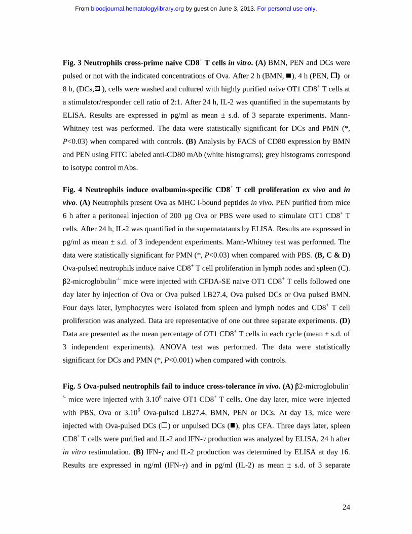
Fig. 3 Neutrophils cross-prime naive CD8+ T cells in vitro. (A) BMN, PEN and DCs were
pulsed or not with the indicated concentrations of Ova. After 2 h (BMN, �), 4 h (PEN, �) or
8 h, (DCs, ), cells were washed and cultured with highly purified naive OT1 CD8+ T cells at
a stimulator/responder cell ratio of 2:1. After 24 h, IL-2 was quantified in the supernatants by
ELISA. Results are expressed in pg/ml as mean ± s.d. of 3 separate experiments. Mann-
Whitney test was performed. The data were statistically significant for DCs and PMN (*,
P<0.03) when compared with controls. (B) Analysis by FACS of CD80 expression by BMN
and PEN using FITC labeled anti-CD80 mAb (white histograms); grey histograms correspond
to isotype control mAbs.
Fig. 4 Neutrophils induce ovalbumin-specific CD8+ T cell proliferation ex vivo and in
vivo. (A) Neutrophils present Ova as MHC I-bound peptides in vivo. PEN purified from mice
6 h after a peritoneal injection of 200 µg Ova or PBS were used to stimulate OT1 CD8+ T
cells. After 24 h, IL-2 was quantified in the supernatatants by ELISA. Results are expressed in
pg/ml as mean ± s.d. of 3 independent experiments. Mann-Whitney test was performed. The
data were statistically significant for PMN (*, P<0.03) when compared with PBS. (B, C & D)
Ova-pulsed neutrophils induce naive CD8+ T cell proliferation in lymph nodes and spleen (C).
β2-microglobulin-/- mice were injected with CFDA-SE naive OT1 CD8+ T cells followed one
day later by injection of Ova or Ova pulsed LB27.4, Ova pulsed DCs or Ova pulsed BMN.
Four days later, lymphocytes were isolated from spleen and lymph nodes and CD8+ T cell
proliferation was analyzed. Data are representative of one out three separate experiments. (D)
Data are presented as the mean percentage of OT1 CD8+ T cells in each cycle (mean ± s.d. of
3 independent experiments). ANOVA test was performed. The data were statistically
significant for DCs and PMN (*, P<0.001) when compared with controls.
Fig. 5 Ova-pulsed neutrophils fail to induce cross-tolerance in vivo. (A) β2-microglobulin-
/- mice were injected with 3.106 naive OT1 CD8+ T cells. One day later, mice were injected
with PBS, Ova or 3.106 Ova-pulsed LB27.4, BMN, PEN or DCs. At day 13, mice were
injected with Ova-pulsed DCs (�) or unpulsed DCs (�), plus CFA. Three days later, spleen
CD8+ T cells were purified and IL-2 and IFN-γ production was analyzed by ELISA, 24 h after
in vitro restimulation. (B) IFN-γ and IL-2 production was determined by ELISA at day 16.
Results are expressed in ng/ml (IFN-γ) and in pg/ml (IL-2) as mean ± s.d. of 3 separate
For personal use only. by guest on June 3, 2013. bloodjournal.hematologylibrary.orgFrom

25
experiments. Mann-Whitney test was performed. The data were statistically significant for
BMN, PEN and DC (*, P<0.03) when compared with controls.
Fig. 6 Ovalbumin-pulsed neutrophils induce naive CD8+ T cell differenciation into
effector cells. (A) OT1 CD8+ T cells were adoptively transferred into β2-microglobulin-/-
mice. One day later, the mice were injected intravenously with PBS, Ova, Ova-pulsed
LB27.4, Ova-pulsed BMN or Ova-pulsed DCs. Three days later, spleen CD8+ T cells were
purified and IL-2 and IFN-γ production was analyzed by ELISA or mice were injected
intravenously with target cells prepared as described in materials and methods. After 3 hours,
cytolysis was analyzed by FACS. (B) At day 5, IFN-γ and IL-2 production was determined by
ELISA, 24 h after in vitro restimulation. Results are expressed in ng/ml (IFN-γ) and in pg/ml
(IL-2) as mean ± s.d. of 3 separate experiments. Mann-Whitney test was performed. The data
were statistically significant for DCs and PMN (*, P<0.03) when compared with controls. (C)
In vivo cytolysis assay was performed by injecting 3x10⁶ unpulsed CFDA-SElow labeled
splenocytes and 3x10⁶ SIINFEKL-pulsed splenocytes CFDA-SEhigh labeled. Three hours later,
CDFA-SE expression was analyzed by FACS. Percentage of lysis was calculated as described
in materials and methods. Results are expressed as mean ± s.d. of 3 separate experiments.
ANOVA test was performed. The data were statistically significant for DCs and PMN (*,
P<0.001) when compared with controls.
Figure 7: Ovalbumin-pulsed neutrophils injected via s.c. route induce naive CD8+ T cell
differenciation into effector cells. (A) CFDA-SE-labeled or unlabeled OT1 CD8+ T cells
were adoptively transferred into β2-microglobulin-/- mice. One day later, mice were injected
subcutaneously with CFA plus PBS, Ova, Ova-pulsed LB27.4, Ova-pulsed BMN or Ova-
pulsed DCs and OT1 activation was measured at day 5. (B) OT1 CD8+ T cell proliferation
was expressed as the percentage of CD8+ T cells in each cycle (mean ± s.d. of 3 separate
experiments). ANOVA test was performed. The data were statistically significant for DCs and
PMN (*, P<0.001) when compared with controls. (C) At day 5, IFN-γ and IL-2 production
was determined by ELISA, 24 h after in vitro restimulation. Results are expressed in ng/ml as
mean ± s.d. of 3 separate experiments. Mann-Whitney test was performed. The data were
statistically significant for BMN, PEN and DCs (*, P<0.03) when compared with controls.
(D) In vivo cytolysis assays were performed by injecting 3x10⁶ unpulsed CFDA-SElow-labeled
splenocytes and 3x10⁶ CFDA-SEhigh-labeled SIINFEKL-pulsed splenocytes. Three hours
For personal use only. by guest on June 3, 2013. bloodjournal.hematologylibrary.orgFrom

26
later, CDFA-SE expression was analyzed by FACS. Percentage of lysis was calculated as
described in Materials and Methods. Data are presented as a percentage of lysis of peptide-
pulsed targets of 3 separate experiments; mean ± s.d. The data were statistically significant for
DCs and PMN when compared with controls (ANOVA test, *, P<0.001).
For personal use only. by guest on June 3, 2013. bloodjournal.hematologylibrary.orgFrom
27
Beauvillain et al. Figure 1
For personal use only. by guest on June 3, 2013. bloodjournal.hematologylibrary.orgFrom

28
For personal use only. by guest on June 3, 2013. bloodjournal.hematologylibrary.orgFrom

29
For personal use only. by guest on June 3, 2013. bloodjournal.hematologylibrary.orgFrom

30
For personal use only. by guest on June 3, 2013. bloodjournal.hematologylibrary.orgFrom
31
For personal use only. by guest on June 3, 2013. bloodjournal.hematologylibrary.orgFrom
32
For personal use only. by guest on June 3, 2013. bloodjournal.hematologylibrary.orgFrom

33
For personal use only. by guest on June 3, 2013. bloodjournal.hematologylibrary.orgFrom