heterofucan from sargassum filipendula induces apoptosis in hela cells
TRANSCRIPT
Mar. Drugs 2011, 9, 603-614; doi:10.3390/md9040603
Marine Drugs
ISSN 1660-3397
www.mdpi.com/journal/marinedrugs
Article
Heterofucan from Sargassum filipendula Induces Apoptosis in
HeLa Cells
Leandro Silva Costa 1,2
, Cinthia Beatrice Silva Telles 1, Ruth Medeiros Oliveira
1,
Leonardo Thiago Duarte Barreto Nobre 1, Nednaldo Dantas-Santos
1,
Rafael Barros Gomes Camara 1, Mariana Santana Santos Pereira Costa
1,
Jailma Almeida-Lima 1, Raniere Fagundes Melo-Silveira
1, Ivan Rui Lopes Albuquerque
1,
Edda Lisboa Leite 1 and Hugo Alexandre Oliveira Rocha
1,*
1 Laboratory of Biotechnology of Natural Polymers (BIOPOL), Departament of Biochemistry,
Federal University of Rio Grande do Norte (UFRN), Natal-RN, Brazil;
E-Mails: [email protected] (C.B.S.T), [email protected] (R.M.O.);
[email protected] (L.T.D.B.N.); [email protected] (N.D.-S.);
[email protected] (R.B.G.C.); [email protected] (M.S.S.P.C.);
[email protected] (J.A.-L.); [email protected] (R. F.M.-S.);
[email protected] (I.R.L.A.); [email protected] (E.L.L.) 2
Federal Institute of Education, Science and Technology of Rio Grande do Norte (IFRN), Santa
Cruz-RN, Brazil; E-Mail: [email protected] (L.S.C.)
* Author to whom correspondence should be addressed; E-Mail: [email protected];
Tel.: +55-84-32153416 (Branch line: 207); Fax: +55-84-32119208.
Received: 22 February 2011; in revised form: 29 March 2011 / Accepted: 8 April 2011 /
Published: 14 April 2011
Abstract: Fucan is a term used to denominate a family of sulfated polysaccharides rich in
sulfated L-fucose. Heterofucan SF-1.5v was extracted from the brown seaweed Sargassum
filipendula by proteolytic digestion followed by sequential acetone precipitation. This
fucan showed antiproliferative activity on Hela cells and induced apoptosis. However,
SF-1.5v was not able to activate caspases. Moreover, SF-1.5v induced glycogen synthase
kinase (GSK) activation, but this protein is not involved in the heterofucan SF-1.5v
induced apoptosis mechanism. In addition, ERK, p38, p53, pAKT and NFκB were not
affected by the presence of SF-1.5v. We determined that SF-1.5v induces apoptosis in
HeLa mainly by mitochondrial release of apoptosis-inducing factor (AIF) into cytosol. In
addition, SF-1.5v decreases the expression of anti-apoptotic protein Bcl-2 and increased
expression of apoptogenic protein Bax. These results are significant in that they provide a
OPEN ACCESS
Mar. Drugs 2011, 9
604
mechanistic framework for further exploring the use of SF-1.5v as a novel
chemotherapeutics against human cervical cancer.
Keywords: fucoidan; sulfated polysaccharides; anticancer; apoptosis-inducing factor (AIF)
1. Introduction
Carcinoma of the uterine cervix is the second most common female tumor worldwide, surpassed
only by breast cancer and its incidence is disproportionately high (>80%) in the developing world.
Primary treatment can be either surgery or a combination of radiotherapy/chemotherapy for early-stage
patients. Treatment of distant disease is usually palliative, aimed at symptom control. Targeted
radiotherapy may be useful for controlling local symptoms. While chemotherapy may sometimes
shrink tumor masses, there is no survival advantage [1]. Better and more effective chemotherapeutics
are apparently needed for these patients to improve survival rates.
Evidence has accumulated in recent years, showing that many cancer chemotherapeutic agents kill
cancer cells by inducing cell death. Cells die in a process that is reversible until a first irreversible
phase or ‘point-of-no-return’ is reached, but this is not a clearly defined biochemical event [2]. Thus,
identifying the mode of cell death is recognized as a novel strategy for screening anticancer drugs. As
a very valuable source for novel chemotherapeutic reagents, active sulfated homo-heterofucans
isolated from the brown seaweed have shown effective antitumor activities with a wide range of
mechanisms [3].
Fucan is a term used to define a family of L-fucose-containing sulfated polysaccharides found in
brown seaweed and several species of echinoderms, mostly from the egg jelly of sea urchins [4]. The
structures of these fucans vary among species and sometimes among different parts of the seaweed [5].
Furthermore, in contrast to animal fucans, algal fucans may have portions of other neutral sugars and
uronic acids in addition to sulfate and fucose in their structures. Some algal fucans exhibit important
pharmacological activities such as anticoagulant [6], antipeptic [7], anticomplementary,
antiinflammatory, antiviral [3], antiadhesive [8], antiproliferative [9], antioxidant [10] and
apoptosis-inducing [11,12]. As a result, fucans have a multitude of potential applications in human
health care. Additionally, biomaterials derived from seaweed generally have an advantage in that there
is no potential risk of contamination from animal viruses and bovine spongiform encephalopathy
(BSE) pathogens [13].
In a program aimed at determining the bioactivity of sulfated polysaccharides from tropical brown
seaweeds, we found that the polysaccharide-rich extract from Sargassum filipendula C.Agardh showed
significant antiproliferative effect on HeLa cell (human uterine adenocarcinoma cell line) proliferation
[10]. In the preceding article a bioassay-guided fractionation of this extract led to the isolation of an
antioxidant heterofucan denominated SF-1.5v, which exhibits antiproliferative activity against HeLa
cells. However, the molecular mechanism underlying the SF-1.5v-induced antiproliferative process
remains unclear.
The primary objective of this study was to determine the relevant mechanisms for an
antiproliferative effect of the heterofucan SF1.5v. We determined that SF-1.5v induces apoptosis in
Mar. Drugs 2011, 9
605
HeLa mainly by releasing the apoptosis-inducing factor (AIF) from mitochondria into cytosol. These
results are significant in that they provide a mechanistic framework for further exploring the use of
SF-1.5v as a novel chemotherapeutics for human cervical cancer.
2. Results and Discussion
2.1. Growth Inhibition by Heterofucan SF-1.5v
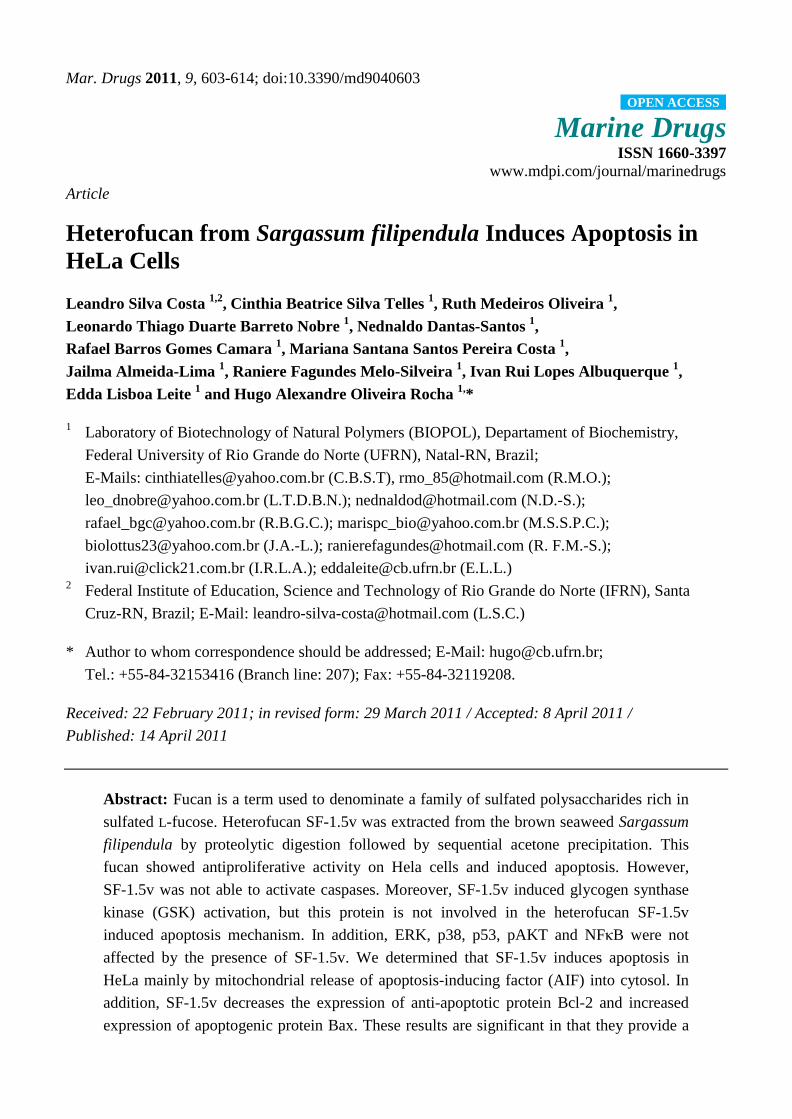
We studied the inhibitory effect of heterofucan SF-1.5v (from 0.1 to 2.0 mg/mL) on the
proliferation of HeLa cells cultured for 24, 48 and 72 hours. Figure 1 displays MTT assay results as a
measure of cell growth. Proliferation is presented as a percentage of cell proliferation under no
treatment conditions. A significant time and dose dependent decrease in cell proliferation was
observed. The effect was significant at 24 hours, but optimized at 72 hours (Figure 1), showing
antiproliferative activity between 32.7% and 72.5% at concentrations from 0.1 to 2.0 mg/mL.
Figure 1. HeLa cell proliferation in the presence of sulfated polysaccharide from
Sargassum filipendula. Each value is the mean ± SD of seven determinations. a,b,c
Indicate
a significant difference (p < 0.05) between treatments at the same concentration.
Antiproliferative activity of the heterofucan SF-1.5v was considerably higher than that of fucans
from Sargassum kjellmanianum and Sargassum stenophyllum, which showed no more than 40%
inhibition activity on the growth of L-1210 leukemia and HeLa cells, respectively [14,15]. These
considerable variations in antiproliferative activity between fucans likely result from the various
chemical compositions of fucan polymers originating in the different species, anatomical regions,
growing conditions of brown seaweeds, extraction and purification procedures as well as the use of
different cancer cell lines.
Mar. Drugs 2011, 9
606
2.2. Heterofucan SF-1.5v-Induced Apoptosis in HeLa Cells
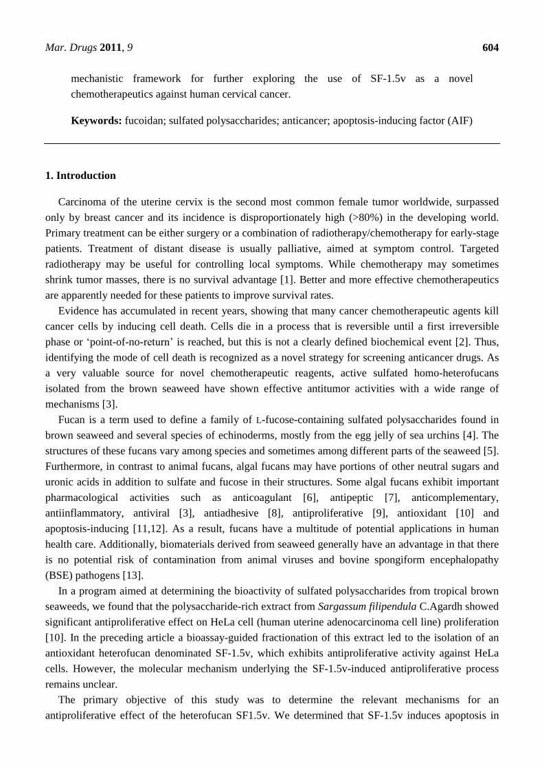
A typical assay was performed to characterize whether cell death resulting from fucan treatment
was caused by apoptosis induction. Thus, we examined the effect of the fucan (1.5 mg/mL) on
apoptosis using annexin V/PI double staining. One of the early features of cells undergoing apoptosis
is phosphatidylserine (PS) translocation from the inner to the outer leaflet of the plasma membrane.
Figure 2 shows PI vs. annexin V-FITC fluorescence. The lower right quadrants represent the early
apoptotic cells: annexin V binding and PI negative. Annexin V and PI staining revealed that SF-1.5v
increased apoptosis compared to the control.
Figure 2. Flow cytometry analysis of apoptotic death of HeLa cells by SF-1.5v. Dot plots
display the apoptotic death of HeLa cells treated with 1.5 mg/ml of SF-1.5v.
Annexin-/PI- (LL), viable cells; Annexin+/PI- (LR), cells undergoing apoptosis;
Annexin+/PI+ (UR), cells that are in end-stage apoptosis or are already dead.
LL, lower left; LR, lower right; UR, Upper right. One representative FACS assay of
three independent experiments is presented. The percentage in LR corresponds to
Annexin+/PI- cells.
2.3. Heterofucan SF-1.5v Treatment-Induced Apoptosis Did Not Require Activation of Caspases in
HeLa Cells
As the family of aspartate-specific cysteinyl proteases (caspases) plays a pivotal role in the
execution of programmed cell death, we determined whether the apostosis induction by the
heterofucan SF-1.5v resulted in activation of caspase-9 and caspase-3. Caspase activations were
measured using western blot analysis. Cells received no treatment (control) or were treated with
heterofucan SF-1.5v (1.5 mg/mL) for 24 hours. In response to the heterofucan, the activation of
pro-caspase-9 and pro-caspase-3 did not increase (Figure 3A). In order to rule out caspase participation
Mar. Drugs 2011, 9
607
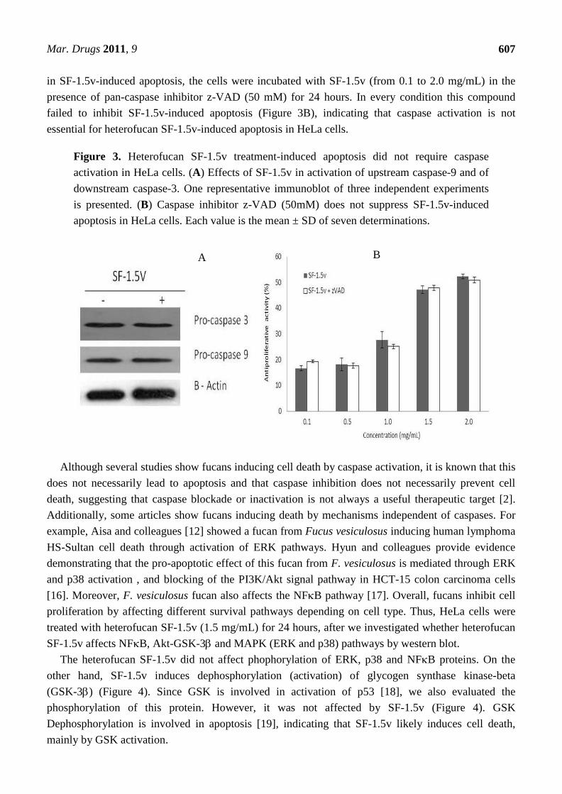
in SF-1.5v-induced apoptosis, the cells were incubated with SF-1.5v (from 0.1 to 2.0 mg/mL) in the
presence of pan-caspase inhibitor z-VAD (50 mM) for 24 hours. In every condition this compound
failed to inhibit SF-1.5v-induced apoptosis (Figure 3B), indicating that caspase activation is not
essential for heterofucan SF-1.5v-induced apoptosis in HeLa cells.
Figure 3. Heterofucan SF-1.5v treatment-induced apoptosis did not require caspase
activation in HeLa cells. (A) Effects of SF-1.5v in activation of upstream caspase-9 and of
downstream caspase-3. One representative immunoblot of three independent experiments
is presented. (B) Caspase inhibitor z-VAD (50mM) does not suppress SF-1.5v-induced
apoptosis in HeLa cells. Each value is the mean ± SD of seven determinations.
Although several studies show fucans inducing cell death by caspase activation, it is known that this
does not necessarily lead to apoptosis and that caspase inhibition does not necessarily prevent cell
death, suggesting that caspase blockade or inactivation is not always a useful therapeutic target [2].
Additionally, some articles show fucans inducing death by mechanisms independent of caspases. For
example, Aisa and colleagues [12] showed a fucan from Fucus vesiculosus inducing human lymphoma
HS-Sultan cell death through activation of ERK pathways. Hyun and colleagues provide evidence
demonstrating that the pro-apoptotic effect of this fucan from F. vesiculosus is mediated through ERK
and p38 activation , and blocking of the PI3K/Akt signal pathway in HCT-15 colon carcinoma cells
[16]. Moreover, F. vesiculosus fucan also affects the NFB pathway [17]. Overall, fucans inhibit cell
proliferation by affecting different survival pathways depending on cell type. Thus, HeLa cells were
treated with heterofucan SF-1.5v (1.5 mg/mL) for 24 hours, after we investigated whether heterofucan
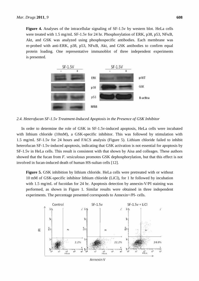
SF-1.5v affects NFB, Akt-GSK-3 and MAPK (ERK and p38) pathways by western blot.
The heterofucan SF-1.5v did not affect phophorylation of ERK, p38 and NFB proteins. On the
other hand, SF-1.5v induces dephosphorylation (activation) of glycogen synthase kinase-beta
(GSK-3) (Figure 4). Since GSK is involved in activation of p53 [18], we also evaluated the
phosphorylation of this protein. However, it was not affected by SF-1.5v (Figure 4). GSK
Dephosphorylation is involved in apoptosis [19], indicating that SF-1.5v likely induces cell death,
mainly by GSK activation.
A B
Mar. Drugs 2011, 9
608
Figure 4. Analyses of the intracellular signaling of SF-1.5v by western blot. HeLa cells
were treated with 1.5 mg/mL SF-1.5v for 24 hr. Phosphorylation of ERK, p38, p53, NFB,
Akt, and GSK was analyzed using phosphospecific antibodies. Each membrane was
re-probed with anti-ERK, p38, p53, NFB, Akt, and GSK antibodies to confirm equal
protein loading. One representative immunoblot of three independent experiments
is presented.
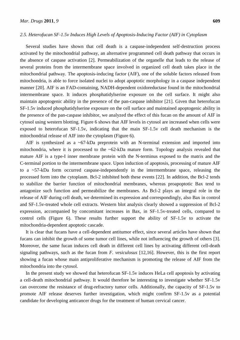
2.4. Heterofucan SF-1.5v Treatment-Induced Apoptosis in the Presence of GSK Inhibitor
In order to determine the role of GSK in SF-1.5v-induced apoptosis, HeLa cells were incubated
with lithium chloride (10mM), a GSK-specific inhibitor. This was followed by stimulation with
1.5 mg/mL SF-1.5v for 24 hours and FACS analysis (Figure 5). Lithium chloride failed to inhibit
heterofucan SF-1.5v-induced apoptosis, indicating that GSK activation is not essential for apoptosis by
SF-1.5v in HeLa cells. This result is consistent with that shown by Aisa and colleages. These authors
showed that the fucan from F. vesiculosus promotes GSK dephosphorylation, but that this effect is not
involved in fucan-induced death of human HS-sultan cells [12].
Figure 5. GSK inhibition by lithium chloride. HeLa cells were pretreated with or without
10 mM of GSK-specific inhibitor lithium chloride (LiCl), for 1 hr followed by incubation
with 1.5 mg/mL of fucoidan for 24 hr. Apoptosis detection by annexin-V/PI staining was
performed, as shown in Figure 1. Similar results were obtained in three independent
experiments. The percentage presented corresponds to Annexin+/PI- cells.
Mar. Drugs 2011, 9
609
2.5. Heterofucan SF-1.5v Induces High Levels of Apoptosis-Inducing Factor (AIF) in Cytoplasm
Several studies have shown that cell death is a caspase-independent self-destruction process
activated by the mitochondrial pathway, an alternative programmed cell death pathway that occurs in
the absence of caspase activation [2]. Permeabilization of the organelle that leads to the release of
several proteins from the intermembrane space involved in organized cell death takes place in the
mitochondrial pathway. The apoptosis-inducing factor (AIF), one of the soluble factors released from
mitochondria, is able to force isolated nuclei to adopt apoptotic morphology in a caspase independent
manner [20]. AIF is an FAD-containing, NADH-dependent oxidoreductase found in the mitochondrial
intermembrane space. It induces phosphatidylserine exposure on the cell surface. It might also
maintain apoptogenic ability in the presence of the pan-caspase inhibitor [21]. Given that heterofucan
SF-1.5v induced phosphatidylserine exposure on the cell surface and maintained apoptogenic ability in
the presence of the pan-caspase inhibitor, we analyzed the effect of this fucan on the amount of AIF in
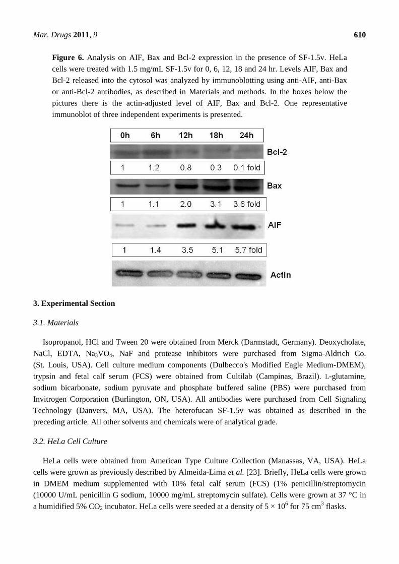
cytosol using western blotting. Figure 6 shows that AIF levels in cytosol are increased when cells were
exposed to heterofucan SF-1.5v, indicating that the main SF-1.5v cell death mechanism is the
mitochondrial release of AIF into the cytoplasm (Figure 6).
AIF is synthesized as a ~67-kDa preprotein with an N-terminal extension and imported into
mitochondria, where it is processed to the ~62-kDa mature form. Topology analysis revealed that
mature AIF is a type-I inner membrane protein with the N-terminus exposed to the matrix and the
C-terminal portion to the intermembrane space. Upon induction of apoptosis, processing of mature AIF
to a ~57-kDa form occurred caspase-independently in the intermembrane space, releasing the
processed form into the cytoplasm. Bcl-2 inhibited both these events [22]. In addition, the Bcl-2 tends
to stabilize the barrier function of mitochondrial membranes, whereas proapoptotic Bax tend to
antagonize such function and permeabilize the membranes. As Bcl-2 plays an integral role in the
release of AIF during cell death, we determined its expression and correspondingly, also Bax in control
and SF-1.5v-treated whole cell extracts. Western blot analysis clearly showed a suppression of Bcl-2
expression, accompanied by concomitant increases in Bax, in SF-1.5v-treated cells, compared to
control cells (Figure 6). These results further support the ability of SF-1.5v to activate the
mitochondria-dependent apoptotic cascade.
It is clear that fucans have a cell-dependent antitumor effect, since several articles have shown that
fucans can inhibit the growth of some tumor cell lines, while not influencing the growth of others [3].
Moreover, the same fucan induces cell death in different cell lines by activating different cell-death
signaling pathways, such as the fucan from F. vesiculosus [12,16]. However, this is the first report
showing a fucan whose main antiproliferative mechanism is promoting the release of AIF from the
mitochondria into the cytosol.
In the present study we showed that heterofucan SF-1.5v induces HeLa cell apoptosis by activating
a cell-death mitochondrial pathway. It would therefore be interesting to investigate whether SF-1.5v
can overcome the resistance of drug-refractory tumor cells. Additionally, the capacity of SF-1.5v to
promote AIF release deserves further investigation, which might confirm SF-1.5v as a potential
candidate for developing anticancer drugs for the treatment of human cervical cancer.
Mar. Drugs 2011, 9
610
Figure 6. Analysis on AIF, Bax and Bcl-2 expression in the presence of SF-1.5v. HeLa
cells were treated with 1.5 mg/mL SF-1.5v for 0, 6, 12, 18 and 24 hr. Levels AIF, Bax and
Bcl-2 released into the cytosol was analyzed by immunoblotting using anti-AIF, anti-Bax
or anti-Bcl-2 antibodies, as described in Materials and methods. In the boxes below the
pictures there is the actin-adjusted level of AIF, Bax and Bcl-2. One representative
immunoblot of three independent experiments is presented.
3. Experimental Section
3.1. Materials
Isopropanol, HCl and Tween 20 were obtained from Merck (Darmstadt, Germany). Deoxycholate,
NaCl, EDTA, Na3VO4, NaF and protease inhibitors were purchased from Sigma-Aldrich Co.
(St. Louis, USA). Cell culture medium components (Dulbecco's Modified Eagle Medium-DMEM),
trypsin and fetal calf serum (FCS) were obtained from Cultilab (Campinas, Brazil). L-glutamine,
sodium bicarbonate, sodium pyruvate and phosphate buffered saline (PBS) were purchased from
Invitrogen Corporation (Burlington, ON, USA). All antibodies were purchased from Cell Signaling
Technology (Danvers, MA, USA). The heterofucan SF-1.5v was obtained as described in the
preceding article. All other solvents and chemicals were of analytical grade.
3.2. HeLa Cell Culture
HeLa cells were obtained from American Type Culture Collection (Manassas, VA, USA). HeLa
cells were grown as previously described by Almeida-Lima et al. [23]. Briefly, HeLa cells were grown
in DMEM medium supplemented with 10% fetal calf serum (FCS) (1% penicillin/streptomycin
(10000 U/mL penicillin G sodium, 10000 mg/mL streptomycin sulfate). Cells were grown at 37 °C in
a humidified 5% CO2 incubator. HeLa cells were seeded at a density of 5 × 10⁶ for 75 cm
3 flasks.
Mar. Drugs 2011, 9
611
3.3. Antiproliferative Activity
Antiproliferative activity of SF-1.5v was determined as previously described by Amoli et al. [24].
Briefly, HeLa cells were grown in 75 cm3 flasks in DMEM medium plus 10% FCS. Cells were seeded
into 96-well plates at a density of 5 × 10³
cells/well and allowed to attach overnight in 300 μL medium
FCS free incubated at 37 ºC, 5% CO2. The medium was then removed and 300 μL of medium/ plus
FCS was added, followed by heterofucan SF-1.5v at a final concentration of 0.1; 0.5; 1.0; 1.5 and
2.0 mg/mL. Cells growing under these conditions for 24 h, 48 h and 72 h at 37 ºC at 5% CO2. After
incubation, traces of SF-1.5v were removed by washing the cells twice with 200 μL PBS and applying
100 μL of fresh medium and 10 μL of 12 mM MTT dissolved in PBS to determine the effects of the
heterofucan on cell proliferation. Cells were then incubated for 4 h at 37 ºC, 5% CO2. To solubilize the
product of MTT cleavage, 100 μL of isopropanol containing 0.04 N HCl was added to each well and
thoroughly mixed using a multichannel pipettor. Within 1 h of HCl-isopropanol addition, absorbance
at 570 nm was read using a Multiskan Ascent Microplate Reader (Thermo Labsystems, Franklin, MA,
USA). The percent inhibition of cell proliferation was calculated as follows:
% Inhibition = Abs. 570 nm Control − Abs. 570 nm sample × 100
Abs. 570 nm Control
3.4. Apoptosis Assay
The apoptotic status of HeLa cells was evaluated by measuring the exposure of phosphatidylserine
on cell membranes using annexin V-fluorescein isothiocyanate (annexin V-FITC) and propidium
iodide (PI) staining. A BD Pharmingen Annexin V-FITC Apoptosis Detection Kit (BD Biosciences,
Franklin Lakes, NJ) was used for the apoptosis assay. HeLa cells were placed in a 24-well plate
(1 × 10⁶ cells/mL), and after 24 h of incubation, cells were treated with SF-1.5v for 24h and then
harvested. After centrifugation, cell pellets were washed twice with cold phosphate-buffered saline
(PBS: 137 mM NaCl, 2.7 mM KCl, 10 mM Na2HPO4, pH 7.4) and suspended in 100 μL of 1 × binding
buffer (10 mM HEPES/NaOH, 140 m M NaCl, 2.5 mM CaCl2, pH 7.4). Cells were then incubated
with 5 μL of annexin V-FITC and 10 μL of PI at room temperature for 15 min in the dark. After
incubation, 400 μL of 1 × binding buffer was added to each tube. The cells were immediately analyzed
by FACSCalibur flow cytometry (Becton Dickinson, USA).
3.5. Western Blotting
HeLa cells at 80% confluence were incubated with SF-1.5v and washed after 24 h in ice-cold PBS
and scraped into 200 mL lysis buffer [50 mM Tris-HCl (pH 7.4), 1% Tween 20, 0.25% sodium
deoxycholate, 150 mM NaCl, 1 mM EDTA, 1 mM Na3VO4, 1 mM NaF, and protease inhibitors
(1 mg/mL aprotinin, 10 mg/mL leupeptin and 1 mM 4-(2-aminoethyl) benzenesulfonyl fluoride] for
2 h in ice. Protein extracts were cleared by centrifugation and protein concentrations were determined
using BCA protein assay kit (Pierce, USA) with bovine serum albumin as standard. An equal volume
of sodium dodecyl sulfate (SDS) gel loading buffer [100 mM Tris-HCl (pH 6.8), 200 mM
dithiothreitol (DTT), 4% SDS, 0.1% bromophenol blue and 20% glycerol] was added to samples,
Mar. Drugs 2011, 9
612
which were subsequently boiled for 10 min. From each sample, 50 mg of protein was loaded onto
SDS-PAGE and blotted onto PVDF membranes (Millipore, Bedford, MA, USA). Membranes were
blocked in 1% fat-free dried milk or 2% bovine serum albumin in Tris-buffered saline (TBS) with
0.05% Tween 20 (TBST) and incubated overnight at 4 °C with appropriate primary antibody at
1:1000 dilution. After washing in TBST, membranes were incubated with anti-rabbit horseradish
peroxidase-conjugated secondary antibodies, at 1:2000 dilution; in blocking buffer for 1 h. The
intensity of the specific immunoreactive bands were detected by enhanced chemiluminescence (ECL),
using the manufacturer's protocol (Kirkegared and Perry Laboratories) and quantified by densitometry
and expressed as a ratio to actin, as previously described [22].
3.6. Statistical Analysis
All data were expressed as mean ± standard deviation. Statistical analysis was done by one-way
Anova using the SIGMAStat 2.01 software. Student-Newmans-Keuls post-tests were performed for
multiple group comparison. In all cases statistical significance was set at p < 0.05.
4. Conclusions
Our studies demonstrate that heterofucan SF-1.5v inhibited growth of the Hela human uterine
adenocarcinoma cell line by inducing apoptosis using a mechanism independent of caspases activation.
SF-1.5v also induces GSK activation, but this protein is not involved in the heterofucan
SF-1.5v-induced apoptosis mechanism. SF-1.5v induces apoptosis mainly by inducing AIF release
from mitochondria into cytosol. These data support the hypothesis that SF-1.5v may have potential for
treating cervical cancer.
Acknowledgements
Research was supported by CAPES, MCT and CNPq, Brazil. RBG Câmara, N Dantas-Santos, J
Almeida-Lima, GP Fidelis, CBS Telles, RF Melo-Silveira and RM Oliveira thanks CNPq and CAPES
for fellowship support.
References
1. Scarinci, I.C.; Garcia, F.A.R.; Kobetz, E.; Partridge, E.E.; Brandt, H.M.; Bell, M.C.; Dignan, M.;
Ma, G.X.; Daye, J.L.; Castle, P.E. Cervical cancer prevention: new tools and old barriers. Cancer
2010, 116, 2531–2542.
2. Kroemer, G.; Galluzzi, L.; Vandenabeele, P.; Abrams, J.; Alnemri, E.S.; Baehrecke, E.H.;
Blagosklonny, M.V.; El-Deiry, W.S.; Golstein, P.; Green, D.R.; Hengartner, M.; Knight, R.A.;
Kumar, S.; Lipton, S.A.; Malorni, W.; Nuñez, G.; Peter, M.E.; Tschopp, J.; Yuan, J.; Piacentini,
M.; Zhivotovsky, B.; Melino, G. Classification of cell death: recommendations of the
Nomenclature Committee on Cell Death 2009. Cell Death Differ. 2009, 16, 3–11.
3. Li, B.; Lu, F.; Wei, X.; Zhao, R. Fucoidan: Structure and Bioactivity. Molecules 2008, 13,
1671–1695.
Mar. Drugs 2011, 9
613
4. Pomin, V.H. Review: an overview about the structure-function relationship of marine sulfated
homopolysaccharides with regular chemical structures. Biopolymers 2009, 91, 601–609.
5. Dietrich, C.P.; Farias, G.G.M.; Abreu, L.R.; Leite, E.L.; Silva, L.F.; Nader, H.B. A new approach
for the characterization of polysaccharides from algae: presence of four main acidic
polysaccharides in three species of the class Phaeophycea. Plant Sci. 1995, 108, 143–153.
6. Albuquerque, I.R.L.; Queiroz, K.C.S.; Alves, L.G.; Santos, E.A.; Leite, E.L.; Rocha, H.A.O.
Heterofucans from Dictyota menstrualis have anticoagulant activity. Braz. J. Med. Biol. Res.
2004, 37, 167–171.
7. Choi, J.I.; Raghavendran, H.R.B.; Sung, N.Y.; Kim, J.H.; Chun, B.S.; Ahn, D.H.; Choi, H.S.;
Kang, K.W.; Lee, J.W. Effect of fucoidan on aspirin-induced stomach ulceration in rats. Chem.
Biol. Interact. 2010, 183, 249–254.
8. Rocha, H.A.O.; Franco, C.R.C.; Trindade, E.S.; Carvalho, L.C.M.; Veiga, S.S.; Leite, E.L.;
Dietrich, C.P.; Nader, H.B. A fucan from the brown seaweed Spatoglossum schröederi inhibits
Chinese hamster ovary cell adhesion to several extracellular matrix proteins. Braz. J. Med. Biol.
Res. 2001, 34, 621–626.
9. Synytsya, A.; Kim, W.J.; Kim, S.M.; Pohl, R.; Synytsya, A.; Kvasnička F.; Čopíková, J.; Park,
Y.L. Structure and antitumour activity of fucoidan isolated from sporophyll of Korean brown
seaweed Undaria pinnatifida. Carbohydr. Polym. 2010, 81, 41–48.
10. Costa, L.S.; Fidelis, G.P.; Cordeiro, S.L.; Oliveira, R.M.; Sabry, D.A; Câmara, R.B.G.; Nobre,
L.T.D.B.; Costa, M.S.S.P.; Almeida-Lima, J.; Farias, E.H.C.; Leite, E.L.; Rocha, H.A.O.
Biological activities of sulfated polysaccharides from tropical seaweeds. Biomed. Pharmacother.
2010, 64, 21–28.
11. Kim, E.J.; Park, S.Y.; Lee, J.Y.; Park J.H.Y. Fucoidan present in brown algae induces apoptosis of
human colon cancer cells. BMC Gastroenterol. 2010, 10, 1–11.
12. Aisa, Y.; Miyakawa, Y.; Nakazato, T.; Shibata, H.; Saito, K.; Ikeda, Y.; Kizaki, M. Fucoidan
induces apoptosis of human HS-sultan cells accompanied by activation of caspase-3 and down-
regulation of ERK pathways. Am. J. Hematol. 2005, 78, 7–14.
13. Berteau, O.; Mulloy, B. Sulfated fucans, fresh perspectives: structures, functions, and biological
properties of sulfated fucans and an overview of enzymes active toward this class of
polysaccharide. Glycobiology 2003, 13, 29–40.
14. Yamamoto, I.; Takahashi, M.; Suzuki, T.; Seino, H.; Mori, H. Antitumor effect of seaweeds. IV.
Enhancement of antitumor activity by sulfation of a crude fucoidan fraction from Sargassum
kjellmanianum. Jpn. J. Exp. Med. 1984, 54, 143–151.
15. Stevan, F.R.; Oliveira, M.B.; Bucchi, D.F.; Noseda, M.; Iacomini, M.; Duarte, M.E. Cytotoxic
effects against HeLa cells of polysaccharides from seaweeds. J. Submicrosc. Cytol. Pathol. 2001,
33, 477–484.
16. Hyun, J.H.; Kim, S.C.; Kang, J.I.; Kim, M.K.; Boo, H.J.; Kwon, J.M.; Koh, Y.S.; Hyun, J.W.;
Park, D.B.; Yoo, E.S.; Kang, H.K. Apoptosis inducing activity of fucoidan in HCT-15 colon
carcinoma cells. Biol. Pharm. Bull. 2009, 32, 1760–1764.
17. Nakamura, T.; Suzuki, H.; Wada, Y.; Kodama, T.; Doi, T. Fucoidan induces nitric oxide
production via p38 mitogen-activated protein kinase and NF-κB-dependent signaling pathways
through macrophage scavenger receptors. Biochem. Biophys. Res. Commun. 2006, 343, 286–294.
Mar. Drugs 2011, 9
614
18. King, T.D.; Bijur, G.N.; Jope, R.S. Caspase-3 activation induced by inhibition of mitochondrial
complex I is facilitated by glycogen synthase kinase-3beta and attenuated by lithium. Brain Res.
2001, 919, 106–114.
19. Arboleda, G.; Cárdenas, Y.; Rodríguez, Y.; Morales, L.C.; Matheus, L.; Arboleda, H. Differential
regulation of AKT, MAPK and GSK3 during C2-ceramide-induced neuronal death.
Neurotoxicology 2010, 31, 687–693.
20. Norberg, E.; Orrenius, S.; Zhivotovsky, B. Mitochondrial regulation of cell death: Processing of
apoptosis-inducing factor (AIF). Biochem. Biophys. Res. Commun. 2010, 396, 95–100.
21. Hangen, E.; Blomgren, K.; Bénit, P.; Kroemer, G.; Modjtahedi, N. Life with or without AIF.
Trends Biochem. Sci. 2010, 35, 278–287.
22. Selvakumar E.; Hsieh T. Regulation of cell cycle transition and induction of apoptosis in HL-60
leukemia cells by lipoic acid: role in cancer prevention and therapy. J. Hematol. Oncol. 2008, 1,
1–8.
23. Almeida-Lima, J.; Costa, L.S.; Silva, N.B.; Melo-Silveira, R.F.; Silva, F.V.; Felipe, M.B.M.C.;
Medeiros, S.R.B.M.; Leite, E.L.; Rocha, H.A.O. Evaluating the possible genotoxic, mutagenic and
tumor cell proliferation-inhibition effects of a non-anticoagulant, but antithrombotic algal
heterofucan. J. Appl. Toxicol. 2010, 30, 708–715.
24. Amoli, J.S.; Sadighara, P.; Barin, A.; Yazdani, A; Satari, S. Biological screening of Amaranthus
retroflexus L. (Amaranthaceae). Rev. Bras. Farmacogn. 2009, 19, 617–620.
© 2011 by the authors; licensee MDPI, Basel, Switzerland. This article is an open access article
distributed under the terms and conditions of the Creative Commons Attribution license
(http://creativecommons.org/licenses/by/3.0/).