sargassum polyceratium (phaeophyceae, fucaceae) surface
TRANSCRIPT
HAL Id: hal-00746082https://hal.univ-antilles.fr/hal-00746082
Submitted on 26 Oct 2012
HAL is a multi-disciplinary open accessarchive for the deposit and dissemination of sci-entific research documents, whether they are pub-lished or not. The documents may come fromteaching and research institutions in France orabroad, or from public or private research centers.
L’archive ouverte pluridisciplinaire HAL, estdestinée au dépôt et à la diffusion de documentsscientifiques de niveau recherche, publiés ou non,émanant des établissements d’enseignement et derecherche français ou étrangers, des laboratoirespublics ou privés.
Sargassum polyceratium (Phaeophyceae, Fucaceae)surface molecule activity towards fouling organisms and
embryonic development of benthic speciesMarie Thabard, Olivier Gros, Claire Hellio, Jean-Philippe Maréchal
To cite this version:Marie Thabard, Olivier Gros, Claire Hellio, Jean-Philippe Maréchal. Sargassum polyceratium (Phaeo-phyceae, Fucaceae) surface molecule activity towards fouling organisms and embryonic developmentof benthic species. Botanica Marina, De Gruyter, 2011, 54 (2), pp.147-157. �10.1515/BOT.2011.014�.�hal-00746082�
Botanica Marina 54 (2011): 147–157 � 2011 by Walter de Gruyter • Berlin • New York. DOI 10.1515/BOT.2011.014
2010/056
Article in press - uncorrected proof
Sargassum polyceratium (Phaeophyceae, Fucaceae) surface
molecule activity towards fouling organisms and embryonic
development of benthic species
Marie Thabard1,2,*, Olivier Gros3, Claire Hellio2 and
Jean-Philippe Marechal1
1 Observatoire du Milieu Marin Martiniquais (OMMM),
3 avenue Condorcet, 97200 Fort de France, Martinique,
French West Indies, e-mail: [email protected] School of Biological Sciences, King Henry Building,
University of Portsmouth, Portsmouth PO1 2DY, UK3 UMR-CNRS 7138 Systematique-Adaptation-Evolution,
equipe ‘‘Biologie de la mangrove’’, Universite des Antilles
et de la Guyane, UFR des Sciences Exactes et Naturelles,
departement de Biologie, B.P. 592. 97159 Pointe-a-Pitre
Cedex, Guadeloupe, French West Indies
* Corresponding author
Abstract
Coral reefs have undergone profound ecological changes
over recent decades. Areas formerly covered by scleractinian
coral species are now often overgrown by macroalgae. In
Martinique (West Indies), this phenomenon has lead to the
colonisation of numerous coral reefs by algae, amongst
which Sargassum is one of the most prominent. This study
focuses on potential defence molecules produced by Sargas-
sum polyceratium. The hexane dipping method was
employed to extract surface molecules on fresh material, and
their bioactivities were assessed against bacteria (marine and
estuarine), and marine tropical invertebrates wan annelid
(Pseudonereis sp.), a bivalve (Codakia orbicularis) and a sea
urchin (Diadema antillarum)x. Extracts were active against
all microorganisms tested (MICs150 or 300 mg ml-1), early
stages of development in Pseudonereis sp. (MICs100 mg
ml-1) and embryos of C. orbicularis and D. antillarum
(MICs5 mg ml-1), suggesting the production of defence
compounds by S. polyceratium.
Keywords: bacteria; embryonic development; hexane
dipping; Sargassum polyceratium; toxicity.
Introduction
Studies on production of marine natural products (MNPs)
and their activities have increased over the past 20 years.
Particular attention has been given to the production of sec-
ondary metabolites by macroalgae, microalgae, invertebrates,
Cyanobacteria, octocorals, sponges and ascidians (Hay and
Fenical 1996, Paul and Puglisi 2004, Paul et al. 2007, Hellio
et al. 2009). Algae produce secondary metabolites with a
wide range of biological activities including antifungal, anti-
bacterial, antibiotic, antifouling (AF), UV radiation protec-
tion, feeding deterrence, inhibition of competitors, gamete
attraction and inhibition of larval settlement and develop-
ment (Hay and Fenical 1988, Paul et al. 1988, Hay and Feni-
cal 1996, Hellio et al. 2001, 2002, 2005, Steinberg and de
Nys 2002, Birrell 2003, Paul and Puglisi 2004, Paul et al.
2007, Plouguerne et al. 2010a).
Several examples demonstrate that specific structures loca-
lised on the surfaces of algae release active compounds into
the environment (Dworjanyn et al. 1999, De Nys and Stein-
berg 2002); Vesicular physodes located on the surfaces of
phaeophyceans contain phlorotannins (Ragan and Glombitza
1986). The role of phlorotannins as macroalgal defence
mechanism has been extensively described in the literature,
but yet remains not fully understood. These compounds have
been described as UV screens, herbivore deterrents and anti-
microbial agents, but are also known to play a role in primary
metabolism (Paul and Puglisi 2004). Moreover, fluorescence
microscopy, combined with chemical analyses, has demon-
strated that Delisea pulchra (Grev.) Mont. (Rhodophyceae)
releases AF compounds (halogenated furanones) from gland
cells located on its surface (Dworjanyn et al. 1999, De Nys
and Steinberg 2002).
The ‘‘corps en cerise’’ found on cortical cells of Lauren-
cia snyderae E.Y. Dawon (Rhodophyceae) may be a primary
location of halogenated MNPs (Young et al. 1980). However,
even though it has been stated that the ‘‘corps en cerise’’
are main reserves for halogenated compounds in the alga
Laurencia obtusa Lamour and store high concentrations of
bromine and chlorine (Salgado et al. 2008), they are not
found at the surface of the alga and no structure linking them
to the algal surface have been found (De Nys et al. 1998).
Sargassum species have been extensively analysed for
their allelochemical activities for several reasons: 1) some of
them are lightly fouled in the field, 2) the genus is one of
the most conspicuous algae in numerous areas, especially the
tropics (Ang 1986, De Ruyter van Stevenick and Breeman
1987, Littler et al. 1993, Lapointe 1997, Engelen et al. 2001),
3) some species have invaded many parts of the world (Plou-
guerne et al. 2006). Antibacterial, anti-algal, anti-fungal and
anti-invertebrate activities have been demonstrated in S.
muticum (Yendo) Fensholt samples (Hellio et al. 2002, Plou-
guerne et al. 2008, 2010b, Marechal and Hellio 2011), S.
wightii Greville ex J. Agardh and S. johnshonii V.J. Chapman
(Sastry and Rao 1994).
AUTHOR’S COPY | AUTORENEXEMPLAR
AUTHOR’S COPY | AUTORENEXEMPLAR
148 M. Thabard et al.: Sargassum polyceratium surface molecule bioactivity
Article in press - uncorrected proof
Sieburth and Conover (1965) found antibiotic activity in
phlorotannins extracted from S. vestitum (R. Brown ex Turn-
er) C. Agardh and S. natans (Linnaeus) Gaillon, and Tanaka
and Asakawa (1988) found antialgal activity in extracts from
S. horneri (Turner) C. Agardh. S. vulgare C. Agardh extracts
from Brazil had AF activity against microalgae and mussel
settlement (Plouguerne et al. 2010a). S. muricatum and S.
tenerrimum J. Agardh induce modifications of swimming
activity in two-day-old larvae of Platygyra daedalea Ellis et
Solander (coral) and decrease their proportional settlement,
indicating chemical defences (Diaz-Pulido et al. 2010).
Sargassum polyceratium W.R. Taylor, one of the most
abundant macroalgae of Martinique tropical reefs (M. Tha-
bard, unpublished data), has, however, never been investi-
gated. Sargassum species have been recorded since the 1970s
on the Atlantic coast of the island (Battistini 1978). Coral
reef colonisation by macroalgae has induced significant
changes in the structure and diversity of communities. As an
example, S. polyceratium and S. hystrix J. Agardh have sup-
planted hermatypic coral species on the Jamaican north coast
(Lapointe 1997, Lapointe and Thacker 2002). Algal canopies
are known to affect understorey species (Eckman and Dug-
gins 1991) and may interfere with invertebrate larval recruit-
ment processes (Pawlik 1992, Birrell 2003, Titlyanov et al.
2005). To survive the environmental pressures they are sub-
jected to, and to successfully colonise new areas, macroalgae
have developed several strategic mechanisms, including both
specific morphological characteristics and production of
deterrents to avoid epibiont overgrowth (Littler and Littler
1980, Littler et al. 1983a,b, Paul and Puglisi 2004).
In a survey conducted in Martinique in 2007 (M. Thabard
unpublished data), the sea urchin Diadema antillarum Phi-
lippi was found to be almost absent from the Sargassum area,
but a sea urchin population had developed nearby, forming
distinct belts. D. antillarum, which is considered as a key-
stone herbivore species on Caribbean coral reefs (Knowlton
2001), suffered mass mortality in 1983 in this region (Lessios
et al. 1983, 1984). This induced profound changes on coral
reefs, and is thought to have played a major role in the com-
munity composition shift observed nowadays in the Carib-
bean due to reduced grazing pressure on macroalgae (Hughes
1994). Diadema recovery is proving to be a very slow proc-
ess in the Caribbean although its reproductive biology seems
to indicate that the species should recover rapidly (Lessios
1995). There are several hypotheses (too few adults, patho-
gens remaining in water, etc.) for this phenomenon (Lessios
2005). It may be that fast colonisation by Sargassum sp. on
coral reefs in the 1980s (Littler et al. 1993) is in part respon-
sible for this slow recovery process as it is possible that the
seaweed prevents the larval recruitment process.
The present study focuses on potential antibacterial and
deterrent activities of Sargassum polyceratium and their pos-
sible interactions with other tropical species. In order to per-
form a broad spectrum analysis of the toxic activity of MNPs
extracted from S. polyceratium surface, three marine tropical
invertebrates were tested, one sea urchin, one bivalve and
one worm, representing organisms from different phyla. In
vitro effects of S. polyceratium extracts towards bacteria and
embryos of marine invertebrates were investigated.
Materials and methods
Algal collection site
Martinique is a volcanic island located in the eastern Car-
ibbean Sea (Lesser Antilles) between latitudes 148509N and
148239N and at mean longitude of 628129W. It has a land
area of 1128 km2 and is bordered by the Caribbean Sea to
the west and the Atlantic Ocean to the east. The climate is
defined by distinct dry and wet periods, the dry season last-
ing from December to July and the wet season from August
to early December.
The site chosen for this study is a fringing coral reef to
the south of Martinique, close to the mouth of Trois Rivieres
river. This site was chosen for the dominant presence of Sar-
gassum polyceratium.
Study organism
Sargassum polyceratium is commonly found in moderately
turbulent habitats from the lower intertidal zone to depths
over 50 m (Littler and Littler 2000, Engelen 2004). It has a
tough crowded thallus and dense branches reaching to
100 cm. The main axes roughened with small spines may be
numerous. The blades measure 3–8 mm in width and
1.5–2.0 cm in length. The holdfast is strong and disc like.
The importance of recruitment vs. regeneration was modeled
and demonstrated to vary with population, year and distur-
bance (Engelen et al. 2005).
Algal extractions
Sargassum polyceratium samples were collected in October
2008 and January 2009 (wet and dry seasons). Thalli were
removed with their holdfasts by breaking away pieces of
substratum, thus reducing stress on the algae; stress may be
responsible for secondary metabolite production. The sam-
ples were collected by SCUBA diving at 18 m depth. The
algae were cleaned of epiphytes, rinsed with seawater (SW)
and transported to the laboratory in a container filled with
clean SW. The fresh samples were soaked in hexane (Fisher,
Loughborough, UK) in the ratio 1 l hexane kg-1 wet weight
S. polyceratium (De Nys et al. 1998). All the extractions
were performed in the dark as some secondary metabolites,
such as the polyphenolics are known to react to light. The
goal of using the hexane dipping method was to extract mol-
ecules produced both by the alga at its surface and by its
associated biofilm in order to obtain the whole range of
MNPs that might interact with eukaryotic and/or prokaryotic
organisms present in the algal environment.
Two protocols were used:
• Protocol A (October 2008 samples, rainy season), Sar-
gassum polyceratium thalli were dipped for 30 s in hex-
ane for preliminary tests.
• Protocol B (January 2009 samples, dry season) was devel-
oped based on results of both tests conducted on 30 s
extracts (October) and the observations of algal surfaces
for breaks (see results). This amended protocol was used
to test for the effect of dipping time on extract efficiency.
AUTHOR’S COPY | AUTORENEXEMPLAR
AUTHOR’S COPY | AUTORENEXEMPLAR
M. Thabard et al.: Sargassum polyceratium surface molecule bioactivity 149
Article in press - uncorrected proof
This was done by dipping algae in hexane for two dif-
ferent periods (10 s and 30 s), with the algal surface
remaining intact.
Observation of algal surface
The objective of our experiments was to select the molecules
present at the surface of Sargassum polyceratium only. The
hexane dipping method was shown to break some algal sur-
face cells when thalli were dipped for more than 30 s (De
Nys et al. 1998). The algal surface was therefore checked
for breaks that would result in leaks of cell contents in thalli
dipped for 30 s, 10 min and 30 min to determine the best
dipping time. As UV excitation of plant leaves is known to
induce two distinct types of fluorescence, damage caused to
the surface cells of S. polyceratium by hexane dipping was
investigated by epifluorescent microscopy (Nikon, Tokyo,
Japan, Eclipse 80i microscope; Filter FITC 494 nm excita-
tion/514 nm emission; Nikon Dxm1200F camera) using a
protocol adapted from Cerocic et al. (1999).
Bioassays
Bacteria Culture of bacteria: Four marine bacterial strains
were used: Halomonas marina (Cobet et al.) Dobson et
Franzmann (ATCC 25374), Pseudoalteromonas elyakovii
(Ivanova et al.) Sawabe et al. (ATCC 700519), Polaribacter
irgensii Gosink et al. (ATCC 700398) and Vibrio aestuaria-
nus Tilson et Seidler (ATCC 35048). These bacteria were
chosen because they are typical marine fouling bacteria
(Plouguerne et al. 2010b). Pseudoalteromonas elyakovii and
Vibrio aestuarianus are also known to cause infections in
marine organisms, such as molluscs, crustacean, fishes or
algae, the latter being thought responsible for the summer
mortality of Crassostrea gigas Thunberg (Labreuche et al.
2005). Marine bacteria were cultivated with marine broth
(5% tryptone, Oxoid, Basingstoke, UK, diluted in SW) and
incubated at 308C to allow development (Plouguerne et al.
2008).
Five terrestrial bacterial strains known to be present in
estuaries and coastal environment (Mokrini et al. 2008) were
used: Bacillus subtilis (Ehrenberg) Cohn (NCIMB 1026),
Enterobacter aerogenes Hormaeche et Edwards (ATCC
13048), Escherichia coli (Migula) Castellani et Chalmers (B
81), Pseudomonas aeruginosa (Schroeter) Migula (NCIMB
10390) and Staphylococcus aureus Rosenbach (NCIMB
8625). B. subtilis and S. aureus are Gram-positive bacteria.
The remainder are Gram-negative. Terrestrial bacteria were
cultivated on a nutrient broth (CM0067, No. 2, Nutrient
media Powder Oxoid 25 g l-1) and incubated at 308C. Bio-
logical activities of extracts were evaluated following the
method of Amsterdam (1996).
Antibacterial assays: Aliquots of 100 ml of each hexane
extract were poured in six wells of 96 well plates (Fisher)
for each bacterial assay following the protocols of Plou-
guerne et al. (2010b). The solutions were tested at three con-
centrations: 15, 150 and 300 mg ml-1. In addition, six wells
free of extracts and six wells containing hexane were used
as controls. The plates were first dried in a flow cabinet to
evaporate the solvent and then left for 15 min in a UV cab-
inet for sterilization.
The optical densities (OD) of bacterial stock cultures were
measured at 630 nm for every sample to determine the quan-
tity of solution required to obtain 1 mOD (mili optical den-
sity). Then, 100 ml of bacterial solutions were added under
aseptic conditions and the plates were incubated for 48 h at
308C for bacterial growth. Activity was obtained comparing
the controls and the wells containing the extracts. Solutions
were considered to be active when bacteria did not grow in
four, five or six wells. Bacterial growth was noted by the
presence of a cloudy solution. One plate was used for each
strain to limit the cross-contamination risk.
Invertebrates Organisms: The toxicity of the extracts
was tested against larvae of Codakia orbicularis L., Diadema
antillarum and Pseudonereis sp. These organisms are tropical
and represent typical species from three marine ecosystems
(seagrass bed, reef and mangrove). Their spawning and early
larval development has been described previously (Gros et
al. 1997, Eckert 1998). For both C. orbicularis and D. anti-
llarum, spawns were induced in the laboratory under con-
trolled conditions, while Pseudonereis sp. embryos were
collected from the wild.
Codakia orbicularis is a tropical bivalve mollusc distrib-
uted from Florida to Brazil (Abbott 1974). Adult C. orbi-
cularis (between 40 and 60 mm shell length) were collected
by hand from seagrass beds in Ilet Cochon (Guadeloupe,
Figure 1B) in July 2009. Fertilization was induced following
the method described by Gros et al. (1997). Adults were
cleaned with a brush and spawning was induced by injection
of 0.3 ml of a 4 mM serotonin solution in 0.22 mm filtered
SW into the visceral mass. Sperm and oocytes were mixed
in a 1 l cylinder until the appearance of two-cell embryos.
Fertilization occurred under constant aeration to hold eggs
in suspension as they are slightly negatively buoyant. C.
orbicularis embryonic development follows the general
development of bivalves (Gros et al. 1997). Appearance of
the first polar body (indicating fertilization) is not always
visible under a dissecting microscope, thus the two cells
embryos were chosen to ensure the fertilization had occurred,
and these were used in toxicity experiments.
Diadema antillarum: The black spined sea urchin D. anti-
llarum was selected for experiments. Adults were collected
on the shore at Port-Louis (Guadeloupe, Figure 1B) during
summer 2009. Urchins were acclimated in the laboratory
(258C) for a week and fed on agar pellets containing a mix-
ture of algae (including Ulva lactuca L. and Sargassum sp.)
following the protocol of Pereira et al. (2003). After a week,
the urchins were transferred to another tank containing 298C
filtered SW. Thermal shocks (3–58C) that induce spawning
of D. antillarum (M. Moe, personal communication) were
applied over a few minutes. Both male and female gametes
were pipetted from this tank and diluted in 10 l of 0.22 mm
filtered SW (258C) to induce the fertilization. The eggs are
slightly negatively buoyant, thus aeration was used to keep
them suspended. Embryos at the two cells stage wfertilization
(To)q1hx were chosen for experiments.
AUTHOR’S COPY | AUTORENEXEMPLAR
AUTHOR’S COPY | AUTORENEXEMPLAR
150 M. Thabard et al.: Sargassum polyceratium surface molecule bioactivity
Article in press - uncorrected proof
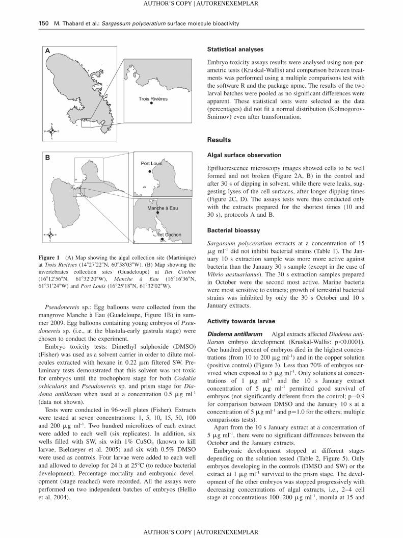
Figure 1 (A) Map showing the algal collection site (Martinique)
at Trois Rivieres (148279220N, 608589030W). (B) Map showing the
invertebrates collection sites (Guadeloupe) at Ilet Cochon
(168129560N, 618329200W), Manche a Eau (168169360N,
618319240W) and Port Louis (168259180N, 618329020W).
Pseudonereis sp.: Egg balloons were collected from the
mangrove Manche a Eau (Guadeloupe, Figure 1B) in sum-
mer 2009. Egg balloons containing young embryos of Pseu-
donereis sp. (i.e., at the blastula-early gastrula stage) were
chosen to conduct the experiment.
Embryo toxicity tests: Dimethyl sulphoxide (DMSO)
(Fisher) was used as a solvent carrier in order to dilute mol-
ecules extracted with hexane in 0.22 mm filtered SW. Pre-
liminary tests demonstrated that this solvent was not toxic
for embryos until the trochophore stage for both Codakia
orbicularis and Pseudonereis sp. and prism stage for Dia-
dema antillarum when used at a concentration 0.5 mg ml-1
(data not shown).
Tests were conducted in 96-well plates (Fisher). Extracts
were tested at seven concentrations: 1, 5, 10, 15, 50, 100
and 200 mg ml-1. Two hundred microlitres of each extract
were added to each well (six replicates). In addition, six
wells filled with SW, six with 1% CuSO4 (known to kill
larvae, Bielmeyer et al. 2005) and six with 0.5% DMSO
were used as controls. Four larvae were added to each well
and allowed to develop for 24 h at 258C (to reduce bacterial
development). Percentage mortality and embryonic devel-
opment (stage reached) were recorded. All the assays were
performed on two independent batches of embryos (Hellio
et al. 2004).
Statistical analyses
Embryo toxicity assays results were analysed using non-par-
ametric tests (Kruskal-Wallis) and comparison between treat-
ments was performed using a multiple comparisons test with
the software R and the package npmc. The results of the two
larval batches were pooled as no significant differences were
apparent. These statistical tests were selected as the data
(percentages) did not fit a normal distribution (Kolmogorov-
Smirnov) even after transformation.
Results
Algal surface observation
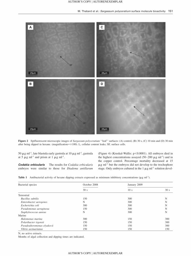
Epifluorescence microscopy images showed cells to be well
formed and not broken (Figure 2A, B) in the control and
after 30 s of dipping in solvent, while there were leaks, sug-
gesting lyses of the cell surfaces, after longer dipping times
(Figure 2C, D). The assays tests were thus conducted only
with the extracts prepared for the shortest times (10 and
30 s), protocols A and B.
Bacterial bioassay
Sargassum polyceratium extracts at a concentration of 15
mg ml-1 did not inhibit bacterial strains (Table 1). The Jan-
uary 10 s extraction sample was more more active against
bacteria than the January 30 s sample (except in the case of
Vibrio aestuarianus). The 30 s extraction samples prepared
in October were the second most active. Marine bacteria
were most sensitive to extracts; growth of terrestrial bacterial
strains was inhibited by only the 30 s October and 10 s
January extracts.
Activity towards larvae
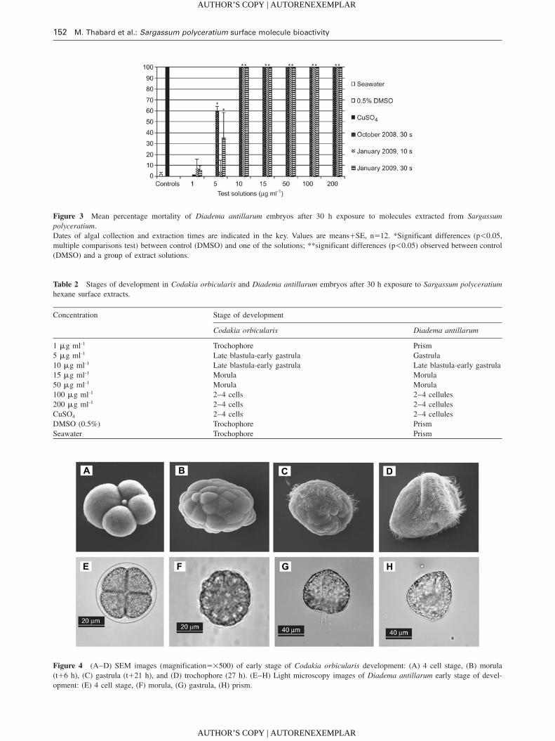
Diadema antillarum Algal extracts affected Diadema anti-
llarum embryo development (Kruskal-Wallis: p-0.0001).
One hundred percent of embryos died in the highest concen-
trations (from 10 to 200 mg ml-1) and in the copper solution
(positive control) (Figure 3). Less than 70% of embryos sur-
vived when exposed to 5 mg ml-1. Only solutions at concen-
trations of 1 mg ml-1 and the 10 s January extract
concentration of 5 mg ml-1 permitted good survival of
embryos (not significantly different from the control; ps0.9
for comparison between DMSO and the January 10 s at a
concentration of 5 mg ml-1 and ps1.0 for the others; multiple
comparisons tests).
Apart from the 10 s January extract at a concentration of
5 mg ml-1, there were no significant differences between the
October and the January extracts.
Embryonic development stopped at different stages
depending on the solution tested (Table 2, Figure 5). Only
embryos developing in the controls (DMSO and SW) or the
extract at 1 mg ml-1 survived to the prism stage. The devel-
opment of the other embryos was stopped progressively with
decreasing concentrations of algal extracts, i.e., 2–4 cell
stage at concentrations 100–200 mg ml-1, morula at 15 and
AUTHOR’S COPY | AUTORENEXEMPLAR
AUTHOR’S COPY | AUTORENEXEMPLAR
M. Thabard et al.: Sargassum polyceratium surface molecule bioactivity 151
Article in press - uncorrected proof
Figure 2 Epifluorescent microscopic images of Sargassum polyceratium ‘‘leaf’’ surfaces: (A) control, (B) 30 s, (C) 10 min and (D) 30 min
after being dipped in hexane. (magnifications=100). L, cellular content leaks; SF, surface cells.
Table 1 Antibacterial activity of hexane dipping extracts expressed as minimum inhibitory concentrations (mg ml-1).
Bacterial species October 2008 January 2009
30 s 10 s 30 s
Terrestrial
Bacillus subtilis 150 300 N
Enterobacter aerogenes N 300 N
Escherichia coli 300 300 N
Pseudomonas aeruginosa 150 300 N
Staphylococcus aureus N 300 N
Marine
Halomonas marina 300 150 300
Polaribacter irgensii 150 150 300
Pseudoalteromonas elyakovii 150 150 300
Vibrio aestuarianus 150 150 150
N, no active extracts.
Months of algal collection and dipping times are indicated.
50 mg ml-1, late blastula early gastrula at 10 mg ml-1, gastrula
at 5 mg ml-1 and prism at 1 mg ml-1.
Codakia orbicularis The results for Codakia orbicularis
embryos were similar to those for Diadema antillarum
(Figure 4) (Kruskal-Wallis: p-0.0001). All embryos died in
the highest concentrations assayed (50–200 mg ml-1) and in
the copper control. Percentage mortality decreased at 15
mg ml-1 but the embryos did not develop to the trochophore
stage. Only embryos cultured in the 1 mg ml-1 solution devel-
AUTHOR’S COPY | AUTORENEXEMPLAR
AUTHOR’S COPY | AUTORENEXEMPLAR
152 M. Thabard et al.: Sargassum polyceratium surface molecule bioactivity
Article in press - uncorrected proof
Figure 3 Mean percentage mortality of Diadema antillarum embryos after 30 h exposure to molecules extracted from Sargassum
polyceratium.
Dates of algal collection and extraction times are indicated in the key. Values are meansqSE, ns12. *Significant differences (p-0.05,
multiple comparisons test) between control (DMSO) and one of the solutions; **significant differences (p-0.05) observed between control
(DMSO) and a group of extract solutions.
Table 2 Stages of development in Codakia orbicularis and Diadema antillarum embryos after 30 h exposure to Sargassum polyceratium
hexane surface extracts.
Concentration Stage of development
Codakia orbicularis Diadema antillarum
1 mg ml-1 Trochophore Prism
5 mg ml-1 Late blastula-early gastrula Gastrula
10 mg ml-1 Late blastula-early gastrula Late blastula-early gastrula
15 mg ml-1 Morula Morula
50 mg ml-1 Morula Morula
100 mg ml-1 2–4 cells 2–4 cellules
200 mg ml-1 2–4 cells 2–4 cellules
CuSO4 2–4 cells 2–4 cellules
DMSO (0.5%) Trochophore Prism
Seawater Trochophore Prism
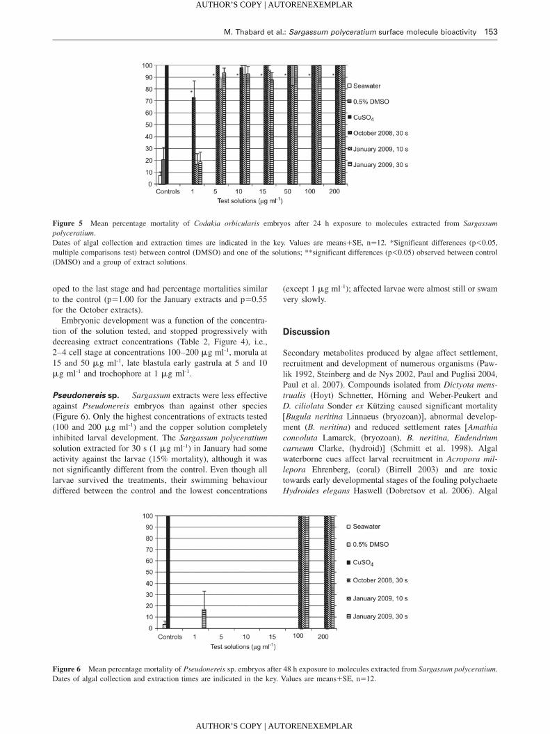
Figure 4 (A–D) SEM images (magnifications=500) of early stage of Codakia orbicularis development: (A) 4 cell stage, (B) morula
(tq6 h), (C) gastrula (tq21 h), and (D) trochophore (27 h). (E–H) Light microscopy images of Diadema antillarum early stage of devel-
opment: (E) 4 cell stage, (F) morula, (G) gastrula, (H) prism.
AUTHOR’S COPY | AUTORENEXEMPLAR
AUTHOR’S COPY | AUTORENEXEMPLAR
M. Thabard et al.: Sargassum polyceratium surface molecule bioactivity 153
Article in press - uncorrected proof
Figure 5 Mean percentage mortality of Codakia orbicularis embryos after 24 h exposure to molecules extracted from Sargassum
polyceratium.
Dates of algal collection and extraction times are indicated in the key. Values are meansqSE, ns12. *Significant differences (p-0.05,
multiple comparisons test) between control (DMSO) and one of the solutions; **significant differences (p-0.05) observed between control
(DMSO) and a group of extract solutions.
Figure 6 Mean percentage mortality of Pseudonereis sp. embryos after 48 h exposure to molecules extracted from Sargassum polyceratium.
Dates of algal collection and extraction times are indicated in the key. Values are meansqSE, ns12.
oped to the last stage and had percentage mortalities similar
to the control (ps1.00 for the January extracts and ps0.55
for the October extracts).
Embryonic development was a function of the concentra-
tion of the solution tested, and stopped progressively with
decreasing extract concentrations (Table 2, Figure 4), i.e.,
2–4 cell stage at concentrations 100–200 mg ml-1, morula at
15 and 50 mg ml-1, late blastula early gastrula at 5 and 10
mg ml-1 and trochophore at 1 mg ml-1.
Pseudonereis sp. Sargassum extracts were less effective
against Pseudonereis embryos than against other species
(Figure 6). Only the highest concentrations of extracts tested
(100 and 200 mg ml-1) and the copper solution completely
inhibited larval development. The Sargassum polyceratium
solution extracted for 30 s (1 mg ml-1) in January had some
activity against the larvae (15% mortality), although it was
not significantly different from the control. Even though all
larvae survived the treatments, their swimming behaviour
differed between the control and the lowest concentrations
(except 1 mg ml-1); affected larvae were almost still or swam
very slowly.
Discussion
Secondary metabolites produced by algae affect settlement,
recruitment and development of numerous organisms (Paw-
lik 1992, Steinberg and de Nys 2002, Paul and Puglisi 2004,
Paul et al. 2007). Compounds isolated from Dictyota mens-
trualis (Hoyt) Schnetter, Horning and Weber-Peukert and
D. ciliolata Sonder ex Kutzing caused significant mortality
wBugula neritina Linnaeus (bryozoan)x, abnormal develop-
ment (B. neritina) and reduced settlement rates wAmathia
convoluta Lamarck, (bryozoan), B. neritina, Eudendrium
carneum Clarke, (hydroid)x (Schmitt et al. 1998). Algal
waterborne cues affect larval recruitment in Acropora mil-
lepora Ehrenberg, (coral) (Birrell 2003) and are toxic
towards early developmental stages of the fouling polychaete
Hydroides elegans Haswell (Dobretsov et al. 2006). Algal
AUTHOR’S COPY | AUTORENEXEMPLAR
AUTHOR’S COPY | AUTORENEXEMPLAR
154 M. Thabard et al.: Sargassum polyceratium surface molecule bioactivity
Article in press - uncorrected proof
MNPs are often species-specific and can have an effect on
different larval development stages. Dictyota spp. and Lau-
rencia sp. are toxic against larvae of H. elegans and B. neri-
tina; Padina sp. and Halimeda sp. inhibit their larval
settlement; Hypnea sp. and Ulva sp. stimulate larval settle-
ment and Sargassum spp. have no effect (Walters et al.
1996).
Our results demonstrate that Sargassum polyceratium sur-
face molecules significantly affect embryonic development
of three tropical invertebrates and bacterial growth. These
organisms are not all in direct contact with S. polyceratium
(except Diadema antillarum), but they represent species from
three invertebrate phyla and thus give some indication of
potential toxic activity of Sargassum towards several types
of organisms. In the embryo toxicity assays, the active
extracts concentrations varied by species tested. Both Coda-
kia orbicularis and D. antillarum had similar responses
although C. orbicularis was expected to be more resistant
because of the large envelope surrounding its embryos (Gros
et al. 1997). Pseudonereis embryos were the most resistant
to the extracts. In the control, C. orbicularis embryos devel-
oped in 24 h to the trochophore stage and D. antillarum
embryos developed in 30 h to the prism stage, consistent
with previously reported values (Gros et al. 1997, Eckert
1998). However, S. polyceratium surface compound treat-
ments had progressive effects (according to increasing con-
centration) on embryonic development, suggesting the
extracts block egg cleavage. The highest concentrations
blocked development at the 2–4 cells stage, the medium con-
centrations allowed development until the morula stage and
the lowest concentrations until the blastula, gastrula and
finally trochophore or prism stages (respectively for C. orbi-
cularis and D. antillarum). Similarly, Paul and Fenical
(1986), showed that caulerpyne blocks the cleavage of devel-
oping sea urchin eggs. The division of Paracentrotus lividus
Lamarck (urchin) embryos was inhibited by algal extracts
(Martin and Uriz 1993). Dihydrorhipocephalin, aldehyde,
udoteal, petiodal, dihydroudoteal, rhipocephalin, halimeda-
trial, halimeda tetraacetate (4,9-diacetoxy-udoteal) isolated
from siphonous green algal species (Halimeda spp., Penicil-
lus spp., Rhipocephalus phoenix Ellis et Solander, Udotea
spp.) are toxic against developing sea urchin eggs, sperm and
larvae (Paul and Fenical 1986), with ED100 levels (lowest
concentration leading to 100% inhibition of cell division)
ranging from 0.2 to 16 mg ml-1. Concentrations of MNPs
extracted from siphonous algae have higher activity against
both Codakia and Diadema than the solutions tested in this
study (ED100 ranging between 50 and 100 mg ml-1). How-
ever, the tests performed by Paul and Fenical (1986) were
performed with isolated secondary metabolites and not crude
extracts. It is possible that the active compounds tested in
this study were present in the crude extract at very low con-
centration (mg ml-1). On the other hand, some molecules act
synergistically such that a pool of molecules is required to
induce a biological effect (Hay 1996). Further analytical
chemistry tests involving molecule purification and analyti-
cal chemistry analyses (GC-MS and NMR) will give further
insights into the chemical nature of the bioactive compounds
and their activity levels.
Molecules extracted at the algal surface with hexane are
non-polar. Sargassum species produce polyphenols (polar
metabolites), but some non-polar extracts, such as S. vulgare
hexane extracts, are highly active towards the development
of microalgae, suggesting production of active non-polar sec-
ondary metabolites by this alga (Plouguerne et al. 2010b).
So far, no methods allowing the extraction of polar mole-
cules located at the surface of the algae have been developed
and the activity observed here thus represents only a portion
of the possible MNPs present at the surface of S.
polyceratium.
There were no differences in toxic activity towards embry-
os between the 30 s extracts in October and January. How-
ever, antibacterial activity was different between these two
extracts, suggesting a possible seasonality in surface mole-
cule activities. Temperate algal extracts have a seasonal var-
iation in molecular composition, antimicrobial activity, and
AF activity (Steinberg and Van Altena 1992, Hellio et al.
2004, Marechal et al. 2004, Plouguerne 2006). The October
extracts were from the rainy season, while those from Jan-
uary were collected in the dry season. Hellio et al. (2004)
demonstrated that S. muticum secondary metabolite activity
varies with seasons and is higher during the summer months
when fouling pressure (including fouling by bacteria) is most
intense. It is thus possible that macroalgae develop specific
mechanisms for protection in the rainy season when bacterial
concentrations are high in coastal environments (Futch et al.
2010). In the present study, the levels at which extracts were
active towards bacteria were high (150–300 mg ml-1) in
comparison with other algal crude extracts. Crude extracts
obtained from seaweeds collected in Brittany were active
when concentrations ranged between 24 and 96 mg ml-1
(Hellio et al. 2001) and between 0.1 and 100 mg ml-1 (Plou-
guerne et al. 2008). Marine bacteria were the most sensitive
strains, suggesting that defence strategies of S. polyceratium
are specific. Such targeted defence strategies have been
described for other algal species (Paul and Puglisi 2004).
Bioactivity of Sargassum polyceratium extracts was
marked; however, we do not know whether the active mol-
ecules were produced by Sargassum polyceratium or by its
associated biofilm. Secondary metabolite isolation from
algae can be confounded by associated microorganisms. As
this study focused on surface molecules only, it was impos-
sible to clean the macroalgal surface from microepiphytes
using existing methods, such as ethanol, without breaking
the surface cells (De Nys et al. 1998). The extract obtained
therefore corresponds to Sargassum and/or its associated bio-
film. Numerous bacteria living in SW produce active sec-
ondary metabolites (Jensen and Fenical 1994). Moreover,
there are host-specific associations between algae and bac-
teria, and algae may control associated bacteria (Lachnit et
al. 2009). The bacterial biofilm may in turn confer a protec-
tion to the host alga through production of secondary
metabolites.
Sargassum polyceratium surface extracts inhibited bacte-
rial growth and embryo development of three tropical marine
invertebrates one of which was Diadema antillarum, a trop-
ical herbivorous keystone species that controls macroalgal
AUTHOR’S COPY | AUTORENEXEMPLAR
AUTHOR’S COPY | AUTORENEXEMPLAR
M. Thabard et al.: Sargassum polyceratium surface molecule bioactivity 155
Article in press - uncorrected proof
populations. Testing surface molecules was a first investi-
gation step and further work will be carried out to relate the
natural compounds to their possible ecological role. These
results require more work focused on 1) concentrations pro-
duced on the algal surface, 2) the source of production (alga
or biofilm, or both) and 3) the release of compounds into the
water column, a process that might interact with embryonic
development of organisms surrounding Sargassum. We are
currently performing tests on molecules present in the waters
in which algae are immersed (conditioned water) in order to
assess the activity of these cues against tropical invertebrate
marine larvae in the same environment.
Acknowledgements
This work was financially supported by the Ministere de l’Ecologie,
de l’Energie, du Developpement Durable et de la Mer (IFRECOR
program), Ministere de l’Outremer, the European Community
(FEDER), the Regional Council of Martinique and the School of
Biological Sciences of the University of Portsmouth (UK).
References
Abbott, R.T. 1974. American seashells. Van Nostrand Reinhold
Company (New York). pp. 663.
Amsterdam, D. 1996. Susceptibility testing of antimicrobials in liq-
uid media. In: (V. Loman, ed) Antibiotics in Laboratory Medi-
cine 4th edition. Williams and Wilkins, Baltimore, MD. pp
52–111.
Ang, P.O. 1986. Analysis of the vegetation structure of a Sargassum
community in the Philippines. Mar. Ecol Prog Ser. 28: 9–19.
Battistini, R. 1978. Les recifs coralliens de la Martinique: compa-
raison avec ceux au sud-ouest de l’Ocean Indien. Cah.
O.R.S.T.O.M. ser. Oceanogr. 16: 157–177.
Bielmeyer, G.K., K.V. Brix, T.R. Capo and M. Grosell. 2005. The
effects of metals on embryo-larval and adult life stages of the
sea urchin, Diadema antillarum. Aquat. Toxicol. 74: 254–263.
Birrell, C.L. 2003. Influences of benthic algae on coral settlement
and post-settlement survival: implications for the recovery of
disturbed and degraded reefs. Masters thesis, James Cook Uni-
versity, Townsville. pp. 132.
Cerocic, Z.G., G. Samson, F. Morales, N. Tremblay and I. Moya.
1999. Ultraviolet-induced fluorescence for plant monitoring
present state and prospects. Agronomie 19: 543–578.
De Nys, R. and P.D. Steinberg. 2002. Linking marine biology and
biotechnology. Curr. Op. Biotechnol. 13: 244–248.
De Nys, R., S.A. Dworjanyn and P.D. Steinberg. 1998. A new meth-
od for determining surface concentrations of marine natural
products on seaweeds. Mar. Ecol. Prog. Ser. 162: 79–87.
De Ruyter van Stevenick, E.D. and A.M. Breeman. 1987. Popula-
tion dynamics of a tropical intertidal and deep-water population
of Sargassum polyceratium (Phaeophyta). Aquat. Bot. 29:
139–156.
Diaz-Pulido, G., S. Harii, L.J. McCook and O. Hoegh-Guldberg.
2010. The impact of benthic algae on the settlement of a reef-
building coral. Coral Reefs 29: 203–208.
Dobretsov, S., H.U. Dahms, T. Harder and P.Y. Qian. 2006. Alle-
lochemical defense against epibiosis in the macroalga Caulerpa
racemosa var. turbinata. Mar. Ecol. Prog. Ser. 318: 165–175.
Dworjanyn, S.A., R. de Nys and P.D. Steinberg. 1999. Localisation
and surface quantification of secondary metabolites in the red
alga Delisea pulchra. Mar. Biol. 133: 727–736.
Eckert, G.L. 1998. Larval development, growth and morphology of
the sea urchin Diadema antillarum. Bull. Mar. Sci. 63: 443–451.
Eckman, J.E. and D.O. Duggins. 1991. Life and death beneath mac-
rophyte canopies: effects of understorey kelps on growth rates
and survival of marine, benthic suspension feeders. Oecologia
87: 473–487.
Engelen, A.H. 2004. Flexibility without compromises. Population
biology of the brown seaweed Sargassum polyceratium around
the island of Curacao. PhD thesis, University of Groningen. pp¸
180.
Engelen, A.H., J.L. Olsen, A.M. Breeman and W.T. Stam. 2001.
Genetic differentiation in Sargassum polyceratium (Fucales:
Phaeophyceae) around the island of Curacao (Netherlands Antil-¸
les). Mar. Biol. 139: 267–277.
Engelen, A.H., A.M. Breeman, J.L. Olsen, W.T. Stam and P. Aberg.
2005. Life history flexibility allows Sargassum polyceratium to
persist in different environments subjected to stochastic distur-
bance events. Coral Reefs 24: 670–680.
Futch, J.C., D.W. Griffin and E.K. Lipp. 2010. Human enteric virus-
es in groundwater indicate offshore transport of human sewage
to coral reefs of the Upper Florida Keys. Environ. Microbiol.
12: 964–974.
Gros, O., L. Frenkiel and M. Moueza. 1997. Embryonic, larval, and
post-larval development in the symbiotic clam Codakia orbi-
cularis (Bivalvia: Lucinidae). Invert. Biol. 116: 86–101.
Hay, M.E. 1996. Marine chemical ecology: what’s known and
what’s next? J. Exp. Mar. Biol. Ecol. 200: 103–134.
Hay, M.E. and W. Fenical. 1988. Marine plant-herbivore interac-
tions: the ecology of chemical defense. Ann. Rev. Ecol. Syst. 19:
111–145.
Hay, M. and W. Fenical. 1996. Chemical ecology and marine bio-
diversity: insights and products from the sea. Oceanogr. 9:
10–20.
Hellio, C., D. De La Broise, L. Dufosse, Y. Le Gal and N. Bour-
gougnon. 2001. Inhibition of marine bacteria by extracts of
macroalgae: potential use for environmentally friendly antifou-
ling paints. Mar. Environ. Res. 52: 231–247.
Hellio, C., J.P. Berge, C. Beaupoil, Y. Le Gal and N. Bourgougnon.
2002. Screening of marine algal extracts for anti-settlement
activities against microalgae and macroalgae. Biofouling 18:
205–215.
Hellio, C., C. Simon-Colin, A.S. Clare and E. Deslandes. 2004.
Isethionic acid and floridoside isolated from the red alga, Gra-
teloupia turuturu, inhibit settlement of Balanus amphitrite
cyprid Larvae. Biofouling 20: 139–145.
Hellio, C., M. Tsoukatou, J.P. Marechal, N. Aldred, C. Beaupoil,
A.S. Clare, C. Vagias and V. Roussis. 2005. Inhibitory effects
of Mediterranean sponge extracts and metabolites on larval set-
tlement of the barnacle Balanus amphitrite. Mar. Biotechnol. 7:
297–305.
Hellio, C., J.P. Marechal, B.A.P. Da Gama, R.C. Pereira and A.S.
Clare. 2009. Natural marine products with antifouling activities.
In: (C. Hellio and D.M.Y. Yebra, eds.) Advances in marine anti-
fouling coatings and technologies. Woodshead Publishing, Cam-
bridge, UK, pp. 572–622.
Hughes, T.P. 1994. Catastrophes, phase-shifts, and large-scale deg-
radation of a Caribbean coral reef. Science 265: 1547–1551.
Jensen, P.R. and W. Fenical. 1994. Strategies for the discovery of
secondary metabolites from marine bacteria: ecological perspec-
tives. Ann. Rev. Microbiol. 48: 559–584.
AUTHOR’S COPY | AUTORENEXEMPLAR
AUTHOR’S COPY | AUTORENEXEMPLAR
156 M. Thabard et al.: Sargassum polyceratium surface molecule bioactivity
Article in press - uncorrected proof
Knowlton, N. 2001. Sea urchin recovery from mass mortality: new
hope for Caribbean coral reefs? Proc. Natl. Acad. Sci. USA 98:
4822–4824.
Labreuche, Y., P. Soudant, M. Gonzalves, C. Lambert and J.L. Nico-
las. 2005. Effects of the extracellular products from the patho-
genic Vibrio aestuarianus strain 01/32 on lethality and cellular
immune responses of the oyster Crassostrea gigas. Develop.
Comp. Immunol. 30: 367–379.
Lachnit, T., M. Bluemel, J.F. Imhoff and M. Wahl. 2009. Specific
epibactrial communities on macroalgae: phylogeny matters more
than habitat. Aquat. Biol. 5: 181–186.
Lapointe, B.E. 1997. Nutrient thresholds for bottom-up control of
macroalgal blooms on coral reefs in Jamaica and southeast Flor-
ida. Limnol. Oceanogr. 42: 1119–1131.
Lapointe, B.E. and K. Thacker. 2002. Community-based water qual-
ity and coral reef monitoring in the Negril marine park, Jamaica:
Land-based nutrient inputs and their ecological consequences.
In: (J.W. Porter and K.G. Porter, eds.) The Everglades, Florida
Bay and coral reefs of the Florida Keys an ecosystem source-
book. CRC Press, Boca Raton, FL. pp. 939–963.
Lessios, H.A. 1995. Diadema antillarum 10 years after mass mor-
tality: still rare, despite help from a competitor. Proc. Roy. Soc.
B. 259: 331–337.
Lessios, H.A. 2005. Diadema antillarum populations in Panama
twenty years following mass mortality. Coral Reefs 24:
125–127.
Lessios, H.A., P.W. Glynn and D.R. Robertson. 1983. Mass mor-
talities of Coral reef organisms. Science 222: 715–715.
Lessios, H.A., D.R. Robertson and J.D. Cubit. 1984. Spread of Dia-
dema mass mortality through the Caribbean. Science 226:
335–337.
Littler, M.M. and D.S. Littler. 1980. The evolution of thallus form
and survival strategies in benthic marine macroalgae: field and
laboratory tests of a functional form model. Am. Nat. 116:
25–44.
Littler, D.S. and M.M. Littler. 2000. Caribbean reef plants, an iden-
tification guide to the reef plants of the Caribbean, Bahamas,
Florida and Gulf of Mexico. Offshore Graphics Inc. Washington,
DC. pp. 542.
Littler, M.M., D.S. Littler and P.R. Taylor. 1983a. Evolutionary strat-
egies in a tropical barrier reef system-functional-form-groups of
marine macroalgae. J. Phycol. 19: 229–237.
Littler, M.M., P.R. Taylor and D.S. Littler. 1983b. Algal resistance
to herbivory on a Caribbean barrier reef. Coral Reefs 2:
111–118.
Littler, M.M., D.S. Littler and B.E. Lapointe. 1993. Modification of
tropical reef community structure due to cultural eutrophication:
the southwest coast of Martinique. Proc. 7th Int. Coral Reef
Symp. 1: 335–343.
Marechal, J.P. and C. Hellio. 2011. Antifouling activity against bar-
nacle cypris larvae: Do target species matter (Amphibalanus
Amphitrite and Semibalanus balanoides)? Int. Biodet. Biodeg.
65: 92–101.
Marechal, J.P., G. Culiolib, C. Hellio, H. Thomas-Guyonc, M.E.
Callow, A.S. Clare and A. Ortalo-Magne. 2004. Seasonal vari-
ation in antifouling activity of crude extracts of the brown alga
Bifurcaria bifurcata (Cystoseiraceae) against cyprids of Balanus
amphitrite and the marine bacteria Cobetia marina and Pseu-
doalteromonas haloplanktis. J. Exp. Mar. Biol. Ecol. 313:
47–62.
Martin, D. and M.J. Uriz. 1993. Chemical bioactivity of Mediter-
ranean benthic organisms against embryos and larvae of marine
invertebrates. J. Exp. Mar. Biol. Ecol. 173: 11–27.
Mokrini, R., M. Ben Mesaoud, M. Daoudi, C. Hellio, J.P. Marechal,
M. El Hattab, A. Ortalo-Magne, L. Piovetti and C. Culioli. 2008.
Meroditerpenoids and derivatives from the brown alga Cysto-
seira baccata and their antifouling Properties J. Nat. Prod. 71:
1806–1811.
Paul, V.J. and W. Fenical. 1986. Chemical defense in tropical green
algae, order Caulerpales. Mar. Ecol. Prog. Ser. 34: 157–169.
Paul, V.J. and M.P. Puglisi. 2004. Chemical mediation of interac-
tions among marine organisms. Nat. Prod. Rep. 21: 189–209.
Paul, V.J., C.R. Wylie and B.R. Sanger. 1988. Effects of algal chem-
ical defenses toward different coral-reef herbivorous fishes: a
preliminary study. Proc. 6th Int. Coral Reef Symp. 3: 73–78.
Paul, V.J., K.E. Arthur, R. Ritson-Williams, C. Ross and K. Sharp.
2007. Chemical defenses: from compounds to communities.
Biol. Bull. 213: 226–251.
Pawlik, J.R. 1992. Chemical ecology of the settlement of benthic
marine invertebrates. Oceanogr. Mar. Biol. Annu. Rev. 30:
273–335.
Pereira, R.C., B.A.P. Da Gama, V.L. Teixeira and Y. Yoneshigue-
Valentin. 2003. Ecological roles of natural products of the Bra-
zilian red seaweed Laurencia obtusa. Brazilian J. Biol. 63:
665–672.
Plouguerne, E. 2006. Etude ecologique et chimique de deux algues
introduites sur les cotes bretonnes, Grateloupia turuturu Yamada
et Sargassum muticum (Yendo) Fensholt: nouvelles ressources
biologiques de composes a activite antifouling. PhD thesis, Uni-
versity of Brittany. pp. 251.
Plouguerne, E., K. Le Lann, S. Connan, G. Jechoux, E. Deslandes
and V. Stiger-Pouvreau. 2006. Spatial and seasonal variation in
density, reproductive status, length and phenolic content of the
invasive brown macroalgae Sargassum muticum (Yendo) Fens-
holt along the coast of Western Brittany (France). Aquat. Bot.
85: 337–344.
Plouguerne, E., C. Hellio, E. Deslandes, B. Veron and V. Stiger-
Pouvreau. 2008. Anti-microfouling activities in extracts of two
invasive algae: Grateloupia turuturu and Sargassum muticum.
Bot. Mar. 51: 202–208.
Plouguerne, E., C. Hellio, C. Cesconetto, M. Thabard, K. Mason,
B. Veron, R.C. Pereira and B.A.P. da Gama. 2010a. Antifouling
activity as a function of population variation in Sargassum vul-
gare from the littoral of Rio de Janeiro (Brazil). J Appl. Phycol.
22: 717–724.
Plouguerne, E., E. Ioannou, P. Georgantea, C. Vagias, V. Roussis,
C. Hellio, E. Kraffe and V. Stiger-Pouvreau. 2010b. Anti-micro-
fouling activity of lipidic metabolites from the invasive brown
alga Sargassum muticum (Yendo) Fensholt. Mar. Biotechnol. 12:
52–61.
Ragan, M.A. and K.W. Glombitza. 1986. Phlorotannins, brown algal
polyphenols. Prog. Phycol. Res. 4: 129–241.
Salgado, L.T., N.B. Viana, L.R. Andrade, R.N. Leal, B.A.P. da
Gama, M. Attias, R.C. Pereira and G.M. Amado Filho. 2008.
Intra-cellular storage, transport and exocytosis of halogenated
compounds in marine red alga Laurencia obtusa. J. Struct. Biol.
162: 345–355.
Sastry, V.M.V.S. and G.R.K. Rao. 1994. Antibacterial substances
from marine algae: successive extraction using benzene, chlo-
roform and methanol. Bot. Mar. 37: 357–360.
Schmitt, T.M., N. Lindquist and M.E. Hay. 1998. Seaweed second-
ary metabolites as antifoulants: Effects of Dictyota spp. diter-
penes on survivorship, settlement, and development of marine
invertebrate larvae. Chemoecology 8: 125–131.
Sieburth, J. and J.T. Conover. 1965. Sargassum tannin, an antibiotic
which retards fouling. Nature 208: 52–53.
AUTHOR’S COPY | AUTORENEXEMPLAR
AUTHOR’S COPY | AUTORENEXEMPLAR
M. Thabard et al.: Sargassum polyceratium surface molecule bioactivity 157
Article in press - uncorrected proof
Steinberg, P.D. and R. de Nys. 2002. Chemical mediation of colo-
nization of seaweed surfaces. J. Phycol. 38: 621–629.
Steinberg, P.D. and I. Van Altena. 1992. Tolerance of marine inver-
tebrate herbivores to brown algal phlorotannins in temperate
Australasia. Ecol. Monogr. 62: 189–222.
Tanaka, N. and A. Asakawa. 1988. Allelopathic effect of mucilage
released from a brown alga Sargassum horneri on marine dia-
toms. Nippon Suisan Gakkaishi. 54: 1711–1714.
Titlyanov, E.A., T.V. Titlyanova, I.M. Yakovleva, Y. Nakano and R.
Bhagooli. 2005. Regeneration of artificial injuries on scleracti-
nian corals and coral/algal competition of newly formed sub-
strate. J. Exp. Mar. Biol. Ecol. 323: 27–42.
Walters, L.J., M.G. Hadfield and C.M. Smith. 1996. Waterborne
chemical compounds in tropical macroalgae: positive and neg-
ative cues for larval settlement. Mar. Biol. 126: 383–393.
Young, D., B. Howard and W. Fenical. 1980. Subcellular localiza-
tion of brominated secondary metabolites in the red alga Lau-
rencia synderae. J. Phycol. 16: 182–185.
Received 13 May, 2010; accepted 22 November, 2010; online first
15 March, 2011
AUTHOR’S COPY | AUTORENEXEMPLAR
AUTHOR’S COPY | AUTORENEXEMPLAR