final accepted version altered control of visual fixation and saccadic eye movements in...
TRANSCRIPT

JN-00192-2003
FINAL ACCEPTED VERSION
ALTERED CONTROL OF VISUAL FIXATION AND SACCADIC EYE MOVEMENTS IN ATTENTION-DEFICIT HYPERACTIVITY DISORDER
Douglas P. Munoz, Irene T. Armstrong, Karen A. Hampton, Kimberly D. Moore
Centre for Neuroscience Studies,CIHR Group in Sensory-Motor Systems,
Department of Physiology,Queen’s UniversityKingston, Ontario,
K7L 3N6CANADA
Running Head: Eye movements and ADHD
9 Figures, 2 TablesABSTRACT: 340 wordsTOTAL TEXT (Introduction, Methods, Results, Discussion): 7267 words
Address Correspondence:
Dr. Douglas P. MunozDepartment of PhysiologyQueen’s UniversityKingston, OntarioK7L 3N6 CANADA
phone: 613-533-2111FAX: 613-533-6840email: [email protected]
Copyright (c) 2003 by the American Physiological Society.
Articles in PresS. J Neurophysiol (April 2, 2003). 10.1152/jn.00192.2003
JN-00192-2003
2
ABSTRACT
Attention-deficit hyperactivity disorder (ADHD) is characterized by the overt
symptoms of impulsiveness, hyperactivity, and inattention. A frontostriatal
pathophysiology has been hypothesized to produce these symptoms and lead to reduced
ability to inhibit unnecessary or inappropriate behavioral responses. Oculomotor tasks
can be designed to probe the ability of subjects to generate or inhibit reflexive and
voluntary responses. Because regions of the frontal cortex and basal ganglia have been
identified in the control of voluntary responses and saccadic suppression, we
hypothesized that children and adults diagnosed with ADHD may have specific
difficulties in oculomotor tasks requiring the suppression of reflexive or unwanted
saccadic eye movements. To test this hypothesis, we measured eye movement
performance in pro- and anti-saccade tasks of 114 ADHD and 180 control participants
ranging in age from 6 – 59 years. In the pro-saccade task, participants were instructed to
look from a central fixation point toward an eccentric visual target. In the anti-saccade
task, stimulus presentation was identical but participants were instructed to suppress the
saccade to the stimulus and instead look from the central fixation point to the side
opposite the target. The state of fixation was manipulated by presenting the target either
when the central fixation point was illuminated (overlap condition), or at some time after
it disappeared (gap condition). In the pro-saccade task, ADHD participants had longer
reaction times, greater intra-subject variance, and their saccades had reduced peak
velocities and increased durations. In the anti-saccade task, ADHD participants had
greater difficulty suppressing reflexive pro-saccades toward the eccentric target,

JN-00192-2003
3
increased reaction times for correct anti-saccades, and greater intra-subject variance. In a
third task requiring prolonged fixation, ADHD participants generated more intrusive
saccades during periods when they were required to maintain steady fixation. The results
suggest that ADHD participants have reduced ability to suppress unwanted saccades and
control their fixation behavior voluntarily, a finding that is consistent with a fronto-
striatal pathophysiology. The findings are discussed in the context of recent
neurophysiological data from non-human primates that have identified important control
signals for saccade suppression that emanate from frontostriatal circuits.
Key words: cerebral cortex, basal ganglia, response inhibition, saccade, eye movement,
visual fixation
JN-00192-2003
4
INTRODUCTION
Attention-deficit hyperactivity disorder (ADHD), characterized by the symptoms
of impulsiveness, hyperactivity, and inattention, is a prevalent neurobehavioral disorder
estimated to affect approximately 5% of children and, for some, these core symptoms are
believed to persist into adulthood (Barkley, 1997). As part of these core behavioral
symptoms, response inhibition, may be an important component of the disability because
ADHD subjects have difficulty suppressing inappropriate behavioral responses (Shue et
al., 1992; Mostofsky et al., 2001).
At present, the etiology of ADHD remains poorly defined. Several observations
support a hypothesis of a frontostriatal deficit (Castellanos, 2001), possibly involving
dysfunction in dopamine transmission, which may produce the symptoms of ADHD.
First, ADHD individuals lack adequate inhibitory control and often act impulsively
(Chelune et al., 1986; Grodzinsky and Diamond, 1992), which is a classic sign of frontal
lobe dysfunction (Fuster, 1997). Second, regional blood flow and glucose metabolic
studies have revealed frontal and/or striatal abnormalities in ADHD individuals (Lou et
al., 1984, 1989; Zametkin et al., 1990). Third, anatomical neuroimaging studies have
revealed altered architecture in the frontal lobes, caudate nucleus, and rostrum of the
corpus callosum (Giedd et al., 1994; Castellanos et al., 1996a; 2001; Rubia et al., 1999).
Fourth, dopamine, an important neurotransmitter in the striatum and frontal cortex, has
been implicated in the disorder (Levy, 1991) because methylphenidate, the main
pharmacotherapy for ADHD, blocks dopamine reuptake. Fifth, there is evidence for

JN-00192-2003
5
abnormal levels of catecholamine metabolites in the cerebrospinal fluid of ADHD
subjects (Castellanos et al., 1996b).
Saccades are rapid eye movements used to move the high acuity fovea of the
retina to visual targets for detailed visual analysis. Characteristics of saccades can be
measured precisely and several behavioral tasks have been designed to test specific
aspects of oculomotor control (see Leigh and Zee, 1999 for review). In addition, the
premotor circuitry controlling eye movements is now understood at a level that is
sufficient to provide a basis for designing and interpreting more complex experiments to
probe higher brain functions (Wurtz and Goldberg, 1989; Moschovakis et al., 1996;
Munoz et al., 2000). Saccades can be divided into two broad classes: reflexive, sensory-
triggered movements and volitional movements. Initiation of visually-triggered saccades
involves occipital and parietal cortex and their inputs to the superior colliculus which
then projects to the premotor circuit in the brainstem and cerebellum. Planning of
volitional saccades and suppression of reflexive saccades is under the control of frontal
cortex and basal ganglia, which also project to the superior colliculus and brainstem
premotor circuit (Schall, 1997; Hikosaka et al., 2000: Munoz et al., 2000 for review).
We hypothesize that, because of the overlap in the brain areas implicated in
ADHD and in the control of volitional saccade behavior, ADHD individuals will have
difficulty suppressing reflexive saccades and generating volitional saccades. To test this
hypothesis, we recorded the eye movements of children and adults diagnosed with
ADHD and controls recruited to perform a series of saccadic eye movement tasks. The
pro-saccade task (Fig. 1A) is used to test the ability of subjects to generate reflexive,
visually-triggered saccades. In this task, subjects are required to look from a central

JN-00192-2003
6
fixation point (FP) to an eccentric target stimulus as soon as it appears. We hypothesize
that ADHD subjects will not be impaired in this task. The anti-saccade task (Fig. 1B) is
used to test the ability of subjects to suppress reflexive saccades and instead generate
voluntary saccades. In this task, subjects must suppress the reflexive saccade to the
eccentric stimulus, and instead generate a voluntary saccade to the mirror position, where
no stimulus appeared. Recent brain imaging studies have identified specific activation in
the frontal cortex that varies between pro- and anti-saccade tasks (O’Driscoll et al. 1995;
Sweeney et al. 1996; Doricchi et al. 1997; Muri et al. 1998; Connolly et al. 2000; 2002).
In addition, neurophysiological studies in non-human primates have identified various
frontal regions that are active in the anti-saccade task (Funahashi et al. 1993; Schlag-Rey
et al. 1997; Everling and Munoz 2000). Clinical studies have identified specific deficits
in anti-saccade performance in various patient groups with a pathophysiology affecting
the frontal cortex and/or basal ganglia (Guitton et al., 1985; Everling and Fischer, 1998).
Because of the documented difficulties with response inhibition that are evident in
ADHD, we predict that ADHD participants will have difficulty suppressing reflexive pro-
saccades in the anti-saccade task.
There are both exogenous and endogenous components of fixation control
(Reuter-Lorenz et al., 1995; Paré and Munoz, 1996). The endogenous component of
fixation is required to maintain steady fixation independent of whether there is a visual
stimulus on the fovea, while the exogenous component is mediated by the presence of a
visible stimulus on the fovea. The presence or absence of the exogenous component
influences performance in pro- and anti-saccade tasks (Fischer and Weber 1997; Munoz
et al., 1998a). Removing the central FP prior to the appearance of the eccentric stimulus

JN-00192-2003
7
(gap task; Fig. 1D) reduces saccadic reaction time (SRT) and increases the percentage of
direction errors on anti-saccade trials. In contrast, SRT is increased and error rates
reduced when the FP remains illuminated during the appearance of the saccade target
(overlap task; Fig. 1C).
We reason that if ADHD individuals have a frontostriatal deficit, then they will
have difficulty maintaining the endogenous component of visual fixation. Such a deficit
will result in difficulty regulating the processes of saccade suppression and saccade
initiation, leading to the generation of more reflexive saccades and more direction errors
in the anti-saccade task. Further, these deficits should be present in both gap and overlap
conditions. We show that ADHD participants have difficulty maintaining endogenous
fixation and suppressing reflexive saccades in the anti-saccade task and they also have
longer and more variable reaction times. Preliminary reports of these data have appeared
(Munoz et al., 1998b; 1999).
METHODS
Subjects
All experimental procedures were reviewed and approved by the Queen’s
University Human Research Ethics Board. Participants between the ages of 6 and 59
years were recruited from the greater Kingston area via newspaper advertisements and
physician referral. They were informed of the general nature of the study prior to
consenting to participate and were reimbursed $10.00 per recording session. Parents
provided informed consent for minors. All participants reported no known visual

JN-00192-2003
8
disorders other than refractive errors and they were permitted to wear their prescription
lenses during the recording sessions.
This report describes the eye movement behavior of 294 participants sorted into
four groups: control or ADHD children (age 6-16 years) or adults (age 18 – 59 years).
Each of the four groups contained between 38 – 105 subjects (see Table 1). A portion of
the data collected from control subjects were presented in a previous paper describing
developmental changes in the control of visual fixation and saccade generation among
normal subjects between the ages of 5 – 79 years (Munoz et al. 1998a).
All 114 ADHD participants included in this report were diagnosed in the
community, initially by various health care professionals. Diagnosis was confirmed and
co-morbidity assessed during an interview with a clinical psychologist using DSM-IV
criteria, Conner Parent’s Rating Scales (CPRS) for children and the Brown Attention-
Deficit Disorder Scale (BADDS) for adults. Inclusion criteria for the ADHD pool
included meeting DSM-IV criteria and criteria established from the CPRS (subjects 6 -
16 years) or BADDS (subjects 18-59 years). Other ADHD participants (N = 34; 23
children, 11 adults) were excluded because we identified the following co-morbid signs:
learning disabilities resulting in delayed advancement in school, Tourette’s syndrome, or
bipolar disease. Control participants or their guardians reported no known neurological or
psychiatric disorders. Control children were also assessed using the CPRS. Impulsiveness
scores were reliably different between ADHD and control children (mean ± SD: ADHD:
75.8 ± 10.8; Control: 47.1 ± 9.1), F(1,142) = 258.8, p < 0.0001. Hyperactivity scores
were also significantly elevated for the ADHD children (ADHD: 84.2 ± 11.5; Control:
45.8 ± 8.8), F(1,142) = 443.97, p < 0.0001. Only ADHD adults were administered the
JN-00192-2003
9
BADDS and all ADHD adults scored higher that 50 (range 64-96) which is indicative of
ADHD.
Seventy-six of the ADHD participants (64/76 children, 12/38 adults) were on a
prescribed drug treatment regime consisting of daily stimulant medication (e.g.,
methylphenidate, cylert) to help ameliorate the symptoms of the disorder. Data were
collected from these participants on days when they did not take any prescribed drug
treatment. The minimal time between the previous dose and testing for all participants
was at least 20 hours to minimize any carryover effects.
Handedness of all subjects was assessed with a modified version of the Edinburgh
Handedness Inventory (Oldfield 1971). Participants were asked to rate hand preference in
the following tasks: writing, drawing, throwing, using scissors, using a toothbrush, using
a knife without a fork, using a spoon, swinging a hockey stick, turning a key, and opening
a box. Each right-handed response was assigned +1 and each left-handed response was
assigned –1. Therefore a score of +10 or –10 indicated extreme right- or left-handedness,
respectively. Mean handedness scores are presented in Table 1. Most interestingly,
control groups were significantly more right-handed than the ADHD groups, F(1, 290) =
9.82, p < 0.01. This trend toward non right-handedness among the ADHD participants is
consistent with previous reports (Reid and Norvilitis 2000) and may indicate anomalous
laterality of function in ADHD.
Experimental set-up and behavioral paradigms
Participants were seated upright in a dental chair equipped with a head rest that
was adjusted for height so that they faced the center of a translucent visual screen, 100

JN-00192-2003
10
cm away. The experiments were performed in darkness and silence except for the
controlled presentation of visual stimuli that consisted of red light emitting diodes (LEDs;
chromaticity coordinates CIEx = 0.73, CIEy = 0.26). One LED (2.0 cd/m2) was back-
projected onto the center of the translucent screen and served as a central fixation point
(FP) to start all trials. Eccentric target LEDs (5.0 cd/m2) were mounted into small boxes
positioned 20° to the left and right of the central FP. Between trials the screen was
illuminated diffusely (1.0 cd/m2) with background slides depicting various visual scenes
to reduce dark adaptation and boredom.
In the pro-saccade task (Fig. 1A), participants were instructed to fixate initially
upon a central FP and then look from the location of the FP to an eccentric target
stimulus that appeared randomly either 20° to the left or right. Each trial began when the
background illumination was turned off. After a 250 ms period of darkness, the FP
appeared. After an additional 1000 ms one of two events occurred. In the overlap
condition (Fig. 1C), the FP remained illuminated when the eccentric target stimulus
appeared. In the gap condition (Fig. 1D), the FP disappeared and, after a gap period of
200 ms, the eccentric target stimulus appeared. The target stimulus remained illuminated
for 1000 ms, after which all LEDs were turned off and the background illumination came
on to indicate the end of the trial. Target location (20° right or left) and fixation condition
(gap or overlap) were randomly interleaved within a block of trials.
In the anti-saccade task (Fig. 1B), the presentation of stimuli was identical to that
described above. Participants were instructed to look at the central FP but then, after the
appearance of the eccentric stimulus, they were asked to look away from the stimulus, to
the opposite side of the vertical meridian. Once again, stimulus location (20° right or left)

JN-00192-2003
11
and fixation condition (gap or overlap) were randomly interleaved within a block of
trials.
In a third experiment, designed to study the ability to maintain prolonged fixation,
participants performed pro-saccades in the gap condition only. In this experiment, the FP
was illuminated for 1500 ms and the gap period was varied randomly among one of five
intervals: 0, 200, 400, 600, or 800 ms. The target then appeared randomly either 20° right
(40% of trials), 20° left (40% of trials), or the FP reappeared in the center of the screen
(20% of trials).
The results of three separate experiments are described. In the first experiment,
participants performed the pro-saccade task in randomized overlap and gap conditions
with the gap duration fixed at 200 ms and the eccentric target stimulus appeared
randomly 20º to the right or left of center. In the second experiment, performed on the
same day, stimulus conditions remained identical but participants were instructed to
generate anti-saccades following the appearance of the eccentric target stimulus. In the
third experiment, performed on a separate day, participants were instructed to generate
pro-saccades in the prolonged fixation experiment and the gap duration was varied
randomly from 0 - 800 ms in 200 ms increments.
Experiments were conducted in two separate sessions, separated by 1-14 days. In
the first experimental session, participants completed 1 block of pro-saccades followed
by 2 blocks of anti-saccades. Each block consisted of 80-120 trials equally distributed
between gap/overlap and target right/left conditions. Participants were given no practice
but were asked to repeat the task instructions prior to the onset of data collection. In the
second session, participants returned to perform the prolonged fixation experiment with

JN-00192-2003
12
randomized gap durations, completing 3 blocks of 100-150 trials per block. All
participants in the study completed the pro- and anti-saccade tasks on Day 1. Only a
subset of the participants (73%; 215 / 294) completed the prolonged fixation experiment
on Day 2. Each recording session lasted not more than 45 minutes and there were breaks
between blocks of trials during which participants were provided with snacks and drinks
to maintain alertness.
Data collection and analysis
Horizontal eye movements were measured using DC electrooculography (EOG).
Ag-AgCl skin electrodes were placed bitemporally to record horizontal eye position. A
ground electrode was placed just above the eyebrows in the center of the forehead.
Participants were instructed to rest their head comfortably against the headrest and during
data collection, move only their eyes. The EOG signal was amplified and low pass
filtered with a Grass P18 amplifier rated for human use. To minimize EOG drift,
participants wore the electrodes for approximately 10 minutes before the onset of
calibration and recording. Calibration was performed by having participants look back
and forth between the targets located at 20° right, 20° left, and the central FP. Calibration
was repeated between each block of trials.
The experimental paradigms, visual displays, and storage of eye movement data
were under the control of a 486 computer running a real-time data acquisition system
(REX; Hays et al. 1982). Horizontal eye position was digitized at a rate of 500 Hz.
Digitized data were stored on a hard disk, and subsequent off-line analysis was performed
on a Sun Sparcstation. Horizontal eye velocity was computed from the position traces by

JN-00192-2003
13
applying software differentiation (finite impulse response filter). The onset and
termination of each saccade was determined when eye velocity respectively increased or
decreased beyond 30°/s. Saccades were scored as correct if the first movement after
appearance of the eccentric stimulus was > 5º in amplitude and in the correct direction
(i.e., toward the stimulus in the pro-saccade task; away from the stimulus in the anti-
saccade task). Saccades were scored as incorrect if the first saccade after appearance of
the eccentric stimulus was in the wrong direction (i.e., away from the target in the pro-
saccade task; toward the target in the anti-saccade task). Saccadic reaction time (SRT)
was measured as the time from stimulus appearance to the onset of the first saccade.
Mean SRT in the pro- and anti-saccade tasks was computed from trials with reaction
latencies between 90 ms and 1000 ms. Movements were classified as anticipatory and
were excluded from analysis if they were initiated less than 90 ms after target
appearance. This anticipatory cut-off was obtained from viewing SRT distributions for
correct and incorrect movements in the pro-saccade task (Kalesnykas and Hallett 1987;
Munoz et al. 1998a). Saccades that were initiated less than 90 ms after target appearance
were correct about 50% of the time, whereas saccades initiated more than 90 ms after
target appearance were correct more than 95% of the time (see Fig. 2). We also computed
the percentage of express saccades generated by each subject in all conditions. Express
saccades are the shortest latency visually-triggered saccades (Fischer and Rampsberger
1984; Fischer et al. 1993; Paré and Munoz 1996). In humans, they are initiated between
90 - 140 ms after target appearance. From the data of each participant, we computed the
following values: the mean SRT for correct trials; the coefficient of variation of SRT for
correct trials (CV = (standard deviation / mean) * 100); the percentage of express

JN-00192-2003
14
saccades (latency: 90 - 140 ms); and the percentage of direction errors (saccades away
from the target in the pro-saccade task; saccades toward the target in the anti-saccade
task).
We also measured the amplitude, peak velocity, and duration of horizontal
saccades made in the pro-saccade task only. This analysis was performed for data from
291 participants. Data from 3 participants (1 ADHD child, 1 control child, 1 control
adult) were excluded due to abnormally high levels of noise in the EOG signals that
compromised these measures. For all correct movements in the pro-saccade task initiated
between 90-1000 ms after stimulus appearance, we computed the mean amplitude of the
first saccade following target appearance. Additionally, for saccades with amplitudes
between 18º and 21º, we computed the mean peak velocity and duration. This narrow
window of amplitudes was used to control for well-known main sequence effects of
amplitude on velocity and duration (Leigh and Zee 1999). Subjects were not provided
any feedback regarding the accuracy of their anti-saccades, so that variability in the
amplitude of these movements was considerable for participants of all ages. Thus, anti-
saccade amplitude, peak velocity and duration were not quantified.
Intrusive saccades were identified as unnecessary rapid shifts in eye position that
exceeded 2° in amplitude and 70°/s in velocity. We counted the number of intrusive
saccades that occurred in the prolonged fixation experiment with randomized gap
duration in two separate epochs: during the final 1000 ms of visual fixation and during
the randomized gap period.
The first two experiments (pro- and anti-saccade tasks) contained two within-
subject factors: direction (right vs. left) and fixation state (gap vs. overlap); and two

JN-00192-2003
15
between-group factors: age (child vs. adult) and psychopathology (ADHD vs. none).
Despite the fact that all subjects performed the pro-saccade task before the anti-saccade
task, there were no differences in ordering affects between subjects groups. Perhaps even
more surprising, the error rates from the first 20 anti- saccade trials did not differ
significantly from the overall error rates among any of the groups. The third experiment
(prolonged fixation with randomized gap) contained two within-subject factors: direction
(right vs. left) and gap duration (0, 200, 400, 600, 800); and two between-group factors of
age (child vs. adult), and psychopathology (ADHD vs. none). All dependent measures
were analyzed using analysis of variance with alpha set at 0.05.
Because the focus of this paper is on differences between ADHD and control
subjects, most analyses contrasted psychopathology with the within-subject factors
separately for adults and children. Unequal sample sizes complicated analyses,
especially for adults where the samples were extremely different (38 ADHD adults vs.
105 control adults). These extreme differences bring into question any interactions that
we found among the data. We therefore randomly selected subjects to balance the
number of subjects in the ADHD/control groups by age. In the case of the children’s
data, we removed 3 ADHD subjects at random from the analysis of each dependent
measure. In the case of the adult data, we removed all but 38 of the control adults and
performed an analysis on each dependent measure for groups of equal size. Because this
excluded almost 2/3 of the adult control data, we performed the removal/analysis
procedure several times. Because no differences were noted, we retained the unequal
numbers of subjects because the control adult data are necessary to establish reliably the
developmental trends presented in the RESULTS.

JN-00192-2003
16
RESULTS
Pro- Saccade Task
SACCADIC REACTION TIMES. Figure 2 illustrates the distribution of SRTs
obtained from control and ADHD children and adults in the pro-saccade task with gap
and overlap conditions. The vast majority of the responses were correct (positive values
on the ordinate); only a very small percentage of responses were direction errors
(negative values on the ordinate). For all subject groups, most of the direction errors were
triggered with reaction times < 90 ms and, presumably were due to anticipation of target
appearance because the distribution of correct and incorrect responses was mirrored
during this interval. Among the correct pro-saccades initiated > 90 ms after target
appearance, most were triggered before 300 ms in the gap condition and before 400 ms in
the overlap condition.
From the correct responses initiated 90–1000 ms after target appearance, we
computed the mean SRT, the coefficient of variation in SRT, and percentage of express
saccades for each participant in the gap and overlap conditions and then calculated mean
values for each of the four groups (Fig. 3). Figure 3A and B illustrates the mean SRT in
the gap and overlap conditions for the 4 subject groups. Mean SRT was significantly
elevated in ADHD, F(1, 290) = 5.06, p < 0.05. Consistent with previous studies (Saslow
1967; Kalesnykas and Hallett 1987; Fischer et al. 1997; Munoz et al. 1998a), mean SRT
for all groups was significantly increased in the overlap condition compared to the gap
condition, F(1, 290) = 719.01, p < 0.001. This pro-saccade gap effect (overlap SRT – gap

JN-00192-2003
17
SRT), ranged from 52-64 ms, and was larger for children than for adults, F(1, 290) =
4.05, p < 0.05. Consistent with a previous study (Cairney et al. 2001), there was no
difference in gap effect between ADHD and control groups. We have reported previously
the influence of age on SRT among control participants (Munoz et al. 1998a). The mean
SRT for the children was greater than for the adults in both control and ADHD groups,
F(1, 290) = 5.75, p = 0.01. For all groups there was a modest directional asymmetry with
rightward pro-saccades having shorter mean SRT than leftward pro-saccades (Table 2).
This directional asymmetry was specific to the overlap condition, F(1, 290) = 28.66, p <
0.001, and more pronounced in the children F(1, 290) = 3.59, p = 0.05.
An analysis of intra-subject variance in SRT, expressed as the coefficient of
variation (CV), revealed that ADHD participants were far more variable in the time of
their responses, F(1, 290) = 38.41, p < 0.001 (Fig. 3C,D). In addition, responses of
children were more variable than adults, F(1, 290) = 135.36, p < 0.001, as described
previously (Munoz et al. 1998a). The differences between control and ADHD groups
were consistent for children and adults; thus there was no interaction between the factors,
F(1, 290) < 1, p > 0.80.
It has been suggested that abnormally high percentages of express saccades,
especially in the overlap condition, may reflect underlying pathology in brain areas
controlling visual fixation and/or saccade initiation (Biscaldi et al. 1996). We
hypothesized initially that ADHD participants would make more express saccades than
control participants. Figure 3E and F contrasts the percentage of express saccades for the
groups in the gap and overlap conditions. All groups generated more express saccades in
the gap, compared to overlap conditions, F(1, 290) = 202.40, p < 0.001. Consistent with

JN-00192-2003
18
previous reports (Fischer et al. 1997; Munoz et al. 1998a), children generated more
express saccades than adults, F(1, 290) = 14.65, p < 0.001. Note however that although
ADHD subject groups appeared to generate slightly more express saccades than control
groups, this difference was not significant, F(1, 290) = 2.82, p > 0.05.
METRICS. ADHD and control children and ADHD adults showed no difference
in the amplitude of the first saccade to target in the pro-saccade task, but control adults
made larger saccades, F(1, 287) = 4.76, p < 0.05 (control children 19.3°; ADHD children
19.3°; control adults 19.5°; ADHD adults 19.3°). For saccades between 18°-21° in
amplitude, there were significant differences in the duration and peak velocity of the
saccades between the groups. First, compared to adults, children generated saccades with
higher peak velocities (children: 408 °/s; adults: 375 °/s; F(1, 287) = 18.07, p <0.001),
and shorter durations (children: 72.9 ms; adults: 78.7 ms; F(1, 287) = 18.74, p < 0.001).
Second, control subjects generated saccades with shorter durations than ADHD subjects
(ADHD: 79.0 ms; Control 72.6 ms; F(1, 287) = 23.69, p < 0.001), and peak velocity was
larger for control compared to ADHD participants (ADHD: 383 °/s; Control 399 °/s; F(1,
287) = 4.48, p < 0.05).
Anti-saccade task
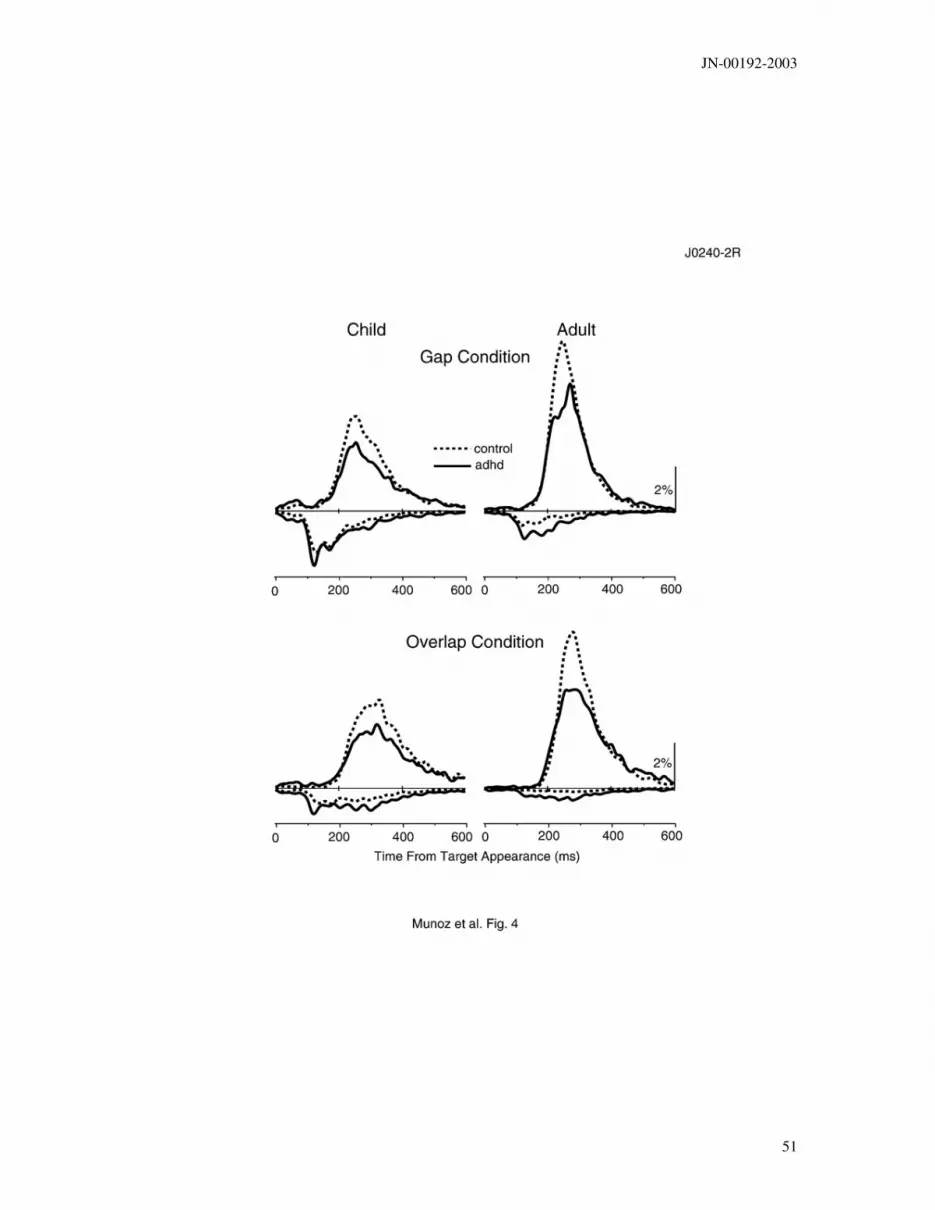
SACCADIC REACTION TIMES. Figure 4 illustrates the distribution of correct
and incorrect SRTs in the anti-saccade task. At latencies of < 90 ms, the distribution of
correct and incorrect responses mirrored each other. However, in the interval 90–180 ms
after target appearance there were more incorrect responses than correct responses. Most

JN-00192-2003
19
interestingly, there was a paucity of correct responses in the interval spanning from 90–
140 ms, the express saccade interval. Most correct responses were initiated only after 180
ms in both the gap and overlap conditions.
A comparison of the distribution of correct and error responses among ADHD and
control participants revealed: 1) ADHD participants made more errors; and 2) the onset
of correct responses of ADHD participants was more variable than the correct responses
obtained from the control participants. This increase in the percentage of direction errors
among the ADHD participants occurred across the entire range of error SRTs. An
analysis of error SRTs was not possible due to the extreme variability in error rates
among individual participants (see Fig. 6).
Figure 5A and B contrasts the mean SRT obtained in the anti-gap and anti-overlap
conditions for the different subject groups. Note that the mean SRT of correct anti-
saccades was significantly elevated for children compared to adults for both control and
ADHD groups, F(1, 290) = 46.42, p < 0.001. More importantly, mean SRT was greater
among the ADHD participants, F(1, 290) = 7.24, p < 0.01.
Mean SRT for correct anti-saccades was far greater than the mean SRT for correct
pro-saccades, F(1, 290) = 637.82, p < 0.001. This anti-effect (anti-saccade SRT – pro-
saccade SRT) was significant for each group but the difference of SRT between tasks was
larger in children than adults and increased from overlap to gap conditions for all groups
except ADHD adults, F(1, 290) = 5.28, p < 0.05. Similar to pro-saccades, there was also
a significant directional bias in anti-saccade SRT with all subject groups being faster
moving to the right side, F(1, 290) = 15.09, p < 0.001 (Table 2). There was also a
significant gap effect (overlap SRT - gap SRT) among anti-saccades that ranged from 41-

JN-00192-2003
20
59 ms, F(1, 290) = 584.96, p < 0.001. The gap effect was larger for ADHD adults than
for control adults and larger for control children than for ADHD children, resulting in an
interaction of the factors of age and fixation state, F(1, 290) = 12.06, p < 0.05.
Figure 5C and D illustrates the intra-subject variance in SRT for the subject
groups in the anti-saccade task. Once again, the amount of intra-subject variance in anti-
saccade SRT, expressed as the CV, was elevated in the ADHD groups compared to the
control groups, F(1, 290) = 58.02, p < 0.001. In addition, as with the pro-saccade task,
the CV of children was significantly elevated compared to the adult values among both
control and ADHD subjects, F(1, 290) = 95.27, p < 0.001.
DIRECTION ERRORS. The percentage of direction errors in the anti-saccade
task is illustrated in Fig. 5E and F. All groups generated more direction errors in the gap
condition than in the overlap condition, F (1, 290) = 168.94, p < 0.001, and the effect was
greater for children resulting in an interaction between age and fixation state, F(1, 290) =
5.44, p < 0.05. In addition, ADHD participants generated a greater percentage of
direction errors than did control participants, F(1, 290) = 38.78, p < 0.001. Consistent
with previous studies (Munoz et al. 1998a; Fischer et al. 1997), children made
significantly more direction errors than adults, F(1, 290) = 83.79, p < 0.001.
Figure 6 illustrates, in greater detail, the age-dependent changes in performance of
the anti-saccade task for both ADHD and control groups. Performance, expressed as the
percentage of direction errors, improved steadily across subject age from 6 – 16 years.
Among control participants, adult levels of performance were only achieved at
approximately age 16 years (see also Munoz et al. 1998a). As a group, the ADHD curve

JN-00192-2003
21
lagged the control curve and appeared to asymptote at a higher level. Most importantly
however note that among both children and adult groups, some ADHD participants
clustered along the control curve, while other ADHD participants were clearly impaired.
Thus, while some ADHD subjects had difficulties suppressing reflexive saccades, other
ADHD subjects were no different from control subjects, suggesting that there may be
subgroups within the ADHD spectrum.
Prolonged Fixation Task
The increased error rates in the anti-saccade task and the increased variance in
SRT suggest that the ADHD groups had some difficulties in controlling or gating fixation
signals. To evaluate fixation instability in greater detail, participants performed a separate
experiment consisting of blocks of pro-saccade gap trials in which the FP was visible for
1500 ms, followed by a random gap period of 0 – 800 ms in duration, which preceded the
appearance of either an eccentric target at 20° left or right, or the reappearance of the FP
at center.
INTRUSIVE SACCADES. To quantify the frequency of occurrence of intrusive
saccades we counted the frequency of saccades > 2º in amplitude that participants
initiated in the final 1000 ms of fixation upon the visible FP and during the gap period.
Figure 7 contrasts the mean rate of intrusive saccades during these two intervals for
ADHD and control children and adults. The rate of intrusive saccades among ADHD
participants was elevated above control values, F(1, 211) = 6.06, p = 0.01, and the

JN-00192-2003
22
difference in intrusive saccade rate was greater for children than for adults,. F(1, 211) =
39.76, p < 0.001.
Figure 8 illustrates, in greater detail, the rate of intrusive saccades made by
individual participants. The data were collapsed across the fixation and gap epochs. Both
control and ADHD children showed continuous improvement (i.e., reduced frequency of
intrusive saccades) across subject age. Control participants only reached adult levels of
fixation stability at approximately age 16 years. The ADHD curve lagged the control
curve and appeared to asymptote at a higher level than the control curve. Once again,
note that among both children and adult groups, some ADHD participants clustered along
the control curve, while other ADHD participants were clearly impaired, suggesting that
only a subset of ADHD subjects had difficulty suppressing intrusive saccades.
GAP EFFECT. The gap effect reflects the exogenous component of visual
fixation (i. e., difference in SRT between overlap and gap conditions). The gap effect
measured from the pro- and anti-saccade tasks among ADHD and control participants
was comparable, F(4, 844) < 1, p > 0.50. In the varied gap experiment, we measured
mean SRT for each gap interval and compared across groups. For all groups, the longest
mean SRT was obtained when the gap duration was 0 ms, the shortest mean SRT was
obtained when the gap duration was 200 ms, and longer gap durations led to an increase
in SRT from the minimal obtained at 200 ms gap duration, F(4, 844) = 99.06, p < 0.001.
Thus, it would appear that because the gap effect was comparable in all groups; the
exogenous component of fixation was not impaired in ADHD.

JN-00192-2003
23
Relation between anti-saccade direction errors and intrusive saccades
We have described two specific deficits in saccade suppression among ADHD
participants that co-varied with subject age: a difficulty in suppressing reflexive saccades
following stimulus appearance in the anti-saccade task (Fig. 6), and an inability to
suppress unwanted saccades during prolonged periods of fixation (visual and non-visual)
in the randomized gap experiment (Fig. 8). These tasks were performed on separate days.
Figure 9 contrasts the relationship between these two measures for each participant.
Figure 9A shows the intrusive saccade rate plotted against the anti-saccade error rate for
all participants. A linear regression was fit separately to the data for the control (dotted
line) and ADHD (solid line) participants which produced significant correlation values of
0.76 and 0.60, respectively. Thus, subjects with greater anti-saccade error rates were
more inclined to trigger excessive intrusive saccades. Because both the anti-saccade error
rate and the intrusive saccade rate co-varied with age, we recalculated the correlations
after removing variability related to age effects. To perform this calculation, we
subtracted the control age-matched value obtained from the cubic spline fits illustrated by
the dashed lines in Figs. 6 and 8 from the values obtained for each subject. After
removing the age-matched control values, the correlation values both remained
significant but dropped to 0.46 and 0.42 for control and ADHD participants respectively
(Fig. 9B).
DISCUSSION

JN-00192-2003
24
We have shown that individuals diagnosed with ADHD have difficulty
maintaining prolonged fixation and suppressing intrusive or unnecessary saccades. These
deficits, revealed as both an increase in intrusive saccade rate (Fig. 7) and increased
direction errors in the anti-saccade task in both the gap and overlap conditions (Fig.
5E,F), compromised task performance compared to control participants. In addition, the
greater variability in SRT (Figs. 3C, 3D, 5C, 5D) suggests that ADHD subjects had
difficulty in regulating the processes of saccade initiation. Despite these pronounced
deficits in regulating the occcurrence of saccades, ADHD subjects generated saccades
with normal or near-normal metrics, suggesting that the brainstem saccadic burst
generator circuitry was functioning normally (Leigh and Zee, 1999). We first contrast
these observations with previous studies and then we relate these findings to known
neurophysiological data obtained from non-human primates performing similar tasks to
speculate on the possible signals that are dysfunctional in ADHD.
Relation to Previous studies
Our results confirm and extend previous reports describing deficits in eye
movement control in children diagnosed with ADHD. Early studies described difficulties
in suppressing intrusive saccades during either visual fixation (Shapira et al., 1980; Paus,
1992) or during smooth pursuit eye movements (Bala et al., 1981; Blysma and Pivik,
1989). These earlier studies all noted an increase in the frequency of inappropriate
intrusive saccades during purposive behaviors. In experiments requiring subjects to delay
a saccade to a remembered target location, children diagnosed with ADHD often looked
at the target location prematurely, before the GO signal was provided (Ross et al., 1994;

JN-00192-2003
25
Mostofsky et al., 2001). In a recent study employing the anti-saccade task, Mostofsky and
colleagues (2001) found that ADHD children made significantly more direction errors
than did age-matched control children. This result is consistent with our findings (see Fig.
5E, F). In another study employing the anti-saccade task, Rothlind and colleagues (1991)
noted modest increases in the frequency of direction errors among ADHD children that
failed to reach statistical significance. However, in that study each participant performed
only 10 trials in each condition, whereas we collected between 40-60 trials in each
condition (fixation state, stimulus direction) from each participant. Another study that
failed to find a difference in the proportion of correct anti-saccades between ADHD and
control children using a gap anti-saccade task included a concurrent task in which
participants also had to indicate the open side of a target square at the anti-location
(Aman et al., 1998). Either the added task complexity or the reduced number of gap trials
(42 trials versus 80-120 in our experiments) may have accounted for this discrepancy.
The characteristics of eye movement abnormalities identified among ADHD
subjects are similar to abnormalities described for patients with frontal lobe lesions.
Frontal patients have difficulties suppressing reflexive pro-saccades in the anti-saccade
task (Guitton et al., 1985). More recently, Gaymard and colleagues (1999) have revealed
that lesions confined to the frontal eye field do not lead to increased error rates. Instead,
lesions to the dorsolateral prefrontal cortex, rostral to the frontal eye fields have been
attributed to increased error rates on anti-saccade trials (Gaymard et al., 1998). Lesions
confined to the frontal eye fields instead lead to prolonged latencies and reduced
accuracy of correct pro- and anti-saccades (Rivaud et al., 1994).

JN-00192-2003
26
Eye movement abnormalities among patients with basal ganglia dysfunction have
produced a variety of results (see Everling and Fischer, 1998 for review), only some of
which match the abnormalities we have reported for ADHD. Patients with Parkinson’s
disease, in which dopaminergic input to the striatum is reduced, have relatively normal
pro-saccades, while performance on the anti-saccade task has produced contradictory
results with some studies reporting no differences in reaction times and error rates (Lueck
et al., 1990; Fukushima et al., 1994) and other studies finding increased reaction times
and error rates (Briand et al., 1999). In Tourette’s syndrome, in which it is hypothesized
that the direct pathway through the basal ganglia may be overactive (Hallett, 1993),
patients have increased reaction times on pro- and anti-saccade tasks, no increased error
rates in an immediate anti-saccade task, and instead they have a difficulty withholding
saccadic responses in tasks with prolonged delay intervals (LeVasseur et al., 2001). In
Huntington’s disease, in which there is initial degeneration in the indirect pathway
through the basal ganglia, patients produce increased direction errors on anti-saccade
tasks (Lasker et al., 1987), increased reaction times (Lasker et al., 1988), and increased
fixation instability (Leigh et al., 1983).
Precise control over saccade suppression is also diminished in young children
(Munoz et al., 1998a); they also have difficulty suppressing reflexive pro-saccades in the
anti-saccade task, have greater intra-subject variance in SRT, and generate more intrusive
saccades. Task performance improves steadily in children up to the age of approximately
16 years and is correlated with maturation across a network of brain areas that includes
the frontal cortex and basal ganglia (Luna et al. 2001).
JN-00192-2003
27
An intriguing aspect of our data is the developmental progression of saccadic
suppression ability in ADHD and normal participants. While our data are suggestive of a
developmental delay in ADHD, some caution is warranted. The best way to investigate
developmental changes in ability is to follow the same individual subjects longitudinally.
In the present study, we only compared across subjects of different ages.
Relation to Neurophysiology
The deficits in fixation control and saccade suppression observed among ADHD
participants can be best understood in the context of recent neurophysiological studies in
non-human primates that have identified neural mechanisms of fixation control, saccade
suppression, and saccade production. Several brain areas are involved in the control of
visual fixation and saccade production, including regions within the cerebral cortex
(posterior parietal and frontal cortex), basal ganglia (caudate, substantia nigra,
subthalamic nucleus), thalamus, superior colliculus, brainstem reticular formation, and
cerebellum (see Wurtz and Goldberg, 1989; Leigh and Zee, 1999; Hikosaka et al., 2000;
Munoz et al., 2000; Scudder et al 2002 for review). Two important nodes in this network
are the frontal eye fields (FEF) in the frontal lobes and the intermediate layers of the
superior colliculus (SC). These structures appear to work in concert in the initiation of
saccades (Munoz and Schall 2003).
Fixation neurons in the SC and FEF are tonically active during visual fixation and
pause during saccades, while saccade neurons have a reciprocal pattern, being silent
during visual fixation and active prior to and during saccade generation (Munoz et al.,
2000; Munoz and Fecteau 2002; Munoz and Schall 2003). Removal of an exogenous

JN-00192-2003
28
fixation target leads to a reduction in fixation activity and disinhibition of saccade
generating neurons in the FEF and SC (Dias and Bruce 1994; Dorris and Munoz, 1995;
Dorris et al., 1997; Everling et al. 1999; Everling and Munoz 2000). However, there is
also an endogenous (non-visual) component in the discharge of these fixation neurons
(Munoz and Wurtz, 1993a; Dorris et al., 1997). On anti-saccade trials, fixation activity in
the SC and FEF is enhanced relative to pro-saccade trials, whereas saccade neurons in the
SC and FEF are at a reduced level of excitability (Everling et al., 1999; Everling and
Munoz, 2000). This task-dependent modulation of fixation and saccade signals in the SC
and FEF is apparent immediately following the initiation of a trial, well before the
appearance of the eccentric target stimulus dictating the correct direction for a response,
and represents selective control of endogenous fixation signals required to suppress
reflexive or unwanted saccades.
What happens on error trials in the anti-saccade task when a non-human primate
triggers a direction error and instead looks at the target stimulus? Among saccade
neurons in the SC and FEF, an elevated level of pre-target activity is combined with the
phasic visual response produced by the appearance of the target stimulus to trigger
reflexive pro-saccades (Everling et al. 1998; Everling and Munoz 2000). Thus, to
correctly perform the anti-saccade task, suppression signals must be boosted to reduce the
excitability of these saccade neurons. Because the location of the eccentric stimulus is
varied randomly from trial to trial, successful performance on anti-saccade trials requires
inhibition of all saccade generating neurons in advance of appearance of the eccentric
stimulus. The saccade neurons required to drive the correct anti-saccade are then

JN-00192-2003
29
activated only after the successful suppression of the reflexive pro-saccade. Thus, the
SRT for correct anti-saccades exceeds that of correct pro-saccades (see Table 2).
What are the likely sources of important endogenous control signals to the FEF
and SC required for saccadic suppression? Two likely structures are the dorsolateral
prefrontal cortex (DLPFC) and the substantia nigra pars reticulata (SNr). Neurons in both
of these structures are modulated by voluntary tasks (Funahashi et al. 1993; Hikosaka and
Wurtz 1983). The DLPFC projects directly to the FEF and SC (see Munoz and Schall
2003 for review) and this input could terminate on fixation neurons in these structures. In
addition, the DLPFC and FEF project to the caudate nucleus, which contains GABAergic
neurons that project directly to the SNr (see Hikosaka et al. 2000 for review). The SNr
contains GABAergic neurons that project to the SC and indirectly back to the FEF via the
thalamus.
The above pattern of connectivity is also consistent with the effects of reversible
lesions in brain regions known to play a role in the endogenous control of fixation.
Microinjection of the GABAA agonist muscimol into either the rostral superior colliculus
(Munoz and Wurtz 1992; 1993b) or substantia nigra pars reticulata (Hikosaka and Wurtz
1985) leads to artificial inhibition of important endogenous fixation signals. These
experimental manipulations lead to increased frequencies of intrusive saccades and
inability to suppress reflexive saccades to suddenly appearing visual stimuli, deficits that
are very similar to those presented by a subset of the ADHD participants. These brain
areas provide important inhibitory input to the saccade generating neurons in the SC
(Hikosaka and Wurtz, 1983; Munoz and Wurtz, 1993a; Munoz and Istvan, 1998), and
possibly the FEF via a thalamic relay (Lynch et al., 1994). We hypothesize that, due to a

JN-00192-2003
30
frontostriatal pathophysiology, these suppression signals are weak or dysfunctional in
ADHD.
Altered frontostriatal function could also explain the subtle but significant
reduction in velocity and increase in duration of saccades in ADHD. In non-human
primates, small reversible lesions of the frontal eye fields produced with injection of
lidocaine (Sommer and Tehovnik, 1997) or the GABAA agonist muscimol (Dias and
Segraves, 1999) lead to small but significant reductions in peak saccade velocity and
increases in saccade duration. Thus, the slowed but accurate saccades generated by
ADHD participants are consistent with altered descending input to the brainstem rather
than pathophysiology affecting the brainstem and cerebellar portions of the saccadic burst
generator circuitry (Leigh and Zee, 1999).
Conclusions
There are both exogenous and endogenous components of fixation control that
were examined in the current study. The exogenous component of fixation appears intact
in ADHD because the gap effect was normal and the occurrence of express saccades was
not increased. In contrast, the ADHD group had poor control over the endogenous
component of fixation that required task dependent modulation in the diligence of
fixation. This was evident from their impaired ability to suppress reflexive pro-saccades
in the anti-saccade task and intrusive saccades during periods of prolonged fixation, and
the increased variability in reaction times of their saccades.
JN-00192-2003
31
We hypothesize that important saccade suppression signals related to voluntary
control of endogenous fixation emanate from the prefrontal cortex and/or the basal
ganglia. These signals are required to selectively inhibit saccade neurons in the frontal
eye fields and superior colliculus to control precisely the timing of saccades. Without
such precise control, it is easier for the saccadic system to reach the threshold for
triggering saccades. We suggest that it is the precise control of these saccade suppression
signals that is disrupted in ADHD. This lack of inhibitory control is the hallmark of the
ADHD phenotype and is consistent with a frontostriatal pathophysiology. As a result,
ADHD subjects have increased error rates in the anti-saccade task, increased rates of
intrusive saccades, and increased intra-subject variance in SRT.

JN-00192-2003
32
ACKNOWLEDGEMENTS
We thank Dr. Deb Thompson for clinical assessment of ADHD subjects, Jaana
Broughton, Florence Chan, Joan Dafoe, Jenny Goldring, Christine Hing, Karen Kazman,
and Adrienne Levasseur for assisting in the data collection and analysis, and Dave
Hamburger for providing computer assistance. This work was supported by the EJLB
Foundation. DP Munoz is supported by a Canada Research Chair in Neuroscience.

JN-00192-2003
33
REFERENCES
Aman CJ, Roberts RJ Jr, Pennington BF. A neuropsychological examination of the
underlying deficit in attention deficit hyperactivity disorder: frontal lobe versus right
parietal lobe theories. Dev Psychol. 34: 956-969, 1998.
Armstrong, IT and Munoz, DP. Unnecessary eye movements and blinks reduce
attentional blink performance in adults with attention-deficit/hyperactivity disorder.
Soc. Neurosci. Abstr. 26: 2309, 2000.
Bala SP, Cohen B, Morris AG, Atkin A, Gittelman R, Kates W. Saccades of hyperactive
and normal boys during ocular pursuit. Develop Med Child Neurol 23: 323-336,
1981.
Barkley RA. ADHD and the nature of self-control. Guildford Press, New York; 1997.
Biscaldi M, Fischer B, Stuhr V. Human express-saccade makers are impaired at
suppressing visually-evoked saccades. J Neurophysiol 76: 199-214, 1996.
Briand KA, Strallow D, Hening W, Poizner H, Sereno AB. Control of voluntary and
reflexive saccades in Parkinson's disease. Exp Brain Res 129: 38-48, 1999.
Bylsma FW, Pivik RT. The effects of background illumination and stimulant medication
on smooth pursuit eye movements of hyperactive children. J Abnormal Child Psych
17: 73-90, 1989.
Cairney S, Maruff P, Vance A, Barnett R, Luk E, Currie J. Contextual abnormalities of
saccadic inhibition in children with attention deficit hyperactivity disorder. Exp
Brain Res. 141:507-518, 2001.
Castellanos FX. Neural substrates of attention-deficit hyperactivity disorder. Adv Neurol
85:197-206, 2001.

JN-00192-2003
34
Castellanos FX, Giedd JN, Berquin PC, Walter JM, Sharp W, Tran T, Vaituzis AC,
Blumenthal JD, Nelson J, Bastain TM, Zijdenbos A, Evans AC, Rapoport JL.
Quantitative brain magnetic resonance imaging in girls with attention-
deficit/hyperactivity disorder. Arch Gen Psychiatry 58:289-295, 2001.
Castellanos FX, Giedd JN, Marsh WL, Hamburger SD, Vaituzis AC, Dickstein DP,
Sarfatti SE, Vauss YC, Snell JW, Rajapakse JC, Rapoport JL. Quantitative brain
magnetic resonance imaging in attention-deficit hyperactivity disorder. Arch Gen
Psychiatry 53: 607-616, 1996a.
Castellanos FX, Elia J, Kruesi MJP, Marsh WL, Gulotta CS, Potter, WZ, Ritchie GF,
Hamburger SD, Rapoport JL. Cerebrospinal fluid homovanillic acid predicts
behavioral response to stimulants in 45 boys with attention deficit/ hyperactivity
disorder. Neuropsychopharm 14: 125-137, 1996b.
Chelune GJ, Ferguson W, Koon R, Dickey TO. Frontal lobe disinhibition in attention
deficit disorder. Child Psychiat Human Dev 16: 221-234, 1986.
Connolly JD, Goodale MA, Desouza JF, Menon RS, Vilis T. A comparison of
frontoparietal fMRI activation during anti-saccades and anti-pointing. J
Neurophysiol. 84:1645-1655, 2000.
Connolly JD, Goodale MA, Menon RS, Munoz DP. Human fMRI evidence for the neural
correlates of preparatory set. Nat. Neurosci. 5:1345-1352, 2002.
De Boor C. A practical guide to splines. In: Applied Mathematical Sciences, eds. F John,
J Lasalle, L Sirovich, G Whitman. Springer-Verlag, New York vol 27, p 235-249;
1978.

JN-00192-2003
35
Dias EC, Bruce CJ. Physiological correlate of fixation disengagement in the primate's
frontal eye field. J Neurophysiol. 72: 2532-2537, 1994.
Dias EC, Segraves MA. Muscimol-induced inactivation of monkey frontal eye field:
effects on visually and memory-guided saccades. J Neurophysiol 81: 2191-2214,
1999.
Dorris MC, Munoz DP. A neural correlate for the gap effect on saccadic reaction times in
monkey. J. Neurophysiol. 73: 2558-2562, 1995.
Dorris MC, Munoz DP. Saccadic probability influences motor preparation signals and
time to saccadic initiation. J. Neurosci. 18: 7015-7026, 1998.
Dorris MC, Paré M, Munoz DP. Neuronal activity in monkey superior colliculus related
to the initiation of saccadic eye movements. J Neurosci 17: 8566-8579, 1997.
Everling S, Dorris MC, Munoz DP. Reflex suppression in the anti-saccade task is
dependent on prestimulus neural processes. J. Neurophysiol. 80:1584-1589, 1998.
Everling S, Dorris MC, Klein RM, Munoz DP. Role of primate superior colliculus in
preparation and execution of anti- and pro-saccades. J. Neurosci. 19: 2740-2754,
1999.
Everling S, Fischer B. The antisaccade: a review of basic research and clinical studies.
Neuropsychologia 36: 885-899, 1998.
Everling S, Munoz DP. Neuronal correlates for preparatory set associated with saccade
generation in the primate frontal eye field. J. Neurosci. 20: 387-400, 2000.
Fischer B, Biscaldi M, Gezeck S. On the development of voluntary and reflexive
components in human saccade generation. Brain Res 754: 285-297, 1997.

JN-00192-2003
36
Fischer B, Ramsperger E. Human express saccades: extremely short reaction times of
goal directed eye movements. Exp Brain Res 57: 191-195, 1984.
Fischer B, Weber H. Characteristics of “anti” saccades in man. Exp Brain Res 89: 415-
424, 1992.
Fischer B, Weber H. Express saccades and visual attention. Behav Brain Sci 16: 553-610,
1993.
Fischer B, Weber H. Effects of stimulus conditions on the performance of antisaccades in
man. Exp Brain Res 116: 191-200, 1997.
Fischer B, Weber H, Biscaldi M, Aiple F, Otto P, Stuhr V. Separate populations of
visually guided saccades in humans: reaction times and amplitudes. Exp Brain Res
92: 528-541, 1993.
Fukushima J, Fukushima K, Miyasaka K, Yamashita I. Voluntary control of saccadic eye
movement in patients with frontal cortical lesions and parkinsonian patients in
comparison with that in schizophrenics. Biol Psychiatry. 36: 21-30, 1994.
Funahashi S, Chafee MV, Goldman-Rakic PS. Prefrontal neuronal activity in rhesus
monkeys performing a delayed anti-saccade task. Nature 365:753-756, 1993.
Fuster JM The Prefrontal Cortex: Anatomy, Physiology, and Neuropsychology of the
frontal lobe. Lippincott- Raven, Philadelphia; 1997.
Gaymard B, Ploner CJ, Rivaud-Pechoux S, Pierrot-Deseilligny C. The frontal eye field is
involved in spatial short-term memory but not in reflexive saccade inhibition. Exp
Brain Res 129:288-301, 1999.
Gaymard B, Ploner CJ, Rivaud S, Vermersch AI, Pierrot-Deseilligny C. Cortical control
of saccades. Exp Brain Res 123:159-163, 1998.

JN-00192-2003
37
Giedd JN, Castellanos FX, Casey BJ, Kozuch P, King AC, Hamburger SD, Rapoport JL.
Quantitative morphology of the corpus callosum in attention deficit hyperactivity
disorder. Am J Psychiatry 151: 665-669, 1994.
Grodzinsky GM, Diamond R. Frontal lobe functioning in boys with attention-deficit
hyperactivity disorder. Dev Neuropsych 8: 427-445, 1992.
Guitton D, Buchtel H, Douglas R. Frontal lobe lesions in man cause difficulties in
suppressing reflexive saccades and in generating goal-directed saccades. Exp Brain
Res 58: 455-472, 1985.
Hallett M. Physiology of basal ganglia disorders: an overview. Can J Neurol Sci 20: 177-
183, 1993.
Hallett P. Primary and secondary saccades to goals defined by instructions. Vision Res
18: 1279-1296, 1978.
Hays AV, Richmond BJ, Optican LM A UNIX-based multiple process system for real-
time data acquisition and control. WESCON Conf Proc 2: 1-10, 1982.
Hikosaka O, Takikawa Y, Kawagoe R. Role of the basal ganglia in the control of
purposive saccadic eye movements. Physiol Rev 80:953-978, 2000.
Hikosaka O, Wurtz RH. Modification of saccadic eye movements by GABA-related
substances. II. Effects of muscimol in monkey substantia nigra pars reticulata. J
Neurophysiol 53: 292-308, 1985.
Hikosaka O, Wurtz RH. Visual and oculomotor functions of monkey substantia nigra
pars reticulata. II. Visual responses related to fixation of gaze. J Neurophysiol 49:
1254-1267, 1983.
JN-00192-2003
38
Kalesnykas RP, Hallett PE. The differentiation of visually guided and anticipatory
saccades in gap and overlap paradigms. Exp Brain Res 68:115-121, 1987.
Lasker AG, Zee DS, Hain TC, Folstein SE, Singer HS. Saccades in Huntington's disease:
initiation defects and distractibility. Neurology 37: 364-370, 1987.
Lasker AG, Zee DS, Hain TC, Folstein SE, Singer HS. Saccades in Huntington's disease:
slowing and dysmetria. Neurology 38: 427-431, 1988.
Leigh RJ, Newman SA, Folstein SE, Lasker AG, Jensen BA. Abnormal ocular motor
control in Huntington's disease. Neurology 33: 1268-1275, 1983.
Leigh RJ, Zee DS The neurology of eye movements. FA Davis, Philadelphia; 1999.
LeVasseur AL, Flanagan JR, Riopelle RJ, Munoz DP. Control of volitional and reflexive
saccades in Tourette’s syndrome. Brain 124: 2045-2058, 2001.
Levy F. The dopamine theory of attention deficit hyperactivity disorder (ADHD).
Australian NZ J Psychiatry 25: 277-283, 1991.
Lou HC, Henriksen L, Bruhn P. Focal cerebral hypoperfusion in children with dysphasia
and/or attention deficit disorder. Arch Neurol 41: 825-829, 1984.
Lou HC, Henriksen L, Bruhn P, Borner H, Nielsen JB. Striatal dysfunction in attention
deficit and hyperkinetic disorder. Arch Neurol 46: 48-52, 1989.
Luna B, Thulborn KR, Munoz DP, Merriam EP, Garver KE, Minchew NJ, Keshavan MS,
Genovese CR, Eddy WF, Sweeney JA. Maturation of widely distributed brain
function subserves cognitive development. Neuroimage 13: 786-793, 2001.
Lueck CJ, Tanyeri S, Crawford TJ, Henderson L, Kennard C. Antisaccades and
remembered saccades in Parkinson's disease. J Neurol Neurosurg Psychiatry. 53:
284-288, 1990.
JN-00192-2003
39
Lynch JC, Hoover JE, Strick PL. Input to the primate frontal eye field from the substantia
nigra, superior colliculus, and dentate nucleus demonstrated by transneuronal
transport. Exp Brain Res. 100: 181-186, 1994.
Moschovakis AK, Scudder CA, Highstein SM. The microscopic anatomy and physiology
of the mammalian saccadic system. Prog Neurobiol. 50:133-254, 1996.
Mostofsky SH Lasker AG, Cutting LE, Denckla MB, Zee DS. Oculomotor abnormalities
in attention deficit hyperactivity disorder. A preliminary study. Neurology 57:423-
430, 2001.
Munoz DP, Broughton JR, Goldring JE, Armstrong IT Age-related performance of
human subjects on saccadic eye movement tasks. Exp. Brain Res. 121: 391-400,
1998a.
Munoz DP, Corneil BD Evidence for interactions between target selection and visual
fixation for saccade generation in humans. Exp Brain Res 103: 168-173, 1995.
Munoz DP, Dorris MC, Paré M, Everling S On your mark, get set: brainstem circuitry
underlying saccadic initiation. Can. J. Physiol. Pharmacol. 78: 934-944, 2000.
Munoz DP and Fecteau JH. Vying for dominance: dynamic interactions control visual
fixation and saccadic initiation in the superior colliculus. Prog. Brain Res. 140: (in
press), 2002.
Munoz DP, Hampton KA, Moore KD, Armstrong IT. Control of saccadic eye movements
and visual fixation in children and adults with attention deficit hyperactivity
disorder. Soc. Neurosci. Abstr. 24: 671, 1998b.
Munoz DP, Hampton KA, Moore KD, Goldring JE. Control of purposive saccadic eye
movements and visual fixation in children with attention deficit hyperactivity

JN-00192-2003
40
disorder. In: Current Oculomotor Research: Physiological and Psychological
Aspects, eds. W. Becker, H. Deubel, and T. Mergner, Plenum, New York. pp. 415-
423; 1999.
Munoz DP, Istvan PJ. Lateral inhibitory interactions in the intermediate layers of the
monkey superior colliculus. J. Neurophysiol 79: 1193-1209, 1998.
Munoz DP and Schall JD. Concurrent, distributed control of saccade initiation in the
frontal eye field and superior colliculus. In: The Oculomotor System: New
Approaches for Studying Sensorimotor Integration. eds. WC Hall and AK
Moschovakis, CRC Press, Boca Raton, FL (in press), 2003.
Munoz DP, Wurtz RH. Role of the rostral superior colliculus in active visual fixation and
execution of express saccades. J Neurophysiol 67: 1000-1002, 1992.
Munoz DP, Wurtz RH. Fixation cells in monkey superior colliculus. I. Characteristics of
cell discharge. J. Neurophysiol. 70: 559-575, 1993a.
Munoz DP, Wurtz RH. Fixation cells in monkey superior colliculus. II. Reversible
activation and deactivation. J. Neurophysiol. 70: 576-589, 1993b.
Muri RM, Heid O, Nirkko AC, Ozdoba C, Felblinger J, Schroth G, Hess CW. Functional
organisation of saccades and antisaccades in the frontal lobe in humans: a study with
echo planar functional magnetic resonance imaging. J Neurol Neurosurg Psychiatry.
65: 374-377, 1998.
Oldfield RC The assessment and analysis of handedness. Neuropsychologia 9: 97-113,
1971.
JN-00192-2003
41
Paré M, Munoz DP. Saccadic reaction time in the monkey: advanced preparation of
oculomotor programs is primarily responsible for express saccade occurrence. J
Neurophysiol 76: 3666-3681, 1996.
Paus T. Impaired voluntary suppression of reflexive saccades in attention-deficit
hyperactivity disordered boys. Thalamus 8: 1-23, 1992.
Pierrot-Deseilligny Ch, Ploner CJ, Muri RM, Gaymard B, Rivaud-Pechoux S. Effects of
cortical lesions on saccadic: eye movements in humans. Ann N Y Acad Sci. 956: 216-
229, 2002.
Reuter-Lorenz PA, Oonk HM, Barnes LL, Hughes HC. Effects of warning signals and
fixation point offsets on the latencies of pro- versus antisaccades: implications for an
interpreatation of the gap effect. Exp Brain Res 103: 287-293, 1995.
Rivaud S, Muri RM, Gaymard B, Vermersch AI, Pierrot-Deseilligny C. Eye movement
disorders after frontal eye field lesions in humans. Exp Brain Res 102: 110-120,
1994.
Ross RG, Hommer D, Breiger D, Varley C, Radant A. Eye movement task related to
frontal lobe functioning in children with attention deficit disorder. J Am Acad Child
Adolesc Psychiatry 33: 869-874, 1994.
Rothlind JC, Posner MI, Schaughency EA. Lateralized control of eye movements in
attention deficit hyperactivity disorder. J Cog Neurosci 3: 377-381, 1991.
Rubia K, Overmeyer S, Taylor E, Brammer M, Williams SC, Simmons A, Bullmore ET.
Hypofrontality in attention deficit hyperactivity disorder during higher-order motor
control: a study with functional MRI. Am J Psychiatry. 156:891-896, 1999.

JN-00192-2003
42
Saslow MG. Effects of component displacement-step stimuli upon latency of saccadic
eye movements. J Opt Soc Am 57: 1024-1029, 1967.
Schall JD. Visuomotor areas of the frontal lobe. Cereb Cortex 12: 527-638, 1997.
Scudder CA, Kaneko CS, Fuchs AF. The brainstem burst generator for saccadic eye
movements: a modern synthesis. Exp. Brain Res. 142: 439-462, 2002.
Shapira YA, Jones MH, Sherman SP. Abnormal eye movements in hyperkinetic children
with learning disability. Neuropadiatrie 11: 36-44, 1980.
Schlag-Rey M, Amador N, Sanchez H, Schlag J. Antisaccade performance predicted by
neuronal activity in the supplementary eye field. Nature 390: 398-401, 1997.
Shue KL, Douglas V. Attention deficit hyperactivity disorder and the frontal lobe
syndrome. Brain Cogn 20:104-124, 1992.
Sommer MA, Tehovnik EJ. Reversible inactivation of macaque frontal eye field. Exp
Brain Res 116: 229-249, 1997.
Wurtz RH, Goldberg ME. The neurobiology of saccadic eye movements. Elsevier,
Amsterdam; 1989.
Zametkin AJ, Nordahl TE, Gross M, King C, Semple WE, Rumset J, Hamburger S,
Cohen RM. Cerebral glucose metabolism in adults with hyperactivity of childhood
onset. N Engl J Med 323: 1361-1366, 1990.

JN-00192-2003
43
Table 1. Characteristics of subject pool utilized in the present study. Values expressed as
mean ± standard error.
Subject Group N (female) Age Handedness CPRSHyperactivity
CPRSImpulsivity
Child Control 75 (35) 10.7 (± 0.3) 7.8 (± 0.4) 45.9 (± 1.0) 47.2 (± 1.1)
ADHD 76 (15) 10.3 (± 0.3) 6.1 (± 0.6) 84.1 (± 1.4) 75.7 (± 1.3)
Adults Control 105 (59) 34.2 (± 1.1) 6.8 (± 0.4) - -
ADHD 38 (20) 31.0 (± 1.4) 4.8 (± 1.0) - -

JN-00192-2003
44
Table 2. Mean saccadic reaction times of studied groups contrasting direction differences
in correct responses.
Pro-saccade Anti-saccade
Subject group
Left Right Left Right
Child
Control 257 253 367 356
Adhd 282 265 382 378
Adult
Control 244 243 305 300
Adhd 255 252 335 321
JN-00192-2003
45
FIGURE LEGENDS
Figure 1. Schematic showing the behavioral paradigms used. Within a block of trials,
subjects were instructed to either look from the fixation point (FP) to the eccentric target
(T) (A: Pro-saccade Task) or from the FP to the mirror position of the T (B: Anti-saccade
Task). Solid arrows represent the correct response, dashed arrow represents a direction
error. In the overlap condition (C), the central FP remained illuminated when T appeared.
In the gap condition (D), the FP disappeared before T appearance. The gap duration was
either kept constant at 200 ms or varied randomly from 0 – 800 ms (see METHODS).
SRT: saccadic reaction time.
Figure 2. Distribution of saccadic reaction times (SRTs) from all subjects in the pro-
saccade task for gap and overlap conditions. Curves were generated by binning all
latencies into 10 ms bins and then fitting a cubic spline function (de Boor 1978) through
the data. Solid lines (ADHD), dotted lines (control). Positive percentage on ordinate
represents correct responses, while negative values represent direction errors.
Figure 3. Quantification of parameters in the gap (left column) and overlap (right
column) conditions for the pro-saccade task. A,B. Mean saccadic reaction times for
correct responses. C,D. Coefficient of variation (standard deviation of SRT / mean SRT x
100%). E,F. Percentage of express saccades (SRT: 90 – 140 ms). Values represent mean
(± SEM). Gap duration was fixed at 200 ms.

JN-00192-2003
46
Figure 4. Distribution of saccadic reaction times (SRTs) from all subjects in the anti-
saccade task for gap and overlap conditions. Curves were generated by binning all
latencies into 10 ms bins and then fitting a cubic spline function (de Boor 1978) through
the data. Solid lines (ADHD), dotted lines (control). Positive percentage on ordinate
represents correct responses, while negative values represent direction errors.
Figure 5. Quantification of parameters in the gap (left column) and overlap (right
column) conditions for the anti-saccade task. A,B. Mean saccadic reaction times for
correct responses. C,D. Coefficient of variation (standard deviation of SRT / mean SRT x
100%). E,F. Percentage of direction errors in the anti-saccade task. Values represent
mean (± SEM). Gap duration was fixed at 200 ms.
Figure 6. Anti-saccade error rate as a function of age for control (empty circles, dotted
line) and ADHD (filled circles, solid line) participants. Curves were fit through the data
using a cubic spline function (de Boor 1978).
Figure 7. Quantification of mean fixation instability for control and ADHD children and
adults. Instability was measured as the rate of generation of saccades ≥ 2° in amplitude
during instructed fixation of the visible FP (A) and the randomized gap period (B).
Figure 8. Intrusive saccade rate as a function of age for control (empty circles, dotted
line) and ADHD (filled circles, solid line) participants. Curve were fit through the data
using a cubic spline function (de Boor 1978).
JN-00192-2003
47
Figure 9. A. Intrusive saccade rate as a function of anti-saccade error rate for control
(empty circles, dotted line) and ADHD (filled crosses, solid line) participants (control
line: correlation = 0.76, slope = 0.0067, intercept = 0.098; ADHD line: correlation = 0.60,
slope = 0.0070, intercept = 0.17). B. Intrusive saccade rate versus anti-saccade error rate
after subtracting age-matched control values (value of cubic spline fits in Figs. 7 and 9,
respectively) from each subject’s data (control line: correlation = 0.46, slope = 0.0045,
intercept = -0.0005; ADHD line: correlation = 0.42, slope = 0.0054, intercept = 0.085).

JN-00192-2003
48

JN-00192-2003
49

JN-00192-2003
50
JN-00192-2003
51

JN-00192-2003
52

JN-00192-2003
53

JN-00192-2003
54

JN-00192-2003
55

JN-00192-2003
56

JN-00192-2003
57