feature paper in antibiotics for 2019 - mdpi
TRANSCRIPT
Feature Paper in Antibiotics for 2019
Printed Edition of the Special Issue Published in Antibiotics
www.mdpi.com/journal/antibiotics
Jeffrey LipmanEdited by
Feature Paper in Antibiotics for 2019 • Jeffrey Lipman

Feature Paper in Antibiotics for 2019

Feature Paper in Antibiotics for 2019
Editor
Jeffrey Lipman
MDPI • Basel • Beijing • Wuhan • Barcelona • Belgrade • Manchester • Tokyo • Cluj • Tianjin
Editor
Jeffrey Lipman
The University of Queensland School of Medicine
Australia
Editorial Office
MDPI
St. Alban-Anlage 66
4052 Basel, Switzerland
This is a reprint of articles from the Special Issue published online in the open access journal Antibiotics
(ISSN 2079-6382) (available at: https://www.mdpi.com/journal/antibiotics/special issues/feature
paper antibiotics).
For citation purposes, cite each article independently as indicated on the article page online and as
indicated below:
LastName, A.A.; LastName, B.B.; LastName, C.C. Article Title. Journal Name Year, Article Number,
Page Range.
ISBN 978-3-03943-122-9 (Hbk) ISBN 978-3-03943-123-6 (PDF)
c© 2020 by the authors. Articles in this book are Open Access and distributed under the Creative
Commons Attribution (CC BY) license, which allows users to download, copy and build upon
published articles, as long as the author and publisher are properly credited, which ensures maximum
dissemination and a wider impact of our publications.
The book as a whole is distributed by MDPI under the terms and conditions of the Creative Commons
license CC BY-NC-ND.
Contents
About the Editor . . . . . . . . . . . . . . . . . . . . . . . . . . . . . . . . . . . . . . . . . . . . . . vii
Dagan O Lonsdale and Jeffrey Lipman
Antimicrobial Resistance: We Must Pursue a Collaborative, Global Approach and Use a “OneHealth” ApproachReprinted from: Antibiotics 2019, 8, 237, doi:10.3390/antibiotics8040237 . . . . . . . . . . . . . . . 1
Aleksandra J. Borek, Marta Wanat, Anna Sallis, Diane Ashiru-Oredope, Lou Atkins,
Elizabeth Beech, Susan Hopkins, Leah Jones, Cliodna McNulty, Karen Shaw, Esther Taborn,
Christopher Butler, Tim Chadborn and Sarah Tonkin-Crine
How Can National Antimicrobial Stewardship Interventions in Primary Care Be Improved?A Stakeholder ConsultationReprinted from: Antibiotics 2019, 8, 207, doi:10.3390/antibiotics8040207 . . . . . . . . . . . . . . . 5
Shweta Rajkumar Singh, Alvin Qijia Chua, Sok Teng Tan, Clarence C. Tam, Li Yang Hsu and
Helena Legido-Quigley
Combating Antimicrobial Resistance in Singapore: A Qualitative Study Exploring the PolicyContext, Challenges, Facilitators, and Proposed StrategiesReprinted from: Antibiotics 2019, 8, 201, doi:10.3390/antibiotics8040201 . . . . . . . . . . . . . . . 21
Julia S. Vianna, Diana Machado, Ivy B. Ramis, Fabia P. Silva, Dienefer V. Bierhals, Michael Andres Abril, Andrea von Groll, Daniela F. Ramos, Maria Cristina S. Lourenco, Miguel Viveiros and Pedro E. Almeida da Silva
The Contribution of Efflux Pumps in Mycobacterium abscessus Complex Resistance to ClarithromycinReprinted from: Antibiotics 2019, 8, 153, doi:10.3390/antibiotics8030153 . . . . . . . . . . . . . . . 39
Joana P. Costa, M. Joana F. Pinheiro, Sılvia A. Sousa, Ana M. Botelho do Rego, Fernanda Marques, M. Conceicao Oliveira, Jorge H. Leitao, Nuno P. Mira and M. Fernanda N. N. Carvalho
Antimicrobial Activity of Silver Camphorimine Complexes against Candida StrainsReprinted from: Antibiotics 2019, 8, 144, doi:10.3390/antibiotics8030144 . . . . . . . . . . . . . . . 55
Graeme Hood, Lina Toleikyte and Diane Ashiru-Oredope
Assessing National Antimicrobial Resistance Campaigns Using a Health Equity AssessmentTool (HEAT)Reprinted from: Antibiotics 2019, 8, 121, doi:10.3390/antibiotics8030121 . . . . . . . . . . . . . . . 69
Marthe Sunde, Marthe Marie Nygaard and Sigurd Høye
General Practitioners’ Attitudes toward Municipal Initiatives to Improve AntibioticPrescribing—A Mixed-Methods StudyReprinted from: Antibiotics 2019, 8, 120, doi:10.3390/antibiotics8030120 . . . . . . . . . . . . . . . 77
Larissa Grigoryan, Susan Nash, Roger Zoorob, George J. Germanos, Matthew S. Horsfield,
Fareed M. Khan, Lindsey Martin and Barbara W. Trautner
Qualitative Analysis of Primary Care Provider Prescribing Decisions for UrinaryTract InfectionsReprinted from: Antibiotics 2019, 8, 84, doi:10.3390/antibiotics8020084 . . . . . . . . . . . . . . . . 85
v
Annelies Colliers, Niels Adriaenssens, Sibyl Anthierens, Stephaan Bartholomeeusen, Hilde Philips, Roy Remmen and Samuel Coenen
Antibiotic Prescribing Quality in Out-of-Hours Primary Care and Critical Appraisal of Disease-Specific Quality IndicatorsReprinted from: Antibiotics 2019, 8, 79, doi:10.3390/antibiotics8020079 . . . . . . . . . . . . . . . 97
Rosa Elvira Gavilan, Carolina Nebot, Ewelina Patyra, Beatriz Vazquez, Jose Manuel Miranda
and Alberto Cepeda
Determination of Florfenicol, Thiamfenicol and Chloramfenicol at Trace Levels in Animal Feedby HPLC–MS/MSReprinted from: Antibiotics 2019, 8, 59, doi:10.3390/antibiotics8020059 . . . . . . . . . . . . . . . 107
Eunice Ego Mgbeahuruike, Milla Stalnacke, Heikki Vuorela and Yvonne Holm
Antimicrobial and Synergistic Effects of Commercial Piperine and Piperlongumine inCombination with Conventional AntimicrobialsReprinted from: Antibiotics 2019, 8, 55, doi:10.3390/antibiotics8020055 . . . . . . . . . . . . . . . . 117
Graeme Hood, Kieran S. Hand, Emma Cramp, Philip Howard, Susan Hopkins and Diane Ashiru-Oredope
Measuring Appropriate Antibiotic Prescribing in Acute Hospitals: Development of a National Audit Tool Through a Delphi ConsensusReprinted from: Antibiotics 2019, 8, 49, doi:10.3390/antibiotics8020049 . . . . . . . . . . . . . . . 129
Helene L. Robertsen and Ewa M. Musiol-Kroll
Actinomycete-Derived Polyketides as a Source of Antibiotics and Lead Structures for theDevelopment of New Antimicrobial DrugsReprinted from: Antibiotics 2019, 8, 157, doi:10.3390/antibiotics8040157 . . . . . . . . . . . . . . . 141
Danitza Romero-Calle, Raquel Guimaraes Benevides, Aristoteles Goes-Neto and Craig Billington
Bacteriophages as Alternatives to Antibiotics in Clinical CareReprinted from: Antibiotics 2019, 8, 138, doi:10.3390/antibiotics8030138 . . . . . . . . . . . . . . . 193
Majdi N. Al-Hasan, Hana Rac Winders, P. Brandon Bookstaver and Julie Ann Justo
Direct Measurement of Performance: A New Era in Antimicrobial StewardshipReprinted from: Antibiotics 2019, 8, 127, doi:10.3390/antibiotics8030127 . . . . . . . . . . . . . . . 213
Lucıa Fernandez, Diana Gutierrez, Pilar Garcıa and Ana Rodrıguez
The Perfect Bacteriophage for Therapeutic Applications—A Quick GuideReprinted from: Antibiotics 2019, 8, 126, doi:10.3390/antibiotics8030126 . . . . . . . . . . . . . . . 233
Beatriz Suay-Garcıa and Marıa Teresa Perez-Gracia
Present and Future of Carbapenem-Resistant Enterobacteriaceae (CRE) InfectionsReprinted from: Antibiotics 2019, 8, 122, doi:10.3390/antibiotics8030122 . . . . . . . . . . . . . . . 249
Emily A. F. Holmes and Dyfrig A. Hughes
Challenges for Economic Evaluation of Health Care Strategies to ContainAntimicrobial ResistanceReprinted from: Antibiotics 2019, 8, 166, doi:10.3390/antibiotics8040166 . . . . . . . . . . . . . . . 265
vi
About the Editor
Jeffrey Lipman is Executive Director of the Burns Trauma & Critical Care Research Centre,
a professor of Anesthesiology & Critical Care at The University of Queensland, and until recently
(for 23 years), he was Director of the Department of Intensive Care Medicine at Royal Brisbane and
Women’s Hospital. He currently holds honorary professorial positions at the Chinese University of
Hong Kong, University of Witwatersrand (South Africa) and Queensland University of Technology.
He has qualifications in anesthesia and intensive care, and set up and was in charge of a number
of intensive care and trauma units in South Africa, before coming to Australia in 1997. he currently
manages a large multidisciplinary research team with an output of over 120 peer-reviewed articles
per annum. He has supervised dozens of Ph.D. students to completion and is currently supervising 6
Ph.D., 1 MPhil and 1 MBBS/Hons students. Prof Lipman has been instrumental in developing the
anesthesiology and critical care component of a graduate medical program for Queensland, and
continues to lecture to medical and postgraduate students. Prof Lipman is the author of over 550
peer reviewed publications, 30 book chapters and has been invited to deliver over 120 lectures at
national and international conferences in many countries across the world. His research interests
include all aspects of infection management in intensive care, and he has a special interest in the
pharmacokinetics of antibiotic dosage, an area in which he received his MD in 2006. His research
into antibiotic usage in acute situations has received international recognition and he is regarded as
an expert in the field. As such, he and his research team have conducted and presently conduct a
number of clinical trials in Australia, New Zealand, Hong Kong, Europe and the UK. Prof Lipman
is an Editorial Board Member of 10 international journals, is Section Editor of four antibiotic-related
journals, reviews for 23 journals, and is an external reviewer for NHMRC project grants (local), as
well as the equivalent for a number of overseas countries. He is Chief Investigator on a 7000-patient
international randomized controlled trial comparing bolus dosing versus continuous infusions of
meropenem and piperacillin-tazobactam.
vii

antibiotics
Editorial
Antimicrobial Resistance: We Must Pursue aCollaborative, Global Approach and Use a “OneHealth” Approach
Dagan O Lonsdale 1,2 and Jeffrey Lipman 3,4,*
1 Department of Intensive Care Medicine, St George’s University Hospitals NHS Foundation Trust, LondonSW17 0QT, UK; [email protected]
2 Department of Clinical Pharmacology and Therapeutics, St George’s, University of London,London SW17 0RE, UK
3 Royal Brisbane and Womens’ Hospital, University of Queensland, Brisbane 4029, Australia4 Nimes University Hospital, University of Montpellier, 30029 Nimes, France* Correspondence: [email protected]; Tel.: +61-7-3636-8897; Fax: +61-7-3636-3542
Received: 14 November 2019; Accepted: 25 November 2019; Published: 27 November 2019
Treating infection is a key part of the work of most clinicians. Whilst new drug technologieslike biologics have begun a revolution in the treatment of cancer and autoimmune disease, there hasbeen a conspicuous absence of new classes of antibiotic over the last 30 years. This, coupled with themass use of antibiotics in farming and the continued emergence of resistant pathogens has created aperfect storm, and antimicrobial resistance is now viewed as a global public health emergency [1,2].Combating the threat posed by the failure of current antibiotics presents a unique need to co-ordinateresearch and intervention policy across the spectrum of primary and secondary care, the private andpublic sector, and public health alongside working with colleagues in agriculture and farming aimingtowards a “one health” approach. In this issue of Antibiotics, a variety of articles are presented thatcover the breadth of human research in this field from in vitro work on novel therapies to commentaryon public health strategies.
The emerging crisis of antibiotic resistance and paucity of novel therapies, has led to a resurrectionof historic drug development pipelines. Robertson and Musiol-Kroll [3] provide a comprehensiveaccount of the part actinomycetes have played in the history of antimicrobial therapy. The origins ofß-lactams, macrolides, and tetracyclines (among others) lie in the exploration of these organisms inthe mid twentieth century and the article details their discovery and utility, as well as outlining thepotential discovery pipeline for future development of naturally occurring antimicrobials. Previouslydiscarded treatment options are also undergoing a resurgence. In their articles, Romero-Calle andcolleagues [4] and Fernandez et al. [5] discuss the potential of bacteriophages (bactericidal viruses) incomprehensive summaries that discuss the history, mechanism of action, and current state of earlyphase research of these therapies. Mgbeahurulike et al. [6] utilize another strategy for developing newantimicrobial treatments by combining a novel synergistic compound with an established antibiotic.They provide evidence in their in vitro work of the synergistic effect of the alkaloids piperine andpiplartin with rifampicin against Staphylococcus aureus. Infection caused by carbapenem-resistantEnterobacteriaceae (CRE) provide a particular challenge to clinicians worldwide. Suay-Garcia andPérez-Gracia [7] provide a concise summary of the history, epidemiology and resistance mechanismsof these pathogens, and outline the treatment strategies that may be employed to treat them. Old(fosfomycin), newer (‘double carbapenem’), and novel (ceftazidime/avibactam) treatment strategiesare described, with a clear message that global cooperation is paramount to combating CRE.
Antimicrobial stewardship, including the prevention of inappropriate antibiotic prescribing is keyto preventing the continued rise and spread of resistant pathogens. However, there is currently nointernational consensus on the definition or accurate quantification of the global burden of inappropriate
Antibiotics 2019, 8, 237; doi:10.3390/antibiotics8040237 www.mdpi.com/journal/antibiotics1
Antibiotics 2019, 8, 237
prescribing. Hood and colleagues [8] provide commentary on some of the audit tools available inAustralia [9] and the USA [10] and present a novel approach, developed through an expert Delphiprocess, that they aim to use in UK secondary care. Al-Hasan et al. [11] in their review, argue for a morestraightforward metric for antimicrobial stewardship performance–institutional antimicrobial use. Inprimary care, Colliers et al. [12] present an analysis of the burden of infection and antibiotic prescribingin out of hours contact between practitioners and patients in Belgium. They found that more than one infive out of hours appointments resulted in an antibiotic prescription. They also found that out of hoursprescribing was often not in keeping with local guidelines. Sunde, Nygaard, and Høye [13] presentsome of the challenges faced by General Practitioners when deciding whether to give antimicrobialprescriptions, highlighting in their qualitative and quantitative study that patient expectations remaina significant driver of prescribing for practitioners. Grigoryan and colleagues [14] present a qualitativeanalysis of antimicrobial prescribing for perhaps one of the more common indications in primary care,urinary tract infections. They include a report on a wide variety of resources used by practitionerswhen making prescribing decisions, pointing out that stewardship interventions must consider whereand how practitioners seek information. Borek et al. [15] further describe some of the barriers to successof antimicrobial stewardship interventions. They suggest some strategies, sourced from a stakeholderengagement exercise, to improve the success rates.
For many clinicians, the threat or challenge of managing infection due to antimicrobial resistantorganisms is often focused on a single patient, ward or practice. In this issue, Holmes and Hughesdiscuss the wider health economic implications of failing to act to combat resistant pathogens [16]. Theheadline healthcare cost of no action, $100 trillion by 2050 [2], should prompt action from even the mostskeptical of policymakers. However, the authors provide insightful commentary on the challengesin economic evaluation of interventions that may provide benefit to only individual patients or topopulations over a long time period. They argue succinctly that economic assessment must be pairedalongside evaluation of clinical efficacy of healthcare interventions to combat antimicrobial resistance, iffunds are to be targeted efficiently and effectively. More broadly, it is clear that antimicrobial resistanceis not an issue that is related to, or originates solely from humans. Although common sense dictatesthat policy and interventions to combat antimicrobial resistance must be multi-faceted and includestakeholders from public health, hospitals, and the community alongside colleagues from agriculture,farming and veterinary medicine. Singh et al. [17] provide a commentary of the situation in Singapore,pointing out that even in an economy with significant resource, combating antimicrobial resistance iscomplex and challenging to coordinate. Their work, based on a qualitative analysis from stakeholderinterviews, highlights the need to understand and address cultural, social, and behavioral expectationsof antibiotic use, alongside implementing public health policy.
Articles on in vitro work by Vianna et al. [18] on antimicrobial efflux pumps and Costa et al. [19]outlining the antimicrobial activity of silver camphro-imine complexes alongside work from Gavilánet al. [20] on a novel and sensitive assay for detecting low levels of antibiotic in animal feed, completethis innovative and exciting multi-disciplinary issue of Antibiotics.
Conflicts of Interest: The authors declare no conflict of interest.
References
1. WHO. Antibiotic resistance. 2018. Available online: https://www.who.int/news-room/fact-sheets/detail/antibiotic-resistance (accessed on 11 November 2019).
2. Tackling Antimicrobial Resistance 2019–2024. The UK’s Five-Year National Action Plan. HM Government.2019. Available online: https://assets.publishing.service.gov.uk/government/uploads/system/uploads/attachment_data/file/784894/UK_AMR_5_year_national_action_plan.pdf (accessed on 11 November 2019).
3. Robertsen, H.L.; Musiol-Kroll, E.M. Actinomycete-Derived Polyketides as a Source of Antibiotics and LeadStructures for the Development of New Antimicrobial Drugs. Antibiotics 2019, 8, 157. [CrossRef] [PubMed]
4. Romero-Calle, D.; Guimarães Benevides, R.; Góes-Neto, A.; Billington, C. Bacteriophages as Alternatives toAntibiotics in Clinical Care. Antibiotics 2019, 8, 138. [CrossRef] [PubMed]
2
Antibiotics 2019, 8, 237
5. Fernández, L.; Gutiérrez, D.; García, P.; Rodríguez, A. The Perfect Bacteriophage for TherapeuticApplications—A Quick Guide. Antibiotics 2019, 8, 126. [CrossRef] [PubMed]
6. Mgbeahuruike, E.E.; Stålnacke, M.; Vuorela, H.; Holm, Y. Antimicrobial and Synergistic Effects of CommercialPiperine and Piperlongumine in Combination with Conventional Antimicrobials. Antibiotics 2019, 8, 55.[CrossRef] [PubMed]
7. Suay-García, B.; Pérez-Gracia, M.T. Present and Future of Carbapenem-resistant Enterobacteriaceae (CRE)Infections. Antibiotics 2019, 8, 122. [CrossRef] [PubMed]
8. Hood, G.; Hand, K.S.; Cramp, E.; Howard, P.; Hopkins, S.; Ashiru-Oredope, D. Measuring AppropriateAntibiotic Prescribing in Acute Hospitals: Development of a National Audit Tool Through a DelphiConsensus. Antibiotics 2019, 8, 49. [CrossRef] [PubMed]
9. James, R.; Upjohn, L.; Cotta, M.; Luu, S.; Marshall, C.; Buising, K.; Thursky, K. Measuring antimicrobialprescribing quality in Australian hospitals: Development and evaluation of a national antimicrobialprescribing survey tool. J. Antimicrob. Chemother. 2015, 70, 1912–1918. [PubMed]
10. Spivak, E.S.; Cosgrove, S.E.; Srinivasan, A. Measuring Appropriate Antimicrobial Use: Attempts at Openingthe Black Box. Clin. Infect. Dis. 2016, 63, 1639–1644. [PubMed]
11. Al-Hasan, M.N.; Winders, H.R.; Bookstaver, P.B.; Justo, J.A. Direct Measurement of Performance: A New Erain Antimicrobial Stewardship. Antibiotics 2019, 8, 127. [CrossRef] [PubMed]
12. Colliers, A.; Adriaenssens, N.; Anthierens, S.; Bartholomeeusen, S.; Philips, H.; Remmen, R.; Coenen, S.Antibiotic Prescribing Quality in Out-of-Hours Primary Care and Critical Appraisal of Disease-SpecificQuality Indicators. Antibiotics 2019, 8, 79. [CrossRef] [PubMed]
13. Sunde, M.; Nygaard, M.M.; Høye, S. General Practitioners’ Attitudes toward Municipal Initiatives to ImproveAntibiotic Prescribing—A Mixed-Methods Study. Antibiotics 2019, 8, 120. [CrossRef] [PubMed]
14. Grigoryan, L.; Nash, S.; Zoorob, R.; Germanos, G.J.; Horsfield, M.S.; Khan, F.M.; Martin, L.; Trautner, B.W.Qualitative Analysis of Primary Care Provider Prescribing Decisions for Urinary Tract Infections. Antibiotics2019, 8, 84. [CrossRef] [PubMed]
15. Borek, A.J.; Wanat, M.; Sallis, A.; Ashiru-Oredope, D.; Atkins, L.; Beech, E.; Hopkins, S.; Jones, L.; McNulty, C.;Shaw, K.; et al. How Can National Antimicrobial Stewardship Interventions in Primary Care Be Improved?A Stakeholder Consultation. Antibiotics 2019, 8, 207. [CrossRef] [PubMed]
16. Holmes, E.A.F.; Hughes, D.A. Challenges for Economic Evaluation of Health Care Strategies to ContainAntimicrobial Resistance. Antibiotics 2019, 8, 166. [CrossRef] [PubMed]
17. Singh, S.R.; Chua, A.Q.; Tan, S.T.; Tam, C.C.; Hsu, L.Y.; Legido-Quigley, H. Combating AntimicrobialResistance in Singapore: A Qualitative Study Exploring the Policy Context, Challenges, Facilitators, andProposed Strategies. Antibiotics 2019, 8, 201. [CrossRef] [PubMed]
18. Vianna, J.S.; Machado, D.; Ramis, I.B.; Silva, F.P.; Bierhals, D.V.; Abril, M.A.; von Groll, A.; Ramos, D.;Lourenço, M.C.S.; Viveiros, M.; et al. The Contribution of Efflux Pumps in Mycobacterium abscessusComplex Resistance to Clarithromycin. Antibiotics 2019, 8, 153. [CrossRef] [PubMed]
19. Costa, J.P.; Pinheiro, M.J.F.; Sousa, S.A.; Botelho do Rego, A.M.; Marques, F.; Oliveira, M.C.; Leitão, J.H.; PMira, N.; Carvalho, N.N.; Fernanda, M.; et al. Antimicrobial Activity of Silver Camphorimine Complexesagainst Candida Strains. Antibiotics 2019, 8, 144. [CrossRef] [PubMed]
20. Gavilán, R.E.; Nebot, C.; Patyra, E.; Vazquez, B.; Miranda, J.M.; Cepeda, A. Determination of Florfenicol,Thiamfenicol and Chloramfenicol at Trace Levels in Animal Feed by HPLC–MS/MS. Antibiotics 2019, 8, 59.[CrossRef] [PubMed]
© 2019 by the authors. Licensee MDPI, Basel, Switzerland. This article is an open accessarticle distributed under the terms and conditions of the Creative Commons Attribution(CC BY) license (http://creativecommons.org/licenses/by/4.0/).
3

antibiotics
Article
How Can National Antimicrobial StewardshipInterventions in Primary Care Be Improved?A Stakeholder Consultation
Aleksandra J. Borek 1,*, Marta Wanat 1, Anna Sallis 2, Diane Ashiru-Oredope 3, Lou Atkins 4,
Elizabeth Beech 5, Susan Hopkins 3,6, Leah Jones 3, Cliodna McNulty 3, Karen Shaw 3,7,
Esther Taborn 5,8, Christopher Butler 1, Tim Chadborn 2 and Sarah Tonkin-Crine 1,6
1 Nuffield Department of Primary Care Health Sciences, University of Oxford, Radcliffe Observatory Quarter,Oxford OX2 6GG, UK; [email protected] (M.W.); [email protected] (C.B.);[email protected] (S.T.-C.)
2 Public Health England Behavioural Insights, London SE1 8UG, UK; [email protected] (A.S.);[email protected] (T.C.)
3 Public Health England, London SE1 8UG, UK; [email protected] (D.A.-O.);[email protected] (S.H.); [email protected] (L.J.); [email protected] (C.M.);[email protected] (K.S.)
4 Centre for Behaviour Change, University College London, London WC1E 6BT, UK; [email protected] NHS England and NHS Improvement, London SE1 6LH, UK; [email protected] (E.B.);
[email protected] (E.T.)6 NIHR Health Protection Research Unit in Healthcare Associated Infections and Antimicrobial Resistance,
University of Oxford in Partnership with Public Health England, Wellington Square, Oxford OX1 2JD, UK7 University College London Hospitals, London NW1 2PG, UK8 NHS East Kent Clinical Commissioning Groups, Canterbury CT1 1YW, UK* Correspondence: [email protected]; Tel.: +44-186-528-9337
Received: 1 October 2019; Accepted: 22 October 2019; Published: 31 October 2019
Abstract: Many antimicrobial stewardship (AMS) interventions have been implemented in England,facilitating decreases in antibiotic prescribing. Nevertheless, there is substantial variation in antibioticprescribing across England and some healthcare organizations remain high prescribers of antibiotics.This study aimed to identify ways to improve AMS interventions to further optimize antibioticprescribing in primary care in England. Stakeholders representing different primary care settingswere invited to, and 15 participated in, a focus group or telephone interview to identify ways toimprove existing AMS interventions. Forty-five intervention suggestions were generated and 31 wereprioritized for inclusion in an online survey. Fifteen stakeholders completed the survey appraising eachproposed intervention using the pre-defined APEASE (i.e., Affordability, Practicability, Effectiveness,Acceptability, Safety, and Equity) criteria. The highest-rated nine interventions were prioritized as mostpromising and feasible, including: quality improvement, multidisciplinary peer learning, appointingAMS leads, auditing individual-level prescribing, developing tools for prescribing audits, improvinginductions for new prescribers, ensuring consistent local approaches to antibiotic prescribing, providingonline AMS training to all patient-facing staff, and increasing staff time available for AMS work withstandardizing AMS-related roles. These prioritized interventions could be incorporated into existingnational interventions or developed as stand-alone interventions to help further optimize antibioticprescribing in primary care in England.
Keywords: antimicrobial stewardship; antibiotic prescribing; primary care; implementation; behaviorchange; stakeholder consultation
Antibiotics 2019, 8, 207; doi:10.3390/antibiotics8040207 www.mdpi.com/journal/antibiotics5
Antibiotics 2019, 8, 207
1. Introduction
Conserving antibiotics by optimizing antibiotic prescribing to reduce the spread of antimicrobialresistance is a key public health priority both globally and nationally in the UK [1–3]. In England, 81%of antibiotics were prescribed in primary care in 2017 [4], and up to 23% of these are estimated to beprescribed inappropriately, mostly (unnecessarily) for self-limiting respiratory tract infections (RTIs) [5].While antibiotic prescribing in primary care in England reduced by 13.2% between 2013 and 2017 [4],antibiotic use in the community is still higher than in several other European countries [6]. There isalso a considerable variation in antibiotic prescribing between general practices, with many practicesremaining high prescribers [7], and between practices and other types of healthcare providers in thecommunity (e.g., out-of-hours, urgent care) [4]. The variations in antibiotic use are not (fully) explainedby differences in patient characteristics, such as clinical presentation or prevalence of comorbidities [7,8].
Changing healthcare professionals’ (HCP) prescribing behaviors can help reduce antibiotic useand many factors influencing antibiotic prescribing for RTIs in primary care have been identified [9–13].A range of antimicrobial stewardship (AMS) interventions targeting HCPs have been developed, withmany shown effective in trials [14–16]. However, despite the recent decrease in antibiotic prescribingand availability of AMS interventions, further optimizing and reducing inappropriate antibiotic use inEnglish primary care remains critical, especially among the higher prescribers. Further progress hasbeen included in the recent National Health Service (NHS) long-term plan [17] and is required to meetthe UK five-year target to reduce antibiotic prescribing in community by 25% by 2024 [2]. Behavioralscience evidence shows that to be effective, behavior change interventions need to target relevantdeterminants of behavior and needs of the targeted population, and fit within the contexts wherethey are implemented [18,19]. Thus, further improving antibiotic prescribing might involve adaptingand implementing effective AMS interventions that have not been yet widely used in England [14],and/or addressing contextual and implementation-specific influences experienced by those using AMSinterventions [11].
A recent study aimed to explore nationally implemented AMS interventions in the UK and theextent to which they target behaviors related to antibiotic use. Twenty-two interventions for primarycare prescribers and eight for community pharmacy staff were identified, targeting on average 5.8HCPs’ behaviors [10]. A follow-up study identified barriers and facilitators to appropriate antibioticprescribing in primary care and found nine interventions evaluated in the UK and shown effective atreducing antibiotic prescribing [11]; these included five research-only interventions [20–24] and fournationally available interventions: communication skills training [25], FeverPAIN clinical score [26],the TARGET toolkit [27], and the Chief Medical Officer’s letters with prescribing feedback to thehighest-prescribing practices [28]. Analyzing the behavioral content of the identified AMS interventionsand comparing the extent to which they address relevant behaviors and key barriers and facilitators ledto identification of potential changes to, or gaps to be addressed by, AMS interventions. However, suchtheoretical analysis lacks the insight from the targeted population and intervention users, and does notaddress factors related to context and implementation of interventions. Therefore, we aimed to buildon this recent research by consulting stakeholders (i.e., HCPs from general practices, out-of-hours,community pharmacies and commissioning organizations in England) to: (a) identify barriers andfacilitators to optimizing antibiotic prescribing and implementing AMS interventions specific to theirsettings, (b) generate suggestions for improvements of AMS interventions in their specific, primary caresettings in England, and (c) prioritize interventions (using pre-specified feasibility and acceptabilitycriteria). This paper reports the findings of this stakeholder consultation.
2. Results
2.1. Stakeholder Focus Group and Telephone Interviews
Twelve stakeholders attended the focus group and three participated individually by telephone.Seven were representatives from Clinical Commissioning Groups (CCGs, i.e., organizations responsible
6
Antibiotics 2019, 8, 207
for planning and commissioning of health care services for their local areas in England), three werefrom NHS England, two from out-of-hours (OOH) organizations, one from a chain of communitypharmacies, and two were general practitioners (GPs).
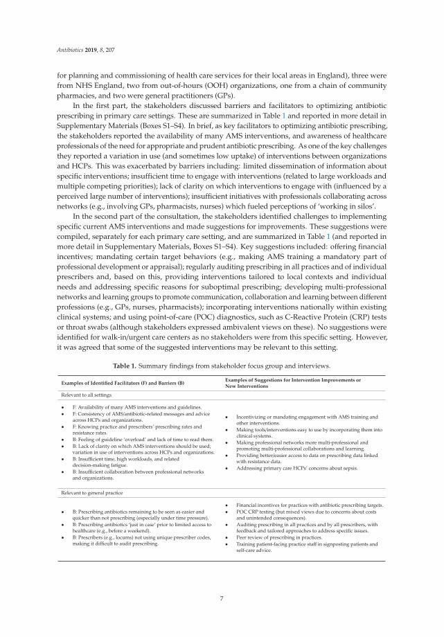
In the first part, the stakeholders discussed barriers and facilitators to optimizing antibioticprescribing in primary care settings. These are summarized in Table 1 and reported in more detail inSupplementary Materials (Boxes S1–S4). In brief, as key facilitators to optimizing antibiotic prescribing,the stakeholders reported the availability of many AMS interventions, and awareness of healthcareprofessionals of the need for appropriate and prudent antibiotic prescribing. As one of the key challengesthey reported a variation in use (and sometimes low uptake) of interventions between organizationsand HCPs. This was exacerbated by barriers including: limited dissemination of information aboutspecific interventions; insufficient time to engage with interventions (related to large workloads andmultiple competing priorities); lack of clarity on which interventions to engage with (influenced by aperceived large number of interventions); insufficient initiatives with professionals collaborating acrossnetworks (e.g., involving GPs, pharmacists, nurses) which fueled perceptions of ‘working in silos’.
In the second part of the consultation, the stakeholders identified challenges to implementingspecific current AMS interventions and made suggestions for improvements. These suggestions werecompiled, separately for each primary care setting, and are summarized in Table 1 (and reported inmore detail in Supplementary Materials, Boxes S1–S4). Key suggestions included: offering financialincentives; mandating certain target behaviors (e.g., making AMS training a mandatory part ofprofessional development or appraisal); regularly auditing prescribing in all practices and of individualprescribers and, based on this, providing interventions tailored to local contexts and individualneeds and addressing specific reasons for suboptimal prescribing; developing multi-professionalnetworks and learning groups to promote communication, collaboration and learning between differentprofessions (e.g., GPs, nurses, pharmacists); incorporating interventions nationally within existingclinical systems; and using point-of-care (POC) diagnostics, such as C-Reactive Protein (CRP) testsor throat swabs (although stakeholders expressed ambivalent views on these). No suggestions wereidentified for walk-in/urgent care centers as no stakeholders were from this specific setting. However,it was agreed that some of the suggested interventions may be relevant to this setting.
Table 1. Summary findings from stakeholder focus group and interviews.
Examples of Identified Facilitators (F) and Barriers (B)Examples of Suggestions for Intervention Improvements orNew Interventions
Relevant to all settings
• F: Availability of many AMS interventions and guidelines.• F: Consistency of AMS/antibiotic-related messages and advice
across HCPs and organizations.• F: Knowing practice and prescribers’ prescribing rates and
resistance rates.• B: Feeling of guideline ‘overload’ and lack of time to read them.• B: Lack of clarity on which AMS interventions should be used;
variation in use of interventions across HCPs and organizations.• B: Insufficient time, high workloads, and related
decision-making fatigue.• B: Insufficient collaboration between professional networks
and organizations.
• Incentivizing or mandating engagement with AMS training andother interventions.
• Making tools/interventions easy to use by incorporating them intoclinical systems.
• Making professional networks more multi-professional andpromoting multi-professional collaborations and learning.
• Providing better/easier access to data on prescribing data linkedwith resistance data.
• Addressing primary care HCPs’ concerns about sepsis.
Relevant to general practice
• B: Prescribing antibiotics remaining to be seen as easier andquicker than not prescribing (especially under time pressure).
• B: Prescribing antibiotics ‘just in case’ prior to limited access tohealthcare (e.g., before a weekend).
• B: Prescribers (e.g., locums) not using unique prescriber codes,making it difficult to audit prescribing.
• Financial incentives for practices with antibiotic prescribing targets.• POC CRP testing (but mixed views due to concerns about costs
and unintended consequences).• Auditing prescribing in all practices and by all prescribers, with
feedback and tailored approaches to address specific issues.• Peer review of prescribing in practices.• Training patient-facing practice staff in signposting patients and
self-care advice.
7
Antibiotics 2019, 8, 207
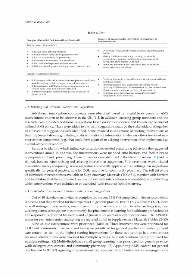
Table 1. Cont.
Examples of Identified Facilitators (F) and Barriers (B)Examples of Suggestions for Intervention Improvements orNew Interventions
Relevant to out of hours (OOH)
• B: Lack of stable patient population.• B: Prescribers not using unique prescriber codes.• B: Lack of accountability for prescribing.• B: Variation in awareness of local guidelines.• B: Lack of/limited support from commissioners.• B: Different clinical systems limiting access to patient records.
• Developing tools/system to enable/ automate prescribing auditsin OOH.
• Making AMS interventions (e.g., training) provided bycommissioners available and improving dissemination ofinformation about them to OOH staff.
• Improving induction of new prescribers in OOH to ensureawareness of local guidelines.
Relevant to community pharmacy
• B: Variation in skills and experience between pharmacy staff, withsome having low confidence in providing self-care advice.
• B: Limited access to POC diagnostics across pharmacies andconcern about using them for financial benefit.
• B: Different computer systems limiting access to, and use of,patient records.
• Providing training in giving self-care advice to improve skills andconfidence of staff.
• Providing access to POC diagnostics and training to helppharmacy staff distinguish between serious and less serious illness(thus improving confidence in giving self-care advice).
• Promoting use of patient records to identify potentiallyinappropriate use of antibiotics.
2.2. Revising and Selecting Intervention Suggestions
Additional intervention components were identified based on available evidence on AMSinterventions shown to be effective in the UK [11]. In addition, steering group members and theresearch team provided additional suggestions based on their experience and knowledge of currentnational AMS policy. These were added to the list of suggestions made by the stakeholders. Altogether,45 intervention suggestions were identified. Some involved modifications of existing interventions ortheir implementation (e.g., relating to dissemination of information), whereas others involved newintervention components (e.g., that could form a part of an existing intervention or be implemented asa stand-alone intervention).
In order to identify which influences on antibiotic-related prescribing behaviors the suggestedinterventions aimed to address, the interventions were mapped onto barriers and facilitators toappropriate antibiotic prescribing. These influences were identified in the literature review [11] and bythe stakeholders. After revising and selecting intervention suggestions, 31 interventions were includedin an online survey comprising: seven suggestions potentially applicable to all settings, ten suggestionsspecifically for general practice, nine for OOH, and five for community pharmacy. The full list of the45 identified interventions is available in Supplementary Materials (Table S1), together with barriersand facilitators that they addressed, source of how each intervention was identified, and indicatingwhich interventions were included in or excluded (with reasons) from the survey.
2.3. Stakeholder Survey and Prioritized Intervention Suggestions
Out of 40 stakeholders invited to complete the survey, 15 (38%) completed it. Seven respondentsindicated that they worked (or had expertise) in general practice, five in CCGs, four in OOH, threein walk-in/urgent care centers, one in community pharmacy, and four in other settings (i.e., twoworking across settings; one in community hospital; one in e-learning for healthcare professionals).The respondents reported between 4 and 23 (mean 10.7) years of relevant experience. The APEASEscores for each intervention and setting are reported in full in Supplementary Materials (Tables S2–S5).
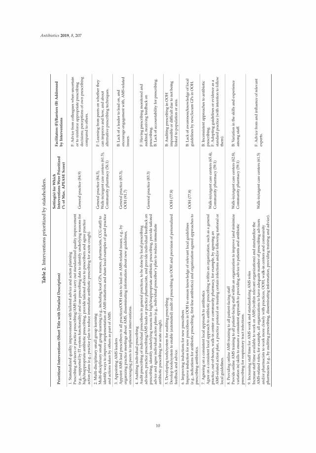
Nine unique interventions were prioritized (Table 2). Three interventions were prioritized forOOH and community pharmacy, and four were prioritized for general practice and walk-in/urgentcare centers (as two of the highest-scoring interventions for these two settings had even scores).As some interventions were assessed for multiple settings, four interventions were prioritized formultiple settings: ‘(2) Multi-disciplinary small group learning’ was prioritized for general practice,walk-in/urgent care centers, and community pharmacy; ‘(3) Appointing AMS leaders’ for generalpractice and OOH; ‘(7) Agreeing on a consistent local approach to antibiotics’ for walk-in/urgent care
8
Antibiotics 2019, 8, 207
centers and community pharmacy; ‘(8) Providing online AMS training to all patient-facing staff’ forwalk-in/urgent care centers and community pharmacy.
The lowest scoring intervention (with 22.7% of the maximum APEASE score) was ‘providingdiagnostic point-of-care CRP testing, including training in using it, interpreting the results andmaintaining the equipment’ in community pharmacy setting, which was assessed by only five out of 11respondents as relevant to the setting, by two as practical and acceptable and by none as affordable. Thisintervention was also rated as the second lowest for general practice (44% of the maximum APEASEscore), walk-in/urgent care centers (48.7%) and OOH (50%). Participants’ comments provided as freetext in the survey indicated that cost and funding, time to do the tests, and concern about over-use of thetests by patients and clinicians were considered the main barriers to using this intervention; for example:
“Would need to have clear guidance and uptake would depend on who was funding [POC CRP tests].Barriers to GP practices are cost of equipment and cost of tests, as well as time it takes to perform thetest when only have 5–10 min consultation and test takes a few minutes to perform so practical andaffordability issues are the main barriers.”
“Concerns [that POC CRP tests] may increase attendance to ‘get a test’. May involve cliniciansoverly relying on a test which is not always accurate or there may be a time lag in the increase in CRP.Time taken in consultation to administer test is a barrier and test strips are costly.”
The two second lowest scoring (with 33.3% of maximum APEASE score) interventions were alsoin community pharmacy setting; although both were assessed by only three participants. One was‘providing training and resources to structure the way(s) of asking patients the right questions aboutself-limiting infections and identifying red flags to help decide what to advise patients’. In comments,the participants suggested that: “this sort of training is available via CPPE, however uptake is voluntary”and “this should already be done as part of the core community pharmacy contract.” The other interventionwas ‘promoting the use of patient records by pharmacists (e.g., by digital prompts) to review whetherantibiotics were prescribed appropriately’. Participants’ comments suggested that:
“At present community pharmacists do not have access to enough information to be able to do thiseffectively. There may need to be specialist clinical training for community pharmacists to do this.”
“The relevance will depend on where the community pharmacist is in the patient pathway. If contractuallevers remain as is the community pharmacist may require remuneration.”
Other lowest scoring interventions (see a full list in Supplementary Materials) were: ‘providinginformation on opening hours of all local healthcare services’ for general practice (31.8% of maximumAPEASE score) which was scored particularly low on ‘effectiveness’, and ‘co-organizing national AMSevents together with different professional networks’ for OOH and walk-in/urgent care centers (50%and 38.5%, respectively) which was rated low on ‘affordability’.
9
Antibiotics 2019, 8, 207
Ta
ble
2.
Inte
rven
tion
spr
iori
tize
dby
stak
ehol
ders
.
Pri
ori
tized
Inte
rven
tio
ns
(Sh
ort
Tit
lew
ith
Deta
iled
Desc
rip
tio
n)
Sett
ing
(s)
for
Wh
ich
Inte
rven
tio
ns
Were
Pri
ori
tized
(%o
fM
ax.
AP
EA
SE
Sco
re)
Faci
lita
tors
(F)/
Barr
iers
(B)
Ad
dre
ssed
by
Inte
rven
tio
ns
1.St
anda
rdiz
edqu
alit
yim
prov
emen
twit
hta
ilore
dad
vice
and
acti
onpl
anni
ngPr
escr
ibin
gad
viso
rsor
prac
tice
pres
crib
ing/
AM
Sle
ads
toca
rry
outs
tand
ardi
zed
qual
ityim
prov
emen
t(e
.g.,
supp
orte
dby
ITsy
stem
func
tiona
lity)
and
use
pres
crib
ing
data
toid
entif
yun
derl
ying
reas
ons
for
high/in
appr
opri
ate
anti
biot
icpr
escr
ibin
g,pr
ovid
eta
ilore
dad
vice
topr
escr
iber
san
dag
ree
prac
tice
acti
onpl
ans
(e.g
.,pr
acti
cepl
anto
redu
ceim
med
iate
anti
biot
icpr
escr
ibin
gfo
rac
ute
coug
h).
Gen
eral
prac
tice
(84.
9)
F:A
dvic
efr
omco
lleag
ues
whe
nun
cert
ain
orto
rein
forc
eap
prop
riat
epr
escr
ibin
gde
cisi
ons;
perc
epti
ons
ofow
npr
escr
ibin
gco
mpa
red
toot
hers
.
2.M
ulti
-dis
cipl
inar
ysm
allg
roup
lear
ning
Mul
ti-d
isci
plin
ary
smal
lgro
uple
arni
ng(e
.g.,
incl
udin
glo
calG
Ps,n
urse
s,ph
arm
acis
ts,C
CG
staff
)to
iden
tify
way
sto
impr
ove
impl
emen
tatio
nof
AM
Sin
itiat
ives
and
shar
elo
cale
xam
ples
ofgo
odpr
actic
ean
dac
tion
sta
ken
byot
hers
aspa
rtof
AM
S.
Gen
eral
prac
tice
(84.
5),
Wal
k-in/u
rgen
tcar
ece
nter
s(6
1.5)
,C
omm
unit
yph
arm
acy
(56.
1)
F:Le
arni
ngfr
ompe
ers
onw
heth
erth
eyca
nim
prov
ean
dho
w,a
ndab
out
alte
rnat
ive
pres
crib
ing
tech
niqu
es.
3.A
ppoi
ntin
gA
MS
lead
ers
App
oint
AM
Sle
adpr
escr
iber
sin
allp
ract
ices/O
OH
site
sto
lead
onA
MS-
rela
ted
issu
es,e
.g.,
byor
gani
zing
prac
tice
mee
ting
sab
outA
MS,
diss
emin
atin
gin
form
atio
nab
outn
ewgu
idel
ines
,en
cour
agin
gpe
ers
toim
plem
enti
nter
vent
ions
.
Gen
eral
prac
tice
(83.
3),
OO
H(9
1.7)
B:La
ckof
ale
ader
tole
adon
,and
enco
urag
een
gage
men
twith
,AM
S-re
late
dis
sues
.
4.A
udit
ing
indi
vidu
alpr
escr
ibin
gA
udit
pres
crib
ing
ofin
divi
dual
pres
crib
ers
inge
nera
lpra
ctic
es,t
obe
done
bylo
calp
resc
ribi
ngad
viso
rs,p
ract
ice
pres
crib
ing/
AM
Sle
ads
orpr
acti
ceph
arm
acis
ts,a
ndpr
ovid
ein
divi
dual
feed
back
onpr
escr
ibin
g,id
entif
yun
derl
ying
reas
ons
for
high/in
appr
opri
ate
antib
iotic
pres
crib
ing,
prov
ide
tailo
red
advi
cean
dag
ree
indi
vidu
alac
tion
plan
s(e
.g.,
indi
vidu
alpr
escr
iber
’spl
anto
redu
ceim
med
iate
anti
biot
icpr
escr
ibin
gfo
rac
ute
coug
h).
Gen
eral
prac
tice
(83.
3)
F:H
avin
gpr
escr
ibin
gm
onit
ored
and
audi
ted,
rece
ivin
gfe
edba
ckon
pres
crib
ing.
B:La
ckof
acco
unta
bilit
yfo
rpr
escr
ibin
g.
5.D
evel
opin
gto
ols/
syst
emfo
rau
diti
ngpr
escr
ibin
gD
evel
opto
ols/
syst
emto
enab
le(a
utom
ated
)aud
itof
pres
crib
ing
inO
OH
and
prov
isio
nof
pers
onal
ized
feed
back
and
advi
ce.
OO
H(7
7.8)
B:A
udit
ing
pres
crib
ing
inO
OH
impo
ssib
leor
diffi
cult
due
tono
tbei
nglin
ked
topo
pula
tion
orar
ea.
6.Im
prov
ing
indu
ctio
nsfo
rne
wpr
escr
iber
sIm
prov
ein
duct
ion
for
new
pres
crib
ers
inO
OH
toen
sure
know
ledg
eof
rele
vant
loca
lgui
delin
es(e
.g.,
indi
catio
nsfo
ran
tibio
ticpr
escr
ibin
g,fir
st-l
ine
antib
iotic
s)an
dor
gani
zatio
n-ag
reed
appr
oach
esto
pres
crib
ing
anti
biot
ics.
OO
H(7
7.8)
B:La
ckof
awar
enes
s/kn
owle
dge
oflo
cal
guid
elin
esby
new/lo
cum
GPs
inO
OH
.
7.A
gree
ing
ona
cons
iste
ntlo
cala
ppro
ach
toan
tibi
otic
sA
gree
ona
cons
iste
ntlo
cala
ppro
ach
toan
tibio
ticpr
escr
ibin
gw
ithin
anor
gani
zatio
n,su
chas
age
nera
lpr
acti
ce,o
ut-o
f-ho
urs,
wal
k-in
cent
eror
com
mun
ity
phar
mac
y,fo
rex
ampl
e,by
agre
eing
anA
MS-
rela
ted
acti
onpl
an,a
prac
tice
prot
ocol
ontr
eati
ngce
rtai
nin
fect
ions
and/
orfo
llow
ing
nati
onal
orlo
calg
uide
lines
.
Wal
k-in/u
rgen
tcar
ece
nter
s(6
5.4)
,C
omm
unit
yph
arm
acy
(59.
1)
B:In
cons
iste
ntap
proa
ches
toan
tibi
otic
pres
crib
ing.
F:A
dopt
ing
guid
elin
esor
evid
ence
asa
stan
dard
prac
tice
(with
inte
ntio
nto
follo
wth
em).
8.Pr
ovid
ing
onlin
eA
MS
trai
ning
toal
lpat
ient
-fac
ing
staff
Prov
ide
onlin
eA
MS
trai
ning
toal
lpat
ient
-fac
ing
staff
with
inan
orga
niza
tion
toim
prov
e(a
ndm
inim
ize
vari
atio
nin
)ski
llsto
ensu
rea
cons
iste
ntap
proa
chto
prov
idin
gad
vice
topa
tien
tsan
dan
tibi
otic
pres
crib
ing
for
resp
irat
ory
trac
tinf
ecti
ons.
Wal
k-in/u
rgen
tcar
ece
nter
s(6
2.8)
,C
omm
unit
yph
arm
acy
(59.
1)B:
Var
iati
onin
the
skill
san
dex
peri
ence
amon
gst
aff.
9.In
crea
sing
staff
tim
efo
rA
MS
wor
kan
dst
anda
rdiz
ing
AM
Sro
les
Incr
ease
staff
tim
eav
aila
ble
tow
ork
onA
MS
(wit
hin
rele
vant
orga
niza
tion
s)an
dst
anda
rdiz
eth
eA
MS-
rela
ted
role
s;fo
rex
ampl
e,al
lorg
aniz
atio
nsto
have
adeq
uate
num
ber
ofpr
escr
ibin
gad
viso
rsan
d/or
phar
mac
ists
tow
ork
mor
ecl
osel
yw
ith
prac
tice
s,O
OH
,wal
k-in
cent
ers
and
com
mun
ity
phar
mac
ies
(e.g
.,by
audi
ting
pres
crib
ing,
diss
emin
atin
gin
form
atio
n,pr
ovid
ing
trai
ning
and
advi
ce).
Wal
k-in/u
rgen
tcar
ece
nter
s(6
1.5)
F:A
dvic
efr
oman
din
fluen
ceof
rele
vant
expe
rts.
10
Antibiotics 2019, 8, 207
3. Discussion
The stakeholder consultation identified setting-specific barriers and facilitators to current antibioticoptimization, and generated and prioritized suggestions for improvements of AMS interventionsin primary care in England. Stakeholders’ appraisal of relevance, feasibility and acceptability of 31intervention suggestions led to nine interventions being prioritized across settings. These prioritizedinterventions address some of the identified influences on antibiotic prescribing. They could beincorporated as part of existing AMS interventions or further developed and implemented asstand-alone interventions.
3.1. Implications within the Context of Current AMS Research and Practice
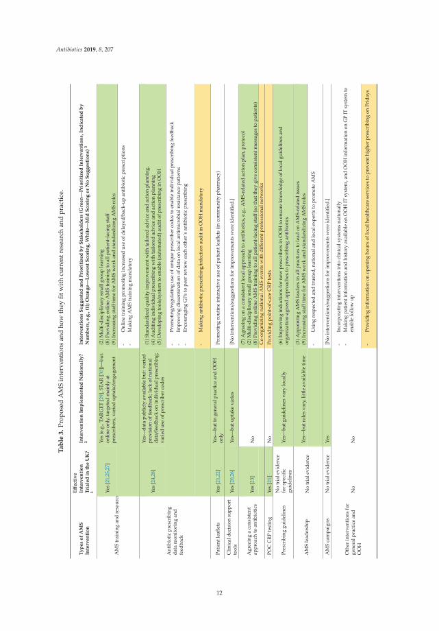
The interventions assessed and prioritized (or not) by the stakeholders build on AMS interventionscurrently implemented in England [10] and effective interventions tested in UK-based researchstudies [11]. How the 31 interventions fit with current research and practice, and how those prioritizedmay be implemented, is discussed below and summarized in Table 3. Interventions are grouped byintervention ‘type’, for ease of reference.
Improving engagement with AMS training and resources: There are many AMS interventions available,including AMS training and resources tested and shown effective in trials [21,25,27]. For example, theTARGET antibiotic toolkit (with training and resources primarily targeted at GPs) is available online [29]and practice workshops promoting the TARGET resources were shown effective [27]. Similarly, theSTAR online communication skills training (with a practice seminar) was shown effective in a trial [25]and is now available online on the clinical professional development website [30]. CCG professionalsreport that HCPs are aware of, and promote or engage with, different AMS training and resources (suchas the TARGET toolkit), but time, reaching the correct people and lack of clarity on which online trainingto promote were reported as the main issues with AMS education [31]. Similarly, the stakeholders inour study identified improving engagement with AMS interventions as a challenge, influenced by lackof time and priority, clarity on which interventions to use, incentives, and opportunities (e.g., protectedtime, training for HCPs in organizations other than general practice). Current AMS interventions may,at least in theory and research trials, facilitate change but improving the uptake of and engagement withAMS interventions in the real world is critical to further optimizing antibiotic use. This may be facilitatedby the ‘train the trainers’ opportunities provided by the TARGET team [29] and by increasing numbersof pharmacists appointed in primary care settings with AMS as part of their roles. The importanceof improving engagement with AMS training and resources was reflected by suggestions prioritizedby the stakeholders, such as organizing ‘(2) multi-disciplinary small group learning’ (that could bedelivered face-to-face, in addition to online resources, and focus on identifying challenges and solutionsspecific to local contexts), ‘(8) providing online AMS training to all patient-facing staff’ (rather than,as currently, targeting primarily GPs), and ‘(9) increasing staff time available for AMS work andstandardizing AMS roles’. Another suggestion to improve engagement with AMS training that wasmade by the stakeholders but received medium APEASE score was to make the AMS training mandatoryin general practices.
11
Antibiotics 2019, 8, 207
Ta
ble
3.
Prop
osed
AM
Sin
terv
enti
ons
and
how
they
fitw
ith
curr
entr
esea
rch
and
prac
tice
.
Ty
pes
of
AM
SIn
terv
en
tio
n
Eff
ect
ive
Inte
rven
tio
nT
riale
din
the
UK
?1
Inte
rven
tio
nIm
ple
men
ted
Nati
on
all
y?
2In
terv
en
tio
ns
Su
gg
est
ed
an
dP
rio
riti
zed
by
Sta
keh
old
ers
(Gre
en
—P
rio
riti
zed
Inte
rven
tio
ns,
Ind
icate
db
yN
um
bers
,e.g
.,(1
);O
ran
ge—
Lo
west
Sco
rin
g,W
hit
e—
Mid
Sco
rin
go
rN
oS
ug
gest
ion
s)3
AM
Str
aini
ngan
dre
sour
cesYe
s[2
1,25
,27]
Yes
(e.g
.,TA
RG
ET[2
9],S
TAR
[30]
)—bu
t:on
line
only
,tar
gete
dm
ainl
yat
pres
crib
ers,
vari
edup
take/e
ngag
emen
t
(2)M
ulti
-dis
cipl
inar
ysm
allg
roup
lear
ning
(8)P
rovi
ding
onlin
eA
MS
trai
ning
toal
lpat
ient
-fac
ing
staff
(9)I
ncre
asin
gst
affti
me
for
AM
Sw
ork
and
stan
dard
izin
gA
MS
role
s
-O
nlin
etr
aini
ngpr
omot
ing
incr
ease
dus
eof
dela
yed/
back
-up
anti
biot
icpr
escr
ipti
ons
-M
akin
gA
MS
trai
ning
man
dato
ry
Ant
ibio
tic
pres
crib
ing
data
mon
itor
ing
and
feed
back
Yes
[24,
28]
Yes—
data
publ
icly
avai
labl
ebu
t:va
ried
prov
isio
nof
feed
back
;lac
kof
nati
onal
data/f
eedb
ack
onin
divi
dual
pres
crib
ing;
vari
edus
eof
pres
crib
erco
des
(1)S
tand
ardi
zed
qual
ity
impr
ovem
entw
ith
tailo
red
advi
cean
dac
tion
plan
ning
,(4
)Aud
itin
gin
divi
dual
pres
crib
ing
wit
hta
ilore
dad
vice
and
acti
onpl
anni
ng(5
)Dev
elop
ing
tool
s/sy
stem
toen
able
(aut
omat
ed)a
udit
ofpr
escr
ibin
gin
OO
H
-Pr
omot
ing/
regu
lati
ngus
eof
uniq
uepr
escr
iber
code
sto
enab
lein
divi
dual
pres
crib
ing
feed
back
-Im
prov
ing
diss
emin
atio
nof
data
onlo
cala
ntim
icro
bial
resi
stan
cepa
tter
ns-
Enco
urag
ing
GPs
tope
erre
view
each
othe
r’s
anti
biot
icpr
escr
ibin
g
-M
akin
gan
tibi
otic
pres
crib
ing/
infe
ctio
nau
diti
nO
OH
man
dato
ry
Pati
entl
eafle
tsYe
s[2
1,22
]Ye
s—bu
tin
gene
ralp
ract
ice
and
OO
Hon
lyPr
omot
ing
rout
ine
inte
ract
ive
use
ofpa
tien
tlea
flets
(in
com
mun
ity
phar
mac
y)
Clin
ical
deci
sion
supp
ort
tool
sYe
s[2
0,26
]Ye
s—bu
tupt
ake
vari
es[N
oin
terv
enti
ons/
sugg
esti
ons
for
impr
ovem
ents
wer
eid
enti
fied.
]
Agr
eein
ga
cons
iste
ntap
proa
chto
anti
biot
ics
Yes
[23]
No
(7)A
gree
ing
ona
cons
iste
ntlo
cala
ppro
ach
toan
tibi
otic
s,e.
g.,A
MS-
rela
ted
acti
onpl
an,p
roto
col
(2)M
ulti
-dis
cipl
inar
ysm
allg
roup
lear
ning
(8)P
rovi
ding
onlin
eA
MS
trai
ning
toal
lpat
ient
-fac
ing
staff
(so
that
they
give
cons
iste
ntm
essa
ges
topa
tien
ts)
Co-
orga
nizi
ngna
tion
alA
MS
even
tsw
ith
diff
eren
tpro
fess
iona
lnet
wor
ks
POC
CR
Pte
stin
gYe
s[2
1]N
oPr
ovid
ing
poin
t-of
-car
eC
RP
test
s
Pres
crib
ing
guid
elin
esN
otr
iale
vide
nce
for
spec
ific
guid
elin
esYe
s—bu
tgui
delin
esva
rylo
cally
(6)I
mpr
ovin
gin
duct
ions
for
new
pres
crib
ers
inO
OH
toen
sure
know
ledg
eof
loca
lgui
delin
esan
dor
gani
zati
on-a
gree
dap
proa
ches
topr
escr
ibin
gan
tibi
otic
s
AM
Sle
ader
ship
No
tria
levi
denc
eYe
s—bu
trol
esva
ry,l
ittl
eav
aila
ble
tim
e(3
)App
oint
ing
AM
Sle
ader
sin
allp
ract
ices
tole
adon
AM
S-re
late
dis
sues
(9)I
ncre
asin
gst
affti
me
for
AM
Sw
ork
and
stan
dard
izin
gA
MS
role
s
-U
sing
resp
ecte
dan
dtr
uste
d,na
tion
alan
dlo
cale
xper
tsto
prom
ote
AM
S
AM
Sca
mpa
igns
No
tria
levi
denc
eYe
s[N
oin
terv
enti
ons/
sugg
esti
ons
for
impr
ovem
ents
wer
eid
enti
fied.
]
Oth
erin
terv
enti
ons
for
gene
ralp
ract
ice
and
OO
HN
oN
o
-In
corp
orat
ing
inte
rven
tion
sin
tocl
inic
alsy
stem
sna
tion
ally
-M
akin
gpa
tient
info
rmat
ion
and
hist
ory
avai
labl
eon
OO
HIT
syst
em,a
ndO
OH
info
rmat
ion
onG
PIT
syst
emto
enab
lefo
llow
up
-Pr
ovid
ing
info
rmat
ion
onop
enin
gho
urs
oflo
calh
ealt
hcar
ese
rvic
esto
prev
enth
ighe
rpr
escr
ibin
gon
Frid
ays
12
Antibiotics 2019, 8, 207
Ta
ble
3.
Con
t.
Ty
pes
of
AM
SIn
terv
en
tio
n
Eff
ect
ive
Inte
rven
tio
nT
riale
din
the
UK
?1
Inte
rven
tio
nIm
ple
men
ted
Nati
on
all
y?
2In
terv
en
tio
ns
Su
gg
est
ed
an
dP
rio
riti
zed
by
Sta
keh
old
ers
(Gre
en
—P
rio
riti
zed
Inte
rven
tio
ns,
Ind
icate
db
yN
um
bers
,e.g
.,(1
);O
ran
ge—
Lo
west
Sco
rin
g,W
hit
e—
Mid
Sco
rin
go
rN
oS
ug
gest
ion
s)3
Oth
erin
terv
enti
ons
for
com
mun
ity
phar
mac
yN
oN
o
-Ph
arm
acy
staff
topr
ompt
GPs
tore
view
long
-ter
man
dre
peat
anti
biot
icpr
escr
ipti
ons
-En
cour
age
phar
mac
ists
tofe
edba
ckto
GPs
whe
rean
tibi
otic
sw
ere
notp
resc
ribe
dac
cord
ing
togu
idel
ines
-Pr
omot
eth
eus
eof
pati
entr
ecor
dsby
phar
mac
ists
tore
view
whe
ther
anti
biot
ics
wer
epr
escr
ibed
appr
opri
atel
y-
Prov
ide
trai
ning
and
reso
urce
sto
stru
ctur
eth
ew
ay(s
)ofa
skin
gpa
tien
tsth
eri
ghtq
uest
ions
abou
tsel
f-lim
itin
gin
fect
ions
and
iden
tify
ing
red-
flags
tohe
lpde
cide
wha
tto
advi
sepa
tien
ts
Not
es:1
Nin
eU
K-b
ased
stud
ies
ofeff
ectiv
eA
MS
inte
rven
tions
[8,2
0–24
,26–
28]w
ere
iden
tified
and
are
repo
rted
else
whe
re[1
1].2
Twen
tysi
xna
tiona
llyim
plem
ente
dA
MS
inte
rven
tions
wer
eid
entifi
edpr
evio
usly
and
are
repo
rted
else
whe
re[1
0].3
The
nine
prio
ritiz
edin
terv
entio
nsar
enu
mbe
red
asin
Tabl
e2
and
incl
ude
the
high
est-
scor
ing
inte
rven
tions
(3–4
per
sett
ing)
(gre
enro
ws)
.Low
est-
scor
ing
inte
rven
tions
(3pe
rse
ttin
g)ar
ein
oran
gero
ws;
the
rem
aini
ngin
terv
entio
nsw
ithth
eA
PEA
SEsc
ores
inth
em
iddl
ear
ein
whi
tero
ws.
All
APE
ASE
scor
esfo
rea
chin
terv
enti
onan
dse
ttin
gar
ere
port
edin
the
Supp
lem
enta
ryM
ater
ials
(Tab
les
S2–S
5).
13
Antibiotics 2019, 8, 207
Enhancing prescribing data monitoring and feedback: Antibiotic prescribing data has been publiclyavailable and fed back to prescribers by a vast majority of CCGs for many years and interventionsinvolving prescribing feedback have been shown effective [24,28]. In contrast, detailed action planningis rarely used (reported for only 16% of CCGs [31], often due to insufficient time for it [32]), as is feedbackon individual prescribing—both of these strategies were prioritized. Including them may enhancethe impact of monitoring of and feedback on prescribing by specifying and tailoring actions (settinggoals and/or ‘if-then’ plans) to address specific reasons for inappropriate antibiotic prescribing and byactivating individual accountability for prescribing. Based on available evidence from lifestyle-relatedinterventions, such behavioral regulation strategies (i.e., self-monitoring, especially when combinedwith other ‘regulatory’ techniques such as goal-setting, problem solving or ‘if-then’ plans) and individualtailoring can be effective behavior change techniques (e.g., [33,34]). Moreover, while monitoring/auditingof, and feedback on, prescribing have likely contributed to reduced antibiotic prescribing in generalpractice, the stakeholders reported barriers to using these strategies in OOH, such as lack of stable patientpopulation and different computer systems. Developing tools to enable and automate prescribing auditand provision of individualized feedback might further optimize antibiotic prescribing in OOH.
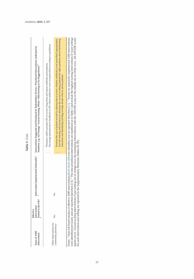
Ensuring consistency in AMS approaches: The suggestion to ‘(7) agree on a consistent local approachto antibiotics’ (prioritized for walk-in/urgent care centers and community pharmacies and also highlyscored for general practice and OOH) highlights the importance of consistency in managing infectionsand reinforcing consistent messages to patients by different HCPs and across organizations. It could beimplemented by developing and agreeing within-and between-organization action plans or protocols,for example, on using patient leaflets and other resources promoting messages about infectionprevention and self-care. The importance of consistency between HCPs in antibiotic prescribing andthe messages given to patients was also reflected in the following interventions prioritized by thestakeholders: ‘(2) multidisciplinary small group learning’ and ‘(8) providing online AMS training toall patient-facing staff’. Moreover, four interventions were prioritized for multiple settings (i.e., ‘(7)agreeing on a consistent local approach to antibiotics’; ‘(2) multi-disciplinary small group learning’; ‘(8)providing online AMS training to all patient-facing staff’; ‘(3) appointing AMS leaders’) and couldbe considered for implementation across settings and by involving HCPs from different professionalnetworks. This could help promote a more integrated, system-wide approach to AMS [17]. Respondentsin a recent survey representing 99% of Clinical Commissioning Groups (CCGs) reported that AMStraining was targeted primarily at GPs, compared to 67% of CCG professionals reporting focus onother prescribers, 42% on all practice staff and only 28% on OOH staff; consequently, a system andpractice-wide approach was one of the top suggestions for AMS training by the CCG professionals [31].
Finally, it may also be important to consider interventions that have not been prioritized bystakeholders. Providing POC CRP tests was among the lowest-scoring suggestions in all settings.The stakeholders considered it not affordable or practical to deliver. While POC CRP testing is supportedby examples of countries with low prescribing rates that routinely use it (e.g., Netherlands) and trialevidence showing it as an effective and safe strategy to reduce antibiotic prescribing for RTIs in generalpractice [14,35], it may not have sustained effects on prescribing behavior [36] and is often met withmixed views on its usefulness and feasibility from HCPs [37,38]. Moreover, ‘co-organizing national AMSevents for participants from different professional networks to facilitate multi-disciplinary AMS work’and ‘promoting the use of patient records by pharmacists to review whether antibiotics were prescribedappropriately’ seemed to be considered by stakeholders as unaffordable; ‘providing information onopening hours of local healthcare services’ (to reduce prescribing when concerned about limited accessto healthcare) was considered unlikely to be effective; and ‘providing training and resources to structurethe ways of asking patients the right questions about self-limiting infections and identifying red flagsto help decide what to advise patients’ in community pharmacy was seen as of low relevance. Ourfindings suggest that these interventions might be less promising ways to optimize antibiotic use forRTIs, at least as seen by the small number of stakeholders consulted in this study. Further researchmay need to explore and identify ways to address barriers related to these interventions with a larger
14
Antibiotics 2019, 8, 207
group of stakeholders and intervention users. In addition to the nine prioritized interventions and fivelowest-scoring interventions, there were also 17 other interventions included in the survey and another14 suggestions that were not prioritized for inclusion in the survey (all available in the SupplementaryMaterials) that could potentially be considered and refined.
3.2. Strengths and Limitations
This study was based on expert input from stakeholders and the study steering group, whohad practical experience and knowledge about national AMS interventions and policy. Views ofstakeholders and intervention users are critical, yet at times under-represented, in theoretical approachesto developing and refining AMS interventions. Our study, focused on views of commissioners andother key stakeholders, may help address this gap and provide some indication for future research onimplementation of AMS interventions. However, our findings need to be interpreted with caution.The stakeholders were identified by the steering group members from their own professional networks,and out of 40 stakeholders invited to the focus group and survey, only 15 responded. It is possiblethat a larger number of stakeholders or intervention users might have generated suggestions for, andprioritized, different interventions. The stakeholders had varied levels of experience and knowledge ofAMS interventions and different settings (with less experience specific to OOH, walk-in centers andcommunity pharmacies). This made it difficult to identify very specific improvements to current AMSinterventions and to capture a wider range of views from different organizations and settings. As thestudy focused on stakeholders and interventions in England, the generalizability of the findings maybe limited beyond England.
4. Materials and Methods
The following steps were taken: (1) a stakeholder focus group and telephone interviews to identifybarriers and facilitators to appropriate antibiotic prescribing and to generate intervention suggestions;(2) revision and selection of intervention suggestions; (3) a stakeholder survey to assess and prioritizeinterventions according to pre-specified criteria of relevance, feasibility and acceptability. We focusedon interventions targeting HCPs’ antibiotic prescribing for RTIs in the following settings: generalpractice, out-of-hours (OOH), walk-in/urgent care center, and community pharmacy. The study wasreviewed by the University of Oxford Clinical Trials and Research Governance team and classifiedas service development, and as such it did not require research ethics review. Participants were freeto participate in any stage of the consultation and withdraw at any point without any consequences.They were offered reimbursement of travel expenses but no payment for participation.
4.1. Stakeholder Focus Group and Telephone Interviews
Relevant stakeholders (including HCPs with expertise in and/or experience of antibiotic prescribingin relevant settings) were identified by the study steering group members. The steering group includedexperts in AMS with expertise in, and experience of, designing and implementing AMS interventionsand influencing national AMS policy in the UK. Identified stakeholders were invited by email to attend a3-hour face-to-face focus group held at Public Health England premises in London. They were followedup once in cases of non-response. Those who could not attend the focus group in person were invitedto a telephone interview.
The aims of the focus group were (a) to identify barriers and facilitators to appropriate antibioticprescribing and to implementing AMS interventions in relevant settings; and (b) to generate suggestionsfor improvements of current AMS interventions and/or their implementation or for new interventionsaddressing the identified barriers and facilitators. In the first part of the focus group, the stakeholderswere presented with barriers and facilitators identified from a literature review [11]. This was followedby a discussion of stakeholders’ experiences and examples of these, and of any other influences onantibiotic prescribing (especially in settings under-represented in the literature, such as communitypharmacy, OOH and walk-in centers). In the second part of the focus group, the stakeholders were
15
Antibiotics 2019, 8, 207
presented with examples of nationally available AMS interventions and/or interventions trialed inresearch studies in the UK. This was followed by a discussion about stakeholders’ experiences of usingthese interventions, any challenges to their use or implementation, and suggestions for improvementsand/or any new interventions.
The focus group discussions were facilitated by two researchers in a semi-structured way, whichincluded short presentations and general discussion, followed by more specific questions aboutparticipants’ views, experiences and examples. A third researcher took detailed notes. The focusgroup was audio-recorded and the recording was subsequently used to check the notes and add moredetail. Additionally, participants were provided with handouts including the information on barriersand facilitators and AMS interventions identified in the literature, and with questions for discussion.They could provide additional comments on the handouts and return them to the researchers. Thestakeholders interviewed by telephone were given information and asked questions in a similar formatto the focus group. Detailed notes were made on each telephone interview. All suggestions made duringthe focus group, telephone interviews and on the handouts were collated and summarized. The summaryof notes was used to generate a list of barriers and facilitators and a list of intervention suggestions.
4.2. Revision and Selection of Intervention Suggestions for a Survey
Barriers and facilitators to appropriate antibiotic prescribing and effective research interventionswere identified in a literature review [11]); these were used to generate additional suggestions whichwere added to the list of interventions identified by the stakeholders. The intervention suggestionswere divided into those potentially applicable to all settings or specific to each relevant setting. Theywere mapped onto barriers and facilitators identified by the stakeholders and in a preceding literaturereview [11].
The list of suggested interventions was reviewed by the researchers (AB, MW, STC, AS) and steeringgroup members (remaining authors) who provided feedback on and suggestions for rephrasing thesuggested interventions, and made additional suggestions that were added to the list of interventions.To reduce a relatively large number of identified suggestions, the comments from the steering groupmembers (who have knowledge of existing AMS interventions and those currently in development)were used to prioritize selection of interventions for the survey; suggestions were excluded if they wereconsidered unfeasible, already implemented, already under development, or insufficiently specific.
4.3. Stakeholder Survey and Prioritization of Interventions
An online survey was used for the stakeholders to assess intervention suggestions using theAPEASE criteria [39]: Affordability (is an intervention affordable?), Practicability (can it be deliveredeasily?), Effectiveness (is it likely to be effective?), Acceptability (is it acceptable to staff?), Side effects andsafety (is it safe to implement?), and Equity (can it avoid inequalities in patient care?). The survey wasdesigned and delivered using online Survey Monkey software (www.surveymonkey.com). Stakeholdersidentified by the steering group (those invited to the focus group) were invited by email, including abrief description about the survey and a link to complete it online. Participants were sent one reminder;responses were anonymous.
The survey asked about participants’ roles, work setting or expertise, and years of relevantexperience. Participants were then presented with interventions that could be potentially applicableto all settings (i.e., general practice, OOH, walk-in/urgent care center, community pharmacy) andsuggestions specific to each setting: general practice, OOH, and community pharmacy. For eachsuggestion, participants were asked to: (a) assess whether the intervention was relevant to the setting,and, if yes, then (b) to assess it, for that setting, using the APEASE criteria.
Participants could skip questions and whole sections so the numbers of respondents that assessedeach intervention for each setting varied. We calculated the number of responses for each interventionin each setting, the maximum possible APEASE score for each intervention/setting, and the actualAPEASE score for each intervention/setting. The percentage of the maximum possible APEASE scoreobtained for each intervention/setting was calculated to allow comparison between interventions.
16
Antibiotics 2019, 8, 207
In order to identify interventions which were rated highly, the following criteria were used toprioritize interventions in each setting: (a) at least 50% of respondents for that question assessed theintervention as relevant to that setting; and (b) intervention was scored as one of the top three (or fourin cases of equal scores) based on the percentage of the maximum APEASE score.
5. Conclusions
The study identified a number of barriers and facilitators to optimizing antibiotics and engagingwith AMS interventions, and possible ways in which current AMS interventions in primary carein England could be optimized. Nine interventions were prioritized by stakeholders as relevant,acceptable, affordable and feasible to implement in English primary care. They involve suggestions tohelp improve engagement with existing AMS training and resources (e.g., by face-to-face small grouplearning, tailoring AMS training to all patient-facing staff rather than prescribers only), enhancing datamonitoring and feedback (e.g., audit and feedback on individual’s prescribing, or developing toolsto automate prescribing audits), and promoting consistency in AMS approaches across healthcareprofessionals and services (e.g., by agreeing consistent local approaches, upskilling all patient-facingstaff). These can be adapted and further developed as part of current AMS initiatives. Additionally,stakeholders prioritized suggestions to incorporate AMS interventions into clinical systems at a nationallevel and enable sharing of patient information between general practice and OOH clinical systems.Future work needs to also focus more on addressing particular barriers to engagement with specificAMS interventions.
Supplementary Materials: The following Supplementary Materials and Data are available online at http://www.mdpi.com/2079-6382/8/4/207/s1: Box S1. Summary findings from stakeholder consultation: relevant to all settings.Box S2. Summary findings from stakeholder consultation: relevant to general practice. Box S3. Summary findingsfrom stakeholder consultation: relevant to out-of-hours. Box S4. Summary findings from stakeholder consultation:relevant to community pharmacy. Table S1. All identified intervention suggestions. Table S2. Stakeholder ratingsof interventions for general practice. Table S3. Stakeholder ratings of interventions for out-of-hours. Table S4.Stakeholder ratings of interventions for walk-in/urgent care centers. Table S5. Stakeholder ratings of interventionsfor community pharmacy.
Author Contributions: Conceptualization: S.T.-C., A.J.B., M.W., A.S., T.C.; methodology: all authors; formalanalysis: A.J.B., M.W., S.T.-C.; interpretation of results: all authors; writing—original draft preparation: A.J.B.;writing—review and editing: all authors; study administration: A.J.B.; funding acquisition: S.T.-C., T.C. The studysteering group members included: D.A.-O., E.B., S.H., L.J., C.M., K.S., E.T., C.B., and T.C.
Funding: This study was a part of a consultation commissioned and funded by Public Health England BehaviouralInsights, and was carried out as a collaboration between the University of Oxford and Public Health England.S.T.-C. was supported by funding from the National Institute for Health Research (NIHR) Health ProtectionResearch Unit in Healthcare Associated Infections and Antimicrobial Resistance at University of Oxford inpartnership with Public Health England [HPRU-2012-10041]. The views and opinions expressed in this paper arethose of the authors and not necessarily those of the NHS, NIHR, the Department of Health and Social Care orPublic Health England.
Acknowledgments: We thank the participants in this study for their helpful contributions.
Conflicts of Interest: C.B. reports receiving advisory board fees from Pfizer and Roche Molecular Systems, andgrant support from Roche Molecular Diagnostics. Other authors declare no conflict of interest.
Abbreviations
AMS: Antimicrobial Stewardship; CCG: Clinical Commissioning Group; CRP: C-Reactive Protein; GP: GeneralPractitioner; HCP: Healthcare professional; NHS: National Health Service; NIHR: National Institute of HealthResearch; OOH: Out-of-hours; POC: Point-of-care; RTI: Respiratory tract infection.
References
1. Davies, S.; Gibbens, N. UK Five Year Antimicrobial Resistance Strategy 2013 to 2018; Department of Health:London, UK, 2013.
2. Department of Health and Social Care. UK Five Year Action Plan for Antimicrobial Resistance 2019 to 2024;Department of Health and Social Care: London, UK, 2019.
17
Antibiotics 2019, 8, 207
3. World Health Organization. Global Action Plan to Control the Spread and Impact of Antimicrobial Resistance inNeisseria Gonorrhoeae; World Health Organization: Geneva, Switzerland, 2015.
4. Public Health England. English Surveillance Programme for Antimicrobial Utilisation and Resistance (ESPAUR).Report 2018; Public Health England: London, UK, 2018.
5. Smieszek, T.; Pouwels, K.B.; Dolk, F.C.K.; Smith, D.R.; Hopkins, S.; Sharland, M.; Hay, A.D.; Moore, M.V.;Robotham, J.V. Potential for reducing inappropriate antibiotic prescribing in English primary care. J. Antimicrob.Chemother. 2018, 73, ii36–ii43. [CrossRef] [PubMed]
6. European Centre for Disease Prevention and Control. Summary of the Latest Data on Antibiotic Consumption inEU: 2017; European Centre for Disease Prevention and Control: Stockholm, Sweden, 2017.
7. Pouwels, K.B.; Dolk, F.C.K.; Smith, D.R.M.; Smieszek, T.; Robotham, J.V. Explaining variation in antibioticprescribing between general practices in the UK. J. Antimicrob. Chemother. 2018, 73, ii27–ii35. [CrossRef] [PubMed]
8. Butler, C.C.; Hood, K.; Verheij, T.; Little, P.; Melbye, H.; Nuttall, J.; Kelly, M.J.; Molstad, S.; Godycki-Cwirko, M.;Almirall, J.; et al. Variation in antibiotic prescribing and its impact on recovery in patients with acute coughin primary care: Prospective study in 13 countries. BMJ 2009, 338, b2242. [CrossRef]
9. Pinder, R.J.; Berry, D.; Sallis, A.; Chadborn, T. Antibiotic Prescribing and Behaviour Change in Healthcare Settings:Literature Review and Behavioural Analysis; Department of Health & Public Health England: London, UK, 2015.
10. Atkins, L.C.; Tim; Bondaronek, P.; Ashiru-Oredope, D.; Beech, E.; Herd, N.; Lyon, V.; González-Iraizoz, M.;Hopkins, S.; McNulty, C.; et al. Which behaviours and mechanisms of action are targeted by nationalantimicrobial stewardship interventions for patients, community pharmacy staff, primary care prescribers,providers and commissioners and what is their content? Unpublished work.
11. Borek, A.J.; Nia, W.M.R.; Atkins, L.; Sallis, A.; Tonkin-Crine, S. Exploring the Implementation of Interventions toReduce Antibiotic Use (ENACT Study): Report; Public Health England: London, UK, 2019.
12. Tonkin-Crine, S.; Yardley, L.; Little, P. Antibiotic prescribing for acute respiratory tract infections in primary care:A systematic review and meta-ethnography. J. Antimicrob. Chemother. 2011, 66, 2215–2223. [CrossRef] [PubMed]
13. Germeni, E.; Frost, J.; Garside, R.; Rogers, M.; Valderas, J.M.; Britten, N. Antibiotic prescribing for acuterespiratory tract infections in primary care: An updated and expanded meta-ethnography. Br J. Gen. Pr.2018, 68, e633–e645. [CrossRef]
14. Tonkin-Crine, S.K.; San Tan, P.; van Hecke, O.; Wang, K.; Roberts, N.W.; McCullough, A.; Hansen, M.P.;Butler, C.C.; Del Mar, C.B. Clinician-targeted interventions to influence antibiotic prescribing behaviour foracute respiratory infections in primary care: An overview of systematic reviews. Cochrane Database Syst. Rev.2017, 9, CD012252. [CrossRef]
15. Köchling, A.; Löffler, C.; Reinsch, S.; Hornung, A.; Böhmer, F.; Altiner, A.; Chenot, J.F. Reduction of antibioticprescriptions for acute respiratory tract infections in primary care: A systematic review. Implement. Sci. 2018,13, 47. [CrossRef]
16. McDonagh, M.S.; Peterson, K.; Winthrop, K.; Cantor, A.; Lazur, B.H.; Buckley, D.I. Interventions to reduceinappropriate prescribing of antibiotics for acute respiratory tract infections: Summary and update of asystematic review. J. Int. Med. Res. 2018, 46, 3337–3357. [CrossRef]
17. National Health Service. The NHS Long Term Plan. 2019. Available online: https://www.longtermplan.nhs.uk/wp-content/uploads/2019/08/nhs-long-term-plan-version-1.2.pdf (accessed on 30 October 2019).
18. Michie, S.; Johnston, M.; Francis, J.; Hardeman, W.; Eccles, M. From theory to intervention: Mappingtheoretically derived behavioural determinants to behaviour change techniques. Appl. Psychol. 2008, 57,660–680. [CrossRef]
19. Yardley, L.; Morrison, L.; Bradbury, K.; Muller, I. The person-based approach to intervention development:Application to digital health-related behavior change interventions. J. Med. Internet Res. 2015, 17, e30. [CrossRef]
20. Gulliford, M.C.; van Staa, T.; Dregan, A.; McDermott, L.; McCann, G.; Ashworth, M.; Charlton, J.; Little, P.;Moore, M.V.; Yardley, L. Electronic health records for intervention research: A cluster randomized trial toreduce antibiotic prescribing in primary care (eCRT study). Ann. Fam. Med. 2014, 12, 344–351. [CrossRef]
21. Little, P.; Stuart, B.; Francis, N.; Douglas, E.; Tonkin-Crine, S.; Anthierens, S.; Cals, J.W.; Melbye, H.; Santer, M.;Moore, M.; et al. Effects of internet-based training on antibiotic prescribing rates for acute respiratory-tractinfections: A multinational, cluster, randomised, factorial, controlled trial. Lancet 2013, 382, 1175–1182. [CrossRef]
22. Francis, N.A.; Butler, C.C.; Hood, K.; Simpson, S.; Wood, F.; Nuttall, J. Effect of using an interactive bookletabout childhood respiratory tract infections in primary care consultations on reconsulting and antibioticprescribing: A cluster randomised controlled trial. BMJ 2009, 339, b2885. [CrossRef] [PubMed]
18
Antibiotics 2019, 8, 207
23. Cox, C.M.; Jones, M. Is it possible to decrease antibiotic prescribing in primary care? An analysis of outcomesin the management of patients with sore throats. Fam. Pract. 2001, 18, 9–13. [CrossRef] [PubMed]
24. McNulty, C.A.; Kane, A.; Foy, C.J.; Sykes, J.; Saunders, P.; Cartwright, K.A. Primary care workshops can reduceand rationalize antibiotic prescribing. J. Antimicrob. Chemother. 2000, 46, 493–499. [CrossRef] [PubMed]
25. Butler, C.C.; Simpson, S.A.; Dunstan, F.; Rollnick, S.; Cohen, D.; Gillespie, D.; Evans, M.R.; Alam, M.F.;Bekkers, M.J.; Evans, J.; et al. Effectiveness of multifaceted educational programme to reduce antibioticdispensing in primary care: Practice based randomised controlled trial. BMJ 2012, 344, d8173. [CrossRef]
26. Little, P.; Hobbs, F.R.; Moore, M.; Mant, D.; Williamson, I.; McNulty, C.; Cheng, Y.E.; Leydon, G.; McManus, R.;Kelly, J.; et al. Clinical score and rapid antigen detection test to guide antibiotic use for sore throats: Randomisedcontrolled trial of PRISM (primary care streptococcal management). BMJ 2013, 347, f5806. [CrossRef]
27. McNulty, C.; Hawking, M.; Lecky, D.; Jones, L.; Owens, R.; Charlett, A.; Butler, C.; Moore, P.; Francis, N. Effectsof primary care antimicrobial stewardship outreach on antibiotic use by general practice staff: Pragmaticrandomized controlled trial of the TARGET antibiotics workshop. J. Antimicrob. Chemother. 2018, 73, 1423–1432.[CrossRef]
28. Hallsworth, M.; Chadborn, T.; Sallis, A.; Sanders, M.; Berry, D.; Greaves, F.; Clements, L.; Davies, S.C.Provision of social norm feedback to high prescribers of antibiotics in general practice: A pragmatic nationalrandomised controlled trial. Lancet 2016, 387, 1743–1752. [CrossRef]
29. TARGET (Treat Antibiotics Responsibly, Guidance, Education, Tools) Antibiotic Toolkit. Available online:https://www.rcgp.org.uk/clinical-and-research/resources/toolkits/target-antibiotic-toolkit.aspx (accessed on30 October 2019).
30. STAR: Stemming the Tide of Antibiotic Resistance. Available online: https://www.healthcarecpd.com/course/star-stemming-the-tide-of-antibiotic-resistance (accessed on 30 October 2019).
31. Alison, R.L.D.M.; Beech, E.; Ashiru-Oredope, D.; Costelloe, C.; Owens, R.; McNulty, C.A.M. What resourcesdo NHS commissioning organisations use to support antimicrobial stewardship in primary care in England?Unpublished work.
32. Jones, L.F.; Hawking, M.K.; Owens, R.; Lecky, D.; Francis, N.A.; Butler, C.; Gal, M.; McNulty, C.A.M. Anevaluation of the TARGET (Treat Antibiotics Responsibly; Guidance, Education, Tools) Antibiotics Toolkitto improve antimicrobial stewardship in primary care—Is it fit for purpose? Fam. Pract. 2017, 35, 461–467.[CrossRef]
33. Greaves, C.; Sheppard, K.E.; Abraham, C.; Hardeman, W.; Roden, M.; Evans, P.H.; Schwarz, P. Systematicreview of reviews of intervention components associated with increased effectiveness in dietary and physicalactivity interventions. BMC Public Health 2011, 11, 119. [CrossRef]
34. Michie, S.; Abraham, C.; Whittington, C.; McAteer, J.; Gupta, S. Effective techniques in healthy eating andphysical activity interventions: A meta-regression. Health Psychol. 2009, 28, 690–701. [CrossRef] [PubMed]
35. Cooke, J.; Butler, C.; Hopstaken, R.; Dryden, M.S.; McNulty, C.; Hurding, S.; Moore, M.; Livermore, D.M.Narrative review of primary care point-of-care testing (POCT) and antibacterial use in respiratory tractinfection (RTI). BMJ Open Respir. Res. 2015, 2, e000086. [CrossRef] [PubMed]
36. Little, P.; Stuart, B.; Francis, N.; Douglas, E.; Tonkin-Crine, S.; Anthierens, S.; Cals, J.W.; Melbye, H.; Santer, M.;Moore, M. Antibiotic Prescribing for Acute Respiratory Tract Infections 12 Months After Communicationand CRP Training: A Randomized Trial. Ann. Fam. Med. 2019, 17, 125–132. [CrossRef] [PubMed]
37. Huddy, J.R.; Ni, M.Z.; Barlow, J.; Majeed, A.; Hanna, G.B. Point-of-care C reactive protein for the diagnosisof lower respiratory tract infection in NHS primary care: A qualitative study of barriers and facilitators toadoption. BMJ Open 2016, 6. [CrossRef]
38. Eley, C.V.; Sharma, A.; Lecky, D.M.; Lee, H.; McNulty, C.A.M. Qualitative study to explore the views ofgeneral practice staff on the use of point-of-care C reactive protein testing for the management of lowerrespiratory tract infections in routine general practice in England. BMJ Open 2018, 8. [CrossRef]
39. Michie, S.; Atkins, L.; West, R. The behaviour change wheel. In A Guide to Designing Interventions, 1st ed.;Silverback Publishing: Sutton, UK, 2014; pp. 1003–1010.
© 2019 by the authors. Licensee MDPI, Basel, Switzerland. This article is an open accessarticle distributed under the terms and conditions of the Creative Commons Attribution(CC BY) license (http://creativecommons.org/licenses/by/4.0/).
19

antibiotics
Article
Combating Antimicrobial Resistance in Singapore:A Qualitative Study Exploring the Policy Context,Challenges, Facilitators, and Proposed Strategies
Shweta Rajkumar Singh 1,†, Alvin Qijia Chua 1,†, Sok Teng Tan 1, Clarence C. Tam 1,
Li Yang Hsu 1 and Helena Legido-Quigley 1,2,*1 Saw Swee Hock School of Public Health, National University of Singapore, Singapore 117549, Singapore;
[email protected] (S.R.S.); [email protected] (A.Q.C.); [email protected] (S.T.T.);[email protected] (C.C.T.); [email protected] (L.Y.H.)
2 London School of Hygiene and Tropical Medicine, London WC1H 9SH, UK* Correspondence: [email protected]† Authors contributed equally to this paper.
Received: 6 October 2019; Accepted: 27 October 2019; Published: 29 October 2019
Abstract: Antimicrobial resistance (AMR) is a global public health threat that warrants urgentattention. However, the multifaceted nature of AMR often complicates the development andimplementation of comprehensive policies. In this study, we describe the policy context and exploreexperts’ perspectives on the challenges, facilitators, and strategies for combating AMR in Singapore.We conducted semi-structured interviews with 21 participants. Interviews were transcribed verbatimand were analyzed thematically, adopting an interpretative approach. Participants reported that theMinistry of Health (MOH) has effectively funded AMR control programs and research in all publichospitals. In addition, a preexisting One Health platform, among MOH, Agri-Food & VeterinaryAuthority (restructured to form the Singapore Food Agency and the Animal & Veterinary Serviceunder NParks in April 2019), National Environment Agency, and Singapore’s National Water Agency,was perceived to have facilitated the coordination and formulation of Singapore’s AMR strategies.Nonetheless, participants highlighted that the success of AMR strategies is compounded by variouschallenges such as surveillance in private clinics, resource constraints at community-level healthfacilities, sub-optimal public awareness, patchy regulation on antimicrobial use in animals, andenvironmental contamination. This study shows that the process of planning and executing AMRpolicies is complicated even in a well-resourced country such as Singapore. It has also highlightedthe increasing need to address the social, political, cultural, and behavioral aspects influencing AMR.Ultimately, it will be difficult to design policy interventions that cater for the needs of individuals,families, and the community, unless we understand how all these aspects interact and shape theAMR response.
Keywords: Antimicrobial resistance; policy analysis; One Health; public health; Singapore
1. Introduction
Antimicrobial resistance (AMR) has gained considerable recognition as a major global publichealth threat [1–4]. The impact of AMR has been described as a “doomsday scenario” where antibioticscan no longer be relied upon and even minor infections could become untreatable and result insevere morbidity, death and significant economic losses [1,4,5]. The World Health Organization(WHO) released a Global Action Plan for AMR in 2015, setting out five strategic objectives to combatAMR [2]. It also highlighted the importance of a “One Health” approach, requiring collaboration amongnumerous sectors and actors including human and animal health, agriculture, finance, environment,and well-informed consumers.
Antibiotics 2019, 8, 201; doi:10.3390/antibiotics8040201 www.mdpi.com/journal/antibiotics21
Antibiotics 2019, 8, 201
Singapore is an island nation with a diverse population of 5.6 million people [6]. Public hospitalscater for 80% of tertiary care services while private sector practitioners account for the majority (80%)of primary care services [7]. There is a large, private animal health sector catering for companionanimals, but the agricultural animal health sector is small since Singapore has little local agriculturalproduction [8]. Food requirements are almost entirely supported by imports, which are regulated byfood laws focused on ensuring consistent foreign supply of food and agricultural products. Importedfood is strictly monitored by the Singapore Food Agency (SFA), while non-food related animal, plantand wildlife management services are provided by National Parks Board (NParks) and Animal &Veterinary Service (AVS) [9]. The National Environment Agency (NEA) and the Public Utilities Board(now known as PUB, Singapore’s National Water Agency) overlooks the public health aspects ofenvironment [10,11].
In line with the Global Action Plan for AMR, Singapore launched its own National StrategicAction Plan on AMR in November 2017 [12]. This multisectoral action plan was jointly developed bythe Ministry of Health (MOH), Agri-Food & Veterinary Authority (AVA) of Singapore (restructuredto form the SFA and the AVS under NParks in April 2019), NEA, and PUB. It sets a national OneHealth framework to reduce the emergence and spread of drug-resistant microorganisms throughfive core strategies: (1) Surveillance and risk assessment; (2) Research; (3) Education; (4) Preventionand control of infection; and (5) Optimization of antimicrobial use. Chua et al. recently published apaper on AMR in Singapore, detailing the changes in efforts against AMR in Singapore from 2008 to2018 [13]. The paper mentioned that although a better understanding of AMR as a One Health issuehas developed, with significant efforts in improving antibiotic prescribing and controlling AMR inSingapore, it was largely focused on the public hospital setting. Even then, a recent national pointprevalence survey conducted across 13 acute private and public hospitals in Singapore reported thatmore than half the patients in acute hospitals were on at least one systemic antibiotic, a much higherproportion compared to inpatients in European hospitals [14]. Based on the few published AMRepidemiology papers, the actual impact on the control of AMR has been mixed, with better control ofmethicillin-resistant Staphylococcus aureus (MRSA), coupled with an increase in resistant Gram-negativebacteria in the hospitals and the community [15–18]. It has been suggested that more efforts arerequired in the various sectors, especially the animal/farming and environment sectors, for better AMRcontrol [13].
The AMR issue has been described as a “wicked problem”, where there are many stakeholdersinvolved from the various sectors and several ways to frame the problem [19]. This results in a policyarena where politics and conflict are very evident, and can potentially overwhelm policymakers indevising plans to tackle AMR with a One Health approach. Insights into the policy process for AMRwill help to disentangle this complexity and provide a better understanding of support and oppositionby key stakeholders for alternative AMR mitigation policies [19]. To date, no qualitative study hasbeen conducted to analyze the factors that can aid in successful implementation of AMR policies inSingapore. Therefore, the aim of our study is to discuss the facilitators and challenges to addressingAMR and the dynamics in the policy process for AMR in Singapore via in-depth interviews. In thisstudy, we interviewed experts in human, animal, and environmental health from both public andprivate sectors to gain their insights on the policy context, AMR awareness, its emergence in Singapore,and steps taken to mitigate AMR generation and spread. We subsequently identified facilitators andkey challenges, and discussed proposed strategies for creating more awareness and combating AMR.
2. Materials and Methods
2.1. Data Collection
This qualitative study was conducted from September 2017 to May 2018. We used a purposivesampling method to recruit informants in key roles across sectors relevant to the issue of AMR inSingapore. Potential participants were identified from experts in the area of AMR in Singapore,
22
Antibiotics 2019, 8, 201
and were approached for an in-depth interview via email explaining the purpose of research. Atotal of 21 participants from within and outside Singapore were recruited in this study (Table 1).The participants held expertise in human, animal, or environmental health and worked at eitherministry, healthcare institution, academia, or civil society. Most participants were from the humanhealth sector. We had difficulty recruiting participants from the environmental sector as potentialparticipants showed more reservation in accepting the invitation to be interviewed, perhaps based onless knowledge about AMR. We were eventually able to recruit an academic and public health officialfrom the environmental sector.
Table 1. Participants’ characteristics according to profession and organization.
Sector Profession Count (n) Organization Count (n)
HumanMedical Practitioners 4 Government agency 3
Academics 3 Hospital (public and private) 6Managers 6 Primary care 1
AnimalAcademics 2 University 1
Laboratory Manager 1 Veterinary Clinic 2Private practice 2 Government agency 2
Government agency manager 1
EnvironmentPolicy maker 1 Government agency 1
Academic 1 University 1
InternationalWHO, NGOs, etc. 4
Total 21 Total 21
WHO =World Health Organization; NGOs = Non-governmental organizations.
Face-to-face in-depth interviews were conducted by three of the researchers (S.R.S., S.T.T. andH.L.-Q.) in English. Among the interviewers, two were Research Associates and one was an AssociateProfessor. All interviewers were trained in qualitative research. The interviews were conducted in aquiet space at the preferred location of the participants. Each interview lasted an hour on average andwas audio-recorded. There were no repeat interviews. A semi-structured question guide was used toexplore the main areas of concern for the participants, in their fields and areas of interest. The questionguide was developed with questions focusing on participants’ experiences and perceptions of AMRpolicy and practice including awareness, key challenges and facilitators in their particular area ofexpertise, and possible strategies and solutions to address key challenges in the Singaporean context.Participants were not remunerated for the interview.
2.2. Ethics
Ethical approval was obtained from the National University of Singapore Institutional ReviewBoard (NUS-IRB). Each participant was provided an information sheet stating the objectives andmethods of the research, at the point of recruitment. The confidentiality and anonymity of participants’responses was also highlighted. Written consent was sought before the beginning of each interview,requesting for permission to be audio-recorded and to be quoted anonymously in research outputs.Participants could refuse any of these options, as well as any questions posed to them during theinterview itself. All participants were de-identified to maintain confidentiality.
2.3. Data Analysis
All interviews were transcribed verbatim and identifying data were removed from all researchdocuments to ensure confidentiality. QSR NVivo 11 software was used to organize and share the data
23
Antibiotics 2019, 8, 201
among study team members. We used an interpretive approach which focuses on the participants’perceptions and interpretations of the topic of discussion. Thematic analysis was used to inductivelyidentify themes from the data. We drew on techniques from the constant comparative method, such asline by line analysis, naming each line and segment of data, and the use of subsequent interviews to testpreliminary assumptions. In addition, deviant cases were explored and examined [20,21]. The conductof the interviews and coding occurred concurrently to enable the researchers to determine when themesaturation had been reached and to cease recruitment. Thematic saturation was established when theresearch team discussed and agreed that no new themes were emerging from the data. To improve thecredibility of our findings, we conducted a member check at the final stage of manuscript preparationto validate our interpretation of the data and to ensure an accurate representation of participantperspectives. We were unable to contact a few of the participants as they have moved on to anotherrole and their contact information was not readily available. See Supplementary Materials for theConsolidated criteria for Reporting Qualitative research checklist (COREQ form).
3. Results
We present our findings under five main themes identified from analysis of participants’ responses.The first theme examines the policy context and discusses whether AMR as a policy issue is a priority inSingapore. The second theme discusses the level of AMR awareness among policymakers, professionals,and the general population. The third and fourth theme discusses the perceived facilitators andchallenges to combat AMR in the Singaporean context. Finally, the fifth theme reflects on the reportedstrategies to increase awareness and combat AMR based on participants’ perceptions.
3.1. The Policy Context
Policymakers that participated in the study reflected on the importance of addressing infectiousdiseases in the Singaporean context, especially after the outbreaks of severe acute respiratory syndrome(SARS) and H1N1 influenza which took place in 2003 and 2009, respectively. As a physician highlighted:
“A lot of incidents drive behavior in Singapore, like for example SARS drove infection controlto the front of everything. If you speak to some people, (they) will say infection controlbecame prominent feature with SARS happening.” —I11, Human Health
In 2009, the MOH initiated the National Antimicrobial Taskforce (NAT) that helped lay downthe basics of mandatory surveillance, stewardship, and infection prevention principles in publicrestructured hospitals. The NAT was tasked to formulate hospital programs and policies, as well as toimplement measure for monitoring and evaluation of these policies. It was later reorganized into theNational Antimicrobial Resistance Control Committee (NARCC) in 2014. Participants highlightedthat the strategy adopted to keep efforts sustainable was by working with people on the ground toreinforce the implementation of these programs. The MOH’s knack for looking at global health issuesand comparing itself to other countries also helped in funding and formulation of AMR strategies.The next quote highlights the ways in which the MOH operates including the dedicated funding forantimicrobial stewardship programs:
“The Ministry keeps an eye out for how it compares itself to other countries. So that drivestheir interest in looking at where we stand, how good, how bad ( . . . ) They directly fundquite a lot of the manpower in the hospital systems, certainly for antimicrobial stewardship.”—I02, Human Health
Participants also remarked that the ministry looked at other countries and internationalorganizations, such as the WHO and the European Union, for inspiration and implemented policiesrelevant to the Singapore context.
24
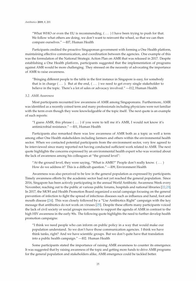
Antibiotics 2019, 8, 201
“What WHO or even the EU is recommending, ( . . . ) I have been trying to push for that.We follow what others are doing, we don’t want to reinvent the wheel, so that we can thencompare ourselves.” —I07, Human Health
Participants credited the proactive Singaporean government with forming a One Health platform,maintaining effective communication, and coordination between the agencies. One example of thiswas the formulation of the National Strategic Action Plan on AMR that was released in 2017. Despiteestablishing a One Health platform, participants suggested that the implementation of programsagainst AMR would be more challenging. They stressed on the necessity of advocating the importanceof AMR to raise awareness.
“Bringing different people to the table in the first instance in Singapore is easy, for somebodythat is in charge ( . . . ). But at the end, ( . . . ) we need to get every single stakeholder tobelieve in the topic. There’s a lot of sales or advocacy involved.” —I12, Human Health
3.2. AMR Awareness
Most participants recounted low awareness of AMR among Singaporeans. Furthermore, AMRwas identified as a recently coined term and many professionals including physicians were not familiarwith the term even though they were knowledgeable of the topic itself. The next quote is an exampleof such reports:
“I guess AMR, this phrase ( . . . ) if you were to tell me it’s AMR, I would not know it’santimicrobial resistance.” —I01, Human Health
Participants also remarked there was low awareness of AMR both as a topic as well a termamong other One Health stakeholders including farmers and others within the environmental healthsector. When we contacted potential participants from the environment sector, very few agreed tobe interviewed since many reported not having conducted sufficient work related to AMR. The nextquote highlights the concerns expressed by an environmental health expert who was worried aboutthe lack of awareness among his colleagues at “the ground level”:
“At the ground level, they were saying, “What is AMR?” People don’t really know. ( . . . )How do we address it? This is a difficult question.” —I09, Environment Health
Awareness was also perceived to be low in the general population as expressed by participants.Timely awareness efforts by the academic sector had not yet reached the general population. Since2016, Singapore has been actively participating in the annual World Antibiotic Awareness Week everyNovember, reaching out to the public at various public forums, hospitals and national libraries [22,23].In 2017, the MOH and Health Promotion Board organized a social campaign focusing on the generalprevention of infection to fight the spread of infectious diseases such as influenza and hand, foot andmouth disease [24]. This was closely followed by a “Use Antibiotics Right” campaign with the keymessage that antibiotics do not work on viruses [25]. Despite these efforts many participants voicedthe lack of civil society or social groups movements to support the agenda of AMR in contrast to thehigh HIV awareness in the early 90s. The following quote highlights the need to further develop healthpromotion campaigns:
“I think we need people who can inform on public policy in a way that would make ourpopulation understand. So we don’t have those communication agencies. I think we havethink tanks, right? And we have scientific groups. But we don’t quite have that translationinto a public health campaign.” —I02. Human Health
Some participants stated the importance of raising AMR awareness to counter its emergence.It was suggested that by raising awareness of the topic and getting more funds to drive AMR programsfor the general population and stakeholders alike, AMR emergence could be tackled better.
25
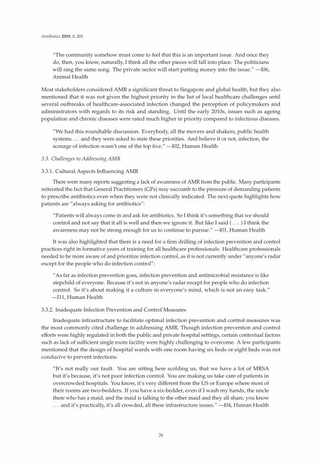
Antibiotics 2019, 8, 201
“The community somehow must come to feel that this is an important issue. And once theydo, then, you know, naturally, I think all the other pieces will fall into place. The politicianswill sing the same song. The private sector will start putting money into the issue.” —I06,Animal Health
Most stakeholders considered AMR a significant threat to Singapore and global health, but they alsomentioned that it was not given the highest priority in the list of local healthcare challenges untilseveral outbreaks of healthcare-associated infection changed the perception of policymakers andadministrators with regards to its risk and standing. Until the early 2010s, issues such as ageingpopulation and chronic diseases were rated much higher in priority compared to infectious diseases.
“We had this roundtable discussion. Everybody, all the movers and shakers, public healthsystems . . . and they were asked to state these priorities. And believe it or not, infection, thescourge of infection wasn’t one of the top five.” —I02, Human Health
3.3. Challenges to Addressing AMR
3.3.1. Cultural Aspects Influencing AMR
There were many reports suggesting a lack of awareness of AMR from the public. Many participantsreiterated the fact that General Practitioners (GPs) may succumb to the pressure of demanding patientsto prescribe antibiotics even when they were not clinically indicated. The next quote highlights howpatients are “always asking for antibiotics”:
“Patients will always come in and ask for antibiotics. So I think it’s something that we shouldcontrol and not say that it all is well and then we ignore it. But like I said ( . . . ) I think theawareness may not be strong enough for us to continue to pursue.” —I01, Human Health
It was also highlighted that there is a need for a firm drilling of infection prevention and controlpractices right in formative years of training for all healthcare professionals. Healthcare professionalsneeded to be more aware of and prioritize infection control, as it is not currently under “anyone’s radarexcept for the people who do infection control”:
“As far as infection prevention goes, infection prevention and antimicrobial resistance is likestepchild of everyone. Because it’s not in anyone’s radar except for people who do infectioncontrol. So it’s about making it a culture in everyone’s mind, which is not an easy task.”—I11, Human Health
3.3.2. Inadequate Infection Prevention and Control Measures
Inadequate infrastructure to facilitate optimal infection prevention and control measures wasthe most commonly cited challenge in addressing AMR. Though infection prevention and controlefforts were highly regulated in both the public and private hospital settings, certain contextual factorssuch as lack of sufficient single room facility were highly challenging to overcome. A few participantsmentioned that the design of hospital wards with one room having six beds or eight beds was notconducive to prevent infections:
“It’s not really our fault. You are sitting here scolding us, that we have a lot of MRSAbut it’s because, it’s not poor infection control. You are making us take care of patients inovercrowded hospitals. You know, it’s very different from the US or Europe where most oftheir rooms are two-bedders. If you have a six-bedder, even if I wash my hands, the unclethere who has a maid, and the maid is talking to the other maid and they all share, you know. . . and it’s practically, it’s all crowded, all these infrastructure issues.” —I04, Human Health
26
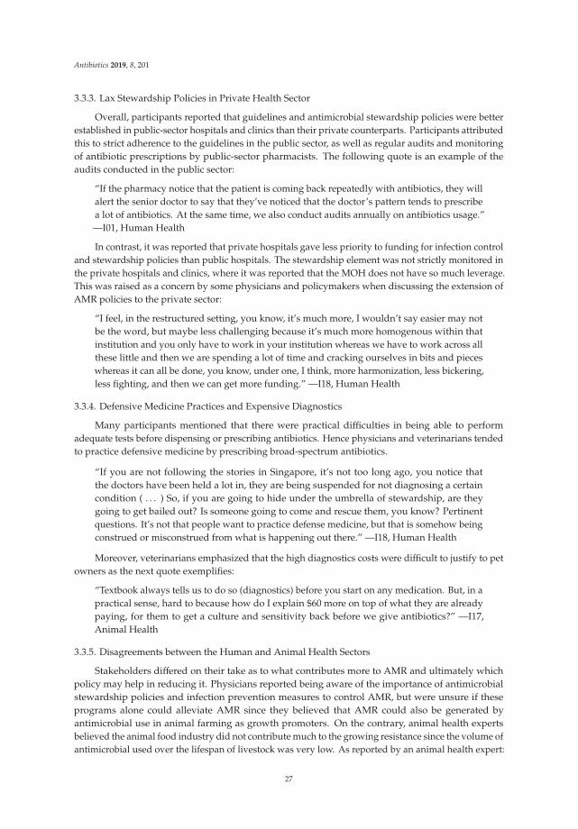
Antibiotics 2019, 8, 201
3.3.3. Lax Stewardship Policies in Private Health Sector
Overall, participants reported that guidelines and antimicrobial stewardship policies were betterestablished in public-sector hospitals and clinics than their private counterparts. Participants attributedthis to strict adherence to the guidelines in the public sector, as well as regular audits and monitoringof antibiotic prescriptions by public-sector pharmacists. The following quote is an example of theaudits conducted in the public sector:
“If the pharmacy notice that the patient is coming back repeatedly with antibiotics, they willalert the senior doctor to say that they’ve noticed that the doctor’s pattern tends to prescribea lot of antibiotics. At the same time, we also conduct audits annually on antibiotics usage.”—I01, Human Health
In contrast, it was reported that private hospitals gave less priority to funding for infection controland stewardship policies than public hospitals. The stewardship element was not strictly monitored inthe private hospitals and clinics, where it was reported that the MOH does not have so much leverage.This was raised as a concern by some physicians and policymakers when discussing the extension ofAMR policies to the private sector:
“I feel, in the restructured setting, you know, it’s much more, I wouldn’t say easier may notbe the word, but maybe less challenging because it’s much more homogenous within thatinstitution and you only have to work in your institution whereas we have to work across allthese little and then we are spending a lot of time and cracking ourselves in bits and pieceswhereas it can all be done, you know, under one, I think, more harmonization, less bickering,less fighting, and then we can get more funding.” —I18, Human Health
3.3.4. Defensive Medicine Practices and Expensive Diagnostics
Many participants mentioned that there were practical difficulties in being able to performadequate tests before dispensing or prescribing antibiotics. Hence physicians and veterinarians tendedto practice defensive medicine by prescribing broad-spectrum antibiotics.
“If you are not following the stories in Singapore, it’s not too long ago, you notice thatthe doctors have been held a lot in, they are being suspended for not diagnosing a certaincondition ( . . . ) So, if you are going to hide under the umbrella of stewardship, are theygoing to get bailed out? Is someone going to come and rescue them, you know? Pertinentquestions. It’s not that people want to practice defense medicine, but that is somehow beingconstrued or misconstrued from what is happening out there.” —I18, Human Health
Moreover, veterinarians emphasized that the high diagnostics costs were difficult to justify to petowners as the next quote exemplifies:
“Textbook always tells us to do so (diagnostics) before you start on any medication. But, in apractical sense, hard to because how do I explain $60 more on top of what they are alreadypaying, for them to get a culture and sensitivity back before we give antibiotics?” —I17,Animal Health
3.3.5. Disagreements between the Human and Animal Health Sectors
Stakeholders differed on their take as to what contributes more to AMR and ultimately whichpolicy may help in reducing it. Physicians reported being aware of the importance of antimicrobialstewardship policies and infection prevention measures to control AMR, but were unsure if theseprograms alone could alleviate AMR since they believed that AMR could also be generated byantimicrobial use in animal farming as growth promoters. On the contrary, animal health expertsbelieved the animal food industry did not contribute much to the growing resistance since the volume ofantimicrobial used over the lifespan of livestock was very low. As reported by an animal health expert:
27
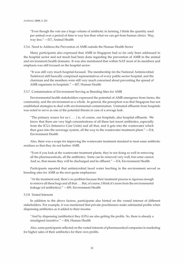
Antibiotics 2019, 8, 201
“Even though the vets use a huge volume of antibiotic in farming, I think the quantity usedper animal over a period of time is way less than what we can get from human clinics. Way,way less.” —I17, Animal Health
3.3.6. Need to Address the Prevention of AMR outside the Human Health Sector
Many participants also expressed that AMR in Singapore had so far only been addressed inthe hospital sector and not much had been done regarding the prevention of AMR in the animaland environment health domains. It was also mentioned that within NAT most of its members andemphasis was still focused on the hospital sector:
“It was still very much hospital-focused. The membership (in the National AntimicrobialTaskforce) still basically comprised representatives of every public-sector hospital, and thechairman and the members were still very much concerned about preventing the spread ofAMR organisms in hospitals.” —I07, Human Health
3.3.7. Contamination of Environment Serving as Breeding Sites for AMR
Environmental health stakeholders expressed the potential of AMR emergence from farms, thecommunity, and the environment as a whole. In general, the perception was that Singapore has notestablished strategies to deal with environmental contamination. Untreated effluents from hospitalswas noted to serve as one of the potential threats in case of a sewage leak.
“The primary source for us ( . . . ) is, of course, our hospitals, also hospital effluents. Weknow that there are very high concentrations of all these last resort antibiotics, especiallyfrom the ICUs (Intensive Care Units) and all that, and it gets into the wastewater whichthen goes into the sewerage system, all the way to the wastewater treatment plant.” —I14,Environment Health
Also, there was a scope for improving the wastewater treatment standard to treat some antibioticresidues so that they do not harbor AMR.
“Even if you look at the wastewater treatment plants, they’re not doing so well in removingall the pharmaceuticals, all the antibiotics. Some can be removed very well, but some cannot.And so, that means they will be discharged and be effluent.” —I14, Environment Health
Participants reported that antimicrobial laced water leeching in the environment served asbreeding sites for AMR as the next quote emphasizes:
“At the treatment end, there’s no problem because their treatment process is rigorous enoughto remove all these bugs and all that . . . But, of course, I think it’s more from the environmentalleakage (of antibiotics).” —I09, Environment Health
3.3.8. Vested Interests
In addition to the above factors, participants also hinted on the vested interest of differentstakeholders. For example, it was mentioned that private practitioners make substantial profits whendispensing antibiotics as it added to their income.
“And by dispensing (antibiotics) they (GPs) are also getting the profits. So, there is already amisaligned incentive.” —I04, Human Health
Also, some participants reflected on the vested interests of pharmaceutical companies in marketingfor higher sales of their antibiotics for their own profits.
28
Antibiotics 2019, 8, 201
“There is a conflict of interest already, because, they (pharmaceuticals) are producing, theyare just selling to the farmers ( . . . ) in a way they are trying to encourage certain resistance.”—I17, Animal Health
Animal health stakeholders expressed that the not so strict regulations on the import of newerantibiotics with a broader spectrum of activity served as a leeway for some veterinarians to prescribethem even when not clinically needed. Participants pointed out that the abuse of reserved antibioticsfavored acquisition of resistance genes by microorganisms, resulting in loss of antibiotic efficacy.
“There are those not very good ones out in the market that actually goes for the most exoticantibiotics because they distinguish themselves in that sense. Some of them actually usewhat I call the third-level antibiotic as a first line treatment which I personally don’t agree. Ofcourse, they charge exorbitant amount because it’s ‘special’ antibiotics.” —I17, Animal Health
In addition, owing to a small farming sector, existing veterinarians had limited expertise inagriculture and aquaculture and hence farmers in Singapore were not mandated to attain prescriptionfor accessing antibiotics independently. Participants alluded that farmers not needing to consult aveterinarian for a prescription for antibiotics served in the farmer’s interest.
“Vets were not familiar to treat fish, so farmers didn’t trust the vets to treat their fish, andvets then became very unwilling to treat farm fish. That’s not changed very much, and thereare still issues in Singapore. For example, fish farmers can now buy any antibiotic they wantby themselves.” —I15, Animal Health
3.4. Facilitators to Addressing AMR
3.4.1. Leading Role of the MOH and Provision of Appropriate Funding
Singapore’s MOH was unanimously praised by all participants in its proactive approach towardsAMR and making way for changes amid very complex issues. It was mentioned that the MOH wastransparent and accountable and was operating an efficient public healthcare system in Singapore. Yet,as a drawback some participants argued that having a system that relies too much on decisions madeat the highest political level makes the general population and service providers inert.
“That’s why I think in Singapore, unless it comes directly from the Prime Minister’s office, itwill never move anything.” —I11, Human Health
In terms of the resources made available to fund AMR programs, it was reported that MOH hadeffectively funded AMR control programs and research in all public hospitals.
3.4.2. Trust in the Ability of the Government to Design Policies
Most participants affirmed that the Singaporean population and healthcare professionals bestowtrust in government programs and policies. The next quote highlights that whether participants agreedor not with the proposed policy, there was a sense that the government is “trying to do a good thing”:
“When the Singaporean government gets into doing something, they tend to do it verythoroughly. ( . . . ) You may not always agree with the strategy, but you have to give themfull marks for trying.” —I04, Human Health
In addition, a preexisting One Health platform among MOH, SFA, AVS, NEA, and PUB wasperceived to have facilitated the coordination and formulation of Singapore’s AMR strategies.
29
Antibiotics 2019, 8, 201
3.4.3. Stewardship Programs
Laying the foundation of surveillance in public hospitals and making the data transparent wasappreciated by all participants. Participants also shared recent advances in the capacity to implementstewardship and infection prevention and control policies among different Singapore institutionsincluding nursing homes.
“We are looking to reaching out to even nursing homes to provide that kind of bold infectionprevention, but also infection control.” —I03, Human Health
3.4.4. Setting up a Surveillance System
AMR indicators, segregated mainly into process and outcome indicators, were set up in thepublic hospital surveillance system. Some of the process indicators were antimicrobial use for humanand animal health, and infection prevention and control measures such as hand hygiene in hospitals.It was suggested that the Singapore government played a prime role in mandating the standardization,transparency, and sharing of data with the NAT.
“We wanted to measure stuff in Singapore, but really to do that you really need the governmentinvolved, ( . . . ) otherwise if it’s a voluntary basis, and there are certain biases about data.Whereas if it’s systematic surveillance and it’s mandatory you get a more comprehensiveand representative sample.” —I04, Human Health
Participants mentioned that though the AMR surveillance indicators were defined and well setup in the public hospital domain, surveillance needed to be at a national level, extended to privatehospitals, primary care and intermediate/long-term care facilities (GPs, community hospitals andnursing homes), as well as animal and environment sectors.
“The indicators we sort of have been following is still hospital-based indicators. We don’t havecountry-wide, AMR indicators which internationally people are talking about. For example,we don’t know what is the level of antibiotic consumption at the country level, we don’tknow what, for example, when people are talking about “oh, for this bacterial infection, whatproportion of this bacterial infection is resistant to what antibiotics” you know? We haven’tgone to measuring that kind of level.” —I07, Human Health
3.4.5. Highly Motivated Workforce
The motivation of the workforce was attributed to the competition, prestige, and reputation of theworkforce. In the hospital setting, metrics and outcomes of antimicrobial stewardship such as averagedefined daily dose and percentage of appropriate antibiotics prescribed, and length of hospitalizationfor each department are often reported and shared among the various departments within the hospital.The transparency of data highly motivated the workforce to perform well in these benchmarkingexercises. The following quote is an example of such occurrence:
“More important I think is the peer pressure and peer standing. And when it’s made thedepartment target and communicated by the head of the department, no one wants to be theworst department in the stats when they are shown up. So there’s lot of department prideand that’s what drives stuff. Doctors are often competitive, some of the nurses too. And noone wants to be the idiot that lets the team down.” —I03, Human Health
3.4.6. Research Excellence
Research in Singapore was unanimously considered to be well promoted and funded, albeit itwas also rated to be very competitive in terms of securing research funding. There was ongoing AMRresearch including in the areas of surveillance, prevention of transmission, whole genome sequencing,
30
Antibiotics 2019, 8, 201
among others. Of the various arms of AMR research, surveillance was considered to be one of themain pillars in AMR research:
“One of the key reasons in the national action plan is research. And among the researchactivities there have been sort of highlighted in terms of priority, is better surveillance.You know, how to better measure the different perspectives and areas that we need to, outsidethe hospitals: primary care, the community and understanding the sort of pathways ofantibiotic resistances, what is driving, the drivers of antibiotic resistance locally . . . ” —I07,Human Health
3.4.7. Close Connections between Researchers and Policymakers
The researcher and academic participants mentioned that the government and ministry were verytightly knit in Singapore. Leadership was very open and attentive to research findings that could betranslated into better health practices in Singapore. As the following quote highlights:
“There’s that translation angle number one. There is the One Health angle number two,and then of course number three we also engage with basic science collaborators, to try tounderstand the basic science mechanisms of this issue.” —I08, Human Health
3.4.8. Role of Singapore in Regional AMR
As AMR is a global health issue, participants remarked that Singapore alone could do very littlein controlling AMR as a whole. Despite all policies Singapore will not be untouched by rising AMR inthe region which can be ultimately transmitted via large imports of agricultural products and humanstravelling for trade and tourism. However, most participants mentioned that Singapore could playa role by coordinating and sharing expertise with neighboring member states of the Association ofSoutheast Asian Nations (ASEAN).
Many participants proposed the idea that Singapore can serve as a research hub owing to itsunique positioning and research capacity. Sharing innovative findings both on science, clinical practiceand exemplary policy implementation strategies could prove helpful to neighboring countries thatshare some contextual factors with Singapore.
“It is an ideal geographic position as well as possibly a good political one with the recognitionby the west that Singapore is a high-income country as well as intellectually advanced.That bridge between the west and low- and middle-income countries especially in the AsiaPacific region could really be bridged by your university and your researchers.” —I20,Human Health
A few ministerial participants affirmed Singapore’s role of coordinating regional efforts on AMR.
“What Singapore is trying to do is trying to harmonize efforts in the agriculture sector andthe livestock sector because these are under different ASEAN stream workgroups. So wealso work with FAO to coordinate these.” —I13, Animal Health
3.5. Strategies to Raise AMR Awareness and Combat AMR
3.5.1. Projecting AMR Costs
Participants articulated that highlighting the increasing cost of managing patients infected withresistant organisms and the economic loss from AMR could be a potential way of drawing attentionto the topic and getting funds to drive programs. As specific examples, participants illustrated notjust the costs of treating resistant infections, but also the costs associated with implementing stricterinfection control measures.
Some participants believed that healthcare costs would increase due to AMR. These were attributedto many reasons, one of which was the cost of more effective antibiotics required to treat infections
31
Antibiotics 2019, 8, 201
with resistant organisms. Another prominent reason mentioned was the costs towards infectionprevention and control in healthcare settings. This was considered a necessary step to reduce the riskof healthcare-associated infections, especially in the face of worsening AMR. The following factorswere suggested by one participant to prevent the spread of resistant pathogens and to cater to patientsharboring resistant or infectious organisms:
“That is going to jack up costs in all kinds of ways. Number 1, you are going to have topay for all those gowns, gloves, single rooms, for isolation, etc. Number 2 you need moremanpower, because suddenly a nurse is going to be slowed down right? Or a doctor. Youhave to put on all this gear, you are going to be seeing fewer patients in the same amount oftime, just because of . . . it’s a bother.” —I04, Human Health
3.5.2. Push Forward AMR Agenda by Fear of no Treatment
Many participants believed that unless AMR’s potential damage to the population and theeconomy is projected, people are not going to take AMR seriously. Some participants mentioned thatonly a grave incident such as an AMR outbreak would be able to catch the attention of the generalpublic and extend the prevention efforts of AMR.
3.5.3. Building Communities of Practice
A few participants articulated that rather than forcing a top-down policy approach, AMR couldbe better tackled by the sharing of best practices through communities of practitioners and creatingsocial movements to encourage and sustain behavior change.
“Practitioners come together to share best practices, through the ID networks, through thehospital networks, through the volunteer networks, you know, build communities of practiceuntil this becomes a culture. And people are learning from each other ( . . . ) But whether itgets done is a question of culture, a culture needs strategy every day ( . . . ) so you want todo change, and you want to do large scale change and you want to sustain the change youneed to create a movement not a directive . . . I think it’s more important to you to create thesocial movement around what that is, because if you are going to achieve this style changeof behavior you need a more behavioral approach to this problem not a clinical approach.”—I05, Human Health
3.5.4. Engaging the Community and Public Campaigns on the Drivers of AMR
Participants reflected on the need to develop public campaigns to sensitize people to becomeaware of and understand the fundamentals of programs and strategies against AMR. These includestewardship, adherence to vaccination guidelines, better infection prevention and control practices inhospitals as well as programs in the community. All these programs needed to take into account social,behavioral, and cultural aspects to accommodate the needs and perceptions of the community.
“There is always a need to build a culture and to build a system for people to do the rightthing. And the same applies outside the hospitals. So I think you know the schools havehand washing to teach the next generation.” —I05, Human Health
3.5.5. Increase Vaccination
Most participants asserted that one of the probable strategies to counter AMR was to increasevaccination rates in children and adult population. However, increasing vaccination uptake willuncover another set of issues including population awareness, behavior changes, funding for regularvaccination, workforce to administer vaccination, and availability of vaccines.
32
Antibiotics 2019, 8, 201
4. Discussion
This paper has discussed the challenges and facilitators to addressing AMR in a well-resourcedsetting such as Singapore. Despite Singapore having an established One Health platform representingall the different stakeholders; appropriate funding to address the response; existence of stewardship andsurveillance programs in public hospitals; an active program in research; and a motivated workforce;most participants raised several challenges to address AMR efficiently. These included, among other,vested interests among some stakeholders; disagreements between the human and animal healthsectors; low public awareness; the need to address prevention of AMR in the animal and environmentalsector; political and cultural aspects influencing AMR; and lax stewardship policies in the private sector.
Therefore, the results of this paper highlight how difficult it can be to address AMR, especiallywhen considering that Singapore has well-functioning stewardship programs, comprehensive hospitalsurveillance systems, and some of the latest technologies and innovations to address it. The exampleof AMR in Singapore brings to light the increasing need to address the social, economic, political,cultural, and behavioral aspects influencing AMR rather than just focusing on the technical solutions.Unless we understand how all these aspects operate and drive the response, it will be difficult to designappropriate policy interventions. In the next paragraphs we provide an example of each of these aspectsto highlight their importance in the Singaporean setting. We address “the social” by analyzing howAMR has been socially constructed in Singapore; we discuss the influence of “politics” by consideringpower relations and governance approaches for the successful control of AMR; we address “behavioralaspects” by analyzing vested interests; and we consider “cultural components” by exploring howpatients’ cultural beliefs influence antibiotic consumption.
First, it is important to understand how AMR has been socially constructed in Singapore andhow power relations operate between the different stakeholders. Wernli et al. distinguished fivedifferent frames that map the global policy discourse on AMR [26]. These are: a One Health approachcombining in one paradigm human, animal, and environment health; a health security threat givingrise to the global health security agenda; a healthcare policy issue with the dominance of the medicalprofession; a development issue where it is considered that low-and-middle income countries driveAMR; and an innovation issue with a focus on new diagnostics and antibiotics. Wernli et al. alsodistinguish a set of actors and policies for each of the frames described. In the Singaporean context, it isinteresting to note that several frames coexist with their respective actors leading the selected frame.First, it was reported that there is a One Health platform where all stakeholders meet. Second, there isa strong healthcare policy frame as it was reported that Singapore’s AMR policies were largely drivenby a group of infectious disease clinicians. Third, AMR is also seen as an innovation issue whereresearchers are working to identify novel solutions on all fronts. The results of this study suggestthat a deeper understanding is needed of the different power relations between these distinct groupsto ensure the successful implementation of policies designed to respond to the threat of resistance.However, from our participants’ responses we can tentatively conclude that AMR is predominantlyseen as a biomedical problem within the context of healthcare facilities and within the pursuit ofclinicians controlling infectious diseases even when they still participate in the One Health platform.It could also be argued that historically the emphasis on AMR has been biomedical because this iswhere the health burden was seen and therefore where the expertise developed. This, however, doesnot mean that other areas are not seen as important.
Second, considering governance approaches for the successful control of AMR requires a focuson both whole of society and sector specific approaches [19]. This was detailed in a previous paperwhere we proposed a new governance framework to investigate power relations and responses fordiverse stakeholders addressing AMR [19]. As Kickbusch mentioned, and further discussed in ourpaper, recently there has been a diffusion of governance moving from a model dominated by thestate to a model co-produced by a wide range of actors [27]. As a result, Kickbusch has identifiedfive types of governance present in whole-of-government approaches. These include: (1) governingamong others by mixing regulation and persuasion; (2) by collaborating; (3) by engaging citizens;
33
Antibiotics 2019, 8, 201
(4) through independent agencies and expert bodies; (5) and by adaptive policies, resilient structures,and foresight.
In the Singapore setting, many participants highlighted the leading role of the MOH in regulatingthe health sector, but acknowledged challenges in regulating the private and animal health sector.These challenges present examples where governance structures will need to weigh up the balancebetween regulation and persuasion as policy approaches. In the Singaporean context, differentialpolicies in the public and private human healthcare sectors, whether by design or practice, are likelyto be counterproductive, as patients use both sectors and the occurrence of resistant infections inone sector has implications for the other. On the other hand, the agricultural sector in Singaporeis small and the availability of adequate veterinary expertise limited; imposing tight regulationson antibiotic access in the absence of qualified prescribers is therefore challenging, and persuasivestrategies to reduce antibiotic use in this sector are likely to be more realistic, at least in the short term.On their collaborative approach towards governance, many participants highlighted the One Healthplatform as a good way of bringing stakeholders together. The good relationship between researchersand policymakers was also emphasized via the many avenues for researchers to discuss and sharetheir research findings and discuss policy implications with policymakers. However, there were alsoconflicts reported specifically between human and animal health experts. This was crystalized ontheir different takes on what contributes more to AMR. Most physicians believed that antimicrobialuse in animal farming as growth promoters might be a more significant driver of AMR, as has beenexplained by studies [28]. In contrast, animal health experts believed the animal food industry did notcontribute much to the growing resistance when compared to AMR infection rates in the health sector.Finally, governing AMR through the engagement of citizens and civil society was not reported as astrategy in Singapore. However, this is not unique to Singapore, as it is well recognized that globallythere has been a lack of engagement of civil society organizations in addressing AMR, especially whencomparing and contrasting to the high HIV involvement in the early 90s.
Third, the AMR policy context represents a policy arena where certain groups engage in “behaviors”that might conflict with the overall goal of controlling AMR. Vested interests are more noticeablethan in other health policy areas since different professional groups coexist in the AMR policy arenaand there is substantial profit to be made from selling antibiotics [29,30]. In Singapore, vestedinterests were mentioned among pharmaceutical companies in marketing for higher sales of theirantibiotics for their own profits; among private practitioners as they make substantial profits whendispensing antibiotics; and among some veterinarians who prescribed antibiotics even when notclinically needed. Our previous research in other settings (i.e., Pakistan and Cambodia) also suggestedseveral considerations and vested interests around antibiotic use which can hinder the control ofAMR in the human and animal health sectors [29,30]. For example, it was reported that amongdoctors there are monetary incentives to prescribe certain antibiotics as these are often negotiated withpharmaceutical companies. It was also recounted that a large proportion of the income at hospital leveland in pharmacies comes from prescribing antibiotics and as a result it might be difficult to introducestewardship programs to curb their prescription. While these reports were present in Cambodia andPakistan, they were absent in Singapore. Monetary incentives were not mentioned that often by ourparticipants and were absent at hospital level.
Fourth, many participants reported that “cultural aspects” and perception regarding medicationinfluenced patients’ requests for antibiotics, with patients requesting them, and GPs recognizing feelingthe pressure to prescribe them. Other research has found similar findings, with Lam et al. observingthat primary healthcare physicians over-prescribed antibiotics in order to satisfy their patients [31].There were also reports suggesting a lack of awareness of AMR from the public and as a result, patientscontinued demanding antibiotics even when they were not clinically indicated. This was also reportedin a Singaporean study that surveyed patients and found that most patients seeking primary health carein Singapore were misinformed about the role of antibiotics, with poor knowledge being associatedwith wanting antibiotics [32].
34
Antibiotics 2019, 8, 201
To our knowledge, this is one of the first studies to explore the social, political, behavioral, andcultural aspects influencing AMR in a well-resourced setting. A key strength of this study is that wewere able to gather the perspectives of multiple stakeholders in the human, animal, and environmentalsector. However, we had difficulty in recruiting participants from the environmental sector, and wewere only able to interview two participants. A limitation of this study is that participants could havedownplayed some of the challenges and some strategies that are considered key to address AMR couldhave been missed. For example, the fact that Singapore has a well-functioning system for dispensingantibiotics in the human sector, with antibiotics only available with a physician’s prescription, was notmentioned as often as a facilitator as one might expect. This could be the case because it was alreadytaken for granted as it has been in existence for several years.
Finally, participants in this study were asked to describe the most important strategies to combatAMR and to raise awareness among the population. The most often mentioned responses were: theneed to project AMR costs to draw more attention to the topic; pushing forward the AMR agenda byfear of no treatment; creating a community of practice; considering the social aspects of AMR in orderto develop prevention efforts against AMR; engaging the community and public campaigns on thedrivers of AMR; and increasing vaccination rates in children and adult population. One importantstep to realizing these strategies would be to have good quality data obtained from standardizedsurveillance platforms at the national level. This will allow the conduct of outcome research, which caneventually be translated into messages to increase awareness and to push forward the AMR agenda.For example, Naylor et al. used a national mandatory surveillance database in England to quantify thecost and mortality burden of Escherichia coli bacteremia, as well as the influence different resistanceshave on them. Such findings will be useful for understanding the health and economic impact offuture trends in resistance, and for prioritization of funding and strategies to tackle the problem [33].In addition, future research could focus on exploring further how the current Singapore NationalStrategic Action Plan is being implemented; exploring in more detail the social, political, behavioraland cultural components affecting AMR; and analyzing at greater length what type of AMR awarenesscampaigns could be developed to reach the community and engage civil society organizations.
5. Conclusions
The process of designing and implementing AMR policies is complicated even in a country such asSingapore, which has dedicated funding and has developed a multisectoral approach to address AMR.This paper has highlighted the increasing need to address the social, political, cultural, and behavioralaspects influencing AMR rather than just focusing on the technical solutions. Unless we understandhow all these aspects operate and drive the response, it will be difficult to design policy interventionsthat produce the desired results and that cater for the needs of individuals, families, and the communityas a whole.
Supplementary Materials: The following are available online at http://www.mdpi.com/2079-6382/8/4/201/s1,COREQ (COnsolidated criteria for REporting Qualitative research) Checklist.
Author Contributions: Conceptualization, H.L.-Q.; methodology and conducting interviews, H.L.-Q., S.R.S.,S.T.T.; formal analysis, S.R.S., A.Q.C. and H.L.-Q.; writing—original draft preparation, S.R.S., A.Q.C. and H.L.-Q.;writing—review and editing, all authors; supervision, H.L.-Q.; funding acquisition, L.Y.H. and H.L.-Q.
Funding: This research is funded through the CoSTAR-HS and SPHERiC Collaborative Center Grants from theNational Medical Research Council, Singapore.
Acknowledgments: We would like to thank the participants of this study for their time and for sharing theirexperiences and insights on the topic.
Conflicts of Interest: The authors declare no conflict of interest.
References
1. World Health Organization. Antimicrobial Resistance Global Report on Surveillance. Available online: http://www.who.int/iris/bitstream/10665/112642/1/9789241564748_eng.pdf?ua=1 (accessed on 20 September 2019).
35
Antibiotics 2019, 8, 201
2. World Health Organization. Global Action Plan on Antimicrobial Resistance. Available online: http://www.wpro.who.int/entity/drug_resistance/resources/global_action_plan_eng.pdf (accessed on 20 September 2019).
3. United Nations. Political Declaration of the High-Level Meeting of the General Assembly on AntimicrobialResistance. Available online: https://digitallibrary.un.org/record/842813?ln=en (accessed on 20 September 2019).
4. O’Neill, J. Tackling Drug-Resistant Infections Globally: Final Report and Recommendations. Availableonline: https://amr-review.org/sites/default/files/160518_Final%20paper_with%20cover.pdf (accessed on 20September 2019).
5. Adeyi, O.O.; Baris, E.; Jonas, O.B.; Irwin, A.; Berthe, F.C.J.; Le Gall, F.G.; Marquez, P.V.; Nikolic, I.A.;Plante, C.A.; Schneidman, M.; et al. Drug-resistant Infections: A Threat to Our Economic Future (FinalReport). Available online: http://documents.worldbank.org/curated/en/323311493396993758/final-report(accessed on 20 September 2019).
6. Department of Statistics Singapore. Singapore Population. Available online: https://www.singstat.gov.sg/modules/infographics/population (accessed on 20 September 2019).
7. U.S. Commercial Service. Singapore: Healthcare Overview. Available online: http://files.export.gov/x_5985.pdf (accessed on 20 September 2019).
8. Export.gov. Singapore—Agriculture Sectors. Available online: https://www.export.gov/article?id=Singapore-Agricultural-Sectors (accessed on 20 September 2019).
9. Singapore Food Agency. Agri-Food & Veterinary Authority of Singapore (AVA). Available online: https://www.sfa.gov.sg/ava (accessed on 20 September 2019).
10. PUB. PUB Singapore’s National Water Agency: About Us. Available online: https://www.pub.gov.sg/about(accessed on 20 September 2019).
11. National Environment Agency. National Environment Agency: About Us. Available online: https://www.nea.gov.sg/corporate-functions/who-we-are/about-us (accessed on 20 September 2019).
12. Agri-Food & Veterinary Authority of Singapore; Ministry of Health; National Environment Agency; NationalWater Agency. The National Strategic Action Plan on Antimicrobial Resistance, Singapore. Available online:http://extwprlegs1.fao.org/docs/pdf/sin171511.pdf (accessed on 20 September 2019).
13. Chua, A.Q.; Kwa, A.L.; Tan, T.Y.; Legido-Quigley, H.; Hsu, L.Y. Ten-year narrative review on antimicrobialresistance in Singapore. Singap. Med. J. 2019, 60, 387–396. [CrossRef] [PubMed]
14. Cai, Y.; Venkatachalam, I.; Tee, N.W.; Tan, T.Y.; Kurup, A.; Wong, S.Y.; Low, C.Y.; Wang, Y.; Lee, W.; Liew, Y.X.;et al. Prevalence of healthcare-associated infections and antimicrobial use among adult inpatients inSingapore acute-care hospitals: Results from the first national point prevalence survey. Clin. Infect Dis. 2017,64, S61–S67. [CrossRef] [PubMed]
15. Chow, A.; Lim, V.W.; Khan, A.; Pettigrew, K.; Lye, D.C.B.; Kanagasabai, K.; Phua, K.; Krishnan, P.; Ang, B.;Marimuthu, K.; et al. MRSA transmission dynamics among interconnected acute, intermediate-term, andlong-term healthcare facilities in Singapore. Clin. Infect Dis. 2017, 64, S76–S81. [CrossRef] [PubMed]
16. Philomin, L. Hospitals Step up Measures to Curb ‘Superbug’ that Spreads via Touch. Available online:https://www.todayonline.com/singapore/hospitals-step-measures-curb-superbug-spreads-touch (accessedon 20 September 2019).
17. Teo, J.Q.; Cai, Y.; Lim, T.P.; Tan, T.T.; Kwa, A.L. Carbapenem resistance in gram-negative bacteria: Thenot-so-little problem in the little red dot. Microorganisms 2016, 4, 13. [CrossRef]
18. Teo, J.W.P.; Tan, P.; La, M.-V.; Krishnan, P.; Tee, N.; Koh, T.H.; Deepak, R.N.; Tan, T.Y.; Jureen, R.;Lin, R.T.P. Surveillance trends of carbapenem-resistant Enterobacteriaceae from Singapore, 2010–2013.J. Glob. Antimicrob. Resist. 2014, 2, 99–102. [CrossRef]
19. Legido-Quigley, H.; Khan, M.S.; Durrance-Bagale, A.; Hanefeld, J. Something borrowed, something new:A governance and social construction framework to investigate power relations and responses of diversestakeholders to policies addressing antimicrobial resistance. Antibiotics 2018, 8, 3. [CrossRef]
20. Strauss, A.L. Qualitative Analysis for Social Scientists; Cambridge University Press: New York, NY, USA, 1987.21. Charmaz, K. Constructing Grounded Theory: A Practical Guide through Qualitative Analysis; Sage: London, UK, 2006.22. Choy, C.Y.; Hsu, L.Y. World Antibiotic Awareness Week. Ann. Acad. Med. Singap. 2017, 46, 413–414.23. Hsu, L.Y. The Campaign for Global Antibiotic Awareness. Available online: https://www.todayonline.com/
daily-focus/health/campaign-global-antibiotic-awareness (accessed on 5 October 2018).24. Health Promotion Board. FIGHT The Spread of Infectious Diseases. Available online: https://www.healthhub.
sg/programmes/52/Fight_The_Spread (accessed on 5 October 2018).
36
Antibiotics 2019, 8, 201
25. Health Promotion Board. Use Antibiotics Right. Available online: https://www.healthhub.sg/programmes/146/use-antibiotics-right (accessed on 5 October 2019).
26. Wernli, D.; Jorgensen, P.S.; Morel, C.M.; Carroll, S.; Harbarth, S.; Levrat, N.; Pittet, D. Mapping global policydiscourse on antimicrobial resistance. BMJ Glob. Health 2017, 2, e000378. [CrossRef]
27. Kickbusch, I.; Gleicher, D. Governance for Health in the 21st Century. Available online: http://www.euro.who.int/__data/assets/pdf_file/0019/171334/RC62BD01-Governance-for-Health-Web.pdf (accessed on 4 October 2019).
28. Pires, S.M.; Vieira, A.R.; Hald, T.; Cole, D. Source attribution of human salmonellosis: An overview ofmethods and estimates. Foodborne Pathog. Dis. 2014, 11, 667–676. [CrossRef] [PubMed]
29. Khan, M.S.; Durrance-Bagale, A.; Legido-Quigley, H.; Mateus, A.; Hasan, R.; Spencer, J.; Hanefeld, J. ‘LMICsas reservoirs of AMR’: A comparative analysis of policy discourse on antimicrobial resistance with referenceto Pakistan. Health Policy Plan. 2019, 34, 178–187. [CrossRef]
30. Suy, S.; Rego, S.; Bory, S.; Chhorn, S.; Phou, S.; Prien, C.; Heng, S.; Wu, S.; Legido-Quigley, H.; Hanefeld, J.; et al.Invisible medicine sellers and their use of antibiotics: A qualitative study in Cambodia. BMJ Glob. Health2019, 4, e001787. [CrossRef]
31. Lam, T.P.; Lam, K.F. What are the non-biomedical reasons which make family doctors over-prescribeantibiotics for upper respiratory tract infection in a mixed private/public Asian setting? J. Clin. Pharm. Ther.2003, 28, 197–201. [CrossRef]
32. Pan, D.S.; Huang, J.H.; Lee, M.H.; Yu, Y.; Chen, M.I.; Goh, E.H.; Jiang, L.; Chong, J.W.; Leo, Y.S.; Lee, T.H.; et al.Knowledge, attitudes and practices towards antibiotic use in upper respiratory tract infections amongpatients seeking primary health care in Singapore. BMC Fam. Pract. 2016, 17, 148. [CrossRef]
33. Naylor, N.R.; Pouwels, K.B.; Hope, R.; Green, N.; Henderson, K.L.; Knight, G.M.; Atun, R.; Robotham, J.V.;Deeny, S.R. The health and cost burden of antibiotic resistant and susceptible Escherichia coli bacteraemia inthe English hospital setting: A national retrospective cohort study. PLoS ONE 2019, 14, e0221944. [CrossRef][PubMed]
© 2019 by the authors. Licensee MDPI, Basel, Switzerland. This article is an open accessarticle distributed under the terms and conditions of the Creative Commons Attribution(CC BY) license (http://creativecommons.org/licenses/by/4.0/).
37

antibiotics
Article
The Contribution of Efflux Pumps in Mycobacteriumabscessus Complex Resistance to Clarithromycin
Júlia S. Vianna 1,†, Diana Machado 2,†, Ivy B. Ramis 1, Fábia P. Silva 1, Dienefer V. Bierhals 1,
Michael Andrés Abril 1, Andrea von Groll 1, Daniela F. Ramos 1, Maria Cristina S. Lourenço 3,
Miguel Viveiros 2,*,‡ and Pedro E. Almeida da Silva 1,‡1 Núcleo de Pesquisas em Microbiologia Médica, Universidade Federal de Rio Grande, Rua Visconde de
Paranaguá, 102, Rio Grande 96200-190, RS, Brazil; [email protected] (J.S.V.);[email protected] (I.B.R.); [email protected] (F.P.S.); [email protected] (D.V.B.);[email protected] (M.A.A.); [email protected] (A.v.G.); [email protected] (D.F.R.);[email protected] (P.E.A.d.S.)
2 Unidade de Microbiologia Médica, Global Health and Tropical Medicine, Instituto de Higiene e MedicinaTropical, Universidade NOVA de Lisboa, 1349-008 Lisboa, Portugal; [email protected]
3 Instituto Nacional de Infectologia Evandro Chagas, Fundação Oswaldo Cruz, Campus Manguinhos,Rio de Janeiro 21040-360, Brazil; [email protected]
* Correspondence: [email protected]; Tel.: +351-213-652-653† Shared first co-authorship.‡ Shared senior co-authorship.
Received: 5 September 2019; Accepted: 16 September 2019; Published: 18 September 2019
Abstract: The basis of drug resistance in Mycobacterium abscessus is still poorly understood.Nevertheless, as seen in other microorganisms, the efflux of antimicrobials may also play a rolein M. abscessus drug resistance. Here, we investigated the role of efflux pumps in clarithromycinresistance using nine clinical isolates of M. abscessus complex belonging to the T28 erm(41) sequevarresponsible for the inducible resistance to clarithromycin. The strains were characterized by drugsusceptibility testing in the presence/absence of the efflux inhibitor verapamil and by genetic analysisof drug-resistance-associated genes. Efflux activity was quantified by real-time fluorometry. Effluxpump gene expression was studied by RT-qPCR upon exposure to clarithromycin. Verapamilincreased the susceptibility to clarithromycin from 4- to ≥64-fold. The efflux pump genes MAB_3142and MAB_1409 were found consistently overexpressed. The results obtained demonstrate that the T28erm(41) polymorphism is not the sole cause of the inducible clarithromycin resistance in M. abscessussubsp. abscessus or bolletii with efflux activity providing a strong contribution to clarithromycinresistance. These data highlight the need for further studies on M. abscessus efflux response toantimicrobial stress in order to implement more effective therapeutic regimens and guidance in thedevelopment of new drugs against these bacteria.
Keywords: efflux inhibitors; efflux pumps; erm(41); mutations; mycobacteria; verapamil
1. Introduction
Species belonging to the Mycobacterium abscessus complex are responsible for pulmonary andextrapulmonary infections affecting mostly the skin and soft-tissues [1,2]. These species are estimated toaccount for 5–20% of all nontuberculous mycobacteria (NTM) infections worldwide [3]. Nevertheless,the incidence is likely underestimated due to the absence of compulsory notification in most countries.The occurrence of outbreaks of M. abscessus (sensu lato) infections has been related to healthcare settingsand occurs mainly in immunocompromised patients [3,4].
M. abscessus complex comprises three subspecies: M. abscessus subsp. abscessus, M. abscessussubsp. massiliense and M. abscessus subsp. bolletii [1,5]. The pattern of drug susceptibility differs
Antibiotics 2019, 8, 153; doi:10.3390/antibiotics8030153 www.mdpi.com/journal/antibiotics39
Antibiotics 2019, 8, 153
according to the subspecies stressing the importance of their differentiation among the complex, as theywill respond differently to the therapeutic regimen [1,6–8]. Therapeutic regimens are based on amacrolide, usually clarithromycin, an aminoglycoside, generally amikacin, and a β-lactam, imipenemor cefoxitin [9,10]. M. abscessus subspecies differ on their antimicrobial susceptibilities, particularly tomacrolides, the first-line antibiotic for the treatment of these infections [10].
Macrolide resistance in M. abscessus can be due to mutations in the gene erm(41), encoding theerythromycin ribosome methyltransferase Erm(41), and to mutations in the gene rrl, encoding the 23SrRNA. M. abscessus subsp. massiliense has a non-functional Erm(41) [6,11,12]. Consequently, this geneis not associated with clarithromycin resistance in this subspecies. On the contrary, M. abscessus subsp.abscessus and M. abscessus subsp. bolletii have a functional Erm(41) protein. Mutations in the erm(41)gene are the most common mechanism of resistance to clarithromycin in these subspecies [6,12–14].Resistance to aminoglycosides is mainly associated with mutations in the rrs gene, encoding the16S rRNA or aminoglycoside-modifying enzymes [15] and resistance to β-lactams occurs due to theproduction of BlaMab, a chromosome-encoded Ambler class A β-lactamase [16].
Poor treatment outcomes of M. abscessus infections have been attributed to innate and acquireddrug resistance. Current treatment options are suboptimal and therapeutic options are limited [17,18].M. abscessus is intrinsically resistant to several classes of antibiotics mainly due to the impermeability ofits cell membrane. Efflux activity has been proposed as a drug resistance mechanism in mycobacteriawhere the induction of efflux pumps due to the exposure to non-inhibitory concentrations of antibioticsis proposed as the first step of acquired drug resistance that leads to high-level chromosomal-mutation-related resistance that may hamper the activity of several antibiotics [19,20]. Recently,efflux activity has been correlated with clinical resistance to macrolides, aminoglycosides, bedaquiline,and clofazimine also in M. abscessus [21–23]. Therefore, an alternative approach to overcome M. abscessusdrug resistance driven by efflux activity is the inhibition of efflux of antibiotics with the aid of effluxinhibitors, compounds that are able to reduce the intrinsic resistance of the bacteria by increasing theintracellular concentration of antimicrobial compounds [23]. Real-time fluorometric assays have beenused to detect and quantify increased efflux activity using fluorescent substrates, such as ethidiumbromide, which is capable of accumulating inside the bacteria in the presence of an efflux inhibitor andthis increased efflux activity has been correlated with clinically relevant antibiotic resistance [20,24–27].
The increasing number of infections caused by M. abscessus, together with the reduced therapeuticoptions available, highlights the need to increase our knowledge on the mechanisms of drug resistancein M. abscessus. Previous studies have shown that the inhibition of efflux activity by verapamil inM. tuberculosis and M. avium complex can improve the activity of antimicrobial compounds [20,28–30].In this study, we have evaluated the contribution of efflux to M. abscessus clarithromycin resistancecorrelating the levels of resistance, in the presence and absence of verapamil, with the presence ofmutations in the clarithromycin resistance-associated genes, erm(41) and rrl. Furthermore, we haveinvestigated the role of increased efflux pump gene mRNA levels and the consequently increased effluxactivity in six M. abscessus strains when exposed to sub-inhibitory concentrations of clarithromycin asone of the mechanisms contributing to inducible macrolide resistance.
2. Results
2.1. Phenotypic and Genotypic Characterization of the M. abscessus Strains
In this work, we studied four M. abscessus subsp. abscessus strains (three clinical isolates andthe ATCC19977T reference strain) plus three M. abscessus subsp. bolletii and three M. abscessus subsp.massiliense clinical isolates from two geographically distinct origins, Brazil and Portugal. We used theGenoType NTM-DR assay to determine the erm(41) sequevars and search for mutations in the rrl andrrs genes (Table 1). We found that all the strains belong to the erm(41) T28 sequevar.
40
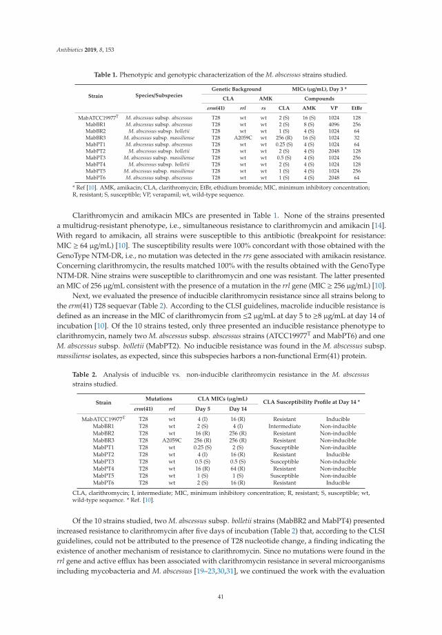
Antibiotics 2019, 8, 153
Table 1. Phenotypic and genotypic characterization of the M. abscessus strains studied.
Strain Species/SubspeciesGenetic Background MICs (μg/mL), Day 3 *
CLA AMK Compounds
erm(41) rrl rs CLA AMK VP EtBr
MabATCC19977T M. abscessus subsp. abscessus T28 wt wt 2 (S) 16 (S) 1024 128MabBR1 M. abscessus subsp. abscessus T28 wt wt 2 (S) 8 (S) 4096 256MabBR2 M. abscessus subsp. bolletii T28 wt wt 1 (S) 4 (S) 1024 64MabBR3 M. abscessus subsp. massiliense T28 A2059C wt 256 (R) 16 (S) 1024 32MabPT1 M. abscessus subsp. abscessus T28 wt wt 0.25 (S) 4 (S) 1024 64MabPT2 M. abscessus subsp. bolletii T28 wt wt 2 (S) 4 (S) 2048 128MabPT3 M. abscessus subsp. massiliense T28 wt wt 0.5 (S) 4 (S) 1024 256MabPT4 M. abscessus subsp. bolletii T28 wt wt 2 (S) 4 (S) 1024 128MabPT5 M. abscessus subsp. massiliense T28 wt wt 1 (S) 4 (S) 1024 256MabPT6 M. abscessus subsp. abscessus T28 wt wt 1 (S) 4 (S) 2048 64
* Ref [10]. AMK, amikacin; CLA, clarithromycin; EtBr, ethidium bromide; MIC, minimum inhibitory concentration;R, resistant; S, susceptible; VP, verapamil; wt, wild-type sequence.
Clarithromycin and amikacin MICs are presented in Table 1. None of the strains presenteda multidrug-resistant phenotype, i.e., simultaneous resistance to clarithromycin and amikacin [14].With regard to amikacin, all strains were susceptible to this antibiotic (breakpoint for resistance:MIC ≥ 64 μg/mL) [10]. The susceptibility results were 100% concordant with those obtained with theGenoType NTM-DR, i.e., no mutation was detected in the rrs gene associated with amikacin resistance.Concerning clarithromycin, the results matched 100% with the results obtained with the GenoTypeNTM-DR. Nine strains were susceptible to clarithromycin and one was resistant. The latter presentedan MIC of 256 μg/mL consistent with the presence of a mutation in the rrl gene (MIC ≥ 256 μg/mL) [10].
Next, we evaluated the presence of inducible clarithromycin resistance since all strains belong tothe erm(41) T28 sequevar (Table 2). According to the CLSI guidelines, macrolide inducible resistance isdefined as an increase in the MIC of clarithromycin from ≤2 μg/mL at day 5 to ≥8 μg/mL at day 14 ofincubation [10]. Of the 10 strains tested, only three presented an inducible resistance phenotype toclarithromycin, namely two M. abscessus subsp. abscessus strains (ATCC19977T and MabPT6) and oneM. abscessus subsp. bolletii (MabPT2). No inducible resistance was found in the M. abscessus subsp.massiliense isolates, as expected, since this subspecies harbors a non-functional Erm(41) protein.
Table 2. Analysis of inducible vs. non-inducible clarithromycin resistance in the M. abscessusstrains studied.
StrainMutations CLA MICs (μg/mL)
CLA Susceptibility Profile at Day 14 *erm(41) rrl Day 5 Day 14
MabATCC19977T T28 wt 4 (I) 16 (R) Resistant InducibleMabBR1 T28 wt 2 (S) 4 (I) Intermediate Non-inducibleMabBR2 T28 wt 16 (R) 256 (R) Resistant Non-inducibleMabBR3 T28 A2059C 256 (R) 256 (R) Resistant Non-inducibleMabPT1 T28 wt 0.25 (S) 2 (S) Susceptible Non-inducibleMabPT2 T28 wt 4 (I) 16 (R) Resistant InducibleMabPT3 T28 wt 0.5 (S) 0.5 (S) Susceptible Non-inducibleMabPT4 T28 wt 16 (R) 64 (R) Resistant Non-inducibleMabPT5 T28 wt 1 (S) 1 (S) Susceptible Non-inducibleMabPT6 T28 wt 2 (S) 16 (R) Resistant Inducible
CLA, clarithromycin; I, intermediate; MIC, minimum inhibitory concentration; R, resistant; S, susceptible; wt,wild-type sequence. * Ref. [10].
Of the 10 strains studied, two M. abscessus subsp. bolletii strains (MabBR2 and MabPT4) presentedincreased resistance to clarithromycin after five days of incubation (Table 2) that, according to the CLSIguidelines, could not be attributed to the presence of T28 nucleotide change, a finding indicating theexistence of another mechanism of resistance to clarithromycin. Since no mutations were found in therrl gene and active efflux has been associated with clarithromycin resistance in several microorganismsincluding mycobacteria and M. abscessus [19–23,30,31], we continued the work with the evaluation
41
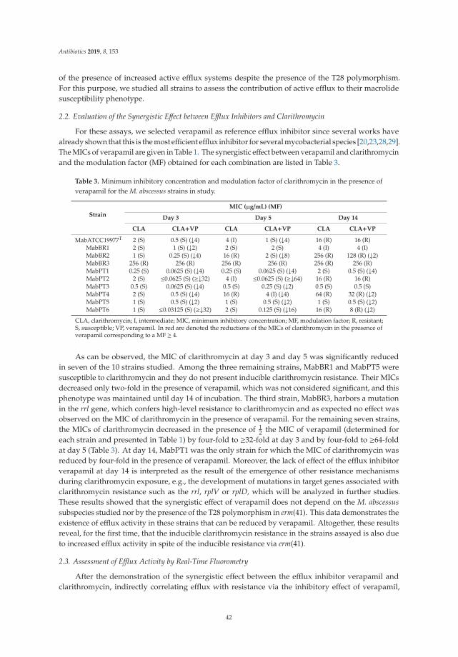
Antibiotics 2019, 8, 153
of the presence of increased active efflux systems despite the presence of the T28 polymorphism.For this purpose, we studied all strains to assess the contribution of active efflux to their macrolidesusceptibility phenotype.
2.2. Evaluation of the Synergistic Effect between Efflux Inhibitors and Clarithromycin
For these assays, we selected verapamil as reference efflux inhibitor since several works havealready shown that this is the most efficient efflux inhibitor for several mycobacterial species [20,23,28,29].The MICs of verapamil are given in Table 1. The synergistic effect between verapamil and clarithromycinand the modulation factor (MF) obtained for each combination are listed in Table 3.
Table 3. Minimum inhibitory concentration and modulation factor of clarithromycin in the presence ofverapamil for the M. abscessus strains in study.
Strain
MIC (μg/mL) (MF)
Day 3 Day 5 Day 14
CLA CLA+VP CLA CLA+VP CLA CLA+VP
MabATCC19977T 2 (S) 0.5 (S) (↓4) 4 (I) 1 (S) (↓4) 16 (R) 16 (R)MabBR1 2 (S) 1 (S) (↓2) 2 (S) 2 (S) 4 (I) 4 (I)MabBR2 1 (S) 0.25 (S) (↓4) 16 (R) 2 (S) (↓8) 256 (R) 128 (R) (↓2)MabBR3 256 (R) 256 (R) 256 (R) 256 (R) 256 (R) 256 (R)MabPT1 0.25 (S) 0.0625 (S) (↓4) 0.25 (S) 0.0625 (S) (↓4) 2 (S) 0.5 (S) (↓4)MabPT2 2 (S) ≤0.0625 (S) (≥↓32) 4 (I) ≤0.0625 (S) (≥↓64) 16 (R) 16 (R)MabPT3 0.5 (S) 0.0625 (S) (↓4) 0.5 (S) 0.25 (S) (↓2) 0.5 (S) 0.5 (S)MabPT4 2 (S) 0.5 (S) (↓4) 16 (R) 4 (I) (↓4) 64 (R) 32 (R) (↓2)MabPT5 1 (S) 0.5 (S) (↓2) 1 (S) 0.5 (S) (↓2) 1 (S) 0.5 (S) (↓2)MabPT6 1 (S) ≤0.03125 (S) (≥↓32) 2 (S) 0.125 (S) (↓16) 16 (R) 8 (R) (↓2)
CLA, clarithromycin; I, intermediate; MIC, minimum inhibitory concentration; MF, modulation factor; R, resistant;S, susceptible; VP, verapamil. In red are denoted the reductions of the MICs of clarithromycin in the presence ofverapamil corresponding to a MF ≥ 4.
As can be observed, the MIC of clarithromycin at day 3 and day 5 was significantly reducedin seven of the 10 strains studied. Among the three remaining strains, MabBR1 and MabPT5 weresusceptible to clarithromycin and they do not present inducible clarithromycin resistance. Their MICsdecreased only two-fold in the presence of verapamil, which was not considered significant, and thisphenotype was maintained until day 14 of incubation. The third strain, MabBR3, harbors a mutationin the rrl gene, which confers high-level resistance to clarithromycin and as expected no effect wasobserved on the MIC of clarithromycin in the presence of verapamil. For the remaining seven strains,the MICs of clarithromycin decreased in the presence of 1
2 the MIC of verapamil (determined foreach strain and presented in Table 1) by four-fold to ≥32-fold at day 3 and by four-fold to ≥64-foldat day 5 (Table 3). At day 14, MabPT1 was the only strain for which the MIC of clarithromycin wasreduced by four-fold in the presence of verapamil. Moreover, the lack of effect of the efflux inhibitorverapamil at day 14 is interpreted as the result of the emergence of other resistance mechanismsduring clarithromycin exposure, e.g., the development of mutations in target genes associated withclarithromycin resistance such as the rrl, rplV or rplD, which will be analyzed in further studies.These results showed that the synergistic effect of verapamil does not depend on the M. abscessussubspecies studied nor by the presence of the T28 polymorphism in erm(41). This data demonstrates theexistence of efflux activity in these strains that can be reduced by verapamil. Altogether, these resultsreveal, for the first time, that the inducible clarithromycin resistance in the strains assayed is also dueto increased efflux activity in spite of the inducible resistance via erm(41).
2.3. Assessment of Efflux Activity by Real-Time Fluorometry
After the demonstration of the synergistic effect between the efflux inhibitor verapamil andclarithromycin, indirectly correlating efflux with resistance via the inhibitory effect of verapamil,
42
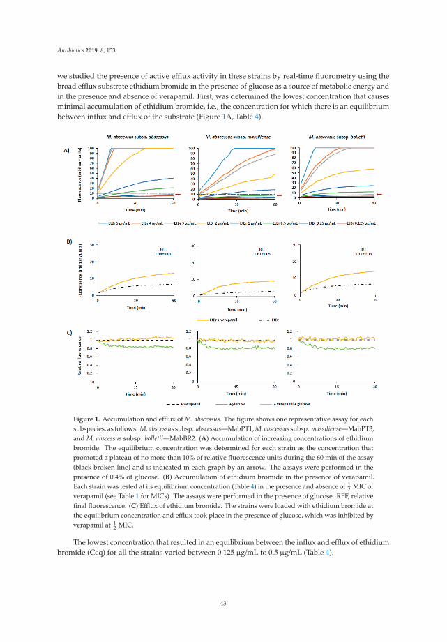
Antibiotics 2019, 8, 153
we studied the presence of active efflux activity in these strains by real-time fluorometry using thebroad efflux substrate ethidium bromide in the presence of glucose as a source of metabolic energy andin the presence and absence of verapamil. First, was determined the lowest concentration that causesminimal accumulation of ethidium bromide, i.e., the concentration for which there is an equilibriumbetween influx and efflux of the substrate (Figure 1A, Table 4).
Figure 1. Accumulation and efflux of M. abscessus. The figure shows one representative assay for eachsubspecies, as follows: M. abscessus subsp. abscessus—MabPT1, M. abscessus subsp. massiliense—MabPT3,and M. abscessus subsp. bolletii—MabBR2. (A) Accumulation of increasing concentrations of ethidiumbromide. The equilibrium concentration was determined for each strain as the concentration thatpromoted a plateau of no more than 10% of relative fluorescence units during the 60 min of the assay(black broken line) and is indicated in each graph by an arrow. The assays were performed in thepresence of 0.4% of glucose. (B) Accumulation of ethidium bromide in the presence of verapamil.Each strain was tested at its equilibrium concentration (Table 4) in the presence and absence of 1
2 MIC ofverapamil (see Table 1 for MICs). The assays were performed in the presence of glucose. RFF, relativefinal fluorescence. (C) Efflux of ethidium bromide. The strains were loaded with ethidium bromide atthe equilibrium concentration and efflux took place in the presence of glucose, which was inhibited byverapamil at 1
2 MIC.
The lowest concentration that resulted in an equilibrium between the influx and efflux of ethidiumbromide (Ceq) for all the strains varied between 0.125 μg/mL to 0.5 μg/mL (Table 4).
43
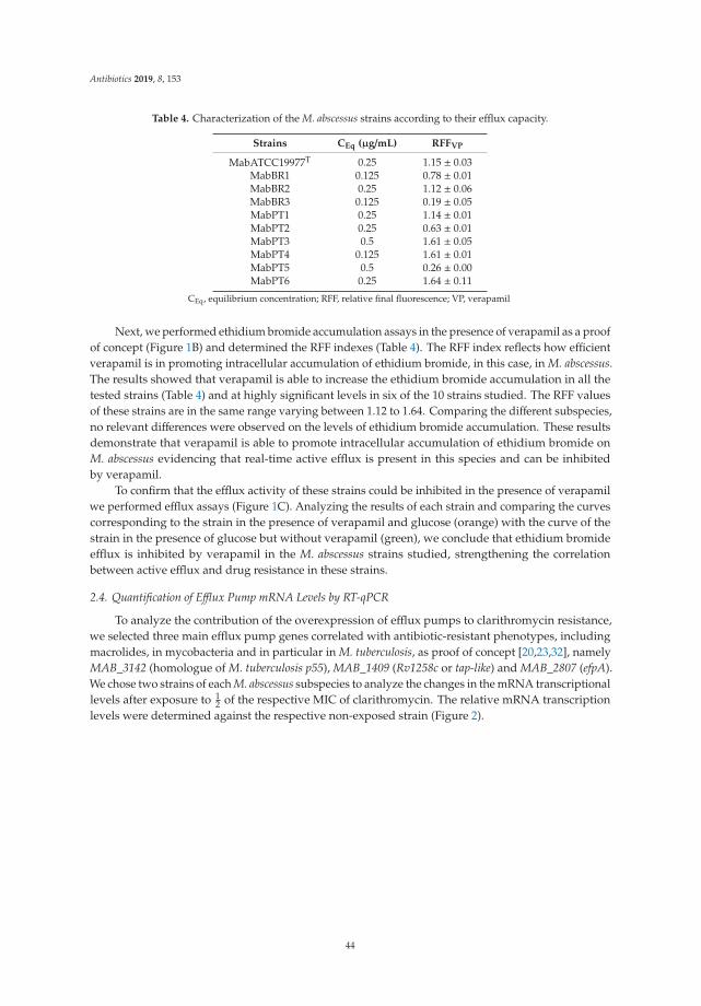
Antibiotics 2019, 8, 153
Table 4. Characterization of the M. abscessus strains according to their efflux capacity.
Strains CEq (μg/mL) RFFVP
MabATCC19977T 0.25 1.15 ± 0.03MabBR1 0.125 0.78 ± 0.01MabBR2 0.25 1.12 ± 0.06MabBR3 0.125 0.19 ± 0.05MabPT1 0.25 1.14 ± 0.01MabPT2 0.25 0.63 ± 0.01MabPT3 0.5 1.61 ± 0.05MabPT4 0.125 1.61 ± 0.01MabPT5 0.5 0.26 ± 0.00MabPT6 0.25 1.64 ± 0.11
CEq, equilibrium concentration; RFF, relative final fluorescence; VP, verapamil
Next, we performed ethidium bromide accumulation assays in the presence of verapamil as a proofof concept (Figure 1B) and determined the RFF indexes (Table 4). The RFF index reflects how efficientverapamil is in promoting intracellular accumulation of ethidium bromide, in this case, in M. abscessus.The results showed that verapamil is able to increase the ethidium bromide accumulation in all thetested strains (Table 4) and at highly significant levels in six of the 10 strains studied. The RFF valuesof these strains are in the same range varying between 1.12 to 1.64. Comparing the different subspecies,no relevant differences were observed on the levels of ethidium bromide accumulation. These resultsdemonstrate that verapamil is able to promote intracellular accumulation of ethidium bromide onM. abscessus evidencing that real-time active efflux is present in this species and can be inhibitedby verapamil.
To confirm that the efflux activity of these strains could be inhibited in the presence of verapamilwe performed efflux assays (Figure 1C). Analyzing the results of each strain and comparing the curvescorresponding to the strain in the presence of verapamil and glucose (orange) with the curve of thestrain in the presence of glucose but without verapamil (green), we conclude that ethidium bromideefflux is inhibited by verapamil in the M. abscessus strains studied, strengthening the correlationbetween active efflux and drug resistance in these strains.
2.4. Quantification of Efflux Pump mRNA Levels by RT-qPCR
To analyze the contribution of the overexpression of efflux pumps to clarithromycin resistance,we selected three main efflux pump genes correlated with antibiotic-resistant phenotypes, includingmacrolides, in mycobacteria and in particular in M. tuberculosis, as proof of concept [20,23,32], namelyMAB_3142 (homologue of M. tuberculosis p55), MAB_1409 (Rv1258c or tap-like) and MAB_2807 (efpA).We chose two strains of each M. abscessus subspecies to analyze the changes in the mRNA transcriptionallevels after exposure to 1
2 of the respective MIC of clarithromycin. The relative mRNA transcriptionlevels were determined against the respective non-exposed strain (Figure 2).
44
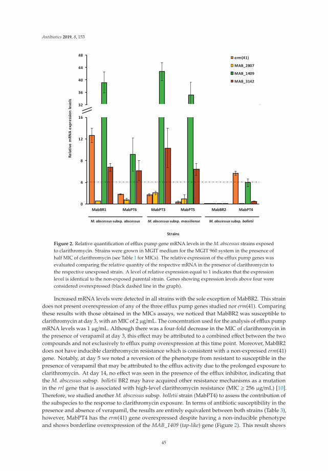
Antibiotics 2019, 8, 153
Figure 2. Relative quantification of efflux pump gene mRNA levels in the M. abscessus strains exposedto clarithromycin. Strains were grown in MGIT medium for the MGIT 960 system in the presence ofhalf MIC of clarithromycin (see Table 1 for MICs). The relative expression of the efflux pump genes wasevaluated comparing the relative quantity of the respective mRNA in the presence of clarithromycin tothe respective unexposed strain. A level of relative expression equal to 1 indicates that the expressionlevel is identical to the non-exposed parental strain. Genes showing expression levels above four wereconsidered overexpressed (black dashed line in the graph).
Increased mRNA levels were detected in all strains with the sole exception of MabBR2. This straindoes not present overexpression of any of the three efflux pump genes studied nor erm(41). Comparingthese results with those obtained in the MICs assays, we noticed that MabBR2 was susceptible toclarithromycin at day 3, with an MIC of 2 μg/mL. The concentration used for the analysis of efflux pumpmRNA levels was 1 μg/mL. Although there was a four-fold decrease in the MIC of clarithromycin inthe presence of verapamil at day 3, this effect may be attributed to a combined effect between the twocompounds and not exclusively to efflux pump overexpression at this time point. Moreover, MabBR2does not have inducible clarithromycin resistance which is consistent with a non-expressed erm(41)gene. Notably, at day 5 we noted a reversion of the phenotype from resistant to susceptible in thepresence of verapamil that may be attributed to the efflux activity due to the prolonged exposure toclarithromycin. At day 14, no effect was seen in the presence of the efflux inhibitor, indicating thatthe M. abscessus subsp. bolletii BR2 may have acquired other resistance mechanisms as a mutationin the rrl gene that is associated with high-level clarithromycin resistance (MIC ≥ 256 μg/mL) [10].Therefore, we studied another M. abscessus subsp. bolletii strain (MabPT4) to assess the contribution ofthe subspecies to the response to clarithromycin exposure. In terms of antibiotic susceptibility in thepresence and absence of verapamil, the results are entirely equivalent between both strains (Table 3),however, MabPT4 has the erm(41) gene overexpressed despite having a non-inducible phenotypeand shows borderline overexpression of the MAB_1409 (tap-like) gene (Figure 2). This result shows
45
Antibiotics 2019, 8, 153
that erm(41) gene expression is not the sole cause of clarithromycin resistance in this strain. Effluxactivity is present in this strain (Table 4) representing a late stress response to clarithromycin exposure.Concerning M. abscessus subsp. massiliense, we analyzed MabPT3 and MabPT5, both susceptible toclarithromycin without inducible resistance consistent with the lack of expression of erm(41) encodinga non-functional Erm(41) protein. Although susceptible, the MICs of clarithromycin decreased in thepresence of verapamil by four-fold in MabPT3 and two-fold in MabPT5 (Table 3). Both strains showedoverexpression of MAB_3142 and MAB_1409 (Figure 2). Comparing to the real-time efflux assays,MabPT3 has increased efflux activity although MabPT5 showed lower efflux activity than MabPT3.This result shows that efflux activity acts also as a first-line response in these M. abscessus subsp.massiliense strains. Finally, we studied two strains of M. abscessus subsp. abscessus, namely MabBR1 andMabPT6. MabBR1 showed intermediate resistance at day 14, and consequently, non-inducible resistanceto clarithromycin, whereas MabPT6 was resistant at day 14 through an inducible resistant phenotypeto this antibiotic. Comparing this data with the RT-qPCR assays, MabBR1 exhibited overexpression ofMAB_3142 and MAB_1409 efflux pump genes and, somehow unexpected, also erm(41). On the contrary,MabPT6 that presents inducible resistance to clarithromycin, although at low levels (16 μg/mL), showedno expression of erm(41). However, this strain shows overexpression of MAB_3142 and MAB_1409efflux pump genes. Comparing with the real-time efflux assays, MabBR1 showed lower levels of effluxthan MabPT6, probably due to the presence of an overexpressed erm(41). No overexpression wasobserved concerning the gene MAB_2807 for any of the six strains analyzed.
3. Discussion
Members of the M. abscessus complex are clinically important pathogens. At present, only fewantibiotics showed antimicrobial activity with clinical significance against this species, namely,clarithromycin and amikacin, and either cefoxitin or imipenem. The main reasons commonly acceptedfor this scarce therapeutic panel is the low permeability of M. abscessus complex cell wall, the presenceof mutations in the drug target genes and, in the case of clarithromycin, the presence of an inducibleerm(41) gene [14]. Recently, the hypothesis of efflux-mediated drug resistance has been appointed as aputative contributor to drug resistance in M. abscessus [21,22].
All bacteria express efflux pumps. Efflux transporters are transmembrane proteins involvedin the extrusion of harmful compounds and cellular metabolites from the cells into the externalenvironment [33]. Indirectly, they are also involved in drug resistance by expelling antimicrobials,reducing their intracellular concentration. Treatment failure has been mainly attributed to M. abscessusbelonging to the T28 sequevar [12,34,35]. Similar to what is observed in M. avium complex infections,acquired resistance to clarithromycin also evolves rapidly during therapy in M. abscessus [36]. Therefore,it is legitimate to raise the hypothesis of whether the overexpression of efflux pumps is also involvedin the resistance to clarithromycin even in the M. abscessus sequevar T28? Ongoing work showedthat resistance towards clarithromycin in M. avium results from the balance between the presence ofdrug resistance-associated mutations and increased efflux activity. We also noticed that this increasedefflux activity was accompanied by the overexpression of several efflux pump genes upon exposureto clarithromycin. Therefore, to answer this question, we investigated the contribution of effluxtransporters to the induced clarithromycin resistance in M. abscessus complex belonging to the erm(41)T28 sequevar.
The strains were blindly selected in relation to their clarithromycin susceptibility status andincluded a panel of nine clinical strains, comprising three isolates of each M. abscessus subspecies,namely, subsp. abscessus, bolletii and massiliense. The M. abscessus subsp. abscessus ATCC19977T referencestrain was included as control. Firstly, we searched for mutations associated with clarithromycinresistance the M. abscessus strains. We found that all belong to the erm(41) T28 sequevar which, at leasttheoretically, is responsible for the inducible resistance to clarithromycin in M. abscessus [12,14]. One ofthe strains harbors a mutation in rrl, and consequently, displays high-level resistance to clarithromycin,and was used as mutation-driven resistance control. These initial results are in accordance with those
46
Antibiotics 2019, 8, 153
described in the literature that states that the majority of the M. abscessus strains are susceptible toclarithromycin and resistance is acquired during treatment [1,14]. To test the role of the erm(41) T28polymorphism in the induced resistance of the strains analyzed, we read the MICs at day 5 of incubationand prolonged the incubation until day 14. Surprisingly, of the eight strains, only two clinical strainsand the reference strain showed induced resistance to clarithromycin, one M. abscessus subsp. abscessusand the other M. abscessus subsp. bolletii. Interestingly, two M. abscessus subsp. bolletii strains wereshown to be already resistant to clarithromycin at day 5 and according to the CLSI guidelines, they donot fit in the category of clarithromycin-inducible resistance. These results demonstrate that the T28polymorphism in erm(41) is not the sole cause of clarithromycin-induced resistance in these M. abscessusstrains indicating the existence of another mechanism of resistance in this collection of strains.
We then studied the presence of active efflux activity in these strains through the determinationof the MICs of clarithromycin in the presence of the efflux inhibitor verapamil. The results obtainedsupported our hypothesis that efflux is also involved in M. abscessus induced resistance to clarithromycin.At day 3 and day 5, all the MICs of clarithromycin for all strains, with the sole exception of the strainwith the rrl mutation, could be reduced in the presence of the efflux inhibitor verapamil. In particular,it was possible to reverse to clinically significant levels the resistance to clarithromycin at day 5 for fourstrains, one from resistant to susceptible, one from resistant to intermediate and two from intermediateto susceptible. After 14 days of exposure, no inhibitory effect was noted for these, meaning thatthese strains may have acquired other mutational mechanisms of resistance, a subject that will beaddressed in subsequent studies. Concurrently with real-time fluorometric assays, using verapamil asreference efflux inhibitor and ethidium bromide as substrate, we detected significant efflux activityin the 10 strains, which could be inhibited in the presence of verapamil. It was also demonstratedthat this efflux activity does not depend on subspecies of the strains, as it was detected in all thethree subspecies, although at different levels, nor due to their phenotype since it was detected ininducible and non-inducible clarithromycin resistance strains. These data demonstrated the presenceand importance of active efflux in M. abscessus strains resistant to clarithromycin, which could beinhibited by verapamil.
Finally, to further analyze the contribution of the expression of efflux pumps to clarithromycinresistance in these strains, three efflux pump genes, MAB_3142, MAB_1409 and MAB_2807, and theerm(41) gene, were selected to examine changes in mRNA transcriptional levels after exposure to 1
2MIC of clarithromycin determined for each of the six strains selected. We noted increased mRNAexpression levels of the efflux pumps MAB_3142 and MAB_1409 in four out of six of the strains andfound also erm(41) overexpression in two strains, one presenting inducible resistance while the otherdoes not have induced resistance to clarithromycin.
This exploratory study consolidates and strengthens the hypothesis that efflux activity plays arole in M. abscessus intrinsic resistance to clarithromycin in strains belonging to the T28 sequevar andthat the inhibition of this efflux activity by compounds such as verapamil could potentiate the clinicaleffect of clarithromycin, the most efficient antibiotic to treat M. abscessus infections, certainly reducingor preventing the emergence of the, so far almost inexorable, acquired resistance to macrolides duringtreatment against M. abscessus infections.
4. Materials and Methods
4.1. M. abscessus Complex Strains
The strains selected for this study are described in Table 1 and were isolated from Brazilian (BR) andPortuguese (PT) patients. Brazilian clinical strains were granted by Fundação Oswaldo Cruz (Rio deJaneiro, Brazil). The Portuguese clinical strains are part of the strain collection of the MycobacteriologyLaboratory of the Instituto de Higiene e Medicina Tropical, Universidade NOVA de Lisboa (Lisboa,Portugal). The strains were identified using the GenoType NTM-DR assay (Hain Lifescience, GmbH,Nehren, Germany) according to the manufacturer’s instructions. Informed consent was not required
47
Antibiotics 2019, 8, 153
for the present study since it corresponds to a retrospective study from which all patient identificationwas unlinked from the results, and no patient information was collected. M. abscessus subsp. abscessusATCC19977T reference strain was obtained from the American Type Culture Collection (Virginia, USA)and was included as a control strain.
4.2. Compounds and Reagents
Amikacin, clarithromycin, verapamil, ethidium bromide, dimethyl sulfoxide (DMSO), and glucosewere purchased from Sigma-Aldrich (St. Louis, MO, USA). Clarithromycin was prepared in DMSO,while the remaining drugs were prepared in sterile deionized water. Stock solutions were stored at−20 ◦C. Working solutions of both antibiotics and ethidium bromide were prepared in Mueller–Hintonbroth (MHB; Oxoid, Hampshire, UK) on the day of the experiments. Löwenstein–Jensen medium,Middlebrook 7H9, BBL OADC (oleic acid/albumin/dextrose/catalase) supplement and the mycobacteriagrowth indicator tubes (MGITs) were purchased from Becton Dickinson (Diagnostic Systems, Sparks,MD, USA).
4.3. Detection of Mutations Associated with Clarithromycin and Amikacin Resistance
Total DNA was extracted from clinical strains and the reference strain using the GenoLyse kit(Hain Lifescience) according to the manufacturer’s instructions. The presence of mutations in rrl,erm(41) and rrs were analyzed with the Genotype NTM-DR assay.
4.4. Susceptibility Testing
4.4.1. Growth of the Strains
All strains were maintained in MGITs tubes supplemented with 10% OADC. For minimuminhibitory concentration (MIC) determination and synergism assays, the M. abscessus strains wereinoculated in Löwenstein–Jensen medium and incubated at 37 ◦C for three to five days.
4.4.2. MIC Determination of Compounds
For the determination of the MICs of the antibiotics, the efflux inhibitor verapamil and theefflux substrate ethidium bromide, the inoculum of each strain was prepared by diluting the bacterialcultures in MHB to a final density of approximately 105 cells/mL [37]. The MICs were determinedusing a tetrazolium microplate-based assay (TEMA) [38] with small modifications. Briefly, aliquotsof 0.1 mL of inoculum were transferred to each well of the plate that contained 0.1 mL of eachcompound at concentrations prepared from two-fold serial dilutions in MHB medium. Growth controls(only inoculum without drug) and a sterility control (only medium) were included in each plate.Two hundred microliters of sterile deionized water were added to the outer-perimeter wells of the96-well plates to reduce evaporation of the medium during the incubation. The inoculated plates weresealed in permeable plastic bags and incubated at 37 ◦C for three days. After this period, 10 μL of asolution of 5×MTT/10%T80 (1:1) was added to each well and the plates incubated for four hours atroom temperature. The bacterial growth was registered for each well based on the MTT color change.As the intensity of the color generated is directly proportional to the number of viable cells, a precipitateof cells stained black can be observed in the bottom of the well. The MIC was defined as the lowestconcentration of compound that totally inhibited bacterial growth, i.e., no color change [39]. All assayswere performed in triplicate. For amikacin, the breakpoints used were: ≤16 μg/mL, susceptible;32 μg/mL, intermediate; and ≥64 μg/mL, resistant. For clarithromycin at day 3, the breakpoints usedwere: ≤2 μg/mL, susceptible; 4 μg/mL, intermediate and ≥8 μg/mL, resistant [10].
To evaluate clarithromycin induced resistance, the MIC determination of clarithromycin wasextended for 14 days. To do this, plates containing clarithromycin were prepared in quadruplicate to beread at the following time points: 3, 5 and 14 days [14]. After each period of incubation, MTT was addedand the results read and interpreted as described above. Susceptible isolates at day 3 (MIC ≤ 2 μg/mL)
48
Antibiotics 2019, 8, 153
and resistant at day 5 (MIC ≥ 8 μg/mL), were considered to have an inducible erm(41) gene [10]. Allassays were carried out in triplicate.
4.4.3. MIC Determination of Antibiotics and Ethidium Bromide in the Presence of Verapamil
The MICs of amikacin, clarithromycin, and ethidium bromide were determined in the presenceof verapamil. The assays were performed as described above with the exception that verapamil wasadded to each well to a final concentration of 1
2 of the MIC. The results were read and interpreted asdescribed above. The modulation factor (MF) was calculated and used to quantify the inhibitory effectof verapamil on the MICs of clarithromycin through the following formula: MF =MIC antimicrobial /MIC antimicrobial + efflux inhibitor [40]. The MF reflects the reduction of the MIC values of a givenantimicrobial in the presence of an efflux inhibitor, being considered significant when MF ≥ 4 (four-foldreduction) [41]. Each assay was carried out in triplicate.
4.4.4. Semi-Automated Fluorometric Method
The evaluation of efflux activity on a real-time basis was performed using a semi-automatedfluorometric method [26] with modifications [Machado et al., unpublished]. The M. abscessus strainswere grown in 10 ml of 7H9/10%OADC with 0.05% tyloxapol at 37 ◦C until they reached an OD600
of 0.8. For the accumulation assays, the cultures were centrifuged at 3500 rpm for three minutes,the supernatant discarded and the pellet washed in 7H9. The OD600 was adjusted to 0.8 with 7H9.In order to determine the concentration of ethidium bromide that establishes the equilibrium betweenefflux and influx, aliquots of 0.05 mL of the bacterial suspension were added to 0.2 mL microtubescontaining 0.05 mL different concentrations of ethidium bromide that ranged from 0.625 μg/mL to5 μg/mL with and without 0.4% glucose. The assays were conducted at 37 ◦C in a Rotor-Gene 3000(Corbett Research, Sydney, Australia), using the 530 nm band-pass and the 585 nm high-pass filtersas the excitation and detection wavelengths, respectively. Fluorescence data were acquired every60 seconds for 60 min. The selected concentration of ethidium bromide was further used for theevaluation of the capacity of verapamil to retain ethidium bromide inside the cells. Verapamil wastested at 1
2 MIC with and without 0.4% glucose and ethidium bromide at the equilibrium concentrationdetermined for each strain and the assays performed as described above. The inhibitory activity ofverapamil was determined through the determination of the relative final fluorescence (RFF) valueaccording to the following formula [42]: RFF = (RFtreated-RFuntreated)/RFuntreated. In this formula,the RFtreated corresponds to the relative fluorescence at the last time point of ethidium bromideaccumulation curve (min 60) in the presence of verapamil, and the RFuntreated corresponds to therelative fluorescence at the last time point of the ethidium bromide accumulation curve of the untreatedcontrol tube. An index of activity above zero indicate that the cells accumulate more ethidium bromideunder the condition used than those of the control (untreated cells). Negative RFF values indicate thatthe treated cells accumulated less ethidium bromide than those of the control condition [28,32].
For the efflux assays, the strains were exposed to conditions that promote the maximumaccumulation of ethidium bromide, i.e., ethidium bromide equilibrium concentration for each strain,no glucose, presence of verapamil at 1
4 MIC, and incubation at 20 ◦C for one hour [26]. Before incubation,the cultures were centrifuged at 3500 rpm for three minutes, resuspended in 7H9, centrifuged againand OD600 adjusted to 0.4. The suspension was incubated with ethidium bromide and verapamil underthe conditions described above. Aliquots of 0.05 mL of cells were transferred to 0.2 mL microtubescontaining 0.05 mL of verapamil at 1
2 MIC without ethidium bromide. Control tubes with only cellsand cells with and without 0.4% glucose were included. Fluorescence was measured in a Rotor-Gene™3000 and data were acquired every 30 seconds for 30 min. Efflux activity was quantified by comparingthe fluorescence data obtained under conditions that promote efflux (presence of glucose and absenceof efflux inhibitor) with the data from the control in which the mycobacteria are under conditions of noefflux (presence of an inhibitor and no energy source). Thus, the relative fluorescence corresponds
49
Antibiotics 2019, 8, 153
to the ratio of fluorescence that remains per unit of time, relatively to the ethidium bromide-loadedcells [27].
4.5. Efflux Pump Gene Expression
4.5.1. Sample Preparation
For the analysis of efflux pump gene expression, each strain was inoculated into 7H9 containing10% OADC plus clarithromycin at 1
2 the MIC (see Table 1 for MIC values). The strains were thenincubated at 37 ◦C within the MGIT system until growth was achieved [20]. At this time point, samplesof each culture were taken for RNA extraction and RT-qPCR analysis.
4.5.2. RNA Extraction
Total RNA was isolated from the cells using the RNeasy mini kit (QIAGEN, GmbH, Hilden,Germany) according to the manufacturer’s instructions with modifications [20]. Briefly, from a culturewith 100–200 GU (about 106–108 cells/ml), 1 ml aliquot was removed and centrifuged at 13,000 rpmduring 10 min. Then, 500 μL supernatant was removed and 1 mL of RNAprotect Bacteria Reagent(QIAGEN) added. An enzymatic lysis step was carried out with lysozyme at 3 mg/mL (Sigma) for10 min, followed by lysis in an ultrasonic bath at 35 kHz (Gen-Probe, CA, USA) during 15 min. The RNAwas purified using the RNeasy kit (QIAGEN) and treated with RNase-free DNase I (QIAGEN) during2 h and 15 min by on-column digestion at room temperature to reduce the presence of contaminatingDNA. All RNA samples were stored at −20 ◦C until required.
4.5.3. RT-qPCR Assay
The relative expression level of mRNA of genes that code for membrane efflux transportersin M. abscessus (MAB_2807, MAB_1409 and MAB_3142) and erm(41) was analyzed by RT-qPCR.The normalization of the data was done using the M. abscessus 16S rDNA. The RT-qPCR procedurewas performed in a Rotor-Gene 3000 thermocycler and followed the protocol recommended for usewith the One-step NZY RT-qPCR Green kit (Nzytech, Portugal). The determination of the relativemRNA expression level was performed using the comparative quantification cycle (Cq) method [43].The relative expression of the genes analyzed was assessed by comparison of the relative quantityof the respective mRNA in the presence of clarithromycin to the non-exposed culture, following thesame technical approach previously published [20]. Each culture was assayed in triplicate using totalRNA obtained from three independent cultures. A level of relative expression equal to 1 indicates thatthe expression level was identical to the unexposed strain. Genes showing expression levels equal orabove four, when compared with the unexposed strain, were considered to be overexpressed [20].
5. Conclusions
The results obtained in this study provide evidence that efflux activity is an intrinsic characteristicof M. abscessus clinical strains and is directly involved in clarithromycin resistance. Literature reviewindicated that M. abscessus subsp. abscessus T28 and M. abscessus subsp. bolletii T28 are intrinsicallyresistant to clarithromycin conferred by an altered Erm(41) methylase [1]. The change of cytosine bythymine at position T28 on Erm(41) causes resistance to clarithromycin due to prolonged exposureleading to treatment failure [12–14]. In our hands, the results obtained pointed out that erm(41)expression is not the sole cause of the inducible clarithromycin resistance in M. abscessus subsp.abscessus and M. abscessus subsp. bolletii T28 sequevars and that efflux pump overexpression has astrong contribution to clarithromycin resistance acting both as a first-line response or a late response todrug pressure. This study was designed as a proof of concept. Therefore, due to the small number ofstrains studied, we could not generalize the results obtained here. Future work will include a highernumber of strains belonging to the three subspecies. Another limitation of this study, consequence ofthe random selection of the strains, is the absence of strains from the C28 sequevar, which will be also
50
Antibiotics 2019, 8, 153
studied further. Nevertheless, our results point out efflux as an important contributor to the emergenceof M. abscessus clarithromycin resistance and highlight the need of further studies on M. abscessussenso lato efflux response to stress imposed by antimicrobials in order to implement more effectivetherapeutic regimens and aid in the development of new drugs against these bacteria.
Author Contributions: Conceptualization, J.S.V., D.M., I.B.R., D.F.R., M.V. and P.E.A.d.S.; data curation, J.S.V.,D.M., I.B.R. and M.V.; funding acquisition, D.M., M.V. and P.E.A.d.S.; methodology, J.S.V., D.M., I.B.R., F.P.S.,D.V.B., M.A.A. and D.F.R.; resources, D.M., I.B.R., M.C.S.L., M.V. and P.E.A.d.S.; supervision, J.S.V., D.M., I.B.R.,A.v.G., M.V. and P.E.A.d.S.; writing—original draft, J.S.V. and I.B.R.; writing—review & editing, J.S.V., D.M., I.B.R.,M.V. and P.E.A.d.S.
Funding: Coordenação de Aperfeiçoamento de Pessoal de Nível Superior (CAPES, MEC, Brazil) for financialsupport to Ivy B. Ramis and Miguel Viveiros. through the program “Ciências sem Fronteiras/Professor VisitanteEspecial” (Projecto 88881.064961/2014-01 - José R. Lapa e Silva/UFRJ coordinator). Diana Machado and MiguelViveiros work was partially supported by project PTDC/BIA-MIC/30692/2017 and the Global Health andTropical Medicine (GHTM) Research Center (Grant UID/Multi/04413/2013) from Fundação para a Ciência ea Tecnologia, Portugal.
Acknowledgments: Diana Machado would like to acknowledge Fundação para a Ciência e a Tecnologia, Portugal,for the support under the program “Stimulus of Scientific Employment”.
Conflicts of Interest: The authors declare no conflict of interest. The funders had no role in the design of thestudy; in the collection, analyses, or interpretation of data; in the writing of the manuscript, or in the decision topublish the results.
References
1. Mougari, F.; Guglielmetti, L.; Raskine, L.; Sermet-Gaudelus, I.; Veziris, N.; Cambau, E. Infections caused byMycobacterium abscessus: Epidemiology, diagnostic tools and treatment. Expert Rev. Anti Infect. Ther. 2016, 14,1139–1154. [CrossRef] [PubMed]
2. Mukherjee, D.; Wu, M.; Teo, J.; Dick, T. Vancomycin and clarithromycin show synergy against Mycobacteriumabscessus in vitro. Antimicrob. Agents Chemother. 2017, 61, e01298–e01317. [CrossRef] [PubMed]
3. Roux, A.-L.; Catherinot, E.; Ripoll, F.; Soismier, N.; Macheras, E.; Ravilly, S.; Bellis, G.; Vibet, M.-A.; LeRoux, E.; Lemonnier, L.; et al. Multicenter study of prevalence of nontuberculous mycobacteria in patientswith cystic fibrosis in France. J. Clin. Microbiol. 2009, 47, 4124–4128. [CrossRef] [PubMed]
4. Mirsaeidi, M.; Farshidpour, M.; Allen, M.; Ebrahimi, G.; Falkinham, J. Highlight on advances innontuberculous mycobacterial disease in North America. Biomed. Res. Int. 2014, 919474, 1–10. [CrossRef][PubMed]
5. Leão, S.; Tortoli, E.; Euzéby, J.; Garcia, M. Proposal that Mycobacterium massiliense and Mycobacterium bolletiibe united and reclassified as Mycobacterium abscessus subsp. bolletii comb. nov., designation of Mycobacteriumabscessus subsp. abscessus subsp. nov. and emended description of Mycobacterium abscessus. Int. J. Syst. Evol.Microbiol. 2011, 61, 2311–2313. [CrossRef]
6. Kim, H.-Y.; Kim, B.; Kook, Y.; Yun, Y.-J.; Shin, J.; Hwan, J.; Kim, B.-J.; Kook, Y.-H. Mycobacteriummassiliense is differentiated from Mycobacterium abscessus and Mycobacterium bolletii by erythromycin ribosomemethyltransferase gene (erm) and clarithromycin susceptibility patterns. Microbiol. Immunol. 2010, 54,347–353. [CrossRef] [PubMed]
7. Singh, S.; Bouzinbi, N.; Chaturvedi, V.; Godreuil, S.; Kremer, L. In vitro evaluation of a new drug combinationagainst clinical isolates belonging to the Mycobacterium abscessus complex. Clin. Microbiol. Infect. 2014, 20,O1124–O1127. [CrossRef] [PubMed]
8. Zhang, Z.; Lu, J.; Liu, M.; Wang, Y.; Zhao, Y.; Pang, Y. In vitro activity of clarithromycin in combination withother antimicrobial agents against Mycobacterium abscessus and Mycobacterium massiliense. Int. J. Antimicrob.Agents 2017, 49, 383–386. [CrossRef]
9. Griffith, D.; Aksamit, T.; Brown-Elliott, B.; Catanzaro, A.; Daley, C.; Gordin, F.; Holland, S.; Horsburgh, R.;Huitt, G.; Iademarco, M.; et al. An official ATS/IDSA statement: Diagnosis, treatment, and prevention ofnontuberculous mycobacterial diseases. Am. J. Respir. Crit. Care Med. 2007, 175, 367–416. [CrossRef]
10. Clinical Laboratory Standards Institute. Susceptibility Testing of Mycobacteria, Nocardiae, and Other AerobicActinomycetes; Approved Standard; CLSI document M24-A2; Clinical Laboratory Standards Institute (CLSI):Wayne, PA, USA, 2011.
51
Antibiotics 2019, 8, 153
11. Wallace, R.; Meier, A.; Brown, B.; Zhang, Y.; Sander, P.; Onyi, G.; Böttger, E. Genetic basis for clarithromycinresistance among isolates of Mycobacterium chelonae and Mycobacterium abscessus. Antimicrob. Agents Chemother.1996, 40, 1676–1681. [CrossRef]
12. Nash, K.; Brown-Elliott, B.; Wallace, R. A novel gene, erm (41), confers inducible macrolide resistance toclinical isolates of Mycobacterium abscessus but is absent from Mycobacterium chelonae. Antimicrob. AgentsChemother. 2009, 53, 1367–1376. [CrossRef] [PubMed]
13. Bastian, S.; Veziris, N.; Roux, A.; Brossier, F.; Gaillard, J.; Jarlier, V.; Cambau, E. Assessment of clarithromycinsusceptibility in strains belonging to the Mycobacterium abscessus group by erm41 and rrl sequencing.Antimicrob. Agents Chemother. 2011, 55, 775–781. [CrossRef] [PubMed]
14. Mougari, F.; Amarsy, R.; Veziris, N.; Bastian, S.; Brossier, F.; Berçot, B.; Raskine, L.; Cambau, E. Standardizedinterpretation of antibiotic susceptibility testing and resistance genotyping for Mycobacterium abscessus withregard to subspecies and erm41 sequevar. J. Antimicrob. Chemother. 2016, 71, 2208–2212. [CrossRef] [PubMed]
15. Maurer, F.; Bruderer, V.; Castelberg, C.; Ritter, C.; Scherbakov, D.; Bloemberg, G.; Böttger, E. Aminoglycoside-Modifying enzymes determine the innate susceptibility to aminoglycoside antibiotics in rapidly growingmycobacteria. J. Antimicrob. Chemother. 2015, 70, 1412–1419. [CrossRef] [PubMed]
16. Dubée, V.; Bernut, A.; Cortes, M.; Lesne, T.; Dorchene, D.; Lefebvre, A.; Hugonnet, J.-E.; Gutmann, F.;Mainardi, J.-L.; Herrmann, J.-L.; et al. β-Lactamase inhibition by avibactam in Mycobacterium abscessus.J. Antimicrob. Chemother. 2014, 70, 1051–1058. [CrossRef]
17. Oh, W.; Stout, J.; Yew, W. Advances in the management of pulmonary disease due to Mycobacterium abscessuscomplex. Int. J. Tuberc. Lung Dis. 2014, 18, 1141–1148. [CrossRef]
18. Diel, R.; Ringshausen, F.; Richter, E.; Welker, L.; Schmitz, J.; Nienhaus, A. Microbiological and clinicaloutcomes of treating non-Mycobacterium avium complex nontuberculous mycobacterial pulmonary disease:A systematic review and meta-analysis. Chest 2017, 152, 120–142. [CrossRef] [PubMed]
19. Schmalstieg, A.; Srivastava, S.; Belkaya, S.; Deshpande, D.; Meek, C.; Leff, R.; van Oers, N.; Gumbo, T.The antibiotic resistance arrow of time: Efflux pump induction is a general first step in the evolution ofmycobacterial drug resistance. Antimicrob. Agents Chemother. 2012, 56, 4806–4815. [CrossRef] [PubMed]
20. Machado, D.; Couto, I.; Perdigão, J.; Rodrigues, L.; Portugal, I.; Baptista, P.; Veigas, B.; Amaral, L.; Viveiros, M.Contribution of efflux to the emergence of isoniazid and multidrug resistance in Mycobacterium tuberculosis.PLoS ONE 2012, 7, e34538. [CrossRef]
21. Ramis, I.; Vianna, J.; Silva, L., Jr.; von Groll, A.; Ramos, D.; Zanatta, N.; Viveiros, M.; da Silva, P.E.A. In silicoand in vitro evaluation of tetrahydropyridine compounds as efflux inhibitors in Mycobacterium abscessus.Tuberculosis 2019, 118, 101853. [CrossRef]
22. Gutiérrez, A.; Richard, M.; Roquet-Banères, F.; Viljoen, A.; Kremer, L. The TetR-family transcription factorMAB_2299c regulates the expression of two distinct MmpS-MmpL efflux pumps involved in cross-resistanceto clofazimine and bedaquiline in Mycobacterium abscessus. Antimicrob. Agents Chemother. 2019, AAC.01000-19.[CrossRef]
23. Da Silva, P.E.A.; Machado, D.; Ramos, D.; Couto, I.; von Groll, A.; Viveiros, M. Efflux pumps in mycobacteria:Antimicrobial resistance, physiological functions, and role in pathogenicity. In Efflux-Mediated AntimicrobialResistance in Bacteria; Li, X.-Z., Elkins, C.A., Zgurskaya, H.I., Eds.; Springer International Publishing: Cham,Switzerland, 2016; pp. 527–559. [CrossRef]
24. Marquez, B. Bacterial efflux systems and efflux pumps inhibitors. Biochimie 2005, 87, 1137–1147. [CrossRef][PubMed]
25. Shapiro, H.M. Flow cytometry of bacterial membrane potential and permeability. Methods Mol. Med. 2008,142, 175–186. [CrossRef] [PubMed]
26. Viveiros, M.; Rodrigues, L.; Martins, M.; Couto, I.; Spengler, G.; Martins, A.; Amaral, L. Evaluation ofefflux activity of bacteria by a semi-automated fluorometric system. Methods Mol. Biol. 2010, 642, 159–172.[CrossRef] [PubMed]
27. Blair, J.; Piddock, L. How to measure export via bacterial multidrug resistance efflux pumps. MBio 2016, 7,e00840–e00916. [CrossRef] [PubMed]
28. Machado, D.; Perdigão, J.; Portugal, I.; Pieroni, M.; Silva, P.; Couto, I.; Viveiros, M. Efflux activity differentiallymodulates the levels of isoniazid and rifampicin resistance among multidrug resistant and monoresistantMycobacterium tuberculosis strains. Antibiotics 2018, 7, 18. [CrossRef] [PubMed]
52
Antibiotics 2019, 8, 153
29. Gupta, S.; Tyagi, S.; Almeida, D.; Maiga, M.; Ammerman, N.; Bishai, W. Acceleration of tuberculosis treatmentby adjunctive therapy with verapamil as an efflux inhibitor. Am. J. Respir. Crit. Care Med. 2013, 188, 600–607.[CrossRef]
30. Rodrigues, L.; Sampaio, D.; Couto, I.; Machado, D.; Kern, W.; Amaral, L.; Viveiros, M. The role of effluxpumps in macrolide resistance in Mycobacterium avium complex. Int. J. Antimicrob. Agents 2009, 34, 529–533.[CrossRef]
31. Versalovic, J.; Shortridge, D.; Kibler, K.; Griffy, M.; Beyer, J.; Flamm, R.; Tanaka, S.; Graham, D.; Go, M.Mutations in 23S rRNA are associated with clarithromycin resistance in Helicobacter pylori. Antimicrob. AgentsChemother. 1996, 40, 477–480. [CrossRef]
32. Machado, D.; Coelho, T.; Perdigão, J.; Pereira, C.; Couto, I.; Portugal, I.; Maschmann, R.; Ramos, D.; vonGroll, A.; Rossetti, M.; et al. Interplay between mutations and efflux in drug resistant clinical isolates ofMycobacterium tuberculosis. Front. Microbiol. 2017, 8, 711. [CrossRef]
33. Piddock, L. Multidrug-resistance efflux pumps? not just for resistance. Nat. Rev. Microbiol. 2006, 4, 629.[CrossRef] [PubMed]
34. Koh, W.; Jeon, K.; Lee, N.; Kim, B.; Kook, Y.; Lee, S.; Park, Y.; Kim, C.; Shin, S.; Huitt, G.; et al. Clinicalsignificance of differentiation of Mycobacterium massiliense from Mycobacterium abscessus. Am. J. Respir. Crit.Care Med. 2011, 183, 405–410. [CrossRef] [PubMed]
35. Harada, T.; Akiyama, Y.; Kurashima, A.; Nagai, H.; Tsuyuguchi, K.; Fujii, T.; Yano, S.; Shigeto, E.; Kuraoka, T.;Kajiki, A.; et al. Clinical and microbiological differences between Mycobacterium abscessus and Mycobacteriummassiliense lung diseases. J. Clin. Microbiol. 2012, 50, 3556–3561. [CrossRef] [PubMed]
36. Nash, K.; Inderlied, C. Genetic basis of macrolide resistance in Mycobacterium avium isolated from patientswith disseminated disease. Antimicrob. Agents Chemother. 1995, 29, 2625–2630. [CrossRef] [PubMed]
37. Eliopoulos, G.; Moellering, R., Jr. Antimicrobial combinations. In Antibiotics in Laboratory Medicine, 4th ed.;Lorian, V., Ed.; Lippincott Williams & Wilkins: Philadelphia, PA, USA, 1996; pp. 330–396.
38. Caviedes, L.; Delgado, J.; Gilman, R. Tetrazolium microplate assay as a rapid and inexpensive colorimetricmethod for determination of antibiotic susceptibility of Mycobacterium tuberculosis. J. Clin. Microbiol. 2002, 40,1873–1874. [CrossRef] [PubMed]
39. Gomez-Flores, R.; Gupta, S.; Tamez-Guerra, R.; Mehta, R. Determination of MICs for Mycobacterium avium-M.intracellulare complex in liquid medium by a colorimetric method. J. Clin. Microbiol. 1995, 33, 1842–1846.[PubMed]
40. Gröblacher, B.; Kunert, O.; Bucar, F. Compounds of Alpinia katsumadai as potential efflux inhibitors inMycobacterium smegmatis. Bioorg. Med. Chem. 2012, 20, 2701–2706. [CrossRef]
41. Coelho, T.; Machado, D.; Couto, I.; Maschmann, R.; Ramos, D.; von Groll, A.; Rossetti, M.; da Silva, P.A.;Viveiros, M. Enhancement of antibiotic activity by efflux inhibitors against multidrug resistant Mycobacteriumtuberculosis clinical isolates from Brazil. Front. Microbiol. 2015, 6, 330. [CrossRef]
42. Machado, L.; Spengler, G.; Evaristo, M.; Handzlik, J.; Molnár, J.; Viveiros, M.; Kiec-Kononowicz, K.; Amaral, L.Biological activity of twenty-three hydantoin derivatives on intrinsic efflux pump system of Salmonellaenterica serovar Enteritidis NCTC 13349. In Vivo 2011, 25, 769–772.
43. Livak, K.; Schmittgen, T. Analysis of relative gene expression data using real-time quantitative PCR and the2−ΔΔCT method. Methods 2001, 25, 402–408. [CrossRef]
© 2019 by the authors. Licensee MDPI, Basel, Switzerland. This article is an open accessarticle distributed under the terms and conditions of the Creative Commons Attribution(CC BY) license (http://creativecommons.org/licenses/by/4.0/).
53

antibiotics
Article
Antimicrobial Activity of Silver CamphorimineComplexes against Candida Strains
Joana P. Costa 1, M. Joana F. Pinheiro 1,2, Sílvia A. Sousa 2, Ana M. Botelho do Rego 3,
Fernanda Marques 4, M. Conceição Oliveira 1, Jorge H. Leitão 2,*, Nuno P. Mira 2,* and
M. Fernanda N. N. Carvalho 1,*
1 Centro de Química Estrutural, Instituto Superior Técnico, Universidade de Lisboa, Av. Rovisco Pais,1049-001 Lisboa, Portugal
2 Instituto de Bioengenharia e Biociências, Departamento de Bioengenharia, Instituto Superior TécnicoUniversidade de Lisboa, Av. Rovisco Pais, 1049-001 Lisboa, Portugal
3 Centro de Química-Física Molecular e BSIRG, Instituto de Bioengenharia e Biociências,Instituto Superior Técnico Universidade de Lisboa, Av. Rovisco Pais, 1049-001 Lisboa, Portugal
4 C2TN Centro de Ciências e Tecnologias Nucleares (C2TN), Instituto Superior Técnico,Universidade de Lisboa, Estrada Nacional 10 (km 139,7), 2695-066 Bobadela LRS, Portugal
* Correspondence: [email protected] (J.H.L.); [email protected] (N.P.M.);[email protected] (M.F.N.N.C.)
Received: 22 August 2019; Accepted: 9 September 2019; Published: 10 September 2019
Abstract: Hydroxide [Ag(OH)L] (L = IVL, VL, VIL, VIIL), oxide [{AgL}2}(μ-O)] (L = IL, IIL, IIIL,VL, VIL) or chloride [AgIIL]Cl, [Ag(VIL)2]Cl complexes were obtained from reactions of mono- orbicamphorimine derivatives with Ag(OAc) or AgCl. The new complexes were characterized byspectroscopic (NMR, FTIR) and elemental analysis. X-ray photoelectron spectroscopy (XPS), ESImass spectra and conductivity measurements were undertaken to corroborate formulations. Theantimicrobial activity of complexes and some ligands were evaluated towards Candida albicansand Candida glabrata, and strains of the bacterial species Escherichia coli, Burkholderia contaminans,Pseudomonas aeruginosa and Staphylococcus aureus based on the Minimum Inhibitory Concentrations(MIC). Complexes displayed very high activity against the Candida species studied with the lowestMIC values (3.9 μg/mL) being observed for complexes 9 and 10A against C. albicans. A significantfeature of these redesigned complexes is their ability to sensitize C. albicans, a trait that was notfound for the previously investigated [Ag(NO3)L] complexes. The MIC values of the complexestowards bacteria were in the range of those of [Ag(NO3)L] and well above those of the precursorsAg(OAc) or AgCl. The activity of the complexes towards normal fibroblasts V79 was evaluated bythe MTT (3-[4,5-dimethylthiazol-2-yl]-2,5 diphenyl tetrazolium bromide) assay. Results showed thatthe complexes have a significant cytotoxicity.
Keywords: silver complexes; camphorimine; anti-Candida activity; antifungals; antibacterials
1. Introduction
The resistance of microorganisms to conventional antimicrobials is presently a serious threat topublic health worldwide, representing a huge financial burden for public health systems. The groupof bacterial pathogens known as ESKAPE is of particular concern, which includes Enterococcusfaecium, Staphylococcus aureus, Klebsiella pneumoniae, Acinetobacter baumannii, Pseudomonas aeruginosa,and Enterobacter spp. [1]. In addition, fungal infections, and notably candidiasis caused by members ofthe Candida genus, are also of increasing concern worldwide. These infections range from superficialinfections to life-threatening disseminated mycoses [2,3]. Although Candida albicans remains the majorcausative agent of candidiasis, there is an increase of the incidence of disseminated infections caused
Antibiotics 2019, 8, 144; doi:10.3390/antibiotics8030144 www.mdpi.com/journal/antibiotics55
Antibiotics 2019, 8, 144
by C. glabrata, together with an increased resistance to antifungals among clinical isolates of thisspecies [4,5].
The increasing resistance of pathogenic microorganisms to existing antimicrobials has beenaccompanied by a scarce increment in the number of alternative compounds commercially available,due to the low interest shown by the pharma industry in the search and development of newantimicrobials [6,7]. The present situation is an explosive combination of increasing resistance toantimicrobials and the lack of investment in novel antimicrobials. Therefore, it is urgent to find novelmolecules with chemical characteristics different from those commercially available. Ideally, thesemolecules should display new modes of action and point to new microbial targets [8]. Aware ofsuch a need, the scientific community has engaged in a search for novel antifungals alternative toazoles, echinocandins, or polyenes [9], as well as for novel antibacterials as alternative to penicillins,cephalosporin, tetracyclines, macrolides, quinolones or sulphonamides [10,11]. Consequently, manynew molecules with antimicrobial activity have been described, including peptides from a multitudeof species [12,13], natural extracts from plants, herbs, and spices [14], polymers modified withantimicrobial functional groups [15], and metal-based molecules [16].
Complexes are feasible alternatives to the most used organic compounds, since the specificproperties of the metal site introduces steric and electronic characteristics relevant to switch distinctmechanisms of action (e.g. electron transfer and redox processes) [17].
The design of complexes to tailor efficient antimicrobial agents requires that the metal and theligands are chosen according to the antifungal or antibacterial purpose, since the activity towards fungior bacteria is commonly different [18]. The choice of silver and copper as precursors for the synthesisof complexes is attractive as these metals have been in use for thousands of years. For instance, silverand copper vessels have been used for water and food preservation since the Persian kings due to theirantimicrobial properties [16].
Silver-based camphorimine complexes [Ag(NO3)L] emerged among the newly developedmolecules with strong antimicrobial potential, having excellent antifungal activity against severalpathogenic species of the Candida genus [18]. Despite the demonstrated efficacy of the developedcamphorimine complexes in inhibiting growth of C. glabrata, C. tropicalis, and C. parapsilosis, there wasno inhibition against C. albicans by silver nitrate camphorimine complexes [18], a drawback that hasnow been reported to be surpassed by redesign of the complexes. The characteristics of the camphorligands to tune the properties, reactivity, and applications of the complexes was evidenced in formerwork [19–22]. Therefore, a new set of camphorimine complexes was synthesized that exhibits veryhigh activity against C. albicans, highlighting the relevance of both the ligands (camphor derivatives)and the co-ligands (NO3
−, OH− or O2−) to achieve microbial growth inhibition.
2. Results
A new set of camphorimine silver complexes were synthesized using silver acetate (AgOAc) orsilver chloride (AgCl) as metal precursors to tune the properties and the antimicrobial activity of thecomplexes. The objective was to keep the structure of the complexes while replacing the nitrate ionby a less acidic anionic co-ligand (OAc− or Cl−), aiming to overcome the lack of antifungal activitydisplayed by the nitrate complexes [Ag(NO3)L] towards C. albicans. The absence of antifungal activitywas accompanied by formation of silver nanoparticles (AgNPs) [18]. The acidic character of the nitrateion was hypothesized to favor the reduction processes mediated by a protein existing in C. albicans, butnot at other Candida species. Less acidic co-ligands (OAc−, Cl−) are expected to be less efficient in theactivation of AgNPs formation.
The low solubility of silver acetate (AgOAc) in solvents other than water requires that reactionswith camphor compounds are carried out in H2O/EtOH since the camphor derivatives used as ligandsare not soluble in H2O. Solutions of silver acetate in water have acidic character (pH ca. 4), consistentwith release of acetic acid (pKa = 4.76) [23] and formation of silver hydroxide (AgOH) or silver oxide(Ag2O) solutions (Scheme 1).
56
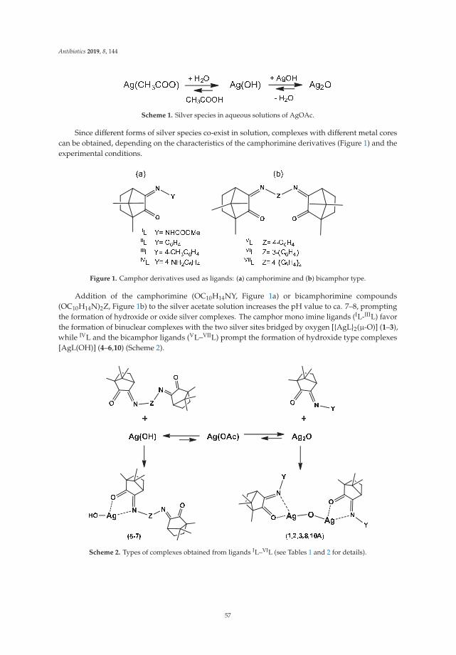
Antibiotics 2019, 8, 144
Scheme 1. Silver species in aqueous solutions of AgOAc.
Since different forms of silver species co-exist in solution, complexes with different metal corescan be obtained, depending on the characteristics of the camphorimine derivatives (Figure 1) and theexperimental conditions.
Figure 1. Camphor derivatives used as ligands: (a) camphorimine and (b) bicamphor type.
Addition of the camphorimine (OC10H14NY, Figure 1a) or bicamphorimine compounds(OC10H14N)2Z, Figure 1b) to the silver acetate solution increases the pH value to ca. 7–8, promptingthe formation of hydroxide or oxide silver complexes. The camphor mono imine ligands (IL-IIIL) favorthe formation of binuclear complexes with the two silver sites bridged by oxygen [{AgL}2(μ-O)] (1–3),while IVL and the bicamphor ligands (VL–VIIL) prompt the formation of hydroxide type complexes[AgL(OH)] (4–6,10) (Scheme 2).
Scheme 2. Types of complexes obtained from ligands IL–VIL (see Tables 1 and 2 for details).
57
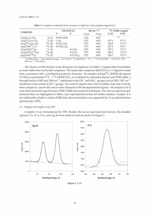
Antibiotics 2019, 8, 144
Table 1. Complexes obtained from reaction of Ag(OAc) with camphor ligands (L).
COMPLEXLIGAND (L) IR (cm−1) a 13C NMR (δ ppm)
Y Z νC=O νC=N C=O C=N
[{AgIL}2(μ-O)] IL (1) NHCOOMe 1722 1649 (b)
[{Ag(IIL)}2(μ-O)] IIL (2) C6H5 1745 1652 207.4 173.3 c
[{Ag(IIIL)}2(μ-O)] IIIL (3) 4-CH3C6H4 1747 1653 206.8 171.9 d
[Ag(OH)(IVL´)] e IVL (4) 4-NH2C6H4 1733 1642 f 207.8 169.1 c
[{Ag(OH)}(VL)] VL (5) 4-C6H4 1754 1685 207.3 173.2 c
[{Ag(OH)}3(VIL)2] VIL (6) 3-C6H4 1751 1668 206.6 173.0 g
[Ag(OH)(VIIL)] VIIL (7) 4-(C6H4)2 1745 1660 206.7 172.5 g
a In KBr pellets. b Not soluble enough. c In CD3CN. d In MeOH-d4. e IVL´ = IVL·CH3COOH. f ν (O=CO), 1593,1567 cm−1. g In CD2Cl2.
The absence of OH stretches in the IR spectra of complexes 1–3 (Table 1) support their formulationas oxide rather than hydroxide complexes. The hydroxide complexes (4,5,7) fit in a 1:1 ligand to metalratio, consistent with a coordination polymer character. At complex 4 ([Ag(IVL´)(OH)]) the ligand(Y=NH2) is protonated (IVL´ = IVL·HOOCCH3), as confirmed by elemental analysis and FTIR (Table 1)through bands at 3455 and 3340 cm−1 (attributed to the OH− and NH4
+ groups) and at 1593, 1567 cm−1
(attributed to the acetate (COO−) group). The metal to ligand ratio at 6 (3:2) differs from that of all theother complexes, conceivably due to steric demands of the bicamphorimine ligand. All complexes (1–7)were characterized by spectroscopic (FTIR, NMR) and analytical techniques. The relevant spectroscopiccharacteristics are highlighted in Table 1 (see experimental section for further details). Complex 1 isnot sufficiently soluble to obtain NMR data, thus formulation was supported by X-ray photoelectronspectroscopy (XPS).
2.1. Analysis of Complex 1 by XPS
Complex 1 was characterized by XPS. Besides the survey spectrum (not shown), the detailedregions C 1s, N 1s, O 1s, and Ag 3d were analyzed and are shown in Figure 2.
Figure 2. Cont.
58
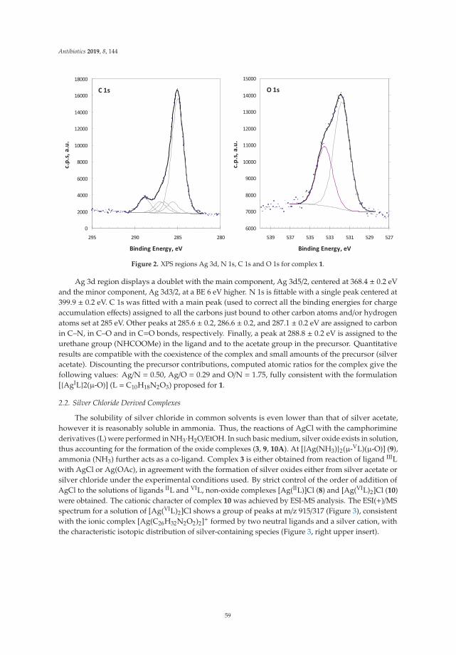
Antibiotics 2019, 8, 144
Figure 2. XPS regions Ag 3d, N 1s, C 1s and O 1s for complex 1.
Ag 3d region displays a doublet with the main component, Ag 3d5/2, centered at 368.4 ± 0.2 eVand the minor component, Ag 3d3/2, at a BE 6 eV higher. N 1s is fittable with a single peak centered at399.9 ± 0.2 eV. C 1s was fitted with a main peak (used to correct all the binding energies for chargeaccumulation effects) assigned to all the carbons just bound to other carbon atoms and/or hydrogenatoms set at 285 eV. Other peaks at 285.6 ± 0.2, 286.6 ± 0.2, and 287.1 ± 0.2 eV are assigned to carbonin C–N, in C–O and in C=O bonds, respectively. Finally, a peak at 288.8 ± 0.2 eV is assigned to theurethane group (NHCOOMe) in the ligand and to the acetate group in the precursor. Quantitativeresults are compatible with the coexistence of the complex and small amounts of the precursor (silveracetate). Discounting the precursor contributions, computed atomic ratios for the complex give thefollowing values: Ag/N = 0.50, Ag/O = 0.29 and O/N = 1.75, fully consistent with the formulation[{AgIL}2(μ-O)] (L = C10H18N2O3) proposed for 1.
2.2. Silver Chloride Derived Complexes
The solubility of silver chloride in common solvents is even lower than that of silver acetate,however it is reasonably soluble in ammonia. Thus, the reactions of AgCl with the camphoriminederivatives (L) were performed in NH3·H2O/EtOH. In such basic medium, silver oxide exists in solution,thus accounting for the formation of the oxide complexes (3, 9, 10A). At [{Ag(NH3)}2(μ-VL)(μ-O)] (9),ammonia (NH3) further acts as a co-ligand. Complex 3 is either obtained from reaction of ligand IIILwith AgCl or Ag(OAc), in agreement with the formation of silver oxides either from silver acetate orsilver chloride under the experimental conditions used. By strict control of the order of addition ofAgCl to the solutions of ligands IIL and VIL, non-oxide complexes [Ag(IIL)]Cl (8) and [Ag(VIL)2]Cl (10)were obtained. The cationic character of complex 10 was achieved by ESI-MS analysis. The ESI(+)/MSspectrum for a solution of [Ag(VIL)2]Cl shows a group of peaks at m/z 915/317 (Figure 3), consistentwith the ionic complex [Ag(C26H32N2O2)2]+ formed by two neutral ligands and a silver cation, withthe characteristic isotopic distribution of silver-containing species (Figure 3, right upper insert).
59
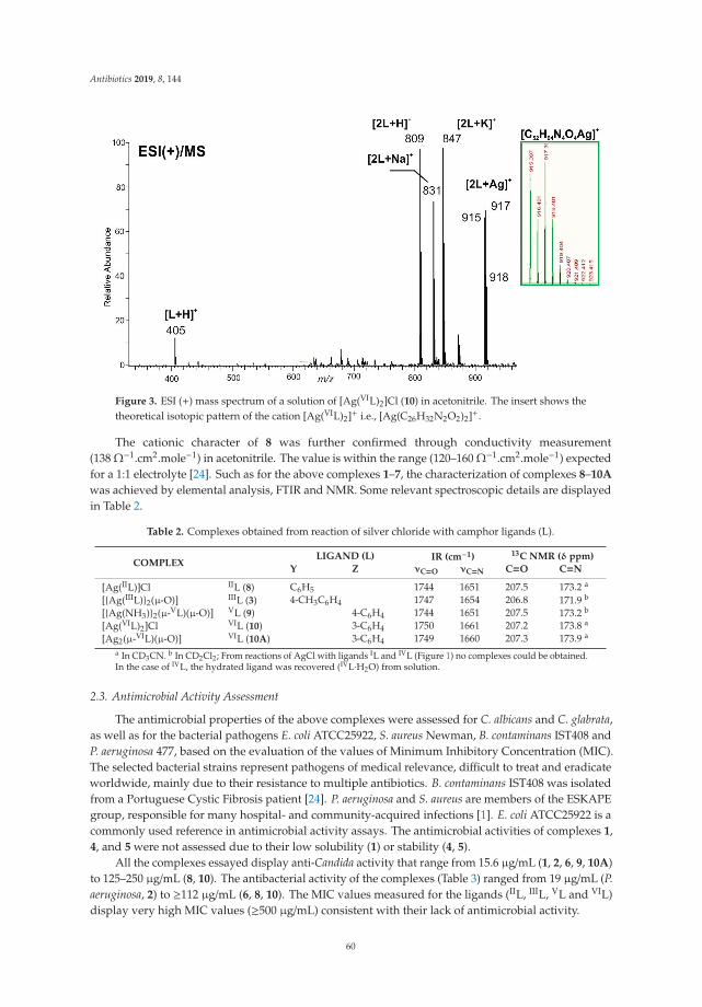
Antibiotics 2019, 8, 144
Figure 3. ESI (+) mass spectrum of a solution of [Ag(VIL)2]Cl (10) in acetonitrile. The insert shows thetheoretical isotopic pattern of the cation [Ag(VIL)2]+ i.e., [Ag(C26H32N2O2)2]+.
The cationic character of 8 was further confirmed through conductivity measurement(138 Ω−1.cm2.mole−1) in acetonitrile. The value is within the range (120–160 Ω−1.cm2.mole−1) expectedfor a 1:1 electrolyte [24]. Such as for the above complexes 1–7, the characterization of complexes 8–10A
was achieved by elemental analysis, FTIR and NMR. Some relevant spectroscopic details are displayedin Table 2.
Table 2. Complexes obtained from reaction of silver chloride with camphor ligands (L).
COMPLEXLIGAND (L) IR (cm−1) 13C NMR (δ ppm)
Y Z νC=O νC=N C=O C=N
[Ag(IIL)]Cl IIL (8) C6H5 1744 1651 207.5 173.2 a
[{Ag(IIIL)}2(μ-O)] IIIL (3) 4-CH3C6H4 1747 1654 206.8 171.9 b
[{Ag(NH3)}2(μ-VL)(μ-O)] VL (9) 4-C6H4 1744 1651 207.5 173.2 b
[Ag(VIL)2]Cl VIL (10) 3-C6H4 1750 1661 207.2 173.8 a
[Ag2(μ-VIL)(μ-O)] VIL (10A) 3-C6H4 1749 1660 207.3 173.9 a
a In CD3CN. b In CD2Cl2; From reactions of AgCl with ligands IL and IVL (Figure 1) no complexes could be obtained.In the case of IVL, the hydrated ligand was recovered (IVL·H2O) from solution.
2.3. Antimicrobial Activity Assessment
The antimicrobial properties of the above complexes were assessed for C. albicans and C. glabrata,as well as for the bacterial pathogens E. coli ATCC25922, S. aureus Newman, B. contaminans IST408 andP. aeruginosa 477, based on the evaluation of the values of Minimum Inhibitory Concentration (MIC).The selected bacterial strains represent pathogens of medical relevance, difficult to treat and eradicateworldwide, mainly due to their resistance to multiple antibiotics. B. contaminans IST408 was isolatedfrom a Portuguese Cystic Fibrosis patient [24]. P. aeruginosa and S. aureus are members of the ESKAPEgroup, responsible for many hospital- and community-acquired infections [1]. E. coli ATCC25922 is acommonly used reference in antimicrobial activity assays. The antimicrobial activities of complexes 1,4, and 5 were not assessed due to their low solubility (1) or stability (4, 5).
All the complexes essayed display anti-Candida activity that range from 15.6 μg/mL (1, 2, 6, 9, 10A)to 125–250 μg/mL (8, 10). The antibacterial activity of the complexes (Table 3) ranged from 19 μg/mL (P.aeruginosa, 2) to ≥112 μg/mL (6, 8, 10). The MIC values measured for the ligands (IIL, IIIL, VL and VIL)display very high MIC values (≥500 μg/mL) consistent with their lack of antimicrobial activity.
60
Antibiotics 2019, 8, 144
Table 3. Biological values assessed for the camphorimine silver complexes.
Complex.MIC50 (μg/mL) MIC (μg/mL)
IC50a
(μg/mL)
C. albicans C. glabrata E. coliATCC25922
B. contaminansIST408
P. aeruginosa477
S. aureusNewman
V79 Cells
2 7.8 ± 0.1 15.6 ± 0.1 59.4 ± 0.3 47 ± 7 19 ± 3 125 7 ± 53 31.3 ± 0.1 31.3 ± 0.1 56 ± 5 78 ± 2 43 ± 11 58 ± 2 8 ± 56 7.8 ± 0.1 15.6 ± 0.1 125 125 60 ± 7 125 3 ± 17 15.6 ± 0.1 15.6 ± 0.1 54 ± 3 >125 61 ± 4 >125 12 ± 58 125 ± 1 250 ± 1 250 >250 112± 14 250 25 ± 149 3.9 ± 0.4 7.8 ± 0.1 32 ± 1 125 19 ± 3 125 2 ± 110 - - >250 >250 >250 >250 -10A 3.9 ± 0.1 15.6 ± 0.1 52.2 ± 0.2 23 ± 3 43 ± 10 125 1.7 ± 0.9Ag(OAc) >500 >500 30.9 ± 0.4 12 ± 2 16 ± 3 29.5 ± 0.1 0.6 ± 0.2AgCl 500 ± 1 >500 14 ± 2 10 ± 1 12 ± 1 30 ± 2 >30
a IC50 values for 48 h incubation.
The MIC values (Table 3) show that the complexes are active against bacterial strains E. coliATCC25922, B. contaminans IST408, P. aeruginosa 477, and S. aureus Newman, although at relatively highMIC values. In contrast, the MIC50 values obtained for the two Candida species are very low, therebyshowing that the complexes have higher antifungal than antibacterial activity. Although previouswork showed that silver camphorimine complexes [Ag(NO3)L] have high anti Candida spp. activity(MIC50 2.0–15.6 μg/mL for C. parapsilosis) the complexes 2, 6, 9, 10A, display values (7.8 μg/mL, 9)that are even lower than those formerly reported against C. glabrata (MIC50 ≥15.6 μg/mL) [18]. Moreimportant, all complexes (except 8) are efficient against C. albicans, a feature not observed for the nitratecomplexes [Ag(NO3)L] [18]. So, by replacing nitrate by hydroxide or oxide co-ligands, we were able tosynthesize complexes that inhibit growth of C. albicans even more efficiently (MIC50 3.9 μg/mL; 9, 10A)than C. glabrata. The lack of activity of [Ag(IIL)]Cl (8) towards the two Candida species and the bacterialstrains under study is attributed to its cationic character. Complex 10, which is also cationic, displaysa relatively low activity (Table 3). These results reinforce those previously obtained for the cationic[Ag(OC10H13NOH)2]NO3 [19]. The ionic character of the complexes decreases their lipophilicity andconceivably makes it more difficult for their penetration into the intracellular environment of Candidaspp. or bacterial cells, thereby reducing their activity.
In general, the complexes display both antibacterial and antifungal activities, although theyperform better as antifungals than as antibacterials. The MIC values show that some of thecomplexes have very high antibacterial activity (40–60 μg/mL) and/or excellent antifungal activities(4–8 μg/mL). Overall, the complexes perform better for Gram-negative (MIC 19–61 μg/mL) thanfor Gram-positive bacteria (S. aureus Newman, 58–250 μg/mL). Complexes [{Ag(IIL)}2(μ-O)] (2) and[{Ag(NH3)}2(μ-VL)(μ-O)] (9) display the highest activity against P. aeruginosa 477 (MIC, 19 μg/mL) whilecomplex [Ag2(μ-VIL)(μ-O)] (10A) displays the highest activity towards B. contaminans IST408 (MIC23 ± 3 μg/mL). These complexes have in common a dinuclear character with the two metals sharing anoxygen atom and camphorimine ligands that may prompt electron delocalization through the aromaticring. Such characteristics conceivably are not just circumstantial for their antimicrobial activity.
To obtain insights into the toxicity of the complexes towards mammalian cells, the IC50
measurements of representative complexes were evaluated towards V79 normal fibroblasts which arecells commonly used to assess the toxicological effects of drugs [25]. Data shows that IC50 values of thecomplexes are low and comparable or even lower than those of the MIC values obtained for fungi(Table 3).
These results were not completely unexpected since fungi and mammalian cells are eukaryotesand therefore some of the mechanisms by which the complexes exert toxicity against the Candidaspp. may be conserved in the mammalian cells [26]. This difficulty in achieving specificity is arecognized challenge in the design of new compounds selectively targeting fungal cells. Futurework will focus on the design of silver camphorimine complexes with both reduced cytotoxicity andenhanced antimicrobial activities.
61
Antibiotics 2019, 8, 144
3. Materials and Methods
3.1. General Procedures
The camphorimines were obtained from camphorquinone by reaction with the appropriate amineor hydrazine in ethanol using reported procedures [18,27–29]. In the case of air sensitive complexes,Schlenk and vacuum techniques were used. Ethanol was purchased from Fisher Scientific, ammoniafrom Sigma-Aldrich and acetonitrile from Carlo Erba. The amines and hydrazines were purchasedfrom Sigma-Aldrich and silver acetate and silver chloride from Merck.
The IR spectra were obtained from KBr pellets using a JASCO FT/IR 4100 spectrometer. The NMRspectra (1H, 13C, DEPT, HSQC and HMBC) were obtained from MeOH-d4, CD3CN or CD2Cl2 solutionsusing Bruker Avance II+ spectrometers (300 or 400 MHz). NMR chemical shifts are referred totetramethylsilane (TMS) (δ = 0 ppm).
The ESI mass spectrum was obtained on a LCQFleet ion trap mass spectrometer equipped withan electrospray source (Thermo ScientificTM, Waltham, MA USA), operating in the positive ion mode.The XPS data was obtained using a Kratos XSAM800 equipment.
Conductivity was measured in acetonitrile (1.0 × 10⁻³ M solution) at 25 ◦C using a CON 510bench conductivity meter provided with a Conductivity/TDS electrode (code No. ECCONSEN91W/35608-50, K = 1).
3.2. Synthesis
Complexes 1–7 were obtained from reaction of the suitable ligand with silver acetate (1:1). Airwas partially excluded by bubbling of nitrogen (3 minutes). The reaction mixtures were protected fromlight to preclude reduction of Ag+ to Ag0. The typical procedure is described for 1. Complexes 8–10
were obtained from reaction of the suitable ligand with silver chloride (1:1). A typical procedure isdescribed for 8.
[{AgIL}2(μ-O)] (1)—A solution of the camphorimine OC10H14NNHCOOMe (IL, 72 mg, 0.35 mmol)in EtOH (5 mL) was added to a suspension of Ag(OAc) (50 mg, 0.35 mmol) in H2O (5 mL) under N2.The whitish suspension was stirred for ca. 1h. The slight suspension still remaining was then removedby filtration and the solution was evaporated to dryness to yield the complex. Yield 55%. Elem. Anal.(%) for Ag2C24H36N4O7. Found: C, 41.6; N, 7.9; H, 5.1; Calc.: C, 41.4; N, 7.5; H, 5.5. IR (cm−1): 1722(C=O), 1649 (C=N), 1588 (O=COMe). 1H NMR (400 MHz, MeOH-d4, δ ppm): 4.56 (s, 3H), 3.81 (sbr,1H), 1.88–1.81 (m, 2H), 1.54–1.44 (m, 2H), 1.18 (s, 3H), 1.04 (s, 3H), 0.85 (s, 3H). 13C NMR Decomposesduring acquisition.
[{AgIIL}2(μ-O)] (2)—OC10H14NC6H4 (IIL, 72 mg; 0.3 mmol) in ethanol (5 mL) and silver acetate(50 mg; 0.3 mmol) in H2O (5 mL) were stirred for 4 h. Yield 74%. Elem. Anal. (%) for Ag2C32H36N2O3.Found: C, 54.9 N, 3.5; H, 5.8; Calc.: C, 54.7; N, 3.8; H, 6.2. IR (cm−1): 1745 (C=O), 1652 (C=N), 1567(CHarom). 1H NMR (300 MHz, CD3CN, δ ppm) 7.40 (t, J = 7.8 Hz, 2H), 7.19 (t, J = 7.5 Hz, 1H), 6.90 (d,J = 7.5 Hz, 2H), 2.74 (d, J = 5.1 Hz, 1H), 1.90 (mc, 2H), 1.61 (mc, 2H) 1.04 (s, 3H), 0.97 (s, 3H), 0.86 (s,3H). 13C NMR (300 MHz, CD3CN, δ ppm): 207.4, 173.3, 130.1, 126.0, 120.9, 59.0, 51.0, 45.3, 30.8, 24.8,21.16, 17.6, 9.4.
[{AgIIIL}2(μ-O)] (3)—OC10H14NC6H4CH3-4 (IIIL, 76 mg; 0.3 mmol) in ethanol (5 mL) and silveracetate (50 mg; 0.3 mmol) in H2O (5 mL) were stirred for 4 h. Yield 89%. Elem. Anal. (%) forAg2C34H42N2O3. Found: C, 55.3 N, 3.5; H, 5.9; Calc.: C, 55.0; N, 3.8; H, 5.7. IR (cm−1): 1747 (C=O),1653 (C=N), 1565 (CHarom). 1H NMR (300 MHz, CD2Cl2, δ ppm): 7.24 (d, J = 7.5 Hz, 2H), 6.97 (d,J = 7.5 Hz, 2H), 2.92 (d, J = 4.8 Hz, 1H), 2.36 (s, 3H), 2.3–1.9 (m, 2H), 1.6–1.5 (m, 2H), 1.61 (s, 3H) 1.07 (s,3H), 0.98 (s, 3H), 0.87 (s, 3H). 13C NMR (300 MHz, CD2Cl2, δ ppm): 206.9, 174.2, 146.2, 138.6, 130.9,123.2, 59.1, 52.2, 46.0, 31.3, 24.9, 21.3, 21.0, 17.4, 9.2.
Compound 3 can alternatively be obtained from reaction of AgCl (50 mg; 0.35 mmol in 5 mLof ammonia 33%) with IIIL (89 mg; 0.35mmol in 5 mL EtOH) upon stirring overnight, filtration to
62
Antibiotics 2019, 8, 144
eliminate residues of silver followed by solvent evaporation until precipitation which is then driedunder vacuum to obtain 3. The yield (60%) is lower than that in reaction of IIIL with Ag(OAc).
[Ag(IVL)(OH)]·CH3COOH (4)—OC10H14NC6H4NH2-4 (IVL, 46 mg; 0.18 mmol) in ethanol (3 mL)and silver acetate (30 mg; 0.18 mmol) in H2O (3 mL). The solutions were degassed. The mixture wasstirred for 4 h under N2. Yield 57%. Elem. Anal. (%) for AgC18H25N2O4. Found: C, 49.3; N, 6.2; H,5.4; Calc.: C, 49.0; N, 6.4; H, 5.7; IR (cm−1): 3454 (OH), 3339 (NH2), 1733 (C=O), 1625 (C=N), 1593(CHarom), 1567 (O=CO), 1504 (NH2). 1H NMR (300 MHz, CD3CN, δ ppm): 6.93 (d, J = 8.6 Hz, 2H),6.70 (d, J = 8.6 Hz, 2H), 4.30 (sbr, 2H) 2.98 (d, J = 4.7 Hz, 1H), 1–91–1.84 (m, 3H), 1.68–1.51 (m, 3H), 1.06(s, 3H), 1.00 (s, 3H), 0.83 (s, 3H). 13C NMR (300 MHz, CD3CN, δ ppm): 207.0, 169.1, 161.3, 146.1, 139.8,124.5, 115.3, 58.0, 51.0, 45.3, 30.8, 24.4, 20.9, 17.7, 9.2.
[{Ag(OH)(VL)] (5)—A solution of 4-C6H4(OC10H14N)2 (VL, 117 mg; 0.3 mmol) in ethanol (5 mL)and silver acetate (50 mg; 0.3 mmol) in H2O (5 mL) were stirred for 5 h. The complex precipitates fromreaction mixture. Yield 54%. Elem. Anal. (%) for AgC26H33N2O3·H2O. Found: C, 57.2 N, 4.8; H, 6.1;Calc.: C, 57.0; N, 5.1; H, 6.4. IR (cm−1) 1750 (C=O), 1649 (C=N), 1559 (CHarom). 1H NMR(300 MHz,CD3CN, δ ppm) 6.98 (s, 4H), 2.86 (d, J = 4.9 Hz, 2H), 1.9 (mc, 3H), 1.05 (s, 6H), 0.99 (s, 6H), 0.86 (s, 6H).13C NMR (300 MHz, CD3CN, δ ppm): 207.3, 173.2, 148.0, 122.4, 58.9, 51.2, 45.3, 30.9, 24.9, 21.2, 17.6, 9.4.
[{Ag(OH)}3(VIL)2] (6)—3-C6H4(OC10H14N)2 (VIL, 199 mg; 0.5 mmol) in ethanol (8.5 mL) wasadded to a solution of silver acetate (95 mg; 0.5 mmol) in H2O (8.5 mL) and the mixture stirredovernight. Yield 62%. Elem. Anal. (%) for Ag3C52H67N4O7 Found: C, 53.1 N, 4.3; H, 5.6; Calc.: C, 52.8;N, 4.7; H, 5.7. IR (cm−1): 1751 (C=O), 1668 (C=N), 1566 (CHarom). 1H NMR (400 MHz, CD2Cl2, δ ppm):7.34 (t, J = 7.9, Hz,1H), 6.68 (2d, J = 1.9 Hz, 2H), 6.34 (s, 1H), 2.77 (d, J = 4.4 Hz, 2H), 2.16–2.01 (m, 2H),1.94–1.80 (m, 2H), 1.68–1.54 (m, 4H), 1.03 (s, 6H), 0.94 (s, 6H), 0.85 (s, 6H). 13C NMR(400 MHz, CD2Cl2,δ ppm): 206.6, 173.0, 151.1, 130.1, 117.2, 111.4, 58.5, 50.6, 44.8, 30.5, 24.7, 21.1, 17.6, 9.2.
[Ag(OH)(VIIL)] (7)—VIIL (44 mg; 0.3 mmol) in ethanol (10 mL) was added to Ag(OAc) (100 mg;0.6 mmol) in H2O (10 mL) and stirred overnight. Yield 88%. Elem. Anal. (%) for AgC32H37N2O3
Found: C, 64.0; N, 4.3; H, 6.1; Calc.: C, 63.6; N, 4.6; H, 6.2. IR (cm−1): 3421 (OH), 1745 (C=O), 1660(C=N), 1566 (CHarom). 1H NMR (300 MHz, CD2Cl2, δ ppm): 7.65 (d, J = 8.1 Hz, 4H), 7.03 (d, J = 7.9 Hz,4H), 2.89 (d, J = 4.5 Hz, 2H), 2.21–2.07 (m, 2H), 1.96–1.81 (m, 2H), 1.72–1.60 (m, 4H), 1.10 (s, 6H), 1.01 (s,6H), 0.92 (s, 6H). 13C NMR (300 MHz, CD2Cl2, δ ppm): 206.7, 172.5, 149.3, 137.9, 127.8, 121.5, 58.4, 50.7,44.9, 30.6, 24.7, 21.1, 17.7, 9.2.
[{AgCl(IIL)] (8)—To a solution of AgCl (50 mg; 0.35 mmol) in ammonia (33%,5 mL) a solution ofthe ligand IIIL (84 mg; 0.35 mmol) in EtOH (5 mL) was added. N2 was then bubbled for a few minutes.The mixture was stirred for 3 h at RT. The slight suspension was filtered off and the volume of thesolution was reduced until precipitation. Upon filtration the title compound was obtained and driedunder vacuum. Yield 59%. Elem. Anal. (%) for AgClC16H19NO. Found: C, 50.3; N, 3.6; H, 5.1; Calc.: C,50.0; N, 3.6; H, 5.0. IR (cm−1): 1744 (CO), 1651 (CN), 1589 (CHarom). 1H NMR (400 MHzCD3CN, δppm): 7.40 (t, J = 7.5 Hz, 2H), 7.19 (t, J = 7.3 Hz 1H), 6.90 (d, J = 7.7 Hz, 2H), 2.74 (d, J = 4.3 Hz, 1H),1.91–1.84 (m, 2H), 1.66–1.55 (m, 2H) 1.04 (s, 3H), 0.97 (s, 3H), 0.86 (s, 3H). 13C NMR(400 MHz, CD3CN,δ ppm): 207.5, 150.9, 130.1, 126.0, 120.9, 58.9, 51.1, 45.2, 30.8, 24.8, 21.2, 17.6, 9.4.
[{Ag(NH3)}2(μ-VL)(μ-O)] (9)—A solution of the ligand VL (75 mg; 0.19 mmol) in EtOH (2.5 mL)was added to a solution of AgCl (23 mg; 0.16 mmol) in ammonia (33%, 2.5 mL). The mixture wasstirred for 4 h at RT. Yield 69%. Anal. (%) for Ag2C26H38N4O3. Found: C, 46.4 N, 8.0; H, 5.3; Calc.: C,46.6; N, 8.4; H, 5.7. IR (cm−1): 3341 (NH), 1753 (CO), 1621 (CN), 1595 (CHarom), 1505 (NH). 1H NMR(400 MHz, MeOH-d4, δ ppm): 7.06 (s, 4H), 2.92 (d, J = 4.4 Hz, 2H), 2.29–2.17 (m, 2H), 2.02–1.93 (m,2H), 1.75–1.56 (m, 4H) 1.09 (s, 6H), 1.07 (s, 6H), 0.83 (s, 6H). 13C NMR (400 MHz, MeOH-d4, δ ppm):190.5, 169.8, 148.8, 125.4, 123.1, 116.2, 58.9, 49.5, 45.7, 31.3, 25.0, 21.3, 17.9, 9.3. There is evidence for twoisomers in solution that were not further investigated.
[AgCl(VIL)2] (10)—Solid AgCl (50 mg; 0.35 mmol) was added to VIL (137 mg; 0.35 mmol) in Et2OH(5 mL) followed by addition of ammonia (33%, 5 mL). The mixture was stirred for 3 h. Yield 72%. Anal.(%) AgClC52H64N4O4. Found: C, 65.9 N, 5.8; H, 6.8; Calc.: C, 65.6; N, 5.9; H, 6.8. IR (cm−1): 1750
63
Antibiotics 2019, 8, 144
(CO), 1661 (CN), 1579 (CHarom). 1H NMR (400 MHz, CD3CN, δ ppm): 7.4 (t, J = 7.9 Hz, 1H); 6.7 (2d,J = 7.9 Hz, 2H), 6.34 (m, 1H), 2.77 (d, J = 4.5 Hz, 2H), 1.04 (s, 3H), 0.98 (s, 3H), 0.87 (s, 3H). 13C NMR(400 MHz, CD3CN, δ ppm): 207.2, 173.8, 152.1, 131.1, 111.3, 59.0, 51.2, 45.2, 30.8, 24.9, 21.2, 17.6, 9.4.
[Ag2(μ-VIL)(μ-O)] (10A)—Addition of VIL (137 mg; 0.35 mmol, 5 mL H2O) after completedissolution of AgCl (50 mg; 0.35 mmol) in ammonia (33%, 5 mL) followed by stirring overnight. Yield56%. Anal. (%) Ag2C26H32N2O3·H2O. Found: C, 47.7; N, 4.5; H, 5.0. Calc.: C, 47.7; N, 4.3; H, 5.2. IR(cm−1): 1749 (CO), 1660 (CN), 1579 (CHarom). 1H NMR (400 MHz, CD3CN, δ ppm): 7.43 (t, J = 7.9 Hz,1H), 6.7 (dd, J = 1.7, Hz J = 7.9 Hz, 2H), 6.34 (m, 1H), 2.77 (d, J = 4.9 Hz, 2H), 2.12–2.03 (m, 2H), 1.91–1.75(m, 2H), 1.64–1.56 (m, 4H), 1.04 (s, 6H), 0.98 (s, 6H), 0.87 (s, 6H). 13C NMR (400 MHz, CD3CN, δ ppm):207.3, 173.9, 152.0, 130.9, 117.4, 111.3, 59.0, 51.1, 45.1, 30.8, 24.9, 21.2, 17.6, 9.3.
3.3. Antibacterial Activity Determinations
The antibacterial activity of compounds was assessed by determining their Minimal InhibitoryConcentration (MIC) towards the Gram-positive Staphylococcus aureus Newman and the Gram-negativeEscherichia coli ATCC 25922, Pseudomonas aeruginosa 477 and Burkholderia contaminans IST408. Thesebacterial strains are clinical isolates and were chosen as representatives of important bacterialpathogens [1,25,30–32]. MICs were determined using Mueller Hinton Broth (MHB; Becton, Dickinsonand Company) as growth medium, based on microdilution assays, using previously describedmethods [20,21]. In brief, a colony from a bacterial culture freshly grown in MHB solid medium wastransferred into MHB liquid medium and incubated for 4–5 h with agitation (250 rpm) at 37 ◦C. Bacterialcultures were then diluted using fresh MHB to obtain ca. 106 colony forming units (CFUs) per mL. Thesecultures were used to inoculate approximately 5 × 10⁵ CFUs per mL in 96-well polystyrene microtiterplates containing 100 μL of MHB supplemented with different concentrations of each compound,obtained by 1:2 serial dilutions ranging 0.5 to 512 μg/mL. Stock solutions of compounds were preparedwith DMSO. After inoculation, microtiter plates were incubated at 37 ◦C for 20 h. Bacterial growthwas assessed by measuring the cultures optical density at 640 nm, in a SPECTROstarNano (BMGLABTECH) microplate reader. Experiments were carried out at least four times in duplicates. Wellscontaining 100 μL of 1× concentrated MHB and 100 μL of 106 CFUs per mL were used as positivecontrols, while wells containing 200 μL of sterile MHB 1 × concentrated were used as negative controls.
3.4. Assessment of Complexes Anti-Candida Activity
The ability of the complexes 1–10A or of the ligands to inhibit growth of C. albicans or C. glabrata wasassessed using the highly standardized microdilution method recommended by EUCAST (EuropeanCommittee on Antimicrobial Susceptibility Testing). The MIC50 values were considered to be theconcentration of drug that reduced yeast growth by more than 50% of the growth registered in drug-freemedium [33]. The strains used in this work were C. albicans SC5314 and C. glabrata CBS138, largely usedas reference strains. Briefly, cells of the different species were cultivated (at 30 ◦C and with 250 rpmorbital agitation) for 17 h in Yeast Potato Dextrose (YPD) growth medium and then diluted in freshRoswell Park Memorial Institute (RPMI) growth medium (Sigma) to obtain a cell suspension havingan OD530nm of 0.05. From these cell suspensions, 100 μL aliquots were mixed in the 96-multiwellpolystyrene plates with 100 μL of fresh RPMI medium (control) or with 100 μL of this same mediumsupplemented with 0.98–500 μg/mL of the different compounds. As a control we also examined theinhibitory effect of Ag(OAc) or of AgCl. After inoculation, the 96-multiwell plates were incubatedwithout agitation at 37 ◦C for 24 h. After that time, cells were re-suspended and the OD530nm of thecultures was measured in a SPECTROstarNano (BMG LABTECH) microplate reader. The MIC50 valuewas taken as being the highest concentration tested at which the growth of the strains was 50% of thevalue registered in the control lane.
64
Antibiotics 2019, 8, 144
3.5. Toxicity Assessment
The toxicity of the compounds was evaluated towards normal fibroblasts V79, obtained from theAmerican Type Culture Collection (ATCC). The cell lines were grown in Dulbeco´s Modified EagleMedium (DMEM) + Glutamax® medium supplemented with 10% Fetal Bovine serum (FBS) andmaintained in a humidified atmosphere at 37 ◦C using an incubator (Heraeus, Germany) with 5%CO2. Cell viability was measured by the MTT (3-[4,5-dimethylthiazol-2-yl]-2,5 diphenyl tetrazoliumbromide) assay, based on the conversion of the tetrazolium bromide into formazan crystals by livingcells which determines mitochondria activity [34]. For the assay, cells were seeded in 96-well plates ata density of 104 cells per well in 200 μL medium and allowed to attach overnight. Complexes werefirst diluted in DMSO to solubilize and then in medium to prepare the serial dilutions in the range10−7–10−4 M. The maximum concentration of DMSO in the medium (1%) had no toxicity effect. Aftercareful removal of the medium, 200 μL of each dilution were added to the cells, and incubated foranother 48 h at 37 ◦C. At the end of the treatment, the medium was aspirated and 200 μL of MTTsolution (1.5 mM in PBS) was applied to each well. After 3 h at 37 ◦C, the medium was discardedand 200 μL of DMSO was added to solubilize the formazan crystals. The cellular viability wasassessed by measuring the absorbance at 570 nm using a plate spectrophotometer (Power Wave Xs,Bio-Tek). The IC50 values were calculated using the GraphPad Prism software (version 5.0). Resultsare mean ± SD of at least two independent experiments done with six replicates each and representthe percentage of cellular viability related to the controls (no treatment).
3.6. X-ray Photoelectron Spectroscopy
For X-ray photoelectron spectroscopy characterization (XPS), a XSAM800 XPS dual anodespectrometer from KRATOS was used. The unmonochromatic Mg Kα radiation (main line athν = 1256.6 eV) was used. Operating conditions, data acquisition and data treatment are describedelsewhere [35]. For charge correction purposes, carbon bound to carbon and hydrogen in C 1s peakwas set to a binding energy (BE) of 285 eV. For quantification purposes, the following sensitivity factorswere used: 0.318 for C 1s, 0.736 for O 1s, 0.505 for N 1s, and 6.345 for Ag3d.
4. Conclusions
Silver hydroxide [Ag(OH)L] (L = IVL, VL, VIL, VIIL), oxide [{AgL}2}(μ-O)] (L = IL, IIL, IIIL, VL, VIL)and homoleptic [AgIIL]Cl, [Ag(VIL)2]Cl camphorimine complexes were synthesized using Ag(OAc)or AgCl as metal sources. The basic characteristics of the reaction medium prompted the formationof hydroxide or oxide rather than acetate or chloride complexes. The selection of the camphorimineligands (L) that encompass mono- and bi-camphors, allowed the design of complexes with considerabledistinct electronic and steric properties and thus different biological activities.
In summary, the most relevant achievement of this study is that the new oxo and hydroxo silvercamphorimine complexes overreach the resistance of C. albicans. Additionally, the complexes reachMIC50 values for C. glabrata even lower than those previously reported for the camphorimine nitratecomplexes [Ag(NO3)L]. In fact, the antifungal activity of the oxo and hydroxo silver camphoriminecomplexes is even higher towards C. albicans than C. glabrata.
Author Contributions: Conceptualization, M.F.N.N.C., N.P.M., and J.H.L.; Experimental data acquisition, J.P.C.,M.J.F.P., S.A.S., M.C.O., A.M.B.R., F.M., M.F.N.N.C.; methodology and data curation: all authors; writing—originaldraft preparation, M.F.N.N.C., J.H.L., N.P.M., A.M.B.R., F.M.; writing—review and editing, all authors; fundingacquisition, M.F.N.N.C., N.P.M., J.H.L., F.M.
Funding: This research received no external funding.
Acknowledgments: Financial support by FCT-Fundação para a Ciência e Tecnologia throughprojects ID/QUI/00100/2019, UID/BIO/04565/2019, UID/MULTI/04349/2019, Grant BL-CQE/2018-013, contractPTDC/BIA-MIC/31515/2017 and Programa Operacional Regional de Lisboa 2020 (Project No. 007317) is gratefullyacknowledged as well as facilities from the NMR and Mass Spectra Networks (IST-UTL Nodes).
65
Antibiotics 2019, 8, 144
Conflicts of Interest: The authors declare no conflict of interest. The funders had no role in the design of thestudy; in the collection, analyses, or interpretation of data; in the writing of the manuscript, or in the decision topublish the results.
References
1. Boucher, H.W.; Talbot, G.H.; Bradley, J.S.; Edwards, J.E., Jr.; Gilbert, D.; Rice, L.B.; Scheld, M.; Spellberg, B.;Bartlett, J. Bad bugs, no drugs: No ESKAPE! An update from the Infectious Diseases Society of America.Clin. Infect. Dis. 2009, 48, 1–12. [CrossRef] [PubMed]
2. Lim, C.S.; Rosli, R.; Seow, H.F.; Chong, P.P. Candida and invasive candidiasis: Back to basics. Eur. J. Clin.Microbiol. Infect. Dis. 2012, 31, 21–31. [CrossRef] [PubMed]
3. Witherden, E.A.; Shoaie, S.; Hall, R.A.; Moyes, D.L. The Human Mucosal Mycobiome and Fungal CommunityInteractions. J. Fungi 2017, 3, 56. [CrossRef] [PubMed]
4. Pfaller, M.A.; Diekema, D.J.; Gibbs, D.L.; Newell, V.A.; Ellis, D.; Tullio, V.; Rodloff, A.; Fu, W.; Ling, T.A.;Global Antifungal Surveillance Group. Results from the ARTEMIS DISK Global Antifungal SurveillanceStudy, 1997 to 2007: A 10.5-year analysis of susceptibilities of Candida Species to fluconazole and voriconazoleas determined by CLSI standardized disk diffusion. J. Clin. Microbiol. 2010, 48, 1366–1377. [PubMed]
5. Pfaller, M.A.; Diekema, D.J.; Turnidge, J.D.; Castanheira, M.; Jones, R.N. Twenty Years of the SENTRYAntifungal Surveillance Program: Results for Candida Species From 1997–2016. Open Forum Infect. Dis. 2019,15, S79–S94. [CrossRef] [PubMed]
6. Theuretzbacher, U. Future antibiotics scenarios: Is the tide starting to turn? Int. J. Antimicrob. Agents 2009,34, 15–20. [CrossRef] [PubMed]
7. Aslam, B.; Wang, W.; Arshad, M.I.; Khurshid, M.; Muzammil, S.; Rasool, M.H.; Nisar, M.A.; Alvi, R.F.;Aslam, M.A.; Qamar, M.U.; et al. Antibiotic resistance: A rundown of a global crisis. Infect. Drug Resist.2018, 11, 1645–1658. [CrossRef] [PubMed]
8. Singh, S.B.; Young, K.; Silver, L.L. What is an “ideal” antibiotic? Discovery challenges and path forward.Biochem. Pharmacol. 2017, 133, 63–73. [CrossRef]
9. Nett, J.E.; Andes, D.R. Antifungal Agents: Spectrum of Activity, Pharmacology, and Clinical Indications.Infect. Dis. Clin. N. Am. 2016, 30, 51–83. [CrossRef]
10. Etubu, E.; Arikekpar, I. Antibiotics: Classification and mechanisms of action with emphasis on molecularperspectives. Int. J. Appl. Microbiol. Biotechnol. Res. 2016, 4, 90–101.
11. Kapoor, G.; Saigal, S.; Elongavan, A. Action and resistance mechanisms of antibiotics: A guide for clinicians.J. Anaesthesiol. Clin. Pharmacol. 2017, 33, 300–305. [CrossRef] [PubMed]
12. Park, Y.-K.; Kyung-Soo, H. Antimicrobial Peptides (AMPs): Peptide Structure and Mode of Action. BMB Rep.2005, 38, 507–516. [CrossRef]
13. Ghosh, C.; Sarkar, P.; Issa, R.; Aldar, J. Alternatives to Conventional Antibiotics in the Era of AntimicrobialResistance. Trends Microbiol. 2019, 27, 323–338. [CrossRef]
14. Singh, P.A.; Desai, S.D.; Singh, J. A Review on Plant Antimicrobials of Past Decade. Curr. Top. Med. Chem.2018, 18, 812. [CrossRef] [PubMed]
15. Kenawy, E.R.; Worley, S.D.; Broughton, R. The chemistry and applications of antimicrobial polymers: Astate-of-the-art review. Biomacromolecules 2007, 8, 1359–1384. [CrossRef] [PubMed]
16. Lemire, J.A.; Harrison, J.J.; Turner, R.J. Antimicrobial activity of metals: Mechanisms, molecular targets andapplications. Nat. Rev. Microbiol. 2013, 11, 371–384. [CrossRef] [PubMed]
17. Liang, X.; Luan, S.; Yin, Z.; He, M.; He, C.; Yin, L.; Zou, Y.; Yuan, Z.; Li, L.; Song, X.; et al. Recent advances inthe medical use of silver complex. Eur. J. Med. Chem. 2018, 157, 62–80. [CrossRef]
18. Cardoso, J.M.; Guerreiro, S.I.; Lourenco, A.; Alves, M.M.; Montemor, M.F.; Mira, N.P.; Leitão, J.H.;Carvalho, M.F.N. Ag(I) camphorimine complexes with antimicrobial activity towards clinically importantbacteria and species of the Candida genus. PLoS ONE 2017, 12, e0177355. [CrossRef]
19. Carvalho, M.F.N.; Leite, S.; Costa, J.P.; Galvão, A.M.; Leitão, J.H. Ag(I) camphor complexes: Antimicrobialactivity by design. J. Inorg. Biochem. 2019, 199, 110791. [CrossRef]
20. Leitão, J.H.; Sousa, S.A.; Leite, S.A.; Carvalho, M.F.N. Silver Camphor Imine Complexes: Novel AntibacterialCompounds from Old Medicines. Antibiotics 2018, 7, 65. [CrossRef]
66
Antibiotics 2019, 8, 144
21. Cardoso, J.M.S.; Galvão, A.M.; Guerreiro, S.I.; Leitão, J.H.; Suarez, A.C.; Carvalho, M.F.N. Antibacterialactivity of silver camphorimine coordination polymers. Dalton Trans 2016, 45, 7114–7123. [CrossRef][PubMed]
22. Cardoso, J.M.S.; Correia, I.; Galvão, A.M.; Marques, F.; Carvalho, M.F.N. Synthesis of Ag(I) camphorsulphonylimine complexes and assessment of their cytotoxic properties against cisplatin-resistant A2780cisRand A2780 cell lines. J. Inorg. Biochem. 2017, 166, 55–63. [CrossRef] [PubMed]
23. Bordwell, F.G.; Algrim, D. Nitrogen acids. 1. Carboxamides and sulfonamides. J. Org. Chem. 1976, 41,2507–2508. [CrossRef]
24. Geary, W.J. The use of conductivity measurements in organic solvents for the characterisation of coordinationcompounds. Coord. Chem. Rev. 1971, 7, 81–122. [CrossRef]
25. Richau, J.A.; Leitão, J.H.; Sá-Correia, I. Enzymes leading to the nucleotide sugar precursors forexopolysaccharide synthesis in Burkholderia cepacia. Biochem. Biophys. Res. Commun. 2000, 276, 71–76.[CrossRef] [PubMed]
26. Doehmer, J. Predicting Drug Metabolism–dependent Toxicity for Humans with a Genetically EngineeredCell Battery. Altern. Lab. Anim. 2006, 34, 561–575. [CrossRef] [PubMed]
27. Mazu, T.K.; Bricker, B.A.; Flores-Rozas, H.; Ablordeppey, S.Y. The Mechanistic Targets of Antifungal Agents:An Overview. Mini-Rev. Med. Chem. 2016, 16, 555–578. [CrossRef] [PubMed]
28. Fernandes, T.A.; Mendes, F.; Roseiro, A.P.S.; Santos, I.; Carvalho, M.F.N. Insight into the cytotoxicity ofpolynuclear Cu(I) camphor complexes. Polyhedron 2015, 87, 215–219. [CrossRef]
29. Carvalho, M.F.N.; Costa, L.M.G.; Pombeiro, A.J.L.; Schier, A.; Scherer, W.; Harbi, S.K.; Verfürth, U.;Herrmann, R. Synthesis, structure, and electrochemistry of palladium complexes with camphor-derivedchiral ligands. Inorg. Chem. 1994, 33, 6270–6277. [CrossRef]
30. Denmark, S.D.; Rivera, I. Asymmetric Carboalkoxyalkylidenation with a Chiral Horner-Wadsworth-EmmonsReagent. J. Org. Chem. 1994, 59, 6887–6889. [CrossRef]
31. Rice, L.B. Federal funding for the study of antimicrobial resistance in nosocomial pathogens: No ESKAPE.J. Infect. Dis. 2008, 197, 1079–1081. [CrossRef] [PubMed]
32. Sousa, S.A.; Feliciano, J.R.; Pita, T.; Guerreiro, S.I.; Leitão, J.H. Burkholderia cepacia Complex Regulation ofVirulence Gene Expression: A Review. Genes 2017, 8, 43. [CrossRef] [PubMed]
33. Arendrup, M.C.; Cuenca-Estrella, M.; Lass-Flörl, C.; Hope, W.; EUCAST-AFST. EUCAST technical noteon the EUCAST definitive document EDef 7.2: Method for the determination of broth dilution minimuminhibitory concentrations of antifungal agents for yeasts EDef 7.2 (EUCAST-AFST). Clin. Microbiol. Infect.2012, 18, E246–E247. [CrossRef] [PubMed]
34. Van Meerloo, J.; Kaspers, G.; Cloos, J. Cell sensitivity assays: The MTT assay. Mol. Biol. 2011, 731, 237–245.35. Carapeto, A.P.; Ferraria, A.M.; do Rego, A.M.B. Unraveling the reaction mechanism of silver ions reduction
by chitosan from so far neglected spectroscopic features. Carbohydr. Polym. 2017, 174, 601–609. [CrossRef][PubMed]
© 2019 by the authors. Licensee MDPI, Basel, Switzerland. This article is an open accessarticle distributed under the terms and conditions of the Creative Commons Attribution(CC BY) license (http://creativecommons.org/licenses/by/4.0/).
67

antibiotics
Article
Assessing National Antimicrobial ResistanceCampaigns Using a Health Equity AssessmentTool (HEAT)
Graeme Hood, Lina Toleikyte and Diane Ashiru-Oredope *
Public Health England, London SE1 8UG, UK* Correspondence: [email protected]; Tel.: +44-020-7811-7240
Received: 15 June 2019; Accepted: 14 August 2019; Published: 17 August 2019
Abstract: It has been widely recognised that a significant proportion of the world’s population sufferinequalities in accessing high quality healthcare and wider services. Within healthcare, antimicrobialresistance (AMR) is a global threat to public health affecting all healthcare systems and growing atan alarming pace. To ensure that national AMR campaigns developed by Public Health Englandare inclusive of all populations within the target audience a health equity assessment tool (HEAT)was used. The project leads for each campaign completed the HEAT independently with a follow upmeeting with the study team to discuss and clarify the responses. A trend analysis was carried outwith common themes being used to provide recommendations. The campaigns have demonstratedequality and diversity based on the requirements of the Equality Act 2010, particularly age, sex, andrace protected characteristics. Some notable results include the translation of website materials inover 30 languages and reaching individuals in 122 countries. It was however noted that several ofthe protected characteristics were not applicable. The continuous development of resources withcollaboration from a variety of diverse user groups would be advantageous towards aiding futurecampaign reach. The use of the HEAT has demonstrated the ease and cost-effective way to assessany health inequalities and would be a useful addition to antimicrobial stewardship and publichealth campaigns.
Keywords: antimicrobial stewardship; health inequalities; health equity assessment tool; public health
1. Introduction
Research has shown that there are inequalities in accessing high quality healthcare across theworld, which affects a large proportion of people [1]. It has been estimated by the World HealthOrganisation (WHO) that 400 million people do not have access to one of seven essential health services,such as drugs and vaccines [2]. The variations in health inequalities have been reported both at anational and at an international level with the average life expectancy in the UK (81.2 years) lower thanJapan (83.7 years) but higher than Sierra Leone (50.1 years) [1]. The health inequalities are classified asunjust differences in health and wellbeing between different groups of people which are systematicand avoidable and may be driven by (Supplementary Figure S1):
• Different experiences of the wider determinants of health or structural factors, for example theenvironments, income or housing.
• Differences in health behaviours or other risk factors between groups, for example smoking,diet, and physical activity levels have different social distributions. Health behaviours may beinfluenced by wider determinants of health, like income.
• Unequal access to or experience of health and other services between social groups.
Antibiotics 2019, 8, 121; doi:10.3390/antibiotics8030121 www.mdpi.com/journal/antibiotics69
Antibiotics 2019, 8, 121
The monitoring of health inequalities is important as it can identify progress linked to specifichealth policies or programmes and help to focus interventions at the most disadvantaged cohorts ofthe population. Through continual monitoring, the reduction or widening of health differences canbe highlighted [3,4]. To aid the assessment of health interventions, policies, programmes, servicesand reduce health inequalities, specific assessment tools have been created such as the health equityassessment toolkit (HEAT) that was developed by the World Health Organisation (WHO) between 2014and 2016 [5]. The WHO HEAT is a software, that contains the WHO’s health equity monitor database,supporting countries to assess inequalities within the country with over 30 indicators includingreproductive, maternal, new-born and child health and five dimensions of inequality (economic status,education, place of residence, subnational region and child’s sex, where applicable) [5].
The reduction of health inequalities within England is a responsibility for Public Health England(PHE), an executive agency of the UK Government [6]. To help the organisation assess the impact ofthe public facing campaigns for health equity, PHE’s Health Inequalities team designed an internalHEAT. The assessment consists of a series of questions, which are designed to support systematicassessment of health inequalities related to work programmes, initiatives, policies and identify actionsthat need to be taken to reduce any potential identified inequalities.
The HEAT was built on international experience from the WHO and the Ministry of Health in NewZealand [5,7]. Unlike the WHO and New Zealand HEAT, the PHE tool considers the requirements ofthe Equality Act 2010 and therefore the set protected characteristics (Box 1) in addition to inequalities.
Box 1. Protected characteristics (Equality Act 2010).
• age• sex• race• religion or belief• disability• sexual orientation• gender reassignment• pregnancy and maternity• marriage and civil partnership
As well as considerations about socio-economic differences, area variations by the deprivationlevel (IMD), service provisions, urban/rural populations or in general and excluded and underservedgroups for example homeless people, people in prison, or young people leaving care were considered.
It also recommends five stages for the assessment which include prepare, assess, refine, applyand review. Each assessment is likely to be iterative and can help areas continuously improve thecontribution of work streams to reducing health inequalities. There have been notable local resultswithin New Zealand from the application of this toolkit, including health promotion and health careclinics in low decile schools; dental care for those on low incomes; free interpreter services; andmobile Maori nursing services [8]. The WHO HEAT is pre-populated with the health equity monitordatabase, allowing users to explore the situation in one setting of interest (e.g., a country, province,or district) to determine the latest situation of inequality and changes in inequalities over time aswell as to benchmark settings/countries. The PHE and New Zealand tools can be considered qualityimprovement tools for assessing and developing programmes, initiatives and policies.
It is important to use the HEAT to assess that the impact of public facing campaigns. Thedevelopment of an internal HEAT has helped PHE to assess such interventions and provides apragmatic and systematic set of steps for health policy makers to assess their initiative in relation tohealth equity. The application of the toolkit can occur at different stages of the program or policydevelopment including the initial planning phase, the implementation stages or in the programmereview phase [7]. Within the development of proposals or policies, there can be a tendency to leave
70
Antibiotics 2019, 8, 121
the target groups as wide as possible so that more people are included. However, this approach canpotentially result in the reduction of certain cohorts of the population that would benefit most fromthe programme or policy. This can result in unintended and unanticipated health impacts on certainpopulation groups [9]. The use of HEAT can potentially help to reduce this unintended outcome forwhich PHE has started to incorporate this toolkit within the assessment process of national campaigns.The outcome of health equity assessments can inform decisions on how to build and strengthen policies,programmes and future services that organisations provide [8]. The antimicrobial resistance team hasbeen one of the pioneers to adopt it within its campaigns.
Antimicrobial resistance (AMR) is a global threat to public health affecting all healthcare systemsand growing at an alarming pace [10,11]. The implementation of antimicrobial stewardship programmeshas been used within secondary care to help reduce this burden, with a focus on the UK Governmentambition to reduce inappropriate antibiotic prescribing [12]. An important element of these programmesis the use of national campaigns to increase the overall reach. There is currently a lack of researchassessing the potential health inequalities within public facing antimicrobial stewardship campaigns.The primary aim of this study was to assess whether the public facing AMR initiatives are reachinga diverse population within England. The secondary aim included providing individual campaignrecommendations and general trend recommendations for the overall campaigns. Following a literaturereview, the authors believe there is currently limited research available that specifically assesses healthinequalities related to AMR national campaigns.
National Campaigns
The following four national AMR campaigns run by Public Health England were assessed:Antibiotic GuardianAs part of the European Antibiotic Awareness Week (EAAD), this campaign was launched in
September 2014. The aim was to increase commitment by healthcare professionals and membersof the public to reduce AMR, change behaviour and increase knowledge. This is done through apledge-based system where healthcare professionals and members choose a relevant pledge relating totheir practice or personal situation [13].
Target audience: Broad audience
e-BugThis campaign was developed as a European Union project and created a junior and senior
school educational resource for teachers covering microbes, hygiene, antibiotics and the prevention ofinfection. There is both a paper based educational pack along with a web site that hosts games foryoung people and their families to play in the classroom or at home [14].
Target audience: School age children and teachers
TARGET (Treat Antibiotics Responsibly, Guidance, Education, Tools)The TARGET antibiotics toolkit was developed by PHE in collaboration with the Royal College
of General Practitioners (RCGP) and other professional societies. The main aim of this campaign isto improve antimicrobial prescribing in primary care through multiple channels such as guidance,interactive workshops, patient facing educational and audit materials [15].
Target audience: Prescribing clinicians
Keep Antibiotics WorkingThis is a consumer-led campaign to help raise awareness of antibiotic resistance to members of
the public and highlight the dangers of not using antibiotics appropriately.There are three main aims within this campaign—alert the public to the issue of AMR, reduce
public expectation for antibiotics and support change amongst healthcare professionals [16].
Target audience: Women aged 20–45 and Men and women aged 50+
71
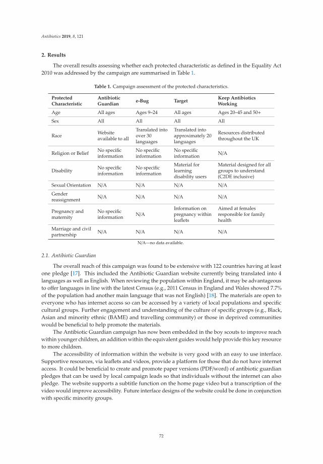
Antibiotics 2019, 8, 121
2. Results
The overall results assessing whether each protected characteristic as defined in the Equality Act2010 was addressed by the campaign are summarised in Table 1.
Table 1. Campaign assessment of the protected characteristics.
ProtectedCharacteristic
AntibioticGuardian
e-Bug TargetKeep AntibioticsWorking
Age All ages Ages 9–24 All ages Ages 20–45 and 50+
Sex All All All All
Race Websiteavailable to all
Translated intoover 30languages
Translated intoapproximately 20languages
Resources distributedthroughout the UK
Religion or Belief No specificinformation
No specificinformation
No specificinformation N/A
Disability No specificinformation
No specificinformation
Material forlearningdisability users
Material designed for allgroups to understand(C2DE inclusive)
Sexual Orientation N/A N/A N/A N/A
Genderreassignment N/A N/A N/A N/A
Pregnancy andmaternity
No specificinformation N/A
Information onpregnancy withinleaflets
Aimed at femalesresponsible for familyhealth
Marriage and civilpartnership N/A N/A N/A N/A
N/A—no data available.
2.1. Antibiotic Guardian
The overall reach of this campaign was found to be extensive with 122 countries having at leastone pledge [17]. This included the Antibiotic Guardian website currently being translated into 4languages as well as English. When reviewing the population within England, it may be advantageousto offer languages in line with the latest Census (e.g., 2011 Census in England and Wales showed 7.7%of the population had another main language that was not English) [18]. The materials are open toeveryone who has internet access so can be accessed by a variety of local populations and specificcultural groups. Further engagement and understanding of the culture of specific groups (e.g., Black,Asian and minority ethnic (BAME) and travelling community) or those in deprived communitieswould be beneficial to help promote the materials.
The Antibiotic Guardian campaign has now been embedded in the boy scouts to improve reachwithin younger children, an addition within the equivalent guides would help provide this key resourceto more children.
The accessibility of information within the website is very good with an easy to use interface.Supportive resources, via leaflets and videos, provide a platform for those that do not have internetaccess. It could be beneficial to create and promote paper versions (PDF/word) of antibiotic guardianpledges that can be used by local campaign leads so that individuals without the internet can alsopledge. The website supports a subtitle function on the home page video but a transcription of thevideo would improve accessibility. Future interface designs of the website could be done in conjunctionwith specific minority groups.
72
Antibiotics 2019, 8, 121
2.2. e-Bug
The e-Bug focuses on the education element of infections within children, young people andthose hard to reach in the community. This interactive campaign has been translated into over 30languages and has approved trainers throughout England, Wales and Northern Ireland which improvesaccessibility for these resources. The e-Bug trainer events should be targeted at areas with greaterantibiotic use which have greater deprivation and ethnicity. It was found that all resources are freelyavailable to download and print from the internet. The resources are developed for a range of ages andtherefore abilities within the community. In the Beat the Bugs resources, each lesson plan has a rangeof activities to suit a range of abilities. The antibiotic Beat the Bug resources include a specific patientfacing pictorial resource for the public with language or learning difficulties. There is an increasedawareness within the project team of pictorial and foreign language resources, with a commitmentto continue to reassess the language being used is inclusive of all, especially for disadvantaged andminority groups. There is also an increase focus in the implementation of resources in deprived areasusing additional work force and targeted training to local clinical commissioning group staff.
An analytical review of the e-Bug website usage and views would help identify furtherdevelopment and improvement to reduce health inequalities and could be beneficial.
2.3. TARGET
The TARGET resources were found to be easily accessible to all healthcare professionals via anopen access website. The distribution was extensive and resources can be accessed to permanent,training or temporary general practitioners (GPs). This was also the case for the out of hour GPs,though further publicity within this group would aid the reach. The use of community pharmacists inpromoting the use of TARGET resources would help improve reach to patients. The TARGET websitecontains patient facing resources to share in consultations for respiratory tract infections (includinga pictorial version) and urinary tract infections for patients under and over 65 years. These patientleaflets are available in the most common non-English languages spoken in the UK.
2.4. Keep Antibiotics Working
The quantitative research completed by the internal marketing team demonstrated a representativesample of the population within England, however due to the size of the sample, it was not possible orappropriate to break down the data by every protected characteristic. The choice of media via the TVensured the campaigned reached a broad cross section of the population. During the campaign period,over 766,000 posters and leaflets were distributed to a range of partners including local authorities,health care centres and housing associations with 92% of GP practices in England engaged [19]. Prisonswere also included within the distribution along with GPs who reach those from lower socioeconomicbackgrounds. The advertising featured red and white pills which have no gender or racial bias. Allcampaign research, including campaign tracking, strategic and creative development research wascarried out across all socioeconomic groups. The campaign materials were designed for all groups tounderstand and were C2DE—skilled working class, working class and non-working (the three lowersocial and economic groups in a society) inclusive. It would be recommended to conduct a reviewimpact analysis of the campaign by age, sex and socioeconomic group, and where appropriate, changethe campaign strategy based on this evidence. It was noted that more data is required to changethe marketing or targeting specific populations but the use of advertising routes that are set up toengage with minority groups (e.g. radio channels focusing on certain groups, promote in religiousareas, prisons) may be beneficial. It could be advantageous to translate resource leaflets into multiplelanguages in line with latest national Census and local population.
73
Antibiotics 2019, 8, 121
3. Discussion
The assessments highlight the diverse and inclusive nature of the four campaigns run by PublicHealth England. There have been notable successes in reaching a wider audience such as 92% of GPpractices in England being engaged with the Keep Antibiotic Campaign. Interestingly, this wider reachmay have contributed to a positive impact on the intended behaviour, with 78% of the public statingthat they would be unlikely to ask their GP for antibiotics [19]. It is important to note that the reach ofa campaign on the population can also have an influence on healthcare professionals with 93% of GPssaying they felt the materials supported them to say no to antibiotics when they were not needed [19].As technology advances, it has been demonstrated that there is more of an emphasis being put onthe use of technology within healthcare over the last few years. It is vital that campaigns use thisto reach greater audiences but also remembering an ageing population who potentially do not haveaccess to technology or would prefer not to be contacted through this means. The use of paper-basedapproaches are still of value and should be utilised to make sure it is inclusive of all.
The assessments were conducted by the project lead and team members which could cause biaswhen completing the assessment. However, through the use of an independent study team who arenot actively involved within the projects, this was partially negated during the review meeting wherethe member of the study team provided a focus on the context of each question and did not aid orprompt the project lead around a particular response. It would be ideal to have an independent personcompleting the assessment without the involvement of the project lead but due to the complexities ofthe campaigns, specialist knowledge is required to provide meaningful results.
During the assessments, it became apparent that not all protected characteristics were appropriatefor each campaign, for example, the e-Bug where the target audience is children, the marriage and civilpartnership criteria for assessment was not applicable.
The way in which each campaign runs alongside each other is important with the integration andcollaboration being key to improving the overall AMR message. The involvement of a senior sponsorof the proposed campaign during the review stage would aid this further integration and also makesure that organisational oversight is achieved.
The use of behavioural insight teams can also play a significant part in helping change anindividual’s views when it comes to antibiotic use with a greater input in all campaigns would helpachieve a long-term improvement when the public use antibiotics.
One of the limitations of this study is that the analysed/numeric data from the national campaignsare not presented. However, it is important to note that whilst these are not presented within themanuscript, the data and evidence are an essential part of section A—prepare the health equitiesassessment tool (HEAT) (S1). This study focuses its attention on the importance of national campaignsconsidering and monitoring health inequalities in their development and revisions. This will ensure thatinterventions do not widen health inequalities and also, they can be focused at the most disadvantagedcohorts of the population.
To improve the output of future campaigns, further research on health inequalities and AMR isneeded. This would provide corresponding data to help tailor campaigns to specific individuals thatmay not be reached. Of the limited data available, there have been significant findings, includingthe increase of AMR among refugees and asylum seekers in high-migrant community settings [20].This has highlighted where future policy could be implemented, but with this data being available itcan also contribute to the effectiveness of using the HEAT in other health economies. Although thisstudy only assessed each campaign once, the use of HEAT is an iterative process which can help tocontinuously improve the contribution of campaigns to reduce health inequalities. To consider theimpact this assessment has in reducing health inequalities, a subsequent review is advised.
4. Materials and Methods
An internal HEAT (Supplementary Figure S1) developed by PHE was used. The project leads foreach campaign in England, UK were asked to complete the HEAT independently, over an eight-week
74
Antibiotics 2019, 8, 121
period from October to November 2018, with input from their team members as required. There was a100% (4/4) completion rate to the HEAT during the allocated period. An initial meeting was conductedwith the project lead and one member of the study team to discuss the rationale of the assessmenttool and explain how the tool should be used. The HEAT was sent to each project lead via email withresponses also returned in the same way. A follow up meeting between the same individuals wasarranged to discuss and clarify queries and to standardise the approach for each separate assessment.This was done to be able to provide trend analysis over all the assessed campaigns. All the responseswere collated by one member of the study team with a summary of the responses being discussed withthe remaining members of the study team. The main focus of the overall analysis was assessing whethereach protected characteristic as defined in the Equality Act 2010 and is the focus of the PHE HEATwas included within the individual campaigns. A trend analysis was carried out on all four campaignassessments with common themes of the responses being used to provide recommendations for futurework. A common theme was confirmed if two or more of the campaign responses identified that theme.A qualitative analysis was used to summarise each campaign against the protected characteristics.An action plan for each project was developed inclusive of common themes but also of any specificrecommendations relevant to that campaign.
5. Conclusions
The use of the HEAT comes at a significant time when the threat of AMR is well publicised.Making sure any stewardship campaign is as diverse and inclusive as possible is essential to improvethe reach of the target audience. The greater the reach, the more impact national campaigns have withgreater flexibility to make them accessible for a diverse population. To help tailor campaigns to hardto reach groups, further research into health inequalities relating to accessing antimicrobials wouldbe beneficial. The assessment has shown to be an easy and cost-effective way to assess any healthinequalities within a campaign and could be a useful addition to local level stewardship programmesthat are trying to reduce the amount of inappropriate antimicrobial use. This systematic approachhelps facilitate opportunities to address any health inequalities that have been identified and can alsobe applied to a range of services and programmes. The communication of information within diversepopulations is vital in reducing the burden of AMR and addressing health inequalities. To improve thereach of this information, the use of HEAT is also vital.
Supplementary Materials: The following are available online at http://www.mdpi.com/2079-6382/8/3/121/s1,Figure S1: The PHE Health Equity Assessment Tool. * This assessment tool is currently a working draft that thePHE’s Health inequalities team is planning to revise in line with stakeholder feedback.
Author Contributions: Conceptualization, D.A.-O.; data curation, G.H.; formal analysis, G.H.; methodology,L.T. and D.A.-O.; project administration, G.H.; supervision, D.A.-O. and L.T.; writing—original draft, G.H.;writing—review and editing, G.H., L.T. and D.A.-O.
Funding: This research received no external funding and was conducted by Public Health England.
Acknowledgments: The authors would like to thank all the campaign leads that participated in the assessmentprocess. Antibiotic Guardian: Diane Ashiru-Oredope. Keep Antibiotics Working: Eleanor Walsh, Jennie Snook.e-Bug: Cliodna McNulty, Charlotte Eley. TARGET: Cliodna McNulty, Donna Lecky.
Conflicts of Interest: The authors declare no conflicts of interest.
References
1. Global Health Inequalities. Available online: https://researchbriefings.parliament.uk/ResearchBriefing/Summary/POST-PN-0553 (accessed on 17 January 2018).
2. Tracking Universal Health Coverage: First Global Monitoring Report. Available online: https://www.who.int/healthinfo/universal_health_coverage/report/2015/en/ (accessed on 17 January 2018).
3. WHO Handbook on Health Inequality Monitoring with a Special Focus on Low and Middle Income Countries.Available online: http://www.who.int/gho/health_equity/handbook/en/ (accessed on 17 January 2018).
75
Antibiotics 2019, 8, 121
4. State of Inequality: Reproductive, Maternal, Newborn and Child Health. Available online: http://www.who.int/gho/health_equity/report_2015/en/ (accessed on 17 January 2018).
5. Hosseinpoor, A.R.; Nambiar, D.; Schlotheuber, A.; Reidpath, D.; Ross, Z. Health Equity Assessment Toolkit(HEAT): Software for exploring and comparing health inequalities in countries. BMC Med. Res. Methodol.2016, 16, 141. [CrossRef] [PubMed]
6. Public Health England About Us. Available online: https://www.gov.uk/government/organisations/public-health-england/about (accessed on 17 January 2018).
7. The Health Equity Assessment Tool: A User’s Guide. Available online: https://www.health.govt.nz/publication/health-equity-assessment-tool-users-guide (accessed on 17 January 2018).
8. Sheridan, N.F.; Kenealy, T.W.; Connolly, M.J.; Mahony, F.; Barber, P.A.; Bpyd, M.A.; Carswell, P.; Clinton, J.;Delvin, G.; Doughty, R.; et al. Health equity in the New Zealand health care system: A national survey. Int. J.Equity Health 2011, 10, 45. [CrossRef] [PubMed]
9. Equity Focussed Health Impact Assessment Framework. Available online: http://hiaconnect.edu.au/old/files/efhia_framework.pdf (accessed on 17 January 2018).
10. WHO Antimicrobial Resistance. Available online: http://www.who.int/mediacentre/factsheets/fs194/en/(accessed on 17 January 2018).
11. Review on Antimicrobial Resistance. Available online: https://amr-review.org/background.html (accessedon 17 January 2018).
12. Research Reveals Levels of Inappropriate Prescriptions in England. Available online: https://www.gov.uk/government/news/research-reveals-levels-of-inappropriate-prescriptions-in-england (accessed on 17July 2019).
13. Keston, J.M.; Bhattacharya, A.; Ashiru-Oredope, D.; Gobin, M.; Audrey, S. The Antibiotic Guardian campaign:A qualitative evaluation of an online pledge-based system focused on making better use of antibiotics.BMC Public Health 2017, 18, 5. [CrossRef] [PubMed]
14. McNulty, C.A.; Lecky, D.M.; Farrell, D.; Kostkova, P.; Adriaenssens, N.; Koprivova Herotova, T.; Holt, J.;Touboul, P.; Merakou, K.; Koncan, R.; et al. e-Bug Working Group. Overview of e-Bug: An antibiotic andhygiene educational resource for schools. J. Antimicrob. Chemother. 2011, 66, 5. [CrossRef] [PubMed]
15. Jones, L.F.; Hawking, M.K.D.; Owens, R.; Lecky, D.; Francis, N.A.; Butler, C.; Gal, M.; McNulty, C.A.M. Anevaluation of the TARGET (Treat Antibiotics Responsibly; Guidance, Education, Tools) Antibiotics Toolkit toimprove antimicrobial stewardship in primary care—Is it fit for purpose? Fam. Pract. 2017, 35, 4. [CrossRef][PubMed]
16. Keep Antibiotics Working. Available online: https://campaignresources.phe.gov.uk/resources/campaigns/58-keep-antibiotics-working/Overview (accessed on 17 January 2018).
17. Newitt, S.; Oloyede, O.; Puleston, R.; Hopkins, S.; Ashiru-Oredope, D. Demographic, Knowledge andImpact Analysis of 57,627 Antibiotic Guardians Who Have Pledged to Contribute to Tackling AntimicrobialResistance. Antibiotics 2019, 8, 21. [CrossRef] [PubMed]
18. Language in England and Wales: 2011. Available online: https://www.ons.gov.uk/peoplepopulationandcommunity/culturalidentity/language/articles/languageinenglandandwales/2013-03-04 (accessed on 17 July 2019).
19. English Surveillance Programme for Antimicrobial Utilisation and Resistance Report. Availableonline: https://assets.publishing.service.gov.uk/government/uploads/system/uploads/attachment_data/file/759975/ESPAUR_2018_report.pdf (accessed on 2 August 2019).
20. Nellums, L.B.; Thompson, H.; Holmes, A.; Castro-Sanchez, E.; Otter, J.A.; Norredam, M.; Friedland, J.S.;Hargreaves, S. Antimicrobial resistance among migrants in Europe: A systematic review and meta-analysis.Lancet Infect. Dis. 2018, 18, 796–811. [CrossRef]
© 2019 by the authors. Licensee MDPI, Basel, Switzerland. This article is an open accessarticle distributed under the terms and conditions of the Creative Commons Attribution(CC BY) license (http://creativecommons.org/licenses/by/4.0/).
76
antibiotics
Article
General Practitioners’ Attitudes toward MunicipalInitiatives to Improve Antibiotic Prescribing—AMixed-Methods Study
Marthe Sunde 1,†, Marthe Marie Nygaard 1,† and Sigurd Høye 2,*
1 Faculty of Medicine, University of Oslo, 0318 Oslo, Norway2 Antibiotic Centre for Primary Care, Department of General Practice, Institute of Health and Society,
University of Oslo, 0318 Oslo, Norway* Correspondence: [email protected]† These authors contributed equally to this work, and share first authorship.
Received: 12 July 2019; Accepted: 15 August 2019; Published: 17 August 2019
Abstract: Antimicrobial stewardship (AMS) interventions directed at general practitioners (GPs)contribute to an improved antibiotic prescribing. However, it is challenging to implement andmaintain such interventions at a national level. Involving the municipalities’ Chief Medical Officer(MCMO) in quality improvement activities may simplify the implementation and maintenance,but may also be perceived challenging for the GPs. In the ENORM (Educational intervention inNORwegian Municipalities for antibiotic treatment in line with guidelines) study, MCMOs acted asfacilitators of an AMS intervention for GPs. We explored GPs’ views on their own antibiotic prescribing,and their views on MCMO involvement in improving antibiotic prescribing in general practice.This is a mixed-methods study combining quantitative and qualitative data from two data sources:e-mail interviews with 15 GPs prior to the ENORM intervention, and online-form answers to closedand open-ended questions from 132 GPs participating in the ENORM intervention. The interviewsand open-ended responses were analyzed using systematic text condensation. Many GPs admittedto occasionally prescribing antibiotics without medical indication, mainly due to pressure frompatients. Too liberal treatment guidelines were also seen as a reason for overtreatment. The MCMOwas considered a suitable and acceptable facilitator of quality improvement activities in generalpractice, and their involvement was regarded as unproblematic (scale 0 (very problematic) to 10(not problematic at all): mean 8.2, median 10). GPs acknowledge the need and possibility to improvetheir own antibiotic prescribing, and in doing so, they welcome engagement from the municipality.MCMOs should be involved in quality improvement and AMS in general practice.
Keywords: quality improvement; general practitioners; primary care; antibiotics; guideline
1. Introduction
The presence of antibiotic resistance is closely connected to the consumption of antibiotics, both ata societal and an individual level, and most antibiotics consumed by humans are prescribed in primarycare [1]. Compared to other European countries, the use of antibiotics in Norwegian primary care isrelatively low, but there is still room for improvement [2]. Accordingly, in 2016 the Norwegian Ministryof Health and Care Services published an action plan with the aim of an overall 30% reduction in theuse of antibiotics by 2020 [3].
As general practitioners (GPs) have a key role in regulating the amount of consumed antibiotics inprimary care, numerous antimicrobial stewardship interventions directed at GPs have been tried out.In the Norwegian Rx-PAD (Prescription Peer Academic Detailing) study [4], effective strategies such as
Antibiotics 2019, 8, 120; doi:10.3390/antibiotics8030120 www.mdpi.com/journal/antibiotics77
Antibiotics 2019, 8, 120
educational outreach visits [5] and audit and feedback [6] were combined, resulting in a relative reductionin antibiotic prescribing of 9%, and an increased proportion of narrow spectrum antibiotic of 24%.
The participating GPs found the intervention acceptable [7], and especially valued that theintervention was implemented by a peer GP and not by an external authority.
In Norway, GPs are mainly self-employed and have no clinical superiors. Professional autonomyis highly valued [8]. The municipality, with which the GP has a contract to deliver primary careservices, has a legal responsibility to ensure quality improvement and patient safety measures for theGPs [9]. However, how this responsibility should be interpreted and implemented is very unclear [10].
The Rx-PAD intervention was resource intensive, as it included training, travel expenses,and payment for the GPs who acted as peer academic detailers. Hence, the intervention hasnot been implemented as a freely available course for Norwegian GPs. The same is the case formany antimicrobial stewardship interventions both in primary and secondary care. To overcomethese challenges, we developed the ENORM (Educational intervention in NORwegian Municipalitiesfor antibiotic treatment in line with guidelines) study [11], which aims at testing an antimicrobialstewardship intervention that, if effective and acceptable, can be easily implemented throughout primarycare. The intervention; a 15-hour course consisting of 3 e-learning sessions and 3 Continuing MedicalEducation (CME) group meetings where the GPs are to discuss their personal prescription reports,is rolled out by a representative of the municipality, the Municipal Chief Medical Officer (MCMO).In small municipalities, the MCMO post usually is a part-time position for a GP, while full-timeMCMO position is the norm in large municipalities. The ENORM intervention was tested in 30randomly selected, medium-size (5.000–25.000 inhabitants), high-antibiotic-consumption Norwegianmunicipalities between February 2017 and September 2018. The effect of the intervention will bereported in a separate publication.
The aim of this study is to explore the GPs’ judgment of their own antibiotic prescribing, and theirviews and attitudes toward MCMOs’ involvement in antimicrobial stewardship efforts toward GPs.
2. Results
2.1. Part 1—E-Mail Interviews
During our analysis, we defined three main themes: antibiotic prescribing, reduction, and qualityimprovement.
2.1.1. Antibiotic Prescribing
The GPs were divided in their views of own antibiotic prescribing without medical indication.Many stated that this was rarely or never the case. An equally common statement was that the GPadmitted to doing this on some occasions. A view was held that the correct prescription practice is noteasily defined:
“I don’t prescribe antibiotics without a medical indication, but here I mean there are grey areas ofright/wrong use, and not a black/white picture on indication.” (GP, board member)
Perceived pressure from patients was the most common explanation as to why the GPs prescribedantibiotics without medical indication. While some GPs had recently experienced patients to be moreknowledgeable in their views on antibiotic use, the majority still experienced pressure from patients toprescribe antibiotics. They expressed that this demand could be challenging to withstand. Fear ofunder-treating, the patient’s strong opinion of the need for antibiotic treatment, and lack of time toexplain and convince the patient that the treatment is unnecessary, were pointed out as reasons for notwithstanding the pressure. Some pointed out patients’ lack of understanding the difference betweenbacterial and viral infections, and a perception of antibiotics as an easy quick-fix.
Treatment guidelines were mentioned within the topic of reasons for antibiotic overuse. Some GPsclaimed that the actual guidelines on antibiotic use are an obstacle for reducing antibiotic prescribing,
78
Antibiotics 2019, 8, 120
and pointed to several areas where they found that this was the case. Otitis, respiratory tract infections,uncomplicated cystitis, and acne were some of the indications suggested for revision.
”I think (...) the indications are too spacious. We are over-treating when we follow guidelines.”(GP with board membership)
2.1.2. Reduction
The GPs held that antimicrobial resistance is a serious threat, and they strongly agreed thatantibiotic use had to be greatly reduced. The GPs that reflected on their own ability to contribute toreducing prescription rates were concerned about the goal being set too high. The majority stated thatthey were unsure of their ability to cut their own prescription rates to match the 30% reduction goal setby the Government. One GP stated that their prescription rate was at a level where 30% reductionwould not be a justifiable correction.
The GPs had several ideas on how to reduce antibiotic prescribing. Guideline revision, informationto the patients and public on antibiotic resistance, and the use of delayed prescribing were suggested.Although most examples of ways to reduce antibiotic prescriptions were very specific, one GP hadthoughts on a more long-term measure:
“Make the GPs proud of their professional mandate, including their ability to single out the fewseriously ill patients who actually need antibiotics.” (GP with board membership)
This view was opposed by another GP who pointed out how their professional freedom isrightfully challenged when matters of serious public concern are debated:
“As all my colleagues, I value my independence as a general practitioner, but certain cases(like the problem of bacterial resistance) are too important not to manage centrally.” (GP withoutboard membership)
2.1.3. Quality Improvement
The GPs welcomed the MCMO as a facilitator of quality improvement activities in primarycare. It was described as a praiseworthy and reasonable use of the MCMO’s time, and an interestingquality improvement approach. Some informants explicitly missed the municipalities’ engagement inquality improvement.
“(If the municipality initiated quality improvement measures) I would probably faint by thesurprise of it. My municipality does nothing to improve the content of the primary care service.”(GP with board membership)
A view was held that the MCMO would be in an appropriate professional position to initiatequality improvement initiatives toward GPs.
“Professional independence is a sensitive topic for general practitioners. An arrangement like thisby the pharmacy, for an example, would end up wrong. A municipality doctor or infection doctorwould possibly be acceptable.” (GP without board membership)
2.2. Part 2—Online-Form Answers
2.2.1. Own Potential for Improvement
We received 132 answers to the question “What do you consider to be your potential forimprovement?” All respondents presented one or several specific goals for their antibiotic prescribing.They pointed out specific diagnoses and age groups where they could reduce their use of antibiotics,and considered tools to help them achieving their goals, such as enhanced consultation techniques.Some of the respondents also set a time limit for when they should have reached their goals. Even thoughthe specificity and comprehensiveness of the answers varied, no respondents argued that they sawsmall, or no, room for improvement.
79
Antibiotics 2019, 8, 120
2.2.2. The Municipal Chief Medical Officer’s Role
About 85% (112) of the 132 participants responded to the rating scale question “What is youropinion on the Municipal Chief Medical Officer’s role in this course?” On the 11 points scale (0: veryproblematic to 10: not problematic at all), the mean value was 8.2 (95% CI: 7.6–8.7), and the medianvalue was 10.
Twenty five percent (33) of the 132 participants responded to the open-ended question “Providecomments on the Municipal Chief Medical Officer’s role.” Four respondents commented that theirMCMO did not have any role in the implementation of the ENORM course in their CME group, and fiverespondents commented that they themselves were MCMOs, in addition to being GPs. The remaining24 respondents all gave positive comments on the MCMOs role. Two main explanations wereidentified: First, the participants appreciated having a designated course facilitator. They experiencedthat the MCMO coordinated and structured the meeting, gave useful information and reminders, andinitiated valuable group discussions with relevant and relatable cases. Second, it was appreciatedthat the MCMO engaged in the issue of antibiotic prescribing and set an example by recommendingnational guidelines.
Some of the respondents commented that the MCMO also participated in the course as a GP,or that they knew them as a GP, and that they considered the MCMO to be more a peer than a manager.
3. Discussion
3.1. Main Findings
The general practitioners in this study stated that there is an overuse of antibiotics in generalpractice, and that the use should be reduced. Perceived pressure from patients, lack of time, fear ofunder-treatment, and too liberal guidelines were pointed out as drivers of unnecessary antibioticprescribing. Both arguments that GPs themselves could bring about the needed reduction and thatexternal efforts were justified for GPs to reduce prescribing existed among the GPs. The MunicipalChief Medical Officer (MCMO) was seen as a suitable and acceptable facilitator of quality improvementactivities in general practice. This view was supported by GPs taking part in the ENORM intervention,facilitated by their MCMO.
3.2. Strengths and Limitations
This study has some limitations. The response rate on the open-ended questions was relativelylow in both part 1 (15% of invited GPs without board position) and part 2 (25% of participating GPs)of the study. GPs with strong views on antimicrobial stewardship and GP’s autonomy, work frames,and quality improvement may have been more inclined to participate, as suggested by the fact that62% of GPs with board positions responded. However, the aim in qualitative studies is not to recruit arepresentative sample of informants within the given population, but to explore existing views amonga variety of informants [12]. We find that the selection of GPs was diverse, including both opinionleaders and GPs without any board membership, and we achieved variety with respect to geography,gender, municipality’s size, and municipality’s antibiotic consumption.
Our selection of informants is quite small. In a paper on sample sizes in qualitative studies [13],Malterud presents “information power” as a concept. Malterud suggests that the more relevantinformation a sample holds for a study, the lower number of participants is needed. Our informantsmay be considered to provide information of high value, as they included both potential participantsof a planned intervention and participants of the actual intervention.
The respondents gave relatively short answers to the open-ended questions, ranging from 1to 180 words. As held by Meho [14], e-mail interviewing can be a viable alternative to face-to-faceinterviews in qualitative research. The opportunity of easy outreach to informants over a largegeographic area was a great benefit in our study. The challenge of using this method, however, is theless opportunity for continuous correspondence, and the information gathered is less in-depth than
80
Antibiotics 2019, 8, 120
what could be expected in a face-to-face interview. We also experienced that some GPs agreed toparticipate but did not respond when the e-mail with the actual interview was sent, suggesting thate-mail interviews may be perceived as less binding than a face-to-face interview.
Due to the wording of the question “What do you consider to be your potential for improvement?”respondents may have felt obliged to suggest such a potential, even if they found that there was noneed for improvement. Also, the respondents in part 2 had agreed to participate in a course facilitatedby the MCMO. GPs with negative views on such cooperation may have rejected the MCMOs invitation,thus introducing a selection bias in this part of the study. However, no negative remarks regarding theMCMOs’ involvement were expressed in part 1 of the study.
3.3. Results and Discussion
Many of the informants admitted to occasionally prescribing antibiotics without clinicalindication. Their reasons for doing so were to a large extent coherent with previous findingson “non-pharmacological” antibiotic prescribing [15]: the lack of opportunity for continued care,pressure from patients, and the GP’s uncertainty. However, the view that antibiotic overtreatmentmay be due to adherence to guidelines is to our knowledge a new finding. GPs experience thatobligations to adhere to several single-disease guidelines may lead to polypharmacy and overtreatmentin multimorbid patients [16]. Especially, guidelines on prevention of cardiovascular disease are held tobe inadequate and cause overtreatment in primary care [17]. The existence of this view among GPswhen it comes to guidelines for antibiotic use may indicate that the guidelines could be stricter, but atthe same time it also indicates that there is a risk of under-treatment of infections in primary care.
The MCMO’s role as facilitator and organizer of quality improvement activities in general practicewas approved, both as a general idea among GPs not exposed to such activities, and based on theexperience of GPs taking part in the ENORM intervention. Previous research has demonstratedthe importance for GPs that quality improvement tutors are “one of them,” and independent ofhealth authorities [7]. The MCMO is indeed a representative of the health authorities, but to a largeextent, the participants regarded the MCMO as a peer, since many of them worked as both GP andMCMO. Also, the MCMOs in the ENORM intervention seem to have acted as facilitators, organizers,and initiators rather than experts or superiors.
There is an ongoing frustration among GPs, both in Norway and internationally, that GPsare imposed an increasing number of tasks and obligations without corresponding financial andorganizational support [18,19]. The participants in our study valued the engagement from themunicipality, possibly indicating that quality improvement and antimicrobial stewardship areperceived as necessary and wanted activities within general practice and not as obligations imposedby external authorities.
Preliminary results from part 1 of this study have been utilized to develop and tailor the contentand implementation of the ENORM intervention.
4. Materials and Methods
This is a mixed-methods study combining quantitative and qualitative data from two data sources:Part 1: E-mail interviews with a sample of GPs prior to the ENORM intervention.Part 2: Online-form answers to closed and open-ended questions from GPs participating in the
ENORM intervention.
4.1. Part 1—E-Mail Interviews
4.1.1. Interview Guide
We developed a list of open-ended questions covering the themes—unnecessary antibioticprescribing, own potential for reducing antibiotic prescribing, and the MCMO’s potential role in quality
81
Antibiotics 2019, 8, 120
improvement in primary care. The questions were piloted among three GPs and modified according tothe GPs’ comments (Supplementary File 1).
4.1.2. Selection and Recruitment
Two groups of informants were recruited:(1) GPs who were considered to be opinion leaders among their peers, especially concerning
clinical practice and quality improvement; board members of the General Practitioners’ Association(Allmennlegeforeningen), the Norwegian College of General Practitioners (Norsk forening forallmennmedisin) and the General Practice committee for Quality and Patient Safety (Allmennmedisinskutvalg for kvalitet og pasientsikkerhet), a subcommittee of the Norwegian College of General Practitioners.
(2) GPs in high-antibiotic-consuming, medium-size (5.000–25.000 inhabitants) municipalities, aiming atvariety with respect to geography, gender, municipality size, and municipality antibiotic consumption.
Invitations were sent out by either mail or e-mail. GPs that consented to participate were sent thequestions by e-mail, and asked to reply to the e-mail. We aimed at about equal numbers of informantsin group (1) and (2), and continued to recruit GPs to group (2) until this was achieved. In group (1),8 out of 13 GPs consented to participate, and in group (2), 7 out of 47 GPs consented to participate,resulting in a total of 15 respondents. The interviews were performed between June 2016 and June2017. None of the informants were exposed to the ENORM intervention.
4.1.3. Analysis
The e-mail answers were anonymized and then analyzed using systematic text condensation [20].MS and SH read all the text thoroughly, and agreed upon a list of initial themes, resulting in a codingframe. MS coded the text. The analysis followed four steps: (1) reading the complete material to obtainan overall impression; (2) identifying units of meaning representing different aspects of antibiotic useand antimicrobial stewardship efforts, and coding for these units; (3) condensing and summarizingthe contents of each of the coded groups; and (4) generalizing descriptions and concepts. MS andSH read the condensates and agreed to the analysis. Illustrative quotes were translated into English.The software tool NVIVO was used in the analysis.
4.2. Part 2—Online-Form Answers
4.2.1. Online Form
We developed an online form to be filled out by the participating GPs in the ENORM interventiondirectly after the first group meeting. The form had three purposes—to register completion of eachparticipant’s group meeting, to aid as a quality improvement tool as part of a Plan-Do-Study-Act(PDSA) cycle [21], and to register the participants views on the Municipal Chief Medical Officers’ rolein the intervention. Three of the online-form questions are used in this study:
• What do you consider to be your potential for improvement?• What is your opinion on the Municipal Chief Medical Officer’s role in this course? (11-points
rating scale, from 0 (very problematic) to 10 (not problematic at all))• Provide comments on the Municipal Chief Medical Officer’s role.
The first question was mandatory in order to register completion of the group meeting, while thetwo other questions were optional. Hence, all GPs participating in the ENORM intervention wereasked to fill the online form.
4.2.2. Selection, Recruitment, and Analysis
MCMOs in the 30 ENORM intervention municipalities were informed that they belonged toa high-antibiotic-consuming municipality, and invited to recruit their local GPs to participate in a15-hour antibiotic-prescribing improvement course. MCMOs who were also GPs could participate
82
Antibiotics 2019, 8, 120
alongside the other GPs in their municipality. Participating GPs gave written consent to analysis oftheir online-form answers. By the end of the ENORM intervention in September 2018, 132 GPs hadcompleted the first group meeting, resulting in 132 online-form answers.
The material was analyzed in the same manner as described in Part 1. MMN and SH read throughall the responses, and MMN coded the text. The rating scale answers were analyzed using MicrosoftExcel 2016.
4.3. Ethics (Part 1 and 2)
All participants gave informed consent to the study. The ENORM project was presented forthe Regional Ethics Committee South/East (2016/1491/REK sør-øst C), which concluded that ethicalapproval was not mandatory. Data protection was approved by NSD—Norwegian Centre for ResearchData (48136/3 and 50740/3).
5. Conclusions
GPs acknowledge the need and possibility to improve their own antibiotic prescribing. In doingso, they welcome engagement from the municipality, and they find that the MCMO is a suitable andacceptable quality improvement facilitator. MCMOs should be involved in quality improvement andantimicrobial stewardship efforts in general practice.
Supplementary Materials: The following are available online at http://www.mdpi.com/2079-6382/8/3/120/s1, File1: E-mail with questions sent to general practitioners who consented to take part in the interview study.
Author Contributions: Idea, S.H.; Data Collection, M.S. and S.H.; Validation, M.S., M.M.N., and S.H.; FormalAnalysis M.S. (Part 1), M.M.N (Part 2), and S.H.; writing—original draft preparation, M.S. and M.M.N.;writing—review and editing, M.S., M.M.N., and S.H.; supervision, S.H.; project administration, S.H.; fundingacquisition, S.H.
Funding: This research was funded through a postdoctoral scholarship to S.H. from The Research Council ofNorway, grant 228971/H10.
Acknowledgments: We thank the participating GPs. The online form was developed in collaboration with SKIL(Senter for kvalitet i legekontor; The Norwegian Quality Centre for Outpatient Clinics).
Conflicts of Interest: The authors declare no conflict of interest.
References
1. NORM/NORM-VET. Usage of Antimicrobial Agents and Occurrence of Antimicrobial Resistance in Norway;Tromsø/Oslo 2017. 2016. Available online: https://unn.no/Documents/Kompetansetjenester,%20-sentre%20og%20fagr%C3%A5d/NORM%20-%20Norsk%20overv%C3%A5kingssystem%20for%20antibiotikaresistens%20hos%20mikrober/Rapporter/NORM_NORM-VET_2017.pdf (accessed on 16 August 2019).
2. Gjelstad, S.; Straand, J.; Dalen, I.; Fetveit, A.; Strom, H.; Lindbaek, M. Do general practitioners’ consultationrates influence their prescribing patterns of antibiotics for acute respiratory tract infections? J. Antimicrob.Chemother. 2011, 66, 2425–2433. [CrossRef] [PubMed]
3. National strategy against antibiotic resistance 2015–2020. Available online: https://www.regjeringen.no/contentassets/5eaf66ac392143b3b2054aed90b85210/antibiotic-resistance-engelsk-lavopploslig-versjon-for-nett-10-09-15.pdf (accessed on 16 August 2019).
4. Gjelstad, S.; Høye, S.; Straand, J.; Brekke, M.; Dalen, I.; Lindbæk, M. Improving antibiotic prescribing inacute respiratory tract infections: Cluster randomised trial from Norwegian general practice (prescriptionpeer academic detailing (Rx-PAD) study). B.M.J. 2013, 347, f4403. [CrossRef] [PubMed]
5. Thomson O’Brien, M.A.; Oxman, A.D.; Davis, D.A.; Haynes, R.B.; Freemantle, N.; Harvey, E.L. Educationaloutreach visits: effects on professional practice and health care outcomes. Cochrane Database Syst. Rev. 2000, 2.[CrossRef]
6. Ivers, N.; Jamtvedt, G.; Flottorp, S.; Young, J.M.; Odgaard-Jensen, J.; French, S.D.; O’Brien, M.A.; Johansen, M.;Grimshaw, J.; Oxman, A.D. Audit and feedback: effects on professional practice and healthcare outcomes.Cochrane Database Syst. Rev. 2012, 6. [CrossRef]
83
Antibiotics 2019, 8, 120
7. Frich, J.C.; Høye, S.; Lindbæk, M.; Straand, J. General practitioners and tutors’ experiences with peer groupacademic detailing: A qualitative study. BMC. Fam. Pract. 2010, 11, 12. [CrossRef] [PubMed]
8. Spehar, I.; Sjovik, H.; Karevold, K.I.; Rosvold, E.O.; Frich, J.C. General practitioners’ views on leadershiproles and challenges in primary health care: A qualitative study. Scand. J. Prim. Health Care 2017, 35, 105–110.[CrossRef] [PubMed]
9. Regulations on the general practitioner scheme in the municipalities. Ministry of Health and Care Services: 2012.Available online: https://lovdata.no/dokument/SF/forskrift/2012-08-29-842 (accessed on 16 August 2019).
10. Gaski, M.; Abelsen, B. Reinforced medical service in the municipalities. Effects on the municipalities of anew regular general practitioner regulation. Available online: https://norut.no/sites/default/files/norut_alta_rapport_2013_8_legetjeneste.pdf (accessed on 16 August 2019).
11. The ENORM study. Available online: https://www.riktigantibiotika.no/the-enorm-study/ (accessed on08 September 2019).
12. Malterud, K. Qualitative research: Standards, challenges, and guidelines. Lancet 2001, 358, 483–488.[CrossRef]
13. Malterud, K.; Siersma, V.D.; Guassora, A.D. Sample Size in Qualitative Interview Studies: Guided byInformation Power. Qual. Health Res. 2016, 13, 1753–1760. [CrossRef] [PubMed]
14. Meho, L.I. E-mail interviewing in qualitative research: A methodological discussion. J. Am. Soc. Inf. Sci. Tec.2006, 57, 1284–1295. [CrossRef]
15. Petursson, P. GPs’ Reasons for "non-pharmacological" prescribing of antibiotics. A phenomenological study.Scand. J. Prim. Health Care 2005, 23, 120–125. [CrossRef] [PubMed]
16. Austad, B.; Hetlevik, I.; Mjolstad, B.P.; Helvik, A.S. Applying clinical guidelines in general practice:A qualitative study of potential complications. BMC. Fam. Pract. 2016, 17, 92. [CrossRef] [PubMed]
17. Getz, L.; Sigurdsson, J.A.; Hetlevik, I.; Kirkengen, A.L.; Romundstad, S.; Holmen, J. Estimating the high riskgroup for cardiovascular disease in the Norwegian HUNT 2 population according to the 2003 Europeanguidelines: modelling study. BMJ. 2005, 331, 551. [CrossRef] [PubMed]
18. Croxson, C.H.; Ashdown, H.F.; Hobbs, F.R. GPs’ perceptions of workload in England: A qualitative interviewstudy. Br. J. Gen. Pract. 2017, 67, e138–e147. [CrossRef] [PubMed]
19. Theie, M.G.; Lind, L.H.; Haugland, L.M.; Skogli, E. The regular general practitioner scheme in crisis—what dothe numbers say? Menon Economics: Oslo, 2018. Available online: https://beta.legeforeningen.no/contentassets/1f3039425ea744adab5e11ac5706b85a/fastlegeordningen-i-krise-hva-sier-tallene-endelig-rapport.pdf (accessed on16 August 2019).
20. Malterud, K. Qualitative Research Methods for Medicine and Health Sciences [Kvalitative Forskningsmetoder forMedisin og Helsefag], 3rd ed.; Universitetsforlaget: Oslo, Norway, 2017.
21. Plan-Do-Study-Act (PDSA) Worksheet. Available online: http://www.ihi.org/resources/Pages/Tools/PlanDoStudyActWorksheet.aspx (accessed on 24 June 2019).
© 2019 by the authors. Licensee MDPI, Basel, Switzerland. This article is an open accessarticle distributed under the terms and conditions of the Creative Commons Attribution(CC BY) license (http://creativecommons.org/licenses/by/4.0/).
84
antibiotics
Article
Qualitative Analysis of Primary Care ProviderPrescribing Decisions for Urinary Tract Infections
Larissa Grigoryan 1,*, Susan Nash 1, Roger Zoorob 1, George J. Germanos 1,
Matthew S. Horsfield 1, Fareed M. Khan 1, Lindsey Martin 2,3 and Barbara W. Trautner 2,3
1 Department of Family and Community Medicine, Baylor College of Medicine, Houston, TX 77098, USA;[email protected] (S.N.); [email protected] (R.Z.); [email protected] (G.J.G.);[email protected] (M.S.H.); [email protected] (F.M.K.)
2 Center for Innovations in Quality, Effectiveness and Safety (IQuESt), Michael E. DeBakey Veterans AffairsMedical Center, Houston, TX 77030, USA; [email protected] (L.M.); [email protected] (B.W.T.)
3 Section of Health Services Research, Department of Medicine, Baylor College of Medicine,Houston, TX 77030, USA
* Correspondence: [email protected]; Tel.: +1-713-798-9181; Fax: +1-832-787-1307
Received: 14 May 2019; Accepted: 14 June 2019; Published: 19 June 2019
Abstract: Inappropriate choices and durations of therapy for urinary tract infections (UTI) area common and widespread problem. In this qualitative study, we sought to understand whyprimary care providers (PCPs) choose certain antibiotics or durations of treatment and the sources ofinformation they rely upon to guide antibiotic-prescribing decisions. We conducted semi-structuredinterviews with 18 PCPs in two family medicine clinics focused on antibiotic-prescribing decisionsfor UTIs. Our interview guide focused on awareness and familiarity with guidelines (knowledge),acceptance and outcome expectancy (attitudes), and external barriers. We followed a six-phaseapproach to thematic analysis, finding that many PCPs believe that fluoroquinolones achieve more arapid and effective control of UTI symptoms than trimethoprim-sulfamethoxazole or nitrofurantoin.Most providers were unfamiliar with fosfomycin as a possible first-line agent for the treatment ofacute cystitis. PCPs may be misled by advanced patient age, diabetes, and recurrent UTIs to makeinappropriate choices for the treatment of acute cystitis. For support in clinical decision making, fewproviders relied on guidelines, preferring instead to have decision support embedded in the electronicmedical record. Knowing the PCPs’ knowledge gaps and preferred sources of information will guidethe development of a primary care-specific antibiotic stewardship intervention for acute cystitis.
Keywords: antibiotic stewardship; antibiotics; fluoroquinolones; guidelines; urinary tract infections
1. Introduction
Antimicrobial resistance is increasingly being recognized as one of the major threats to humanhealth globally [1]. The 2015 United States (U.S.) National Action Plan for Combatting AntibioticResistant Bacteria set a goal to reduce inappropriate antibiotic use by 50% in outpatient settings [2].Primary care providers (PCPs) constitute the largest proportion of antibiotic prescribers in the U.S.;therefore, engaging these practitioners is essential to improving outpatient antibiotic stewardship [3].In 2011, outpatient healthcare providers in the U.S. prescribed 262.5 million courses of antibiotics, withthe largest proportion (24%) being prescribed by family medicine physicians [3].
Most studies in outpatient settings have addressed implementing antibiotic stewardship forupper respiratory infections, typically viral infections in which antibiotics are not indicated [4,5].Few studies have addressed inappropriately prescribing for urinary tract infections (UTI) [6], one ofthe top three diagnoses for which antibiotics are prescribed in outpatient settings [7]. The first-lineagents recommended by the updated 2010 Infectious Diseases Society of America (IDSA) guidelines to
Antibiotics 2019, 8, 84; doi:10.3390/antibiotics8020084 www.mdpi.com/journal/antibiotics85
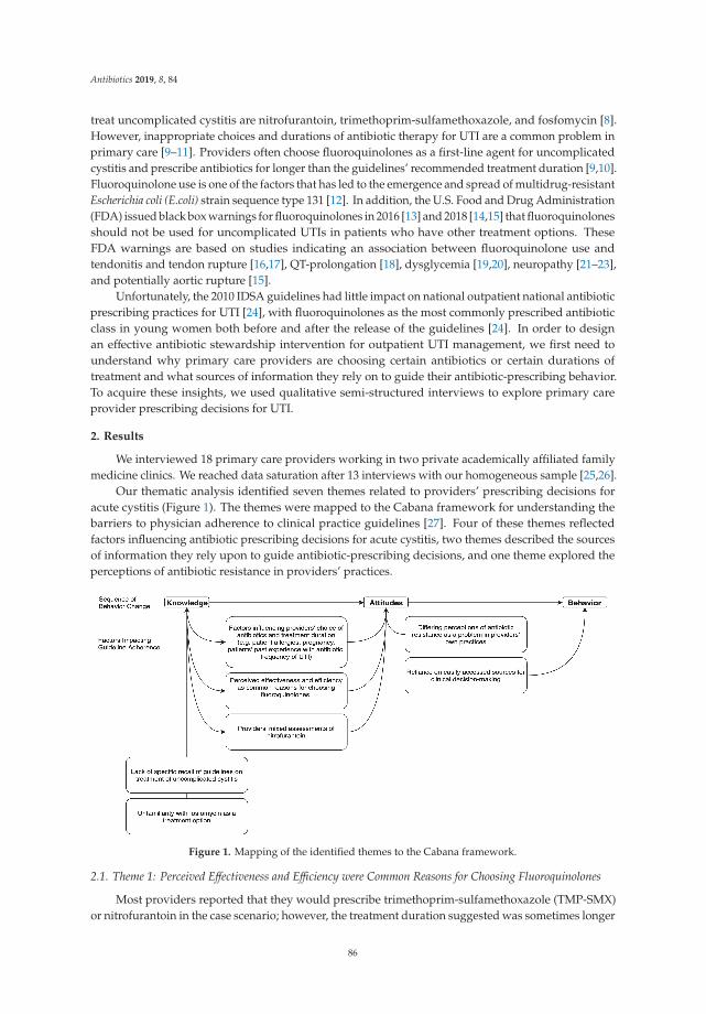
Antibiotics 2019, 8, 84
treat uncomplicated cystitis are nitrofurantoin, trimethoprim-sulfamethoxazole, and fosfomycin [8].However, inappropriate choices and durations of antibiotic therapy for UTI are a common problem inprimary care [9–11]. Providers often choose fluoroquinolones as a first-line agent for uncomplicatedcystitis and prescribe antibiotics for longer than the guidelines’ recommended treatment duration [9,10].Fluoroquinolone use is one of the factors that has led to the emergence and spread of multidrug-resistantEscherichia coli (E.coli) strain sequence type 131 [12]. In addition, the U.S. Food and Drug Administration(FDA) issued black box warnings for fluoroquinolones in 2016 [13] and 2018 [14,15] that fluoroquinolonesshould not be used for uncomplicated UTIs in patients who have other treatment options. TheseFDA warnings are based on studies indicating an association between fluoroquinolone use andtendonitis and tendon rupture [16,17], QT-prolongation [18], dysglycemia [19,20], neuropathy [21–23],and potentially aortic rupture [15].
Unfortunately, the 2010 IDSA guidelines had little impact on national outpatient national antibioticprescribing practices for UTI [24], with fluoroquinolones as the most commonly prescribed antibioticclass in young women both before and after the release of the guidelines [24]. In order to designan effective antibiotic stewardship intervention for outpatient UTI management, we first need tounderstand why primary care providers are choosing certain antibiotics or certain durations oftreatment and what sources of information they rely on to guide their antibiotic-prescribing behavior.To acquire these insights, we used qualitative semi-structured interviews to explore primary careprovider prescribing decisions for UTI.
2. Results
We interviewed 18 primary care providers working in two private academically affiliated familymedicine clinics. We reached data saturation after 13 interviews with our homogeneous sample [25,26].
Our thematic analysis identified seven themes related to providers’ prescribing decisions foracute cystitis (Figure 1). The themes were mapped to the Cabana framework for understanding thebarriers to physician adherence to clinical practice guidelines [27]. Four of these themes reflectedfactors influencing antibiotic prescribing decisions for acute cystitis, two themes described the sourcesof information they rely upon to guide antibiotic-prescribing decisions, and one theme explored theperceptions of antibiotic resistance in providers’ practices.
Figure 1. Mapping of the identified themes to the Cabana framework.
2.1. Theme 1: Perceived Effectiveness and Efficiency were Common Reasons for Choosing Fluoroquinolones
Most providers reported that they would prescribe trimethoprim-sulfamethoxazole (TMP-SMX)or nitrofurantoin in the case scenario; however, the treatment duration suggested was sometimes longer
86
Antibiotics 2019, 8, 84
than recommended by the guideline. Specifically, the preferred treatment duration of nitrofurantoinwas 7 days instead of the guideline recommendation of 5 days among approximately half of theproviders who selected this medication as their first choice for treatment. The suggested duration ofTMP-SMX was 5 days instead of the guideline recommendation of 3 days among a few providers whoselected this medication as a first choice, though most providers correctly chose a guideline concordantwith the 3-day duration. Relatively few providers chose fluoroquinolones in the case scenario, thougha third listed fluoroquinolones second among their top three choices of antibiotics most frequentlyprescribed for uncomplicated cystitis in response to a different question. In all quotations below,alias initials of providers were included to indicate that the quotations presented come from a varietyof participants.
Common reasons given for why providers might choose fluoroquinolones included prescribingfamiliarity, “I think a lot of it has to do with familiarity and comfort.” (DS); perceived effectiveness, “Nomatter what kind of bacteria it is, it would be responsive to Cipro.” (WC); short course of treatment,“In most instances, I just choose Cipro, and it’s easy; it’s for three days.” (WC); broad-spectrumcoverage, “Ciprofloxacin is a very strong antibiotic, and it is broad-spectrum.” (JG); and previouspatient experience, “If the patient had reported that Cipro had worked well before for them in the past,that might be a consideration.” (SL). Providers also expressed confidence in the ability of ciprofloxacinto kill a wide range of bacteria, reporting, for example, “Physicians don’t want the patient to have atreatment failure and come back in for a second antibiotic.” (AA).
2.2. Theme 2: Factors Influencing Providers’ Choice of Antibiotics and Treatment Duration
Providers described multiple considerations when choosing the best antibiotic for acute cystitis.These considerations included allergies, patient sex, pregnancy, patients’ past experience with theantibiotic, familiarity with the antibiotic, previous antibiotic susceptibilities, older age, shorter treatmentduration and better compliance, frequency of UTI, presence of diabetes, and cost of antibiotics. A fewproviders mentioned the potential effect of fluoroquinolones on tendons. For example, one providernoted, “There is concern about the fluoroquinolones and their effect on tendons, but I don’t knowwhether it’s a media sort of blown up case of concern or whether it’s real.” (MB). Another providerdescribed various factors affecting their choice of antibiotic for particular patients, “Age, whetheror not they have frequent UTI, maybe information from their previous urine cultures which showssensitivities.” (AA).
When the interviewer asked probing questions about the treatment of older patients, someproviders mentioned that they would be more likely to choose ciprofloxacin and a longer duration oftreatment, while others mentioned that the management of UTI would not change. Similarly, whenquestioned about recurrent UTIs and the management of diabetic patients with UTI, some providersreported that they would go with “a stronger, broad-spectrum antibiotic such as ciprofloxacin” (BC)and a longer treatment duration. When questioned about male UTIs, providers emphasized the needto rule out a sexually transmitted infection and recommended a treatment duration of up to 14 days.
2.3. Theme 3: Providers’ Mixed Assessments of Nitrofurantoin
Few side effects, low cost, and good tolerance were mentioned by the majority of providers asthe benefits of nitrofurantoin, with providers giving such responses as, “Nitrofurantoin, it is one ofmy “go to”. It is a good medication for uncomplicated cystitis.” (JQ). However, many providers alsomentioned that nitrofurantoin is not as “strong” and not as “quick” as fluoroquinolones. One providerexplained, “We have a patient population that wants their symptoms to be resolved immediately.Therefore, nitrofurantoin is not a good choice as it is bacteriostatic, and I want to make sure the patientis satisfied and just want to give them something that will resolve symptoms faster.” (BC).
A few providers were concerned about birth defects based on published case-control studies [28,29],while others thought nitrofurantoin is much safer than TMP-SMX or fluoroquinolones for pregnantwomen with UTIs, reporting, for example, “It’s one of the ones I use the most, especially in women of
87
Antibiotics 2019, 8, 84
childbearing age.” (AV). Provider-reported drawbacks included concerns about lung problems withprolonged use, “When I was on the pulmonary rotation, they were talking about the risk of nitrofurantoincausing pulmonary fibrosis, and I’ve never forgotten that.” (JS), as well as contraindication in patientswith low creatinine clearance or in older patients in general, “It’s on the Beers list.” (AA).
2.4. Theme 4: Unfamiliarity with Fosfomycin as a Treatment Option
Most providers were unfamiliar with fosfomycin as a possible treatment option for cystitis,although it is a first-line agent per guidelines [8]. Providers gave such responses as, “I don’t usefosfomycin. I’ve read about it maybe once or twice.” (AV) and “I don’t think one can use it in the UnitedStates. I’ve never seen anyone try it, and I’ve never looked into it.” (WC). One provider reportedhaving had a patient who had been prescribed fosfomycin by an infectious disease specialist.
2.5. Theme 5: Reliance on Easily Accessed Sources for Clinical Decision-making
In terms of sources of support for clinical decision-making, few providers directly relied onguidelines. Instead, many reported that they use clinical decision support resources, such as UpToDate,accessed through the institution’s electronic health record, “I use UpToDate. That’s simple. I canclick from Epic, so it’s right there on my chart. [I’m] talking to the patient, and I just click and pop itup.” (AZ) or through a mobile phone app for staying informed about new information on antibiotics,“I use UpToDate. I have the app on my phone, so I am always searching.” (BC). A few providersmentioned publications such as Prescriber’s Letter and resources available from the American Academyof Family Physicians.
2.6. Theme 6: Lack of Specific Recall of Guidelines on Treatment of Uncomplicated Cystitis
Only two providers mentioned the IDSA guidelines, though most respondents reported varyingdegrees of familiarity with unspecified guidelines, saying, for example, “I think I am reasonablyup-to-date.” (SC), “I guess I would be fairly familiar.” (JQ), and “It’s been about 10 years since Ireviewed the guidelines.” (AV). A few providers acknowledged limited recall of the details of anyguidelines regarding the treatment of uncomplicated cystitis, as indicated in the following statements:“Not very. There’s very little reason in something as simple as acute cystitis for me to go searching outthe guidelines.” (MB), “Try to stay away from Cipro first line. That’s about all I remember.” (DT), and“I have to admit I’m not particularly conversant. I will usually look at UpToDate and just refresh mymemory.” (JS).
2.7. Theme 7: Differing Perceptions of Antibiotic Resistance as a Problem in Providers’ Own Practices
When discussing the extent to which antibiotic resistance is a problem in their own practices,providers had widely differing opinions, ranging from little or no resistance, “I have never noticedresistance within my practice.” (GS), to clear, affirmative responses, “Yes, based on cultures, I have hadpatients who have had organisms identified that have a pretty concerning resistance profile.” (DS).Some providers expressed uncertainty regarding the extent of antibiotic resistance, for example, “Itis so hard for me to predict what’s going to be resistant.” (EJ), while others indicated a need for anantibiogram. One provider stated, “I don’t know if there’s actually resistance in this area or not. Itwould be nice to get, like, a report to say that.” (AA).
3. Discussion
In our study PCPs’ antibiotic choices and durations in the case scenario of acute uncomplicatedcystitis were generally in line with the guidelines’ recommended first-line therapy. Most providerscorrectly chose TMP-SMX or nitrofurantoin for the treatment of uncomplicated cystitis in the casescenario. However, ciprofloxacin was often the second antibiotic mentioned when providers respondedto the question about the top three most frequently prescribed antibiotics for uncomplicated cystitis.
88
Antibiotics 2019, 8, 84
A number of providers chose treatment durations longer than the guideline recommendations. Inparticular, many were unaware that the current recommended duration of therapy for nitrofurantoin hasbeen reduced to 5 days [8] compared with the 7-day duration in the previous 1999 IDSA guideline [30].
We found that many PCPs believe that fluoroquinolones are more likely to achieve rapidand effective control of UTI symptoms than TMP-SMX or nitrofurantoin. This finding is in linewith a previous qualitative study on the effects of knowledge, attitudes, and practices of PCPs onantibiotic selection [31]. Belief that broad-spectrum antibiotics may be more likely to cure an infectionwas the main reason for a lack of adherence to the guidelines [31]. However, clinical trials haveshown that fluoroquinolones are comparable with first-line recommended agents in both clinicaland microbiological efficacy in UTIs [32]. Although fluoroquinolones are highly efficacious foruncomplicated cystitis if the uropathogens are susceptible, increasing resistance rates may hamperthe effectiveness of fluoroquinolones for empirical antibiotic use. Fluroquinolone resistance increaseddramatically in recent years, with >10–30% of community-associated Enterobacteriaceae beingquinolone resistant in many parts of the United States and with even higher rates (>50%) in otherparts of the world [33]. Furthermore, fluoroquinolones have been moved to the last class of agents(after β-lactams) and should be used only when no other oral options are available because of threewarnings from the U.S. FDA that the risk for serious harms outweighs the benefits for patients withuncomplicated UTIs [13–15]. Of note, the initial warning preceded our interviews by approximatelya year.
In our study, PCPs’ prescribing decisions took into account multiple factors in determining the bestcourse of treatment for patients with acute cystitis such as pregnancy, patients’ past experience with aspecific antibiotic, previous antibiotic susceptibilities, frequency of UTI, older age, and presence ofdiabetes. Some PCPs chose a longer duration of treatment when the patient was older or had diabetes.These results are consistent with our previous observational database study, performed in the samesetting, showing that older age and the presence of diabetes were independent, significant predictors of alonger treatment duration for UTIs [10]. Older women were also more likely to receive fluoroquinolonesfor uncomplicated UTIs in a study on national antibiotic prescribing trends in outpatient settings in theU.S. [9]. Advanced age, diabetes, and recurrent UTIs may be cognitive biases that drive inappropriateantibiotic use for uncomplicated UTI. Treatment recommendations for otherwise healthy older women,with or without diabetes, are similar to those for younger women [34–36]. Similarly, antibiotic choiceand duration for sporadic and recurrent UTIs are not different. Broadening the antibiotic spectrumor lengthening the treatment course have not been proven efficacious and elicit concern for potentialharm to the individual and the community [37].
Another condition that triggered uncertainty for choosing the right antibiotic among the PCPs wasUTI in pregnancy. Some were concerned about the association between the use of nitrofurantoin andbirth defects reported in case-control studies [28,29]. However, no relationship between nitrofurantoinuse and birth defects was observed in a recent large cohort study [38]. An alternative first-line antibioticthat can be used in pregnancy is fosfomycin. However, PCPs in our study were not familiar withfosfomycin as a possible treatment option for cystitis, although it is a first-line agent per IDSA guidelines.
The fact that our providers did not seem to be aware of fosfomycin as a first-line recommendedantibiotic for uncomplicated UTI was not surprising. Fosfomycin was not prescribed in any visits forUTIs in our previous database study [10], and it was prescribed in less than 0.1% of all visits in a recentstudy of national antibiotic prescribing practices for UTIs in the U.S. [24]. Some of the reasons may bethat susceptibility to fosfomycin is not included in the reports received by the providers from the lab, aswell as insurance barriers and higher cost of fosfomycin compared with other UTI-relevant antibiotics.
Primary care physicians increasingly have multiple and sometimes conflicting guidelines [39]for patient care and are challenged to maintain the knowledge base represented by the array ofguidelines for common problems. In a nationally representative sample of family physicians, Wolffand colleagues [40] found that time constraints, concern for patient well-being, and considerationof particular circumstances were all potential obstacles to guideline adherence; such concerns (e.g.,
89
Antibiotics 2019, 8, 84
compliance concerns, patient preferences, and previously reported side effects) were noted by thephysicians interviewed for the current study. Likewise, nearly half of our study sample thought itwould be useful to have guidelines as a component of an electronic medical record, implying thatspeed and ease of access are important to them. Most providers interviewed were unable to quotefrom guidelines presented in monograph form but reported routinely accessing various electronicmedia (e.g., UpToDate or Family Practice Notebook) for prescribing support.
Although most of the primary care providers were concerned about antibiotic resistance in theirpractice, some did not consider antibiotic resistance to be a problem in their own practice, citinggreater resistance problems in inpatient settings or in non-UTI conditions. Likewise, in a Swedishqualitative study, providers were aware of antibiotic resistance but thought of it as a national problemthat does not concern their own practice [41]. Some of our interviewed providers indicated a need foran antibiogram specific to their setting to guide their empiric choices and a tool that can help predictthe risk of resistance in a specific patient. Unfortunately, an outpatient-specific antibiogram is notavailable for the participating clinics, and we suspect that few settings have an outpatient-specificantibiogram [42,43].
The main limitation of our study is the relatively small sample size. However, we performed thisqualitative study in a preparatory phase of a stewardship intervention and included all PCPs workingin two clinics. As with any qualitative study, the results are not meant to be generalizable. In particular,our findings may not be generalizable to public clinics or clinics that are not academically affiliated. Ourfindings represent an in-depth exploration of the attitudes and beliefs of the study sample. However,findings are transferable as the issues we identified have been seen in other settings [9,31,41]. We areusing these findings to develop provider-focused materials for a subsequent clinic-based interventiondesigned to support providers in their efforts to reduce the risks of drug resistance, to minimize seriousside effects, and to enhance overall patient care.
Our findings highlight the necessity of providing point-of-care information for primary careproviders from a trusted and easy to access source. This information will help providers to choose theright antibiotic with the right duration, a fundamental aspect of antimicrobial stewardship for UTI.
4. Materials and Methods
4.1. Ethics
The study was conducted in accordance with the Declaration of Helsinki, and the protocol wasapproved by the Ethics Committee of Baylor College of Medicine on 7 February 2019 (H-38265).
4.2. Setting and Participants
We selected individual interviews as the data collection strategy most appropriate for our researchobjectives of exploring individual prescribing decisions. Individual interviews allow for in-depthdata collection and minimize the potential influence of group interaction or seniority and supervisoryrelationships that could have been introduced with focus groups. In addition, individual interviewsallowed a much greater flexibility with scheduling, making it more feasible to reach and engageparticipants. We conducted brief (30 minutes or less), individual, semi-structured interviews with 18primary care providers (15 family medicine physicians and three physician assistants) in two private,academically affiliated family medicine clinics in a large urban area between late July 2017 and earlyNovember 2017. Both clinics utilize Epic Electronic Health Records. The characteristics of our studysample are described in Table 1.
90
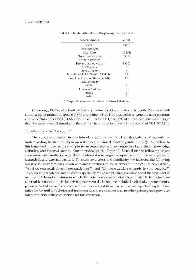
Antibiotics 2019, 8, 84
Table 1. The characteristics of the primary care providers.
Characteristic n (%)
Female 9 (50)Provider type
Physician 15 (83)Physician assistant 3 (17)
Years in practiceFewer than ten years 9 (50)
10–20 years 3Over 21 years 6
Board certified in Family Medicine 14Board certified in other specialty 1 a
Race/ethnicityWhite 6
Hispanic/Latino 3Black 1Asian 8
a One physician was board certified in Internal Medicine.
On average, 19,777 patients attend 3248 appointments at these clinics each month. Patients in bothclinics are predominantly female (58%) and white (54%). Fluoroquinolones were the most commonantibiotic class prescribed (51.6%) for uncomplicated UTI, and 75% of all prescriptions were longerthan the recommended duration in these clinics in our previous study in the period of 2011–2014 [10].
4.3. Interview Guide Development
The concepts included in our interview guide were based on the Cabana framework forunderstanding barriers to physician adherence to clinical practice guidelines [27]. According tothis framework, three factors affect physician compliance with evidence-based guidelines: knowledge,attitudes, and external factors. Our interview guide (Figure 2) focused on the following issues:awareness and familiarity with the guidelines (knowledge), acceptance and outcome expectancy(attitudes), and external barriers. To assess awareness and familiarity, we included the followingquestions: “How familiar are you with any guidelines on the treatment of uncomplicated cystitis?”,“What do you recall about these guidelines?”, and “Do these guidelines apply to your practice?”.To assess the acceptance and outcome expectancy, we asked probing questions about the treatment ofrecurrent UTIs and situations in which the patients were older, diabetic, or male. To help elucidateexternal factors that might be driving treatment decisions, we included a clinical vignette about apatient who had a diagnosis of acute uncomplicated cystitis and asked the participants to explain theirrationale for antibiotic choice and treatment duration and some reasons other primary care providersmight prescribe a fluoroquinolone for this condition.
91
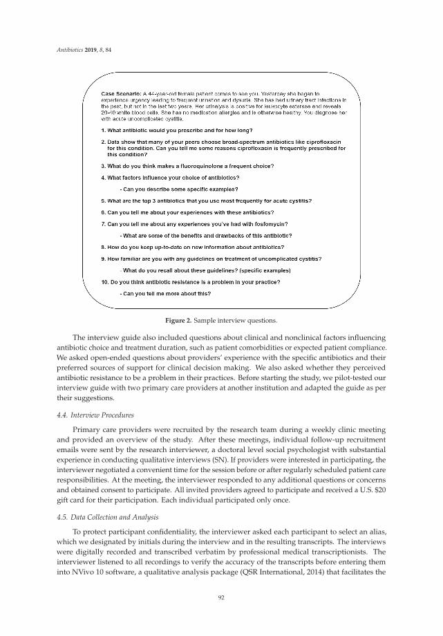
Antibiotics 2019, 8, 84
Figure 2. Sample interview questions.
The interview guide also included questions about clinical and nonclinical factors influencingantibiotic choice and treatment duration, such as patient comorbidities or expected patient compliance.We asked open-ended questions about providers’ experience with the specific antibiotics and theirpreferred sources of support for clinical decision making. We also asked whether they perceivedantibiotic resistance to be a problem in their practices. Before starting the study, we pilot-tested ourinterview guide with two primary care providers at another institution and adapted the guide as pertheir suggestions.
4.4. Interview Procedures
Primary care providers were recruited by the research team during a weekly clinic meetingand provided an overview of the study. After these meetings, individual follow-up recruitmentemails were sent by the research interviewer, a doctoral level social psychologist with substantialexperience in conducting qualitative interviews (SN). If providers were interested in participating, theinterviewer negotiated a convenient time for the session before or after regularly scheduled patient careresponsibilities. At the meeting, the interviewer responded to any additional questions or concernsand obtained consent to participate. All invited providers agreed to participate and received a U.S. $20gift card for their participation. Each individual participated only once.
4.5. Data Collection and Analysis
To protect participant confidentiality, the interviewer asked each participant to select an alias,which we designated by initials during the interview and in the resulting transcripts. The interviewswere digitally recorded and transcribed verbatim by professional medical transcriptionists. Theinterviewer listened to all recordings to verify the accuracy of the transcripts before entering theminto NVivo 10 software, a qualitative analysis package (QSR International, 2014) that facilitates the
92
Antibiotics 2019, 8, 84
management of qualitative data. Based on a preliminary review of the transcripts and the aims of thestudy, we determined that the interview questions and subsequent probes provided an appropriateoutline for the initial identification of key themes [44,45].
Two authors (LG and SN) met regularly to read, code, and discuss the transcribed interviews,maintaining a shared consensus on the coding process. We elected to focus on the explicit content ofproviders’ reported experiences and perspectives [46]. Overall, we followed the six-phase approach tothematic analysis described by Braun and Clark [47], a sequential but flexible set of guidelines thatencompasses familiarization with the data; initial coding; searching, reviewing, and defining themes;and finally, integrating the analytic narrative and data extracts by incorporating illustrative quotesfrom providers into the description of the final themes [44,47]. Although our sample size was limitedto the total number of providers in the clinic setting, we judged that the last 5 to 6 interviews codedwere redundant to those previously coded, in repetition without an expansion of the themes, and wereconfident that data saturation for this sample had been reached [25].
5. Conclusions
Our qualitative analysis found that many PCPs believe that fluoroquinolones achieve more rapidand effective control of UTI symptoms than trimethoprim-sulfamethoxazole or nitrofurantoin. Themajority of the providers were unfamiliar with fosfomycin as a possible treatment option for cystitis,although it is a first-line agent per guidelines. For recurrent UTI, older patients, or those with diabetes,some providers reported that they would go with “a stronger, broad-spectrum antibiotic such asciprofloxacin” and longer treatment duration. For support in clinical decision making, few providersrelied on guidelines, preferring instead to have decision support embedded in the electronic medicalrecord. Understanding primary care providers’ clinical challenges and knowing their preferred sourcesof information will enable us to develop a primary care-specific antibiotic stewardship intervention foracute cystitis.
Author Contributions: L.G., B.W.T., S.N. and R.Z. conceived and designed the study; S.N. and L.G. analyzed thedata; L.G., S.N., M.S.H., F.M.K., L.M., G.J.G. and B.W.T. provided input on the study concept and design and theinterpretation of the data; L.G. wrote the paper.
Funding: This research was funded by Zambon Pharmaceuticals, grant number 47271-I.
Acknowledgments: This investigator-initiated research study was funded by Zambon Pharmaceuticals. Trautner’swork is supported in part by the Houston Veterans Affairs Health Services Research & Development Center forInnovations in Quality, Effectiveness, and Safety (CIN 13-413) and by Veterans Affairs funding (HSR&D 16-025).
Conflicts of Interest: Grigoryan and Trautner report grants from the National Institutes of Health and the U.S.Department of Veterans Affairs Health Services Research & Development Service.
References
1. Centers for Disease Control and Prevention. Antibiotic/Antimicrobial Resistance. Biggest Threats. Availableonline: https://www.cdc.gov/drugresistance/biggest_threats.html (accessed on 17 June 2019).
2. National Action Plan for Combating Antibiotic-Resistant Bacteria. Available online: https://aspe.hhs.gov/system/files/pdf/258516/ProgressYears1and2CARBNationalActionPlan.pdf (accessed on 17 June 2019).
3. The Pew Charitable Trusts. Antibiotic Use in Outpatient Settings. Available online: http://www.pewtrusts.org/~{}/media/assets/2016/05/antibioticuseinoutpatientsettings.pdf (accessed on 17 June 2019).
4. Meeker, D.; Linder, J.A.; Fox, C.R.; Friedberg, M.W.; Persell, S.D.; Goldstein, N.J.; Knight, T.K.; Hay, J.W.;Doctor, J.N. Effect of Behavioral Interventions on Inappropriate Antibiotic Prescribing Among Primary CarePractices: A Randomized Clinical Trial. JAMA 2016, 315, 562–570. [CrossRef] [PubMed]
5. Drekonja, D.M.; Filice, G.A.; Greer, N.; Olson, A.; MacDonald, R.; Rutks, I.; Wilt, T.J. Antimicrobial stewardshipin outpatient settings: A systematic review. Infect. Control Hosp. Epidemiol. 2015, 36, 142–152. [CrossRef][PubMed]
93
Antibiotics 2019, 8, 84
6. Hecker, M.T.; Fox, C.J.; Son, A.H.; Cydulka, R.K.; Siff, J.E.; Emerman, C.L.; Sethi, A.K.; Muganda, C.P.;Donskey, C.J. Effect of a stewardship intervention on adherence to uncomplicated cystitis and pyelonephritisguidelines in an emergency department setting. PLoS ONE 2014, 9, e87899. [CrossRef] [PubMed]
7. Fleming-Dutra, K.E.; Hersh, A.L.; Shapiro, D.J.; Bartoces, M.; Enns, E.A.; File, T.M.; Finkelstein, J.A., Jr.;Gerber, J.S.; Hyun, D.Y.; Linder, J.A.; et al. Prevalence of Inappropriate Antibiotic Prescriptions Among USAmbulatory Care Visits, 2010–2011. JAMA 2016, 315, 1864–1873. [CrossRef] [PubMed]
8. Gupta, K.; Hooton, T.M.; Naber, K.G.; Wullt, B.; Colgan, R.; Miller, L.G.; Moran, G.J.; Nicolle, L.E.; Raz, R.;Schaeffer, A.J.; et al. International clinical practice guidelines for the treatment of acute uncomplicatedcystitis and pyelonephritis in women: A 2010 update by the Infectious Diseases Society of America and theEuropean Society for Microbiology and Infectious Diseases. Clin. Infect. Dis. 2011, 52, e103–e120. [CrossRef]
9. Kobayashi, M.; Shapiro, D.J.; Hersh, A.L.; Sanchez, G.V.; Hicks, L.A. Outpatient Antibiotic PrescribingPractices for Uncomplicated Urinary Tract Infection in Women in the United States, 2002–2011. Open ForumInfect. Dis. 2016, 3, ofw159. [CrossRef] [PubMed]
10. Grigoryan, L.; Zoorob, R.; Wang, H.; Trautner, B.W. Low Concordance With Guidelines for Treatment ofAcute Cystitis in Primary Care. Open Forum Infect. Dis. 2015, 2, ofv159. [CrossRef]
11. Shively, N.R.; Buehrle, D.J.; Clancy, C.J.; Decker, B.K. Prevalence of Inappropriate Antibiotic Prescribing inPrimary Care Clinics within a Veterans Affairs Health Care System. Antimicrob. Agents Chemother. 2018, 62.[CrossRef]
12. Petty, N.K.; Ben Zakour, N.L.; Stanton-Cook, M.; Skippington, E.; Totsika, M.; Forde, B.M.; Phan, M.D.;Gomes Moriel, D.; Peters, K.M.; Davies, M.; et al. Global dissemination of a multidrug resistant Escherichiacoli clone. Proc. Natl. Acad. Sci. USA 2014, 111, 5694–5699. [CrossRef] [PubMed]
13. U.S Food and Drug Administration. FDA Advises Restricting Fluoroquinolone Antibiotic Use for CertainUncomplicated Infections; Warns about Disabling Side Effects that Can Occur Together. 2016. Availableonline: https://www.fda.gov/downloads/Drugs/DrugSafety/UCM500591.pdf (accessed on 17 June 2019).
14. U.S Food and Drug Administration. FDA Updates Warnings for Fluoroquinolone Antibiotics on Risks ofMental Health and Low Blood Sugar Adverse Reactions. Available online: https://www.fda.gov/newsevents/newsroom/pressannouncements/ucm612995.htm (accessed on 17 June 2019).
15. U.S. Food and Drug Administration. FDA Warns about Increased Risk of Ruptures or Tears in theAorta Blood Vessel with Fluoroquinolone Antibiotics in Certain Patients. 2018. Available online: https://www.fda.gov/Drugs/DrugSafety/ucm628753.htm (accessed on 17 June 2019).
16. Stephenson, A.L.; Wu, W.; Cortes, D.; Rochon, P.A. Tendon Injury and Fluoroquinolone Use: A SystematicReview. Drug Saf. 2013, 36, 709–721. [CrossRef]
17. Wise, B.L.; Peloquin, C.; Choi, H.; Lane, N.E.; Zhang, Y. Impact of age, sex, obesity, and steroid use onquinolone-associated tendon disorders. Am. J. Med. 2012, 125, e1223–e1228. [CrossRef] [PubMed]
18. Briasoulis, A.; Agarwal, V.; Pierce, W.J. QT prolongation and torsade de pointes induced by fluoroquinolones:Infrequent side effects from commonly used medications. Cardiology 2011, 120, 103–110. [CrossRef] [PubMed]
19. Aspinall, S.L.; Good, C.B.; Jiang, R.; McCarren, M.; Dong, D.; Cunningham, F.E. Severe dysglycemia with thefluoroquinolones: A class effect? Clin. Infect. Dis. 2009, 49, 402–408. [CrossRef] [PubMed]
20. Chou, H.W.; Wang, J.L.; Chang, C.H.; Lee, J.J.; Shau, W.Y.; Lai, M.S. Risk of severe dysglycemia amongdiabetic patients receiving levofloxacin, ciprofloxacin, or moxifloxacin in Taiwan. Clin. Infect. Dis. 2013, 57,971–980. [CrossRef] [PubMed]
21. Ali, A.K. Peripheral neuropathy and Guillain-Barre syndrome risks associated with exposure to systemicfluoroquinolones: A pharmacovigilance analysis. Ann. Epidemiol. 2014, 24, 279–285. [CrossRef] [PubMed]
22. Aoun, M.; Jacquy, C.; Debusscher, L.; Bron, D.; Lehert, M.; Noel, P.; van der Auwera, P. Peripheral neuropathyassociated with fluoroquinolones. Lancet 1992, 340, 127. [CrossRef]
23. Etminan, M.; Brophy, J.M.; Samii, A. Oral fluoroquinolone use and risk of peripheral neuropathy:A pharmacoepidemiologic study. Neurology 2014, 83, 1261–1263. [CrossRef]
24. Durkin, M.J.; Keller, M.; Butler, A.M.; Kwon, J.H.; Dubberke, E.R.; Miller, A.C.; Polgreen, P.M.; Olsen, M.A.An Assessment of Inappropriate Antibiotic Use and Guideline Adherence for Uncomplicated Urinary TractInfections. Open Forum Infect. Dis. 2018, 5, ofy198. [CrossRef]
25. Saunders, B.; Sim, J.; Kingstone, T.; Baker, S.; Waterfield, J.; Bartlam, B.; Burroughs, H.; Jinks, C. Saturationin qualitative research: Exploring its conceptualization and operationalization. Qual. Quant. 2018, 52,1893–1907. [CrossRef]
94
Antibiotics 2019, 8, 84
26. Guest, G.; Bunce, A.; Johnson, L. How Many Interviews Are Enough? Field Methods 2006, 18, 59–82. [CrossRef]27. Cabana, M.D.; Rand, C.S.; Powe, N.R.; Wu, A.W.; Wilson, M.H.; Abboud, P.A.; Rubin, H.R. Why don’t
physicians follow clinical practice guidelines? A framework for improvement. JAMA 1999, 282, 1458–1465.[CrossRef] [PubMed]
28. Ailes, E.C.; Gilboa, S.M.; Gill, S.K.; Broussard, C.S.; Crider, K.S.; Berry, R.J.; Carter, T.C.; Hobbs, C.A.;Interrante, J.D.; Reefhuis, J.; et al. Association between antibiotic use among pregnant women with urinarytract infections in the first trimester and birth defects, National Birth Defects Prevention Study 1997 to 2011.Birth Defects Res. A Clin. Mol. Teratol. 2016, 106, 940–949. [CrossRef] [PubMed]
29. Crider, K.S.; Cleves, M.A.; Reefhuis, J.; Berry, R.J.; Hobbs, C.A.; Hu, D.J. Antibacterial medication use duringpregnancy and risk of birth defects: National Birth Defects Prevention Study. Arch. Pediatr. Adolesc. Med.2009, 163, 978–985. [CrossRef] [PubMed]
30. Warren, J.W.; Abrutyn, E.; Hebel, J.R.; Johnson, J.R.; Schaeffer, A.J.; Stamm, W.E. Guidelines for antimicrobialtreatment of uncomplicated acute bacterial cystitis and acute pyelonephritis in women. Infectious DiseasesSociety of America (IDSA). Clin. Infect. Dis. 1999, 29, 745–758. [CrossRef] [PubMed]
31. Sanchez, G.V.; Roberts, R.M.; Albert, A.P.; Johnson, D.D.; Hicks, L.A. Effects of knowledge, attitudes, andpractices of primary care providers on antibiotic selection, United States. Emerg. Infect. Dis. 2014, 20,2041–2047. [CrossRef] [PubMed]
32. Grigoryan, L.; Trautner, B.W.; Gupta, K. Diagnosis and management of urinary tract infections in theoutpatient setting: A review. JAMA 2014, 312, 1677–1684. [CrossRef]
33. Spellberg, B.; Doi, Y. The Rise of Fluoroquinolone-Resistant Escherichia coli in the Community: Scarier ThanWe Thought. J. Infect. Dis. 2015, 212, 1853–1855. [CrossRef]
34. Mody, L.; Juthani-Mehta, M. Urinary tract infections in older women: A clinical review. JAMA 2014, 311,844–854. [CrossRef]
35. Grigoryan, L.; Zoorob, R.; Wang, H.; Horsfield, M.; Gupta, K.; Trautner, B.W. Less workup, longer treatment,but no clinical benefit observed in women with diabetes and acute cystitis. Diabetes Res. Clin. Pract. 2017,129, 197–202. [CrossRef]
36. Schneeberger, C.; Stolk, R.P.; Devries, J.H.; Schneeberger, P.M.; Herings, R.M.; Geerlings, S.E. Differencesin the pattern of antibiotic prescription profile and recurrence rate for possible urinary tract infections inwomen with and without diabetes. Diabetes Care 2008, 31, 1380–1385. [CrossRef]
37. Smith, A.L.; Brown, J.; Wyman, J.F.; Berry, A.; Newman, D.K.; Stapleton, A.E. Treatment and Preventionof Recurrent Lower Urinary Tract Infections in Women: A Rapid Review with Practice Recommendations.J. Urol. 2018, 200, 1174–1191. [CrossRef] [PubMed]
38. Muanda, F.T.; Sheehy, O.; Berard, A. Use of antibiotics during pregnancy and the risk of major congenitalmalformations: A population based cohort study. Br. J. Clin. Pharmacol. 2017, 83, 2557–2571. [CrossRef][PubMed]
39. Markowitz, M.A.; Wood, L.N.; Raz, S.; Miller, L.G.; Haake, D.A.; Kim, J.H. Lack of uniformity among UnitedStates recommendations for diagnosis and management of acute, uncomplicated cystitis. Int. Urogynecol. J.2018. [CrossRef] [PubMed]
40. Wolff, M.; Bower, D.J.; Marbella, A.M.; Casanova, J.E. US family physicians’ experiences with practiceguidelines. Fam. Med. Kans. City 1998, 30, 117–121.
41. Bjorkman, I.; Berg, J.; Viberg, N.; Stalsby Lundborg, C. Awareness of antibiotic resistance and antibioticprescribing in UTI treatment: A qualitative study among primary care physicians in Sweden. Scand. J. Prim.Health Care 2013, 31, 50–55. [CrossRef] [PubMed]
42. Rank, E.L.; Lodise, T.; Avery, L.; Bankert, E.; Dobson, E.; Dumyati, G.; Hassett, S.; Keller, M.; Pearsall, M.;Lubowski, T.; et al. Antimicrobial Susceptibility Trends Observed in Urinary Pathogens Obtained From NewYork State. Open Forum Infect. Dis. 2018, 5, ofy297. [CrossRef]
43. Delisle, G.; Quach, C.; Domingo, M.C.; Boudreault, A.A.; Gourdeau, M.; Bernatchez, H.; Lavallee, C.Escherichia coli antimicrobial susceptibility profile and cumulative antibiogram to guide empirical treatmentof uncomplicated urinary tract infections in women in the province of Quebec, 2010–2015. J. Antimicrob.Chemother. 2016, 71, 3562–3567. [CrossRef]
44. King, N. Using templates in the thematic analysis of text. In Essential Guide to Qualitative Methods inOrganizational Research; Cassell, C., Symon, G., Eds.; Sage Publications Ltd.: Thousand Oaks, CA, USA, 2004;pp. 257–270.
95
Antibiotics 2019, 8, 84
45. Rowley, J. Conducting research interviews. Manag. Res. Rev. 2012, 35, 260–271. [CrossRef]46. Riessman, C.K. Narrative Methods for the Human Sciences; Sage Publications Inc.: Thousand Oaks, CA,
USA, 2008.47. Braun, V.; Clarke, V. Using thematic analysis in psychology. Qual. Res. Psychol. 2006, 3, 77–101. [CrossRef]
© 2019 by the authors. Licensee MDPI, Basel, Switzerland. This article is an open accessarticle distributed under the terms and conditions of the Creative Commons Attribution(CC BY) license (http://creativecommons.org/licenses/by/4.0/).
96
antibiotics
Article
Antibiotic Prescribing Quality in Out-of-HoursPrimary Care and Critical Appraisal ofDisease-Specific Quality Indicators
Annelies Colliers 1,*,†, Niels Adriaenssens 1,†, Sibyl Anthierens 1, Stephaan Bartholomeeusen 1,
Hilde Philips 1, Roy Remmen 1 and Samuel Coenen 1,2,3
1 Department of Primary and Interdisciplinary Care (ELIZA)—Centre for General Practice, Faculty ofMedicine and Health Sciences, University of Antwerp, Doornstraat 331, B-2610 Antwerp, Belgium;[email protected] (N.A.); [email protected] (S.A.);[email protected] (S.B.); [email protected] (H.P.);[email protected] (R.R.); [email protected] (S.C.)
2 Department of Epidemiology and Social Medicine (ESOC), Faculty of Medicine and Health Sciences,University of Antwerp, Universiteitsplein 1, B-2610 Antwerp, Belgium
3 Vaccine & Infectious Disease Institute (VAXINFECTIO), Faculty of Medicine and Health Sciences, Universityof Antwerp, Universiteitsplein 1, B-2610 Antwerp, Belgium
* Correspondence: [email protected]; Tel.: +32-3-265-18-32† The first two authors made equal contributions.
Received: 15 May 2019; Accepted: 7 June 2019; Published: 12 June 2019
Abstract: Outpatient antibiotic use in Belgium is among the highest in Europe. The most commonreason for an encounter in out-of-hours (OOH) primary care is an infection. In this study, we assessedall consultations from July 2016 to June 2018 at five OOH services. We described antibiotic prescribingby diagnosis, calculated disease-specific antibiotic prescribing quality indicators’ (APQI) values andcritically appraised these APQI. We determined that 111,600 encounters resulted in 26,436 (23.7%)antibiotic prescriptions. The APQI diagnoses (i.e., bronchitis, upper respiratory infection, cystitis,tonsillitis, sinusitis, otitis media, and pneumonia) covered 14,927 (56.7%) antibiotic prescriptions.Erysipelas (1344 (5.1%)) and teeth/gum disease (982 (3.7%)) covered more prescriptions than sinusitisor pneumonia. Over 75% of patients with tonsillitis and over 50% with bronchitis, sinusitis, andotitis media were prescribed an antibiotic. Only for otitis media the choice of antibiotic was nearthe acceptable range. Over 10% of patients with bronchitis or pneumonia and over 25% of femalepatients with an acute cystitis received quinolones. The APQI cover the diagnoses for only 57% ofall antibiotic prescriptions. As 5.1% and 3.7% of antibiotic prescriptions are made for erysipelasand teeth/gum disease, respectively, we propose to add these indications when assessing antibioticprescribing quality in OOH primary care.
Keywords: antibiotics; out-of-hours care; primary care; quality of care; quality indicators;practitioners cooperative
1. Introduction
Antibiotic use in Belgium is among the highest in Europe, especially in ambulatory care,and thus the risk for antimicrobial resistance is high [1]. The European Surveillance of AntimicrobialConsumption (ESAC) [2] project has developed a set of 21 disease-specific antibiotic prescribing qualityindicators (APQI) to assess the quality of antibiotic prescribing in primary care [3]. These APQI havebeen adopted in several evaluations of antibiotic prescribing in Europe [4–6].
Out-of-hours (OOH) care covers a large part of hours during a week. In Belgium, but alsoin the rest of Europe, the on-going establishment of large-scale general practitioner cooperatives
Antibiotics 2019, 8, 79; doi:10.3390/antibiotics8020079 www.mdpi.com/journal/antibiotics97
Antibiotics 2019, 8, 79
(GPCs) represents one of the most important developments for primary OOH health care. The mostcommon reason for an encounter in Flemish (Northern part of Belgium) OOH primary care is aninfection [7]. Many infections are self-limiting and no antimicrobial treatment is necessary. However,in our qualitative work general practitioners (GPs) describe the difficulties they encounter in OOHcare not to prescribe antibiotics relating to contextual factors such as a larger uncertainty due to anunknown patient, type of patients (e.g., children, elderly, Non-Native Speakers, . . . ), workload, and thelack of diagnostic tools or follow-up. They have a feeling that their professional identity is differentcompared to in-hours care [8,9]. However, previous research in Belgium showed similar APQI valuesfor in- and out-of-hours care, using the data of a singular general practitioner cooperative (GPC)from 2004–2009 [4], which is similar to findings in Norway and Sweden [10,11]. In the Netherlands,research showed higher prescribing rates in OOH care but with equal or even better quality comparedto in-hours care [12]. In the United Kingdom, researchers found an increase in antibiotic prescribingin OOH primary care in contrast to in-hours care, however, this study did not explore the quality ofthe prescriptions [13,14]. Moreover, in Danish OOH care, antibiotics are the most prescribed drugs,but there was no evaluation on the quality of prescribing [15].
Therefore, the objective of this study is to assess the quality of antibiotic prescribing in OOHprimary care to inform the implementation of future interventions to improve the antibiotic prescribingquality in this specific setting [16]. More specifically, we aim to describe antibiotic prescribing in BelgianOOH primary care by indication, assess its quality by updating values for ESAC’s disease-specificAPQI [3] and critically appraise these APQI [4].
2. Methods
2.1. Database and GPCs
Data were extracted from the Improving Care and Research Electronic Data Trust Antwerp(iCAREdata) database, a research database of linked data on OOH primary care [17,18]. On a weeklybasis, routine clinical data and patient characteristics are automatically collected from GPCs, emergencydepartments (EDs) and pharmacists’ electronic records completed during OOH-care. For this studywe only used data from GPCs.
iCAREdata currently covers nine GPCs, that cover a population of 1,127,153. Only data from fiveGPCs were withheld for this study, because of the level of completeness of their data and availabilityat the start of the study. Together, they cover a population of around 811,000 inhabitants (11% of theFlemish population; i.e., the Northern Dutch speaking part of Belgium). The characteristics of thesefive GPCs are presented in Table 1. Because of a lack of registration of home visits in OOH care, onlydata from consultations at the GPC itself were used.
Table 1. Characteristics of the five general practitioner cooperatives (GPCs) providing data for this study.
GPC Population Number of GPs Rural/Urban
Antwerp-East 148,366 112 UrbanAntwerp-North 141,110 119 Urban/ruralAntwerp-Centre 185,358 171 Urban
Tienen 84,430 80 RuralZuiderkempen 251,833 270 Rural
Total 811,097 752
2.2. Study Setting
Since the beginning of the year 2000, Belgian OOH care is increasingly being organized in GPCsin regions that typically cover 80–180 GPs. All practicing GPs in such an area are obliged to participatein this service following a rotation-based system of being ‘on call’ during the weekends in their ownregion and they work in shifts of on average 12 h. GPCs are mostly organized in a fee-for-service
98
Antibiotics 2019, 8, 79
system. There are 80–180 GPs per GPC. Belgian GPs do not have a gatekeeper function. There isfree access to primary, secondary, and tertiary care. They use an electronic medical health recordto register every patient contact. The software can differ depending on the GPC, but diagnosesare always selected using Thesaurus terms in the Belgian Bilingual Biclassified Thesaurus (3BT).By selecting a diagnosis in 3BT, an International Classification of Primary Care (ICPC-2-R) code isautomatically linked [19]. Prescriptions are registered using the database of the Belgian Centre forPharmacotherapeutic Information [20], which is linked to the Anatomical Therapeutic Chemical (ATC)classification system [21,22].
2.3. Data Analysis
Data extraction was done using Microsoft SQL server 2012. We performed descriptive analysesusing Microsoft Excel 2016. Data from 1 July 2016 until 30 June 2018 were included.
2.3.1. Antibiotic Prescribing by Indication
To describe antibiotic prescribing by indication, first we only analyzed all antibiotic prescriptions(ATC code J01: antibacterials for systemic use) delivered during the consultations in five GPCs duringa two-year timeframe and linked them with the diagnosis that was registered in the electronic medicalhealth record.
2.3.2. Quality of Antibiotic Prescribing: Disease-Specific APQI
Second, we used information on all encounters to assess the quality of antibiotic prescribing usingthe disease-specific APQI introduced by ESAC (European Surveillance of Antimicrobial Consumptionproject) in 2007 [3]. The seven most common indications for antibiotic prescribing are in descendingorder: acute bronchitis (ICPC code R78), acute upper respiratory tract infection (RTI) (R74), cystitis/otherurinary infection (UTI; U71), acute tonsillitis (R76), acute/chronic sinusitis (R75), and acute otitis media(H71). For pneumonia (R81), values of three valid APQI were calculated:
a = the percentage of patients with age and/or gender limitation (see legend Table 3) prescribedan antibiotic;b = a and receiving the guideline recommended antibiotic;c = a and receiving quinolones.
We compared them with the findings of an earlier study with data from 2004–2009 from one GPC [4].
2.3.3. Critical Appraisal
We evaluated if APQI is a sufficient method when describing the quality of antibiotic prescribingin OOH care. We used and adapted the concept of drug utilization 90% (DU90%) [23], which analyzesthe number of drugs accounting for 90% of drug use. We used it to see if using APQI-diagnoses covers90% of all antibiotic prescriptions.
2.4. Ethics
The study was approved by the Ethics Committee of the Antwerp University Hospital/Universityof Antwerp (reference number 17/08/089), and registered at clinicaltrials.gov (NCT03082521). The studywas approved by the scientific advisory board of ICAREdata and the different GPCs.
3. Results
From 1/7/2016 until 30/6/2018, 26,436 antibiotic prescriptions were registered in 111,600 visits(excluding home visits) covering a population of 811,097 persons.
For 93.65% of the prescriptions, a diagnosis was registered in the same consultation. A diagnosiswas missing for 1612 (6.35%) antibiotic prescriptions.
99
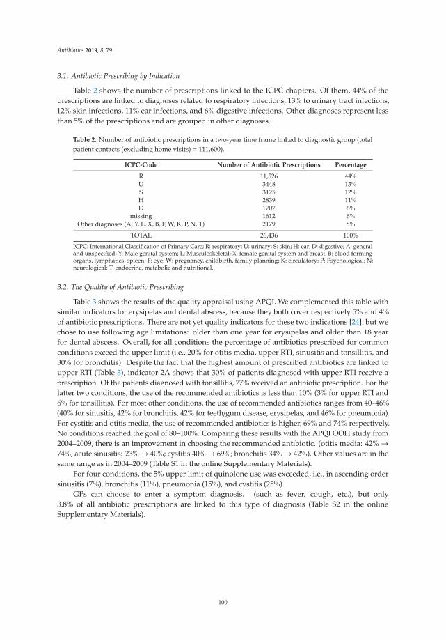
Antibiotics 2019, 8, 79
3.1. Antibiotic Prescribing by Indication
Table 2 shows the number of prescriptions linked to the ICPC chapters. Of them, 44% of theprescriptions are linked to diagnoses related to respiratory infections, 13% to urinary tract infections,12% skin infections, 11% ear infections, and 6% digestive infections. Other diagnoses represent lessthan 5% of the prescriptions and are grouped in other diagnoses.
Table 2. Number of antibiotic prescriptions in a two-year time frame linked to diagnostic group (totalpatient contacts (excluding home visits) = 111,600).
ICPC-Code Number of Antibiotic Prescriptions Percentage
R 11,526 44%U 3448 13%S 3125 12%H 2839 11%D 1707 6%
missing 1612 6%Other diagnoses (A, Y, L, X, B, F, W, K, P, N, T) 2179 8%
TOTAL 26,436 100%
ICPC: International Classification of Primary Care; R: respiratory; U: urinary; S: skin; H: ear; D: digestive; A: generaland unspecified; Y: Male genital system; L: Musculoskeletal; X: female genital system and breast; B: blood formingorgans, lymphatics, spleen; F: eye; W: pregnancy, childbirth, family planning; K: circulatory; P: Psychological; N:neurological; T: endocrine, metabolic and nutritional.
3.2. The Quality of Antibiotic Prescribing
Table 3 shows the results of the quality appraisal using APQI. We complemented this table withsimilar indicators for erysipelas and dental abscess, because they both cover respectively 5% and 4%of antibiotic prescriptions. There are not yet quality indicators for these two indications [24], but wechose to use following age limitations: older than one year for erysipelas and older than 18 yearfor dental abscess. Overall, for all conditions the percentage of antibiotics prescribed for commonconditions exceed the upper limit (i.e., 20% for otitis media, upper RTI, sinusitis and tonsillitis, and30% for bronchitis). Despite the fact that the highest amount of prescribed antibiotics are linked toupper RTI (Table 3), indicator 2A shows that 30% of patients diagnosed with upper RTI receive aprescription. Of the patients diagnosed with tonsillitis, 77% received an antibiotic prescription. For thelatter two conditions, the use of the recommended antibiotics is less than 10% (3% for upper RTI and6% for tonsillitis). For most other conditions, the use of recommended antibiotics ranges from 40–46%(40% for sinusitis, 42% for bronchitis, 42% for teeth/gum disease, erysipelas, and 46% for pneumonia).For cystitis and otitis media, the use of recommended antibiotics is higher, 69% and 74% respectively.No conditions reached the goal of 80–100%. Comparing these results with the APQI OOH study from2004–2009, there is an improvement in choosing the recommended antibiotic. (otitis media: 42%→74%; acute sinusitis: 23%→ 40%; cystitis 40%→ 69%; bronchitis 34%→ 42%). Other values are in thesame range as in 2004–2009 (Table S1 in the online Supplementary Materials).
For four conditions, the 5% upper limit of quinolone use was exceeded, i.e., in ascending ordersinusitis (7%), bronchitis (11%), pneumonia (15%), and cystitis (25%).
GPs can choose to enter a symptom diagnosis. (such as fever, cough, etc.), but only3.8% of all antibiotic prescriptions are linked to this type of diagnosis (Table S2 in the onlineSupplementary Materials).
100
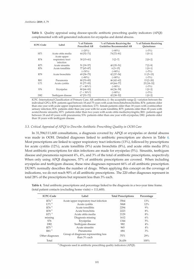
Antibiotics 2019, 8, 79
Table 3. Quality appraisal using disease-specific antibiotic prescribing quality indicators (APQI)complemented with self-generated indicators for erysipelas and dental abscess.
ICPC-Code Label% of Patients
Prescribed AB% of Patients Receiving the
Guideline Recommended AB% of Patients Receiving
Quinolones
(<20%) (>80%) (<5%)H71 Acute otitis media 64 [52–71] 74 [72–81] 1 [0–1]
R74Acute upper
respiratory tractinfection
30 [15–41] 3 [2–7] 2 [0–2]
R75 Acute sinusitis 51 [39–57] 40 [35–51] 7 [2–9]R76 Acute tonsillitis 77 [65–87] 6 [3–15] 1 [0–1]
(<30%) (>80%) (<5%)R78 Acute bronchitis 69 [58–75] 42 [37–54] 11 [5–15]
(>80%) (>80%) (<5%)R81 Pneumonia 80 [70–85] 46 [42–62] 15 [2–20]U71 Acute cystitis 91 [77–95] 69 [64–77] 25 [18–32]
(? > 80%?) (>80%) (<5%)S76 Erysipelas 80 [64–83] 44 [36–59] 1 [0–2]
(? < 30%?) (>80%) (<5%)D82 Teeth/gum disease 67 [53–72] 42 [38–52] 1 [0–2]
ICPC: International Classification of Primary Care; AB: antibiotics; (): the acceptable range []: variation between theindividual GPCs; R78: patients aged between 18 and 75 years with acute bronchitis/bronchiolitis; R74: patients olderthan one year with acute upper respiratory infection; U71: female patients older than 18 years with cystitis/otherurinary infection; R76: patients older than one year with for acute tonsillitis; R75: patients older than 18 years withacute/chronic sinusitis; H71: patients older than two years with acute otitis media/myringitis; R81: patients agedbetween 18 and 65 years with pneumonia; S76: patients older than one year with erysipelas; D82: patients olderthan 18 years with teeth/gum disease.
3.3. Critical Appraisal of APQI to Describe Antibiotic Prescribing Quality in OOH Care
In 31,596/111,600 consultations, a diagnosis covered by APQI or erysipelas or dental abscesswas made in OOH. Detailed diagnoses linked to antibiotic prescription are shown in Table 4.Most prescriptions are linked to upper respiratory tract infections (13%), followed by prescriptionsfor acute cystitis (12%), acute tonsillitis (9%) acute bronchitis (8%), and acute otitis media (8%).Most antibiotic prescriptions for skin infections are made for erysipelas (5%). Sinusitis, teeth/gumdisease and pneumonia represent 4%, 4%, and 3% of the total of antibiotic prescriptions, respectively.When only using APQI diagnoses, 57% of antibiotic prescriptions are covered. When includingerysipelas and teeth/gum disease, these nine diagnoses represent 66% of all antibiotic prescription.DU90% normally describes the number of drugs. When applying this concept on the coverage ofindications, we do not reach 90% of all antibiotic prescriptions. The 220 other diagnoses represent intotal 28% of the prescriptions but represent less than 3% each.
Table 4. Total antibiotic prescriptions and percentage linked to the diagnosis in a two-year time frame.(total patient contacts (excluding home visits) = 111,600).
ICPC-Code Label Total Prescriptions Percentage
R74 † Acute upper respiratory tract infection 3564 13%U71 † Acute cystitis 3068 12%R76 † Acute tonsillitis 2294 9%R78 † Acute bronchitis 2223 8%H71 † Acute otitis media 2129 8%
missing Diagnosis missing 1612 6%S76 Erysipelas 1344 5%D82 Teeth/gum disease 982 4%
R75 † Acute sinusitis 965 4%R81 † Pneumonia 684 3%
Other diagnoses Group of diagnoses representing lessthan 3% each 7571 29%
Total 26,436 100%† Diagnosis used in antibiotic prescribing quality indicators (APQI).
101
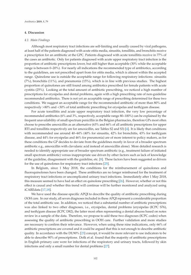
Antibiotics 2019, 8, 79
4. Discussion
4.1. Main Findings
Although most respiratory tract infections are self-limiting and usually caused by viral pathogens,at least half of the patients diagnosed with acute otitis media, sinusitis, tonsillitis, and bronchitis receivea prescription for an antibiotic at the GPC. Patients diagnosed with acute tonsillitis receive in 77% ofthe cases an antibiotic. Only for patients diagnosed with acute upper respiratory tract infection is theproportion of antibiotic prescriptions lower, but still higher than acceptable (30% while the acceptablerange is between 0–20%). For nearly all indications the recommended type of antibiotics, accordingto the guidelines, are not prescribed apart from for otitis media, which is almost within the acceptedrange. Quinolone use is outside the acceptable range for following respiratory infections: sinusitis(7%), bronchitis (11%), and pneumonia (15%), which is in line with previous studies. The highestproportion of quinolones are still found among antibiotics prescribed for female patients with acutecystitis (25%). Looking at the total amount of antibiotic prescribing, we noticed a high number ofprescriptions for erysipelas and dental problems, again with a high prescribing rate of non-guidelinerecommended antibiotics. There is not yet an acceptable range of prescribing determined for these twoconditions. We suggest an acceptable range for the recommended antibiotic of more than 80% andrespectively >80% and <30% of total antibiotic prescribing for erysipelas and teeth/gum disease.
For acute tonsillitis and acute upper respiratory tract infection, the very low percentage ofrecommended antibiotics (6% and 3%, respectively, acceptable range 80–100%) can be explained by thefrequent unavailability of small spectrum penicillin in the Belgian pharmacies, therefore GPs most oftenchoose to prescribe amoxicillin as an alternative (63% and 66% of all antibiotic prescriptions for upperRTI and tonsillitis respectively are for amoxicillin, see Tables S2 and S3) [4]. It is likely that conditionswith recommended use around 40–44% (40% for sinusitis, 42% for bronchitis, 43% for teeth/gumdisease, and 44% for erysipelas and 46% for pneumonia) are of more interest to policy makers, as forthese conditions the GP decides to deviate from the guidelines mostly in favor of a broader spectrumantibiotic e.g., amoxicillin with clavulanic acid instead of amoxicillin alone). More detailed research isneeded to identify appropriate use of broader spectrum antibiotic (e.g., treatment failure of initiatedsmall spectrum antibiotic) versus inappropriate use driven by other factors such as lack of knowledgeof the guideline, disagreement with the guideline, etc. [8]. These factors have been suggested as driversfor the use of quinolones for respiratory tract infections [25].
In Belgium, since 1 May 2018, the conditions for the reimbursement of quinolones andfluoroquinolones have been changed. These antibiotics are no longer reimbursed for the treatment ofrespiratory tract infections or uncomplicated urinary tract infections. Immediately after 1 May 2018,this measure seemed to have had an effect on quinolone prescribing [26]. However, whether or not thiseffect is causal and whether this trend will continue will be further monitored and analyzed usingiCAREdata [17,18].
We have used the disease-specific APQI to describe the quality of antibiotic prescribing duringOOH care. In our study, all seven diagnoses included in these APQI represent a considerable proportionof the total antibiotic use. In addition, we noticed that a substantial number of antibiotic prescriptionswas also linked to two other diagnoses, i.e., erysipelas, dental problems (erysipelas (ICPC S76),and teeth/gum disease (ICPC D82), the latter most often representing a dental abscess based on notesreview in a sample of the data. Therefore, we propose to add these two diagnoses (ICPC codes) whenassessing the quality of antibiotic prescribing in OOH care. Further validation and more studiesare necessary to confirm their relevance. However, when using these nine indications, only 66% ofantibiotic prescriptions are covered and it could be argued that this is not enough to describe antibioticquality. In accordance with the DU90% [23] concept, it would be more relevant to use indicators to beable to describe 90% of prescriptions. Dolk et al. found that the majority of antibiotic prescriptionsin English primary care were for infections of the respiratory and urinary tracts, followed by skininfections and only a small number for dental problems [27].
102
Antibiotics 2019, 8, 79
According to the Belgian antibiotic guidelines, the primary treatment of acute dental problems isdental care by a dentist [28]. However, patients with acute tooth ache often visit the GPC to seek helpduring weekends and receive antibiotics. This has also been shown in previous studies in the UK [29].GPs feel that general practice is not the best setting for managing dental problems [30] and one couldargue whether or not the care for dental problems falls within the remit of GPs. Access to dental careduring the weekends is limited, but patients should be redirected to these services.
APQI’s were calculated for 1 GPC from data from 2004 to 2009. In the current study, with five GPCs(including the one GPC that was used in the 2004–2009 APQI OOH study) we noticed similar results inthe percentage of patients who received an antibiotic, but an improvement in choice of antibiotics.
4.2. Strengths and Limitations
We have used electronic data that were routinely registered by GPs in their electronic healthrecord for each patient contact. We used these observational routinely collected data retrospectivelyand GPs had no knowledge of an ongoing study. We included five GPCs (rural as well as urban GPCs)covering a large population that reflects the general population in Flanders (Belgium). Only GPCswith complete datasets were included. All GPs of the region are obliged to participate in the OOH care.Therefore, maximal coverage of GPs is guaranteed. In this way, we were able to achieve high validityand completeness of the data.
The use of electronic primary care databases such as iCAREdata can produce valid APQIvalues [4]. These values were originally developed for primary care, but not specifically for OOH care.Previous research showed minimal differences in antibiotic prescribing between in and out of hourscare [4]. The quality of the data depends on the quality of the registrations by the GPs. We suspectthat these APQI values give an incomplete picture of antibiotic prescribing. Antibiotics prescriptionslinked to a symptom diagnosis, i.e., coding the diagnosis with a code for reasons for encounter suchas fever, ear ache, cough, sore throat, etc., were not included in calculating APQI values. However,Flemish GPs not often register symptom diagnoses [7]. Indeed, when looking at the total numberof antibiotic prescriptions, only a small percentage was made for symptom diagnosis. Handwrittenprescriptions are still possible at the GPC, but are discouraged. Diagnosis shifting is possible as well,i.e., registering an incorrect diagnosis to justify a prescription [8]. Every prescription made by a GPis registered, and no difference is made between an immediate or a delayed prescription in the data.Although delayed prescriptions will add to the quantity of antibiotic prescribing, we know that around40–60% of patients will collect their antibiotic anyway [31–34]. We register antibiotic prescribing andnot antibiotic consuming or adherence to the prescribed course [31].
4.3. Implications for Practice and Future Research
Feedback of APQI values could serve as benchmarks of (in)appropriate prescribing. Each GPCcould compare their antibiotic prescribing quality with the range of acceptable use to help quantify themagnitude of inappropriate prescribing in OOH primary care. It gives the opportunity to focus on thespecific challenges in every organization and to work on prescribing more accurately.
These data could inform future tailored interventions to improve the quality of antibioticprescribing in OOH primary care, such as we plan to do in the BAbAR project [16], and may alsobe used to monitor effects of other interventions to improve quality of care at the level of GPCs [35].However, these indications do not cover all antibiotic prescribing.
5. Conclusions
Flemish GPs too often prescribe antibiotics for self-limiting infections in OOH primary care anddo not choose the guideline recommended antibiotics. When using disease-specific APQI to definethe quality of antibiotic prescribing in OOH care, we suggest to calculate indicator values for twoadditional disease entities, since a substantial amount of antibiotics are also prescribed for dentalabscess and erysipelas.
103
Antibiotics 2019, 8, 79
Supplementary Materials: The following are available online at http://www.mdpi.com/2079-6382/8/2/79/s1:Table S1: Average value (%) for the disease-specific antibiotic prescribing quality indicators over a six-year periodfrom 1 January 2004 to 31 December 2009 during out-of-hours vs. value (%) for the disease-specific antibioticprescribing quality indicators over a two-year period from 1 July 2016 to 30 June 2018, Table S2: Number ofantibiotic prescriptions in a 2 year time frame linked to symptom diagnosis * (total antibiotic prescriptions =26,436), Table S3: Number of antibiotic prescriptions in a 2 year time frame linked to ICPC code R74, Table S4:Number of antibiotic prescriptions in a 2 year time frame linked to ICPC code R76.
Author Contributions: Conceptualization: A.C., N.A., S.A., H.P., S.B., R.R., and S.C.; methodology, A.C., N.A.,and S.C.; data management: S.B. and H.P.; formal analysis: A.C. and N.A.; writing—original draft preparation:A.C. and N.A.; writing—review and editing: A.C., N.A., S.A., H.P., S.B., R.R., and S.C.
Funding: The study is part of the BAbAR study and has been granted a Ph.D. fellowship by the Faculty ofMedicine and Health Sciences of the University of Antwerp. The iCAREdata database is funded by the ResearchFoundation Flanders.
Acknowledgments: We would like to thank the GPCs who gratefully provided consent to use the data from theirorganisation: Antwerp-East, Antwerp-North, Antwerp-Centre, Tienen, and Zuiderkempen.
Conflicts of Interest: The authors declare no conflict of interest.
References
1. European Centre for Disease Prevention and Control: Summary of the Latest Data on Antibiotic Consumptionin the European Union: 2017. Available online: https://ecdc.europa.eu/en/publications-data/summary-latest-data-antibiotic-consumption-eu-2017 (accessed on 10 May 2019).
2. European Surveillance of Antimicrobial Consumption Network (ESAC-Net). Available online: https://ecdc.europa.eu/en/about-us/partnerships-and-networks/disease-and-laboratory-networks/esac-net (accessed on10 May 2019).
3. Adriaenssens, N.; Coenen, S.; Tonkin-Crine, S.; Verheij, T.J.M.; Little, P.; Goossens, H.; Grp, E.P. EuropeanSurveillance of Antimicrobial Consumption (ESAC): Disease-specific quality indicators for outpatientantibiotic prescribing. BMJ Qual. Saf. 2011, 20, 764–772. [CrossRef] [PubMed]
4. Adriaenssens, N.; Bartholomeeusen, S.; Ryckebosch, P.; Coenen, S. Quality of antibiotic prescription duringoffice hours and out-of-hours in Flemish primary care, using European quality indicators. Eur. J. Gen. Pract.2014, 20, 114–120. [CrossRef]
5. Glinz, D.; Reyes, S.L.; Saccilotto, R.; Widmer, A.F.; Zeller, A.; Bucher, H.C.; Hemkens, L.G. Quality ofantibiotic prescribing of Swiss primary care physicians with high prescription rates: A nationwide survey.J. Antimicrob. Chemother. 2017, 72, 3205–3212. [CrossRef] [PubMed]
6. Tyrstrup, M.; Van der Velden, A.; Engstrom, S.; Goderis, G.; Molstad, S.; Verheij, T.; Coenen, S.; Adriaenssens, N.Antibiotic prescribing in relation to diagnoses and consultation rates in Belgium, the Netherlands andSweden: Use of European quality indicators. Scand. J. Prim. Health Care 2017, 35, 10–18. [CrossRef]
7. Smits, M.; Colliers, A.; Jansen, T.; Remmen, R.; Bartholomeeusen, S.; Verheij, R. Examining differencesin out-of-hours primary care use in Belgium and the Netherlands: A cross-sectional study. Eur. J.Public Health 2019. [CrossRef] [PubMed]
8. Colliers, A.; Coenen, S.; Remmen, R.; Philips, H.; Anthierens, S. How do general practitioners and pharmacistsexperience antibiotic use in out-of-hours primary care? An exploratory qualitative interview study to informa participatory action research project. BMJ Open 2018, 8, e023154. [CrossRef] [PubMed]
9. Williams, S.J.; Halls, A.V.; Tonkin-Crine, S.; Moore, M.V.; Latter, S.E.; Little, P.; Eyles, C.; Postle, K.;Leydon, G.M. General practitioner and nurse prescriber experiences of prescribing antibiotics for respiratorytract infections in UK primary care out-of-hours services (the UNITE study). J. Antimicrob. Chemother. 2018,73, 795–803. [CrossRef]
10. Lindberg, B.H.; Gjelstad, S.; Foshaug, M.; Høye, S. Antibiotic prescribing for acute respiratory tract infectionsin Norwegian primary care out-of-hours service. Scand. J. Prim. Health Care 2017, 35, 178–185. [CrossRef]
11. Cronberg, O. Infections and antibiotic prescribing in Swedish primary care. In Abstracts of the GRIN Meeting;Oral Presentation; UMC Utrecht: Zeist, The Netherlands, 2018; p. 26.
12. Debets, V.E.; Verheij, T.J.; van der Velden, A.W. SWAB’s Working Group on Surveillance of AntimicrobialUse. Antibiotic prescribing during office hours and out-of-hours: A comparison of quality and quantity inprimary care in The Netherlands. Br. J. Gen. Pract. 2017, 67, e178–e186. [CrossRef]
104
Antibiotics 2019, 8, 79
13. Edelstein, M.; Agbebiyi, A.; Ashiru-Oredope, D.; Hopkins, S. Trends and patterns in antibiotic prescribingamong out-of-hours primary care providers in England, 2010–14. J. Antimicrob. Chemother. 2017, 72,3490–3495. [CrossRef]
14. Hayward, G.N.; Fisher, R.F.; Spence, G.T.; Lasserson, D.S. Increase in antibiotic prescriptions in out-of-hoursprimary care in contrast to in-hours primary care prescriptions: Service evaluation in a population of 600,000patients. J. Antimicrob. Chemother. 2016, 71, 2612–2619. [CrossRef] [PubMed]
15. Christensen, M.B.; Noroxe, K.B.; Moth, G.; Vedsted, P.; Huibers, L. Drug prescriptions in Danish out-of-hoursprimary care: A 1-yearpopulation-based study. Scand. J. Prim. Health Care 2016, 34, 453–458. [CrossRef][PubMed]
16. Colliers, A.; Coenen, S.; Philips, H.; Remmen, R.; Anthierens, S. Optimising the quality of antibiotic prescribingin out-of-hours primary care in Belgium: A study protocol for an action research project. BMJ Open 2017, 7,e017522. [CrossRef]
17. Bartholomeeusen, S.; Philips, H.; Van Royen, P.; Remmen, R.; Coenen, S. iCAREdata: Improving Care AndResearch Electronic Data Trust Antwerp. Zenodo 2017. [CrossRef]
18. Colliers, A.; Bartholomeeusen, S.; Remmen, R.; Coenen, S.; Michiels, B.; Bastiaens, H.; Van Royen, P.;Verhoeven, V.; Holmgren, P.; De Ruyck, B.; et al. Improving Care And Research Electronic Data TrustAntwerp (iCAREdata): A research database of linked data on out-of-hours primary care. BMC Res. Notes2016, 9, 259. [CrossRef] [PubMed]
19. Verbeke, M.; Schrans, D.; Deroose, S.; De Maeseneer, J. The International Classification of Primary Care(ICPC-2): An essential tool in the EPR of the GP. Stud. Health Technol. Inform. 2006, 124, 809–814. [PubMed]
20. The Belgian Centre for Pharmacotherapeutic Information. Available online: http://www.bcfi.be (accessed on10 May 2019).
21. WHO Collaborating Centre for Drug Statistics Methodology. Anatomical Therapeutic Chemical (ATC)Classification System: Guidelines for ATC Classification and DDD Assignment 2011; Norwegian Instituteof Public Health: Oslo, Norway, 2011; Available online: http://www.whocc.no/filearchive/publications/2011guidelines.pdf (accessed on 10 May 2019).
22. Lamberts, H.; Wood, M. International Classification of Primary Care; Oxford University Press: Oxford, UK, 1987.23. Bergman, U.; Popa, C.; Tomson, Y.; Wettermark, B.; Einarson, T.R.; Aberg, H.; Sjoqvist, F. Drug utilization
90%—A simple method for assessing the quality of drug prescribing. Eur. J. Clin. Pharmacol. 1998, 54,113–118. [CrossRef] [PubMed]
24. Saust, L.T.; Monrad, R.N.; Hansen, M.P.; Arpi, M.; Bjerrum, L. Quality assessment of diagnosis and antibiotictreatment of infectious diseases in primary care: A systematic review of quality indicators. Scand. J. Prim.Health Care 2016, 34, 258–266. [CrossRef] [PubMed]
25. Coosemans, N.; Anthierens, S.; Adriaenssens, N. Waarom schrijven huisartsen chinolonen voor bijluchtweginfecties? Huisarts Nu 2016, 45, 186–189. [CrossRef]
26. GPs More Cautious When Prescribing Antibiotics. Available online: https://www.uantwerpen.be/popup/nieuwsonderdeel.aspx?newsitem_id=3391&c=HOMEEN&n=101352 (accessed on 10 May 2019).
27. Dolk, F.C.K.; Pouwels, K.B.; Smith, D.R.M.; Robotham, J.V.; Smieszek, T. Antibiotics in primary care inEngland: Which antibiotics are prescribed and for which conditions? J. Antimicrob. Chemother. 2018, 73, 2–10.[CrossRef]
28. Belgian Antibiotic Policy Coordination Committee (BAPCOC). Available online: https://consultativebodies.health.belgium.be/en/advisory-and-consultative-bodies/commissions/BAPCOC (accessed on 10 May 2019).
29. Cope, A.L.; Chestnutt, I.G.; Wood, F.; Francis, N.A. Dental consultations in UK general practice and antibioticprescribing rates: A retrospective cohort study. Br. J. Gen. Pract. 2016, 66, e329–e336. [CrossRef] [PubMed]
30. Cope, A.L.; Wood, F.; Francis, N.A.; Chestnutt, I.G. General practitioners’ attitudes towards the managementof dental conditions and use of antibiotics in these consultations: A qualitative study. BMJ Open 2015, 5,e008551. [CrossRef] [PubMed]
31. Francis, N.A.; Gillespie, D.; Nuttall, J.; Hood, K.; Little, P.; Verheij, T.; Goossens, H.; Coenen, S.; Butler, C.C.Delayed antibiotic prescribing and associated antibiotic consumption in adults with acute cough. Br. J.Gen. Pract. 2012, 62, e639–e646. [CrossRef] [PubMed]
32. Hoye, S.; Gjelstad, S.; Lindbk, M. Effects on antibiotic dispensing rates of interventions to promote delayedprescribing for respiratory tract infections in primary care. Br. J. Gen. Pract. 2013, 63, e777–e786. [CrossRef]
105
Antibiotics 2019, 8, 79
33. Little, P.; Moore, M.; Kelly, J.; Williamson, I.; Leydon, G.; McDermott, L.; Mullee, M.; Stuart, B.; Investigators, P.Delayed antibiotic prescribing strategies for respiratory tract infections in primary care: Pragmatic, factorial,randomised controlled trial. BMJ-Br. Med. J. 2014, 348, g1606. [CrossRef]
34. Francis, N.A.; Gillespie, D.; Nuttall, J.; Hood, K.; Little, P.; Verheij, T.; Coenen, S.; Cals, J.W.; Goossens, H.;Butler, C.C.; et al. Antibiotics for acute cough: An international observational study of patient adherence inprimary care. Br. J. Gen. Pract. 2012, 62, e429–e437. [CrossRef] [PubMed]
35. Willems, L.; Denckens, P.; Philips, H.; Henriquez, R.; Remmen, R. Can we improve adherence to guidelinesfor the treatment of lower urinary tract infection? A simple, multifaceted intervention in out-of-hoursservices. J. Antimicrob. Chemother. 2012, 67, 2997–3000. [CrossRef]
© 2019 by the authors. Licensee MDPI, Basel, Switzerland. This article is an open accessarticle distributed under the terms and conditions of the Creative Commons Attribution(CC BY) license (http://creativecommons.org/licenses/by/4.0/).
106
antibiotics
Article
Determination of Florfenicol, Thiamfenicol andChloramfenicol at Trace Levels in Animal Feed byHPLC–MS/MS
Rosa Elvira Gavilán 1, Carolina Nebot 1,*, Ewelina Patyra 2, Beatriz Vazquez 1,
Jose Manuel Miranda 1 and Alberto Cepeda 1
1 Department of Analytical Chemistry, Nutrition and Bromatology, Faculty of Veterinary Medicine,University of Santiago de Compostela, 27002 Lugo, Spain; [email protected] (R.E.G.);[email protected] (B.V.); [email protected] (J.M.M.); [email protected] (A.C.)
2 Department of Hygiene of Animal Feedingstuffs, National Veterinary Research Institute,24–100 Pulawy, Poland; [email protected]
* Correspondence: [email protected]
Received: 20 March 2019; Accepted: 30 April 2019; Published: 7 May 2019
Abstract: Administration of florfenicol and thiamfenicol through medicated feed is permitted withinthe European Union, always following veterinary prescription and respecting the withdrawal periods.However, the presence of low levels of florfenicol, thiamfenicol, and chloramfenicol in non-targetfeed is prohibited. Since cross-contamination can occur during the production of medicated feedand according to Annex II of the European Regulation 2019/4/EC, the control of residue levels offlorfenicol and thiamfenicol in non-target feed should be monitored and avoided. Based on all theabove, a sensitive and reliable method using liquid chromatography tandem mass spectrometry wasdeveloped for the simultaneous detection of chloramfenicol, florfenicol, and thiamfenicol at tracelevels in animal feed. Analytes were extracted from minced feed with ethyl acetate. Then, the ethylacetate was evaporated, the residue was resuspended in Milli-Q water and the extract filtered.The method was in-house validated at carryover levels, with concentration ranging from 100 to1000 μg/kg. The validation was conducted following the European Commission Decision 2002/657/ECand all performance characteristics were successfully satisfied. The capability of the method to detectamfenicols at lower levels than any prior perspective regulation literature guarantees its applicabilityin official control activities. The developed method has been applied to non-compliant feed sampleswith satisfactory results.
Keywords: non-target feed; florfenicol; thiamfenicol; chloramfenicol; HPLC–MS/MS; validation; swine
1. Introduction
Globalization permits food produced in one country to be sold in other countries, sometimes onan intercontinental level. However, globalization also contributes to competition between productioncompanies. The final goal of any type of business is to have low production cost and high benefits.Low production cost is very important in food of animal origin, for which farmers fight withanimal disease. The use of certain veterinary medicine is permitted to control, prevent, and treatillness. The most employed medicines, in this case, include antibiotics and antiparasitic agents.Antimicrobial medicines are sold as premixes, oral powders, oral solutions, injections, intramammarypreparations pastes, oral pastes, boluses and intrauterine preparations [1]. Fenicols (chloramfenicol,thiamfenicol, and florfenicol) belong to this group of antibiotics; however, even if chloramfenicol isvery effective on a broad spectrum, it is prohibited in food-producing animals within the EuropeanUnion [2]. On the other hand, thiamfenicol and florfenicol can be administrated through feed,
Antibiotics 2019, 8, 59; doi:10.3390/antibiotics8020059 www.mdpi.com/journal/antibiotics107
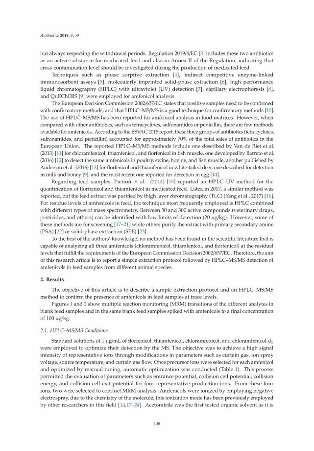
Antibiotics 2019, 8, 59
but always respecting the withdrawal periods. Regulation 2019/4/EC [3] includes these two antibioticsas an active substance for medicated feed and also in Annex II of the Regulation, indicating thatcross-contamination level should be investigated during the production of medicated feed.
Techniques such as phase sorptive extraction [4], indirect competitive enzyme-linkedimmunosorbent assays [5], molecularly imprinted solid-phase extraction [6], high performanceliquid chromatography (HPLC) with ultraviolet (UV) detection [7], capillary electrophoresis [8],and QuEChERS [9] were employed for amfenicol analysis.
The European Decision Commission 2002/657/EC states that positive samples need to be confirmedwith confirmatory methods, and that HPLC–MS/MS is a good technique for confirmatory methods [10].The use of HPLC–MS/MS has been reported for amfenicol analysis in food matrices. However, whencompared with other antibiotics, such as tetracyclines, sulfonamides or penicillin, there are few methodsavailable for amfenicols. According to the ESVAC 2015 report, these three groups of antibiotics (tetracyclines,sulfonamides, and penicillin) accounted for approximately 70% of the total sales of antibiotics in theEuropean Union. The reported HPLC–MS/MS methods include one described by Van de Riet et al.(2013) [11] for chloramfenicol, thiamfenicol, and florfenicol in fish muscle, one developed by Barreto et al.(2016) [12] to detect the same amfenicols in poultry, swine, bovine, and fish muscle, another published byAnderson et al. (2016) [13] for florfenicol and thiamfenicol in white-tailed deer, one described for detectionin milk and honey [9], and the most recent one reported for detection in egg [14].
Regarding feed samples, Pietron et al. (2014) [15] reported an HPLC–UV method for thequantification of florfenicol and thiamfenicol in medicated feed. Later, in 2017, a similar method wasreported, but the feed extract was purified by thigh layer chromatography (TLC) (Yang et al., 2017) [16].For residue levels of amfenicols in feed, the technique most frequently employed is HPLC combinedwith different types of mass spectrometry. Between 30 and 300 active compounds (veterinary drugs,pesticides, and others) can be identified with low limits of detection (20 μg/kg). However, some ofthese methods are for screening [17–21] while others purify the extract with primary secondary amine(PSA) [22] or solid-phase extraction (SPE) [23].
To the best of the authors’ knowledge, no method has been found in the scientific literature that iscapable of analyzing all three amfenicols (chloramfenicol, thiamfenicol, and florfenicol) at the residuallevels that fulfill the requirements of the European Commission Decision 2002/657/EC. Therefore, the aimof this research article is to report a simple extraction protocol followed by HPLC–MS/MS detection ofamfenicols in feed samples from different animal species.
2. Results
The objective of this article is to describe a simple extraction protocol and an HPLC–MS/MSmethod to confirm the presence of amfenicols in feed samples at trace levels.
Figures 1 and 2 show multiple reaction monitoring (MRM) transitions of the different analytes inblank feed samples and in the same blank feed samples spiked with amfenicols to a final concentrationof 100 μg/kg.
2.1. HPLC–MS/MS Conditions
Standard solutions of 1 μg/mL of florfenicol, thiamfenicol, chloramfenicol, and chloramfenicol-d5
were employed to optimize their detection by the MS. The objective was to achieve a high signalintensity of representative ions through modifications in parameters such as curtain gas, ion sprayvoltage, source temperature, and curtain gas flow. Once precursor ions were selected for each amfenicoland optimized by manual tuning, automatic optimization was conducted (Table 1). This processpermitted the evaluation of parameters such as entrance potential, collision cell potential, collisionenergy, and collision cell exit potential for four representative production ions. From these fourions, two were selected to conduct MRM analysis. Amfenicols were ionized by employing negativeelectrospray, due to the chemistry of the molecule; this ionization mode has been previously employedby other researchers in this field [14,17–24]. Acetonitrile was the first tested organic solvent as it is
108
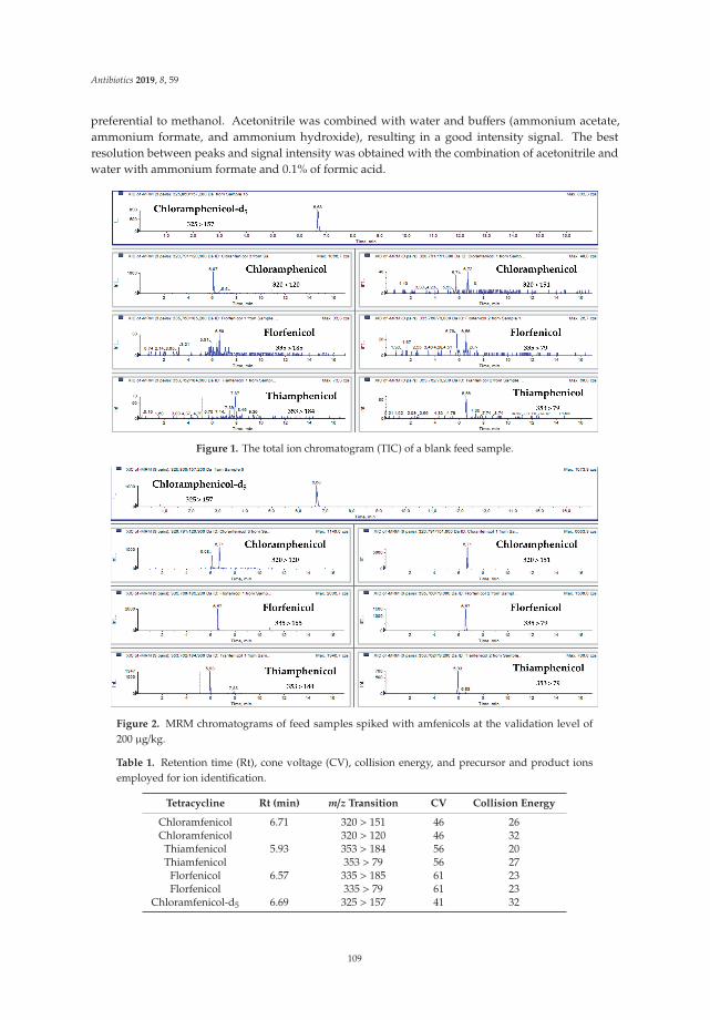
Antibiotics 2019, 8, 59
preferential to methanol. Acetonitrile was combined with water and buffers (ammonium acetate,ammonium formate, and ammonium hydroxide), resulting in a good intensity signal. The bestresolution between peaks and signal intensity was obtained with the combination of acetonitrile andwater with ammonium formate and 0.1% of formic acid.
Figure 1. The total ion chromatogram (TIC) of a blank feed sample.
Figure 2. MRM chromatograms of feed samples spiked with amfenicols at the validation level of200 μg/kg.
Table 1. Retention time (Rt), cone voltage (CV), collision energy, and precursor and product ionsemployed for ion identification.
Tetracycline Rt (min) m/z Transition CV Collision Energy
Chloramfenicol 6.71 320 > 151 46 26Chloramfenicol 320 > 120 46 32
Thiamfenicol 5.93 353 > 184 56 20Thiamfenicol 353 > 79 56 27
Florfenicol 6.57 335 > 185 61 23Florfenicol 335 > 79 61 23
Chloramfenicol-d5 6.69 325 > 157 41 32
109
Antibiotics 2019, 8, 59
Following the requirement of the Decision 2002/657/EC, four identification points were achievedwith the use of two MRM transitions for each amfenicol and the internal standard (IS). While bothMRM transitions gave identification information, quantification was implemented from the MRM thatgave the higher signal to noise ratio (S/N). The ions selected for chloramfenicol and florfenicol were alsoemployed by Cronly et al. (2010) [25] and by Shinoda et al. (2011) [26] for detection in feed matrices.Similar results occurred between the selected ion for florfenicol and the work described by Shinoda et al.(2011) and Cronly et al. (2010), though the latter reference does not include florfenicol in its method.The same ions have also employed in other type of matrices, such as wastewater [27], manure [27],poultry tissues [28], pork, porcine liver, porcine kidney, beef, bovine liver, fish, chicken [29], milk,and honey [9]. The chromatography separation was performed with various C18 columns, includingLiChrospher, Symmetry Shield RP18, ZORBAX Eclipse Plus, XTerra C18, and Hypersil C18-BD. For thepresent work, Sunfire C18 columns were selected.
2.2. Extraction
Independently of the matrix, most methods described in the scientific literature employ ethylacetate to extract amfenicols [12,24,28,30]. Xiao et al. (2015) also employed ethyl acetate for poultrytissue but with a pressurized liquid extraction system [28]. Acetone and dichloromethane were selectedby Van de Riet et al. (2013) to extract the drugs from fish muscle [11]. Acetonitrile with formic acidwas employed by Boix et al. (2014) in feed samples and water combined with buffered acetonitrileby Leon et al. (2016) also for feed samples [21]. More sophisticated extraction techniques, such asQuEChERS, were employed by Liu et al. (2016) for milk and honey [9]. An immunoaffinity column wasused by Luo et al. (2010) [31] for swine muscle and imprinted polymer by Ge et al. (2010) for animaltissue [6]. For the extraction of trace-level amfenicols from feed samples, 2 mL of water and 5 mL ofethyl acetate were employed with satisfactory results. However, after shaking for 20 minutes andcentrifugation for the separation of the different phases, only 2 mL of the organic layer was evaporated.The evaporation of greater volumes (3, 3.5, and 4 mL) was investigated to increase the limit of detection(LOD), however, a higher matrix effect was observed, and the idea discarded.
2.3. Method Validation
The research group has developed, validated, and published various methods for the analysis oftrace levels of veterinary drugs (coccidiostats, sulfonamides, and other antibiotics) in animal food andfeed samples [32–35]. Validation for this type of sample and the matrix has typically been conductedfollowing the European guideline (Decision 2002/657/EC) [10], which permits method implementationand comparisons in EU reference laboratories.
The Decision 2002/657/EC indicates that when no certificate reference material is available, truenessof measurements is assessed through recovery of additions of knowns amount of the analytes to ablank matrix. For amfenicols, recoveries were between −20% and +10% for the three investigatedanalytes (Table 2).
As with recovery, the precision under conditions of repeatability and reproducibility wereevaluated using a standard addition method. Precision values were within the range provided by theEuropean Decision; they ranged from 12% to 21% for chloramfenicol, 6.5% to 22% for thiamfenicol,and 12% to 19% for florfenicol. Currently, the presence of residue of amfenicols in non-target feed isprohibited; therefore, CCα and CCβ were calculated through employing formula for non-permittedsubstances. After peak identification, the area was plotted against the added concentration. CCα
was the corresponding concentration at the y-intercept plus 2.33 times the standard deviation of thewithin-laboratory reproducibility. Similarly, for CCβ calculation, the peak area was plotted against theadded concentration, and the CCα plus 1.64 times the standard deviation of the within-laboratoryreproducibility of the mean measured content at the CCα equals the CCβ (2002/657/EC). While CCα
values were 108, 140, and 110 μg/kg for chloramfenicol, thiamfenicol, and florfenicol, respectively,CCβ values were 116, 180, and 122 μg/kg for chloramfenicol, thiamfenicol, and florfenicol, respectively.
110
Antibiotics 2019, 8, 59
For the selected analytes, the S/N at the CCα was around 100; values for LOD and limit of quantification(LOQ) were calculated with the feed samples spiked with amfenicols. Results indicated that the LODand LOQ values could reach 50 μg/kg. However, to further validate the method, validation wasconducted at 200 μg/kg to confirm satisfactory S/N at the CCα level of each compound and to fulfillthe EU requirements.
The technique itself is very selective/specific as it uses an MRM detection mode; the use of two MRMtransitions reduces the detection of other interfering peaks. Selective/specific proxies were investigatedwith 20 blank feed samples for different animal species. The same 20 samples were spiked at 100 μg/kgwith amfenicols and other antibiotics (sulfadiazine, trimethoprim, tetracycline, and ciprofloxacin).Both the absence of interfering peaks at the Rt of the amfenicols in the two MRM transitions of eachanalyte and the correct identification of the analytes demonstrate the selectivity/specificity of thedeveloped method.
Table 2. Recovery (%), repeatability (CV%), reproducibility (CV%), CCα (μg/kg), CCβ (μg/kg), LOD(μg/kg) and LOQ (μg/kg) of Chloramfenicol, Tiamfenicol and Florfenicol.
FortificationLevel (μg/kg)
Recovery(n = 18)
Repeatability(n = 6)
Reproducibility(n = 18)
CCα CCβ LOD LOQ
Chloramfenicol
100 81 21 16 108 116 25 40
200 88 12 16
300 89 15 19
Mean 86 16 17
Tiamfenicol
100 81.41 6.5 22.35 140 180 75 100
200 97.69 12.58 21.61
300 103.83 18.97 21.98
Mean 94.31 12.69 21.98
Florfenicol
100 96.61 14.64 18.77 110 122 50 75
200 94.680 12.97 12.32
300 90.342 13.22 15.22
Mean 93.88 13.61 15.44
2.4. Real Sample Collection and Analysis
Interlaboratory studies gave satisfactory results indicating the reliability and applicability of thedeveloped method. Furthermore, the presence of the three amfenicols was investigated in 30 feedsamples from feed mills. Florfenicol was the only amfenicol detected, and it was detected in oneindividual sample at a concentration of 0.36 mg/kg. This sample belonged to a group of four samplesused to monitor florfenicol carryover after the production of a bath of medicated feed with 80 mg/kg offlorfenicol. These results indicated that florfenicol carryover may occur during the manufacture ofmedicated feed and should be investigated in more detail. None of the samples collected from pigfarms gave positive results.
3. Materials and Methods
3.1. Chemicals, Reagents, and Stock Solutions
Florfenicol, thiamfenicol, chloramfenicol, and chloramfenicol-d5 (purity > 98%) were purchasedfrom Sigma-Aldrich (St. Louis, MO, USA). Chloramfenicol-d5 was employed as internal standard (IS).Ethyl acetate and acetonitrile was obtained from Scharlau Chemie (Barcelona, Spain), and formic acidand ammonium acetate (purity >99% for analysis) were obtained from Acros Organics (Geel, Belgium).Purified water was prepared in-house with a Milli-Q water system from Millipore (Bedford, MA, USA).
111
Antibiotics 2019, 8, 59
Nitrogen gas was generated using an in-house nitrogen generator from Peak Scientific Instruments,Ltd (Chicago, IL, USA).
In 20 mL volumetric flasks, 20 ± 0.01 mg of each analyte (florfenicol, thiamfenicol, chloramfenicol,and chloramfenicol-d5) were precisely weighed to prepare stock solutions of individual compounds inacetonitrile (to yield a final concentration of 1 mg/mL; the purity was considered when calculatingspecific concentrations). To prepare the intermediate stock solution (10 μg/mL), the individual solutionswere mixed and diluted with acetonitrile. Each day, a working stock solution (1 μg/mL) was freshlyprepared by diluting the intermediate stock solution with acetonitrile. All standards solutions werestored in the dark at −18 ◦C for no longer than three months.
3.2. Conditions for HPLC–MS/MS Analysis
The HPLC–MS/MS analyses were performed using a 1100 HPLC from Agilent Technologies(Waldbronn, Germany) attached to a QTRAP 2000™MS from Applied Biosystems/MDS-Sciex (Toronto,Canada). The software Analyst 1.4.1 from Applied Biosystems (Toronto, Canada) was employed tocontrol the system.
The analysis of the extracts with amfenicols was achieved with a Sunfire C18 (3.5 μm 2.1 ×150 mm) HPLC column from Waters (Milford, PA, USA), water with formic acid (185 μL), and 370 μL ofammonium formate (6.3% in water; mobile phase A), and acetonitrile (mobile phase B). Mobile phasecomponents A and B were freshly prepared with each batch of sample. The solvents were mixed at aconstant flow rate of 250 μL/min, the column temperature was maintained at 50 ◦C, and the injectionvolume was 40 μL. The gradient program was as follows: 0 min, 90% A; 1 min, 50% A; 5 min, 20% A;7 min, 90% A; 17 min, 90% A.
The MS analysis was performed using negative electrospray ionization mode (ESI). The dwelltime was 150 ms between transitions. The system uses nitrogen as nebulized gas set up at 50 psi for ionsource gases 1 and 2. The source temperature was 475 ◦C, the ion spray voltage was −4500 V, and thecurtain gas flow was 25 psi. Analytes were identified by retention time (Rt) and two multiple reactionmonitoring (MRM) transitions. Table 1. compiles the MS parameters employed for the identification ofeach analyte.
3.3. Sample Extraction
To confirm the absence of chloramfenicol, feed samples were sent to an accredited laboratory.Once the results were obtained, these samples were employed as blank feed samples to preparematrix-matched calibration.
Quantification of the analytes was performed with matrix-matched samples spiked with theanalytes at concentrations of 100, 200, 300, 400, and 1000 μg/kg. Samples were ground on a MinimokaGR-020 grinder (Lleida, Spain), and 1 g was transferred to a 15 mL Falcon conical-bottom tube.Then, 2 mL of Milli-Q water and 5 mL of ethyl acetate were added to the tube. The mixture was mixedwith an IKA Minishaker MS2 (Staufen, Germany) for 20 s, shaken for 30 min at 200 rpm on a NewBrunswick Scientific G25 orbital shaker (NJ, USA), and centrifuged at 7000 rpm for 10 minutes ona Kokusan H-103N centrifuge (Tokyo, Japan). Then, a volume of 2 mL was transferred to a conicalglass tube and evaporated to dryness with a stream of nitrogen on a TurboVap® II evaporator fromZymark (MA, USA). The dry residue was dissolved in 500 μL of Milli-Q water, filtered with a GHPAcrodisc syringe filter (0.2 μm; Waters Corporation, MA, USA), and transferred to an HPLC amber vialwith an insert. The extract was stored at −20 ◦C until analysis, which was conducted within a day.Figure 1 shows the total ion chromatogram (TIC) of a blank feed sample, and Figure 2 shows MRMchromatograms of feed samples spiked with amfenicols at the validation level of 200 μg/kg.
3.4. Method Validation
Linearity range, recovery, precision (repeatability and reproducibility), selectivity/specificity,limit of detection (LOD), limit of quantification (LOQ), decision limit (CCα), and detection capability
112
Antibiotics 2019, 8, 59
(CCβ) of the developed method were obtained, along with validation of the method. The validationwas conducted following the criteria included in the Commission Decision 657/2002/EC. Blank feedsamples were spiked with the amfenicols at 100, 200, 300, 400, and 1000 μg/kg for linearity verification.Calibration curves were prepared over four different days; for each day, peak area was correlatedagainst analyte concentrations for linear regression analysis. The accuracy of the method could not bedetermined as certified reference materials were no available. Therefore, trueness and precision wereobtained with blank feed samples spiked at 100, 200, and 300 μg/kg. Six replicates of each concentrationwere extracted and analyzed on the same day to obtain intra-day precision (repeatability). The sameprocedure was repeated over three different days for inter-day precision (reproducibility).
The selectivity/specificity was evaluated with blank feed samples spiked and non-spiked withamfenicols at the validation level (100 μg/kg). The different feed samples were provided by themanufacturers. Selectivity/specificity was evaluated with feed samples for different animal species(swine, poultry, and cattle).
3.5. Sample Collection and Analysis
Feed producers provided non-targeted feed samples (n = 30) to the laboratory. Four of thesesamples belonged to a feed mill and were collected after being manufactured in the same production lineof medicated feed and after the cleaning batches to evaluate carryover contamination. The other feedsamples were collected in pig farms and were feed employed for pig crowing. Once in the laboratory,all samples were ground and keep in plastic bottles in the dark until analysis. Interlaboratory samplesfrom 2018 RIKILT proficiency tests were also conducted.
4. Conclusions
The article describes a simple a rapid confirmatory method based on HPLC–MS/MS for thesimultaneous identification and quantification of residue of chloramfenicol, thiamfenicol and florfenicolin non-target feed. Since the method was validated following the EU guidelines and fulfilledthe requirements of the Decision, the method can be applied by different laboratories, includingreference laboratories.
Author Contributions: Investigation, R.E.G. and C.N.; Methodology, R.E.G. and E.P.; Supervision, J.M.M. andA.C.; Validation, C.N.; Writing—original draft, C.N. and B.V.
Funding: This research was funded by FEADER (The European Agricultural Fund for Rural Development(EAFRD)), grant number 2018/001B and ED431C 2018/05.
Conflicts of Interest: The authors do not have any conflict of interest.
References
1. ESVAC (2015) Fifth ESVAC Report—Sales of Veterinary Antimicrobial Agents in 26 EU/EEA Countries in 2013.Available online: www.ema.europa.eu/docs/en_GB/document_library/Report/2015/10/WC500195687.pdf(accessed on 29 January 2017).
2. Europe Comission. Regulation 37/2010. Commission Regulation (EU) No 37/2010 of 22 December 2009 onpharmacologically active substances and their classification regarding maximum residue limits in foodstuffsof animal origin. Off. J. Eur. Union 2010, 15, 1–72.
3. Europe Comission. Regulation 2019/4. Commission Regulation (EU) 2019/4 of the European Parliamentand of the Council of 11 December 2018 on the manufacture, placing on the market and use of medicatedfeed, amending Regulation (EC) No 183/2005 of the European Parliament and of the Council and repealingCouncil Directive 90/167/EEC (Text with EEA relevance). Off. J. Eur. Union 2019, 4, 1–23.
4. Samanidou, V.; Kaltzi, I.; Kabir, A.; Furton, K.G. Simplifying sample preparation using fabric phase sorptiveextraction technique for the determination of benzodiazepines in blood serum by high performance liquidchromatography. Biomed. Chromatogr. 2016, 30, 829–836. [CrossRef] [PubMed]
113
Antibiotics 2019, 8, 59
5. An, L.; Wang, Y.; Pan, Y.; Tao, Y.; Chen, D.; Liu, Z.; Yuan, Z. Development and validation of a sensitiveindirect competitive enzyme-linked immunosorbent assay for the screening of florfenicol and Tiamfenicol inedible animal tissue and feed. Food Anal. Method 2016, 9, 2434–2443. [CrossRef]
6. Ge, S.; Yan, M.; Cheng, X.; Zhang, C.; Yu, J.; Zhao, P.; Gao, W. On-line molecular imprintedsolid-phase extraction flow-injection fluorescence sensor for determination of florfenicol in animal tissues.J. Pharm. Biomed. 2010, 52, 615–619. [CrossRef] [PubMed]
7. Hayes, J. Determination of florfenicol in fish feeds at high inclusion rates by HPLC-UV. J. AOAC Int. 2013, 96,7–11. [CrossRef] [PubMed]
8. Kowalski, P.; Konieczna, L.; Chmielewska, A.; Oledzka, I.; Plenis, A.; Bieniecki, M.; Lamparczyk, H.Comparative evaluation between capillary electrophoresis and high-performance liquid chromatography forthe analysis of florfenicol in plasma. J. Pharm. Biomed. 2005, 39, 983–989. [CrossRef]
9. Liu, H.Y.; Lin, S.L.; Fuh, M.R. Determination of chloramfenicol, Tiamfenicol and florfenicol in milk andhoney using modified QuEChERS extraction coupled with polymeric monolith-based capillary liquidchromatography tandem mass spectrometry. Talanta 2016, 150, 233–239. [CrossRef] [PubMed]
10. Europe Comission. Comission Decission 2002/657/EC. Commission Decision of 12 August 2002 implementingCouncil Directive 96/23/EC concerning the performance of analytical methods and the interpretation ofresults. Off. J. Eur. Union 2002, 221, 8–36.
11. Van de Riet, J.M.; Potter, R.A.; Christie-Fougere, M.; Burns, B.G. Simultaneous determination of residuesof chloramfenicol, tiamfenicol, florfenicol, and florfenicol amine in farmed aquatic species by liquidchromatography/mass spectrometry. J. AOAC Int. 2003, 86, 510–514. [PubMed]
12. Barreto, F.; Ribeiro, C.; Hoff, R.B.; Dalla Costa, T. Determination of chloramfenicol, Tiamfenicol, florfenicoland florfenicol amine in poultry, swine, bovine and fish by liquid chromatography-tandem mass spectrometry.J. Chromatogr. A 2016, 1449, 48–53. [CrossRef]
13. Anderson, S.C.; Subbiah, S.; Gentles, A.; Austin, G.; Stonum, P.; Brooks, T.A.; Smith, E.E. Qualitative andquantitative drug residue analyses: Florfenicol in white-tailed deer (Odocoileus virginianus) and supermarketmeat by liquid chromatography tandem-mass spectrometry. J. Chromatogr. B 2016, 1033, 73–79. [CrossRef][PubMed]
14. Xie, X.; Wang, B.; Pang, M.; Zhao, X.; Xie, K.; Zhang, Y.; Dai, G. Quantitative analysis of chloramfenicol,tiamfenicol, florfenicol and florfenicol amine in eggs via liquid chromatography-electrospray ionizationtandem mass spectrometry. Food Chem. 2018, 269, 542–548. [CrossRef]
15. Pietro, W.J.; Wozniak, A.; Pasik, K.; Cybulski, W.; Krasucka, D. Amfenicols stability in medicatedfeed–development and validation of liquid chromatography method. Bull. Vet. Inst. Pulawy 2014,58, 621–629. [CrossRef]
16. Yang, J.; Sun, G.; Qian, M.; Huang, L.; Ke, X.; Yang, B. Development of a high-performance liquidchromatography method for the determination of florfenicol in animal feedstuffs. J. Chromatogr. B 2017, 1068,9–14. [CrossRef] [PubMed]
17. Boix, C.; Ibáñez, M.; Sancho, J.V.; León, N.; Yusá, V.; Hernández, F. Qualitative screening of 116 veterinarydrugs in feed by liquid chromatography–high resolution mass spectrometry: Potential application toquantitative analysis. Food Chem. 2014, 160, 313–320. [CrossRef]
18. Robert, C.; Gillard, N.; Brasseur, P.Y.; Ralet, N.; Dubois, M.; Delahaut, P. Rapid multiresidue and multi-classscreening for antibiotics and benzimidazoles in feed by ultra-high performance liquid chromatographycoupled to tandem mass spectrometry. Food Control 2015, 50, 509–515. [CrossRef]
19. Aguilera-Luiz, M.M.; Romero-González, R.; Plaza-Bolaños, P.; Vidal, J.L.M.; Frenich, A.G. Wide-scope analysisof veterinary drug and pesticide residues in animal feed by liquid chromatography coupled toquadrupole-time-of-flight mass spectrometry. Anal. Bioanal. Chem. 2013, 405, 6543–6553. [CrossRef][PubMed]
20. Gómez-Pérez, M.L.; Romero-González, R.; Martínez Vidal, J.L.; Garrido Frenich, A. Analysis of veterinarydrug and pesticide residues in animal feed by high-resolution mass spectrometry: Comparison betweentime-of-flight and Orbitrap. Food Addit. Contam. A 2015, 32, 1637–1646. [CrossRef]
21. León, N.; Pastor, A.; Yusà, V. Target analysis and retrospective screening of veterinary drugs, ergot alkaloids,plant toxins and other undesirable substances in feed using liquid chromatography–high resolution massspectrometry. Talanta 2016, 149, 43–52. [CrossRef]
114
Antibiotics 2019, 8, 59
22. Boscher, A.; Guignard, C.; Pellet, T.; Hoffmann, L.; Bohn, T. Development of a multi-class method forthe quantification of veterinary drug residues in feedingstuffs by liquid chromatography-tandem massspectrometry. J. Chromatogr. A 2010, 1217, 6394–6404. [CrossRef]
23. Piatkowska, M.; Jedziniak, P.; Zmudzki, J. Multiresidue method for the simultaneous determination ofveterinary medicinal products, feed additives and illegal dyes in eggs using liquid chromatography–tandemmass spectrometry. Food Chem. 2016, 197, 571–580. [CrossRef]
24. Zhang, S.; Liu, Z.; Guo, X.; Cheng, L.; Wang, Z.; Shen, J. Simultaneous determination andconfirmation of chloramfenicol, tiamfenicol, florfenicol and florfenicol amine in chicken muscle by liquidchromatography–tandem mass spectrometry. J. Chromatogr. B 2008, 875, 399–404. [CrossRef]
25. Cronly, M.; Behan, P.; Foley, B.; Malone, E.; Martin, S.; Doyle, M.; Regan, L. Rapid multi-class multi-residuemethod for the confirmation of chloramfenicol and eleven nitroimidazoles in milk and honey by liquidchromatography-tandem mass spectrometry (LC-MS). Food Addit. Contam. 2010, 27, 1233–1246. [CrossRef][PubMed]
26. Shinoda, N.; Kojima, F.; Sugiura, K. Simultaneous determination of residues of chloramfenicol and florfenicolin animal feed by liquid chromatography tandem mass spectrometry. J. Resid. Sci. Technol. 2011, 8, 125–129.
27. Wei, R.; Ge, F.; Chen, M.; Wang, R. Occurrence of ciprofloxacin, enrofloxacin, and florfenicol in animalwastewater and water resources. J. Environ. Qual. 2012, 41, 1481–1486. [CrossRef]
28. Xiao, Z.; Song, R.; Rao, Z.; Wei, S.; Jia, Z.; Suo, D.; Fan, X. Development of a subcritical water extractionapproach for trace analysis of chloramfenicol, tiamfenicol, florfenicol, and florfenicol amine in poultry tissues.J. Chromatogr. A 2015, 1418, 29–35. [CrossRef]
29. Chou, K.Y.; Cheng, T.Y.; Chen, C.M.; Hung, P.L.; Tang, Y.Y.; Chung-Wang, Y.J.; Shih, Y.C.Simultaneous determination of residual tiamfenicol and florfenicol in foods of animal origin byHPLC/electrospray ionization-MS/MS. J. AOAC Int. 2009, 92, 1225–1232. [PubMed]
30. Peng, L.I.; Yueming, Q.I.U.; Huixia, C.A.I.; Ying, K.O.N.G.; Yingzhang, T.A.N.G.; Daning, W.A.N.G.;Mengxia, X.I.E. Simultaneous determination of chloramfenicol, tiamfenicol, and florfenicol residues in animaltissues by gas chromatography/mass spectrometry. Chin. J. Chromatogr. 2006, 24, 14–18.
31. Luo, P.; Chen, X.; Liang, C.; Kuang, H.; Lu, L.; Jiang, Z.; Shen, J. Simultaneous determination of thiamfenicol,florfenicol and florfenicol amine in swine muscle by liquid chromatography–tandem mass spectrometrywith immunoaffinity chromatography clean-up. J. Chromatogr. B 2010, 878, 207–212. [CrossRef] [PubMed]
32. Nebot, C.; Iglesias, A.; Regal, P.; Miranda, J.; Cepeda, A.; Fente, C. Development of a multi-class method forthe identification and quantification of residues of antibiotics, coccidiostats and corticosteroids in milk byliquid chromatography–tandem mass spectrometry. Int. Dairy J. 2012, 22, 78–85. [CrossRef]
33. Nebot, C.; Guarddon, M.; Seco, F.; Iglesias, A.; Miranda, J.M.; Franco, C.M.; Cepeda, A. Monitoring thepresence of residues of tetracyclines in baby food samples by HPLC-MS/MS. Food Control 2014, 46, 495–501.[CrossRef]
34. Gavilán, R.; Nebot, C.; Miranda, J.; Martín-Gómez, Y.; Vázquez-Belda, B.; Franco, C.; Cepeda, A. Analysis oftetracyclines in medicated feed for food animal production by HPLC-MS/MS. Antibiotics 2016, 5, 1. [CrossRef][PubMed]
35. Gavilán, R.E.; Nebot, C.; Patyra, E.; Miranda, J.M.; Franco, C.M.; Cepeda, A. Simultaneous analysisof coccidiostats and sulphonamides in non-target feed by HPLC-MS/MS and validation following theCommission Decision 2002/657/EC. Food Addit. Contam. A 2018, 35, 1093–1106. [CrossRef] [PubMed]
© 2019 by the authors. Licensee MDPI, Basel, Switzerland. This article is an open accessarticle distributed under the terms and conditions of the Creative Commons Attribution(CC BY) license (http://creativecommons.org/licenses/by/4.0/).
115

antibiotics
Article
Antimicrobial and Synergistic Effects of CommercialPiperine and Piperlongumine in Combination withConventional Antimicrobials
Eunice Ego Mgbeahuruike 1,*, Milla Stålnacke 2, Heikki Vuorela 1 and Yvonne Holm 1
1 Division of Pharmaceutical Biosciences, Faculty of Pharmacy, University of Helsinki, P.O. Box 56,FI-00014 Helsinki, Finland; [email protected] (H.V.); [email protected] (Y.H.)
2 Department of Pharmacology, Institute of Neuroscience and Physiology, The Sahlgrenska Academy,University of Gothenburg, Box 431, SE-40530 Gothenburg, Sweden; [email protected]
* Correspondence: [email protected] or [email protected]; Tel.: +358442399653
Received: 15 April 2019; Accepted: 2 May 2019; Published: 4 May 2019
Abstract: Microbial resistance to currently available antibiotics is a public health problem in thefight against infectious diseases. Most antibiotics are characterized by numerous side effects thatmay be harmful to normal body cells. To improve the efficacy of these antibiotics and to find analternative way to minimize the adverse effects associated with most conventional antibiotics, piperineand piperlongumine were screened in combination with conventional rifampicin, tetracycline, anditraconazole to evaluate their synergistic, additive, or antagonistic interactions against Staphylococcusaureus, Pseudomonas aeruginosa, and Candida albicans. The fractional inhibitory concentration indexwas used to estimate the synergistic effects of various combination ratios of the piperamides andantibiotics against the bacterial and fungal strains. Both piperine and piperlongumine showedsynergistic effects against S. aureus when combined at various ratios with rifampicin. Synergisticinteraction was also observed with piperine in combination with tetracycline against S. aureus, whileantagonistic interaction was recorded for piperlongumine and tetracycline against S. aureus. All thepiperamide/antibacterial combinations tested against P. aeruginosa showed antagonistic effects, withthe exception of piperine and rifampicin, which recorded synergistic interaction at a ratio of 9:1rifampicin/piperine. No synergistic interaction was observed when the commercial compounds werecombined with itraconazole and tested against C. albicans. The results showed that piperine andpiperlongumine are capable of improving the effectiveness of rifampicin and tetracycline. Dosagecombinations of these bioactive compounds with the antibiotics used may be a better option for thetreatment of bacterial infections that aims to minimize the adverse effects associated with the use ofthese conventional antibacterial drugs.
Keywords: piperine; piperlongumine; antibacterial; antifungal; synergy
1. Introduction
Infectious diseases caused by bacteria and fungi are one of the leading causes of deathworldwide [1–3]. The emergence of multidrug resistance in microbes and the nonavailability ofantibiotics to combat and treat microbial infections have led to a constant search for new antimicrobialagents from natural sources. Previous research has shown that about 13 million deaths are recordedthroughout the world each year as a result of bacterial and fungal diseases that are often caused bymultidrug-resistant pathogens [4,5]. Natural products of plant origin include novel therapeutic andhighly effective antimicrobial agents [6,7]. In clinical practice, one of the leading new advances in thefight against microbial resistance is antimicrobial combination therapy [8–10]. Alternatively, bioactivecompounds from natural sources could also be screened for leads to the discovery of new antibiotics
Antibiotics 2019, 8, 55; doi:10.3390/antibiotics8020055 www.mdpi.com/journal/antibiotics117
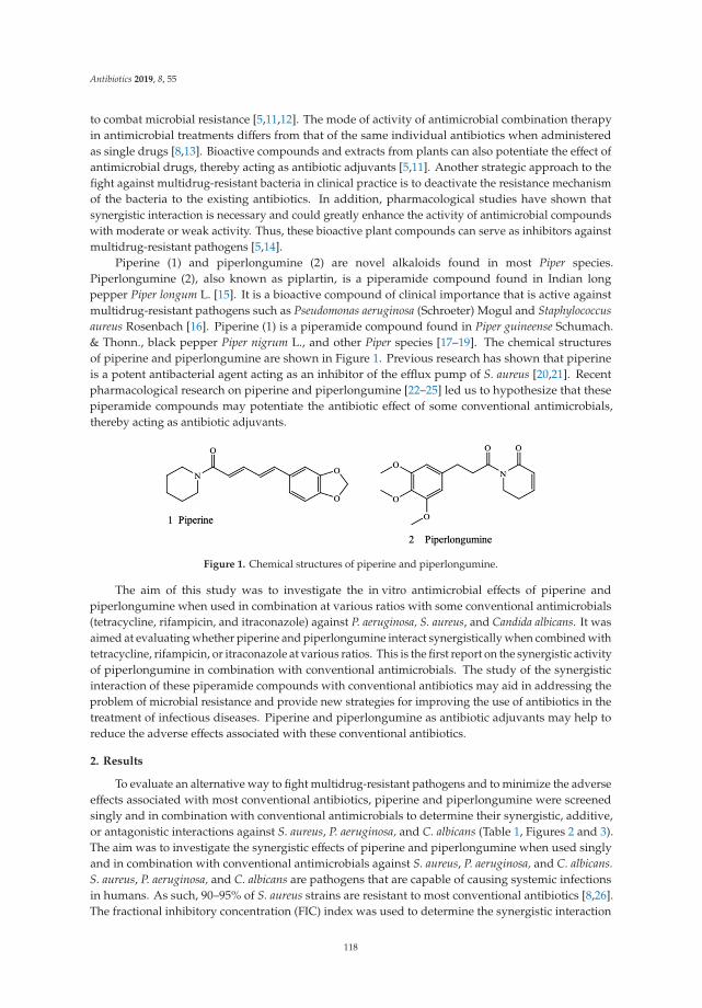
Antibiotics 2019, 8, 55
to combat microbial resistance [5,11,12]. The mode of activity of antimicrobial combination therapyin antimicrobial treatments differs from that of the same individual antibiotics when administeredas single drugs [8,13]. Bioactive compounds and extracts from plants can also potentiate the effect ofantimicrobial drugs, thereby acting as antibiotic adjuvants [5,11]. Another strategic approach to thefight against multidrug-resistant bacteria in clinical practice is to deactivate the resistance mechanismof the bacteria to the existing antibiotics. In addition, pharmacological studies have shown thatsynergistic interaction is necessary and could greatly enhance the activity of antimicrobial compoundswith moderate or weak activity. Thus, these bioactive plant compounds can serve as inhibitors againstmultidrug-resistant pathogens [5,14].
Piperine (1) and piperlongumine (2) are novel alkaloids found in most Piper species.Piperlongumine (2), also known as piplartin, is a piperamide compound found in Indian longpepper Piper longum L. [15]. It is a bioactive compound of clinical importance that is active againstmultidrug-resistant pathogens such as Pseudomonas aeruginosa (Schroeter) Mogul and Staphylococcusaureus Rosenbach [16]. Piperine (1) is a piperamide compound found in Piper guineense Schumach.& Thonn., black pepper Piper nigrum L., and other Piper species [17–19]. The chemical structuresof piperine and piperlongumine are shown in Figure 1. Previous research has shown that piperineis a potent antibacterial agent acting as an inhibitor of the efflux pump of S. aureus [20,21]. Recentpharmacological research on piperine and piperlongumine [22–25] led us to hypothesize that thesepiperamide compounds may potentiate the antibiotic effect of some conventional antimicrobials,thereby acting as antibiotic adjuvants.
Figure 1. Chemical structures of piperine and piperlongumine.
The aim of this study was to investigate the in vitro antimicrobial effects of piperine andpiperlongumine when used in combination at various ratios with some conventional antimicrobials(tetracycline, rifampicin, and itraconazole) against P. aeruginosa, S. aureus, and Candida albicans. It wasaimed at evaluating whether piperine and piperlongumine interact synergistically when combined withtetracycline, rifampicin, or itraconazole at various ratios. This is the first report on the synergistic activityof piperlongumine in combination with conventional antimicrobials. The study of the synergisticinteraction of these piperamide compounds with conventional antibiotics may aid in addressing theproblem of microbial resistance and provide new strategies for improving the use of antibiotics in thetreatment of infectious diseases. Piperine and piperlongumine as antibiotic adjuvants may help toreduce the adverse effects associated with these conventional antibiotics.
2. Results
To evaluate an alternative way to fight multidrug-resistant pathogens and to minimize the adverseeffects associated with most conventional antibiotics, piperine and piperlongumine were screenedsingly and in combination with conventional antimicrobials to determine their synergistic, additive,or antagonistic interactions against S. aureus, P. aeruginosa, and C. albicans (Table 1, Figures 2 and 3).The aim was to investigate the synergistic effects of piperine and piperlongumine when used singlyand in combination with conventional antimicrobials against S. aureus, P. aeruginosa, and C. albicans.S. aureus, P. aeruginosa, and C. albicans are pathogens that are capable of causing systemic infectionsin humans. As such, 90–95% of S. aureus strains are resistant to most conventional antibiotics [8,26].The fractional inhibitory concentration (FIC) index was used to determine the synergistic interaction
118
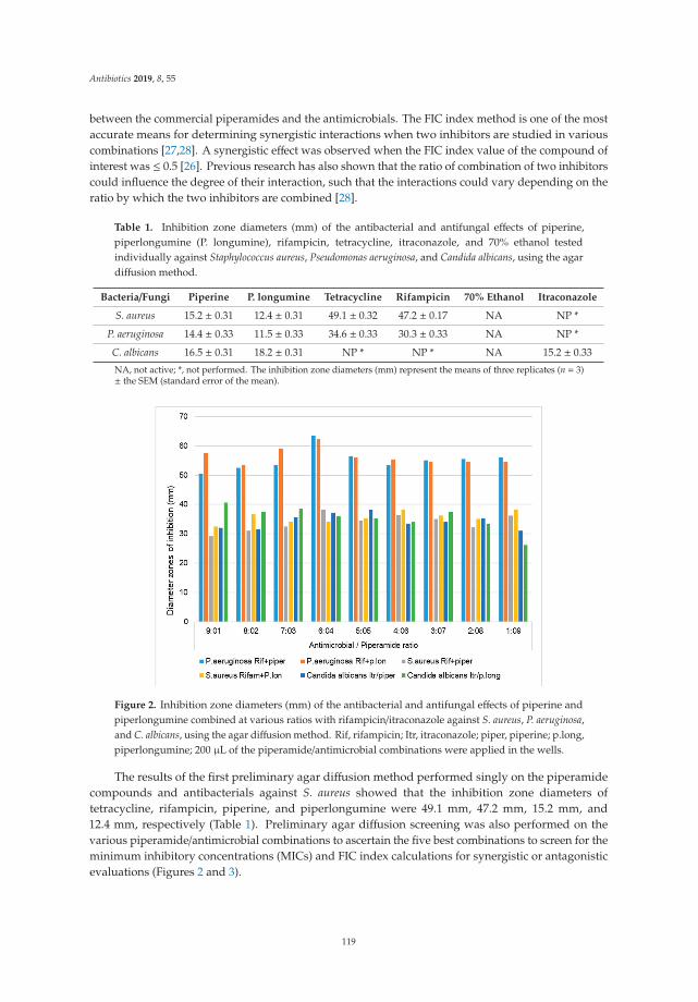
Antibiotics 2019, 8, 55
between the commercial piperamides and the antimicrobials. The FIC index method is one of the mostaccurate means for determining synergistic interactions when two inhibitors are studied in variouscombinations [27,28]. A synergistic effect was observed when the FIC index value of the compound ofinterest was ≤ 0.5 [26]. Previous research has also shown that the ratio of combination of two inhibitorscould influence the degree of their interaction, such that the interactions could vary depending on theratio by which the two inhibitors are combined [28].
Table 1. Inhibition zone diameters (mm) of the antibacterial and antifungal effects of piperine,piperlongumine (P. longumine), rifampicin, tetracycline, itraconazole, and 70% ethanol testedindividually against Staphylococcus aureus, Pseudomonas aeruginosa, and Candida albicans, using the agardiffusion method.
Bacteria/Fungi Piperine P. longumine Tetracycline Rifampicin 70% Ethanol Itraconazole
S. aureus 15.2 ± 0.31 12.4 ± 0.31 49.1 ± 0.32 47.2 ± 0.17 NA NP *
P. aeruginosa 14.4 ± 0.33 11.5 ± 0.33 34.6 ± 0.33 30.3 ± 0.33 NA NP *
C. albicans 16.5 ± 0.31 18.2 ± 0.31 NP * NP * NA 15.2 ± 0.33
NA, not active; *, not performed. The inhibition zone diameters (mm) represent the means of three replicates (n = 3)± the SEM (standard error of the mean).
Figure 2. Inhibition zone diameters (mm) of the antibacterial and antifungal effects of piperine andpiperlongumine combined at various ratios with rifampicin/itraconazole against S. aureus, P. aeruginosa,and C. albicans, using the agar diffusion method. Rif, rifampicin; Itr, itraconazole; piper, piperine; p.long,piperlongumine; 200 μL of the piperamide/antimicrobial combinations were applied in the wells.
The results of the first preliminary agar diffusion method performed singly on the piperamidecompounds and antibacterials against S. aureus showed that the inhibition zone diameters oftetracycline, rifampicin, piperine, and piperlongumine were 49.1 mm, 47.2 mm, 15.2 mm, and12.4 mm, respectively (Table 1). Preliminary agar diffusion screening was also performed on thevarious piperamide/antimicrobial combinations to ascertain the five best combinations to screen for theminimum inhibitory concentrations (MICs) and FIC index calculations for synergistic or antagonisticevaluations (Figures 2 and 3).
119
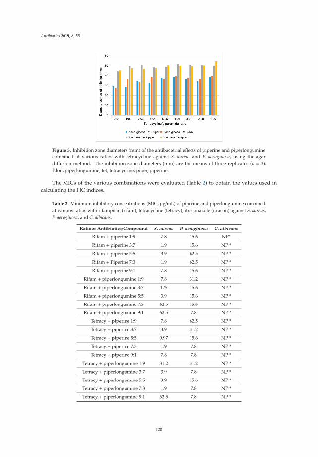
Antibiotics 2019, 8, 55
Figure 3. Inhibition zone diameters (mm) of the antibacterial effects of piperine and piperlonguminecombined at various ratios with tetracycline against S. aureus and P. aeruginosa, using the agardiffusion method. The inhibition zone diameters (mm) are the means of three replicates (n = 3).P.lon, piperlongumine; tet, tetracycline; piper, piperine.
The MICs of the various combinations were evaluated (Table 2) to obtain the values used incalculating the FIC indices.
Table 2. Minimum inhibitory concentrations (MIC, μg/mL) of piperine and piperlongumine combinedat various ratios with rifampicin (rifam), tetracycline (tetracy), itraconazole (itracon) against S. aureus,P. aeruginosa, and C. albicans.
Ratioof Antibiotics/Compound S. aureus P. aeruginosa C. albicans
Rifam + piperine 1:9 7.8 15.6 NP*
Rifam + piperine 3:7 1.9 15.6 NP *
Rifam + piperine 5:5 3.9 62.5 NP *
Rifam + Piperine 7:3 1.9 62.5 NP *
Rifam + piperine 9:1 7.8 15.6 NP *
Rifam + piperlongumine 1:9 7.8 31.2 NP *
Rifam + piperlongumine 3:7 125 15.6 NP *
Rifam + piperlongumine 5:5 3.9 15.6 NP *
Rifam + piperlongumine 7:3 62.5 15.6 NP *
Rifam + piperlongumine 9:1 62.5 7.8 NP *
Tetracy + piperine 1:9 7.8 62.5 NP *
Tetracy + piperine 3:7 3.9 31.2 NP *
Tetracy + piperine 5:5 0.97 15.6 NP *
Tetracy + piperine 7:3 1.9 7.8 NP *
Tetracy + piperine 9:1 7.8 7.8 NP *
Tetracy + piperlongumine 1:9 31.2 31.2 NP *
Tetracy + piperlongumine 3:7 3.9 7.8 NP *
Tetracy + piperlongumine 5:5 3.9 15.6 NP *
Tetracy + piperlongumine 7:3 1.9 7.8 NP *
Tetracy + piperlongumine 9:1 62.5 7.8 NP *
120
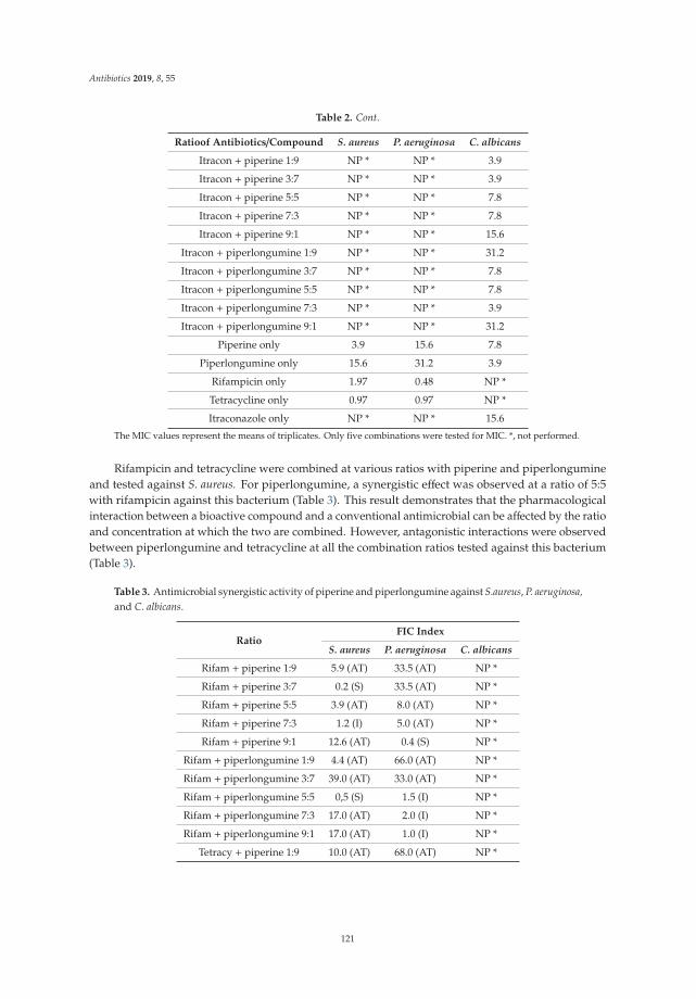
Antibiotics 2019, 8, 55
Table 2. Cont.
Ratioof Antibiotics/Compound S. aureus P. aeruginosa C. albicans
Itracon + piperine 1:9 NP * NP * 3.9
Itracon + piperine 3:7 NP * NP * 3.9
Itracon + piperine 5:5 NP * NP * 7.8
Itracon + piperine 7:3 NP * NP * 7.8
Itracon + piperine 9:1 NP * NP * 15.6
Itracon + piperlongumine 1:9 NP * NP * 31.2
Itracon + piperlongumine 3:7 NP * NP * 7.8
Itracon + piperlongumine 5:5 NP * NP * 7.8
Itracon + piperlongumine 7:3 NP * NP * 3.9
Itracon + piperlongumine 9:1 NP * NP * 31.2
Piperine only 3.9 15.6 7.8
Piperlongumine only 15.6 31.2 3.9
Rifampicin only 1.97 0.48 NP *
Tetracycline only 0.97 0.97 NP *
Itraconazole only NP * NP * 15.6
The MIC values represent the means of triplicates. Only five combinations were tested for MIC. *, not performed.
Rifampicin and tetracycline were combined at various ratios with piperine and piperlongumineand tested against S. aureus. For piperlongumine, a synergistic effect was observed at a ratio of 5:5with rifampicin against this bacterium (Table 3). This result demonstrates that the pharmacologicalinteraction between a bioactive compound and a conventional antimicrobial can be affected by the ratioand concentration at which the two are combined. However, antagonistic interactions were observedbetween piperlongumine and tetracycline at all the combination ratios tested against this bacterium(Table 3).
Table 3. Antimicrobial synergistic activity of piperine and piperlongumine against S.aureus, P. aeruginosa,and C. albicans.
RatioFIC Index
S. aureus P. aeruginosa C. albicans
Rifam + piperine 1:9 5.9 (AT) 33.5 (AT) NP *
Rifam + piperine 3:7 0.2 (S) 33.5 (AT) NP *
Rifam + piperine 5:5 3.9 (AT) 8.0 (AT) NP *
Rifam + piperine 7:3 1.2 (I) 5.0 (AT) NP *
Rifam + piperine 9:1 12.6 (AT) 0.4 (S) NP *
Rifam + piperlongumine 1:9 4.4 (AT) 66.0 (AT) NP *
Rifam + piperlongumine 3:7 39.0 (AT) 33.0 (AT) NP *
Rifam + piperlongumine 5:5 0,5 (S) 1.5 (I) NP *
Rifam + piperlongumine 7:3 17.0 (AT) 2.0 (I) NP *
Rifam + piperlongumine 9:1 17.0 (AT) 1.0 (I) NP *
Tetracy + piperine 1:9 10.0 (AT) 68.0 (AT) NP *
121
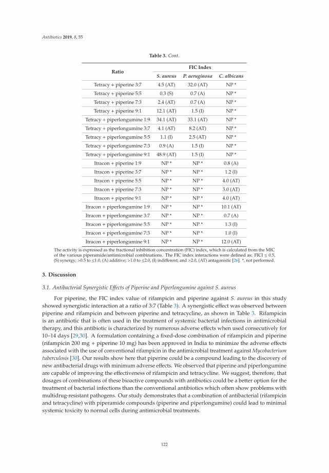
Antibiotics 2019, 8, 55
Table 3. Cont.
RatioFIC Index
S. aureus P. aeruginosa C. albicans
Tetracy + piperine 3:7 4.5 (AT) 32.0 (AT) NP *
Tetracy + piperine 5:5 0.3 (S) 0.7 (A) NP *
Tetracy + piperine 7:3 2.4 (AT) 0.7 (A) NP *
Tetracy + piperine 9:1 12.1 (AT) 1.5 (I) NP *
Tetracy + piperlongumine 1:9 34.1 (AT) 33.1 (AT) NP *
Tetracy + piperlongumine 3:7 4.1 (AT) 8.2 (AT) NP *
Tetracy + piperlongumine 5:5 1.1 (I) 2.5 (AT) NP *
Tetracy + piperlongumine 7:3 0.9 (A) 1.5 (I) NP *
Tetracy + piperlongumine 9:1 48.9 (AT) 1.5 (I) NP *
Itracon + piperine 1:9 NP * NP * 0.8 (A)
Itracon + piperine 3:7 NP * NP * 1.2 (I)
Itracon + piperine 5:5 NP * NP * 4.0 (AT)
Itracon + piperine 7:3 NP * NP * 3.0 (AT)
Itracon + piperine 9:1 NP * NP * 4.0 (AT)
Itracon + piperlongumine 1:9 NP * NP * 10.1 (AT)
Itracon + piperlongumine 3:7 NP * NP * 0.7 (A)
Itracon + piperlongumine 5:5 NP * NP * 1.3 (I)
Itracon + piperlongumine 7:3 NP * NP * 1.0 (I)
Itracon + piperlongumine 9:1 NP * NP * 12.0 (AT)
The activity is expressed as the fractional inhibition concentration (FIC) index, which is calculated from the MICof the various piperamide/antimicrobial combinations. The FIC index interactions were defined as: FICI ≤ 0.5,(S) synergy; >0.5 to ≤1.0, (A) additive; >1.0 to ≤2.0, (I) indifferent; and >2.0, (AT) antagonistic [26]. *, not performed.
3. Discussion
3.1. Antibacterial Synergistic Effects of Piperine and Piperlongumine against S. aureus
For piperine, the FIC index value of rifampicin and piperine against S. aureus in this studyshowed synergistic interaction at a ratio of 3:7 (Table 3). A synergistic effect was observed betweenpiperine and rifampicin and between piperine and tetracycline, as shown in Table 3. Rifampicinis an antibiotic that is often used in the treatment of systemic bacterial infections in antimicrobialtherapy, and this antibiotic is characterized by numerous adverse effects when used consecutively for10–14 days [29,30]. A formulation containing a fixed-dose combination of rifampicin and piperine(rifampicin 200 mg + piperine 10 mg) has been approved in India to minimize the adverse effectsassociated with the use of conventional rifampicin in the antimicrobial treatment against Mycobacteriumtuberculosis [30]. Our results show here that piperine could be a compound leading to the discovery ofnew antibacterial drugs with minimum adverse effects. We observed that piperine and piperlongumineare capable of improving the effectiveness of rifampicin and tetracycline. We suggest, therefore, thatdosages of combinations of these bioactive compounds with antibiotics could be a better option for thetreatment of bacterial infections than the conventional antibiotics which often show problems withmultidrug-resistant pathogens. Our study demonstrates that a combination of antibacterial (rifampicinand tetracycline) with piperamide compounds (piperine and piperlongumine) could lead to minimalsystemic toxicity to normal cells during antimicrobial treatments.
122
Antibiotics 2019, 8, 55
3.2. Antibacterial Synergistic Effects of Piperine and Piperlongumine against P. aeruginosa
Preliminary agar diffusion assays performed individually with piperamides and antibioticsagainst P. aeruginosa (Table 1) showed that the inhibition zone diameters of tetracycline, rifampicin,piperine, and piperlongumine were 34.6 mm, 30.3 mm, 14.4 mm, and 11.5 mm, respectively. Whenthe MIC of the various combinations was determined, the FIC index calculations showed mainlyantagonistic interactions between the piperamides and the antibiotics against P. aeruginosa, withthe exception of the 9:1 ratio of the piperine/rifampicin combination, which showed synergisticactivity (Table 3). P. aeruginosa is a Gram-negative pathogenic bacterium that causes systemicinfections in immunocompromised individuals and is often associated with antibacterial resistance [31].For piperlongumine, no synergistic effect was observed in any of the antibiotic combinations (rifampicinand tetracycline) against P. aeruginosa, although additive and antagonistic interactions were observed.The results of tetracycline/piperlongumine and rifampicin/ piperlongumine combinations tested atvarious ratios demonstrated no synergistic effects between tetracycline and piperlongumine as wellas between rifampicin and piperlongumine against P. aeruginosa. These results suggest that piperineand piperlongumine may not improve the effectiveness of tetracycline against P. aeruginosa infections.Indeed, appropriate measures should be taken to avoid too heavy a consumption of herbal drugs orfoods rich in piperine and piperlongumine when undergoing antimicrobial treatment with tetracyclineagainst diseases caused by P. aeruginosa.
For piperine, a synergistic interaction with rifampicin was observed for a rifampicin/piperinecombination at a ratio of 9:1 against P. aeruginosa, while antagonistic or additive interactions wereobserved for the other combinations tested. Piperine was previously tested for its immunomodulatoryactivity in enhancing the efficacy of rifampicin in a murine model of M. tuberculosis infection, therebyacting in synergy with rifampicin to improve its therapeutic efficacy [32]. However, the synergisticefficacy recorded in our study for a rifampicin/piperine combination showed that the efficacy could beinfluenced by the ratio of the combination, i.e, the synergistic, additive, or antagonistic interactionswere greatly dependent on the ratio between rifampicin and piperine.
3.3. Antifungal Synergistic Effects of Piperine and Piperlongumine against C. albicans
When preliminary agar diffusion assays were performed singly on the piperamides anditraconazole against C. albicans, the inhibition zone diameters of piperine, piperlongumine, anditraconazole were 16.5 mm, 18.2 mm, and 15.2 mm, respectively (Table 1). The results of the FICindex evaluations of the various combinations showed additive or antagonistic interactions (Table 3).No synergistic effects were recorded in our study against C. albicans. C. albicans is an opportunisticpathogen that is responsible for most yeast infections in humans [33]. These results demonstratethat piperine and piperlongumine cannot improve the effectiveness of itraconazole, and thus herbalformulations made from these bioactive compounds should be avoided while undergoing treatmentsfor yeast infections.
4. Materials and Methods
4.1. Sources of Antibiotics and Commercial Compounds
Analytical-grade commercial piperlongumine (≥ 97.0% purity) and piperine (≥ 97.0% purity) werepurchased from TCI Europe N.V. (Zwijndrecht, Belgium). Tetracycline hydrochloride (Sigma-Aldrich,St. Louis MO, USA), rifampicin (Sigma-Aldrich), and itraconazole (Sigma-Aldrich) were the antibioticsused in the investigation. The selection of tetracycline, rifampicin, and itraconazole was based on theiractivity against bacterial and fungal strains in a previous study [34]. S. aureus American Type CultureCollection ATCC 25923, P. aeruginosa ATCC 27853, and C. albicans ATCC 10231 cultures used in thisinvestigation were obtained from the Division of Pharmaceutical Biosciences, Faculty of Pharmacy,University of Helsinki, Finland. The bacterial strains were selected because they are significant human
123
Antibiotics 2019, 8, 55
pathogens capable of causing life-threatening nosocomial infections [35,36], while C. albicans, on theother hand, causes fungal diseases in humans [37].
4.2. Preparation of Piperine, Piperlongumine, and Antimicrobials
Each of the commercial piperamides (piperine and piperlongumine) was prepared to a finalconcentration of 5 mg/mL (stock solution) in 70% ethanol and tested in the initial screening. An initialstock solution of 5 mg/mL was prepared for each of the antibiotics (tetracycline, rifampicin, anditraconazole). Piperine and piperlongumine were also prepared in combination with tetracycline,rifampicin, or itraconazole at various ratios according to the method of Van Vuuren et al. [10].The combinations/dilutions were prepared at nine different ratios of 1:9, 2:8, 3:7, 4:6, 5:5, 6:4, 7:3,8:2, 9:1 as follows: rifampicin/piperlongumine, rifampicin/piperine, tetracycline/piperlongumine,tetracycline/piperine, itraconazole/piperine, and itraconazole/piperlongumine.
4.3. Agar Disk-Diffusion Method
The agar disk-diffusion method was applied for the initial screening of the nine combinationratios according to the method of the Clinical and Laboratory Standards Institute [38]. Sterile Petridishes (∅ = 14 cm, VWR International Oy, Helsinki, Finland) were used for the screening. For theantibacterial screening against S. aureus and P. aeruginosa, 25 mL of sterile base agar (Antibiotic agarNo. 2, Difco, VWR) was applied as a bottom layer into the sterile Petri dishes, using a sterile serologicalpipet (Falcon; BDLabware Europe) and allowed to solidify. Thereafter, 25 mL of isosensitest agar(OXOID, ThermoFisher Scientific, Waltham, MA, USA) was applied as the top layer. For the antifungalscreening, 25 mL of sterile base agar (Antibiotic agar No. 2, Difco VWR) was applied as a bottom layerinto the sterile Petri dishes and 25 mL of Saboraud agar (OXOID, ThermoFisher Scientific, Basingstoke,United Kingdom) was applied as the top layer. The agar in the Petri dishes was allowed to solidifyand then stored at +4 ◦C. The screening was initiated with the inoculation of the bacterial strains orC. albicans onto solid nutrient or Saboraud agar slants that were incubated overnight for the bacterialstrains and 48 h for the fungus at +37 ◦C. Viable bacterial and fungal cultures from the agar slantswere used to prepare an inoculum for the test. Bacterial and fungal specimens from the agar slantswere transferred into 2 mL of a 0.9% (w/v) sodium chloride (NaCl) solution in a sterile glass tube,using a sterile inoculation loop. In all, 1 mL of the suspension was transferred into another sterileglass tube, and the absorbance was measured at 625 nm (UV–Visible Spectrophotometer, PharmaciaLKB-Biochrom 4060; Pfizer Inc., New York, NY, USA). The other milliliter of the suspension (sterile part)was diluted with the 0.9% NaCl solution so that the absorbance at 625 nm became 0.1 (this suspensioncontained approximately 1.5 × 10⁸ colony-forming units (CFUs)/mL). In all, 200 μL of this dilutedbacterial or fungal suspension were spread evenly on each Petri dish and left to dry for several secondswith the lid open. A sterile cork borer (11 mm diameter) was used to make six equidistant holes onthe agar surface of the Petri dishes. A total of 200 μL of the 5 mg/mL concentration of each of thepiperamides and 200 μL of the 10 mg/mL antimicrobial were carefully pipetted into the holes; 70%ethanol was used as the solvent control. The first screening of the combined samples was done inthe same manner as described above for the agar diffusion method, and the various inhibition zoneswere recorded. The Petri dishes were transferred to the cold room and incubated at +4 ◦C for 1 h.Thereafter, they were incubated at +37 ◦C, overnight for the bacterial strains and 48 h for the fungalstrain. The diameters of the zones of inhibition were measured with a caliper under a Petri dishmagnifier and expressed as the mean of the diameters of three replicates ± the standard error of themean (SEM).
4.4. Microdilution Method for Minimum Inhibitory Concentration Estimation
The MIC was estimated for the five best combinations of each piperamide/antimicrobial. The MICwas taken as the lowest concentration of the piperamide/antimicrobial resulting in the inhibition ofat least 90% of the growth of the bacterial or fungal strain. The MIC values were determined using
124
Antibiotics 2019, 8, 55
a microdilution turbidimetric broth method based on the guidelines of the Clinical and LaboratoryStandards Institute [38]. For MIC evaluation, two-fold serial dilutions from 2500 to 9.75 μg/mLwere prepared in sterile Mueller–Hinton broth (bacterial strains) and Saboraud broth (C. albicans) foreach of the piperamide/antimicrobial combinations. The commercially pure compounds, piperineand piperlongumine (5 mg/mL concentrations in 70% ethanol), were two-fold serially diluted inMueller–Hinton broth for the bacterial strains and Saboraud broth for the fungal strain. Tetracycline,rifampicin, and itraconazole were similarly diluted in Mueller–Hinton broth or Saboraud broth from125 to 0.48 μg/mL, respectively; 96-well microtiter plates (Nunc, Nunclone; Nalge Nunc International,Roskilde, Denmark) were used for the tests. The bacterial or fungal cultures were inoculated in5 mL of Mueller–Hinton broth and grown for 24 h at +37 ◦C for the bacterial strains or in 5 mLof Saboraud broth and grown for 48 h at +37 ◦C for C. albicans before the test. Absorbance of1 mL of the overnight bacterial culture was measured for turbidity at 625 nm, using a UV–VisibleSpectrophotometer type 1510 (ThermoFisher Scientific). The absorbance was adjusted to 0.1 at 625 nm(approximately 1.0 × 10⁸ CFUs/mL). A total of 100 μL of this suspension A625 = 0.1 was furtherdiluted a hundredfold to obtain a working suspension or inoculum containing 1.0 × 10⁶ CFUs/mL.In all, 100 μL of this inoculum and 100 μL of each of thepiperamide/antimicrobial combinations andsolvent controls were introduced into the 96-well microtiter plates. Each well therefore contained5 × 105 CFUs/mL. The growth control (GC) wells contained only the bacterial suspension, and thetest (T) wells contained the piperamide/antimicrobial + bacterial suspension. Moreover, negativecontrol wells were prepared for each piperamide/antimicrobial combination ratio tested, and thesewells contained the piperamide/antimicrobial combinations at different ratios and the broth only.The microwell plates were incubated for 24 h in an incubator coupled to a shaker at+37 ◦C. The turbidityof the wells at 620 nm was recorded using a Victor 1420 spectrophotometer (PerkinElmer (Wallac Oy),Turku, Finland). The tests were done in triplicate, and the percentage growth was expressed as themean of these triplicates ± the SEM.
4.5. Fractional Inhibitory Concentration Index
The FIC index was used to estimate the synergy [5,26]. For the FIC index calculation, the MICvalues of five combinations of each piperamide/antimicrobial at different ratios were used to calculateFIC (A) and FIC (B), using the equation below:
The FIC values were calculated as follows:
FIC(A) =MICvalueo f combinedpiperamideandantimicrobial
MICvalueo f antimicrobialalone
FIC(B) = MICvalueo f combinedpiperamideandantimicrobialMICvalueo f piperamidealone
FICindex = FIC(A) + FIC(B)
A FIC index value ≤ 0.5 was interpreted as synergy. The FIC index was calculated on the basis ofthe above-mentioned equation, in which the FIC index = X + Y and the interactions are defined as:FIC index ≤ 0.5, synergy; > 0.5 to ≤ 1.0, additive; > 1.0 to ≤ 2.0, indifferent; and > 2.0, antagonistic [26].
5. Conclusions
Piperine and piperlongumine are possible antibiotic adjuvants that should be applied in drugdiscovery for use as effective antibacterial treatments. The synergistic efficacy of piperlongumine andpiperine in combination with rifampicin has shown that these two piperamides are bioactive scaffoldsthat could aid in fighting against multidrug-resistant pathogens, thereby improving the effectivenessof rifampicin in the treatment of infectious diseases. Our results show that the synergistic efficacyof piperamide compounds is dependent on the concentration and ratio of the combination. Thisindicates that the use of piperine and piperlongumine may minimize the adverse effects associated withconventional tetracycline and rifampicin and improve the effectiveness of these antibiotics. However,the antagonistic effects observed with the commercial compounds/antibiotics used against P. aeruginosa
125
Antibiotics 2019, 8, 55
in this study demonstrate that appropriate caution should be taken to avoid the consumption of herbaldrugs made from Piper species during antibacterial treatment therapies against P. aeruginosa infections.We suggest that piperine and piperlongumine could be considered as therapeutic bioactive compoundsfor antibacterial drug discovery. Further studies should be conducted on the mode of activity of thesynergistic interaction between piperamides and antibiotics.
Author Contributions: Conceptualization, E.E.M., H.V., and Y.H.; methodology, E.E.M. and M.S.; formal analysis,E.E.M., Y.H., and M.S.; resources, H.V.; writing — original draft preparation, E.E.M. and M.S.; supervision, Y.H.and H.V.; project administration, H.V.
Funding: This research received no external funding.
Acknowledgments: The authors are grateful to the Faculty of Pharmacy, Division of Pharmaceutical Biosciences,University of Helsinki, for supporting this work and providing the materials used for the experiments.
Conflicts of Interest: The authors declare no conflicts of interest.
References
1. Brown, G.D.; Denning, D.W.; Gow, N.A.; Levitz, S.M.; Netea, M.G.; White, T.C. Hidden killers: human fungalinfections. Sci. Transl. Med. 2012, 4, rv13–rv165. [CrossRef]
2. Mulholland, E.K.; Adegbola, R.A. Bacterial infections–a major cause of death among children in Africa.N. Engl. J. Med. 2005, 352, 75–77. [CrossRef]
3. Wisplinghoff, H.; Bischoff, T.; Tallent, S.M.; Seifert, H.; Wenzel, R.P.; Edmond, M.B. Nosocomial bloodstreaminfections in US hospitals: analysis of 24,179 cases from a prospective nationwide surveillance study.Clin. Infect. Dis. 2004, 39, 309–317. [CrossRef]
4. Kourtesi, C.; Ball, A.R.; Huang, Y.Y.; Jachak, S.M.; Vera, D.M.A.; Khondkar, P.; Tegos, G.P. Suppl 1: Microbialefflux systems and inhibitors: approaches to drug discovery and the challenge of clinical implementation.Open Microbiol. J. 2013, 7, 34. [CrossRef]
5. Abreu, A.C.; Coqueiro, A.; Sultan, A.R.; Lemmens, N.; Kim, H.K.; Verpoorte, R.; Choi, Y.H. Looking to naturefor a new concept in antimicrobial treatments: Isoflavonoids from Cytisus striatus as antibiotic adjuvantsagainst MRSA. Sci. Rep. 2017, 7, 3777. [CrossRef]
6. Subramani, R.; Narayanasamy, M.; Feussner, K.D. Plant-derived antimicrobials to fight againstmulti-drug-resistant human pathogens. 3 Biotech. 2017, 7, 172. [CrossRef]
7. Alviano, D.S.; Alviano, C.S. Plant extracts: search for new alternatives to treat microbial diseases. Curr. Pharm.Biotechnol. 2009, 10, 106–121. [CrossRef]
8. Hemaiswarya, S.; Kruthiventi, A.K.; Doble, M. Synergism between natural products and antibiotics againstinfectious diseases. Phytomedicine 2008, 15, 639–652. [CrossRef]
9. Lambert, R.J.W. Susceptibility testing: inoculum size dependency of inhibition using the Colworth MICtechnique. J. Appl. Microbiol. 2000, 89, 275–279. [CrossRef]
10. Van Vuuren, S.F.; Suliman, S.; Viljoen, A.M. The antimicrobial activity of four commercial essential oils incombination with conventional antimicrobials. Lett. Appl. Microbiol. 2009, 48, 440–446. [CrossRef]
11. Yang, S.K.; Low, L.Y.; Yap, P.S.X.; Yusoff, K.; Mai, C.W.; Lai, K.S.; Lim, S.H.E. Plant-derived antimicrobials:Insights into mitigation of antimicrobial resistance. Rec. Nat. Prod. 2018, 12, 295–316. [CrossRef]
12. Mgbeahuruike, E.E.; Yrjönen, T.; Vuorela, H.; Holm, Y. Bioactive compounds from medicinal plants: focus onPiper species. S. Afr. J. Bot. 2017, 112, 54–69. [CrossRef]
13. Biavatti, M.W. Synergy: an old wisdom, a new paradigm for pharmacotherapy. Braz. J. Pharm. Sci. 2009, 45,371–378. [CrossRef]
14. Tegos, G.; Stermitz, F.R.; Lomovskaya, O.; Lewis, K. Multidrug pump inhibitors uncover remarkable activityof plant antimicrobials. Antimicrob. Agents Chemother. 2002, 46, 3133–3141. [CrossRef]
15. Piska, K.; Gunia-Krzyzak, A.; Koczurkiewicz, P.; Wójcik-Pszczoła, K.; Pekala, E. Piperlongumine (piplartine)as a lead compound for anticancer agents–Synthesis and properties of analogues. Eur. J. Med. Chem. 2018,156, 13–20. [CrossRef]
126
Antibiotics 2019, 8, 55
16. Naika, R.; Prasanna, K.P.; Ganapathy, P.S. Antibacterial activity of piperlongumine an alkaloid isolated frommethanolic root extract of Piper Longum L. Pharmacophore. 2010, 1, 141–148.
17. Karsha, P.V.; Lakshmi, O.B. Antibacterial activity of black pepper (Piper nigrum Linn.) with special referenceto its mode of action on bacteria. Indian J. Nat. Prod. Resour. 2010, 1, 213–215.
18. Scott, I.M.; Puniani, E.; Jensen, H.; Livesey, J.F.; Poveda, L.; Sánchez-Vindas, P.; Arnason, J.T. Analysis ofPiperaceae germplasm by HPLC and LCMS: a method for isolating and identifying unsaturated amidesfrom Piper spp extracts. J. Agric. Food Chem. 2005, 53, 1907–1913. [CrossRef]
19. Adesina, S.K.; Adebayo, A.S.; Gröning, R. New constituents of Piper guineense fruit and leaf. Die Pharmazie.2003, 58, 423–425. [CrossRef]
20. Mirza, Z.M.; Kumar, A.; Kalia, N.P.; Zargar, A.; Khan, I.A. Piperine as an inhibitor of the MdeA efflux pumpof Staphylococcus aureus. J. Med. Microbiol. 2011, 60, 1472–1478. [CrossRef]
21. Stavri, M.; Piddock, L.J.; Gibbons, S. Bacterial efflux pump inhibitors from natural sources. J. Antimicrob.Chemother. 2006, 59, 1247–1260. [CrossRef] [PubMed]
22. Philipova, I.; Valcheva, V.; Mihaylova, R.; Mateeva, M.; Doytchinova, I.; Stavrakov, G. Synthetic piperineamide analogs with antimycobacterial activity. Chem Biol. Drug Des. 2018, 91, 763–768. [CrossRef] [PubMed]
23. Chavarria, D.; Silva, T.; Magalhães e Silva, D.; Remião, F.; Borges, F. Lessons from black pepper: piperineand derivatives thereof. Expert Opin. Ther. Pat. 2016, 26, 245–264. [CrossRef] [PubMed]
24. Umadevi, P.; Deepti, K.; Venugopal, D.V. Synthesis, anticancer and antibacterial activities of piperine analogs.Med. Chem. Res. 2013, 22, 5466–5471. [CrossRef]
25. Dusane, D.H.; Hosseinidoust, Z.; Asadishad, B.; Tufenkji, N. Alkaloids modulate motility, biofilm formationand antibiotic susceptibility of uropathogenic Escherichia coli. PLoS ONE 2014, 9, e112093. [CrossRef][PubMed]
26. Kang, H.K.; Kim, H.Y.; Cha, J.D. Synergistic effects between silibinin and antibiotics on methicillin-resistantStaphylococcus aureus isolated from clinical specimens. Biotechnol. J. 2011, 6, 1397–1408. [CrossRef]
27. Tallarida, R.J. An overview of drug combination analysis with isobolograms. J. Pharmacol. Exp. Ther. 2006,319, 1–7. [CrossRef] [PubMed]
28. Van Vuuren, S.; Viljoen, A. Plant-based antimicrobial studies–methods and approaches to study the interactionbetween natural products. Planta Med. 2011, 77, 1168–1182. [CrossRef]
29. Dhingra, V.K.; Rajpal, S.; Aggarwal, N.; Aggarwaln, J.K.; Shadab, K.; Jain, S.K. Adverse drug reactionsobserved during DOTS. J. Commun. Dis. 2004, 36, 251–259.
30. Nageswari, A.D.; Rajanandh, M.G.; Uday, M.K.R.A.; Nasreen, R.J.; Pujitha, R.R.; Prathiksha, G. Effect ofrifampin with bio-enhancer in the treatment of newly diagnosed sputum positive pulmonary tuberculosispatients: A double-center study. J. Clin. Tuberc. Mycobac. Dis. 2018, 12, 73–77. [CrossRef]
31. Wu, D.C.; Chan, W.W.; Metelitsa, A.I.; Fiorillo, L.; Lin, A.N. Pseudomonas skin infection. American J. Clin.Dermatol. 2011, 12, 157–169. [CrossRef] [PubMed]
32. Sharma, S.; Kalia, N.P.; Suden, P.; Chauhan, P.S.; Kumar, M.; Ram, A.B.; Khan, I.A. Protective efficacy ofpiperine against Mycobacterium tuberculosis. Tuberculosis 2014, 94, 389–396. [CrossRef] [PubMed]
33. Nobile, C.J.; Johnson, A.D. Candida albicans biofilms and human disease. Annu. Rev. Microbiol. 2015, 69,71–92. [CrossRef] [PubMed]
34. Mgbeahuruike, E.E.; Fyhrquist, P.; Julkunen-Tiitto, R.; Vuorela, H.; Holm, Y. Alkaloid-rich crude extracts,fractions and piperamide alkaloids of Piper guineense possess promising antibacterial effects. Antibiotics 2018,7, 98. [CrossRef] [PubMed]
35. Lister, P.D.; Wolter, D.J.; Hanson, N.D. Antibacterial-resistant Pseudomonas aeruginosa: clinical impactand complex regulation of chromosomally encoded resistance mechanisms. Clin. Microbiol. Rev. 2009, 22,582–610. [CrossRef]
36. Rasamiravaka, T.; Labtani, Q.; Duez, P.; El Jaziri, M. The formation of biofilms by Pseudomonas aeruginosa:A review of the natural and synthetic compounds interfering with control mechanisms. BioMed Res. Int.2015. [CrossRef] [PubMed]
127
Antibiotics 2019, 8, 55
37. Brown, A.J.; Brown, G.D.; Netea, M.G.; Gow, N.A. Metabolism impacts upon Candida immunogenicity andpathogenicity at multiple levels. Trends Microbiol. 2014, 22, 614–622. [CrossRef]
38. Cockerill, F.R.; Wikler, M.; Bush, K.; Dudley, M.; Eliopoulos, G.; Hardy, D. Performance standards for antimicrobialsusceptibility testing: twenty-second informational supplement. Approved Standard—Ninth Edition; Clinical andLaboratory Standards Institute: Wayne, PA, USA, 2012.
© 2019 by the authors. Licensee MDPI, Basel, Switzerland. This article is an open accessarticle distributed under the terms and conditions of the Creative Commons Attribution(CC BY) license (http://creativecommons.org/licenses/by/4.0/).
128
antibiotics
Article
Measuring Appropriate Antibiotic Prescribing inAcute Hospitals: Development of a National AuditTool Through a Delphi Consensus
Graeme Hood 1, Kieran S. Hand 2, Emma Cramp 3, Philip Howard 4, Susan Hopkins 1
and Diane Ashiru-Oredope 1,* on behalf of Antibiotic Prescribing Appropriateness Measures
(APAM) subgroup of the national Advisory Committee on Antimicrobial Resistance, Prescribing
and Healthcare Associated Infection (ARPHAI)
1 Public Health England, London SE1 8UG, UK; [email protected] (G.H.);[email protected] (S.H.)
2 University Hospital Southampton NHS Foundation Trust and School of Health Sciences,University of Southampton, Southampton SO16 6YD, UK; [email protected]
3 University Hospitals of Leicester NHS Trust, Leicester LE1 5WW, UK; [email protected] Leeds Teaching Hospitals NHS Trust and University of Leeds, Leeds LS1 3EX, UK; [email protected]* Correspondence: [email protected]; Tel.: +44-(0)20-781-17240
Received: 6 April 2019; Accepted: 23 April 2019; Published: 29 April 2019
Abstract: This study developed a patient-level audit tool to assess the appropriateness of antibioticprescribing in acute National Health Service (NHS) hospitals in the UK. A modified Delphi processwas used to evaluate variables identified from published literature that could be used to supportan assessment of appropriateness of antibiotic use. At a national workshop, 22 infection expertsreached a consensus to define appropriate prescribing and agree upon an initial draft audit tool.Following this, a national multidisciplinary panel of 19 infection experts, of whom only one waspart of the workshop, was convened to evaluate and validate variables using questionnaires toconfirm the relevance of each variable in assessing appropriate prescribing. The initial evidencesynthesis of published literature identified 25 variables that could be used to support an assessmentof appropriateness of antibiotic use. All the panel members reviewed the variables for the first roundof the Delphi; the panel accepted 23 out of 25 variables. Following review by the project team, one ofthe two rejected variables was rephrased, and the second neutral variable was re-scored. The panelaccepted both these variables in round two with a 68% response rate. Accepted variables were usedto develop an audit tool to determine the extent of appropriateness of antibiotic prescribing at theindividual patient level in acute NHS hospitals through infection expert consensus based on theresults of a Delphi process.
Keywords: Antimicrobial resistance; antibiotics; antimicrobial stewardship; inappropriate prescribing;days of therapy; Start Smart then Focus
1. Introduction
Antimicrobial resistance (AMR) is a global threat to health affecting all healthcare systemsand growing at an alarming pace [1,2]. In the UK an increase in annual secondary care antibioticconsumption by 4.8% (measured as defined daily doses (DDD) of antibiotics per 1000 inhabitantsper day) was reported in England between 2013 to 2017 [3]. Optimising prescribing, through thedevelopment and implementation of antimicrobial stewardship (AMS) programmes is a key areaof the UK five-year AMR strategy [4]. An important element of these programmes is assessingthe quality of antimicrobial prescribing. Several countries now use European Centre for Disease
Antibiotics 2019, 8, 49; doi:10.3390/antibiotics8020049 www.mdpi.com/journal/antibiotics129
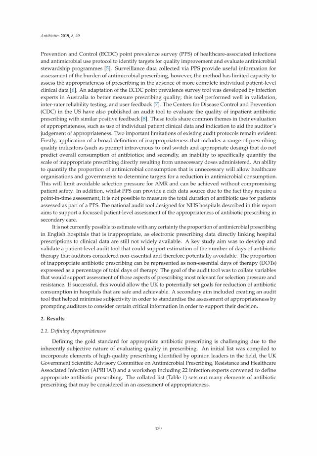
Antibiotics 2019, 8, 49
Prevention and Control (ECDC) point prevalence survey (PPS) of healthcare-associated infectionsand antimicrobial use protocol to identify targets for quality improvement and evaluate antimicrobialstewardship programmes [5]. Surveillance data collected via PPS provide useful information forassessment of the burden of antimicrobial prescribing, however, the method has limited capacity toassess the appropriateness of prescribing in the absence of more complete individual patient-levelclinical data [6]. An adaptation of the ECDC point prevalence survey tool was developed by infectionexperts in Australia to better measure prescribing quality; this tool performed well in validation,inter-rater reliability testing, and user feedback [7]. The Centers for Disease Control and Prevention(CDC) in the US have also published an audit tool to evaluate the quality of inpatient antibioticprescribing with similar positive feedback [8]. These tools share common themes in their evaluationof appropriateness, such as use of individual patient clinical data and indication to aid the auditor’sjudgement of appropriateness. Two important limitations of existing audit protocols remain evident:Firstly, application of a broad definition of inappropriateness that includes a range of prescribingquality indicators (such as prompt intravenous-to-oral switch and appropriate dosing) that do notpredict overall consumption of antibiotics; and secondly, an inability to specifically quantify thescale of inappropriate prescribing directly resulting from unnecessary doses administered. An abilityto quantify the proportion of antimicrobial consumption that is unnecessary will allow healthcareorganisations and governments to determine targets for a reduction in antimicrobial consumption.This will limit avoidable selection pressure for AMR and can be achieved without compromisingpatient safety. In addition, whilst PPS can provide a rich data source due to the fact they require apoint-in-time assessment, it is not possible to measure the total duration of antibiotic use for patientsassessed as part of a PPS. The national audit tool designed for NHS hospitals described in this reportaims to support a focussed patient-level assessment of the appropriateness of antibiotic prescribing insecondary care.
It is not currently possible to estimate with any certainty the proportion of antimicrobial prescribingin English hospitals that is inappropriate, as electronic prescribing data directly linking hospitalprescriptions to clinical data are still not widely available. A key study aim was to develop andvalidate a patient-level audit tool that could support estimation of the number of days of antibiotictherapy that auditors considered non-essential and therefore potentially avoidable. The proportionof inappropriate antibiotic prescribing can be represented as non-essential days of therapy (DOTs)expressed as a percentage of total days of therapy. The goal of the audit tool was to collate variablesthat would support assessment of those aspects of prescribing most relevant for selection pressure andresistance. If successful, this would allow the UK to potentially set goals for reduction of antibioticconsumption in hospitals that are safe and achievable. A secondary aim included creating an audittool that helped minimise subjectivity in order to standardise the assessment of appropriateness byprompting auditors to consider certain critical information in order to support their decision.
2. Results
2.1. Defining Appropriateness
Defining the gold standard for appropriate antibiotic prescribing is challenging due to theinherently subjective nature of evaluating quality in prescribing. An initial list was compiled toincorporate elements of high-quality prescribing identified by opinion leaders in the field, the UKGovernment Scientific Advisory Committee on Antimicrobial Prescribing, Resistance and HealthcareAssociated Infection (APRHAI) and a workshop including 22 infection experts convened to defineappropriate antibiotic prescribing. The collated list (Table 1) sets out many elements of antibioticprescribing that may be considered in an assessment of appropriateness.
130
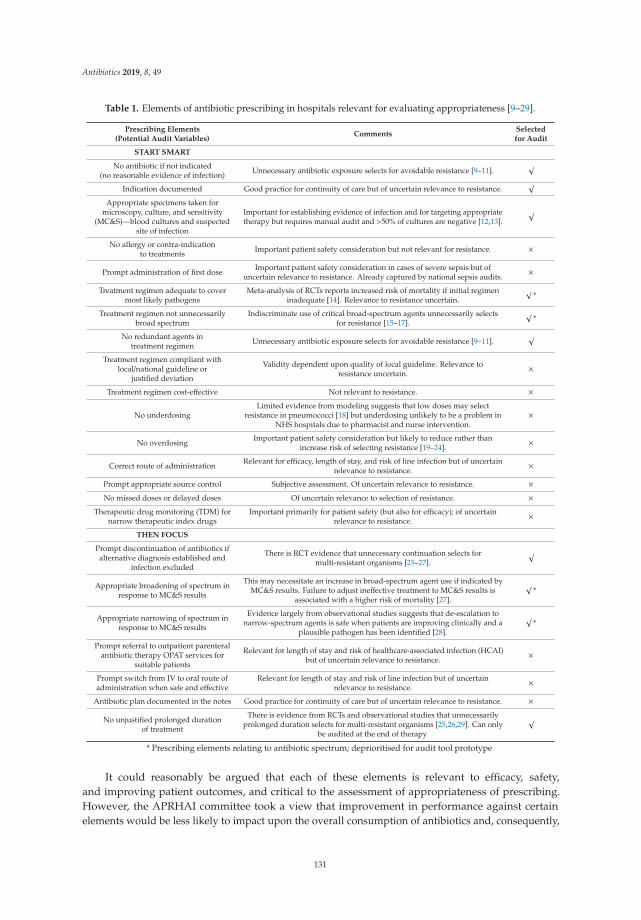
Antibiotics 2019, 8, 49
Table 1. Elements of antibiotic prescribing in hospitals relevant for evaluating appropriateness [9–29].
Prescribing Elements(Potential Audit Variables)
CommentsSelectedfor Audit
START SMART
No antibiotic if not indicated(no reasonable evidence of infection) Unnecessary antibiotic exposure selects for avoidable resistance [9–11].
√
Indication documented Good practice for continuity of care but of uncertain relevance to resistance.√
Appropriate specimens taken formicroscopy, culture, and sensitivity
(MC&S)—blood cultures and suspectedsite of infection
Important for establishing evidence of infection and for targeting appropriatetherapy but requires manual audit and >50% of cultures are negative [12,13].
√
No allergy or contra-indicationto treatments Important patient safety consideration but not relevant for resistance. ×
Prompt administration of first dose Important patient safety consideration in cases of severe sepsis but ofuncertain relevance to resistance. Already captured by national sepsis audits. ×
Treatment regimen adequate to covermost likely pathogens
Meta-analysis of RCTs reports increased risk of mortality if initial regimeninadequate [14]. Relevance to resistance uncertain.
√*
Treatment regimen not unnecessarilybroad spectrum
Indiscriminate use of critical broad-spectrum agents unnecessarily selectsfor resistance [15–17].
√*
No redundant agents intreatment regimen Unnecessary antibiotic exposure selects for avoidable resistance [9–11].
√
Treatment regimen compliant withlocal/national guideline or
justified deviation
Validity dependent upon quality of local guideline. Relevance toresistance uncertain. ×
Treatment regimen cost-effective Not relevant to resistance. ×
No underdosingLimited evidence from modeling suggests that low doses may select
resistance in pneumococci [18] but underdosing unlikely to be a problem inNHS hospitals due to pharmacist and nurse intervention.
×
No overdosing Important patient safety consideration but likely to reduce rather thanincrease risk of selecting resistance [19–24]. ×
Correct route of administration Relevant for efficacy, length of stay, and risk of line infection but of uncertainrelevance to resistance. ×
Prompt appropriate source control Subjective assessment. Of uncertain relevance to resistance. ×No missed doses or delayed doses Of uncertain relevance to selection of resistance. ×
Therapeutic drug monitoring (TDM) fornarrow therapeutic index drugs
Important primarily for patient safety (but also for efficacy); of uncertainrelevance to resistance. ×
THEN FOCUS
Prompt discontinuation of antibiotics ifalternative diagnosis established and
infection excluded
There is RCT evidence that unnecessary continuation selects formulti-resistant organisms [25–27].
√
Appropriate broadening of spectrum inresponse to MC&S results
This may necessitate an increase in broad-spectrum agent use if indicated byMC&S results. Failure to adjust ineffective treatment to MC&S results is
associated with a higher risk of mortality [27].
√*
Appropriate narrowing of spectrum inresponse to MC&S results
Evidence largely from observational studies suggests that de-escalation tonarrow-spectrum agents is safe when patients are improving clinically and a
plausible pathogen has been identified [28].
√*
Prompt referral to outpatient parenteralantibiotic therapy OPAT services for
suitable patients
Relevant for length of stay and risk of healthcare-associated infection (HCAI)but of uncertain relevance to resistance. ×
Prompt switch from IV to oral route ofadministration when safe and effective
Relevant for length of stay and risk of line infection but of uncertainrelevance to resistance. ×
Antibiotic plan documented in the notes Good practice for continuity of care but of uncertain relevance to resistance. ×
No unjustified prolonged durationof treatment
There is evidence from RCTs and observational studies that unnecessarilyprolonged duration selects for multi-resistant organisms [25,26,29]. Can only
be audited at the end of therapy.
√
* Prescribing elements relating to antibiotic spectrum; deprioritised for audit tool prototype
It could reasonably be argued that each of these elements is relevant to efficacy, safety,and improving patient outcomes, and critical to the assessment of appropriateness of prescribing.However, the APRHAI committee took a view that improvement in performance against certainelements would be less likely to impact upon the overall consumption of antibiotics and, consequently,
131
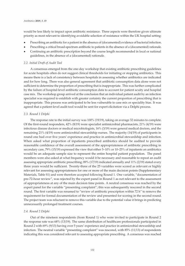
Antibiotics 2019, 8, 49
would be less likely to impact upon antibiotic resistance. Three aspects were therefore given ultimatepriority as most relevant to identifying avoidable selection of resistance within the UK hospital setting:
• Prescribing an antibiotic for a patient in the absence of (documented) evidence of bacterial infection.• Prescribing a critical broad-spectrum antibiotic to patients in the absence of a (documented) rationale.• Continuing an antibiotic prescription beyond the course length recommended in local or national
guidelines, in the absence of a (documented) rationale.
2.2. Initial Draft of Audit Tool
A consensus emerged from the one-day workshop that existing antibiotic prescribing guidelinesfor acute hospitals often do not suggest clinical thresholds for initiating or stopping antibiotics. Thismeans there is a lack of consistency between hospitals in assessing whether antibiotics are indicatedand for how long. There was also general agreement that antibiotic consumption data alone were notsufficient to determine the proportion of prescribing that is inappropriate. This was further complicatedby the failure of hospital-level antibiotic consumption data to account for patient acuity and hospitalcase mix. The workshop group arrived at the conclusion that an individual patient audit by an infectionspecialist was required to establish with greater certainty the current proportion of prescribing that isinappropriate. This process was anticipated to be less vulnerable to case mix or speciality bias. It wasagreed that a patient-level audit tool would be sent for expert elicitation via a Delphi process.
2.3. Round 1 Delphi
The response rate to the initial survey was 100% (19/19), taking on average 32 minutes to complete.Of the first-round responders, 42% (8/19) were specialist antimicrobial pharmacists, 21% (4/19) wereinfectious disease doctors or medical microbiologists, 16% (3/19) were general medical doctors, and theremaining 21% (4/19) were antimicrobial stewardship nurses. The majority (14/19) of participants inround one had over five years’ experience and practice in antimicrobial stewardship and infection.When asked what proportion of inpatients prescribed antibiotics should be audited to providereasonable confidence of the overall assessment of the appropriateness of antibiotic prescribing insecondary care, 79% (15/19) expressed the view that either 5–10% or 10–25% of inpatients on antibioticswould be an adequate sample size to represent the entire hospital patient population. The panelmembers were also asked at what frequency would it be necessary and reasonable to repeat an auditassessing appropriate antibiotic prescribing; 89% (17/19) indicated annually and 11% (2/19) stated everythree years would be sufficient. Twenty-three of the 25 variables were scored as relevant or highlyrelevant for assessing appropriateness for one or more of the main decision points (SupplementaryMaterials, Table S1) and were therefore accepted following Round 1. One variable, “documentation ofpre-72-hour review”, was rejected by the expert panel in Round 1 as not relevant to the assessmentof appropriateness at any of the main decision time points. A neutral consensus was reached by theexpert panel for the variable “presenting complaint”; this was subsequently rescored in the secondround. The first variable was renamed to “review of antibiotic prescription within 72 h” to remove therequirement for formal documentation of the review and presented for scoring in the second round.The project team was reluctant to remove this variable due to the potential value it brings in predictingunnecessarily prolonged treatment courses.
2.4. Round 2 Delphi
Out of the nineteen respondents (from Round 1) who were invited to participate in Round 2the response rate was 68% (13/19). The same distribution of healthcare professionals participated inRound 2 with 69% (9/13) having over 5 years’ experience and practice in antimicrobial stewardship andinfection. The neutral variable “presenting complaint” was rescored, with 85% (11/13) of respondentsindicating this was considered relevant to assessing appropriate prescribing. A consensus was reached
132
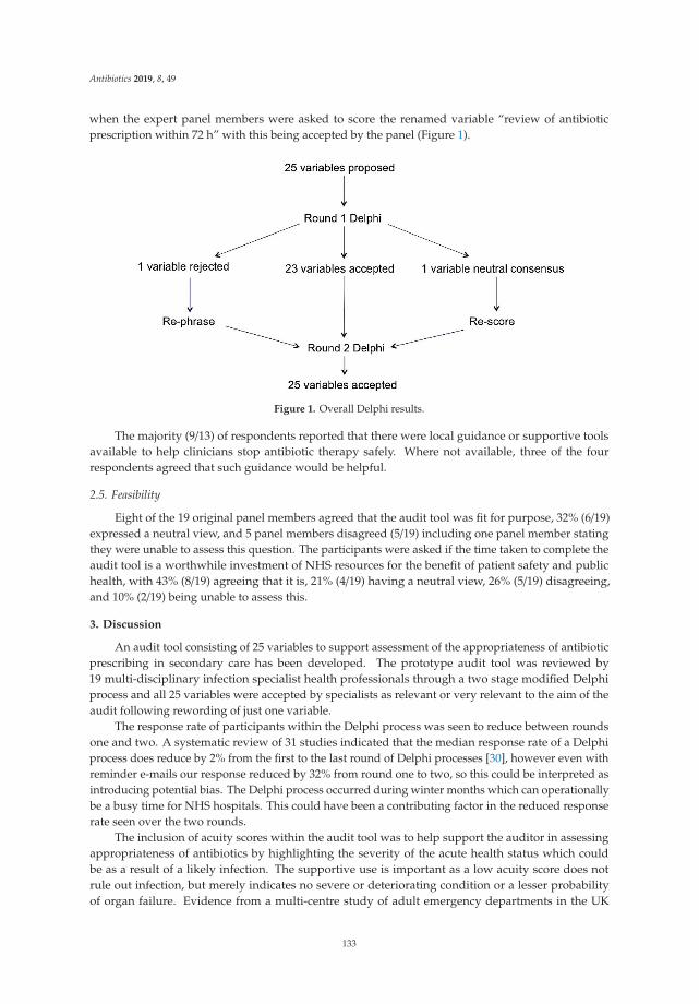
Antibiotics 2019, 8, 49
when the expert panel members were asked to score the renamed variable “review of antibioticprescription within 72 h” with this being accepted by the panel (Figure 1).
Figure 1. Overall Delphi results.
The majority (9/13) of respondents reported that there were local guidance or supportive toolsavailable to help clinicians stop antibiotic therapy safely. Where not available, three of the fourrespondents agreed that such guidance would be helpful.
2.5. Feasibility
Eight of the 19 original panel members agreed that the audit tool was fit for purpose, 32% (6/19)expressed a neutral view, and 5 panel members disagreed (5/19) including one panel member statingthey were unable to assess this question. The participants were asked if the time taken to complete theaudit tool is a worthwhile investment of NHS resources for the benefit of patient safety and publichealth, with 43% (8/19) agreeing that it is, 21% (4/19) having a neutral view, 26% (5/19) disagreeing,and 10% (2/19) being unable to assess this.
3. Discussion
An audit tool consisting of 25 variables to support assessment of the appropriateness of antibioticprescribing in secondary care has been developed. The prototype audit tool was reviewed by19 multi-disciplinary infection specialist health professionals through a two stage modified Delphiprocess and all 25 variables were accepted by specialists as relevant or very relevant to the aim of theaudit following rewording of just one variable.
The response rate of participants within the Delphi process was seen to reduce between roundsone and two. A systematic review of 31 studies indicated that the median response rate of a Delphiprocess does reduce by 2% from the first to the last round of Delphi processes [30], however even withreminder e-mails our response reduced by 32% from round one to two, so this could be interpreted asintroducing potential bias. The Delphi process occurred during winter months which can operationallybe a busy time for NHS hospitals. This could have been a contributing factor in the reduced responserate seen over the two rounds.
The inclusion of acuity scores within the audit tool was to help support the auditor in assessingappropriateness of antibiotics by highlighting the severity of the acute health status which couldbe as a result of a likely infection. The supportive use is important as a low acuity score does notrule out infection, but merely indicates no severe or deteriorating condition or a lesser probabilityof organ failure. Evidence from a multi-centre study of adult emergency departments in the UK
133
Antibiotics 2019, 8, 49
reported a positive predictive value for National Early Warning Score (NEWS) ≥3 of 20% for mortalityor ICU admission [31]. It was therefore considered reasonable to assess what proportion of patientstreated with antibiotics either have NEWS ≥3 or localised evidence of infection at an anatomical site.An abbreviated version of the Sequential Organ Failure Assessment (SOFA) score, quick SOFA (qSOFA),was assessed in almost 75,000 adult patients hospitalised with suspected infection [32]. The predictivevalidity of the qSOFA score for in-hospital mortality was found to be statistically greater than theSOFA or systemic inflammatory response syndrome (SIRS) scores, supporting its use as a promptto consider possible sepsis [32]. Applying qSOFA score ≥2 during emergency department or wardstay as a prediction tool for in-hospital all-cause mortality had a negative predictive value of 97% inadult patients with clinical suspicion of infection in a recent multi-centre study including 27 Frenchemergency departments [33]. A qSOFA score at a threshold of 2 or more may be considered by someclinicians to be a reasonable tool for differentiating patients in whom immediate broad-spectrumantibiotics are justified, given the associated in-hospital mortality of 24% [33].
During the second round of the Delphi process the panel did not accept that formal documentationof a pre-72-hour review was relevant. The panel did however accept that whether a pre-72-hour reviewtook place was relevant, irrespective of documentation (e.g., prescription changed but no correspondingentry in the medical notes) for the purposes of assessing appropriateness of antibiotic prescribing.In the absence of a pre-72-hour review, it is likely that patients for whom diagnostic tests support analternative diagnosis to infection are likely to continue antibiotics longer than necessary. This variableis also important due to the potential value it brings to predicting unnecessarily prolonged treatmentcourses, and it is part of ongoing quality improvement work within English hospitals, so the indicatorwas reworded for Round 2. Secondary healthcare quality improvement work within England hasfocussed on reducing total antibiotic consumption as well as obtaining evidence of antibiotic reviewwithin 72 h of commencing an antibiotic [34].
The addition of separate questions to explore individual views on inappropriate prescribingwithin secondary care was used as it was not possible to estimate a sample size, because the proportionof non-essential DOTs was unknown. Delphi participants were asked what proportion of patients onantibiotics required auditing to give them assurance on appropriateness of antibiotic prescribing fortheir hospital.
The expert panel members were invited to estimate the feasibility and likely value of the audit tool;this saw 26% of the expert panel disagreeing with statements that the audit tool was fit for purposeor a worthwhile investment of NHS resources. This finding was surprising given the high degree ofconsensus over the relevance of individual variables for supporting the assessment of appropriatenessand was not explored in more detail. It may reflect a perception of futility that a simple audit tool couldnot sufficiently capture the complexity of an episode of infection to enable a reproducible assessmentof appropriateness. A pilot study of the audit tool to test the reliability of the instrument is clearly aprerequisite before more widespread adoption.
3.1. Study Strengths and Limitations
Strengths of this project include the development of an audit tool through a consensus of amulti-disciplinary team of experienced infection specialists working in hospital clinical practice.The authors believe the developed audit tool is the first of its kind internationally that can specificallyquantify the scale of inappropriate prescribing directly resulting from unnecessary doses beingadministered. The audit tool was designed to minimise the burden of data collection by limitingthe items of data collected to only those considered relevant or highly relevant to the specific aim ofevaluation of appropriateness of prescribing at three critical prescribing decision time points. The audittool also narrows the focus of the audit to evaluate only those elements of prescribing associatedwith unnecessary antibiotic doses and therefore likely to be most relevant to the selection of resistantmicroorganisms: whether an antibiotic is indicated at the start of treatment, whether treatment beyond72 h is justified, and whether treatment beyond a standard course length is appropriate.
134
Antibiotics 2019, 8, 49
The potential to collect more detailed clinical information to explore the subjective decision thatthe auditors would take was advantageous. Standardisation of the assessment process may be aidedby the inclusion of objective variables to capture evidence of infection or sepsis, along with markers ofseverity including qSOFA score, NEWS, C-reactive protein (CRP), and white blood cell count.
The potential limitations of the audit tool include the time needed to collect data and risk ofsubjectivity in assessment which are inherent limitations of this approach, however a balance isstruck between the value of detailed patient-level data to robustly answer the important questionof appropriateness set against the use of a standardised data collection tool to minimise variabilityin assessment. There was also limited opportunity for experts to consider variables not includedin the tool, such as medical imaging and urine dipstick tests. Presenting an increased number ofpotentially relevant variables such as antibiotic dose and route of administration may have providedgreater opportunity to discriminate between the perceived value of different variables for estimatingnon-essential doses. Whilst a relatively small sample of experts participated in the Delphi process,an important limitation, it is worth noting that 22 experts participated in the workshop to defineappropriate antibiotic prescribing and develop the initial audit tool cascaded for the Delphi consensus.Subsequent piloting of the audit tool will need to include ongoing evaluation of the strengths andweakness of the tool and inform iterative improvements. The minor changes to the audit tool as aresult of this Delphi process provide reassurance that extensive revision of the tool was not required,however a higher number of experts participating would have provided greater assurance of a reliablerating for each of the variables assessed.
Since admission and discharge dates were not included in the tool it was not possible to determineat which point during their admission a patient receives antibiotics and therefore whether a patient isreceiving treatment for a healthcare-associated infection or community-acquired infection unless thisis stated explicitly by the auditor. This was identified by the project team as a priority to capture infuture versions of the audit tool.
The use of a web-based Delphi process limited opportunity for qualitative feedback from aface-to-face discussion and may have contributed to the lower response rate for Round 2. The selectionof expert panel members was via national infection networks and only one healthcare professional alsoparticipated in the previous workshop that convened to define appropriate prescribing. To help reducepotential bias, all participants were blinded to who participated in the expert panel with individualcorrespondence being sent.
The use of the developed audit tool is for active treatment and does not assess long-term andperi-operative surgical prophylaxis antibiotics. An adaptation could be made to the audit tool in orderto assess these groups of patients. Finally, the developed audit tool was prioritised for antibacterialagents only but could also be adapted for antifungal and antiviral agents.
3.2. Future Work
Piloting the audit tool within NHS hospitals to assess usability and operability is the nextstep. This would also provide additional insight and development of the tool and mitigate againstthe limitations of the small consensus panel. The resource burden of data collection and whetherthere is variation between hospitals that use electronic systems or paper-based processes needs tobe quantified. Although it is anticipated that the use of electronic systems should be faster thanpaper-based prescribing, ease of access to information for each variable may vary. Piloting the audittool within 5 to 10 hospitals could provide a preliminary assessment of non-essential days of antibiotictherapy prescribed to inform the sample size calculation for a definitive study. Recruiting a mixtureof hospital types (teaching, district and specialist) will be important to assess the practicality of thedeveloped audit tool. The balance between the amount of information being collected with the timetaken to collect the data in a busy operational hospital will be crucial.
135
Antibiotics 2019, 8, 49
4. Materials and Methods
4.1. Part 1: Development of the Audit Tool
Evidence synthesis of published literature and international guidance contributed to thedevelopment of the initial prototype audit tool to try and quantify and identify inappropriateantimicrobial prescribing. The initial prototype audit tool then underwent preliminary feasibilitytesting at one acute teaching hospital in Southampton, England. This was conducted by a specialistpharmacist supported by a medical microbiologist, with a focus on evaluating the operability of theaudit tool. After a subgroup of the UK government APRHAI scientific advisory committee draftedstandards to define appropriate prescribing in secondary care a finalised prototype audit tool wascreated [4]. The subgroup discussed aspects of auditing including frequency, intensity, resourcing,and mechanism of data collection including feedback and reporting processes.
To elicit further expert opinion, a one-day workshop was held on 6 March 2017 with infectionexperts including secondary care clinicians (10/22), pharmacists (9/22), a nurse, an academic,and a medical director to explore views on the appropriateness of prescribing within hospitals.The project team including two pharmacists of the antimicrobial resistance programme at PublicHealth England and three national antimicrobial pharmacists met to discuss the workshop findingsand updated the audit tool accordingly. Elements of the audit tool that were intended to supportthe assessment of appropriateness of antimicrobial prescribing were termed “variables” for thepurposes of the validation process. The selection of variables to include in the audit tool was based ondiscussions within the antibiotic prescribing quality subgroup of APRHAI, iterative e-mail feedbackfrom co-authors, and prototype audit tool testing in Southampton.
To support the auditor in their assessment of the severity of infection and deterioration as a resultof a likely infection the National Early Warning Score (NEWS) and quick sequential organ failureassessment (qSOFA) score were incorporated into the audit tool [32,35]. A threshold NEWS value of3 or above has been proposed as a suitable trigger to systematically screen for sepsis or septic shock inpatients with suspected infection in a United States Emergency Department (ED) population with areported negative predictive value of 99.5 (95% CI 97.8–99.9) [36].
4.2. Part 2: Validation of the Audit Tool
Between October and December 2017, a RAND-modified Delphi process was conducted to validateeach individual variable within the audit tool. A multi-disciplinary expert panel of infection and publichealth specialists was recruited via e-mail through national infection and microbiology networks andasked to assess the relevance of each variable against a five-point Likert Scale [37]. The relevance wasassessed in relation to three main decision time points during a course of antibiotic therapy:
• Initiation (was the antibiotic indicated and necessary at the start date?);• Early post prescription review (was the antibiotic continued after infection was ruled out?);• End of therapy (was the antibiotic continued beyond the standard duration?).
Non-essential days of antibiotic therapy were included with potential reasons as follows:(1) antibiotic not indicated/unnecessary at start date; (2) unexplained continuation of antibioticafter infection ruled out; and (3) unexplained continuation of antibiotic beyond standard duration.The standard duration was defined by individual hospitals as identified in local antibiotic guidelines.A separate questionnaire was sent to the Delphi participants to explore individual views oninappropriate prescribing within secondary care.
The expert panel participated in two rounds. Invitation via e-mail was sent to 19 participantsbefore the first round. The online survey SelectSurvey.net was used in each Delphi round withparticipant reminder e-mails sent periodically. The results were discussed within the project team afterthe first round and the aggregated scores were shared with the participants prior to the second round.Each of the 25 variables was assessed for its relevance against the three main decision time points.
136
Antibiotics 2019, 8, 49
Qualitative feedback was reviewed by one project team member and common themes reported fordiscussion with the full project team. The threshold for accepting a variable was met if the medianscore was “relevant” or “very relevant” for assessing appropriateness at any one of the three decisiontime points. A variable was rejected if the median score for all three of the critical decision points was“irrelevant” or “very irrelevant” with a neutral score being rescored in the second round to provide adefinitive response. The rewording of questions between each round occurred by the project team andwas presented to the participants from Round 1 for Round 2. After completing both rounds of theDelphi process the accepted variables were incorporated into a finalised audit tool (SupplementaryMaterials, Figure S1).
4.3. Statistics
Data were summarised using non-parametric descriptive statistics which are considered suitableto measure consensus and stability in Delphi studies [38] and due to the number of Delphi participants.The median and percentage “relevant” and “highly relevant” responses for each of the variables inrelation to each decision time point was calculated using univariate analysis.
5. Conclusions
In summary, the RAND-modified Delphi process has provided face validity of this audit tool andselection of critical variables that can be used to support the assessment of inappropriate antibioticprescribing. The feedback from expert panel members helped shape development of the audit tooland provides an important endorsement from practising healthcare professionals within and outsidethe NHS. This audit methodology will help hospitals identify the location, extent, and nature ofinappropriate antibiotic prescribing and provide evidence to drive local antimicrobial stewardship andfocus targeted training to specific areas of improvement.
Supplementary Materials: The following are available online at http://www.mdpi.com/2079-6382/8/2/49/s1.Figure S1: Assessing appropriateness of antibiotic therapy–final national audit tool for pilot. Table S1: Round 1Delphi results
Author Contributions: Conceptualization, D.A.-O., G.H., K.S.H.; data curation, G.H.; formal analysis, G.H.;methodology, G.H., K.S.H., and D.A.-O.; project administration, G.H.; supervision, D.A.-O; validation, K.S.H., E.C.,and D.A.-O.; writing—original draft, G.H.; writing—review & editing, G.H., K.S.H., E.C., P.H., S.H., and D.A.-O.
Funding: This research received no external funding and was conducted by Public Health England
Acknowledgments: The authors would like to thank all the expert panel members who participated in the Delphiprocess who contributed to the development of the audit tool: Janet Appleton, Druin Burch, Kay Cawthron,Abid Hussain, Conor Jamieson, David Jenkins, Alasdair MacGowen, Sanjay Patel, Neil Powell, Jacqueline Sneddon,Adel Sheikh, Paul Wade, Laura Whitney, Antony Zorzi, James Hatcher, Enrique Castro-Sanchez, Emily Bennett,Cairine Gormley and Tania Misra.
Conflicts of Interest: The authors declare no conflict of interest.
References
1. WHO Antimicrobial Resistance. Available online: http://www.who.int/mediacentre/factsheets/fs194/en/(accessed on 17 January 2019).
2. Review on Antimicrobial Resistance. Available online: https://amr-review.org/background.html (accessedon 17 January 2019).
3. English Surveillance Programme for Antimicrobial Utilisation and Resistance (ESPAUR) Report 2018.Available online: https://assets.publishing.service.gov.uk/government/uploads/system/uploads/attachment_data/file/759975/ESPAUR_2018_report.pdf (accessed on 22 March 2019).
4. English Surveillance Programme for Antimicrobial Utilisation and Resistance (ESPAUR) Report 2017.Available online: https://assets.publishing.service.gov.uk/government/uploads/system/uploads/attachment_data/file/656611/ESPAUR_report_2017.pdf (accessed on 18 January 2019).
137
Antibiotics 2019, 8, 49
5. Point Prevalence Survey of Healthcare-Associated Infections and Antimicrobial Use in European AcuteCare Hospitals Surveillance Report 2011–2012. Available online: https://ecdc.europa.eu/sites/portal/files/media/en/publications/Publications/healthcare-associated-infections-antimicrobial-use-PPS.pdf (accessedon 18 January 2019).
6. Kieran, J.A.; O’Doherty, R.G.; Hudson, B. ESAC point prevalence methodology to assess antimicrobialconsumption and quality of prescribing in an Australian setting. Med. J. Aust. 2011, 194, 103–104.
7. James, R.; Upjohn, L.; Cotta, M.; Luu, S.; Marshall, C.; Buising, K.; Thursky, K. Measuring antimicrobialprescribing quality in Australian hospitals: Development and evaluation of a national antimicrobialprescribing survey tool. J. Antimicrob. Chemother. 2015, 70, 1912–1918. [CrossRef] [PubMed]
8. Spivak, E.S.; Cosgrove, S.E.; Srinivasan, A. Measuring appropriate antimicrobial use: Attempts at openingthe black box. Clin. Infect. Dis. 2016, 63, 1639–1644. [PubMed]
9. Lopez-Lozano, J.M.; Monnet, D.L.; Yague, A.; Burgos, A.; Gonzalo, N.; Campillos, P.; Saez, M. Modelling andforecasting antimicrobial resistance and its dynamic relationship to antimicrobial use: A time series analysis.Int..J Antimicrob. Agents 2000, 14, 21–31. [CrossRef]
10. Tacconelli, E.; Cataldo, M.A.; De Pascale, G.; Manno, D.; Spanu, T.; Cambieri, A.; Antonelli, M.; Sanguinetti, M.;Fadda, G.; Cauda, R. Prediction models to identify hospitalized patients at risk of being colonized or infectedwith multidrug-resistant Acinetobacter baumannii calcoaceticus complex. J. Antimicrob. Chemother. 2008, 62,1130–1137. [CrossRef] [PubMed]
11. Tacconelli, E.; De Angellis, G.; Cataldo, M.A.; Mantengoli, E.; Spanu, T.; Pan, A.; Corti, G.; Radice, A.;Stolzuoli, L.; Antinori, S.; et al. Antibiotic usage and risk of colonization and infection with antibiotic-resistantbacteria: A hospital population-based study. Antimicrob. Agents Chemother. 2009, 53, 4264–4269. [CrossRef][PubMed]
12. Braykov, N.P.; Morgan, D.J.; Schweizer, M.L.; Usian, D.Z.; Kelesidis, T.; Weisenberg, S.A.; Johannsson, B.;Young, H.; Cantey, J.; Scrinivason, A.; et al. Assessment of empirical antibiotic therapy optimisation in sixhospitals: An observational cohort study. Lancet Infect. Dis. 2014, 14, 1220–1227. [CrossRef]
13. De Sousa, A.G.; Fernandes Junior, C.J.; Santos, G.P.D.; Laselva, C.R.; Polessi, J.; Lisboa, L.F.; Akamine, N.;Silva, E. The impact of each action in the Surviving Sepsis Campaign measures on hospital mortality ofpatients with severe sepsis/septic shock. Einstein 2008, 6, 323–327.
14. Paul, M.; Shani, V.; Muchtar, E.; Kariv, G.; Robenshtok, E.; Leibovici, L. Systematic review and meta-analysisof the efficacy of appropriate empiric antibiotic therapy for sepsis. Antimicrob. Agents Chemother. 2010, 54,4851–4863. [CrossRef] [PubMed]
15. Harris, A.D.; Perencevich, E.; Roghmann, M.C.; Morris, G.; Kaye, K.S.; Johnson, J.A. Risk factors forpiperacillin-tazobactam-resistant Pseudomonas aeruginosa among hospitalized patients. Antimicrob. AgentsChemother. 2002, 46, 854–858. [CrossRef]
16. Lai, C.C.; Wang, C.Y.; Chu, C.C.; Tan, C.K.; Lu, C.L.; Lee, Y.C.; Huang, Y.T.; Lee, P.I.; Hsueh, P.R. Correlationbetween antibiotic consumption and resistance of Gram-negative bacteria causing healthcare-associatedinfections at a university hospital in Taiwan from 2000 to 2009. J. Antimicrob. Chemother. 2011, 66, 1374–1382.[CrossRef]
17. Pakyz, A.L.; Oinonen, M.; Polk, R.E. Relationship of carbapenem restriction in 22 university teaching hospitalsto carbapenem use and carbapenem-resistant Pseudomonas aeruginosa. Antimicrob. Agents Chemother. 2009,53, 1983–1986. [CrossRef]
18. Opatowski, L.; Mandel, J.; Varon, E.; Boelle, P.Y.; Temime, L.; Guillemot, D. Antibiotic dose impact onresistance selection in the community: A mathematical model of beta-lactams and Streptococcus pneumoniaedynamics. Antimicrob. Agents Chemother. 2010, 54, 2330–2337. [CrossRef]
19. Guillemot, D.; Carbon, C.; Balkau, B.; Geslin, P.; Lecoeur, H.; Vauzelle-Keroedan, F.; Bouvenot, G.; Eschwege, E.Low dosage and long treatment duration of beta-lactam: Risk factors for carriage of penicillin-resistantStreptococcus pneumoniae. JAMA 1998, 279, 365–370. [CrossRef]
20. Handel, A.; Margolis, E.; Levin, B.R. Exploring the role of the immune response in preventing antibioticresistance. J. Theor. Biol. 2009, 256, 655–662. [CrossRef]
21. Martinez, M.N.; Papich, M.G.; Drusano, G.L. Dosing regimen matters: The importance of early interventionand rapid attainment of the pharmacokinetic/pharmacodynamic target. Antimicrob. Agents Chemother. 2012,56, 2795–2805. [CrossRef]
138
Antibiotics 2019, 8, 49
22. Olofsson, S.K.; Cars, O. Optimizing drug exposure to minimize selection of antibiotic resistance. Clin. Infect.Dis. 2007, 45 (Suppl. 2), S129–S136. [CrossRef]
23. Schrag, S.J.; Pena, C.; Fernandez, J.; Sanchez, J.; Gomez, V.; Perez, E.; Feris, J.M.; Besser, R.E. Effect ofshort-course, high-dose amoxicillin therapy on resistant pneumococcal carriage: A randomized trial. JAMA2001, 286, 49–56. [CrossRef]
24. Tam, V.H.; Louie, A.; Deziel, M.R.; Liu, W.; Drusano, G.L. The relationship between quinolone exposures andresistance amplification is characterized by an inverted U: A new paradigm for optimizing pharmacodynamicsto counterselect resistance. Antimicrob. Agents Chemother. 2007, 51, 744–747. [CrossRef]
25. Singh, N.; Rogers, P.; Atwood, C.W.; Wagener, M.M.; Yu, V.L. Short-course empiric antibiotic therapy forpatients with pulmonary infiltrates in the intensive care unit. A proposed solution for indiscriminateantibiotic prescription. Am. J. Respir. Crit. Care Med. 2000, 162, 505–511. [CrossRef]
26. Chastre, J.; Wolff, M.; Fagon, J.Y.; Chevret, S.; Thomas, F.; Wermert, D.; Clementi, E.; Gonzalez, J.; Jusserand, D.;Asfar, P.; et al. PneumA trial group. comparison of 8 vs 15 days of antibiotic therapy for ventilator-associatedpneumonia in adults: A randomized trial. JAMA 2003, 290, 2588–2598. [CrossRef]
27. Palacios-Baena, Z.R.; Gutierrez-Gutierrez, B.; De Cueto, M.; Viale, P.; Venditti, M.; Hernandez-Torres, A.;Oliver, A.; Martinez-Martinez, L.; Calbo, E.; Pintado, V.; et al. REIPI/ESGBIS/INCREMENT Group.Development and validation of the INCREMENT-ESBL predictive score for mortality in patientswith bloodstream infections due to extended-spectrum-beta-lactamase-producing Enterobacteriaceae.J. Antimicrob. Chemother. 2017, 72, 906–913.
28. Schuts, E.C.; Hulscher, M.E.; Mouton, J.W.; Verduin, C.M.; Stuart, J.W.T.C.; Overdiek, H.W.P.M.; van derLinden, P.D.; Natsch, S.; Hertogh, C.M.P.M.; Wolfs, T.F.W.; et al. Current evidence on hospital antimicrobialstewardship objectives: A systematic review and meta-analysis. Lancet Infect. Dis. 2016, 16, 847–856. [CrossRef]
29. Marra, A.R.; de Almeida, S.M.; Correa, L.; Silva, M., Jr.; Martino, M.D.; Silva, C.V.; Cal, R.G.; Edmond, M.B.;dos Santos, O.F. The effect of limiting antimicrobial therapy duration on antimicrobial resistance in thecritical care setting. Am. J. Infect. Control. 2009, 37, 204–209. [CrossRef]
30. Boulkedid, R.; Abdoul, H.; Loustau, M.; Sibony, O.; Alberti, C. Using and reporting the delphi method forselecting healthcare quality indicators: A systematic review. PLoS ONE 2011, 6, e20476. [CrossRef]
31. Corfield, A.R.; Lees, F.; Zealley, I.; Houston, G.; Dickie, S.; Ward, K.; McGuffie, C. Utility of a single early warningscore in patients with sepsis in the emergency department. Emerg. Med. J. 2014, 31, 482–487. [CrossRef]
32. Seymour, C.W.; Liu, V.X.; Iwashyna, T.J.; Iwashyna, T.; Brunkhorst, F.M.; Rea, T.D.; Scherag, A.; Rubenfeld, G.;Kahn, J.M.; Shankar-Hari, M.; et al. Assessment of clinical criteria for sepsis: For the third internationalconsensus definitions for sepsis and septic shock (Sepsis-3). JAMA 2016, 315, 762–774. [CrossRef]
33. Freund, Y.; Lemachatti, N.; Krastinova, E.; Van Laer, M.; Claessens, Y.-E.; Avonso, A.; Occelli, C.;Feral-Pierssens, A.-L.; Truchot, J.; Ortega, M.; et al. Prognostic accuracy of sepsis-3 criteria for in-hospitalmortality among patients with suspected infection presenting to the emergency department. JAMA 2017,317, 301–308. [CrossRef]
34. NHS England National CQUIN Templates 2016/17. Available online: https://www.england.nhs.uk/nhs-standard-contract/cquin/cquin-16-17/ (accessed on 18 January 2019).
35. National Early Warning Score (NEWS): Standardising the Assessment of Acute Illness Severity in the NHS.Report of a Working Party. Available online: https://www.rcplondon.ac.uk/projects/outputs/national-early-warning-score-news (accessed on 18 January 2019).
36. Keep, J.W.; Messmer, A.S.; Sladden, R.; Burrell, N.; Pinate, R.; Tunnicliff, M.; Glucksman, E. National earlywarning score at Emergency Department triage may allow earlier identification of patients with severe sepsisand septic shock: A retrospective observational study. Emerg. Med. J. 2016, 33, 37–41. [CrossRef]
37. Vagias, W.M. Likert-Type Scale Response Anchors; Clemson International Institute for Tourism & ResearchDevelopment, Department of Parks, Recreation and Tourism Management, Clemson University: Clemson,SC, USA, 2006.
38. Holey, E.A.; Feeley, J.L.; Dixon, J.; Whittaker, V.J. An exploration of the use of simple statistics to measureconsensus and stability in Delphi studies. BMC Med. Res. Methodol. 2007, 7, 52. [CrossRef]
© 2019 by the authors. Licensee MDPI, Basel, Switzerland. This article is an open accessarticle distributed under the terms and conditions of the Creative Commons Attribution(CC BY) license (http://creativecommons.org/licenses/by/4.0/).
139

antibiotics
Review
Actinomycete-Derived Polyketides as a Source ofAntibiotics and Lead Structures for the Developmentof New Antimicrobial Drugs
Helene L. Robertsen and Ewa M. Musiol-Kroll *
Interfakultäres Institut für Mikrobiologie und Infektionsmedizin, Eberhard Karls Universität Tübingen,Auf der Morgenstelle 28, 72076 Tübingen, Germany; [email protected]* Correspondence: [email protected]
Received: 27 August 2019; Accepted: 10 September 2019; Published: 20 September 2019
Abstract: Actinomycetes are remarkable producers of compounds essential for human and veterinarymedicine as well as for agriculture. The genomes of those microorganisms possess several sets of genes(biosynthetic gene cluster (BGC)) encoding pathways for the production of the valuable secondarymetabolites. A significant proportion of the identified BGCs in actinomycetes encode pathways forthe biosynthesis of polyketide compounds, nonribosomal peptides, or hybrid products resulting fromthe combination of both polyketide synthases (PKSs) and nonribosomal peptide synthetases (NRPSs).The potency of these molecules, in terms of bioactivity, was recognized in the 1940s, and startedthe “Golden Age” of antimicrobial drug discovery. Since then, several valuable polyketide drugs,such as erythromycin A, tylosin, monensin A, rifamycin, tetracyclines, amphotericin B, and manyothers were isolated from actinomycetes. This review covers the most relevant actinomycetes-derivedpolyketide drugs with antimicrobial activity, including anti-fungal agents. We provide an overviewof the source of the compounds, structure of the molecules, the biosynthetic principle, bioactivity andmechanisms of action, and the current stage of development. This review emphasizes the importanceof actinomycetes-derived antimicrobial polyketides and should serve as a “lexicon”, not only toscientists from the Natural Products field, but also to clinicians and others interested in this topic.
Keywords: actinomycetes; bioactivity; antimicrobials; polyketides; polyketide synthases; biosynthesis
1. Introduction
More than 70 years has passed since the discovery of the first antibiotics from actinomycetes.Although some of the high G+C (bacteria with high guanine- (G) and cytosine- (C) content in theirgenomes) [1,2], Gram-positive bacteria of the order Actinomycetales and their products have beenstudied in depth, they remain one of the most important sources of secondary metabolites, includingthe naturally-derived antimicrobial drugs (e.g., β-lactams, tetracyclines, rifamycins, macrolides,aminoglycosides, and glycopeptides). In the past, the producers of valuable bioactive compounds weremainly isolated from soil samples and either directly or indirectly, using culture filtrates or extracts,subjected to susceptibility testing (diffusion method) (Figure 1). Recent advances in different disciplinesof science, such as robotics [3], biology [4], chemistry [5–7], genetics [8], and/or bioinformatics [9] hasextended the spectrum of methodology applied to the isolation and identification of actinomycetes andthe produced metabolites. In addition to technical innovations which, for example, enable sampling ofunexplored and difficult to access environments, Next Generation Sequencing Technologies (NGST)and genome mining facilitate accurate and cost-effective sequencing of actinomycetes genomes and afast identification of genes encoding the proteins of the secondary metabolite biosynthetic machineries(biosynthetic gene clusters, BGCs). In the past two decades, hundreds of actinomycetes genomes havebeen sequenced and many of them have been fully annotated. The analysis of the data has demonstrated
Antibiotics 2019, 8, 157; doi:10.3390/antibiotics8040157 www.mdpi.com/journal/antibiotics141
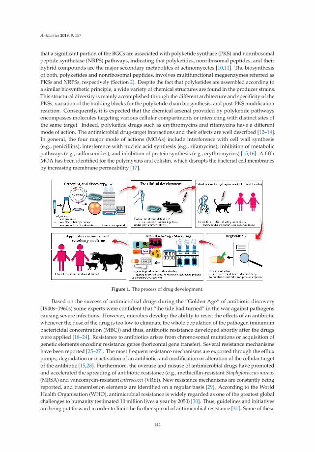
Antibiotics 2019, 8, 157
that a significant portion of the BGCs are associated with polyketide synthase (PKS) and nonribosomalpeptide synthetase (NRPS) pathways, indicating that polyketides, nonribosomal peptides, and theirhybrid compounds are the major secondary metabolites of actinomycetes [10,11]. The biosynthesisof both, polyketides and nonribosomal peptides, involves multifunctional megaenzymes referred asPKSs and NRPSs, respectively (Section 2). Despite the fact that polyketides are assembled according toa similar biosynthetic principle, a wide variety of chemical structures are found in the producer strains.This structural diversity is mainly accomplished through the different architecture and specificity of thePKSs, variation of the building blocks for the polyketide chain biosynthesis, and post-PKS modificationreaction. Consequently, it is expected that the chemical arsenal provided by polyketide pathwaysencompasses molecules targeting various cellular compartments or interacting with distinct sites ofthe same target. Indeed, polyketide drugs such as erythromycins and rifamycins have a differentmode of action. The antimicrobial drug-target interactions and their effects are well described [12–14].In general, the four major mode of actions (MOAs) include interference with cell wall synthesis(e.g., penicillins), interference with nucleic acid synthesis (e.g., rifamycins), inhibition of metabolicpathways (e.g., sulfonamides), and inhibition of protein synthesis (e.g., erythromycins) [15,16]. A fifthMOA has been identified for the polymyxins and colistin, which disrupts the bacterial cell membranesby increasing membrane permeability [17].
Figure 1. The process of drug development.
Based on the success of antimicrobial drugs during the “Golden Age” of antibiotic discovery(1940s–1960s) some experts were confident that “the tide had turned” in the war against pathogenscausing severe infections. However, microbes develop the ability to resist the effects of an antibioticwhenever the dose of the drug is too low to eliminate the whole population of the pathogen (minimumbactericidal concentration (MBC)) and thus, antibiotic resistance developed shortly after the drugswere applied [18–24]. Resistance to antibiotics arises from chromosomal mutations or acquisition ofgenetic elements encoding resistance genes (horizontal gene transfer). Several resistance mechanismshave been reported [25–27]. The most frequent resistance mechanisms are exported through the effluxpumps, degradation or inactivation of an antibiotic, and modification or alteration of the cellular targetof the antibiotic [15,28]. Furthermore, the overuse and misuse of antimicrobial drugs have promotedand accelerated the spreading of antibiotic resistance (e.g., methicillin-resistant Staphylococcus aureus(MRSA) and vancomycin-resistant enterococci (VRE)). New resistance mechanisms are constantly beingreported, and transmission elements are identified on a regular basis [29]. According to the WorldHealth Organisation (WHO), antimicrobial resistance is widely regarded as one of the greatest globalchallenges to humanity (estimated 10 million lives a year by 2050) [30]. Thus, guidelines and initiativesare being put forward in order to limit the further spread of antimicrobial resistance [31]. Some of these
142
Antibiotics 2019, 8, 157
initiatives include the development of vaccines, faster diagnostic tests to ensure appropriate antibioticadministration, and immune-based therapies [32]. These measures could provide promising solutionsfor prevention of infections however, once infection occurs in a human host, antibiotics remain thechoice of treatment for bacterial and fungal infections. Therefore, the need for new antibiotic classesand improvement of the old compounds remain more important than ever. Unfortunately, discovery ofnovel antibiotic classes is halted by the low return on investment, forcing companies to abandon theirdiscovery platforms. According to an issue brief of The Pew Charitable Trusts from March 2019 [33],out of the roughly 38 companies currently invested in antibiotic clinical development, only four ofthese are multinational pharmaceutical companies. Furthermore, 90% of the current products indevelopment are studied by small pharmaceutical or biotech companies, which are ill-equipped todeal with the expense of possible setbacks, delays or even rejections, which are often faced in clinicaltrials [34].
Since polyketide-derived antimicrobials have had a historical significance in human therapy,this group of compounds remain important for continuation of research aiming at: the identification ofnew structures and activities, the biosynthesis and/or chemical semi-synthesis for discovery of novelderivatisation routes, and finally, at understanding of resistance mechanisms to overcome this obstacle.
In this review, we focus on the therapeutically relevant polyketide drugs which were derived fromactinomycetes. We describe compounds active against microbial pathogens, including pathogenicfungi and bacteria, their biosynthesis in the natural producer, and the most recent results onproduction optimization and derivatisation attempts. This review should serve as a “lexicon” ofactinomycetes-derived antimicrobial polyketides, not only to scientists from the Natural Products field,but also to clinicians and others interested in this topic.
2. The Biosynthetic Assembly Lines
The biosynthesis of polyketides and nonribosomal peptides requires the PKS and NRPS enzymaticmachineries. PKSs are grouped into type I, II, and III, and can be iterative (type I, II, III) or modular(type I) [35–39]. The diverse polyketide assembly lines have been previously reviewed [7,40–43].Here, we introduce types of PKSs that are represented in this review.
The modular type I PKSs can give rise to large, complex polyketides. A “minimal PKS module”is composed of three essential domains; the acyltransferase (AT), the ketosynthase (KS), and theacyl carrier protein (ACP). The AT domain selects and loads the building blocks onto the activatedACPs, while the KS domain catalyses the decarboxylative Claisen-like condensation of the newlyloaded unit and the already existing polyketide chain. The β-keto-processing of the generated chain isaccomplished by optional domains, such as the ketoreductase (KRs), the dehydratases (DHs) and/orthe enoyl reductases (ERs). The final chain termination and release of the polyketide intermediatefrom the PKS is facilitated by the thioesterase (TE) domain, often found as the “last domain” in themultimodular PKS.
Type II iterative PKSs are responsible for the biosynthesis of aromatic polyketides [44–46].The overall architecture of this type of PKSs appears simpler than the one of complex modular PKSs,as they only require the presence of three enzymes, the KSα, KSβ (also referred to as chain elongationfactor (CLF)), and an ACP. During elongation, a thioester is bound to the ACP while the KSαKSβcomplex orchestrates chain extension with malonyl-CoA units exclusively. The BGCs of type II PKSsoften contain additional genes encoding cyclases (CYCs) and KRs, which act as chaperones and reductiveenzymes to ensure the correct cyclisation of the polyketide precursor, respectively. Finally, the presenceof aromatases (AROs) leads to the biosynthesis of aromatic ring systems. The iterative type I PKS [47,48]work in a similar fashion as the iterative type II PKSs [44]. However, the bacterial iterative type I PKSsonly facilitate the biosynthesis of small aromatic polyketides [35,49].
NRPSs display a similar architecture to modular type I PKSs and function in an analogousmanner [39,50]. The selection and loading of the building blocks (proteinogenic or non-proteinogenicamino acids) onto peptidyl carrier proteins (PCPs) of NRPSs is catalysed by an adenylation (A) domain,
143
Antibiotics 2019, 8, 157
resulting in an amino acid–PCP–thioester. A condensation (C) domain attaches the amino acid to theso-far synthesised peptide (chain elongation). Modifications, such as epimerisation or N-methylation atthe peptide chain may occur due to the activity of the optional epimerisation (E) and methyltransferase(MT) domain, respectively. The peptide chain is released from the NRPS by a TE domain. In the caseof hybrid PKS/NRPS structures [51], the transfer of the growing precursor chain between the differentmegaenzymes might require additional enzymes. However, this remains speculative and requiresdeeper investigation.
3. Clinically-Relevant Polyketide Derived Antibiotics
3.1. Erythromycin and Derivatives
The history of the macrolide erythromycin began in 1949 with a soil sample, collected in Iloilo City,(Iloilo, Philippine Islands) by Dr. Abelardo Aguilar, who sent the sample to Lilly Research Laboratories,where the soil-dwelling bacterium Saccharopolyspora erythraea (formerly known as Streptomyces erythreus)and the antibiotic erythromycin were isolated [52]. Inspired by the name of the place from which thesoil sample was collected, erythromycin was launched by Eli Lilly Co. in 1952 as Ilosone (also Ilotycin)(Table 1) for treatment of respiratory tract infections, skin infections, and Legionnaire’s disease.
The antibiotic erythromycin is composed of a 14-membered lactone, erythronolide B, to which twodeoxysugars l-mycarose and α-d-desosamine are attached [53,54] (Figure 2). The BGC of erythromycinremains one of the most-studied in terms of the type I PKS [55–58]. The precursor erythronolide Bis generated by the modular type I deoxyerythronolide B synthase (DEBS), composed of the threeenzymes DEBS 1–3, encoded by the genes eryAI–eryAIII (Figure S1). The PKS complex containssix modules, which are responsible for the initial loading of propionyl-CoA, the six steps of chainelongation with methylmalonyl-CoA as extender units, modification of the growing polyketide chainby reductive domains, and finally chain release and cyclization by the TE domain in DEBS 3 [57,59,60].The hydroxylation of the released aglycone is catalysed by the cytochrome P450 enzyme EryF, resultingin the intermediate erythronolide B. The glycosyltransferases EryBV and EryCII/EryCIII attach thesugar structures, derived from thymidine diphosphate (TDP)-l-mycarose and TDP-d-desosamine,which leads to the production of erythromycin D. Erythromycin D is further converted to erythromycinB or C, which are the substrates for erythromycin A. Erythromycin B is formed whenever themethyltransferase EryG is acting on erythromycin D. In the case where the molecule erythromycin D ismodified by the monooxygenase EryK, erythromycin C is produced as an intermediate.
Although the strain produces a mixture of erythromycins, erythromycin A was the most abundantand biologically active compound [61]. Erythromycin shows activity against many Gram-positiveand some Gram-negative bacteria, including Haemophilus influenzae, S. aureus, Streptococcus pyogenes,Streptococcus pneumoniae, Legionella pneumophila, Neisseria gonorrhoeae, and Mycoplasma pneumonia [61,62].The antibiotic interacts with the large 50S subunit of the prokaryotic ribosomes and inhibits the proteinbiosynthesis [63,64]. More specifically, the antibiotic binds to the exit tunnel and blocks its large subunitwhereby the elongation of the nascent peptide chain is stalled. This stalling leads to the prematuredissociation of the peptidyl-transfer RNA (tRNA) from the ribosome [64,65]. As the inhibition occursdirectly after initiation of protein synthesis, the nascent polypeptide chain remains short, however,the length size is determined by binding of the macrolide structure to the complex. Consequently,the size and conformation of erythromycin play a crucial role in the bioactivity of the molecule [66].In case of erythromycin A, binding to the 50S subunit leads to dissociation and accumulation ofpeptidyl-tRNAs with six, seven, or eight amino acid residues. This accumulation causes depletion ofthe free tRNA pool, whereby protein biosynthesis is inhibited [65].
144
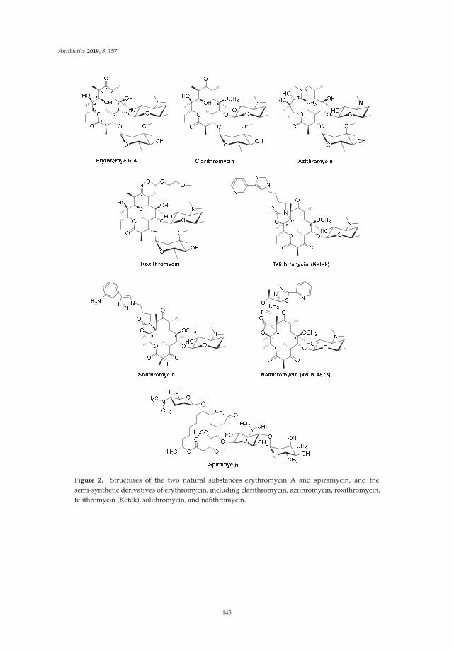
Antibiotics 2019, 8, 157
Figure 2. Structures of the two natural substances erythromycin A and spiramycin, and thesemi-synthetic derivatives of erythromycin, including clarithromycin, azithromycin, roxithromycin,telithromycin (Ketek), solithromycin, and nafithromycin.
145
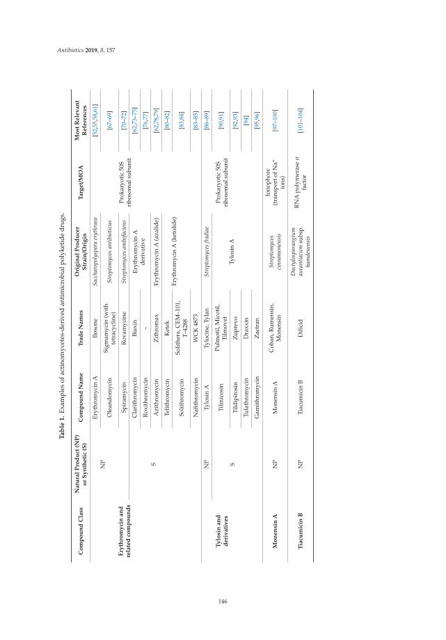
Antibiotics 2019, 8, 157
Ta
ble
1.
Exam
ples
ofac
tino
myc
etes
-der
ived
anti
mic
robi
alpo
lyke
tide
drug
s.
Co
mp
ou
nd
Cla
ssN
atu
ral
Pro
du
ct(N
P)
or
Sy
nth
eti
c(S
)C
om
po
un
dN
am
eT
rad
eN
am
es
Ori
gin
al
Pro
du
cer
Str
ain/O
rig
inT
arg
et/
MO
AM
ost
Rele
van
tR
efe
ren
ces
Ery
thro
my
cin
an
dre
late
dco
mp
ou
nd
s
NP
Eryt
hrom
ycin
AIl
oson
eSa
ccha
ropo
lysp
ora
eryt
hrae
a
Prok
aryo
tic
50S
ribo
som
alsu
buni
t
[52,
55,5
8,61
]
Ole
ando
myc
inSi
gmam
ycin
(wit
hte
trac
yclin
e)St
rept
omyc
esan
tibio
ticus
[67–
69]
Spir
amyc
inR
ovam
ycin
eSt
rept
omyc
esam
bofa
cien
s[7
0–72
]
S
Cla
rith
rom
ycin
Biax
inEr
ythr
omyc
inA
deri
vati
ve[6
2,73
–75]
Rox
ithr
omyc
in–
[76,
77]
Azi
thro
myc
inZ
ithr
omax
Eryt
hrom
ycin
A(a
zalid
e)[6
2,78
,79]
Telit
hrom
ycin
Ket
ekEr
ythr
omyc
inA
(ket
olid
e)[8
0–82
]
Solit
hrom
ycin
Solit
hera
,CEM
–101
,T–
4288
[83,
84]
Nafi
thro
myc
inW
CK
4873
[83–
85]
Ty
losi
nan
dd
eri
vati
ves
NP
Tylo
sin
ATy
loci
ne,T
ylan
Stre
ptom
yces
frad
iae
Prok
aryo
tic
50S
ribo
som
alsu
buni
t
[86–
89]
S
Tilm
icos
inPu
lmot
il,M
icot
il,Ti
lmov
etTy
losi
nA
[90,
91]
Tild
ipir
osin
Zup
revo
[92,
93]
Tula
thro
myc
inD
raxx
in[9
4]
Gam
ithr
omyc
inZ
actr
an[9
5,96
]
Mo
nen
sin
AN
PM
onen
sin
AC
oban
,Rum
ensi
n,M
onen
sin
Stre
ptom
yces
cinn
amon
ensi
s
Iono
phor
e(t
rans
port
ofN
a+
ions
)[9
7–10
0]
Tia
cum
icin
BN
PTi
acum
icin
BD
ifici
dD
acty
losp
oran
gium
aura
ntia
cum
subs
p.ha
mde
nens
is
RN
Apo
lym
eras
eσ
fact
or[1
01–1
04]
146
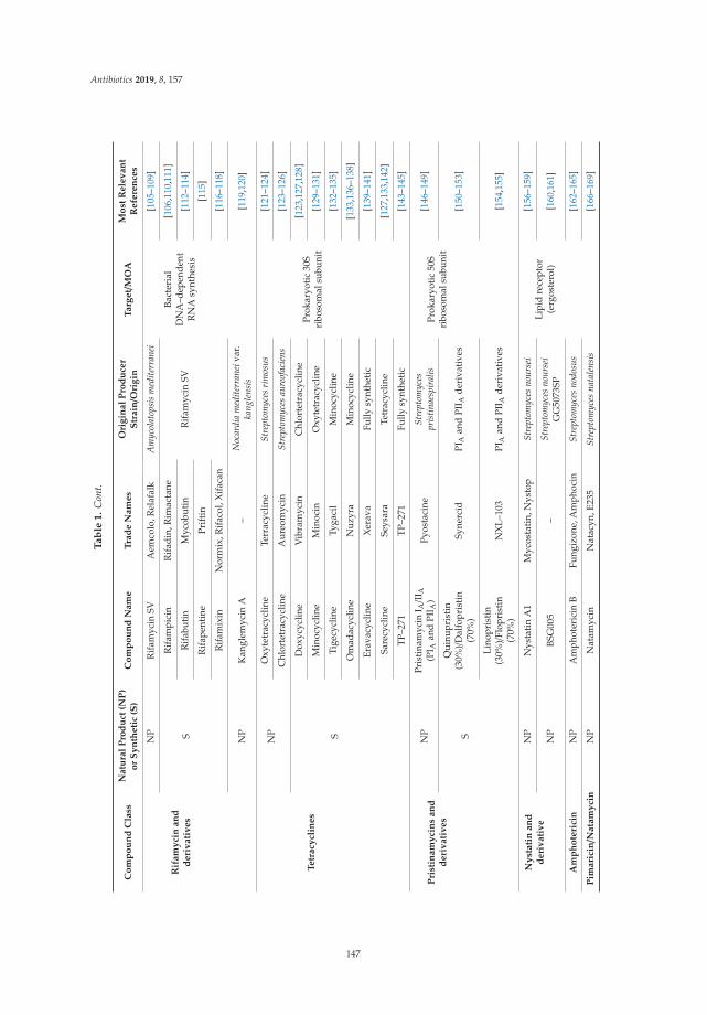
Antibiotics 2019, 8, 157
Ta
ble
1.
Con
t.
Co
mp
ou
nd
Cla
ssN
atu
ral
Pro
du
ct(N
P)
or
Sy
nth
eti
c(S
)C
om
po
un
dN
am
eT
rad
eN
am
es
Ori
gin
al
Pro
du
cer
Str
ain/O
rig
inT
arg
et/
MO
AM
ost
Rele
van
tR
efe
ren
ces
Rif
am
yci
nan
dd
eri
vati
ves
NP
Rif
amyc
inSV
Aem
colo
,Rel
afal
kA
myc
olat
opsi
sm
edite
rran
ei
Bact
eria
lD
NA
–dep
ende
ntR
NA
synt
hesi
s
[105
–109
]
S
Rif
ampi
cin
Rif
adin
,Rim
acta
ne
Rif
amyc
inSV
[106
,110
,111
]
Rif
abut
inM
ycob
utin
[112
–114
]
Rif
apen
tine
Prif
tin
[115
]
Rif
amix
inN
orm
ix,R
ifac
ol,X
ifac
an[1
16–1
18]
NP
Kan
glem
ycin
A–
Noc
ardi
am
edite
rran
eiva
r.ka
ngle
nsis
[119
,120
]
Tetr
acy
clin
es
NP
Oxy
tetr
acyc
line
Terr
acyc
line
Stre
ptom
yces
rim
osus
Prok
aryo
tic
30S
ribo
som
alsu
buni
t
[121
–124
]
Chl
orte
trac
yclin
eA
ureo
myc
inSt
rept
omyc
esau
reof
acie
ns[1
23–1
26]
S
Dox
ycyc
line
Vib
ram
ycin
Chl
orte
trac
yclin
e[1
23,1
27,1
28]
Min
ocyc
line
Min
ocin
Oxy
tetr
acyc
line
[129
–131
]
Tige
cycl
ine
Tyga
cil
Min
ocyc
line
[132
–135
]
Om
adac
yclin
eN
uzyr
aM
inoc
yclin
e[1
33,1
36–1
38]
Erav
acyc
line
Xer
ava
Fully
synt
heti
c[1
39–1
41]
Sare
cycl
ine
Seys
ara
Tetr
acyc
line
[127
,133
,142
]
TP–2
71TP
–271
Fully
synt
heti
c[1
43–1
45]
Pri
stin
am
yci
ns
an
dd
eri
vati
ves
NP
Pris
tinam
ycin
I A/I
I A(P
I Aan
dPI
I A)
Pyos
taci
neSt
rept
omyc
espr
istin
aesp
iral
isPr
okar
yoti
c50
Sri
boso
mal
subu
nit
[146
–149
]
S
Qui
nupr
isti
n(3
0%)/
Dal
fopr
isti
n(7
0%)
Syne
rcid
PIA
and
PII A
deri
vati
ves
[150
–153
]
Lino
pris
tin
(30%
)/Fl
opri
stin
(70%
)N
XL–
103
PIA
and
PII A
deri
vati
ves
[154
,155
]
Ny
stati
nan
dd
eri
vati
ve
NP
Nys
tati
nA
1M
ycos
tati
n,N
ysto
pSt
rept
omyc
esno
urse
iLi
pid
rece
ptor
(erg
oste
rol)
[156
–159
]
NP
BSG
005
–St
rept
omyc
esno
urse
iG
G50
73SP
[160
,161
]
Am
ph
ote
rici
nN
PA
mph
oter
icin
BFu
ngiz
one,
Am
phoc
inSt
rept
omyc
esno
dosu
s[1
62–1
65]
Pim
ari
cin/N
ata
my
cin
NP
Nat
amyc
inN
atac
yn,E
235
Stre
ptom
yces
nata
lens
is[1
66–1
69]
147
Antibiotics 2019, 8, 157
In order to improve the chemical, microbiological, and pharmacokinetic properties of macrolides,a lot of effort was put into the production of semi-synthetic and synthetic derivatives [63,170,171].Some of the most successful semi-synthetic derivatives produced from erythromycin includethe 14-membered clarithromycin and roxithromycin and the nitrogen-containing 15-memberedazithromycin (Figure 2) [73,74]. Clarithromycin (trade name: Biaxin, Table 1) and roxithromycin(not commercially available, Table 1) were discovered in the 1980s in screening programs aiming atidentification of erythromycin A variants with improved acidic stability and oral availability [74,75,172].The pharmacokinetic profiles of erythromycin A and clarithromycin (6-O-methylerythromycin Aderivative, Taisho Pharmaceutical Co.) are similar however, due to the methylation in clarithromycin,the antibiotic displays an increased and more stable absorption [62]. At a Croatian pharmaceuticalcompany PLIVA, chemists succeeded in producing a range of novel erythromycin derivatives basedon chemical modification of an erythromycin A oxime. One of the analogues was roxithromycinwhich contains a N-oxime side chain on the macrolactone ring [76,77,172]. However, since the generalpharmacokinetic properties of roxithromycin are similar to those of erythromycin A, the search for moreactive and stable derivatives continued and ultimately, azithromycin (trade name: Zithromax, Table 1),which was the first azalide, was discovered [172,173]. Azithromycin (also CP 62,993 or XZ-450) differedfrom the other erythromycin derivatives due to its nitrogen-containing macrolactone ring [78,172,173].In addition to its high stability and sustained tissue concentrations, azithromycin also had an extendedactivity spectrum, covering both Gram-positive and Gram-negative bacteria associated with respiratorytract infections [62,78,79,172]. Interestingly, azithromycin was the first derivative identified with aMOA different to that of erythromycin. Similar to other macrolides, azithromycin binds to the 50Sribosomal subunit. However, instead of one molecule, as it is the case for example for erythromycin,two molecules of azithromycin bind simultaneously to the 50S ribosomal subunit [174].
Another class of interesting semi-synthetic erythromycin A derivatives are the ketolides,which were developed as a part of a rational drug design approach [80,81,175]. Telithromycin(also HMR 3647) (Figure 2, Table 1), with the trade name Ketek, was the first ketolide introducedinto clinical practice. The main difference between ketolides and other semi-synthetic erythromycinA derivatives is the exchange of the mycarose sugar moiety with a 3-keto group. Furthermore,telithromycin contains a large aromatic N-substituted carbamate side chain to which an imidazo-pyridylgroup is attached at position C11/C12 of the aglycone structure [175]. Telithromycin was notonly more stable than some of the other semi-synthetic erythromycin A derivatives under acidicconditions, the ketolide also retained activity against erythromycin resistance isolates of S. aureus andS. pneumonia [80]. It was suggested that the exchange of the sugar with a 3-keto group is the reasonfor the lack of macrolide–lincosamide–streptogramin B (MLSB) resistance against ketolides [176].Furthermore, telithromycin binds to prokaryote ribosomes with ten-times higher affinity thanerythromycin A [177]. Unfortunately, shortly after its introduction into the clinics, severe sideeffects were reported (“Ketek effects”). These included visual disturbance and hepatic failure [82].As the reports of fatal liver failures in patients receiving Ketek increased, the US Food and DrugAdministration (FDA) withdrew the approval of the drug for simple infections in 2007 [73].
Since many pharmaceutical companies were in possession of large macrolide and ketolidelibraries, screening for novel macrolide drug candidates continued. Two novel ketolides, solithromycin(trade names: CEM-101 and T-4288) and nafithromycin (trade name: WCK 4873) were identified andused for treatment of community-acquired bacterial pneumonia (CABP) (Figure 2, Table 1) [73,83,84].Solithromycin is metabolically stable and shows potency against CABP-associated bacteria and theGram-negative pathogen N. gonorrhoeae, which is the causative agent of gonorrhea. However, the futureof solithromycin is currently less certain. Despite promising phase III study results, the US FDArejected the approval of the drug for treatment of CABP in 2016, stating their concerns for the lack ofsufficient safety studies addressing the risk of hepatotoxicity [34].
At present, nafithromycin is the only ketolide in global clinical development [85]. The compoundhas been enrolled in a phase II clinical study for treatment of CABP. The structure of nafithromycin
148
Antibiotics 2019, 8, 157
(Figure 2) differs from other ketolides. Instead of a carbamate at position C11/C12, nafithromycincontains a lactone group. In addition, the backbone of the side chain is differentiated by aminogroup and methyl group substituents. Finally, the side chain is attached at another position of thepyridine [73].
Erythromycin remains one of the best-studied 14-membered macrolides however, other naturallyoccurring macrolide antibiotics have been reported. These include oleandomycin (Streptomyces antibioticus,1954), megalomicin (Micromonospora megalomicea, 1968), and pikromycin (Streptomyces venezuelae,1951) [67,178,179], of which only oleandomycin made it into the clinics. Oleandomycin displays lowerin vitro activity against S. aureus and Streptococcus spp. than erythromycin A [180]. However, scientistsworking at Charles Pfizer and Company claimed that the derivative sigmamycin (Table 1), a 2:1 mixtureof tetracycline and oleandomycin, showed synergic effect on 22 S. aureus isolates, which were otherwiseresistant to the two individual antibiotics [68]. Since other groups failed to reproduce the original findings,sigmamycin was removed from the market in the early 1970s [69].
Another naturally occurring macrolide is the 16-membered spiramycin (trade name: Rovamycin)(Figure 2, Table 1), which was discovered in 1954 from culture of Streptomyces ambofaciens, [70,71,181].This was just two years after the discovery of erythromycin and due to a number of studies reportingbetter in vitro antimicrobial activity of erythromycin A compared to spiramycin, the latter nevermade it to clinical development in the US [72]. Although spiramycin is not commercially availablein the US, pregnant women, suspecting an infection with the protozoan parasite Toxoplasma gondii,can request spiramycin for free from the US FDA after a consultation. The details are described inother comprehensive reviews [182,183].
Macrolide antibiotics such as erythromycin and its derivatives remain some of the most successfulantibiotics in human therapy [73]. In addition to their application in treatment of common respiratory,genital, and skin infections, the overall properties of erythromycin and its derivatives still favourtheir use. These properties include, but are not limited to, good oral availability, narrow spectrumof activity, and a strong safety profile [73]. Furthermore, this compound class represents a safe andefficient alternative to patients suffering from allergies to penicillins [184].
3.2. Tylosin
The antibiotic tylosin (Figure 3, Table 1) was discovered in a preliminary screening by Denny andBohrer in 1959 (Washington Research Laboratory, Washington, DC, USA) and later produced by Eli Lillyand Company in Indianapolis, Indiana (trade names: Tylocine, Tylan, tylosin tartrate) [86,185–188].Tylosin was obtained from a fermentation culture of Streptomyces fradiae which was originallyisolated from a soil sample collected in Thailand [86,189]. The compound is also produced byother streptomycetes strains such as Streptomyces rimosus [190] and Streptomyces hygroscopicus [191],however, S. fradiae remains the preferred choice for industrial fermentation of tylosin.
The naturally-derived tylosin is a mixture of the four 16-membered macrolide antibiotics tylosinA, B, C, and D [86,188,192–195]. Although tylosin A is found in highest concentration in the mixture(80–90%), tylosin B (desmycosin), tylosin C (macrocin), and tylosin D (relomycin) are also believed tocontribute to the overall bioactivity of tylosin [195]. The structure of tylosin A (Figure 3) is a 16-atompolyketide lactone with the three deoxyhexose sugars d-mycaminose, mycinose (6-deoxy-d-allose),and l-mycarose attached [87].
149
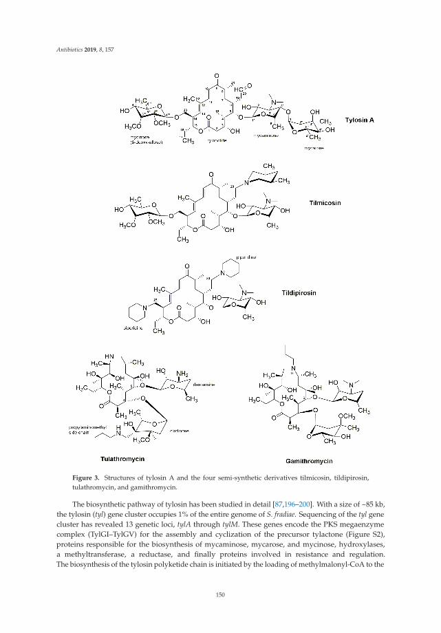
Antibiotics 2019, 8, 157
Figure 3. Structures of tylosin A and the four semi-synthetic derivatives tilmicosin, tildipirosin,tulathromycin, and gamithromycin.
The biosynthetic pathway of tylosin has been studied in detail [87,196–200]. With a size of ~85 kb,the tylosin (tyl) gene cluster occupies 1% of the entire genome of S. fradiae. Sequencing of the tyl genecluster has revealed 13 genetic loci, tylA through tylM. These genes encode the PKS megaenzymecomplex (TylGI–TylGV) for the assembly and cyclization of the precursor tylactone (Figure S2),proteins responsible for the biosynthesis of mycaminose, mycarose, and mycinose, hydroxylases,a methyltransferase, a reductase, and finally proteins involved in resistance and regulation.The biosynthesis of the tylosin polyketide chain is initiated by the loading of methylmalonyl-CoA to the
150
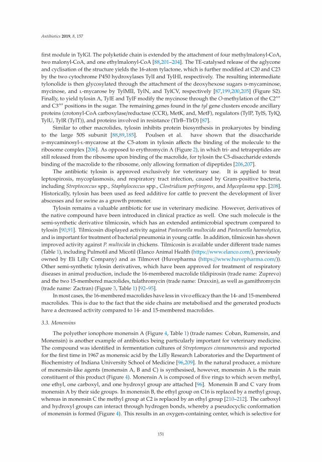
Antibiotics 2019, 8, 157
first module in TylGI. The polyketide chain is extended by the attachment of four methylmalonyl-CoA,two malonyl-CoA, and one ethylmalonyl-CoA [88,201–204]. The TE-catalysed release of the aglyconeand cyclisation of the structure yields the 16-atom tylactone, which is further modified at C20 and C23by the two cytochrome P450 hydroxylases TylI and TylHI, respectively. The resulting intermediatetylonolide is then glycosylated through the attachment of the deoxyhexose sugars d-mycaminose,mycinose, and l-mycarose by TylMII, TylN, and TylCV, respectively [87,199,200,205] (Figure S2).Finally, to yield tylosin A, TylE and TylF modify the mycinose through the O-methylation of the C2′′′and C3′′′ positions in the sugar. The remaining genes found in the tyl gene clusters encode ancillaryproteins (crotonyl-CoA carboxylase/reductase (CCR), MetK, and, MetF), regulators (TylP, TylS, TylQ,TylU, TylR (TylT)), and proteins involved in resistance (TlrB–TlrD) [87].
Similar to other macrolides, tylosin inhibits protein biosynthesis in prokaryotes by bindingto the large 50S subunit [88,89,185]. Poulsen et al. have shown that the disaccharided-mycaminosyl-l-mycarose at the C5-atom in tylosin affects the binding of the molecule to theribosome complex [206]. As opposed to erythromycin A (Figure 2), in which tri- and tetrapeptides arestill released from the ribosome upon binding of the macrolide, for tylosin the C5-disaccharide extendsbinding of the macrolide to the ribosome, only allowing formation of dipeptides [206,207].
The antibiotic tylosin is approved exclusively for veterinary use. It is applied to treatleptospirosis, mycoplasmosis, and respiratory tract infection, caused by Gram-positive bacteria,including Streptococcus spp., Staphylococcus spp., Clostridium perfringens, and Mycoplasma spp. [208].Historically, tylosin has been used as feed additive for cattle to prevent the development of liverabscesses and for swine as a growth promoter.
Tylosin remains a valuable antibiotic for use in veterinary medicine. However, derivatives ofthe native compound have been introduced in clinical practice as well. One such molecule is thesemi-synthetic derivative tilmicosin, which has an extended antimicrobial spectrum compared totylosin [90,91]. Tilmicosin displayed activity against Pasteurella multocida and Pasteurella haemolytica,and is important for treatment of bacterial pneumonia in young cattle. In addition, tilmicosin has shownimproved activity against P. multocida in chickens. Tilmicosin is available under different trade names(Table 1), including Pulmotil and Micotil (Elanco Animal Health (https://www.elanco.com/), previouslyowned by Eli Lilly Company) and as Tilmovet (Huvepharma (https://www.huvepharma.com/)).Other semi-synthetic tylosin derivatives, which have been approved for treatment of respiratorydiseases in animal production, include the 16-membered macrolide tildipirosin (trade name: Zuprevo)and the two 15-membered macrolides, tulathromycin (trade name: Draxxin), as well as gamithromycin(trade name: Zactran) (Figure 3, Table 1) [92–95].
In most cases, the 16-membered macrolides have less in vivo efficacy than the 14- and 15-memberedmacrolides. This is due to the fact that the side chains are metabolised and the generated productshave a decreased activity compared to 14- and 15-membered macrolides.
3.3. Monensins
The polyether ionophore monensin A (Figure 4, Table 1) (trade names: Coban, Rumensin, andMonensin) is another example of antibiotics being particularly important for veterinary medicine.The compound was identified in fermentation cultures of Streptomyces cinnamonensis and reportedfor the first time in 1967 as monensic acid by the Lilly Research Laboratories and the Department ofBiochemistry of Indiana University School of Medicine [96,209]. In the natural producer, a mixtureof monensin-like agents (monensin A, B and C) is synthesised, however, monensin A is the mainconstituent of this product (Figure 4). Monensin A is composed of five rings to which seven methyl,one ethyl, one carboxyl, and one hydroxyl group are attached [96]. Monensin B and C vary frommonensin A by their side groups. In monensin B, the ethyl group on C16 is replaced by a methyl group,whereas in monensin C the methyl group at C2 is replaced by an ethyl group [210–212]. The carboxyland hydroxyl groups can interact through hydrogen bonds, whereby a pseudocyclic conformationof monensin is formed (Figure 4). This results in an oxygen-containing center, which is selective for
151
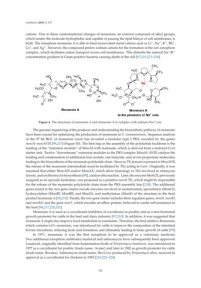
Antibiotics 2019, 8, 157
cations. Due to these conformational changes of monensin, an exterior composed of alkyl groups,which render the molecule hydrophobic and capable of passing the lipid bilayer of cell membranes, isbuilt. The ionophore monensin A is able to bind monovalent metal cations such as Li+, Na+, K+, Rb+,Cs+, and Ag+. However, the compound prefers sodium cations for the formation of the ion–ionophorecomplex, which facilitates cation transport across cell membranes. This disturbs the natural Na+/K+
concentration gradient in Gram-positive bacteria causing death of the cell [97,210,213–216].
Figure 4. The structures of monensin A and monensin A in complex with sodium (Na+) ion.
The genome sequencing of the producer and understanding the biosynthetic pathway of monensinhave been crucial for optimizing the production of monensin in S. cinnamonensis. Sequence analysisof the 97 kb BGC of monensin (mon) has revealed a modular type I PKS, encoded by the genesmonAI–monAVIII [98,217] (Figure S3). The first step in the assembly of the polyketide backbone is theloading of the “initiation module” of MonAI with malonate, which is derived from a malonyl-CoAstarter unit. Twelve “downstream” extension modules in the PKS complex MonAI–AVIII catalyse theloading and condensation of additional four acetate, one butyrate, and seven propionate molecules,leading to the biosynthesis of the monensin polyketide chain. Since no TE domain is present in MonAVII,the release of the monensin intermediate must be facilitated by TEs acting in trans. Originally, it wasassumed that either MonAIX and/or MonAX, which show homology to TEs involved in rifamycin,tylosin, and erythromycin biosynthesis [98], catalyse this reaction. Later, the enzyme MonCII, previouslyassigned as an epoxide hydrolase, was proposed as a putative novel TE, which might be responsiblefor the release of the monensin polyketide chain from the PKS assembly line [218]. The additionalgenes found in the mon gene cluster encode enzymes involved in isomerisation, epoxidation (MonCI),hydroxylation (MonBI, MonBII, and MonD), and methylation (MonE) of the structure to the finalproduct monensin A [98,219]. Finally, the mon gene cluster includes three regulator genes, monH, monRI,and monRII, and the gene monT, which encodes an efflux protein, believed to confer self-resistance tothe host [98,217,220,221].
Monensin A is used as a coccidiostat (inhibitor of coccidiosis) in poultry and as a non-hormonalgrowth promoter for cattle in the beef and dairy industry [97,210]. In addition, it was suggested thatmonensin A might also improve food metabolism in ruminants. Therefore, the feed additive Rumensin,which contains 6.6% monensin, was introduced for cattle to improve the composition of the intestinalbovine microbiota, reducing lactic acid formation, and ultimately leading to faster growth of cattle [99].
In 1971, monensin A was the first ionophore to be approved as a veterinary medicine.Two additional ionophore antibiotics lasalocid and salinomycin have subsequently been approved.Lasalocid, originally identified from fermentation broth of Streptomyces lasaliensis, was introduced in1977 as a coccidiostat for poultry (trade name: Avatec) and later in 1982 as growth promoter for cattle(trade name: Bovatec). Salinomycin (trade name: Bio-Cox), produced by Streptomyces albus, received itsapproval as a coccidiostat for chickens in 1983 [210,222–224].
152
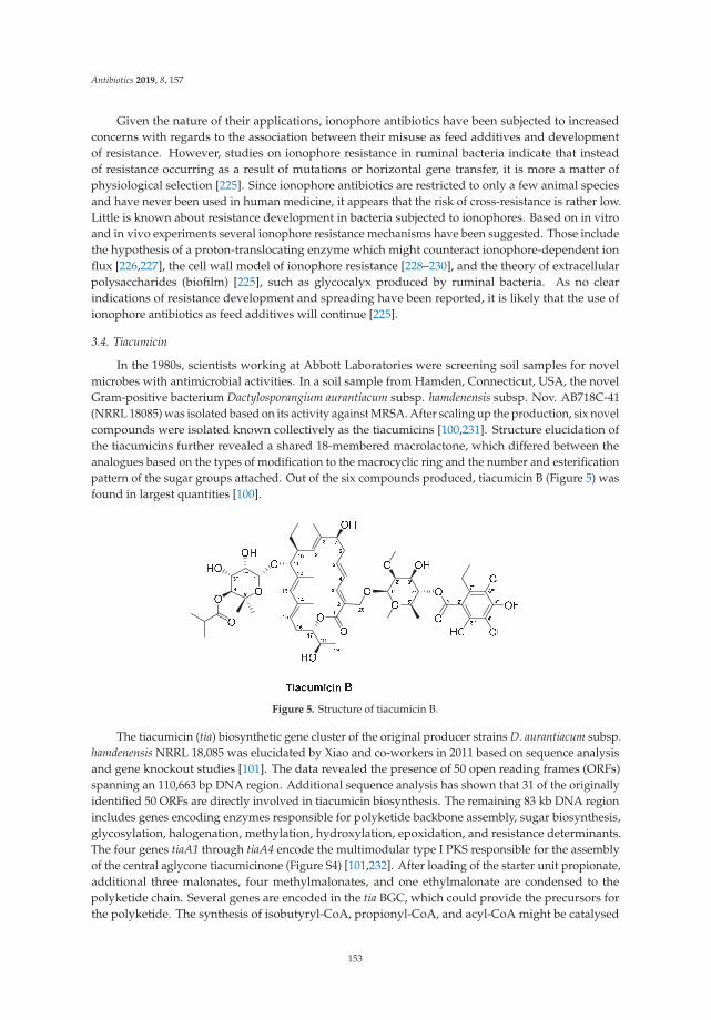
Antibiotics 2019, 8, 157
Given the nature of their applications, ionophore antibiotics have been subjected to increasedconcerns with regards to the association between their misuse as feed additives and developmentof resistance. However, studies on ionophore resistance in ruminal bacteria indicate that insteadof resistance occurring as a result of mutations or horizontal gene transfer, it is more a matter ofphysiological selection [225]. Since ionophore antibiotics are restricted to only a few animal speciesand have never been used in human medicine, it appears that the risk of cross-resistance is rather low.Little is known about resistance development in bacteria subjected to ionophores. Based on in vitroand in vivo experiments several ionophore resistance mechanisms have been suggested. Those includethe hypothesis of a proton-translocating enzyme which might counteract ionophore-dependent ionflux [226,227], the cell wall model of ionophore resistance [228–230], and the theory of extracellularpolysaccharides (biofilm) [225], such as glycocalyx produced by ruminal bacteria. As no clearindications of resistance development and spreading have been reported, it is likely that the use ofionophore antibiotics as feed additives will continue [225].
3.4. Tiacumicin
In the 1980s, scientists working at Abbott Laboratories were screening soil samples for novelmicrobes with antimicrobial activities. In a soil sample from Hamden, Connecticut, USA, the novelGram-positive bacterium Dactylosporangium aurantiacum subsp. hamdenensis subsp. Nov. AB718C-41(NRRL 18085) was isolated based on its activity against MRSA. After scaling up the production, six novelcompounds were isolated known collectively as the tiacumicins [100,231]. Structure elucidation ofthe tiacumicins further revealed a shared 18-membered macrolactone, which differed between theanalogues based on the types of modification to the macrocyclic ring and the number and esterificationpattern of the sugar groups attached. Out of the six compounds produced, tiacumicin B (Figure 5) wasfound in largest quantities [100].
Figure 5. Structure of tiacumicin B.
The tiacumicin (tia) biosynthetic gene cluster of the original producer strains D. aurantiacum subsp.hamdenensis NRRL 18,085 was elucidated by Xiao and co-workers in 2011 based on sequence analysisand gene knockout studies [101]. The data revealed the presence of 50 open reading frames (ORFs)spanning an 110,663 bp DNA region. Additional sequence analysis has shown that 31 of the originallyidentified 50 ORFs are directly involved in tiacumicin biosynthesis. The remaining 83 kb DNA regionincludes genes encoding enzymes responsible for polyketide backbone assembly, sugar biosynthesis,glycosylation, halogenation, methylation, hydroxylation, epoxidation, and resistance determinants.The four genes tiaA1 through tiaA4 encode the multimodular type I PKS responsible for the assemblyof the central aglycone tiacumicinone (Figure S4) [101,232]. After loading of the starter unit propionate,additional three malonates, four methylmalonates, and one ethylmalonate are condensed to thepolyketide chain. Several genes are encoded in the tia BGC, which could provide the precursors forthe polyketide. The synthesis of isobutyryl-CoA, propionyl-CoA, and acyl-CoA might be catalysed
153
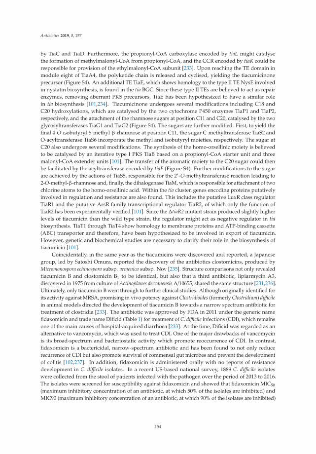
Antibiotics 2019, 8, 157
by TiaC and TiaD. Furthermore, the propionyl-CoA carboxylase encoded by tiaL might catalysethe formation of methylmalonyl-CoA from propionyl-CoA, and the CCR encoded by tiaK could beresponsible for provision of the ethylmalonyl-CoA subunit [233]. Upon reaching the TE domain inmodule eight of TiaA4, the polyketide chain is released and cyclised, yielding the tiacumicinoneprecursor (Figure S4). An additional TE TiaE, which shows homology to the type II TE NysE involvedin nystatin biosynthesis, is found in the tia BGC. Since these type II TEs are believed to act as repairenzymes, removing aberrant PKS precursors, TiaE has been hypothesized to have a similar rolein tia biosynthesis [101,234]. Tiacumicinone undergoes several modifications including C18 andC20 hydroxylations, which are catalysed by the two cytochrome P450 enzymes TiaP1 and TiaP2,respectively, and the attachment of the rhamnose sugars at position C11 and C20, catalysed by the twoglycosyltransferases TiaG1 and TiaG2 (Figure S4). The sugars are further modified. First, to yield thefinal 4-O-isobutyryl-5-methyl-β-rhamnose at position C11, the sugar C-methyltransferase TiaS2 andO-acyltransferase TiaS6 incorporate the methyl and isobutyryl moieties, respectively. The sugar atC20 also undergoes several modifications. The synthesis of the homo-orsellinic moiety is believedto be catalysed by an iterative type I PKS TiaB based on a propionyl-CoA starter unit and threemalonyl-CoA extender units [101]. The transfer of the aromatic moiety to the C20 sugar could thenbe facilitated by the acyltransferase encoded by tiaF (Figure S4). Further modifications to the sugarare achieved by the actions of TiaS5, responsible for the 2′-O-methyltransferase reaction leading to2-O-methyl-β-rhamnose and, finally, the dihalogenase TiaM, which is responsible for attachment of twochlorine atoms to the homo-orsellinic acid. Within the tia cluster, genes encoding proteins putativelyinvolved in regulation and resistance are also found. This includes the putative LuxR class regulatorTiaR1 and the putative ArsR family transcriptional regulator TiaR2, of which only the function ofTiaR2 has been experimentally verified [101]. Since the ΔtiaR2 mutant strain produced slightly higherlevels of tiacumicin than the wild type strain, the regulator might act as negative regulator in tiabiosynthesis. TiaT1 through TiaT4 show homology to membrane proteins and ATP-binding cassette(ABC) transporter and therefore, have been hypothesized to be involved in export of tiacumicin.However, genetic and biochemical studies are necessary to clarify their role in the biosynthesis oftiacumicin [101].
Coincidentally, in the same year as the tiacumicins were discovered and reported, a Japanesegroup, led by Satoshi Omura, reported the discovery of the antibiotics clostomicins, produced byMicromonospora echinospora subsp. armenica subsp. Nov [235]. Structure comparisons not only revealedtiacumicin B and clostomicin B1 to be identical, but also that a third antibiotic, lipiarmycin A3,discovered in 1975 from culture of Actinoplanes deccanensis A/10655, shared the same structure [231,236].Ultimately, only tiacumicin B went through to further clinical studies. Although originally identified forits activity against MRSA, promising in vivo potency against Clostridioides (formerly Clostridium) difficilein animal models directed the development of tiacumicin B towards a narrow spectrum antibiotic fortreatment of clostridia [233]. The antibiotic was approved by FDA in 2011 under the generic namefidaxomicin and trade name Dificid (Table 1) for treatment of C. difficile infections (CDI), which remainsone of the main causes of hospital-acquired diarrhoea [233]. At the time, Dificid was regarded as analternative to vancomycin, which was used to treat CDI. One of the major drawbacks of vancomycinis its broad-spectrum and bacteriostatic activity which promote reoccurrence of CDI. In contrast,fidaxomicin is a bactericidal, narrow-spectrum antibiotic and has been found to not only reducerecurrence of CDI but also promote survival of commensal gut microbes and prevent the developmentof colitis [102,237]. In addition, fidaxomicin is administered orally with no reports of resistancedevelopment in C. difficile isolates. In a recent US-based national survey, 1889 C. difficile isolateswere collected from the stool of patients infected with the pathogen over the period of 2013 to 2016.The isolates were screened for susceptibility against fidaxomicin and showed that fidaxomicin MIC50
(maximum inhibitory concentration of an antibiotic, at which 50% of the isolates are inhibited) andMIC90 (maximum inhibitory concentration of an antibiotic, at which 90% of the isolates are inhibited)
154
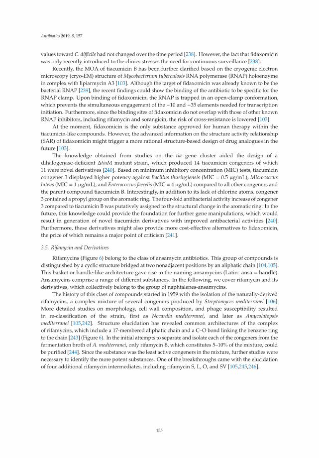
Antibiotics 2019, 8, 157
values toward C. difficile had not changed over the time period [238]. However, the fact that fidaxomicinwas only recently introduced to the clinics stresses the need for continuous surveillance [238].
Recently, the MOA of tiacumicin B has been further clarified based on the cryogenic electronmicroscopy (cryo-EM) structure of Mycobacterium tuberculosis RNA polymerase (RNAP) holoenzymein complex with lipiarmycin A3 [103]. Although the target of fidaxomicin was already known to be thebacterial RNAP [239], the recent findings could show the binding of the antibiotic to be specific for theRNAP clamp. Upon binding of fidaxomicin, the RNAP is trapped in an open-clamp conformation,which prevents the simultaneous engagement of the −10 and −35 elements needed for transcriptioninitiation. Furthermore, since the binding sites of fidaxomicin do not overlap with those of other knownRNAP inhibitors, including rifamycin and sorangicin, the risk of cross-resistance is lowered [103].
At the moment, fidaxomicin is the only substance approved for human therapy within thetiacumicin-like compounds. However, the advanced information on the structure activity relationship(SAR) of fidaxomicin might trigger a more rational structure-based design of drug analogues in thefuture [103].
The knowledge obtained from studies on the tia gene cluster aided the design of adihalogenase-deficient ΔtiaM mutant strain, which produced 14 tiacumicin congeners of which11 were novel derivatives [240]. Based on minimum inhibitory concentration (MIC) tests, tiacumicincongener 3 displayed higher potency against Bacillus thuringiensis (MIC = 0.5 μg/mL), Micrococcusluteus (MIC = 1 μg/mL), and Enterococcus faecelis (MIC = 4 μg/mL) compared to all other congeners andthe parent compound tiacumicin B. Interestingly, in addition to its lack of chlorine atoms, congener3 contained a propyl group on the aromatic ring. The four-fold antibacterial activity increase of congener3 compared to tiacumicin B was putatively assigned to the structural change in the aromatic ring. In thefuture, this knowledge could provide the foundation for further gene manipulations, which wouldresult in generation of novel tiacumicin derivatives with improved antibacterial activities [240].Furthermore, these derivatives might also provide more cost-effective alternatives to fidaxomicin,the price of which remains a major point of criticism [241].
3.5. Rifamycin and Derivatives
Rifamycins (Figure 6) belong to the class of ansamycin antibiotics. This group of compounds isdistinguished by a cyclic structure bridged at two nonadjacent positions by an aliphatic chain [104,105].This basket or handle-like architecture gave rise to the naming ansamycins (Latin: ansa = handle).Ansamycins comprise a range of different substances. In the following, we cover rifamycin and itsderivatives, which collectively belong to the group of naphtalenes-ansamycins.
The history of this class of compounds started in 1959 with the isolation of the naturally-derivedrifamycins, a complex mixture of several congeners produced by Streptomyces mediterranei [106].More detailed studies on morphology, cell wall composition, and phage susceptibility resultedin re-classification of the strain, first as Nocardia mediterranei, and later as Amycolatopsismediterranei [105,242]. Structure elucidation has revealed common architectures of the complexof rifamycins, which include a 17-membered aliphatic chain and a C–O bond linking the benzene ringto the chain [243] (Figure 6). In the initial attempts to separate and isolate each of the congeners from thefermentation broth of A. mediterranei, only rifamycin B, which constitutes 5–10% of the mixture, couldbe purified [244]. Since the substance was the least active congeners in the mixture, further studies werenecessary to identify the more potent substances. One of the breakthroughs came with the elucidationof four additional rifamycin intermediates, including rifamycin S, L, O, and SV [105,245,246].
155
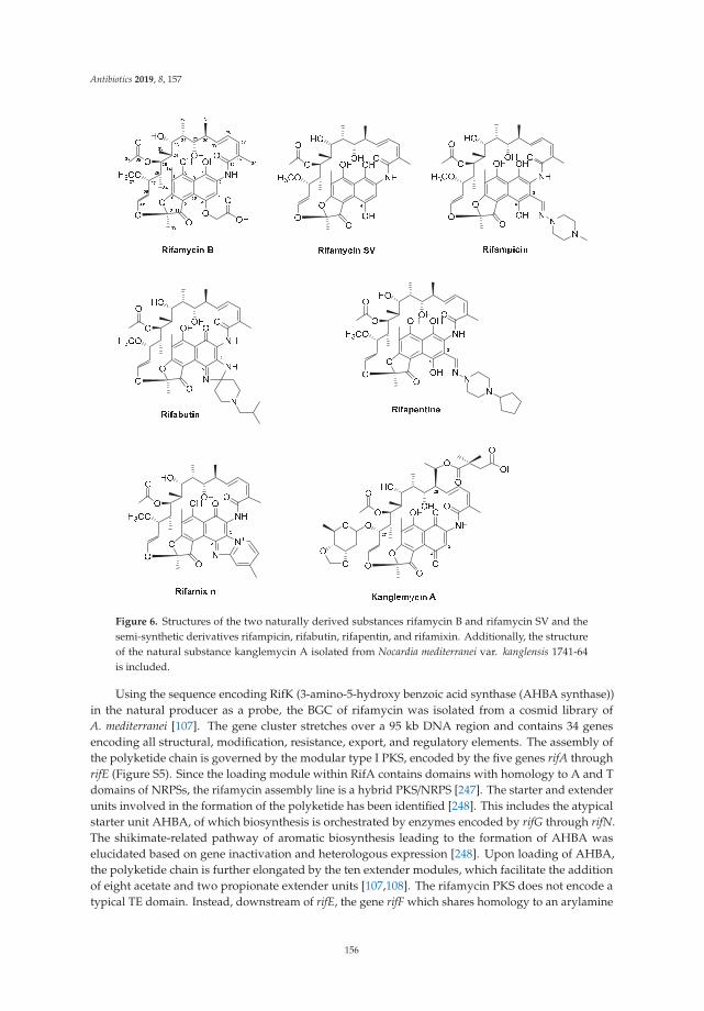
Antibiotics 2019, 8, 157
Figure 6. Structures of the two naturally derived substances rifamycin B and rifamycin SV and thesemi-synthetic derivatives rifampicin, rifabutin, rifapentin, and rifamixin. Additionally, the structureof the natural substance kanglemycin A isolated from Nocardia mediterranei var. kanglensis 1741-64is included.
Using the sequence encoding RifK (3-amino-5-hydroxy benzoic acid synthase (AHBA synthase))in the natural producer as a probe, the BGC of rifamycin was isolated from a cosmid library ofA. mediterranei [107]. The gene cluster stretches over a 95 kb DNA region and contains 34 genesencoding all structural, modification, resistance, export, and regulatory elements. The assembly ofthe polyketide chain is governed by the modular type I PKS, encoded by the five genes rifA throughrifE (Figure S5). Since the loading module within RifA contains domains with homology to A and Tdomains of NRPSs, the rifamycin assembly line is a hybrid PKS/NRPS [247]. The starter and extenderunits involved in the formation of the polyketide has been identified [248]. This includes the atypicalstarter unit AHBA, of which biosynthesis is orchestrated by enzymes encoded by rifG through rifN.The shikimate-related pathway of aromatic biosynthesis leading to the formation of AHBA waselucidated based on gene inactivation and heterologous expression [248]. Upon loading of AHBA,the polyketide chain is further elongated by the ten extender modules, which facilitate the additionof eight acetate and two propionate extender units [107,108]. The rifamycin PKS does not encode atypical TE domain. Instead, downstream of rifE, the gene rifF which shares homology to an arylamine
156
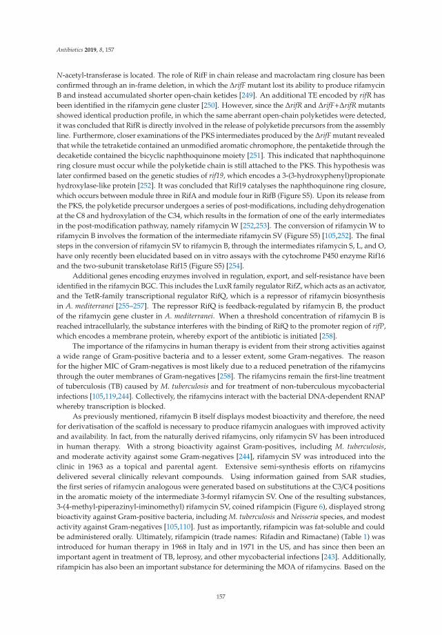
Antibiotics 2019, 8, 157
N-acetyl-transferase is located. The role of RifF in chain release and macrolactam ring closure has beenconfirmed through an in-frame deletion, in which the ΔrifF mutant lost its ability to produce rifamycinB and instead accumulated shorter open-chain ketides [249]. An additional TE encoded by rifR hasbeen identified in the rifamycin gene cluster [250]. However, since the ΔrifR and ΔrifF+ΔrifR mutantsshowed identical production profile, in which the same aberrant open-chain polyketides were detected,it was concluded that RifR is directly involved in the release of polyketide precursors from the assemblyline. Furthermore, closer examinations of the PKS intermediates produced by the ΔrifF mutant revealedthat while the tetraketide contained an unmodified aromatic chromophore, the pentaketide through thedecaketide contained the bicyclic naphthoquinone moiety [251]. This indicated that naphthoquinonering closure must occur while the polyketide chain is still attached to the PKS. This hypothesis waslater confirmed based on the genetic studies of rif19, which encodes a 3-(3-hydroxyphenyl)propionatehydroxylase-like protein [252]. It was concluded that Rif19 catalyses the naphthoquinone ring closure,which occurs between module three in RifA and module four in RifB (Figure S5). Upon its release fromthe PKS, the polyketide precursor undergoes a series of post-modifications, including dehydrogenationat the C8 and hydroxylation of the C34, which results in the formation of one of the early intermediatesin the post-modification pathway, namely rifamycin W [252,253]. The conversion of rifamycin W torifamycin B involves the formation of the intermediate rifamycin SV (Figure S5) [105,252]. The finalsteps in the conversion of rifamycin SV to rifamycin B, through the intermediates rifamycin S, L, and O,have only recently been elucidated based on in vitro assays with the cytochrome P450 enzyme Rif16and the two-subunit transketolase Rif15 (Figure S5) [254].
Additional genes encoding enzymes involved in regulation, export, and self-resistance have beenidentified in the rifamycin BGC. This includes the LuxR family regulator RifZ, which acts as an activator,and the TetR-family transcriptional regulator RifQ, which is a repressor of rifamycin biosynthesisin A. mediterranei [255–257]. The repressor RifQ is feedback-regulated by rifamycin B, the productof the rifamycin gene cluster in A. mediterranei. When a threshold concentration of rifamycin B isreached intracellularly, the substance interferes with the binding of RifQ to the promoter region of rifP,which encodes a membrane protein, whereby export of the antibiotic is initiated [258].
The importance of the rifamycins in human therapy is evident from their strong activities againsta wide range of Gram-positive bacteria and to a lesser extent, some Gram-negatives. The reasonfor the higher MIC of Gram-negatives is most likely due to a reduced penetration of the rifamycinsthrough the outer membranes of Gram-negatives [258]. The rifamycins remain the first-line treatmentof tuberculosis (TB) caused by M. tuberculosis and for treatment of non-tuberculous mycobacterialinfections [105,119,244]. Collectively, the rifamycins interact with the bacterial DNA-dependent RNAPwhereby transcription is blocked.
As previously mentioned, rifamycin B itself displays modest bioactivity and therefore, the needfor derivatisation of the scaffold is necessary to produce rifamycin analogues with improved activityand availability. In fact, from the naturally derived rifamycins, only rifamycin SV has been introducedin human therapy. With a strong bioactivity against Gram-positives, including M. tuberculosis,and moderate activity against some Gram-negatives [244], rifamycin SV was introduced into theclinic in 1963 as a topical and parental agent. Extensive semi-synthesis efforts on rifamycinsdelivered several clinically relevant compounds. Using information gained from SAR studies,the first series of rifamycin analogous were generated based on substitutions at the C3/C4 positionsin the aromatic moiety of the intermediate 3-formyl rifamycin SV. One of the resulting substances,3-(4-methyl-piperazinyl-iminomethyl) rifamycin SV, coined rifampicin (Figure 6), displayed strongbioactivity against Gram-positive bacteria, including M. tuberculosis and Neisseria species, and modestactivity against Gram-negatives [105,110]. Just as importantly, rifampicin was fat-soluble and couldbe administered orally. Ultimately, rifampicin (trade names: Rifadin and Rimactane) (Table 1) wasintroduced for human therapy in 1968 in Italy and in 1971 in the US, and has since then been animportant agent in treatment of TB, leprosy, and other mycobacterial infections [243]. Additionally,rifampicin has also been an important substance for determining the MOA of rifamycins. Based on the
157
Antibiotics 2019, 8, 157
3.3 Å crystal structure of the RNAP core of Thermus aquaticus complexed with rifampicin, Campbell andco-workers determined the binding of the antibiotic to the β subunit within the DNA/RNA channel.This binding resulted in the physical blockage of the channel for the elongating RNA, when thetranscripts reach a size of two to three nucleotides in length [111]. Determining the MOA of rifampicinalso aided in understanding the high frequency of resistance development in rifamycin sensitivemicroorganism. Since pathogens can develop resistance to rifampicin with a rate of 10−8 to 10−9
per cell division, the application of the antibiotic is restricted to combination therapy, in which theantimycobacterial agent isoniazid is often used together with rifampicin, or used only in severe casesof TB infection [105]. The high rate of resistance is most commonly associated with the acquisition ofmutations in the rpoB gene, which encodes the β subunit in the RNAP, whereby the binding affinity ofthe enzyme to the antibiotic is lowered. The rpoB gene is highly conserved in prokaryotes, however,pathogens acquire different levels of mutations in their respective rpoB genes, and consequently thelevel of resistance also can vary amongst isolates. For clinical isolates of M. tuberculosis, the threeamino acids S411, H406, and D396 (T. aquaticus numbering) are most often mutated. Mutations at thesepositions in the RNAP core influence the hydrogen bonds formed between the hydroxyl groups on C8and C21 of rifampicin, which interact with S411 and with H406 and D396, respectively [105,111].
Additional rifamycin analogues obtained through the derivatisation efforts on rifamycin SV havemade it through to the clinics (Figure 6). One of these analogues is the 3-(((4-cyclopentyl-1-piperazinyl)imino) methyl) rifamycin S, also known as rifapentine (trade name: Priftin) (Table 1). Rifapentinewas discovered during the initial derivatisation efforts on the C3/C4 aromatic moiety in rifamycin SV.The analogue was approved for treatment of TB in 1998 and similar to rifampicin can be administeredorally [115,259]. Unfortunately, isolates which are resistant to rifampicin display cross-resistance torifapentine, which has limited the application of the antibiotic.
The two rifamycin analogues rifabutin (trade name: Mycobutin) and rifaximin (trade names:Normix, Rifacol, Xifaxan) were approved for human therapy in 1992 and 2004, respectively(Table 1) [243]. Rifamixin ((4-deoxy-4)-methylpyrido [1),2)-1,2]imidazo-[5,4-c]rifamycin SV) isnon-systemic and can reach high concentrations in the gastrointestinal (GI) tract, enabling its applicationfor treatment of traveller’s diarrhoea caused by enteropathogens, including Escherichia coli [116].More recently, rifaximin has been further implicated in the treatment and prevention of variousGI diseases, including inflammatory bowel diseases such as Crohn’s disease and ulcerative colitis.Although the continuous administration of rifaximin has raised concerns for selection of resistance andundesirable disruption of the gut microbiota, several studies have shown that rifaximin may promotea healthy gut microbiota [117,260,261]. The beneficial effects of the antibiotic are lost quickly after thetreatment with rifaximin was terminated which probably limits its application as a preventive agent forGI-associated diseases [118]. Only recently, an alternative antibiotic combination has been approvedby the US FDA for the treatment of traveller’s diarrhoea. The oral formulation developed at CosmoPharmaceuticals N.V. (Ireland) is based on the unique combination of rifamycin SV together with theMultiMatrix (MMX®) technology, which ensures colonic delivery of the antibiotic [109]. Based onin vitro studies, rifamycin SV MMX® (trade names: Aemcolo and Relafalk) (Table 1) was foundto exhibit antibacterial activity against most enteropathogens associated with traveller’s diarrhoea.Moreover, in later phase III clinical studies, the oral substance was well-tolerated in patients undergoingtreatment and could shorten the duration of non-dysenteric traveller’s diarrhoea [109].
The semi-synthetic derivative rifabutin (4-N-isobutylspiropiperidylrifamycin S) was approved forhuman therapy due to its inhibitory activity against a number of rifampicin-resistant M. tuberculosisclinical isolates in addition to its well-documented activity against Mycobacterium avium andM. intracellulare (also referred to as Mycobacterium avium-intracellulare complex (MAC)) [112,113].MAC infection frequently occurs in patients with acquired immunodeficiency syndrome (AIDS)and when administered prophylactically as a 300 mg daily dose, rifabutin significantly reduces theincidence of MAC bacteraemia. However, due to the risk of cross-resistance between rifampicin and
158
Antibiotics 2019, 8, 157
rifabutin in M. tuberculosis isolates, rifabutin is restricted to MAC patients and for newly diagnosedand multidrug-resistant TB in Europe [114].
During the derivatisation of rifamycins, modifications to the ansa bridge often led to compoundswith lowered activities [262]. Recently, the natural ansamycin antibiotic kanglemycin A, which hasan altered ansa chain, was found to maintain potency against rifampicin-resistant bacterial isolates,including multidrug-resistant M. tuberculosis (Figure 6). Kanglemycin A was originally isolatedfrom Nocardia mediterranei var. kanglensis 1741-64 in 1988, however, the MOA of the compound wasonly recently elucidated. Based on the crystal structure of the RNAP-promoter complex of Thermusthermophiles with kanglemycin A, it became evident that the sugar (β-O-3,4-O,O′-methylene digitoxose)and acid (2,2-dimethyl succinic acid) moieties on the ansa bridge in the molecule increase the bindingsurface of the antibiotic with the RNAP. Furthermore, the additional interaction of the sugar group ofkanglemycin A was hypothesized to limit the frequency of resistance development as this would requiretwo simultaneous mutations in the binding pocket of the RNAP [119,120]. Although kanglemycin A(Table 1) itself has not been introduced to the clinics, its discovery might open up for future structuralderivatisation efforts in which the ansa bridge might prove to be a valuable “target” for generatingnew synthetic ansamycin antibiotics with improved bioactivities.
3.6. Tetracyclines
The tetracycline group of antibiotics covers a variety of natural and semi-synthetic compounds.There are three naturally derived tetracyclines that have been described in detail and have formedthe basis for 2nd and 3rd generation tetracycline derivatives. The three natural substances includechlortetracycline (trade names: Aureomycin, Table 1) discovered from the fermentation broth ofStreptomyces aureofaciens in 1948 [125], oxytetracycline (trade name: Terracycline, Table 1) foundin the broth of Streptomyces rimosus in 1950 [121], and finally tetracycline, which was discoveredas a fermentation product of Streptomyces viridofaciens and the two aforementioned strains [263](Figure 7). Based on the structural elucidation of the three compounds, which revealed a characteristicDCBA naphthacene tetra-cyclic core (Figure 7), the agents later became descriptively known as thetetracyclines [264]. Within the group of tetracyclines, tetracycline represents the simplest of thestructures, whereas chlortetracycline and oxytetracycline contain modifications at the core cyclicstructure. While chlortetracycline contains a chlorine atom at the C7 atom in ring D, oxytetracyclineinstead harbours a hydroxyl group at the C5 position in ring B.
The biosynthesis of chlortetracycline and oxytetracycline follows a similar logic and in thefollowing, we focus on the biosynthetic principle of the latter compound. The BGC of oxytetracyclinecovers a 21.2 kb DNA region in S. rimosus and contains 21 ORFs encoding structural, modification,resistance, and regulatory elements [265]. The biosynthesis of oxytetracycline starts with the assemblyof the polyketide precursor by the type II minimal PKS encoded by the genes oxyA through oxyC(Figure S6). More specifically, the KSα (OxyA) and KSβ (OxyB) form the heterodimer responsible forchain elongation, while the ACP (OxyC) provides the extender units. An interesting feature of thenatural tetracyclines is the unusual starter unit, malonamate, giving rise to the amide group on ringA in the naphthacene core [122,265]. The gene encoding the enzyme responsible for the provisionof this starter unit has been identified as oxyD, which is located immediately downstream of theminimal PKS-encoding genes oxyA through oxyC in the oxytetracycline gene cluster in S. rimosus.The protein sequence of OxyD shows homology to the ATP-dependent class II asparagine synthasesand it is believed to catalyse the transamination of malonate to yield malonamate [266]. Once attachedto the minimal PKS, the malonamate is condensed with eight additional malonates, giving rise tothe amidated decaketide backbone, which is released from the megaenzyme and further processedby AROs and CYCs encoded by genes in the oxytetracycline gene cluster (Figure S6). Based on theheterologous expression of the extended minimal PKS of oxytetracycline (OxyABCD) in Streptomycescoelicolor CH999, three enzymes have been identified and assumed to play a role in the correct cyclisationof the polyketide precursor [267]. This includes the NADPH-dependent KR OxyJ, which catalyses the
159
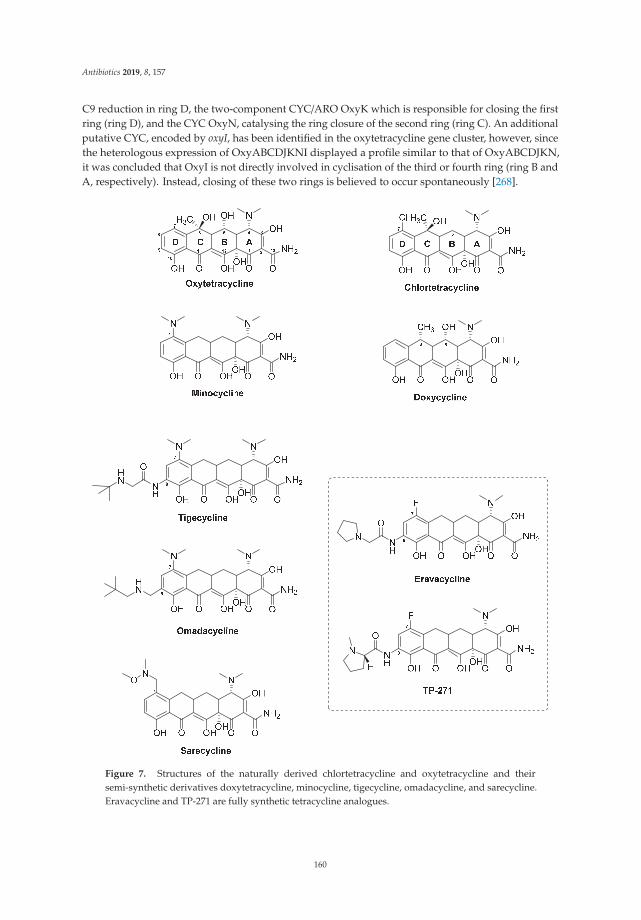
Antibiotics 2019, 8, 157
C9 reduction in ring D, the two-component CYC/ARO OxyK which is responsible for closing the firstring (ring D), and the CYC OxyN, catalysing the ring closure of the second ring (ring C). An additionalputative CYC, encoded by oxyI, has been identified in the oxytetracycline gene cluster, however, sincethe heterologous expression of OxyABCDJKNI displayed a profile similar to that of OxyABCDJKN,it was concluded that OxyI is not directly involved in cyclisation of the third or fourth ring (ring B andA, respectively). Instead, closing of these two rings is believed to occur spontaneously [268].
Figure 7. Structures of the naturally derived chlortetracycline and oxytetracycline and theirsemi-synthetic derivatives doxytetracycline, minocycline, tigecycline, omadacycline, and sarecycline.Eravacycline and TP-271 are fully synthetic tetracycline analogues.
160
Antibiotics 2019, 8, 157
Upon ring closure, the stable intermediate pretetramid is formed, which undergoes a seriesof modification reactions, of which the actual sequence remains to be clarified [269] (Figure S6).The modification of pretetramid involves a C6 methylation in ring C, which is catalysed bythe S-adenosyl methionine (SAM)-dependent methyltransferase OxyF, yielding the intermediate6-methylpretetramide. In the next step, the dihydroxylation of C4 and C12a in ring A, yielding thesubstance 4-keto-anhydrotetracycline, is catalysed by the two oxygenases OxyL and OxyE. While OxyLcan give rise to both hydroxylations, OxyE only catalyses the addition of the hydroxyl group at theC4 position. The reason for the extra function of OxyE seems to be a matter of increasing the rate ofthe enzymatic reaction [268]. The further conversion of 4-keto-anhydrotetracycline is achieved firstby the actions of the aminotransferase OxyQ, yielding the intermediate 4-amino-anhydrotetracycline,which then serves as a substrate for the SAM-dependent methyltransferase OxyT, catalysing aN, N-dimethylation that gives rise to anhydrotetracycline. The final enzymatic reactions leading toformation of oxytetracycline from anhydrotetracycline was recently elucidated by Wang and co-workers(Figure S6) [270]. In this study, OxyS was confirmed to catalyse the two sequential hydroxylations ofC6 and C5 in ring C and B, respectively. Furthermore, OxyR was shown to act as a F420-dependentreductase, which is responsible for the final C5a−C11a reduction affording oxytetracycline (Figure S6).In the case of chlortetracycline, an additional gene, ctcP, encoding a flavin-dependent halogenase,has been identified in the chlortetracycline BGC in S. aureofaciens [126]. CtcP yields the final productchlortetracycline based on the halogenation at position C7 in ring D (Figure S6). In addition to structuraland tailoring genes, the BGCs of chlortetracycline and oxytetracycline include additional genes involvedin resistance (otrA and otrB) and regulation (otcG and otcR). In the case of the oxytetracycline BGC,the two genes otrA and otrB, which encode proteins responsible for host resistance, are found at theflanking regions of the actual gene cluster. Based on sequence similarity, OtrA shows homology to TetMand is believed to provide resistance to S. rimosus by binding the ribosome and competing with theproduced oxytetracycline. OtrB encodes a membrane protein and ensures the efflux of oxytetracyclinefrom the cell [269,271]. The genes for the regulators OtcG and OtcR are also present in the flankingregions of the oxytetracycline BGC in S. rimosus [269,272,273]. OtcG, is a LuxR family transcriptionalactivator which was shown to provide positive regulation of oxytetracycline biosynthesis since aninactivation of otcG resulted in a mutant strain producing 40% less oxytetracycline than the wild typestrain S. rimosus [272]. The overexpression of otcG did not yield a significant increase in production ofoxytetracycline. OtcR was assigned to a Streptomyces antibiotic regulatory protein (SARP) activator ofthe oxytetracycline in S. rimosus [273]. Since a deletion of otcR completely abolished oxytetracyclineproduction in the producer strain, the authors tested whether its overexpression would result inincreased yields of the antibiotic. Indeed, the introduction two additional copies of otcR, under thecontrol of a SF14 promoter, in S. rimosus increased the yields of oxytetracycline to 6.24 g/L (6.49 timeshigher than the yields of the parental strain) [273].
Tetracyclines are broad-spectrum antibiotics with activities against a range of microorganisms,including Gram-positive and Gram-negative bacteria, chlamydiae, mycoplasmas, and protozoanparasites [123]. They act as bacteriostatic drugs by binding to the 30S subunit of the prokaryoteribosome in a reversible fashion whereby protein synthesis is inhibited [132]. Furthermore, crystalstructures of tetracycline in complex with both, the 30S subunit and 70S ribosome of Thermus thermophiles,have expanded the knowledge of the MOA of tetracyclines [124,274]. Based on the first X-ray structureof Brodersen et al., the primary binding site of tetracycline (Tet1) was found to be located between thehead and shoulder of the 30S subunit, just above the transfer RNA (tRNA) binding site. When bindingthe A site, tetracycline act as a physical barrier for aminoacyl-tRNA so that the peptide chain cannotbe elongated and protein synthesis is stalled [123,274]. Furthermore, the oxidised hydrophilic partof the tetracyclines appear to provide the main chemical interactions with the 16S ribosomal RNA(rRNA) and therefore, they are most important for the binding [275]. Another interesting property ofthe tetracyclines is their ability to chelate ions (Mg2+). In addition to the hydrogen bond interactionsbetween the hydrophilic side of tetracycline and the nucleotides on the 16s rRNA, the Mg2+ ions,
161
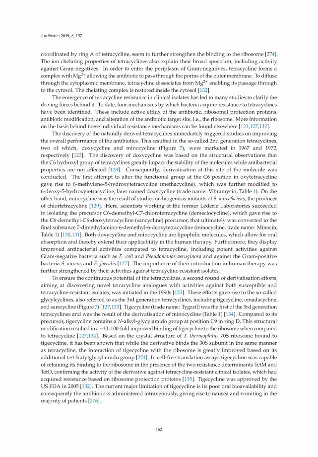
Antibiotics 2019, 8, 157
coordinated by ring A of tetracycline, seem to further strengthen the binding to the ribosome [274].The ion chelating properties of tetracyclines also explain their broad spectrum, including activityagainst Gram-negatives. In order to enter the periplasm of Gram-negatives, tetracycline forms acomplex with Mg2+ allowing the antibiotic to pass through the porins of the outer membrane. To diffusethrough the cytoplasmic membrane, tetracycline dissociates from Mg2+ enabling its passage throughto the cytosol. The chelating complex is restored inside the cytosol [132].
The emergence of tetracycline resistance in clinical isolates has led to many studies to clarify thedriving forces behind it. To date, four mechanisms by which bacteria acquire resistance to tetracyclineshave been identified. These include active efflux of the antibiotic, ribosomal protection proteins,antibiotic modification, and alteration of the antibiotic target site, i.e., the ribosome. More informationon the basis behind these individual resistance mechanisms can be found elsewhere [123,127,132].
The discovery of the naturally derived tetracyclines immediately triggered studies on improvingthe overall performance of the antibiotics. This resulted in the so-called 2nd generation tetracyclines,two of which, doxycycline and minocycline (Figure 7), were marketed in 1967 and 1972,respectively [123]. The discovery of doxycycline was based on the structural observations thatthe C6 hydroxyl group of tetracyclines greatly impact the stability of the molecules while antibacterialproperties are not affected [128]. Consequently, derivatisation at this site of the molecule wasconducted. The first attempt to alter the functional group at the C6 position in oxytetracyclinegave rise to 6-methylene-5-hydroxytetracycline (methacycline), which was further modified to6-deoxy-5-hydroxytetracycline, later named doxycycline (trade name: Vibramycin, Table 1). On theother hand, minocycline was the result of studies on biogenesis mutants of S. aureofaciens, the producerof chlortetracycline [129]. Here, scientists working at the former Lederle Laboratories succeededin isolating the precursor C6-demethyl-C7-chlorotetracycline (demeclocycline), which gave rise tothe C6-demethyl-C6-deoxytetracycline (sancycline) precursor, that ultimately was converted to thefinal substance 7-dimethylamino-6-demethyl-6-deoxytetracycline (minocycline, trade name: Minocin,Table 1) [130,131]. Both doxycycline and minocycline are lipophilic molecules, which allow for oralabsorption and thereby extend their applicability in the human therapy. Furthermore, they displayimproved antibacterial activities compared to tetracycline, including potent activities againstGram-negative bacteria such as E. coli and Pseudomonas aeruginosa and against the Gram-positivebacteria S. aureus and E. faecalis [127]. The importance of their introduction in human therapy wasfurther strengthened by their activities against tetracycline-resistant isolates.
To ensure the continuous potential of the tetracyclines, a second round of derivatisation efforts,aiming at discovering novel tetracycline analogues with activities against both susceptible andtetracycline-resistant isolates, was initiated in the 1990s [123]. These efforts gave rise to the so-calledglycylcyclines, also referred to as the 3rd generation tetracyclines, including tigecycline, omadacycline,and sarecycline (Figure 7) [127,133]. Tigecycline (trade name: Tygacil) was the first of the 3rd generationtetracyclines and was the result of the derivatisation of minocycline (Table 1) [134]. Compared to itsprecursor, tigecycline contains a N-alkyl-glycylamido group at position C9 in ring D. This structuralmodification resulted in a ~10–100-fold improved binding of tigecycline to the ribosome when comparedto tetracycline [127,134]. Based on the crystal structure of T. thermophilus 70S ribosome bound totigecycline, it has been shown that while the derivative binds the 30S subunit in the same manneras tetracycline, the interaction of tigecycline with the ribosome is greatly improved based on itsadditional tert-butylglycylamido group [274]. In cell-free translation assays tigecycline was capableof retaining its binding to the ribosome in the presence of the two resistance determinants TetM andTetO, confirming the activity of the derivative against tetracycline-resistant clinical isolates, which hadacquired resistance based on ribosome protection proteins [135]. Tigecycline was approved by theUS FDA in 2005 [132]. The current major limitation of tigecycline is its poor oral bioavailability andconsequently the antibiotic is administered intravenously, giving rise to nausea and vomiting in themajority of patients [276].
162
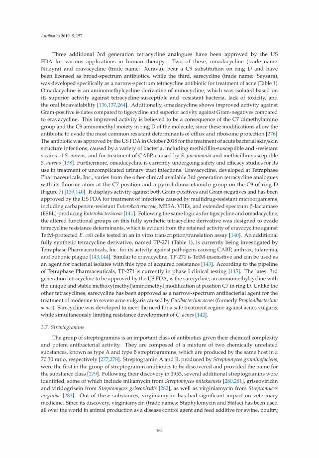
Antibiotics 2019, 8, 157
Three additional 3rd generation tetracycline analogues have been approved by the USFDA for various applications in human therapy. Two of these, omadacycline (trade name:Nuzyra) and eravacycline (trade name: Xerava), bear a C9 substitution on ring D and havebeen licensed as broad-spectrum antibiotics, while the third, sarecycline (trade name: Seysara),was developed specifically as a narrow-spectrum tetracycline antibiotic for treatment of acne (Table 1).Omadacycline is an aminomethylcycline derivative of minocycline, which was isolated based onits superior activity against tetracycline-susceptible and -resistant bacteria, lack of toxicity, andthe oral bioavailability [136,137,264]. Additionally, omadacycline shows improved activity againstGram-positive isolates compared to tigecycline and superior activity against Gram-negatives comparedto eravacycline. This improved activity is believed to be a consequence of the C7 dimethylaminogroup and the C9 aminomethyl moiety in ring D of the molecule, since these modifications allow theantibiotic to evade the most common resistant determinants of efflux and ribosome protection [276].The antibiotic was approved by the US FDA in October 2018 for the treatment of acute bacterial skin/skinstructure infections, caused by a variety of bacteria, including methicillin-susceptible and -resistantstrains of S. aureus, and for treatment of CABP, caused by S. pneumonia and methicillin-susceptibleS. aureus [138]. Furthermore, omadacycline is currently undergoing safety and efficacy studies for itsuse in treatment of uncomplicated urinary tract infections. Eravacycline, developed at TetraphasePharmaceuticals, Inc., varies from the other clinical available 3rd generation tetracycline analogueswith its fluorine atom at the C7 position and a pyrrolidinoacetamido group on the C9 of ring D(Figure 7) [139,140]. It displays activity against both Gram-positives and Gram-negatives and has beenapproved by the US FDA for treatment of infections caused by multidrug-resistant microorganisms,including carbapenem-resistant Enterobacteriaceae, MRSA, VREs, and extended spectrum β-lactamase(ESBL)-producing Enterobacteriaceae [141]. Following the same logic as for tigecycline and omadacycline,the altered functional groups on this fully synthetic tetracycline derivative was designed to evadetetracycline resistance determinants, which is evident from the retained activity of eravacycline againstTetM-protected E. coli cells tested in an in vitro transcription/translation assay [140]. An additionalfully synthetic tetracycline derivative, named TP-271 (Table 1), is currently being investigated byTetraphase Pharmaceuticals, Inc. for its activity against pathogens causing CABP, anthrax, tularemia,and bubonic plague [143,144]. Similar to eravacycline, TP-271 is TetM-insensitive and can be used asan agent for bacterial isolates with this type of acquired resistance [143]. According to the pipelineof Tetraphase Pharmaceuticals, TP-271 is currently in phase I clinical testing [145]. The latest 3rdgeneration tetracycline to be approved by the US FDA, is the sarecycline, an aminomethylcycline withthe unique and stable methoxy(methyl)aminomethyl modification at position C7 in ring D. Unlike theother tetracyclines, sarecycline has been approved as a narrow-spectrum antibacterial agent for thetreatment of moderate to severe acne vulgaris caused by Cutibacterium acnes (formerly Propionibacteriumacnes). Sarecycline was developed to meet the need for a safe treatment regime against acnes vulgaris,while simultaneously limiting resistance development of C. acnes [142].
3.7. Streptogramins
The group of streptogramins is an important class of antibiotics given their chemical complexityand potent antibacterial activity. They are composed of a mixture of two chemically unrelatedsubstances, known as type A and type B streptrogramins, which are produced by the same host in a70:30 ratio, respectively [277,278]. Streptogramin A and B, produced by Streptomyces graminofaciens,were the first in the group of streptogramin antibiotics to be discovered and provided the name forthe substance class [279]. Following their discovery in 1953, several additional streptogramins wereidentified, some of which include mikamycin from Streptomyces mitakaensis [280,281], griseoviridinand viridogrisein from Streptomyces griseoviridis [282], as well as virginiamycin from Streptomycesvirginiae [283]. Out of these substances, virginiamycin has had significant impact on veterinarymedicine. Since its discovery, virginiamycin (trade names: Staphylomycin and Stafac) has been usedall over the world in animal production as a disease control agent and feed additive for swine, poultry,
163
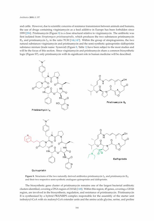
Antibiotics 2019, 8, 157
and cattle. However, due to scientific concerns of resistance transmission between animals and humans,the use of drugs containing virginiamycin as a feed additive in Europe has been forbidden since1999 [284]. Pristinamycin (Figure 8) is a close structural relative to virginiamycin. The antibiotic wasfirst isolated from Streptomyces pristinaespiralis, which produces the two substances pristinamycinIIA and pristinamycin IA in the ratio 70:30 [146,147]. Within the group of streptogramins, the twonatural substances virginiamycin and pristinamycin and the semi-synthetic quinupristin–dalfopristinsubstance mixture (trade name: Synercid) (Figure 8, Table 1) have been subject to the most studies andwill be the focus of this section. Since virginiamycin and pristinamycin share a common biosyntheticlogic (Figure S7), only pristinamycin with its significant role in human medicine will be described.
Figure 8. Structures of the two naturally derived antibiotics pristinamycin IA and pristinamycin IIA
and their two respective semi-synthetic analogues quinupristin and dalfopristin.
The biosynthetic gene cluster of pristinamycin remains one of the largest bacterial antibioticclusters identified, covering a DNA region of 210 kb [148]. Within this region, 45 genes, covering a 120 kbregion, are involved in the biosynthesis, regulation, and resistance of pristinamycin. PristinamycinII is synthesised by a hybrid PKS/NRPS complex responsible for the assembly of the starter unitisobutyryl-CoA with six malonyl-CoA extender units and the amino acids glycine, serine, and proline
164
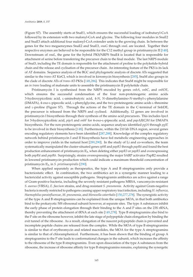
Antibiotics 2019, 8, 157
(Figure S7). The assembly starts at SnaE1, which ensures the successful loading of isobuturyl-CoAfollowed by its extension with two malonyl-CoA and glycine. The following four modules in SnaE2and SnaE3 attach additional four malonyl-CoA extender units to the growing chain. In between thegenes for the two megaenzymes SnaE2 and SnaE3, snaG through snaL are located. Together theirrespective enzymes are believed to be responsible for the C12 methyl group in pristinamycin II [148].Downstream of snaL, the gene for the hybrid PKS/NRPS SnaE4 is located that is responsible forattachment of serine before transferring the precursor chain to the final module. The last NRPS moduleof SnaD, including the TE domain is responsible for the attachment of proline to the polyketide-hybridchain and the release and cyclisation of the precursor chain. An interesting feature of the PKS is its lackof AT domains. Sequence analysis of the BGC and phylogenetic analysis of discrete ATs suggested thatsimilar to the trans-AT KirCI, which is involved in kirromycin biosynthesis [285], SnaM also groups inthe clade of discrete ATs of trans-AT-PKSs [148,286]. This indicates that SnaM might be responsible foran in trans loading of malonate units to assemble the pristinamycin II polyketide chain.
Pristinamycin I is synthesised from the NRPS encoded by genes snbA, snbC, and snbDE,which ensures the successful condensation of the four non-proteinogenic amino acids3-hydroxypicolinic acid, l-aminobutyric acid, 4-N, N-dimethylamino-N-methyl-l-phenylalanine(DMAPA), 4-oxo-l-pipecolic acid, l-phenylglycine, and the two proteinogenic amino acids l-threonineand l-proline (Figure S7). Through the actions of the TE domain in the C-terminal of SnbDE,the precursor is released from the NRPS and cyclised. Additionally, 12 genes are involved inpristinamycin I biosynthesis through their synthesis of the amino acid precursors. This includes hpaAfor 3-hydroxypicolinic acid, pipA and snbF for 4-oxo-l-pipecolic acid, and papA/B/C/M for DMAPAbiosynthesis. For the non-proteinogenic amino acids, sequence analyses identified pglA through pglEto be involved in their biosynthesis [148]. Furthermore, within the 210 kb DNA region, several genesencoding regulatory elements have been identified [287,288]. Knowledge of the complex regulatorynetwork behind pristinamycin I and II biosynthesis have fed metabolic engineering approaches inorder to improve yields in the natural host [289,290]. In the study of Li and co-workers, the teamsystematically manipulated the cluster-situated genes spbR and papR1 through papR6 and found the bestproduction enhancement of pristinamycin IIA when deleting either papR3 or papR5 and overexpressingboth papR4 and papR6. Surprisingly, strains overexpressing the major SARP activator PapR2 resultedin lowered pristinamycin production which could indicate a maximum threshold concentration ofpristinamycin IIA in S. pristinaespiralis [290].
When applied separately as therapeutics, the type A and B streptogramins only provide abacteriostatic effect. In combination, the two antibiotics act in a synergetic manner leading to abactericidal activity against susceptible pathogens. Streptogramin antibiotics are active against a rangeof Gram-positive bacteria, including the severely resistant pathogens MRSA, vancomycin-resistantS. aureus (VRSA), E. faecium strains, and drug-resistant S. pneumonia. Activity against Gram-negativebacteria is mostly restricted to pathogens causing upper respiratory tract infection, including H. influenza,Haemophilus parainfluenzae, M. pneumonia, and Moraxella catarrhalis [150,277,278]. The synergistic activityof the type A and B streptogramins can be explained from the unique MOA, in that both antibioticsbind to the prokaryotic 50S ribosomal subunit however, at separate sites. The type A substances inhibitthe early phase of protein elongation through their binding to the A and P sites on the 23S rRNA,thereby preventing the attachment of tRNA at each site [149,278]. Type B streptogramins also bind tothe P site on the ribosome however, inhibit the late stage of polypeptide chain elongation by binding theexit tunnel of the ribosome. As a result, elongation of the nascent polypeptide chain is prevented andincomplete peptide chains are released from the complex. While the MOA of type B streptograminsis similar to that of erythromycin and related macrolides, the MOA for the type A streptograminsis similar to that of chloramphenicol. Furthermore, it has been shown that the binding of group Astreptogramins to the P site leads to conformational changes in the subunit, which increases the affinityto the ribosome of the type B streptogramins. Even upon dissociation of the type A substances from theribosome, the increase of ribosome affinity for type B streptogramins remains, explaining the synergetic
165
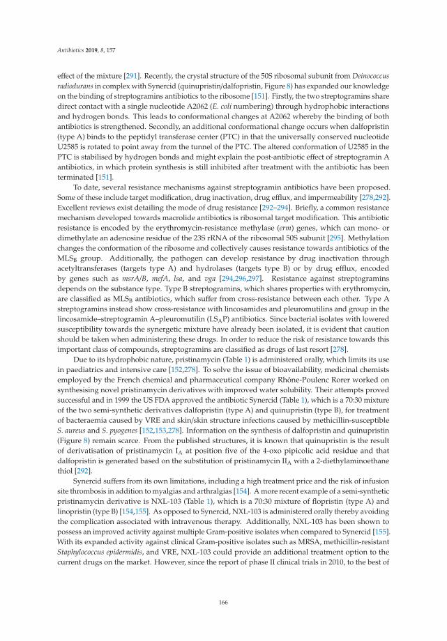
Antibiotics 2019, 8, 157
effect of the mixture [291]. Recently, the crystal structure of the 50S ribosomal subunit from Deinococcusradiodurans in complex with Synercid (quinupristin/dalfopristin, Figure 8) has expanded our knowledgeon the binding of streptogramins antibiotics to the ribosome [151]. Firstly, the two streptogramins sharedirect contact with a single nucleotide A2062 (E. coli numbering) through hydrophobic interactionsand hydrogen bonds. This leads to conformational changes at A2062 whereby the binding of bothantibiotics is strengthened. Secondly, an additional conformational change occurs when dalfopristin(type A) binds to the peptidyl transferase center (PTC) in that the universally conserved nucleotideU2585 is rotated to point away from the tunnel of the PTC. The altered conformation of U2585 in thePTC is stabilised by hydrogen bonds and might explain the post-antibiotic effect of streptogramin Aantibiotics, in which protein synthesis is still inhibited after treatment with the antibiotic has beenterminated [151].
To date, several resistance mechanisms against streptogramin antibiotics have been proposed.Some of these include target modification, drug inactivation, drug efflux, and impermeability [278,292].Excellent reviews exist detailing the mode of drug resistance [292–294]. Briefly, a common resistancemechanism developed towards macrolide antibiotics is ribosomal target modification. This antibioticresistance is encoded by the erythromycin-resistance methylase (erm) genes, which can mono- ordimethylate an adenosine residue of the 23S rRNA of the ribosomal 50S subunit [295]. Methylationchanges the conformation of the ribosome and collectively causes resistance towards antibiotics of theMLSB group. Additionally, the pathogen can develop resistance by drug inactivation throughacetyltransferases (targets type A) and hydrolases (targets type B) or by drug efflux, encodedby genes such as msrA/B, mefA, lsa, and vga [294,296,297]. Resistance against streptograminsdepends on the substance type. Type B streptogramins, which shares properties with erythromycin,are classified as MLSB antibiotics, which suffer from cross-resistance between each other. Type Astreptogramins instead show cross-resistance with lincosamides and pleuromutilins and group in thelincosamide–streptogramin A–pleuromutilin (LSAP) antibiotics. Since bacterial isolates with loweredsusceptibility towards the synergetic mixture have already been isolated, it is evident that cautionshould be taken when administering these drugs. In order to reduce the risk of resistance towards thisimportant class of compounds, streptogramins are classified as drugs of last resort [278].
Due to its hydrophobic nature, pristinamycin (Table 1) is administered orally, which limits its usein paediatrics and intensive care [152,278]. To solve the issue of bioavailability, medicinal chemistsemployed by the French chemical and pharmaceutical company Rhône-Poulenc Rorer worked onsynthesising novel pristinamycin derivatives with improved water solubility. Their attempts provedsuccessful and in 1999 the US FDA approved the antibiotic Synercid (Table 1), which is a 70:30 mixtureof the two semi-synthetic derivatives dalfopristin (type A) and quinupristin (type B), for treatmentof bacteraemia caused by VRE and skin/skin structure infections caused by methicillin-susceptibleS. aureus and S. pyogenes [152,153,278]. Information on the synthesis of dalfopristin and quinupristin(Figure 8) remain scarce. From the published structures, it is known that quinupristin is the resultof derivatisation of pristinamycin IA at position five of the 4-oxo pipicolic acid residue and thatdalfopristin is generated based on the substitution of pristinamycin IIA with a 2-diethylaminoethanethiol [292].
Synercid suffers from its own limitations, including a high treatment price and the risk of infusionsite thrombosis in addition to myalgias and arthralgias [154]. A more recent example of a semi-syntheticpristinamycin derivative is NXL-103 (Table 1), which is a 70:30 mixture of flopristin (type A) andlinopristin (type B) [154,155]. As opposed to Synercid, NXL-103 is administered orally thereby avoidingthe complication associated with intravenous therapy. Additionally, NXL-103 has been shown topossess an improved activity against multiple Gram-positive isolates when compared to Synercid [155].With its expanded activity against clinical Gram-positive isolates such as MRSA, methicillin-resistantStaphylococcus epidermidis, and VRE, NXL-103 could provide an additional treatment option to thecurrent drugs on the market. However, since the report of phase II clinical trials in 2010, to the best of
166
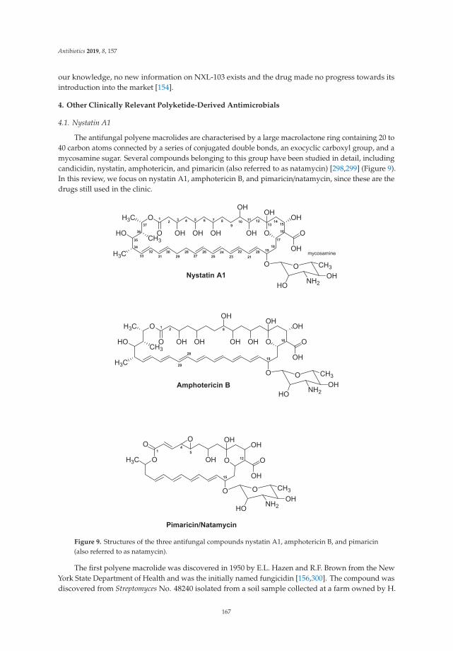
Antibiotics 2019, 8, 157
our knowledge, no new information on NXL-103 exists and the drug made no progress towards itsintroduction into the market [154].
4. Other Clinically Relevant Polyketide-Derived Antimicrobials
4.1. Nystatin A1
The antifungal polyene macrolides are characterised by a large macrolactone ring containing 20 to40 carbon atoms connected by a series of conjugated double bonds, an exocyclic carboxyl group, and amycosamine sugar. Several compounds belonging to this group have been studied in detail, includingcandicidin, nystatin, amphotericin, and pimaricin (also referred to as natamycin) [298,299] (Figure 9).In this review, we focus on nystatin A1, amphotericin B, and pimaricin/natamycin, since these are thedrugs still used in the clinic.
Figure 9. Structures of the three antifungal compounds nystatin A1, amphotericin B, and pimaricin(also referred to as natamycin).
The first polyene macrolide was discovered in 1950 by E.L. Hazen and R.F. Brown from the NewYork State Department of Health and was the initially named fungicidin [156,300]. The compound wasdiscovered from Streptomyces No. 48240 isolated from a soil sample collected at a farm owned by H.
167
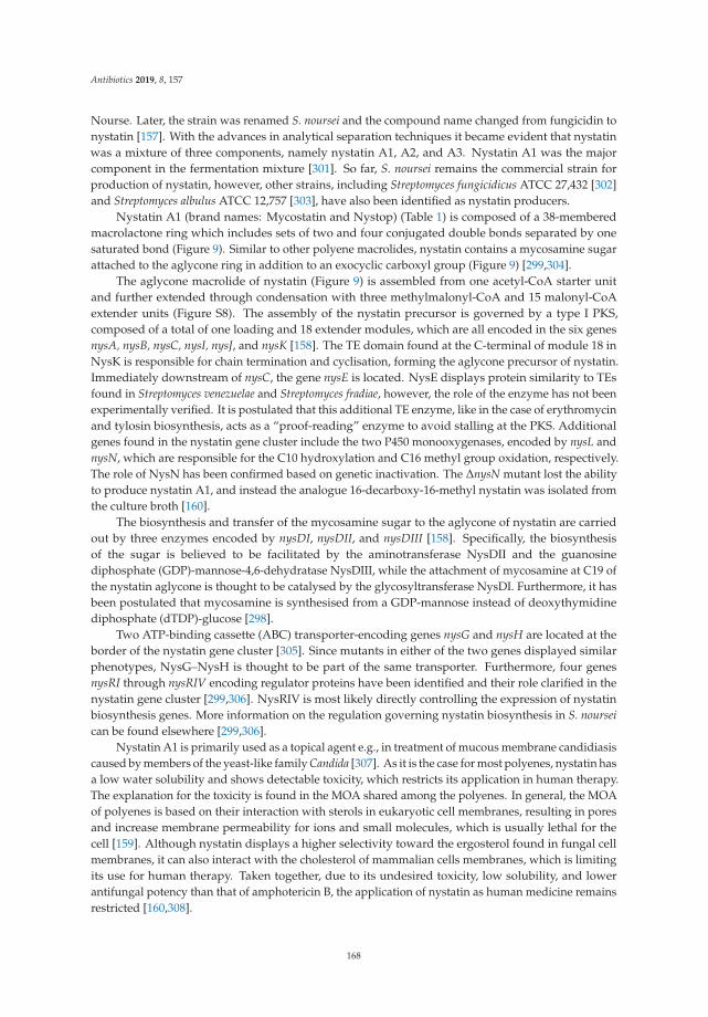
Antibiotics 2019, 8, 157
Nourse. Later, the strain was renamed S. noursei and the compound name changed from fungicidin tonystatin [157]. With the advances in analytical separation techniques it became evident that nystatinwas a mixture of three components, namely nystatin A1, A2, and A3. Nystatin A1 was the majorcomponent in the fermentation mixture [301]. So far, S. noursei remains the commercial strain forproduction of nystatin, however, other strains, including Streptomyces fungicidicus ATCC 27,432 [302]and Streptomyces albulus ATCC 12,757 [303], have also been identified as nystatin producers.
Nystatin A1 (brand names: Mycostatin and Nystop) (Table 1) is composed of a 38-memberedmacrolactone ring which includes sets of two and four conjugated double bonds separated by onesaturated bond (Figure 9). Similar to other polyene macrolides, nystatin contains a mycosamine sugarattached to the aglycone ring in addition to an exocyclic carboxyl group (Figure 9) [299,304].
The aglycone macrolide of nystatin (Figure 9) is assembled from one acetyl-CoA starter unitand further extended through condensation with three methylmalonyl-CoA and 15 malonyl-CoAextender units (Figure S8). The assembly of the nystatin precursor is governed by a type I PKS,composed of a total of one loading and 18 extender modules, which are all encoded in the six genesnysA, nysB, nysC, nysI, nysJ, and nysK [158]. The TE domain found at the C-terminal of module 18 inNysK is responsible for chain termination and cyclisation, forming the aglycone precursor of nystatin.Immediately downstream of nysC, the gene nysE is located. NysE displays protein similarity to TEsfound in Streptomyces venezuelae and Streptomyces fradiae, however, the role of the enzyme has not beenexperimentally verified. It is postulated that this additional TE enzyme, like in the case of erythromycinand tylosin biosynthesis, acts as a “proof-reading” enzyme to avoid stalling at the PKS. Additionalgenes found in the nystatin gene cluster include the two P450 monooxygenases, encoded by nysL andnysN, which are responsible for the C10 hydroxylation and C16 methyl group oxidation, respectively.The role of NysN has been confirmed based on genetic inactivation. The ΔnysN mutant lost the abilityto produce nystatin A1, and instead the analogue 16-decarboxy-16-methyl nystatin was isolated fromthe culture broth [160].
The biosynthesis and transfer of the mycosamine sugar to the aglycone of nystatin are carriedout by three enzymes encoded by nysDI, nysDII, and nysDIII [158]. Specifically, the biosynthesisof the sugar is believed to be facilitated by the aminotransferase NysDII and the guanosinediphosphate (GDP)-mannose-4,6-dehydratase NysDIII, while the attachment of mycosamine at C19 ofthe nystatin aglycone is thought to be catalysed by the glycosyltransferase NysDI. Furthermore, it hasbeen postulated that mycosamine is synthesised from a GDP-mannose instead of deoxythymidinediphosphate (dTDP)-glucose [298].
Two ATP-binding cassette (ABC) transporter-encoding genes nysG and nysH are located at theborder of the nystatin gene cluster [305]. Since mutants in either of the two genes displayed similarphenotypes, NysG–NysH is thought to be part of the same transporter. Furthermore, four genesnysRI through nysRIV encoding regulator proteins have been identified and their role clarified in thenystatin gene cluster [299,306]. NysRIV is most likely directly controlling the expression of nystatinbiosynthesis genes. More information on the regulation governing nystatin biosynthesis in S. nourseican be found elsewhere [299,306].
Nystatin A1 is primarily used as a topical agent e.g., in treatment of mucous membrane candidiasiscaused by members of the yeast-like family Candida [307]. As it is the case for most polyenes, nystatin hasa low water solubility and shows detectable toxicity, which restricts its application in human therapy.The explanation for the toxicity is found in the MOA shared among the polyenes. In general, the MOAof polyenes is based on their interaction with sterols in eukaryotic cell membranes, resulting in poresand increase membrane permeability for ions and small molecules, which is usually lethal for thecell [159]. Although nystatin displays a higher selectivity toward the ergosterol found in fungal cellmembranes, it can also interact with the cholesterol of mammalian cells membranes, which is limitingits use for human therapy. Taken together, due to its undesired toxicity, low solubility, and lowerantifungal potency than that of amphotericin B, the application of nystatin as human medicine remainsrestricted [160,308].
168
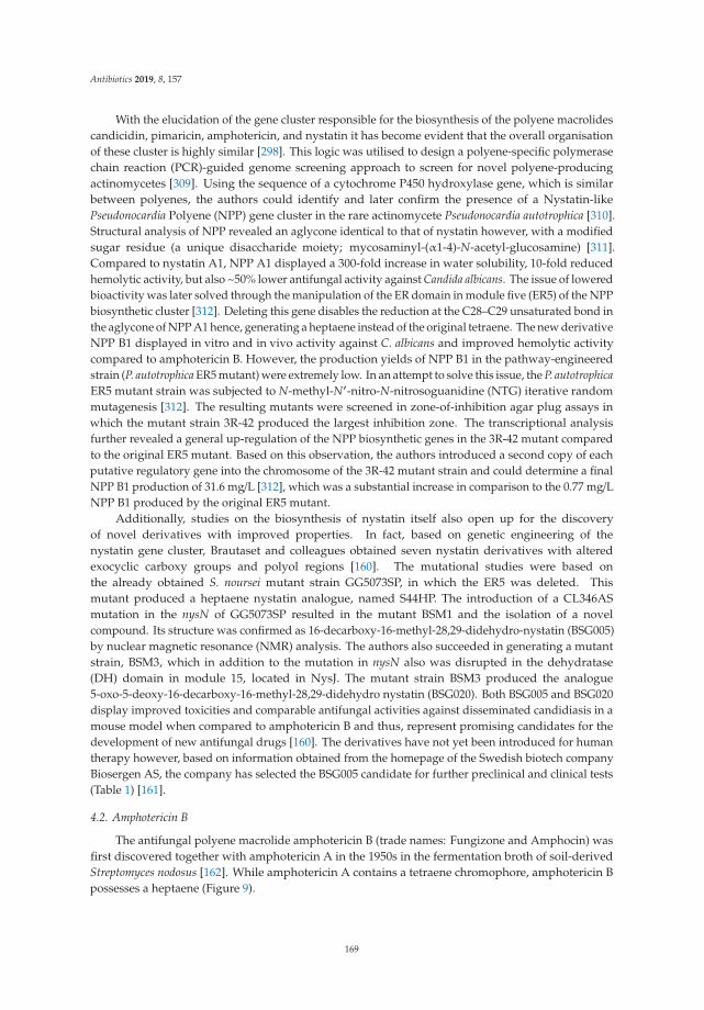
Antibiotics 2019, 8, 157
With the elucidation of the gene cluster responsible for the biosynthesis of the polyene macrolidescandicidin, pimaricin, amphotericin, and nystatin it has become evident that the overall organisationof these cluster is highly similar [298]. This logic was utilised to design a polyene-specific polymerasechain reaction (PCR)-guided genome screening approach to screen for novel polyene-producingactinomycetes [309]. Using the sequence of a cytochrome P450 hydroxylase gene, which is similarbetween polyenes, the authors could identify and later confirm the presence of a Nystatin-likePseudonocardia Polyene (NPP) gene cluster in the rare actinomycete Pseudonocardia autotrophica [310].Structural analysis of NPP revealed an aglycone identical to that of nystatin however, with a modifiedsugar residue (a unique disaccharide moiety; mycosaminyl-(α1-4)-N-acetyl-glucosamine) [311].Compared to nystatin A1, NPP A1 displayed a 300-fold increase in water solubility, 10-fold reducedhemolytic activity, but also ~50% lower antifungal activity against Candida albicans. The issue of loweredbioactivity was later solved through the manipulation of the ER domain in module five (ER5) of the NPPbiosynthetic cluster [312]. Deleting this gene disables the reduction at the C28–C29 unsaturated bond inthe aglycone of NPP A1 hence, generating a heptaene instead of the original tetraene. The new derivativeNPP B1 displayed in vitro and in vivo activity against C. albicans and improved hemolytic activitycompared to amphotericin B. However, the production yields of NPP B1 in the pathway-engineeredstrain (P. autotrophica ER5 mutant) were extremely low. In an attempt to solve this issue, the P. autotrophicaER5 mutant strain was subjected to N-methyl-N′-nitro-N-nitrosoguanidine (NTG) iterative randommutagenesis [312]. The resulting mutants were screened in zone-of-inhibition agar plug assays inwhich the mutant strain 3R-42 produced the largest inhibition zone. The transcriptional analysisfurther revealed a general up-regulation of the NPP biosynthetic genes in the 3R-42 mutant comparedto the original ER5 mutant. Based on this observation, the authors introduced a second copy of eachputative regulatory gene into the chromosome of the 3R-42 mutant strain and could determine a finalNPP B1 production of 31.6 mg/L [312], which was a substantial increase in comparison to the 0.77 mg/LNPP B1 produced by the original ER5 mutant.
Additionally, studies on the biosynthesis of nystatin itself also open up for the discoveryof novel derivatives with improved properties. In fact, based on genetic engineering of thenystatin gene cluster, Brautaset and colleagues obtained seven nystatin derivatives with alteredexocyclic carboxy groups and polyol regions [160]. The mutational studies were based onthe already obtained S. noursei mutant strain GG5073SP, in which the ER5 was deleted. Thismutant produced a heptaene nystatin analogue, named S44HP. The introduction of a CL346ASmutation in the nysN of GG5073SP resulted in the mutant BSM1 and the isolation of a novelcompound. Its structure was confirmed as 16-decarboxy-16-methyl-28,29-didehydro-nystatin (BSG005)by nuclear magnetic resonance (NMR) analysis. The authors also succeeded in generating a mutantstrain, BSM3, which in addition to the mutation in nysN also was disrupted in the dehydratase(DH) domain in module 15, located in NysJ. The mutant strain BSM3 produced the analogue5-oxo-5-deoxy-16-decarboxy-16-methyl-28,29-didehydro nystatin (BSG020). Both BSG005 and BSG020display improved toxicities and comparable antifungal activities against disseminated candidiasis in amouse model when compared to amphotericin B and thus, represent promising candidates for thedevelopment of new antifungal drugs [160]. The derivatives have not yet been introduced for humantherapy however, based on information obtained from the homepage of the Swedish biotech companyBiosergen AS, the company has selected the BSG005 candidate for further preclinical and clinical tests(Table 1) [161].
4.2. Amphotericin B
The antifungal polyene macrolide amphotericin B (trade names: Fungizone and Amphocin) wasfirst discovered together with amphotericin A in the 1950s in the fermentation broth of soil-derivedStreptomyces nodosus [162]. While amphotericin A contains a tetraene chromophore, amphotericin Bpossesses a heptaene (Figure 9).
169
Antibiotics 2019, 8, 157
The BGC of amphotericin B has been fully sequenced. The sequence analysis revealed that thecluster organisation is similar to the BGC of nystatin [313]. The polyketide chain is biosynthesised byan assembly line involving one loading (encoded by amphA) and 18 extension modules (encoded bythe five genes amphB, amphC, and amphI through amphK) (Figure S8). Assembly of the precursoron the amphotericin PKS is initiated by the loading of a malonyl-CoA starter unit which is furtherelongated by additional 15 acetate and nine propionate extender units (Figure S8) [163]. In the lastmodule, encoded by amphK, a TE domain is responsible for chain termination and release from the PKS.Two putative cytochrome P450 enzymes AmphL and AmphN are possibly involved in the modificationof the amphotericin B structure. While AmphL most likely catalyses the C8 hydroxylation in themacrolactone, AmphN may facilitate the oxidation of the methyl group on C16 to yield a carboxylgroup. Targeted deletion of amphN resulted in a S. nodosus mutant strain producing a amphotericinanalogue in which the exocyclic methyl group is retained [314]. The fact that the antifungal activityof this analogue was unchanged, and the haemolytic activity reduced compared to amphotericin B,makes the derivative an interesting candidate for clinical studies.
Additional modification of amphotericin is facilitated by the glycosyltransferase, encodedby amphDI, which is responsible for the attachment of the mycosamine to the aglycone core ofamphotericin [313,314]. The biosynthesis of the mycosamine is believed to be catalysed by aGDP-mannose-4,6-dehydratase encoded by amphDIII, which uses GDP-mannose derived from primarymetabolism as substrate [298]. The product of the AmphDIII-catalysed dehydratase reaction isGDP-4-keto-6-deoxy-d-mannose and not GDP-3-keto-6-deoxy-d-mannose, which is the substraterecognised by the transaminase AmphDII. So far, no GDP-4-keto-6-deoxy-d-mannose-3,4-isomerasehas been identified in any of the polyene gene clusters and it is hypothesised that the ketoisomerisationreaction is the result of a spontaneous, non-enzymatic reaction [298].
Export of amphotericin B has been hypothesised to be facilitated by two putative ABC transporters,encoded by amphG and amphH. The reason why two transporters are present in the gene cluster remainsunknown. It has been speculated that ABC transporters can confer self-resistance of the producinghost [313]. Based on the high degree of sequence similarity between nystatin and amphotericin PKSgenes, it has been postulated that the genes amphRI through amphRIV are homologues of the genesnysRI–nysRIV in nystatin and encode regulatory proteins, which act in a very similar fashion [298,315].
Since its marketing in 1957, amphotericin B (Table 1) has been used as the “gold standard” fortreatment of the most severe dimorphic fungal and yeast infections, caused by Blastomyces, Candida,Cryptococcus, and Histoplasma spp. [307]. The MOA of amphotericin B is identical to that of nystatin.Despite its preference for ergosterol found in fungal cellular membranes, amphotericin B also interactsto a lesser extend with the cholesterol found in mammalian cell membranes [164]. This and the sideeffects, including nephrotoxicity present a major limitation to the application of amphotericin B asan antibiotic for human therapy. In addition to its antifungal properties, amphotericin has also beenimplemented in delaying onset of prion disease in cultured cells with human immunodeficiency virus(HIV) and in inhibition of the protozoal parasite Leishmania [313].
Although amphotericin B shows a promising spectrum of activity and potential applications,the compound is poorly soluble in water and displays certain toxicity, which restricts its use inintravenous therapy. Consequently, only life-threatening fungal infections are treated with amphotericinB. Interestingly, despite its use as an antifungal drug for more than 40 years, reports of mycologicalresistance development in clinical fungi isolates against amphotericin B remain relatively scarce [316].Nonetheless, resistance occurs, as it was demonstrated by the isolation of resistant Candida spp.,Fusarium spp., and Scedosporium apiospermum [317]. In these fungi, drug resistance is most likelyconferred by either the production of alternative ergosterols to which the amphotericin B is less efficientor simply by decreasing the ergosterol level in the fungal cell membranes. Both mechanisms reducethe potency of the antifungal drug [316].
The promising features of amphotericin B, such as broad-spectrum activity and low resistanceagainst the compound were encouraging for numerous engineering attempts in order to improve
170
Antibiotics 2019, 8, 157
the properties of the antifungal drug. A great improvement in the solubility of the amphotericin Bwas already achieved in the case of Fungizone, which is a mixture of amphotericin B and the bileacid deoxycholate [165]. Additionally, reduction in the overall toxicity of amphotericin B was furtherachieved through liposome encapsulation, which has resulted in the three formulations; Amphotec,AmBisome, and Abelcet [165]. Unfortunately, the reduced toxicity of the liposome-packed amphotericinB seems to come at a cost in antifungal efficiency.
The extensive investigations on amphotericin and polyene biosynthesis are of advantage fortargeted engineering to increase production yields and to generate new derivatives with improvedtherapeutic properties. Furthermore, it should be noted that great efforts have been made in the fieldof semi-synthesis and several amphotericin B analogues with improved solubility and toxicity havebeen generated using different chemical approaches. Many of these have already been reviewedelsewhere [318]. Despite the many advances in both semi-synthesis and genetic engineering forobtaining amphotericin B analogues with improved properties, the yields of the derivatives are oftenvery low, and to date, none of the reported amphotericin B analogues have made it through to themarket. Consequently, the actinomycetes-derived substance amphotericin B remains one of the mostimportant polyketides in the sparse portfolio of antifungal drugs. This urges the need for improvedproduction titers in the natural producer strain. Recently, Zhang and co-workers set out to improveyields of amphotericin B in a newly isolated strain Streptomyces spp. ZJB 2013082, which produced theantifungal substance in low yields [319]. Using a combination of ultraviolet (UV) and NTG mutagenesis,the mutant strain N3 was isolated, which produced 1735 mg/L, a substantially increased amount ofthe product compared to the 56.2 mg/L obtained from the parent strain ZJB 2013082. Additionally,the N3 mutant accumulated less amphotericin A than the parent strain ZJB 2013082. This could beof industrial importance, since substance A is only allowed to account for more than five percent inthe amphotericin mixture. The genome sequence of the N3 mutant is not yet published however,identification of the genomic architecture in the N3 mutant could help guide future engineering effortsto obtain a stable production host for amphotericin B.
4.3. Pimaricin/Natamycin
Pimaricin (later renamed natamycin, trade names: Natacyn and E235) (Figure 9, Table 1) wasfirst discovered in the 1950s as the product of soil-derived Streptomyces natalensis isolated from theSouth African region of Natal [166,320]. Additional producer strains have been identified, includingStreptomyces chattanoogensis [321,322] and Streptomyces lydicus [323].
The structure of pimaricin varies slightly from those of amphotericin B and nystatin A1 (Section 4.1)owing to the smaller size of the macrolactone (Figure 9). Pimaricin is composed of a 26-memberedaglycone, containing four conjugated double bonds, to which the characteristic mycosamine sugar isattached at the C15 atom. The tetraene polyene further contains an exocyclic carboxyl group at C12,a functionally interestingly epoxide at C4/C5, and an internal hemiketal ring, which originates from aspontaneous cyclisation of the C9 keto group with a hydroxyl group on C13 [167].
The elucidation of pimaricin biosynthesis has relied primarily on genome sequencing and geneticstudies of the two pimaricin-producer strains S. chattanoogensis and S. natalensis [320]. In the case ofS. natalensis, an 85 kb-large genomic region containing 16 ORFs was identified from a cosmid libraryas the pimaricin gene cluster [167]. Pimaricin biosynthesis follows a logic, which is highly similarto that governing amphotericin B and nystatin biosynthesis. Since the gene clusters identified fromS. chattanoogensis and S. natalensis are nearly identical, the following subsection will describe the studieson S. natalensis.
The assembly of the 26-membered lactone, termed pimaricinolide, is catalysed by a type I PKScomposed of 13 (one starter and 12 extender) modules, encoded by the genes pimS0–pimS4 (Figure S9).Chain initiation starts at PimS0 with the loading of a malonyl-CoA. Further elongation, catalysed byPimS1 through PimS4, leads to the condensation of additional 12 acetate units and one propionate unitto the growing polyketide precursor. In PimS4, the last domain, a TE, is responsible for the release and
171
Antibiotics 2019, 8, 157
cyclisation of pimaricinolide [167]. Further examination of the pimaricin gene cluster has revealed anadditional gene pimI, which encodes an enzyme with homology to the TE found in the candicidin genecluster in S. griseus and to the TylO in the tylosin gene cluster in S. fradiae. It has been postulated that theadditional TE in the pimaricin gene cluster helps to remove aberrant precursors from the PKS, ensuringcontinuous biosynthesis [167,204]. Upon its release from the PKS, pimaricinolide undergoes oxidationof the methyl group on C12 resulting in the formation of a carboxylic acid. This is catalysed by PimG,a cytochrome P450 enzyme. The resulting 12-carboxy-pimaricinolide is then glycosylated at the C15hydroxyl group through the attachment of a mycosamine by the actions of the glycosyltransferasePimK. The final modification of the pimaricin precursor involves another cytochrome P450 (encoded bythe gene pimD) which catalyse the oxidation leading to the spontaneous formation of an epoxy groupbetween C4 and C5.
Sugar biosynthesis is believed to involve only two enzymes; PimJ, a GDP-mannose-4,6-dehydrataseresponsible for the conversion of GDP-mannose (from primary metabolism) to GDP-4-keto-6-deoxymannose,and PimC, a GDP-3-keto-6-deoxymannose aminotransferase, which synthesises GDP-mycosamine fromGDP-3-keto-6-deoxymannose. Similar to what has been described for mycosamine biosynthesis inamphotericin B and nystatin, no gene encoding an enzyme responsible for the 3,4-isomerisation required forthe conversion of GDP-4-keto-6-deoxymannose to GDP-3-keto-6-deoxymannose was found in the pimaricingene cluster, and the reaction is believed to occur spontaneously [298,320].
The three gene products of pimA, pimB, and pimH have been putatively assigned to proteinsensuring the export of pimaricin in S. natalensis. While PimA and PimB group in the family ofABC transporters, PimH might encode an efflux pump [320,324]. The functions of PimA and PimBremain to be experimentally verified. For the homologues of PimA and PimB (ScnA and ScnB) inS. chattanoogensis, it was reported that they are involved in primary exporters of natamycin [324].With amino acid sequence similarities of above 95% for ScnA/ScnB and PimA/PimB, it is likely that thelater enzymes have a similar function and also act as primary transporters of pimaricin.
The regulatory mechanisms governing pimaricin biosynthesis has been studied in S. natalensisand S. chattanoogensis. Two transcriptional regulators PimM and PimR play an important role inpimaricin production in S. natalensis [325,326]. Furthermore, the amino acid exporter PimT and theputative cholesterol oxidase PimE add an extra layer of regulation to the pimaricin biosynthesis.While PimT was found to play a role in export of quorum-sensing pimaricin-inducer (PI) factor [327],PimE could act as a signalling molecule, triggering production of pimaricin in the producer inthe presence of fungi [328,329]. The complex regulation cascade contains potential “targets” forengineering of the producer and increasing the production of the antifungal compound. Some of themost successful examples include overexpression of the regulator-encoding gene scnRII (homologue topimM) in S. chattanoogensis, deletion of sngR, a γ-butyrolactone receptor-encoding gene, in S. natalensis,and chromosomal integration of the Vitreoscilla haemoglobin vgb gene in S. gilvosporeus, resulting in460%, 460%, and 407% increase in pimaricin yields, respectively, in the mutant strains compared towild types [320,330–332].
More than 40 years after the introduction of pimaricin to the market, it remains an importantantifungal agent and it is still used in the treatment of fungal keratitis, an infection of the cornea causedprimarily by filamentous fungi Fusarium and Aspergillus, and yeast-like Candida [168]. Due to its lowwater solubility and limited oral absorption, pimaricin is mainly available as a topical agent in humanmedicine. Recently, an antiprotozoan activity of pimaricin was detected which makes the compoundattractive for potential treatment of keratitis caused by Acanthamoeba.
The MOA of pimaricin differs from that of nystatin and amphotericin B. While the main target ofthe pimaricin is ergosterol, which is the major sterol found in fungal cells membranes, pimaricin onlybinds to the lipid receptor. The interaction between pimaricin and ergosterol has been examined inAspergillus niger, showing that upon its binding, pimaricin blocks transport of amino acids and glucoseacross the fungal plasma membrane, which leads to cell death [169]. Due to its specific interactionwith ergosterol, the development of microbial resistance towards pimaricin is seen as posing only a
172
Antibiotics 2019, 8, 157
minor risk [320]. This, combined with its low oral absorption, has paved the way for pimaricin as afood preservative. Sold under the label E235, pimaricin is approved as a protecting agent against yeastand mould and is used for surface treatment of hard, semi-hard, and semi-soft cheeses, and of dried,cured sausage in Europe. Other applications of pimaricin are summarised in the review by Aparicioand co-workers [320]. Additionally, pimaricin is the only antifungal agent to date, which has gainedthe generally regarded as safe (GRAS) status.
While pimaricin itself remains an important agent for treatment of fungal keratitis and asa food preservative, efforts to engineer strains which produce novel pimaricin analogues withimproved solubility and toxicity have been undertaken. These efforts have been greatly aidedby the complete genome sequencing of S. natalensis and S. chattanoogensis, both harbouring theBGC of pimaricin. Recently, Qi and co-workers could identify three novel pimaricin analoguesbased on a single mutation of the gene scnG in S. chattanoogensis (pimG in S. natalensis) [333],which encodes the cytochrome P450 enzyme responsible for the formation of carboxyl group atthe C12 in pimaricin. Out of the three identified derivatives, 12-decarboxy-12-methyl pimaricin and4,5-desepoxy-12-decarboxy-12-methyl pimaricin, both displayed reduced cytotoxicity compared topimaricin. Additionally, 12-decarboxy-12-methyl pimaricin showed a two-fold increase in antifungalactivity against C. albicans ATCC 14,053 compared to pimaricin. Through further biochemical andgenetic analyses, 4,5-desepoxy-12-decarboxy-12-methyl pimaricin was found to be the precursorof 12-decarboxy-12-methyl pimaricin in the reaction catalysed by the C4/C5 epoxidase encoded byscnD (pimD). Therefore, to ensure the complete conversion of 4,5-desepoxy-12-decarboxy-12-methylpimaricin into 12-decarboxy-12-methyl pimaricin, scnD was overexpressed in the ΔscnG mutant,which led to a 20% increase in production of the latter derivative. However, with an overall yieldof 268 ± 10 mg/L for 12-decarboxy-12-methyl pimaricin in the best performing mutant, the needs forfurther engineering to optimise yields are necessary. In this case, the pathway-specific regulators PimMand PimR or the PI-factor could be the next targets for improving pimaricin derivative production [333].
5. Strategies and Tools for the Discovery of Natural Products
The emergence of antibiotic resistant microbes is alarming and underlines the urgent need fornew drugs to combat the pathogens. However, the discovery and approval of new antibiotics ismore difficult than expected [334–336]. Therefore, the question arises: how to improve the chancesfor finding new antimicrobial compounds? Recently, new approaches and advances of the existingtechnologies within the early stage of drug discovery and development were reported.
For natural product-derived antimicrobial compounds, the “journey” starts with the identificationof the source (e.g., producer organism) and/or the bioactive molecule, responsible for the inhibitionor killing of a pathogen. Already at this stage, the re-discovery rate of known structures might bereduced by taking samples and isolation of potential producers or compounds from undiscoveredenvironments [337–343]. This is often limited by the fact that organisms originating from “extreme”habitats require special cultivation conditions and thus, many strategies were developed to overcomethis barrier (e.g., co-cultivation [344–346], iChip [347–352], or combination of both [353]).
Confirmed or potential producers of new antimicrobial agents are further analysed by diverse“omics” approaches [342,354–357]. The downstream evaluation of the collected data sets usingbioinformatics tools enables for example the identification of the BGC for the product of interest and/orprovides an overview on the overall biosynthetic potential of target strain (genome mining) [9,358–360].In cases where promising BGCs were identified, however, no products were found with the availablefermentation and analytic methods [361–364], the expression of the clusters and production of therespective metabolite might be achieved by addition of elicitors [341,342,365–369] or the heterologousexpression of the BGC in optimized hosts [370–375].
In order to improve the production of relevant products, including polyketides, molecularbiology tools and methods (e.g., vectors, plasmids, recombinases, CRISPR-Cas9, promoters, and othersynthetic parts as well as methods for their delivery (conjugation, protoplast transformation, and direct
173
Antibiotics 2019, 8, 157
transformation)) are used. They play an important role for engineering of both, the natural producer andheterologous hosts. Challenges and new opportunities for the genetic manipulation of actinomyceteswere recently reviewed [376–381].
Although each one of these cutting-edge technologies and approaches already contributed to theidentification of new compounds, the interplay of the different disciplines will grant a better access tonovel natural products with valuable bioactivities.
6. Conclusions and Outlook
Actinomycetes are one of the most prolific sources of biologically active secondary metabolites,including polyketides. In the past decades, numerous polyketide compounds were isolatedand developed to highly potent antimicrobial drugs, which have saved millions of lives.However, rapid emergence of multidrug resistant pathogens is occurring worldwide which poses asevere threat to human health. This calls for the discovery and development of new antibiotics andantimicrobial strategies. Unlike drugs used in case of chronic diseases (e.g., diabetes, cardiovasculardisease, cancer, arthritis, asthma), antibiotics are taken for a short period of time and thus, they arenon-profitable and economically unattractive. This and several other obstacles such as high costsof the research and development and insufficient investment from stakeholders has prompted thebig pharmaceutical companies to terminate the development of new antibiotics. Currently, it seemsthat the screening and development of new lead structures for novel antimicrobial agents is mainlyconduced at public research and non-profit institutions.
The fact that many habitats around the globe are unexplored and poorly investigated for thepresence of antibiotic-producing microbes, such as actinomycetes, motived researchers to collectsamples from these environments and isolate the diverse producers of potentially new bioactivecompounds. The valuable knowledge obtained from the investigation of the biosynthesis, regulationand natural resistance in the natural host of the old drugs as well as the recent developments withinscreening and isolation methods, sequencing and genome mining, and analytics potentiate the platformsfor drug discovery. For example, the analysis of a relatively underexplored genus of Actinoallomurusled to the discovery of two new spirotetronate polyketide antibiotics NAI414-A and NAI414-B [382].
As exemplified in this review (Section 3.1), the success in derivatisation and combination ofexisting compound classes has enabled the continuous efficient treatment of otherwise resistant clinicalisolates. In particular, the knowledge gained from detailed MOA and SAR studies of the antimicrobialshave paved the way for the development of drugs with improved pharmacokinetic properties andexpanded spectrum of bioactivity, compared to the original substance. In the future, semi-synthesiswill continue to play an important role in drug development, of both, old and new drug candidates.
Last, but not least, support from governments and cooperation across the world e.g., public researchinstitutions as well as United Nations organisations, the WHO, the Food and Agriculture Organization,and the inter-governmental World Organisation for Animal Health, combined with strategies offeringlong-term incentive for the pharmaceutical companies to reinvigorate their antimicrobial drug discoveryplatforms are an important political aspect that has been gaining more attention these days. The optionof the US FDA to gain a fast-track approval of drug leads which can be used to treat serious orlife-threatening conditions might further enhance the chances of taking on the expenses associatedwith drug discovery by pharmaceutical companies. The combination of all these efforts may give acompetitive advantage in the never-ending race between the discovery of antimicrobials and the riseof drug resistance in pathogens.
Supplementary Materials: The following are available online at http://www.mdpi.com/2079-6382/8/4/157/s1,Figure S1: The erythromycin biosynthetic pathway, Figure S2: The tylosin biosynthetic pathway, Figure S3: Themonensin biosynthetic pathway, Figure S4: The biosynthetic pathway of tiacumicin, Figure S5: The biosyntheticpathway of rifamycin, Figure S6: The biosynthetic pathway of oxytetracycline and chlortetracycline, Figure S7:The biosynthetic pathways of pristinamycin II and pristinamycin I, Figure S8: The biosynthetic pathways ofnystatin and amphotericin B, Figure S9: The biosynthetic pathway of pimaricin.
174
Antibiotics 2019, 8, 157
Author Contributions: The review was written and edited by the authors H.R. and E.M.-K.
Funding: The authors and work in their laboratory are supported by the Eberhard Karls Universität Tübingen, theDeutsche Forschungsgemeinschaft (DFG), the Bundesministeriums für Bildung und Forschung (BMBF) (FKZ 031L0018A, ERASysApp), the German Center for Infection Research (DZIF) (TTU 09.912), and Biovet (Sofia, Bulgaria).
Conflicts of Interest: The authors declare no conflict of interest.
Abbreviations
A adenylation domain MBC minimum bactericidal concentrationABC ATP-binding cassette MIC minimum inhibitory concentrationACP acyl carrier protein MLSB macrolide–lincosamide–streptogramin BAHBA 3-amino 5-hydroxybenzoic acid MOA mode of actionARO aromatase MRSA methicillin-resistant Staphylococcus aureusAT acyltransferase MT methyltransferase domainBGC biosynthetic gene cluster NADPH nicotinamide adenine dinucleotide phosphateC condensation domain NGST Next Generation Sequencing TechnologiesCABP community-acquired bacterial pneumonia NMR nuclear magnetic resonanceCCR crotonyl-CoA carboxylase/reductase NRPS nonribosomal peptide synthetaseCDI Clostridioides difficile infection NTG N-methyl-N′-nitro-N-nitrosoguanidineCLF chain elongation factor ORF open reading frameCoA coenzyme A PCP peptidyl carrier proteincryo EM cryogenic electron microscopy PCR polymerase chain reactionCYC cyclase PKS polyketide synthaseDEBS deoxyerythronolide B synthase PTC peptidyl transferase centerDH dehydratase RNA ribonucleic acidDNA deoxyribonucleic acid RNAP RNA polymerasedTDP deoxythymidine diphosphate rRNA ribosomal RNAE epimerisation domain SAM S-adenosyl methionineER enoyl reductase SAR structure activity relationshipESBL extended spectrum β-lactamase SARP Streptomyces antibiotic regulatory proteinFDA Food and Drug Administration Spp. SpeciesGDP guanosine diphosphate TB TuberculosisGI gastrointestinal TDP thymidine diphosphateKb kilobase TE ThioesteraseKR ketoreductase tRNA transfer RNAKS ketosynthase VRE vancomycin-resistant EnterococciLSAP Lincosamide–streptogramin A–pleuromutilin VRSA vancomycin-resistant Staphylococcus aureusMAC Mycobacterium avium-intracellulare complex WHO World Health Organisation
References
1. Mohammadipanah, F.; Dehhaghi, M. Classification and taxonomy of actinobacteria. In Biology andBiotechnology of Actinobacteria; Wink, J., Mohammadipanah, F., Hamedi, J., Eds.; Springer: Berlin/Heidelberg,Germany, 2017; pp. 51–77.
2. Barka, E.A.; Vatsa, P.; Sanchez, L.; Gaveau-Vaillant, N.; Jacquard, C.; Klenk, H.P.; Clément, C.; Ouhdouch, Y.;van Wezel, G.P. Taxonomy, physiology, and natural products of Actinobacteria. Microbiol. Mol. Biol. Rev.2016, 80, 1–43. [CrossRef] [PubMed]
3. García, J.C.; Patrão, B.; Almeida, L.; Pérez, J.; Menezes, P.; Dias, J.; Sanz, P.J. A natural interface for remoteoperation of underwater robots. IEEE Comput. Graph. Appl. 2015, 37, 34–43. [CrossRef] [PubMed]
4. Cook, T.B.; Pfleger, B.F. Leveraging synthetic biology for producing bioactive polyketides and non-ribosomalpeptides in bacterial heterologous hosts. Medchemcomm 2019, 10, 668–681. [CrossRef] [PubMed]
5. Abu-Melha, S. Design, Synthesis and DFT/DNP Modeling Study of New 2-Amino-5-arylazothiazoleDerivatives as Potential Antibacterial Agents. Molecules 2018, 23, 434. [CrossRef] [PubMed]
6. Lenci, E.; Trabocchi, A. Smart Design of Small-Molecule Libraries: When Organic Synthesis MeetsCheminformatics. ChemBioChem 2019, 20, 1115–1123. [CrossRef] [PubMed]
175
Antibiotics 2019, 8, 157
7. Musiol-Kroll, E.; Wohlleben, W. Acyltransferases as tools for polyketide synthase engineering. Antibiotics2018, 7, 62. [CrossRef]
8. Tong, Y.; Robertsen, H.L.; Blin, K.; Weber, T.; Lee, S.Y. CRISPR-Cas9 toolkit for Actinomycete genome editing.In Synthetic Metabolic Pathways; Springer: Berlin/Heidelberg, Germany, 2018; pp. 163–184.
9. Blin, K.; Shaw, S.; Steinke, K.; Villebro, R.; Ziemert, N.; Lee, S.Y.; Medema, M.H.; Weber, T. antiSMASH5.0: Updates to the secondary metabolite genome mining pipeline. Nucleic Acids Res. 2019, 47, W81–W87.[CrossRef]
10. Komaki, H.; Sakurai, K.; Hosoyama, A.; Kimura, A.; Igarashi, Y.; Tamura, T. Diversity of nonribosomalpeptide synthetase and polyketide synthase gene clusters among taxonomically close Streptomyces strains.Sci. Rep. 2018, 8, 6888. [CrossRef]
11. Nett, M.; Ikeda, H.; Moore, B.S. Genomic basis for natural product biosynthetic diversity in the actinomycetes.Nat. Prod. Rep. 2009, 26, 1362–1384. [CrossRef]
12. Santos, R.; Ursu, O.; Gaulton, A.; Bento, A.P.; Donadi, R.S.; Bologa, C.G.; Karlsson, A.; Al-Lazikani, B.;Hersey, A.; Oprea, T.I.; et al. A comprehensive map of molecular drug targets. Nat. Rev. Drug Discov.2017, 16, 19. [CrossRef]
13. Wilson, D.N.; Harms, J.M.; Nierhaus, K.H.; Schlünzen, F.; Fucini, P. Species-specific antibiotic-ribosomeinteractions: Implications for drug development. Biol. Chem. 2005, 386, 1239–1252. [CrossRef]
14. Koehbach, J.; Craik, D.J. The Vast Structural Diversity of Antimicrobial Peptides. Trends Pharmacol. Sci.2019, 40, 517–528. [CrossRef]
15. Tenover, F.C. Mechanisms of antimicrobial resistance in bacteria. Am. J. Med. 2006, 119 (Suppl. 1), S3–S10;discussion S62–S70. [CrossRef]
16. Du Toit, A. Antimicrobials: Putting antibiotic action into context. Nat. Rev. Microbiol. 2016, 14, 725.[CrossRef]
17. Levy, S.B.; Marshall, B. Antibacterial resistance worldwide: Causes, challenges and responses. Nat. Med.2004, 10, S122. [CrossRef]
18. Beckh, W.; Kulchar, G.V. Treatment-Resistant Syphilis: An Evaluation of the Causative Factors in EighteenCases. Arch. Derm. Syphilol. 1939, 40, 1–12. [CrossRef]
19. Andersson, D.I.; Nicoloff, H.; Hjort, K. Mechanisms and clinical relevance of bacterial heteroresistance.Nat. Rev. Microbiol. 2019, 17, 479–496. [CrossRef]
20. Hofer, U. The cost of antimicrobial resistance. Nat. Rev. Microbiol. 2019, 17, 3. [CrossRef]21. Eagle, H. The binding of penicillin in relation to its cytotoxic action: II. The reactivity with penicillin of
resistant variants of Streptococci, Pneomocci, and Staphylococci. J. Exp. Med. 1954, 100, 103–115. [CrossRef]22. Stekel, D. First report of antimicrobial resistance pre-dates penicillin. Nature 2018, 562, 192. [CrossRef]23. Frieri, M.; Kumar, K.; Boutin, A. Antibiotic resistance. J. Infect. Public Health 2017, 10, 369–378. [CrossRef]24. Turner, N.A.; Sharma-Kuinkel, B.K.; Maskarinec, S.A.; Eichenberger, E.M.; Shah, P.P.; Carugati, M.;
Holland, T.L.; Fowler, V.G., Jr. Methicillin-resistant Staphylococcus aureus: An overview of basic andclinical research. Nat. Rev. Microbiol. 2019, 17, 203–218. [CrossRef]
25. Blair, J.M.A.; Webber, M.A.; Baylay, A.J.; Ogbolu, D.O.; Piddock, L.J.V. Molecular mechanisms of antibioticresistance. Nat. Rev. Microbiol. 2015, 13, 42. [CrossRef]
26. Kaur, P.; Peterson, E. Antibiotic resistance mechanisms in bacteria: Relationships between resistancedeterminants of antibiotic producers, environmental bacteria, and clinical pathogens. Front. Microbiol.2018, 9, 2928.
27. Pambos, O.J.; Kapanidis, A.N. Tracking antibiotic mechanisms. Nat. Rev. Microbiol. 2019, 17, 201. [CrossRef]28. Alanis, A.J. Resistance to antibiotics: Are we in the post-antibiotic era? Arch. Med. Res. 2005, 36, 697–705.
[CrossRef]29. Carroll, L.M.; Gaballa, A.; Guldimann, C.; Sullivan, G.; Henderson, L.O.; Wiedmann, M. Identification of
Novel Mobilized Colistin Resistance Gene mcr-9 in a Multidrug-Resistant, Colistin-Susceptible Salmonellaenterica Serotype Typhimurium Isolate. MBio 2019, 10, e00853-19. [CrossRef]
30. World Health Organization. New report calls for urgent action to avert antimicrobial resistance crisis. JointNews Release. 29 April 2019. Available online: https://www.who.int/news-room/detail/29-04-2019-new-report-calls-for-urgent-action-to-avert-antimicrobial-resistance-crisis (accessed on 25 August 2019).
31. World Health Organization. Antibiotic Resistance. 2019. Available online: https://www.who.int/news-room/fact-sheets/detail/antibiotic-resistance (accessed on 25 August 2019).
176
Antibiotics 2019, 8, 157
32. Spellberg, B. The future of antibiotics. Crit. Care 2014, 18, 228. [CrossRef]33. Pew Charitable Trusts. Tracking the Pipeline of Antibiotics in Development. 2019. Available
online: http://www.pewtrusts.org/en/research-and-analysis/issue-briefs/2014/03/12/tracking-the-pipeline-of-antibiotics-in-development (accessed on 25 August 2019).
34. Owens, B. Solithromycin rejection chills antibiotic sector. Nat. Biotechnol. 2017, 35, 187–188. [CrossRef]35. Weissman, K. Chapter 1 Introduction to Polyketide Biosynthesis. In Methods in Enzymology; Academic Press:
Cambridge, MA, USA, 2009; Volume 459, pp. 3–16.36. Shen, B. Polyketide biosynthesis beyond the type I, II and III polyketide synthase paradigms. Curr. Opin.
Chem. Biol. 2003, 7, 285–295. [CrossRef]37. Hertweck, C. The biosynthetic logic of polyketide diversity. Angew. Chemie Int. Ed. 2009, 48, 4688–4716.
[CrossRef]38. Ridley, C.P.; Lee, H.Y.; Khosla, C. Evolution of polyketide synthases in bacteria. Proc. Natl. Acad. Sci. USA
2008, 105, 4595–4600. [CrossRef]39. Süssmuth, R.D.; Mainz, A. Nonribosomal peptide synthesis—Principles and prospects. Angew. Chem. Int.
Ed. 2017, 56, 3770–3821. [CrossRef]40. Challis, G.L.; Wilkinson, B. Biosynthetic assembly lines themed issue. Nat. Prod. Rep. 2016, 33, 120–121.
[CrossRef]41. Staunton, J.; Weissman, K.J. Polyketide biosynthesis: A millennium review. Nat. Prod. Rep. 2001, 18, 380–416.
[CrossRef]42. Helfrich, E.J.N.; Piel, J. Biosynthesis of polyketides by trans-AT polyketide synthases. Nat. Prod. Rep.
2016, 33, 231–316. [CrossRef]43. Musiol, E.M.; Weber, T. Discrete acyltransferases involved in polyketide biosynthesis. Medchemcomm
2012, 3, 871–886. [CrossRef]44. Meurer, G.; Gerlitz, M.; Wendt-Pienkowski, E.; Vining, L.C.; Rohr, J.; Hutchinson, C.R. Iterative type II
polyketide synthases, cyclases and ketoreductases exhibit context-dependent behavior in the biosynthesis oflinear and angular decapolyketides. Chem. Biol. 1997, 4, 433–443. [CrossRef]
45. Caffrey, P. Dissecting complex polyketide biosynthesis. Comput. Struct. Biotechnol. J. 2012, 3, e201210010.[CrossRef]
46. Chen, A.; Re, R.N.; Burkart, M.D. Type II fatty acid and polyketide synthases: Deciphering protein–proteinand protein–substrate interactions. Nat. Prod. Rep. 2018, 35, 1029–1045. [CrossRef]
47. Herbst, D.A.; Townsend, C.A.; Maier, T. The architectures of iterative type I PKS and FAS. Nat. Prod. Rep.2018, 35, 1046–1069. [CrossRef]
48. Chen, H.; Du, L. Iterative polyketide biosynthesis by modular polyketide synthases in bacteria. Appl. Microbiol.Biotechnol. 2016, 100, 541–557. [CrossRef]
49. Weber, T. Antibiotics: Biosynthesis, Generation of Novel Compounds. Encycl. Ind. Biotechnol. 2010, 1–12.[CrossRef]
50. Bloudoff, K.; Schmeing, T.M. Structural and functional aspects of the nonribosomal peptide synthetasecondensation domain superfamily: Discovery, dissection and diversity. BBA Proteins Proteom.2017, 1865, 1587–1604. [CrossRef]
51. Miyanaga, A.; Kudo, F.; Eguchi, T. Protein–protein interactions in polyketide synthase–nonribosomal peptidesynthetase hybrid assembly lines. Nat. Prod. Rep. 2018, 35, 1185–1209. [CrossRef]
52. McGuire, J.M.; Bunch, R.L.; Anderson, R.C.; Boaz, H.E.; Flynn, E.H.; Powell, H.M.; Smith, J.W. Ilotycin, anew antibiotic. Antibiot. Chemother. (Northfield, III.) 1952, 2, 281–283.
53. Wiley, P.F.; Gerzon, K.; Flynn, E.H.; Sigal, M.V., Jr.; Weaver, O.; Quarck, U.C.; Chauvette, R.R.; Monahan, R.Erythromycin. X. 1 Structure of Erythromycin. J. Am. Chem. Soc. 1957, 79, 6062–6070. [CrossRef]
54. Harris, D.R.; McGeachin, S.G.; Mills, H.H. The structure and stereochemistry of erythromycin A. TetrahedronLett. 1965, 6, 679–685. [CrossRef]
55. Cortes, J.; Haydock, S.F.; Roberts, G.A.; Bevitt, D.J.; Leadlay, P.F. An unusually large multifunctionalpolypeptide in the erythromycin-producing polyketide synthase of Saccharopolyspora erythraea. Nature1990, 348, 176. [CrossRef]
56. Summers, R.G.; Donadio, S.; Staver, M.J.; Wendt-Pienkowski, E.; Hutchinson, C.R.; Katz, L. Sequencing andmutagenesis of genes from the erythromycin biosynthetic gene cluster of Saccharopolyspora erythraea that areinvolved in L-mycarose and D-desosamine production. Microbiology 1997, 143, 3251–3262. [CrossRef]
177
Antibiotics 2019, 8, 157
57. Donadio, S.; Staver, M.; McAlpine, J.; Swanson, S.J.; Katz, L. Modular Organization of Genes Required forComplex Polyketide Biosynthesis. Science 1991, 252, 675–679. [CrossRef]
58. Oliynyk, M.; Samborskyy, M.; Lester, J.B.; Mironenko, T.; Scott, N.; Dickens, S.; Haydock, S.F.; Leadlay, P.F.Complete genome sequence of the erythromycin-producing bacterium Saccharopolyspora erythraea NRRL23338.Nat. Biotechnol. 2007, 25, 447. [CrossRef]
59. Zhang, H.; Wang, Y.; Wu, J.; Skalina, K.; Pfeifer, B.A. Complete biosynthesis of erythromycin A and designedanalogs using E. coli as a heterologous host. Chem. Biol. 2010, 17, 1232–1240. [CrossRef]
60. Weissman, K.J. Genetic engineering of modular PKSs: From combinatorial biosynthesis to synthetic biology.Nat. Prod. Rep. 2016, 33, 203–230. [CrossRef]
61. Kibwage, I.O.; Hoogmartens, J.; Roets, E.; Vanderhaeghe, H.; Verbist, L.; Dubost, M.; Pascal, C.; Petitjean, P.;Levol, G. Antibacterial activities of erythromycins A, B, C, and D and some of their derivatives. Antimicrob.Agents Chemother. 1985, 28, 630–633. [CrossRef]
62. Amsden, G.W. Erythromycin, clarithromycin, and azithromycin: Are the differences real? Clin. Ther.1996, 18, 56–72. [CrossRef]
63. Mazzei, T.; Mini, E.; Novelli, A.; Periti, P. Chemistry and mode of action of macrolides. J. Antimicrob.Chemother. 1993, 31, 1–9. [CrossRef]
64. Vester, B.; Douthwaite, S. Macrolide resistance conferred by base substitutions in 23S rRNA. Antimicrob.Agents Chemother. 2001, 45, 1–12. [CrossRef]
65. Tenson, T.; Lovmar, M.; Ehrenberg, M. The mechanism of action of macrolides, lincosamides and streptograminB reveals the nascent peptide exit path in the ribosome. J. Mol. Biol. 2003, 330, 1005–1014. [CrossRef]
66. Svetlov, M.S.; Plessa, E.; Chen, C.W.; Bougas, A.; Krokidis, M.G.; Dinos, G.P.; Polikanov, Y.S. High-resolutioncrystal structures of ribosome-bound chloramphenicol and erythromycin provide the ultimate basis for theircompetition. RNA 2019, 25, 600–606. [CrossRef]
67. Sobin, B.A.; English, A.R.; Celmer, W.D. PA 105, a new antibiotic. In Antibiotics Annual; Welch, H.,Marti-Ibannez, F., Eds.; Medical Encyclopedia Inc.: New York, NY, USA, 1955; pp. 827–830.
68. English, A.R.; McBride, T.J.; Van Halsema, G.; Caklozzi, M. Biologic studies on PA 775, a combination oftetracycline and oleandomycin with synergistic activity. Antibiot. Chemother. 1956, 6, 511–522.
69. Podolsky, S.H. The Antibiotic Era: Reform, Resistance, and the Pursuit of a Rational Therapeutics; JHU Press:Baltimore, MD, USA, 2015; pp. 1–328.
70. Albouy, R.; Duchesnay, G.; Eloy, P.; Pestel, M.; Ravina, A.; Rey, M. A new French antibiotic: Spiramycin.Antibiot. Annu. 1955, 3, 223.
71. Kellow, W.F.; Lepper, M.H.; Plaut, S.; Rosenthal, I.M.; Spies, H.W. Spiramycin in the treatment of infection.Antibiot. Annu. 1955, 3, 658.
72. Sutherland, R. Spiramycin: A reappraisal of its antibacterial activity. Br. J. Pharmacol. Chemother.1962, 19, 99–110. [CrossRef]
73. Fernandes, P.; Martens, E.; Pereira, D. Nature nurtures the design of new semi-synthetic macrolide antibiotics.J. Antibiot. (Tokyo) 2016, 70, 527. [CrossRef]
74. Barry, A.L.; Jones, R.N.; Thornsberry, C. In vitro activities of azithromycin (CP 62,993), clarithromycin(A-56268; TE-031), erythromycin, roxithromycin, and clindamycin. Antimicrob. Agents Chemother.1988, 32, 752–754. [CrossRef]
75. Watanabe, Y.; Moritomo, S.; Adachi, T.; Kashimure, M.; Asaka, T. Chemical modification of erythromycin. IX.1. J. Antibiot. (Tokyo) 1993, 46, 647–660. [CrossRef]
76. Barlam, T.; Neu, H.C. In vitro comparison of the activity of RU 28965, a new macrolide, with that oferythromycin against aerobic and anaerobic bacteria. Antimicrob. Agents Chemother. 1984, 25, 529–531.[CrossRef]
77. Jorgensen, J.H.; Redding, J.S.; Howell, A.W. In vitro activity of the new macrolide antibiotic roxithromycin(RU 28965) against clinical isolates of Haemophilus influenzae. Antimicrob. Agents Chemother. 1986, 29, 921–922.[CrossRef]
78. Mutak, S. Azalides from azithromycin to new azalide derivatives. J. Antibiot. (Tokyo) 2007, 60, 85. [CrossRef]79. Retsema, J.; Girard, A.; Schelkly, W.; Manousos, M.; Anderson, M.; Bright, G.; Borovoy, R.; Brennan, L.;
Mason, R. Spectrum and mode of action of azithromycin (CP-62,993), a new 15-membered-ring macrolidewith improved potency against gram-negative organisms. Antimicrob. Agents Chemother. 1987, 31, 1939–1947.[CrossRef]
178
Antibiotics 2019, 8, 157
80. Bryskier, A. Ketolides—Telithromycin, an example of a new class of antibacterial agents. Clin. Microbiol.Infect. 2000, 6, 661–669. [CrossRef]
81. Ednie, L.M.; Jacobs, M.R.; Appelbaum, P.C. Comparative antianaerobic activities of the ketolides HMR 3647(RU 66647) and HMR 3004 (RU 64004). Antimicrob. Agents Chemother. 1997, 41, 2019–2022. [CrossRef]
82. Ross, D.B. The FDA and the case of Ketek. N. Engl. J. Med. 2007, 356, 1601–1604. [CrossRef]83. McGhee, P.; Clark, C.; Kosowska-Shick, K.M.; Nagai, K.; Dewasse, B.; Beachel, L.; Appelbaum, P.C. In vitro
activity of CEM-101 against Streptococcus pneumoniae and Streptococcus pyogenes with defined macrolideresistance mechanisms. Antimicrob. Agents Chemother. 2010, 54, 230–238. [CrossRef]
84. Rodvold, K.A.; Gotfried, M.H.; Chugh, R.; Gupta, M.; Friedland, H.D.; Bhatia, A. Comparison of plasma andintrapulmonary concentrations of nafithromycin (WCK 4873) in healthy adult subjects. Antimicrob. AgentsChemother. 2017, 61, e01096-17. [CrossRef]
85. World Health Organisation. Antibacterial Agents in Clinical Development. 2018. Available online:https://apps.who.int/iris/handle/10665/275487 (accessed on 25 August 2019).
86. McGuire, J.M.; Boniece, W.S.; Higgens, C.E.; Hoehn, M.M.; Stark, W.M.; Westhead, J.; Wolfe, R.N. Tylosin,a New Antibiotic: I. Microbiological Studies. Antibiot. Chemother. 1961, 11, 320–327.
87. Cundliffe, E.; Bate, N.; Butler, A.; Fish, S.; Gandecha, A.; Merson-Davies, L. The tylosin-biosynthetic genes ofStreptomyces fradiae. Antonie Van Leeuwenhoek 2001, 79, 229–234. [CrossRef]
88. Baltz, R.H.; Seno, E.T. Genetics of Streptomyces fradiae and tylosin biosynthesis. Annu. Rev. Microbiol.1988, 42, 547–574. [CrossRef]
89. Tejedor, F.; Ballesta, J.P.G. Ribosome structure: Binding site of macrolides studied by photoaffinity labeling.Biochemistry 1985, 24, 467–472. [CrossRef]
90. Ose, E.E. In vitro antibacterial properties of EL-870, a new semi-synthetic macrolide antibiotic. J. Antibiot.(Tokyo) 1987, 40, 190–194. [CrossRef]
91. Debono, M.; Willard, K.E.; Kirst, H.A.; Wind, J.A.; Crouse, G.D.; Tao, E.V.; Vicenzi, J.T.; Counter, F.T.;Ott, J.L.; Ose, E.E.; et al. Synthesis and antimicrobial evaluation of 20-deoxo-20-(3,5-dimethylpiperidin-1-yl)desmycosin (tilmicosin, EL-870) and related cyclic amino derivatives. J. Antibiot. (Tokyo) 1989, 42, 1253–1267.[CrossRef]
92. Michael, G.B.; Eidam, C.; Kadlec, K.; Meyer, K.; Sweeney, M.T.; Murray, R.W.; Watts, J.L.; Schwarz, S.Increased MICs of gamithromycin and tildipirosin in the presence of the genes erm (42) and msr(E)-mph(E)for bovine Pasteurella multocida and Mannheimia haemolytica. J. Antimicrob. Chemother. 2012, 67, 1555–1557.[CrossRef]
93. Menge, M.; Rose, M.; Bohland, C.; Zschiesche, E.; Kilp, S.; Metz, W.; Allan, M.; Röpke, R.; Nürnberger, M.Pharmacokinetics of tildipirosin in bovine plasma, lung tissue, and bronchial fluid (from live, nonanesthetizedcattle). J. Vet. Pharmacol. Ther. 2012, 35, 550–559. [CrossRef]
94. Evans, N.A. Tulathromycin: An overview of a new triamilide antimicrobial for livestock respiratory disease.Vet. Ther. 2005, 6, 83.
95. Huang, R.A.; Letendre, L.T.; Banav, N.; Fischer, J.; Somerville, B. Pharmacokinetics of gamithromycin incattle with comparison of plasma and lung tissue concentrations and plasma antibacterial activity. J. Vet.Pharmacol. Ther. 2010, 33, 227–237. [CrossRef]
96. Agtarap, A.; Chamberlin, J.W.; Pinkerton, M.; Steinrauf, L.K. Structure of monensic acid, a new biologicallyactive compound. J. Am. Chem. Soc. 1967, 89, 5737–5739. [CrossRef]
97. Chapman, H.D.; Jeffers, T.K.; Williams, R.B. Forty years of monensin for the control of coccidiosis in poultry.Poult. Sci. 2010, 89, 1788–1801. [CrossRef]
98. Oliynyk, M.; Stark, C.B.; Bhatt, A.; Jones, M.A.; Hughes-Thomas, Z.A.; Wilkinson, C.; Oliynyk, Z.;Demydchuk, Y.; Staunton, J.; Leadlay, P.F. Analysis of the biosynthetic gene cluster for the polyetherantibiotic monensin in Streptomyces cinnamonensis and evidence for the role of monB and monC genes inoxidative cyclization. Mol. Microbiol. 2003, 49, 1179–1190. [CrossRef]
99. Goodrich, R.D.; Garrett, J.E.; Gast, D.R.; Kirick, M.A.; Larson, D.A.; Meiske, J.C. Influence of monensin onthe performance of cattle. J. Anim. Sci. 1984, 58, 1484–1498. [CrossRef]
100. Hochlowski, J.E.; Swanson, S.J.; Ranfranz, L.M.; Whittern, D.N.; Buko, A.M.; McAlpine, J.B. Tiacumicins, ANovel Complex of 18-Membered Macrolides. J. Antibiot. (Tokyo) 1987, 40, 575–588. [CrossRef]
179
Antibiotics 2019, 8, 157
101. Xiao, Y.; Li, S.; Niu, S.; Ma, L.; Zhang, G.; Zhang, H.; Zhang, G.; Ju, J.; Zhang, C. Characterization of tiacumicinB biosynthetic gene cluster affording diversified tiacumicin analogues and revealing a tailoring dihalogenase.J. Am. Chem. Soc. 2011, 133, 1092–1105. [CrossRef]
102. Swanson, R.N.; Hardy, D.J.; Shipkowitz, N.L.; Hanson, C.W.; Ramer, N.C.; Fernandes, P.B.; Clement, J.J.In vitro and in vivo evaluation of tiacumicins B and C against Clostridium difficile. Antimicrob. AgentsChemother. 1991, 35, 1108–1111. [CrossRef]
103. Lin, W.; Das, K.; Degen, D.; Mazumder, A.; Duchi, D.; Wang, D.; Ebright, Y.W.; Ebright, R.Y.; Sineva, E.;Gigliotti, M.; et al. Structural Basis of Transcription Inhibition by Fidaxomicin (Lipiarmycin A3). Mol. Cell2018, 70, 60–71.e15. [CrossRef]
104. Prelog, V.; Oppolzer, W. Ansamycine, eine neuartige Klasse von mikrobiellen Stoffwechselprodukten.Helv. Chim. Acta 1973, 56, 2279–2287. [CrossRef]
105. Floss, H.G.; Yu, T.W. Rifamycin mode of action, resistance, and biosynthesis. Chem. Rev. 2005, 105, 621–632.[CrossRef]
106. Sensi, P.; Margalith, P.; Timbal, M.T. Rifomycin, a new antibiotic; preliminary report. Farm. Sci. 1959, 14, 146.107. August, P.R.; Tang, L.; Yoon, Y.J.; Ning, S.; Müller, R.; Yu, T.W.; Taylor, M.; Hoffmann, D.; Kim, C.G.; Zhang, X.;
et al. Biosynthesis of the ansamycin antibiotic rifamycin: Deductions from the molecular analysis of the rifbiosynthetic gene cluster of Amycolatopsis mediterranei S699. Chem. Biol. 1998, 5, 69–79. [CrossRef]
108. Watanabe, K.; Rude, M.A.; Walsh, C.T.; Khosla, C. Engineered biosynthesis of an ansamycin polyketideprecursor in Escherichia coli. Proc. Natl. Acad. Sci. USA 2003, 100, 9774–9778. [CrossRef]
109. Hoy, S.M. Rifamycin SV MMX®: A Review in the Treatment of Traveller’s Diarrhoea. Clin. Drug Investig.2019, 39, 691–697. [CrossRef]
110. Maggi, N.; Pasqualucci, C.R.; Ballotta, R.; Sensi, P. Rifampicin: A new orally active rifamycin. Chemotherapy1966, 11, 285–292. [CrossRef]
111. Campbell, E.A.; Korzheva, N.; Mustaev, A.; Murakami, K.; Nair, S.; Goldfarb, A.; Darst, S.A. Structuralmechanism for rifampicin inhibition of bacterial RNA polymerase. Cell 2001, 104, 901–912. [CrossRef]
112. Della Bruna, C.; Schioppacassi, G.; Ungheri, D.; Jabès, D.; Morvillo, E.; Sanfilippo, A. LM 427, a newspiropiperidylrifamycin: In vitro and in vivo studies. J. Antibiot. (Tokyo) 1983, 36, 1502–1506. [CrossRef]
113. Brogden, R.N.; Fitton, A. Rifabutin. Drugs 1994, 47, 983–1009. [CrossRef]114. Kunin, C.M. Antimicrobial activity of rifabutin. Clin. Infect. Dis. 1996, 22, S3–S14. [CrossRef]115. Jarvis, B.; Lamb, H.M. Rifapentine. Drugs 1998, 56, 607–616. [CrossRef]116. Scarpignato, C.; Pelosini, I. Rifaximin, a poorly absorbed antibiotic: Pharmacology and clinical potential.
Chemotherapy 2005, 51, 36–66. [CrossRef]117. Koo, H.L.; DuPont, H.L. Rifaximin: A unique gastrointestinal-selective antibiotic for enteric diseases.
Curr. Opin. Gastroenterol. 2010, 26, 17. [CrossRef]118. Fodor, A.A.; Pimentel, M.; Chey, W.D.; Lembo, A.; Golden, P.L.; Israel, R.J.; Carroll, I.M. Rifaximin is associated
with modest, transient decreases in multiple taxa in the gut microbiota of patients with diarrhoea-predominantirritable bowel syndrome. Gut Microbes 2019, 10, 22–33. [CrossRef]
119. Mosaei, H.; Molodtsov, V.; Kepplinger, B.; Harbottle, J.; Moon, C.W.; Jeeves, R.E.; Ceccaroni, L.; Shin, Y.;Morton-Laing, S.; Marrs, E.C.L.; et al. Mode of Action of Kanglemycin A, an Ansamycin Natural Product thatIs Active against Rifampicin-Resistant Mycobacterium tuberculosis. Mol. Cell 2018, 72, 263–274.e5. [CrossRef]
120. Peek, J.; Lilic, M.; Montiel, D.; Milshteyn, A.; Woodworth, I.; Biggins, J.B.; Ternei, M.A.; Calle, P.Y.; Danziger, M.;Warrier, T.; et al. Rifamycin congeners kanglemycins are active against rifampicin-resistant bacteria via adistinct mechanism. Nat. Commun. 2018, 9, 4147. [CrossRef]
121. Finlay, A.C.; Hobby, G.L. Terramycin, a new antibiotic. Science 1950, 85–87. [CrossRef]122. Thomas, R.; Williams, D.J. Oxytetracycline biosynthesis: Origin of the carboxamide substituent. J. Chem. Soc.
Chem. Commun. 1983, 677–679. [CrossRef]123. Chopra, I.; Roberts, M. Tetracycline antibiotics: Mode of action, applications, molecular biology, and
epidemiology of bacterial resistance. Microbiol. Mol. Biol. Rev. 2001, 65, 232–260. [CrossRef]124. Brodersen, D.E.; Clemons, W.M., Jr.; Carter, A.P.; Morgan-Warren, R.J.; Wimberly, B.T.; Ramakrishnan, V.
The structural basis for the action of the antibiotics tetracycline, pactamycin, and hygromycin B on the 30Sribosomal subunit. Cell 2000, 103, 1143–1154. [CrossRef]
125. Duggar, B.M. Aureomycin: A product of the continuing search for new antibiotics. Ann. N. Y. Acad. Sci.1948, 51, 177–181. [CrossRef]
180
Antibiotics 2019, 8, 157
126. Zhu, T.; Cheng, X.; Liu, Y.; Deng, Z.; You, D. Deciphering and engineering of the final step halogenase forimproved chlortetracycline biosynthesis in industrial Streptomyces aureofaciens. Metab. Eng. 2013, 19, 69–78.[CrossRef]
127. Nguyen, F.; Starosta, A.L.; Arenz, S.; Sohmen, D.; Dönhöfer, A.; Wilson, D.N. Tetracycline antibiotics andresistance mechanisms. Biol. Chem. 2014, 395, 559–575. [CrossRef]
128. Stephens, C.R.; Beereboom, J.J.; Rennhard, H.H.; Gordon, P.N.; Murai, K.; Blackwood, R.K.; von Wittenau, M.S.6-Deoxytetracyclines. IV.1,2 Preparation, C-6 Stereochemistry, and Reactions. J. Am. Chem. Soc.1963, 85, 2643–2652. [CrossRef]
129. McCormick, J.R.D.; Sjolander, N.O.; Hirsch, U.; Jensen, E.R.; Doerschuk, A.P. A new family of antibiotics:The demethyltetracyclines. J. Am. Chem. Soc. 1957, 79, 4561–4563. [CrossRef]
130. McCormick, J.R.D.; Hirsch, U.; Sjolander, N.O.; Doerschuk, A.P. Cosynthesis of tetracyclines by pairs ofStreptomyces aureofaciens mutants. J. Am. Chem. Soc. 1960, 82, 5006–5007. [CrossRef]
131. Martell, M.J.; Boothe, J.H. The 6-deoxytetracyclines. VII. Alkylated aminotetracyclines possessing uniqueantibacterial activity. J. Med. Chem. 1967, 10, 44–46. [CrossRef]
132. Zakeri, B.; Wright, G.D. Chemical biology of tetracycline antibiotics. Biochem. Cell Biol. 2008, 86, 124–136.[CrossRef]
133. Sum, P.E.; Lee, V.J.; Testa, R.T.; Hlavka, J.J.; Ellestad, G.A.; Bloom, J.D.; Gluzman, Y.; Tally, F.P. Glycylcyclines.1. A new generation of potent antibacterial agents through modification of 9-aminotetracyclines. J. Med.Chem. 1994, 37, 184–188. [CrossRef]
134. Petersen, P.J.; Jacobus, N.V.; Weiss, W.J.; Sum, P.E.; Testa, R.T. In vitro and in vivo antibacterial activities ofa novel glycylcycline, the 9-t-butylglycylamido derivative of minocycline (GAR-936). Antimicrob. AgentsChemother. 1999, 43, 738–744. [CrossRef]
135. Bergeron, J.; Ammirati, M.; Danley, D.; James, L.; Norcia, M.; Retsema, J.; Strick, C.A.; Su, W.G.; Sutcliffe, J.;Wondrack, L. Glycylcyclines bind to the high-affinity tetracycline ribosomal binding site and evade Tet(M)-andTet(O)-mediated ribosomal protection. Antimicrob. Agents Chemother. 1996, 40, 2226–2228. [CrossRef]
136. Nelson, M.L.; Ismail, M.Y.; McIntyre, L.; Bhatia, B.; Viski, P.; Hawkins, P.; Rennie, G.; Andorsky, D.;Messersmith, D.; Stapleton, K.; et al. Versatile and facile synthesis of diverse semisynthetic tetracyclinederivatives via Pd-catalyzed reactions. J. Org. Chem. 2003, 68, 5838–5851. [CrossRef]
137. Draper, M.P.; Weir, S.; Macone, A.; Donatelli, J.; Trieber, C.A.; Tanaka, S.K.; Levy, S.B. Mechanism of action ofthe novel aminomethylcycline antibiotic omadacycline. Antimicrob. Agents Chemother. 2014, 58, 1279–1283.[CrossRef]
138. Dougherty, J.A.; Sucher, A.J.; Chahine, E.B.; Shihadeh, K.C. Omadacycline: A New Tetracycline Antibiotic.Ann. Pharmacother. 2019, 53, 486–500. [CrossRef]
139. Sun, C.; Wang, Q.; Brubaker, J.D.; Wright, P.M.; Lerner, C.D.; Noson, K.; Charest, M.; Siegel, D.R.; Wang, Y.M.;Myers, A.G.; et al. A robust platform for the synthesis of new tetracycline antibiotics. J. Am. Chem. Soc.2008, 130, 17913–17927. [CrossRef]
140. Grossman, T.H.; Starosta, A.L.; Fyfe, C.; O’Brien, W.; Rothstein, D.M.; Mikolajka, A.; Wilson, D.N.;Sutcliffe, J.A. Target-and resistance-based mechanistic studies with TP-434, a novel fluorocycline antibiotic.Antimicrob. Agents Chemother. 2012, 56, 2559–2564. [CrossRef]
141. Lee, Y.R.; Burton, C.E. Eravacycline, a newly approved fluorocycline. Eur. J. Clin. Microbiol. Infect. Dis.2019, 1–8. [CrossRef]
142. Zhanel, G.; Critchley, I.; Lin, L.Y.; Alvandi, N. Microbiological profile of sarecycline, a novel targeted spectrumtetracycline for the treatment of acne vulgaris. Antimicrob. Agents Chemother. 2019, 63, e01297-18. [CrossRef]
143. Grossman, T.H.; Fyfe, C.; O’Brien, W.; Hackel, M.; Minyard, M.B.; Waites, K.B.; Dubois, J.; Murphy, T.M.;Slee, A.M.; Weiss, W.J.; et al. Fluorocycline TP-271 Is Potent against Complicated Community-AcquiredBacterial Pneumonia Pathogens. mSphere 2017, 2, e00004-17. [CrossRef]
144. Liu, F.; Myers, A.G. Development of a platform for the discovery and practical synthesis of new tetracyclineantibiotics. Curr. Opin. Chem. Biol. 2016, 32, 48–57. [CrossRef]
145. Tetraphase-Pharmaceuticals. Pipeline. Available online: https://www.tphase.com/products/pipeline/(accessed on 25 August 2019).
146. Preud’homme, J.; Tarridec, P.; Belloc, A. 90. Isolation, characterization and identification of the componentsof pristinamycin. Bull. Soc. Chim. Fr. 1968, 2, 585–591.
181
Antibiotics 2019, 8, 157
147. Celmer, W.D.; Sobin, B.A. The isolation of two synergistic antibiotics from a single fermentation source.Antibiot. Annu. 1955, 3, 437–441.
148. Mast, Y.; Weber, T.; Gölz, M.; Ort-Winklbauer, R.; Gondran, A.; Wohlleben, W.; Schinko, E. Characterizationof the ‘pristinamycin supercluster’of Streptomyces pristinaespiralis. Microb. Biotechnol. 2011, 4, 192–206.[CrossRef]
149. Cocito, C.; Di Giambattista, M.; Nyssen, E.; Vannuffel, P. Inhibition of protein synthesis by streptograminsand related antibiotics. J. Antimicrob. Chemother. 1997, 39 (Suppl. A), 7–13. [CrossRef]
150. Bouanchaud, D.H. In vitro and in vivo antibacterial activity of quinupristin/dalfopristin. J. Antimicrob.Chemother. 1997, 39, 15–21. [CrossRef]
151. Harms, J.M.; Schlünzen, F.; Fucini, P.; Bartels, H.; Yonath, A. Alterations at the peptidyl transferase centre ofthe ribosome induced by the synergistic action of the streptogramins dalfopristin and quinupristin. BMC Biol.2004, 2, 4. [CrossRef] [PubMed]
152. Barriere, J.C.; Bouanchaud, D.H.; Paris, J.M.; Rolin, O.; Harris, N.V.; Smith, C. Antimicrobial activityagainst Staphylococcus aureus of semisynthetic injectable streptogramins: RP 59500 and related compounds.J. Antimicrob. Chemother. 1992, 30, 1–8. [CrossRef] [PubMed]
153. Finch, R.G. Antibacterial Activity of Quinupristin/Dalfopristin. Drugs 1996, 51, 31–37. [CrossRef] [PubMed]154. Politano, A.D.; Sawyer, R.G. NXL-103, a combination of flopristin and linopristin, for the potential treatment
of bacterial infections including community-acquired pneumonia and MRSA. Curr. Opin. Investig. Drugs(Lond. UK 2000) 2010, 11, 225.
155. Noeske, J.; Huang, J.; Olivier, N.B.; Giacobbe, R.A.; Zambrowski, M.; Cate, J.H.D. Synergy ofstreptogramin antibiotics occurs independently of their effects on translation. Antimicrob. Agents Chemother.2014, 58, 5269–5279. [CrossRef]
156. Hazen, E.L.; Brown, R. Fungicidin, an Antibiotic Produced by a Soil Actinomycete. Proc. Soc. Exp. Biol. Med.1951, 76, 93–97. [CrossRef] [PubMed]
157. Hazen, E.L.; Brown, R.; Mason, A. Protective action of fungicidin (nystatin) in mice against virulenceenhancing activity of oxytetracycline on Candida albicans. Antibiot. Chemother. (Northfield, III.) 1953, 3, 1125.
158. Brautaset, T.; Sekurova, O.N.; Sletta, H.; Ellingsen, T.E.; Strøm, A.R.; Valla, S.; Zotchev, S.B. Biosynthesis ofthe polyene antifungal antibiotic nystatin in Streptomyces noursei ATCC 11455: Analysis of the gene clusterand deduction of the biosynthetic pathway. Chem. Biol. 2000, 7, 395–403. [CrossRef]
159. Bolard, J. How do the polyene macrolide antibiotics affect the cellular membrane properties? BBA Rev.Biomembr. 1986, 864, 257–304. [CrossRef]
160. Brautaset, T.; Sletta, H.; Nedal, A.; Borgos, S.E.; Degnes, K.F.; Bakke, I.; Volokhan, O.; Sekurova, O.N.;Treshalin, I.D.; Mirchink, E.P.; et al. Improved antifungal polyene macrolides via engineering of the nystatinbiosynthetic genes in Streptomyces noursei. Chem. Biol. 2008, 15, 1198–1206. [CrossRef]
161. Biosergen. BSG005 for Systemic Fungal Infections. Available online: http://biosergen.se/products-pipeline/bsg005-for-systemic-fungal-infections/ (accessed on 25 August 2019).
162. Dutcher, J.D. The Discovery and Development of Amphotericin B. Dis. Chest 1968, 54, 296–298. [CrossRef][PubMed]
163. McNamara, C.; Crawforth, J.; Hickman, B.; Norwood, T.; Rawlings, B. Biosynthesis of amphotericin B.J. Chem. Soc. Perkin Trans. 1 1998, 83–88. [CrossRef]
164. Brajtburg, J.; Powderly, W.G.; Kobayashi, G.S.; Medoff, G. Amphotericin B: Current understanding ofmechanisms of action. Antimicrob. Agents Chemother. 1990, 34, 183. [CrossRef] [PubMed]
165. Clemons, K.V.; Stevens, D.A. Comparative Efficacies of Four Amphotericin B Formulations—Fungizone,Amphotec (Amphocil), AmBisome, and Abelcet—Against Systemic Murine Aspergillosis. Antimicrob. AgentsChemother. 2004, 48, 1047–1050. [CrossRef] [PubMed]
166. Struyk, A.P.; Drost, G.; Haisvisz, J.M.; van Eek, T.; Hoogerheide, J.C. Pimaricin, a new antifungal antibiotic.In Antibiotics Annual 1957–1958; Welch, H., Marti-Ibanez, F., Eds.; Medical Encylopedia, Inc.: New York, NY,USA, 1958; pp. 878–885.
167. Aparicio, J.F.; Fouces, R.; Mendes, M.V.; Olivera, N.; Martín, J.F. A complex multienzyme system encodedby five polyketide synthase genes is involved in the biosynthesis of the 26-membered polyene macrolidepimaricin in Streptomyces natalensis. Chem. Biol. 2000, 7, 895–905. [CrossRef]
168. Ansari, Z.; Miller, D.; Galor, A. Current thoughts in fungal keratitis: Diagnosis and treatment. Curr. FungalInfect. Rep. 2013, 7, 209–218. [CrossRef] [PubMed]
182
Antibiotics 2019, 8, 157
169. Te Welscher, Y.M.; van Leeuwen, M.R.; de Kruijff, B.; Dijksterhuis, J.; Breukink, E. Polyene antibiotic thatinhibits membrane transport proteins. Proc. Natl. Acad. Sci. USA 2012, 109, 11156–11159. [CrossRef]
170. Ma, C.X.; Lv, W.; Li, Y.X.; Fan, B.Z.; Han, X.; Kong, F.S.; Tian, J.C.; Cushman, M.; Liang, J.H. Design,synthesis and structure-activity relationships of novel macrolones: Hybrids of 2-fluoro 9-oxime ketolidesand carbamoyl quinolones with highly improved activity against resistant pathogens. Eur. J. Med. Chem.2019, 169, 1–20. [CrossRef]
171. Ma, C.; Ma, S. Various novel erythromycin derivatives obtained by different modifications: Recent advancein macrolide antibiotics. Mini Rev. Med. Chem. 2010, 10, 272–286. [CrossRef]
172. Jelic, D.; Antolovic, R. From erythromycin to azithromycin and new potential ribosome-binding antimicrobials.Antibiotics 2016, 5, 29. [CrossRef]
173. Aronoff, S.C.; Laurent, C.; Jacobs, M.R. In vitro activity of erythromycin, roxithromycin and CP 62993 againstcommon paediatric pathogens. J. Antimicrob. Chemother. 1987, 19, 275–276. [CrossRef] [PubMed]
174. Schlünzen, F.; Harms, J.M.; Franceschi, F.; Hansen, H.A.S.; Bartels, H.; Zarivach, R.; Yonath, A. Structuralbasis for the antibiotic activity of ketolides and azalides. Structure 2003, 11, 329–338. [CrossRef]
175. Ackermann, G.; Rodloff, A.C. Drugs of the 21st century: Telithromycin (HMR 3647)—The first ketolide.J. Antimicrob. Chemother. 2003, 51, 497–511. [CrossRef] [PubMed]
176. Bonnefoy, A.; Girard, A.M.; Agouridas, C.; Chantot, J.F. Ketolides lack inducibility properties of MLS (B)resistance phenotype. J. Antimicrob. Chemother. 1997, 40, 85–90. [CrossRef] [PubMed]
177. Douthwaite, S. Structure-activity relationships of ketolides vs. macrolides. Clin. Microbiol. Infect.2001, 7, 11–17. [CrossRef] [PubMed]
178. Brockmann, H.; Henkel, W. Pikromycin, ein bitter schmeckendes Antibioticum aus Actinomyceten(Antibiotica aus Actinomyceten, VI. Mitteil. Chem. Ber. 1951, 84, 284–288. [CrossRef]
179. Weinstein, M.J.; Wagman, G.H.; Marquez, J.A.; Testa, R.T.; Oden, E.; Waitz, J.A. Megalomicin, a new macrolideantibiotic complex produced by Micromonospora. J. Antibiot. (Tokyo) 1969, 22, 253–258. [CrossRef] [PubMed]
180. Garrod, L.P. The erythromycin group of antibiotics. Br. Med. J. 1957, 2, 57. [CrossRef] [PubMed]181. Pinnerts-Indico, S. Une nouvelle espèce de Streptomyces productrice d’antibiotiques: Streptomyces ambofaciens
n. sp. Ann. L Inst. Pasteur 1954, 87, 702–707.182. Rubinstein, E.; Keller, N. Spiramycin renaissance. J. Antimicrob. Chemother. 1998, 42, 572–576. [CrossRef]183. Kanfer, I.; Skinner, M.F.; Walker, R.B. Analysis of macrolide antibiotics. J. Chromatogr. A 1998, 812, 255–286.
[CrossRef]184. Washington, J.A.; Wilson, W.R. Erythromycin: A Microbial and Clinical Perspective after 30 Years of Clinical
Use (Second of Two Parts). Mayo Clin. Proc. 1985, 60, 271–278. [CrossRef]185. Papich, M.G. Tylosin. In Saunders Handbook of Veterinary Drugs-E-Book: Small and Large Animal; Elsevier
Health Sciences: St. Louis, MO, USA, 2015; pp. 826–827.186. Denny, C.B.; Sharpe, L.E.; Bohrer, C.W. Effects of tylosin and nisin on canned food spoilage bacteria.
Appl. Microbiol. 1961, 9, 108. [PubMed]187. Denny, C.B.; Bohrer, C.W. Effect of antibiotics on the thermal death rate of spores of food spoilage organisms.
J. Food Sci. 1959, 24, 247–252. [CrossRef]188. Hamill, R.L.; Haney, M.E., Jr.; Stamper, M.; Wlley, P.F. Tylosin, a New Antibiotic: II. Isolation, Properties,
and Preparation of Pesmycosin, a Microbiologically Active Degradation Product. Antibiot. Chemother.1961, 11, 328–334.
189. Stark, W.M.; Daily, W.A.; McGuire, J.M. A fermentation study of the biosynthesis of tylosin in syntheticmedia. Sci. Rep. Ist. Super. Sanita 1961, 1, 340–354. [PubMed]
190. Pape, H.; Brillinger, G.U. Metabolic products of microorganisms. 113. Biosynthesis of thymidine diphosphomycarose in a cell-free system from Streptomyces rimosus. Arch. Mikrobiol. 1973, 88, 25. [CrossRef]
191. Jensen, A.L.; Darken, M.A.; Schultz, J.S.; Shay, A.J. Relomycin: Flask and Tank Fermentation Studies.Antimicrob. Agents Chemother. 1963, 161, 49–53.
192. Hamill, R.L.; Stark, W.M. Macromicin, a new antibiotic, and Lactenocin, an active degradation product.J. Antibiot. (Tokyo) 1964, 17, 133–139.
193. Whaley, H.A.; Patterson, E.L.; Dornbush, A.C.; Backus, E.J.; Bohonos, N. Isolation and characterization ofrelomycin, a new antibiotic. Antimicrob. Agents Chemother. 1963, 161, 45.
194. Roets, E.; Beirinckx, P.; Quintens, I.; Hoogmartens, J. Quantitative analysis of tylosin by column liquidchromatography. J. Chromatogr. A 1993, 630, 159–166. [CrossRef]
183
Antibiotics 2019, 8, 157
195. Loke, M.L.; Ingerslev, F.; Halling-Sørensen, B.; Tjørnelund, J. Stability of tylosin A in manure containing testsystems determined by high performance liquid chromatography. Chemosphere 2000, 40, 759–765. [CrossRef]
196. Cox, K.L.; Fishman, S.E.; Larson, J.L.; Stanzak, R.; Reynolds, P.A.; Yeh, W.K.; van Frank, R.M.;Birmingham, V.A.; Hershberger, C.L.; Seno, E.T. The use of recombinant DNA techniques to study tylosinbiosynthesis and resistance in Streptomyces fradiae. J. Nat. Prod. 1986, 49, 971–980. [CrossRef]
197. Merson-Davies, L.A.; Cundiiffe, E. Analysis of five tylosin biosynthetic genes from the tyllBA region of theStreptomyces fradiae genome. Mol. Microbiol. 1994, 13, 349–355. [CrossRef] [PubMed]
198. Gandecha, A.R.; Large, S.L.; Cundliffe, E. Analysis of four tylosin biosynthetic genes from the tylLM regionof the Streptomyces fradiae genome. Gene 1997, 184, 197–203. [CrossRef]
199. Fouces, R.; Mellado, E.; Diez, B.; Barredo, J.L. The tylosin biosynthetic cluster from Streptomyces fradiae:Genetic organization of the left region. Microbiology 1999, 145, 855–868. [CrossRef] [PubMed]
200. Stratigopoulos, G.; Cundliffe, E. Expression Analysis of the Tylosin-Biosynthetic Gene Cluster: PivotalRegulatory Role of the tylQ Product. Chem. Biol. 2002, 9, 71–78. [CrossRef]
201. Baltz, R.H.; Seno, E.T. Properties of Streptomyces fradiae mutants blocked in biosynthesis of the macrolideantibiotic tylosin. Antimicrob. Agents Chemother. 1981, 20, 214–225. [CrossRef]
202. Rodriguez, E.; Ward, S.; Fu, H.; Revill, W.P.; McDaniel, R.; Katz, L. Engineered biosynthesis of 16-memberedmacrolides that require methoxymalonyl-ACP precursors in Streptomyces fradiae. Appl. Microbiol. Biotechnol.2004, 66, 85–91. [CrossRef]
203. Castonguay, R.; Valenzano, C.R.; Chen, A.Y.; Keatinge-Clay, A.; Khosla, C.; Cane, D.E. Stereospecificityof ketoreductase domains 1 and 2 of the tylactone modular polyketide synthase. J. Am. Chem. Soc.2008, 130, 11598–11599. [CrossRef]
204. Butler, A.R.; Bate, N.; Cundliffe, E. Impact of thioesterase activity on tylosin biosynthesis in Streptomycesfradiae. Chem. Biol. 1999, 6, 287–292. [CrossRef]
205. Butler, A.R.; Flint, S.A.; Cundliffe, E. Feedback control of polyketide metabolism during tylosin production.Microbiology 2001, 147, 795–801. [CrossRef] [PubMed]
206. Poulsen, S.M.; Kofoed, C.; Vester, B. Inhibition of the ribosomal peptidyl transferase reaction by the mycarosemoiety of the antibiotics carbomycin, spiramycin and tylosin. J. Mol. Biol. 2000, 304, 471–481. [CrossRef][PubMed]
207. Hansen, J.L.; Ippolito, J.A.; Ban, N.; Nissen, P.; Moore, P.B.; Steitz, T.A. The structures of four macrolideantibiotics bound to the large ribosomal subunit. Mol. Cell 2002, 10, 117–128. [CrossRef]
208. Lyutskanova, D.G.; Stoilova-Disheva, M.M.; Peltekova, V.T. Increase in tylosin production by a commercialstrain of Streptomyces fradiae. Appl. Biochem. Microbiol. 2005, 41, 165–168. [CrossRef]
209. Haney, M.E.J.; Hoehn, M.M. Monensin, a new biologically active compound. I. Discovery and isolation.Antimicrob. Agents Chemother. 1967, 7, 349–352. [PubMed]
210. Łowicki, D.; Huczynski, A. Structure and antimicrobial properties of monensin A and its derivatives:Summary of the achievements. BioMed Res. Int. 2013, 742149. [CrossRef]
211. Duax, W.L.; Smith, G.D.; Strong, P.D. Complexation of metal ions by monensin. Crystal and molecularstructure of hydrated and anhydrous crystal forms of sodium monensin. J. Am. Chem. Soc.1980, 102, 6725–6729. [CrossRef]
212. Barrans, Y.; Alleaume, M.; Jeminet, G. Complexe de sodium de l’ionophore monensine B monohydrate.Acta Crystallogr. Sect. B Struct. Sci. Cryst. Eng. Mater. 1982, 38, 1144–1149. [CrossRef]
213. Riddell, F.G.; Arumugam, S.; Cox, B.G. The monesin-mediated transport of Na+ and K+ through phospholipidbilayers studied by 23Na-and 39K-NMR. BBA Biomembr. 1988, 944, 279–284. [CrossRef]
214. Nakazato, K.; Hatano, Y. Monensin-mediated antiport of Na+ and H+ across liposome membrane.BBA Biomembr. 1991, 1064, 103–110. [CrossRef]
215. Sandeaux, R.; Seta, P.; Jeminet, G.; Alleaume, M.; Gavach, C. The influence of pH on the conductance of lipidbimolecular membranes in relation to the alkaline ion transport induced by carboxylic carriers grisorixin,alborixin and monensin. BBA Biomembr. 1978, 511, 499–508. [CrossRef]
216. Antonenko, Y.N.; Yaguzhinsky, L.S. The ion selectivity of nonelectrogenic ionophores measured on a bilayerlipid membrane: Nigericin, monensin, A23187 and lasalocid A. BBA Biomembr. 1988, 938, 125–130. [CrossRef]
217. Zhang, Y.; Lin, C.Y.; Li, X.M.; Tang, Z.K.; Qiao, J.; Zhao, G.R. DasR positively controls monensin productionat two-level regulation in Streptomyces cinnamonensis. J. Ind. Microbiol. Biotechnol. 2016, 43, 1681–1692.[CrossRef] [PubMed]
184
Antibiotics 2019, 8, 157
218. Harvey, B.M.; Hong, H.; Jones, M.A.; Hughes-Thomas, Z.A.; Goss, R.M.; Heathcote, M.L.;Bolanos-Garcia, V.M.; Kroutil, W.; Staunton, J.; Leadlay, P.F.; et al. Evidence that a Novel Thioesteraseis Responsible for Polyketide Chain Release during Biosynthesis of the Polyether Ionophore Monensin.ChemBioChem 2006, 7, 1435–1442. [CrossRef] [PubMed]
219. Hüttel, W.; Spencer, J.B.; Leadlay, P.F. Intermediates in monensin biosynthesis: A late step in biosynthesisof the polyether ionophore monensin is crucial for the integrity of cation binding. Beilstein J. Org. Chem.2014, 10, 361–368. [CrossRef] [PubMed]
220. Lu, F.; Hou, Y.; Zhang, H.; Chu, Y.; Xia, H.; Tian, Y. Regulatory genes and their roles for improvement ofantibiotic biosynthesis in Streptomyces. 3 Biotech 2017, 7, 250. [CrossRef] [PubMed]
221. Tang, Z.K.; Li, X.M.; Pang, A.P.; Lin, C.Y.; Zhang, Y.; Zhang, J.; Qiao, J.; Zhao, G.R. Characterization of threepathway-specific regulators for high production of monensin in Streptomyces cinnamonensis. Appl. Microbiol.Biotechnol. 2017, 101, 6083–6097. [CrossRef] [PubMed]
222. Jiang, C.; Wang, H.; Kang, Q.; Liu, J.; Bai, L. Cloning and Characterization of the Polyether SalinomycinBiosynthesis Gene Cluster of Streptomyces albus XM211. Appl. Environ. Microbiol. 2012, 78, 994–1003.[CrossRef] [PubMed]
223. Migita, A.; Watanabe, M.; Hirose, Y.; Watanabe, K.; Tokiwano, T.; Kinashi, H.; Oikawa, H. Identificationof a gene cluster of polyether antibiotic lasalocid from Streptomyces lasaliensis. Biosci. Biotechnol. Biochem.2009, 73, 169–176. [CrossRef]
224. Roder, J.D. Ionophore Toxicity and Tolerance. Vet. Clin. N. Am. Food Anim. Pract. 2011, 27, 305–314.[CrossRef] [PubMed]
225. Russell, J.B.; Houlihan, A.J. Ionophore resistance of ruminal bacteria and its potential impact on humanhealth. FEMS Microbiol. Rev. 2003, 27, 65–74. [CrossRef]
226. Bergen, W.G.; Bates, D.B. Ionophores: Their effect on production efficiency and mode of action. J. Anim. Sci.1984, 58, 1465–1483. [CrossRef] [PubMed]
227. Chen, M.; Wolin, M.J. Effect of monensin and lasalocid-sodium on the growth of methanogenic and rumensaccharolytic bacteria. Appl. Environ. Microbiol. 1979, 38, 72–77. [PubMed]
228. Newbold, C.J.; Wallace, R.J.; Watt, N.D. Properties of ionophore-resistant Bacteroides rurninicola enriched bycultivation in the presence of tetronasin. J. Appl. Bacteriol. 1992, 72, 65–70. [CrossRef] [PubMed]
229. Callaway, T.R.; Adams, K.A.; Russell, J.B. The ability of “low G+C gram-positive” ruminal bacteria to resistmonensin and counteract potassium depletion. Curr. Microbiol. 1999, 39, 226–230. [CrossRef] [PubMed]
230. Callaway, T.R.; Russell, J.B. Variations in the ability of ruminal gram-negative Prevotella species to resistmonensin. Curr. Microbiol. 2000, 40, 185–189. [CrossRef]
231. McAlpine, J.B. The ups and downs of drug discovery: The early history of Fidaxomicin. J. Antibiot. (Tokyo)2017, 70, 492. [CrossRef]
232. Niu, S.; Hu, T.; Li, S.; Xiao, Y.; Ma, L.; Zhang, G.; Zhang, H.; Yang, X.; Ju, J.; Zhang, C. Characterizationof a Sugar-O-methyltransferase TiaS5 Affords New Tiacumicin Analogues with Improved AntibacterialProperties and Reveals Substrate Promiscuity. ChemBioChem 2011, 12, 1740–1748. [CrossRef]
233. Erb, W.; Zhu, J. From natural product to marketed drug: The tiacumicin odyssey. Nat. Prod. Rep.2013, 30, 161–174. [CrossRef]
234. Koglin, A.; Löhr, F.; Bernhard, F.; Rogov, V.V.; Frueh, D.P.; Strieter, E.R.; Mofid, M.R.; Güntert, P.; Wagner, G.;Walsh, C.T.; et al. Structural basis for the selectivity of the external thioesterase of the surfactin synthetase.Nature 2008, 454, 907–911. [CrossRef] [PubMed]
235. Omura, S.; Imamura, N.; Oiwa, R.; Kuga, H.; Iwata, R.; Masuma, R.; Iwai, Y. Clostomicins, new antibioticsproduced by Micromonospora echinospora subsp. armenica subsp. Nov. I Production, isolation andphysico-chemical and biological properties. J. Antibiot. 1986, 39, 1407–1412.
236. Coronelli, C.; White, R.J.; Lancini, G.C.; Parenti, F. Lipiarmycin, a new antibiotic from Actinoplanes. II.Isolation, chemical, biological and biochemical characterization. J. Antibiot. (Tokyo) 1975, 28, 253–259.[CrossRef] [PubMed]
237. Louie, T.J.; Emery, J.; Krulicki, W.; Byrne, B.; Mah, M. OPT-80 Eliminates Clostridium Difficile and Is Sparing ofBacteroides Species during Treatment of C. Difficile Infection. Antimicrob. Agents Chemother. 2009, 53, 261–263.[CrossRef] [PubMed]
185
Antibiotics 2019, 8, 157
238. Thorpe, C.M.; McDermott, L.A.; Tran, M.K.; Chang, J.; Jenkins, S.G.; Goldstein, E.J.C.; Patel, R.; Forbes, B.A.;Johnson, S.; Gerding, D.N.; et al. US-based National Surveillance for Fidaxomicin Susceptibility ofClostridioides difficile (formerly Clostridium) Associated Diarrheal Isolates from 2013–2016. Antimicrob. AgentsChemother. 2019, 63, e00391-19. [CrossRef] [PubMed]
239. Talpaert, M.; Campagnari, F.; Clerici, L. Lipiarmycin: An antibiotic inhibiting nucleic acid polymerases.Biochem. Biophys. Res. Commun. 1975, 63, 328–334. [CrossRef]
240. Zhang, H.; Tian, X.; Pu, X.; Zhang, Q.; Zhang, W.; Zhang, C. Tiacumicin Congeners with ImprovedAntibacterial Activity from a Halogenase-Inactivated Mutant. J. Nat. Prod. 2018, 81, 1219–1224. [CrossRef][PubMed]
241. Bartsch, S.M.; Umscheid, C.A.; Fishman, N.; Lee, B.Y. Is fidaxomicin worth the cost? An economic analysis.Clin. Infect. Dis. 2013, 57, 555–561. [CrossRef]
242. Lechevalier, M.P.; Prauser, H.; Labeda, D.P.; Ruan, J.S. Two new genera of nocardioform actinomycetes:Amycolata gen. nov. and Amycolatopsis gen. nov. Int. J. Syst. Evol. Microbiol. 1986, 36, 29–37. [CrossRef]
243. Mariani, R.; Maffioli, S.I. Bacterial RNA polymerase inhibitors: An organized overview of their structure,derivatives, biological activity and current clinical development status. Curr. Med. Chem. 2009, 16, 430–454.[CrossRef]
244. Sensi, P. History of the Development of Rifampin. Clin. Infect. Dis. 1983, 5, S402–S406. [CrossRef] [PubMed]245. Oppolzer, W.; Prelog, V.; Sensi, P. Konstitution des Rifamycins B und verwandter Rifamycine. Experientia
1964, 20, 336–339. [CrossRef] [PubMed]246. Lancini, G.G.; Gallo, G.G.; Sartori, G.; Sensi, P. Isolation and structure of rifamycin L and its biogenetic
relationship with other rifamycins. J. Antibiot. (Tokyo) 1969, 22, 369–377. [CrossRef] [PubMed]247. Admiraal, S.J.; Walsh, C.T.; Khosla, C. The Loading Module of Rifamycin Synthetase Is an
Adenylation−Thiolation Didomain with Substrate Tolerance for Substituted Benzoates. Biochemistry2001, 40, 6116–6123. [CrossRef] [PubMed]
248. Yu, T.W.; Müller, R.; Müller, M.; Zhang, X.; Draeger, G.; Kim, C.G.; Leistner, E.; Floss, H.G. Mutational analysisand reconstituted expression of the biosynthetic genes involved in the formation of 3-amino-5-hydroxybenzoicacid, the starter unit of rifamycin biosynthesis in Amycolatopsis mediterranei S699. J. Biol. Chem.2001, 276, 12546–12555. [CrossRef]
249. Yu, T.W.; Shen, Y.; Doi-Katayama, Y.; Tang, L.; Park, C.; Moore, B.S.; Hutchinson, C.R.; Floss, H.G. Directevidence that the rifamycin polyketide synthase assembles polyketide chains processively. Proc. Natl. Acad.Sci. USA 1999, 96, 9051–9056. [CrossRef] [PubMed]
250. Doi-Katayama, Y.; Yoon, Y.J.; Choi, C.Y.; Yu, T.W.; Floss, H.G.; Hutchinson, C.R. Thioesterases and thepremature termination of polyketide chain elongation in rifamycin B biosynthesis by Amycolatopsis mediterraneiS699. J. Antibiot. (Tokyo) 2000, 53, 484–495. [CrossRef]
251. Floss, H.G.; Yu, T.W. Lessons from the rifamycin biosynthetic gene cluster. Curr. Opin. Chem. Biol.1999, 3, 592–597. [CrossRef]
252. Xu, J.; Wan, E.; Kim, C.J.; Floss, H.G.; Mahmud, T. Identification of tailoring genes involved in the modificationof the polyketide backbone of rifamycin B by Amycolatopsis mediterranei S699. Microbiology 2005, 151, 2515–2528.[CrossRef]
253. White, R.J.; Martinelli, E.; Lancini, G. Ansamycin biogenesis: Studies on a novel rifamycin isolated from amutant strain of Nocardia mediterranei. Proc. Natl. Acad. Sci. USA 1974, 71, 3260–3264. [CrossRef]
254. Qi, F.; Lei, C.; Li, F.; Zhang, X.; Wang, J.; Zhang, W.; Fan, Z.; Li, W.; Tang, G.; Xiao, Y.; et al. Deciphering thelate steps of rifamycin biosynthesis. Nat. Commun. 2018, 9, 2342. [CrossRef] [PubMed]
255. Li, C.; Liu, X.; Lei, C.; Yan, H.; Shao, Z.; Wang, Y.; Zhao, G.; Wang, J.; Ding, X. RifZ (AMED_0655) is apathway-specific regulator for rifamycin biosynthesis in Amycolatopsis mediterranei. Appl. Environ. Microbiol.2017, 83, e03201-16. [CrossRef] [PubMed]
256. Lei, C.; Wang, J.; Liu, Y.; Liu, X.; Zhao, G.; Wang, J. A feedback regulatory model for RifQ-mediated repressionof rifamycin export in Amycolatopsis mediterranei. Microb. Cell Fact. 2018, 17, 14. [CrossRef] [PubMed]
257. Absalon, A.E.; Fernández, F.J.; Olivares, P.X.; Barrios-González, J.; Campos, C.; Mejía, A. RifP; a membraneprotein involved in rifamycin export in Amycolatopsis mediterranei. Biotechnol. Lett. 2007, 29, 951–958.[CrossRef] [PubMed]
258. Wehrli, W. Rifampin: Mechanisms of action and resistance. Rev. Infect. Dis. 1983, 5, S407–S411. [CrossRef][PubMed]
186
Antibiotics 2019, 8, 157
259. Boucher, H.W.; Talbot, G.H.; Benjamin, D.K., Jr.; Bradley, J.; Guidos, R.J.; Jones, R.N.; Murray, B.E.;Bonomo, R.A.; Gilbert, D.; The Infectious Diseases Society of America. 10×’20 progress—Development ofnew drugs active against gram-negative bacilli: An update from the Infectious Diseases Society of America.Clin. Infect. Dis. 2013, 56, 1685–1694. [CrossRef] [PubMed]
260. Ponziani, F.R.; Scaldaferri, F.; Petito, V.; Paroni Sterbini, F.; Pecere, S.; Lopetuso, L.R.; Palladini, A.; Gerardi, V.;Masucci, L.; Pompili, M.; et al. The role of antibiotics in gut microbiota modulation: The eubiotic effects ofrifaximin. Dig. Dis. 2016, 34, 269–278. [CrossRef]
261. Weber, D.; Oefner, P.J.; Dettmer, K.; Hiergeist, A.; Koestler, J.; Gessner, A.; Weber, M.; Stämmler, F.; Hahn, J.;Wolff, D.; et al. Rifaximin preserves intestinal microbiota balance in patients undergoing allogeneic stem celltransplantation. Bone Marrow Transplant. 2016, 51, 1087. [CrossRef] [PubMed]
262. Aristoff, P.A.; Garcia, G.A.; Kirchhoff, P.D.; Showalter, H.D. Rifamycins-obstacles and opportunities.Tuberculosis (Edinb.) 2010, 90, 94–118. [CrossRef]
263. Conover, L.H.; Moreland, W.T.; English, A.R.; Stephens, C.R.; Pilgrim, F.J.; Terramycin, X.I. Tetracycline.J. Am. Chem. Soc. 1953, 75, 4622–4623. [CrossRef]
264. Nelson, M.L.; Levy, S.B. The history of the tetracyclines. Ann. N. Y. Acad. Sci. 2011, 1241, 17–32. [CrossRef][PubMed]
265. Zhang, W.; Ames, B.D.; Tsai, S.C.; Tang, Y. Engineered biosynthesis of a novel amidated polyketide, using themalonamyl-specific initiation module from the oxytetracycline polyketide synthase. Appl. Environ. Microbiol.2006, 72, 2573–2580. [CrossRef] [PubMed]
266. Wang, P.; Gao, X.; Chooi, Y.H.; Deng, Z.; Tang, Y. Genetic characterization of enzymes involved in the primingsteps of oxytetracycline biosynthesis in Streptomyces rimosus. Microbiology 2011, 157, 2401–2409. [CrossRef][PubMed]
267. Zhang, W.; Watanabe, K.; Wang, C.C.C.; Tang, Y. Heterologous biosynthesis of amidated polyketides withnovel cyclization regioselectivity from oxytetracycline polyketide synthase. J. Nat. Prod. 2006, 69, 1633–1636.[CrossRef] [PubMed]
268. Pickens, L.B.; Tang, Y. Oxytetracycline biosynthesis. J. Biol. Chem. 2010, 285, 27509–27515. [CrossRef][PubMed]
269. Petkovic, H.; Lukežic, T.; Šuškovic, J. Biosynthesis of oxytetracycline by Streptomyces rimosus: Past, presentand future directions in the development of tetracycline antibiotics. Food Technol. Biotechnol. 2017, 55, 3–13.[CrossRef]
270. Wang, P.; Bashiri, G.; Gao, X.; Sawaya, M.R.; Tang, Y. Uncovering the enzymes that catalyze the final steps inoxytetracycline biosynthesis. J. Am. Chem. Soc. 2013, 135, 7138–7141. [CrossRef]
271. Ohnuki, T.; Katoh, T.; Imanaka, T.; Aiba, S. Molecular cloning of tetracycline resistance genes from Streptomycesrimosus in Streptomyces griseus and characterization of the cloned genes. J. Bacteriol. 1985, 161, 1010–1016.
272. Lešnik, U.; Gormand, A.; Magdevska, V.; Fujs, Š.; Raspor, P.; Hunter, I.; Petkovic, H. Regulatory elements intetracycline-encoding gene clusters: The otcG gene positively regulates the production of oxytetracycline inStreptomyces rimosus. Food Technol. Biotechnol. 2009, 47, 323–330.
273. Yin, S.; Wang, W.; Wang, X.; Zhu, Y.; Jia, X.; Li, S.; Yuan, F.; Zhang, Y.; Yang, K. Identification of a cluster-situatedactivator of oxytetracycline biosynthesis and manipulation of its expression for improved oxytetracyclineproduction in Streptomyces rimosus. Microb. Cell Fact. 2015, 14, 46. [CrossRef]
274. Jenner, L.; Starosta, A.L.; Terry, D.S.; Mikolajka, A.; Filonava, L.; Yusupov, M.; Blanchard, S.C.; Wilson, D.N.;Yusupova, G. Structural basis for potent inhibitory activity of the antibiotic tigecycline during proteinsynthesis. Proc. Natl. Acad. Sci. USA 2013, 110, 3812–3816. [CrossRef] [PubMed]
275. Nelson, M.L.; Park, B.H.; Levy, S.B. Molecular requirements for the inhibition of the tetracycline antiportprotein and the effect of potent inhibitors on the growth of tetracycline-resistant bacteria. J. Med. Chem.1994, 37, 1355–1361. [CrossRef] [PubMed]
276. Villano, S.; Steenbergen, J.; Loh, E. Omadacycline: Development of a novel aminomethylcycline antibioticfor treating drug-resistant bacterial infections. Future Microbiol. 2016, 11, 1421–1434. [CrossRef] [PubMed]
277. Barriere, J.C.; Berthaud, N.; Beyer, D.; Dutka-Malen, S.; Paris, J.M.; Desnottes, J.F. Recent developments instreptogramin research. Curr. Pharm. Des. 1998, 4, 155. [PubMed]
278. Mast, Y.; Wohlleben, W. Streptogramins—Two are better than one! Int. J. Med. Microbiol. 2014, 304, 44–50.[CrossRef] [PubMed]
187
Antibiotics 2019, 8, 157
279. Charney, J.; Fisher, W.P.; Curran, C.; Machlowitz, R.A.; Tytell, A.A. Streptogramin, a new antibiotic. Antibiot.Chemother. (Northfield, III.) 1953, 3, 1283–1286.
280. Arai, M.; Karasawa, K.; Nakamura, S.; Yonehara, H.; Umezawa, H. Studies on mikamycin. I. J. Antibiot.(Tokyo) 1958, 11, 14–20.
281. Watanabe, K. Studies on mikamycin. VII. Structure of mikamycin B. J. Antibiot. Ser. A 1961, 14, 14–17.282. Bartz, Q.R.; Standiford, J.; Mold, J.D.; Johannessen, D.W.; Ryder, A.; Maretski, A.; Haskell, T.H. Antibiotics
Annual (1954–1955); Medical Encyclopedia Inc.: New York, NY, USA, 1954; pp. 777–783.283. De Somer, P.; Van Dijck, P. A preliminary report on antibiotic number 899, a streptogramin-like substance.
Antibiot. Chemother. (Northfield, III.) 1955, 5, 632–639.284. Casewell, M.; Friis, C.; Marco, E.; McMullin, P.; Phillips, I. The European ban on growth-promoting antibiotics
and emerging consequences for human and animal health. J. Antimicrob. Chemother. 2003, 52, 159–161.[CrossRef]
285. Musiol, E.M.; Greule, A.; Härtner, T.; Kulik, A.; Wohlleben, W.; Weber, T. The AT2 domain of KirCI loadsmalonyl extender units to the ACPs of the kirromycin PKS. ChemBioChem 2013, 14, 1343–1352. [CrossRef]
286. Weber, T.; Laiple, K.J.; Pross, E.K.; Textor, A.; Grond, S.; Welzel, K.; Pelzer, S.; Vente, A.; Wohlleben, W.Molecular analysis of the kirromycin biosynthetic gene cluster revealedβ-alanine as precursor of the pyridonemoiety. Chem. Biol. 2008, 15, 175–188. [CrossRef] [PubMed]
287. Mast, Y.; Guezguez, J.; Handel, F.; Schinko, E. A complex signaling cascade governs pristinamycin biosynthesisin Streptomyces pristinaespiralis. Appl. Environ. Microbiol. 2015, 81, 6621–6636. [CrossRef] [PubMed]
288. Wang, W.; Tian, J.; Li, L.; Ge, M.; Zhu, H.; Zheng, G.; Huang, H.; Ruan, L.; Jiang, W.; Lu, Y. Identification oftwo novel regulatory genes involved in pristinamycin biosynthesis and elucidation of the mechanism forAtrA-p-mediated regulation in Streptomyces pristinaespiralis. Appl. Microbiol. Biotechnol. 2015, 99, 7151–7164.[CrossRef] [PubMed]
289. Meng, J.; Feng, R.; Zheng, G.; Ge, M.; Mast, Y.; Wohlleben, W.; Gao, J.; Jiang, W.; Lu, Y. Improvementof pristinamycin I (PI) production in Streptomyces pristinaespiralis by metabolic engineering approaches.Synth. Syst. Biotechnol. 2017, 2, 130–136. [CrossRef] [PubMed]
290. Li, L.; Zhao, Y.; Ruan, L.; Yang, S.; Ge, M.; Jiang, W.; Lu, Y. A stepwise increase in pristinamycin II biosynthesisby Streptomyces pristinaespiralis through combinatorial metabolic engineering. Metab. Eng. 2015, 29, 12–25.[CrossRef]
291. Nyssen, E.; Di Giambattista, M.; Cocito, C. Analysis of the reversible binding of virginiamycin M toribosome and particle functions after removal of the antibiotic. Biochim. Biophys. Acta Gene Struct. Expr.1989, 1009, 39–46. [CrossRef]
292. Johnston, N.J.; Mukhtar, T.A.; Wright, G.D. Streptogramin antibiotics: Mode of action and resistance.Curr. Drug Targets 2002, 3, 335–344. [CrossRef] [PubMed]
293. Roberts, M.C. Environmental macrolide–lincosamide–streptogramin and tetracycline resistant bacteria.Front. Microbiol. 2011, 2, 40. [CrossRef] [PubMed]
294. Roberts, M.C. Resistance to macrolide, lincosamide, streptogramin, ketolide, and oxazolidinone antibiotics.Mol. Biotechnol. 2004, 28, 47. [CrossRef]
295. Poehlsgaard, J.; Douthwaite, S. The bacterial ribosome as a target for antibiotics. Nat. Rev. Microbiol.2005, 3, 870. [CrossRef] [PubMed]
296. Allignet, J.; Loncle, V.; Mazodier, P.; El Solh, N. Nucleotide sequence of a staphylococcal plasmid gene,vgb, encoding a hydrolase inactivating the B components of virginiamycin-like antibiotics. Plasmid1988, 20, 271–275. [CrossRef]
297. Allignet, J.; Loncle, V.; El Solh, N. Sequence of a staphylococcal plasmid gene, vga, encoding a putativeATP-binding protein involved in resistance to virginiamycin A-like antibiotics. Gene 1992, 117, 45–51.[CrossRef]
298. Aparicio, J.F.; Caffrey, P.; Gil, J.A.; Zotchev, S.B. Polyene antibiotic biosynthesis gene clusters. Appl. Microbiol.Biotechnol. 2003, 61, 179–188. [CrossRef] [PubMed]
299. Fjærvik, E.; Zotchev, S.B. Biosynthesis of the polyene macrolide antibiotic nystatin in Streptomyces noursei.Appl. Microbiol. Biotechnol. 2005, 67, 436–443. [CrossRef] [PubMed]
300. Hazen, E.L.; Brown, R. Two antifungal agents produced by a soil actinomycete. Science 1950, 112, 423.[PubMed]
188
Antibiotics 2019, 8, 157
301. Mechlinski, W.; Schaffner, C.P. Separation of polyene antifungal antibiotics by high-speed liquidchromatography. J. Chromatogr. A 1974, 99, 619–633. [CrossRef]
302. Matsuoka, M. Biological studies on antifungal substances produced by Streptomyces fungicidicus. J. Antibiot.(Tokyo) 1960, 13, 121–124.
303. Veiga, M.; Fabregas, J. Tetrafungin, a new polyene macrolide antibiotic. I. Fermentation, isolation,characterization, and biological properties. J. Antibiot. (Tokyo) 1983, 36, 770–775. [CrossRef]
304. Chong, C.N.; Rickards, R.W. Macrolide antibiotic studies. XVI. The structure of nystatin. Tetrahedron Lett.1970, 11, 5145–5148. [CrossRef]
305. Sletta, H.; Borgos, S.E.F.; Bruheim, P.; Sekurova, O.N.; Grasdalen, H.; Aune, R.; Ellingsen, T.E.; Zotchev, S.B.Nystatin biosynthesis and transport: nysH and nysG genes encoding a putative ABC transporter systemin Streptomyces noursei ATCC 11455 are required for efficient conversion of 10-deoxynystatin to nystatin.Antimicrob. Agents Chemother. 2005, 49, 4576–4583. [CrossRef] [PubMed]
306. Sekurova, O.N.; Brautaset, T.; Sletta, H.; Borgos, S.E.F.; Jakobsen, Ø.M.; Ellingsen, T.E.; Strøm, A.R.; Valla, S.;Zotchev, S.B. In vivo analysis of the regulatory genes in the nystatin biosynthetic gene cluster of Streptomycesnoursei ATCC 11455 reveals their differential control over antibiotic biosynthesis. J. Bacteriol. 2004, 186, 1345–1354.[CrossRef] [PubMed]
307. Gupte, M.; Kulkarni, P.; Ganguli, B. Antifungal antibiotics. Appl. Microbiol. Biotechnol. 2002, 58, 46–57.[CrossRef] [PubMed]
308. Hamilton-Miller, J.M. Chemistry and biology of the polyene macrolide antibiotics. Bacteriol. Rev. 1973, 37, 166.[PubMed]
309. Lee, M.Y.; Myeong, J.S.; Park, H.J.; Han, K.; Kim, E.S. Isolation and partial characterization of a crypticpolyene gene cluster in Pseudonocardia autotrophica. J. Ind. Microbiol. Biotechnol. 2006, 33, 84–87. [CrossRef][PubMed]
310. Kim, B.G.; Lee, M.J.; Seo, J.; Hwang, Y.B.; Lee, M.Y.; Han, K.; Sherman, D.H.; Kim, E.S. Identification offunctionally clustered nystatin-like biosynthetic genes in a rare actinomycetes, Pseudonocardia autotrophica.J. Ind. Microbiol. Biotechnol. 2009, 36, 1425. [CrossRef] [PubMed]
311. Lee, M.J.; Kong, D.; Han, K.; Sherman, D.H.; Bai, L.; Deng, Z.; Lin, S.; Kim, E.S. Structural analysis andbiosynthetic engineering of a solubility-improved and less-hemolytic nystatin-like polyene in Pseudonocardiaautotrophica. Appl. Microbiol. Biotechnol. 2012, 95, 157–168. [CrossRef] [PubMed]
312. Kim, H.J.; Han, C.Y.; Park, J.S.; Oh, S.H.; Kang, S.H.; Choi, S.S.; Kim, J.M.; Kwak, J.H.; Kim, E.S. Nystatin-likePseudonocardia polyene B1, a novel disaccharide-containing antifungal heptaene antibiotic. Sci. Rep. 2018, 8,13584. [CrossRef] [PubMed]
313. Caffrey, P.; Lynch, S.; Flood, E.; Finnan, S.; Oliynyk, M. Amphotericin biosynthesis in Streptomyces nodosus:Deductions from analysis of polyketide synthase and late genes. Chem. Biol. 2001, 8, 713–723. [CrossRef]
314. Carmody, M.; Murphy, B.; Byrne, B.; Power, P.; Rai, D.; Rawlings, B.; Caffrey, P. Biosynthesis of amphotericinderivatives lacking exocyclic carboxyl groups. J. Biol. Chem. 2005, 280, 34420–34426. [CrossRef]
315. Carmody, M.; Byrne, B.; Murphy, B.; Breen, C.; Lynch, S.; Flood, E.; Finnan, S.; Caffrey, P. Analysis andmanipulation of amphotericin biosynthetic genes by means of modified phage KC515 transduction techniques.Gene 2004, 343, 107–115. [CrossRef] [PubMed]
316. Ellis, D. Amphotericin B: Spectrum and resistance. J. Antimicrob. Chemother. 2002, 49, 7–10. [CrossRef][PubMed]
317. Goldman, C.; Akiyama, M.J.; Torres, J.; Louie, E.; Meehan, S.A. Scedosporium apiospermum infections and therole of combination antifungal therapy and GM-CSF: A case report and review of the literature. Med. Mycol.Case Rep. 2016, 11, 40–43. [CrossRef] [PubMed]
318. Volmer, A.A.; Szpilman, A.M.; Carreira, E.M. Synthesis and biological evaluation of amphotericin Bderivatives. Nat. Prod. Rep. 2010, 27, 1329–1349. [CrossRef] [PubMed]
319. Zhang, B.; Zhang, H.; Zhou, Y.; Huang, K.; Liu, Z.; Zheng, Y. Improvement of amphotericin B productionby a newly isolated Streptomyces nodosus mutant. Biotechnol. Appl. Biochem. 2018, 65, 188–194. [CrossRef][PubMed]
320. Aparicio, J.F.; Barreales, E.G.; Payero, T.D.; Vicente, C.M.; de Pedro, A.; Santos-Aberturas, J. Biotechnologicalproduction and application of the antibiotic pimaricin: Biosynthesis and its regulation. Appl. Microbiol.Biotechnol. 2016, 100, 61–78. [CrossRef]
189
Antibiotics 2019, 8, 157
321. Divekar, P.V.; Bloomer, J.L.; Eastham, J.F.; Holtman, D.F.; Shirley, D.A. The isolation of crystalline tennecetinand the comparison of this antibiotic with pimaricin. Antibiot. Chemother. (Northfield, III.) 1961, 11, 377.
322. Burns, J. Tennecetin: A new antifungal antibiotic. Antibiot. Chemother. 1959, 9, 398–405.323. Sui, Q.; Liu, W.; Lu, C.; Liu, T.; Qiu, J.; Liu, X. Extraction and structural identification of the antifungal
metabolite of Streptomyces lydicus A02. Chin. J. Biotechnol. 2009, 25, 840–846.324. Wang, T.J.; Shan, Y.M.; Li, H.; Dou, W.W.; Jiang, X.H.; Mao, X.M.; Liu, S.P.; Guan, W.J.; Li, Y.Q.
Multiple transporters are involved in natamycin efflux in Streptomyces chattanoogensis L10. Mol. Microbiol.2017, 103, 713–728. [CrossRef]
325. Anton, N.; Santos-Aberturas, J.; Mendes, M.V.; Guerra, S.M.; Martin, J.F.; Aparicio, J.F. PimM, a PAS domainpositive regulator of pimaricin biosynthesis in Streptomyces natalensis. Microbiology 2007, 153, 3174–3183.[CrossRef] [PubMed]
326. Anton, N.; Mendes, M.V.; Martin, J.F.; Aparicio, J.F. Identification of PimR as a positive regulator of pimaricinbiosynthesis in Streptomyces natalensis. J. Bacteriol. 2004, 186, 2567–2575. [CrossRef] [PubMed]
327. Vicente, C.M.; Santos-Aberturas, J.; Guerra, S.M.; Payero, T.D.; Martin, J.F.; Aparicio, J.F. PimT, an amino acidexporter controls polyene production via secretion of the quorum sensing pimaricin-inducer PI-factor inStreptomyces natalensis. Microb. Cell Fact. 2009, 8, 33. [CrossRef] [PubMed]
328. Mendes, M.V.; Recio, E.; Antón, N.; Guerra, S.M.; Santos-Aberturas, J.; Martín, J.F.; Aparicio, J.F. Cholesteroloxidases act as signaling proteins for the biosynthesis of the polyene macrolide pimaricin. Chem. Biol.2007, 14, 279–290. [CrossRef] [PubMed]
329. Aparicio, J.F.; Martín, J.F. Microbial cholesterol oxidases: Bioconversion enzymes or signal proteins?Mol. Biosyst. 2008, 4, 804–809. [CrossRef] [PubMed]
330. Du, Y.L.; Chen, S.F.; Cheng, L.Y.; Shen, X.L.; Tian, Y.; Li, Y.Q. Identification of a novel Streptomyceschattanoogensis L10 and enhancing its natamycin production by overexpressing positive regulator ScnRII.J. Microbiol. 2009, 47, 506–513. [CrossRef] [PubMed]
331. Wang, S.; Liu, F.; Hou, Z.; Zong, G.; Zhu, X.; Ling, P. Enhancement of natamycin production on Streptomycesgilvosporeus by chromosomal integration of the Vitreoscilla hemoglobin gene (vgb). World J. Microbiol.Biotechnol. 2014, 30, 1369–1376. [CrossRef]
332. Lee, K.M.; Lee, C.K.; Choi, S.U.; Park, H.R.; Kitani, S.; Nihira, T.; Hwang, Y.I. Cloning and in vivo functionalanalysis by disruption of a gene encoding the γ-butyrolactone autoregulator receptor from Streptomycesnatalensis. Arch. Microbiol. 2005, 184, 249–257. [CrossRef]
333. Qi, Z.; Kang, Q.; Jiang, C.; Han, M.; Bai, L. Engineered biosynthesis of pimaricin derivatives with improvedantifungal activity and reduced cytotoxicity. Appl. Microbiol. Biotechnol. 2015, 99, 6745–6752. [CrossRef]
334. Li, X.Z.; Plésiat, P.; Nikaido, H. The challenge of efflux-mediated antibiotic resistance in Gram-negativebacteria. Clin. Microbiol. Rev. 2015, 28, 337–418. [CrossRef]
335. Coates, A.R.M.; Halls, G.; Hu, Y. Novel classes of antibiotics or more of the same? Br. J. Pharmacol.2011, 163, 184–194. [CrossRef] [PubMed]
336. Spellberg, B.; Bartlett, J.G.; Gilbert, D.N. The future of antibiotics and resistance. N. Engl. J. Med.2013, 368, 299–302. [CrossRef] [PubMed]
337. Blunt, J.W.; Copp, B.R.; Munro, M.H.G.; Northcote, P.T.; Prinsep, M.R. Marine natural products. Nat. Prod.Rep. 2011, 28, 196–268. [CrossRef] [PubMed]
338. Katz, L.; Baltz, R.H. Natural product discovery: Past, present, and future. J. Ind. Microbiol. Biotechnol.2016, 43, 155–176. [CrossRef] [PubMed]
339. Kealey, C.; Creaven, C.A.; Murphy, C.D.; Brady, C.B. New approaches to antibiotic discovery. Biotechnol. Lett.2017, 39, 805–817. [CrossRef] [PubMed]
340. Iorio, M.; Tocchetti, A.; Cruz, J.; Del Gatto, G.; Brunati, C.; Maffioli, S.; Sosio, M.; Donadio, S. Novel Polyethersfrom Screening Actinoallomurus spp. Antibiotics 2018, 7, 47. [CrossRef] [PubMed]
341. Hug, J.; Bader, C.; Remškar, M.; Cirnski, K.; Müller, R. Concepts and methods to access novel antibioticsfrom actinomycetes. Antibiotics 2018, 7, 44. [CrossRef] [PubMed]
342. Genilloud, O. Mining actinomycetes for novel antibiotics in the omics era: Are we ready to exploit this newparadigm? Antibiotics 2018, 7, 85. [CrossRef]
343. Amoutzias, G.; Chaliotis, A.; Mossialos, D. Discovery strategies of bioactive compounds synthesized bynonribosomal peptide synthetases and type-I polyketide synthases derived from marine microbiomes.Mar. Drugs 2016, 14, 80. [CrossRef]
190
Antibiotics 2019, 8, 157
344. Adnani, N.; Chevrette, M.; Adibhatla, S.N.; Zhang, F.; Yu, Q.; Braun, D.R.; Nelson, J.; Simpkins, S.W.;McDonald, B.R.; Myers, C.L.; et al. Coculture of marine invertebrate-associated bacteria and interdisciplinarytechnologies enable biosynthesis and discovery of a new antibiotic, keyicin. ACS Chem. Biol.2017, 12, 3093–3102. [CrossRef]
345. Goers, L.; Freemont, P.; Polizzi, K.M. Co-culture systems and technologies: Taking synthetic biology to thenext level. J. R. Soc. Interface 2014, 11, 20140065. [CrossRef]
346. Dashti, Y.; Grkovic, T.; Abdelmohsen, U.; Hentschel, U.; Quinn, R. Production of induced secondarymetabolites by a co-culture of sponge-associated actinomycetes, Actinokineospora sp. EG49 and Nocardiopsissp. RV163. Mar. Drugs 2014, 12, 3046–3059. [CrossRef]
347. Piel, J. Metabolites from symbiotic bacteria. Nat. Prod. Rep. 2009, 26, 338–362. [CrossRef]348. Ling, L.L.; Schneider, T.; Peoples, A.J.; Spoering, A.L.; Engels, I.; Conlon, B.P.; Mueller, A.; Schäberle, T.F.;
Hughes, D.E.; Epstein, S.; et al. A new antibiotic kills pathogens without detectable resistance. Nature2015, 517, 455. [CrossRef]
349. Nichols, D.; Cahoon, N.; Trakhtenberg, E.M.; Pham, L.; Mehta, A.; Belanger, A.; Kanigan, T.; Lewis, K.;Epstein, S.S. Use of ichip for high-throughput in situ cultivation of “uncultivable” microbial species.Appl. Environ. Microbiol. 2010, 76, 2445–2450. [CrossRef]
350. Piddock, L.J.V. Teixobactin, the first of a new class of antibiotics discovered by iChip technology? J. Antimicrob.Chemother. 2015, 70, 2679–2680. [CrossRef]
351. Sherpa, R.T.; Reese, C.J.; Aliabadi, H.M. Application of iChip to grow “uncultivable” microorganisms and itsimpact on antibiotic discovery. J. Pharm. Pharm. Sci. 2015, 18, 303–315. [CrossRef]
352. Alessi, A.M.; Redeker, K.R.; Chong, J.P.J. A practical introduction to microbial molecular ecology through theuse of isolation chips. Ecol. Evol. 2018, 8, 12286–12298. [CrossRef]
353. Lodhi, A.F.; Zhang, Y.; Adil, M.; Deng, Y. Antibiotic discovery: Combining isolation chip (iChip) technologyand co-culture technique. Appl. Microbiol. Biotechnol. 2018, 102, 7333–7341. [CrossRef]
354. Mohana, N.C.; Rao, H.C.Y.; Rakshith, D.; Mithun, P.R.; Nuthan, B.R.; Satish, S. Omics based approach forbiodiscovery of microbial natural products in antibiotic resistance era. J. Genet. Eng. Biotechnol. 2018, 16, 1–8.[CrossRef]
355. Vijayakumar, S.; Conway, M.; Lió, P.; Angione, C. Optimization of multi-omic genome-scale models:Methodologies, hands-on tutorial, and perspectives. In Metabolic Network Reconstruction and Modeling;Springer: Berlin/Heidelberg, Germany, 2018; pp. 389–408.
356. Franzosa, E.A.; Hsu, T.; Sirota-Madi, A.; Shafquat, A.; Abu-Ali, G.; Morgan, X.C.; Huttenhower, C.Sequencing and beyond: Integrating molecular’omics’ for microbial community profiling. Nat. Rev.Microbiol. 2015, 13, 360. [CrossRef]
357. Palazzotto, E.; Weber, T. Omics and multi-omics approaches to study the biosynthesis of secondary metabolitesin microorganisms. Curr. Opin. Microbiol. 2018, 45, 109–116. [CrossRef]
358. Nikolouli, K.; Mossialos, D. Bioactive compounds synthesized by non-ribosomal peptide synthetasesand type-I polyketide synthases discovered through genome-mining and metagenomics. Biotechnol. Lett.2012, 34, 1393–1403. [CrossRef]
359. Medema, M.H.; Fischbach, M.A. Computational approaches to natural product discovery. Nat. Chem. Biol.2015, 11, 639. [CrossRef]
360. Carbonell, P.; Currin, A.; Jervis, A.J.; Rattray, N.J.W.; Swainston, N.; Yan, C.; Takano, E.; Breitling, R.Bioinformatics for the synthetic biology of natural products: Integrating across the Design-Build-Test cycle.Nat. Prod. Rep. 2016, 33, 925–932. [CrossRef]
361. Ito, T.; Masubuchi, M. Dereplication of microbial extracts and related analytical technologies. J. Antibiot.(Tokyo) 2014, 67, 353–360. [CrossRef]
362. Seger, C.; Sturm, S.; Stuppner, H. Mass spectrometry and NMR spectroscopy: Modern high-end detectorsfor high resolution separation techniques–state of the art in natural product HPLC-MS, HPLC-NMR, andCE-MS hyphenations. Nat. Prod. Rep. 2013, 30, 970–987. [CrossRef]
363. Perez-Victoria, I.; Martin, J.; Reyes, F. Combined LC/UV/MS and NMR Strategies for the Dereplication ofMarine Natural Products. Planta Med. 2016, 82, 857–871. [CrossRef]
364. Spraker, J.E.; Luu, G.T.; Sanchez, L.M. Imaging mass spectrometry for natural products discovery: A reviewof ionization methods. Nat. Prod. Rep. 2019. [CrossRef]
191
Antibiotics 2019, 8, 157
365. Zhu, H.; Sandiford, S.K.; van Wezel, G.P. Triggers and cues that activate antibiotic production by actinomycetes.J. Ind. Microbiol. Biotechnol. 2014, 41, 371–386. [CrossRef]
366. Rutledge, P.J.; Challis, G.L. Discovery of microbial natural products by activation of silent biosynthetic geneclusters. Nat. Rev. Microbiol. 2015, 13, 509–523. [CrossRef]
367. Seyedsayamdost, M.R. High-throughput platform for the discovery of elicitors of silent bacterial geneclusters. Proc. Natl. Acad. Sci. USA 2014, 111, 7266–7271. [CrossRef]
368. Rosen, P.C.; Seyedsayamdost, M.R. Though Much Is Taken, Much Abides: Finding New Antibiotics UsingOld Ones. Biochemistry 2017, 56, 4925–4926. [CrossRef]
369. Okada, B.K.; Seyedsayamdost, M.R. Antibiotic dialogues: Induction of silent biosynthetic gene clusters byexogenous small molecules. FEMS Microbiol. Rev. 2017, 41, 19–33. [CrossRef]
370. Altaee, N.; Kadhim, M.J.; Hameed, I.H. Characterization of metabolites produced by E. coli and analysis ofits chemical compounds using GC-MS. Int. J. Curr. Pharm. Rev. Res. 2017, 7, 13–19.
371. Wexler, M.; Johnston, A.W.B. Wide host-range cloning for functional metagenomics. Methods Mol. Biol.2010, 668, 77–96.
372. Ongley, S.E.; Bian, X.; Neilan, B.A.; Muller, R. Recent advances in the heterologous expression of microbialnatural product biosynthetic pathways. Nat. Prod. Rep. 2013, 30, 1121–1138. [CrossRef]
373. Kallifidas, D.; Jiang, G.; Ding, Y.; Luesch, H. Rational engineering of Streptomyces albus J1074 for theoverexpression of secondary metabolite gene clusters. Microb. Cell Fact. 2018, 17, 25. [CrossRef]
374. Gomez-Escribano, J.P.; Bibb, M.J. Engineering Streptomyces coelicolor for heterologous expression of secondarymetabolite gene clusters. Microb. Biotechnol. 2011, 4, 207–215. [CrossRef]
375. Bonet, B.; Teufel, R.; Crusemann, M.; Ziemert, N.; Moore, B.S. Direct capture and heterologous expression ofSalinispora natural product genes for the biosynthesis of enterocin. J. Nat. Prod. 2015, 78, 539–542. [CrossRef]
376. Deng, Y.; Zhang, X.; Zhang, X. Recent advances in genetic modification systems for Actinobacteria.Appl. Microbiol. Biotechnol. 2017, 101, 2217–2226. [CrossRef]
377. Dhakal, D.; Sohng, J.K.; Pandey, R.P. Engineering actinomycetes for biosynthesis of macrolactone polyketides.Microb. Cell Fact. 2019, 18, 137. [CrossRef]
378. Baltz, R.H. Genetic manipulation of secondary metabolite biosynthesis for improved production inStreptomyces and other actinomycetes. J. Ind. Microbiol. Biotechnol. 2016, 43, 343–370. [CrossRef]
379. Lee, N.; Hwang, S.; Lee, Y.; Cho, S.; Palsson, B.; Cho, B.K. Synthetic Biology Tools for Novel SecondaryMetabolite Discovery in Streptomyces. J. Microbiol. Biotechnol. 2019, 29, 667–686. [CrossRef]
380. Palazzotto, E.; Tong, Y.; Lee, S.Y.; Weber, T. Synthetic biology and metabolic engineering of actinomycetes fornatural product discovery. Biotechnol. Adv. 2019, 37, 107366. [CrossRef]
381. Genilloud, O. Actinomycetes: Still a source of novel antibiotics. Nat. Prod. Rep. 2017, 34, 1203–1232.[CrossRef]
382. Mazzetti, C.; Ornaghi, M.; Gaspari, E.; Parapini, S.; Maffioli, S.; Sosio, M.; Donadio, S. Halogenatedspirotetronates from Actinoallomurus. J. Nat. Prod. 2012, 75, 1044–1050. [CrossRef]
© 2019 by the authors. Licensee MDPI, Basel, Switzerland. This article is an open accessarticle distributed under the terms and conditions of the Creative Commons Attribution(CC BY) license (http://creativecommons.org/licenses/by/4.0/).
192
antibiotics
Review
Bacteriophages as Alternatives to Antibiotics inClinical Care
Danitza Romero-Calle 1, Raquel Guimarães Benevides 1, Aristóteles Góes-Neto 1 and
Craig Billington 2,*
1 Postgraduate Program in Biotechnology, State University of Feira de Santana (UEFS),Av. Transnordestina S/N, Feira de Santana-BA 44036-900, Brazil
2 Health & Environment Group, Institute of Environmental Sciences and Research, PO Box 29-181,Christchurch 8540, New Zealand
* Correspondence: [email protected]
Received: 2 August 2019; Accepted: 3 September 2019; Published: 4 September 2019
Abstract: Antimicrobial resistance is increasing despite new treatments being employed. With a decreasein the discovery rate of novel antibiotics, this threatens to take humankind back to a “pre-antibiotic era”of clinical care. Bacteriophages (phages) are one of the most promising alternatives to antibiotics forclinical use. Although more than a century of mostly ad-hoc phage therapy has involved substantialclinical experimentation, a lack of both regulatory guidance standards and effective execution of clinicaltrials has meant that therapy for infectious bacterial diseases has yet to be widely adopted. However,several recent case studies and clinical trials show promise in addressing these concerns. With theantibiotic resistance crisis and urgent search for alternative clinical treatments for bacterial infections,phage therapy may soon fulfill its long-held promise. This review reports on the applications of phagetherapy for various infectious diseases, phage pharmacology, immunological responses to phages, legalconcerns, and the potential benefits and disadvantages of this novel treatment.
Keywords: bacteriophages; clinical trials; antibiotic resistance; infectious disease; phage therapy
1. Introduction
There are approximately 1030-31 bacteriophages (phages) in the biosphere [1,2], which is estimatedto be 10-fold higher than the total number of bacterial cells [3]. Phages are also an inherent part ofthe human microbiome, and so are usually well-tolerated when used in phage therapy [4–6]. Phagesare one of the most promising alternatives to antibiotics, which can be used for medicine, agriculture,and related fields [7]. The evolution of multidrug-resistant and pan-drug-resistant bacteria posesa real threat to the control of infectious diseases globally, so it is urgent to have new therapeutic toolsavailable. The United States National Institutes of Health have stated that phages are promising toolsfor combatting microbial resistance [8].
A post-antibiotic era in which minor injuries and common infections can kill because of the lackof drugs or their ineffectiveness is nowadays not an apocalyptic fantasy, but a real 21st-century threat.For example, ESKAPE organisms (Enterococcus faecium, Staphylococcus aureus, Klebsiella pneumoniae,Acinetobacter baumannii, Pseudomonas aeruginosa, and Enterobacter spp.) are extremely resistant tomultiple antimicrobial agents [9] and are a serious challenge in medicine today. On the other hand,there historically has been no fit for purpose regulatory framework to deal with novel flexible andsustainable therapeutic approaches such as phages. For phages, this includes oversight of the setupand approval of adequate clinical trials, so as a result, there is no standard protocol for phage therapy.
In this review, we summarize the phage therapy clinical trials that have shown promising resultsin patients. We cover several diseases, immunological responses to phages, phage pharmacology, legal
Antibiotics 2019, 8, 138; doi:10.3390/antibiotics8030138 www.mdpi.com/journal/antibiotics193
Antibiotics 2019, 8, 138
concerns about phage therapy, phage genetic modification, and a description of the advantages anddisadvantages of phage therapy when compared to conventional treatments with antibiotics.
2. Phage Biology
Viruses that infect bacteria and Achaea are called phages, which have no machinery for generatingenergy and no ribosomes for making proteins. They are obligate bacterial parasites that carry all thegenetic information required to undertake their reproduction in an appropriate host. The genome sizeof phages varies from a few thousand base pairs up to 498 kilobase pairs in phage G, which is thelargest phage sequenced to date [10]. Most phages have a high level of host specificity (though someare broad in range), high durability in natural systems, and the inherent potential to reproduce rapidlyin an appropriate host. They can be found associated with a great diversity of bacterial species in anynatural ecosystem [11].
Phages can be characterized by their size and shape into three general groups: icosahedron,filamentous, and complex. Members of these groups may contain nucleic acid of various typesincluding single-stranded DNA (ssDNA), double-stranded DNA (dsDNA), single-stranded RNA(ssRNA), or double-stranded RNA (dsRNA). Phages can be further classified with respect to theiractions that follow infection of the bacterial cell. Virulent bacteriophages reproduce immediately andinduce lysis of the cell to enable progeny release, whereas temperate phages insert their genetic materialinto the host genome or accessory elements, where they reproduce with the host until triggered toenter the lytic pathway as observed for virulent phages [12].
Virulent tailed phages of the Caudovirales order have been the best described for phagetherapeutic applications. Within this group, the Myoviridae have a large capsid head and contractiletail, the Siphoviridae have a relatively small capsid and a long flexible non-contractile tail, and thePodoviridae have a small capsid head and short tail [13]. The virulent tailed phages follow a lytic cyclethat begins with the specific attachment of phage anti-receptors to host cell surface receptor molecules.This interaction is often two-step, with an initial reversible phase and then irreversible phase. Onceirreversibly bound, enzymes degrade the cell wall and the genetic material is ejected into the cell with(usually) the assistance of processive host enzymes. Once transcribed, the phage genome begins toredirect the host cell metabolism including DNA replication and protein biosynthesis to the reproductionof viral nucleic acid and proteins. Often, the host genome is degraded during this process. Once completedaughter viral particles are assembled, cell lysis is initiated to release the particles. Bacterial lysis istriggered by late encoded phage proteins including holins (to permeabilize the inner cell membrane) andendolysins (to degrade the peptidoglycan) with the loss of cell wall integrity causing lysis due to osmoticdifferential [14].
Specificity
Host specificity (range) of phages is variable, with some phages infecting multiple species andothers only growing on one known isolate. However, their specificity is much higher than that ofantibiotics. The phage host cell surface receptors and antiviral defense mechanisms (genetic andphysical) are the main properties that determine specificity. For some highly conserved species, a singlephage can kill the majority of strains (e.g., phage P100 infects >90% Listeria monocytogenes isolatestested [15]). Phages that propagate on species with high clonal diversity (e.g., Pseudomonas aeruginosa)typically only kill a small cohort of strains [16].
Establishment of phage banks or training (in vitro evolution) of phages to become more active andto elicit less bacterial resistance against the infecting bacterial strain can be valid strategies to overcomelimited host specificity for targeted phage therapy [16]. This strategy likely works best for chronicinfections where the target bacterium is well characterized. In order to treat acute infections, phagecocktails including phages that together span the whole spectrum of potential strains are proposed.However, the research and resources needed for the production of suitable and stable multi-componentcocktails are disadvantages of this approach. An alternative approach is to use phage lytic enzymes
194
Antibiotics 2019, 8, 138
(endolysins), which show broader host specificity at the genus and species level. Endolysins have beenthe subject of a recent review by our group [17], so are not discussed further here.
Antibiotics typically kill a broad-spectrum of either Gram-positive and Gram-negative bacteriaincluding benign flora, which is increasingly considered to be non-desirable due to their adverse effectson the whole microbiota and potential to spread antibiotic resistance [18,19]. Phage therapy meetsthese challenges by its superior specificity and ability to treat drug-resistant isolates.
3. Phage Pharmacology
The pharmacology of phages necessitates the study of interactions between phages and bacteriaas well as interactions between phages and body tissues [14]. Successful and safe phage therapyinvolves the effective control of phage–host interactions involving two fundamental components:pharmacodynamics and pharmacokinetics [20].
3.1. Pharmacodynamics
Pharmacodynamics is the study of the interaction of drugs with their receptors, the transductionsystems to which they are related, and the changes in cells, organs, and the whole organism. The drugs’impact on the body can either be positive, thus maintaining or restoring health, or negative such ascausing toxic side effects [20].
Phages can be applied via active or passive therapeutic strategies. In active treatment regimes,phages are introduced at low concentrations relative to the bacteria concentration and therapy relieson the production and release of progeny phages to infect all bacteria. Active treatments with phagesare considered to have features of automated dosing and to mimic the bodies’ homeostatic mechanismbetter than standard pharmaceuticals through the targeted killing of bacteria and phage production atactual sites of infection rather than systemically [21]. In contrast, passive phage treatment relies onsingle, or multiple rounds of sufficient phage concentrations to infect all target bacteria.
Compared to antibiotics, only a single phage is required to kill a single bacterium and so fewerunits are required per treatment. Phages also do not dissociate from bacterial targets once irreversiblyadsorbed. However, multiple phages may adsorb to individual bacteria. For these reasons, it isimportant to understand the concepts of multiplicity of infection (MOI), which is the ratio of phageinfections per bacteria, and MOI input, which is the number of phages that are administered per cell.The killing titer is another concept that can be used to guide phage therapy and is the number of effectivebactericidal phage particles delivered (c.f. the number of plaque-based phage counts) [14,20–23].Failure to recognize the special requirements of phage pharmacodynamics could result in compromisesto phage therapy efficacy [20].
The degradation of phages by antibodies and other aspects of the immune system do not lead to theproduction and accumulation of toxic by-products. The low toxicity of phages is a consequence of theircomposition which, for tailed phages, is entirely protein and nucleic acid. As a result, phage therapy canbe considered comparatively physiologically benign when compared to standard antibiotic therapies.
3.2. Pharmacokinetics
Pharmacokinetics describes the absorption, distribution, metabolism, and excretion of a drug.Absorption and distribution of the drug require its movement throughout the body, at first to the bloodand then beyond the blood into specific tissues or compartments where the drug may accumulateat different densities [20]. Phage pharmacokinetics are also influenced by decay and proliferation asa result of the self-replication of bacteriophages.
The route of administration for phages will also affect in situ pharmacokinetics. In clinical cases,phages are frequently delivered by parenteral administration with oral dosing, topical application,and aerosolization also common. Data on the relative effectiveness of these approaches is largelydrawn from animal studies. For instance, intramuscular, intraperitoneal, and subcutaneous injectionof a phage cocktail were compared for efficacy in treating a P. aeruginosa in a murine burn model where
195
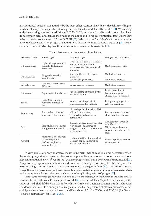
Antibiotics 2019, 8, 138
intraperitoneal injection was found to be the most effective, most likely due to the delivery of highernumbers of phages more quickly and for a greater sustained period than other routes [24]. When usingoral phage dosing in mice, the addition of 0.025% CaCO3 was found to effectively protect the phagefrom stomach acids and deliver the phage to the upper and lower gastrointestinal tract where theyreduced numbers of the targeted E. coli O157:H7 [25]. When treating Burkholderia infections induced inmice, the aerosolization of phages was found to be superior to intraperitoneal injection [26]. Someadvantages and disadvantages of the administration routes are shown in Table 1.
Table 1. Routes of administration for phage therapy.
Delivery Route Advantages Disadvantages Mitigations to Hurdles
IntraperitonealHigher dosage volumespossible. Diffusion toother sites.
Extent of diffusion to other sitesmay be overestimated inhumans (most data from smallanimals).
Multiple delivery sites.
Intramuscular Phages delivered atinfection site.
Slower diffusion of phages(possibly).Lower dosage volumes.
Multi-dose courses.
Multi-dose courses.
Subcutaneous Localized and systemicdiffusion. Lower dosage volumes. Multi-dose courses.
Intravenous Rapid systemic diffusion. Rapid clearing of phages by theimmune system.
In vivo selection oflow-immunogenicphages may be possible.
TopicalHigh dose of phagesdelivered at infectionsite.
Run-off from target site ifphages suspended in liquid.
Incorporate phages intogels and dressings.
Suppository Slow, stable release ofphages over long time.
Limited applications/sites. Riskof insufficient dosing.Technically challenging tomanufacture.
Careful consideration ofphage kinetics required.
Oral Ease of delivery. Higherdosage volumes possible.
Stomach acid reduces phage titer.Non-specific adherence ofphages to stomach contents andother microflora.
Add calcium carbonateto buffer pH.Microencapsulation todeliver phages to targetarea.
Aerosol
Relative ease of delivery.Can reach poorlyperfused regions ofinfected lungs.
High proportion of phages lost.Delivery can be impaired bymucus and biofilms
Use of depolymerases toreduce mucus.
In vitro studies of phage pharmacokinetics using mathematical models do not necessarily reflectthe in vivo phage kinetics observed. For instance, phage T4 was reported to not replicate in vitro athost concentrations below 104 per mL, but evidence suggests that this is possible in murine models [27].Phage feeding experiments in animals and humans frequently report irregular shedding and thepassage of high percentages (up to 90% administered) of phages in feces [27]. The failure of manyphage therapy experiments has been related to a poor understanding of phage pharmacokinetics,for instance, when dosing relies too much on the self-replicating nature of phages [20].
Phage lytic enzymes (endolysins) can also be used for therapy, but their kinetics are more similarto conventional treatments. For example, Jun et al. [28] determined that a Staphylococcus aureus specificendolysin had a half-life between 0.04 and 0.38 h after intravenous administration in healthy volunteers.The decay kinetics of this endolysin is likely explained by the presence of plasma proteases. Otherendolysins have demonstrated a longer half-life such as 11.3 h for CF-301 and 5.2–5.6 h (for 30 and60 mg/kg, respectively) for P128 [29,30].
196
Antibiotics 2019, 8, 138
Toxin (e.g., endotoxin) release due to significant bacterial cell lysis could potentially trigger septicshock during phage therapy. However, antibiotics like amikacin, cefoxitin, and imipenem have beenshown to induce higher amounts of released endotoxin than coliphages [31]. The increase in endotoxinproduced after 180 min incubation of E. coli LM33 was 3.8-fold with phage LM33_P1, 5.5-fold withamikacin, 8.7-fold with cefoxitin, and 30-fold with imipenem. With E. coli strain 536, there wasa 19.8-fold increase in endotoxin with amikacin, 29.9-fold with phage 536_P1, 53.7-fold with imipenem,and 125.1-fold with ceftriaxone [31].
So, whilst less of an issue than for most conventional antibiotics, high fragmentation of the cellwall must be minimized with either phage or phage endolysin therapies to prevent an increase inpro-inflammatory cytokines [19,32]. To address this potential issue, several groups have proposedgenetically engineering phages to prevent or reduce cell lysis, whilst still causing cell death bymechanisms such as degrading the host genome (see Section 7 and [33]).
4. Role of the Immune Response in Phage Therapy
Phages can potentially trigger innate and adaptive immune cells that may influence the successof phage therapy. Three major fields of phage-immune interaction can be discerned. First, involvingimmune recognition via pattern recognition receptor (PRR), which is a means for the recruitment ofphagocytes to the infection site [34]. Phages can mediate the activation of innate immune cells whenPRR recognizes phage-derived DNA and RNA. The extent of immune activation will differ dependingon the phage type, the phage dose, and in vivo nucleic acid synthetic activity.
Second, promoted phage-neutralizing antibodies can hamper therapeutic success and this effectcan increase with repeated administration [35]. Antibody induction against phages is considered to behighly variable, thus immunogenicity should be considered during phage screening prior to phagetherapy. There are several externally presenting proteins on phages such as Hoc, which can potentiallyinduce such an immune response [36–38]. Strategies to avoid phage-induced neutralizing antibodyformation include refining dose concentrations, the use of low-multi-dose regimes, or low-dose passivetherapy approaches.
Third, the inhibitory effect of humoral (adaptive) immunity and anti-phage antibody production onphages in the mammalian system is broadly known. Effects seem dose-dependent, with only high dosesfor long periods inducing specific responses. For instance, Majewska [39] developed a long-term studyof antibody induction (IgM, IgG, secretory IgA) in mice fed T4 phage orally at high doses (109 PFU/mLdrinking water). No effect was noted in the first two weeks, then in weeks 3–5, there was an increasein blood serum IgG. IgM did not increase until IgG began increasing, while IgA did not increase untildays 63–79, but when it reached its maximum, no phage was found in the mouse feces. Increased IgAconcentrations antagonized the gut transit of active phage and phage resistant hosts dominated thegut flora by day 92. However, IgA was rapidly cleared after phage withdrawal [39]. A similar studydetermined the immunological response of Pseudomonas phage F8 and T4 treatment in a murine systemicinflammatory response syndrome (SIR) model. The primary (IgM) and the secondary (IgG) responsesinhibited the phages, and phage concentration in the spleen was significantly decreased [40].
Human trials in 26 patients with immunodeficiency diseases were undertaken to evaluateimmunologic responses to phage φX174. An intravenous dose of 109 PFU/kg body weight was given,and the phage titer measured in blood. No antibody response was detected in eight cases of infantileX-linked agammaglobulinemia with circulating phages present for up to 11 days. The other 18 patientsproduced antibodies and phages were cleared from circulation within four days. Ten of these patientsshowed the IgM antibody, and eight patients produced both IgM and IgG [41]. Other work usingφX174 [42] has demonstrated that repeated (up to quaternary) dosing of phages does not lead toserious adverse reactions.
It is currently not well understood if anti-phage antibodies could prevent bacterial resistancedevelopment to phages and if the pre-existing immunity to natural phages could affect phage therapy.Furthermore, there is no clear information about the impact of phage-specific factors on phage clearance
197
Antibiotics 2019, 8, 138
mechanisms. There are also gaps in our understanding of the clinical relevance of the phage immuneinteraction. Nevertheless, the immunogenicity of phages itself does not seem to represent a significantsafety risk for patients. Reports about immune effects in clinical studies using virulent phages arelimited. The introduction of validated in vitro and in vivo methods to determine the comparabilityof immune effects of different phages and phage combinations would be indispensable. This wouldallow for valid conclusions on the value of immune-based parameters for the selection of phages,identification of responsive patient populations, exchangeability of phages, and the importance ofindividualized phage cocktails [33]. The engineering of phages to make them less immunogenic is alsoan area of active research (see Section 7).
5. Resistance to Phages
An important consideration for phage therapy is the potential for bacterial resistance.Phage-resistant bacteria have been noted in up to 80% of studies targeting the intestines and 50% ofstudies using sepsis models, with phage-resistant variants also observed in human studies [43].
As with resistance to classical antibiotics, spontaneous resistance to phages may occur througha number of mechanisms. For example, the cell surface target receptor(s) may not be expressed orbecome mutated, thus causing a complete loss of adsorption or decreased adsorption. This is a limitationof both phage and conventional antibiotic therapy. For both approaches, knowing the receptor site(s),their stability, and conservation across strains will help with the mitigation of resistance.
Acquired resistance is another area that requires investigation for both therapeutic approaches.Accessory genetic elements such as plasmids, temperate phages, and mobile genetic islands can carrygenes coding for resistance to antibiotics. For phages, acquired resistance can encompass CRISPR-Cassystems [33], immunity proteins produced by temperate phages (though rare) and the acquisition ofDNA restriction-modification systems.
A key advantage of phage therapy over conventional treatments for the avoidance of resistancedevelopment is the deployment of phage cocktails. The use of several phages, each targeting differentreceptors and each of a diverse genetic clade will enhance the ability to mitigate against the loss ofadsorption or host genetic protection mechanisms. Genetic engineering may also provide a means toimprove the diversity and targeting efficiency of phages for the avoidance of resistance (see Section 7).Another consideration is that bacterial mutations that confer phage-resistance often result in fitnesscosts to the resistant bacterium. Therefore, understanding and exploiting the fitness costs to resistantpathogens during therapy is a potentially promising research avenue [43].
6. Phage Therapy Clinical Trials in Humans
To date, human phage therapy trials have largely been empirical, with routine use limited toGeorgia, Poland, and Russia [44]. In particular, the George Eliava Institute in Georgia has longstandingexperience with the selection, isolation, and preparation of monophage and phage cocktails againsta variety of bacterial pathogens for phage therapy. The therapeutic application of phages has alsobeen undertaken for several decades at the Institute of Immunology and Experimental Therapy inPoland [45]. However, the experimental clinical data published in Russian and Polish journals aredifficult to access due to security and language barriers.
Although the reporting and assessment of phage therapy need to improve, particularly withregard to efficacy and tolerability and the use of adequate patient numbers, several successful casereports have been published. The reports do provide some evidence that the development of phagetherapy is a promising alternative to combat bacterial resistance to antibiotics.
In France, the national health regulator has authorized the first treatment of patients with extremelydrug-resistant and difficult to treat infections using phage therapy. Since then, six cases with variousbacterial infections have been successfully treated [44]. Even though several treatments were notconducted using clinical standards suitable for drug approval in the Western world, they showedtherapeutic potential for phages and how phages can be applied [45].
198
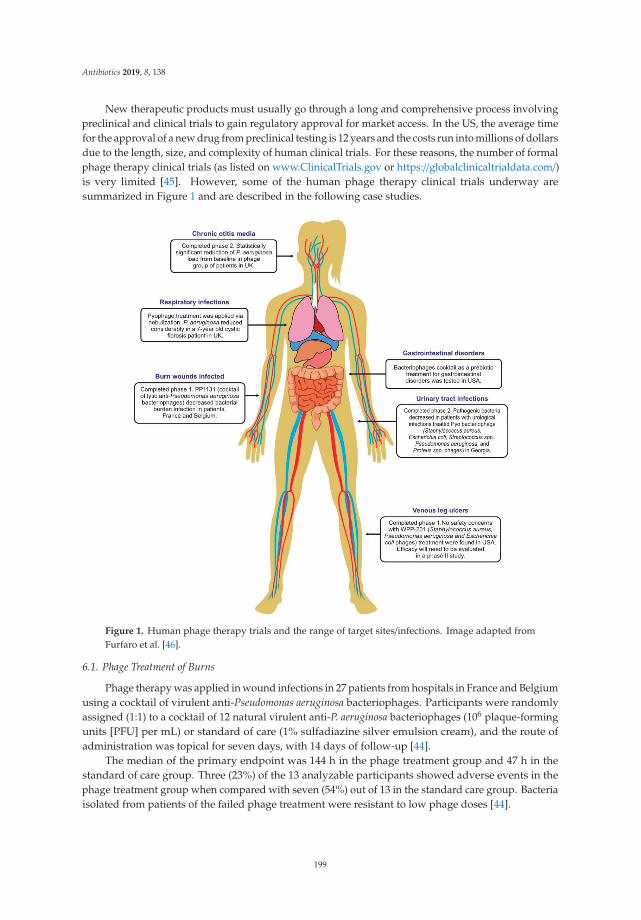
Antibiotics 2019, 8, 138
New therapeutic products must usually go through a long and comprehensive process involvingpreclinical and clinical trials to gain regulatory approval for market access. In the US, the average timefor the approval of a new drug from preclinical testing is 12 years and the costs run into millions of dollarsdue to the length, size, and complexity of human clinical trials. For these reasons, the number of formalphage therapy clinical trials (as listed on www.ClinicalTrials.gov or https://globalclinicaltrialdata.com/)is very limited [45]. However, some of the human phage therapy clinical trials underway aresummarized in Figure 1 and are described in the following case studies.
Figure 1. Human phage therapy trials and the range of target sites/infections. Image adapted fromFurfaro et al. [46].
6.1. Phage Treatment of Burns
Phage therapy was applied in wound infections in 27 patients from hospitals in France and Belgiumusing a cocktail of virulent anti-Pseudomonas aeruginosa bacteriophages. Participants were randomlyassigned (1:1) to a cocktail of 12 natural virulent anti-P. aeruginosa bacteriophages (106 plaque-formingunits [PFU] per mL) or standard of care (1% sulfadiazine silver emulsion cream), and the route ofadministration was topical for seven days, with 14 days of follow-up [44].
The median of the primary endpoint was 144 h in the phage treatment group and 47 h in thestandard of care group. Three (23%) of the 13 analyzable participants showed adverse events in thephage treatment group when compared with seven (54%) out of 13 in the standard care group. Bacteriaisolated from patients of the failed phage treatment were resistant to low phage doses [44].
199
Antibiotics 2019, 8, 138
This study showed that phage treatment decreased bacterial burden in burn wounds in more timethan the standard treatment. In this regard, studies increasing the phage concentration and the use of“phagograms” (as used for antibiograms) with more patients are warranted.
6.2. Treatment of A Septicemia Patient with Acute Kidney Damage
A man in his sixties was hospitalized for Enterobacter cloacae peritonitis and severe abdominalsepsis, dispersed intravascular coagulation, herniation, and bowel strangulation. Following prolongedtreatment for these ailments, the patient developed gangrene and pressure sores colonized bydrug-resistant P. aeruginosa. The infection developed to septicemia and colistin treatment (the onlydrug sensitivity) was carried out, however, acute kidney damage was detected, and the treatment wassuspended. Subsequently, phage therapy against P. aeruginosa was conducted using a mixture of twophages active against the isolate in vitro, under the umbrella of Article 37 (Unproven Interventions inClinical Practice) of the Declaration of Helsinki [46]. Following phage therapy, the patient showedimproved kidney function, which returned to normal function after a few days, and blood cultureswere negative.
However, the patient’s pressure sores remained infected with P. aeruginosa and other species andfour months later, the patient developed a refractory cardiac arrest due to blood culture-confirmedKlebsiella pneumoniae sepsis and the patient died. In vivo studies revealed that a K. pneumoniae strainisolated from the patient was sensitive to the antibiotics. According to historical reports, the use ofphages by intravenous route in typhoid fever and Staphylococcus aureus [47] bacteremia were efficacious,nevertheless, this is the first contemporary report using phage monotherapy against P. aeruginosasepticemia in humans through the intravenous route [48].
6.3. Engineered Phages for Treatment of Mycobacteria in A Cystic Fibrosis Patient
Therapeutic phage treatment for mycobacteria has been explored in several animal models [49,50],but until recently had not been successfully used for mycobacterial infections in humans. A 15-year-oldpatient with cystic fibrosis and extensive comorbidities was referred for lung transplant with a disseminatedinfection of Mycobacterium abscessus. Following bilateral lung transplantation and persistent M. abscessusinfections, phage genome engineering and forward genetics were used to engineer phages to target andkill the infectious M. abscessus strain. Therapy was conducted using an intravenous three-phage cocktail of109 PFU of each phage every 12 h for 32 weeks [51].
Intravenous phage treatment was well tolerated, clinical improvement including sternal woundclosure, improved liver and lung function, and substantial resolution of infected skin nodules weredetected in the six months following therapy. No evidence of phage neutralization was detected insera, although weak antibody responses to phage proteins were identified. Weak cytokine responseswere reported for interferon-γ, interleukin-6, interleukin-10, and tumor necrosis factor-α [51]. Someevidence was presented that indicated active in vivo phage replication was taking place. Despitethe apparent success of this therapy, the authors did caution that there was significant variation inM. abscessus phage susceptibility, so the treatment of similar patients will require more work to beundertaken to understand the science underlying this observation.
6.4. Phage Therapy for Respiratory Infections
There have been several pre-clinical studies describing the use of phage therapy against chronicbacterial lung infections using murine models. Pabary et. al. [52] determined that phage treatmentreduced the infective burden and inflammatory response in the murine lung. All phage-treatedmice cleared P. aeruginosa infection at 24 h, whereas infection persisted in all of the control mice.Phage also reduced infection and inflammation in bronchoalveolar lavage fluid when administeredprophylactically. Another study showed that intranasal treatment with phage rescued mice fromAcinetobacter baumannii-mediated pneumonia. Microcomputed tomography also indicated a reductionin lung inflammation in mice given phage [53]. In a study using a biofilm-associated murine model
200
Antibiotics 2019, 8, 138
of chronic lung infection, phage therapy was effective seven days post-infection. Additionally, thesestudies established the potential for phage therapy in established and recalcitrant chronic respiratorytract infections [54].
Notwithstanding the reported treatment of Mycobacterium described in Section 6.3, reports ofphage therapy of human bacterial respiratory infections are rare. In a 2011 case study from Georgia,a seven-year-old cystic fibrosis (CF) patient presented with chronic colonization of P. aeruginosa andS. aureus with antibiotic treatments having limited effect. Phage therapy was undertaken usinga “Pyo phage” phage cocktail produced by the Eliava Institute, which reportedly contains phagesactive against S. aureus, Streptococcus, Proteus, P. aeruginosa, and E. coli [55]. The Pyo phage cocktail wasdelivered to the patient via nebulization at four-to-six week intervals for nine rounds of treatment.The P. aeruginosa numbers reduced considerably, however, the treatment was not effective againstS. aureus. Consequently, Sb-1 phage (a phage targeting S. aureus) was added to the Pyo phage cocktailand administered five times with a nebulizer. This treatment reduced the concentration of S. aureussignificantly. No adverse effects were detected in the patient upon Sb-1 phage treatment [55].
Recent advances in the spray drying of phages that have achieve increased numbers of phagesdelivered to the lungs (up to 108 PFU/aspiration) may considerably improve clinical outcomes forrespiratory infections such as these [56]. Work has also shown that a cocktail of ten phages significantlydecreased P. aeruginosa numbers in sputum samples from 58 CF patients collected from hospitals inParis [45,57]. Forty-eight of 58 samples were positive for P. aeruginosa and the addition of phagessignificantly decreased the concentrations of P. aeruginosa in the sputum. An increase in the number ofbacteriophages in 45.8% of these samples was also detected, demonstrating the potential for activephage therapy of respiratory infections in vivo.
6.5. Phage Therapy for Urinary Tract Infections
Therapy for treating urinary tract infections (UTIs) is one of the most promising applications forphages and one of the few that have been studied in a multi-stage clinical trial. In the first stage ofthe trial, 130 patients planned for transurethral resection of the prostate were screened for UTIs and118 patients enrolled [58]. Criteria for inclusion in the trial were having ≥104 cfu/mL of the pathogensS. aureus, E. coli, Streptococcus, P. aeruginosa, or Proteus mirabilis in their urine culture. Initial in vitroscreening of these cultures against the Pyo phage cocktail, a commercial product produced by theEliava Institute, revealed that the sensitivity was 41% (48/118). Directed evolution experiments wereapplied to the cocktail to select for expanded host range phages, and the sensitivity was improved to75% (88/118).
In the second stage, nine patients who had infections caused by bacteria sensitive to the Pyococktail underwent non-blinded phage therapy. Administration of 20 mL 107–109 PFU/mL phageswas via a suprapubic catheter twice every 24 h for seven days, starting the first day after surgery.Urine from the patients was subsequently cultured seven days after surgery or at the time of adverseindications. Prior to therapy, the patients’ urine screening revealed the presence of E. coli in four cases,Enterococcus in two cases, Streptococcus in two cases, and P. aeruginosa in one case. Following therapy,titers of the pathogens decreased by 1–5 log cfu/mL in six out of nine patients. One of the four E. colicases had no detectable pathogen, one of two Streptococcus cases had no detectable pathogen, oneof the Enterococcus cases had no pathogens, but the other case detected E. coli. The patient with theP. aeruginosa infection required antibiotic therapy following a spike in fever and became asymptomatic;however, P. aeruginosa was detected in his urine. No adverse effects of phage therapy were detected.The study authors hope to further progress this work to full randomized and blinded control studiesin the future.
6.6. Phage Therapy for Diarrhea
Whilst no longer an active partnership, the Nestlé Research Centre in Switzerland and theInternational Centre for Diarrhoeal Diseases Research in Bangladesh have undertaken joint research
201
Antibiotics 2019, 8, 138
projects over a number of years that have explored the efficacy of phage therapy for the treatment ofdiarrheal diseases. In one of the studies, 120 Bangladeshi male children (6–24 months) presenting withacute bacterial diarrhea were given either 3.6 × 10⁸ PFU of a T4-like coliphage cocktail (39 children),1.4 × 109 PFU of a commercial coliphage preparation (Coliproteus from Microgen, 40 children),or a placebo (0.9% NaCl, 41 children) suspended in oral rehydration solution.
Results of this randomized blind trial indicated no adverse effects of oral phage treatment of thechildren. The phage survived the gastric passage, but there was no strong evidence of intestinal replicationoccurring in patients. Neither the T4-like nor the Microgen coliphage cocktail showed a significantclinical effect when compared to the control group for stool output or frequency, or rehydration. Likelyreasons for the lack of significant effects were the lower than expected incidence of E. coli (60%) and theincidence of mixed species infections, the presence of non-susceptible coliforms (phage cocktail was notoptimized for local isolates), and insufficient phage titer [59,60].
6.7. Treatment of Peri-Prosthetic Joint Infection
In this case study [61], an 80-year-old female patient with obesity and a history of relapsingprosthetic joint infection of the right hip presented with a S. aureus postoperative infection and wastreated with debridement, antibiotics, and implant retention (DAIR). Four years later, another DAIRwas performed for fluoroquinolone-resistant E. coli, following reimplantation surgery in the prioryear. Then, due to a relapse including positive E. coli cultures, another DAIR was performed threeweeks later. Antibacterial therapy with ceftriaxone was started; however, there were further signs ofrelapse and antibiotic treatment was stopped. Multidrug-resistant P. aeruginosa and penicillin-resistantS. aureus were identified in swabs of the wound discharge.
To undertake phage therapy, three phages targeted against the P. aeruginosa isolate were firstprepared by Pherecydes Pharma (France). The S. aureus isolate was lost, so three phages from thePherecydes Pharma phage bank were used. Phages were produced in a research environment withthe manufacture overseen by The French National Agency for Medicines and Health Products Safety(ANSM). The final formulation of the P. aeruginosa and S. aureus phages were undertaken by the hospitalpharmacy by mixing equal volumes of 1010 PFU/mL phage stocks. During the following DAIR, 20 mLof the phage cocktail was injected into the joint region. Co-therapy with antibiotics (daptomycin,amoxicillin, and clindamycin) was followed for the next six months without signs of P. aeruginosa orS. aureus infection. The patient later had a Citrobacter infection, which required DAIR, but once this wastreated with Ciprofloxacin, no further infection was found in the joint (18 months post-phage therapy).
The bespoke use of phage and antibiotic combinations to treat a patient’s infection has the potentialto be utilized to create personalized therapy for deep and persistent tissue infections such as thosefound associated with peri-prosthetic joints (Figure 2).
202
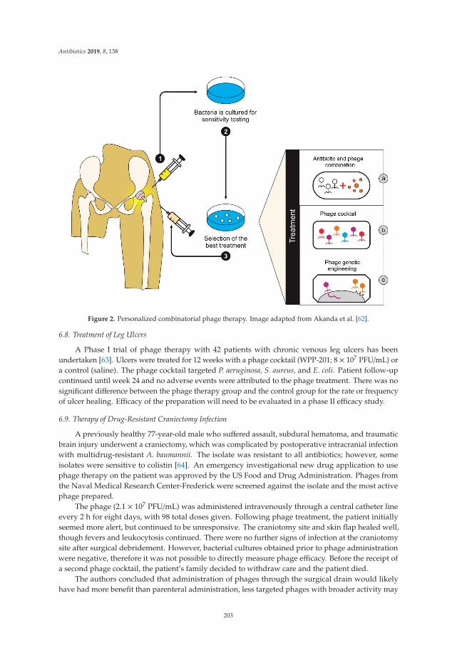
Antibiotics 2019, 8, 138
Figure 2. Personalized combinatorial phage therapy. Image adapted from Akanda et al. [62].
6.8. Treatment of Leg Ulcers
A Phase I trial of phage therapy with 42 patients with chronic venous leg ulcers has beenundertaken [63]. Ulcers were treated for 12 weeks with a phage cocktail (WPP-201; 8 × 10⁷ PFU/mL) ora control (saline). The phage cocktail targeted P. aeruginosa, S. aureus, and E. coli. Patient follow-upcontinued until week 24 and no adverse events were attributed to the phage treatment. There was nosignificant difference between the phage therapy group and the control group for the rate or frequencyof ulcer healing. Efficacy of the preparation will need to be evaluated in a phase II efficacy study.
6.9. Therapy of Drug-Resistant Craniectomy Infection
A previously healthy 77-year-old male who suffered assault, subdural hematoma, and traumaticbrain injury underwent a craniectomy, which was complicated by postoperative intracranial infectionwith multidrug-resistant A. baumannii. The isolate was resistant to all antibiotics; however, someisolates were sensitive to colistin [64]. An emergency investigational new drug application to usephage therapy on the patient was approved by the US Food and Drug Administration. Phages fromthe Naval Medical Research Center-Frederick were screened against the isolate and the most activephage prepared.
The phage (2.1 × 10⁷ PFU/mL) was administered intravenously through a central catheter lineevery 2 h for eight days, with 98 total doses given. Following phage treatment, the patient initiallyseemed more alert, but continued to be unresponsive. The craniotomy site and skin flap healed well,though fevers and leukocytosis continued. There were no further signs of infection at the craniotomysite after surgical debridement. However, bacterial cultures obtained prior to phage administrationwere negative, therefore it was not possible to directly measure phage efficacy. Before the receipt ofa second phage cocktail, the patient’s family decided to withdraw care and the patient died.
The authors concluded that administration of phages through the surgical drain would likelyhave had more benefit than parenteral administration, less targeted phages with broader activity may
203
Antibiotics 2019, 8, 138
have been more efficacious, and a better outcome might have been possible if personalized phagetherapy had been developed more quickly and administered earlier in the course of infection.
6.10. Therapy of Ear Infections
A Phase I/II research trial was conducted in the UK to test the efficacy and safety of phagesfor the treatment of chronic ear infections (otitis media), where the infection is known to harborantibiotic-resistant P. aeruginosa [65]. In this randomized double-blinded study, a cocktail of six phagesproduced by Biocontrol Limited (BiophagePA, 6 × 10⁵ PFU), or placebo (glycerol-PBS solution) wereadministered to the ear canal of 24 patients. The follow-up to treatment was at 7, 21, and 43 daysand revealed a statistically significant improvement in both clinical condition and patient-reportedindicators for the phage treated group when compared to the control. No adverse reactions were notedin the phage-treated group.
In vivo replication of phages in the patients was evident for up to 23 days, with the mean recoveryof phages during the trial sampling points being 200 times the input concentration. Clearance ofphages was noted when the P. aeruginosa infection was resolved in patients. Reductions in overallP. aeruginosa numbers in the phage treatment group, whilst statistically significant, were generallymodest, but measurement was likely to be compromised by the lack of access to deep parts of theear canal. When P. aeruginosa was not completely cleared by phage therapy, there was an increase inclinical scores for some patients. The study authors suggest that repeated phage therapy after three tofour weeks may be beneficial to these types of patients in any future work.
7. Engineering and Other Genetic Technologies for Phage Therapy
The advent of whole-genome sequencing and metagenomics have rapidly increased the numberof phage genomes sequenced and is unlocking new insights into phage genetics. The use of this newknowledge for phage engineering holds great potential to increase the utility of phages for therapy,however, there are additional considerations such as ethical, safety, and regulatory, which need to beaccounted for above that of ‘natural’ phage therapy. Engineering can be used to produce new variantsof phages with expanded host range, decreasing the number of phage strains needed to cover bacterialdiversity, and generating patentable phage variants [66–68]. For example, the host specificity of theE. coli K12-specific phage T2 was able to be changed by swapping gene products expressed at the tipof the long tail fiber with those of the PP01 phage, which is an E. coli O157: H7-specific phage [66].The recombinant phage was able to infect E. coli O157: H7 and related strains, but could not infectE. coli K12 or its derivatives. Similarly, homologous recombination was used to replace the long tailfiber genes (genes 37 and 38) from the genome of T2 with those of the IP008 phage. The recombinantT2 phage had a host range identical to that of IP008 [67]. Lin et. al. [68] were also able to modifythe E. coli female-specific T7 phage to overcome male exclusion by recombination with phage T3.The recombinant phages of T3 and T7 carried altered tail fibers and had better adsorption efficiencythan T3.
Genetic engineering of phage permits the addition of novel functionality such as bacteriocins,enzybiotics, quorum sensing inhibitors, CRISPRs, and biofilm degrading enzymes that can enhancetheir killing potential [69–73]. Phages can be modified using the RNA-guided nuclease Cas9 to createsequence-specific antimicrobials. Cas9 was reprogrammed to target virulence genes and killed virulent,but not avirulent, strains of S. aureus in a mouse skin colonization model [65]. Another study usedCRISPR-Cas technology to create RNA-guided nucleases delivered by phages to target specific DNAsequences in carbapenem-resistant Enterobacteriaceae and enterohemorrhagic E. coli [70]. Delivery ofthe nucleases improved the survival in a Galleria mellonella infection model. Phage-borne CRISPR-Cassystems can also be used to enable site-specific cleavage to induce cytotoxicity, activate toxin-antitoxinsystems, re-sensitize bacterial populations to antibiotics, and modulate bacterial consortia [70].
Biofilms are the major cause of persistent infections in clinical settings, thus phage treatment tolyse bacteria in biofilms has attracted growing interest. An engineered T7 phage was constructed to
204
Antibiotics 2019, 8, 138
encode a lactonase enzyme with broad-range activity for the quenching of quorum sensing moleculesnecessary for biofilm formation. The T7 phage incorporating the AHL lactonase aiiA gene from Bacillusanthracis degraded AHLs from diverse bacteria and caused the inhibition of a mixed-species biofilmcomposed of P. aeruginosa and E. coli [71]. In another approach using the T7 phage, a biofilm-degradingenzyme, DspB, produced by Actinobacillus actinomycetemcomitans, was inserted into the T7 genome andthe resultant phage reduced E. coli biofilm cell counts by an additional 2 log when compared to theunmodified T7 [72].
As described in Section 4, components of the innate immune system can remove a significantproportion of administered phage. Studies have shown that long-circulating phage mutants can beisolated to address this issue. Vitiello et. al. [73] determined that a single specific substitution in themajor phage capsid (E) protein of the lambda Argo phage was enough to confer a long-circulatingphenotype that enhanced phage survival in the mouse circulatory system by more than a 1000-fold.Merril et. al. [74] used a serial passage selection method to isolate phage mutants with a greater capacityto remain in the circulatory system of the mouse. Lambda phage mutants with 13,000–16,000-fold bettercapacity to stay in the mouse circulatory system for 24 h after intraperitoneal injection were isolated.
Many antibiotics, as well as phage therapy, can present side effects due to endotoxin release fromGram-negative bacteria. To address this, genetic engineering was used to generate non-replicatingnon-lytic phage targeting P. aeruginosa. An export protein gene of the P. aeruginosa filamentous phagePf3 was replaced with a restriction endonuclease gene and the variant (Pf3R) was non-replicative andprevented the release of phage from the target cell. Endotoxin release was kept to a minimum and thePf3R phage efficiently killed a wild-type host in vitro. Phage therapy using Pf3R showed comparableor increased survival rates (depending on dose) when compared to Pf3 upon challenge in the micemodel. Higher survival rates were correlated with a reduced inflammatory response when using Pf3Rtreatment [75]. Matsuda et. al. [76] also produced lysis-deficient T4 phages for this purpose. Mutant tamber A3 T4 phages were compared to wild-type T4 in mouse bacterial peritonitis model. Survival wassignificantly higher in mice treated with the lysis deficient phage when compared to the wild-type, andenterotoxin levels were significantly lower in the t A3 T4-treated mice at 12 hours after infection [76].
8. The Medicinal Regulatory Status of Phages
Phages are not specifically classified as living or chemical agents in any national medicinallegislation (as far as we are aware). This considerably complicates the regulation of human phagetherapy clinical trials and commercialization of phage products as well-established safety, goodmanufacturing practice, and efficacy benchmarks are lacking [77]. Another barrier is that in orderto prove the efficiency of phage preparations, their effectiveness and host range toward currentlycirculating pathogenic strains must be constantly monitored. This is most likely why the RussianFederation and Georgia approved phage preparations are continuously updated to target newlyemerging pathogenic strains [78]. Therefore, any specific legislation regarding phage products wouldideally permit these formulation updates as required to avoid repeated registration procedures.
A breakthrough for the regulation of phage therapy occurred in 2016, when the BelgianMinister of Social Affairs and Public Health defined the status of therapeutic phage preparations asindustrially-prepared medicinal products (subjected to constraints related to marketing authorization)or as magistral (compounded) preparations prepared in the pharmacies’ officinal [79]. Naturalphages and their products can be processed by a pharmacist as raw materials (active ingredients)in magistral preparations, providing there is compliance with several provisions of the EuropeanDirective requirements for medicinal products for human use [78].
Several jurisdictions also permit the use of phages on compassionate grounds, where all othertherapies have failed, and the condition is immediately life-threatening. These include the US FDAExpanded Access Program (www.fda.gov/news-events/public-health-focus/expanded-access) andInvestigational Drug Program (https://www.fda.gov/drugs/types-applications/investigational-new-
205
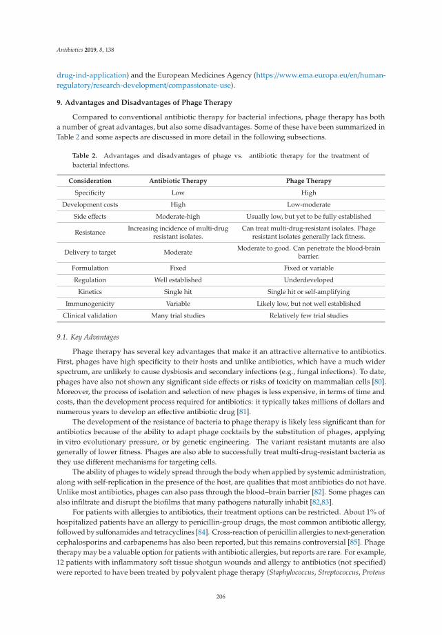
Antibiotics 2019, 8, 138
drug-ind-application) and the European Medicines Agency (https://www.ema.europa.eu/en/human-regulatory/research-development/compassionate-use).
9. Advantages and Disadvantages of Phage Therapy
Compared to conventional antibiotic therapy for bacterial infections, phage therapy has botha number of great advantages, but also some disadvantages. Some of these have been summarized inTable 2 and some aspects are discussed in more detail in the following subsections.
Table 2. Advantages and disadvantages of phage vs. antibiotic therapy for the treatment ofbacterial infections.
Consideration Antibiotic Therapy Phage Therapy
Specificity Low High
Development costs High Low-moderate
Side effects Moderate-high Usually low, but yet to be fully established
Resistance Increasing incidence of multi-drugresistant isolates.
Can treat multi-drug-resistant isolates. Phageresistant isolates generally lack fitness.
Delivery to target Moderate Moderate to good. Can penetrate the blood-brainbarrier.
Formulation Fixed Fixed or variable
Regulation Well established Underdeveloped
Kinetics Single hit Single hit or self-amplifying
Immunogenicity Variable Likely low, but not well established
Clinical validation Many trial studies Relatively few trial studies
9.1. Key Advantages
Phage therapy has several key advantages that make it an attractive alternative to antibiotics.First, phages have high specificity to their hosts and unlike antibiotics, which have a much widerspectrum, are unlikely to cause dysbiosis and secondary infections (e.g., fungal infections). To date,phages have also not shown any significant side effects or risks of toxicity on mammalian cells [80].Moreover, the process of isolation and selection of new phages is less expensive, in terms of time andcosts, than the development process required for antibiotics: it typically takes millions of dollars andnumerous years to develop an effective antibiotic drug [81].
The development of the resistance of bacteria to phage therapy is likely less significant than forantibiotics because of the ability to adapt phage cocktails by the substitution of phages, applyingin vitro evolutionary pressure, or by genetic engineering. The variant resistant mutants are alsogenerally of lower fitness. Phages are also able to successfully treat multi-drug-resistant bacteria asthey use different mechanisms for targeting cells.
The ability of phages to widely spread through the body when applied by systemic administration,along with self-replication in the presence of the host, are qualities that most antibiotics do not have.Unlike most antibiotics, phages can also pass through the blood–brain barrier [82]. Some phages canalso infiltrate and disrupt the biofilms that many pathogens naturally inhabit [82,83].
For patients with allergies to antibiotics, their treatment options can be restricted. About 1% ofhospitalized patients have an allergy to penicillin-group drugs, the most common antibiotic allergy,followed by sulfonamides and tetracyclines [84]. Cross-reaction of penicillin allergies to next-generationcephalosporins and carbapenems has also been reported, but this remains controversial [85]. Phagetherapy may be a valuable option for patients with antibiotic allergies, but reports are rare. For example,12 patients with inflammatory soft tissue shotgun wounds and allergy to antibiotics (not specified)were reported to have been treated by polyvalent phage therapy (Staphylococcus, Streptococcus, Proteus
206
Antibiotics 2019, 8, 138
vulgaris, Proteus mirabilis, P. aeruginosa, E. coli, and K. pneumoniae) for 15 days [86]. The concentrations ofbacteria and the areas of wound healing were similar in the phage treatment group when compared toa control group of 35 patients receiving antibiotics. The authors concluded that phages were a reliablemethod for reducing microbial infection and that treatment led to a rapid epithelialization of thewound site [86].
9.2. Key Disadvantages
There are currently some key disadvantages of using phages as alternatives for antibiotics.However, these are predominantly due to gaps in knowledge and regulations, which may be resolvedin the future. Critically, there is a lack of depth of information about the clinical application of phagesfor controlling bacterial infections. Much experimental clinical data published in Russian and Polishjournals are difficult to access due to security and language barriers. There are also many morechallenges for scientists in obtaining regulatory approval for phage-based therapeutic applicationswhen compared to conventional therapies [80].
There is a lack of common established and validated protocols for the routes of administration,dose, frequency, and duration of phage treatment, which hampers inter-study comparison [87]. Often,the purity and stability of phage preparations used for clinical trials are also uncertain, with insufficientquality control data presented.
The concentration of phages may be reduced significantly during therapy by the reticuloendothelialsystem or be neutralized by antibodies, thus inhibiting their antimicrobial activity [39,88]. However,the effect of phage-neutralizing antibodies can be mitigated by refining dosing regimens and breedingphages to evade the immune system.
The genetic biosafety of phages is complex to assess. Phages used for therapy must not containtoxin or virulence genes, antibiotic resistance genes, or be able to horizontally transfer genes in thehuman microflora. Whilst whole-genome sequencing is a powerful tool to assist with these analyses,there is still an incomplete understanding of the functions of all encoded phage genes. Geneticengineering of phages will also likely invite greater scrutiny of safety which practitioners will need toaddress before application.
10. Conclusions
Antimicrobial resistance is increasing globally, and new treatments are urgently needed to meetthis challenge in medical care. Whilst phage therapy for bacterial infections has been around for morethan a century, the antibiotic-resistance crisis is providing renewed impetus for phage therapy to deliveron its long-held promise as a clinical treatment. As described here, there is an increasing number ofwell-executed Phase I/II clinical trials describing the safety and efficacy of phage therapy. There isan improved understanding of the pharmacology, immunology, safety, and potential for bacterialresistance. Technologies such as genetic engineering, whole-genome sequencing, and metagenomicsalso provide new tools to optimize phage therapeutic strategies. However, there are still data gaps onits efficacy and a lack of standardization and suitable regulatory frameworks that need to be resolvedbefore phage therapy can take its place in mainstream medicine. Given the renewed interest andimpetus in the field of phage therapy, there are reasons to be optimistic that these challenges can bemet in the coming years.
Author Contributions: Conceptualization, D.R.-C. and C.B.; methodology, D.R.-C.; writing—original draftpreparation, D.R.-C. and C.B.; writing—review and editing, D.R.-C., R.G.-B., A.G.-N. and C.B.; visualization,D.R.-C.; supervision, R.G.-B., A.G.-N. and C.B.; project administration, C.B.; funding acquisition, C.B.
Funding: This research was funded by an ESR Strategic Science Investment Fund grant to CB.
Conflicts of Interest: The authors declare no conflict of interest. The funders had no role in the design of thestudy; in the collection, analyses, or interpretation of data; in the writing of the manuscript, or in the decision topublish the results.
207
Antibiotics 2019, 8, 138
References
1. Hendrix, R.W.; Smith, M.C.M.; Burns, R.N.; Ford, M.E.; Hatfull, G.F. Evolutionary relationships amongdiverse bacteriophages and prophages: All the world’s a phage. Proc. Natl. Acad. Sci. USA 1999, 96,2192–2197. [CrossRef] [PubMed]
2. Hendrix, R.W. Bacteriophages: Evolution of the Majority. Theoretical Population Biology. Popul. Biol. 2002,61, 471–480. [CrossRef]
3. Abedon, S.T.; Kuhl, S.J.; Blasdel, B.G.; Kutter, E.M. Phage treatment of human infections. Bacteriophage 2011,1, 66–85. [CrossRef] [PubMed]
4. Miedzybrodzki, R.; Borysowski, J.; Weber-Dabrowska, B.; Fortuna, W.; Letkiewicz, S.; Szufnarowski, K.;Pawełczyk, Z.; Rogóz, P.; Kłak, M.; Wojtasik, E.; et al. Clinical aspects of phage therapy. Adv. Virus Res. 2012,83, 73–121. [PubMed]
5. Reyes, A.; Semenkovich, N.P.; Whiteson, K.; Rohwer, F.; Gordon, J.I. Going viral: Next-generation sequencingapplied to phage populations in the human gut. Nat. Rev. Microbiol. 2012, 10, 607–617. [CrossRef] [PubMed]
6. Weber-Dabrowska, B.; Jónczyk-Matysiak, E.; ·Zaczek, M.; Łobocka, M.; Łusiak-Szelachowska, M.; Górski, A.Bacteriophage Procurement for Therapeutic Purposes. Front. Microbiol. 2016, 7, 1177. [CrossRef] [PubMed]
7. Aminov, R.; Caplin, J.; Chanishvili, N.; Coffey, A.; Cooper, I.; De Vos, D.; Doškar, J.; Friman, V.; Kurtböke, I.;Pantucek, R.; et al. Application of bacteriophages. Microbiol. Aust. 2018, 38, 63–66.
8. NIH NIAID’s Antibacterial Resistance Program: Current Status and Future Directions. 2014. Availableonline: http://www.niaid.nih.gov/topics/antimicrobialresistane/documents/arstrategicplan2014.pdf (accessedon 23 September 2015).
9. Moellering, R.C. NDM-1—A cause for worldwide concern. N. Eng. J. Med. 2010, 363, 2377–2379. [CrossRef][PubMed]
10. Ceyssens, P.-J.; Lavigne, R. Introduction to Bacteriophages Biology and Diversity. In Bacteriophages in Controlof Food and Waterborne Pathogens; Sabour, P.M., Griffiths, M.W., Eds.; American Society of Microbiology:Washington, DC, USA, 2010.
11. Guttman, B.; Raya, R.; Kutter, E. Basic Phage Biology. In Bacteriophages: Biology and Applications; Kutter, E.,Sulakvelidze, A., Eds.; CRC Press Florida: Boca Raton, FL, USA, 2011; pp. 29–66.
12. Engelkirk, G.; Duben-Engelkirk, P. Burton’s Microbiology for the Health Sciences, 9th ed.; Lippincott Williamsand Wilkins: Philadelphia, PA, USA, 2011.
13. Drulis-Kawa, Z.; Majkowska-Skrobek, G.; Maciejewska, B.; Delattre, A.S.; Lavigne, R. Learning frombacteriophages—Advantages and limitations of phage and phage-encoded protein applications. Curr. ProteinPept. Sci. 2012, 13, 699–722. [CrossRef] [PubMed]
14. Abubakar, S.; Hauwa-Suleiman, B.; Ali Abbagana, B.; Alhaji-Mustafa, I.; Abbas-Musa, I. Novel Uses ofBacteriophages in the Treatment of Human Infections and Antibiotic Resistance. Am. J. Biosci. 2016, 4, 34–40.[CrossRef]
15. Available online: https://en.wikipedia.org/wiki/Listeria_phage_P100 (accessed on 1 August 2019).16. Friman, V.P.; Soanes-Brown, D.; Sierocinski, P.; Molin, S.; Johansen, H.K.; Merabishvili, M.; Pirnay, J.P.;
De Vos, D.; Buckling, A. Pre-adapting parasitic phages to a pathogen leads to increased pathogen clearanceand lowered resistance evolution with Pseudomonas aeruginosa cystic fibrosis bacterial isolates. J. Evol. Biol.2016, 29, 188–198. [CrossRef] [PubMed]
17. Love, M.; Bhandari, D.; Dobson, R.; Billington, C. Potential for Bacteriophage Endolysins to Supplement orReplace Antibiotics in Food Production and Clinical Care. Antibiotics 2018, 7, 17. [CrossRef] [PubMed]
18. Rafii, F.; Sutherland, J.B.; Cerniglia, C.E. Effects of treatment with antimicrobial agents on the human colonicmicroflora. Clin. Risk Manag. 2008, 4, 1343. [CrossRef] [PubMed]
19. Abdelkader, K.; Gerstmans, H.; Saafan, H.; Dishisha, T.; Briers, Y. The Preclinical and Clinical Progress ofBacteriophages and Their Lytic Enzymes: The Parts are Easier than the Whole. Viruses 2019, 24, 11.
20. Abedon, S.T.; Thomas-Abedon, C. Phage therapy Pharmacology. Cur. Pharm. Biotechnol. 2010, 11, 28–47.[CrossRef]
21. Payne, R.J.H.; Phil, D.; Jansen, V.A. Phage therapy: The peculiar Kinetics of self-replicating pharmaceuticals.Clin. Pharm. Ther. 2000, 68, 225–230. [CrossRef]
22. Loc-Carrillo, C.; Abedon, S.T. Pros and Cons of phage therapy. Bacteriophage 2011, 2, 111–114. [CrossRef][PubMed]
208
Antibiotics 2019, 8, 138
23. Bull, J.J.; Regoes, R.R. Pharmacodynamics of non-replicating viruses, bacteriocins and lysins. Proc. Biol. Sci.2006, 273, 2703–2712. [CrossRef] [PubMed]
24. McVay, C.S.; Velasquez, M.; Fralick, J.A. Phage therapy of Pseudomonas aeruginosa infection in a mouse burnwound model. Antimicrob. Agents Chemother. 2007, 51, 1934–1938. [CrossRef]
25. Tanji, Y.; Shimada, T.; Fukudomi, H.; Miyanaga, K.; Nakai, Y.; Unno, H. Therapeutic use of phage cocktailfor controlling Escherichia coli O157:H7 in gastrointestinal tract of mice. J. Biosci. Bioeng. 2005, 100, 280–287.[CrossRef]
26. Semler, D.D.; Goudie, A.D.; Finlay, W.H.; Dennis, J.J. Aerosol phage therapy efficacy in Burkholderia cepaciacomplex respiratory infections. Antimicrob. Agents Chemother. 2014, 58, 4005–4013. [CrossRef]
27. Brüssow, H. Phage therapy: The Escherichia coli experience. Microbiology 2005, 151, 2133–2140. [CrossRef][PubMed]
28. Jun, S.Y.; Jang, I.J.; Yoon, S.; Jang, K.; Yu, K.-S.; Cho, J.Y.; Seong, M.-W.; Jung, G.M.; Yoon, S.J.; Kang, S.H.Pharmacokinetics and tolerance of the phage endolysin-based candidate drug SAL200 after a singleintravenous administration among healthy volunteers. Antimicrob. Agents Chemother. 2017, 24, 61. [CrossRef][PubMed]
29. Cassino, C.; Murphy, M.; Boyle, J.; Rotolo, J.; Wittekind, M. Results of the first in human study of lysin CF-301evaluating the safety, tolerability and pharmacokinetic profile in healthy volunteers. In Proceedings of the26th European Congress of Clinical Microbiology and Infectious Diseases, Amsterdam, The Netherlands,9–12 April 2016.
30. Sriram, B.; Chikkamadaiah, S.C.R.; Durgaiah, M.; Hariharan, S.; Jayaraman, R.; Kumar, S.; Maheshwari, U.;Nandish, P. Pharmacokinetics and efficacy of ectolysin P128 in a mouse model of systemic Methicillinresistant Staphylococcus aureus (MRSA) infection. In Proceedings of the ASM Microbe 2017, New Orleans, LA,USA, 1–5 June 2017.
31. Dufour, N.; Delattre, R.; Ricard, J.D.; Debarbieux, L. The lysis of pathogenic Escherichia coli by bacteriophagesreleases less endotoxin than betalactams. Clin. Infect. Dis. 2017, 64, 1582–1588. [CrossRef]
32. Fischetti, V.A. Bacteriophage endolysins: A novel anti-infective to control Gram-positive pathogens. Int. J.Med. Microbiol. 2010, 300, 357–362. [CrossRef]
33. Pires, D.P.; Cleto, S.; Sillankorva, S.; Azeredo, J.; Lu, T.K. Genetically engineered phages: A review ofadvances over the last decade. Microbiol. Mol. Biol. Rev. 2016, 80, 523–543. [CrossRef] [PubMed]
34. Roach, D.R.; Leung, C.Y.; Henry, M.; Morello, E.; Singh, D.; Di Santo, J.P.; Weitz, J.S.; Debarbieux, L. Synergybetween the host immune system and bacteriophage is essential for successful phage therapy against an acuterespiratory pathogen. Cell Host Microbe 2017, 22, 38–47.e4. [CrossRef] [PubMed]
35. Biswas, B.; Adhya, S.; Washart, P.; Paul, B.; Trostel, A.N.; Powell, B.; Carlton, R.; Merril, C.R. Bacteriophagetherapy rescues mice bacteremic from a clinical isolate of vancomycin-resistant Enterococcus faecium. Infect.Immun. 2002, 70, 204–210. [CrossRef]
36. Fishman, M. Antibody formation in vitro. J. Exp. Med. 1961, 114, 837–856. [CrossRef] [PubMed]37. Dabrowska, K.; Swita1a-Jelen, K.; Opolski, A.; Górski, A. Possible association between phages, Hoc protein,
and the immune system. Arch. Virol. 2006, 151, 209–215. [CrossRef]38. Belleghem, J.D.; Clement, F.; Merabishvili, M.; Lavigne, R.; Vaneechoutte, M. Pro- and anti-inflammatory
responses of peripheral blood mononuclear cells induced by Staphylococcus aureus and Pseudomonas aeruginosaphages. Sci. Rep. 2017, 7, 8004. [CrossRef] [PubMed]
39. Majewska, J.; Beta, W.; Lecion, D.; Hodyra-Stefaniak, K.; Kłopot, A.; Kazmierczak, Z.; Miernikiewicz, P.;Piotrowicz, A.; Ciekot, J.; Owczarek, B.; et al. Oral application of T4 phage induces weak antibody productionin the gut and in the blood. Viruses 2015, 7, 4783–4799. [CrossRef] [PubMed]
40. Hodyra-Stefaniak, K.; Miernikiewicz, P.; Drapa, J.; Drab, M.; Jonczyk-Matysiak, E.; Lecion, D.; Kazmierczak, Z.;Beta, M.; Harhala, J.M.; Bubak, B.; et al. Mammalian host-versus-phage immune response determines phagefate in vivo. Sci. Rep. 2015, 5, 14802. [CrossRef] [PubMed]
41. Ochs, H.D.; Davis, S.D.; Wedgwood, R.J. Immunologic responses to bacteriophage phi-X 174 inimmunodeficiency diseases. J. Clin. Investig. 1971, 50, 2559–2568. [CrossRef] [PubMed]
42. Smith, L.L.; Buckley, R.; Lugar, P. Diagnostic Immunization with Bacteriophage ΦX 174 in Patients withCommon Variable Immunodeficiency/Hypogammaglobulinemia. Front. Immunol. 2014, 5, 410. [CrossRef][PubMed]
209
Antibiotics 2019, 8, 138
43. Oechslin, F. Resistance development to bacteriophages occurring during bacteriophage therapy. Viruses 2018,10, 351. [CrossRef]
44. Jault, P.; Leclerc, T.; Jennes, S.; Pirnay, J.; Que, Y.A.; Resch, G.; Rousseau, A.F.; Ravat, F.; Carsin, H.;Floch, R.L.; et al. Efficacy and tolerability of a cocktail of bacteriophages to treat burn wounds infected byPseudomonas aeruginosa (PhagoBurn): A randomised, controlled, double-blind phase 1/2 trial. Lancet Infect.Dis. 2019, 19, 35–45. [CrossRef]
45. Chang, R.Y.K.; Wallin, M.; Lin, Y.; Leung, S.S.Y.; Wang, H.; Morales, S.; Chan, H.K. Phage therapy forrespiratory infections. Adv. Drug Deliv. Rev. 2018, 133, 76–86. [CrossRef]
46. Furfaro, L.L.; Payne, M.S.; Chang, B.J. Bacteriophage therapy: Clinical trials and regulatory hurdles.Front. Cell. Infect. Microbiol. 2018, 8, 376. [CrossRef]
47. Speck, P.; Smithyman, A. Safety and efficacy of phage therapy via the intravenous route. FEMS Microbiol.Lett. 2016, 363, 242. [CrossRef]
48. Jennes, S.; Merabishvili, M.; Soentjens, P.; Pang, K.W.; Rose, T.; Keersebilck, E.; Soete, O.; François, P.M.;Teodorescu, S.; Verween, G.; et al. Use of bacteriophages in the treatment of colistin-only-sensitive Pseudomonasaeruginosa septicaemia in a patient with acute kidney injury—A case report. Crit. Care 2017, 21, 129. [CrossRef][PubMed]
49. Sula, L.; Sulova, J.; Stolcpartova, M. Therapy of experimental tuberculosis in guinea pigs with mycobacterialphages DS-6A, GR-21 T, My-327. Czech. Med. 1981, 4, 209–214. [PubMed]
50. Trigo, G.; Martins, T.G.; Fraga, A.G.; Longatto-Filho, A.; Castro, A.G.; Azeredo, J.; Pedrosa, J. Phage TherapyIs Effective against Infection by Mycobacterium ulcerans in a Murine Footpad Model. PLoS Negl. Trop. Dis.2013, 7, 2183. [CrossRef] [PubMed]
51. Dedrick, R.M.; Guerrero-Bustamante, C.A.; Garlena, R.A.; Russell, D.A.; Ford, K.; Harris, K.; Gilmour, K.C.;Soothill, J.; Jacobs-Sera, D.; Schooley, R.T.; et al. Engineered bacteriophages for treatment of a patient witha disseminated drug-resistant Mycobacterium abscessus. Nat. Med. 2019, 25, 730–733. [CrossRef] [PubMed]
52. Pabary, R.; Singh, C.; Morales, S.; Bush, A.; Alshafi, K.; Bilton, D.; Alton, E.; Smithyman, A.; Davies, J.Anti pseudomonal bacteriophage reduces infective burden and inflammatory response in the murine lung.Antimicrob. Agents Chemother. 2016, 60, 744–751. [CrossRef] [PubMed]
53. Wang, Y.; Mi, Z.; Niu, W.; An, X.; Yuan, X.; Liu, H.; Li, L.; Liu, Y.; Feng, Y.; Huang, Y.; et al. Intranasal treatmentwith bacteriophage rescues mice from Acinetobacter baumannii-mediated pneumonia. Future Microbiol. 2016,11, 631–641. [CrossRef]
54. Waters, E.M.; Neill, D.R.; Kaman, B.; Sahota, J.S.; Clokie, M.R.J.; Winstanley, C.; Kadioglu, A. Phage therapyis highly effective against chronic lung infections with Pseudomonas aeruginosa. Thorax 2017, 72, 666–667.[CrossRef]
55. Kutateladze, M.; Adamia, R. Phage therapy experience at the Eliava Institute. Médecine et Maladies Infectieuses2008, 38, 426–430. [CrossRef]
56. Matinkhoo, S.; Lynch, K.; Dennis, J.; Finlay, W.; Vehring, R. Spray-dried respirable powders containingbacteriophages for the treatment of pulmonary infections. J. Pharm. Sci. 2011, 100, 5197–5205. [CrossRef]
57. Saussereau, E.; Vachier, I.; Chiron, R.; Godbert, B.; Sermet, I.; Dufour, N.; Pirnay, J.D.; De Vos, F.; Carrié, N.;Debarbieux, L. Effectiveness of bacteriophages in the sputum of cystic fibrosis patients. Clin. Microbiol. Infect.2014, 20, 983–990. [CrossRef]
58. Ujmajuridze, A.; Chanishvili, N.; Goderdzishvili, M.; Leitner, L.; Mehnert, U.; Chkhotua, A.; Kessler, K.;Sybesma, W. Adapted Bacteriophages for Treating Urinary Tract Infections. Front. Microbiol. 2018, 9, 1832.[CrossRef] [PubMed]
59. Sarker, S.A.; Berger, B.; Deng, Y.; Kieser, S.; Foata, F.; Moine, D.; Descombes, P.; Sultana, S.; Huq, S.;Kumar-Bardhan, P.; et al. Oral application of Escherichia coli bacteriophage: Safety tests in healthy anddiarrheal children from Bangladesh. Environ. Microbiol. 2016, 19, 237–250. [CrossRef] [PubMed]
60. Sarker, S.A.; Brüssow, H. From bench to bed and back again: Phage therapy of childhood Escherichia colidiarrhea. Ann. N. Y. Acad. Sci. 2016, 1372, 42–52. [CrossRef] [PubMed]
61. Ferry, T.; Leboucher, G.; Fevre, C.; Herry, Y.; Conrad, A.; Josse, J.; Batailler, C.; Chidiac, C.; Medina, M.;Lustig, S.; et al. Salvage Debridement, Antibiotics and Implant Retention (“DAIR”) with local injection ofa selected cocktail of bacteriophages: Is it an option for an elderly patient with relapsing Staphylococcusaureus prosthetic-joint infection? Open Forum Infect. Dis. 2018, 24, 5. [CrossRef] [PubMed]
210
Antibiotics 2019, 8, 138
62. Akanda, Z.Z.; Taha, M.; Abdelbary, H. Current review-The rise of bacteriophage as a unique therapeuticplatform in treating peri-prosthetic joint infections. J. Orthop. Res. 2017, 36, 1051–1060. [CrossRef] [PubMed]
63. Rhoads, D.D.; Wolcott, R.D.; Kuskowski, M.A.; Wolcott, B.M.; Ward, L.S.; Sulakvelidze, A. Bacteriophagetherapy of venous leg ulcers in humans: Results of a phase I safety trial. J. Wound Care 2009, 238, 240–243.[CrossRef]
64. Rose, T.; Verbeken, G.; De Vos, D.; Merabishvili, M.; Vaneechoutte, M.; Lavigne, R.; Jennes, S.; Zizi, M.;Pirnay, J.P. Experimental phage therapy of burn wound infection: difficult first steps. Int. J. Burns Trauma2014, 4, 66–73. [PubMed]
65. Wright, A.; Hawkins, C.H.; Anggard, E.E.; Harper, D.R. A controlled clinical trial of a therapeutic phagepreparation in chronic otitis due to antibiotic-resistant Pseudomonas aeruginosa; a preliminary report ofefficacy. Clin. Otolaryngol. 2009, 34, 349–357. [CrossRef]
66. Yoichi, M.; Abe, M.; Miyanaga, K.; Unno, H.; Tanji, Y. Alteration of tail fiber protein gp38 enables T2 phage toinfect Escherichia coli O157: H7. J. Biotechnol. 2005, 115, 101–107. [CrossRef]
67. Mahichi, F.; Synnott, A.J.; Yamamichi, K.; Osada, T.; Tanji, Y. Site-specific recombination of T2 phage usingIP008 long tail fiber genes provides a targeted method for expanding host range while retaining lytic activity.FEMS Microbiol. Lett. 2009, 295, 211–217. [CrossRef]
68. Lin, T.-Y.; Lo, Y.-H.; Tseng, P.-W.; Chang, S.-F.; Lin, Y.-T.; Chen, T.-S. A T3 and T7 recombinant phage acquiresefficient adsorption and a broader host range. PLoS ONE 2012, 7, e30954. [CrossRef] [PubMed]
69. Bikard, D.; Euler, C.W.; Jiang, W.; Nussenzweig, P.M.; Goldberg, G.W.; Duportet, X.; Fischetti, V.A.;Marraffini, L.A. Exploiting CRISPR-Cas nucleases to produce sequence-specific antimicrobials. NatureBiotechnol. 2014, 32, 1146. [CrossRef]
70. Citorik, R.J.; Mimee, M.; Lu, T.K. Sequence-specific antimicrobials using efficiently delivered RNA-guidednucleases. Nature Biotechnol. 2014, 32, 1141. [CrossRef] [PubMed]
71. Pei, R.; Lamas-Samanamud, G.R. Inhibition of biofilm formation by T7 bacteriophages producing quorumquenching enzymes. Appl. Environ. Microbiol. 2014, 80, 5340–5348. [CrossRef] [PubMed]
72. Lu, T.K.; Collins, J.J. Dispersing biofilms with engineered enzymatic bacteriophage. Proc. Natl. Acad. Sci. USA2007, 104, 11197–11202. [CrossRef] [PubMed]
73. Vitiello, C.L.; Merril, C.R.; Adhya, S. An amino acid substitution in a capsid protein enhances phage survivalin the mouse circulatory system more than a 1000-fold. Virus Res. 2005, 114, 101–103. [CrossRef] [PubMed]
74. Merril, C.R.; Biswas, B.; Carlton, R.; Jensen, N.C.; Creed, G.J.; Zullo, S.; Adhya, S. Long-circulatingbacteriophage as antibacterial agents. Proc. Natl. Acad. Sci. USA 1996, 93, 3188–3192. [CrossRef] [PubMed]
75. Hagens, S.; Habel, A.; Von Ahsen, U.; Von Gabain, A.; Bläsi, U. Therapy of experimental Pseudomonasinfections with a nonreplicating genetically modified phage. Antimicrob. Agents Chemother. 2004, 48,3817–3822. [CrossRef]
76. Matsuda, T.; Freeman, T.A.; Hilbert, D.W.; Duff, M.; Fuortes, M.; Stapleton, P.P.; Daly, J.M. Lysis-deficientbacteriophage therapy decreases endotoxin and inflammatory mediator release and improves survival ina murine peritonitis model. Surgery 2005, 137, 639–646. [CrossRef]
77. Fauconnier, A. Regulating phage therapy: The biological master file concept could help to overcome theregulatory challenge of personalized medicines. EMBO Rep. 2017, 18, 198–200. [CrossRef]
78. Kutter, E.; De Vos, D.; Gvasalia, G.; Alavidze, Z.; Gogokhia, L.; Kuhl, S.; Abedon, S.T. Bacteriophage therapyof venous leg ulcers in humans: Results of a phase I safety trial. Curr. Pharm. Biotechnol. 2010, 11, 69–86.[CrossRef] [PubMed]
79. Commission de la santé publique, de l’environnement et du renouveau de la société. Questions jointesde Mme Muriel Gerkenset, M. Philippe Blanchart àlaministredes Affaires sociales et de la Santé publiquesur’la phagothérapie’ àla ministre des Affaires sociales et de la Santé publique’ (N 11955 and N 12911). 2016.Available online: https://www.dekamer.be/doc/CCRA/pdf/54/ac464.pdf (accessed on 1 August 2019).
80. El-Shibiny, A.; El-Sahhar, S. Bacteriophages: The possible solution to treat infections caused by pathogenicbacteria. Can. J. Microbiol. 2017, 63, 865–879. [CrossRef] [PubMed]
81. Golkar, Z.; Bagasra, O.; Pace, D.G. Bacteriophage therapy: A potential solution for the antibiotic resistancecrisis. J. Infect. Dev. Ctries. 2014, 8, 129–136. [CrossRef] [PubMed]
82. Wittebole, X.; De Roock, S.; Opal, S.M. A historical overview of bacteriophage therapy as an alternative toantibiotics for the treatment of bacterial pathogens. Virulence 2014, 5, 226–235. [CrossRef] [PubMed]
211
Antibiotics 2019, 8, 138
83. Azeredo, J.; Sutherland, I.W. The use of phages for the removal of infectious biofilms. Cur. Pharm. Biotechnol.2008, 9, 261–266. [CrossRef]
84. Anon. When is an allergy to an antibiotic really an allergy? Best Pract. J. 2015, 68, 22.85. Terico, A.T.; Gallagher, J.C. Beta-lactam hypersensitivity and cross-reactivity. J. Pharm. Pract. 2014, 27,
530–544. [CrossRef] [PubMed]86. Ligonenko, O.V.; Borysenko, M.M.; Digtyar, I.I.; Ivashchenko, D.M.; Zubakha, A.B.; Chorna, I.O.;
Shumeyko, I.A.; Storozhenko, O.V.; Gorb, L.I.; Ligonenko, O.O. Application of bacteriophages in complexof treatment of a shot-gun wounds of soft tissues in the patients, suffering multiple allergy for antibiotics.Klin. Khir. 2015, 10, 65–66.
87. Ghannad, M.S.; Mohammadi, A. Bacteriophage: Time to re-evaluate the potential of phage therapy asa promising agent to control multidrug-resistant bacteria. Iran. J. Basic Med. Sci. 2012, 15, 693–701.
88. Kucharewicz-Krukowsk, A.; Slopek, S. Immunogenic effect of bacteriophage in patients subjected to phagetherapy. Arch. Immunol. Ther. Exp. (Warsz.) 1987, 35, 553–561.
© 2019 by the authors. Licensee MDPI, Basel, Switzerland. This article is an open accessarticle distributed under the terms and conditions of the Creative Commons Attribution(CC BY) license (http://creativecommons.org/licenses/by/4.0/).
212
antibiotics
Review
Direct Measurement of Performance: A New Era inAntimicrobial Stewardship
Majdi N. Al-Hasan 1,2,*, Hana Rac Winders 3, P. Brandon Bookstaver 3,4 and Julie Ann Justo 3,4
1 School of Medicine, University of South Carolina, Columbia, SC 29209, USA2 Department of Medicine, Division of Infectious Diseases, Palmetto Health University of South Carolina
Medical Group, Columbia, SC 29203, USA3 Department of Clinical Pharmacy and Outcomes Sciences, University of South Carolina College of Pharmacy,
Columbia, SC 29208, USA4 Department of Pharmacy, Prisma Health Richland, Columbia, SC 29203, USA* Correspondence: [email protected]; Tel.: +1-803-540-1062; Fax: +1-803-540-1079
Received: 18 July 2019; Accepted: 21 August 2019; Published: 24 August 2019
Abstract: For decades, the performance of antimicrobial stewardship programs (ASPs) has beenmeasured by incidence rates of hospital-onset Clostridioides difficile and other infections due tomultidrug-resistant bacteria. However, these represent indirect and nonspecific ASP metrics. They areoften confounded by factors beyond an ASP’s control, such as changes in diagnostic testing methods oralgorithms and the potential of patient-to-patient transmission. Whereas these metrics remain usefulfor global assessment of healthcare systems, antimicrobial use represents a direct metric that separatesthe performance of an ASP from other safety and quality teams within an institution. The evolution ofelectronic medical records and healthcare informatics has made measurements of antimicrobial use areality. The US Centers for Disease Control and Prevention’s initiative for reporting antimicrobial useand standardized antimicrobial administration ratio in hospitals is highly welcomed. Ultimately, ASPsshould be evaluated based on what they do best and what they can control, that is, antimicrobial usewithin their own institution. This narrative review critically appraises existing stewardship metricsand advocates for adopting antimicrobial use as the primary performance measure. It proposes novelformulas to adjust antimicrobial use based on quality of care and microbiological burden at eachinstitution to allow for meaningful inter-network and inter-facility comparisons.
Keywords: Antibiotics; resistance; broad-spectrum agents; hospital epidemiology; antibioticutilization; infection control; infection prevention; Pseudomonas aeruginosa; Acinetobacter baumannii;extended-spectrum beta-lactamases; carbapenem-resistant Enterobacteriaceae; methicillin-resistantStaphylococcus aureus
1. Introduction: Importance of Antimicrobial Stewardship Metrics
It is imperative for the success of any antimicrobial stewardship program (ASP) to have objectivemeasures for performance evaluation. Direct measurement of ASP performance via process measures(e.g., antimicrobial use) and/or outcome measures (e.g., Clostridioides difficile infection [CDI]) is currentlyrecommended by clinical guidelines to improve quality care and prevent antimicrobial resistance [1].This process ensures that both hospital administration and ASP team members have consistent goalsand expectations. It provides ASPs with the opportunity to periodically self-reflect on their performanceand discuss long-term planning to achieve their aims. This also creates national and local standardsto compare ASPs in different healthcare systems after adjustments for potential differences acrossinstitutions [1].
ASP metrics are often categorized by type into antimicrobial use (AU) measures, process measures,quality measures, costs and clinical outcome measures. Expert panels assembled among adult and
Antibiotics 2019, 8, 127; doi:10.3390/antibiotics8030127 www.mdpi.com/journal/antibiotics213
Antibiotics 2019, 8, 127
pediatric stewards were challenged to develop a set of metrics perceived as both useful and logisticallyfeasible for adoption by ASPs as performance metrics [2,3]. Variability in practice areas, institutions,resources and infrastructure all impede the utility of many proposed ASP metrics. The true impactof an ASP on quality and clinical outcome measures specifically is also debatable, given the patientcomplexities and confounders present. Do these metrics actually measure ASP “performance” or“value” or “efficiency”, a combination of these factors, or none at all?
While several quantitative measures (e.g., antimicrobial use and costs) are often consideredfrontline metrics and central to ASP operations, noted expert stewards have proposed a shift in focusto quality and patient outcomes to demonstrate enhanced program value [4–6]. Many regulatory andquality improvement organizations (e.g., Agency for Healthcare Research and Quality) have establishedinfectious diseases metrics designed to measure quality which are often tied to reimbursement [4].The changing landscape of reimbursement in the US healthcare system and growing transparency ofquality and safety measures through public reporting will likely impact ASPs and potentially influencekey metrics tied to performance evaluation. Collaboration between ASPs, healthcare administrationand quality divisions is imperative in order to maintain consistency in measured success. In thisnarrative review, we discuss the landscape of proposed ASP metrics and compare their value andutility as measures of ASP performance focusing on acute care hospitals.
2. Dynamics of Antimicrobial Stewardship and Infection Prevention and Control Programs
The re-emergence of CDI as a significant threat in the early 2000s was arguably the single mostimportant factor increasing awareness of risks associated with antimicrobials at both the public andinstitutional level. The potential of patient-to-patient transmission of C. difficile spores make clusters oroutbreaks of CDI an imminent threat to hospitals. CDI created a common target for both InfectionPrevention and Control Programs and ASPs and facilitated a dynamic relationship between both teams.Contact isolation of hospitalized patients with CDI and hand hygiene of healthcare workers with soapand water have been the cornerstone of Infection Prevention and Control Program efforts to reducetransmission of C. difficile spores within the hospital. At the same time, reduction in unnecessary use ofbroad-spectrum antimicrobials may reduce the risk of hospital-onset CDI (HO-CDI). For this reason,many Infection Prevention and Control Programs started monitoring antimicrobial use in the hospitalto enhance their CDI interventions, often utilizing the same team members. On other occasions, ASPsemerged under the Infection Prevention and Control Program umbrella. It was convenient to usethe incidence rate of HO-CDI as a metric for both Infection Prevention and Control Programs andASPs. The requirement for hospitals within the United States to publicly report the incidence rate ofHO-CDI through National Healthcare Safety Network (NHSN) and paucity of other measures of ASPperformance only emphasized this existing concept.
Similarly, institutional Infection Prevention and Control Programs have been monitoring andintervening to prevent patient-to-patient transmission of multidrug-resistant (MDR) bacteria: initially,extended-spectrum beta-lactamase-producing Enterobacteriaceae (ESBLE) and methicillin-resistantStaphylococcus aureus (MRSA), then carbapenem-resistant Enterobacteriaceae (CRE). Since antimicrobialuse predisposes to colonization and infections with MDR bacteria [7,8], incidence rates of infections orcolonization with MDR bacteria were often used as a quality measure of ASP performance. Mandatoryreporting of these infections added another layer of convenience.
Over time, ASPs have evolved and have become more focused on quality of patient care, includingoptimization of antimicrobial management. At the same time, C. difficile, MRSA and ESBLE haveemerged as community-onset pathogens rather than predominant causes of nosocomial infections [9–12].Antimicrobial resistance of predominantly hospital-onset bacteria, such as Pseudomonas aeruginosa andAcinetobacter baumannii, has become the most imminent threat to hospitals in the US [13].
Moreover, with the emergence of community-acquired MRSA, intravenous vancomycin has becomethe most commonly used antimicrobial in US hospitals. The increasing use of vancomycin, by itself orin combination with piperacillin/tazobactam, has contributed to an increase in antimicrobial-associated
214
Antibiotics 2019, 8, 127
nephrotoxicity in hospitalized patients [14,15]. Monitoring the use of nephrotoxic antimicrobial agentshas been added to the daily duties of ASPs. This shifting focus has allowed a greater degree ofindependence from Infection Prevention and Control Programs and has made it difficult to use thesame personnel for both Infection Prevention and Control Programs and ASPs.
3. Comparison of Various Antimicrobial Stewardship Metrics
3.1. Clostridioides difficile Infection
Prevention of CDI is one of the main benefits of antimicrobial stewardship. Current or priorantimicrobial use contributes to CDI due to changes in intestinal microbiota and decreased competitionagainst C. difficile [16]. Therefore, it is intuitive to use CDI as a measure of ASP performance inhospitals [1,2]. However, the multifactorial nature of CDI, possibility of person-to-person transmissionirrespective of antimicrobial use and site of acquisition, and changing incidence rate of CDI based ondiagnostic testing methods or algorithms, argue against its use as the primary ASP metric.
3.1.1. CDI Diagnosis
CDI can only be diagnosed based on laboratory testing. Using a highly sensitive test, such as PCR,would increase the incidence of CDI compared to toxin A/B antibody–antigen assays [17]. An increase inthe incidence rate of HO-CDI was observed when switching from toxin A/B antibody–antigen testing toPCR [18]. The use of diagnostic tests with varying sensitivities makes comparison of HO-CDI incidencerates across hospitals impractical. A recent study demonstrated that only 20% of in-hospital testingfor CDI was appropriate [19]. Inappropriate testing for CDI resulted in overtreatment and inaccuratepublicly reported metrics [19]. Even when the same laboratory diagnostic test is used, a change ininstitutional policy or algorithm for CDI testing influenced CDI incidence rates. An institutionalrequirement for testing all hospitalized patients with liquid stools was associated with higher incidencerate of HO-CDI in Scotland [20]. C. difficile PCR does not differentiate colonization from infection.Given the large proportion of inappropriate CDI testing in hospitals, a clinical decision-making tool toimprove the appropriateness of testing likely has a much higher impact in reducing HO-CDI ratesthan any ASP intervention. This confounding makes it difficult to correlate incidence of HO-CDI withASP activities aiming to optimize antimicrobial use. Instead, this argues for design of a diagnosticstewardship metric in collaboration with microbiology, central laboratory and Infection Preventionand Control Programs.
3.1.2. Relatively Low Incidence of CDI
Most clinical studies of CDI adopt a case-control design due to the relative infrequency of CDIin hospitalized patients. In two cohorts, only 2–4% of hospitalized patients with gram-negativebloodstream infections developed CDI, despite receipt of broad-spectrum antimicrobial therapy [21,22].Although still considered a major risk, the relatively large number needed to harm constitutes achallenge for ASPs attempting to demonstrate effectiveness of their interventions. Based on suchdata, ASPs are required to streamline or discontinue 25–50 courses of broad-spectrum antimicrobialsto potentially prevent one case of CDI. Discontinuation of antimicrobial therapy is one of the mostimpactful outcomes of any ASP intervention; however, it is a much less frequent intervention thande-escalation of antimicrobial therapy or reduction in proposed treatment duration [23]. Designingan ASP intervention to reduce the incidence rate of CDI requires a tremendous amount of time,resources and dedication. This is likely the reason for the relatively small number of published studiesdemonstrating successful reduction in CDI via ASP interventions, despite several decades of focusin this area. To our knowledge, only early de-escalation of broad-spectrum antimicrobial therapy(within 48 h) has been associated with a reduction in CDI risk [22]. Early de-escalation of antimicrobialtherapy requires robust ASP, rapid diagnostics, timely electronic alerts, and experienced personnel to
215
Antibiotics 2019, 8, 127
act on these alerts. Conventional de-escalation of antimicrobial therapy (after 4 days) has demonstratednon-inferiority to broad-spectrum therapy, but is yet to show a significant reduction in CDI [24].
3.1.3. Multifactorial Etiology of CDI
In addition to antimicrobials, many other independent risk factors have been associated withdevelopment of CDI, such as chemotherapy and proton-pump inhibitors [25]. Moreover, potential forperson-to-person transmission of C. difficile spores makes Infection Prevention and Control Programefforts far more important than those of ASPs in reducing the incidence of HO-CDI. Given the largenumber of ASP interventions required to prevent CDI, a cluster of CDI in one unit of the hospital maycancel out an entire year’s worth of ASP efforts. Another factor that has not been widely studied is theimpact of antimicrobials used prior to hospital admission on the risk of HO-CDI. Hospital admissionssecondary to community-onset CDI continue to rise and are not as intensely monitored by InfectionPrevention and Control Programs or ASPs, but may also pose similar risk to institutional outbreaks.Most institutional ASPs have no control over antimicrobials received in ambulatory settings or otherhospitals prior to referral.
3.1.4. Difficulty of Designing a Successful ASP Intervention for CDI
Although the association between antimicrobial use and CDI is strong, it remains controversialwhich antimicrobial agents/classes are more likely to contribute to CDI [26]. There is general agreementthat the broader the spectrum of antimicrobials, the higher the risk of CDI; however, there arenotable exceptions to this rule, such as clindamycin [27]. Interpretation of the literature is difficult,due to use of different definitions and inconsistent methodology. To increase sample size andpower, community-onset and HO-CDI were merged despite vast differences in the spectrum ofactivity of oral and intravenous antimicrobials used in the two respective settings [28]. Moreover,all penicillins were classified in one category, despite the huge difference in the spectrum ofactivity of piperacillin-tazobactam and penicillin G, for example [29]. Even the well-designedinterventions which have reported a reduction in the incidence rate of CDI after antimicrobial formularychanges did not assess the collateral damage of the intervention on antimicrobial resistance [20,30].It is worrisome that some formulary restrictions designed for reducing CDI risk may encourage theuse of antipseudomonal beta-lactams and carbapenems [30]. This contradicts recent large cohortsdemonstrating the highest odds of CDI among hospitalized patients receiving antipseudomonalbeta-lactams [22,31]. In addition, the linear increase in antimicrobial resistance of E. coli and otherEnterobacteriaceae bloodstream isolates to aminopenicillins and first-generation cephalosporinslimits de-escalation options from antipseudomonal beta-lactams to intravenous ceftriaxone or oralfluoroquinolones on many occasions [22,32,33]. The long-term consequences of increasing antimicrobialresistance rates to antipseudomonal beta-lactams and carbapenems secondary to excessive use mayexceed any potential early benefits from this strategy [34,35]. A subtle decline in CDI at the expenseof increasing antimicrobial resistance rates of already difficult to treat bacteria, such as P. aeruginosaand A. baumannii, constitutes one step forward and two steps back for the longevity of the ASP andthe institution.
3.2. Incidence Rates of Infections or Colonization with MDR Bacteria
3.2.1. Extended-Spectrum Beta-Lactamase-Producing Enterobacteriaceae (ESBLE)
Controlling outbreaks and reducing transmission of ESBLE in the hospital setting have beencommon goals for both Infection Prevention and Control Programs and ASPs since this resistancemechanism was first described in 1983 [36]. This is conceivable since exposure to antimicrobials,particularly beta-lactams and fluoroquinolones, is a risk factor for infection or colonization with ESBLE [7,8].Moreover, ESBLE may be transmitted from person-to-person within hospitals or other settings. At theturn of the century, ESBLE emerged as community-onset bacteria likely due to availability and widespread
216
Antibiotics 2019, 8, 127
use of broad-spectrum oral antimicrobials in the community, such as extended-spectrum cephalosporinsand fluoroquinolones [11,37]. The incidence rate of hospital-acquired ESBLE infections have remainedrelatively stable over the past decade, due to effective Infection Prevention and Control Programs andASPs [12]. On the other hand, the lack of such programs in ambulatory settings and long-term carefacilities has contributed to an increase in the incidence rate of community-onset ESBLE infections [12].It is estimated that 80% of ESBLE infections in the US are acquired outside the hospital [8,12].
Another limitation of using ESBLE as a measure of ASP performance is the lag between ESBLEcolonization and infection. Colonization with ESBLE within the past one year has been associated withincreased risk of ESBLE infections [8,38]. Patients may be colonized with ESBLE due to antimicrobialuse in the community. If a urinary culture is obtained on the fourth day of hospitalization forappropriate or inappropriate indications, the ESBLE isolate will be classified as nosocomial, even inthe absence of any antimicrobial use during the index hospitalization [39]. This limits the utility ofESBLE as a measure of ASP performance.
3.2.2. Methicillin-Resistant Staphylococcus aureus (MRSA)
The emergence of MRSA as community-acquired bacteria by the end of last century makes theincidence of hospital-onset MRSA infections or colonization a less useful ASP metric. There is asuggestion that antimicrobial use may predispose to MRSA colonization or infection. A recent studydemonstrated that the restriction of fluoroquinolone and macrolide use, among other antimicrobials,was associated with a reduction in MRSA infection rates [40]. However, this association was temporal,at best, and a decline in MRSA infection/colonization rates was demonstrated elsewhere withoutformulary changes [41–43]. In the era of increasing antimicrobial resistance rates, class restrictionsof already limited antimicrobial treatment options for hospitalized patients with serious infectionsseem counterproductive. Given the high prevalence of community-acquired MRSA strains andwidespread use of fluoroquinolones in the community, it is unrealistic to expect formulary restrictionsof fluoroquinolones in hospitals to impact overall MRSA rates. Restricting fluoroquinolone use in thecommunity to specific indications, such as acute pyelonephritis and community-onset pneumonia,seems more reasonable [44].
3.2.3. Carbapenem-Resistant Enterobacteriaceae (CRE)
Carbapenem exposure is a risk factor for CRE infections or colonization [45]. Since carbapenemsare currently only available in intravenous form in the US, the incidence rate of CRE appears morerelevant to institutional ASPs than that of ESBLE and MRSA. The potential for person-to-persontransmission have made long-term care facilities reservoirs for CRE, likely due to lack of effectiveInfection Prevention and Control Programs and ASPs. The lag between CRE colonization and infection,as well as the potential for receiving carbapenems at other facilities, pose some limitations to usingCRE incidence rates to evaluate ASP performance. The potential availability of oral carbapenems inthe US in the near future may change the epidemiology of CRE infections, in an unfortunate repeat ofthe community-onset ESBLE phenomenon.
3.2.4. Antimicrobial-Resistant P. aeruginosa and A. baumannii
P. aeruginosa and A. baumannii are predominantly hospital-onset pathogens [46]. They are ubiquitousbacteria which are difficult to eliminate from hospital environments. Hospitalized patients may becomecolonized with these bacteria due to either heavy exposure from prolonged hospitalization or antimicrobialselection pressure [47,48]. The presence of open wounds, mechanical ventilation, and urinary or centralvenous catheters place hospitalized patients at higher risk of infections with these bacteria [46,49,50].Resistance to antipseudomonal beta-lactams and carbapenems among these isolates poses seriouschallenges to hospitals due to the lack of safe and effective antimicrobial treatment options [35].Outbreaks of infections due to MDR P. aeruginosa and A. baumannii are devastating to both patientsand hospitals, associated with high mortality rates and high costs of treatment. The amount of time,
217
Antibiotics 2019, 8, 127
resources and personnel dedicated to the containment of such outbreaks is enormous, occasionallyrequiring unit closures and massive financial burdens [51,52]. For this reason, P. aeruginosa andA. baumannii are at the top of the World Health Organization global list of critical priority [13].
Inpatient antimicrobial use is by far the most important factor influencing antimicrobial resistancerates of these hospital-onset isolates [53–57]. The recent increase in utilization of antipseudomonalbeta-lactams and carbapenems in US hospitals has been temporally associated with an increase inantimicrobial resistance rates of P. aeruginosa [34,35]. Using antimicrobial resistance of P. aeruginosaand A. baumannii as an ASP metric is logical and reasonable, but has limitations. First, changes inreferral patterns may impact antimicrobial resistance at tertiary care centers. Second, nearly one-half ofP. aeruginosa bloodstream isolates are acquired outside the hospital [48]. Community-onset P. aeruginosainfections are particularly common among immune compromised hosts and patients who receivedrecent beta-lactams [48–50]. Antimicrobial resistance rates of strictly hospital-onset P. aeruginosa andA. baumannii isolates are a more equitable measure of ASP performance. This requires stratification bysite of acquisition. If such stratification is not automated by clinical informatics, then resistance rates ofhospital-onset isolates will have to be done manually. This is unlikely to be feasible for many ASPs,based on current resources in time and personnel.
3.3. Quality of Care
Quality of patient care is the most important antimicrobial stewardship principle [2–5]. The ultimategoal of ASPs is to optimize both empirical and definitive antimicrobial therapy for hospitalized patientswith serious infections.
3.3.1. Appropriate Definitive Antimicrobial Therapy
Many ASPs utilize available healthcare informatics resources to identify suboptimal antimicrobialuse among individual patient cases. The objective is to ensure patients who have positive clinicalcultures, particularly from sterile sites, receive the most effective antimicrobial therapy. Appropriatetherapy is not only a matter of receiving an antimicrobial agent with in vitro susceptibility against themicrobial isolate. Rather, it is based on effectiveness as derived from large clinical studies and involvesreceiving an appropriately dosed agent based on the primary source of infection, the patient’s renaland hepatic function, and the minimal inhibitory concertation of the clinical isolate. The antimicrobialshould also be administered via the appropriate route based on severity of infection, reliability of theenteral route, and bioavailability of the agent [58–60].
Since most currently available software for identification of bug–drug mismatches use in vitrosusceptibility as a screening measure for appropriateness, many patients receiving inappropriatedefinitive therapy will not be identified. This includes patients receiving antimicrobial agents withoutactivity at the site of the infection (e.g., daptomycin for MRSA pneumonia or nitrofurantoin forE. coli bloodstream infections). It would require a separate ASP intervention to review all cases ofpneumonia or bloodstream infection to identify such cases. The variety of factors associated with the“appropriateness” of antimicrobial therapy makes it difficult to measure ASP performance based onthis metric alone.
3.3.2. Appropriate Empirical Antimicrobial Therapy
Receipt of appropriate empirical antimicrobial therapy is independently associated with survivaland shorter duration of hospitalization in patients with serious bacterial infections [58,59,61–63].Despite the excessive use of broad-spectrum agents in hospitals, up to 30% of patients still receiveinappropriate empirical therapy [64]. As the focus of ASPs has shifted to quality, many ASPs haveinvested in measuring and improving the appropriateness of empirical therapy. The art is designinginstitutional management guidelines which increase the appropriateness of empirical therapy whilealso reducing overall use of broad-spectrum agents [65]. Institutional management guidelines basedon local evidence, coupled with rapid microbial identification and vigorous ASP monitoring, have
218
Antibiotics 2019, 8, 127
been demonstrated to successfully achieve both goals [66]. The proportion of patients receivingappropriate empirical antimicrobial therapy for serious infections is the most important measurefor the quality of ASP performance. Many host factors, including age, comorbidities and acuteseverity of illness, impact both survival and hospital length of stay in patients with serious bacterialinfections [58,63,67,68]. In reality, the only variable an ASP can directly control and modify is theselection of empirical antimicrobial therapy. In order to use appropriateness of empirical therapy as ametric for ASP performance, it would require identification of every patient with a particular clinicalsyndrome (e.g., bloodstream infections, sepsis, or pneumonia) or a representative random sample.Although this may be time-consuming and labor-intensive, many ASPs are currently monitoring theappropriateness of empirical therapy, particularly in patients with bloodstream infections [66,69–72].
3.4. Cost of Healthcare
Antimicrobial cost was, for the most part, the only specific measure of ASP performance.Nonetheless, an institutional antimicrobial budget may be influenced by changes in acquisition priceor renegotiation of institutional contracts, as dictated by supply and demand in the market.
Cost reduction has historically been used to justify the presence of an ASP, including both theestablishment of a new program and maintenance of an existing program. ASPs have demonstratedsignificant reductions in hospitals’ antimicrobial budgets, mostly by targeting and restrictingunnecessary use of expensive antimicrobials (e.g., daptomycin, ceftaroline) [73–76]. However, costsavings generally plateau after few years. The cost of the acquisition of antimicrobials varies acrossinstitutions based on purchase volume and the ability of the healthcare system to negotiate a better deal.Occasional price hikes of commonly used agents make cost a less attractive metric. More importantly,the biggest cost savings institutional ASPs provide are hard to measure. These cost savings includereduction in length of hospital stay by improving the appropriateness of empirical antimicrobialtherapy for serious infections [62,63]. Second, measurement of cost avoidance is also challenging.An ASP’s efforts in reducing antimicrobial resistance rates of hospital-onset bacteria minimizes theneed for new and often expensive antimicrobials used for treatment of infections due to MDR bacteria(e.g., ceftolozane/tazobactam, ceftazidime/avibactam).
3.5. Antimicrobial Use
3.5.1. Direct and Specific ASP Metric
Since ASPs manage antimicrobial therapy on a daily basis, harnessing antimicrobial use asthe primary ASP metric is intuitive. Antimicrobial use represents the most direct measure of ASPperformance. Most other metrics (e.g., CDI, MDR bacteria, and cost) provide indirect assessment ofantimicrobial use. The assumption is that the higher the antimicrobial use within a hospital, the higherthe HO-CDI and antimicrobial resistance. Contrary to incidence rates of HO-CDI and infections withMDR bacteria, institutional antimicrobial use is not affected by antimicrobials used outside the hospital.In addition, since the incidence of HO-CDI and infections with MDR bacteria may be reduced byInfection Prevention and Control Program efforts, antimicrobial use remains the only metric whichdifferentiates the performance of ASPs from other patient safety and quality teams.
3.5.2. Antimicrobial Use of Broad-Spectrum Agents
ASP priority should be given for measuring antimicrobial use of broad-spectrum agents, such asantipseudomonal beta-lactams and carbapenems. For long-term monitoring, it would be useful tomeasure collective antimicrobial use of all antipseudomonal beta-lactams (e.g., piperacillin/tazobactam,cefepime, meropenem). This would avoid fluctuations associated with a temporary shortage of one agent.Monitoring intravenous vancomycin use is also important given the potential risk of nephrotoxicity [14,15].Monitoring aminoglycosides use is equally important, particularly in institutions with frequent useof aminoglycoside combination regimens. Measurement of antimicrobial use of commonly used
219
Antibiotics 2019, 8, 127
agents for treatment of community-onset infections, such as third-generation cephalosporins andfluoroquinolones, is also useful to ensure antipseudomonal beta-lactams and carbapenems are notcompletely replaced by agents which are still associated with a high risk of CDI. It would be reasonableto monitor institutional antimicrobial use of all agents, resources permitting, in order to ensure adecline of overall antimicrobial use in the hospital. However, most ASPs would not be bothered if thedecline in antimicrobial use of broad-spectrum agents was accompanied by an increase in antimicrobialuse of narrower-spectrum agents (e.g., penicillin G, nafcillin, ampicillin/sulbactam, cefazolin). After all,this is reflective of their hard work in de-escalation of broad-spectrum antimicrobial therapy.
3.5.3. Benefits of Reducing Antimicrobial Use
A reduction in antimicrobial use of antipseudomonal beta-lactams and other broad-spectrumagents is possible through syndrome-specific and other ASP interventions [72]. In addition, monitoringantimicrobial use is also rewarding for an ASP as a decline in antimicrobial use of broad-spectrumagents may be observed as early as 6 months following a successful intervention [72,77,78]. In thelong-term, reduction in antimicrobial use of broad-spectrum agents will result in a decline in HO-CDIand antimicrobial resistance assuming there are no major changes in the healthcare system (e.g., referralpatterns, outpatient antimicrobial prescription rates, clusters of HO-CDI or MDR bacteria).
3.5.4. Measurement of Antimicrobial Use
There are advantages and disadvantages of using days of therapy (DOT) or defined daily dose(DDD) as measures of antimicrobial use which are beyond the scope of this review. However, the goalof an ASP is to optimize, rather than minimize, antimicrobial therapy. Using DDD as a measureof antimicrobial use may punish ASPs for optimization of antimicrobial regimens in patients whotruly need high doses of antimicrobials, such as patients with serious infections and augmented renalclearance. Conversely, institutions with relatively high rates of acute kidney injury due to heavy use ofnephrotoxic agents may benefit from measuring DDD rather than DOT. Overall, DOT seems a fairindicator of performance for a local ASP.
An added benefit of antimicrobial use measurement is the frequent ability to stratify antimicrobialuse by location and by time. This requires detailed knowledge of the denominator used in thegeneration of the antimicrobial use measurement, i.e., patient-days or the newer standard, days-present.Such data allows antimicrobial use to be locally compared across locations, such as hospital campus(in a multi-campus health system) or hospital unit, and by time, such as calendar month. This, in turn,helps ASPs identify areas within the institution where antimicrobial use appears excessive and helpsdesign unit-specific or other targeted interventions.
4. Proposed Novel Antimicrobial Use (AU) Metrics
4.1. Adjustment of AU by Quality of Care
AU by itself is a measure of quantity, not quality of care. It would be valuable to provide reassurancethat a reduction in AU of broad-spectrum agents is not achieved at the expense of appropriateness oftherapy. Adjusting AU of broad-spectrum agents to the proportion of patients receiving appropriateempirical therapy incorporates quality of care and AU in one formula (Equation (1)):
AUadjustedQ =AUlocal
Pappropriate(1)
Equation (1): Antimicrobial use adjusted by quality of care as determined by appropriateness ofempirical antimicrobial therapy.
Where AUadjustedQ is the adjusted AU by quality of care at an institution, AUlocal is the raw AU ata particular local institution; Pappropriate is the proportion of patients receiving appropriate empiricalantimicrobial therapy at that facility.
220
Antibiotics 2019, 8, 127
For example, using 100 DOT/1000 patient-days of antipseudomonal beta-lactams to provideappropriate empirical therapy to 90% of patients with gram-negative bloodstream infections at hospitalA is better than using the same amount to cover only 80% appropriately at hospital B (100/0.9 = 111vs. 100/0.8 = 125). This formula implies it would have taken 111 and 125 DOT/1000 patient-days,respectively, to provide appropriate empirical therapy to virtually all patients with this clinicalsyndrome at hospitals A and B, respectively (Table 1). This provides an assessment of the quantitative(AU) and qualitative (appropriateness of empirical antimicrobial therapy) performance of ASPs.It should be noted that antimicrobial resistance rates at an institution may influence the proportion ofpatients receiving appropriate empirical therapy.
Table 1. Proposed novel metrics for adjustment of antimicrobial use by quality of care.
Adjusted AU Formula
APBLAUAPBL
Proportion o f patients with gram–negative BSI or sepsisreceiving appropriate empirical antimicrobial therapy
CarbapenemsAUCarbapenems
Proportion o f patients with gram–negative BSI or sepsisreceiving appropriate empirical antimicrobial therapy
Anti-MRSA agentsAUAnti−MRSA agents
Proportion o f patients with gram–positive BSI or sepsisreceiving appropriate empirical antimicrobial therapy
Anti-VRE agentsAUAnti−VRE agents
Proportion o f patients with gram–positive BSI or sepsisreceiving appropriate empirical antimicrobial therapy
Note: AU: antimicrobial use; APBL: antipseudomonal beta-lactams; MRSA: methicillin-resistant Staphylococcus aureus;VRE: vancomycin-resistant Enterococcus species; BSI: bloodstream infections.
Many ASPs are currently involved in management of bloodstream infections in order to optimizeempirical therapy. The proportion of patients with bloodstream infections receiving appropriateempirical therapy is already available at such institutions [66,69–71]. Moreover, as institutions adoptnew bundles for improvement of survival in patients with sepsis, it would be useful to measure theappropriateness of empirical antimicrobial therapy as part of this bundle. These bloodstream infectionand sepsis cohorts may be used as representative samples for the adjustment of AU by the quality ofcare received at each institution.
4.2. Adjustment of AU by Institutional Microbiological Burden
Similar to any other ASP metric, comparisons of AU across institutions are not valuable withouttaking into account the differences in patient populations and microbiological burden at thesefacilities. For example, there is a wide variation in the incidence of P. aeruginosa among gram-negativebacteria at various institutions with higher incidence at tertiary care referral centers than communityhospitals [79]. Since broad-spectrum antimicrobial agents are used to treat infections due to certainbacteria (e.g., antipseudomonal beta-lactams for P. aeruginosa), it is reasonable to adjust for the incidenceof such isolates at a particular institution. Equation (2) can be used to adjust AU by institutionalmicrobiological burden:
AUadjustedM =AUlocal( Ilocal
Ioverall
) (2)
Equation (2): Antimicrobial use adjusted by microbiological burden at the institution.Where AUadjustedM is the adjusted AU of antipseudomonal beta-lactams by microbiological burden
at an institution, AUlocal is the raw AU of antipseudomonal beta-lactams at a particular local institution,Ilocal is the incidence of the relevant organism(s) (i.e., P. aeruginosa) at that local institution, and Ioverall isits average incidence within the overall network or region.
221
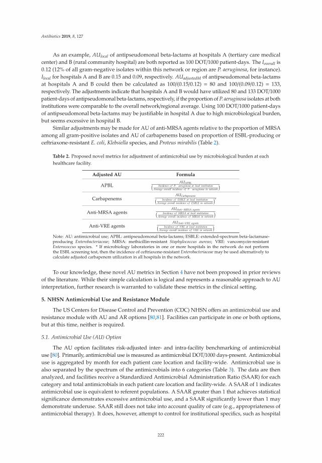
Antibiotics 2019, 8, 127
As an example, AUlocal of antipseudomonal beta-lactams at hospitals A (tertiary care medicalcenter) and B (rural community hospital) are both reported as 100 DOT/1000 patient-days. The Ioverall is0.12 (12% of all gram-negative isolates within this network or region are P. aeruginosa, for instance).Ilocal for hospitals A and B are 0.15 and 0.09, respectively. AUadjustedM of antipseudomonal beta-lactamsat hospitals A and B could then be calculated as 100/(0.15/0.12) = 80 and 100/(0.09/0.12) = 133,respectively. The adjustments indicate that hospitals A and B would have utilized 80 and 133 DOT/1000patient-days of antipseudomonal beta-lactams, respectively, if the proportion of P. aeruginosa isolates at bothinstitutions were comparable to the overall network/regional average. Using 100 DOT/1000 patient-daysof antipseudomonal beta-lactams may be justifiable in hospital A due to high microbiological burden,but seems excessive in hospital B.
Similar adjustments may be made for AU of anti-MRSA agents relative to the proportion of MRSAamong all gram-positive isolates and AU of carbapenems based on proportion of ESBL-producing orceftriaxone-resistant E. coli, Klebsiella species, and Proteus mirabilis (Table 2).
Table 2. Proposed novel metrics for adjustment of antimicrobial use by microbiological burden at eachhealthcare facility.
Adjusted AU Formula
APBLAUAPBL( Incidence o f P. aeruginosa at local institution
Average overall incidence o f P. aeruginosa in network
)
CarbapenemsAUCarbapenems( Incidence o f ESBLE at local institution
Average overall incidence o f ESBLE in network
)∗
Anti-MRSA agentsAUAnti−MRSA agents( Incidence o f MRSA at local institution
Average overall incidence o f MRSA in network
)
Anti-VRE agentsAUAnti−VRE agents( Incidence o f VRE at local institution
Average overall incidence o f VRE in network
)
Note: AU: antimicrobial use; APBL: antipseudomonal beta-lactams; ESBLE: extended-spectrum beta-lactamase-producing Enterobacteriaceae; MRSA: methicillin-resistant Staphylococcus aureus; VRE: vancomycin-resistantEnterococcus species. * If microbiology laboratories in one or more hospitals in the network do not performthe ESBL screening test, then the incidence of ceftriaxone-resistant Enterobacteriaceae may be used alternatively tocalculate adjusted carbapenem utilization in all hospitals in the network.
To our knowledge, these novel AU metrics in Section 4 have not been proposed in prior reviewsof the literature. While their simple calculation is logical and represents a reasonable approach to AUinterpretation, further research is warranted to validate these metrics in the clinical setting.
5. NHSN Antimicrobial Use and Resistance Module
The US Centers for Disease Control and Prevention (CDC) NHSN offers an antimicrobial use andresistance module with AU and AR options [80,81]. Facilities can participate in one or both options,but at this time, neither is required.
5.1. Antimicrobial Use (AU) Option
The AU option facilitates risk-adjusted inter- and intra-facility benchmarking of antimicrobialuse [80]. Primarily, antimicrobial use is measured as antimicrobial DOT/1000 days-present. Antimicrobialuse is aggregated by month for each patient care location and facility-wide. Antimicrobial use isalso separated by the spectrum of the antimicrobials into 6 categories (Table 3). The data are thenanalyzed, and facilities receive a Standardized Antimicrobial Administration Ratio (SAAR) for eachcategory and total antimicrobials in each patient care location and facility-wide. A SAAR of 1 indicatesantimicrobial use is equivalent to referent populations. A SAAR greater than 1 that achieves statisticalsignificance demonstrates excessive antimicrobial use, and a SAAR significantly lower than 1 maydemonstrate underuse. SAAR still does not take into account quality of care (e.g., appropriateness ofantimicrobial therapy). It does, however, attempt to control for institutional specifics, such as hospital
222
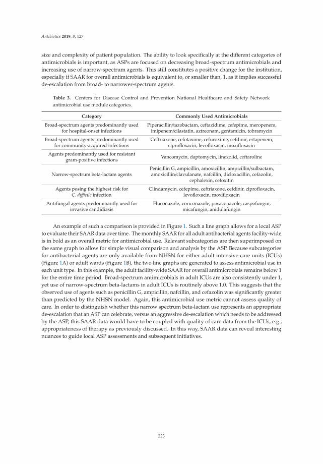
Antibiotics 2019, 8, 127
size and complexity of patient population. The ability to look specifically at the different categories ofantimicrobials is important, as ASPs are focused on decreasing broad-spectrum antimicrobials andincreasing use of narrow-spectrum agents. This still constitutes a positive change for the institution,especially if SAAR for overall antimicrobials is equivalent to, or smaller than, 1, as it implies successfulde-escalation from broad- to narrower-spectrum agents.
Table 3. Centers for Disease Control and Prevention National Healthcare and Safety Networkantimicrobial use module categories.
Category Commonly Used Antimicrobials
Broad-spectrum agents predominantly usedfor hospital-onset infections
Piperacillin/tazobactam, ceftazidime, cefepime, meropenem,imipenem/cilastatin, aztreonam, gentamicin, tobramycin
Broad-spectrum agents predominantly usedfor community-acquired infections
Ceftriaxone, cefotaxime, cefuroxime, cefdinir, ertapenem,ciprofloxacin, levofloxacin, moxifloxacin
Agents predominantly used for resistantgram-positive infections Vancomycin, daptomycin, linezolid, ceftaroline
Narrow-spectrum beta-lactam agentsPenicillin G, ampicillin, amoxicillin, ampicillin/sulbactam,amoxicillin/clavulanate, nafcillin, dicloxacillin, cefazolin,
cephalexin, cefoxitin
Agents posing the highest risk forC. difficile infection
Clindamycin, cefepime, ceftriaxone, cefdinir, ciprofloxacin,levofloxacin, moxifloxacin
Antifungal agents predominantly used forinvasive candidiasis
Fluconazole, voriconazole, posaconazole, caspofungin,micafungin, anidulafungin
An example of such a comparison is provided in Figure 1. Such a line graph allows for a local ASPto evaluate their SAAR data over time. The monthly SAAR for all adult antibacterial agents facility-wideis in bold as an overall metric for antimicrobial use. Relevant subcategories are then superimposed onthe same graph to allow for simple visual comparison and analysis by the ASP. Because subcategoriesfor antibacterial agents are only available from NHSN for either adult intensive care units (ICUs)(Figure 1A) or adult wards (Figure 1B), the two line graphs are generated to assess antimicrobial use ineach unit type. In this example, the adult facility-wide SAAR for overall antimicrobials remains below 1for the entire time period. Broad-spectrum antimicrobials in adult ICUs are also consistently under 1,yet use of narrow-spectrum beta-lactams in adult ICUs is routinely above 1.0. This suggests that theobserved use of agents such as penicillin G, ampicillin, nafcillin, and cefazolin was significantly greaterthan predicted by the NHSN model. Again, this antimicrobial use metric cannot assess quality ofcare. In order to distinguish whether this narrow spectrum beta-lactam use represents an appropriatede-escalation that an ASP can celebrate, versus an aggressive de-escalation which needs to be addressedby the ASP, this SAAR data would have to be coupled with quality of care data from the ICUs, e.g.,appropriateness of therapy as previously discussed. In this way, SAAR data can reveal interestingnuances to guide local ASP assessments and subsequent initiatives.
223
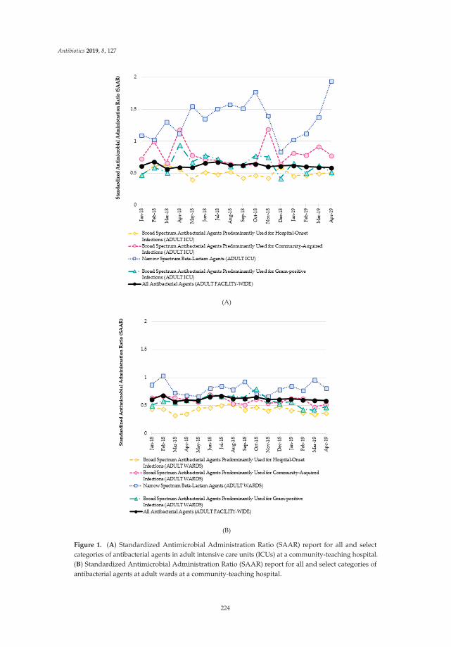
Antibiotics 2019, 8, 127
(A)
(B)
Figure 1. (A) Standardized Antimicrobial Administration Ratio (SAAR) report for all and selectcategories of antibacterial agents in adult intensive care units (ICUs) at a community-teaching hospital.(B) Standardized Antimicrobial Administration Ratio (SAAR) report for all and select categories ofantibacterial agents at adult wards at a community-teaching hospital.
224
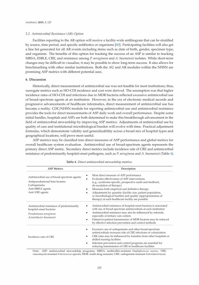
Antibiotics 2019, 8, 127
5.2. Antimicrobial Resistance (AR) Option
Facilities reporting to the AR option will receive a facility-wide antibiogram that can be stratifiedby source, time period, and specific antibiotics or organisms [80]. Participating facilities will also geta line list generated for all AR events including items such as date of birth, gender, specimen type,and organism. The benefits of this option for tracking the success of an ASP is similar to trackingMRSA, ESBLE, CRE, and resistance among P. aeruginosa and A. baumannii isolates. While short-termchanges may be difficult to visualize, it may be possible to show long-term success. It also allows forbenchmarking with other similar institutions. Both the AU and AR modules within the NHSN arepromising ASP metrics with different potential uses.
6. Discussion
Historically, direct measurement of antimicrobial use was not feasible for most institutions; thus,surrogate metrics such as HO-CDI incidence and cost were derived. The assumption was that higherincidence rates of HO-CDI and infections due to MDR bacteria reflected excessive antimicrobial useof broad-spectrum agents at an institution. However, in the era of electronic medical records andprogressive advancements of healthcare informatics, direct measurement of antimicrobial use hasbecome a reality. CDC/NHSN module for reporting antimicrobial use and antimicrobial resistanceprovides the tools for direct measurements of ASP daily work and overall performance. Despite someinitial hurdles, hospitals and ASPs are both determined to make this breakthrough advancement in thefield of antimicrobial stewardship by improving ASP metrics. Adjustments of antimicrobial use byquality of care and institutional microbiological burden will evolve with time. Practical adjustmentformulas, which demonstrate validity and generalizability across a broad mix of hospital types andgeographical locations, will prove most useful.
ASP metrics may be classified into direct measures of ASP performance and global metrics foroverall healthcare system evaluation. Antimicrobial use of broad-spectrum agents represents theprimary direct ASP metric. Secondary direct metrics include incidence rate of CRE and antimicrobialresistance of predominantly hospital-onset pathogens, such as P. aeruginosa and A. baumannii (Table 4).
Table 4. Direct antimicrobial stewardship metrics.
ASP Metrics Description
Antimicrobial use of broad-spectrum agents:
Antipseudomonal beta-lactamsCarbapenemsAnti-MRSA agentsAnti-VRE agents
• Most direct measure of ASP performance• Evaluates effectiveness of ASP interventions
(e.g., syndrome-specific, prospective audit and feedback,de-escalation of therapy)
• Measures both empirical and definitive therapy• Adjustments by quantity (facility size, patient population,
or microbiological burden) and quality (appropriateness oftherapy) at each healthcare facility are possible
Antimicrobial resistance of predominantlyhospital-onset bacteria:
Pseudomonas aeruginosaAcinetobacter baumannii
• Antimicrobial resistance of hospital-onset bacteria is associatedwith use of broad-spectrum antimicrobials at each institution
• Antimicrobial resistance may also be influenced by referrals,especially at tertiary care centers
• Patient-to-patient transmission of MDR bacteria may be reducedby effective infection prevention and control methods
Incidence rate of CRE
• Excessive use of carbapenems and other broad-spectrumantimicrobials increases risk of CRE infections or colonization
• CRE rates may be influenced by transfers from other hospitals orskilled nursing facilities
• Infection prevention and control programs are essential forreducing transmission of CRE in healthcare facilities
Note: ASP: antimicrobial stewardship programs; MRSA: methicillin-resistant Staphylococcus aureus; VRE:vancomycin-resistant Enterococcus species; MDR: multi-drug resistant; CRE: carbapenem-resistant Enterobacteriaceae.
225
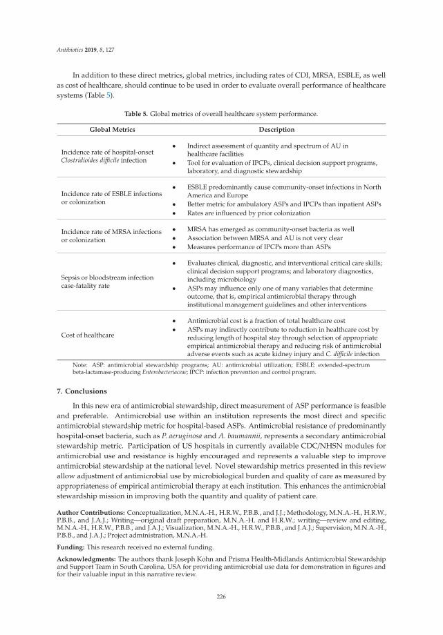
Antibiotics 2019, 8, 127
In addition to these direct metrics, global metrics, including rates of CDI, MRSA, ESBLE, as wellas cost of healthcare, should continue to be used in order to evaluate overall performance of healthcaresystems (Table 5).
Table 5. Global metrics of overall healthcare system performance.
Global Metrics Description
Incidence rate of hospital-onsetClostridioides difficile infection
• Indirect assessment of quantity and spectrum of AU inhealthcare facilities
• Tool for evaluation of IPCPs, clinical decision support programs,laboratory, and diagnostic stewardship
Incidence rate of ESBLE infectionsor colonization
• ESBLE predominantly cause community-onset infections in NorthAmerica and Europe
• Better metric for ambulatory ASPs and IPCPs than inpatient ASPs• Rates are influenced by prior colonization
Incidence rate of MRSA infectionsor colonization
• MRSA has emerged as community-onset bacteria as well• Association between MRSA and AU is not very clear• Measures performance of IPCPs more than ASPs
Sepsis or bloodstream infectioncase-fatality rate
• Evaluates clinical, diagnostic, and interventional critical care skills;clinical decision support programs; and laboratory diagnostics,including microbiology
• ASPs may influence only one of many variables that determineoutcome, that is, empirical antimicrobial therapy throughinstitutional management guidelines and other interventions
Cost of healthcare
• Antimicrobial cost is a fraction of total healthcare cost• ASPs may indirectly contribute to reduction in healthcare cost by
reducing length of hospital stay through selection of appropriateempirical antimicrobial therapy and reducing risk of antimicrobialadverse events such as acute kidney injury and C. difficile infection
Note: ASP: antimicrobial stewardship programs; AU: antimicrobial utilization; ESBLE: extended-spectrumbeta-lactamase-producing Enterobacteriaceae; IPCP: infection prevention and control program.
7. Conclusions
In this new era of antimicrobial stewardship, direct measurement of ASP performance is feasibleand preferable. Antimicrobial use within an institution represents the most direct and specificantimicrobial stewardship metric for hospital-based ASPs. Antimicrobial resistance of predominantlyhospital-onset bacteria, such as P. aeruginosa and A. baumannii, represents a secondary antimicrobialstewardship metric. Participation of US hospitals in currently available CDC/NHSN modules forantimicrobial use and resistance is highly encouraged and represents a valuable step to improveantimicrobial stewardship at the national level. Novel stewardship metrics presented in this reviewallow adjustment of antimicrobial use by microbiological burden and quality of care as measured byappropriateness of empirical antimicrobial therapy at each institution. This enhances the antimicrobialstewardship mission in improving both the quantity and quality of patient care.
Author Contributions: Conceptualization, M.N.A.-H., H.R.W., P.B.B., and J.J.; Methodology, M.N.A.-H., H.R.W.,P.B.B., and J.A.J.; Writing—original draft preparation, M.N.A.-H. and H.R.W.; writing—review and editing,M.N.A.-H., H.R.W., P.B.B., and J.A.J.; Visualization, M.N.A.-H., H.R.W., P.B.B., and J.A.J.; Supervision, M.N.A.-H.,P.B.B., and J.A.J.; Project administration, M.N.A.-H.
Funding: This research received no external funding.
Acknowledgments: The authors thank Joseph Kohn and Prisma Health-Midlands Antimicrobial Stewardshipand Support Team in South Carolina, USA for providing antimicrobial use data for demonstration in figures andfor their valuable input in this narrative review.
226
Antibiotics 2019, 8, 127
Conflicts of Interest: H.R.W.: Speaker, ALK Abello. P.B.B.: Advisory board member, Melinta Therapeutics;Program content developer and speaker, FreeCE.com; Grant support, ALK Abello. M.N.A.-H. and J.A.J.:No conflicts.
Abbreviations
ASP = antimicrobial stewardship program; AR = antimicrobial resistance; AU = antimicrobial use; CDC = Centers forDisease Control and Prevention; CDI = Clostridioides difficile infection; CRE = carbapenem-resistant Enterobacteriaceae;DDD = defined daily dose; DOT = days of therapy; ESBLE = extended-spectrum beta-lactamase-producingEnterobacteriaceae; HO-CDI = hospital-onset Clostridioides difficile infection; ICU = intensive care unit;MDR =multidrug-resistant; MRSA =methicillin-resistant Staphylococcus aureus; NHSN =National HealthcareSafety Network; SAAR=Standardized Antimicrobial Administration Ratio
References
1. Barlam, T.F.; Cosgrove, S.E.; Abbo, L.M.; MacDougall, C.; Schuetz, A.N.; Septimus, E.J.; Srinivasan, A.;Dellit, T.H.; Falck-Ytter, Y.T.; Fishman, N.O.; et al. Implementing an antibiotic stewardship program:Guidelines by the Infectious Diseases Society of America and the Society for Healthcare Epidemiology ofAmerica. Clin. Infect. Dis. 2016, 62, e51–e77. [CrossRef] [PubMed]
2. Moehring, R.W.; Anderson, D.J.; Cochran, R.L.; Hicks, L.A.; Srinivasan, A.; Dodds Ashley, E.S. StructuredTaskforce of Experts Working at Reliable Standards for Stewardship (STEWARDS) Panel. Expert consensuson metrics to assess the impact of patient-level antimicrobial stewardship interventions in acute-care settings.Clin. Infect. Dis. 2017, 64, 377–383. [CrossRef] [PubMed]
3. Science, M.; Timberlake, K.; Morris, A.; Read, S.; Le Saux, N. Groupe Antibiothérapie en Pédiatrie Canada Alliancefor Stewardship of Antimicrobials in Pediatrics (GAP Can ASAP); Quality metrics for antimicrobial stewardshipprograms; Pediatrics: Elk Grove, IL, USA, 2019; p. 143.
4. Nagel, J.L.; Stevenson, J.G.; Eiland, E.H.; Kaye, K.S. Demonstrating the value of antimicrobial stewardshipprograms to hospital administrators. Clin. Infect. Dis. 2014, 59 (Suppl. 3), S146–S153. [CrossRef] [PubMed]
5. Brotherton, A.L. Metrics of antimicrobial stewardship programs. Med. Clin. N. Am. 2018, 102, 965–976.[CrossRef] [PubMed]
6. Bennett, N.; Schulz, L.; Boyd, S.; Newland, J.G. Understanding inpatient antimicrobial stewardship metrics.Am. J. Health Syst. Pharm. 2018, 75, 230–238. [CrossRef] [PubMed]
7. Tumbarello, M.; Trecarichi, E.M.; Bassetti, M.; De Rosa, F.G.; Spanu, T.; Di Meco, E.; Losito, A.R.; Parisini, A.;Pagani, N.; Cauda, R. Identifying patients harboring extended-spectrum-beta-lactamase-producingEnterobacteriaceae on hospital admission: Derivation and validation of a scoring system. Antimicrob.Agents Chemother. 2011, 55, 3485–3490. [CrossRef] [PubMed]
8. Augustine, M.R.; Testerman, T.L.; Justo, J.A.; Bookstaver, P.B.; Kohn, J.; Albrecht, H.; Al-Hasan, M.N. Clinicalrisk score for prediction of extended-spectrum beta-lactamase-producing Enterobacteriaceae in bloodstreamisolates. Infect. Control Hosp. Epidemiol. 2017, 38, 266–272. [CrossRef]
9. Khanna, S.; Pardi, D.S.; Aronson, S.L.; Kammer, P.P.; Orenstein, R.; St Sauver, J.L.; Zinsmeister, A.R.The epidemiology of community-acquired Clostridium difficile infection: A population-based study. Am. J.Gastroenterol. 2012, 107, 89–95. [CrossRef]
10. Lambert, P.J.; Dyck, M.; Thompson, L.H.; Hammond, G.W. Population-based surveillance of Clostridium difficileinfection in Manitoba, Canada, by using interim surveillance definitions. Infect. Control Hosp. Epidemiol.2009, 30, 945–951. [CrossRef]
11. Pitout, J.D.; Hanson, N.D.; Church, D.L.; Laupland, K.B. Population-based laboratory surveillance forEscherichia coli-producing extended-spectrum beta-lactamases: Importance of community isolates withblaCTX-M genes. Clin. Infect. Dis. 2004, 38, 1736–1741. [CrossRef]
12. Thaden, J.T.; Fowler, V.G.; Sexton, D.J.; Anderson, D.J. Increasing incidence of extended-spectrumbeta-lactamase-producing Escherichia coli in community hospitals throughout the Southeastern UnitedStates. Infect. Control Hosp. Epidemiol. 2016, 37, 49–54. [CrossRef] [PubMed]
13. Global Priority List of Antibiotic-Resistant Bacteria to Guide Research, Discovery, and Development ofNew Antibiotics. World Health Organization, 2017. Available online: https://www.who.int/medicines/publications/global-priority-list-antibiotic-resistant-bacteria/en/ (accessed on 30 June 2019).
227
Antibiotics 2019, 8, 127
14. Karino, S.; Kaye, K.S.; Navalkele, B.; Nishan, B.; Salim, M.; Solanki, S.; Pervaiz, A.; Tashtoush, N.; Shaikh, H.;Koppula, S.; et al. Epidemiology of acute kidney injury among patients receiving concomitant vancomycinand piperacillin-tazobactam: Opportunities for antimicrobial stewardship. Antimicrob. Agents Chemother.2016, 60, 3743–3750. [CrossRef] [PubMed]
15. Hammond, D.A.; Smith, M.N.; Li, C.; Hayes, S.M.; Lusardi, K.; Bookstaver, P.B. Systematic review andmeta-analysis of acute kidney injury associated with concomitant vancomycin and piperacillin/tazobactam.Clin. Infect. Dis. 2017, 64, 666–674. [CrossRef] [PubMed]
16. Piacenti, F.J.; Leuthner, K.D. Antimicrobial stewardship and Clostridium difficile-associated diarrhea. J. Pharm.Pract. 2013, 26, 506–513. [CrossRef] [PubMed]
17. Longtin, Y.; Trottier, S.; Brochu, G.; Paquet-Bolduc, B.; Garenc, C.; Loungnarath, V.; Beaulieu, C.; Goulet, D.;Longtin, J. Impact of the type of diagnostic assay on Clostridium difficile infection and complication rates in amandatory reporting program. Clin. Infect. Dis. 2013, 56, 67–73. [CrossRef] [PubMed]
18. Grein, J.D.; Ochner, M.; Hoang, H.; Jin, A.; Morgan, M.A.; Murthy, A.R. Comparison of testing approaches forClostridium difficile infection at a large community hospital. Clin. Microbiol. Infect. 2014, 20, 65–69. [CrossRef][PubMed]
19. Kelly, S.G.; Yarrington, M.; Zembower, T.R.; Sutton, S.H.; Silkaitis, C.; Postelnick, M.; Mikolajczak, A.;Bolon, M.K. Inappropriate Clostridium difficile testing and consequent overtreatment and inaccurate publiclyreported metrics. Infect. Control Hosp. Epidemiol. 2016, 37, 1395–1400. [CrossRef]
20. Graber, C.J. Clostridium difficile infection: Stewardship’s lowest hanging fruit? Lancet Infect. Dis. 2017, 17,123–124. [CrossRef]
21. Al-Hasan, M.N.; Wilson, J.W.; Lahr, B.D.; Thomsen, K.M.; Eckel-Passow, J.E.; Vetter, E.A.; Tleyjeh, I.M.;Baddour, L.M. Beta-lactam and fluoroquinolone combination antibiotic therapy for bacteremia caused bygram-negative bacilli. Antimicrob. Agents Chemother. 2009, 53, 1386–1394. [CrossRef]
22. Seddon, M.M.; Bookstaver, P.B.; Justo, J.A.; Kohn, J.; Rac, H.; Haggard, E.; Mediwala, K.N.; Dash, S.;Al-Hasan, M.N. Role of early de-escalation of antimicrobial therapy on risk of Clostridioides difficile infectionfollowing Enterobacteriaceae bloodstream infections. Clin. Infect. Dis. 2019, 69, 414–420. [CrossRef]
23. Tucker, K.; Lashkova, L.; Flemming, T.; Justo, J.; Kohn, J.; Al-Hasan, M.N.; Sanasi, K.; Bookstaver, P.B.Impact of antimicrobial stewardship initiatives on carbapenem utilization and antimicrobial resistance.In Proceedings of the 55th Interscience Conference on Antimicrobial Agents and Chemotherapy, San Diego,CA, USA, 17–21 September 2015. Abstract #2243.
24. Palacios-Baena, Z.R.; Delgado-Valverde, M.; Valiente Méndez, A.; Almirante, B.; Gómez-Zorrilla, S.;Borrell, N.; Corzo, J.E.; Gurguí, M.; de la Calle, C.; García-Álvarez, L.; et al. Impact of de-escalation onprognosis of patients with bacteraemia due to Enterobacteriaceae: A post-hoc analysis from a multicenterprospective cohort. Clin. Infect. Dis. 2018. [CrossRef] [PubMed]
25. Tariq, R.; Singh, S.; Gupta, A.; Pardi, D.S.; Khanna, S. Association of gastric acid suppression with recurrentClostridium difficile infection: A systematic review and meta-analysis. JAMA Intern. Med. 2017, 177, 784–791.[CrossRef] [PubMed]
26. Thomas, C.; Stevenson, M.; Riley, T.V. Antibiotics and hospital-acquired Clostridium difficile-associateddiarrhoea: A systematic review. J. Antimicrob. Chemother. 2003, 51, 1339–1350. [CrossRef] [PubMed]
27. Brown, K.A.; Khanafer, N.; Daneman, N.; Fisman, D.N. Meta-analysis of antibiotics and the risk ofcommunity-associated Clostridium difficile infection. Antimicrob. Agents Chemother. 2013, 57, 2326–2332. [CrossRef]
28. Pagels, C.M.; McCreary, E.K.; Rose, W.E.; Dodds Ashley, E.S.; Bookstaver, P.B.; Dilworth, T.J. Designingantimicrobial stewardship initiatives to enhance scientific dissemination. J. Am. Coll. Clin. Pharm. 2019, 1–7.[CrossRef]
29. McFarland, L.V.; Surawicz, C.M.; Stamm, W.E. Risk factors for Clostridium difficile carriage and, C. difficile-associated diarrhea in a cohort of hospitalized patients. J. Infect. Dis. 1990, 162, 678–684. [CrossRef] [PubMed]
30. Lawes, T.; Lopez-Lozano, J.M.; Nebot, C.A.; Macartney, G.; Subbarao-Sharma, R.; Wares, K.D.; Sinclair, C.;Gould, I.M. Effect of a national 4C antibiotic stewardship intervention on the clinical and molecularepidemiology of Clostridium difficile infections in a region of Scotland: A non-linear time-series analysis.Lancet Infect. Dis. 2017, 17, 194–206. [CrossRef]
31. Harris, A.D.; Sbarra, A.N.; Leekha, S.; Jackson, S.S.; Johnson, J.K.; Pineles, L.; Thom, K.A. Electronicallyavailable comorbid conditions for risk prediction of healthcare-associated Clostridium difficile infection.Infect. Control Hosp. Epidemiol. 2018, 39, 297–301. [CrossRef]
228
Antibiotics 2019, 8, 127
32. Al-Hasan, M.N.; Lahr, B.D.; Eckel-Passow, J.E.; Baddour, L.M. Antimicrobial resistance trends of Escherichia colibloodstream isolates: A population-based study, 1998–2007. J. Antimicrob. Chemother. 2009, 64, 169–174.[CrossRef]
33. Waltner-Toews, R.I.; Paterson, D.L.; Qureshi, Z.A.; Sidjabat, H.E.; Adams-Haduch, J.M.; Shutt, K.A.;Jones, M.; Tian, G.B.; Pasculle, A.W.; Doi, Y. Clinical characteristics of bloodstream infections due toampicillin-sulbactam-resistant, non-extended-spectrum-beta-lactamase-producing Escherichia coli and therole of TEM-1 hyperproduction. Antimicrob. Agents Chemother. 2011, 55, 495–501. [CrossRef]
34. Baggs, J.; Fridkin, S.K.; Pollack, L.A.; Srinivasan, A.; Jernigan, J.A. Estimating National trends in inpatientantibiotic use among US hospitals from 2006 to 2012. JAMA Intern. Med. 2016, 176, 1639–1648. [CrossRef]
35. Logan, L.K.; Gandra, S.; Mandal, S.; Klein, E.Y.; Levinson, J.; Weinstein, R.A.; Laxminarayan, R. PreventionEpicenters Program, US Centers for Disease Control and Prevention. Multidrug- and carbapenem-resistantPseudomonas aeruginosa in children, United States, 1999–2012. J. Pediatr. Infect. Dis. Soc. 2017, 6, 352–359.
36. Knothe, H.; Shah, P.; Krcmery, V.; Antal, M.; Mitsuhashi, S. Transferable resistance to cefotaxime, cefoxitin,cefamandole and cefuroxime in clinical isolates of Klebsiella pneumoniae and Serratia marcescens. Infection1983, 11, 315–317. [CrossRef]
37. Rodriguez-Bano, J.; Navarro, M.D.; Romero, L.; Martínez-Martínez, L.; Muniain, M.A.; Perea, E.J.;Pérez-Cano, R.; Pascual, A. Epidemiology and clinical features of infections caused by extended-spectrumbeta-lactamase-producing Escherichia coli in nonhospitalized patients. J. Clin. Microbiol. 2004, 42, 1089–1094.[CrossRef]
38. Rottier, W.C.; Bamberg, Y.R.; Dorigo-Zetsma, J.W.; van der Linden, P.D.; Ammerlaan, H.S.; Bonten, M.J.Predictive value of prior colonization and antibiotic use for third-generation cephalosporin-resistantenterobacteriaceae bacteremia in patients with sepsis. Clin. Infect. Dis. 2015, 60, 1622–1630. [CrossRef]
39. Morgan, D.J.; Meddings, J.; Saint, S.; Lautenbach, E.; Shardell, M.; Anderson, D.; Milstone, A.M.;Drees, M.; Pineles, L.; Safdar, N.; et al. SHEA Research Network. Does nonpayment for hospital-acquiredcatheter-associated urinary tract infections lead to overtesting and increased antimicrobial prescribing?Clin. Infect. Dis. 2012, 55, 923–929. [CrossRef]
40. Lawes, T.; Lopez-Lozano, J.M.; Nebot, C.A.; Macartney, G.; Subbarao-Sharma, R.; Dare, C.R.; Wares, K.D.;Gould, I.M. Effects of national antibiotic stewardship and infection control strategies on hospital-associatedand community-associated meticillin-resistant Staphylococcus aureus infections across a region of Scotland:A non-linear time-series study. Lancet Infect. Dis. 2015, 15, 1438–1449. [CrossRef]
41. Jain, R.; Kralovic, S.M.; Evans, M.E.; Ambrose, M.; Simbartl, L.A.; Obrosky, D.S.; Render, M.L.;Freyberg, R.W.; Jernigan, J.A.; Muder, R.R.; et al. Veterans Affairs initiative to prevent methicillin-resistantStaphylococcus aureus infections. N. Engl. J. Med. 2011, 364, 1419–1430. [CrossRef]
42. Klein, E.Y.; Mojica, N.; Jiang, W.; Cosgrove, S.E.; Septimus, E.; Morgan, D.J.; Laxminarayan, R. Trends inmethicillin-resistant Staphylococcus aureus hospitalizations in the United States, 2010–2014. Clin. Infect. Dis.2017, 65, 1921–1923. [CrossRef]
43. Kourtis, A.P.; Hatfield, K.; Baggs, J.; Mu, Y.; See, I.; Epson, E.; Nadle, J.; Kainer, M.A.; Dumyati, G.; Petit, S.;et al. Vital signs: Epidemiology and recent trends in methicillin-resistant and in methicillin-susceptibleStaphylococcus aureus bloodstream infections—United States. MMWR Morb. Mortal. Wkly. Rep. 2019, 68,214–219. [CrossRef]
44. FDA Updates Warnings for Fluoroquinolone Antibiotics. Available online: http://www.fda.gov/NewsEvents/Newsroom/PressAnnouncements/ucm513183.htm (accessed on 4 June 2019).
45. Orsi, G.B.; Bencardino, A.; Vena, A.; Carattoli, A.; Venditti, C.; Falcone, M.; Giordano, A.; Venditti, M. Patientrisk factors for outer membrane permeability and KPC-producing carbapenem-resistant Klebsiella pneumoniaeisolation: Results of a double case-control study. Infection 2013, 41, 61–67. [CrossRef]
46. Al-Hasan, M.N.; Wilson, J.W.; Lahr, B.D.; Eckel-Passow, J.E.; Baddour, L.M. Incidence of Pseudomonasaeruginosa bacteremia: A population-based study. Am. J. Med. 2008, 121, 702–708. [CrossRef]
47. Gransden, W.R.; Leibovici, L.; Eykyn, S.J.; Pitlik, S.D.; Samra, Z.; Konisberger, H.; Drucker, M.; Phillips, I.Risk factors and a clinical index for diagnosis of Pseudomonas aeruginosa bacteremia. Clin. Microbiol. Infect.1995, 1, 119–123. [CrossRef]
48. Hammer, K.L.; Justo, J.A.; Bookstaver, P.B.; Kohn, J.; Albrecht, H.; Al-Hasan, M.N. Differential effect of priorbeta-lactams and fluoroquinolones on risk of bloodstream infections secondary to Pseudomonas aeruginosa.Diagn. Microbiol. Infect. Dis. 2017, 87, 87–91. [CrossRef]
229
Antibiotics 2019, 8, 127
49. Cheong, H.S.; Kang, C.I.; Wi, Y.M.; Kim, E.S.; Lee, J.S.; Ko, K.S.; Chung, D.R.; Lee, N.Y.; Song, J.H.; Peck, K.R.Clinical significance and predictors of community-onset Pseudomonas aeruginosa bacteremia. Am. J. Med.2008, 121, 709–714. [CrossRef]
50. Schechner, V.; Nobre, V.; Kaye, K.S.; Leshno, M.; Giladi, M.; Rohner, P.; Harbarth, S.; Anderson, D.J.;Karchmer, A.W.; Schwaber, M.J.; et al. Gram-negative bacteremia upon hospital admission: When shouldPseudomonas aeruginosa be suspected? Clin. Infect. Dis. 2009, 48, 580–586. [CrossRef]
51. Decraene, V.; Ghebrehewet, S.; Dardamissis, E.; Huyton, R.; Mortimer, K.; Wilkinson, D.; Shokrollahi, K.;Singleton, S.; Patel, B.; Turton, J.; et al. An outbreak of multidrug-resistant Pseudomonas aeruginosa in a burnsservice in the North of England: Challenges of infection prevention and control in a complex setting. J. Hosp.Infect. 2018, 100, e239–e245. [CrossRef]
52. Milan, A.; Furlanis, L.; Cian, F.; Bressan, R.; Luzzati, R.; Lagatolla, C.; Deiana, M.L.; Knezevich, A.; Tonin, E.;Dolzani, L. Epidemic dissemination of a carbapenem-resistant Acinetobacter baumannii clone carrying armAtwo years after its first isolation in an Italian hospital. Microb. Drug Resist. 2016, 22, 668–674. [CrossRef]
53. Paramythiotou, E.; Lucet, J.C.; Timsit, J.F.; Vanjak, D.; Paugam-Burtz, C.; Trouillet, J.L.; Belloc, S.; Kassis, N.;Karabinis, A.; Andremont, A. Acquisition of multidrug-resistant Pseudomonas aeruginosa in patients inintensive care units: Role of antibiotics with antipseudomonal activity. Clin. Infect. Dis. 2004, 38, 670–677.[CrossRef]
54. Montero, M.; Sala, M.; Riu, M.; Belvis, F.; Salvado, M.; Grau, S.; Horcajada, J.P.; Alvarez-Lerma, F.; Terradas, R.;Orozco-Levi, M.; et al. Risk factors for multidrug-resistant Pseudomonas aeruginosa acquisition. Impact ofantibiotic use in a double case-control study. Eur. J. Clin. Microbiol. Infect. Dis. 2010, 29, 335–339. [CrossRef]
55. Nakamura, A.; Miyake, K.; Misawa, S.; Kuno, Y.; Horii, T.; Kondo, S.; Tabe, Y.; Ohsaka, A. Meropenem aspredictive risk factor for isolation of multidrug-resistant Pseudomonas aeruginosa. J. Hosp. Infect. 2013, 83,153–155. [CrossRef]
56. Cobos-Trigueros, N.; Sole, M.; Castro, P.; Torres, J.L.; Hernández, C.; Rinaudo, M.; Fernández, S.; Soriano, Á.;Nicolás, J.M.; Mensa, J.; et al. Acquisition of Pseudomonas aeruginosa and its resistance phenotypes in criticallyill medical patients: Role of colonization pressure and antibiotic exposure. Crit. Care 2015, 19, 218. [CrossRef]
57. Al-Jaghbeer, M.J.; Justo, J.A.; Owens, W.; Kohn, J.; Bookstaver, P.B.; Hucks, J.; Al-Hasan, M.N. Riskfactors for pneumonia due to beta-lactam-susceptible and beta-lactam-resistant Pseudomonas aeruginosa:A case-case-control study. Infection 2018, 46, 487–494. [CrossRef]
58. Cain, S.E.; Kohn, J.; Bookstaver, P.B.; Albrecht, H.; Al-Hasan, M.N. Stratification of the impact of inappropriateempirical antimicrobial therapy for Gram-negative bloodstream infections by predicted prognosis. Antimicrob.Agents Chemother. 2015, 59, 245–250. [CrossRef]
59. Retamar, P.; Portillo, M.M.; Lopez-Prieto, M.D.; Rodríguez-López, F.; de Cueto, M.; García, M.V.; Gómez, M.J.;Del Arco, A.; Muñoz, A.; Sánchez-Porto, A.; et al. SAEI/SAMPAC Bacteremia Group. Impact of inadequateempirical therapy on the mortality of patients with bloodstream infections: A propensity score-based analysis.Antimicrob. Agents Chemother. 2012, 56, 472–478. [CrossRef]
60. Al-Hasan, M.N.; Rac, H. Transition from intravenous to oral antimicrobial therapy in patients withuncomplicated and complicated bloodstream infections. Clin. Microbiol. Infect. 2019. [CrossRef]
61. Paul, M.; Shani, V.; Muchtar, E.; Kariv, G.; Robenshtok, E.; Leibovici, L. Systematic review and meta-analysisof the efficacy of appropriate empiric antibiotic therapy for sepsis. Antimicrob. Agents Chemother. 2010, 54,4851–4863. [CrossRef]
62. Shorr, A.F.; Micek, S.T.; Welch, E.C.; Doherty, J.A.; Reichley, R.M.; Kollef, M.H. Inappropriate antibiotictherapy in Gram-negative sepsis increases hospital length of stay. Crit. Care Med. 2011, 39, 46–51. [CrossRef]
63. Battle, S.E.; Bookstaver, P.B.; Justo, J.A.; Kohn, J.; Albrecht, H.; Al-Hasan, M.N. Association betweeninappropriate empirical antimicrobial therapy and hospital length of stay in Gram-negative bloodstreaminfections: Stratification by prognosis. J. Antimicrob. Chemother. 2017, 72, 299–304. [CrossRef]
64. Sogaard, M.; Norgaard, M.; Dethlefsen, C.; Schonheyder, H.C. Temporal changes in the incidence and 30-daymortality associated with bacteremia in hospitalized patients from 1992 through 2006: A population-basedcohort study. Clin. Infect. Dis. 2011, 52, 61–69. [CrossRef]
65. Nimmich, E.B.; Bookstaver, P.B.; Kohn, J.; Justo, J.A.; Hammer, K.L.; Albrecht, H.; Al-Hasan, M.N.Development of Institutional Guidelines for Management of Gram-Negative Bloodstream Infections:Incorporating Local Evidence. Hosp. Pharm. 2017, 52, 691–697. [CrossRef]
230
Antibiotics 2019, 8, 127
66. Bookstaver, P.B.; Nimmich, E.B.; Smith, T.J.; Justo, J.A.; Kohn, J.; Hammer, K.L.; Troficanto, C.; Albrecht, H.A.;Al-Hasan, M.N. Cumulative effect of an antimicrobial stewardship and rapid diagnostic testing bundle onearly streamlining of antimicrobial therapy in Gram-negative bloodstream infections. Antimicrob. AgentsChemother. 2017, 61, e00189-17. [CrossRef]
67. Al-Hasan, M.N.; Lahr, B.D.; Eckel-Passow, J.E.; Baddour, L.M. Predictive scoring model of mortality inGram-negative bloodstream infection. Clin. Microbiol. Infect. 2013, 19, 948–954. [CrossRef]
68. Al-Hasan, M.N.; Juhn, Y.J.; Bang, D.W.; Yang, H.J.; Baddour, L.M. External validation of bloodstream infectionmortality risk score in a population-based cohort. Clin. Microbiol. Infect. 2014, 20, 886–891. [CrossRef]
69. Huang, A.M.; Newton, D.; Kunapuli, A.; Gandhi, T.N.; Washer, L.L.; Isip, J.; Collins, C.D.; Nagel, J.L. Impactof rapid organism identification via matrix-assisted laser desorption/ionization time-of-flight combined withantimicrobial stewardship team intervention in adult patients with bacteremia and candidemia. Clin. Infect.Dis. 2013, 57, 1237–1245. [CrossRef]
70. MacVane, S.H.; Nolte, F.S. Benefits of adding a rapid PCR-based blood culture identification panel to anestablished antimicrobial stewardship program. J. Clin. Microbiol. 2016, 54, 2455–2463. [CrossRef]
71. Banerjee, R.; Teng, C.B.; Cunningham, S.A.; Ihde, S.M.; Steckelberg, J.M.; Moriarty, J.P.; Shah, N.D.;Mandrekar, J.N.; Patel, R. Randomized trial of rapid multiplex polymerase chain reaction-based blood cultureidentification and susceptibility testing. Clin. Infect. Dis. 2015, 61, 1071–1080. [CrossRef]
72. Mediwala, K.N.; Kohn, J.E.; Bookstaver, P.B.; Justo, J.A.; Rac, H.; Tucker, K.; Lashkova, L.; Dash, S.;Al-Hasan, M.N. Syndrome-specific versus prospective audit and feedback interventions for reducing use ofbroad-spectrum antimicrobial agents. Am. J. Infect. Control 2019. [CrossRef]
73. Sick, A.C.; Lehmann, C.U.; Tamma, P.D.; Lee, C.K.; Agwu, A.L. Sustained savings from a longitudinal costanalysis of an internet-based preapproval antimicrobial stewardship program. Infect. Control Hosp. Epidemiol.2013, 34, 573–580. [CrossRef]
74. Beardsley, J.R.; Williamson, J.C.; Johnson, J.W.; Luther, V.P.; Wrenn, R.H.; Ohl, C.C. Show me the money:Long-term financial impact of an antimicrobial stewardship program. Infect. Control Hosp. Epidemiol. 2012,33, 398–400. [CrossRef]
75. Ozkurt, Z.; Erol, S.; Kadanali, A.; Ertek, M.; Ozden, K.; Tasyaran, M.A. Changes in antibiotic use, cost andconsumption after an antibiotic restriction policy applied by infectious disease specialists. Jpn. J. Infect. Dis.2005, 58, 338–343.
76. Akpan, M.R.; Ahmad, R.; Shebl, N.A.; Ashiru-Oredope, D. A review of quality measures for assessing theimpact of antimicrobial stewardship programs in hospitals. Antibiotics 2016, 5, 5. [CrossRef]
77. Bartlett, J.M.; Siola, P.L. Implementation and first-year results of an antimicrobial stewardship program at acommunity hospital. Am. J. Health Syst. Pharm. 2014, 71, 943–949. [CrossRef]
78. Tamma, P.D.; Avdic, E.; Keenan, J.F.; Zhao, Y.; Anand, G.; Cooper, J.; Dezube, R.; Hsu, S.; Cosgrove, S.E. Whatis the more effective antibiotic stewardship intervention: Preprescription authorization or postprescriptionreview with feedback? Clin. Infect. Dis. 2017, 64, 537–543.
79. Al-Hasan, M.N.; Eckel-Passow, J.E.; Baddour, L.M. Influence of referral bias on the clinical characteristics ofpatients with Gram-negative bloodstream Infection. Epidemiol. Infect. 2011, 139, 1750–1756. [CrossRef]
80. Antimicrobial Use and Resistance Module. 2019. Available online: https://www.cdc.gov/nhsn/PDFs/pscManual/11pscAURcurrent.pdf (accessed on 1 June 2019).
81. van Santen, K.L.; Edwards, J.R.; Webb, A.K.; Pollack, L.A.; O’Leary, E.; Neuhauser, M.M.; Srinivasan, A.;Pollock, D.A. The standardized antimicrobial administration ratio: A new metric for measuring andcomparing antibiotic use. Clin. Infect. Dis. 2018, 67, 179–185. [CrossRef]
© 2019 by the authors. Licensee MDPI, Basel, Switzerland. This article is an open accessarticle distributed under the terms and conditions of the Creative Commons Attribution(CC BY) license (http://creativecommons.org/licenses/by/4.0/).
231


antibiotics
Review
The Perfect Bacteriophage for TherapeuticApplications—A Quick Guide
Lucía Fernández 1,2,*, Diana Gutiérrez 3, Pilar García 1,2 and Ana Rodríguez 1,2
1 Instituto de Productos Lácteos de Asturias (IPLA-CSIC), (DairySafe Group), Paseo Río Linares s/n-Villaviciosa, 33300 Asturias, Spain
2 Instituto de Investigación Sanitaria del Principado de Asturias (ISPA), 33011 Oviedo, Spain3 Laboratory of Applied Biotechnology, Department of Applied Biosciences,
Faculty of Bioscience Engineering, Ghent University, 9000 Ghent, Belgium* Correspondence: [email protected]; Tel.: +34-9858-92131
Received: 29 July 2019; Accepted: 21 August 2019; Published: 23 August 2019
Abstract: The alarming spread of multiresistant infections has kick-started the quest for alternativeantimicrobials. In a way, given the steady increase in untreatable infectious diseases, success in thisendeavor has become a matter of life and death. Perhaps we should stop searching for an antibacterialpanacea and explore a multifaceted strategy in which a wide range of compounds are available ondemand depending on the specific situation. In the context of this novel tailor-made approach tocombating bacterial pathogens, the once forgotten phage therapy is undergoing a revival. Indeed,the compassionate use of bacteriophages against seemingly incurable infections has been attractinga lot of media attention lately. However, in order to take full advantage of this strategy, bacteria’snatural predators must be taken from their environment and then carefully selected to suit our needs.In this review, we have explored the vast literature regarding phage isolation and characterizationfor therapeutic purposes, paying special attention to the most recent studies, in search of findingsthat hint at the most efficient strategies to identify suitable candidates. From this information, wewill list and discuss the traits that, at the moment, are considered particularly valuable in phagesdestined for antimicrobial therapy applications. Due to the growing importance given to biofilmsin the context of bacterial infections, we will dedicate a specific section to those characteristics thatindicate the suitability of a bacteriophage as an antibiofilm agent. Overall, the objective is not just tohave a large collection of phages, but to have the best possible candidates to guarantee elimination ofthe target pathogens.
Keywords: bacteriophages; biofilms; novel antimicrobials
1. Phage Therapy: A Major Comeback with Minor Setbacks
The existence of viruses that can infect and kill bacterial cells, known as bacteriophages (or phages),was discovered almost simultaneously by Frederick Twort and Félix d’Herelle about one centuryago, but it is the latter who has been credited with introducing the concept of using these entitiesas antimicrobial agents [1,2]. Indeed, the French-Canadian microbiologist pioneered the so-calledphage therapy and used phages to treat patients with various infectious diseases [3]. Since that time,the utilization of phages for therapeutic applications has remained common practice in Eastern Europe,saving countless human lives [4]. Conversely, in other parts of the world, the introduction of antibioticspushed this anti-infective strategy out of center stage for many decades. After all, antibiotics werecheaper to manufacture and exhibited a broader spectrum of activity, thereby eliminating the needto identify the etiological agent prior to treatment prescription. However, with time, overuse andmisuse of antibiotics became widespread in different fields, such as human and veterinary medicineor agriculture. In this context, antibiotics exerted the selective pressure that resulted in the spread of
Antibiotics 2019, 8, 126; doi:10.3390/antibiotics8030126 www.mdpi.com/journal/antibiotics233
Antibiotics 2019, 8, 126
resistance markers and led up to the current antimicrobial resistance crisis [5]. Alarmingly, the numberof deaths associated with bacterial infections is rising and threatens to reach levels not seen since thepreantibiotic era. The reason for this is that more and more bacterial strains are acquiring multiresistanceand are, consequently, able to survive treatment with most, if not all, antibiotics commonly usedin the clinic. Given the severity of such a scenario, besides curtailing unnecessary antibiotic use,the scientific community has started a race to find as many alternatives to conventional antibiotics aspossible [6]. Among the many options being explored, the revival of phage therapy presents itselfas an attractive strategy to substitute or complement other therapies [7]. This is especially the casebecause phages, due to their unique mechanism of action, exhibit some notable differences comparedto other types of antimicrobials, some of which are particularly advantageous. These positive attributesinclude their natural origin, lack of toxicity for humans or nontarget microbes (being harmless tothe normal microbiota), and their effectiveness against antibiotic-resistant bacteria, amongst others.On the downside, phage therapy generally requires identification of the target pathogen and phageresistance development remains a possibility, although it is not usually as easily transferable to othermicroorganisms as antibiotic resistance determinants. Moreover, resistance could be easily overcomeby using combinations of different phages (phage cocktails) instead of a single phage for therapeuticapplications [8].
Nonetheless, not everything is rosy on the way toward normalizing the use of bacteriophagesagainst unwanted bacteria in the context of human medicine. Most notably, it will be paramount toconvince both the general public and the competent authorities that these antimicrobials are safe forhuman health and the environment, as well as provide undeniable proof of their efficacy in clinicalsettings. Indeed, these are two key aspects in order to surmount the current regulatory constraintsthat limit the use of bacterial viruses for infection treatment in humans. Until relatively recently,most evidence regarding the effectiveness of phage therapy was the result of direct use in patients,but well-organized and reproducible data will be essential in order to comply with the requirementsset out for the approval of medicinal products [9]. In this regard, the growing number of completedclinical trials will certainly be invaluable. On the other hand, the growing utilization of phagesas biocontrol agents for the disinfection of inert surfaces may also help to pave the way towardregulatory approval [10]. As a matter of fact, the relevant authorities in different countries (USA, Israel,Canada, Switzerland, Australia, New Zealand, and the Netherlands) have already approved this typeof applications against several pathogens in the context of the food industry [11,12]. For example,there are several commercially available products to be used against various food pathogens, such asListeria monocytogenes, Salmonella, or Escherichia coli, as surface disinfectants or processing aids. Indeed,the FDA has recognized some phage-based formulations as “generally regarded as safe” (GRAS) asfood additives. Regarding medicinal use, phage therapy is now under the scrutiny of governmentagencies, which are trying to determine the most adequate regulatory framework considering thespecial characteristics of phages. In the meantime, patients in many countries can have access to thistreatment as compassionate use [13]. One of the major challenges of regulating phage approval is thediversity of phages that will be necessary to successfully implement this therapeutic strategy. Indeed,in order to harness the full potential of phage therapy and its adaptability to changes in pathogenicstrains, there should be room for making changes to phage-based formulations without the need for alengthy and costly approval process. In addition to regulatory hurdles, bringing phage therapy to themarket also has the problem of not being very attractive for pharmaceutical companies. To some extent,this is a consequence of their natural origin, as phages cannot be patented. Also, their use is aimed tobe used as an acute course of treatment rather than for chronic administration, which obviously meansthat they would not be as profitable as other medicines. However, the relevant authorities can help bydesigning an affordable and straightforward pathway for regulatory compliance that might perhapsencourage the participation of smaller specialized companies.
Overall, it is apparent that phages are getting closer and closer to becoming a mainstreamantimicrobial treatment option, especially in the case of difficult-to-cure multiresistant infections.
234
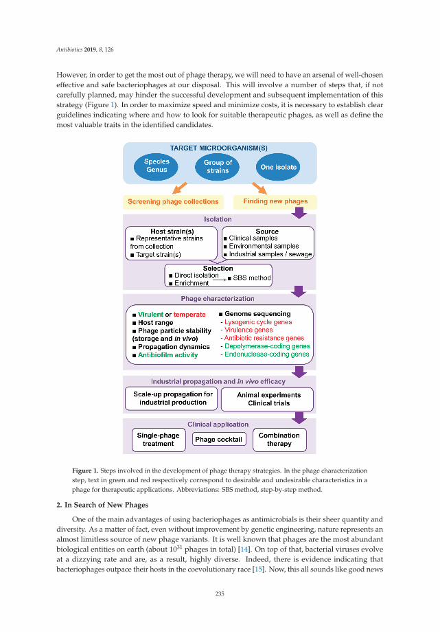
Antibiotics 2019, 8, 126
However, in order to get the most out of phage therapy, we will need to have an arsenal of well-choseneffective and safe bacteriophages at our disposal. This will involve a number of steps that, if notcarefully planned, may hinder the successful development and subsequent implementation of thisstrategy (Figure 1). In order to maximize speed and minimize costs, it is necessary to establish clearguidelines indicating where and how to look for suitable therapeutic phages, as well as define themost valuable traits in the identified candidates.
Figure 1. Steps involved in the development of phage therapy strategies. In the phage characterizationstep, text in green and red respectively correspond to desirable and undesirable characteristics in aphage for therapeutic applications. Abbreviations: SBS method, step-by-step method.
2. In Search of New Phages
One of the main advantages of using bacteriophages as antimicrobials is their sheer quantity anddiversity. As a matter of fact, even without improvement by genetic engineering, nature represents analmost limitless source of new phage variants. It is well known that phages are the most abundantbiological entities on earth (about 1031 phages in total) [14]. On top of that, bacterial viruses evolveat a dizzying rate and are, as a result, highly diverse. Indeed, there is evidence indicating thatbacteriophages outpace their hosts in the coevolutionary race [15]. Now, this all sounds like good news
235
Antibiotics 2019, 8, 126
from the perspective of the antimicrobial development pipeline until coming to the realization thatfinding the right phages for our purpose among this huge set of candidates is quite the gargantuantask. Nevertheless, it is also worth noting that vast phage collections are already available in differentresearch centers and universities around the globe. As a result, this would be a good place to start oncethe target microbe is identified.
It is obviously impossible to screen for phages everywhere, so a major step consists in narrowingdown the most probable reservoirs of viruses that infect and kill our target microbe. As is always thecase with predators, these reservoirs are likely to coincide with the main habitats of their prey, thatis, the target pathogen. In that sense, d’Hérelle suggested that the best source of bacterial viruses forantimicrobial applications are samples from patients who are recovering or who have just recoveredfrom an infection [16]. Indeed, he first remarked the existence of “bacteria eaters” in stool samples ofpatients recovering from bacillary dysentery [1]. Similarly, phages against enterohemorrhagic E. coli(EHEC) O157:H7 were found in fecal samples from human patients and cattle [17]. Additionally, phagesinfecting Cutibacterium acnes (formerly Propionibacterium acnes) and Actinobacillus have respectivelybeen isolated from human skin and dental plaque [18,19]. Given the prevalence of human pathogenicbacteria in infected patients, another good reservoir of phages infecting these microorganisms iswastewater from hospitals [20]. If clinical samples are not available, good results have also beenobtained with sewage water, which has actually become a frequent starting point for phage hunts [21].In the case of microbes with high prevalence in diverse ecological niches, different environmentalsources like river or stream water have also been the place to isolate new bacteriophages [22,23].Additionally, drinking water was the source of phages infecting Enterococcus and Staphylococcus [24],while silage from dairy farms proved a good source of listeriaphages [25].
In some cases, the phage titer in these reservoirs will be high enough to allow direct isolation of thevirus after filtration of the sample; for example, by visualization of lysis plaques on plates containing thehost (double layer assay). However, this is not always the case and enrichment steps are often requiredto detect the presence of the phage. Frequently, it is a good idea to collect large-volume samples inorder to increase the chances of phage isolation, especially when dealing with samples taken fromenvironmental sources. It must also be noted that some phages form very small plaques on a bacteriallawn, which would hinder their identification by the double layer method. For this reason, if no plaquesare observed during the isolation step, it might be worth repeating the process but using techniquesthat increase plaque size, such as addition of subinhibitory concentrations of antibiotics [26,27] or othercompounds like sodium thiosulfate, ferric ammonium citrate or 2,3,5-triphenyltetrazolium [28].
Another important aspect in the quest to find new phages with antimicrobial potential lies inthe selection of the most appropriate host strain or strains. However, this choice will also depend onthe specific goal of the search. Thus, if the objective is the identification of phages against as manystrains of a given pathogen as possible, multihost selection protocols should be used. Additionally,the host strains should preferably be very varied in terms of their characteristics and origin so that theyare representative of the species. In this sense, Casey et al. [29] recommend using bacteria referencecollections for phage identification, as they offer a good representation of the intraspecies diversity,together with strains of clinical relevance. However, in some cases, there may be a specific target strainor group of strains that do not seem to be susceptible to the phages available in different collections.Then, it will be necessary to carry out screenings using these strains as selection hosts. Nonetheless,if finding a new virulent phage that infects our target strain proves to be very difficult, it is possibleto carry out an adaptation strategy in which coevolution rounds may allow the selection of a phagevariant that can effectively infect and kill the pathogenic strain. This protocol is called the step-by-step(SBS) method [30]. The phenomenon of local host adaptation also plays a role in the identification ofnew phages [31]. Indeed, phages are known to co-evolve very closely with their host strains. As aresult, virulent phages from a given reservoir or geographic location are oftentimes more effective attargeting strains from that same milieu.
236
Antibiotics 2019, 8, 126
3. Desirable Traits in Phages with Antimicrobial Potential
3.1. Specific but Not Too Specific
One of the major advantages of bacteriophages as antimicrobials is their specificity. A consequenceof this characteristic is that they are not only innocuous to eukaryotic cells, but also to all prokaryoticcells outside their host range. This would ensure that the normal microbiota remains intact duringtherapy, which in turn would be expected to help prevent secondary infections and probably acceleraterecovery. Indeed, there is growing proof that the microorganisms inhabiting our bodies play veryimportant roles in our overall health. Antibiotic treatment, by contrast, can severely affect our normalmicrobiota due to their more general, broad-range action, which often results in serious side effects [32].Another benefit of this specific action is that we can technically try to identify viruses that onlyinfect certain strains within a bacterial species, distinguishing, for example, between pathogenic andnonpathogenic isolates. Again, some microbes can potentially be helpful or harmful to their humanhosts depending on the presence of sometimes only a few genes that turn them from friend to foe.As a result, it would be ideal to develop antimicrobials that can differentiate between commensal andpathogenic strains of the same species.
Notwithstanding the incredible target precision that can be achieved with phage therapy, it isgenerally preferable to isolate viruses able to attack a wide range of strains within a bacterial species.In some cases, it may also be desirable that a given phage can infect different pathogenic specieswithin a genus. Staphylococcal phages are a good example of this, with some of them infecting thetwo opportunistic pathogens Staphylococcus aureus and Staphylococcus epidermidis, such as myophagesphiIPLA-RODI and phiIPLA-C1C [33]. The polyvalent nature of staphylophages is also useful since itallows carrying out phage propagation on nonpathogenic species, such as Staphylococcus xylosus [34].Remarkably, there are phages capable of infecting different genera within the Enterobacteriaceae family,including E. coli O157:H7, Salmonella enterica ser. Paratyphi, and Shigella dysenteriae [35].
At first glance, it would appear that having a broad host range would be a synonym of success fora bacteriophage, as it would increase its likelihood of encountering a susceptible bacterial cell, which isnecessary for survival of the phage population [36]. However, this hypothesis is not supported bythe available experimental data. Thus, the ability of a given phage to infect and multiply inside ahost depends on factors from both prey and predator. As mentioned previously, some studies haveobserved a very well-tuned coevolution, leading to local specificity in phage–host interactions [31].Indeed, there is evidence that greater specialization can be linked to increased infection efficacy andthe other way around, which suggests that there is an evolutionary compromise between these twoproperties [37,38]. Therefore, while it is possible to broaden the host range of a given phage, this mightlead to a “weaker virus” that cannot propagate as efficiently.
In any case, a bacteriophage used for therapeutic applications should at the very least be ableto infect the different variations of the pathogen population inside the patient. As this is not alwayspossible with single-phage preparations, the use of a phage cocktail, if available, is always a betteroption. Interestingly, study of the coexistence dynamics between the cyanobacterium Prochlorococcusand ten cyanophages revealed that acquisition of resistance to one phage sometimes resulted inenhanced infection by other phages [39]. In some cases, the authors could demonstrate that this wasdue to the increased adsorption of “the other phages” to the phage-resistant mutants than to theoriginal bacterial strain. If this phenomenon is also observed in therapeutic phages, it would be usefulin the context of phage cocktail administration, as it would manifest itself as a synergic rather than anadditive effect between the different viruses. Identification of the potential phage receptors in the hostis an important aspect of designing a phage cocktail. Evidently, it would be better to have experimentalevidence of the nature of the receptor for a specific phage. However, if this information is not available,there are now resources to predict potential receptor molecules based on the phage genome data andinformation from literature or databases [40,41]. Indeed, it would help to avoid combining phagesthat can potentially exhibit cross-resistance. Moreover, it would be preferable to select phages whose
237
Antibiotics 2019, 8, 126
target receptors are essential for bacterial fitness or virulence as the likelihood of phage resistanceselection will be lower or, at the very least, might lead to lesser pathogenicity. Furthermore, if there isreceptor modification, the probability of phage variants that can infect the altered receptor are muchhigher than they are in cases of receptor loss, a possibility with nonessential receptor molecules [42].Indeed, infectivity evolution in phages during therapeutic applications should be studied more indepth. Especially, because it provides a unique opportunity of using an evolving antimicrobial thatcan potentially overcome by itself bacterial resistance mechanisms during therapy [42].
Another strategy for limiting phage resistance is the selection of phages harboring modifiednucleotides, like archeosine or N6-(1-acetamido)-adenine, in their genomes. This would make theviral DNA resistant to the activity of many restriction endonucleases, one of bacteria’s naturalantiphage defense systems [43]. A hint of this trait can be found during genome sequencingthrough the identification of genes involved in the biosynthesis of these nucleotides. For instance,recent studies showed that siphophages Vid5 and BRET, which respectively infect the enterobacteriaPantoea agglomerans and E. coli, carry archeosine biosynthesis genes [44,45].
3.2. Virulent Phages Only, Please
In addition to the lytic cycle, which ends up with the death of the bacterial cell, some bacteriophagescan also undergo the so-called lysogenic cycle. In this case, the viral genome integrates into andreplicates with the bacterial chromosome. The viral genetic material, known as prophage, can stay inthis state for generations until an environmental signal turns on the machinery leading to its excisionfrom the bacterial genome and kick-starts the lytic cycle. This type of phage is called temperate andthey are known to actively contribute to the phenomenon of horizontal gene transfer, one of themain mechanisms involved in the spread of antimicrobial resistance and virulence genes. As a result,temperate phages are not considered good candidates for phage therapy. In contrast, phages thatcan only undergo the lytic cycle, or virulent phages, are ideal for this purpose, as their multiplicationis almost always followed by lysis of the infected cell. These phages are also sometimes called“professionally lytic” [46]. During the phage selection process, virulent and temperate phages cantentatively be differentiated as they respectively produce clear and turbid plaques on a bacterial lawn.
In some cases, phages that can infect and kill the target organism, especially if it is a specificstrain, are scarce and utilization of virulent derivatives from temperate phages is required. Thiscan be achieved by selecting or constructing mutant phage variants through different protocols.For example, phage deletion mutants can be easily isolated by exposing the phage particles to severalrounds of a chelating agent such as ethylenediaminetetraacetic acid (EDTA), sodium citrate, or sodiumpyrophosphate [47]. Chelating agents exert a destabilizing effect on the phage particles by binding thecations located on their surface, which results in conformational defects and, ultimately, in viabilityloss. However, viral particles carrying deletions in their genome are smaller and can retain theirconformation and remain viable under these conditions. From the selected deletion mutants, it wouldbe then possible to identify the virulent variants, as they would produce clear instead of turbidplaques. Alternatively, phages can be genetically manipulated to obtain a virulent variant by removinggenes involved in lysogeny. Indeed, a cocktail containing engineered phages successfully eliminatedan infection by a drug-resistant strain of Mycobacterium abscessus from a cystic fibrosis patient [48].However, the use of genetically modified organisms is still shrouded in controversy and is not even aviable option in many countries.
Nevertheless, strictly virulent phages are not completely free from the danger of facilitatingtransfer of antibiotic resistance genes. For instance, Keen et al. [49] recently described the existence of“superspreader” virulent phages, which do not carry endonucleases in their genome and release thebacterial DNA to the environment. This would make it possible that this now extracellular DNA canenter other microorganisms by natural transformation and potentially spread resistance or virulencemarkers. In fact, this study reported that addition of the phage to cocultures of a kanamycin-sensitiveBacillus sp. and kanamycin-resistant E. coli strains led to a 1000-fold increase in transfer of the antibiotic
238
Antibiotics 2019, 8, 126
resistance marker to Bacillus. This would not be a desirable situation so it would be important to ensurethat a phage with therapeutic potential does degrade the bacterial chromosome prior to cell lysis.
Once confirmed that a candidate phage is strictly virulent and not a superspreader, the next stepis to determine if it can propagate efficiently both in vitro and in vivo. First, it is necessary to studypropagation under in vitro conditions, for example, by performing a one-step growth curve. Thistechnique requires achieving the synchronous infection of all bacterial cells in a population in orderto then monitor how the number of extracellular and intracellular viable phage particles changesthroughout the lytic cycle. This data will allow calculation of valuable propagation parameters, suchas burst size (average number of new phage particles released per infected cell) and latent period(part of the lytic cycle between virion attachment to the cell and start of new phage particle release).In some cases, a shorter latent period has been associated with the possession of DNA-dependent RNApolymerases in the phage genome [50]. Theoretically, this protein might shut down host transcriptionand increase the efficiency of the phage replication cycle, although this phenomenon has not beensupported by experimental evidence yet. After confirming that the phage can propagate under in vitroconditions, which is essential for phage production, in vivo experiments should be performed toconfirm that the virus can also multiply effectively at the infection site if the target host is present. Thisability to self-propagate during treatment is undoubtedly a major advantage compared to other typesof antimicrobials, whose concentration invariably decreases after administration to the patient.
3.3. Safe for Human Health and the Environment
Following the isolation of a phage with therapeutic potential, it is necessary to perform genomesequencing and analysis. Thanks to the advances made in this field, this has become an easy andaffordable task that can actually save us a lot of time and money on additional experiments. On theone hand, examination of the phage genome can confirm that there are no genes involved in lysogeny,such as the lytic cycle repressor, integrases or site-specific recombinases. Identification of genesencoding potential endonucleases can also suggest that it is not a “superspreader”. On top of that,genomic analysis is the single most reliable way to know whether the phage genome encodes toxins orantimicrobial resistance determinants. Indeed, if there are any such genes, the phage should never beconsidered for therapeutic applications. If possible, it is also preferable to choose a propagation host (aso-called surrogate strain) that does not carry prophages or virulence/antibiotic resistance markers [51].
Besides not being carriers of “bad genes”, bacteriophages must be innocuous when administeredto the patients. As mentioned above, one key aspect of phages as therapeutics is the fact that theydo not infect nontarget bacteria, leaving the normal microbiota largely undisturbed after therapy.Regarding their safety for the patient, all clinical trials so far indicate that they do not exhibit significanttoxicity. Nonetheless, it is worth noting that phages need to be well purified in order to remove toxicsubstances before administration. A clear example of this is endotoxin, which could lead to seriousside-effects if not thoroughly removed from the phage suspension. The importance of endotoxinremoval depends, however, on the type of application. For example, it is essential for intravenousadministration, while purification does not need to be so strict for topical or oral administration to thepatient [51,52]. In that sense, it is also important to consider the potential effect of widespread lysis ofthe target bacteria inside the patient. It is worth noting, however, that studies in which filtered lysateswere administered directly to patients without further purification led to mild or, most frequently,no symptoms [53]. Overall, the available data suggests that the doses required for bacteriophages totrigger toxicity exceed the effective concentrations necessary for their application. This holds truenot only for the administered doses but also considering the propagation inside the patient. As such,phages typically exhibit a high therapeutic index, a quantitative measurement of the relative safety of adrug, which is obtained as the ratio between the dose leading to toxicity and the amount that displaysantimicrobial activity.
Due to the relative simplicity and lack of diversity of bacteriophages in terms of their chemicalcomposition, basically consisting of proteins and nucleic acids, their potential interactions with the
239
Antibiotics 2019, 8, 126
immune system are relatively easy to study and predict in stark contrast to other types of antimicrobials.Given that the outward structure of the viral particle consists basically of proteins, phages can beimmunogenic. This issue would be especially problematic when intravenous (i.v.) administration isrequired, as it could potentially lead to an anaphylactic response. However, i.v. administration ofphages has been performed repeatedly without observing major negative side effects [52].
Widespread phage application as an antimicrobial also poses questions regarding its potentialimpact on natural environments [54]. Indeed, it is very important to assess the risk of altering thecomposition of natural microbial communities due to the release of phages from clinical, veterinaryor agricultural applications. These changes could be deleterious to ecosystems as they might alternutrient cycling. Given the structure of these communities, an impact of phages on soil ecologicaldynamics (phage to bacteria ratios of 1:1) would be more expected than on aquatic communities (phageto bacteria ratios of 1:100). However, this disruptive effect would be expected to be much lower thanthat of antibiotics given the specificity of bacteriophage activity.
A key aspect to ensure the safety of phages aimed for therapeutic applications is the developmentof well-defined bacteriophage production and quality control protocols that allow manufacturingof pharmaceutical-grade phage products [55]. These methods should maximize phage yield in theproduction stage followed by highly effective purification steps [55,56]. Thus, while satisfying healthand environmental concerns should be a priority, the economic viability of industrial phage productionmust also be taken into account. All of these steps are also very important in order to achieve regulatoryapproval of phage-based formulations [57].
3.4. Stability
As mentioned above, phage particles consist of a nucleic acid molecule inside a proteinaceousenvelope. As such, they are much more complex and labile compounds than other antimicrobials.This fact makes them unstable outside a specific range of environmental conditions (temperature, pH,UV-light, salt concentrations, proteases). Thorough analysis of phage particle stability is, therefore, anintegral part of the phage characterization process. For example, a very effective virulent phage thathas a very narrow stability range may not be well suited for therapy. Indeed, the viral particles must beable to withstand the production, storage and administration stages in sufficient numbers to reach theirtarget. However, the conditions that allow phage stability maintenance vary enormously dependingon the specific phage [58]. In fact, it appears that neither family nor close structural similarity aregood predictors of stability range, although tailed phages generally seem to be the most stable underadverse environmental conditions [59].
After propagation, the bacteriophage should remain viable and infective throughout storage.As mentioned above, specific storage conditions have to be tested for each phage, as they may varydepending on its characteristics. For example, different storage temperatures are better suited fordifferent phages [59]. Thus, while some are more stable at temperatures around 4 ◦C, others must bekept at freezing temperatures of −20 ◦C or even −80 ◦C. In contrast, some phages exhibit very goodstability at room temperature or even 37 ◦C. Another important factor is the format for storing thephage particles. For instance, some phages remain viable for longer periods of time when stored ina liquid (which can be growth medium or a buffer). An alternative possibility is storing the phageparticles in a dry powder, which can be produced by different techniques such as lyophilization (freezedrying) or spray drying. In this case, it is necessary to consider that the drying process itself may bedeleterious for the viral particles, which might lead to a considerable titer loss prior to storage [22,60].Sometimes, preservation in both liquid and dry form can be improved by adding different stabilizingagents, such as skim milk, glycerol, trehalose, or sorbitol [61]. Alternatively, the phages can be keptas a nucleic acid inside infected cells, which can then be stored frozen at −80 ◦C [62]. The lytic cyclewould continue as soon as bacterial growth is resumed. However, González-Menéndez et al. [60]showed that this technique does not always improve phage particle stability compared to storageof the naked phages. Some studies have also shown that phage encapsulation in nanovesicles can
240
Antibiotics 2019, 8, 126
enhance stability by protecting the particles from environmental factors, although a study showed thatthis improvement was only noticeable during short-term but not during long-term storage [63].
In order to be a good candidate for phage therapy, the viral particles must be stable not only duringthe production and storage stages, but also during its application, that is, it has to remain infectivefrom the moment of administration to the patient until it reaches the target pathogen. In that context,the characteristics of the phage and the type of formulation used will depend on the administrationroute and target organs. With regards to temperature, for example, the phage should be able towithstand the body temperature for long enough to reach its target host. Additionally, if application isto be carried out by topical administration, it would be desirable that the phage is not exceedinglysensitive to UV-light exposure, so that it is not inactivated before infecting the bacterial cells. Iftreatment is aimed at respiratory infections, the phage particles should be able to withstand techniquesused for the preparation of aerosols, such as freeze- or spray-drying [64–66]. In turn, the ability towithstand an acidic pH will be very important if the phage is to be administered by the oral route.
As was the case with storage, encapsulation of the phage particles can also be helpful in orderto improve stability during treatment. For example, by facilitating skin absorption of the product sothat the phage can reach deeper layers of the epidermis, or by protecting the phage particles fromthe acid in the stomach or from bile salts [67]. Encapsulation can also be suitable for inhalation ofa phage formulation that has to reach the lungs. For instance, a study by Singla et al. [68] showedthat phages in liposomes were more effective than nonencapsulated phages for treating pneumoniacaused by Klebsiella pneumoniae. Additionally, encapsulated phages displayed a greater ability to entereukaryotic cells than their nonencapsulated counterparts [69]. This indicates that they have greaterpotential for reaching intracellular pathogens. Recent work by Nobrega et al. [70] demonstrated thatimproved phage stability during oral administration could be achieved by genetically engineering thephage particles of the E. coli phage T7 to display lipids on their surface. The authors proposed thisstrategy as a good alternative to encapsulation. This high stability is very important as otherwise, oraladministration of phages requires very high phage titers (≥1011 PFU/mL) so that at least a dose of 106
PFU can reach the intestine.Topical application of phages is the preferred form of treatment for various skin infections, ranging
from acne to serious burn wound infections. In these cases, the phage will typically be applied asa cream. Good results for phage stability in this format have been obtained so far. For example,phages against C. acnes remained viable for 90 days in a semisolid preparation if stored protected fromlight and at 4 ◦C [18]. In the case of the staphylococcal phage K, stability for several days could beachieved with a cream stored at room temperature, and the results were improved when using anoil-in-water nanoemulsion [71]. The authors hypothesized that this enhanced stability may be the resultof electrostatic forces [72]. Additionally, the nanoemulsion may lead to decreased electrostatic repulsionbetween the negatively charged surfaces of the phage particle and the cell. In the case of productsaimed at treating burn wound infections, Merabishvili et al. [73] examined the stability of phagesagainst the main causative agents (P. aeruginosa, A. baumanii, S. aureus) in different antibiotic-containingformulations, including creams, ointments, and hydrogels. The results of this work indicate that thepH of the product is the most limiting factor for phage stability.
Finally, the bacteriophage must be able to resist the immune response of the patient. However, itseems that a very high dose of phage particles, much higher than that recommended for treatment,is necessary in order to elicit a significant immune response [74]. According to a study byMajewska et al. [74], this response is higher if the phage is applied by subcutaneous injection ratherthan by oral administration. This lack of a very intense response by the immune system is probablythe result of continuous exposure to phages which are part of the human microbiome [75]. In anycase, definitively showing that phages can remain viable and be effective will come from furtherstudies in animal models and clinical trials. It is also important to make sure that the phage particlesare not inactivated by neutralizing antibodies. However, a humoral immune response would bemore of a problem when the phages are administered parenterally rather than after oral or topical
241
Antibiotics 2019, 8, 126
application [76]. Moreover, antibody production dynamics would be expected to occur more slowlythan pathogen elimination by the phage and, as a result, phage neutralization by the immune systemshould not constitute a problem during acute infection therapy. Additionally, PEGylation of the phageparticles, that is, addition of monomethoxy-polyethylene glycol (mPEG), is known to reduce theirimmunogenicity and result in lesser levels of cytokine production in mice [77]. Nonetheless, theseresponses still must be studied in more depth.
3.5. Antibiofilm Potential
Biofilms are the most common lifestyle of bacteria in both natural and artificial environments,including inside the human body. As a result, it is now known that biofilms contribute to bacterialpathogenicity, especially in the case of chronic infections [78]. These multicellular structures allowthe bacterial cells to survive under normally lethal conditions, resisting challenges by the immunesystem or antimicrobial compounds thanks to a combination of multiple mechanisms [79]. This makesbiofilms very difficult to eliminate, and one of the major problems that need to be tackled in order tocombat infections. In that sense, bacteriophages may, given their special characteristics, provide aninteresting alternative to conventional disinfectants or antibiotics [80,81]. However, it is necessary todemonstrate that a given phage is a useful antibiofilm agent. Several studies have already shown thatphage treatment of preformed biofilms can successfully reduce the amount of total attached biomass aswell as the number of viable bacterial cells attached to the surface. For instance, phage phiIPLA-RODI,which infects different staphylococcal species, can successfully kill S. aureus cells from preformedsingle-species and multispecies biofilms [33,82]. A further example was provided by Khalifa et al. [83],who showed that an enterococcal phage could eliminate in vitro biofilms and prevent infection byEnterococcus faecalis in an ex vivo model of root canal infection. Likewise, phages have been usedto kill biofilm cells from numerous important pathogens such as P. aeruginosa [84], A. baumanii [85],and uropathogenic E. coli [86], amongst others. Also, some studies have demonstrated that phagescan move across these complex microbial populations (Figure 2A) and propagate if a suitable host ispresent (Figure 2B) [87,88]. Remarkably, phages also seem to be able to infect the infamous persistercells (Figure 2C) that are known to contribute to recalcitrant infections, even if they cannot proceedwith the lytic cycle until bacterial growth is resumed [89].
An interesting feature of some phages is the possession of genes coding for exopolysaccharidedepolymerases (Figure 2D) that can degrade the polysaccharidic component of the extracellularmatrix of biofilms facilitating biofilm dispersion and access of the phage into the deeper layers of thisstructure [90,91]. It is clear that including at least one such phage in a therapeutic cocktail would be ofhelp to all the different phages. Similarly, production by phage J8-65 of a colinidase, which targetspolysaccharides in the cell envelope, was responsible for synergy between this phage and T7 in atemperature- and media-dependent manner [92]. Alternatively, a bacteriophage of interest could begenetically modified to encode a depolymerase in its genome, as Lu and Collins [93] demonstrated intheir proof-of-principle work with phage T7. Although this strategy would be very controversial atthe moment, if engineered phages become more widely accepted for therapy applications, this wouldopen the door for modification with other types of genes that may control biofilm development. Avery interesting example was provided by Pei et al. [94] who cloned a quorum-quenching enzymein the genome of T7 bacteriophages. While phage degradation of the extracellular matrix is a verydesirable outcome, phage inactivation by matrix components should be avoided (Figure 2E).
242
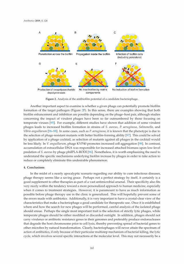
Antibiotics 2019, 8, 126
Figure 2. Analysis of the antibiofilm potential of a candidate bacteriophage.
Another important aspect to examine is whether a given phage can potentially promote biofilmformation of the target pathogen (Figure 2F). In this sense, there are examples showing that bothbiofilm enhancement and inhibition are possible depending on the phage–host pair, although studiesconcerning the impact of virulent phages have been so far outnumbered by those focusing ontemperate viruses [95]. For example, different studies have shown that addition of some virulentphages leads to increased biofilm formation in strains of S. aureus, P. aeruginosa, Salmonella, andVibrio anguillarum [96–98]. In some cases, such as P. aeruginosa, it is known that the phenotype is due tothe selection of phage-resistant mutants with better biofilm-forming ability [97]. This could be solvedby application of a phage cocktail, as selection of mutants against all phages in the cocktail wouldbe less likely. In V. anguillarum, phage KVP40 promotes increased cell aggregation [98]. In contrast,accumulation of extracellular DNA was responsible for increased attached biomass upon low-levelpredation of S. aureus by phage phiIPLA-RODI [96]. Nonetheless, it is worth emphasizing the need tounderstand the specific mechanisms underlying biofilm increase by phages in order to take action toreduce or completely eliminate this undesirable phenomenon.
4. Conclusions
In the midst of a nearly apocalyptic scenario regarding our ability to cure infectious diseases,phage therapy seems like a saving grace. Perhaps not a perfect strategy by itself, it certainly is agood supplement to other therapies as part of a vast antimicrobial arsenal. Their specificity also fitsvery nicely within the tendency toward a more personalized approach to human medicine, especiallywhen it comes to treatment strategies. However, it is paramount to have as much information aspossible before phage therapy use in the clinic is generalized. This will hopefully prevent some ofthe errors made with antibiotics. Additionally, it is very important to have a crystal-clear view of thecharacteristics that make a bacteriophage a good candidate for therapeutic use. Once it is establishedwhere and how the search for new phages will be performed, careful analysis of the isolated phageshould ensue. Perhaps the single most important trait is the selection of strictly lytic phages, whiletemperate phages should be either modified or discarded outright. In addition, phages should notcarry virulence or antibiotic resistance genes in their genomes and preferably produce endonucleasesthat degrade the host chromosome prior to cell lysis, thereby preventing spread of bacterial genes toother microbes by natural transformation. Clearly, bacteriophages will never attain the spectrum ofaction of antibiotics, if only because of their particular multistep mechanism of bacterial killing, the lyticcycle, which involves several specific interactions at the molecular level. This may not necessarily be a
243
Antibiotics 2019, 8, 126
drawback, as phage specificity also allows for tightly controlled killing of pathogenic targets withoutdamaging eukaryotic cells or nontarget microbes. This feature is very important given the increasinglyrecognized role of the microbiota in human health. Another aspect that requires attention duringphage selection is phage particle stability under different conditions, especially those occurring duringstorage and, above all, during treatment. Nonetheless, strategies that allow prolonged stability such asencapsulation should be also developed further. Last but not least, the interactions between phagesand microbial biofilms should be analyzed in depth in order to maximize the potential use of bacterialviruses to reduce or eliminate these structures. Indeed, this might be a key step in the fight againstchronic, recalcitrant infections that do not respond to other types of antimicrobial therapy. Overall,phage therapy seems like a viable, promising strategy that might play a major role in the antibacterialregimes of the future, perhaps even the near future. Nonetheless, further research is still necessary tofill in the gaps that will enable using this strategy while maximizing both efficacy and safety.
Author Contributions: L.F., D.G., P.G. and A.R. wrote the paper.
Funding: This study was funded by grants AGL2015-65673-R (Ministry of Science and Innovation, Spain), EUANIWHA ERA-NET (BLAAT ID: 67)/PCIN-2017-001 (Ministry of Economy, Industry and Competitiveness, Spain),Proyecto Intramural CSIC201670E040, Proyecto Intramural CSIC 201770E016, GRUPIN14-139 (Program of Science,Technology and Innovation 2013-2017, Principado de Asturias), IDI/2018/000119 (iAsturias Innovation, Principadode Asturias) and FEDER EU funds.
Acknowledgments: P.G. and A.R. are members of the bacteriophage network FAGOMA II and the FWOVlaanderen funded “Phagebiotics” research community (WO.016.14).
Conflicts of Interest: The authors declare no conflict of interest.
References
1. D’Hérelle, F. Sur un microbe invisible antagoniste des bacilles dysentériques. C. R. Acad. Sci. 1917,165, 373–375.
2. Twort, F. An investigation on the nature of ultra-microscopic viruses. Lancet 1915, 186, 1241–1243. [CrossRef]3. D’Hérelle, F.; Smith, G.H. The Bacteriophage and Its Clinical Application; Charles, C., Ed.; Thomas: Springfield,
IL, USA, 1930.4. Debarbieux, L.; Pirnay, J.P.; Verbeken, G.; de Vos, D.; Merabishvili, M.; Huys, I.; Patey, O.; Schoonjans, D.;
Vaneechoutte, M.; Zizi, M.; et al. A bacteriophage journey at the European Medicines Agency.FEMS Microbiol. Lett. 2016, 363. [CrossRef] [PubMed]
5. Ventola, C.L. The antibiotic resistance crisis: Part 1: Causes and threats. Pharm. Ther. 2015, 40, 277–283.6. World Health Organization. Global Action Plan on Antimicrobial Resistance; World Health Organization:
Geneva, Switzerland, 2015.7. Gordillo-Altamirano, F.L.; Barr, J.J. Phage therapy in the postantibiotic era. Clin. Microbiol. Rev. 2019, 32.
[CrossRef] [PubMed]8. Chan, B.K.; Abedon, S.T.; Loc-Carrillo, C. Phage cocktails and the future of phage therapy. Future Microbiol.
2013, 8, 769–783. [CrossRef] [PubMed]9. Furfaro, L.L.; Payne, M.S.; Chang, B.J. Bacteriophage therapy: Clinical trials and regulatory hurdles. Front. Cell
Infect. Microbiol. 2018, 8, 376. [CrossRef]10. Moye, Z.D.; Woolston, J.; Sulakvelidze, A. Bacteriophage applications for food production and processing.
Viruses 2018, 10, 205. [CrossRef]11. Fernández, L.; Gutiérrez, D.; Rodríguez, A.; García, P. Application of bacteriophages in the agro-food sector:
A long way toward approval. Front. Cell Infect. Microbiol. 2018, 8, 296. [CrossRef]12. Endersen, L.; O’Mahony, J.; Hill, C.; Ross, R.P.; McAuliffe, O.; Coffey, A. Phage therapy in the food industry.
Annu. Rev. Food Sci. Technol. 2014, 5, 327–349. [CrossRef]13. McCallin, S.; Sacher, J.C.; Zheng, J.; Chan, B.K. Current state of compassionate phage therapy. Viruses 2019,
11, 343. [CrossRef] [PubMed]14. Suttle, C.A. Viruses in the sea. Nature 2005, 437, 356–361. [CrossRef] [PubMed]15. Vos, M.; Birkett, P.J.; Birch, E.; Griffiths, R.I.; Buckling, A. Local adaptation of bacteriophages to their bacterial
hosts in soil. Science 2009, 325, 833. [CrossRef] [PubMed]
244
Antibiotics 2019, 8, 126
16. D’Herelle, F. Le Phénomène de la Guérison des Maladies Infectieuses; Masson: Paris, France, 1938.17. O’Flynn, G.; Ross, R.P.; Fitzgerald, G.F.; Coffey, A. Evaluation of a cocktail of three bacteriophages for
biocontrol of Escherichia coli O157:H7. Appl. Environ. Microbiol. 2004, 70, 3417–3424. [CrossRef] [PubMed]18. Brown, T.L.; Petrovski, S.; Dyson, Z.A.; Seviour, R.; Tucci, J. The formulation of bacteriophage in a semi
solid preparation for control of Propionibacterium acnes growth. PLoS ONE 2016, 11, e0151184. [CrossRef][PubMed]
19. Tylenda, C.A.; Calvert, C.; Kolenbrander, P.E.; Tylenda, A. Isolation of Actinomyces bacteriophage fromhuman dental plaque. Infect. Immun. 1985, 49, 1–6. [PubMed]
20. Latz, S.; Wahida, A.; Arif, A.; Häfner, H.; Hoß, M.; Ritter, K.; Horz, H.P. Preliminary survey of localbacteriophages with lytic activity against multi-drug resistant bacteria. J. Basic Microbiol. 2016, 56, 1117–1123.[CrossRef] [PubMed]
21. Weber-Dabrowska, B.; JonCzyk-Matysiak, E.; Zaczek, M.; Łobocka, M.; Łusiak-Szelachowska, M.; Górski, A.Bacteriophage procurement for therapeutic purposes. Front. Microbiol. 2016, 7, 1177. [CrossRef] [PubMed]
22. Merabishvili, M.; Vervaet, C.; Pirnay, J.-P.; de Vos, D.; Verbeken, G.; Mast, J.; Chanishvili, N.; Vaneechoutte, M.Stability of Staphylococcus aureus phage ISP after freeze-drying (lyophilization). PLoS ONE 2013, 8, e68797.[CrossRef]
23. Ul Haq, I.; Chaudhry, W.N.; Andleeb, S.; Qadr, I.I. Isolation and partial characterization of a virulentbacteriophage IHQ1 specific for Aeromonas punctata from stream water. Microb. Ecol. 2011, 63, 954–963.[CrossRef]
24. Weber-Dabrowska, B.; Zaczek, M.; Dziedzic, B.; Łusiak-Szelachowska, M.; Kiejzik, M.; Górski, A.; Gworek, B.;Wierzbicki, K.; Eymontt, A. Bacteriophages in green biotechnology—The utilization of drinking water.In Industrial, Medical and Environmental Applications of Microorganisms: Current Status and Trends; WageningenAcademic Publishers: Wageningen, The Netherlands, 2014; pp. 500–504.
25. Vongkamjan, K.; Switt, A.M.; den Bakker, H.C.; Fortes, E.D.; Wiedmann, M. Silage collected from dairyfarms harbors an abundance of listeriaphages with considerable host range and genome size diversity.Appl. Environ. Microbiol. 2012, 78, 8666–8675. [CrossRef] [PubMed]
26. Los, J.M.; Golec, P.; Wegrzyn, G.; Wegrzyn, A.; Los, M. Simple method for plating Escherichia coli bacteriophagesforming very small plaques or no plaques under standard conditions. Appl. Environ. Microbiol. 2008,74, 5113–5120. [CrossRef] [PubMed]
27. Kaur, S.; Harjai, K.; Chhibber, S. Methicillin-resistant Staphylococcus aureus phage plaque size enhancementusing sublethal concentrations of antibiotics. Appl. Environ. Microbiol. 2012, 78, 8227–8233. [CrossRef][PubMed]
28. McLaughlin, M.R.; Balaa, M.F. Enhanced contrast of bacteriophage plaques in Salmonella with ferricammonium citrate and sodium thiosulfate (FACST) and tetrazolium red (TZR). J. Microbiol. Methods 2006,65, 318–323. [CrossRef] [PubMed]
29. Casey, E.; van Sinderen, D.; Mahony, J. In vitro characteristics of phages to guide ‘real life’ phage therapysuitability. Viruses 2018, 10, 163. [CrossRef] [PubMed]
30. Gu, J.; Liu, X.; Li, Y.; Han, W.; Lei, L.; Yang, Y.; Zhao, H.; Gao, Y.; Song, J.; Lu, R.; et al. A method forgeneration phage cocktail with great therapeutic potential. PLoS ONE 2012, 7, e31698. [CrossRef]
31. Kawecki, T.J.; Ebert, D. Conceptual issues of local adaptation. Ecol. Lett. 2004, 7, 1225–1241. [CrossRef]32. Langdon, A.; Crook, N.; Dantas, G. The effects of antibiotics on the microbiome throughout development
and alternative approaches for therapeutic modulation. Genome Med. 2016, 8, 39. [CrossRef]33. Gutiérrez, D.; Vandenheuvel, D.; Martínez, B.; Rodríguez, A.; Lavigne, R.; García, P. Two
phages, phiIPLA-RODI and phiIPLA-C1C, lyse mono- and dual-species staphylococcal biofilms.Appl. Environ. Microbiol. 2015, 81, 3336–3348. [CrossRef]
34. El Haddad, L.; Ben Abdallah, N.; Plante, P.L.; Dumaresq, J.; Katsarava, R.; Labrie, S.; Corbeil, J.; St-Gelais, D.;Moineau, S. Improving the safety of Staphylococcus aureus polyvalent phages by their production on aStaphylococcus xylosus strain. PLoS ONE 2014, 9, e102600. [CrossRef]
35. Hamdi, S.; Rousseau, G.M.; Labrie, S.J.; Tremblay, D.M.; Kourda, R.S.; Ben Slama, K.; Moineau, S.Characterization of two polyvalent phages infecting Enterobacteriaceae. Sci. Rep. 2017, 7, 40349. [CrossRef][PubMed]
36. Koskella, B.; Meaden, S. Understanding bacteriophage specificity in natural microbial communities. Viruses2013, 5, 806. [CrossRef] [PubMed]
245
Antibiotics 2019, 8, 126
37. Poullain, V.; Gandon, S.; Brockhurst, M.A.; Buckling, A.; Hochberg, M.E. The evolution of specificity inevolving and coevolving antagonistic interactions between a bacteria and its phage. Evolution 2008, 62, 1–11.[CrossRef]
38. Duffy, S.; Turner, P.E.; Burch, C.L. Pleiotropic costs of niche expansion in the RNA bacteriophage Φ6. Genetics2006, 172, 751–757. [CrossRef] [PubMed]
39. Avrani, S.; Schwartz, D.A.; Lindell, D. Virus-host swinging party in the oceans: Incorporating biologicalcomplexity into paradigms of antagonistic coexistence. Mob. Gent. Elem. 2012, 2, 88–95. [CrossRef] [PubMed]
40. Bertozzi Silva, J.; Storms, Z.; Sauvageau, D. Host receptors for bacteriophage adsorption. FEMS Microbiol. Lett.2016, 363. [CrossRef]
41. Phage Receptor Database. Available online: https://phred.herokuapp.com (accessed on 18 July 2019).42. Buckling, A.; Brockhurst, M. Bacteria-virus coevolution. Adv. Exp. Med. Biol. 2012, 751, 347–370. [CrossRef]43. Tsai, R.; Corrêa, I.R.; Xu, M.Y.; Xu, S.Y. Restriction and modification of deoxyarchaeosine (dG+)-containing
phage 9 g DNA. Sci. Rep. 2017, 7, 8348. [CrossRef]44. Šimoliunas, E.; Šimoliuniene, M.; Kaliniene, L.; Zajanckauskaite, A.; Skapas, M.; Mešky, S.R.; Kaupinis, A.;
Valius, M.; Truncaite, L. Pantoea bacteriophage vB_PagS_Vid5: A low-temperature siphovirus that harbors acluster of genes involved in the biosynthesis of archaeosine. Viruses 2018, 10, 583. [CrossRef]
45. Ngazoa-Kakou, S.; Shao, Y.; Rousseau, G.M.; Addablah, A.A.; Tremblay, D.M.; Hutinet, G.; Lemire, N.;Plante, P.L.; Corbeil, J.; Koudou, A.; et al. Complete genome sequence of Escherichia coli siphophage BRET.Microbiol. Resour. Announc. 2019, 8. [CrossRef]
46. Hobbs, Z.; Abedon, S.T. Diversity of phage infection types and associated terminology: The problem with‘lytic or lysogenic’. FEMS Microbiol. Lett. 2016, 363. [CrossRef] [PubMed]
47. Gutiérrez, D.; Fernández, L.; Rodríguez, A.; García, P. Practical method for isolation of phage deletionmutants. Methods Protoc. 2018, 1, 6. [CrossRef] [PubMed]
48. Dedrick, R.M.; Guerrero-Bustamante, C.A.; Garlena, R.A.; Russell, D.A.; Ford, K.; Harris, K.; Gilmour, K.C.;Soothill, J.; Jacobs-Sera, D.; Schooley, R.T.; et al. Engineered bacteriophages for treatment of a patient with adisseminated drug-resistant Mycobacterium abscessus. Nat. Med. 2019, 25, 730–733. [CrossRef] [PubMed]
49. Keen, E.C.; Bliskovsky, V.V.; Malagon, F.; Baker, J.D.; Prince, J.S.; Klaus, J.S.; Adhya, S.L. Novel “superspreader”bacteriophages promote horizontal gene transfer by transformation. mBio 2017, 8. [CrossRef] [PubMed]
50. Baig, A.; Colom, J.; Barrow, P.; Schouler, C.; Moodley, A.; Lavigne, R.; Atterbury, R. Biology and genomics ofan historic therapeutic Escherichia coli bacteriophage collection. Front. Microbiol. 2017, 8, 1652. [CrossRef][PubMed]
51. Gill, J.J.; Hyman, P. Phage choice, isolation, and preparation for phage therapy. Curr. Pharm. Biotechnol. 2010,11, 2–14. [CrossRef] [PubMed]
52. Speck, P.; Smithyman, A. Safety and efficacy of phage therapy via the intravenous route. FEMS Microbiol. Lett.2016, 363. [CrossRef] [PubMed]
53. Salmon, G.G., Jr.; Symonds, M. Staphage lysate therapy in chronic staphylococcal infections. J. Med. Soc. N. J.1963, 60, 188–193. [PubMed]
54. Meaden, S.; Koskella, B. Exploring the risks of phage application in the environment. Front. Microbiol. 2013,4, 358. [CrossRef]
55. Regulski, K.; Champion-Arnaud, P.; Gabard, J. Bacteriophage manufacturing: From early twentieth-centuryprocesses to current GMP. In Bacteriophages; Harper, D., Ed.; Springer: Cham, Switzerland, 2018.
56. Jurac, K.; Nabergoj, D.; Podgornik, A. Bacteriophage production processes. Appl. Microbiol. Biotechnol. 2019,103, 685–694. [CrossRef]
57. Pirnay, J.P.; Merabishvili, M.; Van Raemdonck, H.; De Vos, D.; Verbeken, G. Bacteriophage production incompliance with regulatory requirements. Methods Mol. Biol. 2018, 1693, 233–252. [CrossRef] [PubMed]
58. Jonczyk, E.; Kłak, M.; Miedzybrodzki, R.; Górski, A. The influence of external factors onbacteriophages—Review. Folia Microbiol. 2011, 56, 191–200. [CrossRef] [PubMed]
59. Ackermann, H.W.; Tremblay, D.; Moineau, S. Long-term bacteriophage preservation. WFCC Newslett. 2004,38, 35–40.
60. González-Menéndez, E.; Fernández, L.; Gutiérrez, D.; Rodríguez, A.; Martínez, B.; García, P. Comparativeanalysis of different preservation techniques for the storage of Staphylococcus phages aimed for the industrialdevelopment of phage-based antimicrobial products. PLoS ONE 2018, 13, e0205728. [CrossRef] [PubMed]
246
Antibiotics 2019, 8, 126
61. Hubalek, Z. Protectants used in the cryopreservation of microorganisms. Cryobiology 2003, 46, 205–229.[CrossRef]
62. Golec, P.; Dabrowski, K.; Hejnowicz, M.S.; Gozdek, A.; Los, J.M.; Wegrzyn, G.; Lobocka, M.B.; Los, M. Areliable method for storage of tailed phages. J. Microbiol. Methods 2011, 84, 486–489. [CrossRef] [PubMed]
63. González-Menéndez, E.; Fernández, L.; Gutiérrez, D.; Pando, D.; Martínez, B.; Rodríguez, A.; García, P.Strategies to encapsulate the Staphylococcus aureus bacteriophage phiIPLA-RODI. Viruses 2018, 10, 495.[CrossRef] [PubMed]
64. Bodier-Montagutelli, E.; Morello, E.; L’Hostis, G.; Guillon, A.; Dalloneau, E.; Respaud, R.; Pallaoro, N.;Blois, H.; Vecellio, L.; Gabard, J.; et al. Inhaled phage therapy: A promising and challenging approach totreat bacterial respiratory infections. Expert Opin. Drug Deliv. 2017, 14, 959–972. [CrossRef]
65. Leung, S.S.Y.; Parumasivam, T.; Gao, F.G.; Carter, E.A.; Carrigy, N.B.; Vehring, R.; Finlay, W.H.; Morales, S.;Britton, W.J.; Kutter, E.; et al. Effects of storage conditions on the stability of spray dried, inhalablebacteriophage powders. Int. J. Pharm. 2017, 521, 141–149. [CrossRef]
66. Leung, S.S.; Parumasivam, T.; Gao, F.G.; Carrigy, N.B.; Vehring, R.; Finlay, W.H.; Morales, S.; Britton, W.J.;Kutter, E.; Chan, H.K. Production of inhalation phage powders using spray freeze drying and spray dryingtechniques for treatment of respiratory infections. Pharm. Res. 2016, 33, 1486–1496. [CrossRef]
67. Ma, Y.; Pacan, J.C.; Wang, Q.; Xu, Y.; Huang, X.; Korenevsky, A.; Sabour, P.M. Microencapsulation ofbacteriophage Felix O1 into chitosan-alginate microspheres for oral delivery. Appl. Environ. Microbiol. 2008,74, 4799–4805. [CrossRef] [PubMed]
68. Singla, S.; Harjai, K.; Katare, O.P.; Chhibber, S. Bacteriophage-loaded nanostructured lipid carrier:Improved pharmacokinetics mediates effective resolution of Klebsiella pneumoniae—Induced lobar pneumonia.J. Infect. Dis. 2015, 212, 325–334. [CrossRef] [PubMed]
69. Nieth, A.; Verseux, C.; Barnert, S.; Süss, R.; Römer, W. A first step toward liposome-mediated intracellularbacteriophage therapy. Expert Opin. Drug Deliv. 2015, 12, 1411–1424. [CrossRef] [PubMed]
70. Nobrega, F.L.; Costa, A.R.; Santos, J.F.; Siliakus, M.F.; van Lent, J.W.; Kengen, S.W.; Azeredo, J.; Kluskens, L.D.Genetically manipulated phages with improved pH resistance for oral administration in veterinary medicine.Sci. Rep. 2016, 6, 39235. [CrossRef]
71. Esteban, P.P.; Alves, D.R.; Enright, M.C.; Bean, J.E.; Gaudion, A.; Jenkins, A.T.; Young, A.E.; Arnot, T.C.Enhancement of the antimicrobial properties of bacteriophage-K via stabilization using oil-in-waternano-emulsions. Biotechnol. Prog. 2014, 30, 932–944. [CrossRef]
72. Esteban, P.P.; Jenkins, A.T.; Arnot, T.C. Elucidation of the mechanisms of action of bacteriophageK/nano-emulsion formulations against Staphylococcus aureus via measurement of particle size and zetapotential. Colloids Surf. B Biointerfaces 2016, 139, 87–94. [CrossRef]
73. Merabishvili, M.; Monserez, R.; van Belleghem, J.; Rose, T.; Jennes, S.; de Vos, D.; Verbeken, G.;Vaneechoutte, M.; Pirnay, J.P. Stability of bacteriophages in burn wound care products. PLoS ONE 2017,12, e0182121. [CrossRef]
74. Majewska, J.; Beta, W.; Lecion, D.; Hodyra-Stefaniak, K.; Kłopot, A.; Kazmierczak, Z.; Miernikiewicz, P.;Piotrowicz, A.; Ciekot, J.; Owczarek, B.; et al. Oral application of T4 phage induces weak antibody productionin the gut and in the blood. Viruses 2015, 7, 2845. [CrossRef]
75. Abedon, S.T.; Kuhl, S.J.; Blasdel, B.G.; Kutter, E.M. Phage treatment of human infections. Bacteriophage 2011,1, 66–85. [CrossRef]
76. Kucharewicz-Krukowska, A.; Slopek, S. Immunogenic effect of bacteriophage in patients subjected to phagetherapy. Arch. Immunol. Ther. Exp. 1987, 35, 553–561.
77. Kim, K.P.; Cha, J.D.; Jang, E.H.; Klumpp, J.; Hagens, S.; Hardt, W.D.; Lee, K.Y.; Loessner, M.J. PEGylationof bacteriophages increases blood circulation time and reduces T-helper type 1 immune response.Microb. Biotechnol. 2008, 1, 247–257. [CrossRef] [PubMed]
78. Costerton, J.W.; Stewart, P.S.; Greenberg, E.P. Bacterial biofilms: A common cause of persistent infections.Science 1999, 284, 1318–1322. [CrossRef] [PubMed]
79. de la Fuente-Núñez, C.; Reffuveille, F.; Fernández, L.; Hancock, R.E.W. Bacterial biofilm development as amulticellular adaptation: Antibiotic resistance and new therapeutic strategies. Curr. Opin. Microbiol. 2013,16, 580–589. [CrossRef] [PubMed]
80. Chan, B.K.; Abedon, S.T. Bacteriophages and their enzymes in biofilm control. Curr. Pharm. Des. 2015,21, 85–99. [CrossRef] [PubMed]
247
Antibiotics 2019, 8, 126
81. Geredew Kifelew, L.; Mitchell, J.G.; Speck, P. Mini-review: Efficacy of lytic bacteriophages on multispeciesbiofilms. Biofouling 2019, 35, 472–481. [CrossRef] [PubMed]
82. González, S.; Fernández, L.; Campelo, A.B.; Gutiérrez, D.; Martínez, B.; Rodríguez, A.; García, P. The behaviorof Staphylococcus aureus dual-species biofilms treated with bacteriophage phiIPLA-RODI depends on theaccompanying microorganism. Appl. Environ. Microbiol. 2017, 83. [CrossRef] [PubMed]
83. Khalifa, L.; Brosh, Y.; Gelman, D.; Coppenhagen-Glazer, S.; Beyth, S.; Poradosu-Cohen, R.; Que, Y.A.;Beyth, N.; Hazan, R. Targeting Enterococcus faecalis biofilms with phage therapy. Appl. Environ. Microbiol.2015, 81, 2696–2705. [CrossRef] [PubMed]
84. Yuan, Y.; Qu, K.; Tan, D.; Li, X.; Wang, L.; Cong, C.; Xiu, Z.; Xu, Y. Isolation and characterization of abacteriophage and its potential to disrupt multi-drug resistant Pseudomonas aeruginosa biofilms. Microb. Pathog.2019, 128, 329–336. [CrossRef] [PubMed]
85. Wintachai, P.; Naknaen, A.; Pomwised, R.; Voravuthikunchai, S.P.; Smith, D.R. Isolation and characterizationof Siphoviridae phage infecting extensively drug-resistant Acinetobacter baumannii and evaluation of therapeuticefficacy In Vitro and In Vivo. J. Med. Microbiol. 2019, 68, 1096–1108. [CrossRef]
86. Gu, Y.; Xu, Y.; Xu, J.; Yu, X.; Huang, X.; Liu, G.; Liu, X. Identification of novel bacteriophage vB_EcoP-EG1 withlytic activity against planktonic and biofilm forms of uropathogenic Escherichia coli. Appl. Microbiol. Biotechnol.2019, 103, 315–326. [CrossRef]
87. Briandet, R.; Lacroix-Gueu, P.; Renault, M.; Lecart, S.; Meylheuc, T.; Bidnenko, E.; Steenkeste, K.;Bellon-Fontaine, M.N.; Fontaine-Aupart, M.P. Fluorescence correlation spectroscopy to study diffusionand reaction of bacteriophages inside biofilms. Appl. Environ. Microbiol. 2008, 74, 2135–2143. [CrossRef][PubMed]
88. González, S.; Fernández, L.; Gutiérrez, D.; Campelo, A.B.; Rodríguez, A.; García, P. Analysis ofdifferent parameters affecting diffusion, propagation and survival of staphylophages in bacterial biofilms.Front. Microbiol. 2018, 9, 2348. [CrossRef] [PubMed]
89. Tkhilaishvili, T.; Lombardi, L.; Klatt, A.B.; Trampuz, A.; Di Luca, M. Bacteriophage Sb-1 enhances antibioticactivity against biofilm, degrades exopolysaccharide matrix and targets persisters of Staphylococcus aureus.Int. J. Antimicrob. Agents 2018, 52, 842–853. [CrossRef] [PubMed]
90. Pires, D.P.; Oliveira, H.; Melo, L.D.; Sillankorva, S.; Azeredo, J. Bacteriophage-encoded depolymerases: Theirdiversity and biotechnological applications. Appl. Microbiol. Biotechnol. 2016, 100, 2141–2151. [CrossRef][PubMed]
91. Latka, A.; Maciejewska, B.; Majkowska-Skrobek, G.; Briers, Y.; Drulis-Kawa, Z. Bacteriophage-encodedvirion-associated enzymes to overcome the carbohydrate barriers during the infection process.Appl. Microbiol. Biotechnol. 2017, 101, 3103–3119. [CrossRef] [PubMed]
92. Schmerer, M.; Molineux, I.J.; Bull, J.J. Synergy as a rationale for phage therapy using phage cocktails. PeerJ2014, 2, e590. [CrossRef] [PubMed]
93. Lu, T.K.; Collins, J.J. Dispersing biofilms with engineered enzymatic bacteriophage. Proc. Natl. Acad. Sci. USA2007, 104, 11197–11202. [CrossRef]
94. Pei, R.; Lamas-Samanamud, G.R. Inhibition of biofilm formation by T7 bacteriophages producingquorum-quenching enzymes. Appl. Environ. Microbiol. 2014, 80, 5340–5348. [CrossRef]
95. Fernández, L.; Rodríguez, A.; García, P. Phage or foe: An insight into the impact of viral predation onmicrobial communities. ISME J. 2018, 12, 1171–1179. [CrossRef]
96. Fernández, L.; González, S.; Campelo, A.B.; Martínez, B.; Rodríguez, A.; García, P. Low-level predation bylytic phage phiIPLA-RODI promotes biofilm formation and triggers the stringent response in Staphylococcusaureus. Sci. Rep. 2017, 7, 40965. [CrossRef]
97. Hosseinidoust, Z.; Tufenkji, N.; van de Ven, T.G. Formation of biofilms under phage predation: Considerationsconcerning a biofilm increase. Biofouling 2013, 29, 457–468. [CrossRef] [PubMed]
98. Tan, D.; Dahl, A.; Middelboe, M. Vibriophages differentially influence biofilm formation by Vibrio anguillarumstrains. Appl. Environ. Microbiol. 2015, 81, 4489–4497. [CrossRef] [PubMed]
© 2019 by the authors. Licensee MDPI, Basel, Switzerland. This article is an open accessarticle distributed under the terms and conditions of the Creative Commons Attribution(CC BY) license (http://creativecommons.org/licenses/by/4.0/).
248
antibiotics
Review
Present and Future of Carbapenem-ResistantEnterobacteriaceae (CRE) Infections
Beatriz Suay-García and María Teresa Pérez-Gracia *
Área de Microbiología, Departamento de Farmacia, Instituto de Ciencias Biomédicas, Facultad de Ciencias de la Salud,Universidad Cardenal Herrera-CEU, C/Santiago Ramón y Cajal, 46115 Alfara del Patriarca, Valencia, Spain* Correspondence: [email protected]; Tel.: +34-96-1369000
Received: 29 July 2019; Accepted: 16 August 2019; Published: 19 August 2019
Abstract: Carbapenem-resistant Enterobacteriaceae (CRE) have become a public health threatworldwide. There are three major mechanisms by which Enterobacteriaceae become resistant tocarbapenems: enzyme production, efflux pumps and porin mutations. Of these, enzyme productionis the main resistance mechanism. There are three main groups of enzymes responsible formost of the carbapenem resistance: KPC (Klebsiella pneumoniae carbapenemase) (Ambler class A),MBLs (Metallo-ß-Lactamases) (Ambler class B) and OXA-48-like (Ambler class D). KPC-producingEnterobacteriaceae are endemic in the United States, Colombia, Argentina, Greece and Italy. On theother hand, the MBL NDM-1 is the main carbapenemase-producing resistance in India, Pakistan andSri Lanka, while OXA-48-like enzyme-producers are endemic in Turkey, Malta, the Middle-East andNorth Africa. All three groups of enzymes are plasmid-mediated, which implies an easier horizontaltransfer and, thus, faster spread of carbapenem resistance worldwide. As a result, there is an urgentneed to develop new therapeutic guidelines to treat CRE infections. Bearing in mind the differentmechanisms by which Enterobacteriaceae can become resistant to carbapenems, there are differentapproaches to treat infections caused by these bacteria, which include the repurposing of alreadyexisting antibiotics, dual therapies with these antibiotics, and the development of new ß-lactamaseinhibitors and antibiotics.
Keywords: Enterobacteriaceae; carbapenem-resistant; CRE; antibiotic resistance; antimicrobials
1. Introduction
Antibiotic resistance occurs when bacteria causing an infection survive after being exposed to adrug that, under normal conditions, would kill it or inhibit its growth [1]. As a result, these survivingstrains multiply and spread due to the lack of competition from other strains sensitive to the samedrug. Due to the inappropriate prescription and administration of antibiotics, resistant bacteria havebecome a public health threat worldwide [2]. In fact, the issue of antibiotic resistant bacteria is suchthat, according to the World Health Organization (WHO) predictions, if antibiotic resistance continuesto increase at this rate, infections caused by resistant bacteria will become the top cause of deathworldwide, ahead of cancer, diabetes and cardiovascular diseases [3].
In 2017, WHO published a list of antibiotic resistant bacteria against which there is an urgentneed to develop new antibiotics [4]. This list is divided into three categories depending on the urgencywith which new antibiotics are needed: critical, high and medium priority. Within the critical prioritygroup are carbapenem and 3rd generation cephalosporin resistant Enterobacteriaceae. These bacteria arecommon pathogens causing severe infections such as bloodstream infections, pneumonia, complicatedurinary tract infections and complicated intra-abdominal infections. As a result, antibiotic resistance inEnterobacteriaceae has significant clinical and socioeconomic consequences [5,6].
Initially, Enterobacteriaceae posed a threat to the public health due to their ability to becomeresistant to antibiotics by producing extended-spectrum ß-lactamases (ESBLs) [7]. To fight this threat,
Antibiotics 2019, 8, 122; doi:10.3390/antibiotics8030122 www.mdpi.com/journal/antibiotics249
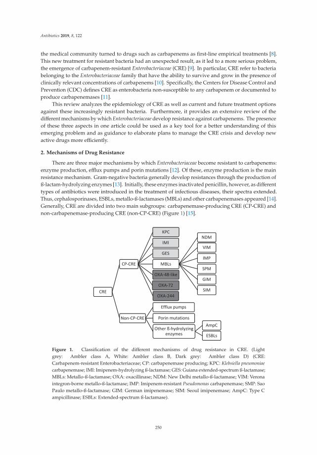
Antibiotics 2019, 8, 122
the medical community turned to drugs such as carbapenems as first-line empirical treatments [8].This new treatment for resistant bacteria had an unexpected result, as it led to a more serious problem,the emergence of carbapenem-resistant Enterobacteriaceae (CRE) [9]. In particular, CRE refer to bacteriabelonging to the Enterobacteriaceae family that have the ability to survive and grow in the presence ofclinically relevant concentrations of carbapenems [10]. Specifically, the Centers for Disease Control andPrevention (CDC) defines CRE as enterobacteria non-susceptible to any carbapenem or documented toproduce carbapenemases [11].
This review analyzes the epidemiology of CRE as well as current and future treatment optionsagainst these increasingly resistant bacteria. Furthermore, it provides an extensive review of thedifferent mechanisms by which Enterobacteriaceae develop resistance against carbapenems. The presenceof these three aspects in one article could be used as a key tool for a better understanding of thisemerging problem and as guidance to elaborate plans to manage the CRE crisis and develop newactive drugs more efficiently.
2. Mechanisms of Drug Resistance
There are three major mechanisms by which Enterobacteriaceae become resistant to carbapenems:enzyme production, efflux pumps and porin mutations [12]. Of these, enzyme production is the mainresistance mechanism. Gram-negative bacteria generally develop resistances through the production ofß-lactam-hydrolyzing enzymes [13]. Initially, these enzymes inactivated penicillin, however, as differenttypes of antibiotics were introduced in the treatment of infectious diseases, their spectra extended.Thus, cephalosporinases, ESBLs, metallo-ß-lactamases (MBLs) and other carbapenemases appeared [14].Generally, CRE are divided into two main subgroups: carbapenemase-producing CRE (CP-CRE) andnon-carbapenemase-producing CRE (non-CP-CRE) (Figure 1) [15].
Figure 1. Classification of the different mechanisms of drug resistance in CRE. (Lightgrey: Ambler class A, White: Ambler class B, Dark grey: Ambler class D) (CRE:Carbapenem-resistant Enterobacteriaceae; CP: carbapenemase producing; KPC: Klebsiella pneumoniaecarbapenemase; IMI: Imipenem-hydrolyzing ß-lactamase; GES: Guiana extended-spectrum ß-lactamase;MBLs: Metallo-ß-lactamase; OXA: oxacillinase; NDM: New Delhi metallo-ß-lactamase; VIM: Veronaintegron-borne metallo-ß-lactamase; IMP: Imipenem-resistant Pseudomonas carbapenemase; SMP: SaoPaulo metallo-ß-lactamase; GIM: German imipenemase; SIM: Seoul imipenemase; AmpC: Type Campicillinase; ESBLs: Extended-spectrum ß-lactamase).
250
Antibiotics 2019, 8, 122
2.1. Carbapenemase-producing CRE
CP-CRE can produce a large variety of carbapenemases which can be divided in three groupsaccording to the Ambler classification: class A, class B and class D ß-lactamases [16]. There is a fourthclass, Ambler class C, however, its clinical relevance remains unknown [17].
Within class A carbapenemases is the clinically relevant Klebsiella pneumoniae carbapenemase(KPC) [18]. This is a plasmid encoded enzyme which actively hydrolyzes carbapenems and ispartially inhibited by clavulanic acid [19]. Its clinical relevance is due to the fact that it is the mostprevalent and most widely spread worldwide [20] Enterobacteriaceae producing KPCs have acquiredmultidrug resistance to ß-lactams, which limits the therapeutic options to treat infections caused bythese bacteria [21]. KPC were originally found in K. pneumoniae isolates, however, clinical isolates ofKPC-producing Escherichia coli, Klebsiella oxytoca, Salmonella enterica, Citrobacter freundii, Enterobacteraerogenes, Enterobacter cloacae, Proteus mirabillis and Serratia marcescens have been identified [22–26](Table 1). According to a study by Perez et al. [27], a total of 12 blaKPC gene variants exist globally.
Table 1. Carbapenemases detected in different species belonging to the Enterobacteriaceae family.
Species Class A Class B (MBLs) Class D Ref.
Klebsiellapneumoniae KPC-3 NDM-1, VIM-1 OXA-48 Okoche et al.
Boutal et al.
Klebsiella oxytoca OXA-48, OXA-181 Okoche et al.Boutal et al.
Escherichia coli KPC NDM-1, NDM-5,NDM-9, VIM
OXA-48, OXA-181,OXA-244
Okoche et al.Boutal et al.
Proteus mirabilis KPC OXA-48 Okoche et al.Boutal et al.
Serratia marcescens KPC VIM Okoche et al.Boutal et al.
Enterobacter cloacae KPC, IMI-1 VIM-4 OXA-48 Okoche et al.Boutal et al.
Enterobacteraerogenes KPC OXA-48 Okoche et al.
Boutal et al.
Citrobacter freundii VIM OXA-48 Okoche et al.Boutal et al.
Citrobacter koseri OXA-48 Okoche et al.Boutal et al.
Salmonella enterica KPC-2NMD-1, NMD-5,
VIM-1, VIM-2,IMP-4
OXA-48 Fernández et al.
Morganella morganii NDM-1 OXA-48 Boutal et al.
Providencia stuartii KPC-2 VIM-1 Abdallah et al.
Providencia rettgeri IMP-1 OXA-72 Abdallah et al.
Another major carbapenemase family belonging to class A are MBLs. These enzymes dependon the interaction with zinc ions in the active site of the enzyme [28]. These enzymes areparticularly problematic as they have a high potential for horizontal transfer, they lack clinicallyuseful inhibitors, and they have broad hydrolytic properties that affect most ß-lactam antibiotics exceptfor monobactams [29]. However, MBL resistance is usually associated with multidrug-resistance,with MBL-producing isolates often co-expressing ESBLs, which inactivate monobactams [13]. The mostcommon families of MBLs found in Enterobacteriaceae were acquired [17]. These families are the NewDelhi metallo-ß-lactamase 1 (NDM-1), Imipenem-resistant Pseudomonas (IMP)-type carbapenemases
251
Antibiotics 2019, 8, 122
and the Verona integron-encoded metallo-ß-lactamases (VIM) [14]. IMP-type carbapenemases werefirst detected in Japan during the 1990s and have up to 18 varieties [30]. Similarly, VIM was first isolatedin Verona, Italy, in 1997 and consists of 14 members [31]. Both MBLs originated in P. aeruginosa andwere transferred to Enterobacteriaceae. In fact, these MBLs share similarities regarding the plasmids theyare carried on and their mechanism of action, as both hydrolyze all ß-lactams except for monobactamsand are susceptible to all ß-lactam inhibitors [32]. Regarding NDM-1, it is the most recently discoveredMBL. It was isolated in India, which is considered the main reservoir of NDM-producing bacteria [33].Since then, it has spread worldwide, reaching Europe and the United States through tourists [34].Currently, NDM is predominant in K. pneumoniae and E. coli [34]. Studies suggest that most plasmidscontaining blaNDM also harbor other resistance determinants encoding different ß-lactamases, quinoloneresistance and 16S rRNA methylases which confer resistance to aminoglycosides [35].
The third clinically relevant group of carbapenemases are OXA-48-like, which belong to Amblerclass D. Six OXA-48-like variants have been identified, OXA-48 being the most widespread [36].The remaining variants are: OXA-162, OXA-163, OXA-181, OXA-204 and OXA-232. They areall grouped within the OXA-48-like category because they only differ on one to five amino acidsubstitutions or deletions [36]. These plasmid-mediated enzymes are primarily found in K. pneumoniae,E. coli, C. freundii and E. cloacae [37]. A major concern with these carbapenemases is that no existinginhibitors work against them and they have an extraordinary ability to mutate and expand theiractivity spectrum [38]. These enzymes are highly active against penicillins, have low activity againstcarbapenems and intermediate activity against broad-spectrum cephalosporins [17].
2.2. Non-Carbapenemase-producing CRE
Besides carbapenemase production, Enterobacteriaceae have alternative mechanisms by which theycan present carbapenem resistance. These are unspecific mechanisms which can result in multi-drugresistance, such as the production of other ß-lactamases, porin loss and efflux pump overexpression [14].These mechanisms generally appear paired among themselves or with carbapenemase-production [39].In fact, while carbapenemases specifically target carbapenems and other ß-lactam antibiotics, effluxpump expression or porin changes are associate with multi-drug resistance [40]. All three alternativemechanisms aim to block the penetration of the antibiotic within the bacterial cell.
Firstly, Enterobacteriaceae can produce different types of ß-lactamases, such as AmpC-typeß-lactamases. These enzymes do not degrade carbapenems [41] but they form a bond with thecarbapenem molecule, preventing it from accessing its target [42]. Specifically, the plasmid-encodedAmpC CMY-2 is frequently found in E. coli and other Enterobacteriaceae worldwide, causing resistanceto carbapenems [43].
Secondly, resistance-nodulation-division (RND) efflux pumps are a major mechanism of multi-drugresistance in Enterobacteriaceae [44]. Among the different efflux systems, the AcrAB-TolC RND systemis the most common [44]. This RND efflux pump, along with the CusABC efflux complex, belongs to E.coli [45]. Similarly, Campylobacter jejuni presents multi-drug resistance through the expression of theCmeABC complex [45]. These resistant genes can be easily transmitted from one microorganism toanother through plasmids [46].
Lastly, alterations of porin synthesis also contribute to blocking penetration of carbapenems intothe bacterial cell [47]. These alterations have been described in AmpC- and carbapenemase-producingK. pneumoniae, which suggests that changes in porin expression play a key role in the ß-lactam resistancedisplayed by multi-drug resistant bacteria [48]. Studies suggest that strains that have their porinsmutated or their expression modulated typically do not have potential for mobilization into communitysettings but may proliferate locally within hospitals [49].
3. Current Resistance Status
Since the detection of the first strain of CRE in the 1980s [50], CRE has rapidly spread worldwide.Epidemiology studies suggest that different carbapenemases predominate in different areas of the
252
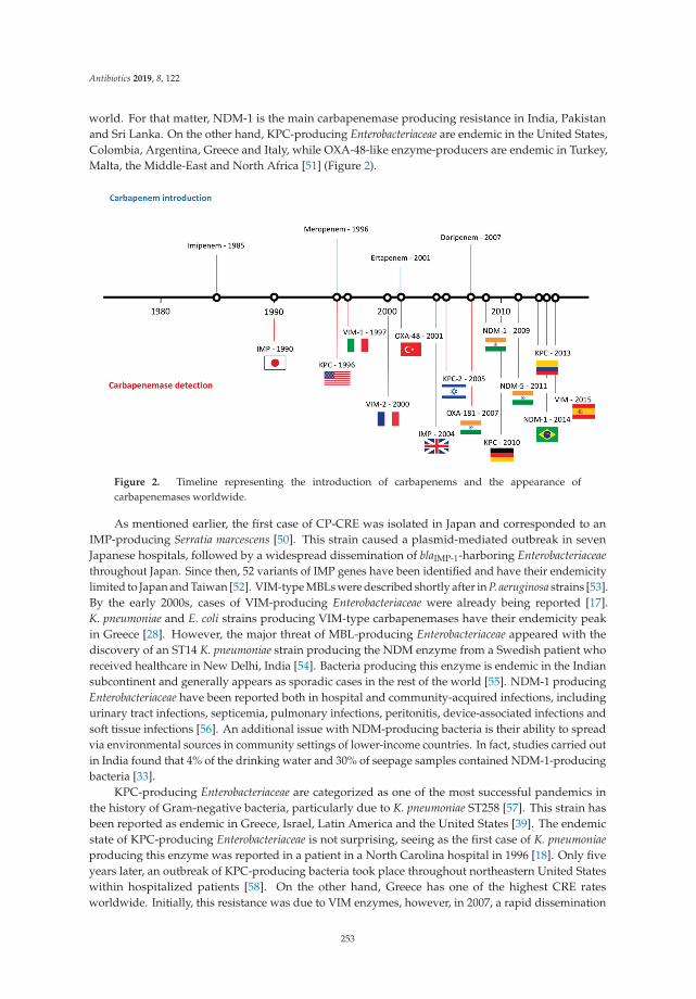
Antibiotics 2019, 8, 122
world. For that matter, NDM-1 is the main carbapenemase producing resistance in India, Pakistanand Sri Lanka. On the other hand, KPC-producing Enterobacteriaceae are endemic in the United States,Colombia, Argentina, Greece and Italy, while OXA-48-like enzyme-producers are endemic in Turkey,Malta, the Middle-East and North Africa [51] (Figure 2).
Figure 2. Timeline representing the introduction of carbapenems and the appearance ofcarbapenemases worldwide.
As mentioned earlier, the first case of CP-CRE was isolated in Japan and corresponded to anIMP-producing Serratia marcescens [50]. This strain caused a plasmid-mediated outbreak in sevenJapanese hospitals, followed by a widespread dissemination of blaIMP-1-harboring Enterobacteriaceaethroughout Japan. Since then, 52 variants of IMP genes have been identified and have their endemicitylimited to Japan and Taiwan [52]. VIM-type MBLs were described shortly after in P. aeruginosa strains [53].By the early 2000s, cases of VIM-producing Enterobacteriaceae were already being reported [17].K. pneumoniae and E. coli strains producing VIM-type carbapenemases have their endemicity peakin Greece [28]. However, the major threat of MBL-producing Enterobacteriaceae appeared with thediscovery of an ST14 K. pneumoniae strain producing the NDM enzyme from a Swedish patient whoreceived healthcare in New Delhi, India [54]. Bacteria producing this enzyme is endemic in the Indiansubcontinent and generally appears as sporadic cases in the rest of the world [55]. NDM-1 producingEnterobacteriaceae have been reported both in hospital and community-acquired infections, includingurinary tract infections, septicemia, pulmonary infections, peritonitis, device-associated infections andsoft tissue infections [56]. An additional issue with NDM-producing bacteria is their ability to spreadvia environmental sources in community settings of lower-income countries. In fact, studies carried outin India found that 4% of the drinking water and 30% of seepage samples contained NDM-1-producingbacteria [33].
KPC-producing Enterobacteriaceae are categorized as one of the most successful pandemics inthe history of Gram-negative bacteria, particularly due to K. pneumoniae ST258 [57]. This strain hasbeen reported as endemic in Greece, Israel, Latin America and the United States [39]. The endemicstate of KPC-producing Enterobacteriaceae is not surprising, seeing as the first case of K. pneumoniaeproducing this enzyme was reported in a patient in a North Carolina hospital in 1996 [18]. Only fiveyears later, an outbreak of KPC-producing bacteria took place throughout northeastern United Stateswithin hospitalized patients [58]. On the other hand, Greece has one of the highest CRE ratesworldwide. Initially, this resistance was due to VIM enzymes, however, in 2007, a rapid dissemination
253
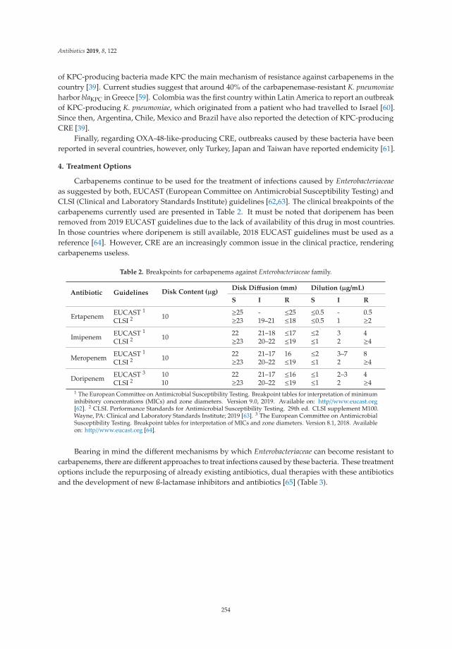
Antibiotics 2019, 8, 122
of KPC-producing bacteria made KPC the main mechanism of resistance against carbapenems in thecountry [39]. Current studies suggest that around 40% of the carbapenemase-resistant K. pneumoniaeharbor blaKPC in Greece [59]. Colombia was the first country within Latin America to report an outbreakof KPC-producing K. pneumoniae, which originated from a patient who had travelled to Israel [60].Since then, Argentina, Chile, Mexico and Brazil have also reported the detection of KPC-producingCRE [39].
Finally, regarding OXA-48-like-producing CRE, outbreaks caused by these bacteria have beenreported in several countries, however, only Turkey, Japan and Taiwan have reported endemicity [61].
4. Treatment Options
Carbapenems continue to be used for the treatment of infections caused by Enterobacteriaceaeas suggested by both, EUCAST (European Committee on Antimicrobial Susceptibility Testing) andCLSI (Clinical and Laboratory Standards Institute) guidelines [62,63]. The clinical breakpoints of thecarbapenems currently used are presented in Table 2. It must be noted that doripenem has beenremoved from 2019 EUCAST guidelines due to the lack of availability of this drug in most countries.In those countries where doripenem is still available, 2018 EUCAST guidelines must be used as areference [64]. However, CRE are an increasingly common issue in the clinical practice, renderingcarbapenems useless.
Table 2. Breakpoints for carbapenems against Enterobacteriaceae family.
Antibiotic Guidelines Disk Content (μg)Disk Diffusion (mm) Dilution (μg/mL)
S I R S I R
Ertapenem EUCAST 1
CLSI 2 10 ≥25≥23
-19–21
≤25≤18
≤0.5≤0.5
-1
0.5≥2
Imipenem EUCAST 1
CLSI 2 10 22≥23
21–1820–22
≤17≤19
≤2≤1
32
4≥4
Meropenem EUCAST 1
CLSI 2 10 22≥23
21–1720–22
16≤19
≤2≤1
3–72
8≥4
Doripenem EUCAST 3
CLSI 21010
22≥23
21–1720–22
≤16≤19
≤1≤1
2–32
4≥4
1 The European Committee on Antimicrobial Susceptibility Testing. Breakpoint tables for interpretation of minimuminhibitory concentrations (MICs) and zone diameters. Version 9.0, 2019. Available on: http//www.eucast.org[62]. 2 CLSI. Performance Standards for Antimicrobial Susceptibility Testing. 29th ed. CLSI supplement M100.Wayne, PA: Clinical and Laboratory Standards Institute; 2019 [63]. 3 The European Committee on AntimicrobialSusceptibility Testing. Breakpoint tables for interpretation of MICs and zone diameters. Version 8.1, 2018. Availableon: http//www.eucast.org [64].
Bearing in mind the different mechanisms by which Enterobacteriaceae can become resistant tocarbapenems, there are different approaches to treat infections caused by these bacteria. These treatmentoptions include the repurposing of already existing antibiotics, dual therapies with these antibioticsand the development of new ß-lactamase inhibitors and antibiotics [65] (Table 3).
254
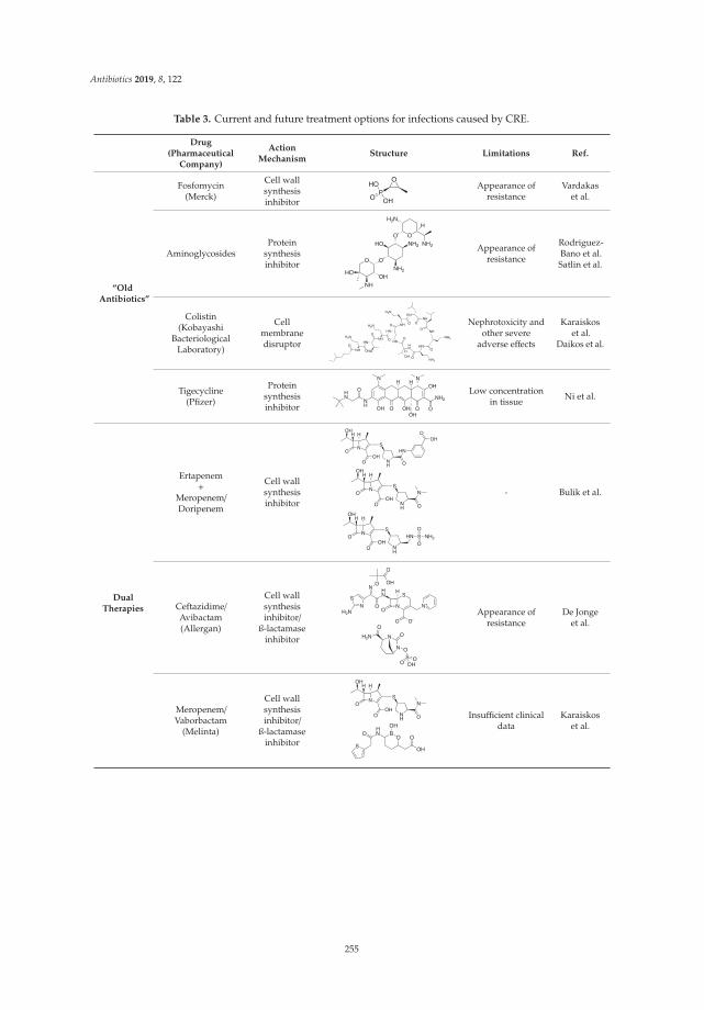
Antibiotics 2019, 8, 122
Table 3. Current and future treatment options for infections caused by CRE.
Drug(Pharmaceutical
Company)
ActionMechanism
Structure Limitations Ref.
“OldAntibiotics”
Fosfomycin(Merck)
Cell wallsynthesisinhibitor
Appearance ofresistance
Vardakaset al.
AminoglycosidesProtein
synthesisinhibitor
Appearance ofresistance
Rodriguez-Bano et al.Satlin et al.
Colistin(Kobayashi
BacteriologicalLaboratory)
Cellmembranedisruptor
Nephrotoxicity andother severe
adverse effects
Karaiskoset al.
Daikos et al.
Tigecycline(Pfizer)
Proteinsynthesisinhibitor
Low concentrationin tissue Ni et al.
DualTherapies
Ertapenem+
Meropenem/Doripenem
Cell wallsynthesisinhibitor
- Bulik et al.
Ceftazidime/Avibactam(Allergan)
Cell wallsynthesisinhibitor/
ß-lactamaseinhibitor
Appearance ofresistance
De Jongeet al.
Meropenem/Vaborbactam
(Melinta)
Cell wallsynthesisinhibitor/
ß-lactamaseinhibitor
Insufficient clinicaldata
Karaiskoset al.
255
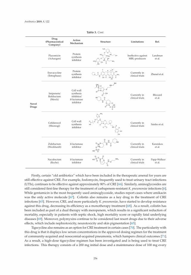
Antibiotics 2019, 8, 122
Table 3. Cont.
Drug(Pharmaceutical
Company)
ActionMechanism
Structure Limitations Ref.
NovelDrugs
Plazomicin(Achaogen)
Proteinsynthesisinhibitor
Ineffective againstMBL-producers
Landmanet al.
Eravacycline(Tetraphase)
Proteinsynthesisinhibitor
Currently inclinical trials Zhanel et al.
Imipenem/Relebactam
(Merck)
Cell wallsynthesisinhibitor/
ß-lactamaseinhibitor
Currently inclinical trials
Blizzardet al.
Cefiderocol(Shionogi)
Cell wallsynthesisinhibitor
Currently inclinical trials Saisho et al.
Zidebactam(Wockhardt)
ß-lactamaseinhibitor
Currently inclinical trials
Karaiskoset al.
Nacubactam(Roche)
ß-lactamaseinhibitor
Currently inclinical trials
Papp-Wallaceet al.
Firstly, certain “old antibiotics” which have been included in the therapeutic arsenal for years arestill effective against CRE. For example, fosfomycin, frequently used to treat urinary tract infections(UTIs), continues to be effective against approximately 80% of CRE [66]. Similarly, aminoglycosides arestill considered first-line therapy for the treatment of carbapenem-resistant K. pneumoniae infections [6].While gentamicin is the most frequently used aminoglycoside, studies report cases where amikacinwas the only active molecule [67]. Colistin also remains as a key drug in the treatment of CREinfections [65]. However, CRE, and more particularly K. pneumoniae, have started to develop resistanceagainst this drug, decreasing its efficiency as a monotherapy treatment [68]. As a result, colistin hasbeen included as part of a dual therapy with meropenem, which results in a significant reduction ofmortality, especially in patients with septic shock, high mortality score or rapidly fatal underlyingdiseases [69]. Moreover, polymyxins continue to be considered last resort drugs due to their adverseeffects, which include nephrotoxicity, neurotoxicity and skin pigmentation [65].
Tigecycline also remains as an option for CRE treatment in certain cases [70]. The particularity withthis drug is that it displays low serum concentrations in the approved dosing regimen for the treatmentof community-acquired and nosocomial-acquired pneumonia, which hampers clinical outcomes [71].As a result, a high-dose tigecycline regimen has been investigated and is being used to treat CREinfections. This therapy consists of a 200 mg initial dose and a maintenance dose of 100 mg every
256
Antibiotics 2019, 8, 122
12 h [65]. This high-dose is particularly effective for the treatment of ventilator-associated pneumoniacaused by CRE [72]. Furthermore, a systematic review comprising 25 studies reporting the efficacy andsafety of tigecycline-based regimens for treating CRE infections concluded that a much lower mortalityrate resulted from high-dose tigecycline than standard-dose tigecycline [70].
Lastly, carbapenems continue to be used for the treatment of CRE infections. This is donethrough the combination of two different carbapenems, which is known as “double carbapenems”.Generally, the combination consists of an initial dose of ertapenem followed by a prolonged infusionof meropenem or doripenem over 3 or 4 h with additional 2 g doses of meropenem every 8 h [73].This therapy is effective against CRE because the greater affinity of ertapenem to KPC makes it playa “sacrificial role”, meaning that it is preferentially hydrolyzed by the carbapenemase, allowing theconcomitant administration of the second carbapenem to sustain a high concentration [74]. Comparatorstudies such as those by Oliva et al. [75] and Venugopalan et al. [76] confirm the efficacy of dualcarbapenem therapy, reporting clinical success rates of more than 70% in both cases.
Regarding novel antibacterial drugs, they can be differentiated in two groups: newly approvedantibiotics and molecules in development stages. The latest antibiotics approved and alreadybeing used to treat CRE infections are ceftazidime/avibactam, meropenem/vaborbactam, plazomicinand eravacycline.
Ceftazidime/avibactam (Allergan) is a novel ß-lactam/ß-lactamase inhibitor combination.The novelty of this combination relies on avibactam, which is a synthetic non-ß-lactam ß-lactamaseinhibitor active against ß-lactamases from Ambler classes A, C and D [77]. Clinical studies usingthis combination are still scarce, however, initial results show an improved mortality rate of9% compared to the 32% obtained when using colistin [78]. Regardless of the promising initialresults, ceftazidime/avibactam resistant strains have already been reported during treatment [79,80].The resistance is due to mutations in the blaKPC-2 and blaKPC-3 genes affecting omega loop D179Y,down-regulation of ompk35/36 and increase in efflux, which could decrease meropenem activity [81].This should be taken into account by clinicians when prescribing this treatment.
Similarly, meropenem/vaborbactam (Melinta) is also a new ß-lactam/ß-lactamase inhibitorconsisting of a carbapenem and a novel boron-containing serine-ß-lactamase inhibitor that potentiatesthe activity of meropenem [65]. This combination inhibits Ambler classes A and C serinecarbapenemases [82]. There are few clinical data with this combination, however, in vivo resultsshowed that, out of 991 clinical isolates of KPC-producing Enterobacteriaceae, 99% were susceptible tomeropenem-vaborbactam [83]. Furthermore, results from the Tango II trial, which compared the efficacyand safety of this combination with the best available therapy in CRE infections, showed a higherclinical cure (65.6% vs 33.3%) and 28-day mortality (15.6% vs 33.3%) for meropenem/vaborbactam [84].
Plazomicin (Achaogen) is a next-generation semisynthetic aminoglycoside with activity againstbacteria producing aminoglycoside-modifying enzymes [85]. Studies report higher potency ofplazomicin compared to other aminoglycosides against KPC-producing Enterobacteriaceae [86].Along these lines, Endimiani et al. [86] analyzed collections of clinically relevant KPC-producerswith resistance to aminoglycosides and observed inhibition using plazomicin, with a minimuminhibitory concentration (MIC90) of ≤2 mg/L [87]. Plazomicin has shown broad-spectrum activityagainst Gram-positive cocci and Gram-negative bacilli [87], however, MBL-producers are resistantto this antibiotic due to the methyltransferase enzymes which are commonly found, especially inNDM-producers [87]. Aminoglycosides are not generally used as monotherapy, however, the broadspectrum of activity along with the low renal toxicity of plazomicin make it an option for a targetedmonotherapy against extensively-drug resistant Enterobacteriaceae causing urinary tract infections [88].
Lastly, eravacycline (Tetraphase) is a synthetic fluorocycline with broad-spectrum antimicrobialactivity against Gram-positive, Gram-negative and anaerobic bacteria, regardless of resistance to otherantibiotic classes [89]. This antibiotic has several potential advantages over tigecycline, which includea more potent in vitro antibacterial activity, excellent oral bioavailability, lower potential for druginteractions and superior activity in biofilm [90]. This drug was also studied in cUTI (complicated
257
Antibiotics 2019, 8, 122
urinary tract infection) in two Phase 3 trials (IGNITE 2/3), failing to meet endpoints in both studies,which could be explained by an erratic pharmacokinetic in urine [91]. However, eravacycline did meetendpoints in the IGNITE 4 Phase 3 study, in which it demonstrated similar activity to ertapenem (100%cure rate for eravacycline vs 92.3% for ertapenem) in the treatment of complicated intra-abdominalinfections [92].
In addition to these already approved drugs, there are six molecules in early developmental stages:imipenem/cilastatin and relebactam (Merck), cediferocol (Shionogi), SPR741 (SperoTherapeutics),zidebactam (Wockhardt), nacubactam (Roche) and VNRX 5133 (VenatoRx Pharmaceuticals). Firstly,imipenem/cilastatin and relebactam shares similarities with previously discussed combinations inthat it combines an approved carbapenem with a novel ß-lactamase inhibitor. In fact, the inhibitorymechanism of relebactam is similar to that of avibactam, as it covalently and reversibly binds to classesA and C ß-lactamases [93]. By including relebactam, the activity of imipenem increases considerablyagainst carbapenemase-producing bacteria, up to >16 fold [94]. In fact, the RESTORE-IMI 1 studyproved this combination to be as effective and better tolerated than colistin/imipenem for the treatmentof infections caused by KPC-producing Enterobacteriaceae [95]. Regarding cefiderocol, it is the firstsiderophore-conjugated cephalosporin antibiotic to advance into late-stage development. This drughas a novel mechanism of action in which the cathecol substituent forms a chelating complex withiron, acting as a trojan horse by using iron active transport systems in gram negative bacteria tobypass the other membrane permeability barrier [96]. This molecule demonstrates potent in vitro andin vivo activity against a variety of Gram-negative bacteria, including CRE [96]. A study analyzed theactivity of cefiderocol and comparative agents against 1,022 isolates of carbapenem-nonsusceptibleEnterobacteriaceae, obtaining MIC50 and MIC90 for cefiderocol of 1 and 4 μg/mL, respectively [97].SPR741 is in very early stages of the development process. This molecule is a polymyxin B potentiatorthat increases ceftazidime and piperazine/tazobactam activity against CRE and ESBLs includingOXA-48 [98].
The remaining molecules under development are ß-lactamase inhibitors. Firstly, zidebactam andnacubactam have high affinity to Ambler classes A and C ß-lactamases [99]. Moreover, they also haveaffinity to PBPs as well as ß-lactam enhancer activity [100]. The cefepime/zidebactam combinationis currently in phase 2 clinical trials for the treatment of Gram-negative bacteria. This combinationshowed potent in vitro activity against carbapenemase-producing Enterobacteriaceae, with MIC50 of0.25 mg/L for KPC-producers and 0.5 mg/L for MBL-producers [101]. On the other hand, nacubactam incombination with meropenem is currently in phase 1 trials against Gram-negative bacteria causing UTIinfections [102]. Results from this study show improved MIC values for the meropenem/nacubactamcombination in comparison with meropenem alone. Furthermore, this combination was active againstceftazidime/avibactam-resistant isolates. Lastly, VNRX 5133 is a cyclic boronate broad spectrumß-lactamase inhibitor in clinical development with cefepime for the treatment of multidrug-resistantbacteria [103].
5. Conclusions
As highlighted by the Global Priority List published by WHO, carbapenem-resistantEnterobacteriaceae pose an exponentially increasing threat for the public health worldwide. These bacteriapossess diverse and versatile mechanisms of drug resistance, which makes control and early detectionof infections caused by CRE difficult. As a result, a joint effort must be made between the scientific andmedical community to slow down the appearance of resistances. Along these lines, there is an urgentneed to develop new therapeutic guidelines to treat CRE infections. This includes the repurposing ofalready existing antibiotics such as fosfomycin, aminoglycosides and colistin and the development ofnovel drugs such as plazomicin, eravacycline or cefiderocol among others.
258
Antibiotics 2019, 8, 122
Author Contributions: B.S.-G. conceived and wrote the paper and M.T.P.-G. conceived and wrote the paper.
Funding: This research received no external funding.
Conflicts of Interest: The authors declare no conflict of interest.
References
1. Centers for Disease Control and Prevention. About Antimicrobial Resistance. 2018. Available online:https://www.cdc.gov/drugresistance/about.html (accessed on 20 July 2019).
2. Niu, G.; Li, W. Next-Generation Drug Discovery to Combat Antimicrobial Resistance. Trends Biochem. Sci.2019, in press. [CrossRef] [PubMed]
3. O’Neill, J. Antimicrobial Resistance: Tackling a Crisis for the Health and Wealth of Nations (HM Governmentand Wellcome Trust). 2014. Available online: https://amr-review.org/sites/default/files/AMR%20Review%20Paper%20-%20Tackling%20a%20crisis%20for%20the%20health%20and%20wealth%20of%20nations_1.pdf (accessed on 20 July 2019).
4. World Health Organization. WHO Priority Pathogens List for R&D of New Antibiotics. 2017. Availableonline: http://www.who.int/bulletin/volumes/94/9/16-020916.pdf (accessed on 20 July 2019).
5. Lee, C.; Lai, C.C.; Chiang, H.T.; Lu, M.C.; Wang, L.F.; Tsai, T.L.; Kang, M.Y.; Jan, Y.N.; Lo, Y.T.; Ko, W.C.; et al.Presence of multidrug-resistant organisms in the residents and environments of long-term care facilities inTaiwan. J. Microbiol. Immunol. Infect. 2017, 50, 133–144. [CrossRef] [PubMed]
6. Rodriguez-Bano, J.; Gutierrez-Gutierrez, B.; Machuca, I.; Pascual, A. Treatment of infectionscaused by extended-spectrum-beta-lactamase-, AmpC-, and carbapenemase-producing Enterobacteriaceae.Clin. Microbiol. Rev. 2018, 31, e00079-17. [CrossRef]
7. Pana, Z.D.; Zaoutis, T. Treatment of extended-spectrum ß-lactamase-producing Enterobacteriaceae (ESBLs)infections: What have we learned until now? F1000Res 2018, 7. [CrossRef]
8. D’Angelo, R.G.; Johnson, J.K.; Bork, J.T.; Heil, E.L. Treatment options for extended-spectrum beta-lactamase(ESBL) and AmpC-producing bacteria. Expert Opin. Pharmacother. 2016, 17, 953–967. [CrossRef] [PubMed]
9. Sheu, C.C.; Lin, S.Y.; Chang, Y.T.; Lee, C.Y.; Chen, Y.H.; Hsueh, P.R. Management of infections caused byextended-spectrum beta-lactamase-producing Enterobacteriaceae: Current evidence and future prospects.Expert Rev. Anti Infect. Ther. 2018, 16, 205–218. [CrossRef] [PubMed]
10. Durante-Mangoni, E.; Andini, R.; Zampino, R. Management of carbapenem-resistant Enterobacteriaceaeinfections. Clin. Microbiol. Infect. 2019, 25, 943–950. [CrossRef] [PubMed]
11. Centers for Disease Control and Prevention. Facility Guidance for Control of Carbapenem-ResistantEnterobacteriaceae (CRE)—November 2015 Update CRE Toolkit. Available online: https://www.cdc.gov/hai/organisms/cre/Cre-toolkit/index.html (accessed on 22 July 2019).
12. Haidar, G.; Clancy, C.J.; Chen, L.; Samanta, P.; Shields, R.K.; Kreiswirth, B.N.; Nguyen, M.H. Identifyingspecra of activity and therapeutic niches for ceftazidime-avibactam and imipenem-relebactam againstcarbapenem-resistant Enterobacteriaceae. Antimicrob. Agents Chemother. 2017, 61. [CrossRef]
13. Tooke, C.L.; Hinchliffe, P.; Bragginton, E.C.; Colenso, C.K.; Hirvonen, V.H.A.; Takebayashi, Y.; Spencer, J.ß-Lactamases and ß-Lactamase Inhibitors in the 21st Century. J. Mol. Biol. 2019. [CrossRef]
14. Codjoe, F.S.; Donkor, E.S. Carbapenem Resistance: A review. Med. Sci. 2018, 6, 1. [CrossRef]15. Lutgring, J.D.; Limbago, B.M. The Problem of Carbapenemase-Producing-Carbapenem-Resistant-
Enterobacteriaceae Detection. J. Clin. Microbiol. 2016, 54, 529–534. [CrossRef]16. Ambler, R.P. The structure of ß-lactamases. Philos. Trans. R. Soc. Lond. B 1980, 289, 321–331. [CrossRef]17. Queenan, A.M.; Bush, K. Carbapenemases: The versatile ß-lactamases. Clin. Microbiol. Rev. 2007, 20, 440–458.
[CrossRef] [PubMed]18. Yigit, H.; Queenan, A.M.; Anderson, G.J.; Domenech-Sanchez, A.; Biddle, J.W.; Steward, C.D.; Alberti, S.;
Bush, K.; Tenover, F.C. Novel carbapenem-hydrolyzing beta-lactamase, KPC-1, from a carbapenem-resistantstrain of Klebsiella pneumoniae. Antimicrob. Agents Chemother. 2001, 45, 1151–1161. [CrossRef] [PubMed]
19. Ji, S.; Lv, F.; Du, X.; Wei, Z.; Fu, Y.; Mu, X.; Jiang, Y.; Yu, Y. Cefepime combined with amoxicillin/clavulanicacid: A new choice for the KPC-producing K. pneumoniae infection. Int. J. Infect. Dis. 2015, 38, 108–114.[CrossRef] [PubMed]
20. Porreca, A.M.; Sullivan, K.V.; Gallagher, J.C. The Epidemiology, Evolution, and Treatment of KPC-ProducingOrganisms. Curr. Infect. Dis. Rep. 2018, 20, 13. [CrossRef] [PubMed]
259
Antibiotics 2019, 8, 122
21. Nordmann, P.; Cuzon, G.; Naas, T. The real threat of Klebsiella pneumoniae carbapenemase-producing bacteria.Lancet Infect. Dis. 2009, 9, 228–233. [CrossRef]
22. Miriagou, V.; Cornaglia, G.; Edelstein, M.; Galani, I.; Giske, G. Acquired carbapenemases in Gram-negativebacterial pathogens: Detection and surveillance issues. Clin. Microbiol. Infect. 2010, 16, 112–122. [CrossRef][PubMed]
23. Okoche, D.; Asiimwe, B.B.; Katabazi, F.A.; Kato, L.; Najjuka, C.F. Prevalence and Characterizationof Carbapenem-Resistant Enterobacteriaceae Isolated from Mulago National Referral Hospital, Uganda.PLoS ONE 2015, 10, e0135745. [CrossRef]
24. Boutal, H.; Vogel, A.; Bernabeu, S.; Devilliers, K.; Creton, E.; Cotellon, G.; Plaisance, M.; Oueslati, S.; Dortet, L.;Jousset, A.; et al. A multiplex lateral flow immunoassay for the rapid identification of NDM-, KPC-, IMP-and VIM-type and OXA-48-like carbapenemase-producing Enterobacteriaceae. J. Antimicrob. Chemother. 2018,73, 909–915. [CrossRef]
25. Fernández, J.; Guerra, B.; Rodicio, M.R. Resistance to Carbapenems in Non-Typhoidal Salmonella entericaSerovars from Humans, Animals and Food. Vet. Sci. 2018, 5, 40. [CrossRef] [PubMed]
26. Abdallah, M.; Balshi, A. First literature review of carbapenem-resistant Providencia. New Microb. New Infect.2018, 25, 16–23. [CrossRef] [PubMed]
27. Perez, F.; van Duin, D. Carbapenem-resistant Enterobacteriaceae: A menace to our most vulnerable patients.Clevel Clin. J. Med. 2013, 80, 225–233. [CrossRef] [PubMed]
28. Walsh, T.R.; Toleman, M.A.; Piorel, L.; Nordman, P. Metallo-b-lactamases: The quiet before the storm?Clin. Microbiol. Rev. 2005, 18, 306–325. [CrossRef] [PubMed]
29. Van Duin, D.; Doi, Y. The global epidemiology of carbapenemase-producing Enterobacteriaceae. Virulence2017, 8, 460–469. [CrossRef] [PubMed]
30. Livermore, D.M.; Woodford, N. Carbapenemases: A problem in waiting? Curr. Opin. Microbiol. 2000, 3,489–495. [CrossRef]
31. Gupta, V. Metallo-b-lactamases in Pseudomonas aeruginosa and Acinetobacter species. Expert Opin. Investig.Drugs 2008, 17, 131–143. [CrossRef]
32. Bush, K. Past and Present Perspectives on ß-Lactamases. Antimicrob. Agents Chemother. 2018, 62, e01076-18.[CrossRef]
33. Walsh, T.R.; Weeks, J.; Livermore, D.M.; Toleman, M.A. Dissemination of NDM-1 positive bacteria in theNew Delhi environment and its implications for human health: An environmental point prevalence study.Lancet Infect. Dis. 2011, 11, 355–362. [CrossRef]
34. Poirel, L.; Hombrouck-Alet, C.; Freneaux, C.; Bernabeu, S.; Nordmann, P. Global spread of New Delhimetallo-ß-lactamase 1. Lancet Infect. Dis. 2010, 10, 832. [CrossRef]
35. Ivanov, I.; Sabtcheva, S.; Dobreva, E.; Todorova, B.; Velinov, T.Z.; Borissova, V.; Kantardijev, T. Prevalence ofcarbapenemase genes among 16S rRNA methyltransferase-producing Enterobacteriaceae isolated for cancerpatients. Probl. Infect. Parasit. Dis. 2014, 42, 10–13.
36. Poirel, L.; Potron, A.; Nordmann, P. OXA-48-like carbapenemases: The phantom menace. J. Antimicrob.Chemother. 2012, 67, 1597–1606. [CrossRef] [PubMed]
37. Rasmussen, J.W.; Hoiby, N. OXA-type carbapenemases. J. Antimicrob. Chemother. 2006, 57, 373–383.[CrossRef] [PubMed]
38. Stewart, A.; Harris, P.; Henderson, A.; Paterson, D. Treatment of Infections by OXA-48-ProducingEnterobacteriaceae. Antimicrob. Agents Chemother. 2018, 62, e01195-18. [CrossRef] [PubMed]
39. Logan, L.K.; Weinstein, R.A. The Epidemiology of Carbapenem-Resistant Enterobacteriaceae: The Impact andEvolution of a Global Menace. J. Infect. Dis. 2017, 215 (Suppl. 1), S28–S36. [CrossRef]
40. Tzouvelekis, L.S.; Marjogiannakis, A.; Psichogiou, M.; Tassios, P.T.; Daikos, G.L. Carbapenemases in Klebsiellapneumoniae and other Enterobacteriaceae: An evolving crisis of global dimensions. Clin. Microbiol. Rev. 2012,25, 682–707. [CrossRef]
41. Queenan, A.M.; Shang, W.; Flamm, R.; Bush, K. Hydrolysis and inhibition profiles of ß-lactamases frommolecular classes A to D with doripenem, imipenem, and meropenem. Antimicrob. Agents Chemother. 2010,54, 565–569. [CrossRef]
42. Goessens, W.H.F.; van der Bij, A.K.; van Boxtel, R.; Pitout, J.D.D.; van Ulsen, P.; Melles, D.C.; Tommassen, J.Antibiotic trapping by plasmid-encoded CMY-2 ß-lactamase combined with reduced outer membrane
260
Antibiotics 2019, 8, 122
permeability as a mechanism of carbapenem resistance in Escherichia coli. Antimicrob. Agents Chemother. 2013,57, 3941–3949. [CrossRef]
43. Philippon, A.; Arlet, G.; Jacoby, G.A. Plasmid-determined AmpC-type ß-lactamases. Antimicrob. AgentsChemother. 2002, 46, 1–11. [CrossRef]
44. Weston, N.; Sharma, P.; Ricci, V.; Piddock, L.J.V. Regulation of the AcrAB-TolC efflux pump in Enterobacteriaceae.Res. Microbiol. 2018, 169, 425–431. [CrossRef]
45. Routh, M.D.; Zalucki, Y.; Su, C.C.; Zhang, Q.; Shager, W.M.; Yu, E.W. Efflux pumps of theresistance-nodulation-division family: A perspective of their structure, function, and regulation ingram-negative bacteria. Adv. Enzymol. Relat. Areas Mol. Biol. 2011, 77, 109–146. [PubMed]
46. Courvalin, P. Transfer of antibiotic resistance genes between gram-positive and gram-negative bacteria.Antimicrob. Agents Chemother. 1994, 38, 1447–1451. [CrossRef] [PubMed]
47. Bialek-Davenet, S.; Mayer, N.; Vergalli, J.; Duprilot, M.; Brisse, S.; Pagès, J.M.; Nicolas-Chanoine, M.H.In-vivo loss of carbapenem resistance by extensively drug-resistant Klebsiella pneumoniae during treatmentvia porin expression modification. Sci. Rep. 2017, 7, 6722. [CrossRef] [PubMed]
48. Masi, M.; Réfrigiers, M.; Pos, K.M.; Pagès, J.M. Mechanisms of envelope permeability and antibiotic influxand efflux in Gram-negative bacteria. Nat. Microbiol. 2017, 2, 17001. [CrossRef] [PubMed]
49. Walsh, C. Molecular mechanisms that confer antibacterial drug resistance. Nature 2000, 406, 775–781.[CrossRef] [PubMed]
50. Ito, H.; Arakawa, Y.; Oshuka, S.; Wacharotayankun, R.; Kato, N.; Ohta, M. Plasmid-mediated dissemination ofthe metallo-beta-lactamase gene blaIMP among clinically isolated strains of Serratia marcescens. J. Antimicrob.Chemother. 1995, 50, 503–511. [CrossRef] [PubMed]
51. Nordmann, P.; Poirel, L. The difficult-to-control spread of carbapenemase producers among Enterobacteriaceaeworldwide. Clin. Microbiol. Infect. 2014, 20, 821–830. [CrossRef]
52. Nordmann, P.; Naas, T.; Poirel, L. Global spread of carbapenemase-producing Enterobacteriaceae. Emerg.Infect. Dis. 2011, 17, 1791–1798. [CrossRef]
53. Lauretti, L.; Riccio, M.K.; Mazzariol, A.; Cornaglia, G.; Amicosante, G.; Fontana, R.; Rossolini, G.M. Cloningand characterization of blaVIM, a new integron-borne metallo-beta-lactamase gene from a Pseudomonasaeruginosa clinical isolate. Antimicrob. Agents Chemother. 1999, 43, 1584–1590. [CrossRef]
54. Yong, D.; Toleman, M.A.; Giske, C.G.; Cho, H.S.; Sundman, K.; Lee, K.; Walsh, T.R. Characterization of newmetallo-beta-lactamase gene, bla(NDM-1), and a novel erythromycin esterase gene carried on a uniquegenetic structure in Klebsiella pneumoniae sequence type 14 from India. Antimicrob. Agents Chemother. 2009,53, 5046–5054. [CrossRef]
55. Dortet, L.; Poirel, L.; Nordmann, P. Worldwide dissemination of the NDM-type carbapenemases inGram-negative bacteria. Biomed. Res. Int. 2014, 2014, 249856. [CrossRef] [PubMed]
56. Poirel, L.; Savov, E.; Nazli, A.; Trifonova, A.; Todorova, I.; Gergova, I.; Nordmann, P. Outbreak caused byNDM-1- and RmtB- producing Escherichia coli in Bulgaria. Antimicrob. Agents Chemother. 2012, 58, 2472–2474.[CrossRef] [PubMed]
57. Patel, G.; Bonomo, R.A. Stormy waters ahead: Global emergence of carbapenemases. Front. Microbiol. 2013,4, 1–17. [CrossRef] [PubMed]
58. Bradford, P.A.; Bratu, S.; Urban, C.; Visalli, M.; Mariano, N.; Landman, D.; Rahal, J.J.; Brooks, S.; Cebular, S.;Quale, J. Emergence of carbapenem-resistant Klebsiella species possessing the class a carbapenem-hydrolyzingKPC-2 and inhibitor-resistant TEM-30 ß-lactamases in New York City. Clin. Infect. Dis. 2004, 39, 55–60.[CrossRef] [PubMed]
59. Munoz-Price, L.S.; Poirel, L.; Bonomo, R.A.; Schwaber, M.J.; Daikos, G.L.; Cormican, M.; Cornaglia, G.;Garau, J.; Gniadkowski, M.; Hayden, M.K. Clinical epidemiology of the global expansion of Klebsiellapneumoniae carbapenemases. Lancet Infect. Dis. 2013, 13, 785–796. [CrossRef]
60. Maya, J.J.; Ruiz, S.J.; Blanco, V.M.; Gotuzzo, E.; Guzman-Blanco, M.; Labarca, J.; Salles, M.; Quinn, J.P.;Villegas, M.V. Current status of carbapenemases in Latin America. Expert Rev. Anti Infect. Ther. 2013, 11,657–667. [CrossRef]
61. Poirel, L.; Bonnin, R.A.; Nordmann, P. Genetic features of the widespread plasmid coding for thecarbapenemase OXA-48. Antimicrob. Agents Chemother. 2012, 56, 559–562. [CrossRef]
62. The European Committee on Antimicrobial Susceptibility Testing. Breakpoint Tables for Interpretation of MICsand Zone Diameters, Version 9.0; 2019. Available online: http//www.eucast.org (accessed on 25 July 2019).
261
Antibiotics 2019, 8, 122
63. CLSI. Performance Standards for Antimicrobial Susceptibility Testing, 29th ed.; CLSI supplement M100; Clinicaland Laboratory Standards Institute: Wayne, PA, USA, 2019.
64. The European Committee on Antimicrobial Susceptibility Testing. Breakpoint Tables for Interpretation of MICsand Zone Diameters, Version 8.1; 2018. Available online: http//www.eucast.org (accessed on 25 July 2019).
65. Karaiskos, I.; Lagou, S.; Pontikis, K.; Rapti, V.; Poulakou, G. The “Old” and the “New” antibiotics for MDRgram-negative pathogens: For whom, when and how. Front. Public Health 2019, 7, 151. [CrossRef]
66. Vardakas, K.Z.; Legakis, N.J.; Triarides, N.; Falagas, M.E. Susceptibility of contemporary isolates toFosfomycin: A systematic review of the literature. Int. J. Antimicrob. Agents 2016, 47, 269–285. [CrossRef]
67. Satlin, M.J.; Kubin, C.J.; Blumental, J.S.; Cohen, A.B.; Furuya, E.Y.; Wilson, S.J.; Jenkins, S.G.; Calfee, D.P.Comparative effectiveness of aminoglycosides, polymyxin B and tigecycline for clearance of carbapenemresistant Klebsiella pneumoniae from urine. Antimicrob. Agents Chemother. 2011, 55, 2528–2531. [CrossRef]
68. Sader, H.S.; Castanheira, M.; Duncan, L.R.; Flamm, R.K. Antimicrobial susceptibility of Enterobacteriaceae andPseudomonas aeruginosa isolates from United States medical centers stratified by infection type: Results fromthe international network for optimal resistance monitoring (INFORM) surveillance program, 2015–2016.Diagn. Microbiol. Infect. Dis. 2018, 92, 69–74. [CrossRef] [PubMed]
69. Daikos, G.L.; Tsaousi, S.; Tzouvelekis, L.S.; Anyfantis, I.; Psichogiou, M.; Argyropoulou, A.; Stefanou, I.;Sypsa, V.; Miriagou, V.; Nepka, M. Carbapenemase-producing Klebsiella pneumoniae bloodstream infections:Lowering mortality by antibiotic combination schemes and the role of carbapenems. Antimicrob. AgentsChemother. 2014, 58, 2322–2328. [CrossRef] [PubMed]
70. Ni, W.; Han, Y.; Liu, J.; Wei, C.; Zhao, J.; Cui, J.; Wang, R.; Liu, Y. Tigecycline treatment for carbapenem-resistantEnterobacteriaceae infections: A systematic review and meta-analysis. Medicine 2016, 95, e3126. [CrossRef][PubMed]
71. Giamarellou, H.; Poulakou, G. Pharmakokinetic and pharmacodynamic evaluation of tigecycline. ExpertOpin. Drug Metab. Toxicol. 2011, 7, 1459–1470. [CrossRef] [PubMed]
72. De Pascale, G.; Montini, L.; Pennisi, M.; Bernini, V.; Maviglia, R.; Bello, G.; Spanu, T.; Tumbarello, M.;Antonelli, M. High dose tigecycline in critically ill patients with severe infections due to multidrug-resistantbacteria. Crit. Care 2014, 18, R90. [CrossRef] [PubMed]
73. Bulik, C.C.; Nicolau, D.P. Double-carbapenem therapy for carbapenemase-producing Klebsiella pneumoniae.Antimicrob. Agents Chemother. 2011, 55, 3002–3004. [CrossRef] [PubMed]
74. Anderson, K.F.; Lonsway, D.R.; Rasheed, J.K.; Biddle, J.; Jensen, B.; McDougal, L.K.; Carey, R.B.; Thompson, A.;Stocker, S.; Limbago, B.; et al. Evaluation of methods to identify the Klebsiella pneumoniae carbapenemase inEnterobacteriaceae. J. Clin. Microbiol. 2007, 45, 2723–2725. [CrossRef] [PubMed]
75. Oliva, A.; Scorzolini, L.; Castaldi, D.; Gizzi, F.; De Angelis, M.; Sotrto, M.; D’Abramo, A.; Aloj, F.;Mascellino, M.T.; Mastroianni, C.M.; et al. Double-carbapenem regimen, alone or in combination withcolistin, in the treatment of infections caused by carbapenem-resistent Klebsiella pneumoniae (CR-Kp). J. Infect.2017, 74, 103–106. [CrossRef]
76. Venugopalan, V.; Nogid, B.; Le, T.N.; Rahman, S.M.; Bias, T.E. Double carbapenem therapy (DCT) forbacteremia due to carbapenem-resistant Klebsiella pneumoniae (CRKP): From the test tube to clinical practice.Infect. Dis. 2017, 49, 867–870. [CrossRef]
77. De Jonge, B.L.; Karlowsky, J.A.; Kazmierczak, K.M.; Biedenbach, D.J.; Sahm, D.F.; Nichols, W.W. In vitrosusceptibility to ceftazidime-avibactam of carbapenem-nonsusceptible Enterobacteriaceae isolates collectedduring the INFORM Global Surveillance Study (2012 to 2014). Antimicrob. Agents Chemother. 2016, 60,3163–3169. [CrossRef]
78. Van Duin, D.; Lok, J.J.; Earley, M.; Cober, E.; Richter, S.S.; Perez, F.; Salata, R.A.; Kalayjian, R.C.;Watkins, R.R.; Doi, Y.; et al. Colistin Versus Ceftazidime-Avibactam in the Treatment of Infections Due toCarbapenem-Resistant Enterobacteriaceae. Clin. Infect. Dis. 2018, 66, 163–171. [CrossRef] [PubMed]
79. Nelson, K.; Hemarajata, P.; Sun, D.; Rubio-Aparicio, D.; Tsivokovski, R.; Yang, S.; Sebra, R.; Kasarskis, A.M.;Nguyen, H.; Hanson, B.M.; et al. Resistance to Ceftazidime-Avibactam Is Due to Transposition of KPC in aPorin-Deficient Strain of Klebsiella pneumoniae with Increased Efflux Activity. Antimicrob. Agents Chemother.2017, 61, e00989-17. [CrossRef] [PubMed]
80. Venditti, C.; Nisii, C.; D’Arezzo, S.; Vulcano, A.; Capone, A.; Antonini, M.; Ippolito, G.; Di Caro, A. Molecularand phenotypical characterization of two cases of antibiotic-driven ceftazidime-avibactam resistance inblaKPC-3-harboring Klebsiella pneumoniae. Infect. Drug Resist. 2019, 12, 1935–1940. [CrossRef] [PubMed]
262
Antibiotics 2019, 8, 122
81. Humphries, R.M. Mechanisms of Resistance to Ceftazidime-Avibactam, 28th ed.; European Congress of ClinicalMicrobiology & Infectious Diseases (ECCMID): Madrid, Spain, 2018.
82. Petty, L.A.; Henig, O.; Patel, T.S.; Pogue, J.M.; Kaye, K.S. Overview of meropenem-vaborbactam and newerantimicrobial agents for the treatment of carbapenem-resistant Enterobacteriaceae. Infect. Drug Resist. 2018, 11,1461–1472. [CrossRef] [PubMed]
83. Lomovskaya, O.; Sun, D.; Rubio-Aparicio, D.; Nelson, K.; Tsivkovski, R.; Griffith, D.C.; Dudley, M.N.Vaborbactam: Spectrum of beta-lactamase inhibition and impact of resistance mechanisms on activity inEnterobacteriaceae. Antimicrob. Agents Chemother. 2017, 61, 1–15. [CrossRef] [PubMed]
84. Wunderink, R.G.; Giamarellos-Bourboulis, E.J.; Rahav, G.; Mathers, A.J.; Bassetti, M.; Vazquez, J.;Cornely, O.A.; Solomkin, J.; Bhowmick, T.; Bishara, J.; et al. Effect and safety of meropenem-vaborbactamversus best-available therapy in patients with carbapenem-resistant Enterobacteriaceae infections: The TANGOII randomized clinical trial. Infect. Dis. Ther. 2018, 7, 439–455. [CrossRef]
85. Landman, D.; Babu, E.; Shah, N.; Kelly, P.; Backer, M.; Bratu, S.; Quale, J. Activity of a novel aminoglycoside,ACHN-490, against clinical isolates of Escherichia coli and Klebsiella pneumoniae from New York City.J. Antimicrob. Chemother. 2010, 65, 2123–2127. [CrossRef]
86. Endimiani, A.; Hujer, K.M.; Hujer, A.M.; Armstrong, E.S.; Choudhary, Y.; Aggen, J.B.; Bonomo, R.A.ACHN-490, a neoglycoside with potent in vitro activity against multidrug-resistant Klebsiella pneumoniaeisolates. Antimicrob. Agents Chemother. 2009, 53, 4504–4507. [CrossRef]
87. Walkty, A.; Adam, H.; Baxter, M.; Denisuik, A.; Lagace-Wiens, P.; Karlowsky, J.A.; Hoban, D.J.; Zhanel, G.G.In vitro activity of plazomicin against 5,015 Gram-negative and Gram-positive clinical isolates obtained frompatients in Canadian hospitals as part of the CANWARD study, 2011–2012. Antimicrob. Agents Chemother.2014, 58, 2554–2563. [CrossRef]
88. Castanheira, M.; Deshpande, L.M.; Woosley, L.N.; Serio, A.W.; Krause, K.M.; Flamm, R.K. Activity ofplazomicin compared with other aminoglycosides against isolates from European and adjacent countries,including Enterobacteriaceae molecularly characterized for aminoglycoside-modifying enzymes and otherresistance mechanisms. J. Antimicrob. Chemother. 2018, 73, 3346–3354. [CrossRef]
89. Zhanel, G.G.; Cheung, D.; Adam, H.; Zelenitsky, S.; Golden, A.; Schweizer, F.; Gorityala, B.; Lagacé-Wiens, P.R.;Walkty, A.; Gin, A.S.; et al. Review of eravacycline, a novel fluorocycline antibacterial agent. Drugs 2016, 76,567–588. [CrossRef] [PubMed]
90. Bassetti, M.; Righi, E. Eravacycline for the treatment of intra-abdominal infections. Expert Opin. Investig.Drugs 2014, 23, 1575–1584. [CrossRef] [PubMed]
91. XERAVA (Eravacycline) for Injection. XERAVA (Eravacycline) IGNITE1 and IGNITE4 Trial Results. 2019.Available online: https://www.xerava.com/efficacy (accessed on 25 July 2019).
92. Solomkin, J.S.; Ramesh, M.K.; Cesnauskas, G.; Novikovs, N.; Stefanova, P.; Sutcliffe, J.A.; Walpole, S.M.;Horn, P.T. Phase 2, randomized, double-blind study of the efficacy and safety of two dose regimens oferavacycline versus ertapenem for adult community-acquired complicated intra-abdominal infections.Antimicrob. Agents Chemother. 2014, 58, 1847–1854. [CrossRef]
93. Blizzard, T.A.; Chen, H.; Kim, S.; Wu, J.; Bodner, R.; Gude, C.; Imbriglio, J.; Young, K.; Park, Y.W.; Ogawa, A.;et al. Discovery of MK-7655, a beta-lactamase inhibitor for combination with Primaxin®. Bioorg. Med. Chem.Lett. 2014, 24, 780–785. [CrossRef] [PubMed]
94. Zhanel, G.G.; Lawrence, C.K.; Adam, H.; Schweizer, F.; Zelenitsky, S.; Zhanel, M.; Lagacé-Wiens, P.R.S.;Walkty, A.; Denisuik, A.; Golden, A.; et al. Imipenem-Relebactam and Meropenem-Vaborbactam: Two NovelCarbapenem-ß-Lactamase Inhibitor Combinations. Drugs 2018, 78, 65–98. [CrossRef] [PubMed]
95. Motsch, J.; Oliveira, C.; Stus, V.; Koksal, I.; Lyulko, O.; Boucher, H.W.; Kaye, K.S.; File, T.M.; Brown, M.L.;Khan, I.; et al. RESTORE-IMI 1: A Multicenter, Randomized, Double-Blind, Comparator-Controlled TrialComparing the Efficacy and Safety of Imipenem/Relebactam Versus Colistin Plus Imipenem in Patients withImipenem-Non-Susceptible Bacterial Infections, 28th ed.; ECCMID: Madrid, Spain, 2018.
96. Saisho, Y.; Katsube, T.; White, S.; Fukase, H.; Shimada, J. Pharmacokinetics, safety, and tolerability ofcefiderocol, a novel siderophore cephalosporin for Gram-negative bacteria, in healthy subjects. Antimicrob.Agents Chemother. 2018, 62, e-02163-17. [CrossRef] [PubMed]
97. Hackel, M.A.; Tsuji, M.; Yamano, Y.; Echols, R.; Karlowsky, J.A.; Sahm, D.F. In Vitro Activity of theSiderophore Cephalosporin, Cediferocol, against Carbapenem-Nonsusceptible and Multidrug-Resistant
263
Antibiotics 2019, 8, 122
Isolates of Gram-Negative Bacilli Collected Worldwide in 2014 to 2016. Antimicrob. Agents Chemother. 2018,62, e01968-17. [CrossRef]
98. Corbett, D.; Wise, A.; Langley, T.; Skinner, K.; Trimby, E.; Birchall, S.; Dorali, A.; Sandiford, S.; Williams, J.;Warn, P.; et al. Potentiation of Antibiotic Activity by a Novel Cationic Peptide: Potency and Spectrum ofActivity of SPR741. Antimicrob. Agents Chemother. 2017, 61. [CrossRef]
99. Karaiskos, I.; Galani, I.; Souli, M.; Giamarellou, H. Novel ß-lactam-ß-lactamase inhibitor combinations:Expectations for the treatment of carbapenem-resistant Gram-negative pathogens. Expert Opin. Drug Metab.Toxicol. 2019, 15, 133–149. [CrossRef]
100. Papp-Wallace, K.M.; Nguyen, N.Q.; Jacobs, M.R.; Bethel, C.R.; Barnes, M.D.; Kumar, V.; Bajaksouzian, S.;Rudin, S.D.; Bhavsar, S.; et al. Strategic Approaches to Overcome Resistance against Gram-Negative PathogensUsing ß-lactamase Inhibitors and ß-Lactam Enhancers: Activity of Three Novel Diazabicyclooctanes WCK5153, Zidebactam (WCK 5107), and WCK 4234. J. Med. Chem. 2018, 61, 4067–4086. [CrossRef]
101. Sader, H.S.; Rhomberg, P.R.; Flamm, R.K.; Jones, R.N.; Castanheira, M. WCK 5222 (cefepime/zidebactam)antimicrobial activity tested against Gram-negative organisms producing clinically relevant ß-lactamases.J. Antimicrob. Chemother. 2017, 72, 1696–1703. [CrossRef] [PubMed]
102. Monogue, M.L.; Giovagnoli, S.; Bissantz, C.; Zampaloni, C.; Nicolau, D.P. In Vivo Efficacy of Meropenem witha Novel Non-ß-Lactam-ß-Lactamase Inhibitor, Nacubactam, against Gram-Negative Organisms ExhibitingVarious Resistance Mechanisms in a Murine Complicated Urinary Tract Infection Model. Antimicrob. AgentsChemother. 2018, 62, e02596-17. [CrossRef] [PubMed]
103. Daigle, D.; Hamrick, J.; Chatwin, C.; Kurepina, N.; Kreiswirth, B.N.; Shields, R.K.; Oliver, A.; Clancy, C.J.;Nguyen, M.H.; Pevear, D.; et al. Cefepime/VNRX-5133 Borad-Spectrum Activity Is Mantained AgainstEmerging KPC- and PDC-Variants in Multidrug-Resistant K. pneumoniae and P. aeruginosa. Open Forum Infect.Dis. 2018, 5 (Suppl. 1), S419–S420. [CrossRef]
© 2019 by the authors. Licensee MDPI, Basel, Switzerland. This article is an open accessarticle distributed under the terms and conditions of the Creative Commons Attribution(CC BY) license (http://creativecommons.org/licenses/by/4.0/).
264
antibiotics
Commentary
Challenges for Economic Evaluation of Health CareStrategies to Contain Antimicrobial Resistance
Emily A. F. Holmes * and Dyfrig A. Hughes
Centre for Health Economics and Medicines Evaluation (CHEME), Bangor University, Normal Site, Bangor,Gwynedd LL57 2PZ, UK; [email protected]* Correspondence: [email protected]; Tel.: +44-1248-382-709
Received: 20 August 2019; Accepted: 24 September 2019; Published: 27 September 2019
Abstract: The threat of antimicrobial resistance has global health and economic consequences.Medical strategies to reduce unnecessary antibiotic prescribing, to conserve the effectiveness of currentantimicrobials in the long term, inevitably result in short-term costs to health care providers.Economic evaluations of health care interventions therefore need to consider the short-term costsof interventions, to gain future benefits. This represents a challenge for health economists, not onlyin terms of the most appropriate methods for evaluation, but also in attributing the potential budgetimpact over time and considering health impacts on future populations. This commentary discussesthe challenge of accurately capturing the cost-effectiveness of health care interventions aimed at tacklingantimicrobial resistance. We reflect on methods to capture and incorporate the costs and health outcomesassociated with antimicrobial resistance, the appropriateness of the quality-adjusted-life year (QALY),individual time preferences, and perspectives in economic evaluation.
Keywords: antimicrobial resistance; economic evaluation; cost-utility analysis; cost-effectivenessanalysis; antibiotics
1. Introduction
Antimicrobial resistance encapsulates the loss of effectiveness of any anti-infective medicine,including antiviral, antifungal, antibacterial, and antiparasitic medicines [1,2]. The threat of antimicrobialresistance has global health and economic consequences; resistant infections are responsible for anestimated 700,000 deaths annually worldwide which, if no action is taken, could result in a cumulativecost of $100 trillion by 2050 [3]. Urgent threats include Clostridioides difficile, carbapenem-resistantEnterobacteriaceae (CRE), and drug-resistant Neisseria gonorrhoeae [4]. The current rate of Clostridioidesdifficile infection in the United States (US) alone is 500,000 cases and 15,000 deaths per year. The urgencyof addressing antimicrobial resistance is recognised widely [4,5], and a range of containing strategies havebeen implemented, including antimicrobial stewardship, which has become a central aspect of deliveringsafe and effective health care [6]. Antimicrobial stewardship involves a coordinated approach to promoteand monitor the judicious use of antimicrobials to preserve their future effectiveness [6]. The UK’sfive-year action plan outlines three key targets for tackling antimicrobial resistance: (i) reducing needand unintentional exposure, (ii) optimizing use, and (iii) investing in innovation, supply, and access [3].A key strategy to achieve these aims includes more appropriate prescribing, which includes reducingunnecessary antibiotic prescribing [7–10]. Not only is unnecessary antibiotic prescribing costly for little orno therapeutic benefit, it places patients at risk of adverse effects [8,11] and contributes to the developmentof antibiotic resistance [7–10]. The potential for improved health outcomes and lower costs are easilyrecognised; however, health care strategies may also include trade-offs between short-term outcomes andlong-term gains, for example, not using the most effective anti-infective medicine, in order to preservefuture effectiveness. This represents a shift of focus of evaluation, from clinical decision making
Antibiotics 2019, 8, 166; doi:10.3390/antibiotics8040166 www.mdpi.com/journal/antibiotics265
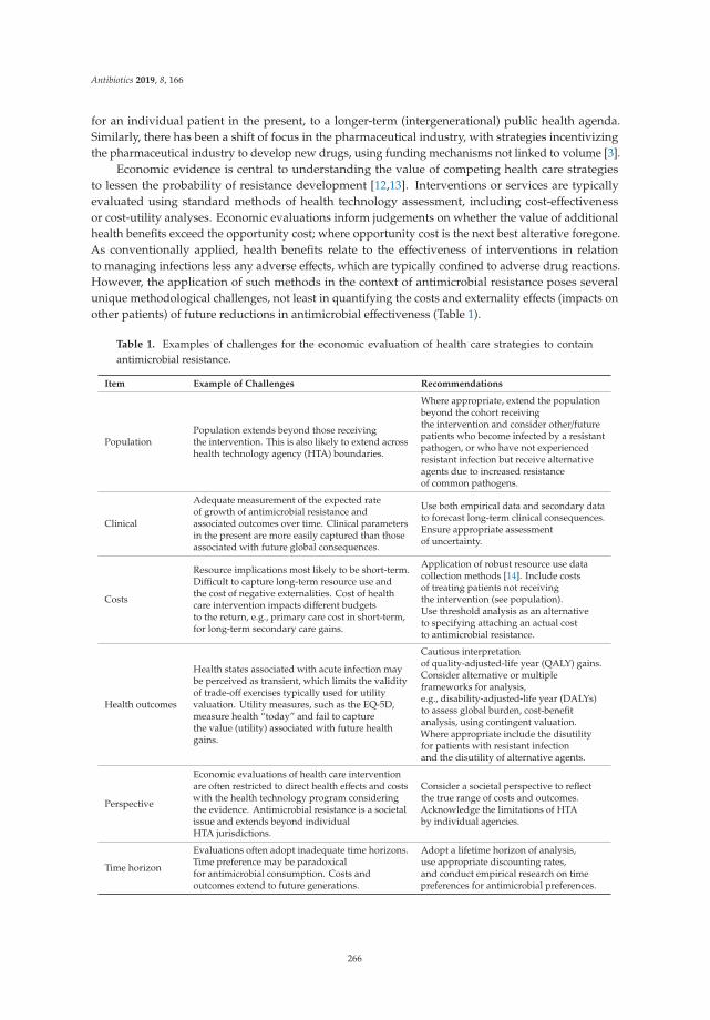
Antibiotics 2019, 8, 166
for an individual patient in the present, to a longer-term (intergenerational) public health agenda.Similarly, there has been a shift of focus in the pharmaceutical industry, with strategies incentivizingthe pharmaceutical industry to develop new drugs, using funding mechanisms not linked to volume [3].
Economic evidence is central to understanding the value of competing health care strategiesto lessen the probability of resistance development [12,13]. Interventions or services are typicallyevaluated using standard methods of health technology assessment, including cost-effectivenessor cost-utility analyses. Economic evaluations inform judgements on whether the value of additionalhealth benefits exceed the opportunity cost; where opportunity cost is the next best alterative foregone.As conventionally applied, health benefits relate to the effectiveness of interventions in relationto managing infections less any adverse effects, which are typically confined to adverse drug reactions.However, the application of such methods in the context of antimicrobial resistance poses severalunique methodological challenges, not least in quantifying the costs and externality effects (impacts onother patients) of future reductions in antimicrobial effectiveness (Table 1).
Table 1. Examples of challenges for the economic evaluation of health care strategies to containantimicrobial resistance.
Item Example of Challenges Recommendations
PopulationPopulation extends beyond those receivingthe intervention. This is also likely to extend acrosshealth technology agency (HTA) boundaries.
Where appropriate, extend the populationbeyond the cohort receivingthe intervention and consider other/futurepatients who become infected by a resistantpathogen, or who have not experiencedresistant infection but receive alternativeagents due to increased resistanceof common pathogens.
Clinical
Adequate measurement of the expected rateof growth of antimicrobial resistance andassociated outcomes over time. Clinical parametersin the present are more easily captured than thoseassociated with future global consequences.
Use both empirical data and secondary datato forecast long-term clinical consequences.Ensure appropriate assessmentof uncertainty.
Costs
Resource implications most likely to be short-term.Difficult to capture long-term resource use andthe cost of negative externalities. Cost of healthcare intervention impacts different budgetsto the return, e.g., primary care cost in short-term,for long-term secondary care gains.
Application of robust resource use datacollection methods [14]. Include costsof treating patients not receivingthe intervention (see population).Use threshold analysis as an alternativeto specifying attaching an actual costto antimicrobial resistance.
Health outcomes
Health states associated with acute infection maybe perceived as transient, which limits the validityof trade-off exercises typically used for utilityvaluation. Utility measures, such as the EQ-5D,measure health “today” and fail to capturethe value (utility) associated with future healthgains.
Cautious interpretationof quality-adjusted-life year (QALY) gains.Consider alternative or multipleframeworks for analysis,e.g., disability-adjusted-life year (DALYs)to assess global burden, cost-benefitanalysis, using contingent valuation.Where appropriate include the disutilityfor patients with resistant infectionand the disutility of alternative agents.
Perspective
Economic evaluations of health care interventionare often restricted to direct health effects and costswith the health technology program consideringthe evidence. Antimicrobial resistance is a societalissue and extends beyond individualHTA jurisdictions.
Consider a societal perspective to reflectthe true range of costs and outcomes.Acknowledge the limitations of HTAby individual agencies.
Time horizon
Evaluations often adopt inadequate time horizons.Time preference may be paradoxicalfor antimicrobial consumption. Costs andoutcomes extend to future generations.
Adopt a lifetime horizon of analysis,use appropriate discounting rates,and conduct empirical research on timepreferences for antimicrobial preferences.
266
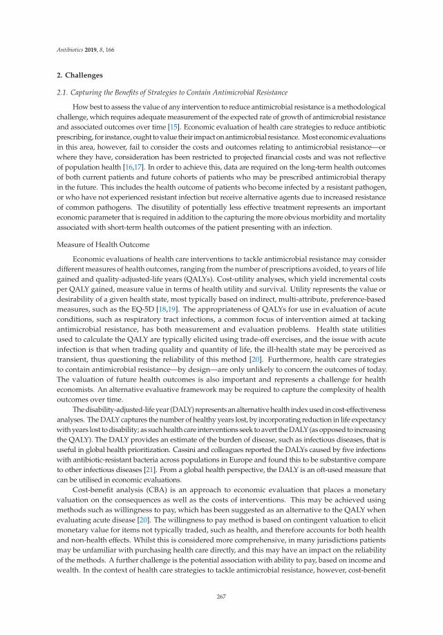
Antibiotics 2019, 8, 166
2. Challenges
2.1. Capturing the Benefits of Strategies to Contain Antimicrobial Resistance
How best to assess the value of any intervention to reduce antimicrobial resistance is a methodologicalchallenge, which requires adequate measurement of the expected rate of growth of antimicrobial resistanceand associated outcomes over time [15]. Economic evaluation of health care strategies to reduce antibioticprescribing, for instance, ought to value their impact on antimicrobial resistance. Most economic evaluationsin this area, however, fail to consider the costs and outcomes relating to antimicrobial resistance—orwhere they have, consideration has been restricted to projected financial costs and was not reflectiveof population health [16,17]. In order to achieve this, data are required on the long-term health outcomesof both current patients and future cohorts of patients who may be prescribed antimicrobial therapyin the future. This includes the health outcome of patients who become infected by a resistant pathogen,or who have not experienced resistant infection but receive alternative agents due to increased resistanceof common pathogens. The disutility of potentially less effective treatment represents an importanteconomic parameter that is required in addition to the capturing the more obvious morbidity and mortalityassociated with short-term health outcomes of the patient presenting with an infection.
Measure of Health Outcome
Economic evaluations of health care interventions to tackle antimicrobial resistance may considerdifferent measures of health outcomes, ranging from the number of prescriptions avoided, to years of lifegained and quality-adjusted-life years (QALYs). Cost-utility analyses, which yield incremental costsper QALY gained, measure value in terms of health utility and survival. Utility represents the value ordesirability of a given health state, most typically based on indirect, multi-attribute, preference-basedmeasures, such as the EQ-5D [18,19]. The appropriateness of QALYs for use in evaluation of acuteconditions, such as respiratory tract infections, a common focus of intervention aimed at tackingantimicrobial resistance, has both measurement and evaluation problems. Health state utilitiesused to calculate the QALY are typically elicited using trade-off exercises, and the issue with acuteinfection is that when trading quality and quantity of life, the ill-health state may be perceived astransient, thus questioning the reliability of this method [20]. Furthermore, health care strategiesto contain antimicrobial resistance—by design—are only unlikely to concern the outcomes of today.The valuation of future health outcomes is also important and represents a challenge for healtheconomists. An alternative evaluative framework may be required to capture the complexity of healthoutcomes over time.
The disability-adjusted-life year (DALY) represents an alternative health index used in cost-effectivenessanalyses. The DALY captures the number of healthy years lost, by incorporating reduction in life expectancywith years lost to disability; as such health care interventions seek to avert the DALY (as opposed to increasingthe QALY). The DALY provides an estimate of the burden of disease, such as infectious diseases, that isuseful in global health prioritization. Cassini and colleagues reported the DALYs caused by five infectionswith antibiotic-resistant bacteria across populations in Europe and found this to be substantive compareto other infectious diseases [21]. From a global health perspective, the DALY is an oft-used measure thatcan be utilised in economic evaluations.
Cost-benefit analysis (CBA) is an approach to economic evaluation that places a monetaryvaluation on the consequences as well as the costs of interventions. This may be achieved usingmethods such as willingness to pay, which has been suggested as an alternative to the QALY whenevaluating acute disease [20]. The willingness to pay method is based on contingent valuation to elicitmonetary value for items not typically traded, such as health, and therefore accounts for both healthand non-health effects. Whilst this is considered more comprehensive, in many jurisdictions patientsmay be unfamiliar with purchasing health care directly, and this may have an impact on the reliabilityof the methods. A further challenge is the potential association with ability to pay, based on income andwealth. In the context of health care strategies to tackle antimicrobial resistance, however, cost-benefit
267
Antibiotics 2019, 8, 166
analysis offers a feasible method for capturing the cost of the negative externalities, on a global scale,to represent the true value future benefits. Where applied with robust formative work, there is potentialfor this method to be used to elicit individuals’ willingness to pay to avoid an illness (resistant infection)or obtain the benefits of a future treatment.
2.2. Perspective
Prior to conducting any economic valuation, the perspective needs to be defined [22]. The perspectiveof the valuation, e.g., whether it is conducted from the point of view of the patient, health care payer,or society, will determine the costs and outcomes that need to be included in the analysis. In the UK,the National Institute for Health and Care Excellence (NICE) recommends the perspective on outcomesshould be all direct health effects, whether for patients or other people; and the perspective adopted oncosts within its Technology Appraisal programme should be that of the National Health Service (NHS) andpersonal and social services (PSS) [23]. Benefits are the assigned general population’s valuation of healthoutcome (obtained from surveys), and costs include the cost of treatment and associated health serviceresource use (e.g., GP visits, hospitalisation, etc.). Whilst this provides a reference case for comparisonof different health technologies, such Technology Appraisals consider individual patients or cohortsof patients, whereas antimicrobial resistance should consider populations. Evaluation of containmentstrategies may necessitate a broader perspective, and evidence in other areas suggests that assessmentof the same strategy from different perspectives can arrive at different conclusions [24].
The broadest perspective adopted in economic evaluation is the societal perspective. This shouldreflect the full range of social opportunity costs associated with different interventions [22], for example,productivity losses due to patients’ inability to work. However, there is a paucity of evidence onthe long-term indirect impact of antimicrobial resistance on costs and outcomes [25], and this representsa significant challenge for health economists evaluating containment strategies. Economic evaluationswith restrictive perspectives have potential to overestimate the value of interventions or services.Where the perspective for costs is limited to hospital or health service, indirect costs to current and futurepatients, such as working days lost due to resistant infection, or treatment toxicity, fail to be captured.
To fully assess the cost-effectiveness of strategies to contain antimicrobial resistance and to avoidthe inefficient allocation of scarce health service resources, decision makers require economicevaluations that incorporate costs and outcomes beyond the patient and hospital and considerthe full ramification of antimicrobial resistance. Defining the “society” then becomes a challenge withinitself, as antimicrobial resistance impacts populations that extends across jurisdictions. Adoption of aglobal multiagency perspective represents new territory and a limitation of conventional healthtechnology assessment (HTA) and health-economic approaches. In the absence of long-term data,economists have taken a pragmatic approach; for example, our evaluation of C-reactive protein testingto guide antibiotic prescribing for lower respiratory tract infection in Wales, included a sensitivityanalysis of the cost of the long term global cost of antibiotic resistance [17]. A challenge for futureeconomic evaluation is to incorporate more robust measurement and modelling of longer-term indirectcosts and outcomes—that go beyond today’s cohorts of patients to consider the population of tomorrow.
2.3. Capturing the Long-Term Costs of Antimicrobial Resistance
The resource implications of antimicrobial resistance are most likely to accumulate in the long-term.Short-term, direct health effects may only include the effect of the intervention on the affected patient;however, longer-term effects on both the patient and others are more likely. It is therefore importantthat all costs are captured and incorporated into economic evaluations across adequate time-horizons.Whilst interventions to reduce unnecessary prescribing in a clinical setting may represent a shorttime horizon, e.g., prescribing outcomes and re-consultation over 28-days, the longer-term effectsof unnecessary antibiotic prescribing extend to current and future patients’ lifetimes [10,26]. Initially,resource use and costs associated with interventions that aim to contain resistance would typically becaptured using health records and/or patient reports in the short-term. Estimating long-term direct
268
Antibiotics 2019, 8, 166
and indirect cost and the cost consequences to others, however, may present more of a challenge.The cost impact has potential to go beyond the lifetime horizon of the patient and impact the populationof tomorrow, and in some situations the product life-cycle [27] may be more relevant than the life timeof the patients being treated.
Where long-term costs have been considered in existing economic evaluations, these have reliedon estimates of per-prescription costs of antimicrobial resistance based on rudimentary calculationsof the global costs of resistance divided by the annual number of prescriptions in each geographicregion [16]. Oppong et al. 2013 [16], reported three estimates for the cost of antibiotic resistance,based on the annual cost of resistance in the US ($55 billion) [28], the cost of multidrug resistancein the European Union (EU) (€1.5 billion) [29], and the cost of global resistance over a 35-year period(US$2.8 trillion annually) [5]. This method assumes that antibiotic prescribing is the main causeof resistance, but whilst more robust estimates are required, it is widely acknowledged that this wouldrequire complex modelling methods [15]. Robust estimates of other long-term health conditions, suchas increased risk of Clostridioides difficile infection, also require consideration. Indirect costs are alsooften difficult to estimate, as they may bias against people who are not in employment and mayfluctuate over time.
A further challenge is that both direct and indirect costs may be attributable to different systemsand payers at different time points. For example, even when restricting analysis to direct health servicecosts, the initial outlay for point of care C-reactive protein testing to avoid unnecessary antibioticprescribing may be borne by a local health service provider; however, the longer-term cost savings arelikely to be at a secondary level. Data collection should not be restricted to a single source, such asprimary care databases, but should utilise appropriate methods (e.g., self-report, hospital episode data)to collect comprehensive data reflective of all costs incurred [14].
2.4. Time Preference
An individual’s time preference represents the extent to which they are willing to trade betweenshort-term costs and/or benefits and long-term costs and/or benefits, attributable with a health-relatedbehaviour, such as the consumption of antimicrobials. The time preference rate (discount rate)quantifies the difference between the perceived value of future outcomes relative to more immediateones. Individuals with lower time preference rates have a higher value for future utility and thereforediscount the future less; and conversely, individuals with higher time preference rates have a lowervalue for future utility and so require greater future reward for their behaviour. Time preferencerates for health are typically between 3–6% per annum [30]. In the UK a consistent societal discountrate of 3.5% per annum is applied as recommended in guidance issued by HM Treasury on howto appraise policies, programs, and projects [31]. Variation in the choice of discount rate has a markedeffect on studies with long time horizons and is therefore an important consideration for evaluationsof strategies aimed to contain resistance—an issue defined by long-term impacts. Given the futurevalue of antibiotics, a lower (zero or perhaps even negative in some circumstances) discount rate maybe more relevant for health outcomes [32]. This issue also applies to vaccination, where differentialdiscounting in model-based cost-effectiveness evaluations is being explored [33]. Empirical evidenceon time preference rates can be estimated using stated preference techniques that rely on hypotheticalscenarios [34]. Participants are typically required to imagine a health state and choose between futureoutcomes related to that hypothetical health state; this method would generate empirical evidence onindividuals time preferences to inform future economic evaluations. A further challenge that will needto be addressed is that in the context of antimicrobial resistance, the future health state to be valued isnot that of the respondent but is more likely to be the future health of others. Sensitive analysis usinga range of discount rates is recommended.
269
Antibiotics 2019, 8, 166
3. Conclusions
Ascertaining the most clinically and cost-effective interventions and services to reduceantimicrobial resistance will require a multifaceted approach, from incentivizing pharmaceuticalcompanies to co-production of patient-centred acceptable interventions that maximise the utility of bothpatients and society. Economic evidence is central to understanding the value of competing strategiesto lessen the probability of resistance development, although the application of such methods posesseveral methodological challenges. When assessment and interpreting the results of existing economicevaluations of health care interventions in the context of antimicrobial resistance, careful considerationis required, of the perspective, costs, and externality effects (impacts on other patients) of futurereductions in antimicrobial effectiveness. We have presented just some of the challenges of accuratelyestimating the cost-effectiveness of interventions to tackle antimicrobial resistance. Further research isrequired to capture the effects of antimicrobial resistance in economic evaluations to reflect the valueof preserving effective medicines for future use. This requires appropriate consideration of the widerexternalities and methodological approaches which differ from standard “reference” case of NICE andother HTA organizations.
Author Contributions: Conceptualization, E.A.F.H. and D.A.H.; Writing—Original draft preparation, E.A.F.H.;Writing—Review and editing, D.A.H.
Funding: This research received no external funding.
Acknowledgments: The authors acknowledge discussion of these issues at the Welsh Health Economics Group(WHEG) Meeting October 2018.
Conflicts of Interest: The authors declare no conflict of interest.
References
1. Coates, A.; Hu, Y.; Bax, R.; Page, C. The future challenges facing the development of new antimicrobial drugs.Nat. Rev. Drug Discov. 2002, 1, 895. [CrossRef] [PubMed]
2. World Health Organization. Antimicrobial Resistance: Global Report on Surveillance; World Health Organization:Geneva, Switzerland, 2014.
3. HM Government. Tackling Antimicrobial Resistance 2019–2024. The UK’s Five-Year National Action Plan.Published 24 January 2019. Available online: https://assets.publishing.service.gov.uk/government/uploads/system/uploads/attachment_data/file/784894/UK_AMR_5_year_national_action_plan.pdf (accessed on15 August 2019).
4. CDC. Antibiotic Resistance Threats in the United States 2013. Available online: https://www.cdc.gov/drugresistance/biggest_threats.html (accessed on 14 September 2019).
5. O’Neill, J. Tackling Drug-Resistant Infections Globally: Final Report and Recommendations. 2016.Available online: https://amr-review.org/sites/default/files/160525_Final%20paper_with%20cover.pdf(accessed on 26 June 2018).
6. National Institute of Health and Care Excellence (NICE) Guidelines on Antimicrobial Stewardship.Quality Standard [QS121]. Available online: https://www.nice.org.uk/guidance/qs121 (accessed on 13 March 2018).
7. Currie, C.J.; Berni, E.; Jenkins-Jones, S.; Poole, C.D.; Ouwens, M.; Driessen, S.; de Voogd, H.; Butler, C.C.;Morgan, C.L. Antibiotic treatment failure in four common infections in UK primary care 1991–2012:Longitudinal analysis. BMJ 2014, 349, g5493. [CrossRef] [PubMed]
8. Llor, C.; Bjerrum, L. Antimicrobial resistance: Risk associated with antibiotic overuse and initiatives to reducethe problem. Ther. Adv. Drug Saf. 2014, 5, 229–241. [CrossRef] [PubMed]
9. Costelloe, C.; Metcalfe, C.; Lovering, A.; Mant, D.; Hay, A.D. Effect of antibiotic prescribing in primarycare on antimicrobial resistance in individual patients: Systematic review and meta-analysis. BMJ 2010,340, c2096. [CrossRef] [PubMed]
10. Bell, B.G.; Schellevis, F.; Stobberingh, E.; Goossens, H.; Pringle, M. A systematic review and meta-analysisof the effects of antibiotic consumption on antibiotic resistance. BMC Infect. Dis. 2014, 14, 13. [CrossRef][PubMed]
270
Antibiotics 2019, 8, 166
11. Owens, R.C., Jr.; Donskey, C.J.; Gaynes, R.P.; Loo, V.G.; Muto, C.A. Antimicrobial-associated risk factorsfor Clostridium difficile infection. Clin. Infect. Dis. 2008, 46, S19–S31. [CrossRef] [PubMed]
12. Megiddo, I.; Drabik, D.; Bedford, T.; Morton, A.; Wesseler, J.; Laxminarayan, R. Investing in antibioticsto alleviate future catastrophic outcomes: What is the value of having an effective antibiotic to mitigatepandemic influenza? Health Econ. 2019, 28, 556–571. [CrossRef] [PubMed]
13. Attema, A.E.; Lugnér, A.K.; Feenstra, T.L. Investment in antiviral drugs: A real options approach. Health Econ.2010, 19, 1240–1254. [CrossRef] [PubMed]
14. Franklin, M.; Thorn, J. Self-reported and routinely collected electronic healthcare resource-use datafor trial-based economic evaluations: The current state of play in England and considerations for the future.BMC Med. Res. Methodol. 2019, 19, 8. [CrossRef]
15. Rothery, C.; Woods, B.; Schmitt, L.; Claxton, K.; Palmer, S.; Sculpher, M. Framework for Value Assessmentof New Antimicrobials; Policy Research Unit in Economic Evaluations of Health & Care Interventions:New York, NY, USA, 2018; Available online: http://www.eepru.org.uk/wp-content/uploads/2017/11/eepru-report-amr-oct-2018-059.pdf (accessed on 19 November 2018).
16. Oppong, R.; Smith, R.D.; Little, P.; Verheij, T.; Butler, C.C.; Goossens, H.; Coenen, S.; Moore, M.; Coast, J.Cost effectiveness of amoxicillin for lower respiratory tract infections in primary care: An economic evaluationaccounting for the cost of antimicrobial resistance. Br. J. Gen. Pract. 2016, 66, e633–e639. [CrossRef]
17. Holmes, E.A.F.; Harris, S.D.; Hughes, A.; Craine, N.; Hughes, D.A. Cost-effectiveness analysis of the useof point of care C-reactive protein testing to reduce antibiotic prescribing in primary care. Antibiotics 2018,7, 106. [CrossRef] [PubMed]
18. Euroqol Group. EQ-5D. Available online: https://euroqol.org/ (accessed on 14 September 2019).19. Wisloff, T.; Hagen, G.; Hamidi, V.; Movik, E.; Klemp, M.; Olsen, J.A. Estimating QALY gains in applied
studies: A review of cost-utility analyses published in 2010. Pharmacoeconomics 2014, 32, 367–375. [CrossRef][PubMed]
20. Bala, M.V.; Zarkin, G.A. Are QALYs an appropriate measure for valuing morbidity in acute diseases?Health Econ. 2000, 9, 177–180. [CrossRef]
21. Cassini, A.; Högberg, L.D.; Plachouras, D.; Quattrocchi, A.; Hoxha, A.; Simonsen, G.S.; Colomb-Cotinat, M.;Kretzschmar, M.E.; Devleesschauwer, B.; Cecchini, M.; et al. Attributable deaths and disability-adjustedlife-years caused by infections with antibiotic-resistant bacteria in the EU and the European Economic Areain 2015: A population-level modelling analysis. Lancet Infect. Dis. 2019, 19, 56–66. [CrossRef]
22. Byford, S.; Raftery, J. Perspectives in economic evaluation. BMJ 1998, 316, 1529–1530. [CrossRef] [PubMed]23. National Institute for Health and Care Excellence. Guide to the Methods of Technology Appraisal 2013.
Process and Methods [PMG9]; National Institute for Health and Care Excellence: London, UK, 2013.24. Drummond, M.; Weatherly, H.; Ferguson, B. Economic evaluation of health interventions. BMJ 2008, 337, 1204.
[CrossRef] [PubMed]25. Eliopoulos, G.M.; Cosgrove, S.E.; Carmeli, Y. The impact of antimicrobial resistance on health and economic
outcomes. Clin. Infect. Dis. 2003, 36, 1433–1437. [CrossRef]26. Wilcox, M.H.; Ahir, H.; Coia, J.E.; Dodgson, A.; Hopkins, S.; Llewelyn, M.J.; Settle, C.; Mclain-Smith, S.;
Marcella, S.W. Impact of recurrent Clostridium difficile infection: Hospitalization and patient quality of life.J. Antimicrob. Chemother. 2017, 72, 2647–2656. [CrossRef]
27. Hoyle, M. Accounting for the drug life cycle and future drug prices in cost-effectiveness analysis.Pharmacoeconomics 2011, 29, 1–15. [CrossRef]
28. Centers for Disease Control and Prevention. Antimicrobial Resistance: No Action Today, No Cure Tomorrow.Available online: http://www.cdc.gov/media/releases/2011/f0407_antimicrobialresistance.pdf (accessed on19 November 2018).
29. European Centre for Disease Prevention and Control, European Medicines Agency. The Bacterial Challenge:Time to React. Joint Technical Report. 2009. Available online: https://ecdc.europa.eu/sites/portal/files/media/en/publications/Publications/0909_TER_The_Bacterial_Challenge_Time_to_React.pdf (accessed on19 November 2018).
30. Cairns, J.A.; van der Pol, M.M. The estimation of marginal time preference in a UK-wide sample (TEMPUS)project. Health Technol. Assess. 2000, 4, i–iv. [CrossRef]
31. HM Treasury. The Green Book Appraisal and Evaluation in Central Government: Treasury Guidance;TSO: Norwich, UK, 2003.
271
Antibiotics 2019, 8, 166
32. Roope, L.S.; Tonkin-Crine, S.; Butler, C.C.; Crook, D.; Peto, T.; Peters, M.; Walker, A.S.; Wordsworth, S.Reducing demand for antibiotic prescriptions: Evidence from an online survey of the general public onthe interaction between preferences, beliefs and information, United Kingdom, 2015. Eurosurveillance 2018,23, 1700424. [CrossRef] [PubMed]
33. Jit, M.; Mibei, W. Discounting in the evaluation of the cost-effectiveness of a vaccination programme: A criticalreview. Vaccine 2015, 33, 3788–3794. [CrossRef] [PubMed]
34. Van der Pol, M.; Cairns, J. Estimating time preferences for health using discrete choice experiments.Soc. Sci. Med. 2001, 52, 1459–1470. [CrossRef]
© 2019 by the authors. Licensee MDPI, Basel, Switzerland. This article is an open accessarticle distributed under the terms and conditions of the Creative Commons Attribution(CC BY) license (http://creativecommons.org/licenses/by/4.0/).
272
MDPISt. Alban-Anlage 66
4052 BaselSwitzerland
Tel. +41 61 683 77 34Fax +41 61 302 89 18
www.mdpi.com
Antibiotics Editorial OfficeE-mail: [email protected]
www.mdpi.com/journal/antibiotics


MDPI St. Alban-Anlage 66 4052 Basel Switzerland
Tel: +41 61 683 77 34 Fax: +41 61 302 89 18
www.mdpi.com ISBN 978-3-03943-123-6