α-amidoamids as new replacements of antibiotics ... - mdpi
TRANSCRIPT
materials
Article
α-Amidoamids as New Replacements ofAntibiotics—Research on the Chosen K12, R2–R4E. coli Strains
Paweł Kowalczyk 1,* , Arleta Madej 2, Mateusz Szymczak 3 and Ryszard Ostaszewski 2,*1 Department of Animal Nutrition, The Kielanowski Institute of Animal Physiology and Nutrition,
Polish Academy of Sciences, Instytucka 3, 05-110 Jabłonna, Poland2 Institute of Organic Chemistry, Polish Academy of Sciences, Kasprzaka 44/52, 01-224 Warsaw, Poland;
[email protected] Department of Molecular Virology, Institute of Microbiology, Faculty of Biology, University of Warsaw,
Miecznikowa 1, 02-096 Warsaw, Poland; [email protected]* Correspondence: [email protected] (P.K.); [email protected] (R.O.)
Received: 9 October 2020; Accepted: 12 November 2020; Published: 16 November 2020�����������������
Abstract: A preliminary study of α-amidoamids as new potential antimicrobial drugs was performed.Special emphasis was placed on selection of structure of α-amidoamids with the highest biologicalactivity against different types of Gram-stained bacteria by lipopolysaccharide (LPS). Herein,Escherichia coli model strains K12 (without LPS in its structure) and R1–R4 (with different length LPS inits structure) were used. The presented work showed that the antibacterial activity of α-amidoamidsdepends on their structure and affects the LPS of bacteria. Moreover, the influence of various newlysynthesized α-amidoamids on bacteria possessing smooth and rought LPS and oxidative damageof plasmid DNA caused by all newly obtained compounds was indicated. The presented studiesclearly explain that α-amidoamids can be used as substitutes for antibiotics. The chemical andbiological activity of the analysed α-amidoamids was associated with short alkyl chain and differentisocyanides molecules in their structure such as: tetr-butyl isocyanide or 2,5-dimethoxybenzylisocyanide. The observed results are especially important in the case of the increasing resistance ofbacteria to various drugs and antibiotics.
Keywords: oxidative stress; Ugi reaction; peptidomimetics; Fpg-N-glycosylase/AP lyase DNA;LPS—lipopolysaccharide; alpha-amidoamides (AAAs)
1. Introduction
Peptides are essential for almost every physiological process in the cell. Small peptides inducedby endogenous factors such as hormones or exogenous e.g., active substances such as toxins havevarious biological activities [1–4].
Such molecules, called peptidomimetics, appear to be an excellent starting material for thedevelopment of new candidate drugs that can mimic the structure or activity of natural peptides.They contain appropriately chemically modified analogues synthesized on the basis of neuropeptidesand peptide hormones, showing species-specific and organ-selective, directed physiological action,which significantly increases their stability under metabolic conditions, but retains biologicalactivity [1–4]. The molecular goal of peptidomimetics is the interaction with receptor ligands,inhibitors of protein-protein interaction, and enzyme inhibitors. These compounds, compared tonormal peptides, show greater stability against proteolysis and bioavailability of the receptor inbiological processes [1–4]. Furthermore, the hydrophilic character of peptides limits their permeabilitythrough biological membranes. Methods for the synthesis of natural peptides are well known but the
Materials 2020, 13, 5169; doi:10.3390/ma13225169 www.mdpi.com/journal/materials
Materials 2020, 13, 5169 2 of 23
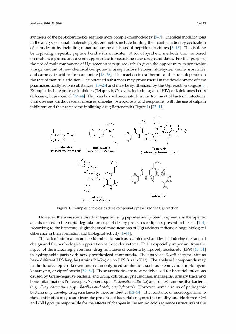
synthesis of the peptidomimetics requires more complex methodology [5–7]. Chemical modificationsin the analysis of small molecule peptidomimetics include limiting their conformation by cyclizationof peptides or by including unnatural amino acids and dipeptide substitutes [8–12]. This is doneby replacing a specific peptide bond with an isoster. A lot of synthetic methods that are basedon multistep procedures are not appropriate for searching new drug candidates. For this purpose,the use of multicomponent of Ugi reaction is required, which gives the opportunity to synthesizea huge amount of new chemical compounds, using various ketones, aldehydes, amine, isonitriles,and carboxylic acid to form an amide [13–26]. The reaction is exothermic and its rate depends onthe rate of isonitrile addition. The obtained substances may prove useful in the development of newpharmaceutically active substances [13–26] and may be synthesized by the Ugi reaction (Figure 1).Examples include protease inhibitors (Telaprevir, Crixivan, Indavir—against HIV) or kainic anesthetics(lidocaine, bupivacaine) [27–44]. They can be used successfully in the treatment of bacterial infections,viral diseases, cardiovascular diseases, diabetes, osteoporosis, and neoplasms, with the use of calpaininhibitors and the proteasome-inhibiting drug Bortezomib (Figure 1) [27–44].
Figure 1. Examples of biologic active compound synthetized via Ugi reaction.
However, there are some disadvantages to using peptides and protein fragments as therapeuticagents related to the rapid degradation of peptides by proteases or lipases present in the cell [1–4].According to the literature, slight chemical modifications of Ugi adducts indicate a huge biologicaldifference in their formation and biological activity [1–44].
The lack of information on peptidomimetics such as α-aminoacyl amides is hindering the rationaldesign and further biological application of these derivatives. This is especially important from theaspect of the increasingly common drug resistance of bacteria by lipopolysaccharide (LPS) [45–51]in hydrophobic parts with newly synthesized compounds. The analyzed E. coli bacterial strainshave different LPS lengths (strains R2–R4) or no LPS (strain K12). The analysed compounds may,in the future, replace known and commonly used antibiotics, such as bleomycin, streptomycin,kanamycin, or ciprofloxacin [52–54]. These antibiotics are now widely used for bacterial infectionscaused by Gram-negative bacteria (including coliforms, pneumoniae, meningitis, urinary tract, andbone inflammation; Proteus spp., Neisseria spp., Pasteurella multocida) and some Gram-positive bacteria.(e.g., Corynebacterium spp., Bacillus anthracis, staphylococci). However, some strains of pathogenicbacteria may develop drug resistance to these antibiotics [52–54]. The resistance of microorganisms tothese antibiotics may result from the presence of bacterial enzymes that modify and block free -OHand -NH groups responsible for the effects of changes in the amino acid sequence (structure) of the
Materials 2020, 13, 5169 3 of 23
ribosome with which the antibiotic cannot bind. Therefore, it is important to develop new substancessimilar to β-lactam antibiotics, which will be able to inhibit the biosynthesis of the bacterial cell wall.The bactericidal effect is due to the disruption of the synthesis of bacterial proteins, including thosethat make up the cell membrane [55–59].
The interaction of imines with isocyanates is mainly focused on the well-known multicomponentUgi reaction (MCR) [1]. This basic process involves a carboxylic acid group that attacks the intermediatenitrile ion, thus leading, after the Mumma rearrangement, to α-amidoamids. Aminoamids are commonin many molecules, such as peptides, proteins, lactams, and many synthetic polymers [59]. However,the direct reaction of imines and isocyanides is less studied in the literature. Reactions between reagentspossessing carbonyl, amine, and isocyanide group lead to α-amidoamids.
The new compounds are an alternative as potential substitutes for antibiotics due to theirspecific structure related to substituents. They can, thus, penetrate the membranes of bacterial cells,damaging the nucleic acid they contain. They usually consist of two functional groups—carbonyl andamines—linked by a single bond between carbon and nitrogen. The biological activity of α-amidoamidsis determined by means of a heterocyclic ring connected to a ketone or aldehyde group leading toimine formation via molecular cyclization.
The aim of our research was to check whether the interaction of Ugi reaction products depends onLPS length in Escherichia coli K-12 and R2–R4 strains that have different LPS in their outermost layer inthe antigen “O” region.
2. Materials and Methods
2.1. Microorganisms and Media
E. coli K-12, R1–R4 strains were received from Prof. Jolanta Łukasiewicz the Ludwik HirszfeldInstitute of Immunology and Experimental Therapy (Polish Academy of Sciences, Warsaw, Poland.Bacteria were cultivated in tryptic soy broth (TSB; Sigma-Aldrich, Saint Louis, MI, USA) liquid mediumand on agar plates containing TSB medium. N,N-Dimethylformamide (DMF) were purchased fromSigma Aldrich (CAS No. 68-12-2, Poznan, Poland), Lanes 1kb-ladder, and Quick Extend DNA ladder,(New England Biolabs, Ipswich, MA, USA).
2.2. Experimental Chemistry
NMR analyzes were performed according to the procedure described in the literature [59,60].
2.3. General Procedure for Synthesis of Compounds 1–20
The described individual substrates in the synthesis of α-amidoamides belong to the standardmethods of synthesizing this type of compounds and are presented unchanged in many publicationsof this type [48]. Standard syntheses of compounds from 5a to 5u are methodologically identical tothose presented in other publications [58]. Therefore, we present a general formula for their synthesis.In addition, examples of the synthesis of each of the analyzed α-amidoamides are also presented below.
White powder; mp. 78–124 ◦C; 1H NMR (400 MHz, CDCl3) δ ppm = 6.73–7.33 (4–12 H, m, Ph),4.85–7.02 (1–6 H, m, Ph + CH or CH2 or C3H3), 4.74–6.85 (1 H, s, m, br, NH or CH or C3 H3), 4.41–5.93(1–2 H, s,t,m, NH or CH or CH2), 4.17 (1–3 H, m,s, CH or CH2 or 2× CH3), 3.66–5.75 (1–6 H, m,s,or CH3, 2× CH3), 0.85 (1–6 H, m, s, CH2 or CH3), 1.57–1.95 (1–6 H, s,m CH or CH2 or CH3 or 2× CH3),0.85–3.62 (1–6 H, m,s, CH or CH2 or CH3 or 2×CH3); 13C NMR (100 MHz; CDCl3) δ ppm = 14.16–41.32,22.37–43.18, 22.71–55.22, 25.14–55.26, 25.30–63.46, 23.10–88.50, 24.56–114.02, 25.17–114.31, 37.10–114.31,40.78–114.05, 41.26–114.23, 41.62–126.95, 42.54–130.38, 42.84–158.81, 48.55–157.71, 55.28–170.69,56.35–172.98, 73.03–171.05, 114.05–173.13, 126.02–158.84, 126.98–170.67, 127.39–173.79; HRMS calcdfrom C29H34N2O3Na [M + Na]+: 481.2467 found: 481.2470 to C30H36N2O4Na [M + Na]+: 511.2573found: 511.2574.
Materials 2020, 13, 5169 4 of 23
Yellow powder or Pale yellow; mp. 92–93 ◦C; 1H NMR (400 MHz; CDCl3) δ ppm = 6.82–7.30(1–15 H, m,s Ph), 6.86 (1–5 H, s,m, Ph, or Ph + CH or NH), 4.52–7.09 (1–4 H, m, Ph), 4.46–7.05(1–4 H, m,s, NH or CH2), 4.19–6.86 (1–4 H, m, NH or CH2 or Ph or CH), 3.71–6.86 (1–6 H, s,2× CH3), 0.68–6.79 (1–3 H, m, CH3), 1.19–6.72 (1–5 H, m, CH or CH2), 1.40–6.33 (1–4 H, m,C2H4), 1.18–1.38 (1–31 H, s br, m, C15H31); 13C NMR (100 MHz; CDCl3) δ ppm = 13.72–22.49,22.43–30.42, 22.66–41.65, 24.73–42.72, 25.15–55.30, 29.88–114.25, 31.20–114.03, 39.01–114.02, 29.61–114.24,29.64–128.67, 29.68–128.93, 31.90–129.15, 34.04–130.35, 55.21–128,81, 56.20–128.66, 113.97–129.55,114.09–158.90, 127.26–129.53, 128.97–134.62, 129.55-, 130.47, 158.79–173.25, 158.87–169.74, 170.81–173.39,170.61–175.80, 160.45–177.43; HRMS calcd from C26H36N2O3Na [M + Na]+: 447.2624 found: 447.2611.toC32H32N2O4Na [M + Na]+: 541.2678 found: 541.2671.
2.3.1. Product 5a (AM 93)
White powder; mp. 104–105 ◦C; 1H NMR (400 MHz, CDCl3) δ (ppm) = 7.04–7.29 (9 H, m, Ph),6.61–6.78 (5 H, m, Ph + CH), 4.74 (1 H, s, NH), 4.41 (2 H, s, CH2), 4.17 (2 H, m, CH2), 3.66–3.72 (6 H,m, 2× CH3), 1.81 (2 H, s, CH2), 1.57 (1 H, s, CH), 0.85 (6 H, s, 2× CH3); 13C NMR (100 MHz; CDCl3)δ ppm = 22.2, 22.7, 25.1, 37.3, 42.7, 51.1, 55.2, 57.9, 113.7, 113.9, 126.7, 128.5, 128.8, 129.0, 129.2, 129.8,130.4, 136.2, 158.8, 170.7, 173.8; HRMS calcd for C29H34N2O4Na [M + Na]+: 497.2416 found: 497.2410.
2.3.2. Product 5b (AM 70)
White powder; mp. 106–107 ◦C; 1H NMR (400 MHz;CDCl3) δ (ppm) = 7.19 (4 H, m, Ph),7.01 (5 H,m, Ph), 6.77 (5 H,m, Ph, CH), 6.71 (1 H, s, CH), 4.94 (1 H, t, NH), 4.44 (2 H, s; CH2), 4.16–4.20(2 H, m, CH2), 3.73 (6 H, s, CH3), 3.55 (2 H, d, CH2), 1.79–1.83 (1 H, m, CH), 1.37–1.43 (2 H, m, CH2),0.75–0.79 (6 H, m, CH3); 13C NMR (100 MHz; CDCl3) δ ppm = 22.4, 22.7, 25.1, 37.0, 41.3, 42.8, 48.2, 55.3,56.5, 114.0, 114.2, 126.9, 127.3, 128.6, 128.8, 129.0, 129.2, 130.3, 134.6, 158.9, 170.5, 173.4; HRMS calcd forC30H36N2O4Na [M + Na]+: 511.2573 found: 511.2574.
2.3.3. Product 5c (AM 121)
White powder; mp. 78–79 ◦C; 1H NMR (400 MHz; CDCl3) δ (ppm) = 6.74–7.26 (14 H m, PhH + CH),5.01 (1 H, m, NH), 4.43 (2 H, m, CH2) 4.23–4.30 (2 H, m, CH2), 3.78 (6 H, s, CH3), 2.86–2.91 (2 H, m,CH2), 2.53–2.58 (2 H, m, CH2), 1.81–1.85 (1 H, m, CH), 1.40–1.44 (2 H, m, CH2), 0.81–0.91 (6 H, m,CH3); 13C NMR (100 MHz; CDCl3) δ ppm = 22.4, 22.7, 25.1, 31.4, 35.7, 37.0, 42.8, 48.0, 55.3, 114.0, 114.1,126.2, 127.2, 128.4, 129.0, 129.3, 130.4, 140.9, 158.8, 170.7, 174.6 ppm; HRMS calcd for C31H38N2O4Na[M + Na]+: 525.2729 found: 525.2717.
2.3.4. Product 5d (AM 84)
White powder; mp. 123–124 ◦C; 1H NMR (400 MHz; CDCl3) δ ppm = 6.73–7.08 (m, 9 H; Ar, CH),4.85–5.03 (m, 1 H; -NH), 4.44 (s, 2 H, CH2), 3.97–4.32 (m, 2 H; CH2), 3.71 (s, 6 H; CH3), 1.95 (s, 3 H;CH3), 1.66–1.84 (m, 1 H, CH), 1.29–1.52 (m, 2 H; CH2), 0.78 (t, 6 H; CH3);13C NMR (100 MHz; CDCl3) δ(ppm) = 22.4, 22.7, 25.2, 37.2, 42.8, 48.7, 55.2, 56.1, 114.0, 114.1, 127.3, 129.0, 129.3, 130.4, 158.8, 158.9,170.7, 173.0; HRMS calcd for C24H32N2O4Na [M + Na]+: 435.2260 found: 435.2260.
2.3.5. Product 5e (AM 119)
White powder; mp. 94–95 ◦C; 1H NMR (400 MHz; CDCl3) δ (ppm) = 6.84–7.33 (9 H, m, PhH),6.35 (1 H, m, CH), 4.94–4.97 (1 H, m, NH), 4.50–4.51 (2 H, m, CH2), 3.78 (3 H, s, CH3), 3.57–3.62 (2 H,m, CH2), 0.79–1.82 (20 H, m, C6H11 + C4H9); 13C NMR (100 MHz; CDCl3) δ (ppm) = 22.4, 22.7, 24.7,25.2, 25.3, 32.7, 32.8, 37.0, 41.4, 47.9, 48.1, 55.3, 56.5, 114.2, 127.0, 127.2, 128.6, 128.7, 129.2, 129.5, 129.9,134.6, 158.9, 169.6, 173.3; HRMS calcd for C31H38N2O4Na [M + Na]+: 525.2729 found: 525.2720.
Materials 2020, 13, 5169 5 of 23
2.3.6. Product 5f (AM 116)
Yellow powder; mp. 92–93 ◦C; 1H NMR (400 MHz; CDCl3) δ (ppm) = 6.95–7.19 (4 H, m, Ph),6.86 (1 H, s, CH), 6.73–6.77 (4 H, m, PhH), 4.94–4.97 (1 H, m, NH), 4.43–4.44 (2 H, m, CH2), 4.19–4.21(2 H, m, CH2), 3.71 (6 H, s, 2× CH3), 2.09–2.25 (3 H, m, CH3), 1.74–1.80 (1 H, m, CH), 1.39–1.45 (4 H, m,C2H4), 1.18 (31 H, s br, C15H31); 13C NMR (100 MHz; CDCl3) δ (ppm) = 14.1, 22.4, 22.7, 22.8, 29.3, 29.4,29.5, 29.6, 29.7, 31.9, 34.0, 55.2, 114.0, 114.1, 127.3, 128.0, 129.5, 130.5, 158.8, 158.9, 170.8, 175.8, 177.4;HRMS calcd for C27H30N2O4Na [M + Na]+: 469.2103 found: 469. 2092.
2.3.7. Product 5g (AM 115)
White powder; mp. 115–116 ◦C; 1H NMR (400 MHz; CDCl3) δ (ppm) = 7.13–7.17 (4 H, m, Ph),6.97–7.02 (5 H, m, Ph), 6.65–6.77 (5 H, m, PhH + CH) 4.97–4.99 (1 H, s br; NH), 4.42–4.44 (2 H, m, CH2),4.15–4.17 (2 H, m, CH2), 3.69 (6 H, s, 2 × CH3), 3.52–3.54 (2 H, m, CH2), 1.18–1.26 (q, 3 H; CH3);13CNMR (100 MHz; CDCl3) δ (ppm) = 14.2, 42.9, 48.0, 53.4, 55.3, 114.0, 114.3, 127.0, 127.1, 128.7, 129.0, 129.3,130.4, 134.6, 159.0, 171.0, 173.1; HRMS calcd for C28H32N2O4Na [M + Na]+: 483.2260 found: 483.2092.
2.3.8. Product 5h (AM 164)
Pale yellow oil; 1 H NMR (400 MHz; CDCl3) δ (ppm) = 7.17–6.96 (9 H, m, PhH), 6.77–6.73 (5 H, m,PhH + CH), 4.83–4.79 (1 H, m, NH), 4.46 (2 H, s, CH2), 4.20–4.15 (2 H, m, CH2), 3.71 (6 H, s, CH3),3.54–3.53 (2 H, m, CH2), 1.89–1.85 (1 H, m, CH), 1.48–1.46 (1 H, m, CH), 1.19–1.17 (2 H, m, CH2),0.78–0.74 (3 H, m, CH3); 13C NMR (100 MHz; CDCl3) δ (ppm) = 13.8, 19.7, 30.4, 41.3, 42.8, 48.3, 55.3,58.2, 114.0, 114.2, 126.9, 127.3, 128.6, 128.8, 129.0, 129.3, 130.3, 134.6, 158.9, 158.9, 170.4, 173.3; HRMScalcd for C29 H34N2O4Na [M + Na]+: 497.2416 found: 475.2416.
2.3.9. Product 5i (AM 165)
Pale yellow oil; 1H NMR (400 MHz; CDCl3) δ (ppm) = 7.19–6.93 (10 H, m, PhH), 6.77–6.70 (3 H,m, PhH + CH), 4.57–4.40 (2 H, m, CH2) 4.29–4.24 (2 H, m, CH2), 4.14–4.09 (1 H, m, NH), 3.71 (3 H,m, CH3), 3.53 (2 H, s, CH2), 2.43–2.37 (1 H, m, CH), 0,86 (2 H, m, CH3), 0.68–0.68 (3 H, m, CH3); 13CNMR (100 MHz; CDCl3) δ (ppm) = 19.0, 19.9, 27.1, 41.6, 42.7, 55.2, 55.3, 114.0, 114.2, 126.9, 127.7, 128.6,128.9, 129.1, 130.1, 130.3, 134.6, 158.9, 170.0, 173.7; HRMS calcd for C29H34N2O4Na [M + Na]+: 497.2416found: 475.2416.
2.3.10. Product 5j (AM 107)
White powder; mp. 86–87 ◦C; 1H NMR (400 MHz; CDCl3) δ (ppm) = 7.07–7.23 (12 H,m, PhH + C2H2), 6.63–6.85 (6 H, m, C3H3), 5.93 (1 H, s, NH), 5.75 (1 H, s, CH), 4.62 (1 H, m,CH), 4.40 (1 H, m, CH), 3.67(6 H, 2 s, 2× CH3), 3.62 (2 H, m, CH2); 13C NMR (100 MHz; CDCl3)δ (ppm) = 41.3, 43.2, 55.2, 55.3, 63.5, 88.5, 113.7, 113.9, 114.0, 114.1, 126.8, 126. 127.5, 128.5, 128.6,128.7, 128.9, 129.0, 129.7, 134.7, 134.9, 135.0, 158.6, 158.7, 158.8, 159.7, 169.4, 172.8; HRMS calcd forC29H34N2O3Na [M + Na]+: 481.2467 found: 481.2470.
2.3.11. Product 5k (AM 91)
White powder; mp. 82–83 ◦C; 1H NMR (400 MHz; CDCl3) δ (ppm) = 7.03–7.34 (12 H, m, PhH),6.82–6.86 (3 H, m, PhH + CH), 5.06–5.10 (1 H, m, NH), 4.60 (1 H, s, CH), 4.23–4.27 (2 H, m, CH2),3.79 (1 H, s, CH3), 3.54–3.65 (2 H, m, CH2), 1.86–1.93 (1 H, m, CH), 1.43–1.47 (2 H, m, CH2), 0.82–0.91(6 H, m, 2× CH3); 13C NMR (100 MHz; CDCl3) δ (ppm) = 21.7, 22.4, 22.8, 23.1, 24.6, 25.2, 37.1, 40.8,41.3, 41.6, 42.5, 42.8, 48.5, 55.3, 56.4, 73.0, 114.0, 126.0, 127.0, 127.4, 128.7, 128.8, 129.0, 130.3, 134.5, 137.5,158.91, 170.4, 173.5; HRMS calcd for C28H40N2O3Na [M + Na]+: 475.2937 found: 475.2934.

Materials 2020, 13, 5169 6 of 23
2.3.12. Product 5l (AM 163)
Pale yellow oil; 1H NMR (400 MHz; CDCl3) δ (ppm) = 7.20–7.16 (3 H, m, PhH), 7.10–7.08 (2 H,m, PhH), 6.88–6.85 (1 H, m, CH),4.87–4.83 (1 H, m, NH), 4.27–4.13 (2 H, m, CH), 3.71 (3 H, s, CH3),3.62 (2 H, s, CH2), 3.15–3.10 (2 H, m, CH2),1.80–1.73 (1 H, m, CH), 1.59–1.52 (1 H, m, CH), 1.40–1.47(2 H, m, CH2), 1.18–1.13 (4 H, m, C2H4), 0.85–0.79 (10 H, m, CH3 + CH); 13C NMR (100MHz; CDCl3)δ (ppm) = 13.6, 20.3, 22.4, 22.8, 24.8, 32.0, 36.7, 41.1, 42.8, 45.6, 55.2, 55.9, 113.9, 126.9, 128.7, 128.7,129.0, 129.3, 130.5, 134.8, 158.8, 171.4, 172.8; HRMS calcd for C26H36N2O3Na [M + Na]+: 447.2624found: 447.2624.
2.3.13. Product 5m (AM 162)
Pale yellow oil; 1H NMR (400 MHz;CDCl3) δ (ppm) = 7.19–7.17 (3 H, m, PhH), 7.09–7.05 (4 H, m,PhH), 6.74–6.74 (2 H, m, PhH), 4.34–4.17 (2 H, m, CH), 3.96–4.00 (1 H, m, NH), 3.71 (3 H, m, CH3), 3.65(2 H, s, CH2), 2.34–2.29 (1 H, m, CH), 1.58–1.52 (2 H, m, CH2), 1.18–1.14 (1 H, m, CH2) 1.02–1.00 (6 H,m, CH3), 0.89–0.88 (6 H, m, CH3); 13C NMR (100 MHz; CDCl3) δ (ppm) = 20.5, 21.0, 22.3, 25.5, 39.6,42.6, 42.9, 50.7, 55.2, 59.5, 113.9, 126.9, 128.4, 128.8, 128.8, 129.5, 130.9, 134.8, 158.7, 172.5, 172.8; HRMScalcd for C26H36N2O3Na [M + Na]+: 447.2624 found: 447.2618.
2.3.14. Product 5n (AM 182)
Pale yellow oil; 1H NMR (400 MHz; CDCl3) δ (ppm) = 7.19–7.14 (4 H, m, PhH), 7.04–6.96 (5 H,m, PhH), 6.83–6.81 (1 H, m, CH), 6.74–6.72 (2 H, m, PhH), 6.37–6.33 (2 H, m, PhH), 5.00–4.96 (1 H, sbr, NH), 4.42 (2 H, s, CH2), 4.22–4.20 (2 H, m, CH2), 3.74–3.69 (9 H, m, 3 × CH3), 3.51–3.50 (2 H, m,CH2), 1.76–1.72 (1 H, m, CH), 1.39–1.35 (2 H, m, CH2), 0.77–0.73 (6 H, m, 2× CH3); 13C NMR (100 MHz;CDCl3) δ (ppm) = 22.5, 22.7, 25.0, 37.0, 38.9, 41.3, 47.8, 55.2, 55.3, 55.4, 98.5, 103.3, 114.2, 118.7, 126.8,127.1, 128.6, 128.8, 129.5, 130.3, 134.7, 158.6, 158.8, 160.4, 170.2, 173.2; HRMS calcd for C31H38N2O5Na[M + Na]+: 541.2678 found: 541.2671.
2.3.15. Product 5o (AM 111)
Pale yellow oil; 1H NMR (400 MHz;CDCl3) δ ppm = 7.15 (5 H, m, PhH), 6.72–7.03 (5 H, m, PhH+ CH), 4.74–5.00 (1 H, m, NH), 4.04–4.32 (2 H, m, CH2), 3.71 (3 H, m, CH3), 3.55–3.65 (2 H, m, CH2),2.95–3.26 (2 H, m, CH2), 1.06–1.89 (12 H, m, C10H10, CH2), 0.81 (9 H, m, CH3); 13C NMR (100 MHz;CDCl3) δ (ppm) = 13.8, 22.3, 22.4, 22.7, 24.7, 26.7, 29.9, 31.2, 36.7, 41.0, 42.7, 45.7, 55.1, 55.8, 98.5, 103.1,114.5, 118.9, 126.8, 127.6, 128.7, 128.8, 129.5, 130.3, 134.7, 158.6, 158.8, 160.4, 170.2, 173.2; HRMS calcdfor C32H32N2O4Na [M + Na]: 475.2934 found: 475.2937.
2.3.16. Product 5p (AM 114)
Pale yellow oil; 1H NMR (400 MHz;CDCl3) δ (ppm) = 6.82–7.30 (15 H, m, PhH + CH), 5.02–4.99(1 H, m, NH), 4.52 (2 H, s, CH2), 4.30–4.35 (2 H, m, CH2), 3.80 (3 H, s, CH3), 3.61–3.63 (2 H, m, CH2),1.87–1.91 (1 H, m, CH), 1.45–1.49 (2 H, m, CH2), 0.82–0.86 (6 H, m, 2 CH3); 13C NMR (100 MHz; CDCl3)δ (ppm) = 22.4, 22.7, 25.2, 43.4, 55.3, 114.2, 127.3, 127.7, 128.6, 128.7, 145.5, 158.9, 170.7; HRMS calcd forC28H32N2O2Na [M + Na]+: 451.2361 found: 451.2358
2.3.17. Product 5r (AM 170)
Pale yellow oil; 1H NMR (400 MHz; CDCl3) δ (ppm) = 7.24–7.15 (4 H, m, PhH), 7.11–7.10 (2 H, m,PhH), 7.05–7.03 (2 H, m, PhH), 6.86 (1 H, s, CH), 6.81–6.79 (2 H, m, CH2), 5.00–4.97 (1 H, s br, NH), 4.46(2 H, s, CH2), 4.15–4.10 (2 H, s, CH2), 3.83–3.80 (2 H, m, CH2), 3.73 (3 H, s, CH3), 3.62 (2 H, m, CH2),1.82–1.78 (1 H, m, CH), 1.43–1.36 (2 H, m, CH2), 1.22–1.18 (3 H, m, CH3), 0.79–0.75 (6 H, m, 2× CH3);13C NMR (100 MHz; CDCl3) δ (ppm) = 14.1, 22.3, 22.7, 25.1 36.8, 41.1, 41.2, 48.3, 55.3, 56.2, 61.3, 114.20,126.7, 127.3, 128.6, 128.8, 129.7, 134.6, 158.8, 169.7, 173.5; HRMS calcd for C26H34N2O5Na [M + Na]+:477.2365 found: 477.2362
Materials 2020, 13, 5169 7 of 23
2.3.18. Product 15s (AM 178)
Pale yellow oil; 1H NMR (400 MHz;CDCl3) δ (ppm) = 7.24–7.15 (4 H, m, PhH), 7.11–7.10 (2 H, m,PhH), 7.05–7.03 (2 H, m, PhH), 6.86 (1 H, s, CH), 6.81–6.79 (2 H, m, CH2), 5.00–4.97 (1 H, s br, NH), 4.46(2 H, s, CH2), 4.15–4.10 (2 H, s, CH2), 3.83–3.80 (2 H, m, CH2), 3.73 (3 H, s, CH3), 3.62 (2 H, m, CH2),1.82–1.78 (1 H, m, CH), 1.43–1.36 (2 H, m, CH2), 1.22–1.18 (3 H, m, CH3), 0.79–0.75 (6 H, m, 2× CH3);13C NMR (100 MHz; CDCl3) δ (ppm) = 14.1, 22.3, 22.7, 25.1 36.8, 41.1, 41.2, 48.3, 55.3, 56.2, 61.3, 114.20,126.7, 127.1, 128.6, 128.6, 129.5, 134.6, 158.9, 169.7, 173.2; HRMS calcd for C28H40N2O3Na [M + Na]+:475.2937 found: 475.2932.
2.3.19. Product 5t (AM 171)
Pale yellow oil; 1H NMR (400 MHz; CDCl3) δ ppm = 7.22–7.19 (4 H, m, PhH), 7.09–7.01 (3 H, m,PhH), 6.82–6.80 (2 H, m, PhH), 6.35 (1 H, s, CH), 4.91 (1 H, s br, NH), 4.45 (2 H, s, CH2), 3.74 (3 H,s, CH3), 3.58–3.56 (2 H, m, CH2), 3.08–3.06 (2 H, m, CH2), 1.80–1.76 (1 H, m, CH), 1.34–1.22 (6 H, m,C3H6), 0.85–0.74 (9 H, m, 3× CH3); 13C NMR (100 MHz; CDCl3) δ (ppm) = 13.7, 20.0, 22.4, 22.8, 25.1,31.5, 37.0, 39.0, 41.3, 48.1, 55.3, 114.2, 127.0, 127.2, 128.7, 128.8, 129.4, 134.6, 158.9, 170.6, 173,4; HRMScalcd for C26H36N2O3Na [M + Na]+: 447.2624 found: 447.2611.
2.3.20. Product 5u (AM 184)
Pale yellow oil; 1H NMR (400 MHz; CDCl3) δ (ppm) = 7.29–7.09 (5 H, m, PhH), 7.04–7.02 (2 H, m,PhH), 6.82–6.77 (2 H, m, PhH), 6.18 (1 H, s, CH), 4.87–4.83 (1 H, s br, NH), 4.44–4.43 (2 H, m, CH2),3.73 (3 H, s, CH3), 3.57–3.56 (2 H, m, CH2), 1.78–1.69 (1 H, m, CH), 1.38–1.17 (10 H, m, 3× CH3 + CH),0.87–0.74 (8 H, m, 2× CH3 + CH2); 13C NMR (100 MHz; CDCl3) δ (ppm) = 22.4, 22.7, 25.2, 28.6, 36.9,41.4, 47.7, 51.0, 55.3, 56.8, 114.2, 127.0, 127.2, 128.6, 128.7, 129.6, 134.7, 158.9, 169.7, 173.2; HRMS calcdfor C26H36N2O3Na [M + Na]+: 447.2624 found: 447.2614.
2.4. Determination of MIC and MBC
Determination of MIC and MBC for selected strains are described in detail in the literature [31,45,51].(An example of the analysis are presented in Figures S1 and S2 in Supplementary Materials.)
2.4.1. Interaction of Bleomycine, Kanamycine, Streptomycine, with Bacterial Plasmid DNA Isolatedfrom K12 and R1–R4 Strains
Plasmid DNA was isolated from the used bacterial influences using Labjot kits according to themanufacturer’s protocols. Next, the obtained DNA plasmid was digested with antibiotics such asbleomycin with final working concentration (2 units/mL), streptomycin (50 µg/mL), ciprofloxacin(10 µg/mL), and kanamycin (50 µg/mL) as per the manufacturer’s instructions. All antibiotics werepurchased from Sigma (Poznan, Poland).
2.4.2. Determination of MIC and MBC after Antibiotics Treatment
Analyzed strains will be treated with antibiotics at identical concentrations as the analyzedcompounds using the MIC and MBC tests according to the procedures described in previouspublications [31,45,51].
2.5. Interaction of the Plasmid DNA from K12 and R4 Strains with α-Amidoamids
Strains K12 and R4 were selected for further studies on the basis of the MIC (Minimum inhibitoryconcentration) and MBC (Minimum bactericidal concentration) analysis. The method of interaction ofplasmid DNA with the analyzed α-amidoamides has been described in detail in the literature [48].
Materials 2020, 13, 5169 8 of 23
2.6. Repair and Cleavage of Oxidative DNA Damage Adducts by Fpg Protein in Bacterial Cells
Analysis of repair and cleavage by Fpg protein were performed on (i) plasmid DNA isolatedfrom different E. coli mutants of R1–R4 and K12 by alkaline lysis and final purification according tothe manufacturer’s protocol (Labjot, Warsaw, Poland). The plasmids were then incubated with theanalysed compounds.
Subsequently, DNA was digested with Fpg enzymes individually as described in detail in theliterature [50].
2.7. Statistical Analysis
The obtained data were used in the statistical analysis (Statistica package ver. 5.0) and presented asstandard error of the mean (SE). Parametric analyzes were performed using Student’s t-test p < 0.05 *,p < 0.01 ** and p < 0.001 ***.
3. Results
3.1. Chemistry
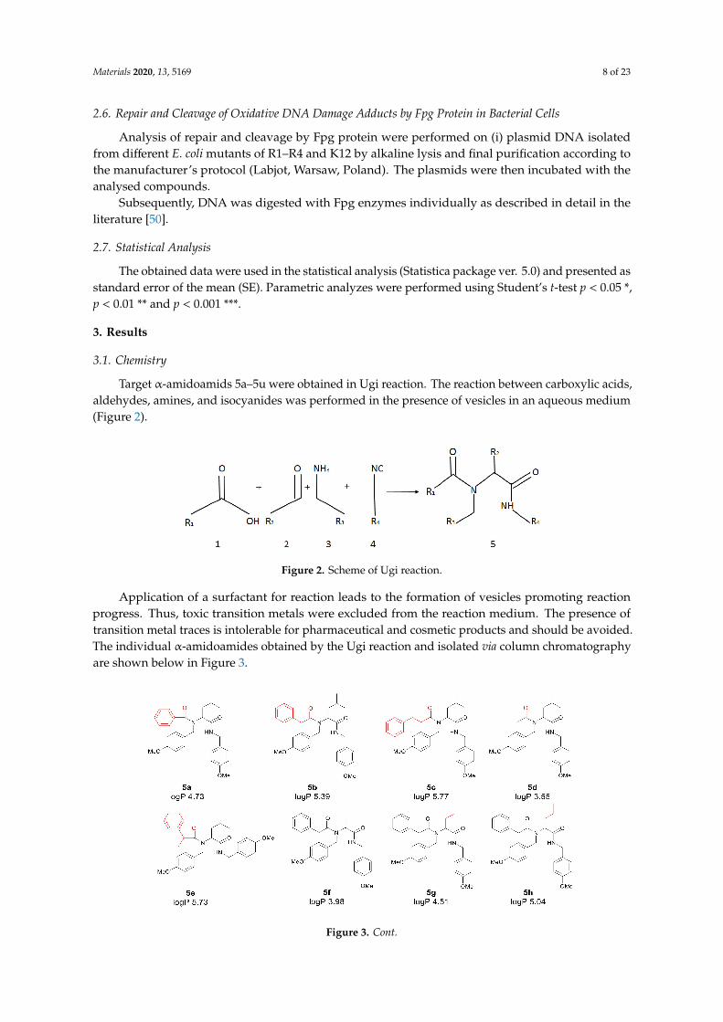
Target α-amidoamids 5a–5u were obtained in Ugi reaction. The reaction between carboxylic acids,aldehydes, amines, and isocyanides was performed in the presence of vesicles in an aqueous medium(Figure 2).
Figure 2. Scheme of Ugi reaction.
Application of a surfactant for reaction leads to the formation of vesicles promoting reactionprogress. Thus, toxic transition metals were excluded from the reaction medium. The presence oftransition metal traces is intolerable for pharmaceutical and cosmetic products and should be avoided.The individual α-amidoamides obtained by the Ugi reaction and isolated via column chromatographyare shown below in Figure 3.
Figure 3. Cont.
Materials 2020, 13, 5169 9 of 23
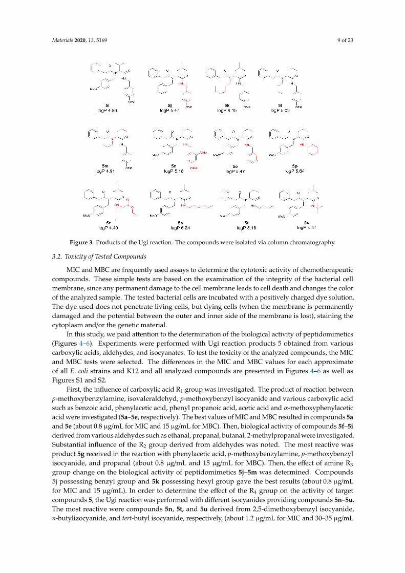
Figure 3. Products of the Ugi reaction. The compounds were isolated via column chromatography.
3.2. Toxicity of Tested Compounds
MIC and MBC are frequently used assays to determine the cytotoxic activity of chemotherapeuticcompounds. These simple tests are based on the examination of the integrity of the bacterial cellmembrane, since any permanent damage to the cell membrane leads to cell death and changes the colorof the analyzed sample. The tested bacterial cells are incubated with a positively charged dye solution.The dye used does not penetrate living cells, but dying cells (when the membrane is permanentlydamaged and the potential between the outer and inner side of the membrane is lost), staining thecytoplasm and/or the genetic material.
In this study, we paid attention to the determination of the biological activity of peptidomimetics(Figures 4–6). Experiments were performed with Ugi reaction products 5 obtained from variouscarboxylic acids, aldehydes, and isocyanates. To test the toxicity of the analyzed compounds, the MICand MBC tests were selected. The differences in the MIC and MBC values for each approximateof all E. coli strains and K12 and all analyzed compounds are presented in Figures 4–6 as well asFigures S1 and S2.
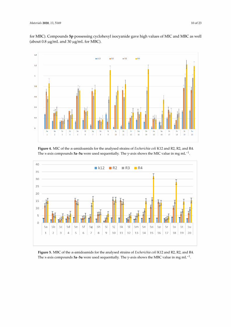
First, the influence of carboxylic acid R1 group was investigated. The product of reaction betweenp-methoxybenzylamine, isovaleraldehyd, p-methoxybenzyl isocyanide and various carboxylic acidsuch as benzoic acid, phenylacetic acid, phenyl propanoic acid, acetic acid and α-methoxyphenylaceticacid were investigated (5a–5e, respectively). The best values of MIC and MBC resulted in compounds 5aand 5e (about 0.8 µg/mL for MIC and 15 µg/mL for MBC). Then, biological activity of compounds 5f–5iderived from various aldehydes such as ethanal, propanal, butanal, 2-methylpropanal were investigated.Substantial influence of the R2 group derived from aldehydes was noted. The most reactive wasproduct 5g received in the reaction with phenylacetic acid, p-methoxybenzylamine, p-methoxybenzylisocyanide, and propanal (about 0.8 µg/mL and 15 µg/mL for MBC). Then, the effect of amine R3
group change on the biological activity of peptidomimetics 5j–5m was determined. Compounds5j possessing benzyl group and 5k possessing hexyl group gave the best results (about 0.8 µg/mLfor MIC and 15 µg/mL). In order to determine the effect of the R4 group on the activity of targetcompounds 5, the Ugi reaction was performed with different isocyanides providing compounds 5n–5u.The most reactive were compounds 5n, 5t, and 5u derived from 2,5-dimethoxybenzyl isocyanide,n-butylizocyanide, and tert-butyl isocyanide, respectively, (about 1.2 µg/mL for MIC and 30–35 µg/mL
Materials 2020, 13, 5169 10 of 23
for MBC). Compounds 5p possessing cyclohexyl isocyanide gave high values of MIC and MBC as well(about 0.8 µg/mL and 30 µg/mL for MBC).
Figure 4. MIC of the α-amidoamids for the analysed strains of Escherichia coli K12 and R2, R2, and R4.The x-axis compounds 5a–5u were used sequentially. The y-axis shows the MIC value in mg mL−1.
Figure 5. MBC of the α-amidoamids for the analysed strains of Escherichia coli K12 and R2, R2, and R4.The x-axis compounds 5a–5u were used sequentially. The y-axis shows the MBC value in mg mL−1.
Materials 2020, 13, 5169 11 of 23
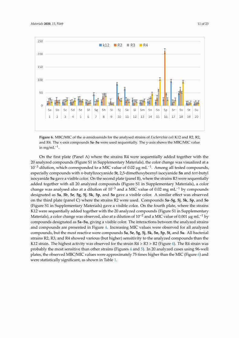
Figure 6. MBC/MIC of the α-amidoamids for the analysed strains of Escherichia coli K12 and R2, R2,and R4. The x-axis compounds 5a–5u were used sequentially. The y-axis shows the MBC/MIC valuein mg/mL−1.
On the first plate (Panel A) where the strains R4 were sequentially added together with the20 analyzed compounds (Figure S1 in Supplementary Materials), the color change was visualized at a10−3 dilution, which corresponded to a MIC value of 0.02 µg mL−1. Among all tested compounds,especially compounds with n-butylizocyanide 5t, 2,5-dimethoxybeznyl isocyanide 5n and tert-butylisocyanide 5u gave a visible color. On the second plate (panel B), where the strains R3 were sequentiallyadded together with all 20 analyzed compounds (Figure S1 in Supplementary Materials), a colorchange was analysed also at a dilution of 10−3 and a MIC value of 0.02 mg mL−1 by compoundsdesignated as 5a, 5b, 5e, 5g, 5j, 5k, 5p, and 5o gave a visible color. A similar effect was observedon the third plate (panel C) where the strains R2 were used. Compounds 5a–5g, 5j, 5k, 5p, and 5o(Figure S1 in Supplementary Materials) gave a visible color. On the fourth plate, where the strainsK12 were sequentially added together with the 20 analyzed compounds (Figure S1 in SupplementaryMaterials), a color change was observed, also at a dilution of 10−2 and a MIC value of 0.001 µg mL−1 bycompounds designated as 5a–5u, giving a visible color. The interactions between the analyzed strainsand compounds are presented in Figure 4. Increasing MIC values were observed for all analyzedcompounds, but the most reactive were compounds 5a, 5e, 5g, 5j, 5k, 5n, 5p, 5t, and 5u. All bacterialstrains R2, R3, and R4 showed various (but higher) sensitivity to the analyzed compounds than theK12 strain. The highest activity was observed for the strain R4 > R3 > R2 (Figure 4). The R4 strain wasprobably the most sensitive than other strains (Figures 4 and 5). In 20 analyzed cases using 96-wellplates, the observed MBC/MIC values were approximately 75 times higher than the MIC (Figure 6) andwere statistically significant, as shown in Table 1.
Materials 2020, 13, 5169 12 of 23
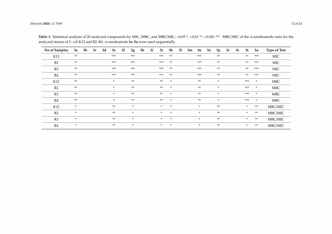
Table 1. Statistical analysis of 20 analyzed compounds by MIC, MBC, and MBC/MIC; <0.05 *, <0.01 **, <0.001 ***. MBC/MIC of the α-amidoamids ratio for theanalyzed strains of E. coli K12 and R2–R4. α-amidoamids 5a–5u were used sequentially.
No of Samples 5a 5b 5c 5d 5e 5f 5g 5h 5i 5j 5k 5l 5m 5n 5o 5p 5r 5s 5t 5u Type of Test
K12 ** *** *** *** ** *** ** ** *** MIC
R2 ** *** *** *** ** *** ** ** *** MIC
R3 ** *** *** *** ** *** ** ** *** MIC
R4 ** *** *** *** ** *** ** ** *** MIC
K12 ** * ** ** * ** * *** * MBC
R2 ** * ** ** * ** * *** * MBC
R3 ** * ** ** * ** * *** * MBC
R4 ** * ** ** * ** * *** * MBC
K12 * ** * * * * ** * ** MBC/MIC
R2 * ** * * * * ** * ** MBC/MIC
R3 * ** * * * * ** * ** MBC/MIC
R4 * ** * * * * ** * ** MBC/MIC
Materials 2020, 13, 5169 13 of 23
3.3. Modification of Plasmid DNA Isolated from E. coli R2–R4 Strains with Tested α-Amidoamids
The increase in toxicity of all the analyzed compounds (high MIC values) depends on thefunctional groups R1, R2, R3, and R4 of α-amidoamides 5 used. We expected a similar effect in plasmidDNA modified with α-amidoamides, in which plasmid damage should be most visible. The testedcompounds showed very strong disturbances in the structure and conformation of DNA, even whendigesting the Fpg protein in vitro during the 24-h experiment.
The analysis of the MIC and MBC values obtained from all the analyzed strains (K12 and R2–R4)prompted us to isolate plasmid DNA from them and treat them with Fpg protein from the group ofrepair glycosases. Fpg protein digestion of the obtained plasmids isolated from both the control (K12)and the analyzed (R2–R4) strain after treatment with α-amidamides, regardless of different functionalgroups, showed clearly visible damage in the topological changes of plasmid DNA forms; covalentlyclosed circle (ccc), linear form, open form (oc), and fuzzy bands (see Figure S3 Supplementary Materials).
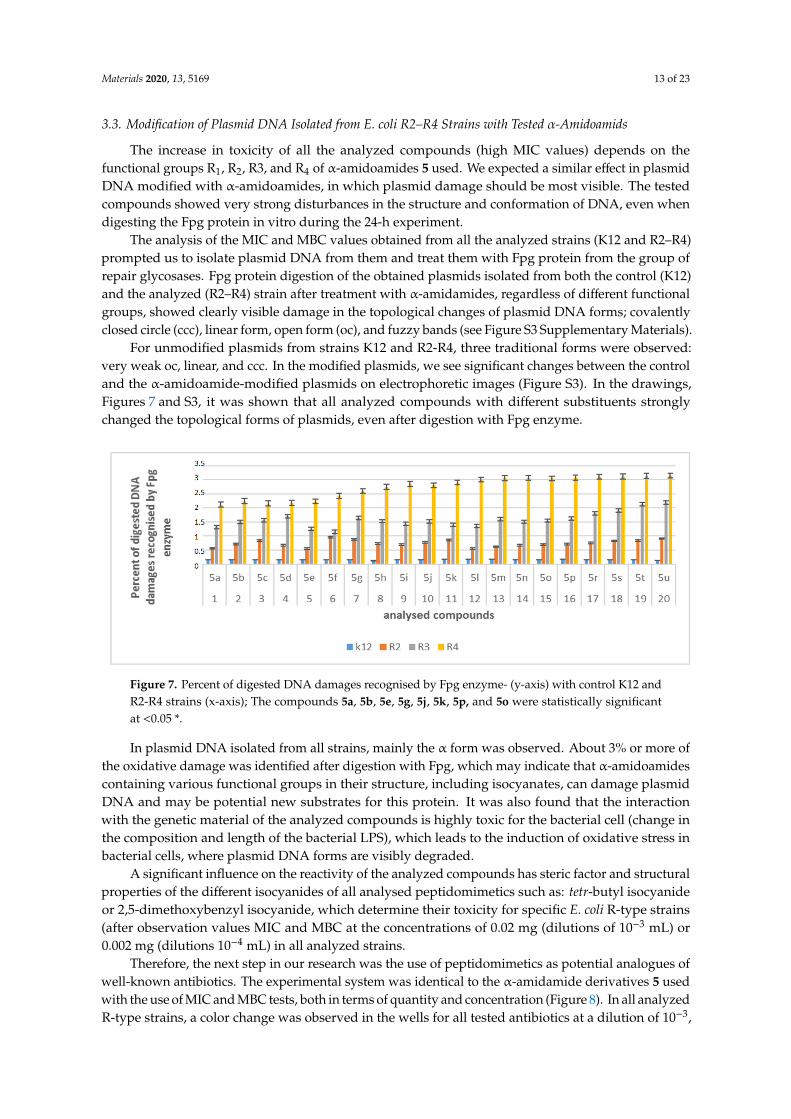
For unmodified plasmids from strains K12 and R2-R4, three traditional forms were observed:very weak oc, linear, and ccc. In the modified plasmids, we see significant changes between the controland the α-amidoamide-modified plasmids on electrophoretic images (Figure S3). In the drawings,Figures 7 and S3, it was shown that all analyzed compounds with different substituents stronglychanged the topological forms of plasmids, even after digestion with Fpg enzyme.
Figure 7. Percent of digested DNA damages recognised by Fpg enzyme- (y-axis) with control K12 andR2-R4 strains (x-axis); The compounds 5a, 5b, 5e, 5g, 5j, 5k, 5p, and 5o were statistically significantat <0.05 *.
In plasmid DNA isolated from all strains, mainly the α form was observed. About 3% or more ofthe oxidative damage was identified after digestion with Fpg, which may indicate that α-amidoamidescontaining various functional groups in their structure, including isocyanates, can damage plasmidDNA and may be potential new substrates for this protein. It was also found that the interactionwith the genetic material of the analyzed compounds is highly toxic for the bacterial cell (change inthe composition and length of the bacterial LPS), which leads to the induction of oxidative stress inbacterial cells, where plasmid DNA forms are visibly degraded.
A significant influence on the reactivity of the analyzed compounds has steric factor and structuralproperties of the different isocyanides of all analysed peptidomimetics such as: tetr-butyl isocyanideor 2,5-dimethoxybenzyl isocyanide, which determine their toxicity for specific E. coli R-type strains(after observation values MIC and MBC at the concentrations of 0.02 mg (dilutions of 10−3 mL) or0.002 mg (dilutions 10−4 mL) in all analyzed strains.
Therefore, the next step in our research was the use of peptidomimetics as potential analogues ofwell-known antibiotics. The experimental system was identical to the α-amidamide derivatives 5 usedwith the use of MIC and MBC tests, both in terms of quantity and concentration (Figure 8). In all analyzedR-type strains, a color change was observed in the wells for all tested antibiotics at a dilution of 10−3,
Materials 2020, 13, 5169 14 of 23
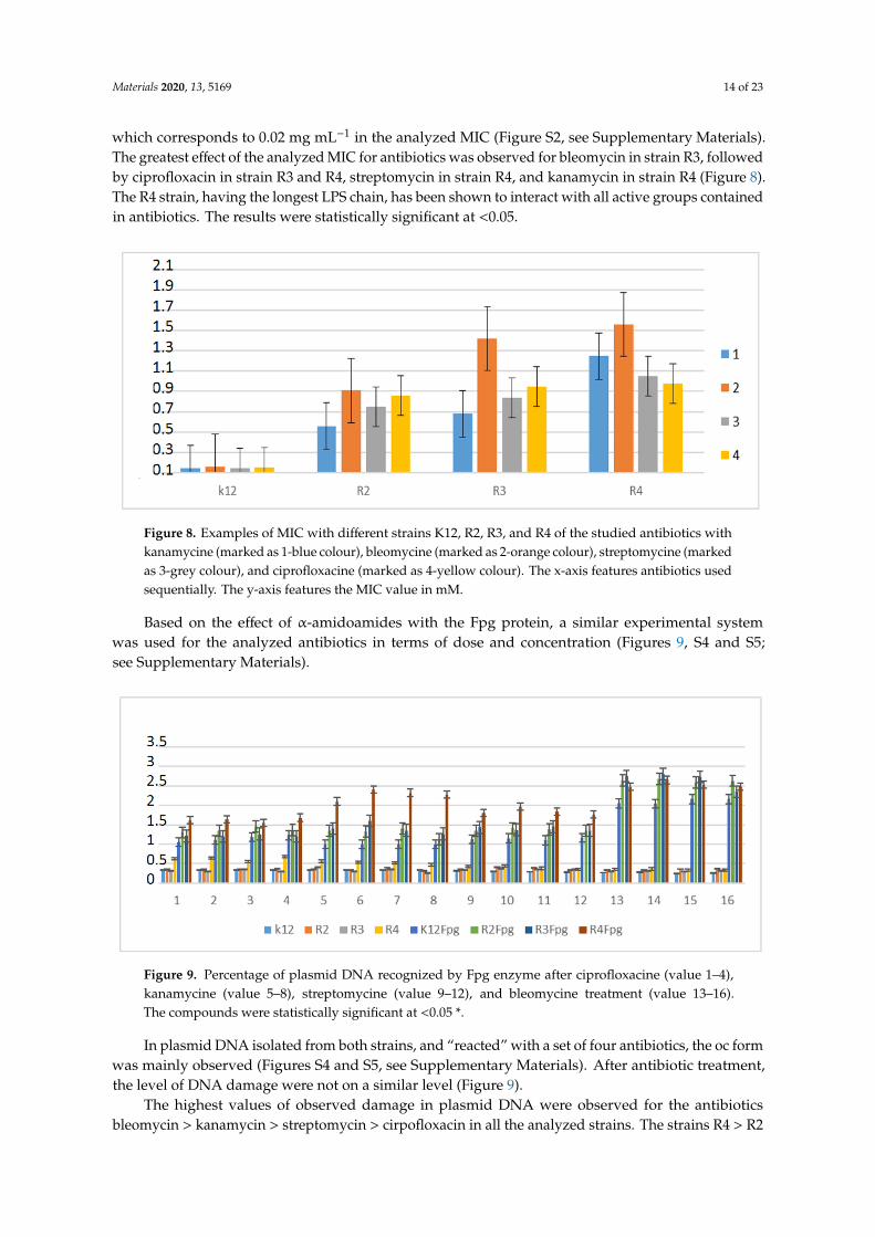
which corresponds to 0.02 mg mL−1 in the analyzed MIC (Figure S2, see Supplementary Materials).The greatest effect of the analyzed MIC for antibiotics was observed for bleomycin in strain R3, followedby ciprofloxacin in strain R3 and R4, streptomycin in strain R4, and kanamycin in strain R4 (Figure 8).The R4 strain, having the longest LPS chain, has been shown to interact with all active groups containedin antibiotics. The results were statistically significant at <0.05.
Figure 8. Examples of MIC with different strains K12, R2, R3, and R4 of the studied antibiotics withkanamycine (marked as 1-blue colour), bleomycine (marked as 2-orange colour), streptomycine (markedas 3-grey colour), and ciprofloxacine (marked as 4-yellow colour). The x-axis features antibiotics usedsequentially. The y-axis features the MIC value in mM.
Based on the effect of α-amidoamides with the Fpg protein, a similar experimental systemwas used for the analyzed antibiotics in terms of dose and concentration (Figures 9, S4 and S5;see Supplementary Materials).
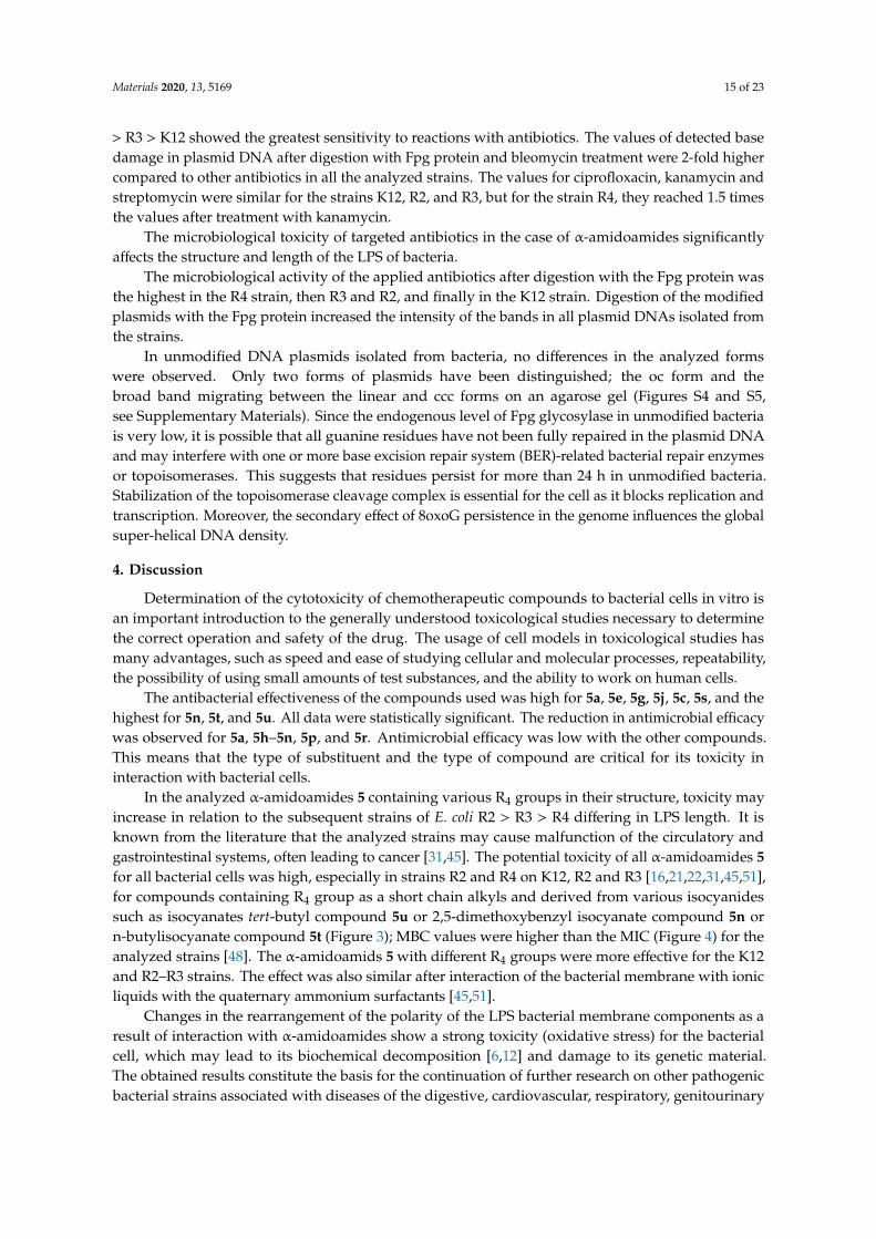
Figure 9. Percentage of plasmid DNA recognized by Fpg enzyme after ciprofloxacine (value 1–4),kanamycine (value 5–8), streptomycine (value 9–12), and bleomycine treatment (value 13–16).The compounds were statistically significant at <0.05 *.
In plasmid DNA isolated from both strains, and “reacted” with a set of four antibiotics, the oc formwas mainly observed (Figures S4 and S5, see Supplementary Materials). After antibiotic treatment,the level of DNA damage were not on a similar level (Figure 9).
The highest values of observed damage in plasmid DNA were observed for the antibioticsbleomycin > kanamycin > streptomycin > cirpofloxacin in all the analyzed strains. The strains R4 > R2
Materials 2020, 13, 5169 15 of 23
> R3 > K12 showed the greatest sensitivity to reactions with antibiotics. The values of detected basedamage in plasmid DNA after digestion with Fpg protein and bleomycin treatment were 2-fold highercompared to other antibiotics in all the analyzed strains. The values for ciprofloxacin, kanamycin andstreptomycin were similar for the strains K12, R2, and R3, but for the strain R4, they reached 1.5 timesthe values after treatment with kanamycin.
The microbiological toxicity of targeted antibiotics in the case of α-amidoamides significantlyaffects the structure and length of the LPS of bacteria.
The microbiological activity of the applied antibiotics after digestion with the Fpg protein wasthe highest in the R4 strain, then R3 and R2, and finally in the K12 strain. Digestion of the modifiedplasmids with the Fpg protein increased the intensity of the bands in all plasmid DNAs isolated fromthe strains.
In unmodified DNA plasmids isolated from bacteria, no differences in the analyzed formswere observed. Only two forms of plasmids have been distinguished; the oc form and thebroad band migrating between the linear and ccc forms on an agarose gel (Figures S4 and S5,see Supplementary Materials). Since the endogenous level of Fpg glycosylase in unmodified bacteriais very low, it is possible that all guanine residues have not been fully repaired in the plasmid DNAand may interfere with one or more base excision repair system (BER)-related bacterial repair enzymesor topoisomerases. This suggests that residues persist for more than 24 h in unmodified bacteria.Stabilization of the topoisomerase cleavage complex is essential for the cell as it blocks replication andtranscription. Moreover, the secondary effect of 8oxoG persistence in the genome influences the globalsuper-helical DNA density.
4. Discussion
Determination of the cytotoxicity of chemotherapeutic compounds to bacterial cells in vitro isan important introduction to the generally understood toxicological studies necessary to determinethe correct operation and safety of the drug. The usage of cell models in toxicological studies hasmany advantages, such as speed and ease of studying cellular and molecular processes, repeatability,the possibility of using small amounts of test substances, and the ability to work on human cells.
The antibacterial effectiveness of the compounds used was high for 5a, 5e, 5g, 5j, 5c, 5s, and thehighest for 5n, 5t, and 5u. All data were statistically significant. The reduction in antimicrobial efficacywas observed for 5a, 5h–5n, 5p, and 5r. Antimicrobial efficacy was low with the other compounds.This means that the type of substituent and the type of compound are critical for its toxicity ininteraction with bacterial cells.
In the analyzed α-amidoamides 5 containing various R4 groups in their structure, toxicity mayincrease in relation to the subsequent strains of E. coli R2 > R3 > R4 differing in LPS length. It isknown from the literature that the analyzed strains may cause malfunction of the circulatory andgastrointestinal systems, often leading to cancer [31,45]. The potential toxicity of all α-amidoamides 5for all bacterial cells was high, especially in strains R2 and R4 on K12, R2 and R3 [16,21,22,31,45,51],for compounds containing R4 group as a short chain alkyls and derived from various isocyanidessuch as isocyanates tert-butyl compound 5u or 2,5-dimethoxybenzyl isocyanate compound 5n orn-butylisocyanate compound 5t (Figure 3); MBC values were higher than the MIC (Figure 4) for theanalyzed strains [48]. The α-amidoamids 5 with different R4 groups were more effective for the K12and R2–R3 strains. The effect was also similar after interaction of the bacterial membrane with ionicliquids with the quaternary ammonium surfactants [45,51].
Changes in the rearrangement of the polarity of the LPS bacterial membrane components as aresult of interaction with α-amidoamides show a strong toxicity (oxidative stress) for the bacterialcell, which may lead to its biochemical decomposition [6,12] and damage to its genetic material.The obtained results constitute the basis for the continuation of further research on other pathogenicbacterial strains associated with diseases of the digestive, cardiovascular, respiratory, genitourinary
Materials 2020, 13, 5169 16 of 23
systems and oral microbiota, and will be necessary to determine the potential mechanisms of theirdegradation in cell membranes, through new synthesized substances as new antibiotic precursors.
Analysis of the toxicity of α-amidoamids 5 used in this research shows that it is strongly relatedto the length of the LPS in the analyzed types of bacteria R2–R4. Our research were estimated by acomparison of samples digested or not by N-glycosylase/AP lyase DNA [61].
The percentage of damage to the plasmids modified by α-amidamides 5 was determined on thebasis of changes in topological forms after digestion with Fpg glycosylase. From the literature data it isknown that the Fpg protein has two activities, glycosylase and A-lase. Fpg glycosylase has a broadspectrum to recognize and eliminate oxidized and alkylated bases that have been modified by ROSor RNS. The Fpg protein is now recognized by many world laboratories as an extremely sensitive“marker” of oxidized bases generated in bacterial cells under the influence of oxidative stress inducedby internal and external factors. The amount of identified oxidative or alkylation damage to DNAbases over 3–4% by this protein is a very important indicator of the degree and strength of modifiedguanines or adenines in the analyzed genetic material [32,61].
In our research, the protein-Fpg selectively recognizes modifications introduced byα-amidoamides5 to plasmid DNA, especially for compounds 5s, 5t and 5u. Visible changes among the threebacterial topological forms of plasmid DNA—the so-called “smear” of bands after digestion withrepair glycosylase.
The results suggest that all peptidomimetics tested modify plasmid DNA, which is recognizedby Fpg glycosylase (see Figure 3, 7 and S3 in Supplementary Materials). Compounds 5a–5e, 5g, 5j,5n, 5s, 5t show the most effective results. This means that in the future, specific α-amidoamides 5marked with symbols 5c–5e, 5g, 5j can be designed as new substitutes for antibiotics with a verysimilar chemical structure.
According to the literature, frequent chemotherapy with antibiotics, such as ciprofloxacin,kanamycin, streptomycin, and bleomycin resulted in immunization of many pathogenicbacteria [60,62–78]. After treating the plasmid DNA with these antibiotics and digested Fpg protein,the highest level of damage was observed for the strain R4 > R2 > R3 > K12 after bleomycin treatmentand were 2-fold higher compared to other antibiotics in all analyzed strains (Figure 9).
The toxicity values for the analyzed ciprofloxacin, kanamycin, and streptomycin were at a similarlevel, but only for the K12, R2, and R3 strains. For the R4 strain, a 1.5-fold increase in value was observedafter treatment with kanamycin compared to the other antibiotics, ciprofloxacin, and streptomycin.
Therefore, it is justified to search for compounds with a similar or greater toxicity to bacterial cells,which are similar in structure and function to the antibiotics used.
Based on the analysis of our research results using the MIC and MBC tests, we can designα-amidoamides 5 with very high toxicity to gram negative bacterial cells. The practical application ofthe α-amidoamides analyzed by us will enable them to be used in the future as new “antibiotics” thatare more toxic and effective than those currently used.
In their biological activity, they are similar to aminoglycosides and β-lactams. At higherconcentrations, similar to aminoglycosides and β-lactams, they destroy the complex of ribosomeswith mRNA, which inhibits protein synthesis in the bacterial cell [54], and also inhibits bacterial DNAtopoisomerase and DNA gyrase [52].
The Ugi multicomponent reaction is employed for drug discovery. Research has shown thatthe water molecule formed as a by-product accelerates the reaction. The reaction is extremely usefulfor easy synthesis libraries of compounds. In DNA isolated from the control K12 strain of E. coli,we observed any changes between the ratio of closed circular (ccc) to (on) forms of the unmodified andmodified bacterial DNA after treatment by peptidomimetics on cleavage with Fpg enzymes in differentcombinations (Figure S3; see Supplementary Materials). In all analyzed DNA plasmids, only the cccform was observed, while the oc and linear forms were lost. In the 5n and 5u samples incubatedwith the analyzed compounds and digested with Fpg glycosylase, we observed a “large smear” and achange in the ccc form ratio. This means that the enzymes recognized any oxidized damage that was
Materials 2020, 13, 5169 17 of 23
more “toxic” to DNA than other compounds in this group. In panel B, we observed very strong fringesof the ccc form on all the pathways digested by the Fpg enzymes, also observed as “smear”. After theenzyme was incubated with all the modified plasmids, no changes in topological form were observedduring electrophoresis in plasmid DNA isolated from E. coli strain R4.
Only one band migrated with significant retardation, suggesting the formation of a high molecularweight complex of DNA with the protein. No change in mobility and separation of the topological formswas observed when unmodified plasmid was incubated with Fpg prior to electrophoresis. The Fpgprotein by β-elimination removes modified oxidized and alkylated bases elimination; and (ii) displaysa dRPase activity [60,62–78]. The structure of Fpg comprises of two domains. The N-terminal domaincontains the active site within the first 72 amino acid residues and the secondary amino group of theconserved Pro1. Pro1 and Glu2 are indispensable to glycosylase activity. Lys56 and Lys154 are involvedin substrate recognition. The enzyme recognizes oxidized purines via the C8 keto group or the carbonylmoiety. The C-terminal domain contains the helix-harpin-helix (HhH) motif and participates in DNAbinding. Other functional homologs of formamidopyrimidine DNA glycosylase have been identifiedin Saccharomyces cerevisiae (yOGG1) and humans (hOGG1). The yeast and human OGG1 proteinspossess a DNA glycosylase/AP-lyase activity that releases 8-oxoguanine and FapyG [60,62–78].
In strain R4, cleavage of the modified plasmid from Fpg resulted in the disappearance of the CCCform and the appearance of a single band that migrated similarly but slightly slower than the OC formin the modified plasmid, forming a high molecular weight complex (see, supplementary materials).This may suggest that Fpg forms a strong complex with certain modified DNA solutions and that thecomplex is stable under electrophoresis conditions. The protein was covalently bound to DNA, andafter digestion with Fpg, amino acids or protein fragments still remained bound to the DNA bases.This suggests that the Fpg protein recognizes some induced DNA damage following modification with5n and 5u compounds. The Fpg protein is also able to bind to the modified DNA, and the use of theenzyme increases the potential formation of a DNA-high molecular weight protein complex.
This suggests that protein-DNA crosslinking via aldehyde adduct groups may be a ubiquitousmechanism; however, the extent of this may be different for different proteins. The repair of theanalyzed three forms of DNA plasmids formed during incubation by four different antibiotics is carriedout at least through the BER pathways. In our study, we found that the system in E. coli is involvedin processing replication forks similar to those blocked by ethene DNA adducts after application ofthe repair glycosylase-Fpg. This enzyme is one of the repair systems by excision of some of the basesinvolved in the removal of modified DNA-DNA base adducts from the template. We also observedthat the modified plasmid was digested with the Fpg protein used as AP lyase. The enzyme from theBER pathway studied in this study—Fpg glycosylase—forms DNA-protein complexes that migratemore slowly during agrose gel electrophoresis.
We postulate that the interaction between DNA, α-amidoamides 5 and antibiotics involved acovalent bond between the aldehyde group and the R4 group of target compounds derived fromisocyanides that can arise in single-stranded plasmid DNA. Each band represents the nucleotideposition at which strand damage or break was induced by digestion with DNA glycosylase/AP-lyase.Analysis of the mapped enzymatic recognition sites suggests that all DNA residues are linked tomodified oxidized bases that may correspond to the 8-oxoguanine sites in the template. Probably thebase pair substituted with a heptyl side chain residue rearranged to lose the side chain is efficientlycleaved by the Fpg glycosylase and is a novel substrate for them.
Base modifications were also detected at sequences that caused steric hindrance for DNApolymerase on an unmodified template, as “smears’ upon modification of DNA. Addition of specificgroups of α-amidoamids cause thermodynamic destabilization of the single helix on cytosine andguanine [78], and relax the compact structure of CG-rich regions, which were bypassed more readilyby Fpg enzymes, allowing identification of damaged cytosine residues. The nature of formation ofDNA derivatives recognized by Fpg glycosylase is not clear, and needs to be further clarified.
Materials 2020, 13, 5169 18 of 23
In the Fpg protein, two lysine residues, Lys-57 and Lys-155, are involved in catalysis and directlyinteract with the C8 of 8-oxopurines [73].
In addition, Fpg protein Pro2 is engaged in N-glycosidic bond breakage by forming a Schiff basewith C1 of deoxyribose. This suggests that the N-terminal amino group is very likely to be availablefor binding specific groups in the components and antibiotics [60,62–78].
In general, the R2-R4 and K12 strains displayed higher sensitivity than the R4 strain (Figures 3–6,S1 and S2 in Supplementary Materials).
Our research shows that both the assays used, MIC and MBC, were successfully applied to allpeptidomimetics of structure 5 (Figures 4–6). In general, the analyzed LPS-containing bacterial strainsshowed an increased, with different severity (compared to the control strain K12), sensitivity to thetoxic effects of all α-amidoamides. The R4 strain was probably the most sensitive and was very similarin terms of sensitivity to the K12 strain. In all analyzed cases, the MBC value was approximately fourtimes higher than that of the MIC (Figure 6).
The conducted research on the biological activity of all obtained compounds will allow for a betterselection of the type and kind of substituents leading to the derivatives with the highest biologicalactivity and the most appropriate tissue properties for the mentioned bacterial cells.
5. Conclusions
The present study validates the utility of massive screening for inhibitors of bacterial-specificDNA damage to expedite the discovery of antibiotics with novel modes of action. We showed that:-α-amidoamids of structure 5 are able to modify all E. coli strains (R2–R4) and their plasmid DNA,changing the spatial structure of LPS contained in their cell membrane.
- The R4-type strain was the most sensitive among the tested E. coli strains.- The insertion of analysed compounds into the leaflet of the outer membrane of the E. coli K-12
and rough strains showed that differences in the O-antigen and truncated oligosaccharide coremay play important roles in the cellular response to alpha-amidoamids.
- The toxicity of alkyl groups depends on their interaction with the membrane, which can incorporateinto cell wall structures and change their hydrophobicity.
- Membrane rearrangements and disruption may, in turn, result in changes of bacterial responsesto other biologically active compounds such as antibiotics [79].
- Plasmid DNA damage has been associated with the structure of verified peptidomimetics,suggesting that the presence of R4 tetr-butyl or 2,5-dimethoxybenzyl groups influences bacterialLPS and generates oxidative stress, which was already observed in our previous studies [50].
- The tested α-amidoamides 5 show a different influence on the MIC, which is strongly correlatedwith the steric factor of the R1, R2, R3 and R4 functional groups, and the presence of a methylgroup with short alkyl chain in the structure of peptidomimetics 5 [50].
The results of our experiments are extremely important in understanding the new biologicalproperties of the analyzed α-amidoamides 5 in the function of antibiotics and their toxic effect ongram-negative bacteria cells in the face of increasing bacterial resistance to various drugs.
Supplementary Materials: The following are available online at http://www.mdpi.com/1996-1944/13/22/5169/s1,Figure S1. Examples of MIC (Minimum inhibitory concentratio). R4 strains, R2 strains, R3 strains, K12 strainswith the tested first 20 compounds (wells from 1–20). Wells estimated as 0- control of compounds with referencestrain, Figure S2. Examples of MIC for investigated K12, R2, R3, R4 strains of E. coli. on microplates with differentconcentration of studied antibiotic kanamycine (well 1, 5, 9, 13), bleomycine (well 2, 6, 10, 14), streptomycine (well 3,7, 11, 15), and ciprofloxacine (well 4, 8, 12, 14) (mg L−1), Figure S3. An example of agarose gel electrophoresisseparation of isolated from R4 strains unmodified and modified plasmid with α-amidoamids solution anddigested (or not) with repair enzymes Fpg. Lanes: 1–20 (controlS R4 modified plasmid- (Panel A), modifiedplasmid (panel B): F- digested with Fpg protein. An additional well contains a Quick-Load® 1 kb Extend DNALadder (New England Biolabs, Ipswich, MA, USA), Figure S4. Example of agarose gel electrophoresis separationof plasmids (lanes 2–13) isolated from K12, R2, R3, and R4 strains and reacted with antibiotics: ciprofloxacin,
Materials 2020, 13, 5169 19 of 23
kanamycin, streptomycin, and not digested by repair enzyme Fpg (Panel A) and (panel B): plasmid reacted withantibiotics digested with Fpg protein. Lanes: C –control plasmid (not reacted with antibiotics). An additional wellcontains a Quick-Load® 1 kb Extend DNA Ladder, Figure S5. Example of agarose gel electrophoresis separationof plasmids (lanes 2, 4, 6, 8) isolated from K12, R2, R3, and R4 strains and reacted with antibiotic: bleomycin anddigested by repair enzyme Fpg (lanes 3,5,7,9) Lanes 1kb-ladder, NEB, Cat. No. (N0559S), (the size of the fivemainly bold bands on the DNA ladder from bottom to top are 400, 500, 1000, 1500 and 2000 bp, respectively).
Author Contributions: Conception or design, P.K. and R.O.; methodology, P.K., A.M., and M.S.; software, P.K. andM.S.; validation, P.K., A.M., and M.S.; formal analysis, P.K.; investigation, P.K., A.M., M.S., and R.O.; interpretationof data for the work, P.K.; drafting the work, P.K. and A.M.; revising it critically for important intellectual content;P.K.; resources, R.O.; data curation, P.K.; writing of the original draft preparation, P.K.; writing of review andediting, P.K.; visualization, P.K.; supervision, P.K. and R.O.; project administration, R.O.; funding acquisition, R.O.All authors have read and agreed to the published version of the manuscript.
Funding: This work was supported by the National Science Center, Poland project OPUS No. 2019/33/B/ST4/01118.
Acknowledgments: The authors thank Jolanta Łukasiewicz from Ludwik Hirszfeld Institute of Immunology andExperimental Therapy (Polish Academy of Sciences) for providing the strains of E. coli.
Conflicts of Interest: The authors declare no conflict of interest.
Compliance with Ethical Standards: Authors declare that they have no conflict of interest. This article does notcontain any studies with human participants performed by any of the authors.
Abbreviations
MIC Minimum inhibitory concentration;MBC Minimum bactericidal concentration;oc open circle;ccc covalently-closed circle;BER base excision repair;ROS reactive oxygen species;RNS reactive nitrogen species.
References
1. Mroczkiewicz, M.; Winkler, K.; Nowis, D.; Placha, G.; Golab, J.; Ostaszewski, R. Studies of the Synthesis ofAll Stereoisomers of MG-132 Proteasome Inhibitors in the Tumor Targeting Approach. J. Med. Chem. 2010,53, 1509–1518. [CrossRef] [PubMed]
2. Tolomelli, A.; Squassabia, F. Peptides and Peptidomimetics in Medicine, Surgery and Biotechnology.Curr. Med. Chem. 2006, 13, 2449–2466. [CrossRef]
3. Vázquez, J.; Durán, A.; Amado, I.R.; Prieto, M.; Rial, D.; Murado, M.A. Evaluation of toxic effects of severalcarboxylic acids on bacterial growth by toxicodynamic modelling. Microb. Cell Factories 2011, 10, 100.[CrossRef] [PubMed]
4. Hatahet, Z.; Kow, Y.W.; Purmal, A.A.; Cunningham, R.P.; Wallace, S.S. New substrates for old enzymes.J. Biol. Chem. 1994, 269, 18814–18820. [PubMed]
5. Ugi, I. The Multicomponent Reactions and their Libraries for Natural and Preparative Chemistry. Comb. Chem.High Throughput Screen. 1970, 4, 1–34. [CrossRef]
6. Bienaymé, H.; Hulme, C.; Oddon, G.; Schmitt, P. Maximizing Synthetic Efficiency: Multi-ComponentTransformations Lead the Way. Chem. Eur. J. 2000, 6, 3321–3329. [CrossRef]
7. El Kaïm, L.; Gizolme, M.; Grimaud, A.L.; Oble, J. Direct Access to Heterocyclic Scaffolds by NewMulticomponent Ugi−Smiles Couplings. Org. Lett. 2006, 8, 4019–4021. [CrossRef]
8. Tempest, P.A. Recent advances in heterocycle generation using the efficient Ugi multiple-componentcondensation reaction. Curr. Opin. Drug Discov. Devel. 2005, 8, 776–788. [CrossRef]
9. Pirrung, M.C.; Sarma, K.D. Multicomponent Reactions Are Accelerated in Water. J. Am. Chem. Soc. 2004, 126,444–445. [CrossRef]
10. Ilyin, A.; Kysil, V.; Krasavin, M.; Kurashvili, I.; Ivachtchenko, A.V. Complexity-Enhancing Acid-PromotedRearrangement of Tricyclic Products of Tandem Ugi 4CC/Intramolecular Diels−Alder Reaction. J. Org. Chem.2006, 71, 9544–9547. [CrossRef]
Materials 2020, 13, 5169 20 of 23
11. Dormán, G.; Frank, R. Small Molecule Microarrays—Mall and smart. QSAR Comb. Sci. 2006, 25, 1007–1008.[CrossRef]
12. Özogul, Y.; Özogul, F. Chapter 1: Biogenic Amines Formation, Toxicity, Regulations in Food. In BiogenicAmines in Food: Analysis, Occurrence and Toxicity; From Book Series: Food Chemistry, Function and Analysis;Royal Society of Chemistry: London, UK, 2019; pp. 1–17, ISBN 978-1-78801-581-3. [CrossRef]
13. Ugi, I.; Meyr, R.; Fetzer, U.; Steinbrückner, C. Versuche mit Isonitrilen. Angew. Chem. 1959, 71, 386.14. Ugi, I.; Steinbrückner, C. Über ein neues Kondensations-Prinzip. Angew. Chem. 1960, 72, 267–268. [CrossRef]15. Ugi, I.; Werner, B.; Dömling, A. The Chemistry of Isocyanides, their MultiComponent Reactions and their
Libraries. Molecules 2003, 8, 53–66. [CrossRef]16. Zhang, J.; Jacobson, A.; Rusche, J.R.; Herlihy, W. Unique Structures Generated by Ugi 3CC Reactions Using
Bifunctional Starting Materials Containing Aldehyde and Carboxylic Acid. J. Org. Chem. 1999, 64, 1074–1076.[CrossRef] [PubMed]
17. Xiang, Z.; Luo, T.; Lu, K.; Cui, J.; Shi, X.; Fathi, R.; Chen, J.; Yang, Z. Novel Pd-II-mediated cascadecarboxylative annulation to construct benzo[b]furan-3-carboxylic acids. Org. Lett. 2004, 6, 3155–3158.[CrossRef]
18. Banfi, L.; Riva, R. The Passerini Reaction. In Organic Reactions; Overman, L.E., Ed.; Wiley: Hoboken, NJ,USA, 2005; Volume 65, ISBN 0-471-68260-8.
19. Ugi, I.; Lohberger, S.; Karl, R. Comprehensive organic synthesis. In The Passerini and Ugi Reactions; Pergamon:Oxford, UK, 1991; pp. 1083–1109, ISBN 0-08-040593-2.
20. Ugi, I. The α-Addition of Immonium Ions and Anions to Isonitriles Accompanied by Secondary Reactions.Angew. Chem. Int. Ed. Engl. 1962, 1, 8–21. [CrossRef]
21. DömLing, A.; Ugi, I. Multicomponent reactions with isocyanides. Angew. Chem. Int. Ed. Engl. 2000, 39,3168–3210. [CrossRef]
22. Denmark, S.E.; Fan, Y. Catalytic, Enantioselective α-Additions of Isocyanides: Lewis Base CatalyzedPasserini-Type Reactions. J. Org. Chem. 2005, 70, 9667–9676. [CrossRef]
23. Bossio, R.; Marcaccini, S.; Pepino, R.; Torroba, T. Synthesis studies on isocyanides and related compounds:A novel synthetic route to furan derivatives. Synthesis 1993, 8, 783–785. [CrossRef]
24. Veena, K.S.; Taniya, M.S.; Ravindran, J.; Thangarasu, A.K.; Priya, S.; Lankalapalli, R.S. Semi-syntheticdiversification of coronarin D, a labdane diterpene, under Ugi reaction conditions. Nat. Prod. Res. 2020, 1–7.[CrossRef] [PubMed]
25. Szymanski, W.; Ostaszewski, R. Toward stereocontrolled, chemoenzymatic synthesis of unnatural peptides.Tetrahedron 2008, 64, 3197–3203. [CrossRef]
26. Szymanski, W.; Ostaszewski, R. Multicomponent diversity and enzymatic enantioselectivity as a routetowards both enantiomers of α-amino acids—A model study. Tetrahedron Asymmetry 2006, 17, 2667–2671.[CrossRef]
27. Rossen, K.; Pye, P.; DiMichele, L.; Volante, R.; Reider, P. An efficient asymmetric hydrogenation approach tothe synthesis of the Crixivan®piperazine intermediate. Tetrahedron Lett. 1998, 39, 6823–6826. [CrossRef]
28. Ghosh, A.K.; Kincaid, J.F.; Cho, W.; Walters, D.; Krishnan, K.; Hussain, K.A.; Koo, Y.; Cho, H.;Rudall, C.; Holland, L.; et al. Potent HIV protease inhibitors incorporating high-affinity P2-ligandsand (R)-(hydroxyethylamino)sulfonamide isostere. Bioorganic Med. Chem. Lett. 1998, 8, 687–690. [CrossRef]
29. Zhao, H.; Neamati, N.; Hong, H.; Mazumder, A.; Wang, S.; Sunder, S.; Milne, G.W.A.; Pommier, Y.; Burke, T.R.Coumarin-Based Inhibitors of HIV Integrase1. J. Med. Chem. 1997, 40, 242–249. [CrossRef] [PubMed]
30. Marieb, E.N.; Hoehn, K. Human Anatomy & Physiology, 9th ed.; Benjamin Cummings: San Francisco, CA,USA, 2012; p. 599.
31. Madej, A.; Paprocki, D.; Koszelewski, D.; Zadło-Dobrowolska, A.; Brzozowska-Elliott, A.; Walde, P.;Ostaszewski, R. Efficient Ugi reactions in an aqueous vesicle system. RSC Adv. 2017, 7, 33344–33354.[CrossRef]
32. Dömling, A.; Wang, W.; Wang, K. Chemistry and Biology of Multicomponent Reactions. Chem. Rev. 2012,112, 3083–3135. [CrossRef]
33. Bartus, R.T.; Baker, K.L.; Heiser, A.D.; Sawyer, S.D.; Dean, R.L.; Elliott, P.J.; Straub, J.A.; Cereb, J.J. Postischemicadministration of AK275, a calpain inhibitor, provides substantial protection against focal ischemic braindamage. Blood Flow Metab. 1994, 14, 537–544. [CrossRef]
Materials 2020, 13, 5169 21 of 23
34. Zeldin, K.R.; Petruschke, R.A. Pharmacological and therapeutic properties of ritonavir-boosted proteaseinhibitor therapy in HIV-infected patients. J. Antimicrob. Chem. 2004, 53, 4–9. [CrossRef]
35. Tan, C.R.C.; Abdul-Majeed, S.; Cael, B.; Barta, S.K. Clinical Pharmacokinetics and Pharmacodynamics ofBortezomib. Clin. Pharmacokinet. 2019, 58, 157–168. [CrossRef]
36. Tsetlin, V. Snake venom alpha-neurotoxins and other ‘three-finger’ proteins. JBIC J. Biol. Inorg. Chem. 1999,264, 281–286. [CrossRef] [PubMed]
37. Xue, H.; Lu, X.; Zheng, P.; Liu, L.; Han, C.; Hu, J.; Liu, Z.; Ma, T.; Li, Y.; Wang, L.; et al. Highly SuppressingWild-Type HIV-1 and Y181C Mutant HIV-1 Strains by 10-Chloromethyl-11-demethyl-12-oxo-calanolide Awith Druggable Profile. J. Med. Chem. 2010, 53, 1397–1401. [CrossRef] [PubMed]
38. Klossowski, S.; Muchowicz, A.; Swiech, M.; Firczuk, M.; Redzej, A.; Golab, J.; Ostaszewski, R. Studies towardnovel peptidomimetic inhibitors of thioredoxin-thioredoxin reductase system. J. Med. Chem. 2012, 55, 55–67.[CrossRef] [PubMed]
39. Zadło-Dobrowolska, A.; Kłossowski, S.; Koszelewski, D.; Paprocki, D.; Ostaszewski, R. Enzymatic Ugireaction with amines and cyclic imines. Chemistry 2016, 22, 16684–16689. [CrossRef]
40. Zaorska, E.; Tomasova, L.; Koszelewski, D.; Ostaszewski, R.; Ufnal, M. Hydrogen sulfide in pharmacotherapy,beyond the hydrogen sulfide-donors. Biomolecules 2020, 10, 323. [CrossRef] [PubMed]
41. Von Minckwitz, G.; Huang, C.-S.; Mano, M.S.; Loibl, S.; Mamounas, E.P.; Untch, M.; Wolmark, N.; Rastogi, P.;Schneeweiss, A.; Redondo, A.; et al. Trastuzumab Emtansine for Residual Invasive HER2-Positive BreastCancer. N. Engl. J. Med. 2019, 380, 617–628. [CrossRef]
42. Von Minckwitz, G.; Procter, M.; De Azambuja, E.; Zardavas, D.; Benyunes, M.M.; Viale, G.; Suter, T.;Arahmani, A.A.; Rouchet, N.N.; Clark, E.E.; et al. Adjuvant Pertuzumab and Trastuzumab in EarlyHER2-Positive Breast Cancer. Steering Committee and Investigators. N. Engl. J. Med. 2017, 377, 122–131.[CrossRef]
43. Szymanski, W.; Zwolinska, M.; Klossowski, S.; Mlynarczuk-Bialy, I.; Bialy, L.P.; Issat, T.; Malejczyk, J.;Ostaszewski, R. Synthesis of novel, peptidic kinase inhibitors with cytostatic/cytotoxic activity.Bioorganic Med. Chem. 2014, 22, 1773–1781. [CrossRef]
44. Karmiris, E.; Vasilopoulou, M.-G.; Chalkiadaki, E. Acute Bilateral Anterior Uveitis followingCyclophosphamide/ Bortezomid/Dexamethasone (CyBorD) Protocol in a Newly Diagnosed MultipleMyeloma Patient with Concomitant Use of Zoledronic Acid. Ocul. Immunol. Inflamm. 2020, 12, 1–4.[CrossRef]
45. Kowalczyk, P.; Borkowski, A.; Czerwonka, G.; Cłapa, T.; Ciesla, J.; Misiewicz, A.; Borowiec, M.; Szala, M.The microbial toxicity of quaternary ammonium ionic liquids is dependent on the type of lipopolysaccharide.J. Mol. Liq. 2018, 266, 540–547. [CrossRef]
46. Appelmelk, B.J.; An, Y.; Hekker, T.A.M.; Thijs, L.G.; MacLaren, D.M.; De Graaf, J. Frequencies oflipopolysaccharide core types in Escherichia coli strains from bacteraemic patients. Microbiology 1994,140, 1119–1124. [CrossRef] [PubMed]
47. Amor, K.; Heinrichs, D.E.; Frirdich, E.; Ziebell, K.; Johnson, R.P.; Whitfield, C. Distribution of CoreOligosaccharide Types in Lipopolysaccharides from Escherichia coli. Infect. Immun. 2000, 68, 1116–1124.[CrossRef] [PubMed]
48. Heinrichs, D.E.; Yethon, J.A.; Amor, P.A.; Whitfield, C. The assembly system for the outer core portion of R1and R4 type lipopolysaccharides of Escherichia coli the r1 core specific β glucosyltransferase provides anovel attachment site for opolysaccharides. J. Biol. Chem. 1998, 273, 29497–29505. [CrossRef] [PubMed]
49. Nnalue, N.A.; Khan, G.N.; Mustafa, N. Cross-reactivity between six Enterobacteriaceae completelipopolysaccharide core chemotypes. J. Med. Microbiol. 1999, 48, 433–441. [CrossRef]
50. Kowalczyk, P.; Madej, A.; Paprocki, D.; Szymczak, M.; Ostaszewski, R. Coumarin Derivatives as New ToxicCompounds to Selected K12, R1–R4 E. coli Strains. Materials 2020, 13, 2499. [CrossRef]
51. Borkowski, A.; Kowalczyk, P.; Czerwonka, G.; Ciesla, J.; Cłapa, T.; Misiewicz, A.; Szala, M.; Drabik, M.Interaction of quaternary ammonium ionic liquids with bacterial membranes—Studies with Escherichia coliR1–R4-type lipopolysaccharides. J. Mol. Liq. 2017, 246, 282–289. [CrossRef]
52. Thai, T.; Zito, P.M. Ciprofloxacin; StatPearls Publishing: Treasure Island, FL, USA, 2020.
Materials 2020, 13, 5169 22 of 23
53. World Health Organization, Regional Office for the Western Pacific. Report of the Supreme Chamber ofControl POST-CONTROL “Safety of Patients When Using Antibiotics in Hospitals”. In Western PacificCountry Health Information Profiles: 2008 Revision; WHO Regional Office for the Western Pacific: Manila,Philippines, 2008; ISBN 9789290613954.
54. Kohanski, M.A.; Dwyer, D.J.; Collins, J.J. How antibiotics kill bacteria: From targets to networks.Nat. Rev. Microbiol. 2010, 8, 423–435. [CrossRef]
55. Gedey, S.; Van Der Eycken, J.; Fülöp, F. Liquid-Phase Combinatorial Synthesis of Alicyclicβ-Lactams via UgiFour-Component Reaction. Org. Lett. 2002, 4, 1967–1969. [CrossRef]
56. Short, K.M.; Mjalli, A.M.M. A solid-phase combinatorial method for the synthesis of novel 5- and 6-memberedring lactams. Tetrahedron Lett. 1997, 38, 359–362. [CrossRef] [PubMed]
57. Ugi, I. From isocyanides via four-component condensations to antibiotic syntheses. J. Ger. Chem. Soc. 1982,21, 810–819.
58. Mamat, C.; Pretze, M.; Gott, M.; Köckerling, M. Synthesis, dynamic NMR characterization and XRD studies ofnovel N,N’-substituted piperazines for bioorthogonal labeling. J. Org. Chem. 2016, 12, 2478–2489. [CrossRef][PubMed]
59. Greenberg, A.; Breneman, C.M.; Liebman, J.F. The Amide Linkage: Structural Significance in Chemistry,Biochemistry and Materials Science; John Wiley & Sons, Inc.: Hoboken, NJ, USA, 2003.
60. Hartwig, A. Sensitive analysis of oxidative DNA damage in mammalian cells: Use of the bacterial Fpgprotein in combination with alkaline unwinding. Toxicol. Lett. 1996, 88, 85–90. [CrossRef]
61. Bailly, V.; Derydt, M.; Verly, W.G. Delta-elimination in the repair of AP (apurinic/apyrimidinic) sites in DNA.Biochem. J. 1989, 261, 707–713. [CrossRef]
62. Jurado, J.; Saparbaev, M.; Matray, T.J.; Greenberg, M.M.; Laval, J. The Ring Fragmentation Product ofThymidine C5-Hydrate When Present in DNA Is Repaired by theEscherichia coliFpg and Nth Proteins.Biochemistry 1998, 37, 7757–7763. [CrossRef]
63. Gilboa, R.; Zharkov, D.O.; Golan, G.; Fernandes, A.S.; Gerchman, S.E.; Matz, E.; Kycia, J.H.; Grollman, A.P.;Shoham, G. Structure of Formamidopyrimidine-DNA Glycosylase Covalently Complexed to DNA.J. Biol. Chem. 2002, 277, 19811–19816. [CrossRef]
64. Graves, R.J.; Felzenszwalb, I.; Laval, J.; O’Connor, T.R. Excision of 5’-terminal deoxyribose phosphate fromdamaged DNA is catalyzed by the Fpg protein of Escherichia coli. J. Biol. Chem. 1992, 267, 14429–14435.
65. Biela, A.; Coste, F.; Culard, F.; Guerin, M.; Goffinont, S.; Gasteiger, K.; Ciesla, J.; Winczura, A.; Kazimierczuk, Z.;Gasparutto, D.; et al. Zinc finger oxidation of Fpg/Nei DNA glycosylases by 2-thioxanthine: Biochemicaland X-ray structural characterization. Nucleic Acids Res. 2014, 42, 10748–10761. [CrossRef]
66. Saparbaev, M.; Sidorkina, O.M.; Jurado, J.; Privezentzev, C.V.; Greenberg, M.M.; Laval, J. Repair of oxidizedpurines and damaged pyrimidines by E. coli Fpg protein: Different roles of proline 2 and lysine 57 residues.Environ. Mol. Mutagen. 2002, 39, 10–17. [CrossRef]
67. Tchou, J.; Kasai, H.; Shibutani, S.; Chung, M.H.; Laval, J.; Grollman, A.P.; Nishimura, S. 8-oxoguanine(8-hydroxyguanine) DNA glycosylase and its substrate specificity. Proc. Natl. Acad. Sci. USA 1991, 88,4690–4694. [CrossRef]
68. Tchou, J.; Michaels, M.L.; Miller, J.H.; Grollman, A.P. Function of the zinc finger in Escherichia coli Fpgprotein. J. Biol. Chem. 1993, 268, 26738–26744. [PubMed]
69. Tchou, J.; Bodepudi, V.; Shibutani, S.; Antoshechkin, I.; Miller, J.; Grollman, A.P.; Johnson, F. Substratespecificity of Fpg protein. Recognition and cleavage of oxidatively damaged DNA. J. Biol. Chem. 1994, 269,15318–15324. [PubMed]
70. Sidorkina, O.M.; Laval, J. Role of lysine-57 in catalytic activities of Escherichia coli.Formamidopirymidine-DNA glycosylase (Fpg protein). Nucleic Acid Res. 1998, 26, 5351–5357. [CrossRef]
71. Sabourin, M.; Osheroff, N. Sensitivity of human type II topoisomerases to DNA damage: Stimulation ofenzyme-mediated DNA cleavage by abasic, oxidized and alkylated lesions. Nucleic Acids Res. 2000, 28,1947–1954. [CrossRef]
72. Nair, J.; Barbin, A.; Velic, I.; Bartsch, H. Etheno DNA-base adducts from endogenous reactive species.Mutat. Res. Mol. Mech. Mutagen. 1999, 424, 59–69. [CrossRef]
73. Rabow, L.E.; Kow, Y.W. Mechanism of Action of Base Release by Escherichia coli Fpg Protein: Role of Lysine155 in Catalysis. Biochemistry 1997, 36, 5084–5096. [CrossRef]
Materials 2020, 13, 5169 23 of 23
74. Cussac, C.; Laval, F. Reduction of the Toxicity and Mutagenicity of Aziridine in Mammalian Cells Harboringthe Escherichia Coli fpg Gene. Nucleic Acids Res. 1996, 24, 1742–1746. [CrossRef]
75. Czene, S.; Harms-Ringdahl, M. Detection of single-strand breaks and formamidopyrimidine-DNAglycosylase-sensitive sites in DNA of cultured human fibroblasts. Mutat. Res. Repair 1995, 336, 235–242.[CrossRef]
76. Tchou, J.; Grollman, A. The catalytic mechanism of Fpg protein. Evidence for a Schiff base intermediate andamino terminus localization of the catalytic site. J. Biol. Chem. 1995, 270, 11671–11677. [CrossRef]
77. Collins, A.; Duthie, S.; Dobson, V. Direct enzymatic detection of endogenous oxidative base damage inhuman lymphocyte DNA. Carcinogenesis 1993, 14, 1733–1735. [CrossRef]
78. Boiteux, S.; O’Connor, T.R.; Laval, J. Formamidopyrimidine-DNA glycosylase of Escherichia coli: Cloningand sequencing of the fpg structural gene and overproduction of the protein. EMBO J. 1987, 6, 3177–3183.[CrossRef] [PubMed]
79. Hurdle, J.; O’Neill, A.; Chopra, I.; Lee, R. Targeting bacterial membrane function: an underexploitedmechanism for target in persistant infections. Nat. Rev. Microbiol. 2011, 9, 62–75. [CrossRef] [PubMed]
Publisher’s Note: MDPI stays neutral with regard to jurisdictional claims in published maps and institutionalaffiliations.
© 2020 by the authors. Licensee MDPI, Basel, Switzerland. This article is an open accessarticle distributed under the terms and conditions of the Creative Commons Attribution(CC BY) license (http://creativecommons.org/licenses/by/4.0/).