topical and intracameral antibiotics for cataract surgery
TRANSCRIPT

As good as cataract surgery has become, as efficient and as rapidas surgery in general has become, endophthalmitis remains amajor issue. Evidence suggests that endophthalmitis may beincreasing instead of decreasing, despite the technology and the
efficiency with which we're able to perform cataract surgery. A recent European Society of Cataract and Refractive Surgeons
(ESCRS) study showed that the rate of endophthalmitis was a shocking0.38% in cataract surgery. So which antibiotic should we use for surgi-cal prophylaxis? Ideally, antibiotics for ocular prophylaxis should beeasily accessible to patients, and they should range across a broad spec-trum because various types of bacteria cause endophthalmitis. In addi-tion, they must have a wide safety margin because we're putting morethan 2 million patients on these topical antibiotics. They must get intothe tissues because that's where the infection actually occurs. Finally,the antibiotics must have rapid bactericidal effects so if bacteria do getinto the tissues, they are killed rapidly.
There are two ways to measure the characteristics of antibiotics:penetration into the ocular tissues (Cmax) and potency, which is deter-mined by the mean inhibitory concentration (MIC). You can determineclinical efficacy from these two characteristics—basically, the higher theC-max and the lower the MIC, the better the clinical efficacy. For mostclasses, there is a ratio (Cmax/MIC) that will predict clinical efficacy.
Beyond MICs, one can also look at mean bactericidal concentration(MBC), which is defined as killing 99.9% of the bacteria, and mutantprevention concentrations (MPCs), which is defined as the concentra-tion of antibiotics that prevents bacterial mutations. The higher theconcentrations, the more bacteria we're able to kill.
We pub-lished a studycomparing allcommerciallyavailable fluoro-quinolonesagainstendoph-thalmitis iso-lates.1 Fourth-generationfluoro-quinolones,includinggatifloxacin(Zymar,
Topical and intracameral antibioticsfor cataract surgeryby Francis S. Mah, M.D.
Optimizing Surgical Outcomes and Preventing Ocular Infection
Supported by an unrestricted educationalgrant from Alcon Laboratories Inc.
Continuing Medical Education
ModeratorRichard L. Lindstrom, M.D.Adjunct Professor Emeritus, University of MinnesotaFounder and Attending Surgeon, Minnesota Eye ConsultantsMinneapolis
FacultyRichard E. Braunstein, M.D.Associate Professor, Department ofOphthalmology, Columbia UniversityDirector of Refractive Surgery, Edward S. Harkness Eye InstituteNew York
Edward J. Holland, M.D.Professor of Ophthalmology, University of CincinnatiDirector of Cornea & External Disease, Cincinnati Eye InstituteCincinnati
Francis S. Mah, M.D.Assistant Professor of Ophthalmology, University of PittsburghPennsylvania
Michael B. Raizman, M.D.Director, Cornea and Anterior SegmentService, New England Eye Center, BostonAssociate Professor of Ophthalmology, Tufts University School of Medicine, Boston
Keith A. Warren, M.D.Professor and Chair of Ophthalmology, University of Kansas School of MedicineConsultant, Private PracticeOverland Park, Kan.
Educational ObjectivesOphthalmologists who take part in this educa-tional activity will:
• Discuss and evaluate the use of topical andintracameral antibiotics and NSAIDs incataract and refractive surgery
• Review the incidence and impact ofpost-cataract CME and evaluate prophylax-is strategies for the condition
• Develop treatment regimens that improvepatient outcomes
continued on page 2
CME EDUCATIONThe Newsmagazine of the American Society of Cataract & Refractive Surgery
A CME Supplement from an EyeWorld Educational Symposium held at ASCRS•ASOA 2007 in San Diego
EyeWorld Supplement — July 2007

Notice About Off-Label Use PresentationsThis EyeWorld Educational Symposium Supplement may include presentations ondrugs or devices or uses of drugs or devices that may not have been approved bythe Food and Drug Administration (FDA) or have been approved by the FDA forspecific uses only. The FDA has stated that it is the responsibility of the physicianto determine the FDA clearance status of each drug or device he wishes to use inclinical practice.
ASCRS is committed to the free exchange of medical education. Inclusion of anypresentation in this program, including presentations of off-label uses, does notimply an endorsement by ASCRS of the uses, products, or techniques presented.
Continuing Medical Education (CME) The American Society of Cataract and Refractive Surgery is accredited by theAccreditation Council for Continuing Medical Education (ACCME) to provide con-tinuing medical education to physicians. ASCRS takes responsibility for the con-tent, quality, and scientific integrity for these activities.
CME CreditsASCRS designates this educational activity for a maximum of 1 AMA PRACategory 1 Credit™. Each physician should claim credit commensurate with theextent of his participation in the activity. Please note physicians must check in atthe symposium registration desk on-site to be eligible for credit.
Claiming CME CreditQuestions have been developed for this written supplement about material cov-ered in the EyeWorld Educational Symposium, Tools & Techniques for OptimizingSurgical Outcomes, presented on Sunday, April 29, 2007, in San Diego.
Participants must take the written test once they have completed their review of allmaterial within this supplement. To receive CME credit, 80% of the questionsmust be answered correctly. The test must be completed individually, and answersmust be based on personal knowledge gained from reviewing the written material.
The test is available online at www.eyeworld.org. Go to Online Education for test.Once the test has been completed with a score of 80% or higher, a CME certificatewill be available to print using the browser's print function.
Participants who score less than 80% will have the opportunity to review thematerial and take the test again.
ASCRS will keep the earned CME information on file for the required time.
Expiration DateCME credit for this educational activity is valid through January 15, 2008. CMEcredit will not be awarded after that date.
CME QuestionsPlease contact Laura Johnson with the American Society of Cataract andRefractive Surgery at 703-591-2220, or e-mail her at [email protected] if youhave any questions regarding CME credit for this educational activity.
Financial Interest DisclosureAs a sponsor accredited by the ACCME, ASCRS•ASOA must ensure balance, independence, objectivity, and scientific rigor in all its individually or jointly sponsored activities.
All individuals participating in an ASCRS-sponsored CME activity must disclose anyfinancial interest or relationship with the manufacturer(s) of any commercial prod-uct(s) and/or provider(s) of commercial services discussed in an educational presen-tation or lack thereof. Persons with financial interests can include employees, con-sultants, major stockholders, members of a speakers bureau, etc. and those whoreceive grants or research support from companies discussed in the presentation.
The intent of this disclosure is not to prevent participants with a significant finan-cial relationship or other relationship from presenting; it is to provide the ProgramCommittee with information so it can design and implement a balanced, independ-ent, and scientific educational activity. The Financial Interest Index within thesponsored activity's Final Program provides information to attendees so they canmake their own judgment regarding the interest or relationship and the materialspresented.
Richard E. Braunstein, MDAlcon Laboratories B
Edward J. Holland, MDAdvanced Vision Science AAlcon Laboratories AAllergan Inc. A
Richard L. Lindstrom, MD Advanced Medical Optics AAlcon Laboratories ABausch & Lomb AGlaukos Corporation AHEAVEN Fund AHigh Performance Optics AImprove Your Vision AIntraLase Corporation AI-Therapeutix ALife Sciences AMidwest Surgical Services AMinnesota Eye Consultants, PA ANeuroVision ANuLens AOcculogix AOcular Surgery News/SLACK A
Category Code Specific Financial Interest
Product P I earn royalty or derive other financial gain froman ophthalmic product or service.
Investor R I have a significant investment interest in a company that makes/develops/provides oph-thalmic products or services.
Consultant A I receive a retainer, ad hoc fees, or other con-sulting income from a company that makes/develops/provides ophthalmic products or services.
B I am a member of the speaker's bureau of a company that makes/develops/provides oph-thalmic products or services.
C I provide practice management or marketing consulting services to ophthalmic practices.
Research D My research is fully or partially funded by a company that makes/develops/provides oph-thalmic products or services.
Travel E My travel expenses have been reimbursed, paidin full or subsidized, by a company that makes/develops/provides ophthalmic products or services.
None N I have no financial interest in any company thatmakes/develops/provides ophthalmic products or services.
Omeros Corporation APixel Optics AQuest ARefractec ARevision Optics ARXVP ASanten, Inc. ASitanium Surgical CDA ASupplyeye ATLC Vision Laser Center ATracey Technologies AVersant AViradax AVision Solutions Technologies A
Francis S. Mah, MDAlcon Laboratories A, B, DAllergan Inc. A, B, DIsta Pharmaceuticals Inc. A, B
Keith A. Warren, MDAlcon Laboratories A, BDORC (Dutch Ophthalmics) A. BGenentech A, B
This supplement was produced by EyeWorld under an educational grant from Alcon Laboratories Inc.
Copyright 2007 ASCRS Ophthalmic Corporation. All rights reserved. The views expressed here do not necessarily reflect those of the editor, editorial board, or the publisher and in no way imply endorsement by EyeWorld or ASCRS.
CME – from page 1

Allergan, Irvine, Calif.)and moxifloxacin(Vigamox, Alcon, FortWorth, Texas), have lowerMICs, so they are morepotent against endoph-thalmitis-causing bacteria.
A study from theWilmer Eye Institute,Johns Hopkins School ofMedicine, Baltimore,showed that in a clinicalsetting the tissue concen-tration of moxifloxacin isabout 3.8 times higherthan gatifloxacin.2
What about BAK?There has been much dis-cussion about benzalko-nium chloride (BAK) as apreservative and an addi-tive.
A recent study lookedat 10 healthy volunteersand concentration overtime.3 These patientsreceived a single dose ofgatifloxacin with BAK.They measured the con-centration of BAK at fivesuccessive time pointsand got a relatively sur-prising result.
We may not realizethat the drugs we put inthe eyes are diluted bythe tear film. The resultsfound that, over time, theamount of BAK in thetear film is not really sig-
nifacant. The concentra-tion started with 50 µL(0.005%). It had dilutedto 6 µL after 30 seconds.It was down to zero afterfive minutes. The effect ofBAK was negligible.
Topical versus intracameral antibiotics There are different mecha-nisms we can use to deliv-er these drugs into theeye, including systemicadministration. We proba-bly don't want to use thismethod in all 2.5 millioncataract surgeries in theUnited States because ana-phylaxis and other unto-ward effects can resultfrom these drugs whenused systemically ratherthan topically.
The efficacy of sub-conjunctival injectionshas been questioned, andwhen coupled with topi-cal anesthesia, it presentspotential pain, discom-fort, and iritis problems.
Today, most peopleuse topical administrationof antibiotics. If therewere an effective way ofdecreasing endophthalmi-tis just by using topicaladministration, thatwould probably be thepreferred method of pro-phylaxis.
The ESCRS studyaddressed intracameralantibiotics.4 We tested thetopical method in a rabbitmodel: saline versus com-mercially available moxi-floxacin. We used a rea-sonable dosing program:four drops before aninjection of bacteria intothe anterior chamber andfour drops over 24 hoursfollowing an injection ofbacteria into the anteriorchamber. We were able toprevent endophthalmitisin 100% of these rabbits.This was the first study toprove that topical antibi-otics alone can preventendophthalmitis.5
Can intracameralantibiotics reduceendophthalmitis? TheESCRS study was the firstlarger prospective studythat looked at usingantibiotics as a prophy-laxis for post-op endoph-thalmitis. I think it's veryimportant to note that allpatients received povi-done-iodine before sur-gery. All groups alsoreceived post-op lev-ofloxacin (Quixin, Santen,Napa, Calif.) starting 18hours after surgery. So asa group, the ESCRS agreedthat the standard of careincluded pre-op povi-done-iodine and post-optopical antibiotics.
Although the studywas a huge and com-mendable effort, it was farfrom being the definitivestudy on antibiotic pro-phylaxis.
Levofloxacin, the pre-op topical antibiotic used,may not have been thebest choice as a compara-tor. In the United States,gatifloxacin and moxi-floxacin have provenimproved spectrum ofcoverage, better tissue
“ A study from theWilmer Eye Instituteshowed that in aclincal setting thetissue concentrationof moxifloxacin isabout 3.8 timeshigher thangatifloxacin.”
Francis S. Mah, M.D.
Francis S. Mah, M.D. is anassistant professor, University ofPittsburgh, director, Clinical VisionResearch Center, University ofPittsburgh, and co-medical director,Charles T. Campbell OphthalmicMicrobiology Laboratory, Universityof Pittsburgh School of Medicine.
Tools and Techniques for Optimizing Surigcal Outcomes 3
continued on page 7

4
Edward J. Holland, M.D. is a professor of ophthalmology,University of Cincinnati, Ohio, anddirector, Cornea and ExternalDisease, Cincinnati Eye Institute.
Tools and Techniques for Optimizing Surgical Outcomes4
inflammation aftercataract surgery.1 It was amulticenter trial thatlooked at dosingnepafenac pre-op threetimes a day on the day ofsurgery and 14 days post-op. We compared it toplacebo, which wasrequired by the Food andDrug Administration.
Clinical cure wasdefined as 0-5 cell and noflare. When comparingnepafenac to placebo, thesignificant data pointswere days one, three,seven, and 14. When wedefined clinical cures aszero cells and no flare, wefound that nepafenacshowed a significant dif-ference over vehicle ondays one, three, seven,and 14.
When we looked atthe other value ofNSAIDs—the reduction inpain—nepafenac versusplacebo at days one,three, seven, and 14
“ The strategy ofprevention ofcystoid macularedema (CME) isvery importantbecause CME isthe most commoncause of loss ofvision aftercataractsurgery.”
Edward J. Holland, M.D.
Inflammation prophylaxis tomaximize visual outcomesby Edward J. Holland, M.D.
Nonsteroidal anti-inflammatory drugs(NSAIDs) reduceinflammation syn-
ergistically with steroids—NSAIDs primarily act onthe cyclooxygenase path-way, and steroids primarilyact on the phospholipasealpha 2.
NSAIDs can reducepost-op inflammation,prevent pain, and add thevalue of mydriasis duringcataract surgery. The strat-egy of prevention of cys-toid macular edema(CME) is very importantbecause CME is the mostcommon cause of loss ofvision after cataract sur-gery. CME can occuracutely or several weeksafter surgery. Dependingon how you diagnoseCME, it can occur in upto 12% of routine cataractsurgeries.
This diagnosis of CMEhas changed tremendous-ly over the last severalyears. As patients' expec-tations increase, even aslight drop in visual func-tion because of CME issignificant. Our diagnosisof CME is changing dras-tically, and optical coher-ence tomography (OCT)is becoming routine fordiagnosing early cases andmild cases of CME.
NSAIDs and CMEI was involved in a studythat examined the effica-cy of nepafenac (Nevanac,Alcon, Fort Worth, Texas)in decreasing pain and
showed clinical reductionof pain in all treatmentgroups.
The European RegistryStudy2 looked at a popula-tion of 227 eyes and com-pared nepafenac toketorolac (Acular LS,Allergan, Irvine, Calif.)and placebo. By day 3,nepafenac 0.1% suspen-sion had statistically sig-nificantly fewer treatmentfailures than ketorolac0.5% solution and place-bo. When looking at thepercent of treatment fail-ures by visit on day seven,nepafenac proved it wassuperior to ketorolac andplacebo.
When pain valueswere considered,nepafenac was found tobe more successful clini-cally by day three thanketorolac.
Dosing of NSAIDsNSAIDs are valuable inpreventing CME, but how

do we use them? Datashow that clinicians rec-ommend administeringNSAIDs one to three dayspre-op for routinepatients and seven dayspre-op for high-riskpatients such as diabetics.
Because of the onsetof late CME, most clini-cians will routinely useNSAIDs for three to fourweeks in cataract patients,but there are ongoingclinical trials looking atthe longer use ofNSAIDs—between two tofour months—to see ifthere is any significantdifference in OCT meas-urements and visual func-tion. Data will play a cru-cial role in changing ourclinical patterns.
Effective use of NSAIDsin refractive surgery Do the benefits ofNSAIDs in refractive sur-gery outweigh the poten-tial risks? If we talk aboutsurface ablation, thenumber one complaint ispost-op pain control.Presently, there is a trendback to the surface inlaser-vision correctionbecause of an increasedrisk of ectasia in LASIKpatients. With surfaceablation procedures, pain
control is important forpatients to have a goodoverall experience.
A recent study lookedat pain scores after PRKand compared nepafenacto ketorolac.3 The studyfound that nepafenac wasbetter at reducing pain.Whether topical NSAIDsreduce epithelial healingwas also addressed in thisstudy. We found thatboth drugs did very well.There was no differencein terms of healing in theepithelial defect follow-ing PRK.
I think it's importantthat we understand howwe're using NSAIDs inour refractive surgerypatients in order toreduce potential prob-lems. We recommendplacing a bandage con-tact lens after PRK orother surface-ablationtechniques and thenplacing a drop of anNSAID in the cul-de-sac.Most refractive surgerypatients incur pain in thefirst few days, so use theNSAIDs for about three tofour days and then stop.NSAIDs should not beused for a prolonged peri-od in a refractive surgerypatient or a patient witha persistent epithelial
defect because of the riskof toxicity.
References1. Lane SS, Modi SS,
Lehmann RP, HollandEJ. Nepafenac ophthalmic suspension0.1% for the preven-tion and treatment ofocular inflammationassociated with cataractsurgery. J CataractRefract Surg.2007;33:53-8.
2. Nardi M, Cunliffe I,Cano J, et al.Nepafenac 0.1% com-pared to ketorolac 0.5%and placebo in cataractsurgery. InvestOphthalmol Vis Sci.2007;48:ARVO E-abstract B684.
3. Donnenfeld, ED, DurrieDS, Holland EJ,Raizman MB. A double-masked study ofnepafenac 0.1% andketorolac 0.4% for painand epithelial healingfollowing PRK. Paperpresented at theAmerican Academy ofOphthalmology; LasVegas: Nov. 11-14,2006.
5
“ A recent studylooked at painscores after PRKand comparednepafenac toketorolac. Thestudy found thatnepafenac wasbetter at reducingpain.”
Edward J. Holland, M.D.
Tools and Techniques for Optimizing Surigcal Outcomes 5
Cystoid macularedema (CME) is themost commoncause of visual loss
following uncomplicatedcataract surgery. It usuallyoccurs four to six weekspost-op. The true inci-dence that is clinicallysignificant varies in anumber of studies, rang-ing from 1% to 12% inthe literature.
Prior research hasdemonstrated that nons-teroidal anti-inflammato-ry drugs (NSAIDs) areeffective at treating CME.NSAIDs also are effectiveat preventing and treatingpseudophakic CME.
A study by Weisz,Bressler, Bressler, andSchachat looked atpatients who were morethan two years out fromcataract surgery and as farout as 10 years aftercataract surgery withCME. A significant num-ber of these eyes was stillsuccessfully treated withNSAIDs even more thantwo years after theircataract surgery.1
Cataract surgeryexpectations havechanged significantly overthe past few years, and ithas become a moredemanding procedurewith high-technologyIOLs. We are using spheri-cal aberration–correctinglenses to reduce the aber-rations following cataractsurgery. We are puttingincreasing effort into bio-metry. We need to makesure our outcomes aremeeting our patients'needs. We must have a
complete discussion abouttheir refractive needs,their goals, and what theyhope to achieve with thesurgical outcome. Patientspaying for these specialtechnologies will be moredemanding than theywere in the past—CME issimply unacceptable inthese patients.
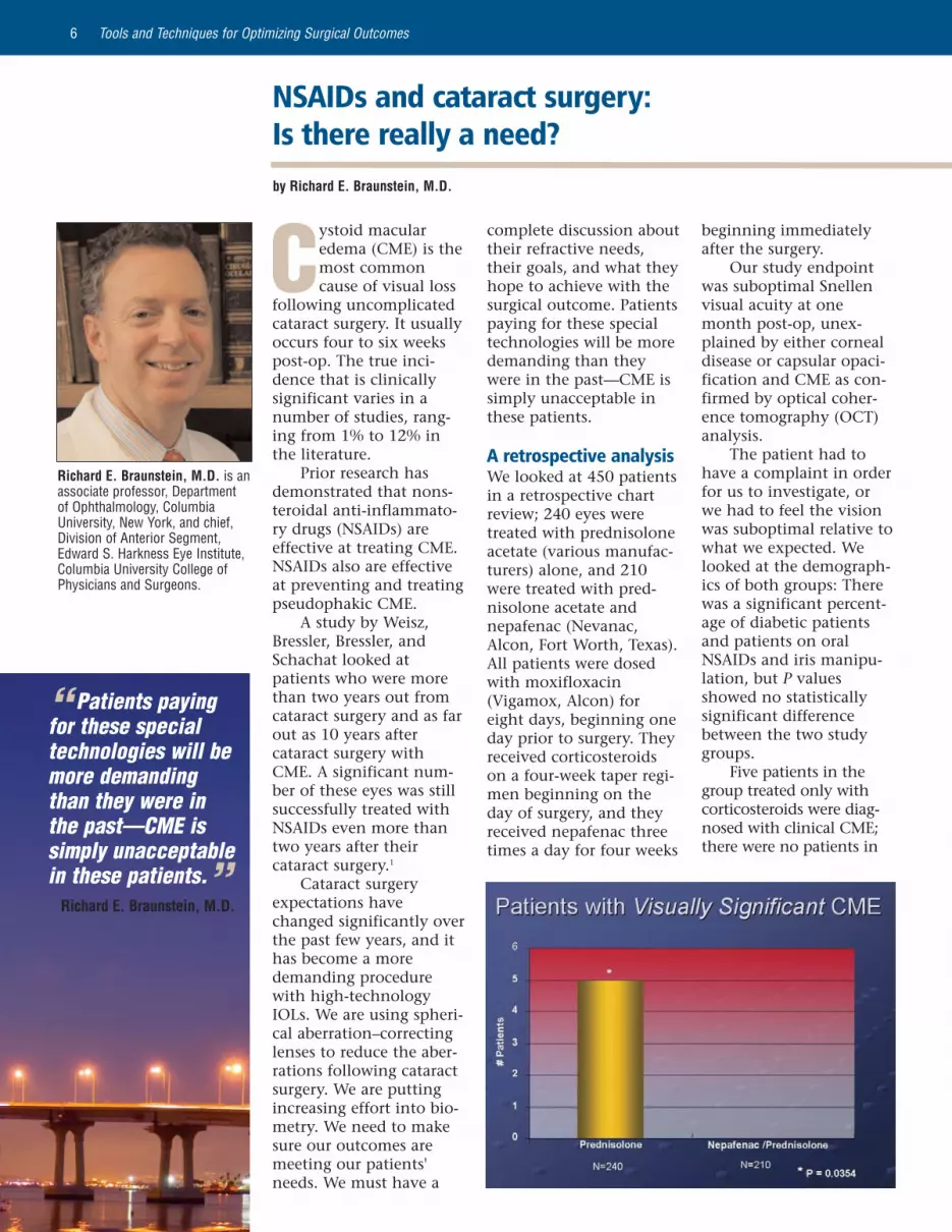
A retrospective analysisWe looked at 450 patientsin a retrospective chartreview; 240 eyes weretreated with prednisoloneacetate (various manufac-turers) alone, and 210were treated with pred-nisolone acetate andnepafenac (Nevanac,Alcon, Fort Worth, Texas).All patients were dosedwith moxifloxacin(Vigamox, Alcon) foreight days, beginning oneday prior to surgery. Theyreceived corticosteroidson a four-week taper regi-men beginning on theday of surgery, and theyreceived nepafenac threetimes a day for four weeks
“ Patients payingfor these specialtechnologies will bemore demandingthan they were inthe past—CME issimply unacceptablein these patients.”
Richard E. Braunstein, M.D.
Tools and Techniques for Optimizing Surgical Outcomes6
NSAIDs and cataract surgery: Is there really a need?by Richard E. Braunstein, M.D.
beginning immediatelyafter the surgery.
Our study endpointwas suboptimal Snellenvisual acuity at onemonth post-op, unex-plained by either cornealdisease or capsular opaci-fication and CME as con-firmed by optical coher-ence tomography (OCT)analysis.
The patient had tohave a complaint in orderfor us to investigate, orwe had to feel the visionwas suboptimal relative towhat we expected. Welooked at the demograph-ics of both groups: Therewas a significant percent-age of diabetic patientsand patients on oralNSAIDs and iris manipu-lation, but P valuesshowed no statisticallysignificant differencebetween the two studygroups.
Five patients in thegroup treated only withcorticosteroids were diag-nosed with clinical CME;there were no patients in
Richard E. Braunstein, M.D. is anassociate professor, Departmentof Ophthalmology, ColumbiaUniversity, New York, and chief,Division of Anterior Segment,Edward S. Harkness Eye Institute,Columbia University College ofPhysicians and Surgeons.

7
the nepafenac/prednisolonegroup diagnosed withCME.
We hoped to spot atrend or risk factors inthe group that developedCME to help us identifypatients to watch out foror to treat as higher-riskpatients. Unfortunately,we were unable to findany particular reasonwhy these five eyesdeveloped CME. None ofthe patients were diabet-ic, had undergone morecomplicated surgery, orhad other contributoryindicators in his medicalhistory.
These five patientswith CME were treated foran average of sevenmonths, with a range offour to 10 months. They
required a number ofadditional office visitsand were all treated suc-cessfully with topicalNSAIDs. This was a signif-icantly slower recoverythan the patients expect-ed and desired.
We had no significantadverse events in eithergroup. One patient waspresented with an iatro-genic corneal abrasion onpost-op day one, and weopted to discontinuenepafenac. She subse-quently developed CME.
Patients treated with apost-op regimen of pred-nisolone and nepafenachad a significantly lowerincidence of pseudopha-kic macular edema thanthose treated with pred-nisolone as a single agent.
Tools and Techniques for Optimizing Surigcal Outcomes 7
References1. Weisz JM, Bressler NM,
Bressler SB, SchachatAP. Ketorolac treatmentof pseudophakic macu-lar edema identifiedmore than 24 monthsafter cataract extrac-tion. Ophthalmology.1999;106:1656-9.
“ Patients treatedwith a post-opregimen ofprednisolone andnepafenac had asignificantly lowerincidence ofpseudophakicmacular edema thanthose treated withprednisolone as asingle agent. ”
Richard E. Braunstein, M.D.
penetration, andimproved potency.
Cefuroxime (Ceftin,GlaxoSmithKline, UnitedKingdom) may not be thebest choice for an antibi-otic either. It has averagecoverage of gram-posi-tives and gram-negatives.There is a very high rateof resistance because it isan older cephalosporin;therefore, methicillin-resistant staph aureus(MRSA) will not be cov-ered.
Cefuroxime is alsotime dependent. It takesabout eight to 12 hoursfor cefuroxime to work,whereas fluoroquinolonescan kill in two hours.
There is the potentialthat an intracameral drugcan be harmful. It cancause a patient to go into
anaphylaxis, so we needlong-term studies as wellas short-term studies tolook at intracameraldrugs, which are the mosteffective, least toxic, andleast likely to cause ana-phylaxis.
References1. Mather R, Karenchak LM,
Romanowski EG, KowalskiRP. Fourth-generation fluoroquinolones: newweapons in the arsenal ofophthalmic antibiotics. AmJ Ophthalmol.2002;133:463-6.
2. Kim DH, Stark WJ, O'BrienTP, et al. Aqueous penetra-tion and biological activityof moxifloxacin 0.5% oph-thalmic solution and gati-floxacin 0.3% ophthalmicsolution in cataract surgerypatients. Ophthalmology2005;112:1992-6.
3. Friedlaender MH,Breshears D, Amoozgar B,et al. The dilution of ben-zalkonium chloride (BAK)in the tear film. Advancesin Therapy. 2006;23:835-41.
4. Barry P, Seal DV, GettinbyG, et al. ESCRSEndophthalmitis StudyGroup. ESCRS study ofprophylaxis of post-opera-tive endophthalmitis aftercataract surgery: prelimi-nary report of principalresults from a Europeanmulticenter study. JCataract Refract Surg.2006;32:407-10.
5. Kowalski RP, RomanowskiEG, Mah FS, et al. Topicalprophylaxis with moxi-floxacin prevents endoph-thalmitis in a rabbitmodel. Am J Ophthalmol.2004;138:33-7.
Topical – from page 3

8
Cystoid macularedema (CME) isone of the most fre-quent causes of
visual decline followinguncomplicated cataractsurgery and can have anonset as late as four to sixweeks post-op.1 Theoccurrence of clinicalCME is greatly reducedafter uncomplicated pha-coemulsification opera-tions, but the incidenceof angiographic CME isstill nearly equal to theincidence of the extracap-sular technique.2 In 1% to3% of cases, it is associat-ed with a decrease in visu-al acuity (VA).3 In addi-tion, CME is estimated tooccur in up to 12% oflow-risk cataract cases,4
and the incidence of CMEafter routine phaco hasbeen reported to be ashigh as 19%.5
Optical coherencetomography (OCT) can beuseful in identifying thosewith significant macularthickening, and in high-risk patients (such asthose with diabetes), itcan help us determinewhich of our patients areat risk of developing CMEor a loss of vision.6,7
As technology hasimproved, the definitionsof CME have changed.Clinical CME currentlyhas been described as ves-sel leakage associated witha VA of 20/40 or worse,and today's definition ofCME is more strict (20/25or worse) because of high-er patient expectations.8
Numerous studies haveidentified risk factors forCME, including preexistingocular inflammation,epiretinal or vitreoretinalinterface membrane prob-lems, diabetic retinopathy,ocular vascular or cardio-vascular disease, retinitispigmentosa, iris trauma,and post-op anteriorchamber inflammation.9
Once macular edemaoccurs, it can take manymonths to resolve. Anumber of studies showsthat a proportion ofpatients will have perma-nent macular edema evenif the edema is recognizedearly and treated aggres-sively.
Patients at higher riskof developing CME maybenefit from treatmentstarting earlier than thosein a lower-risk group, andthe higher-risk patientsshould continue prophy-laxis for an extended peri-od after surgery.10
Miyake et al. offered ahypothesis on the mecha-
nism of CME aftercataract surgery. Pros-taglandins produced bythe anterior uveal tractenter the aqueous and vit-reous and lead to a break-down of the blood-aque-ous barrier and the blood-retina barrier.11
Optimal therapyTopical nonsteroidal anti-inflammatory drugs(NSAIDs) appear to beeffective in preventingpost-surgical CME. Theyalso have been shown tohave a beneficial effect onvisual function.1 Gaynesand Fiscella showed itmay be necessary toachieve therapeutic con-centrations of the NSAIDin the posterior chamberto maximize the effect ofthe NSAID on the targettissue (the retina).12
Steroids alone have notbeen shown to be as effec-tive in preventing ortreating CME.4
O'Brien wrote about
Comparing NSAIDs in aqueous humorby Michael B. Raizman, M.D.
Tools and Techniques for Optimizing Surgical Outcomes8
Michael B. Raizman, M.D. is anophthalmologist, OphthalmicConsultants of Boston, Boston,director, Cornea and CataractService, New England Eye Center,Boston, and associate professorof ophthalmology, Tufts UniversitySchool of Medicine, Boston.
“ ... CME isestimated to occurin up to 12% oflow-risk cataractcases, and theincidence of CMEafter routine phacohas been reportedto be as high as19%.”Michael B. Raizman, M.D.

hour and then drops off.Bromfenac has a very lowconcentration throughout.Nepafenac had the highestarea under the curve overthis four-hour time course;ketorolac had the secondhighest area. Bromfenachad the lowest area underthe curve in this timecourse.
References1. Samiy N, Foster CS. The
role of nonsteroidal anti-inflammatory drugs inocular inflammation. IntOphthalmol Clin.1996;36:195-206.
2. Mentes J, Erakgun T,Afrashi F, Kerci G.Incidence of cystoid mac-ular edema after uncom-plicated phacoemulsifica-tion. Ophthalmologica.2003;217:408-12.
3. Agostini HT, Hansen LL,Feltgen N. Treatment ofpseudophakic cystoidmacular edema.Ophthalmologe. 2007;104:425-30.
4. McColgin AS, RaizmanMB. Efficacy of topicalVoltaren in reducing theincidence of post-opera-tive cystoid macularedema. Invest OphthalmolVis Sci. 1999;40:S289.
5. Ursell PG, Spalton DJ, Whitcup SM,Nussenblatt RB. Cystoid
emerging guidelines forNSAID treatment regimensin cataract surgery andsuggested at-risk patientsreceive NSAIDs one weekpre-op and between fourweeks to several monthsafter surgery. For those notat risk, pre-op dosingshould be between one totwo days, and post-optreatment should be fourweeks.13
Aqueous humorconcentrationsA double-masked multi-center study looked at 75patients randomized toreceive nepafenac 0.1%(Nevanac, Alcon, FortWorth, Texas), amfenac (ametabolite in nepafenac),ketorolac 0.4% (Acular LS,Allergan, Irvine, Calif.), orbromfenac 0.9% (Xibrom,ISTA Pharmaceuticals,Irvine, Calif.).14 The pur-pose of this study was todetermine the aqueoushumor concentrationafter one drop. Allpatients had been sched-uled to undergo cataractsugery. Patients wereadministered one drop ofthe NSAID at 30, 60, 120,180, or 240 minutesbefore surgery. Aqueoushumor samples were col-lected at the time of para-centesis. The patientsreceived no other nons-teroidals, but they didreceive dilating dropsprior to the surgery.
The results proved thatnepafenac initially has avery high concentrationand then drops off aftertwo to four hours.Amfenac, the metabolite,peaks at about threehours.
Ketorolac has a slightlydifferent time course; itrises rapidly over the first
macular edema afterphacoemulsification:relationship to blood-aqueous barrier damageand visual acuity.J Cataract Refract Surg.1999;25:1492-7.Comment in:2000;26:474.
6. Kim SJ, Equi R, BresslerNM. Analysis of macularedema after cataract sur-gery in patients with dia-betes using opticalcoherence tomography.Ophthalmology.2007;114:881-9.
7. Torron-Fernandez-BlancoC, Ruiz-Moreno O, Ferrer-Novella E, Sanchez-CanoA, Honrubia-Lopez Fm.Pseudophakic cystoidmacular edema.Assessment with opticalcoherence tomography.Arch Soc Esp Oftalmol.2006;81:147-53.
8. Heier JS, Topping TM,Baumann W, Dirks MS,Chern S. Ketorolac ver-sus prednisolone versuscombination therapy inthe treatment of acutepseudophakic cystoidmacular edema.Ophthalmology.2000;107:2034-8; discussion 2039.
9. Gulkilik G, Kocabora S,Taskapili M, Engin G.Cystoid macular edema
Tools and Techniques for Optimizing Surigcal Outcomes 9
“ The results[from a studyaddressingaqueous humorconcentrations]found nepafenacinitially has a veryhigh concentrationand then drops offafter two to fourhours.”Michael B. Raizman, M.D.
continued on page 12

Because of its frequen-cy, cystoid macularedema (CME) is per-
haps the most significantcomplication of cataractsurgery—up to 12% ofeyes will have someincrease in retinal thick-ness following uncompli-cated surgery. That meansone in every eightpatients has a likelihoodof having some change inhis visual function.
As retinal specialists,we use the optical coher-ence tomography (OCT)not only diagnosticallybut also as an importantmodality to monitor theresponse to therapy. Weknow there are many riskfactors for CME. Theseinclude vitreoretinal inter-face disorders, systemicand ocular vascu-lopathies, and ocularinflammatory disease.However, any patientundergoing an intraocularprocedure of any type isat risk of developinginflammation and sec-ondary CME. Therefore,we should consider pro-phylaxis for these patientsearlier and for a longerperiod of time.
We believe that CMEis developed, in part,because of a prosta-glandin-induced breach ofthe blood-retinal barrierand, subsequently, sec-ondary retinal edema.Topical nonsteroidal anti-inflammatory drugs(NSAIDs) inhibit the pro-duction of prostaglandinsby their effect on the
cyclooxygenase enzyme.Corticosteroids inhibitphospholipase A2, whichalso reduces arachidonicacid metabolites, particu-larly the leukotrienes thatattract inflammatory cellsand are potent mediatorsof inflammation. There-fore, NSAIDs and corticos-teroids act synergisticallyat different sites in theinflammatory cascade toreduce the production ofinflammatory mediators.
The ideal NSAIDwould have the ability topenetrate target tissues at therapeutic levels,reducing anterior seg-ment inflammation and,more importantly, reduc-ing CME in the posteriorpole of the eye. It alsoshould have excellentanalgesic properties andbe safe and well toleratedfor patient use.
Penetration efficacy A study looked at theability of nepafenac
(Nevanac, Alcon, FortWorth, Texas), diclofenac(Voltaren, Novartis, Basel,Switzerland) and ketoro-lac (Acular LS, Allergan,Irvine, Calif.) to preventthe development ofexperimentally inducedretinal edema in rabbiteyes.1 These rabbits werepretreated with five dropsa day, starting one dayprior to injection, theninjected with a powerfulmitogen. Drops were con-tinued for three days afterinjection. Nepafenac sig-nificantly inhibitedprostaglandin synthesis, aknown mediator ofinflammation, and signif-icantly reduced vitreousprotein concentration, aknown marker of inflam-mation, demonstratingsuppression of blood-reti-nal barrier breakdown.Neither diclofenac norketorolac was able toinhibit these markers ofinflammation.
This study demon-strated that nepafenac
Treatment strategies for CMEby Keith A. Warren, M.D.
Tools and Techniques for Optimizing Surgical Outcomes10
Keith A. Warren, M.D. is aprofessor of ophthalmology,University of Kansas School ofMedicine, Kansas City.
“ Nepafenacsignificantly inhibitedprostaglandin synthe-sis, a known mediatorof inflammation, andsignificantly reducedvitreous protein con-centration, a knownmarker of inflamma-tion, demonstratingsuppresion of blood-retinal barrierbreakdown.”
Keith A. Warren, M.D.

Tools and Techniques for Optimizing Surigcal Outcomes 11
Eight of the 11 patientshad significant improve-ment in their vision—10letters or two ETDRSlines—by week 12. Theremaining three patientswere stable or had lessthan 10 letters or twolines of improvement.Importantly, 10 of 11patients had a correspon-ding improvement in reti-nal thickness with a meanreduction of 196 µm.There was no correlationbetween the amount ofreduction of retinal thick-ness and final visual acu-ity.
In a separate casereport by another col-league, a patient withchronic CME secondaryto pars planitis wasdescribed. One eye withCME received an intravit-real treatment of triam-cinolone because of poorresponsiveness to topicaltherapy. The CMEresponded to treatment;however, this patient subsequently developed ahigh IOP—up to 55 mmHg—so repeating asteroid injection was notconsidered.
The patient began toshow signs of chronic
intraocular triamcinolone(4 mg) at six weeks ifthey're not responsive tothe initial treatment.
The most frequentcomplication is an increasein IOP. Up to 30% of eyesthat get intraocular steroidinjections have beenreported to have anincrease in IOP. Despitetreatment, many patientswith CME will suffer a per-manent loss of visual func-tion. This loss of functioncould manifest as loss ofSnellen vision, reducedcontrast sensitivity, orcolor desaturation. Thishighlights the importanceof prophylaxis.
Steroid respondersand nepafenac We have a case series of 11patients, all of whom weresteroid responders: eighthad cataract surgery, andthree had macular puckeror retinal interface disease.
On average, thecataract patients in ourseries presented at threeweeks, a little earlier thannormal. All patients weretreated with nepafenac asmonotherapy four times aday for six weeks andthen tapered them overan additional six weeks.
exhibited superior phar-macodynamic propertiesin the posterior segment,and this unique therapeu-tic potential was verygood for inflammatoryconditions associatedwith retinal edema.
Current CME treatments The current recommend-ed regimen for treatmentof CME is a combinationof corticosteroids andNSAIDs. Initial treatmentusually consists of topicaladministration of thosemedications. Most retinalspecialists will addresstreatment failures with aposterior subtenon's injec-tion of steroids. Thisroute of administrationprovides a higher concen-tration of the drug to thetreated tissue.
Intraocular steroidinjections are reserved forresistant CME. Because ofthe poor penetration offirst-generation NSAIDs,most retinal specialistsdid not use an NSAIDs fortreatment. I thinknepafenac makes a signifi-cant difference because ofits ability to penetrate theposterior pole.
The treatment regi-men I employ for patientswith CME includes a 20-mg triamcinolone(Kenalog, Bristol-MyersSquibb, New York) poste-rior subtenon's injection.Those patients are man-aged with topicalnepafenac four times aday for six weeks. Wetaper the NSAID dose overthe next six weeks for atotal of 12 weeks of treat-ment for those patients.We treat patients withresistant CME with
“ The currentrecommendedregimen fortreatment of CMEis a combination ofcorticosteroids andNSAIDs.”
Keith A. Warren, M.D.
continued on page 12

Tools and Techniques for Optimizing Surgical Outcomes12
This supplement was produced by EyeWorld under an educational grant from Alcon Laboratories Inc.
Copyright 2007 ASCRS Ophthalmic Corporation. All rights reserved. The views expressed here do not necessarily reflectthose of the editor, editorial board, or the publisher and in no way imply endorsement by EyeWorld or ASCRS.
CME in the other eye andwas started on nepafenac.The retinal thickness inthe right eye was veryhigh: 681 µm with a visu-al acuity of 20/80. Thisretinal thickness reducedto 164 µm on nepafenacsuspension for 12 weeks,with a more normal-look-ing retinal foveal contour.There was no change inIOP when the patient wastreated with nepafenac.
CME is a very signifi-
cant cause of visual lossfollowing uncomplicatedcataract surgery. Deliveryof an effective concentra-tion of a drug to the tar-get tissue may limit thedevelopment of CME.Nepafenac appears tohave excellent penetra-tion and efficacy in treat-ing CME for steroidresponders; however,vision loss is commondespite treatment, mak-ing prophylaxis extreme-
ly important in anypatient undergoingcataract surgery.
References 1. Kapin MA, Yanni JM,
Brady MT, et.al.Inflammation-mediatedretinal edema in therabbit is inhibited bytopical nepafenac.Inflammation.2003;27:281-91.
Treatment – from page 11
“ Nepafenacappears to haveexcellentpenetration andefficacy in treatingCME for steroidresponders; however,vision loss iscommon despitetreatment, makingprophylaxisextremely importantin any patientundergoing cataractsurgery.”
Keith A. Warren, M.D.
after phacoemulsifica-tion: risk factors andeffect on visual acuity.Can J Ophthalmol.2006;41:699-703.
10. Heier JS. Preventingpost-cataract extractionCME: Early identifica-tion of patients at riskand prophylactic treat-ment may avert visionloss. OphthalmologyManagement. 2004;63-72.
11. Miyake K, Shirato S,Oshika T, Eguchi K, et al.Comparison ofdiclofenac and fluo-
rometholone in prevent-ing cystoid macularedema after small inci-sion cataract surgery: amulticentered prospec-tive trial. Jpn JOphthalmol. 2000;44:58-67.
12. Gaynes BI, Fiscella R.Topical nonsteroidalanti-inflammatory drugsfor ophthalmic use: asafety review. Drug Saf.2002;25:233-50.
13. O'Brien TP. Emergingguidelines for use ofNSAID therapy to opti-mize cataract surgery
NSAIDs – from page 9
patient care. Curr MedRes & Opin. 2005;21:1131-7.
14. Walters TR, Raizman M,Ernest P, et al. A double-masked, randomized,single-dose, pharmacoki-netic study ofnepafenac, amfenac,ketorolac, and brom-fenac in human aqueoushumor following topicaladministration ofNevanac, Acular LS, orXibrom. InvestOphthalmol Vis Sci.
The CME test is available online at www.eyeworld.org.Go to Online Education for test.
Please contact Laura Johnson with the American Society of Cataract andRefractive Surgery at 703-591-2220, or e-mail her at [email protected] if you have
any questions regarding CME credit for this educational activity.