facial nerve - ksumsc
TRANSCRIPT
Facial Nerve
Objectives:
➣ Anatomy (course and branches).
➣ Causes of facial palsy (including Bell's palsy, middle ear complication, traumatic
and Ramsey Hunt syndrome).
[ Color index : Important | Notes | Extra | 433 Notes]
Resources: Slides + Notes + Lecture notes of ENT + 433 team.
Done by : Saad Almutairi and Deema AlFaris
Edited by: Saleh Alshawi and Sara Alkhalifah
Revised by : Adel Al Shihri, Lina Alshehri.
Introduction:
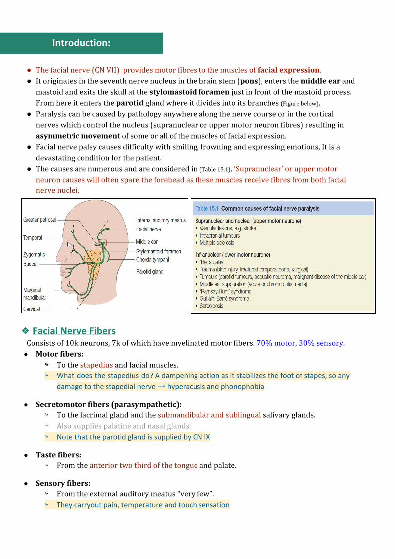
● The facial nerve (CN VII) provides motor fibres to the muscles of facial expression. ● It originates in the seventh nerve nucleus in the brain stem (pons), enters the middle ear and
mastoid and exits the skull at the stylomastoid foramen just in front of the mastoid process. From here it enters the parotid gland where it divides into its branches (Figure below).
● Paralysis can be caused by pathology anywhere along the nerve course or in the cortical nerves which control the nucleus (supranuclear or upper motor neuron fibres) resulting in asymmetric movement of some or all of the muscles of facial expression.
● Facial nerve palsy causes difficulty with smiling, frowning and expressing emotions, It is a devastating condition for the patient.
● The causes are numerous and are considered in (Table 15.1). ‘Supranuclear’ or upper motor neuron causes will often spare the forehead as these muscles receive fibres from both facial nerve nuclei.
❖❖ Facial Nerve Fibers Consists of 10k neurons, 7k of which have myelinated motor fibers. 70% motor, 30% sensory.
● Motor fibers: ↪↪ To the stapedius and facial muscles. ↪ What does the stapedius do? A dampening action as it stabilizes the foot of stapes, so any
damage to the stapedial nerve → hyperacusis and phonophobia
● Secretomotor fibers (parasympathetic): ↪ To the lacrimal gland and the submandibular and sublingual salivary glands. ↪ Also supplies palatine and nasal glands. ↪ Note that the parotid gland is supplied by CN IX
● Taste fibers: ↪ From the anterior two third of the tongue and palate.
● Sensory fibers: ↪ From the external auditory meatus “very few”. ↪ They carryout pain, temperature and touch sensation
Anatomical Divisions:
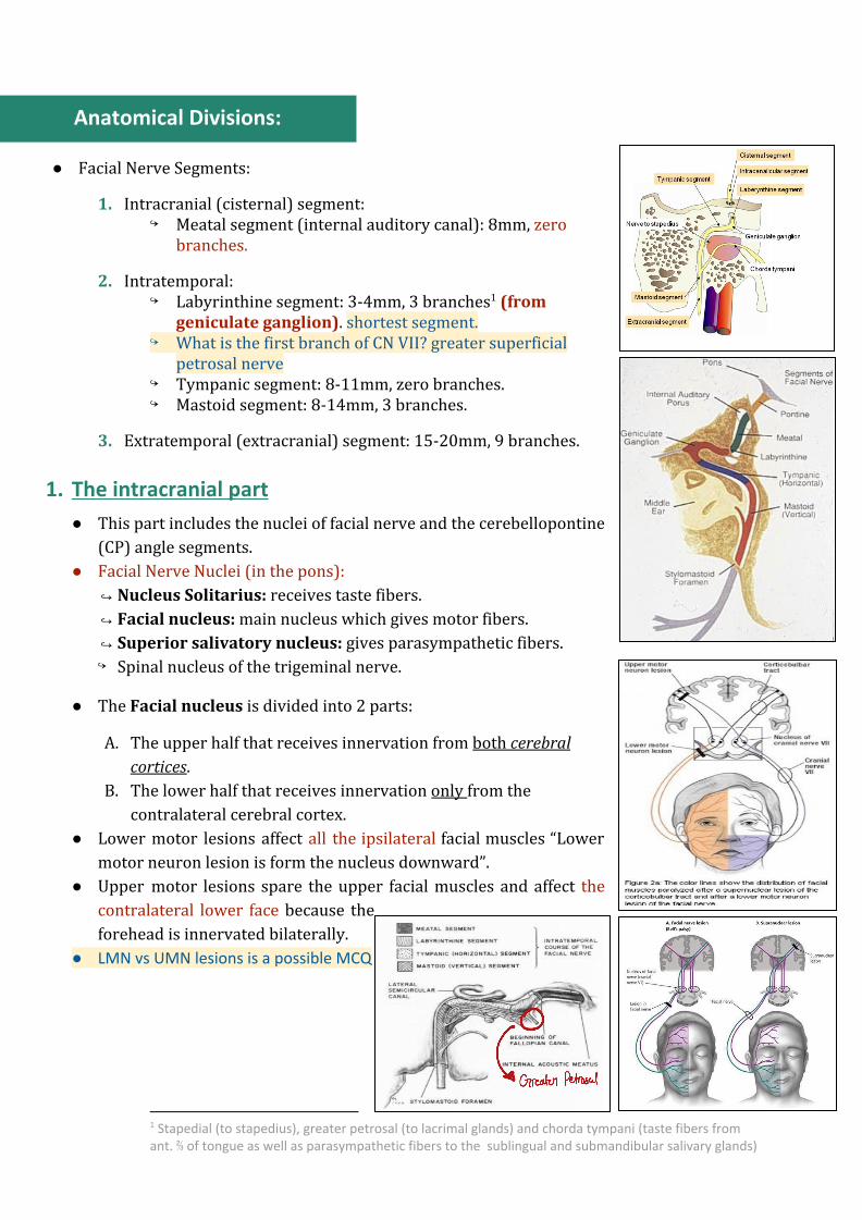
● Facial Nerve Segments:
1. Intracranial (cisternal) segment: ↪ Meatal segment (internal auditory canal): 8mm, zero
branches.
2. Intratemporal: ↪ Labyrinthine segment: 3-4mm, 3 branches (from 1
geniculate ganglion). shortest segment. ↪ What is the first branch of CN VII? greater superficial
petrosal nerve ↪ Tympanic segment: 8-11mm, zero branches. ↪ Mastoid segment: 8-14mm, 3 branches.
3. Extratemporal (extracranial) segment: 15-20mm, 9 branches.
1. The intracranial part
● This part includes the nuclei of facial nerve and the cerebellopontine (CP) angle segments.
● Facial Nerve Nuclei (in the pons): ↪Nucleus Solitarius: receives taste fibers. ↪Facial nucleus: main nucleus which gives motor fibers. ↪Superior salivatory nucleus: gives parasympathetic fibers. ↪ Spinal nucleus of the trigeminal nerve.
● The Facial nucleus is divided into 2 parts:
A. The upper half that receives innervation from both cerebral cortices.
B. The lower half that receives innervation only from the contralateral cerebral cortex.
● Lower motor lesions affect all the ipsilateral facial muscles “Lower motor neuron lesion is form the nucleus downward”.
● Upper motor lesions spare the upper facial muscles and affect the contralateral lower face because the forehead is innervated bilaterally.
● LMN vs UMN lesions is a possible MCQ
1 Stapedial (to stapedius), greater petrosal (to lacrimal glands) and chorda tympani (taste fibers from ant. ⅔ of tongue as well as parasympathetic fibers to the sublingual and submandibular salivary glands)
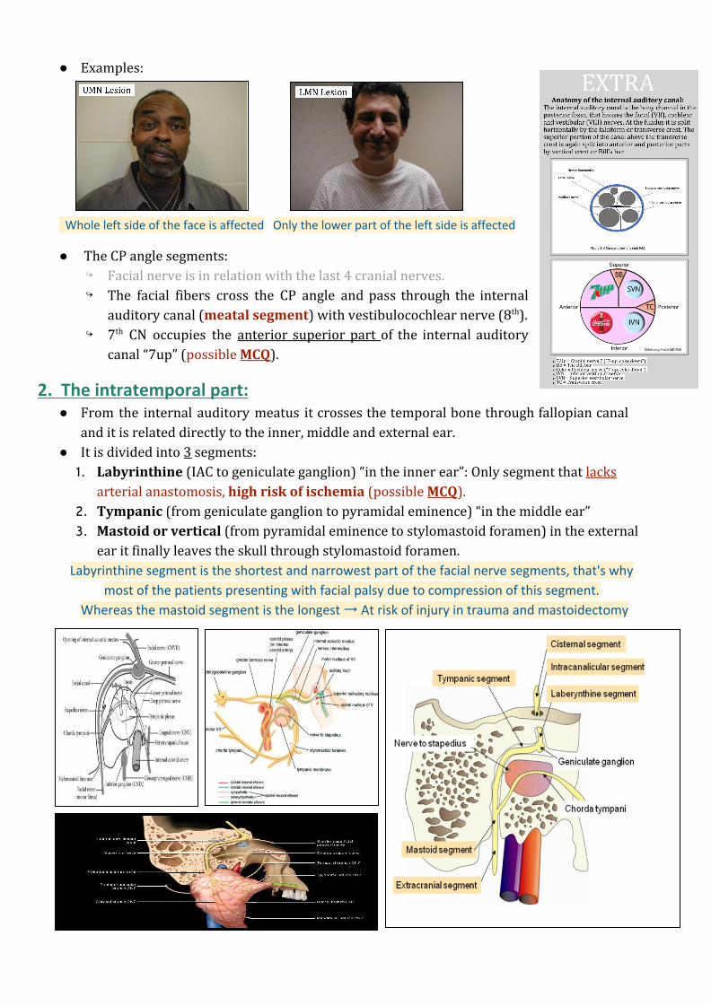
● Examples:
Whole left side of the face is affected Only the lower part of the left side is affected
● The CP angle segments: ↪ Facial nerve is in relation with the last 4 cranial nerves. ↪ The facial fibers cross the CP angle and pass through the internal
auditory canal (meatal segment) with vestibulocochlear nerve (8th). ↪ 7th CN occupies the anterior superior part of the internal auditory
canal “7up” (possible MCQ).
2. The intratemporal part: ● From the internal auditory meatus it crosses the temporal bone through fallopian canal
and it is related directly to the inner, middle and external ear. ● It is divided into 3 segments:
1. Labyrinthine (IAC to geniculate ganglion) “in the inner ear”: Only segment that lacks arterial anastomosis, high risk of ischemia (possible MCQ).
2. Tympanic (from geniculate ganglion to pyramidal eminence) “in the middle ear” 3. Mastoid or vertical (from pyramidal eminence to stylomastoid foramen) in the external
ear it finally leaves the skull through stylomastoid foramen. Labyrinthine segment is the shortest and narrowest part of the facial nerve segments, that's why
most of the patients presenting with facial palsy due to compression of this segment.
Whereas the mastoid segment is the longest → At risk of injury in trauma and mastoidectomy
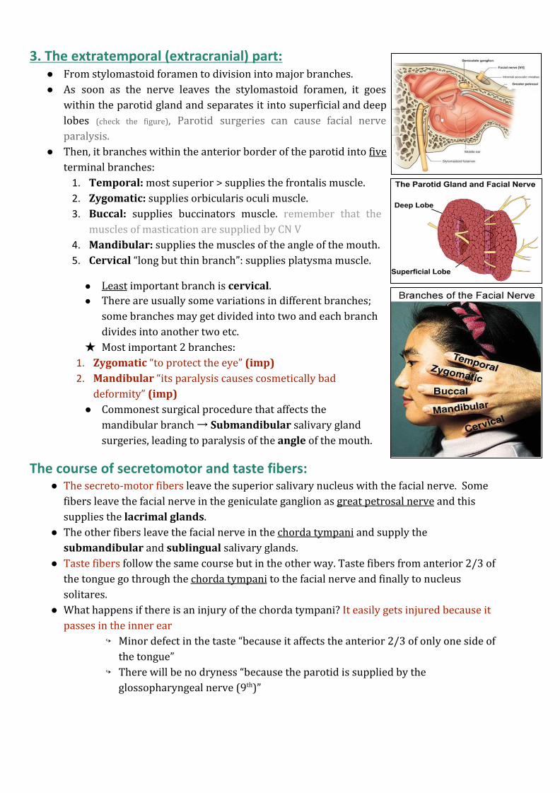
3. The extratemporal (extracranial) part: ● From stylomastoid foramen to division into major branches. ● As soon as the nerve leaves the stylomastoid foramen, it goes
within the parotid gland and separates it into superficial and deep lobes (check the figure), Parotid surgeries can cause facial nerve paralysis.
● Then, it branches within the anterior border of the parotid into five terminal branches:
1. Temporal: most superior > supplies the frontalis muscle. 2. Zygomatic: supplies orbicularis oculi muscle. 3. Buccal: supplies buccinators muscle. remember that the
muscles of mastication are supplied by CN V 4. Mandibular: supplies the muscles of the angle of the mouth. 5. Cervical “long but thin branch”: supplies platysma muscle.
● Least important branch is cervical. ● There are usually some variations in different branches;
some branches may get divided into two and each branch divides into another two etc.
★ Most important 2 branches: 1. Zygomatic “to protect the eye” (imp) 2. Mandibular “its paralysis causes cosmetically bad
deformity” (imp) ● Commonest surgical procedure that affects the
mandibular branch → Submandibular salivary gland surgeries, leading to paralysis of the angle of the mouth.
The course of secretomotor and taste fibers: ● The secreto-motor fibers leave the superior salivary nucleus with the facial nerve. Some
fibers leave the facial nerve in the geniculate ganglion as great petrosal nerve and this supplies the lacrimal glands.
● The other fibers leave the facial nerve in the chorda tympani and supply the submandibular and sublingual salivary glands.
● Taste fibers follow the same course but in the other way. Taste fibers from anterior 2/3 of the tongue go through the chorda tympani to the facial nerve and finally to nucleus solitares.
● What happens if there is an injury of the chorda tympani? It easily gets injured because it passes in the inner ear
↪ Minor defect in the taste “because it affects the anterior 2/3 of only one side of the tongue”
↪ There will be no dryness “because the parotid is supplied by the glossopharyngeal nerve (9th)”
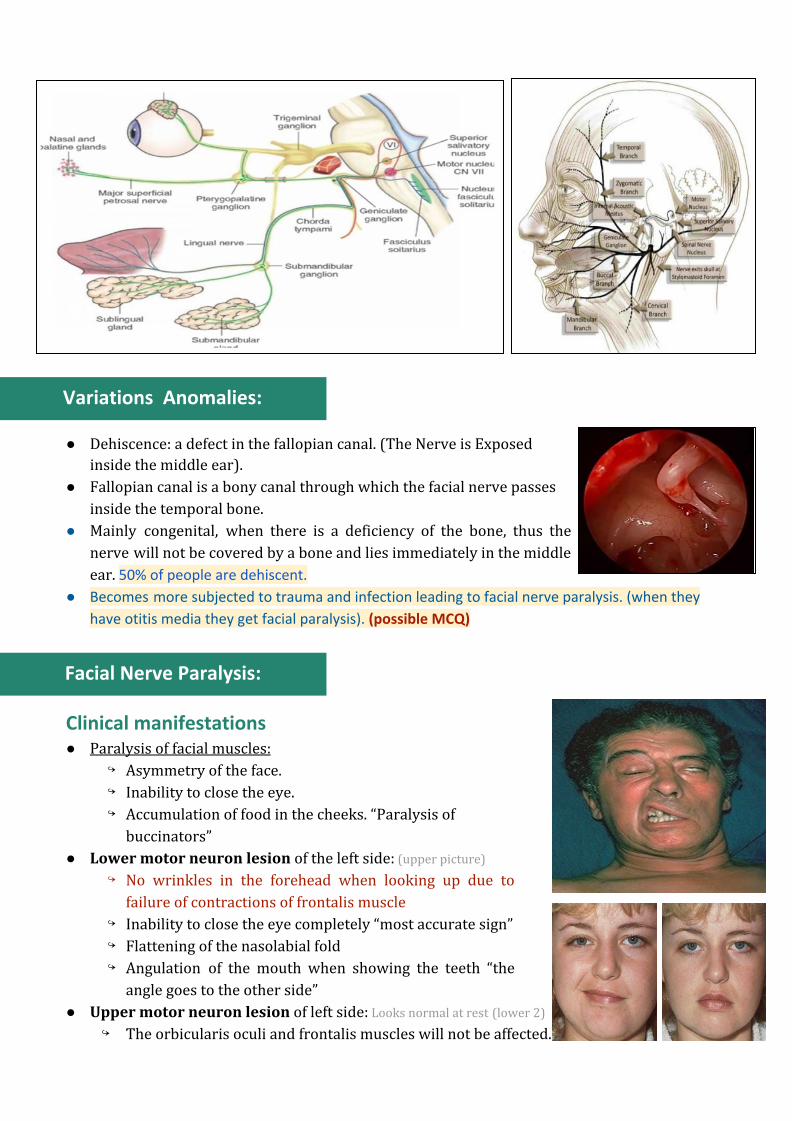
Variations Anomalies:
● Dehiscence: a defect in the fallopian canal. (The Nerve is Exposed inside the middle ear).
● Fallopian canal is a bony canal through which the facial nerve passes inside the temporal bone.
● Mainly congenital, when there is a deficiency of the bone, thus the nerve will not be covered by a bone and lies immediately in the middle ear. 50% of people are dehiscent.
● Becomes more subjected to trauma and infection leading to facial nerve paralysis. (when they
have otitis media they get facial paralysis). (possible MCQ)
Facial Nerve Paralysis:
Clinical manifestations ● Paralysis of facial muscles:
↪ Asymmetry of the face. ↪ Inability to close the eye. ↪ Accumulation of food in the cheeks. “Paralysis of
buccinators” ● Lower motor neuron lesion of the left side: (upper picture)
↪ No wrinkles in the forehead when looking up due to failure of contractions of frontalis muscle
↪ Inability to close the eye completely “most accurate sign” ↪ Flattening of the nasolabial fold ↪ Angulation of the mouth when showing the teeth “the
angle goes to the other side” ● Upper motor neuron lesion of left side: Looks normal at rest (lower 2)
↪ The orbicularis oculi and frontalis muscles will not be affected.
● Other manifestations of facial nerve paralysis: ↪ Phonophobia “due to failure of contractions of strapedius muscle, uncomfortable
feeling in exposure to loud sounds” ■ Acoustic reflex (stapedial reflex) is a useful tool to localize the lesion; if intact
the problem is distal to it and vice versa. ↪ Dryness of the eye “Some people present with lacrimation and others present with
dryness. Why?” ▪ Lacrimation is due to paralysis of orbicularis oculi as this muscle help in
draining the tears ▪ Dryness is due to affection of greater petrosal nerve which arise from
geniculate ganglion ● So if the paralysis is above the level of geniculate ganglion > dryness ● If below it > no dryness
↪ Loss of taste “very little” just in the ant. ⅔ of one side. They feel a metallic taste.
Clinical examination: Ask the patient to:
1. Look up to test frontalis. 2. Close eyes to test orbicularis oculi. 3. Blow the cheek to test buccinators. 4. Show the teeth for angulation.
Bilateral: ● Guillain-Barre syndrome. ● Lyme disease. ● Intracranial neoplasm.
Recurrent: (Pics) ● Melkersson-Rosenthal syndrome 2
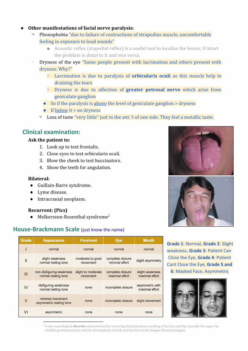
House-Brackmann Scale (just know the name)
Grade 1: Normal, Grade 2: Slight
weakness, Grade 3: Patient Can
Close the Eye, Grade 4: Patient
Cant Close the Eye, Grade 5 and
6: Masked Face, Asymmetric
2 A rare neurological disorder characterized by recurring facial paralysis, swelling of the face and lips (usually the upper lip - cheilitis granulomatosis) and the development of folds and furrows in the tongue (fissured tongue)
Pathophysiology of Nerve Injury:
Not mentioned by our doctor (F group)
Neuropraxia (conductive block): ● In cases of mild trauma causing only functional block of the facial nerve, the fibers still
keep their integrity. ● In Regeneration: there will be restoration of full function if the cause is treated.
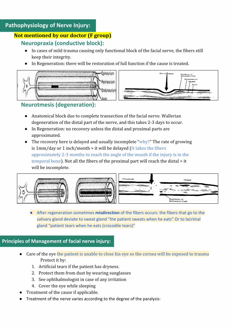
Neurotmesis (degeneration):
● Anatomical block due to complete transection of the facial nerve. Wallerian degeneration of the distal part of the nerve, and this takes 2-3 days to occur.
● In Regeneration: no recovery unless the distal and proximal parts are approximated.
● The recovery here is delayed and usually incomplete “why?” The rate of growing is 1mm/day or 1 inch/month > it will be delayed (It takes the fibers approximately 2-3 months to reach the angle of the mouth if the injury is in the temporal bone). Not all the fibers of the proximal part will reach the distal > it will be incomplete.
● After regeneration sometimes misdirection of the fibers occurs: the fibers that go to the
salivary gland deviate to sweat gland “the patient sweats when he eats” Or to lacrimal
gland “patient tears when he eats (crocodile tears)”
Principles of Management of facial nerve injury:
● Care of the eye the patient is unable to close his eye so the cornea will be exposed to trauma Protect it by:
1. Artificial tears if the patient has dryness. 2. Protect them from dust by wearing sunglasses 3. See ophthalmologist in case of any irritation 4. Cover the eye while sleeping
● Treatment of the cause if applicable. ● Treatment of the nerve varies according to the degree of the paralysis:
○ Partial facial paralysis:
↪ Being partial means that some of the nerve fibers are in continuity. Recovery is
expected by conservative treatment (e.g. removal of pressure, steroid etc.). No
need for surgical intervention.
○ Complete facial paralysis:
↪ Complete paralysis may be a result of neuropraxia or/and degeneration.
↪ If it is due to neuropraxia, recovery is expected by conservative treatment. If it is
due to degeneration, surgical treatment is required.
↪ To differentiate between degeneration and neuropraxia electrophysiological tests
are required.
Investigations:
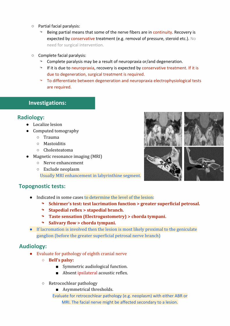
Radiology: ● Localize lesion ● Computed tomography
○ Trauma ○ Mastoiditis ○ Cholesteatoma
● Magnetic resonance imaging (MRI) ○ Nerve enhancement ○ Exclude neoplasm
Usually MRI enhancement in labyrinthine segment.
Topognostic tests:
● Indicated in some cases to determine the level of the lesion: ↪↪ Schirmer's test: test lacrimation function > greater superficial petrosal. ↪↪ Stapedial reflex > stapedial branch. ↪↪ Taste sensation (Electrogustometry) > chorda tympani. ↪↪ Salivary flow > chorda tympani.
● If lacromation is involved then the lesion is most likely proximal to the geniculate ganglion (before the greater superficial petrosal nerve branch)
Audiology: ● Evaluate for pathology of eighth cranial nerve
○ Bell’s palsy: ■ Symmetric audiological function. ■ Absent ipsilateral acoustic reflex.
○ Retrocochlear pathology ■ Asymmetrical thresholds.
Evaluate for retrocochlear pathology (e.g. neoplasm) with either ABR or
MRI. The facial nerve might be affected secondary to a lesion.

Electrophysiology: Not mentioned by our doctor (F group)
● It detects degeneration of the nerve fibers ● Useful only 48-72 hours following the onset of the paralysis. Provides prognostic
information. ● If the nerve is stimulated distal to the injury in the first 2-3 days > there will be a
response in all cases. ● After 3 days > there will be no response in case of degeneration.
● Electrophysiological tests: ○ Principle: stimulate the nerve and look for response:
↪ Nerve Excitability Test (NET) ↪ Electroneurography (ENoG) ↪ Electromyography (EMG) ↪ Maximum stimulation test (MST)
Nerve Excitability Test (NET) Electroneurography (ENoG)
● Stimulate the nerve in the stylomastoid foramen and compare both sides.
● The current’s thresholds required to elicit just-visible muscle contraction on the normal side of the face are compared with those values required over corresponding sites on the side of the paralysis.
● The amplitude of action potentials in the muscles induced by the maximum current is compared with the normal side; and used to calculate the percentage of intact axons.
More objective
Interpretation of the tests: ● Not useful in the first 48-27 hours. ● After 48-72 hours (the time required for degeneration to take place): ● Normal results > no degeneration (neuropraxia) ● Abnormal result > degeneration.
Causes of facial paralysis:
According to the anatomy: ● Intracranial causes “brain tumors and neurosurgical trauma”. ● Cranial (intratemporal) causes “middle ear infection or trauma”. ● Extracranial causes “parotid tumors”.
According to the cause itself:
● Congenital: Birth trauma. ● Traumatic: Head and neck injuries & surgery “parotid, mastoid and intracranial
surgeries”. ● Inflammatory: O.M, Necrotizing O.E., Herpes. ● Neoplastic: Meningioma, malignancy ear or parotid. ● Neurological: Guillain-Barre syndrome, multiple sclerosis. ● Idiopathic: Bell’s palsy “most common”.
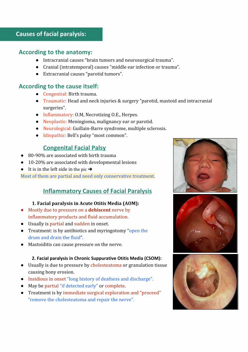
Congenital Facial Palsy
● 80-90% are associated with birth trauma ● 10-20% are associated with developmental lesions ● It is in the left side in the pic ➜
Most of them are partial and need only conservative treatment.
Inflammatory Causes of Facial Paralysis
1. Facial paralysis in Acute Otitis Media (AOM): ● Mostly due to pressure on a dehiscent nerve by
inflammatory products and fluid accumulation. ● Usually is partial and sudden in onset. ● Treatment: is by antibiotics and myringotomy “open the
drum and drain the fluid”. ● Mastoiditis can cause pressure on the nerve.
2. Facial paralysis in Chronic Suppurative Otitis Media (CSOM):
● Usually is due to pressure by cholesteatoma or granulation tissue causing bony erosion.
● Insidious in onset “long history of deafness and discharge”. ● May be partial “if detected early” or complete. ● Treatment is by immediate surgical exploration and “proceed”
“remove the cholesteatoma and repair the nerve”.
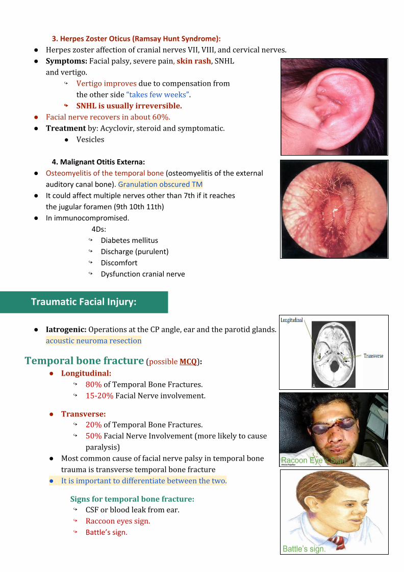
3. Herpes Zoster Oticus (Ramsay Hunt Syndrome):
● Herpes zoster affection of cranial nerves VII, VIII, and cervical nerves. ● Symptoms: Facial palsy, severe pain, skin rash, SNHL
and vertigo. ↪ Vertigo improves due to compensation from
the other side “takes few weeks”. ↪↪ SNHL is usually irreversible.
● Facial nerve recovers in about 60%. ● Treatment by: Acyclovir, steroid and symptomatic.
● Vesicles
4. Malignant Otitis Externa:
● Osteomyelitis of the temporal bone (osteomyelitis of the external
auditory canal bone). Granulation obscured TM
● It could affect multiple nerves other than 7th if it reaches
the jugular foramen (9th 10th 11th)
● In immunocompromised.
4Ds:
↪ Diabetes mellitus
↪ Discharge (purulent)
↪ Discomfort
↪ Dysfunction cranial nerve
Traumatic Facial Injury:
● Iatrogenic: Operations at the CP angle, ear and the parotid glands. acoustic neuroma resection
Temporal bone fracture (possible MCQ): ● Longitudinal:
↪ 80% of Temporal Bone Fractures. ↪ 15-20% Facial Nerve involvement.
● Transverse: ↪ 20% of Temporal Bone Fractures. ↪ 50% Facial Nerve Involvement (more likely to cause
paralysis) ● Most common cause of facial nerve palsy in temporal bone
trauma is transverse temporal bone fracture ● It is important to differentiate between the two.
Signs for temporal bone fracture: ↪ CSF or blood leak from ear. ↪ Raccoon eyes sign. ↪ Battle’s sign.
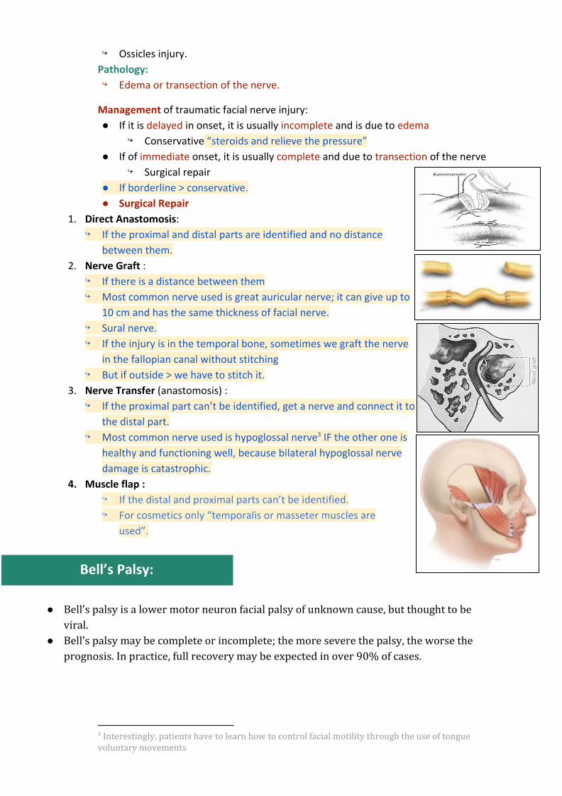
↪ Ossicles injury.
Pathology:
↪ Edema or transection of the nerve.
Management of traumatic facial nerve injury:
● If it is delayed in onset, it is usually incomplete and is due to edema
↪ Conservative “steroids and relieve the pressure”
● If of immediate onset, it is usually complete and due to transection of the nerve
↪ Surgical repair
● If borderline > conservative.
● Surgical Repair
1. Direct Anastomosis: ↪ If the proximal and distal parts are identified and no distance
between them.
2. Nerve Graft : ↪ If there is a distance between them
↪ Most common nerve used is great auricular nerve; it can give up to
10 cm and has the same thickness of facial nerve.
↪ Sural nerve.
↪ If the injury is in the temporal bone, sometimes we graft the nerve
in the fallopian canal without stitching
↪ But if outside > we have to stitch it.
3. Nerve Transfer (anastomosis) :
↪ If the proximal part can’t be identified, get a nerve and connect it to
the distal part.
↪ Most common nerve used is hypoglossal nerve IF the other one is 3
healthy and functioning well, because bilateral hypoglossal nerve
damage is catastrophic.
4. Muscle flap :
↪ If the distal and proximal parts can’t be identified.
↪ For cosmetics only “temporalis or masseter muscles are
used”.
Bell’s Palsy:
● Bell’s palsy is a lower motor neuron facial palsy of unknown cause, but thought to be viral.
● Bell’s palsy may be complete or incomplete; the more severe the palsy, the worse the prognosis. In practice, full recovery may be expected in over 90% of cases.
3 Interestingly, patients have to learn how to control facial motility through the use of tongue voluntary movements
● The remainder may develop persistent paralysis and other complications including ectropion (weakness of the muscles of the lower eyelid causing persistent overflow of tears) or an aberrant sequence of movements of the face (synkinesis ). 4
● CT or MRI scanning may be needed if the symptoms persist or a specific cause (i.e. other than Bell’s palsy) is suspected.
● Electrodiagnosis is used in the assessment of the degree of involvement of the nerve and includes nerve conduction tests and electromyography. These tests are done in a specialist centre and be invaluable in predicting prognosis.
● Most common diagnosis of acute facial paralysis “if slowly progressive it is NOT Bell’s palsy”.
● Diagnosis is by exclusion.
Pathology: ● Edema of the facial nerve sheath along its entire intratemporal course (Fallopian canal)
“if mild edema > neuropraxia, if severe > degeneration”.
Etiology: ● Vascular or viral measles, cold weather (not proven)
Clinical features: ● Sudden onset unilateral LM FP “Occurs after exposure to cold weather > could be vascular
spasm”. Pain behind the ear > few hours later facial paralysis. ● Partial or complete. ● No other manifestations apart from occasional mild pain “No discharge, no parotid
swelling, not following trauma”. ● May recur in 6 – 12% “previous history of paralysis in the same side “12%” other side
“6%”. ● Family history and pregnancy. “risk factors”
Prognosis: “if left untreated”
● 80% complete recovery. ● 10% satisfactory recovery. ● 10% no recovery. Partial usually recovers within 4-6 weeks while complete may take up to 6 months.
*Surgery is not usually done because most of patients recover with conservative treatment.
Treatment: ● Reassurance.
● Eye protection.
● Physiotherapy.
● Medications (steroids “to decrease edema”, antivirals, vasodilators) “antiviral and
vasodilators only given in combination with steroids, not effective alone”.
● Surgical decompression in selected cases:
↪ Patients with 90% degeneration.
4 E.g. squinting when smiling.
↪ Within 14 days of onset.
Ramsay Hunt syndrome:
● This is due to herpes zoster infection of the geniculate ganglion, affecting more rarely the glossopharyngeal (IX) and vagus (X) nerves and, very occasionally, the trigeminal (V), abducens (VI) or hypoglossal (XII) nerves. Mostly 7th and 8th
● The patient is usually elderly, and severe pain precedes the facial palsy. ● The patient often has vertigo (reversible), and the hearing is impaired (irreversible). ● The characteristic clinical feature is a vesicular eruption in the ear (sometimes on the tongue
and palate). ● Recovery of facial nerve function is much less likely than in Bell’s palsy. ● Prompt treatment with acyclovir given orally may improve the prognosis and reduce
post-herpetic neuralgia.
Questions from the doctor’s slides (group A)
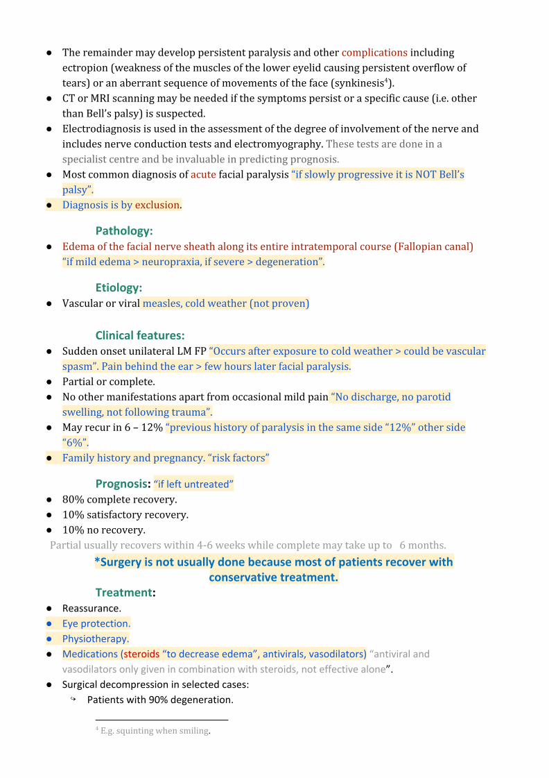
What is the most likely diagnosis? Left lower motor neuron facial paralysis (most likely bell’s palsy). Mention 2 common causes? ● Bell’s palsy (most common) ● Temporal bone fracture ● Acute otitis media 36 years old man with RTA: What is your diagnosis? Transverse fracture of the temporal bone.
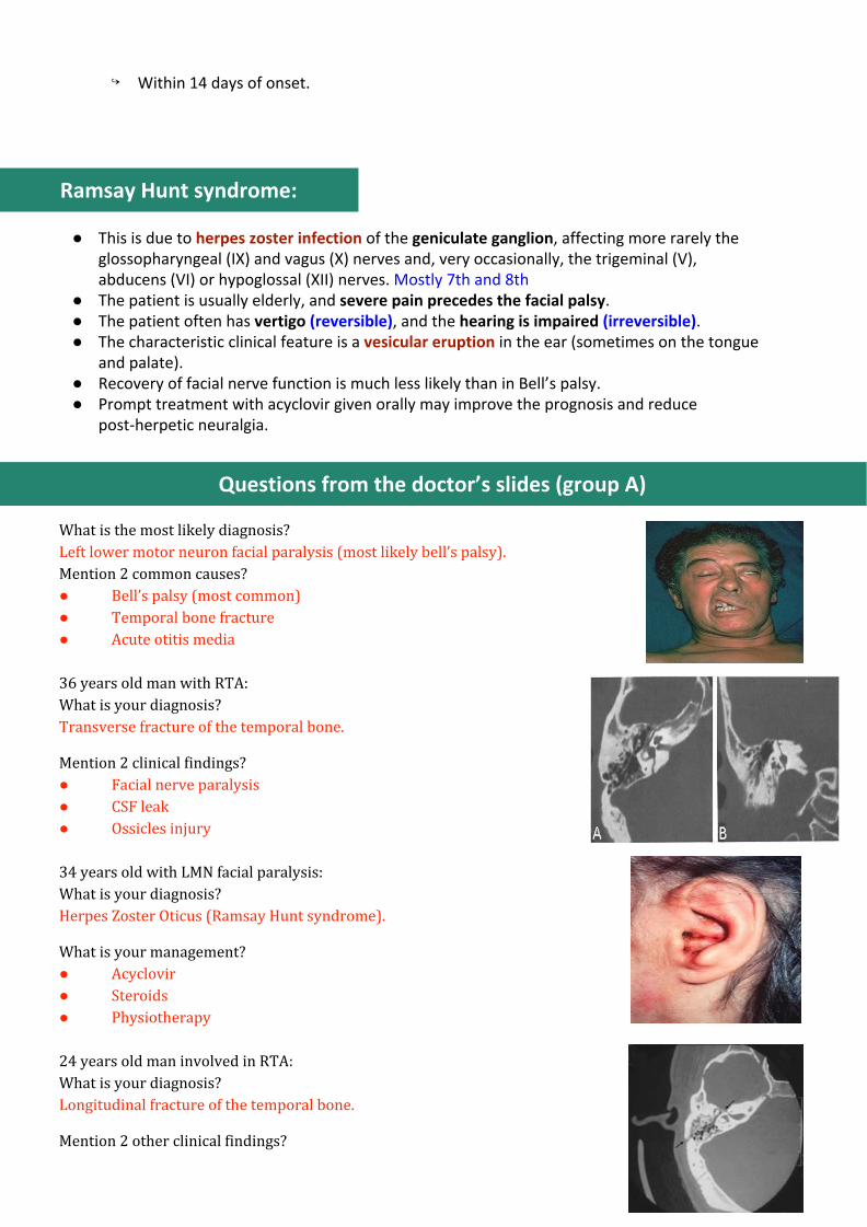
Mention 2 clinical findings? ● Facial nerve paralysis ● CSF leak ● Ossicles injury 34 years old with LMN facial paralysis: What is your diagnosis? Herpes Zoster Oticus (Ramsay Hunt syndrome).
What is your management? ● Acyclovir ● Steroids ● Physiotherapy 24 years old man involved in RTA: What is your diagnosis? Longitudinal fracture of the temporal bone.
Mention 2 other clinical findings?

● Facial nerve paralysis ● CSF leak ● Ossicles injury