re-activation of atrophic motor schwann cells after hypoglossal-facial nerve anastomosis
TRANSCRIPT

A
tritSdrptm©
K
TslmtSrtcsttt
Kf
0d
Available online at www.sciencedirect.com
Neuroscience Letters 434 (2008) 253–259
Re-activation of atrophic motor Schwann cells afterhypoglossal-facial nerve anastomosis
Maria Adele Rueger a,b, Sandra Aras a, Orlando Guntinas-Lichius c, Wolfram F. Neiss a,∗a Institut I fur Anatomie, der Universitat zu Koln, D-50924 Koln, Germany
b Klinik und Poliklinik fur Neurologie der Universitat zu Koln; D-50924 Koln, Germanyc Klinik fur Hals-, Nasen- und Ohrenheilkunde der Friedrich-Schiller-Universitat Jena, D-07740 Jena, Germany
Received 6 November 2007; received in revised form 13 January 2008; accepted 16 January 2008
bstract
Facial nerve lesions are common in humans and often require surgical intervention. If repair is delayed, reinnervation can be facilitated byransposing the freshly cut hypoglossal nerve end-to-end directly to the distal facial nerve, allowing for uncompromised hypoglossal axons toeinnervate the denervated facial musculature (hypoglossal-facial anastomosis, HFA). Schwann cells (SCs) in the distal nerve stump have anmportant function in promoting axonal regeneration by expressing multiple regeneration-associated proteins. Chronically denervated SCs ceaseo express those factors, but it is unknown whether they can be reactivated by fresh axonal sprouts and regain part of their function. We evaluatedC function and viability in distal facial nerve stump of rats at various time points after chronic denervation as well as following immediate orelayed HFA by assessing their expression of growth-associated protein 43 kDa (GAP-43) and the neuregulin receptors erbB2 and erbB4. Ouresults show that maximal upregulation of those factors in denervated SCs occurred a few weeks after nerve transection, indicating that a short
eriod of denervation might even be beneficial before nerve repair. Motor SCs denervated for 32 weeks had downregulated their activity and ceasedo express the regeneration-associated factors. SCs immediately re-expressed GAP-43, erbB2, and erbB4 following contact with fresh hypoglossalotor axons, demonstrating they are competent to promote regeneration even after long-term denervation. 2008 Elsevier Ireland Ltd. All rights reserved.
dener
tmbtpcrHte
eywords: Schwann cells; Hypoglossal-facial anastomosis (HFA); Long-term
he facial nerve in humans is often prone to injuries requiringurgical intervention, but an immediate reconstruction of theesioned nerve is seldom possible. In contrast to typical animal
odels for nerve regeneration studies, there is a certain denerva-ion time of the affected nerve between lesion and reconstruction.everal problems interfere with the success of peripheral nerveeinnervation after extended periods of time. One issue concernshe ability of neurons to regenerate their axons, which is signifi-antly reduced after prolonged axotomy [13,17]. Therefore, theurgical procedure frequently used to address this problem is
o transpose freshly cut hypoglossal nerve end-to-end directlyo the facial nerve (hypoglossal-facial anastomosis, HFA). Inhis paradigm, uncompromised hypoglossal axons regrow into∗ Corresponding author at: Institut I fur Anatomie, Geb. 35, Universitat zuoln, Kerpener Strasse 62, 50937 Koln, Germany. Tel.: +49 221 478 5016;
ax: +49 221 478 7454.E-mail address: [email protected] (W.F. Neiss).
aprfdaoas
304-3940/$ – see front matter © 2008 Elsevier Ireland Ltd. All rights reserved.oi:10.1016/j.neulet.2008.01.073
vation; GAP-43; Neuregulin receptors
he facial nerve branches and reinnervate the denervated facialusculature [1,27]. Yet another difficulty is that the distal nerve
ranches need to be permissive to regeneration as well. Inhis context, Schwann cells (SCs) have an important role inromoting regeneration in the peripheral nerve, by mechani-ally guiding the re-growing axons and by expressing numerousegeneration-associated factors to promote axonal outgrowth.owever, SCs denervated for a long period of time downregulate
heir activity, atrophy and may eventually die [20,24]. Undoubt-dly, long-term denervated SCs are less capable of promotingxonal regeneration, but it is still largely unknown whether atro-hied SCs in the distal stump of the peripheral nerve can beeactivated by fresh axonal sprouts and regain some degree ofunction. Studies in rat models of delayed peripheral nerve sutureemonstrate that SCs from mixed nerves (i.e. conferring sensory
s well as motor functions) such as sciatic, common peroneal,r tibial nerve, are able to support axonal regeneration evenfter long periods of denervation [22,37]. However, motor andensory neurons require different growth factors for regenera-
2 ence L
tmtsdbiptdrbpa
apcnui
efrohid[
ma1SttRsn
miRwtTttwa(lTiwA
otffnsfHavwat8o
lbonfNcwTt1#n((tAecpm
nZasfAfltaaowvvg
54 M.A. Rueger et al. / Neurosci
ion [23], and recent work even suggests that SCs isolated fromotor nerves are less able to promote axonal regeneration than
hose in sensory nerves [39]. A recent study shows that SCs inensory and motor nerves have quite distinct phenotypes thatiffer in their patterns of trophic factor expression followingoth denervation and reinnervation [21]. Since the facial nerves an exclusive motor nerve, findings in animal models of mixederipheral nerve regeneration can therefore not be unequivocallyranslated into the most clinically relevant facial nerve lesion. Toetermine the ability of motor SCs to promote peripheral nerveegeneration, we evaluated Schwann cell function and viabilityy assessing their expression of several regeneration-associatedroteins at various time points after chronic denervation, as wells after immediate or delayed HFA in rats.
Growth-associated protein 43 kDa (GAP-43), recognized asneuronal marker, is also expressed in certain glial cells, e.g. SCrecursors, non-myelinating SCs, immature oligodendroglialells and type 2 astrocytes [9,32]. After injury to the peripheralerve, denervated myelinating SCs of the distal stump upreg-late GAP-43 [29,31], which functions as a plasticity proteinnvolved in cell shape and motility [9,31].
The erbB receptor tyrosine kinases – includingrbB2/p185/HER2/neu, erbB3 and erB4 – bind the neuregulinamily of growth factors [7]. ErbB3 and erbB4 are the specificeceptors for neuregulins, and erbB2 transphosphorylationccurs when ligand binding induces erbB3 or erbB4 to form aeterodimer with erbB2 [6]. Neuregulin receptors are expressedn SC precursons, re-expressed in mature SCs after injury andownregulated again after re-establishment of axonal contact8].
All experiments were conducted in accordance with the Ger-an law on the protection of animals and approved by the local
nimal committee (Bezirksregierung Koln, Az. 23.203.2-K35,3/95). Four different experimental models were used to producechwann cells that had been denervated for varying periods of
ime. In all paradigms, denervation was achieved by transectinghe facial nerve and preventing its spontaneous regeneration.epair was achieved after various periods of denervation by
uturing the proximal stump of freshly transected hypoglossalerve end-to-end to the distal stump of the facial nerve (HFA).
Adult female Wistar rats (Strain HsdCpb:Wu; Harlan Winkel-ann, Borchen, Germany) were anesthetized by intraperitoneal
njection of ketamine plus xylazine (100 mg Ketanest plus 5 mgompun per kilogram of body weight). The right facial nerveas exposed and transected 2 mm beyond its emergence from
he foramen stylomastoideum but distal to its auricular branch.he proximal stump was ligated and sutured into a pouch of the
rapezius muscle to prevent its spontaneous regeneration intohe distal stump. Rats were then treated in one of the followingays: (i) Chronic denervation: after denervation, animals were
llowed to recover for 4 days or 1, 2, 4, 6, 8, 16, or 32 weeksn = 5 in each group). (ii) Immediate HFA: facial nerve and ipsi-ateral hypoglossal nerve were transected in the same surgery.
he hypoglossal nerve was transected proximal to its bifurcationnto a medial and lateral main branch, and its proximal stumpas sutured end-to-end to the distal stump of the facial nerve.nimals were allowed to recover for 4 days or 1, 2, 4, 6, 8, 16,
wn(a
etters 434 (2008) 253–259
r 32 weeks after HFA (n = 4 or 5 in each group). (iii) Short-erm delayed HFA: animals were allowed to recover for 14 daysollowing denervation. At this point, the predegenerated distalacial nerve stump was re-exposed. The ipsilateral hypoglossalerve was transected as described above and its proximal stumputured end-to-end to the distal stump of the predegeneratedacial nerve. Rats were perfused 1, 2, 4, 6, 8, or 16 weeks afterFA (n = 4 or 5 in each group). (iv) Long-term delayed HFA:
nimals were allowed to recover for 32 weeks following dener-ation. At this point, the predegenerated distal facial nerve stumpas re-exposed, the ipsilateral hypoglossal nerve was transected
nd its proximal stump sutured end-to-end to the distal stump ofhe predegenerated facial nerve. Rats were perfused 1, 2, 4, 6,, or 16 weeks after HFA (n = 4 or 5 in each group). For detailsf microsurgery see [27] and [17,19].
Under deep ether anesthesia rats were perfused through theeft heart ventricle with 1.25% paraformaldehyde in phosphateuffer pH 7.4. The buccal and the marginal mandibular branchf the facial nerve were removed 18 mm distal to the crossederve suture and postfixed overnight in 4% paraformaldehyde,ollowed by cryoprotection in 30% sucrose at 4 ◦C for 7 days.erves were embedded in Tissue-Tek and frozen in isopentane
ooled in liquid nitrogen. 10-�m sections of the facial nerveere cut and thaw-mounted on poly-l-lysine precoated slides.he following primary antibodies were used for immunohis-
ochemistry: 1:400 polyclonal anti-GAP-43 (Progen, #16024),:400 polyclonal anti-erbB4 (Santa Cruz Biotechnology Inc.,sc-283), 1:1000 polyclonal anti-erbB2 (Santa Cruz Biotech-ology Inc., #sc-284), 1:2400 polyclonal rabbit-anti-S100DAKO, #Z0311), and 1:400 monoclonal mouse-anti-NF160Boehringer Mannheim Biochemica, #814334). For visualiza-ion we used cy3-conjugated sheep-anti-rabbit IgG (Sigma) andlexaFluor488 goat-anti-mouse IgG (MoBiTec). In parallel to
ach section incubated with a primary antibody, an adjacentontrol section was treated identically but for omission of therimary antibody. All sections were cover-slipped with Fluoro-ount (BDH Laboratory Supplies).A Zeiss Axioplan microscope with an objective Zeiss Plan
eofluar 40/1.3 oil objective, equipped with a power-stabilizedeiss HBO100/XBO75 epifluorescence illumination unit withHBO103 mercury short arc photo optic lamp and a SPOT
low scan CCD camera (Diagnostic Instruments), was usedor image acquisition. Cy3 and Cy2 selective filter sets (AHFnalysetechnik, Tubingen) were used to detect red and greenuorescence, respectively. All images were recorded using iden-
ical illumination parameters and camera settings. The imagenalysis software OPTIMAS 6.5 was used to determine themount of the marked antigen within each section. The intensityf brightness, corresponding to the density of marked antigen,as determined in an area of interest (AOI) analysis of the indi-idual nerve fascicles (endoneural compartment). A mean greyalue “a” was calculated for each stained section, while a meanrey value “b” was determined for the adjacent control section
ithout primary antibody. The amount of specific immunosig-al intensity was then calculated in relative fluorescence unitsRFU) as (a − b)/b, representing the density of immunoreactiventigen in the endoneural space of each nerve studied. Mean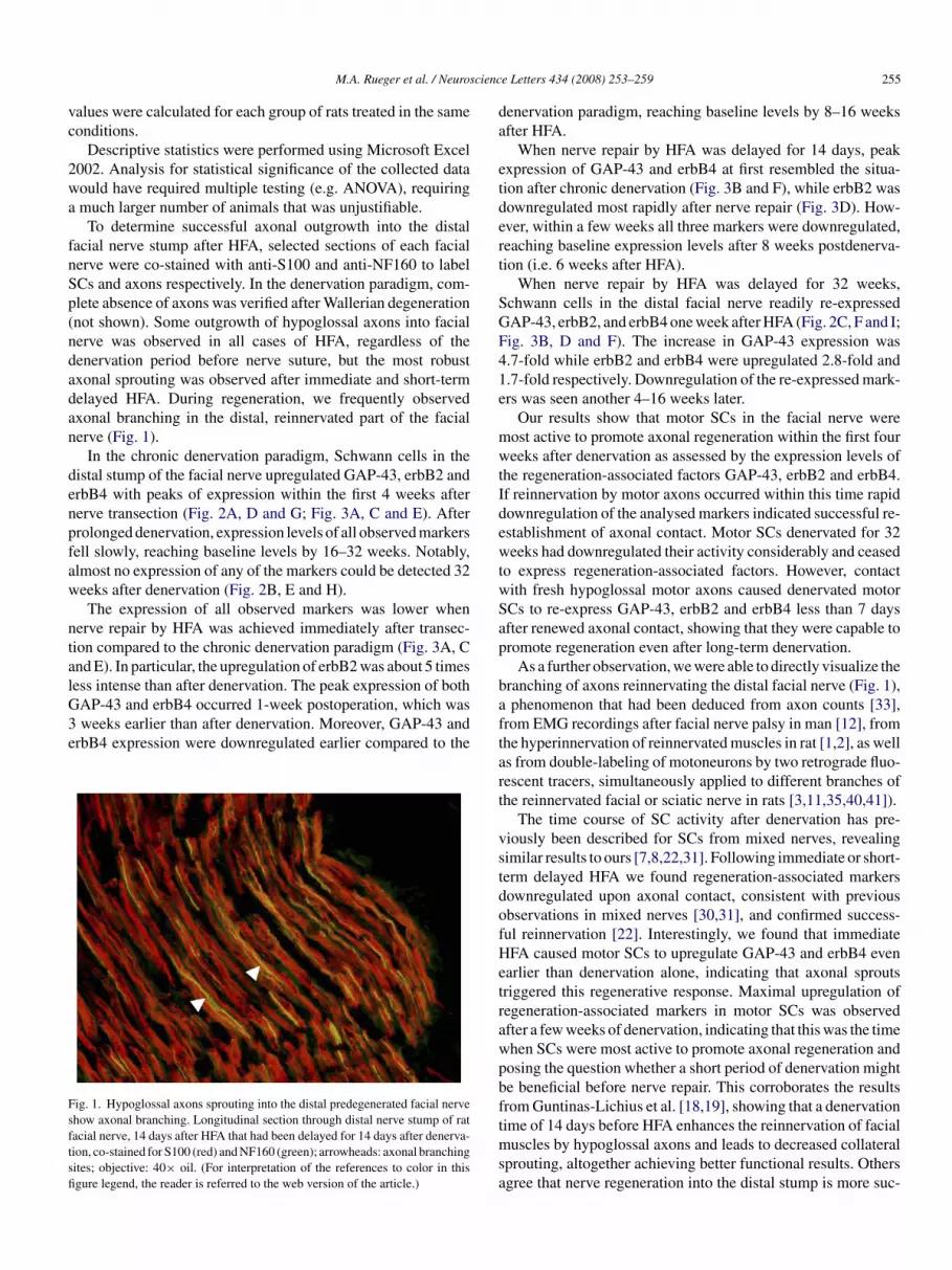
cienc
vc
2wa
fnSp(ndadan
denpfaw
ntalG3e
Fsftsfi
da
etdert
SGF41e
mwtIdewtwSap
b
M.A. Rueger et al. / Neuros
alues were calculated for each group of rats treated in the sameonditions.
Descriptive statistics were performed using Microsoft Excel002. Analysis for statistical significance of the collected dataould have required multiple testing (e.g. ANOVA), requiringmuch larger number of animals that was unjustifiable.
To determine successful axonal outgrowth into the distalacial nerve stump after HFA, selected sections of each facialerve were co-stained with anti-S100 and anti-NF160 to labelCs and axons respectively. In the denervation paradigm, com-lete absence of axons was verified after Wallerian degenerationnot shown). Some outgrowth of hypoglossal axons into facialerve was observed in all cases of HFA, regardless of theenervation period before nerve suture, but the most robustxonal sprouting was observed after immediate and short-termelayed HFA. During regeneration, we frequently observedxonal branching in the distal, reinnervated part of the facialerve (Fig. 1).
In the chronic denervation paradigm, Schwann cells in theistal stump of the facial nerve upregulated GAP-43, erbB2 andrbB4 with peaks of expression within the first 4 weeks aftererve transection (Fig. 2A, D and G; Fig. 3A, C and E). Afterrolonged denervation, expression levels of all observed markersell slowly, reaching baseline levels by 16–32 weeks. Notably,lmost no expression of any of the markers could be detected 32eeks after denervation (Fig. 2B, E and H).The expression of all observed markers was lower when
erve repair by HFA was achieved immediately after transec-ion compared to the chronic denervation paradigm (Fig. 3A, Cnd E). In particular, the upregulation of erbB2 was about 5 timesess intense than after denervation. The peak expression of both
AP-43 and erbB4 occurred 1-week postoperation, which wasweeks earlier than after denervation. Moreover, GAP-43 andrbB4 expression were downregulated earlier compared to the
ig. 1. Hypoglossal axons sprouting into the distal predegenerated facial nervehow axonal branching. Longitudinal section through distal nerve stump of ratacial nerve, 14 days after HFA that had been delayed for 14 days after denerva-ion, co-stained for S100 (red) and NF160 (green); arrowheads: axonal branchingites; objective: 40× oil. (For interpretation of the references to color in thisgure legend, the reader is referred to the web version of the article.)
aftart
vstdofHetrawpbftmsa
e Letters 434 (2008) 253–259 255
enervation paradigm, reaching baseline levels by 8–16 weeksfter HFA.
When nerve repair by HFA was delayed for 14 days, peakxpression of GAP-43 and erbB4 at first resembled the situa-ion after chronic denervation (Fig. 3B and F), while erbB2 wasownregulated most rapidly after nerve repair (Fig. 3D). How-ver, within a few weeks all three markers were downregulated,eaching baseline expression levels after 8 weeks postdenerva-ion (i.e. 6 weeks after HFA).
When nerve repair by HFA was delayed for 32 weeks,chwann cells in the distal facial nerve readily re-expressedAP-43, erbB2, and erbB4 one week after HFA (Fig. 2C, F and I;ig. 3B, D and F). The increase in GAP-43 expression was.7-fold while erbB2 and erbB4 were upregulated 2.8-fold and.7-fold respectively. Downregulation of the re-expressed mark-rs was seen another 4–16 weeks later.
Our results show that motor SCs in the facial nerve wereost active to promote axonal regeneration within the first foureeks after denervation as assessed by the expression levels of
he regeneration-associated factors GAP-43, erbB2 and erbB4.f reinnervation by motor axons occurred within this time rapidownregulation of the analysed markers indicated successful re-stablishment of axonal contact. Motor SCs denervated for 32eeks had downregulated their activity considerably and ceased
o express regeneration-associated factors. However, contactith fresh hypoglossal motor axons caused denervated motorCs to re-express GAP-43, erbB2 and erbB4 less than 7 daysfter renewed axonal contact, showing that they were capable toromote regeneration even after long-term denervation.
As a further observation, we were able to directly visualize theranching of axons reinnervating the distal facial nerve (Fig. 1),phenomenon that had been deduced from axon counts [33],
rom EMG recordings after facial nerve palsy in man [12], fromhe hyperinnervation of reinnervated muscles in rat [1,2], as wells from double-labeling of motoneurons by two retrograde fluo-escent tracers, simultaneously applied to different branches ofhe reinnervated facial or sciatic nerve in rats [3,11,35,40,41]).
The time course of SC activity after denervation has pre-iously been described for SCs from mixed nerves, revealingimilar results to ours [7,8,22,31]. Following immediate or short-erm delayed HFA we found regeneration-associated markersownregulated upon axonal contact, consistent with previousbservations in mixed nerves [30,31], and confirmed success-ul reinnervation [22]. Interestingly, we found that immediateFA caused motor SCs to upregulate GAP-43 and erbB4 even
arlier than denervation alone, indicating that axonal sproutsriggered this regenerative response. Maximal upregulation ofegeneration-associated markers in motor SCs was observedfter a few weeks of denervation, indicating that this was the timehen SCs were most active to promote axonal regeneration andosing the question whether a short period of denervation mighte beneficial before nerve repair. This corroborates the resultsrom Guntinas-Lichius et al. [18,19], showing that a denervation
ime of 14 days before HFA enhances the reinnervation of facialuscles by hypoglossal axons and leads to decreased collateralprouting, altogether achieving better functional results. Othersgree that nerve regeneration into the distal stump is more suc-

256 M.A. Rueger et al. / Neuroscience Letters 434 (2008) 253–259
Fig. 2. Long-term denervated Schwann cells cease to express regeneration-associated markers, but are reactivated by delayed HFA. Longitudinal sections throughd erbB2p ation)h
cpaa
a
eb
istal nerve stumps of rat facial nerves, labeled with anti-GAP-43 (A–C), anti-rotein after short-term denervation (A: 28 days, D: 7 days, G: 42 days denervad been delayed for 32 weeks; objective: 40× oil.
essful after Wallerian degeneration has taken place [25,28,43],ossibly because more space has been provided after removal of
xonal and myelin debris [36] and inhibitors of axonal growthnd regeneration have been removed [5,26].The capability of long-term denervated SCs to promotexonal regeneration is a much debated topic in the field of periph-
ttci
(D–F), and anti-erbB4 (G–I); left row: maximal expression of the respective; middle row: long-term denervation of 32 weeks; right row: 7 days after HFA
ral nerve regeneration. Undoubtedly, long-term denervationefore nerve repair impairs axonal regeneration into the dis-
al stump and worsens the functional result. However, whetherhis is due to denervated SCs lacking the necessary regenerativeapacity has not been comprehensively shown to date. Our datandicate that chronically denervated motor SCs are still able to
M.A. Rueger et al. / Neuroscience Letters 434 (2008) 253–259 257
Fig. 3. Dynamic regulation of regeneration-associated markers in Schwann cells after acute denervation with or without successful reinervation. Synopsis of thee ; lefta yed Hi
ptSioerbaowfatch
oli
tatfSitt
xpression time courses of GAP-43 (A + B), erbB2 (C + D), and erbB4 (E + F)nd after short- or long-term delayed HFA; arrows indicate time points of delammediately following HFA compared to denervation without HFA.
romote regeneration of motor axons by upregulation of pro-eins necessary for axonal outgrowth. In chronically denervatedCs of mixed nerves Li et al. [22] found GAP-43 mRNA signif-
cantly upregulated by nerve suture, but only little upregulationf erbB2 or erbB4 expression. Likewise, Hall [20] did not detectrbB2 or erbB4 after 6 months delayed crossed nerve suture inat sciatic nerve. SCs of pure motor nerves therefore seem toe more permissive to regeneration than those of mixed motornd sensory nerves. This was suggested by Sulaiman et al. [38],bserving a reduced responsiveness of sensory Schwann cellshen regenerating motor axons down a nerve graft. Those dif-
erences between motor and sensory SCs might explain why Fu
nd Gordon [13,14] found less robust reinnervation after long-erm denervation using a posterior tibial – common peronealrossed nerve suture than Guntinas-Lichius et al. [19] using theypoglossal – facial anastomosis. In mixed nerves, the numbervapr
row: after denervation and after immediate HFA; right row: after denervationFA with fine dotted lines illustrating the change in marker expression levels
f neurons successfully reaching the target tissue decreases withonger periods of denervation prior to repair [37,38], leading tompaired restoration of muscle function [14].
Additional explanations for impaired nerve repair after long-erm denervation are progressive Schwann cell atrophy [34]nd discontinuity, dispersion, and partial disappearance ofhe Schwann cell basement membrane as the necessary scaf-old along which axons grow back [15]. However, denervatedchwann cells have been shown to survive for at least 25 months
n denervated distal stumps [10], and to sustain their abilityo properly remyelinate the regenerated axons [37]. Moreover,here is evidence that collagen formation in chronically dener-
ated nerve stumps, often considered as a mechanical barriergainst axonal regeneration [16], is not necessarily harmful,roviding a channel for regenerating axons [42]. Neurons canegenerate through long-term denervated distal nerve stumps
2 ence L
[pFaol
tcob
A
fgT
R
[
[
[
[
[
[
[
[
[
[
[
[
[
[
[
[
[
[
[
[
[
[
[
58 M.A. Rueger et al. / Neurosci
19,42], and more difficult reinnervation can then be com-ensated by allowing for longer periods of regeneration [38].urthermore, re-activation of chronically denervated SCs can beugmented in vitro by applying glial growth factor (GGF [20])r transforming growth factor-ß (TGF-ß [38]), and in vivo withow doses of brain-derived neurotrophic factor (BDNF [4]).
Here we demonstrate the long-term competence of motor SCso facilitate axonal regeneration. Our data indicate that underircumstances where motor neuron repair is delayed, applicationf growth factors in vivo to support denervated SC survival coulde of significant clinical value.
cknowledgements
The authors thank Mrs. D. Mardirian and Mrs. I. Rohrmannor excellent technical assistance, Mrs. I. Koch for the photo-raphic artwork and Dr. D.N. Angelov for fruitful discussions.his work was supported by the Jean Uhrmacher Foundation.
eferences
[1] D.N. Angelov, A. Gunkel, E. Stennert, W.F. Neiss, Recovery of originalnerve supply after hypoglossal-facial anastomosis causes permanent motorhyperinnervation of the whisker-pad muscles in the rat, J. Comp. Neurol.338 (1993) 214–224.
[2] D.N. Angelov, W.F. Neiss, M. Streppel, J. Andermahr, K. Mader, E. Sten-nert, Nimodipine accelerates axonal sprouting after surgical repair of ratfacial nerve, J. Neurosci. 16 (1996) 1041–1048.
[3] D.N. Angelov, E. Skouras, O. Guntinas-Lichius, M. Streppel, A.Popratiloff, M. Walther, J. Klein, E. Stennert, W.F. Neiss, Contralateraltrigeminal nerve lesion reduces polyneuronal muscle innervation afterfacial nerve repair in rats, Eur. J. Neurosci. 11 (1999) 1369–1378.
[4] J.G. Boyd, T. Gordon, A dose-dependent facilitation and inhibition ofperipheral nerve regeneration by brain-derived neurotrophic factor, Eur.J. Neurosci. 15 (2002) 613–626.
[5] T.M. Brushart, J. Gerber, P. Kessens, Y.G. Chen, R.M. Royall, Contributionsof pathway and neuron to preferential motor reinnervation, J. Neurosci. 18(1998) 8674–8681.
[6] K.L. Carraway III, L.C. Cantley, A neu acquaintance for erbB3 and erbB4:a role for receptor heterodimerization in growth signaling, Cell 78 (1994)5–8.
[7] S.L. Carroll, M.L. Miller, P.W. Frohnert, S.S. Kim, J.A. Corbett, Expressionof neuregulins and their putative receptors, ErbB2 and ErbB3, is inducedduring Wallerian degeneration, J. Neurosci. 17 (1997) 1642–1659.
[8] J.A. Cohen, A.T. Yachnis, M. Arai, J.G. Davis, S.S. Scherer, Expression ofthe neu proto-oncogene by Schwann cells during peripheral nerve devel-opment and Wallerian degeneration, J. Neurosci. Res. 31 (1992) 622–634.
[9] R. Curtis, H.J. Stewart, S.M. Hall, G.P. Wilkin, R. Mirsky, K.R. Jessen,GAP-43 is expressed by nonmyelin-forming Schwann cells of the periph-eral nervous system, J. Cell Biol. 116 (1992) 1455–1464.
10] E.I. Dedkov, T.Y. Kostrominova, A.B. Borisov, B.M. Carlson, Survivalof Schwann cells in chronically denervated skeletal muscles, Acta Neu-ropathol. (Berl) 103 (2002) 565–574.
11] S. Dohm, M. Streppel, O. Guntinas-Lichius, P. Pesheva, R. Probstmeier,M. Walther, W.F. Neiss, E. Stennert, D.N. Angelov, Local application ofextracellular matrix proteins fails to reduce the number of axonal branchesafter varying reconstructive surgery on rat facial nerve, Restor. Neurol.Neurosci. 16 (2000) 117–126.
12] E. Esslen, Electromyographic findings on two types of misdirection of
regenerating axons, Electroencephalogr. Clin. Neurophysiol. 12 (1960)738–741.13] S.Y. Fu, T. Gordon, Contributing factors to poor functional recoveryafter delayed nerve repair: prolonged axotomy, J. Neurosci. 15 (1995)3876–3885.
[
[
etters 434 (2008) 253–259
14] S.Y. Fu, T. Gordon, Contributing factors to poor functional recoveryafter delayed nerve repair: prolonged denervation, J. Neurosci. 15 (1995)3886–3895.
15] C. Giannini, P.J. Dyck, The fate of Schwann cell basement membranesin permanently transected nerves, J. Neuropathol. Exp. Neurol. 49 (1990)550–563.
16] T. Gordon, S.Y. Fu, Long-term response to nerve injury, Adv. Neurol. 72(1997) 185–199.
17] O. Guntinas-Lichius, M. Streppel, D.N. Angelov, E. Stennert, W.F. Neiss,Effect of delayed facial-facial nerve suture on facial nerve regeneration.A horseradish peroxidase tracing study in the rat, Acta Otolaryngol. 117(1997) 670–674.
18] O. Guntinas-Lichius, D.N. Angelov, E. Stennert, W.F. Neiss, Delayedhypoglossal-facial nerve suture after predegeneration of the peripheralfacial nerve stump improves the innervation of mimetic muscula-ture by hypoglossal motoneurons, J. Comp. Neurol. 387 (1997) 234–242.
19] O. Guntinas-Lichius, K. Effenberger, D.N. Angelov, J. Klein, M. Streppel,E. Stennert, W.F. Neiss, Delayed rat facial nerve repair leads to acceler-ated and enhanced muscle reinnervation with reduced collateral axonalsprouting during a definite denervation period using a cross-anastomosisparadigm, Exp. Neurol. 162 (2000) 98–111.
20] S.M. Hall, The biology of chronically denervated Schwann cells, Ann. N.Y. Acad. Sci. 883 (1999) 215–233.
21] A. Hoke, R. Redett, H. Hameed, R. Jari, C. Zhou, Z.B. Li, J.W.Griffin, T.M. Brushart, Schwann cells express motor and sensory phe-notypes that regulate axon regeneration, J. Neurosci. 26 (2006) 9646–9655.
22] H. Li, G. Terenghi, S.M. Hall, Effects of delayed re-innervation on theexpression of c-erbB receptors by chronically denervated rat Schwann cellsin vivo, Glia 20 (1997) 333–347.
23] R.D. Madison, S.J. Archibald, T.M. Brushart, Reinnervation accuracy ofthe rat femoral nerve by motor and sensory neurons, J. Neurosci. 16 (1996)5698–5703.
24] R. Mirsky, K.R. Jessen, The neurobiology of Schwann cells, Brain Pathol.9 (1999) 293–311.
25] Y. Miyamoto, T. Sugita, T. Higaki, Y. Ikuta, K. Tsuge, The duration ofdenervation and regeneration in nerve grafting. Quantitative histologicalassessment in the rat, Int. Orthop. 9 (1985) 271–276.
26] G. Mukhopadhyay, P. Doherty, F.S. Walsh, P.R. Crocker, M.T. Filbin, Anovel role for myelin-associated glycoprotein as an inhibitor of axonalregeneration, Neuron 13 (1994) 757–767.
27] W.F. Neiss, L.O. Guntinas, D.N. Angelov, A. Gunkel, E. Stennert, Thehypoglossal-facial anastomosis as model of neuronal plasticity in the rat,Ann. Anat. 174 (1992) 419–433.
28] M. Ochi, M. Wakasa, Y. Ikuta, W.H. Kwong, Nerve regeneration in prede-generated basal lamina graft: the effect of duration of predegeneration onaxonal extension, Exp. Neurol. 128 (1994) 216–225.
29] L.C. Plantinga, J. Verhaagen, P.M. Edwards, E.M. Hol, P.R. Bar, W.H.Gispen, The expression of B-50/GAP-43 in Schwann cells is upregulatedin degenerating peripheral nerve stumps following nerve injury, Brain Res.602 (1993) 69–76.
30] T. Saika, H. Kiyama, M. Tohyama, T. Matsunaga, GAP-43 mRNA expres-sion in facial motoneurons during regeneration: in situ hybridizationhistochemistry study using an alkaline phosphatase-labelled probe, ActaOtolaryngol. Suppl. 501 (1993) 80–84.
31] S.S. Scherer, Y.T. Xu, D. Roling, L. Wrabetz, M.L. Feltri, J. Kamholz,Expression of growth-associated protein-43 kD in Schwann cells is regu-lated by axon-Schwann cell interactions and cAMP, J. Neurosci. Res. 38(1994) 575–589.
32] M. Sensenbrenner, M. Lucas, J.C. Deloulme, Expression of two neuronalmarkers, growth-associated protein 43 and neuron-specific enolase, in ratglial cells, J. Mol. Med. 75 (1997) 653–663.
33] G.D. Shawe, On the number of branches formed by regenerating nerve-fibres, Br. J. Surg. 42 (1955) 474–488.
34] J. Siironen, V. Vuorinen, H.S. Taskinen, M. Roytta, Axonal regenerationinto chronically denervated distal stump. 2. Active expression of type I col-lagen mRNA in epineurium, Acta Neuropathol. (Berl) 89 (1995) 219–226.
cienc
[
[
[
[
[
[
[
[
M.A. Rueger et al. / Neuros
35] E. Skouras, A. Popratiloff, O. Guntinas-Lichius, M. Streppel, K.E. Rehm,W.F. Neiss, D.N. Angelov, Altered sensory input improves the accuracy ofmuscle reinnervation, Restor. Neurol. Neurosci. 20 (2002) 1–14.
36] G. Stoll, J.W. Griffin, C.Y. Li, B.D. Trapp, Wallerian degeneration inthe peripheral nervous system: participation of both Schwann cells andmacrophages in myelin degradation, J. Neurocytol. 18 (1989) 671–683.
37] O.A. Sulaiman, T. Gordon, Effects of short- and long-term Schwann celldenervation on peripheral nerve regeneration, myelination, and size, Glia32 (2000) 234–246.
38] O.A. Sulaiman, R. Midha, C.A. Munro, T. Matsuyama, A. Al Majed, T.
Gordon, Chronic Schwann cell denervation and the presence of a sensorynerve reduce motor axonal regeneration, Exp. Neurol. 176 (2002) 342–354.39] N. Tsubokawa, Y. Maki, T. Yoshizu, H. Narisawa, Comparison of the neu-rotropic effects of motor and sensory Schwann cells during regeneration
[
e Letters 434 (2008) 253–259 259
of peripheral nerves, Scand. J. Plast. Reconstr. Surg. Hand Surg. 33 (1999)379–385.
40] A. Valero-Cabre, K. Tsironis, E. Skouras, G. Perego, X. Navarro, W.F.Neiss, Superior muscle reinnervation after autologous nerve graft or poly-l-lactide-epsilon-caprolactone (PLC) tube implantation in comparison tosilicone tube repair, J. Neurosci. Res. 63 (2001) 214–223.
41] A. Valero-Cabre, K. Tsironis, E. Skouras, X. Navarro, W.F. Neiss, Periph-eral and spinal motor reorganization after nerve injury and repair, J.Neurotrauma 21 (2004) 95–108.
42] V. Vuorinen, J. Siironen, M. Roytta, Axonal regeneration into chronically
denervated distal stump. 1. Electron microscope studies, Acta Neuropathol.(Berl) 89 (1995) 209–218.43] Q. Zhao, J.M. Kerns, Effects of predegeneration on nerve regen-eration through silicone Y-chambers, Brain Res. 633 (1994) 97–104.