upper limb : muscles - ksumsc
TRANSCRIPT
Upper Limb : Muscles"Revision"
Anatomy Team 434
Color Index: ▪ Important Points
▪ Helping notes
▪ Explanation
If you have any complaint or suggestion please don’t
hesitate to contact us on:[email protected]
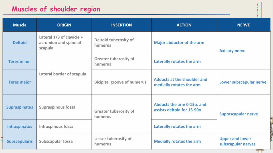
Muscles of shoulder region
Muscle ORIGIN INSERTION ACTION NERVE
DeltoidLateral 1/3 of clavicle + acromion and spine of scapula
Deltoid tuberosity of humerus
Major abductor of the arm
Axillary nerve
Teres minor
Lateral border of scapula
Greater tuberosity of humerus
Laterally rotates the arm
Teres major Bicipital groove of humerusAdducts at the shoulder and medially rotates the arm
Lower subscapular nerve
Supraspinatus Supraspinous fossaGreater tuberosity of humerus
Abducts the arm 0-15o, and assists deltoid for 15-90o
Suprascapular nerve
Infraspinatus Infraspinous fossa Laterally rotates the arm
Subscapularis Subscapular fossaLesser tuberosity of humerus
Medially rotates the armUpper and lower subscapular nerves
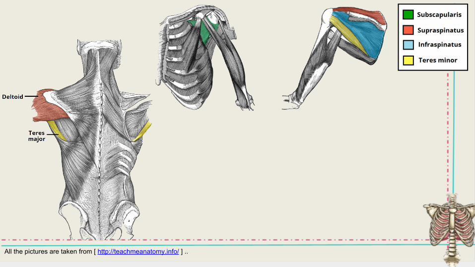
All the pictures are taken from [ http://teachmeanatomy.info/ ] ..
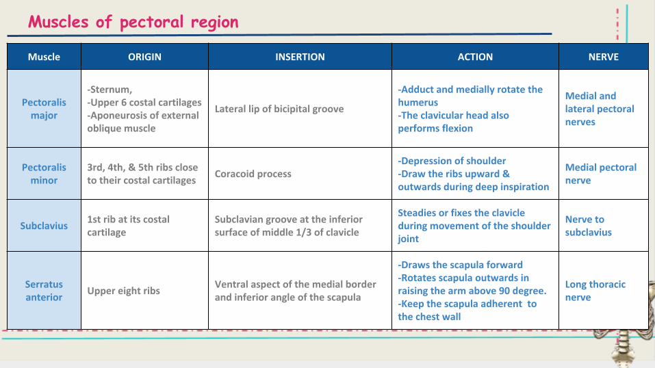
Muscles of pectoral region
Muscle ORIGIN INSERTION ACTION NERVE
Pectoralis major
-Sternum,-Upper 6 costal cartilages-Aponeurosis of external oblique muscle
Lateral lip of bicipital groove
-Adduct and medially rotate the humerus-The clavicular head also performs flexion
Medial and lateral pectoral nerves
Pectoralis minor
3rd, 4th, & 5th ribs close to their costal cartilages
Coracoid process-Depression of shoulder-Draw the ribs upward & outwards during deep inspiration
Medial pectoral nerve
Subclavius1st rib at its costal cartilage
Subclavian groove at the inferior surface of middle 1/3 of clavicle
Steadies or fixes the clavicle during movement of the shoulder joint
Nerve to subclavius
Serratus anterior
Upper eight ribsVentral aspect of the medial border and inferior angle of the scapula
-Draws the scapula forward-Rotates scapula outwards in raising the arm above 90 degree.-Keep the scapula adherent to the chest wall
Long thoracic nerve
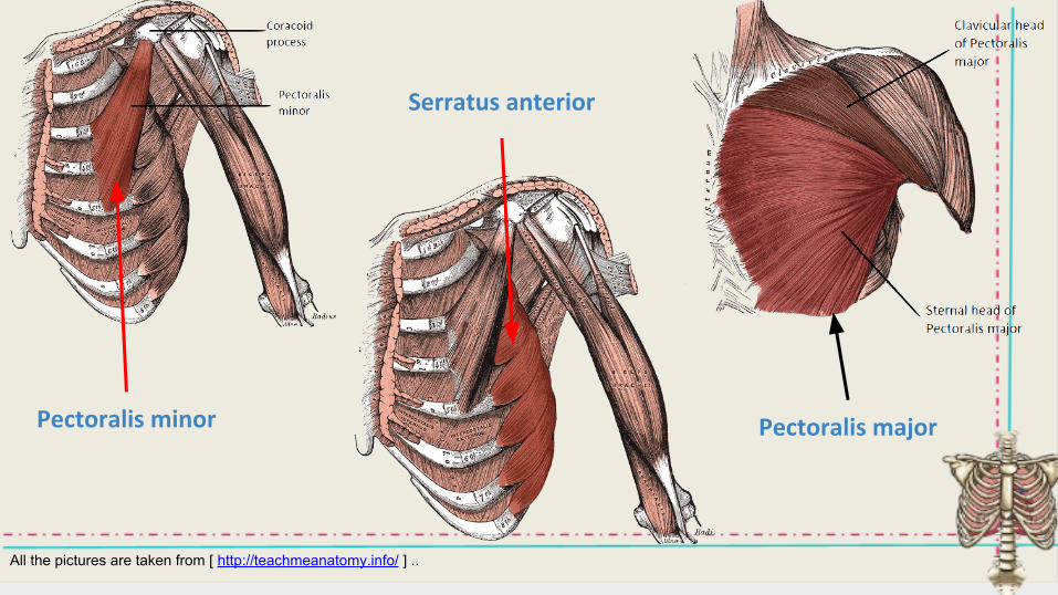
Serratus anterior
Pectoralis majorPectoralis minor
All the pictures are taken from [ http://teachmeanatomy.info/ ] ..
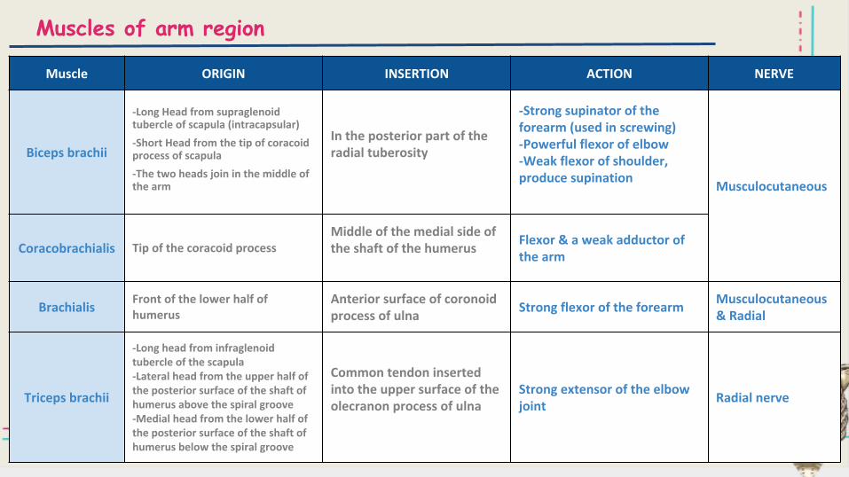
Muscles of arm region
Muscle ORIGIN INSERTION ACTION NERVE
Biceps brachii
-Long Head from supraglenoid tubercle of scapula (intracapsular)
-Short Head from the tip of coracoid process of scapula
-The two heads join in the middle of the arm
In the posterior part of the radial tuberosity
-Strong supinator of the forearm (used in screwing)-Powerful flexor of elbow-Weak flexor of shoulder, produce supination
Musculocutaneous
Coracobrachialis Tip of the coracoid processMiddle of the medial side of the shaft of the humerus
Flexor & a weak adductor of the arm
BrachialisFront of the lower half of humerus
Anterior surface of coronoid process of ulna
Strong flexor of the forearmMusculocutaneous & Radial
Triceps brachii
-Long head from infraglenoid tubercle of the scapula -Lateral head from the upper half of the posterior surface of the shaft of humerus above the spiral groove -Medial head from the lower half of the posterior surface of the shaft of humerus below the spiral groove
Common tendon inserted into the upper surface of the olecranon process of ulna
Strong extensor of the elbow joint
Radial nerve
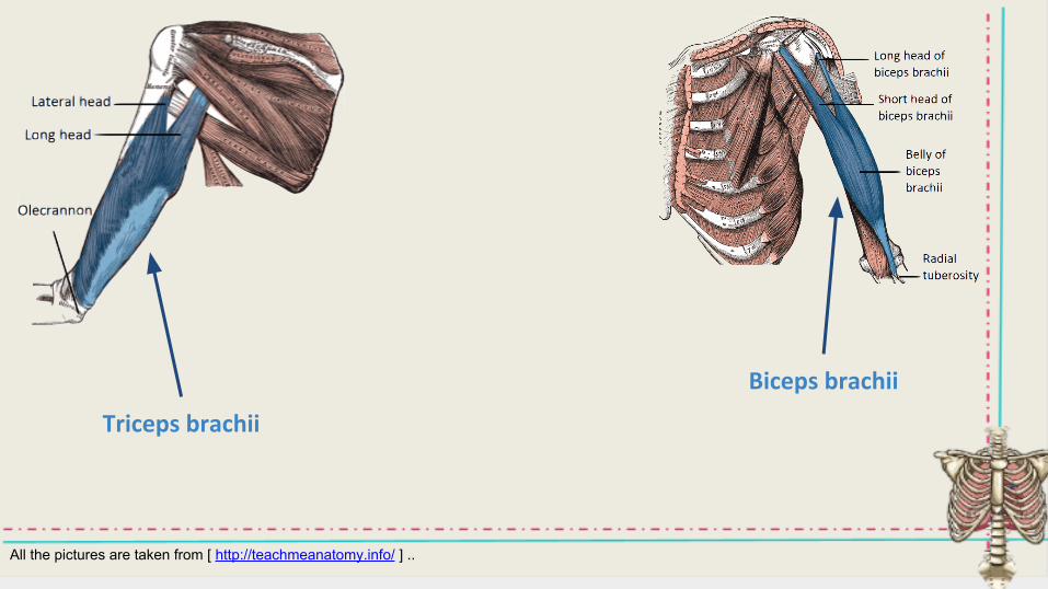
Triceps brachii
Biceps brachii
All the pictures are taken from [ http://teachmeanatomy.info/ ] ..
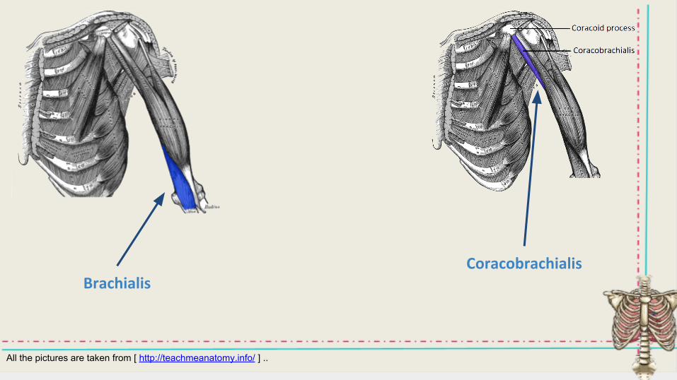
BrachialisCoracobrachialis
All the pictures are taken from [ http://teachmeanatomy.info/ ] ..
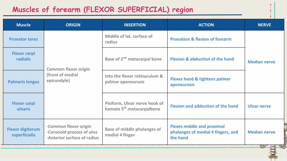
Muscles of forearm (FLEXOR SUPERFICIAL) region
Muscle ORIGIN INSERTION ACTION NERVE
Pronator teres
Common flexor origin (front of medial epicondyle)
Middle of lat. surface of radius
Pronation & flexion of forearm
Median nerve
Flexor carpi radialis Base of 2nd metacarpal bone Flexion & abduction of the hand
Palmaris longusInto the flexor retinaculum & palmar aponeurosis
Flexes hand & tightens palmer aponeurosis
Flexor carpi ulnaris
Pisiform, Ulnar nerve hook of hamate 5th metacarpalbone
Flexion and adduction of the hand Ulnar nerve
Flexor digitorum superficialis
-Common flexor origin-Coronoid process of ulna-Anterior surface of radius
Base of middle phalanges of medial 4 finger
Flexes middle and proximal phalanges of medial 4 fingers, and the hand
Median nerve
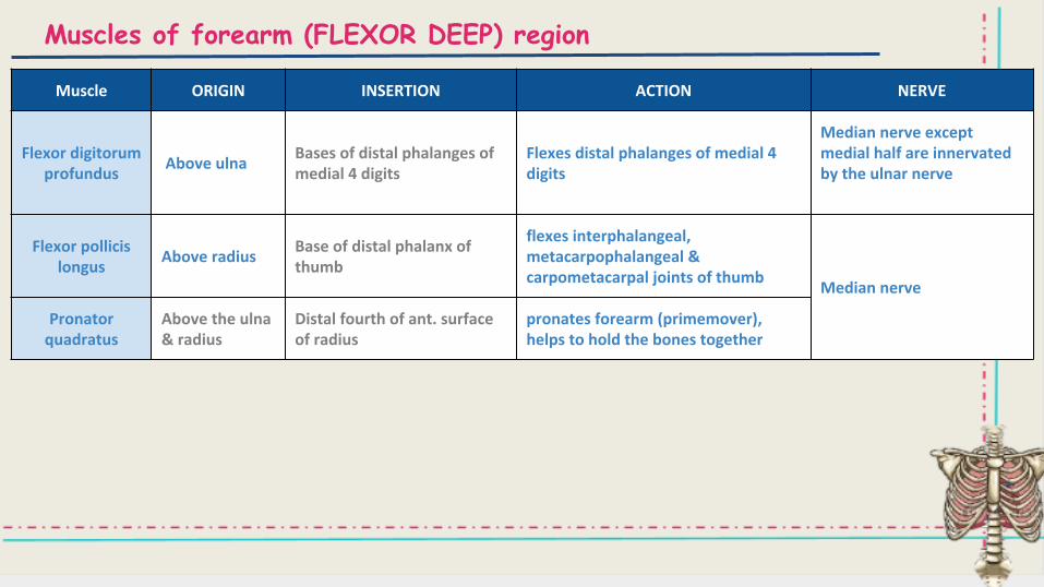
Muscles of forearm (FLEXOR DEEP) region
Muscle ORIGIN INSERTION ACTION NERVE
Flexor digitorum profundus
Above ulnaBases of distal phalanges of medial 4 digits
Flexes distal phalanges of medial 4 digits
Median nerve except medial half are innervated by the ulnar nerve
Flexor pollicis longus
Above radiusBase of distal phalanx of thumb
flexes interphalangeal, metacarpophalangeal & carpometacarpal joints of thumb
Median nerve
Pronator quadratus
Above the ulna & radius
Distal fourth of ant. surface of radius
pronates forearm (primemover), helps to hold the bones together
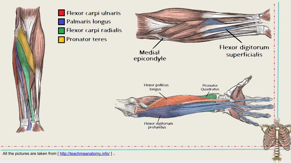
All the pictures are taken from [ http://teachmeanatomy.info/ ] ..
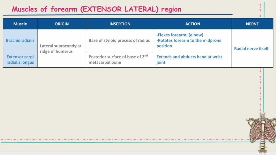
Muscles of forearm (EXTENSOR LATERAL) region
Muscle ORIGIN INSERTION ACTION NERVE
BrachioradialisLateral supracondylar ridge of humerus
Base of styloid process of radius-Flexes forearm; (elbow)-Rotates forearm to the midprone position
Radial nerve itself
Extensor carpi radialis longus
Posterior surface of base of 2nd
metacarpal boneExtends and abducts hand at wrist joint
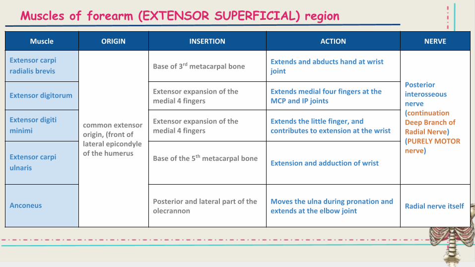
Muscles of forearm (EXTENSOR SUPERFICIAL) region
Muscle ORIGIN INSERTION ACTION NERVE
Extensor carpi
radialis brevis
common extensor origin, (front of lateral epicondyle of the humerus
Base of 3rd metacarpal boneExtends and abducts hand at wrist joint
Posterior interosseous nerve(continuation Deep Branch of Radial Nerve) (PURELY MOTOR nerve)
Extensor digitorum Extensor expansion of the medial 4 fingers
Extends medial four fingers at the MCP and IP joints
Extensor digiti
minimiExtensor expansion of the medial 4 fingers
Extends the little finger, and contributes to extension at the wrist
Extensor carpi
ulnarisBase of the 5th metacarpal bone
Extension and adduction of wrist
Anconeus Posterior and lateral part of the olecrannon
Moves the ulna during pronation and extends at the elbow joint
Radial nerve itself
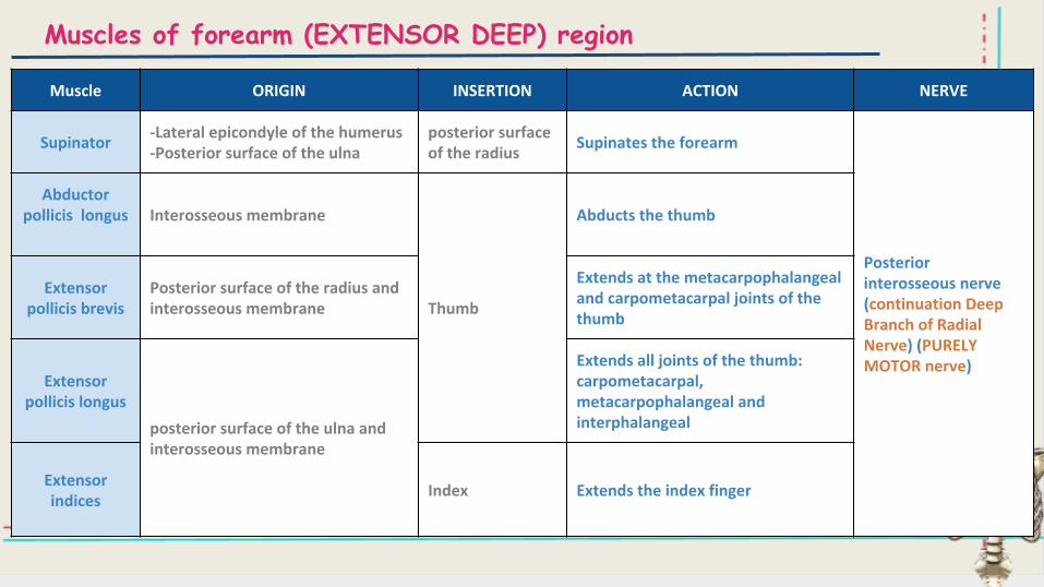
Muscles of forearm (EXTENSOR DEEP) region
Muscle ORIGIN INSERTION ACTION NERVE
Supinator-Lateral epicondyle of the humerus-Posterior surface of the ulna
posterior surface of the radius
Supinates the forearm
Posterior interosseous nerve(continuation Deep Branch of Radial Nerve) (PURELY MOTOR nerve)
Abductor pollicis longus Interosseous membrane
Thumb
Abducts the thumb
Extensor pollicis brevis
Posterior surface of the radius and interosseous membrane
Extends at the metacarpophalangeal and carpometacarpal joints of the thumb
Extensor pollicis longus
posterior surface of the ulna and interosseous membrane
Extends all joints of the thumb: carpometacarpal, metacarpophalangeal and interphalangeal
Extensor indices
Index Extends the index finger
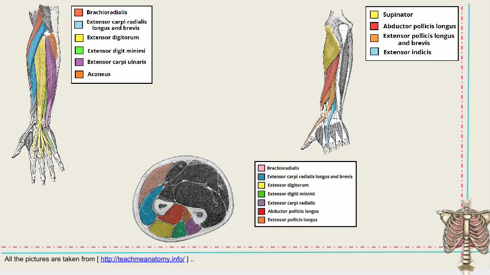
All the pictures are taken from [ http://teachmeanatomy.info/ ] ..
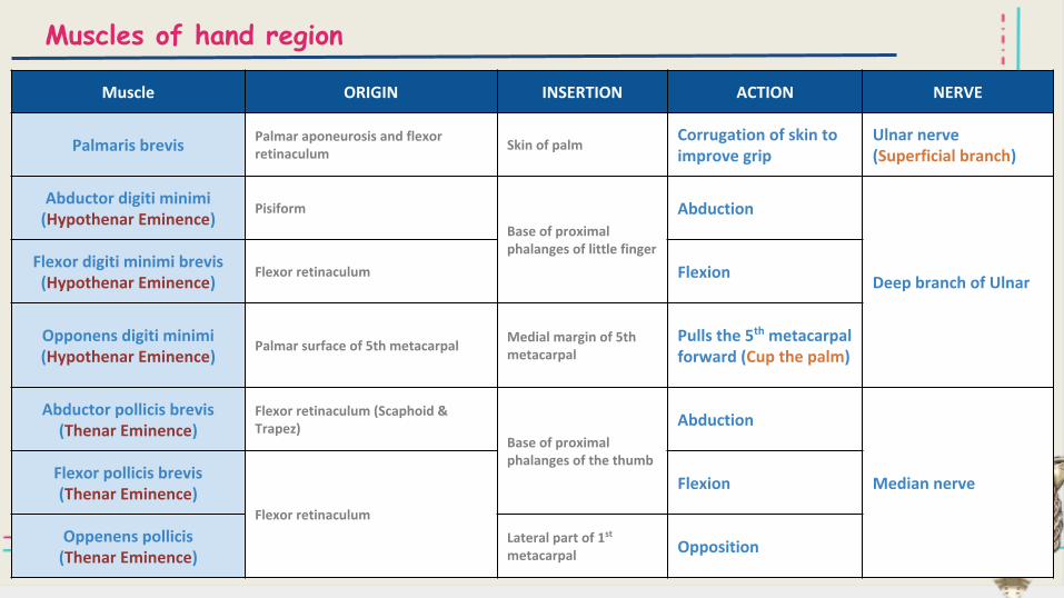
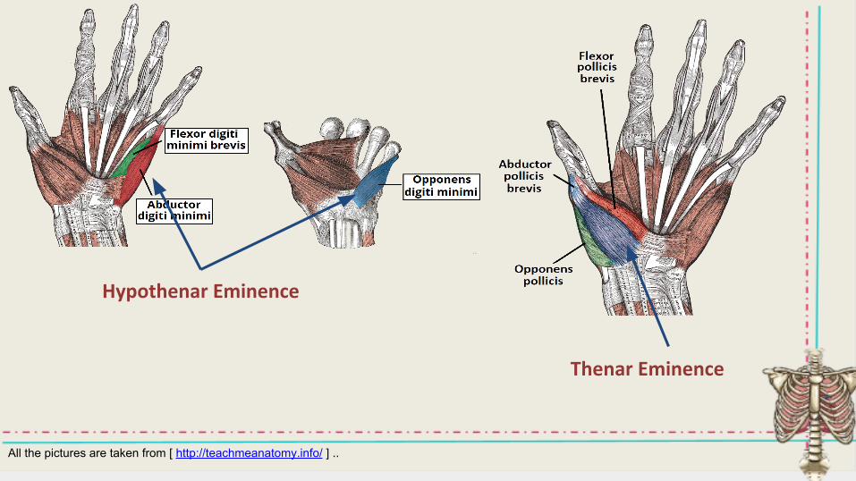
Muscles of hand region
Muscle ORIGIN INSERTION ACTION NERVE
Palmaris brevisPalmar aponeurosis and flexor retinaculum
Skin of palmCorrugation of skin to improve grip
Ulnar nerve (Superficial branch)
Abductor digiti minimi(Hypothenar Eminence)
Pisiform
Base of proximal phalanges of little finger
Abduction
Deep branch of UlnarFlexor digiti minimi brevis(Hypothenar Eminence)
Flexor retinaculum Flexion
Opponens digiti minimi(Hypothenar Eminence)
Palmar surface of 5th metacarpalMedial margin of 5th metacarpal
Pulls the 5th metacarpal forward (Cup the palm)
Abductor pollicis brevis(Thenar Eminence)
Flexor retinaculum (Scaphoid & Trapez)
Base of proximal phalanges of the thumb
Abduction
Median nerveFlexor pollicis brevis(Thenar Eminence)
Flexor retinaculum
Flexion
Oppenens pollicis(Thenar Eminence)
Lateral part of 1st metacarpal Opposition
All the pictures are taken from [ http://teachmeanatomy.info/ ] ..
Hypothenar Eminence
Thenar Eminence
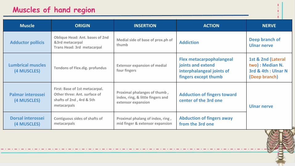
Muscles of hand region
Muscle ORIGIN INSERTION ACTION NERVE
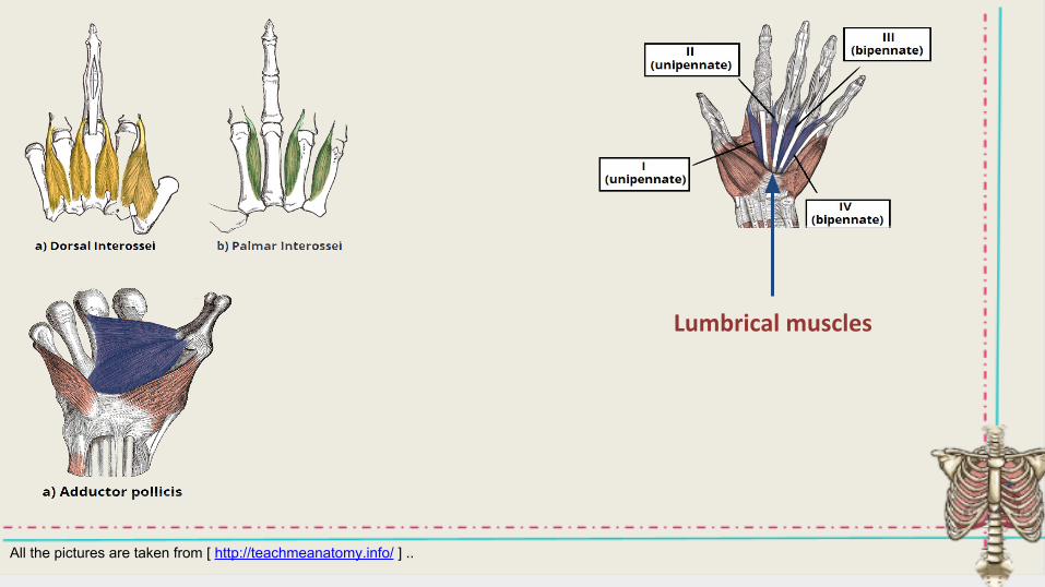
Adductor pollicisOblique Head: Ant. bases of 2nd &3rd metacarpalTrans Head: 3rd metacarpal
Medial side of base of prox.ph of thumb Addiction
Deep branch of Ulnar nerve
Lumbrical muscles (4 MUSCLES)
Tendons of Flex.dig. profundusExtensor expansion of medial four fingers
Flex metacarpophalangeal joints and extend interphalangeal joints of fingers except thumb
1st & 2nd (Lateral two) : Median N.3rd & 4th : Ulnar N (Deep branch)
Palmar interossei (4 MUSCLES)
First: Base of 1st metacarpal.
Other three: Ant. surface of
shafts of 2nd , 4rd & 5th
metacarpals
Proximal phalanges of thumb ,index, ring, & little fingers and extensor expansion
Adduction of fingers toward center of the 3rd one
Ulnar nerve
Dorsal interossei (4 MUSCLES)
Contiguous sides of shafts of metacarpals
Proximal phalang of index, ring ,mid finger & extensor expansion
Abduction of fingers away from the 3rd one
All the pictures are taken from [ http://teachmeanatomy.info/ ] ..
Lumbrical muscles

Done by:Noha AlGwaiz & Tariq AlHassan