anatomy of the breast - ksumsc
TRANSCRIPT

Anatomy of the BreastColor Code
Important
Doctors Notes
Notes/Extra explanationPlease view our Editing File before studying this lecture to check for any changes.
Objectives
By the end of the lecture, the student should be able to:
✓ Describe the shape and position of the female breast.
✓ Describe the structure of the mammary gland.
✓ List the blood supply of the female breast.
✓ Describe the lymphatic drainage of the female breast.
✓ Describe the applied anatomy in the female breast.
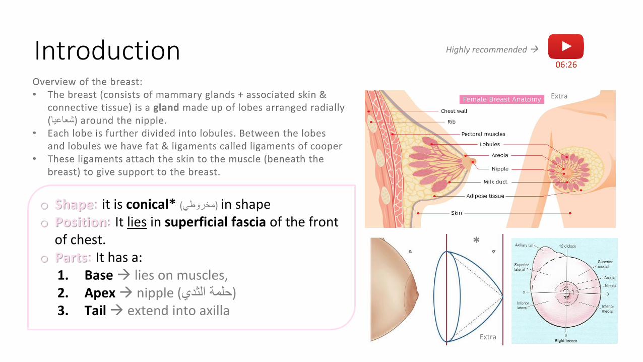
Introduction06:26
Highly recommended
o Shape: it is conical* in shape (مخروطي)o Position: It lies in superficial fascia of the front
of chest.o Parts: It has a:
1. Base lies on muscles, 2. Apex nipple (حلمة الثدي)3. Tail extend into axilla
Overview of the breast:• The breast (consists of mammary glands + associated skin &
connective tissue) is a gland made up of lobes arranged radially .around the nipple (شعاعيا)
• Each lobe is further divided into lobules. Between the lobes and lobules we have fat & ligaments called ligaments of cooper
• These ligaments attach the skin to the muscle (beneath the breast) to give support to the breast.
Extra
Extra
*
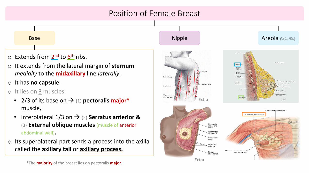
Base
o Extends from 2nd to 6th ribs.
o It extends from the lateral margin of sternummedially to the midaxillary line laterally.
o It has no capsule.
o It lies on 3 muscles:
• 2/3 of its base on (1) pectoralis major*muscle,
• inferolateral 1/3 on (2) Serratus anterior & (3) External oblique muscles (muscle of anterior
abdominal wall).
o Its superolateral part sends a process into the axilla called the axillary tail or axillary process.
Nipple Areola (حلقة ملونة)
Position of Female Breast
Extra
Extra *The majority of the breast lies on pectoralis major.
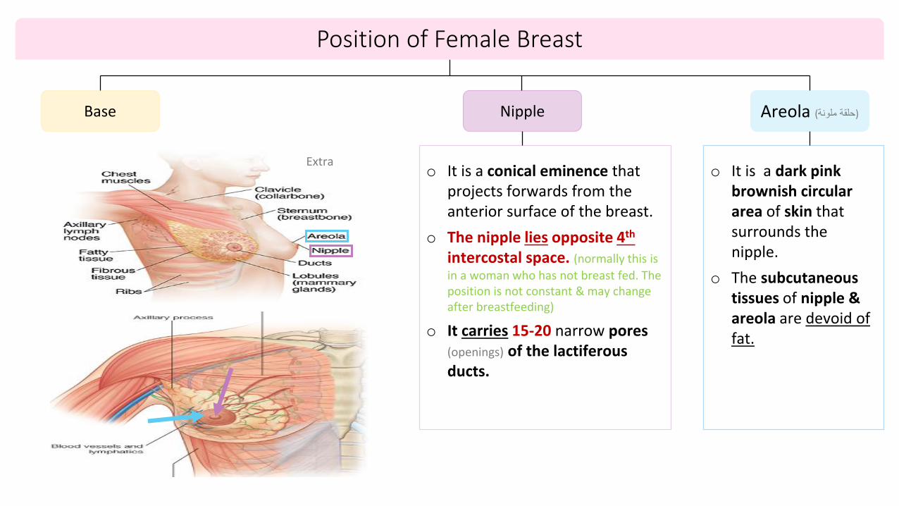
Base Nipple
o It is a conical eminence that projects forwards from the anterior surface of the breast.
o The nipple lies opposite 4th
intercostal space. (normally this is
in a woman who has not breast fed. The position is not constant & may change after breastfeeding)
o It carries 15-20 narrow pores (openings) of the lactiferous ducts.
Areola (حلقة ملونة)
o It is a dark pink brownish circular area of skin that surrounds the nipple.
o The subcutaneous tissues of nipple & areola are devoid of fat.
Position of Female Breast
Extra
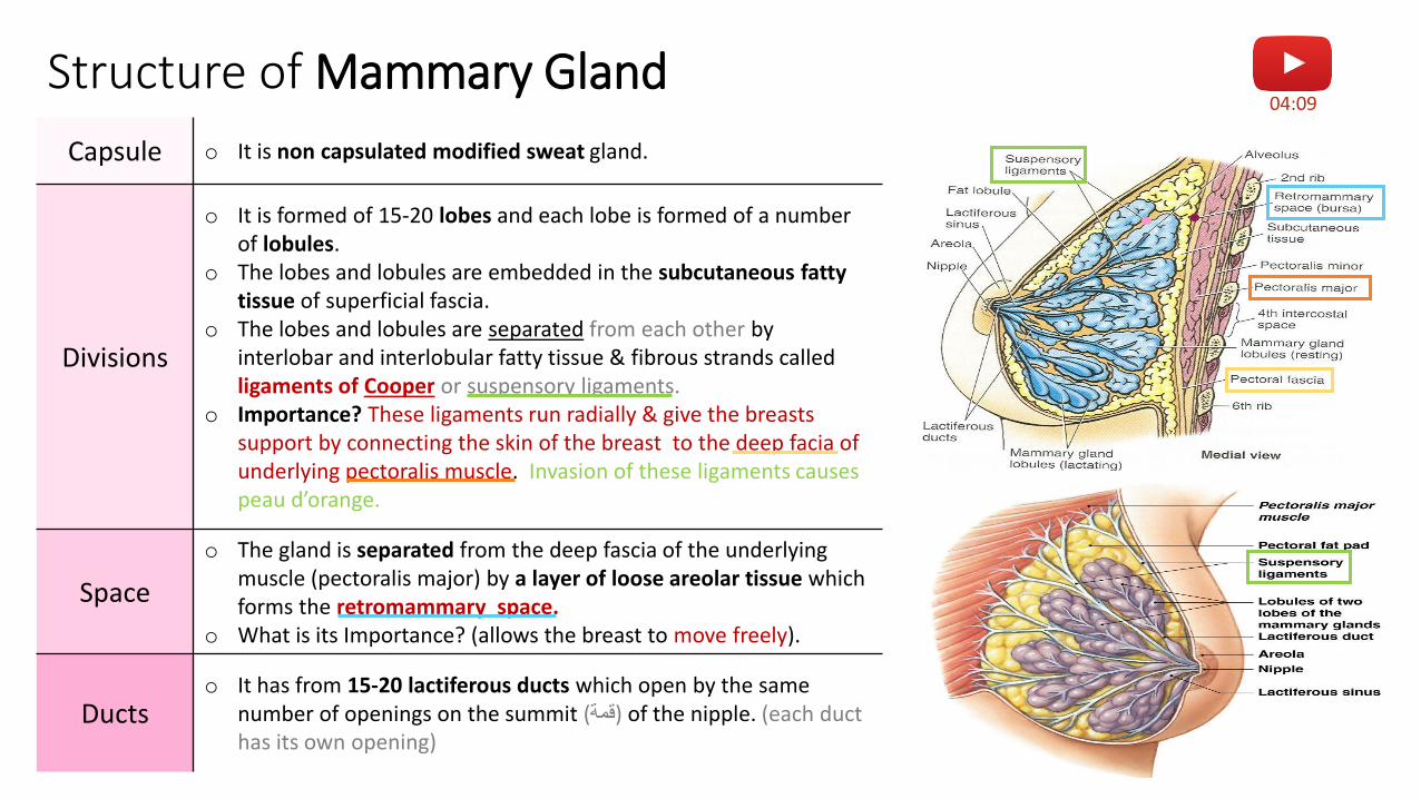
Structure of Mammary Gland
Capsule o It is non capsulated modified sweat gland.
Divisions
o It is formed of 15-20 lobes and each lobe is formed of a number of lobules.
o The lobes and lobules are embedded in the subcutaneous fatty tissue of superficial fascia.
o The lobes and lobules are separated from each other by interlobar and interlobular fatty tissue & fibrous strands called ligaments of Cooper or suspensory ligaments.
o Importance? These ligaments run radially & give the breasts support by connecting the skin of the breast to the deep facia of underlying pectoralis muscle. Invasion of these ligaments causes peau d’orange.
Space
o The gland is separated from the deep fascia of the underlying muscle (pectoralis major) by a layer of loose areolar tissue which forms the retromammary space.
o What is its Importance? (allows the breast to move freely).
Ductso It has from 15-20 lactiferous ducts which open by the same
number of openings on the summit (قمة) of the nipple. (each duct has its own opening)
04:09
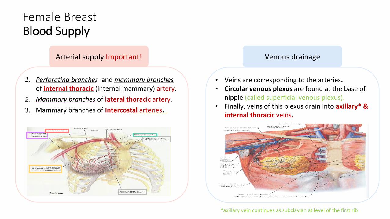
Female BreastBlood Supply
Arterial supply Important!
1. Perforating branches and mammary branches of internal thoracic (internal mammary) artery.
2. Mammary branches of lateral thoracic artery.
3. Mammary branches of Intercostal arteries.
Venous drainage
• Veins are corresponding to the arteries.• Circular venous plexus are found at the base of
nipple (called superficial venous plexus).• Finally, veins of this plexus drain into axillary* &
internal thoracic veins.
*axillary vein continues as subclavian at level of the first rib

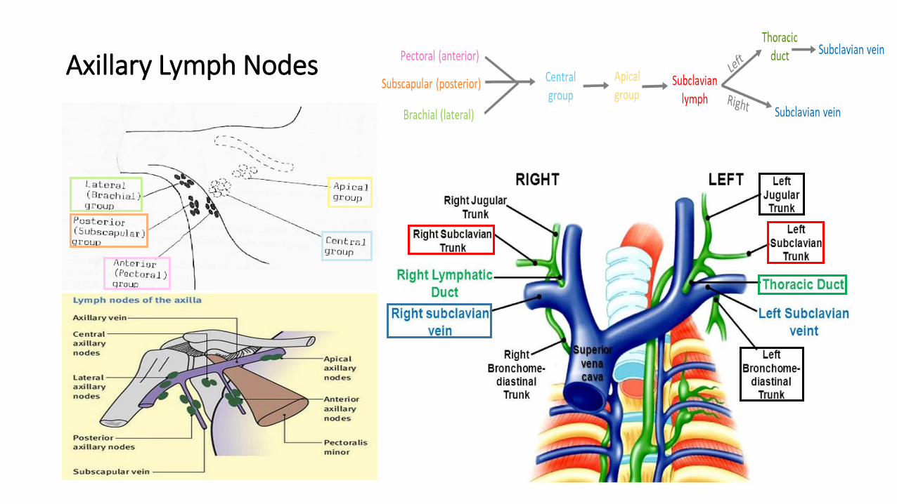
Pectoral Group (Anterior)
Subscapular Group(Posterior)
Brachial Group(Lateral/ Humeral)
Central Group Apical Group
Which lies on the pectoralis minor*along lateralthoracic vessels.
Which lies on posterior wall of axillaon lower border of subscapularis along subscapular vessels.
Lies on lateral wall of axilla along 3rd part of axillary vessels.
Lies in axillary fat at the base of axilla.
They receive lymph from the pectoral, subscapular and humeralaxillary lymph node groups.
Lies at apex of axilla.
They receive lymph from the centralaxillary lymph nodes, therefore from allaxillary lymph node groups.
All of them will go to: Subclavian lymph trunk: It is formed by union of efferent lymph vessels of apical group. • On the right side, It usually opens in the subclavian vein (directly).• On the left side it usually opens into the thoracic duct then into the left subclavian .
Axillary Lymph Nodes Important!o They are arranged into 5 groups which lie in axillary fat:
Extra ExtraExtra
Before we talk about lymphatic drainage of the breast, lets see the axillary nodes since most of the breast drains into them.
*Breast pectoralis majorAxillary nodes pectoralis minor
Axillary Lymph Nodes
Female BreastLymphatic Drainage of Breast
Extra
04:28
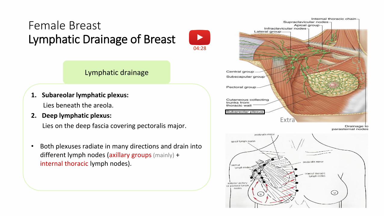
Lymphatic drainage
1. Subareolar lymphatic plexus:
Lies beneath the areola.
2. Deep lymphatic plexus:
Lies on the deep fascia covering pectoralis major.
• Both plexuses radiate in many directions and drain into different lymph nodes (axillary groups (mainly) + internal thoracic lymph nodes).
Female BreastLymphatic Drainage
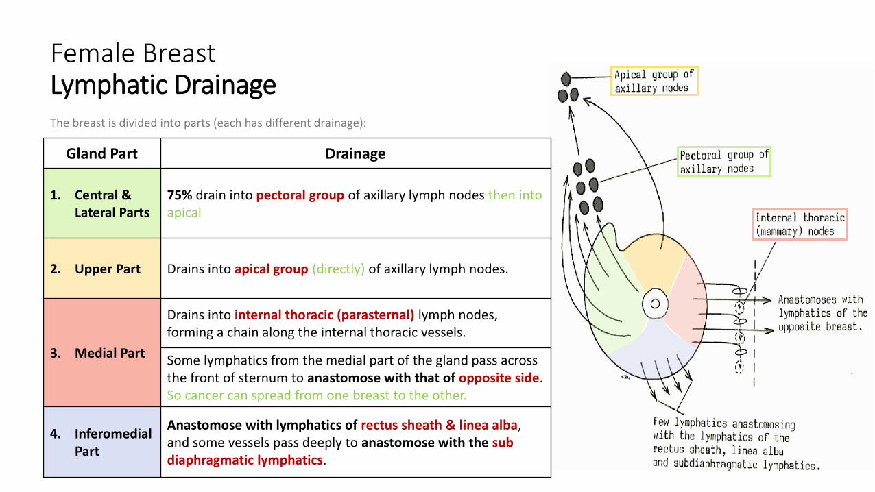
Gland Part Drainage
1. Central & Lateral Parts
75% drain into pectoral group of axillary lymph nodes then into apical
2. Upper Part Drains into apical group (directly) of axillary lymph nodes.
3. Medial Part
Drains into internal thoracic (parasternal) lymph nodes, forming a chain along the internal thoracic vessels.
Some lymphatics from the medial part of the gland pass across the front of sternum to anastomose with that of opposite side. So cancer can spread from one breast to the other.
4. InferomedialPart
Anastomose with lymphatics of rectus sheath & linea alba, and some vessels pass deeply to anastomose with the sub diaphragmatic lymphatics.
The breast is divided into parts (each has different drainage):
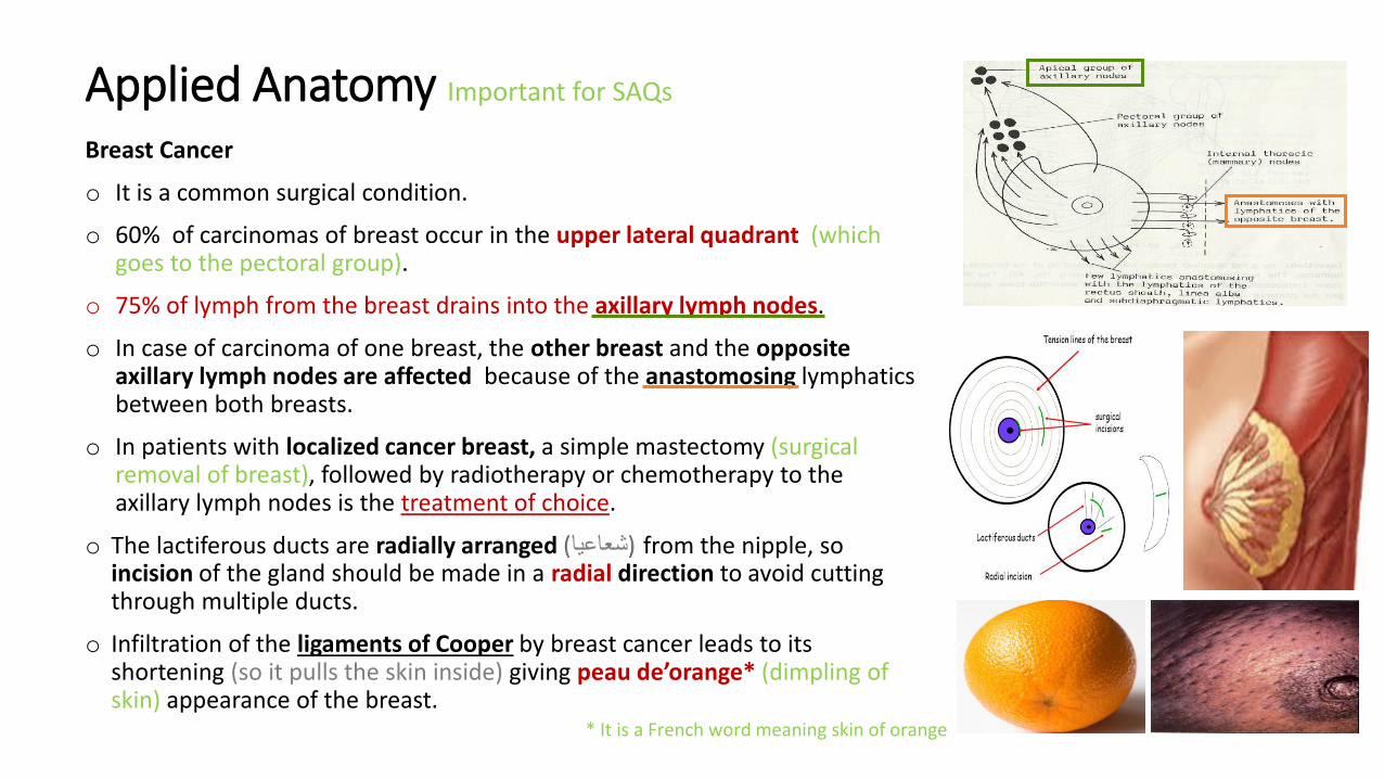
Applied Anatomy Important for SAQs
Breast Cancer
o It is a common surgical condition.
o 60% of carcinomas of breast occur in the upper lateral quadrant (which goes to the pectoral group).
o 75% of lymph from the breast drains into the axillary lymph nodes.
o In case of carcinoma of one breast, the other breast and the opposite axillary lymph nodes are affected because of the anastomosing lymphatics between both breasts.
o In patients with localized cancer breast, a simple mastectomy (surgical removal of breast), followed by radiotherapy or chemotherapy to the axillary lymph nodes is the treatment of choice.
o The lactiferous ducts are radially arranged from the nipple, so (شعاعيا)incision of the gland should be made in a radial direction to avoid cutting through multiple ducts.
o Infiltration of the ligaments of Cooper by breast cancer leads to its shortening (so it pulls the skin inside) giving peau de’orange* (dimpling of skin) appearance of the breast.
* It is a French word meaning skin of orange
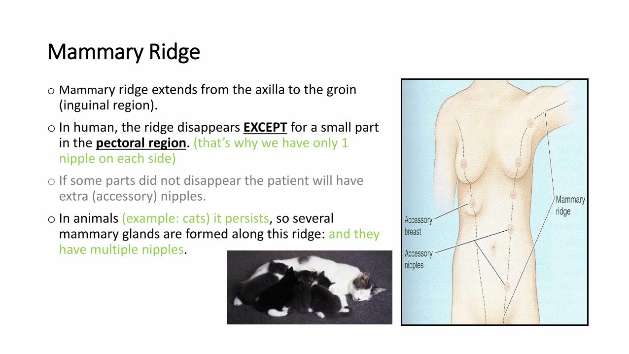
o Mammary ridge extends from the axilla to the groin (inguinal region).
o In human, the ridge disappears EXCEPT for a small part in the pectoral region. (that’s why we have only 1 nipple on each side)
o If some parts did not disappear the patient will have extra (accessory) nipples.
o In animals (example: cats) it persists, so several mammary glands are formed along this ridge: and they have multiple nipples.
Mammary Ridge
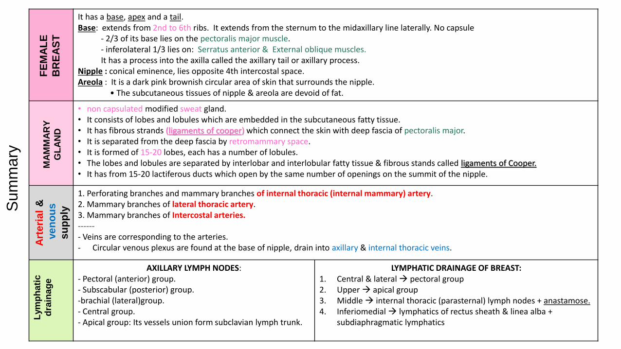
It has a base, apex and a tail.Base: extends from 2nd to 6th ribs. It extends from the sternum to the midaxillary line laterally. No capsule
- 2/3 of its base lies on the pectoralis major muscle.- inferolateral 1/3 lies on: Serratus anterior & External oblique muscles.It has a process into the axilla called the axillary tail or axillary process.
Nipple : conical eminence, lies opposite 4th intercostal space. Areola : It is a dark pink brownish circular area of skin that surrounds the nipple.
• The subcutaneous tissues of nipple & areola are devoid of fat.
• non capsulated modified sweat gland. • It consists of lobes and lobules which are embedded in the subcutaneous fatty tissue. • It has fibrous strands (ligaments of cooper) which connect the skin with deep fascia of pectoralis major. • It is separated from the deep fascia by retromammary space.• It is formed of 15-20 lobes, each has a number of lobules. • The lobes and lobules are separated by interlobar and interlobular fatty tissue & fibrous stands called ligaments of Cooper.• It has from 15-20 lactiferous ducts which open by the same number of openings on the summit of the nipple.
1. Perforating branches and mammary branches of internal thoracic (internal mammary) artery.2. Mammary branches of lateral thoracic artery.3. Mammary branches of Intercostal arteries.------- Veins are corresponding to the arteries. - Circular venous plexus are found at the base of nipple, drain into axillary & internal thoracic veins.
LYMPHATIC DRAINAGE OF BREAST:1. Central & lateral pectoral group2. Upper apical group3. Middle internal thoracic (parasternal) lymph nodes + anastamose.4. Inferiomedial lymphatics of rectus sheath & linea alba +
subdiaphragmatic lymphatics
AXILLARY LYMPH NODES:- Pectoral (anterior) group.- Subscabular (posterior) group.-brachial (lateral)group.- Central group.- Apical group: Its vessels union form subclavian lymph trunk.
FE
MA
LE
BR
EA
ST
MA
MM
AR
Y
GL
AN
D
Art
eri
al
&
ven
ou
s
su
pp
ly
Lym
ph
ati
c
dra
ina
ge
Sum
ma
ry
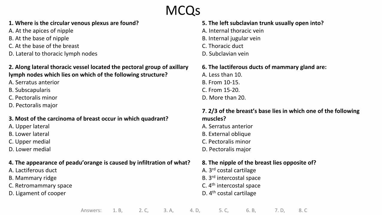
1. Where is the circular venous plexus are found? A. At the apices of nippleB. At the base of nippleC. At the base of the breast D. Lateral to thoracic lymph nodes
2. Along lateral thoracic vessel located the pectoral group of axillary lymph nodes which lies on which of the following structure?A. Serratus anteriorB. SubscapularisC. Pectoralis minorD. Pectoralis major
3. Most of the carcinoma of breast occur in which quadrant? A. Upper lateral B. Lower lateral C. Upper medial D. Lower medial
4. The appearance of peadu’orange is caused by infiltration of what?A. Lactiferous ductB. Mammary ridgeC. Retromammary space D. Ligament of cooper
MCQs
Answers: 1. B, 2. C, 3. A, 4. D, 5. C, 6. B, 7. D, 8. C
5. The left subclavian trunk usually open into? A. Internal thoracic veinB. Internal jugular veinC. Thoracic ductD. Subclavian vein
6. The lactiferous ducts of mammary gland are:A. Less than 10.B. From 10‐15.C. From 15‐20.D. More than 20.
7. 2/3 of the breast’s base lies in which one of the following muscles?A. Serratus anteriorB. External obliqueC. Pectoralis minorD. Pectoralis major
8. The nipple of the breast lies opposite of?A. 3rd costal cartilageB. 3rd intercostal spaceC. 4th intercostal spaceD. 4th costal cartilage
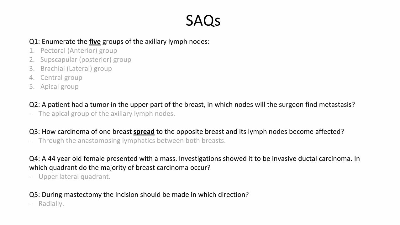
Q1: Enumerate the five groups of the axillary lymph nodes:1. Pectoral (Anterior) group2. Supscapular (posterior) group3. Brachial (Lateral) group4. Central group5. Apical group
Q2: A patient had a tumor in the upper part of the breast, in which nodes will the surgeon find metastasis?- The apical group of the axillary lymph nodes.
Q3: How carcinoma of one breast spread to the opposite breast and its lymph nodes become affected? - Through the anastomosing lymphatics between both breasts.
Q4: A 44 year old female presented with a mass. Investigations showed it to be invasive ductal carcinoma. In which quadrant do the majority of breast carcinoma occur? - Upper lateral quadrant.
Q5: During mastectomy the incision should be made in which direction?- Radially.
SAQs
Leaders:
Nawaf AlKhudairy
Jawaher Abanumy
Members:
Allulu Alsulayhim
Anwar Alajmi
Ghaida Alsaeed
Ghada Alothaim
Lama Alfawzan
Lama AlTamimi
Rawan AlWadee
Safa Al-Osaimi
Shatha Alghaihb
References:
1- Girls’ & Boys’ Slides
2- Greys Anatomy for Students
3- TeachMeAnatomy.com
@anatomy436
Feedback
!
Thank You!
الزايدمنيرهندى الدخيلنوره الحقيل
نوف العقيليوجدان الزيد
العنود الصيخانامال الشيبيبدريه الصباغجواهر الخيال
دانيه سجادينا النويصرذكريات عمر
رزان القحطانيرنا باراسين
:قير فلاةداقيريضخلافاونوابانميجواهر
شواق الماجدأالعنود ابوحيمداللؤلؤ الصليهم
ميره نيازيأأنوار العجمي
دانية الكالبيدعاء عبدالفتاح
روان الوادعيين ار مشلاساره
شذا الغيهبصفاء العصيمي
غادة العثيمغيداء السعيدلمى التميمي
لمى الفوزان
روان الحربيريما الشايع
ريما العتيبيسميه الغامدي
شوق البقميغادة المزروعغاده الهدلقالرا السليم
منيال باوزيرمها العيسى
نجد الذيبنوره السهلينوره الشبيب
هبه الناصر
هذا العملإنجازيف كل الشكر و التقدير لكل من ساهم هللادمحبمت
ومن الشيطانأخطأنا فمن انفسناهللا وإنفمنأصبنانإ
!
حمد الخضيريطالل الحقيلمحمد حبيب
محمد نصرمحمد الغندور
محمد اليوسفماجد الزين
عبدالملك الهدلقعبدالمحسن الخلفعبدالعزيز السلمان
عبدهللا هاشمالراجحيعبدالرحمن
عبدالرحمن المالكيعبدهللا الجماح
عبدالمحسن الغنامخالد العيدانخالد الدخيلركان باهمامريان القرني
ساعد النويصرممحمد الدعيجمحمد الكحيل
مويد احمدعبدالحكيم العنيقعبدالعزيز العنقري
عبدالعزيز آل محمدفهد الزهراني
عصام الشهرانييزيد السحيباني