evaluation and validation of new techniques of facial nerve
TRANSCRIPT

Sorbonne Université
Ecole doctorale 394:
<<Physiologie, Physiopathologie et thérapeutique >>
Thèse de doctorat
Evaluation and validation of new techniques of facial nerve preservation during CPA tumor surgery
Par
Mohamed Ahmed Magdieldin Elsayed
Directeurs : Prof. Olivier Sterkers et Prof. Yann Nguyen
<<Réhabilitation chirurgicale mini-invasive et robotisée de l’audition>>
Inserm/Sorbonne Université
Présentée et soutenue publiquement le 27 Mars 2020
Devant un jury composé de:
Professeur Philippe Cornu Le Président Professeur Mathieu Marx Rapporteurs Professeur Hani Elgarem Rapporteurs Professeur Mohamed Badr-Eldine Examinateur Professeur Daniele De Seta Examinateur

Sorbonne Université
Ecole doctorale 394:
<<Physiologie, Physiopathologie et thérapeutique >>
PhD thesis entitled
Evaluation and validation of new techniques of facial nerve preservation during CPA tumor surgery
Presented by
Mohamed Ahmed Magdieldin Elsayed
Supervised by: Prof. Olivier Sterkers and Prof. Yann Nguyen
<<Réhabilitation chirurgicale mini-invasive et robotisée de l’audition>>
Inserm/Sorbonne Université
Presented and defended at Paris on the 27 March 2020
Dissertation committee:
Professor Philippe Cornu President Professor Mathieu Marx Reviewer Professor Hani Elgarem Reviewer Professor Mohamed Badr-Eldine Examiner Professor Daniele De Seta Examiner

I
To Yasmin, my amazing wife.
To Saleem, my son & my best friend.
To Zain, Maya, Yassin, Adam and Youssef, my little children.
To all my beloved family.

II
Acknowledgement
Completing this PhD has been a truly life-changing experience for me and it would
not have been possible to do without the support and guidance that I received from many
people.
First and foremost, my utmost gratitude my supervisor Professor Olivier Sterkers for
all the support and encouragement he gave me since the first day I arrived at Paris. His
dynamism, vision, sincerity and motivation have deeply inspired me. Without his guidance
and constant feedback this PhD would not have been achievable.
Many thanks and gratitude is due to Dr Yann Nguyen for his patience, supervisory
role, and steadfast encouragement and shared valuable insights in the relevance of the
research work. I do believe that this research work could not be carried out with him being
available for any question or problem. I would also like to thank him for his friendship,
empathy and great sense of humor.
I would like to express my special gratitude and appreciation to Professor Michel
Kalamarides and all the surgical team at Groupe Hospitalier Pitié-Salpêtrière, Paris, France
for sharing their expertise and support in order to complete the clinical work.
I would like to pay my special regards to Dr Evelyne Ferrary and my colleagues Dr
Renato Torres, Dr Ghizlene Lahlou and Dr Huan Jia. I would like to recognize the
invaluable assistance that you all provided during my study.
Special thanks to Anirudhan Narasimhan, Global Product Manager, H&N therapies,
Medtronic ENT, Jacksonville, FL, USA for their materials and technical support during the
research work. Without their support and funding, this project could not have reached its
goal.
I gratefully acknowledge as well the funding received towards my PhD from the
Ministry of Higher Education, Mission sector, Egypt (http://www.mohe-casm.edu.eg) and
the Ministry of Scientific Research, Egypt. We gratefully acknowledge as well the
“Association de Recherche du service d’ORL de l’hopital Beaujon” for its support for travel
and housing.

III
Many thanks and appreciation to the distinguished members of the Jury headed by
Professor Philippe CORNU and all members of the Jury ; Professor Mathieu Marx
,Professor Hani Elgarem , Professor Mohamed Badr-Eldin and Professor Daniele De Seta
for their approval and recognition of my work.
Finally, I am extremely grateful and indebted to my Family for their love, prayers,
understanding and continuous support to complete this research work.

Table of Content
IV
Table of Content
Acknowledgement ....................................................................................................... II
Table of Content ........................................................................................................ IV
List of Figures ........................................................................................................... VII
List of Abbreviations ............................................................................................. VIII
Chapter 1 INTRODUCTION ..................................................................................... 1
Development and Anatomy of the Facial Nerve.................................................. 3
Challenges of facial nerve preservation during CPA tumor surgery ................... 5
Intraoperative neuromonitoring (IONM) for facial nerve ................................... 7
Historical review of FN-IONM ........................................................................ 8
State of art of Electromyography (EMG) monitoring devices ......................... 8
Current Facial nerve IONM practice at our institute ...................................... 13
References .......................................................................................................... 21
3.1.5 Discussion
...................................................................................................29
3.1.4 Results.........................................................................................................27
3.1.3
Methods.......................................................................................................27
3.1.2 Introduction.................................................................................................26
3.1.1 Abstract
.......................................................................................................26
A Cadaveric Study
...............................................................................................................25
3.1 Pig as a Large Animal Model for Posterior Fossa Surgery in Oto-Neurosurgery:
Chapter 3 RESULTS
.................................................................................................24
Chapter 2 OBJECTIVES
..........................................................................................23

Table of Content
V
3.3.6 Results.........................................................................................................64
3.3.5 Statistical analysis
.......................................................................................64
3.3.4 Materials and Methods................................................................................61
3.3.3 Introduction.................................................................................................60
3.3.2 Abstract
.......................................................................................................59
guided microsurgical resection of large vestibular schwannomas...................................58
3.3.1
Optimization
of
facial
nerve
outcome
by
intraoperative
electromyography
guided microsurgical resection of large vestibular schwannomas.......................................57
3.3
Optimization
of
facial
nerve
outcome
by
intraoperative
electromyography
3.2.9
References...................................................................................................55
3.2.8
Acknowledgments.......................................................................................55
3.2.7
Conclusions.................................................................................................54
3.2.6
Discussion
...................................................................................................50
3.2.5
Results.........................................................................................................46
3.2.4
Methods.......................................................................................................40
3.2.3
Introduction.................................................................................................38
3.2.2
Abstract
.......................................................................................................37
preliminary experience.....................................................................................................36
3.2.1
In
vivo
porcine
model
for
posterior
fossa
surgery
in
oto-neurosurgery:
a
preliminary experience.........................................................................................................35
3.2
In
vivo
porcine
model
for
posterior
fossa
surgery
in
oto-neurosurgery:
a
3.1.7 References...................................................................................................33
3.1.6 Conclusion
..................................................................................................33

Table of Content
VI
3.3.7 Discussion ................................................................................................... 72
3.3.8 Conclusions ................................................................................................. 75
3.3.9 Acknowledgment ........................................................................................ 75
3.3.10 References ................................................................................................. 76
Chapter 4 DISCUSSION ........................................................................................... 78
I. Animal models and surgical training ............................................................. 79
II. Animal models and IONM research............................................................... 81
III. Intraoperative neuromonitoring of FN ........................................................... 84
Change of paradigm for surgical decision and strategy ................................. 84
Dual Facial nerve monitoring technique ........................................................ 86
Continuous intraoperative Facial nerve monitoring technique ...................... 87
IV. Recent advances in FN preservation during CPA surgery ............................. 90
Preoperative visualization of the facial nerve ................................................ 90
Intraoperative use of endoscopy ..................................................................... 90
Peri-operative Neuro-protective agents .......................................................... 92
V. References ...................................................................................................... 94
Chapter 5 CONCLUSION AND FUTURE PERSPECTIVES .............................. 97
Chapter 6 ANNEXES ................................................................................................ 98
Annex 1 .................................................................................................................... 98
Annex 2 .................................................................................................................. 104

List of Figures
VII
List of Figures
Figure 1-1 Illustrative Course and relationships of the right facial nerve from the pontomedullary junction to the stylomastoid foramen. ....................................................... 4
Figure 1-2 An example of ventral and inferior displacement of the facial nerve. ............. 6
Figure 1-3 Illustrative examples of three types of EMG activity often seen during vestibular schwannoma (VS) surgery. ............................................................................ 11
Figure 1-4 The StimBurGard (black arrow) system with Visao drill. ............................. 20
Figure 3-1 Operating room set-up. ....................................................................................... 40
Figure 3-2 Pig positioning and setting up of NIM 3.0 subdermal needle electrodes. ...... 41
Figure 3-3 Incision, soft tissue dissection and skull base anatomy. ................................... 43
Figure 3-4 Lateral view of craniotomy of a pig’s ear on the left side. ............................... 44
Figure 3-5 The acoustic-facial bundle (cranial nerves VII/VIII) in the IAC. .................. 45
Figure 3-6 Different views of the facial nerve (FN) in the IAC and CPA. ........................ 45
Figure 3-7 APS electrode. ...................................................................................................... 48
Figure 3-8 Example of APS electrode placement on the facial nerve in the CPA. .......... 49
Figure 3-9 Example of APS electrode placement on the facial nerve in the CPA. .......... 50
Figure 3-10 Postoperative FN function according to HB staging, ..................................... 71
Figure 3-11 Optimization of facial nerve outcome by reducing the nervous conduction blockage severity. ................................................................................................................... 73
Figure 4-1 Comparison between new pledget electrode on the left and APS electrode on the right. .................................................................................................................................. 83
Figure 4-2 The ball-type, continuous facial nerve stimulating electrode (SE) that is placed intraoperatively on the REZ of FN (arrow).[19] ..................................................... 88
Figure 4-3 Intraoperative CPA view of vestibular schwannoma (*) and Facial nerve (white Arrow) on the left side and Positioning of the APS electrode on Facial nerve on the right side. .......................................................................................................................... 89

List of Abbreviations
VIII
List of Abbreviations APS BBB CMAP C-IONM CN CPA CSF DES
DTT FNMEPs FN GTR H-B IAC IGF-I IONM I-IONM
MRI NTR REZ STR VS
Automatic periodic stimulation
Blood-brain barrier
compound muscle action potential
Continuous intraoperative neurophysiological monitoring
Cranial nerve
Cerebellopontine angle
Cerebrospinal fluid
Direct electrical stimulation
Diffusion tensor tractography
Facial nerve motor evoked potentials
Facial nerve
Gross total resection
House-Brackmann
Internal acoustic canal
Insulin-like growth factor 1
Intraoperative neurophysiological monitoring
Intermittent Intraoperative neurophysiological monitoring
Magnetic resonance imaging
Near-total resection
Root Exit zone
Subtotal resection
Vestibular schwannoma


Introduction
1
Chapter 1 INTRODUCTION
Otologic surgery emerged during the 19th century, with a major breakthrough
occurring in the middle of the 20th century. In 1952 Wullstein first used a surgical
microscope in an otological operation, laying a foundation for otomicrosurgery.
Subsequently, surgical microscopes were used extensively; allowing the continuing evolution
of microsurgical techniques in the middle ear. When microsurgical techniques reached the
inner ear and cerebellopontine angle (CPA) area, allowing access to the skull base and lateral
skull base, the era of micro-otoneurosurgery dawned.[1] Otoneurosurgeons are challenged
with eradicating pathology while preserving preoperative functional status. Minimally
invasive techniques have arisen to meet the need to preserve function and promote quick
recovery.
Thus, the aims of surgery in the cerebellopontine angle (CPA) have changed from
tumor resection and prolongation of life to the anatomical and functional preservation of the
cranial nerves (CNs).Dealing with CPA tumors has developed from almost a death sentence
at the beginning of the 20th century to the current concept of functional microsurgery. In this
interim, several developments regarding radiological diagnosis, the introduction of the
operative microscope and microsurgical techniques, advances in the field of neuroanesthesia,
as well as intraoperative neuromonitoring were responsible for significant reductions in the
morbidity and mortality in patients suffering from CPA tumors.[2]
Pathologies common to the CPA include acoustic neuroma, facial neural tumors,
meningioma and dysfunctional cranial nerves. Among these, acoustic neuroma accounts for a
great portion. In the past 10 years, with rapid development of otoneurosurgery and imaging
technologies, early diagnosis of CPA pathologies has improved, broadening scope of surgical
practices for otologists. Acoustic neuroma or vestibular schwannoma (VS) is a benign, slow-
growing tumor of the Schwann cells originating from the vestibular branch of the eighth
cranial nerve, with an incidence of 12.4 tumors per million persons per year. They are extra-
axial brain tumors developing near the internal auditory canal (IAC). When manifested in the
internal auditory canal (IAC), they may grow in the direction of least mechanical resistance:
the cerebellopontine angle (CPA). The vast majority of VS are sporadic and unilateral. [2]

Introduction
2
Therapeutic options include an active wait-and-scan policy, stereotactic radio-surgery,
and microsurgery. Although there is no general consensus for the treatment of small and
medium-sized lesions, surgery still remains the preferred option for large tumors. Advances
in treatment modalities have popularized the application of less invasive management
methods such as radiotherapy and radiosurgery, which carry high efficacy and low morbidity.
However, many acoustic neuromas, particularly those that are large in size, necessitate
surgical intervention.
Surgical approaches to CPA lesions, especially acoustic neuroma, require gaining
access to the CPA and maximal exposure of the internal acoustic meatus, all while attempting
to maintain facial and auditory nerve function. Microscopy coupled with functional facial
nerve monitoring greatly reduces both the mortality and the incidence of facial nerve
paralysis in acoustic neuroma surgeries. Preserving auditory function remains a challenge
because removal of the tumor often damages the cochlear nerve, and ischemia-
induced deafness can happen due to damage to or spasm of the fragile internal auditory
artery. Other CPA surgeries share this difficult bargain between tumor excision and
preservation of function.[3]
Generally, the primary operative goals are gross tumor debulking while safeguarding
the adjacent cranial nerves. Neural preservation is particularly imperative in the
contemporary management of VS. By virtue of their location, these tumors are close to the
facial and vestibulocochlear cranial nerves and can thus severely impair the nerve function at
the time of initial presentation. The VS can directly impinge, tightly adhere to, or overtly
damage the nerves. These goals require good preservation of both facial nerve (FN) and
cochlear nerve (CN) function if the patient had functional hearing preoperatively. However,
preserving these neurological functions is often challenging, especially when a large tumor
must be removed [3].
CPA tumors often present as operative challenges, as resection may cause nerve
irritation or injury leading to neurapraxia, axonotmesis, or neurotmesis. The various options
of surgical approaches (translabyrinthine vs. middle fossa vs. retrosigmoid) for VS and their
respective patterns of postoperative cranial nerve preservation have been described. Although
significant advances have been observed in the recent era, facial nerve (FN) anatomical
preservation during VS surgery is currently around the range of 95% and FN functional

Introduction
3
preservation is in the low range of 70%. FN injury is not exclusively related to VS surgery
and may also occur postoperatively in several tumors of the CPA.[2]
Development and Anatomy of the Facial Nerve
The facial nerve is composed of motor, sensory, and parasympathetic fibers.
Complete separation of the facial and acoustic nerves and development of the nervus
intermedius (or nerve of Wrisberg) occurs by 6 weeks of gestation. By the 16th week, the
neural connections are completely developed. The bony facial canal develops until birth,
enclosing the facial nerve in bone throughout its course except at the facial hiatus (the site of
the geniculate ganglion) in the floor of the middle cranial fossa. The only difference between
the anatomies of the facial nerve in infants compared with adults is in the region of the
stylomastoid foramen. As the mastoid tip develops, the extratemporal facial nerve is
positioned in a more inferior and medial position.
Facial motor fibers originate from cell bodies located in the precentral and postcentral
gyri of the frontal motor cortex. These fibers travel in the posterior limb of the internal
capsule inferiorly to the caudal Pons. There, the motor fibers supplying the facial musculature
beneath the brows cross the midline to reach the contralateral motor nucleus in the reticular
formation of the lower pons (anterior to the fourth ventricle). The majority of motor fibers
that supply the musculature of the forehead also cross the midline; however, a few fibers do
not, instead travel in the ipsilateral motor nucleus. Thus, muscles of the forehead receive
innervations from both sides of the motor cortex, and so forehead-sparing facial paralyses can
be indicative of a central etiology. The motor fibers pass dorsally, loop medial-to-lateral
around the abducens nucleus, and create the facial colliculus, which bulges into the floor of
the fourth ventricle. This loop of the facial nerve forms the internal genu of the facial nerve.
The nervus intermedius contains sensory, special sensory and parasympathetic fibers.
It provides sensation to the posterior concha and external auditory canal. The nervus
intermedius’ special sensory fibers supply taste sensation to the anterior two-thirds of the
tongue. The afferent fibers synapse with cell bodies in the geniculate ganglion at the first
genu of the facial nerve. These sensory afferents then join the parasympathetic fibers, passing
via the nervus intermedius to the nucleus tractus solitarius in the medulla. The
parasympathetic portion of the nervus intermedius originates in the superior salivatory

Introduction
4
nucleus in the dorsal pons and provides the secretomotor function of the ipsilateral lacrimal
gland, submandibular glands, sublingual glands, and minor salivary glands.
Both the motor root of the facial nerve and the nervus intermedius leave the brainstem
near the dorsal Pons at the pontomedullary junction (the cisternal segment of the facial
nerve). Within the cerebellopontine angle (CPA), the nerve travels anterolaterally into the
porus acousticus of the internal auditory canal (IAC), anterior to the vestibulocochlear
nerve.This segment is 24 mm. The nervus intermedius either joins the motor root as it
emerges from the brainstem or near the meatus of the IAC. The facial nerve runs in the
anterior-superior quadrant of the IAC. At the lateral end of the IAC, a horizontal segment of
bone (the transverse of falciform crest) separates the facial nerve from the cochlear nerve
inferiorly. Within this area of the IAC, a vertical segment of bone (Bill’s bar) separates the
facial nerve from the posteriorly located superior vestibular nerve. (Figure 1-1)
Figure 1-1 Illustrative Course and relationships of the right facial nerve from the pontomedullary junction to the stylomastoid foramen.
Roman numerals designate cranial nerves. AICA, anterior inferior cerebellar artery.
The anterior inferior cerebellar artery (AICA) arises from the basilar artery near the
junction of the pons and medulla. The AICA can have a variable course and territory. The
AICA runs within the IAC and is frequently in proximity with the nerve within the IAC. In
some cases, the AICA may run in the IAC between the facial and vestibulocochlear nerve.

Introduction
5
The blood supply to this region of the facial nerve is the labyrinthine artery, a branch of
AICA.[4]
Challenges of facial nerve preservation during CPA tumor surgery
Facial palsy is one of the most serious morbidities because loss of facial expression is
a functionally and psychologically debilitating condition. It is a debilitating condition that
may lead to difficulties in several areas of daily life, including speaking, eating, and blinking
(which may lead to dry eyes and corneal damage). Facial palsy is also a movement disorder
and an emotional and communication disorder. Many patients with facial palsy experience
social isolation and develop depression because of their difficulty in emotionally connecting
or communicating with others. Reducing the high risk of facial palsy after surgery has
become a major issue in treatment of VS.[5]
The factors causing FN impairment could be divided into three general aspects,
according to our experience:
(1) Mechanical injury: when the FN was compressed, stretched, or even cut;
(2) Heat injury: when electro coagulation was used frequently near the FN;
(3) Ischemic injury: when supply arteries or drainage veins of the FN were damaged
The key to improved protection of the facial nerve during surgery includes a complete
understanding of the anatomy of the cerebellopontine angle, proper use of microsurgical
techniques, and intraoperative electrophysiological monitoring of the status of facial nerve
functions to avoid damage to the nerves. [6]
Anatomical and functional preservation of FN in VS surgery is still challenging even
for experienced neurosurgeons (Figure 1-2):
First, the FN near VS tends to be compressed and flattened, and the FN can
even become membranous, adopting a color similar to that of the surrounding tissue.
It is then easily injured during separation because it has become extraordinarily thin
or is adherent to the VS.

Introduction
Second, variations in the locations of the FN and CN are frequently
encountered. The FN and CN typically travel separately because of tumor
enlargement. Although the FN is typically positioned
tumor, anterosuperior, anteroinferior, superior or other positions are also possible
reported to have penetrated the tumor in a small number of cases.
Due to this variability in the
become an important consideration in VS therapy. The surgeon’s experience appears to be an
important factor governing the ability to preserve FN and CN function despite this anatomical
variability. Identification and visualization of facial nerve is a key step for its
large tumors, FNs are always displaced and morphologically changed in an unpredictable
manner. Some are even indistinguishable from the tumor capsule under microscope.
have no models for predicting FN course in VS
monitoring is desirable.[8]
A
Figure 1-2 An example of ventral
A: Preoperative magnetic resonance image. B: A microphotograph taken after removal of the
(yellow*) shows that the facial nerve
Second, variations in the locations of the FN and CN are frequently
encountered. The FN and CN typically travel separately because of tumor
enlargement. Although the FN is typically positioned on the anterior side of the
anterosuperior, anteroinferior, superior or other positions are also possible
reported to have penetrated the tumor in a small number of cases.[7]
Due to this variability in the fiber positions, the anatomical preservation of the FN has
become an important consideration in VS therapy. The surgeon’s experience appears to be an
important factor governing the ability to preserve FN and CN function despite this anatomical
Identification and visualization of facial nerve is a key step for its
, FNs are always displaced and morphologically changed in an unpredictable
manner. Some are even indistinguishable from the tumor capsule under microscope.
have no models for predicting FN course in VS cases so far, although stimulated FN EMG
B example of ventral and inferior displacement of the facial nerve
Preoperative magnetic resonance image. B: A microphotograph taken after removal of the
shows that the facial nerve (black arrows) is flattened and displaced ventrally and inferiorly
6
Second, variations in the locations of the FN and CN are frequently
encountered. The FN and CN typically travel separately because of tumor
on the anterior side of the
anterosuperior, anteroinferior, superior or other positions are also possible
[7]
fiber positions, the anatomical preservation of the FN has
become an important consideration in VS therapy. The surgeon’s experience appears to be an
important factor governing the ability to preserve FN and CN function despite this anatomical
Identification and visualization of facial nerve is a key step for its preservation. In
, FNs are always displaced and morphologically changed in an unpredictable
manner. Some are even indistinguishable from the tumor capsule under microscope. There
cases so far, although stimulated FN EMG
and inferior displacement of the facial nerve.
Preoperative magnetic resonance image. B: A microphotograph taken after removal of the tumor
is flattened and displaced ventrally and inferiorly.

Introduction
7
Intraoperative neuromonitoring (IONM) for facial nerve
Since the introduction of intraoperative neuromonitoring (IONM) for facial nerve in
1979, electromyography (EMG) is widely used in microsurgery to aid the identification as
well as dissection of the FN. EMG is especially useful for removal of large VS, in which the
facial nerve is displaced and tends to be very thin due to the mass effect of the tumor and its
close relationship with the vestibular nerve.[9]
The main purpose of intraoperative monitoring is to make the surgical team aware of
the ongoing changes in the neural function, thereby permitting modifications in surgical
strategies that can ultimately avoid neural damage.
Effective neurophysiologic monitoring requires knowledge of pertinent anatomy and
physiology; selection of the appropriate monitoring techniques based on the structures at risk
for each surgical procedure; and appropriate interpretation of the evoked responses based on
knowledge of the normal activity.
Thus, the objectives of IOFNM include:
1. Identifying precociously the FN in soft tissue, tumor, and bone;
2. Warning the surgeon of an unexpected facial stimulation;
3. Mapping the course of the FN in the temporal bone or tumor by using electrical
stimulation;
4. Enhancing neural preservation by reducing mechanical trauma to the FN during
rerouting or tumor dissection;
5. Assessing the prognosis of the FN function at the end of tumor removal.
The percentage of severe postoperative facial dysfunction has fallen from some
15%---59% in the era before monitoring to approximately 10%---33% with the use of
facial nerve monitoring. Facial nerve monitoring has been established as an essential
part of cranial base surgery.

Introduction
8
Historical review of FN-IONM
Monitoring of cranial nerve function during posterior fossa surgery was first applied
almost a century ago. Dr Fedor Krause, on July 14th, 1898, performed a cochlear nerve
section for tinnitus and reported that "unipolar faradic irritation of the nerve trunk with the
weakest possible current of the induction apparatus resulted in contractions of the ipsilateral
facial muscles, especially of the orbicularis occuli, as well as of the branches supplying the
nose and mouth.
Describing a similar technique used in 1912, Frazier pointed out the importance of
preserving the facial nerve (FN) and possibility of identifying it by "galvanic current". These
early techniques of observing the face for visible contractions induced by electrical
stimulation remained the state of the art for intraoperative identification of the FN until facial
electromyography (EMG) was introduced in 1979.
In 1979, Delgado et al. introduced the use of intraoperative electromyography (EMG)
surface electrodes for FN monitoring during CPA surgeries to improve identification and to
facilitate the dissection of the FN. a new device that consisted of a pair of accelerometers
attached to the Orbicularis oculi and oris that converted facial movements into audible sounds
through a loudspeaker to provide acoustic feedback to the surgeons. Thereby the surgeons
were able to recognize facial responses without the necessity of having a member of the
surgical staff observing the patients face.
State of art of Electromyography (EMG) monitoring devices
Electromyography (EMG) enables the recording of electrical activity produced by
skeletal muscles.EMG device consists of a stimulator probe and a “sensor” that detects
contractions of the facial muscles. Formerly, most operations used a minimum of 2 channels
to observe the activity of the Orbicularis oris and Orbicularis oculi muscles, although the use
of additional channels to observe other facial muscles may provide further benefit. When
considering a 2-channel system, a pair of needle electrodes are usually planted in the
Orbicularis oris and Orbicularis oculi muscles while another is placed on the forehead or
shoulder for grounding. Another essential requirements for facial EMG monitoring are a
stimulator that can be precisely controlled at low levels, one or more low noise amplifiers

Introduction
9
capable of amplifying microvolt level signals ,an oscilloscope ,and an audio monitor with a
squelch circuit to mute the output during electrocautery.
Intraoperative electromyography (EMG) detection (neurotonic discharge in response
to metabolic or mechanical stimulus) or a stimulation method (discharge induced by an
electrical stimulation) may be used to assess facial nerve function. Measuring the stimulation
threshold by the latter technique evaluates the excitability of the nerve fibers but yields partial
information on the functional integrity of the fibers because at the threshold level, only a
limited number of large nerve fibers adjacent to the probe will be stimulated. On the other
hand, supramaximal stimulation induces depolarization of all the functional fibers and is
largely used to evaluate nerve lesions in neurophysiology
A) Free-running EMG detects mechanical and/or metabolic irritation of the nerve. It
can be recorded in the innervated muscles without electrical stimulation of the nerve. Two
types of discharge, each with different clinical significance, can be observed using free-
running EMG monitoring: tonic discharge and phasic discharge.
Train (tonic) activity:
consists of repetitive and steady episodes of activity from grouped motor units that
can last from several seconds to minutes; it can be observed in nerve ischemia due to traction,
heat spread from electrocautery, or irrigation with saline. Tonic or “train” activity consists of
episodes of prolonged, asynchronous grouped motor unit discharges that lasted up to
several minutes. Romstock and coworkers[10] classified train activity into three distinct
patterns:
a) A-train (dense train): symmetrical sequence of high-frequency and low-amplitude
signals that have a sudden onset.
b) B-train (interrupted train, popcorn train): less dense, lower frequency, regular,
sustained discharge, usually in the form of interrupted spikes or bursts.
c) C-train: characterized by continuous irregular EMG responses that have many
overlapping components.
Although B- and C-trains did not correlate with postoperative function, the authors
suggested a relation between the occurrence of A trains and poor postoperative FN function.

Introduction
10
In contrast, Phasic (burst) discharge:
Are short and relatively synchronous burst of motor unit potentials, which is mostly
associated with blunt mechanical trauma. When burst activity occurs, it usually indicates that
the FN is being stimulated enough to result in depolarization and production of EMG
response but not necessarily to the point of injury. It generally is accepted that the occurrence
of burst activity of small amplitude (less than 500 µV in amplitude) is not of major concern,
and accordingly, the surgeon need not be warned each time such activity occurs. On the other
hand, the occurrence of large-amplitude burst activity (greater than 500 µV in amplitude)
during the critical stages of dissection or final stages of drilling usually indicates a degree of
FN injury, the extent of which differs with the force and number of impacts. (Figure 1-
3).[11]

Introduction
11
Figure 1-3 Illustrative examples of three types of EMG activity often seen during vestibular schwannoma (VS) surgery.
A, Dense tonic (sustained) activity, often associated with nerve stretch and demonstrating a
sinusoidal pattern. B, Lower tonic activity, called popcorn activity. C, Phasic (transient) burst
activity typically associated with direct contact with the nerve. Such events are not of major
significance unless they involve large-amplitude trains and occur during critical stages of
dissection. D, Burst activity superimposed on ongoing small-amplitude train; it is important
not to overlook such events overlapping on background activity, because they may pass unnoticed
despite their significance.
B) Stimulated EMG is performed by electrically stimulating the nerves and recording
the resulting compound muscle action potentials in the innervated muscle. Stimulated EMG
is advantageous for the following reasons:
1. Operating surgeons can be informed of the anatomical variation of the
motor nerves in individual patients;
2. Functions of the nerves can be identified, i.e., motor or sensory;
3. And the proximity of the surgical procedures or device, such as a
pedicle screw of the spine, to the endangered nerves can be
observed.[12]

Introduction
12
The use of direct electrical stimulation (DES) is very simple in its concept. A
stimulating probe is used to apply DES over the posterior fossa structures and to generate the
compound muscle action potentials (CMAP) that are recorded through paired electrodes
placed on the patients face in the ipsilateral facial muscles, whereas a ground electrode is
positioned at the forehead. The response of the facial muscles is monitored acoustically
through a loudspeaker, and the magnitude of the muscle contractions is visually observed on
the monitor. It is important to recognize that the stimulus intensity required to evoke CMAPs
is higher in injured nerves or nerve roots. DES may be used as an adjunct during surgical
exploration within the anatomical regions traversed by FN, providing real-time information
about the nerve status .The safety of nerve stimulation has been well established, in both
animal models and clinical practice.
The stimulator probe is applied to determine the location of the facial nerve. During
an operation, the ideal location for applying the probe on the facial nerve is near the
brainstem because it is proximal to the area of resection. Distal stimulation, while possible,
yields limited data, as stimulation is being directed on the portion of the nerve that is virtually
unaffected by resection. However, distal stimulation is not to be ignored, as several studies
have found that higher proximal-to-distal EMG amplitude ratios successfully predict
postoperative facial nerve function. When delivering the stimulus, the amount of current that
is administered by the probe can be adjusted. Once the amount of current applied exceeds the
action potential threshold of the patient’s facial nerve, an action potential is fired that causes
twitching of the facial muscles. The sensor detects these facial movements and emits a sound
alarm, thereby providing direct, immediate, and real-time feed-back. The facial muscle MUPs
corresponding to this stimulation are also projected onto an oscilloscope to facilitate
visualization.
Both monopolar and bipolar stimulating probes have been employed. Theoretically a
bipolar electrode should show more specificity and precision of localization because there
would be less likelihood of spread of current to adjacent structures than with a distant
reference monopolar configuration. In practice, however, this appears not to be the case. The
effectiveness of bipolar stimulation is highly dependent on the orientation of the two tips of
the probe with respect to the axis of the nerve. The increased bulk of a bipolar electrode
makes maintenance of the desired orientation difficult in the close confines of the posterior

Introduction
13
fossa. A monopolar electrode does not have this disadvantage and if the stimulus intensity is
kept at the appropriate level can provide spatial resolution of less than 1 mm.[2]
Nerve monitoring is used in surgical procedures where nerves are at risk. A system
including a nerve integrity monitor and a hand held stimulator probe having an electrode
provides intermittent stimulation only when the surgeon probes the nerve. Nerves can be at
risk, however, in between stimulations due to surgical incision “blind” trauma caused by
manipulation and stretching during tumor removal, and cumulative trauma or damage that
may result in neurapraxia. Automatic periodic stimulation (APS) provides continuous
intraoperative nerve monitoring (CIONM). The electrode provides continuous, periodic
stimulation of nerve used for trending amplitude and latency in real time which includes
adjustable alarm limits for significant baseline changes. This early warning helps alert the
surgeon to stop surgical trauma as most injury is immediacy reversible but can become
permanent if prolonged. Recording the EMG responses is typically conducted by recording
EMG from innervated muscle with an EMG tube (end tracheal tube with integrated recording
electrodes) or invasive needle electrodes placed in the muscles of the larynx percutaneously
or intraorally. The present inventors have discovered current methods have shortcomings.
The APS electrodes need to place circumferentially around the stimulated nerve which
invasive and presents risk for neurological damage without careful dissection surgical skill.
EMG tubes are specialty electrodes are complex and expensive. Both conventional devices
are dependent on operator placement to be effective and time consuming to reposition.
Current Facial nerve IONM practice at our institute
At our tertiary academic referral center, Otolaryngology-Head and Neck Surgery
Department, Pitié Salpêtrière Hospital, AP-HP, Paris, Intraoperative neuromonitoring has
been established as one of the methods by which our oto-neurosurgery team can improve
surgical results while reducing morbidity of patients who underwent surgery for CPA tumors
over the past 2 decades. our oto-neurosurgical team has been performing more than 100
surgical procedures for sporadic VS per year .

Introduction
14
Device
Intraoperative four-channel facial electromyography (EMG) monitoring (NIM
Response3.0, Medtronic Xomed, and Jacksonville, FL, U.S.A.) was used in all cases.
Four bipolar-paired sub-dermal needle electrodes were placed in the Frontalis, O.
oculi, O. oris, and mentalis Muscles. The difference between electrode impedances remained
less than 1 KΩ during the recordings for all channels. A monopolar probe with a 0.5-mm tip
electrically stimulated the facial nerve. Square current waves of 100-msduration at 4-Hz
frequency were applied as stimulation. The availability of more channels allows simultaneous
monitoring of multiple divisions of the facial nerve independently as well as other cranial
motor nerves such as V and XI, which are often involved in acoustic tumor surgery. [13]
The Stim Bur Guard system (Medtronic, Xomed) is used for the translabyrinthine
approach. In the case of a middle cranial fossa approach, the Digi-pointeur computer-assisted
navigation system (Collin, Bagneux, France) is used to help identify the position of the
internal auditory canal with a titanium screw as an invasive landmark and a bone anchored
receptor. Surgery is performed with VISAO otologic drill (Medtronic, Xomed) with the
continuous stimulating burr (SBG) system (Medtronic Xomed)) connected to the FN monitor
(4-channel NIM-Response 3; Medtronic Xomed). The SBG system delivers a continuous
stimulating monopolar current on the tip and handle of the drill and the intensity of such
current can be varied as for the nerve stimulator monopolar probe. All measures are
performed with a diamond burr with a diameter varying from 6 to 4 mm, depending on the
size of the mastoid, under a continuous saline serum irrigation provided through both suction
and drill to avoid heating the FN. The measured values obtained with the burr are compared
with those obtained with a monopolar probe. In case of a retrosigmoid approach, an INDIGO
otologic drill is preferred as the thinner hand piece allows a better exposure of the surgical
field in case of IAC drilling.[14]
Anesthetic considerations
Fortunately, the EMG are not affected by routine concentrations of common
anesthetics, such as nitrous oxide, opiates, or halogenated agents, so no other constraints on
anesthetic technique are necessary. Short-acting agents such as succinylcholine may be given
to facilitate intubation, but it must be verified that such agents have cleared before any

Introduction
15
manipulations that might affect the facial nerve are undertaken. For a suboccipital approach,
this would be the time of opening the dura and retraction of the cerebellum; in a
translabyrinthine approach, the facial nerve is first at risk during skeletonization of the
horizontal portion in the temporal bone. Fortunately these events typically occur far enough
into the procedure that any relaxants given at intubation will have cleared in time.[13]
For all patients, general anesthesia is induced with a mixture of sufentanil (0.15–1.3
μg/kg), propofol (2–3 mg/kg), and atracurium (0.5 mg/kg) before endotracheal intubation. In
order to avoid confounding electrophysiological issues, anesthesia was maintained during the
procedure with sevoflurane (1.5%–2.5%) and intravenous infusion of sufentanil (0.1–0.2
μg/kg/h), without any myorelaxant. FN dissection is performed after checking that
myorelaxation was abolished with more than 90% train-of-four response. Body temperature
is monitored and maintained between 36°C and 37°C with a warming blanket. Mean blood
pressure is maintained between 65 and 75 mm Hg and end-tidal CO2 between 30 and 35 mm
Hg to preserve brain perfusion.
All patients receive an intravenous bolus of methylprednisolone (1 mg/kg) during
surgery. In the case of impaired facial nerve function at postoperative day 1, corticosteroid
therapy (methylprednisolone, 1 mg/kg/ d) was administered for 1 week. No antiviral therapy
was administered as prophylaxis or in the case of facial palsy.
Surgical procedure
All patients were operated on in the supine position under general anesthesia through
a translabyrinthine, retrosigmoid or middle cranial fossa approach.
Patients’ positioning on the operating table and opening up to the dura mater were
performed by the otologists through a retro-auricular incision that allowed a 3-cm craniotomy
limited by the sigmoid and transverse sinus in retrosigmoid approach. When necessary,
mastoid air cells were obliterated with bone wax. Then, the neurosurgeon access the CPA
through an anterior and superior-based dural flap. After opening the dura mater, the VS is
initially de-bulked intra-tumorally with tumor capsule preservation using the ultrasonic
aspirator, the proximal segment of the FN at the brainstem always being rapidly identified.
The FN is then stimulated supramaximally using a 2-mA stimulating current, and the
peak response obtained from the channel having the greatest amplitude response was

Introduction
16
recorded as a baseline. Then, dissection of the FN proceeds laterally toward the porus with
tumor reduction performed under constant visual observation of the FN. The initial recorded
value was considered as a baseline. If necessary, IAC bone was drilled by the otologist to
remove the intra-meatal VS root. After sectioning the vestibular nerves (superior and
inferior), the tumor was removed. In the case of strong FN adherence or a decrease in the
supramaximal amplitude response at the proximal segment by more than 40% compared to
the baseline value, FN dissection from the tumor was stopped and a near (strong adherence
on a short distance) or subtotal (strong adherence on a long distance) resection was
performed. In selected patients (large VS in elderly, anatomical considerations, or NF2), a
partial resection could be planned preoperatively.
Postoperative Facial nerve outcome
Even if the rate of good FN function outcomes has increased over time, facial palsy is
still a complication of major concern for patients undergoing VS surgery [15-16].
Intraoperative neuromonitoring has been established as one of the methods by which modern
neuro-otological surgery can improve surgical results while reducing morbidity. Despite
routine use of intraoperative facial nerve (FN) monitoring, FN injury still is a complication of
major concern due to severe negative impact on patient`s quality of life. It should be
remembered that regardless of the monitoring technique, IOFNM is merely a technical
adjunct that can aid both the experienced and the inexperienced surgeon and does not replace
surgical skills and experience. Furthermore, IOFNM may also improve hearing preservation
outcomes because of the likelihood of reducing surgical trauma that may jeopardize both
nerves.[2]
Intraoperative electromyographic FN monitoring can not only supply the surgical
team for the FN location in CPA and on the tumor capsule but also provide information on
the occurrence of a conductive nervous blockage during FN dissection from the tumor
surface. Intraoperative electromyography (EMG) detection (neurotonic discharge in response
to metabolic or mechanical stimulus) or a stimulation method (discharge
induced by an electrical stimulation) may be used to assess facial nerve function .Measuring
the stimulation threshold by the latter technique evaluates the excitability of the nerve fibers
but yields partial information on the functional integrity of the fibers because at the threshold
level, only a limited number of large nerve fibers adjacent to the probe will be stimulated. On
the other hand, supramaximal stimulation induces depolarization of all the functional fibers

Introduction
17
and is largely used to evaluate nerve lesions in neurophysiology. Indeed, among several
parameters, the decrease of amplitude responses at the brain stem by supramaximal
stimulation of the nerve is a marker of the onset of a conductive nervous block [17-18]. If so,
decision to shift from a total to near/subtotal VS resection can be taken to avoid postoperative
facial palsy [19].
The first report of intraoperative monitoring with a 4-channel recording device of
the Frontalis, chin muscles , and the 2 orbiculari muscles described using the 4-channel facial
nerve EMG monitor , NIM response 2.0 (Medtronic Xomed). The addition of the chin
muscles (CM) allowed studying a greater number of facial nerve fibers because the CM
group contains the depressor labii inferioris, depressor anguli inferioris, and mentalis mus-
cles, which represent the bulkiest facial muscle groups with significant electrical response
amplitude. From November 2005 to July 2007, 140 patients with no facial palsy who
underwent VS surgery with continuous facial nerve monitoring were enrolled, and 120 were
included in this study.[17] The most significant prognostic indicators were after VS resection,
the combination of a low stimulation threshold intensity (<0.04 mA), a maximal CM
response amplitude greater than 800 KV, and a proximal/distal ratio of the maximal response
amplitudes of greater than 0.6 predicts a good facial outcome (Grade 1 or 2) with a 90%
sensitivity, a 78% specificity, a 93% positive predictive value, and a 70%
negative predictive value. Additional predictive parameters of postoperative facial nerve
paralysis included the course of the facial nerve in the IAM and CPA.
Several reports were continuously published in order to analyse the evolution in
surgical outcomes of VSs operated by a neurotological team by different approaches. Zhang
et al [15], conducted a monocentric retrospective review performed on medical charts of 1006
consecutive patients operated for sporadic VS removal between January 1990 and December
2006. Mean follow-up was 5.9 ± 2.4 years. Overall, complete VS removal was achieved in
99.4% of cases. Mortality rate was 0.3%, meningitis and CSF leaks were observed in 1.2 %
and 9 % of the cases, respectively. Facial nerve was anatomically preserved in 97.7% of
cases. At one year, a good facial nerve function was observed in 85.1% of patients (grade I
and II of House-Brackmann grading scale. At one year, hearing preservation was obtained in
61.6% of patients (class A+B+C from the AAO-HNS classification). Useful hearing (class
A+B) was observed in 33.5% of cases overall. FN anatomical integrity does not necessarily
correspond to good FN function. In our series, with 97.7% of FN anatomical integrity, good

Introduction
18
FN function was achieved only in 85.1% of patients. The good FN function rate varied from
91.7% to 79.9%, and it was inversely proportional to the tumor size. Thus, a larger tumor size
implies a worse postoperative functional outcome. Many factors influence the surgical
outcomes of sporadic VS. Our experience in a large series of 1006 sporadic VS treated over a
17-year period revealed that good functional outcomes depend on several critical variables,
including tumor size, surgical approach, surgeon’s experience and development of technique.
In addition, Torres et al.[14], performed a retrospective study from January 2009 to
December 2011 on 272 consecutive patients who had undergone vestibular schwannoma
surgery. We observed that 74% and 84% of patients had good facial function (House-
Brackmann [HB] I-II) at day 8 and 1 year, respectively. Of 60 patients, 26 (43%) who had
impaired facial function (HB III-VI) at day 8 recovered good facial function (HB I-II) 1 year
after surgery. A structured equation model showed that advanced tumor stage and strong
facial nerve adhesion were independently associated with facial nerve conduction block at
day 8. Our results showed that only stage 3 and 4 tumors and a strong nerve adhesion to the
tumor were associated with impaired facial nerve function at day 8. We presumed that
medium-sized and large lesions lead to chronic nerve injury as a result of stretching or
compression. In addition, strong nerve adhesion makes surgical dissection of the nerve
difficult and would also independently contribute to postoperative nerve injury. At day 8, our
study showed that 16 mm was the cutoff value for extracanalicular tumor diameter to
minimize risk to facial nerve function. Henceforward, the importance of Near/Subtotal
resection strategy in larger tumors to improve facial nerve outcome have been taken into
consideration by our surgical team.
Bernardeschi et al.[19], discussed the FN outcome and radiological results in patients
undergoing near/Subtotal VS resection guided by electromyographic supramaximal
stimulation of the FN at the brainstem. In large vestibular schwannoma (VS) surgery, the
facial nerve (FN) is at high risk of injury. Near-total resection has been advocated in the case
of difficult facial nerve dissection, but the amount of residual tumor that should be left and
when dissection should be stopped remained controversial factors. In this study, VS surgery
was guided primarily by electrophysiological responses rather than just surgical observation.
Twenty five patients with large solitary VS (Stage III or IV) between January 2014 and
December 2014 with electrophysiological monitoring, normal preoperative FN function, and
interruption of dissection of the FN from the tumor because of loss
Introduction
19
of around 50% of the response of the nerve to a supramaximal stimulation (2 mA) at the
brainstem associated with strong adherence of the tumor to the FN. They have been able to
document Grade I or II FN function in 64% of patients at Day 8 and 84% at 1 year after
surgery.
During the last decade, our oto-neurosurgical team has been performing about 100
surgical procedures for sporadic VS per year mainly through TL, especially for stage III and
IV tumors. Recently, the proportion of VS resection through RS has increased over the last 5
years from 30% (2013–16) to 60 %( 2017) and to 85% in the last 2 years. RS was more and
more performed to attempt to preserve hearing, even in large VS. On the other hand, the
authors’ surgical strategies have recently shifted; using a retrosigmoid (RS) approach even in
larges VS, with resection guided by facial nerve (FN) monitoring in order to minimize the
risk of facial nerve injury.
Despite all these data, the most important limitation in FN monitoring is that during
drilling, the system does not alert the surgeon if he is drilling close to the FN, and the
electromyographic (EMG) responses could be elicited when injury of the FN has already
been done; furthermore, with a direct trauma or progressive heating, the IOM could fail to
alert the surgeon. Moreover, the intermittently working conventional handheld stimulation
electrode is potentially limited because the nerve is still at risk for damage between two FN
stimulations. The inability to monitor nerve function between two stimulation cycles puts the
nerve at risk during unmonitored dissection intervals, and remains to be the most significant
disadvantage of intermittent intraoperative neuromonitoring (IIONM).
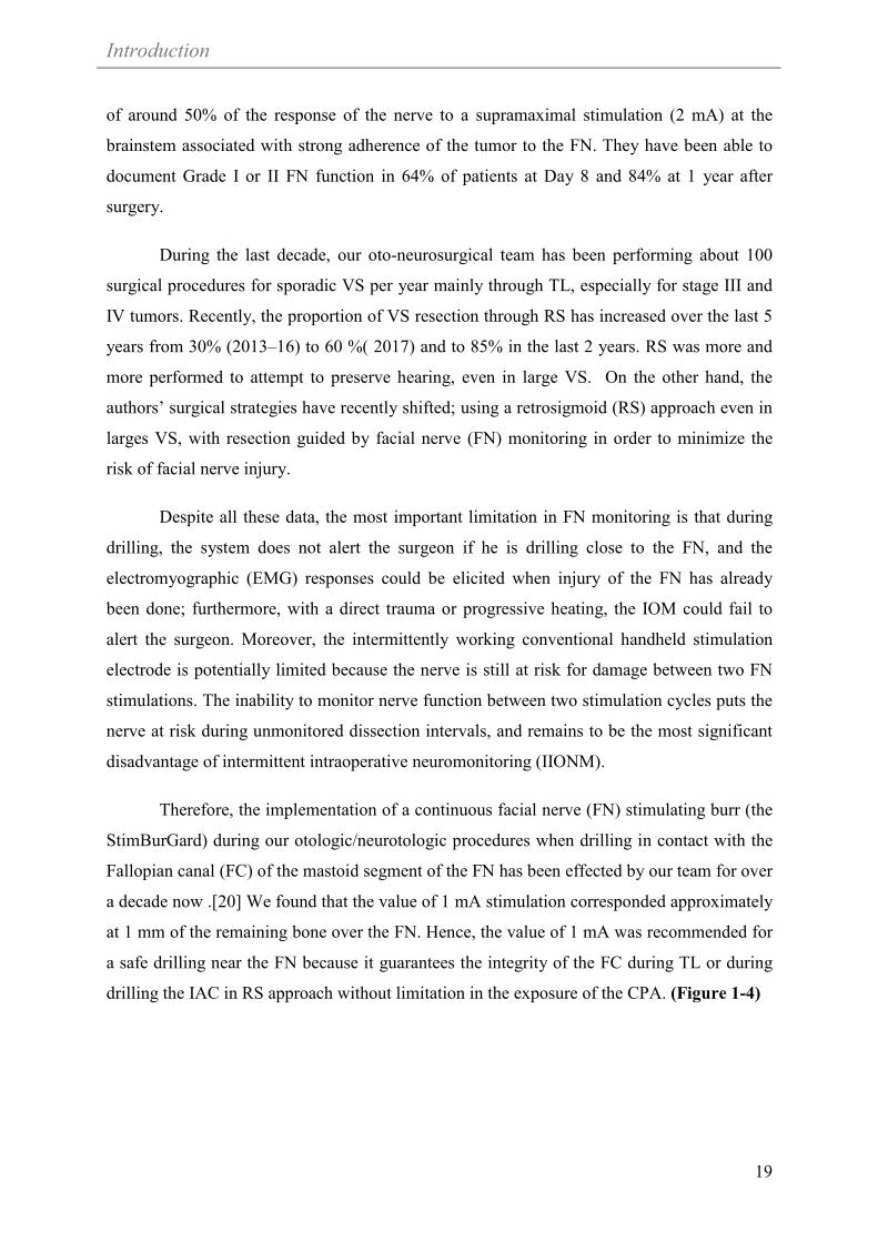
Therefore, the implementation of a continuous facial nerve (FN) stimulating burr (the
StimBurGard) during our otologic/neurotologic procedures when drilling in contact with the
Fallopian canal (FC) of the mastoid segment of the FN has been effected by our team for over
a decade now .[20] We found that the value of 1 mA stimulation corresponded approximately
at 1 mm of the remaining bone over the FN. Hence, the value of 1 mA was recommended for
a safe drilling near the FN because it guarantees the integrity of the FC during TL or during
drilling the IAC in RS approach without limitation in the exposure of the CPA. (Figure 1-4)

Introduction
20
Figure 1-4 The StimBurGard (black arrow) system with Visao drill.
Because of anatomical integrity of FN does not guarantee its functional integrity. In
clinical practice of CPA surgery, FN palsies can be caused by transection, thermal injury,
pressure, and traction. There are different strategies to avoid these lesions: Transection may
be omitted by consequent application of nerve identification tools, i.e., intermittent
intraoperative nerve monitoring (I-IONM). Pressure and tensile stress to the nerve may be
prevented by the continuous intraoperative neuromonitoring (C-IONM). C-IONM is thought
to recognize emerging trauma, thus enabling the surgeon to react instantly when the signal
decreases.

Introduction
21
References
1. House WF. Acoustic neuroma. Case summaries. Arch Otolaryngol. 1968 Dec;88(6):586-91.
2. Acioly MA, Liebsch M, de Aguiar PH, Tatagiba M. Facial nerve monitoring during cerebellopontine angle and skull base tumor surgery: a systematic review from description to current success on function prediction. World Neurosurg. 2013 Dec;80(6):e271-300.
3. Shi-Ming Y, Li-Mei Y, Yi-Hui Z, Li-Ming Y, Fei J, Wei-Yan Y, et al. Endoscope-assisted cerebellopontine angle surgery. Journal of Otology. 2009;4(1):44-9.
4. Gupta S, Mends F, Hagiwara M, Fatterpekar G, Roehm PC. Imaging the facial nerve: a contemporary review. Radiol Res Pract. 2013;2013:248039.
5. Lee S, Seol HJ, Park K, Lee JI, Nam DH, Kong DS, et al. Functional Outcome of the Facial Nerve After Surgery for Vestibular Schwannoma: Prediction of Acceptable Long-Term Facial Nerve Function Based on Immediate Postoperative Facial Palsy. World neurosurgery. 2016 May;89:215-22.
6. You YP, Zhang JX, Lu AL, Liu N. Vestibular schwannoma surgical treatment. CNS neuroscience & therapeutics. 2013 May;19(5):289-93.
7. Esquia-Medina GN, Grayeli AB, Ferrary E, Tubach F, Bernat I, Zhang Z, et al. Do facial nerve displacement pattern and tumor adhesion influence the facial nerve outcome in vestibular schwannoma surgery? Otol Neurotol. 2009 Apr;30(3):392-7.
8. Zhang Y, Chen Y, Zou Y, Zhang W, Zhang R, Liu X, et al. Facial nerve preservation with preoperative identification and intraoperative monitoring in large vestibular schwannoma surgery. Acta neurochirurgica. 2013 Oct;155(10):1857-62.
9. Liu SW, Jiang W, Zhang HQ, Li XP, Wan XY, Emmanuel B, et al. Intraoperative neuromonitoring for removal of large vestibular schwannoma: Facial nerve outcome and predictive factors. Clinical neurology and neurosurgery. 2015 Jun;133:83-9.
10. Romstock J, Strauss C, Fahlbusch R. Continuous electromyography monitoring of motor cranial nerves during cerebellopontine angle surgery. J Neurosurg. 2000 Oct;93(4):586-93.
11. Yingling C, Ashram Y. Intraoperative monitoring of cranial nerves in neurotologic surgery. Otolaryngology Head and Neck Surgery Elsevier Mosby St Louis. 2005:3877-910.
12. Kim SM, Kim SH, Seo DW, Lee KW. Intraoperative neurophysiologic monitoring: basic principles and recent update. J Korean Med Sci. 2013 Sep;28(9):1261-9.
13. Yingling CD, Gardi JN. Intraoperative monitoring of facial and cochlear nerves during acoustic neuroma surgery. 1992. Neurosurg Clin N Am. 2008 Apr;19(2):289-315, vii.
14. Torres R, Nguyen Y, Vanier A, Smail M, Ferrary E, Sterkers O, et al. Multivariate Analysis of Factors Influencing Facial Nerve Outcome following Microsurgical Resection of Vestibular Schwannoma. Otolaryngol Head Neck Surg. 2017 Mar;156(3):525-33.

Introduction
22
15. Zhang Z, Nguyen Y, De Seta D, Russo FY, Rey A, Kalamarides M, et al. Surgical treatment of sporadic vestibular schwannoma in a series of 1006 patients. Acta Otorhinolaryngol Ital. 2016 Oct;36(5):408-14.
16. Sterkers JM, Morrison GA, Sterkers O, El-Dine MM. Preservation of facial, cochlear, and other nerve functions in acoustic neuroma treatment. Otolaryngol Head Neck Surg. 1994 Feb;110(2):146-55.
17. Bernat I, Grayeli AB, Esquia G, Zhang Z, Kalamarides M, Sterkers O. Intraoperative electromyography and surgical observations as predictive factors of facial nerve outcome in vestibular schwannoma surgery. Otol Neurotol. 2010 Feb;31(2):306-12.
18. Goldbrunner RH, Schlake HP, Milewski C, Tonn JC, Helms J, Roosen K. Quantitative parameters of intraoperative electromyography predict facial nerve outcomes for vestibular schwannoma surgery. Neurosurgery. 2000 May;46(5):1140-6; discussion 6-8.
19. Bernardeschi D, Pyatigorskaya N, Vanier A, Bielle F, Smail M, Lamas G, et al. Role of electrophysiology in guiding near-total resection for preservation of facial nerve function in the surgical treatment of large vestibular schwannomas. J Neurosurg. 2018 Mar;128(3):903-10.
20. Bernardeschi D, Meskine N, Otaibi NA, Ablonczy R, Kalamarides M, Grayeli AB, et al. Continuous facial nerve stimulating burr for otologic surgeries. Otol Neurotol. 2011 Oct;32(8):1347-51.


Objectives
23
Chapter 2 OBJECTIVES
This thesis had an objective of evaluating new methods and parameters of facial nerve
preservation during CPA surgery. Our main objective was to analyze and implement the state
of art of continuous-IONM (C-IONM) in our oto-neurosurgical procedure.
A new animal model has been proposed and outlined where a complete exposure of
FN has been achieved successfully. Furthermore, our preliminary experience on FN testing in
the living animal model has been evaluated. That was essential for evaluating the safety and
reliability of the new intraoperative technique. Its applicability in future research and clinical
work has been discussed.
Also, we have been able to highlight our current experience with Intraoperative
neuromonitoring (IONM) technique of Facial nerve.
Ultimately, this has been satisfactory accomplished through:
Establishing a porcine model for posterior fossa surgery with exposure of the course of
the facial and vestibulocochlear nerves emerging from the brain stem in the CPA and then
within the IAC.
Reproducing the Porcine mode in vivo where its advantages and pitfalls are described.
Highlighting our preliminary experience with Intraoperative neuromonitoring technique
in the identification and dissection of the FN in the CPA and IAC in the porcine model.
The NIM 3.0 – Nerve Integrity Monitoring Systems (Medtronic, Jacksonville, FL,
USA) is currently routinely used in our oto-neurosurgical daily practice.
We evaluated the conventional intermittent-IONM (I-IONM) using a hand-held
probe. In addition, we evaluated the applicability of the more recently developed
technique; continuous-IONM (C-IONM), using the Automated Periodic
Stimulation (APS) Electrode Stimulator probe (Medtronic) and how it could be
further implemented in future research work.
Investigating and refining the predictive value of present and new intraoperative FN
monitoring parameters clinically, to determine if they could prognosticate FN outcome.


Results
24
Chapter 3 RESULTS
In the coming sections we are going to represent our results as following:
I. Section 3.1 "Pig as a large animal model for posterior fossa surgery in oto-
neurosurgery: A cadaveric study."
Demonstrates the development of the porcine animal model of posterior fossa
surgery in cadavers.
II. Section 3.2 “In vivo porcine model for posterior fossa surgery in oto-
neurosurgery: a preliminary experience.”
Discusses the reproducibility of the former porcine model in living animals and
highlights the feasibility of this approach and preliminary experience on FN
monitoring and testing.
III. Section 3.3." Optimization of facial nerve outcome by intraoperative
electromyography guided microsurgical resection of large vestibular
schwannomas. “
Reviews the clinical peri-operative parameters that can predict the postoperative
facial nerve function .in addition, reinvestigating some new intraoperative
monitoring value that could predict the postoperative outcome especially in large
tumors.

Results
25
(2019).
posterior
fossa
surgery
in
oto-neurosurgery:
A
cadaveric
study."
PloS
one
14,
no.
2
Sterkers,
Daniele
Bernardeschi,
and
Yann
Nguyen.
"Pig
as
a
large
animal
model
for
This
work
has
been
published
as
:
Mohamed
Elsayed,
Renato
Torres,
Olivier
Surgery in Oto-Neurosurgery: A Cadaveric Study3.1
Pig
as
a
Large
Animal
Model
for
Posterior
Fossa

Results
26

Results
27

Results
28

Results
29

Results
30

Results
31

Results
32

Results
33

Results
34

Results
35
3.2 In vivo porcine model for posterior fossa surgery in oto-neurosurgery: a preliminary experience.
This Manuscript has been submitted as: Mohamed Elsayed, Fabienne Carré, Vittoria
Sykopetrites, Renato Torres, Olivier Sterkers, and Yann Nguyen. “In vivo porcine model
for posterior fossa surgery in oto-neurosurgery: a preliminary experience.” PloS one,
February 2020.

Results
36
3.2.1 In vivo porcine model for posterior fossa surgery in oto-neurosurgery: a preliminary experience
Mohamed Elsayed1,2,3*, Fabienne Carré2, Vittoria Sykopetrites2,4, Renato Torres1,2,5, Olivier
Sterkers1,2, and Yann Nguyen1,2
1 Sorbonne Université, Inserm, Unité "Réhabilitation chirurgicale mini-invasive et robotisée
de l’audition", Paris, France
2AP-HP, GHU Pitié-Salpêtrière, Service ORL, Otologie, implants auditifs et chirurgie de la
base du crâne, Paris, France
3Alexandria University, Faculty of Medicine, Otorhinolaryngology Department, Alexandria,
Egypt 4Department of Otorhinolaryngology and Neuro-Otology, Gruppo Otologico, Piacenza,
Rome, Italy 5Universidad Nacional de San Agustín, Facultad de Medicina, Centro de Investigación y
Desarrollo Científico (CIDEC), Arequipa, Peru
Short title: Large living animal model for posterior fossa surgery *Corresponding author
Email: [email protected]

Results
37
3.2.2 Abstract Porcine living experimental models have been essential not only for surgical training
but also for investigation and electrophysiological evaluation of injury and
regenerative processes of the peripheral and cranial nerves. Accordingly, our study
seeks to establish a porcine living model for posterior fossa surgery. Our recent
experience with this in vivo porcine model is outlined, and its advantages and pitfalls
are described. Moreover, our preliminary experience with intraoperative facial nerve
neuromonitoring is highlighted, indicating where it could be successfully
implemented in future research work.
Key words (MeSH): In vivo model, Posterior fossa surgery, Facial nerve, Porcine
neuroanatomy

Results
38
3.2.3 Introduction
At present, supplemental surgical education models are desirable and essential for
oto-neurosurgical training. The routine use of human cadaver specimens and virtual reality
models has been widely described in the literature [1-4]; however, with regard to soft tissue,
and in particular, brain parenchyma and cranial nerve manipulations after fixation, these
models do not fulfill the properties of vital tissue with regard to texture and haptic
perceptions [4].
Therefore, a number of authors have described an in vivo porcine model for general
neurosurgical training. Surgical procedures included craniotomy, dural opening, brain surgery
and excision of an artificial tumor. Microscopy and bleeding management were also an
integral part of training with the aim of developing a laboratory setting imitating an operating
room as closely as possible [4-6].
The porcine experimental model has considerable similarities to human anatomy and
physiology [2, 7-11]. Generally, pig brain is comparable to the human brain in gross
anatomy. Much likeness exists in the relationship between pig and human skull base with a
similar comparative anatomy with regard to brain and cranial nerves [12]. In fact, the
differences between the anatomies of the posterior fossa in the two brains are negligible
under microscopic view with regard to microsurgical techniques involving the cranial nerves
around the brain stem [12-13]. In addition, the pig is a widely available and relatively cheap
experimental animal.
Living animal models are excellent candidates for the investigation and
electrophysiological evaluation of injury and regenerative processes of the peripheral and
cranial nerves. Intraoperative neural monitoring (IONM) has gained wide acceptance as an
adjunct technique in cranial surgery. For instance, in our oto-neurosurgical daily practice,
when we are dealing with pathologies located in the cerebellopontine angle (CPA) or at the
skull base, surgical manipulations involving the facial and vestibulocochlear nerves carry a
high risk of iatrogenic traumatic insult [14-15].
In general, the objectives of IONM include: (1) identifying the nerve in soft tissue,
tumor, and bone as early as possible; (2) warning the surgeon of an unexpected nerve
stimulation; (3) mapping the course of the nerve in temporal bone, soft tissue or tumor using

Results
39
electrical stimulation; (4) enhancing neural preservation by reducing mechanical trauma
during rerouting or tumor dissection; (5) detecting, and understanding the mechanisms of
nerve injury, and predicting the outcome at the end of tumor removal [14]. Although it does
not replace the knowledge of surgical anatomy or excellent surgical technique, IONM has
added a new functional dynamic to surgery as it empowers the surgeon beyond what is
available to them through visual information alone thereby permitting modifications in
surgical strategies that can ultimately avoid neural damage [16]. The porcine model has been
widely used in IONM research for many years and in many studies, and as a result, it is now
considered to be a reliable and reproducible model for evaluating the electrophysiologic
correlates of electromyography (EMG) during IONM [16-18].
In our previous work [1], we used the pig as a nonliving model to outline a detailed
surgical approach to the posterior fossa and to demonstrate that this approach can be used to
access the cranial nerves, specifically, the facial and vestibulocochlear nerves in the CPA and
the internal auditory canal (IAC). We have focused our attention on the pig’s posterior fossa
as this is an important anatomical area that is implicated in many lesions, which, while
extremely debilitating, are very susceptible to surgical resection [12].
Accordingly, the main goal of our study was to establish a porcine living model for
posterior fossa surgery with exposure of the course of the facial and vestibulocochlear nerves
emerging from the brain stem in the CPA and then within the IAC. Our recent experience
with this in vivo porcine model is outlined, and its advantages and pitfalls are described. The
porcine anatomy and approach with respect to oto-neurosurgical requirements are illustrated
in detail for the first time and might help others to reproduce the model for training purposes.
Our second goal was to highlight our preliminary experience with IONM in the
identification and dissection of the FN in the CPA and IAC. The NIM 3.0 – Nerve Integrity
Monitoring Systems (Medtronic, Jacksonville, FL, USA) is currently routinely used in our
oto-neurosurgical daily practice. We evaluated the conventional intermittent-IONM (I-
IONM) using a hand-held probe. In addition, we evaluated the applicability of the more
recently developed technique, continuous-IONM (C-IONM), using the Automated Periodic
Stimulation (APS) Electrode Stimulator probe (2.0 mm; Medtronic) and how it could be
further implemented in future research work.

Results
40
3.2.4 Methods
Permission to perform this study was approved by the Ethics Committee on animal
testing (Paris Descartes University, PROJET APAFIS #13860 N° 2018020112434493) under
supervision from the French Ministry of Research. The experiments took place at Fer a
Moulin Animal Hospital (APHP, Paris, France). This facility offered full veterinary and
anesthetic support for practical training purposes in the laboratory. An operating surgical
microscope (Zeiss, Germany), bipolar and monopolar coagulation, suction, and microsurgical
instruments provided as realistic an experience as possible of the daily situation in the
operating room [Fig. 3-1].
Figure 3-1 Operating room set-up.
3.2.4.1 Animals and anesthesia
Six live pigs weighing 30–40 kg were used in this prospective experiment. The pigs
were fasted for 8 hours but were allowed water before the experiment. Thirty minutes after
intramuscular administration of pre-anesthetic medication (STRESNIL® solution –
Azapérone 2 mg/kg, Atropine 1 mg, Diazepam 10 mg, ketamine 20 mg/kg), the pigs were
placed on an operating table in the prone position. An intravenous catheter was placed in a
superficial vein in one of the lower limbs through which the animal received a continuous
intravenous infusion of lactated Ringer’s solution (5 mL/kg/h) throughout the operation.
Anesthesia was supplemented with Thiopental (Nesdonal): 2 mg/kg. Analgesics were given
in the form of Nalbuphine 0.4 mg/kg. General anesthesia was induced and maintained by

Results
41
Isoflurane (Forene, Abbott, France) in oxygen after endotracheal intubation. No
neuromuscular blocking agents were allowed. Heart rate and oxygen saturation were
monitored during the entire surgical procedure. Body core temperature was stabilized by a
heating mattress and covering blankets. At the end of each experiment, the pigs were
sacrificed by an intravenous injection of DOLÉTHAL® (pentobarbital) (1 mL/kg).
3.2.4.2 Intraoperative neuro-monitoring equipment and setting
For EMG monitoring, we used NIM 3.0 (Medtronic). Paired ,four bipolar subdermal
needle 25-mm electrodes were placed in the frontalis, orbicularis oculi, orbicularis oris, and
chin muscles [Fig. 3-2].
Figure 3-2 Pig positioning and setting up of NIM 3.0 subdermal needle electrodes.
(A) The pig was placed in the prone position. (B) Set-up of subdermal needle paired electrodes attached to the NIM 3.0 (Medtronic). Frontalis, orbicularis oculi (o. oculi), and orbicularis oris (o. oris) electrodes are shown in the figure.

Results
42
We used the VISAO otologic drill (Medtronic) with the continuous stimulating bur
(Stim Bur Gard, SBG) system (Medtronic) connected to the NIM 3.0. The SBG system
delivers a continuous stimulating monopolar current on the tip and handle of the drill and the
intensity of such current can be varied with the same way like the nerve stimulator monopolar
probe. This stimulation system allows a precise and reliable localization and stimulation of
the FN during drilling.
In order to investigate the real-time EMG changes in the facial nerve more accurately
and efficiently during the procedure, we also tried continuous monitoring via automated
periodic stimulation (APS; APS Electrode Stimulator probe [2.0 mm; Medtronic]). APS is a
pulse generator for continuous stimulation (1 pulse/6 s, 100 µs, 1 mA). The conventional
monopolar stimulation probe (4 pulses/s, 100 µs, 1 mA; Medtronic) was used for
identification and intermittent stimulation of the facial nerve.
3.2.4.3 Design
In our previous work [1], we established a detailed surgical approach to access the
posterior fossa in pigs’ specimens using very simple and easily identifiable landmarks. Full
exposure of the acoustic-facial-cranial nerve bundle in the CPA and within the IAC was
accomplished.
A midline skin incision was made extending in a curvilinear fashion in the
postauricular area on both sides. Soft tissue dissection was performed to expose the temporal
bone including the external bony canal and mastoid area. It was necessary to partially dissect
the muscle overlying the atlanto-occipital joint (AOJ) to facilitate subsequent drilling during
craniotomy. The surgical field was maintained bloodless using monopolar or bipolar
diathermy [Fig. 3-3].

Results
43
Figure 3-3 Incision, soft tissue dissection and skull base anatomy.
(A,B) In the prone position, a midline skin incision was made extending in a curvilinear fashion into the postauricular area. (C–E) Dissection of soft tissue exposed the occipital bone (blue *), part of the temporal bone and mastoid area covering the posterior fossa (white **) on both sides of the pig.
Craniotomy was performed to skeletonize the posterior fossa dura on both sides using
the otologic drill and different diamond burs with a diameter varying from 5 to 3 mm.
Continuous saline serum irrigation was provided throughout the drilling to avoid heating.
Drilling started along the direction of the posterior wall of the external auditory canal (EAC)
which was used as a landmark. Next, an important landmark, the posterior arcade, could be
easily identified by its typical yellow color similar to the otic capsule in humans.
Skeletonization of the dura was performed medially to the occipital bone and inferiorly to the
AOJ. Bone wax and bipolar electrocautery were used to control bleeding from the bony
surfaces and from the dura.
A dural incision was carefully performed and brain tissue was exposed. Cerebrospinal
fluid (CSF) was gently suctioned from the operating field. The cerebellum was immediately
identified and retracted posteriorly [Fig 3-4].

Results
44
Figure 3-4 Lateral view of craniotomy of a pig’s ear on the left side.
(A) Drilling of the posterior wall of the external auditory canal (EAC) as far as the posterior arcade can be identified as a yellow bone (*) beside the dura of the posterior fossa. The mastoid area is partially covered by the atlanto-occipital joint (AOJ). (C,D) Dural opening and hemostasis using bipolar electrocautery.
The acoustic-facial bundle (cranial nerves VII/VIII) could easily be identified
emerging from the pons and entering the IAC. Further exposure of the facial and
vestibulocochlear nerve bundle in the IAC could be achieved by careful drilling of the
posterior arcade and IAC. Drilling was performed slowly and deliberately using a diamond
bur drill at low speed under continuous irrigation. Accordingly, exposure of the complete
course of the acoustic-facial bundle (CN VII/VIII) was effected from their emergence in the
CPA until they entered the IAC.
The facial/vestibulocochlear nerve complex could be separated from each other. The
facial nerve was successfully localized and identified using microsurgical instruments and the
conventional hand-held monopolar stimulation probe of the NIM 3 (Medtronic) [Figs 3-5, 6].
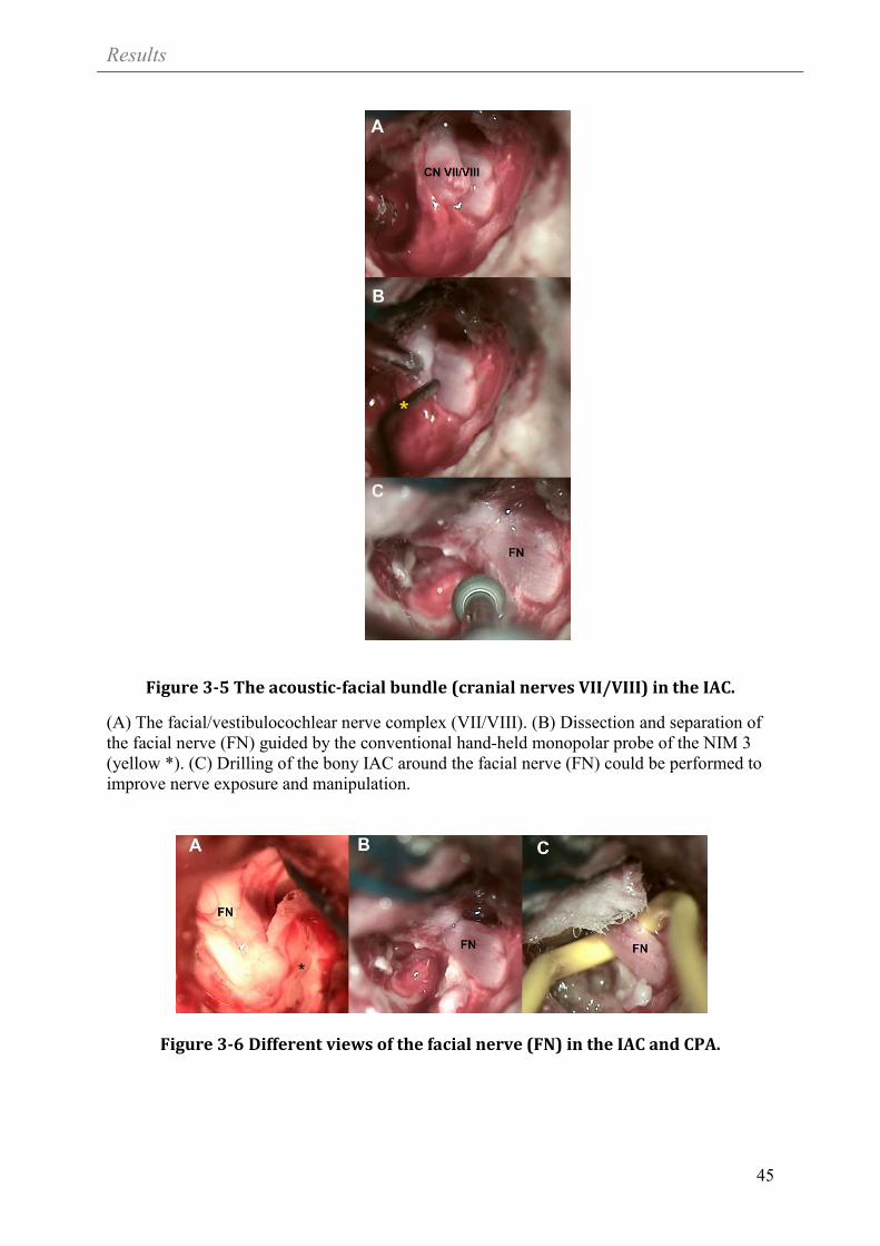
Results
45
Figure 3-5 The acoustic-facial bundle (cranial nerves VII/VIII) in the IAC.
(A) The facial/vestibulocochlear nerve complex (VII/VIII). (B) Dissection and separation of the facial nerve (FN) guided by the conventional hand-held monopolar probe of the NIM 3 (yellow *). (C) Drilling of the bony IAC around the facial nerve (FN) could be performed to improve nerve exposure and manipulation.
Figure 3-6 Different views of the facial nerve (FN) in the IAC and CPA.

Results
46
3.2.5 Results
A total of six animals (12 sides) were operated (Table 1). Average duration for skin
incision, soft tissue dissection of neck muscles and exposure of that part of the temporal bone
covering the posterior fossa bilaterally was 38.7±5 min (mean ± SD).
Table 1. Summary of posterior fossa surgical procedures performed in six animals (total of 12 sides)
Animal Side Dural opening (n=12)
FN/Vestibulococ
hlear nerve identification
(n=10)
FN identification
and separation (n=9)
APS electrode placement
(n=6) Complication
1 Right + - - - Sever Bleeding
Left + + + - -
2 Right + + - -
Injury to acoustic-facial bundle during drilling of
IAC Left + - - - Sever Bleeding
3 Right + + + +
- Left + + + +
4 Right + + + + -
Left + + + - FN transected during dissection
5 Right + + + + -
Left + + + - FN transected during dissection
6 Right + + + +
- Left + + + +
APS, automated periodic stimulation; FN, facial nerve; IAC, internal auditory canal.
In total, 12 craniotomies were performed as described earlier. Care was taken because
thin dura might easily be injured during craniotomy. Venous bleeding from bone or violated
sinuses had to be managed by compression or the early use of bone wax and other
hemostatics. In fact, severe bleeding was experienced in two sides; hence we had to abandon
further work there. The dura was opened in a U-shaped fashion using a scalpel and micro-
scissors. If required, a suture was made in the dura to lift it up.

Results
47
Careful microscopic dissection was performed to expose the acoustic-facial bundle
entering the IAC. Experienced use of suction, bipolar cautery, cottonoids, microsurgical
instruments and microscope is essential during this step.
After this, exposure of the bundle in the IAC was achieved by slow, deliberate drilling
of the posterior arcade and IAC. Injury to the bundle was only encountered once during this
study. Microdissection was performed to separate the FN using microsurgical instruments
and guided by the NIM 3 conventional hand-held probe as realized in our daily practice
during oto-neurosurgical procedures for pathology in the CPA and IAC. Unfortunately, the
FN was transected in two sides during dissection.
Electrophysiological testing of the FN in the IAC was performed by supramaximal
stimulation at 2 mA and values for both amplitude and latency were recorded. The average
maximal amplitude of the responses at 2 mA at the IAC was 4458.7±1604 µV. The latency
value at 2 mA was 5.1 ± 0.98 ms (mean ± SD).
In order to evaluate the minimal stimulation threshold, an increasing intensity of
stimulation was applied in increments of 0.01 mA between 0.03 and 0.10 mA, and in
increments of 0.05 mA between 0.10 and 3.0 mA, until a response greater than100 kV was
obtained on at least 1 channel. The average minimal stimulation threshold was 0.037± 0.011
mA.
The first trials to position the APS electrode on the facial nerve in the CPA were
performed by keeping the enclosure tabs open with a microforceps; however, some
modification to the 2.0 mm APS electrode size was essential for proper handling, insertion,
and placement of electrodes and probes on the FN proximally in the CPA. Successful
placement of the electrode was accomplished in 6 sides out of the 12 nerves [Figs 3-7, 8].

Results
48
Figure 3-7 APS electrode.
(A) The original 2 mm APS electrode; a vertical cut was made on each side of the probe parallel to the electrode knob, as indicated by the dashed lines. (B) The shape of the APS after the recommended adjustment.

Results
49
Figure 3-8 Example of APS electrode placement on the facial nerve in the CPA.
(A) Using a microforceps, the APS electrode was positioned by keeping the enclosure tabs open. (B, C) APS electrode after placement on the FN in two different models.
After determining the APS baseline for amplitude and latency of the evoked response,
continuous stimulated EMG signals were recorded. An upper threshold for latency (+10%)
and a lower threshold for amplitude (–50%) were set as separate alarm thresholds. In
addition, acoustic and optical signals alerted the surgeon if a preset threshold had been
crossed or if an electrode had been dislodged. Throughout the procedure, we encountered
intermittent difficulties associated with excessive CSF production and accumulation, blood
accumulation, and the relatively strongly pulsating brain which restricted reliable recording
of continuous EMG signals for sufficient time [Fig 9].

Results
50
Figure 3-9 Example of APS electrode placement on the facial nerve in the CPA.
FN, facial nerve; APS, a circumferential clip electrode that is placed around the FN for “Automated Periodic Stimulation” with real-time EMG signals of amplitude and latency on the right side.
3.2.6 Discussion
Oto-neurosurgical porcine models, both nonliving and living, are well established for
familiarizing trainees with surgical techniques used in cranial approaches, especially around
the CPA [4-5, 19]. Cadavers allow the study of neuro-anatomy in a practical way.
Identification of anatomical landmarks, skull base anatomy, and training in surgical
approaches are strong advantages of using cadaveric specimens. The neuro-anatomy of pigs
differs from but remains similar to humans, and allows neurosurgical trainees to quickly
focus on their practical skills [2, 4-5, 8, 12-13, 19-20].
Despite all available data on porcine models, few published studies can be found with
an oto-neurosurgical background. In an earlier study, we outlined a practical detailed model
for a new approach to the posterior fossa in pig cadavers [1]. This unique approach offered
satisfactory exposure of the acoustic-facial-cranial nerve bundle with relatively minimally
invasive steps and easily identifiable landmarks. Several surgical procedures and
manipulations could be simulated on cadavers which represent important steps in all surgical
procedures involving the posterior fossa and CPA.
In this study, we were able to reproduce this approach in living animals at our facility.
This novel and unique experience ideally mimics daily microneurosurgical practice. Cadaver
training allows the trainee to focus more quickly on surgical skills. However, microsurgical

Results
51
dissection and bleeding management of a pulsating brain under the microscope are
particularly challenging skills which can only be acquired or mastered on a living model.
Skin incision and soft tissue dissection were performed in a fashion much as in
humans, although thicker skin and massive muscle mass could be observed in the occipital
and temporal area. The surgical field could easily be maintained bloodless using
compression, hemostatics and monopolar diathermy.
Furthermore, the in vivo model presented here has major advantages in regard to
bleeding management, craniotomy, dura opening and appropriate CSF suction without
damaging brain tissue, suctioning while bleeding, and finally handling the microscope and
microsurgical instruments. These skills are major components of cranial surgery, and, using
this in vivo model, it is possible to train without risk to the patient (Table 2).

Results
52
Table 2. Comparison between in vivo and cadaveric porcine model
specimen related to the surgical skills which can be obtained
Surgical skills and learning objectives Cadaver In vivo
Set-up compared to real operating room 0 ++
Skin incision, soft tissue preparation and
dissection
+ ++
Hemostasis and bleeding management
(compression, hemostatics and bipolar
coagulation)
0 +
Identification of anatomical landmarks
and skull base anatomy
+ +
Craniotomy, bone drilling + ++
Dura opening + ++
Brain parenchyma and CPA anatomy + ++
Bipolar cautery and suction in CPA 0 ++
Cranial nerve microsurgical
manipulation (identification, dissection,
electrophysiological testing)
0 ++
CPA, cerebellopontine angle. 0, ineligible 0; +, appropriate+; ++, ideal.
A valuable advantage of this study is the adequate exposure of the acoustic-facial
bundle in the CPA and IAC. For otologists and neurosurgeons, this porcine model offers
surgical landmarks that are directly analogous to those encountered in current microsurgical
practice in the posterior fossa during removal of pathology such as acoustic neuroma or
meningioma.
In contrast to cadavers, this in vivo model permitted us to implement IONM using
conventional hand-held stimulation electrodes to guide the dissection and exposure of the FN.
IONM helped in FN dissection in about 75% of specimens.
The C-IONM system has become an area of keen interest as it offers advantages over
conventional I-IONM. The main drawback of the intermittently working conventional hand-
held stimulation electrodes is that the FN is still at risk of damage between the two

Results
53
stimulation cycles. A continuous neuromonitoring system would be able to test the functional
integrity of the recurrent laryngeal nerve (RLN) in real time and might prevent nerve damage.
Thus, ongoing animal and clinical studies have been trying to improve our understanding of
IONM technology and the electrophysiology of nerve injury specifically in thyroid surgery to
avoid RLN injury. Consequently, several varieties of electrodes have been designed for C-
IONM for stimulation of the vagus nerve, which is reported to offer more seamless
monitoring of the nerve’s functional integrity during surgery [16, 21-23].
Despite all the available data in the literature, the current status of C-IONM of the FN
during CPA surgery has not yet been elaborated. In this study, we tried to show the feasibility
of placement of the APS electrode on the FN and recording satisfactory EMG signals. The
2.0 mm APS electrode is relatively large in comparison to the more limited dimensions of the
porcine posterior fossa. Hence, the commercially available electrode was modified as
described earlier to safely position the electrode on the FN. Subsequent successful trials of
implantation and removal of the electrode were achieved atraumatically.
Another challenge was encountered after electrode placement; that the duration of
stimulation by APS was not stable or continuous for sufficient time to evaluate the
continuous monitoring of the nerve. Recurrent incidental dislocation of the electrode or
instability due to excessive CSF and/or blood accumulation was evident. Moreover, the
pulsating brain might cause electrode displacement in the relatively small surgical field.
The main goal of our study was effectively accomplished. The in vivo model and
approach to the posterior fossa presented here appears technically feasible in regard to oto-
neurosurgical daily practice. It could complement other surgical educational programs for
young surgeons such as using cadavers or virtual reality models. In addition, preliminary
experience with electrophysiological testing of the FN was highlighted using combined
intermittent and continuous IONM systems.
This study had several limitations. This is an animal model with small sample size.
The specific data in this study concerning electrophysiological evaluation of the FN cannot
be completely generalized. First, anesthetic factors such as mean blood pressure, end-tidal
CO2 and core body temperature should be controlled in future experiments to allow a more
extended duration of stimulation by APS without excessive pulsation, bleeding and CSF
accumulation. Another limitation was the APS electrode size. Modification of the electrode

Results
54
was mandatory for it to fit in the posterior fossa without injury to the FN. Finally, another
drawback was the limited histopathological analysis of the FN after electrode placement.
3.2.7 Conclusions
The present study is the first attempt to develop a simple in vivo surgical model
specifically to approach the CPA and IAC that can be used as an outline for complementary
surgical training of otologists and neurosurgeons with successful implementation of IONM of
the FN. Further research is needed on facial nerve testing and on its neurophysiological
behavior. Trials of different electrodes for C-IONM of the FN and under different controlled
stresses to the nerve will be implemented in future work. This may lead to new concepts in
operative strategy on the basis of real-time C-IONM data during CPA surgery and hence,
reduce the risk of inadvertent iatrogenic injury of the FN.

Results
55
3.2.8 Acknowledgments
We would like to gratefully thank Medtronic Xomed (Jacksonville, FL, USA) for
their materials and technical support.
3.2.9 References
1. Elsayed M, Torres R, Sterkers O, Bernardeschi D, Nguyen Y. Pig as a large animal model for posterior fossa surgery in oto-neurosurgery: A cadaveric study. PLoS One. 2019;14(2):e0212855.
2. Aurich LA, Silva Junior LF, Monteiro FM, Ottoni AN, Jung GS, Ramina R. Microsurgical training model with nonliving swine head. Alternative for neurosurgical education. Acta cirurgica brasileira. 2014 Jun;29(6):405-9.
3. Gurr A, Stark T, Probst G, Dazert S. [The temporal bone of lamb and pig as an alternative in ENT-education]. Laryngo- rhino- otologie. 2010 Jan;89(1):17-24.
4. Regelsberger J, Eicker S, Siasios I, Hanggi D, Kirsch M, Horn P, et al. In vivo porcine training model for cranial neurosurgery. Neurosurgical review. 2015 Jan;38(1):157-63; discussion 63.
5. Regelsberger J, Heese O, Horn P, Kirsch M, Eicker S, Sabel M, et al. Training microneurosurgery - four years experiences with an in vivo model. Cent Eur Neurosurg. 2011 Nov;72(4):192-5.
6. Parsak T. Posterior fossa approach: microneurosurgical training model in cadaveric sheep. Turkish Neurosurgery. 2006;16(3):111-4.
7. Gurr A, Kevenhorster K, Stark T, Pearson M, Dazert S. The common pig: a possible model for teaching ear surgery. European archives of oto-rhino-laryngology : official journal of the European Federation of Oto-Rhino-Laryngological Societies. 2010 Feb;267(2):213-7.
8. Lind NM, Moustgaard A, Jelsing J, Vajta G, Cumming P, Hansen AK. The use of pigs in neuroscience: modeling brain disorders. Neuroscience and biobehavioral reviews. 2007;31(5):728-51.
9. Hoffstetter M, Lugauer F, Kundu S, Wacker S, Perea-Saveedra H, Lenarz T, et al. Middle ear of human and pig: a comparison of structures and mechanics. Biomedizinische Technik Biomedical engineering. 2011 Jun;56(3):159-65.
10. Lovell JM, Harper GM. The morphology of the inner ear from the domestic pig (Sus scrofa). Journal of microscopy. 2007 Dec;228(Pt 3):345-57.
11. Garcia Lde B, Andrade JS, Testa JR. Anatomical study of the pigs temporal bone by microdissection. Acta cirurgica brasileira. 2014;29 Suppl 3:77-80.

Results
56
12. Jarrahy R, Shahinian HK, Young J, Berci G. Endoscopic skull base surgery II: a new animal model for surgery of the posterior fossa. Journal of investigative surgery : the official journal of the Academy of Surgical Research. 1999 Nov-Dec;12(6):335-9.
13. Hamamcioglu MK, Hicdonmez T, Tiryaki M, Cobanoglu S. A laboratory training model in fresh cadaveric sheep brain for microneurosurgical dissection of cranial nerves in posterior fossa. British journal of neurosurgery. 2008 Dec;22(6):769-71.
14. Acioly MA, Liebsch M, de Aguiar PH, Tatagiba M. Facial nerve monitoring during cerebellopontine angle and skull base tumor surgery: a systematic review from description to current success on function prediction. World Neurosurg. 2013 Dec;80(6):e271-300.
15. Oertel J, Tschan CA, Keiner D. Animal Models for Experimental Neurosurgery of Peripheral and Cranial Nerves. Experimental Neurosurgery in Animal Models. 2016:151-63.
16. Wu CW, Randolph GW, Lu IC, Chang PY, Chen YT, Hun PC, et al. Intraoperative neural monitoring in thyroid surgery: lessons learned from animal studies. Gland Surg. 2016 Oct;5(5):473-80.
17. Knight MJ, McDonald SE, Birchall MA. Intrinsic muscles and distribution of the recurrent laryngeal nerve in the pig larynx. Eur Arch Otorhinolaryngol. 2005 Apr;262(4):281-5.
18. Tanaka H, Kobayashi E. Education and research using experimental pigs in a medical school. J Artif Organs. 2006;9(3):136-43.
19. Turan Suslu H, Ceylan D, Tatarli N, Hicdonmez T, Seker A, Bayri Y, et al. Laboratory training in the retrosigmoid approach using cadaveric silicone injected cow brain. British journal of neurosurgery. 2013 Dec;27(6):812-4.
20. Sauleau P, Lapouble E, Val-Laillet D, Malbert CH. The pig model in brain imaging and neurosurgery. Animal : an international journal of animal bioscience. 2009 Aug;3(8):1138-51.
21. Schneider R, Przybyl J, Pliquett U, Hermann M, Wehner M, Pietsch UC, et al. A new vagal anchor electrode for real-time monitoring of the recurrent laryngeal nerve. Am J Surg. 2010 Apr;199(4):507-14.
22. Lee HY, Cho YG, You JY, Choi BH, Kim JY, Wu CW, et al. Traction injury of the recurrent laryngeal nerve: Results of continuous intraoperative neuromonitoring in a swine model. Head Neck. 2016 Apr;38(4):582-8.
23. Lin YC, Dionigi G, Randolph GW, Lu IC, Chang PY, Tsai SY, et al. Electrophysiologic monitoring correlates of recurrent laryngeal nerve heat thermal injury in a porcine model. Laryngoscope. 2015 Aug;125(8):E283-90.

Results
57
3.3 Optimization of facial nerve outcome by intraoperative electromyography guided microsurgical resection of large vestibular schwannomas.
This Manuscript has been submitted as: Mohamed Elsayed , Huan Jia, Baptiste Hochet,
Olivier Sterkers, Renato Torres, Yann Nguyen, Isabelle Bernat, Ghizlene Lahlou , Michel
Kalamarides." Optimization of facial nerve outcome by intraoperative electromyography
guided microsurgical resection of large vestibular schwannomas. “Otology & Neurotology,
February 2020.

Results
58
3.3.1 Optimization of facial nerve outcome by intraoperative electromyography guided microsurgical resection of large
vestibular schwannomas
Running title: Optimization of facial nerve outcome in vestibular schwannoma
surgery
Mohamed Elsayed MD1,2,4, Huan Jia MD, PhD1,2,5, Baptiste Hochet MD1,2, Olivier
Sterkers MD, PhD1,2, Renato Torres MD, PhD1,6, Yann Nguyen MD, PhD1,2, Isabelle Bernat
MD1,2, Ghizlene Lahlou MD1,2, Michel Kalamarides MD, PhD1,3
1 Sorbonne Université-APHP, Groupe Hospitalier Pitié-Salpêtrière, Department of
Otology, Auditory Implants and Skull Base Surgery, 75013, Paris, France
2 Sorbonne Université-APHP, Groupe Hospitalier Pitié-Salpêtrière, Department of
Clinical Neurophysiology, Paris
3 Sorbonne Université-APHP, Groupe Hospitalier Pitié-Salpêtrière, Department of
Neurosurgery, Paris, France
4 Alexandria University, Faculty of Medicine, ORL Department, Alexandria, Egypt
5 Shanghai Key Laboratory of Translational Medicine on Ear and Nose Diseases
(14DZ2260300), Shanghai, China; Sino-francais joint clinic research center on Otology and
Neuro-Otology, Shanghai 9th People’s Hospital, Shanghai Jiatotong University School of
Medecine, Shanghai, China
6. Universidad Nacional de San Agustín, Facultad de Medicina, Centro de
Investigación y Desarrollo Científico, Arequipa, Peru
Conflicts of Interest and Source of Funding: The authors report no conflict of
interest concerning the materials or methods used in this study or the findings specified in
this paper. This work was supported by the Ministry of Higher Education, Mission sector,
Egypt (http://www.mohe-casm.edu.eg) and the Ministry of Scientific Research, Egypt to
Mohamed Elsayed; and was supported by the Shanghai Scientific and Technological

Results
59
Innovation Action Plan (17441903600) to Huan Jia. The funders had no role in study design,
data collection and analysis, decision to publish, or preparation of the manuscript.
3.3.2 Abstract Objective: Decision making for large sporadic vestibular schwannomas (VS) guided by the
intraoperative change in supramaximal facial nerve (FN) amplitude and latency response to
optimize FN outcome.
Study Design: Monocentric prospective study.
Setting: Tertiary referral care center.
Patients: From January to December 2018, 43 large sporadic VS and 2 neurinoma-like
meningiomas with preoperative normal FN function were included.
Intervention(s): Tumors were removed through retrosigmoid (84%) or translabyrinthine
(16%) approaches with FN monitoring.
Main Outcome Measure(s): Intraoperative pre- and post-VS resection supramaximal (2
mA) amplitude and latency responses and post-resection threshold at 100 µV at the proximal
FN root were recorded.
Results: Total, near-/subtotal VS resections (TR, NTR, STR) were achieved in 51%, 38%,
and 11% of tumors, respectively, guided by no more than a 30–40% decrease in
supramaximal amplitude. Pre- and post-resection supramaximal amplitude and latency
responses were lower and longer, respectively, in NTR+STR than in TR. At day 8, FN
function was grade I-II in 78% of patients and grade III-V in 22%, and after 6 months to 1
year, it was in grade I-II in 96% and grade III in 4%, and there was no significant difference
between TR and NTR+STR. Facial palsy occurred in older patients and in the case of severe
FN adhesion. At day 8, pre- and post-resection supramaximal amplitude but not latency
responses were different between FN grade III-V and grade I-II. Serviceable hearing was
preserved in 27% of large VS.
Conclusions: Intraoperative FN monitoring guided VS resection in large VS so that 49%
retained some residual tumor. Accordingly, 96% good postoperative FN function and
significant hearing preservation were achieved.
Keywords (MeSH): Vestibular schwannoma, Intraoperative facial nerve monitoring,
Electromyography supramaximal amplitude and latency responses, Total and near-/sub-total
surgical resection, Facial nerve outcome, Hearing preservation

Results
60
3.3.3 Introduction
Vestibular schwannoma (VS) is a benign, slow-growing tumor of the Schwann cells
originating from the vestibular branch of the eighth cranial nerve, with an incidence of 12.4
tumors per million persons per year. Therapeutic options include an active wait-and-scan
policy, stereotactic radiosurgery, and microsurgical resection [1].
The generally accepted goals of VS surgery are gross total removal of the tumor and
maintenance of a good postoperative quality of life. Facial nerve function preservation is a
primary goal because postoperative facial palsy is a devastating complication. Loss of facial
expression is a functionally and psychologically debilitating condition [2].
FN preservation, both anatomically and functionally, is often challenging because
anatomical integrity of the nerve does not necessarily imply functional neural integrity. FN in
contact with VS tends to be compressed and flattened, and it can even become membranous,
adopting a color similar to that of the surrounding tissue. Moreover, variations in the location
of the FN and other cranial nerves are frequently encountered during surgery [3,4]. Despite
major advances in the field of VS diagnosis and surgery, 6–20% of poor facial function
(grade III–VI in the House-Brackmann [HB] classification [5]) can occur after surgery [6].
Prognostic factors influencing the early and long-term FN postoperative outcomes
have been widely studied. For instance, the tumor size and characteristics, adhesion of the FN
to surrounding tissue, and electromyography (EMG) responses have all been recognized as
significant prognostic factors influencing FN function [6,7].
Perhaps the most significant advancement in FN preservation came in the 1980s with
the widespread use of intraoperative FN-EMG. Routine monitoring during microsurgical
resection has now become an essential part of cranial base surgery [8]. Intraoperative
monitoring aims to make the surgical team aware of the ongoing changes in neural function,
thereby permitting modifications in surgical strategies that can ultimately avoid neural
damage.[3]
Intraoperative electrophysiological responses that can accurately predict postoperative
FN outcome would improve the ability to perform tailored resections, such as sub-total
resection (STR) and near-total resection (NTR), which are now being performed more
frequently to minimize the risk of FN injury [1,8].

Results
61
In this context, we conducted a prospective study of patients who were operated for
microsurgical resection of VS with anatomic preservation of the facial nerve. The primary
goal of this study was to identify the clinical peri-operative factors that could determine the
occurrence of an immediate postoperative impaired facial function and recovery in the long-
term. Secondarily, to refine the predictive value of intraoperative monitoring, we thought it
would be interesting to investigate FN monitoring parameters (i.e. pre- and post-surgical
resection ratios of amplitude and latency values at supramaximal stimulation) to determine if
either value could prognosticate FN outcome.
3.3.4 Materials and Methods
The work was designed as a monocentric prospective study from January to
December 2018 in a tertiary referral center. Institutional review board approval was obtained
from the Ethics Committee of our institution (CPP–Ile de France VI, Groupe Hospitalier
Pitié-Salpêtrière) for analysis of medical records and imaging data.
The study included 45 patients who had surgery for solitary VS or neurinoma-like
meningioma with normal preoperative facial palsy. Pre- and intraoperative factors that could
influence immediate FN outcome at day 1 and day 8 (maximal nervous conduction block
[7]), and subsequent FN function recovery at 1 month, and 6 months to 1 year were studied in
all patients.
Exclusion criteria were preoperative facial palsy, neurofibromatosis type 2 (NF2),
recurrent VS, and interruption of the facial nerve during surgery.
In all cases, written informed consent was obtained for the surgery and the research
studies (CNIL#2211758). Data was analyzed anonymously and consideration was given to
confidentiality.
Intraoperative monitoring of facial nerve function was achieved with a four-channel
nerve monitoring system (NIM® 3.0, Medtronic, Jacksonville, FL, USA). For the
translabyrinthine approach, a StimBurr guard system (Medtronic) was used.
Electrophysiological monitoring of the facial nerve requires avoidance of neuromuscular
blockade during the intervention. Subdermal needle electrodes were placed in optimal
locations to record activity from the facial muscles, typically in the four muscle areas
innervated by the facial nerve: frontal, orbicularis oculi, orbicularis ori, and mentalis.

Results
62
Ground and stimulator anode electrodes were also placed. A monopolar probe with a 0.5-mm
tip was included in the sterile operating field. All electrode wires were connected to an
interface circuit box. Stimulus intensity, duration, rate, and event threshold were set. Typical
parameters used at our institution were: stimulus intensity ranging from 0.01 mA to 3 mA;
duration, 100 µs; and event threshold, 100 µV.
All patients received an intravenous bolus of methylprednisolone (1 mg/kg) at
anesthesia induction. In the case of impaired FN function at postoperative day 1,
corticosteroid therapy (methylprednisolone, 1 mg/kg/day) was administered for 1 week. No
antiviral therapy was administered as prophylaxis or in the case of facial palsy.
3.3.4.1 Clinical characteristics
Patient characteristics included age, sex, symptoms at diagnosis, and delay between
the first diagnostic MRI until the time of surgery.
3.3.4.2 Audiometric evaluation
Pre- and postoperative hearing was assessed by pure tone average (PTA) at 0.5, 1, 2,
and 3 kHz, and speech discrimination score (SDS) with monosyllabic lists (Lafon list).
Audiometric tests were performed in a soundproof booth. PTA was combined with SDS to
report hearing using the 4-class system according to the recommendations of the AAO-HNS
hearing loss scale [9] as class A (pure-tone thresholds (PTA) ≤ 30 dB, and speech
discrimination (SD) ≥ 70%), class B (30 dB < PTA ≤50 dB, and SD ≥ 50%), class C (PTA >
50 dB, and SD ≥ 50%), and class D (whatever the PTA value, and SD < 50%).
3.3.4.3 Radiological characteristics
Tumors were categorized according to the largest diameter in the axial or coronal
view, and if necessary, in the magnetic resonance imaging (MRI) view of the
cerebellopontine angle (CPA) on gadolinium-enhanced sequences as already described
[1,4,6,7,10] in four functional stages: stage 1 (intracanalicular), stage 2 (≤15 mm maximal
extracanalicular diameter), stage 3 (15–30 mm), or stage 4 (>30 mm); the cystic character of
the tumor was defined on the preoperative MRI and classified as solid or cystic vestibular
schwannoma. Fundus involvement was determined by considering the tumor extension

Results
63
toward the lateral end of the internal auditory meatus (IAM), as A (fundus invasion), B
(fundus free of tumor), and C (internal auditory meatus free) [6].
3.3.4.4 Intraoperative surgical data
All patients were operated on in the supine position under general anesthesia through
a translabyrinthine (TL) or retrosigmoid (RS) approach. When the VS totally invaded the
IAM fundus with no direct microscopic visualization, dissection of the tumor from the FN
was performed under endoscopic view. The facial nerve position relative to the tumor was
identified [4] (type 1: anterior-inferior; type 2: anterior; type 3: superior; or type 4: posterior).
Tumor adhesion was defined by the senior surgeons as: weak (the dissection plane between
the tumor capsule and the facial nerve was easily recognized and detachable), moderate (the
dissection plane was recognizable but adhesion to the facial nerve sheath was significant),
and strong (the dissection plane was not recognizable).
3.3.4.5 Intraoperative FN monitoring parameters
A mono-polar probe with a 0.5-mm tip electrically stimulated the FN at constant
current. Before tumor excision, the FN was stimulated at its root entry zone (proximal-pre), at
a sufficient distance of the adhesion zone of the FN to the tumor capsule to avoid
concomitant stimulation of its distal axons. The FN was exposed either immediately after
opening the cisterna for middle-sized VS or after first debulking its posterior extension for
larger VS. Then, during VS removal, the FN was stimulated either to locate the course of the
nerve on the VS capsule at 0.8–1 mA or to identify a significant amplitude response decrease
at a supramaximal value of 2 mA. After VS excision, the FN was stimulated again at the root
entry zone (proximal post), and when possible in the IAM (distal post). The following
parameters were measured:
1) Maximal amplitude of the responses (in microvolts; µV) after supramaximal
stimulation at 2 mA before and after tumor excision to determine if it had decreased by not
more than 30–40% of its initial value throughout the surgical procedure.
2) Latency values (in milliseconds; ms) after supramaximal stimulation at 2 mA
before and after tumor excision to disclose an increase in responses.

Results
64
3) Stimulation threshold (in milliamperes; mA), with increasing intensity of
stimulation in increments of 0.01 mA between 0.03 and 0.10 mA, and of 0.05 mA further, if
necessary, until a response greater than 100 µV on at least one channel was obtained at the
end of VS resection.
3.3.4.6 Assessment of FN function
FN function was clinically determined by a senior ENT surgeon at day 1, day 8
(during the subsequent week), 1 month, and between 6 months and 1 year after surgery. Day
8 was identified as being, in most cases, the time of maximal nervous conduction block in the
early postoperative period [7]. In a few cases, the day 8 outcome deteriorated later, and this
was indicated as delayed facial palsy. The House–Brackmann facial nerve grading scale (HB)
[5] was used to assess FN function. Good FN function was defined as HB I–II and impaired
FN function as HB III–V.
3.3.5 Statistical analysis
All statistical analyses were performed with IBM SPSS statistical software package
version 20.0 (Armonk, NY: IBM Corp.). Qualitative data were described using numbers and
percentages. The Kolmogorov–Smirnov test was used to verify the normality of the
distribution. Descriptive data were reported using mean±SD, range [minimum and
maximum], and median value. Univariate logistic regression models were performed to
assess the association between baseline characteristics and facial function at day 8 and 1-year
postoperatively. All differences between groups with a p-value ≤ 0.05 were considered to be
significant.
3.3.6 Results
3.3.6.1 Population and intraoperative data (Table 1)
In total, 45 patients hosting 43 unilateral VS and 2 neurinoma-like CPA meningioma
(considered to be VS) were included in this study with a mean age of 51±11.4 years (range
20–78 years), equally distributed below and above 50 years, 20 males and 25 females.
At diagnosis, hearing loss was the prevalent symptom (93% of cases), with a sudden
onset in 16% of patients, and then tinnitus (24%), trigeminal hypoesthesia (21%), and

Results
65
instability (12%) or vertigo (10%); all symptoms observed in large tumors. Indeed, tumors
were almost all stage 3 and 4 with frequent invasion of the fundus of the IAM (58%).
Surgery was performed at least 1 year after the diagnosis was made (mean±SD,
15±24.5 months; range, 1 month to 10 years), 73% being operated on during the first post-
diagnosis year. VS were resected mainly through a RS approach (84%), a TL approach being
reserved for VS with poor hearing. In this large VS population, hearing was generally
preserved preoperatively, and still serviceable in 78% of patients. Intraoperatively, the FN
was more frequently located anteriorly to the VS (73%) than superiorly (20%), and its
adhesion to the VS capsule was distributed equally between weak (40%), moderate (31%),
and severe (29%). VS resection was considered to be total, near-total or sub-total in 51%,
38%, and 11% of cases, respectively. No major complications were observed, apart from a
retro-auricular hematoma in a wound and a CSF leakage, both cured by revision surgery.
The pre- and post-resection supramaximal amplitude responses were 2212±1368.9
and 1572±1044.4 µV, respectively, with a mean amplitude decrease of 28±23.7%. The
supramaximal latency values remained stable at 6.3 ms.

Results
66
Table 1. Patients’ characteristics and general outcomes No. %
Gender Male 20 44 Female 25 56
Age(year) 51±11.4[20-78]
Symptoms Progressive HL 35 78 Tinnitus 24 53 Trigeminal defect 21 47 Instability 12 27 Vertigo 10 23 Sudden HL 7 16 Headache 3 7 Caudal nerve defect 1 2
Hearing (AAO-HNS)
A 21 47 B 14 31 C 1 2 D 9 20
VS volume (Functional staging)
I 0 0 II 2 5 III 28 62 IV 15 33
IAM invasion A 26 58 B 12 27 C 7 15
Cystic VS (n=43)
No 26 60 Yes 17 40
Approach RS 38 84 TL 7 16
Origin of VS Inf. Vest. N 20 45 Sup. Vest. N 19 42 unsure 6 13
FN positioning Anterior-inferior 3 7 Anterior 33 73 Superior 9 20
FN adherence to VS Weak 18 40 Moderate 14 31 Severe 13 29
VS type of resection Total 23 51 Near-total 17 38 Sub-total 5 11
Proximal supramaximal FN amplitude response (µV)
Pre-resection 2212±1386.9 [170- 6446] Post-resection 1572±1044.4 [122- 4519]
Amplitude loss 28±23. 7 %

Results
67
Proximal supramaximal FN latency (ms)
Pre-resection 6.3±1.32 [4.4-10.0] Post-resection 6.3±1.48 [4.4-10.9]
3.3.6.2 Type of VS resection (Table 2)
Tumors were removed using an RS approach in the majority of patients (84%) to
attempt to preserve hearing whatever the preoperative state of hearing or VS size, and a TL
approach (16%) was used only if anacusis (n=6) or a rapidly growing stage 4 VS (n=1)
invaded the IAM fundus.
Gross-total resection was performed in 23 cases (51%), and in the remaining cases,
VS were partially resected leaving a small piece of tumor adherent to the FN considered as a
function of the length of the adherent zone as near- or sub-total resection in 17 and 5 patients,
respectively. Stage 3 VS were more frequently totally resected and stage 4 partially resected
(p<0.05). Accordingly, all small tumors were completely removed, but this dropped to 60%
for mid-sized tumors while only 27% of large tumors were completely removed. Adhesion
was estimated to be weak in the majority of TR (p<0.005) and severe in the majority of
NTR/STR (p<0.005). Moderate adhesion was distributed equally between the two groups.
Decision making for a partial (NTR/STR) resection was guided by both intraoperative
evaluation of severe FN adhesion to the VS and a decrease of at least 30–40% of the
supramaximal amplitude response. Before dissection of the tumor from the FN, the
supramaximal amplitude responses were lower for partial resections than for total resections
(p<0.05) and latencies were higher for partial resections than for total resections (p=0.05). In
the group with partial resections, amplitude responses less than 1000 µV were recorded (n=3)
but not in the group with total resections. For pre-VS resection latencies longer than 8 ms
(n=6), TR was performed for only 1 VS. At variance with this, resection had been total when
the pre-VS resection amplitude responses were higher than 3500 µV (n=5). Nevertheless,
supramaximal amplitude response losses between pre- and post-VS resection were similar in
both groups, from 24% to 34% in total and partial excision groups. No changes in
supramaximal stimulation latencies were noted in the two groups. The 100 µV threshold
could be obtained at low stimulation values in both groups.
For NTR, in most cases, the residual tumor was not enhanced on early postoperative
contrast MRI although it was enhanced for STR. MRI scans performed from 1 week to 1 year

Results
68
postoperatively were available in only 18 cases. They were all negative (n=5) among 23 TR,
either negative (n=7) or with residual tumor (n=4; mean volume 492 mm3 [25–756]) among
17 NTR, and with residual tumor (n=2; 245 and 3366 mm3) among 5 STR.
Table 2: Factors influencing decision making for VS resection type
Surgical decision Total resection (n=23)
Partial resection (n=22) P-value
Tumor Stage II 2 (9%) - - III 17 (74%) 11(50%) <0.05 IV 4 (17%) 11(50%)
FN adhesion to VS
Weak
14 (61%)
4(18%)
<0.05
Moderate 7 (30%) 7(32%) n.s. Severe 2 (9%) 11(50%) <0.05
Proximal supramaximal FN amplitude response (µV)
Pre-resection
2659±1624.1 [1000–6446]
1744±904.9 [170–3197]
<0.05
Post-resection
1951±1158,1 [835-4519]
1175±746,9 [122–3009] <0.001
Proximal supramaximal FN latency (ms)
Pre-resection
5.9±1,17 [4.4-8.7]
6.7±1.37 [4.9-10.0]
<0.05
Post-resection
5.8±1.14 [4.4-8.6]
6.9±1.64 [4.6-10.9] <0.02
Post-resection threshold at 100µV (mA)
0.06±0.14[0.03-0.7] 0.05±0.10 [0.03-0.5] n.s.
FN outcome Day 8
I – II
19(82.6%)
16(72.7%)
III – VI 4(17.4%) 6(22.9%) V - 1(4.4) 1 year I – II 22(95.7%) 21(95.5%) III – VI 1(4.3%) 1(4.5%)
Hearing preservation (n=36) A/B/C 5/21 (24%) 7/15 (47%)
FN: facial nerve; Proximal supramaximal (2mA) FN amplitude responses or latencies, and threshold at 100µV determined at cerebellopontine FN root.
3.3.6.3 Facial nerve function outcome (Table 3, and Fig. 3-10)
The day after surgery, FN function was considered to be good in 91% of patients (19
and 22 patients with Grade I and Grade II, respectively). Only 4 of them were evaluated as
Grade III. At day 8, when nervous conduction block is known to be maximal, a good FN
function was achieved in 78% and distributed as follows: Grade I in 20 patients, Grade II in

Results
69
15, Grade III in 6, Grade IV in 3, and Grade V in 1 patient. Facial paralysis (FP) was
observed in 6 more patients than at day 1 (Fig. 1); no further delayed FP was noted. FP
occurred in older patients compared with those with good FN function (58±8.5 years vs.
49±11.5 years, respectively; p<0.05).
The symptoms at diagnosis, VS volume, IAM invasion, hearing level, surgical
approach, FN position, vestibular nerve origin, and resection type did not influence the onset
of FP. At variance with this, severe FN adhesion to the VS capsule was a negative factor
(7/35 Grade I–II vs 6/10 Grade III–V, p<0.02), but this was not the case with either weak or
moderate FN adhesion.
Pre- and post-resection supramaximal FN amplitude responses were lower when FN
function was maximally altered at day 8 than in the absence of major FN impairment. The
decrease in responses was similar in the two groups (Table 3). However, the ratio between
distal, as recorded at the fundus or nearby, and proximal post-resection supramaximal FN
amplitude responses reached higher values in the presence of FP (62±20.2% [32–85], n=8
Grade III–V vs 39±13.1% [15–59], n=23 Grade I–II p<0.01). The supramaximal FN latencies
and the threshold at 100 µV were not different between the two groups (Table 3).
The electrophysiological parameters influencing the onset of a nervous conduction
block were evaluated at day 8 by analyzing separately Grade I considered to be a normal
nerve, Grade II as weakly altered, and Grade III–V as a moderately to severely injured nerve.
Pre- and postoperative supramaximal amplitude responses were similar in the two former
groups but lower in the latter group indicating that Grade I and Grade II could be analyzed
together.
As shown in Fig. 1, the FN recovered with 89% Grade I–II at 1 month, and 96% after
more than 6 months (35 Grade I and 8 Grade II). Only two patients still had Grade III FP
after 6 months to 1 year of postoperative follow-up, one in each of the two groups, total and
partial VS resections. Neither clinical, nor radiological, nor intraoperative observations nor
EMG recordings were found to influence FN recovery.

Results
70
Table 3. EMG parameters influencing FN function (HB scaling) at post-
operative day 8
FN function at D8
P-value Grade I-II
(n=35)
Grade III-V
(n=10)
Proximal
supramaximal
FN amplitude
response (µV)
Pre-resection 2433±11436.7
[753-6446]
1436±924.3
[170-3029] <0.05
Post-resection 1746±1069.2
[393-4519]
960±691.4
[122-2547] <0.01
Proximal
supramaximal
FN latency (ms)
Pre-resection
6.3±1.37
[4.5-10.0]
6.2±1.15
[4.4-8.0]
n.s.
Post-resection 6.4±1.57
[4.4-10.9]
6.3±1.16
[4.5-7.5] n.s.
Post-resection threshold at 100µV (mA)
0.03±0.0
[0.03-0.04]
0.15±0.24
[0.03-0.7]
<0.01
FN: facial nerve; Proximal supramaximal (2mA) FN amplitude responses or latencies,
and threshold at 100µV determined at cerebellopontine FN root.

Results
71
Figure 3-10 Postoperative FN function according to HB staging,
Stage 1-2 and stage 3-5 were considered as good and poor, respectively (n = 45).
3.3.6.4 Hearing preservation
An attempt was made to preserve hearing using an RS approach in patients with a
large VS whatever the preoperative hearing level and VS size, apart from when there was a
preoperative dead ear. Among 38 VS operated on through RS, 2 audiograms were missing
postoperatively (2 class B). Preoperative hearing was classified as 20 class A, 12 class B, and
3 class D (persisting PTA with no intelligibility). Serviceable hearing (class A+B) was
preserved in 9 among 33 patients (27%; 8 class A and 1 class B) and measurable hearing in
12 among all 36 VS operated on through an RS approach (33%; 1 class A and 1 class B
decreased to class C, although 1 class D improved to class C). Thus, hearing was preserved in
50% of preoperative class A patients, and was at the same class level in 40% of them.
Hearing was serviceable in 5 stage 3 patients with 2 TR and 3 NTR, and 4 stage 4 patients
with 1 TR and 3 NTR. Hearing was measurable in 3 additional class C patients with stage 3
VS (2 TR and 1 STR). Among 21 cases of TR, measurable hearing was preserved in 5 cases
(24%), but among 15 cases of NTR and STR, this rate increased to 47%.
The interval between VS diagnosis and surgery was shorter in the 9 patients with
preserved serviceable hearing than in the remaining 26 class C+D patients (7±16.0 months
[1–50] vs 16±22. 3 months [1–87]; p<0.01). Preoperative normal hearing was more frequent

Results
72
when hearing could be retained (4 out of 9 in the former group versus 21 out of 26 in the
latter group; p<0.05). No other clinical, radiological, intraoperative observational or
electrophysiological factors could account for the preservation of serviceable hearing.
3.3.7 Discussion
This prospective study was aimed at minimizing the onset of FN conduction nervous
blockage while managing surgically sporadic VS. We have previously shown that the
occurrence of a postoperative FP was maximal at the end of the first week and then gradually
recovered, in many cases in a few weeks although it may last for up to several months in
some others [7]. These different FP recovery patterns may be accounted for not only by the
severity of FN injury during the surgical manipulations, which results mainly from FN
adherence to the tumor capsule, but also by the preoperative FN impairment due to its
stretching induced by the VS volume [4,7]. Accordingly these two factors, VS volume and
FN adherence, were found to be independently predictive of FP occurrence in a multivariate
analysis [6]. Indeed, in most studies, patients with a preoperative facial weakness were
excluded although a normal facial function does not preclude some FN damage. Previous
studies have demonstrated that intraoperative monitoring by FN electromyography,
regardless of the stimulation level, could be predictive of FN function at 1 year, which results
from combined factors including preoperative FN status, severity of initial postoperative FN
blockage, and quality of recovery, depending also on complex mechanisms [3]. After total
VS resection, a good FN recovery was achieved at 6 months if amplitude responses ≥100 µV
could be obtained by stimulation of the proximal root of the nerve at ≤0.04 mA [11]. In
addition, conduction nervous blockage resulting in postoperative Grade III–VI FP at day 8
emerged if either the nerve pre-resection amplitude responses were under 800 µV or the post-
/pre-resection amplitude response ratio had declined by ≥40% after supramaximal stimulation
(2 mA) of the proximal root [7]. According to this last parameter and/or because of severe
adherence of the FN to the VS capsule, instead of performing the usual total VS removal, a
small tongue of tumor was left adherent to the nerve yielding a near-/subtotal resection
depending on the length and width of the nerve–tumor adherence, especially in large VS [1].
Although partial resections of large VS have been reported, there is no clear definition of the
residual volume for near- or sub-total resection [1, 8, 12].

Results
73
In this study, decision making for a total or partial resection depended on both
intraoperative evaluation of FN to VS adherence and intraoperative FN electromyography
parameters before and during VS resection. It was concluded that a partial resection was
indicated if FN adherence to the VS was severe and/or there was a 30–40% decrease in post-
/pre supramaximal amplitude response ratio at the proximal FN root in the CPA. As shown in
Figure 3-11 , the markers of a severe adherence between FN and VS were pre-resection
supramaximal low amplitude responses (<1200 µV) or high latencies (>8 ms) as in all cases
except 2 (n=13), partial resections were performed. At variance with this, high amplitude
responses (>3500 µV, n=5) but not low latencies indicated a weak adherence allowing a total
resection. Although for weak and severe FN adherences to the VS capsule, more TR and
NTR/STR were achieved, respectively, moderate adherence had an intermediate status which
needs to be further evaluated presumably by its length and its width as intraoperative
recordings did not provide valuable markers.
Figure 3-11 Optimization of facial nerve outcome by reducing the nervous conduction blockage severity.
FN: facial nerve; VS: vestibular schwannoma; TR: total resection; NRT: near-total resection;
STR: subtotal resection.
FN function outcomes are generally reported 1 year after surgical gross-total or partial
VS removal and are greatly dependent on VS volume: the smaller the tumor, the better the
FN function [13–15]. The cutoff value for extra-canal tumor diameter to minimize the risk of
impaired facial function was 16 mm after gross-total resection [6], which nicely corresponded
to the limit between stages 2 and 3 in our classification [1,4,7,11,13]. In the present series of

Results
74
large VS managed surgically with an equal proportion of total or partial tumor removal, a
good FN function was achieved in 96% of cases after 6 months to 1 year, and the only two
poor outcome cases were evaluated as Grade 3 at the last visit. Previously, we reported that
good FN function at 1 year ranged from 80% to 85% after gross-total resection of large
tumors (139 stage 4 and 192 stage 3, respectively) [13]. Indeed, intraoperative monitoring
provides information on the functional status of the nerve at the beginning of the surgical
procedure, and then on its evolution during VS dissection from the nerve. An initial impaired
FN can be suspected if the supramaximal amplitude responses and/or latencies were low and
high, respectively, assuming axon fiber and/or myelin sheath alterations related to both VS
volume and FN adhesion to the tumor capsule. During VS resection, only the decrease in
supramaximal amplitude responses indicated the development of a conductive nervous
blockage which may lead to a Grade 3 or more FP. Taking this marker into account could
preclude the onset of a severe conductive nerve blockage as 71% of VS had no or only a
slight FN impairment at day 8 and, then it recovered to 89% and 96% after 1 month and at the
last visit after at least 6 months, respectively. Less than a 40% decrease in the supramaximal
amplitude responses at the proximal FN root seems to be critical since we reported previously
64% and 84% Grade I–II FN outcome at day 8 and 1 year, respectively, in 25 consecutive
NTR/STR for large VS based on 50% loss of the initial value [1].
Hearing preservation is one of the most challenging goals of VS removal. Most of
these large VS were resected through an RS approach with 27% and 33% of postoperative
serviceable and measurable hearing, respectively. Thus, the intraoperative FN monitoring
criteria used to minimize the occurrence of FN blockage could also provide benefit for
hearing preservation. Many factors have been found to influence the preservation of hearing
such as the tumor size, tumor growth rate, preoperative hearing level, etc. [13,16,17]. Herein
a short interval between the diagnosis and surgical treatment was a good prognostic factor
together with normal hearing.
Residual tumor volumes after NTR and STR were evaluated by gadolinium injected
MRI performed during the months following microsurgery only in some instances, and
showing that the residual tumor was not enhanced by the contrast in most NTR. This suggests
that the residual tumor adherent to the FN had been substantially devascularized during
microsurgery with a limited risk of regrowth. In a previous series of large partially resected
VS, regrowth was observed in 2 out 25 tumors [1]. The need for subsequent complementary

Results
75
radiotherapy treatment depends on the regrowth rate and the volume achieved. In these cases,
revision surgery should not be performed when adherence between the FN and VS capsule
was not relieved after the first surgery.
3.3.8 Conclusions
Supramaximal stimulation of the FN proximal to the VS is mandatory during VS
surgery because, on the one hand, the values of amplitude responses and latencies provide
information on the pre-resection nerve status, and on the other hand, the amplitude responses
should not decrease by more than 30–40% so as to avoid a severe conduction nervous
blockage. Following these indicators, half of the large VS were totally removed and half
near-/sub-totally removed with 96% of normal/sub-normal FN function after 6 months to 1
year. In addition, this guidance could allow hearing to be preserved in one-third of patients.
Continuous intraoperative monitoring of the FN would help the surgeon to make an
immediate decision on a partial VS resection if a decrease in amplitude responses occurred,
and to modify his procedure to preserve a good FN function as well as hearing in most cases.
3.3.9 Acknowledgment
We gratefully thank Amgad Hamza (S&S for Biostatistical Analysis, Alexandria,
Egypt) for assistance with statistical analysis.

Results
76
3.3.10 References 1 Bernardeschi D, Pyatigorskaya N, Vanier A, et al. Role of electrophysiology in guiding
near-total resection for preservation of facial nerve function in the surgical treatment of
large vestibular schwannomas. J Neurosurg 2018 Mar;128(3):903-10.
2 Bloch O, Sughrue ME, Kaur R, et al. Factors associated with preservation of facial
nerve function after surgical resection of vestibular schwannoma. J Neurooncol 2011
Apr;102(2):281-6.
3 Acioly MA, Liebsch M, de Aguiar PH, Tatagiba M. Facial nerve monitoring during
cerebellopontine angle and skull base tumor surgery: a systematic review from
description to current success on function prediction. World Neurosurg 2013
Dec;80(6):e271-300.
4 Esquia-Medina GN, Grayeli AB, Ferrary E, et al. Do facial nerve displacement pattern
and tumor adhesion influence the facial nerve outcome in vestibular schwannoma
surgery? Otol Neurotol 2009 Apr;30(3):392-7.
5 House JW, Brackmann DE. Facial nerve grading system. Otolaryngol Head Neck Surg
1985 Apr;93(2):146-7.
6 Torres R, Nguyen Y, Vanier A, et al. Multivariate analysis of factors influencing facial
nerve outcome following microsurgical resection of vestibular schwannoma.
Otolaryngol Head Neck Surg 2017 Mar;156(3):525-33.
7 Bernat I, Grayeli AB, Esquia G, Zhang Z, Kalamarides M, Sterkers O. Intraoperative
electromyography and surgical observations as predictive factors of facial nerve
outcome in vestibular schwannoma surgery. Otol Neurotol 2010 Feb;31(2):306-12.
8 Schmitt WR, Daube JR, Carlson ML, et al. Use of supramaximal stimulation to predict
facial nerve outcomes following vestibular schwannoma microsurgery: results from a
decade of experience. J Neurosurg 2013 Jan;118(1):206-12.
9 Committee on Hearing and Equilibrium guidelines for the evaluation of hearing
preservation in acoustic neuroma (vestibular schwannoma). American Academy of
Otolaryngology-Head and Neck Surgery Foundation, INC. Otolaryngol Head Neck Surg
1995 Sep;113(3):179-80.
10 Grayeli AB, Kalamarides M, Bouccara D, Rey A, Sterkers O. Functional staging of
acoustic neuromas. In: Kanzaki J, Tos M, Sanna M, et al. eds. Acoustic Neuroma.
Consensus on Systems for Reporting Results. Tokyo: Springer, 2003; 141-6.

Results
77
11 Grayeli AB, Guindi S, Kalamarides M, et al. Four-channel electromyography of the
facial nerve in vestibular schwannoma surgery: sensitivity and prognostic value for
short-term facial function outcome. Otol Neurotol. 2005 Jan;26(1):114-20.
12 Gurgel RK, Theodosopoulos PV, Jackler RK. Subtotal/near-total treatment of vestibular
schwannomas. Curr Opin Otolaryngol Head Neck Surg 2012 Oct;20(5):380-4.
13 Zhang Z, Nguyen Y, De Seta D, et al. Surgical treatment of sporadic vestibular
schwannoma in a series of 1006 patients. Acta Otorhinolaryngol Ital 2016
Oct;36(5):408-14.
14 Liu SW, Jiang W, Zhang HQ, et al. Intraoperative neuromonitoring for removal of large
vestibular schwannoma: Facial nerve outcome and predictive factors. Clin Neurol
Neurosurg 2015 Jun;133:83-9.
15 Taha I, Hyvarinen A, Ranta A, et al. Facial nerve function and hearing after
microsurgical removal of sporadic vestibular schwannomas in a population-based
cohort. Acta Neurochir (Wien) 2019 Sep 7. doi: 10.1007/s00701-019-04055-4. [Epub
ahead of print]
16 Yang J, Grayeli AB, Barylyak R, Elgarem H. Functional outcome of retrosigmoid
approach in vestibular schwannoma surgery. Acta Otolaryngol 2008 Aug;128(8):881-6.
17 Zhu W, Chen H, Jia H, et al. Long-term hearing preservation outcomes for small
vestibular schwannomas: retrosigmoid removal versus observation. Otol Neurotol 2018
Feb;39(2):e158-e65.


Discussion
78
Chapter 4 DISCUSSION
Along with the improvement of surgical techniques, the acquiring importance of
radio-surgery and the advancement of diagnostic equipment led to a change in the objectives
of VS treatment. Modern CPA surgery is reaching the goal of preservation of function.
Although facial nerve preservation was the major concern in the past decades, currently
hearing preservation is the main challenge. To date, the choice of the surgical strategy
depends on tumor size, hearing and facial nerve function, patient age and the presence of
other disabling symptoms. The challenge and the matter of the actual debate are to achieve
major tumor removal, with a minimal impairment of these functions.
Over years, we were able to achieve 97.7% of FN anatomical integrity, although,
good FN function was achieved only in 85.1% of patients. Surgical outcome depends on
several critical variables, including tumor size, surgical approach, surgeon’s experience and
development of technique.[1-4]
In order to minimize FN injury and improve hearing preservation, a strategic shift in
our surgical approach, decision making and improved operative techniques and parameters
has been successfully implemented.
Ultimately, Continuous intra-operative monitoring of nerve function is becoming an
essential tool of cranial base surgery because of the problem of the conventional handheld
intraoperative monitoring probe and how the surgical team is completely blind between 2
cycles of stimulations with quite long unmonitored dissection intervals. Therefore, the
continuous intraoperative neuromonitoring (C-IONM) is thought to recognize emerging
trauma, thus enabling the surgeon to change his strategy and operative technique in real time
before nerve injury.

Discussion
79
I. Animal models and surgical training
Developing and establishing a porcine model for posterior fossa surgery was a
primary goal of our study. Despite all available data on porcine models, a large animal model
with an oto-neurosurgical background was not described before. The availability and low cost
of the pig, combined with the ease of preparation made it a strong candidate. Also, the fact
that we can reproduce this model in vivo later at our facility made the pig more convenient
than other models such as sheep or cows. It is easy to obtain and maintain living pigs and to
anesthetize them.
The anatomy of the skull base, it is highly similar to that of humans especially when it
comes to the anatomy of cranial nerves and vessels in the posterior fossa. Yet, drilling of that
part of temporal bone covering the posterior fossa is difficult because the classic landmarks
that are used in humans are either missing or have a different orientation. Following many
trials on cadavers, we were able to use other simple and easily identifiable landmarks instead,
starting with the posterior wall of the EAC, the posterior arcade and the dura of the posterior
fossa.[5]
The use of animal models is very important for providing the education and training
needed to master clinical skills. They can be used for practicing various procedures and also
to improve our understanding of the basic anatomical structures and topography. Many
Surgical procedures could be simulated aiming at developing a laboratory setting imitating an
almost realistic operating room. Oto-neurosurgical porcine models, both nonliving and living,
are well established for familiarizing trainees with surgical techniques used in cranial
approaches, especially around the CPA .The porcine experimental model has considerable
similarities to human anatomy and physiology.[5]
Several surgical procedures and manipulations could be simulated in vivo, in regard
to bleeding management, craniotomy, dura opening and appropriate CSF suction without
damaging brain tissue, suctioning while bleeding, and finally handling the microscope and
microsurgical instruments. These skills are major components of cranial surgery, however
with this model; it is possible to train without risk to the patient.

Discussion
80
We have successfully developed a simple surgical model specifically to approach the
CPA and IAC that can be used as an outline for complementary surgical training of otologists
and neurosurgeons.

Discussion
81
II. Animal models and IONM research
Many animal studies have attempted to solve common pitfalls of intraoperative
neuromonitoring (IONM) and to investigate new applications .The advent of animal studies
has substantially improved understanding of IONM technology. Lessons learned from animal
studies have immediate clinical applications in establishing reliable strategies for preventing
intraoperative nerve injury.
Several studies and reports have described the use of several verities of C-IONM
electrodes that simulate the vagus nerve during thyroid surgery and that offer seamless
monitoring of the functional integrity of the vagus and the recurrent laryngeal nerve (RLN)
throughout the course of the operation. The feasibility, safety and reliability of the new C-
IONM technique were studied and analyzed over years using well established in vivo animal
models that allowed for nerve testing and electrophysiological evaluation.[6-10]
The Automatic Periodic Stimulation (APS) provides continuous intraoperative nerve
monitoring (CIONM). The electrode provides continuous, periodic stimulation of nerve used
for trending amplitude and latency in real time which includes adjustable alarm limits for
significant baseline changes. This early warning helps alert the surgeon to stop surgical
trauma as most injury is immediacy reversible but can become permanent if prolonged.
Quantitative measurements of amplitude, latency, and wave duration in healthy and injured
nerves are feasible. Eventually, improved understanding of distinguishing EMG
characteristics of different nerves and normative data for different stimulation devices,
different recording devices, and different conditions serves to optimize the efficacy of IONM
performed by surgeons, but would also improve their ability to predict the pathologic neural
states.
In animal studies, researchers and surgeons can repeatedly stimulate or intentionally
injure target nerves to compare the relative strengths or shortcomings of
different stimulation or recording techniques and different IONM devices. We are able to
compare either different recording electrode configurations or different methods of
stimulation.
Specifically, the combination of IONM technology and living animal models is a
valuable tool for studying the pathophysiology of nerve injury. The real strength of animal

Discussion
82
modeling is in models of nerve injury which occur rarely in humans and can’t be accurately
quantified in human.
For instance, elucidating the mechanisms of RLN injury not only enabled further
refinement of surgical techniques, but also reduced RLN palsy rates .Researchers and
surgeons could successfully induce RLN injuries of varying severity and with varying causal
mechanisms (e.g. thermal injury, traction, compression). Real-time EMG evolutions were
then recorded and correlated with vocal cord function outcome and histopathology results.
Therefore, combining IONM technology with animal models provides a valuable tool for
studying electrophysiology, severity, and recovery in various RLN injuries.[7, 9-12]
Respectively, our particular surgical model offered satisfactory exposure of the
acoustic-facial-cranial nerve bundle with relatively minimally invasive steps and easily
identifiable landmarks. Later, this valuable merit in living surgical models enabled to use the
IONM using The NIM 3.0 – Nerve Integrity Monitoring Systems (Medtronic) which is
currently routinely used in our oto-neurosurgical daily practice. The conventional hand-held
stimulation electrode is used to guide the dissection and exposure of the FN both in CPA and
IAC in most of specimens simulating microsurgical resection in patients.
Nevertheless, the APS electrodes need to place circumferentially around the
stimulated nerve which invasive and presents risk for neurological damage without careful
dissection surgical skill. Consequent trials of placement of the APS electrode on the FN and
recording satisfactory EMG signals were performed in our study. The 2.0 mm APS electrode
was found to be relatively large in comparison to the more limited dimensions of the porcine
posterior fossa. Another challenge was encountered after electrode placement; that the
duration of stimulation by APS was not stable or continuous for sufficient time to evaluate
the continuous monitoring of the nerve. Recurrent incidental dislocation of the electrode or
instability due to excessive CSF and/or blood accumulation was evident. Moreover, the
pulsating brain might cause electrode displacement in the relatively small surgical field.
Hence, Subsequent trials of recently developed C-IONM electrodes, pledget
stimulation / recording electrode were explored. (Figure 4-1) They are particularly useful for
automatic periodic stimulation. The pledget substrate is applied to the tissue. Therefore, no
dissection of the nerve or tissue on which the pledget substrate is secured is required. Such
application can include optionally wetting the pledget substrate with saline and then wrapping

Discussion
83
the pledget substrate around the tissue. In architecture where micro hooks or micro needles
are provided on the pledget substrate, they may be applied to be inserted within the tissue.
These pledgets can be sewn or stapled into the tissue through the apertures. Due to its
hydrophilic nature, the pledget substrate will naturally absorb moisture present at the target
tissue, which will retain the pledget substrate to the nerve .
Figure 4-1 Comparison between new pledget electrode on the left and APS electrode on the right.
Once applied, methods can include recording bioelectric responses of the tissue
sensed from one or more electrodes of the electrode assembly. The bioelectric response can
include EMG activity or direct nerve recording. In some embodiments the stimulation is
therapeutic stimulation applied to the tissue. It can also be used in evoked potential
intraoperative monitoring systems during surgical procedures and are an alternative which
simplifies stimulation of tissue over current methods including cuffed APS electrodes or
needle electrodes used for stimulation.[13]

Discussion
84
III. Intraoperative neuromonitoring of FN
Neurophysiologic monitoring of the facial nerve was first introduced into the
operating theatre in the 1970s .Monitoring techniques were subsequently refined by other
investigators with the addition of free-running electromyography (EMG), the use of different
stimulating and recording electrodes, and with the addition of audible feedback signifying
facial EMG activities .The use of EMG monitoring has been shown to improve the outcome
of facial nerve function in surgery of the lateral skull base and cerebellopontine angle and is
currently the gold standard in neurophysiologic monitoring of the facial nerve in lateral skull
base surgery. However, there are limitations to these techniques. For example, in free-running
EMG, non-injurious maneuvers can produce irritation and neurotonic discharges, which do
not necessarily indicate permanent facial nerve injury. The proximal facial nerve may be
inaccessible in the early stage of the surgery with large tumors making the use of stimulus-
triggered compound muscle action potential (CMAP) difficult. Furthermore, stimulus-
triggered CMAP is intermittently requiring the surgeon to stop the surgical manipulation of
the tumor from time to time and may not provide the desired ongoing functional
assessment.[14-15]
Change of paradigm for surgical decision and strategy
During the last decade, our oto-neurosurgical team has been performing about 100
surgical procedures for sporadic VS per year mainly through TL, especially for stage III and
IV tumors. Recently, the proportion of VS resection through RS has increased over the last 5
years from 30% (2013–16) to 60 %( 2017) and to 85% in the last 2 years. RS was more and
more performed to attempt to preserve hearing, even in large VS. On the other hand, the
authors’ surgical strategies have recently shifted; using a retrosigmoid (RS) approach even in
larges VS, with resection guided by facial nerve (FN) monitoring in order to minimize the
risk of facial nerve injury. Accumulated surgical experience often allows the complete
removal of large VSs with functional preservation of the FN. This remains the best treatment
because it is definitive.[1-3]

Discussion
85
A good FN function was observed for 89% of patients with stage III and IV which
reached similar values than those previously reported by our team either after total VS
removal through RS (91%) of smaller tumors, or after VS resection guided by FN monitoring
through TL (84%) of larger VS stage IV. The improvement in postoperative facial outcomes
may be accounted for by the absence of dissection of FN from a strongly adherent tumor
capsule as indicated by a decrease by at most 40% of the supramaximal FN amplitude
responses at in CPA in near-/subtotal resections mostly performed for large VS. It appears
that NTR is optimal in cases in which the facial nerve is threatened (strong adhesion) with
disappearance of the plane between the nerve, pia mater, and tumor). In this way, the patient
should be able to retain normal facial function with a very small tumor
remnant, which could require additional treatment by stereotactic radio-surgery.
Generally, the treatment paradigm for large VS, which has traditionally focused on
total excision of the tumor, but with a consequent important risk of facial and cochlear nerve
damage, has recently changed with an increased emphasis on preservation of facial function
and hearing. A combined approach with planned subtotal removal followed by GKS has been
increasingly adopted as the main strategy for preserving cranial nerve functions along with
long-term tumor control, as a paradigm shift in the past decade. The mechanical stress related
with direct dissection can be reduced or avoided in case of subtotal resection and represents
the “nerve-centered” tumor surgery approach inherent in this treatment philosophy. This
surgical perspective reintegrates the preservation of auditory function as an essential part of
the treatment strategy, often neglected because of the need of total resection.[16]
Our strategy adopted the use of electrophysiology that help to maximize the extent of
resection while preserving good postoperative FN function in the case of surgical difficulties
when dissecting the FN from the tumor. The value of supramaximal stimulation of the FN as
a prognostic factor for early and long-term FN function has been recognized in other
retrospective and prospective studies. Since this stimulation allows depolarization of all of
the nerve fibers, the responses give a more precise status of the FN than stimulation at
threshold. We can speculate whether a 50% loss of response is an appropriate value. We have
used a 50% cutoff. It is possible that adopting a 30% drop as the criterion for stopping
dissection of the VS from the FN could improve our results even more, especially in older
patients. Using a 50% cutoff, we have documented Grade I or II FN function in 64% of
patients at Day 8 and 84% at 1 year after surgery.[3] In this study we improved our strategy

Discussion
86
and decision making for a partial (NTR/STR) resection that was guided by both
intraoperative evaluation of severe FN adhesion to the VS and a decrease of at least 30–40%
of the supra-maximal amplitude response not 50%. Accordingly, Tumors were removed
using an RS approach in the majority of patients (84%) to attempt to preserve hearing. The
sub-total or near-total removal rate increased to 49%, yet, about 95% of good postoperative
FN function and significant hearing preservation were achieved.
Nevertheless, the use of the supramaximal response during surgery has some
difficulties. First of all, the range of response is wide, because it depends on the concentration
of endplates near the recording electrodes, and this varies from patient to patient. Then, in
patients with large tumors, the FN is not immediately accessible at the brainstem, and some
minor damage could be done during the debulking or during dissection of the tumor from the
brainstem before identification of the FN. To overcome this drawback, Fukuda et al.
proposed transcranial stimulation of the FN rather than stimulation at the brainstem to obtain
an earlier response; but with this method, the value obtained is only one-fifth of what is
routinely obtained with direct stimulation of the FN at the brainstem, limiting the stimulation
to a few axons.[3, 17]
Dual Facial nerve monitoring technique
Facial muscle motor evoked potential (MEP) monitoring is an established method for
assessment of the corticospinal system .This technique could be extended to the corticobulbar
pathway for facial nerve monitoring that may allow us to circumvent the limitations of
standard EMG method .The event-to-baseline FMEPs could provide ongoing functional
assessment of the facial nerve during surgery and a >0.60 FMEP ratio accurately predicted
satisfactory postoperative facial function. Furthermore, because of continuous monitoring
throughout VS surgery, intraoperative FMEP ratio can also be used to allow intraoperative
warning. FMEP can be used to be conservative on the one hand but more aggressive on the
other in tumor resection. If the FMEP drops more than 50 to 60% compared with the baseline
during tumor dissection and does not recover after a surgical pause further dissection should
be avoided. FMEP allows monitoring FN before being able to identifying the nerve,
especially during the initial stages, and in large tumors. Therefore, if the FMEP continues to
be normal despite difficulty obtaining stimulus-triggered CMAPs, dissection can continue
with the knowledge of physiologically intact FN.[17]

Discussion
87
Recent reports discussed improved early facial nerve outcome by a dual monitoring
technique of motor cranial nerves. In addition to EMG monitoring, mainly useful for nerve
localization and mechanic reactivity of fiber groups, facial nerve motor evoked potentials
(FNMEPs), by transcranial activation, gives feedback on the integrity of the whole facial
motor tract at any time, independent of the surgical exposure of the nerves and therefore
independent of tumor size. By this multimodality monitoring technique, the rate of useful
early function is increased significantly from 75 to 82%. Complete eye closure 1 week after
surgery is the best predictor for long-term normal facial nerve function and good quality of
life.
Continuous intraoperative Facial nerve monitoring technique
The monitoring by transcranial facial motor-evoked potentials (FMEP) is surely
effective, particularly until the surgeon finds the REZ of the facial nerve. However, body
movement at electrical stimulation is considered a problem. In FMEP, as they point out, it is
necessary to stop microsurgical procedures to record electromyograms from the viewpoint of
safety. We consider FMEP to be a different type of monitoring compared to our method,
which allows us to assess the condition of the facial nerve continuously during the process of
dissecting the tumor from the facial nerve in real-time without stopping microsurgical
procedures.
Tokimura et al.,[18-19] have been using intraoperative continuous monitoring of
evoked FN electromyography (EMG) in addition to conventional methods. This technique
involves continuous electrical stimulation with a ball type stimulating electrode (SE) that is
placed on the root exit zone (REZ). (Figure 4-2)
Nevertheless, concerns about movement of the electrode for continuous stimulation
are expressed and replacement of the electrode could take place most twice or three times in a
surgery. A small cotton pad is used to fix the electrode in place.

Discussion
88
Figure 4-2 The ball-type, continuous facial nerve stimulating electrode (SE) that is placed intraoperatively on the REZ of FN (arrow).[19]
An important part of developing new surgical methodologies is to keep them simple,
effective and easy to perform. For a new method to become popular and widely used over
time, however, certain techniques and tips are required for every operation in the
neurosurgical field. In particular with acoustic neuroma surgery, our team has gained a very
wide experience in the field of Facial nerve intraoperative neuromonitoring using The NIM
3.0 – Nerve Integrity Monitoring Systems (Medtronic) which is currently routinely used in
our oto-neurosurgical daily practice.
Accordingly, the use of the C-IONM during CPA surgery combined with free running
facial electromyography and occasional stimulation of the facial nerve will be superior in our
surgical results. The NIM 3.0 – Nerve Integrity Monitoring Systems (Medtronic,) provide
two important modalities of C-IONM.
The StimBurGar system which is continuous facial nerve (FN) stimulating burr.
It delivers a continuous stimulating monopolar current on the tip and handle of the
drill that offer real time feedback when drilling in contact facial nerve has
overcome the drawback of former monitoring systems that could not alert the
surgeon during drilling close to FN.
That system is safe and easy to use and time saving during surgery. It does allow a
precise and reliable localization and stimulation of the FN during mastoidectomy.
Obviously, the advantage of using such a system seems especially in revision cases,
where the anatomy is distorted by previous operation, or in malformation cases. It

Discussion
helps residents to be more confident during mastoid bone work, and for
surgeons, it could reduce the operative time
The Automatic periodic stimulation (APS)nerve monitoring (CIONM). The electrode provides continuous, periodic
stimulation of nerve used for trend
includes adjustable alarm limits for significant baseline changes.
One of the most important trends in intraoperative neural monitoring (IONM) in
thyroid surgery is currently the real
prevent RLN iatrogenic damages.
electrode in CPA surgery for
Accordingly, we have tried as proof of concept to cl
patient undergoing CPA surgery
Figure 4-3 Intraoperative CPA view of vestibular schwannoma (*) and Facial Arrow) on the left side and Posi
helps residents to be more confident during mastoid bone work, and for
surgeons, it could reduce the operative time.[20]
The Automatic periodic stimulation (APS) provides continuous intraoperative
nerve monitoring (CIONM). The electrode provides continuous, periodic
stimulation of nerve used for trending amplitude and latency in real time which
includes adjustable alarm limits for significant baseline changes.
One of the most important trends in intraoperative neural monitoring (IONM) in
thyroid surgery is currently the real-time monitoring of the vagus nerve (VN) in order to
prevent RLN iatrogenic damages. However, and to the best of our knowledge, trials of APS
electrode in CPA surgery for continuous monitoring of FN has not been elaborated before.
Accordingly, we have tried as proof of concept to clinically implement the use of APS in
patient undergoing CPA surgery. (Figure 4-3)
CPA view of vestibular schwannoma (*) and Facial
Arrow) on the left side and Positioning of the APS electrode on Facial nerve on the right side.
89
helps residents to be more confident during mastoid bone work, and for more trained
provides continuous intraoperative
nerve monitoring (CIONM). The electrode provides continuous, periodic
ing amplitude and latency in real time which
includes adjustable alarm limits for significant baseline changes.
One of the most important trends in intraoperative neural monitoring (IONM) in
us nerve (VN) in order to
and to the best of our knowledge, trials of APS
ontinuous monitoring of FN has not been elaborated before.
inically implement the use of APS in
CPA view of vestibular schwannoma (*) and Facial nerve (white
tioning of the APS electrode on Facial nerve on the right

Discussion
90
IV. Recent advances in FN preservation during CPA surgery
As a consequence of conservative strategies such as watchful waiting or radio-
surgical management, surgeons are faced mainly with medium to large vestibular
schwannomas. Preservation of facial nerve function still remains the most important goal.
Over years, many advances in operative tools, imaging techniques and surgical
strategy are constantly developing in order to improve functional outcome. Our study reviews
other technological amendments in intraoperative neuromonitoring, imaging, drug therapy
and strategic shift in our surgical planning and tools.
Preoperative visualization of the facial nerve
Preoperative visualization of the facial nerve location is a long-desired wish of oto-
neurosurgeons. Fortunately, the application of diffusion tensor tractography in VS surgery
has turned it into reality. In recent years, many authors have reported their experience with
preoperative identification of the facial nerve in VS surgery using DTT. However, their
methods and results have varied.
Diffusion tensor imaging is an MRI technique based on the principle that water
molecule diffusion is anisotropic in white matter tracts. DTT uses this information to
reconstruct the white matter tracts three dimensionally. DTT has been widely used to trace
several fibers such as the pyramidal tract, medial lemniscuses, optic radiation, etc. Hodaie
,[21] first introduced DTT to the field of cranial nerve tracing. Taoka [22], first demonstrated
the use of tracing the facial nerve with diffusion tensor tractography in patients with VS. This
technique provides a new noninvasive method to predict the location of the facial nerve.
Since then, several authors have reported their experience of applying this technique to
reconstruct the facial nerve tract in VS patients. Nevertheless, the ability to estimate degree of
adherence of tumor capsule to FN cannot be achieved.[23]
Intraoperative use of endoscopy
The main drawback of the posterior fossa approach using the retrosigmoid corridor is
the inadequate control over the fundus of the internal auditory canal (IAC) increasing the
chances of leaving residual disease in this anatomical site. The use of the endoscope in the
retrosigmoid approach was introduced in recent decades as an adjunctive tool in microsurgery

Discussion
91
to overcome this problem. The advantages of the endoscope are its improved visualization of
anatomical structures of the CPA, minimizing cerebellar retraction, and a reduction in the rate
of complications. [24]
More recently, a minimally invasive approach has been proposed with a fully
endoscopic resection of the tumor via a retrosigmoid keyhole craniotomy. [25-26]
Furthermore, in recent years, a transcanal transpromontorial approach was developed to reach
the inner ear and the CPA through the external auditory canal. Despite the advent of
endoscopic approaches in the otological field, the microscope is still the dominant tool in
vestibular schwannoma surgery. Although the procedures for vestibular schwannoma surgery
are well codified, there is a possibility of leaving residual diseases in the IAC fundus, porus,
or CPA.[24]
Corrivetti et al. [27], demonstrated that flexible endoscope appears to be particularly
suitable for IAC inspection. The main advantages consist in endoscopic tip orientation and in
the appropriate dimensions (4-mm × 65- cm, Karl Storz Endoscopy, Germany.) in order to be
inserted and directed towards the fundus of the IAC. In addition, flexible endoscope carries
further advantages when introduced along the corridors between the cranial nerves.
Particularly, it is appreciated because of the possibility to arrange the shape in order to obtain
a safe corridor between the neurovascular structures of the CPA. Furthermore, it can be easily
adapted in a comfortable position allowing keeping the microscope in place in order to be
inserted and manipulated under direct microscopic view, avoiding injuries to neurovascular
structures of CPA during in-and-out movements. They proposed that this technique provides
a tandem endoscopic and microscopic view improving the safety of endoscope manipulation
in the surgical field. Flexible endoscope was shown to provide more surgical freedom
comparing to the rigid endoscope, during parasellar region dissection, limiting the conflict
between instruments, and a remarkable working area and angle of attack during
transphenoidal surgery.

Discussion
92
Peri-operative Neuro-protective agents
Routine use of glucocorticoids intraoperatively and postoperatively aims at reducing
cerebral edema and to suppress inflammatory responses. Many other neuroprotective agents
and drugs have been described in literature in order to enhance neural preservation during and
after surgery.[28]
The postoperative facial palsy can be immediate or delayed .It can occur in spite of
preserved nerve continuity .The mechanisms involved in this complication are thought to be
ischemia, inflammation, neurotmesis or a combination of all these factor .Steroid therapy
after facial nerve trauma is extensively prescribed even if its efficacy has not been
demonstrated .This widespread attitude is based on the observation that high-dose
methylprednisolone is effective in the treatment of Bell’s palsy. . Both facial nerve trauma
and Bell’s palsy are associated with inflammation and edema of the labyrinthine portion of
the nerve.
Grayeli et al., [28] conducted a multicenter, prospective, randomized, double-blind
and versus-placebo study between 2006 and 2010. Three hundred and ten patients operated
on for a CPA tumor. They investigated the effect of Methylprednisolone administered
intraoperatively and on the 5 following postoperative days on the incidence of Facial palsy.
They showed that high-dose methylprednisolone administered intra and postoperatively after
CPA tumor surgery does not affect the facial function outcome. They concluded that
administration of intravenous methylprednisolone 1 mg/kg/day intra- and postoperatively for
6 days did not reduce the facial palsy rate significantly at day 8, but a much larger study
would be needed to statistically prove no effect. Moreover, they the use of steroids for 6 days
did not increase the postoperative complications.
Previous experimental and clinical studies demonstrated the neuroprotective potency
of the calcium channel blocker Nimodipine. For instance, intra- and postoperative application
of hydroxyethylstarch and nimodipine showed a beneficial effect on hearing preservation in
a subgroup of patients. Prevention of intracellular calcium overload seems to have
antiapoptotic effects. Nimodipine also improves resprouting, axonal growth, and re-
myelination of cranial nerves. The reduction of the hematocrit by using hydroxyethylstarch
and the vasoactive effect of nimodipine may improve the intraneural microcirculation of the
facial and cochlear nerves. Because of these two different effects, the combination of cellular

Discussion
93
mechanisms (calcium channel blockade) and improvement of the intraneural microcirculation
may be superior to a monotherapy. Prophylactic vasoactive treatment consisting of
nimodipine and hydroxyethylstarch shows significantly better results concerning preservation
of the facial and cochlear nerve function in vestibular schwannoma surgery. Therefore,
prophylactic medication protects neuronal cells from intraoperative injury. It is obvious that
the efficiency of an intraoperative start of treatment is worse than prophylactic treatment.[29]
Somatomedin C or IGF-I (Insulin-like growth factor 1), is an insulin analog, protein
macromolecule (hormone). The potential neuroprotective role of IGF-I has been confirmed in
many studies. IGF-I has potent neurotrophic and neuroprotective effects, and extensive
preclinical evidence supports the hypothesis of attenuation of motor neuron loss and
maintenance of neuronal synapses and neuromuscular junctions by IGF-I.
Recent study showed that there was a correlation between elevated IGF-I levels and
postoperative hearing and/or facial nerve impairment in large VS cases. Thus, the association
of good surgical plan and equipments, with possible IGF-I-mediated myelin stimulation in
injured nerves, could help achieve a better result. It is also possible that increased IGF-I
levels hold a key role in nerve recovery.[30]
Bevacizumab is a humanized monoclonal antibody against VEGF and inhibits the
blood-brain barrier (BBB) permeability and over-pruning of blood vessels. Despite proven
efficacy, Bevacizumab still remains a controversial drug and there are only level II studies
approving its benefit. It has been reported to induce both tumor regression and hearing
improvement in patients with NF2 associated VS. Chemotherapy with Bevacizumab has been
shown to reduce the growth of vestibular schwannoma. Apart from it, it has also been
reported to be a useful drug for the management of radiation induced brain edema and
radiation necrosis. Early intervention with Bevacizumab has been shown to revert the
complications of sudden hearing and facial nerve dysfunction to a significant extent.[31-32]

Discussion
94
V. References
1. Zhang Z, Nguyen Y, De Seta D, Russo FY, Rey A, Kalamarides M, et al. Surgical treatment of sporadic vestibular schwannoma in a series of 1006 patients. Acta Otorhinolaryngol Ital. 2016 Oct;36(5):408-14.
2. Torres R, Nguyen Y, Vanier A, Smail M, Ferrary E, Sterkers O, et al. Multivariate Analysis of Factors Influencing Facial Nerve Outcome following Microsurgical Resection of Vestibular Schwannoma. Otolaryngol Head Neck Surg. 2017 Mar;156(3):525-33.
3. Bernardeschi D, Pyatigorskaya N, Vanier A, Bielle F, Smail M, Lamas G, et al. Role of electrophysiology in guiding near-total resection for preservation of facial nerve function in the surgical treatment of large vestibular schwannomas. J Neurosurg. 2018 Mar;128(3):903-10.
4. Bernat I, Grayeli AB, Esquia G, Zhang Z, Kalamarides M, Sterkers O. Intraoperative electromyography and surgical observations as predictive factors of facial nerve outcome in vestibular schwannoma surgery. Otol Neurotol. 2010 Feb;31(2):306-12.
5. Elsayed M, Torres R, Sterkers O, Bernardeschi D, Nguyen Y. Pig as a large animal model for posterior fossa surgery in oto-neurosurgery: A cadaveric study. PLoS One. 2019;14(2):e0212855.
6. Wu CW, Randolph GW, Lu IC, Chang PY, Chen YT, Hun PC, et al. Intraoperative neural monitoring in thyroid surgery: lessons learned from animal studies. Gland Surg. 2016 Oct;5(5):473-80.
7. Schneider R, Przybyl J, Pliquett U, Hermann M, Wehner M, Pietsch UC, et al. A new vagal anchor electrode for real-time monitoring of the recurrent laryngeal nerve. Am J Surg. 2010 Apr;199(4):507-14.
8. Lin YC, Dionigi G, Randolph GW, Lu IC, Chang PY, Tsai SY, et al. Electrophysiologic monitoring correlates of recurrent laryngeal nerve heat thermal injury in a porcine model. Laryngoscope. 2015 Aug;125(8):E283-90.
9. Lee HY, Cho YG, You JY, Choi BH, Kim JY, Wu CW, et al. Traction injury of the recurrent laryngeal nerve: Results of continuous intraoperative neuromonitoring in a swine model. Head Neck. 2016 Apr;38(4):582-8.
10. Wu CW, Dionigi G, Sun H, Liu X, Kim HY, Hsiao PJ, et al. Intraoperative neuromonitoring for the early detection and prevention of RLN traction injury in thyroid surgery: a porcine model. Surgery. 2014 Feb;155(2):329-39.
11. Dionigi G, Wu CW, Kim HY, Rausei S, Boni L, Chiang FY. Severity of Recurrent Laryngeal Nerve Injuries in Thyroid Surgery. World J Surg. 2016 Jun;40(6):1373-81.
12. Chiang FY, Lu IC, Kuo WR, Lee KW, Chang NC, Wu CW. The mechanism of recurrent laryngeal nerve injury during thyroid surgery--the application of intraoperative neuromonitoring. Surgery. 2008 Jun;143(6):743-9.
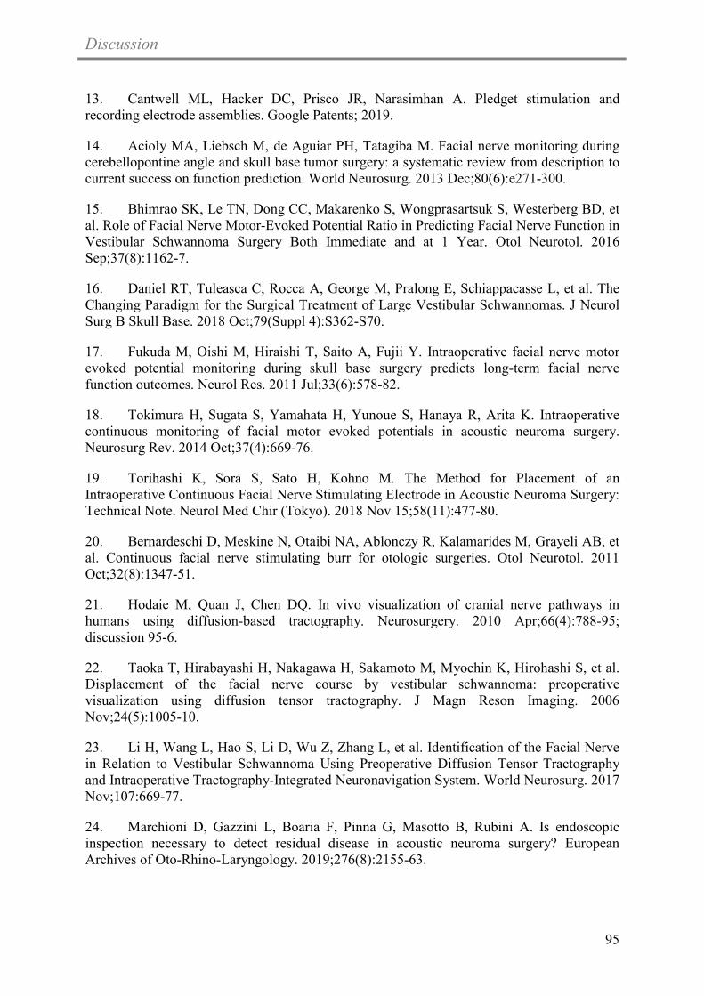
Discussion
95
13. Cantwell ML, Hacker DC, Prisco JR, Narasimhan A. Pledget stimulation and recording electrode assemblies. Google Patents; 2019.
14. Acioly MA, Liebsch M, de Aguiar PH, Tatagiba M. Facial nerve monitoring during cerebellopontine angle and skull base tumor surgery: a systematic review from description to current success on function prediction. World Neurosurg. 2013 Dec;80(6):e271-300.
15. Bhimrao SK, Le TN, Dong CC, Makarenko S, Wongprasartsuk S, Westerberg BD, et al. Role of Facial Nerve Motor-Evoked Potential Ratio in Predicting Facial Nerve Function in Vestibular Schwannoma Surgery Both Immediate and at 1 Year. Otol Neurotol. 2016 Sep;37(8):1162-7.
16. Daniel RT, Tuleasca C, Rocca A, George M, Pralong E, Schiappacasse L, et al. The Changing Paradigm for the Surgical Treatment of Large Vestibular Schwannomas. J Neurol Surg B Skull Base. 2018 Oct;79(Suppl 4):S362-S70.
17. Fukuda M, Oishi M, Hiraishi T, Saito A, Fujii Y. Intraoperative facial nerve motor evoked potential monitoring during skull base surgery predicts long-term facial nerve function outcomes. Neurol Res. 2011 Jul;33(6):578-82.
18. Tokimura H, Sugata S, Yamahata H, Yunoue S, Hanaya R, Arita K. Intraoperative continuous monitoring of facial motor evoked potentials in acoustic neuroma surgery. Neurosurg Rev. 2014 Oct;37(4):669-76.
19. Torihashi K, Sora S, Sato H, Kohno M. The Method for Placement of an Intraoperative Continuous Facial Nerve Stimulating Electrode in Acoustic Neuroma Surgery: Technical Note. Neurol Med Chir (Tokyo). 2018 Nov 15;58(11):477-80.
20. Bernardeschi D, Meskine N, Otaibi NA, Ablonczy R, Kalamarides M, Grayeli AB, et al. Continuous facial nerve stimulating burr for otologic surgeries. Otol Neurotol. 2011 Oct;32(8):1347-51.
21. Hodaie M, Quan J, Chen DQ. In vivo visualization of cranial nerve pathways in humans using diffusion-based tractography. Neurosurgery. 2010 Apr;66(4):788-95; discussion 95-6.
22. Taoka T, Hirabayashi H, Nakagawa H, Sakamoto M, Myochin K, Hirohashi S, et al. Displacement of the facial nerve course by vestibular schwannoma: preoperative visualization using diffusion tensor tractography. J Magn Reson Imaging. 2006 Nov;24(5):1005-10.
23. Li H, Wang L, Hao S, Li D, Wu Z, Zhang L, et al. Identification of the Facial Nerve in Relation to Vestibular Schwannoma Using Preoperative Diffusion Tensor Tractography and Intraoperative Tractography-Integrated Neuronavigation System. World Neurosurg. 2017 Nov;107:669-77.
24. Marchioni D, Gazzini L, Boaria F, Pinna G, Masotto B, Rubini A. Is endoscopic inspection necessary to detect residual disease in acoustic neuroma surgery? European Archives of Oto-Rhino-Laryngology. 2019;276(8):2155-63.

Discussion
96
25. Setty P, D'Andrea KP, Stucken EZ, Babu S, LaRouere MJ, Pieper DR. Endoscopic Resection of Vestibular Schwannomas. J Neurol Surg B Skull Base. 2015 Jun;76(3):230-8.
26. Sun JQ, Sun JW. Endoscope-assisted retrosigmoid keyhole approach for cerebellopontine angle: cadaveric study. Acta Otolaryngol. 2013 Nov;133(11):1154-7.
27. Corrivetti F, Cacciotti G, Scavo CG, Roperto R, Stati G, Sufianov A, et al. Flexible endoscopic assistance in the surgical management of vestibular schwannomas. Neurosurgical review. 2019:1-9.
28. Bozorg Grayeli A, Ferrary E, Tubach F, Bernat I, Deguine O, Darrouzet V, et al. Effect of corticosteroids on facial function after cerebellopontine angle tumor removal: a double-blind study versus placebo. Audiol Neurootol. 2015;20(4):213-21.
29. Scheller C, Richter HP, Engelhardt M, Koenig R, Antoniadis G. The influence of prophylactic vasoactive treatment on cochlear and facial nerve functions after vestibular schwannoma surgery: a prospective and open-label randomized pilot study. Neurosurgery. 2007 Jul;61(1):92-7; discussion 7-8.
30. Fotakopoulos G, Fountas K, Tsianaka E, Kotlia P, Pachatouridis D, Paschalis T, et al. Could IGF-I levels play a neuroprotective role in patients with large vestibular schwannomas? Future Sci OA. 2018 Feb;4(2):FSO260.
31. Tripathi M, Deora H, Kumar N, Batish A, Dutta P, Gurnani J, et al. Role of Bevacizumab as a prophylactic and rehabilitative treatment modality in cases of sporadic and syndromic vestibular schwannoma: Fifty shades of grey! Interdisciplinary Neurosurgery. 2020;19:100607.
32. Jia H, Elsayed M, Smail M, Mosnier I, Wu H, Sterkers O, et al. Neurofibromatosis type 2: Hearing preservation and rehabilitation. Neurochirurgie. 2018 Nov;64(5):348-54.


Conclusion And Future Perspectives
97
Chapter 5 CONCLUSION AND FUTURE PERSPECTIVES
The aims of surgery in the cerebellopontine angle (CPA) have changed from tumor
resection and prolongation of life to the anatomical and functional preservation of the cranial
nerves (CNs). The challenge and the matter of the actual debate are to achieve major tumor
removal, with a minimal impairment of these functions.
We do believe that implementation of the state of art of C-IONM during CPA surgery
combined with the conventional method of free running facial electromyography and
occasional stimulation of the facial nerve will have a superior outcome in our surgical
results. The use of APS or the pledget electrode in CPA surgery is still under further study for
its safety and efficacy.
The advent of animal studies can improve our understanding of this technology and
have immediate clinical applications. Our particular surgical animal model offered
satisfactory exposure of the acoustic-facial-cranial nerve bundle with relatively minimally
invasive steps and easily identifiable landmarks. The feasibility, safety and reliability of the
new C-IONM technologies could be easily studied and analyzed through our well established
in vivo animal models that allowed for future nerve testing and electrophysiological
evaluation.
Meanwhile, our animal model is very important for providing supplementary surgical
education and training needed to master clinical skills based specifically on an oto-
neurosurgery background. Several surgical procedures and manipulations could be simulated
in vivo, which are major components of daily cranial surgery, however in animals; it is
possible to train without risk to the patient.


Annexes
98
Chapter 6 ANNEXES
Annex 1
This article has been published as: Mohamed Elsayed; Baptiste Hochet; Renato
Torres; Olivier Sterkers; Yann Nguyen, Ghizlene Lahlou; and Michel Kalamarides .
"Metachronous bilateral vestibular schwannomas,” The Laryngoscope, 17 Feb., 2020.
DOI: 10.1002/lary.28571


Case Report
Metachronous Bilateral Vestibular Schwannomas
Mohamed Elsayed, MD; Baptiste Hochet, MD; Renato Torres, MD, PhD; Olivier Sterkers, MD, PhD;Yann Nguyen, MD, PhD; Ghizlene Lahlou, MD ; Michel Kalamarides, MD, PhD
Bilateral vestibular schwannoma (BVS) is the hallmark of neurofibromatosis type 2 (NF2), both of them being present at diagno-sis. We report four cases of metachronous BVS, a contralateral intracanalicular vestibular schwannomas (VS) being visible 2 to13 years after resection of a unilateral VS. NF2 workup was negative except in one case where two NF2 gene mutations werefound in tumor analysis. These cases raise the questions of whether the contralateral VS occurred by chance and how to manageit on the only hearing ear. Otologists should be aware of this rare eventuality for decision making of the first unilateral VS.
Key Words: Bilateral vestibular schwannoma, sporadic vestibular schwannoma, neurofibromatosis type 2, metachronousbenign tumors.
Laryngoscope, 00:1–5, 2020
INTRODUCTIONBilateral vestibular schwannoma (BVS) is rare,
occurring mainly in the context of neurofibromatosis type2 (NF2), an autosomal-dominant disease.1 However, spo-radic BVS occurring by chance has been reported, espe-cially in elderly patients.2 NF2 has an incidence of1/33,000 births and a prevalence of 1/56,000. Multiplebenign nervous system tumors, such as BVS, representthe hallmark of the disease.3 NF2 diagnostic criteriainclude the presence of BVS according to the Manchesterdiagnostic criteria. These criteria expand on the originalNational Institutes of Health criteria with additionalcriteria.2
Approximately half of NF2 mutations occur inpatients with a family history, and the newly diagnosedcases are spontaneous (de novo mutation in the NF2gene.) Such spontaneous mutations may be present in allcells if they occur in the gametes (germline mutation) orlead to mosaicism if they occur during the postzygoticstages of embryogenesis.
We report herein four cases of unilateral vestibularschwannoma (VS) operated on with subsequent
anacusis, which developed a contralateral VS later on,raising the questions of its nature, NF2 versus sporadic,and of its management on the only hearing ear(TABLE I).
CASE REPORTS
Case 1A 42-year-old male presented with a left unilateral
VS removed through a retrosigmoid approach with postop-erative normal facial nerve function (FNF) and anacusis.No recurrence was observed at the 5-year magnetic reso-nance imaging (MRI) follow-up. A contralateral 8-mmintracanalicular VS was diagnosed 13 years later (Fig. 1A)when the patient presented vertigo. No other NF2 fea-tures (personal, familial, or gene mutation in the blood)were present, but analysis of the removed VS revealed twomutations in one NF2 gene. Right sudden sensorineuralhearing loss (SNHL) occurred 1 year later, which recov-ered under intravenous steroid therapy. The VS grew from8 to 12 mm during the 3 following years (Fig. 1B). Hearingremained stable, and bevacizumab treatment was started(5 mg/kg every 2 weeks). Under chemotherapy, the VS didnot grow (Fig. 1C), but hearing declined moderately(Fig. 1D, E).
Case 2A 36-year-old female presented a right unilateral VS
excised through a translabyrinthine (TL) approach in2004 in another hospital. Postoperative FNF was grade II(House-Brackmann scale4). MRI follow-up showed neitherrecurrence nor contralateral VS 5 years postoperatively(Fig. 2A). Twelve years later, MRI performed for persis-tent headaches revealed a contralateral asymptomatic5-mm intracanalicular VS (Fig. 2B). NF2 workup was
From the Sorbonne University (M.E., B.H., R.T., O.S., Y.N., G.L., M.K.),Paris, France; Otology, Hearing Implants, and Skull Base Surgery Unit,AP-HP Pitié-Salpêtrière University Hospital (M.E., B.H., R.T., O.S., Y.N., G.L.),Paris, France; Faculty of Medicine, Otorhinolaryngology Department (M.E.),Alexandria University, Alexandria, Egypt; Faculty of Medicine, Research andScientific Development Center (R.T.), National University of Saint Agustine,Arequipa, Peru; and theNeurosurgery Department (M.K.), AP-HP Pitié-Salpêtrière University Hospital, Paris, France.
Editor’s Note: This Manuscript was accepted for publication on Jan-uary 27, 2020.
The authors have no funding, financial relationships, or conflicts ofinterest to disclose.
Send correspondence to Ghizlene Lahlou, MD, Otology, HearingImplants, and Skull Base Surgery Unit, AP-HP Pitié-Salpêtrière Univer-sity Hospital, 47-83 Boulevard de l’Hôpital, 75651 Paris, Cedex 13, France.E-mail: [email protected]
DOI: 10.1002/lary.28571
Laryngoscope 00: 2020 Elsayed et al.: Bilateral Vestibular Schwannomas
1
The Laryngoscope© 2020 The American Laryngological,Rhinological and Otological Society, Inc.

negative with no family history. The tumor grew from 5 to7.6 mm (Fig. 2C) in 2 years follow-up with normal hearing(Fig. 2D). Treatment with bevacizumab is beingconsidered.
Case 3A 44-year-old male presented a left unilateral VS
(Fig. 3A) removed through a TL approach in 2009 withnormal postoperative FNF. Two years later, MRI showed
TABLE I.Characteristics of the Four Patients.
Patient 1 Patient 2 Patient 3 Patient 4
Sex M F M M
Age at first presentation, yr 42 36 44 68
Symptoms revealing the first VS Progressive SNHL,dizziness, and tinnitus
Progressive SNHL,dizziness, and tinnitus
Sudden SNHLand tinnitus
Progressive SNHL
Preoperative first-side hearing level* Class B Class C Class C Class B
Surgical approach for the first side RS TL TL TL
First preoperative VS stage† 2 3 2 4
Interval between first VS surgery andoccurrence of the contralateral VS, yr
13 12 2 2
Symptoms revealing the second VS Vertigo No No Sudden SNHL
Hearing in only hearing ear Class A Class A Class A Class C
Management of contralateral VS Chemotherapy Proposed chemotherapy Observation Surgery (TL) andcochlear implantation
*Hearing is evaluated by audiometry according to the guidelines of the American Academy of Otolaryngology–Head and Neck Surgery Foundation.14†Tumor size was evaluated by magnetic resonance imaging and classified as follows: stage I (intracanalicular), stage II (≤15 mm maximal extracanalicular
diameter), stage III (16–30 mm), or stage IV (>30 mm).15
F = female; M = male; RS = retrosigmoid; SNHL = sensorineural hearing loss; TL = translabyrinthine; VS = vestibular schwannoma.
Fig. 1. Case 1. (A) Postoperative axial T1-weighted gadolinium-enhanced magnetic resonance imaging (MRI) showing contralateral right-sideintracanalicular vestibular schwannoma (VS) (white arrow) 14 years after left VS removal. (B) MRI showing growth of the tumor (stage II) in3 years (white arrow). (C) Follow-up MRI after chemotherapy with no VS evolution (white arrow). (D, E) Audiometric evaluations before (D) andafter (E) treatment with bevacizumab showing slight high frequencies and speech recognition threshold decline. [Color figure can be viewed inthe online issue, which is available at www.laryngoscope.com.]
Laryngoscope 00: 2020 Elsayed et al.: Bilateral Vestibular Schwannomas
2

Fig. 2. Case 2. (A) Axial T1-weighted gadolinium-enhanced magnetic resonance imaging (MRI) 5 years after vestibular schwannoma(VS) removal on the right side showing no enhancement on both sides. (B) Follow-up gadolinium-enhanced MRI T1 image showing a left-side
Fig. 3. Case 3. (A) Preoperative axial T1-weighted gadolinium-enhanced magnetic resonance imaging (MRI) showing left-side vestibularschwannoma (VS) (white arrow). (B–D) Postoperative axial T1-weighted gadolinium-enhanced MRI scans showing the initial growth (B, C) andsecondary stabilization (C, D) of the contralateral right side VS in (whites arrows).
Laryngoscope 00: 2020 Elsayed et al.: Bilateral Vestibular Schwannomas
3

incidentally a 1-mm enhancement of the contralateral ves-tibular nerve (Fig. 3B). A 3-mm intracanalicular VS wasconfirmed 4 years later (Fig. 3C), that grew to 6.5 mm in
2016 (Fig. 3D), and then remained stable. NF2 workupwas negative with no family history. The patient remainedasymptomatic and is under annual observation.
Fig. 4. Case 4. (A) Audiogram showing the hearing loss before right vestibular schwannoma removal through a translabyrinthine approach.(B–D) Audiograms showing sudden left sensorineural haring loss (SNHL) 2 years later (B) with partial recovery after intravenous steroid therapyand no benefit of hearing aid (D). (C) Postoperative axial T1-weighted gadolinium-enhanced magnetic resonance imaging showing evolution ofa contralateral left side vestibular schwannoma (VS) (white arrow) revealed by sudden SNHL. (E) Postoperative computed tomography(CT) scan of the patient after removal of the left-sided VS through a translabyrinthine approach and insertion of a cochlear implant at the sameside. (F) Patient’s moderate left cochlear implant performance 1 year after implantation. [Color figure can be viewed in the online issue, whichis available at www.laryngoscope.com.]
Laryngoscope 00: 2020 Elsayed et al.: Bilateral Vestibular Schwannomas
4

Case 4A 67-year-old male presented with a right unilat-
eral VS (Fig. 4A) removed in 2011 through a TLapproach in another hospital. Sudden SNHL occurred2 years later on the contralateral ear with partial recoveryunder intravenous steroid therapy (Fig. 4B), revealing asmall intracanalicular left VS (Fig. 4C). No other NF2 fea-tures or family history were found. Because VS was slowlygrowing (1 mm/yr) with a nonserviceable hearing loss(Fig. 4D), TL excision with simultaneous cochlear implan-tation was performed in 2015 (Fig. 4E). MRI follow-up dis-closed a right small recurrence 1 year later and the patientunderwent radiosurgery in 2016 with no further growth.The patient is a daily cochlear implant user with moderatecochlear implant performance (Fig. 4F).
DISCUSSIONThe wide use of MRI explains the increase of VS
incidence in the last decades (1.9/100,000 annually5). Thisis consistent with a 1/1,000 lifetime incidence and apotential 1 in 2 million risk of chance of BVS, assuming a1/2,000 chance on the contralateral side.2
NF2 predominant feature remains BVS develop-ment, although 12% to 18% of NF2 patients present anunilateral tumor.6 Atypical forms of NF2 with low growthpotential of tumors are usually encountered in theelderly. Frequent occurrence of somatic mosaicism inNF27 explains why germline mutation is rarely found.Alternatively, when BVS appears with no other NF2 fea-tures, the two tumors could have developed sporadicallywith no molecular event in common between them, espe-cially in the elderly,8,9 or after radiation treatment.10
Evans et al. has estimated that 25% of BVS in those over50 years old and 50% in those over 70 years old, with noother features of NF2, represent a chance occurrencerather than the expression of NF2.2 That implies anextended monitoring by MRI for 10 years after an initialunilateral surgery to search for an eventual recurrence,and a new MRI if any new symptom appears.
The risk of familial NF2 in de novo NF2 patientsafter negative blood analysis is below the 7% and 5% forpatients presenting BVS tumors after 40 years old. Nev-ertheless, these numbers did not take into account thepossibility of sporadic VS occurring by chance.2
Although rare, if a contralateral VS occurs afterremoval of a first VS considered initially as sporadic, it willappear as a small intracanalicular VS on the only hearingear. A wait-and-scan policy enables the evaluation of the
VS growth and hearing evolution. Rarely, a bilateralsevere-to-profound hearing loss may require removal of thecontralateral VS with concomitant cochlear implantation(case 4).11,12 If hearing declines and/or tumor extends intocerebellopontine angle, chemotherapy (bevacizumab) isstrongly advocated to control VS growth and preservehearing.12,13
CONCLUSIONA low but true risk of occurrence of a delayed contra-
lateral VS exists, which may define either an NF2 orbilateral sporadic VS. Active monitoring for new symp-toms and the analysis of the postoperative imaging on thecontralateral side should be performed. If a contralateralVS appears, the treatment strategy should be carefullytailored to each case, especially if the first VS led toanacusis.
BIBLIOGRAPHY1. Evans DGR, Ramsden RT, Gokhale C, Bowers N, Huson SM, Wallace A.
Should NF2 mutation screening be undertaken in patients with an appar-ently isolated vestibular schwannoma? Clin Genet 2007;71:354-358.
2. Evans DGR, Freeman S, Gokhale C, et al. Bilateral vestibular schwannomasin older patients: NF2 or chance? J Med Genet 2015;52:422-424.
3. Peyre M, Goutagny S, Bah A, et al. Conservative management of bilateralvestibular schwannomas in neurofibromatosis type 2 patients: hearingand tumor growth results. Neurosurgery 2013;72:907-913.
4. House J, Brackmann D. Facial nerve grading system. Otolaryngol HeadNeck Surg 1985;93:146-147.
5. Stangerup S-E, Caye-Thomasen P. Epidemiology and natural history of ves-tibular schwannomas. Clin Otolaryngol 2012;45:257-268.
6. Evans DGR, Lye R, Neary W, et al. Probability of bilateral disease in peoplepresenting with a unilateral vestibular schwannoma. J Neurol NeurosurgPsychiatry 1999;66:764-767.
7. Goutagny S, Bah AB, Parfait B, Sterkers O, Kalamarides M. Neurofibroma-tosis type 2 in the elderly population: clinical and molecular features.Am J Med Genet A 2013;161:667-670.
8. Evans DGR, Baser ME, O’Reilly B, et al. Management of the patient andfamily with neurofibromatosis 2: a consensus conference statement. Br JNeurosurg 2005;19:5-12.
9. Rao A, Lawrie A, Bodkin P, Tighe J, Kamel M. Bilateral cerebellopontine anglelesions not always NF2: diagnostic pitfall. Br J Neurosurg 2012;26:275-277.
10. Carlson M, Gompel J. Multiple unilateral vestibular schwannomas: segmen-tal NF2 or sporadic occurrence? J Neurol Surg Rep 2016;77:e106-e108.
11. Jia H, Nguyen Y, De Seta D, et al. Management of sporadic vestibularschwannoma with contralateral nonserviceable hearing [published onlineOctober 23, 2019]. Laryngoscope. https://doi.org/10.1002/lary.28369.
12. Jia H, El Sayed MME, Smail M, et al. Neurofibromatosis type 2: hearingpreservation and rehabilitation. Neurochirurgie 2018;64:348-354.
13. Goutagny S, Kalamarides M. Medical treatment in neurofibromatosis type2. Review of the literature and presentation of clinical reports.Neurochirurgie 2018;64:370-374.
14. American Academy of Otolaryngology-Head and Neck Surgery Foundation.Committee on hearing and equilibrium guidelines for the evaluation ofhearing preservation in acoustic neuroma (vestibular schwannoma).Otolaryngol Head Neck Surg 1995;113:179-180.
15. Torres R, Nguyen Y, Vanier A, et al. Multivariate analysis of factorsinfluencing facial nerve outcome following microsurgical resection of ves-tibular schwannoma. Otolaryngol Neck Surg 2017;156:525-533.
Laryngoscope 00: 2020 Elsayed et al.: Bilateral Vestibular Schwannomas
5


Annexes
104
Annex 2
Jia H, Elsayed M, Smail M, Mosnier I, Wu H, Sterkers O, Kalamarides M,
Bernardeschi D. “Neurofibromatosis type 2: Hearing preservation and rehabilitation.”
Neurochirurgie. 2018 Nov 1; 64(5):348-54.


Neurochirurgie
64
(2018)
348–354
Disponible
en
ligne
sur
ScienceDirectwww.sciencedirect.com
a
r
t
i
c
l
e
i
n
f
o
Article
history:Received
1st March
2018Received
in
revised
form
4
August
2018Accepted
7
September
2018Available
online
9
October
2018
Keywords:Vestibular
schwannomaHearing
lossCochlear
implantAuditory
brainstem
implant
a
b
s
t
r
a
c
t
The
main
manifestation
of
neurofibromatosis
type
2
(NF2)
is
the
development
of
bilateral
vestibularschwannomas
(VS).
Consequently,
one
of
the
most
severe
functional
sequelae
is
bilateral
sensorineuralhearing
loss,
caused
by
spontaneous
tumor
progression
and/or
treatment-related
damage
(surgery
orradiosurgery).
Preserving
or
restoring
hearing
is
still
challenging
in
NF2
no
matter
the
strategy
appliedto
each
individual
based
on
the
natural
history
of
VS.
In
this
review,
the
different
strategies
for
hearingpreservation
or
rehabilitation
are
discussed
and
illustrated
by
several
cases.
A
decisional
algorithm
forNF2
patients
with
VS
is
proposed
that
takes
into
consideration
the
tumor
size
and
hearing
level.©
2018
Elsevier
Masson
SAS.
All
rights
reserved.
1.
Introduction
Neurofibromatosis
type
2
(NF2)
is
a
genetic
disorder
that
occurswith
a
birth
incidence
of
approximately
1
in
25,000
with
a
dom-inant
pattern
of
inheritance.
Bilateral
vestibular
schwannomas(VS)
appear
in
90–95%
of
NF2
patients,
as
well
as
a
predis-position
to
other
benign
nervous
system
tumors
[1].
Variousstrategies
have
been
used,
including
“wait
and
scan”
(peri-odic
radiological
and
audiological
evaluations),
surgical
treatment(decompression
of
internal
auditory
meatus,
partial
removal,total
removal),
radiation-based
therapy
(single-fraction
stereotac-tic
radiosurgery,
fractionated
stereotactic
radiation
therapy),
andchemotherapy
(Bevacizumab).
Because
most
patients
suffer
bilat-eral
sensorineural
hearing
loss
(SNHL),
hearing
preservation
byacoustic
amplification
(hearing
aid,
HA)
or
rehabilitation
by
elec-trical
stimulation
to
the
remaining
auditory
structures
(cochlear
∗ Corresponding
author.
Service
neurochirurgie,
hôpital
Pitié-Salpetrière,
AP–HP,47-83,
boulevard
de
l’hôpital,
75651
Paris
cedex
13,
France.∗∗ Co-corresponding
author.
Department
of
Otolaryngology
Head
and
NeckSurgery,
Shanghai
Ninth
People’s
Hospital,
Shanghai
Jiaotong
University
School
ofMedicine,
639
Zhizaoju
Road,
200011
Shanghai,
PR
China.E-mail
addresses:
(H.
Wu),
(M.Kalamarides).
implant,
CI)
or
the
cochlear
nucleus
(auditory
brainstem
implant,ABI)
is
also
an
essential
element
of
treatment
[2–14].Analyzing
and
comparing
different
therapeutic
strategies
forNF2
patients
is
difficult
because
of
the
existence
of
multipleclassifications.
These
classifications
induce
and
perpetuate
knowl-edge
silos
and
references
used
only
by
certain
specialists
[15].To
evaluate
hearing,
neuro-otologists
normally
use
the
AAOHNSclassifications,
neuro-oncologists
commonly
use
the
GardnerRobertson
classification,
and
some
surgeons
directly
use
the
SDS.Similarly,
there
are
several
classifications
available
for
tumor
sizedepending
on
the
author’s
preference.
For
example,
the
Koos
clas-sification
takes
into
account
indirectly
the
volume
of
the
posteriorfossa
which
tends
to
over-evaluate
the
tumor
volume
and
resultsin
more
Grade
IV
tumors.To
make
a
better
decision
for
each
individual
patient
by
con-sidering
the
symptoms,
tumor
size
and
invasiveness,
surgical
risks,and
natural
history
of
tumor
growth,
a
multidisciplinary
medicalteam
has
been
set
up
at
NF2
reference
centers
composed
of
anotologist,
neurosurgeon,
audiologist,
speech
therapist,
neuroradi-ologist
and
psychologist
[16,17].
In
this
article,
we
present
a
shortand
thus
non-exhaustive
review
of
the
literature
on
the
subject
ofhearing
and
NF2,
and
a
summary
of
the
extensive
experience
of
ourNational
NF2
Reference
Center,
which
is
similar
to
that
of
most
thelarge
international
expert
centers
on
NF2.
https://doi.org/10.1016/j.neuchi.2018.09.0030028-3770/©
2018
Elsevier
Masson
SAS.
All
rights
reserved.
f Sino-French Joint Clinical Research Center of Otology and Neuro-otology, 200011 Shanghai, PR ChinaShanghai, PR China
e Department of Otolaryngology Head and Neck Surgery, Shanghai Ninth People’s Hospital, Shanghai Jiaotong University School of Medicine, 200011
d Service de neurochirurgie, GHU Pitié Salpêtrière, Sorbonne Université, AP–HP, 75651 Paris, France
c Service d’ORL, otologie, implants auditifs et chirurgie de la base du crâne, GHU Pitié Salpêtrière, AP–HP, 75651 Paris, France
b CMR neurofibromatosis type 2, GHU Pitié Salpêtrière, AP–HP, 75651 Paris, FranceParis 6, 75013 Paris, France
a Inserm, UMRS 1159, “unité de réhabilitation chirurgicale mini-invasive robotisée de l’audition”, Sorbonne Universités, université Pierre et Marie-Curie
Neurofibromatosis type 2: Hearing preservation and rehabilitation
Original article
M. Kalamarides b,d,f,∗, D. Bernardeschia,b,cH. Jiaa,c,e,f, M. Elsayed a,b, M. Smaila,b,c, I. Mosnierb,c, H. Wu e,∗∗, O. Sterkersa,b,c,f,

H.
Jia
et
al.
/
Neurochirurgie
64
(2018)
348–354
349
1.1.
Hearing
preservation
strategies
1.1.1.
Wait
and
scanVS
in
NF2
are
usually
slow
growing
tumors,
although
there
isa
wide
individual
range
(0.4–10
mm/year)
[18–20].
In
our
series,25%
of
92
VS
in
46
patients
had
fast
growth
(>
3
mm/year),
andrapid
tumor
growth
was
more
frequent
in
younger
patients
[19].This
suggests
that
the
first
magnetic
resonance
imaging
(MRI)scan
should
be
performed
6
months
after
the
diagnosis
and
thenannually.
Meanwhile,
spontaneous
VS
shrinkage
or
no
growth
wasobserved
in
some
cases
(14%
in
our
series).
It
should
be
empha-sized
that,
after
removing
a
large
one-sided
VS,
the
contralateral
VSmay
grow
quicker.
This
may
be
due
to
repositioning
of
the
brain-stem
to
operated
side
where
the
cerebellopontine
angle
(CPA)
spacewas
released,
which
releases
the
compression
on
the
tumor
of
thenon-operated
side
[21].Although
a
correlation
between
tumor
growth
and
hearing
dete-rioration
was
observed
in
sporadic
VS,
it
seems
that
VS
in
NF2
has
aspecial
feature
and
this
correlation
has
not
been
confirmed
in
smallNF2
patient
cohorts
[18,22,23].
Relative
to
tumor
growth
observedin
31%
cases
at
1
year,
64%
at
2
years,
and
79%
at
3
years,
hearingdeterioration
was
observed
in
5%
cases
at
1
year,
13%
at
2
years,and
16%
at
3
years
[24].
Some
authors
thought
that
profound
SNHLwould
present
approximately
3–5
years
after
the
diagnosis
of
NF2[25],
but
others
demonstrated
that
useful
hearing
was
still
presentin
65%
to
74%
of
NF2
patients
after
6–7
years
[18,19].
This
hearingdeterioration
may
develop
as
a
result
of
cochlear
aperture
obstruc-tion
and
accumulation
of
intralabyrinthine
protein
[26];
it
might
bealso
due
to
a
compression
injury
of
the
cochlear
nerve
and/or
vas-cular
occlusion
of
the
auditory
artery
by
the
intracanalicular
part
of
the
tumor,
or
stretching
of
the
nerve
by
the
extension
of
the
CPApart
of
the
tumor
[22].As
a
consequence,
the
“wait
and
scan”
strategy
can
be
rec-ommended
as
a
natural
hearing
preservation
strategy,
especiallyin
middle-age
or
older
patients
with
small-
or
mid-sized
tumors,because
the
VS
might
not
grow
quickly
and
hearing
may
continueto
be
at
a
useful
level
for
a
long
time
in
the
majority
of
these
NF2patients
(Case
1,
Fig.
1).
1.1.2.
RadiosurgeryRadiation
therapy
is
an
alternative
treatment
for
NF2,
includ-ing
two
main
techniques,
single-fraction
stereotactic
radiosurgery(SRS)
and
fractionated
stereotactic
radiation
therapy
(FSRT),
whoseradiation
schedules
and
doses
are
different.
Up
to
now,
their
out-comes
are
controversial.
Some
institutions
reported
that
small-
andmid-sized
tumors
were
well
controlled,
with
a
5-year
local
con-trol
rate
between
75%
and
87%
[27–32].
However,
there
is
someconcern
because
in
most
cases,
natural
growth
of
the
VS
was
notdocumented
before
treatment
and
was
not
analyzed
in
results
[33].The
hearing
preservation
rate
was
inconsistent,
depending
on
thechosen
articles,
all
of
which
had
shortcomings.
Kruyt
et
al.
reporteda
5-year
hearing
preservation
rate
of
59%
[30],
and
Lloyd
et
al.concluded
that
generally
41.1%
patients
maintained
serviceablehearing
long-term
with
a
mean
tumor
control
rate
of
81.4%
[34].Increasing
of
the
marginal
dose
led
to
better
control
of
NF2-relatedVS
but
resulted
in
a
higher
rate
of
hearing
loss.
No
prospective
andrandomized
studies
comparing
the
hearing
outcomes
of
SRS
andFSRT
have
been
completed
in
the
NF2
reference
centers
becauseof
the
small
numbers
of
patients,
tumor
size
bias
and
physicianpreferences.
The
risk
of
VS
malignant
transformation
as
well
as
Fig.
1.
Hearing
maintenance
by
decompression
of
internal
auditory
meatus.
A
14-year-old
male
patient
was
diagnosed
with
NF2
in
2003
because
of
a
familial
history
ofNF2.
He
had
an
11
mm
tumor
(maximum
diameter)
on
the
left
and
an
8
mm
tumor
on
the
right
side.
Because
of
Class
A
hearing
(AAOHNS
Classification)
of
both
sides,
the“wait
and
scan”
strategy
was
used
initially.
Three
years
later
(2006),
his
right
tumor
grew
quickly
(22
mm)
with
hearing
deterioration
(Class
C)
while
his
left
tumor
grewslowly
(12
mm)
with
stable
hearing
(Class
A),
so
his
right
tumor
was
removed
through
a
translabyrinthine
approach
and
the
right
hearing
was
sacrificed.
Five
years
afterthe
surgery
(2011),
his
left
hearing
showed
signs
of
deterioration
but
was
still
useful
(Class
B)
when
the
tumor
grew
to
23
mm,
so
a
decompression
of
the
internal
auditorymeatus
was
performed
to
recover
or
delay
hearing
loss.
His
left
hearing
was
successful
preserved
and
was
still
useful
(Class
C)
for
3
years
(2014),
despite
the
tumor
growingslowly
(28
mm).
Later,
he
suffered
a
sudden
hearing
loss
in
his
only
functioning
ear
and
did
not
recover
with
corticosteroid
therapy.
Therefore,
cochlear
implantation
wasperformed
in
2017
on
the
left
side
with
the
tumor
in
situ.
Good
auditory
performance
was
achieved
which
was
useful
for
daily
conversation.

350
H.
Jia
et
al.
/
Neurochirurgie
64
(2018)
348–354
meningioma
induction
cannot
be
ignored
since
the
NF2
gene
is
atumor
suppressor
gene
predisposing
to
the
development
of
tumors,which
is
accelerated
after
radiation
therapy
[35,36].
Nevertheless,late
malignant
transformation
can
occur
in
the
absence
of
irradia-tion
[37]
and
no
malignant
transformation
was
observed
in
somelong-term
follow-up
studies
[38].
1.1.3.
Microsurgical
resectionThe
availability
of
high-definition
stereo-microscopes,
saferhigh-speed
drills
with
integrated
nerve
stimulation,
efficient
localhemostatic
agents,
navigation
and
intraoperative
nerve
monitor-ing
with
better
anti-interference
capabilities
have
contributed
tothe
goals
of
microsurgery
for
VS
evolving
from
preserving
lifeto
preserving
neurological
functions.
Microsurgical
resection
withhearing
preservation
can
be
performed
through
two
approaches:middle
cranial
fossa
approach
(MFA)
and
retrosigmoid
(RS).
MFA
isindicated
for
small-
or
mid-sized
tumors,
and
standard
RS
approachis
proposed
for
VS
without
IAM
fundus
involvement.
Recently,endoscopes
are
becoming
widely
used
in
skull
base
surgery,
andsince
the
relationship
in
the
IAM
fundus
can
be
viewed,
it
haseliminated
the
major
drawbacks
of
RS.
With
the
development
ofintraoperative
facial
nerve
monitoring
[39],
the
risk
of
facial
nerveparalysis
remains
low
(inferior
to
10%)
in
these
small
or
mid-sizedVS
1
year
after
surgery
[40].With
modern
intraoperative
cochlear
nerve
monitoring,
the
rateof
hearing
preservation
is
higher
than
before.
The
rate
was
60–70%via
MFA
and
30–40%
via
RS
[31],
but
the
two
approaches
could
notbe
compared
directly
because
the
tumor
size
and
location
differed.Further,
the
risk
of
recurrence
was
higher
with
MCF
than
RS
in
the
cases
without
tumor
involvement
of
IAM
fundus
[41].
In
some
casesof
NF2-related
VS,
postoperative
hearing
might
be
lost
even
if
audi-tory
monitoring
showed
a
preserved
response
at
the
end
of
surgery.Destruction
of
the
auditory
nerve’s
blood
supply
is
the
main
reasondespite
the
nerve
being
preserved.
No
predictive
factors
for
hearingpreservation
following
microsurgery
have
been
identified
in
spo-radic
VS,
but
the
hearing
preservation
rates
seemed
higher
in
casesof
stable
PTA
with
normal
ABR
during
follow-up,
in
cases
withoutcochlear
fossa
tumors
for
MFA,
or
in
cases
without
fundus
tumorsfor
RS
[42,43].
1.1.4.
Decompression
of
internal
auditory
meatusAs
hearing
preservation
by
MSR
varied
and
could
not
be
pre-dicted
accurately,
a
surgical
technique
with
less
trauma
to
theauditory
nerve
and
its
surroundings
was
proposed:
decompressionof
the
IAM
via
MFA.
Image-guided
navigation
systems
have
beenused
more
and
more
in
this
technique,
helping
to
precisely
locateand
open
the
IAM
roof
[44].
This
surgical
technique
results
in
agood
rate
of
hearing
preservation
(approximately
90%
short-term)and
is
accepted
as
a
safe
procedure
to
maintain
useful
hearing
inNF2
patients
with
monoaural
hearing,
which
becomes
paramountto
their
quality
of
life
[45,46].
Furthermore,
a
cochlear
implantcould
be
used
when
these
patients
lose
their
hearing
later
in
life,because
the
auditory
nerve
was
not
damaged
by
the
surgical
manip-ulations
(Case
2,
Fig.
2).
Thus,
IAM
decompression
is
proposedto
NF2
patients
with
an
objective
hearing
modification
(progres-sive,
fluctuating,
or
sudden
hearing
loss);
this
treatment
couldbe
combined
with
chemotherapy
if
the
VS
is
fast
growing.
Someteams
have
proposed
operating
on
patients
before
the
hearing
Fig.
2.
Hearing
rehabilitation
by
cochlear
implantation
without
tumor
removal.
A
female
patient
(born
in
1944)
was
diagnosed
as
having
NF2
when
she
was
young.
She
wasoperated
on
twice
for
a
left
coronal
meningioma
(1982)
and
posterior
fossa
meningioma
(1998).
In
2002,
her
left
hearing
was
normal
(Class
A,
4
mm
intracanalicular
tumor)and
right
hearing
was
moderately
altered
(Class
B,
12
mm
tumor).
The
“wait
and
scan”
strategy
was
initially
adopted
for
her.
In
2006,
her
right
tumor
was
stable
(12
mm)with
slight
deterioration
of
hearing
(Class
B),
but
the
left
one
grew
(11
mm)
with
progressive
hearing
loss
(Class
C).
“Wait
and
scan”
was
continued.
In
2009,
she
had
totalhearing
loss
on
the
left
and
Class
C
hearing
on
the
right,
which
meant
her
hearing
level
was
insufficient
for
day-to-day
life.
Considering
her
age
(65
years
old)
and
slowgrowth
of
the
tumor,
cochlear
implantation
into
her
left
ear
was
proposed
without
tumor
removal.
The
auditory
brainstem
response
was
confirmed
to
be
evoked
by
electricstimulation
on
the
promontory
before
implantation.
Seven
years
after
her
cochlear
implantation
(2016),
the
hearing
in
her
right
non-implanted
ear
was
still
Class
C.
With
ahearing
aid
in
her
right
ear
and
a
cochlear
implant
in
her
left
ear,
the
intelligibility
in
free
field
measurement
was
quite
good
(SRT
65
dB,
SDS
80%).
CT
follow-up
imaging
didnot
show
any
evident
tumor
growth.

H.
Jia
et
al.
/
Neurochirurgie
64
(2018)
348–354
351
impairment
develops,
making
their
decision
on
the
alteration
ofEABR.
To
date,
no
study
has
shown
the
validity
of
this
approach.
1.1.5.
ChemotherapyNearly
all
VS
express
vascular
endothelial
growth
factor
(VEGF,MGF-A).
bevacizumab,
a
human
IgG
monoclonal
antibody,
whichbinds
to
VEGF
with
high
specificity,
was
given
to
NF2
patients
inclinical
trials.
Short-term
studies
showed
good
efficacy
for
control-ling
tumor
growth
as
well
as
preserving
useful
hearing,
althoughside
effects
appeared
with
long-term
use
[47–49].
Additionally,it
seems
the
treatment
effect
was
temporary
and
the
benefi-cial
effects
subsided
when
the
treatment
was
discontinued.
A
prospective
14-patient
study
aimed
at
minimizing
side
effects
witha
lower
bevacizumab
dosage
found
a
hearing
response
in
at
leastone-third
of
patients
for
3
months
[50].
This
hearing
responsewas
correlated
to
biomarkers
indicating
reduced
tumor-associatededema
and
improved
oxygenation
that
impact
on
tumor
volume.
Insome
patients,
hearing
was
preserved
during
long-term
treatmentwith
everolimus
[51,52].
The
number
of
patients
treated
with
otherdrugs
is
too
small
to
reach
a
conclusion.Currently,
it
appears
that
bevacizumab
has
significantly
reducedthe
number
of
surgeries
on
the
contralateral
VS
and
consequentlydecreased
the
number
of
ABI
implantations
at
NF2
reference
cen-ters
around
the
world.
Fig.
3.
Hearing
rehabilitation
by
auditory
brainstem
implantation.
A
female
patient
(born
in
1973)
was
diagnosed
as
NF2
in
1997,
presenting
bilateral
vestibular
schwannomasand
right
middle
fossa
meningioma.
In
1998,
her
right
schwannoma
was
removed
via
RS
at
another
hospital
but
the
hearing
was
not
preserved.
During
the
follow-up,
shehad
several
sudden
hearing
loss
episodes
on
the
left
side
but
all
finally
recovered
(Class
A).
Decompression
of
the
left
internal
auditory
meatus
(8
mm
tumor
in
CPA)
wasproposed
in
2004
to
reduce
the
frequency
and
risk
of
hearing
loss,
and
her
hearing
was
stable
for
about
7
years
(Class
A,
2011)
although
the
left
tumor
was
growing
slowly(22
mm,
2011).
Meanwhile,
a
recurrence
of
the
tumor
was
observed
in
the
right
internal
auditory
meatus
in
2011;
it
was
removed
at
the
same
hospital
that
performed
theprevious
removal.
In
2015,
her
left
hearing
was
totally
lost
with
a
growing
tumor
(31
mm),
so
the
tumor
was
removed
via
TL
and
an
ABI
was
implanted
to
restore
her
hearing.One
year
later
(2016),
there
was
no
recurrence
on
the
left
side,
and
the
patient
was
satisfied
with
the
ABI
outcome
(word
and
sentence
recognition
score
with
lip-readingwere
both
60%).

352
H.
Jia
et
al.
/
Neurochirurgie
64
(2018)
348–354
1.2.
Hearing
rehabilitation
strategy
1.2.1.
Hearing
aidsHearing
aids
deliver
amplified
sound
through
the
damagedcochlea
and
nerves
to
brain.
In
NF2
patients
with
mild
to
severehearing
loss,
conventional
hearing
aids
could
be
proposed
as
atemporary
but
low
risk
and
atraumatic
strategy.
The
type
of
hear-ing
loss
in
NF2
patients
is
retrocochlear,
which
causes
a
decreasein
the
speech
intelligibility
score
and
desynchronization
of
audi-tory
brainstem
responses.
Consequently,
the
real
benefit
of
soundamplification
by
a
hearing
aid
is
uncertain.
Most
patients
prefernot
to
wear
it
because
sounds
appear
distorted
and
intelligibility
innoisy
conditions
is
relatively
poor.
1.2.2.
Cochlear
implant
(CI)A
cochlear
implant
is
an
implanted
device
that
provides
directelectrical
stimulation
into
the
inner
ear,
which
requires
a
functionalauditory
nerve
as
a
traditional
indication.
Because
the
integrityor
function
of
the
auditory
nerve
cannot
be
guaranteed,
cochlearimplantation
in
NF2-related
VS
was
not
considered
until
recently.The
main
reasons
may
be
the
insufficient
benefit
and
weak
perfor-mance
achieved
with
ABI,
and
lack
of
ABI
support
in
some
medicalcenters
or
countries.
In
this
situation,
the
cochlear
nerve
was
pre-served
as
best
possible.
Anatomical
preservation
of
the
cochlearnerve
is
sometimes
very
difficult
in
NF2,
particularly
because
ofthe
variety
of
lesions.
The
recent
use
of
intraoperative
monitoringwith
DNAP
and
CNAP
(dorsal
nucleus
and
cochlear
nerve
actionpotentials,
respectively)
may
be
to
be
a
solution
to
improving
thenumber
of
functional,
and
also
electrical,
cochlear
nerve
preser-vations,
allowing
CI
placement
at
the
same
time
or
secondarily.In
any
case,
the
peak
performance
of
ABI
is
generally
lower
thanthat
achievable
with
cochlear
implantation
[53].
The
promontorytest
evaluating
the
electrical
integrity
of
the
cochlear
nerve
has
a
good
positive
predictive
value
notwithstanding
many
false
nega-tives.
Therefore,
some
centers
prefer
to
implant
a
CI
first
insteadof
an
ABI
[54].
Because
of
the
uncertainty
of
hearing
preservationvia
the
RS
approach,
some
authors
suggest
removing
tumor
viaa
translabyrinthine
(TL)
approach
that
can
identify
the
cochlearnerve
early
and
expose
it
entirely,
no
matter
the
preoperativehearing
level,
and
simultaneously
perform
cochlear
implantation[12,13,55].
In
order
to
ensure
maximum
integrity
of
the
auditorynerve,
cochlear
implantation
without
tumor
removal
was
pro-posed
and
applied
in
older
patients
whose
tumors
were
stable
orincreased
slowly
in
volume
[12,56–58]
(Case
2,
Fig.
2).
CI
mightbe
also
proposed
to
patients
whose
tumor
is
controlled
by
Beva-cizumab
but
there
are
no
published
study
on
this
topic.
Becausesome
patients
obtained
better
long-term
audition
with
CI,
cochlearimplantation
has
been
the
main
alternative
for
hearing
restorationin
NF2
patients
[12–14,58,59].Indeed,
CI
in
NF2
patients
performs
better
than
ABI,
but
the
audi-ological
features,
tumor
features
and
age
of
the
two
populationsdiffers.
With
recent
technological
developments,
radiological
eval-uation
by
MRI
at
1.5
T
or
3.0
T
can
still
be
performed
for
some
CIor
ABI
devices.
Furthermore,
previous
SRS
does
not
contraindicatethe
placement
of
CI
or
ABI
[2,60].
1.2.3.
Auditory
brainstem
implant
(ABI)The
ABI
was
developed
by
House
and
Hitselberger
in
the
1980s.This
device
uses
technology
similar
to
that
of
cochlear
implants,but
instead
of
electrical
stimulation
to
the
cochlea,
ABI
stimulatesthe
cochlear
nucleus
of
the
brainstem
which
bypasses
the
damagedauditory
nerve.
The
implanted
electrode
should
be
placed
on
and/orinserted
into
the
cochlear
nucleus
in
the
lateral
recess
of
the
4thventricle;
this
could
be
performed
in
the
same
surgical
procedureas
intracranial
tumor
resection.
In
most
cases,
ABI
was
implantedon
the
second
side
(Case
3,
Fig.
3),
but
in
some
cases
it
was
also
Fig.
4.
Decisional
algorithm
for
NF2
patients
with
vestibular
schwannomas
and
monaural
hearing.

H.
Jia
et
al.
/
Neurochirurgie
64
(2018)
348–354
353
implanted
on
the
first
side
with
the
device
switched
off
until
totalcontralateral
hearing
loss
has
developed
(Sleeper
ABI)
[61].
The
ABIachieved
the
expected
auditory
benefit
with
lip-reading
in
mostcases.
But
the
intelligibility
remains
uncertain
and
few
NF2
patientshad
good
open
set
performance
without
lip-reading
[3,5,6,8,9].
Inorder
to
improve
the
performance
of
ABI,
many
studies
have
beenundertaken,
such
as
penetrating
electrodes
designed
to
improvethe
contact
between
the
electrodes
and
the
coding
areas
of
thenucleus,
but
this
technique
did
not
produce
better
outcomes
[7].Positron
emission
tomography
scans
showed
the
auditory
cortexcould
be
activated
though
ABI
[62];
this
result
meant
that
the
cod-ing
strategies
developed
for
CI
might
not
be
suitable
for
the
cochlearnucleus
mapping
for
ABI
[63].However
ABI
could
be
recommended
in
young
NF2
patientswhose
VS
are
growing
rapidly
and
often
large,
because
there
is
noother
choice.
ABI
performs
better
here
than
in
other
populations,probably
due
to
the
high
brain
plasticity
in
younger
people.
2.
Conclusion
A
combination
of
different
treatment
options
should
now
beconsidered
for
hearing
preservation
and
tumor
volume
control(Fig.
4),
if
there
are
no
other
indications
for
surgical
interventionsuch
as
encephalic
stage
or
spinal
tumor.
These
different
treat-ment
strategies
are
to
be
tailored
to
each
patient,
in
particular
tothe
severity
of
the
illness,
the
rate
of
tumor
progression
and
thepersonal
history
of
the
illness
and
sometimes,
the
family
history.Decompression
of
the
IAM
and
chemotherapy,
principally
Beva-cizumab,
are
now
the
best
options
to
preserve
or
improve
usefulhearing
in
NF2
patients
especially
as
the
VS
is
growing.
The
pres-ence
of
artificial
hearing
by
electrical
stimulation
(ABI,
CI)
couldimpact
the
synchronism
of
binaural
signals
in
auditory
cortex
whencontralateral
natural
hearing
is
present.
Hence,
when
the
devicesare
implanted
during
the
initial
surgery,
they
are
usually
switchedoff
until
patients
lose
their
bilateral
useful
hearing.
Generally,
olderpatients
with
no
or
slow
growth
of
small-
and
mid-sized
VS
are
can-didates
for
CI
without
tumor
resection,
and
young
patients
withlarge
progressive
VS
are
candidates
for
ABI
with
tumor
resection.Because
of
small
sample
sizes
and
large
individual
differences,
noprospective
and
randomized
studies
have
been
completed
in
NF2patients.
Further
study
are
needed
to
allow
us
to
draw
a
conclusionwith
high
confidence.
Grant
Support
The
authors
from
China
were
supported
by
the
projects
ofShanghai
Science
and
Technology
Committee
(17441903600)
inthis
work.
Disclosure
of
interest
The
authors
declare
that
they
have
no
competing
interest.
References
[1]
Evans
DG,
Moran
A,
King
A,
Saeed
S,
Gurusinghe
N,
Ramsden
R.
Incidence
ofvestibular
schwannoma
and
neurofibromatosis
2
in
the
North
West
of
Englandover
a
10-year
period:
higher
incidence
than
previously
thought.
Otol
Neurotol2005;26(1):93–7.
[2]
Kalamarides
M,
Grayeli
AB,
Bouccara
D,
Dahan
EA,
Sollmann
WP,
Sterkers
O,et
al.
Hearing
restoration
with
auditory
brainstem
implants
after
radiosurgeryfor
neurofibromatosis
type
2.
J
Neurosurg
2001;95(6):1028–33.[3]
Nevison
B,
Laszig
R,
Sollmann
WP,
Lenarz
T,
Sterkers
O,
Ramsden
R,
et
al.
Resultsfrom
a
European
clinical
investigation
of
the
Nucleus
multichannel
auditorybrainstem
implant.
Ear
Hear
2002;23(3):170–83.[4]
Kanowitz
SJ,
Shapiro
WH,
Golfinos
JG,
Cohen
NL,
Roland
Jr
JT.
Auditory
brain-stem
implantation
in
patients
with
neurofibromatosis
type
2.
Laryngoscope2004;114(12):2135–46.
[5]
Vincenti
V,
Pasanisi
E,
Guida
M,
Di
Trapani
G,
Sanna
M.
Hearing
rehabilita-tion
in
neurofibromatosis
type
2
patients,
cochlear
versus
auditory
brainstemimplantation.
Audiol
Neurootol
2008;13(4):273–80.[6]
Grayeli
AB,
Kalamarides
M,
Bouccara
D,
Ambert-Dahan
E,
Sterkers
O.
Auditorybrainstem
implant
in
neurofibromatosis
type
2
and
non-neurofibromatosistype
2
patients.
Otol
Neurotol
2008;29(8):1140–6.[7]
Otto
SR,
Shannon
RV,
Wilkinson
EP,
Hitselberger
WE,
McCreery
DB,
Moore
JK,Brackmann
DE.
Audiologic
outcomes
with
the
penetrating
electrode
auditorybrainstem
implant.
Otol
Neurotol
2008;29(8):1147–54.[8]
Maini
S,
Cohen
MA,
Hollow
R,
Briggs
R.
Update
on
long-term
resultswith
auditory
brainstem
implants
in
NF2
patients[J].
Cochlear
Implants
Int2009;10(Suppl
1):33–7.[9]
Sanna
M,
Di
Lella
F,
Guida
M,
Merkus
P.
Auditory
brainstem
implants
in
NF2patients,
results
and
review
of
the
literature.
Otol
Neurotol
2012;33(2):154–64.[10]
Behr
R,
Colletti
V,
Matthies
C,
Morita
A,
Nakatomi
H,
Dominique
L,
et
al.
Newoutcomes
with
auditory
brainstem
implants
in
NF2
patients.
Otol
Neurotol2014;35(10):1844–51.
[11]
Ramsden
RT,
Freeman
SR,
Lloyd
SK,
King
AT,
Shi
X,
Ward
CL,
et
al.
AuditoryBrainstem
Implantation
in
Neurofibromatosis
Type
2.
Experience
from
theManchester
programme.
Otol
Neurotol
2016;37(9):1267–74.[12]
Carlson
ML,
Breen
JT,
Driscoll
CL,
Link
MJ,
Neff
BA,
Gifford
RH.
Experiencerom
the
Manchester
Programme.
Cochlear
implantation
in
patients
with
neu-rofibromatosis
type
2,
variables
affecting
auditory
performance.
Otol
Neurotol2012;33(5):853–62.
[13]
Lloyd
SK,
Glynn
FJ,
Rutherford
SA,
King
AT,
Mawman
DJ,
O’Driscoll
MP.
Experi-ence
from
the
Manchester
Programme.
Ipsilateral
cochlear
implantation
aftercochlear
nerve
preserving
vestibular
schwannoma
surgery
in
patients
withneurofibromatosis
type
2.
Otol
Neurotol
2014;35(1):43–51.[14]
Neff
BA,
Wiet
RM,
Lasak
JM,
Cohen
NL,
Pillsbury
HC,
Ramsden
RT,
WellingDB.
Cochlear
implantation
in
the
neurofibromatosis
type
2
patient,
long-termfollow-up.
Laryngoscope
2007;117(6):1069–72.[15]
Schnurman
Z,
Golfinos
JG,
Roland
Jr
JT,
Kondziolka
D.
Knowledge
silos:assessing
knowledge
sharing
between
specialties
through
the
vestibularschwannoma
literature.
J
Neurosurg
2017;1:1–8.[16]
Aboukais
R,
Bonne
NX,
Baroncini
M,
Zairi
F,
Schapira
S,
Vincent
C,
et
al.
Manage-ment
of
multiple
tumors
in
neurofibromatosis
type
2
patients.
Neurochirurgie2015
[pii:
S0028-3770(15)00055-7].[17]
Lloyd
SK,
Evans
DG.
Neurofibromatosis
type
2
service
delivery
in
England.Neurochirurgie
2016
[pii:
S0028-3770(15)00279-9].[18]
Kontorinis
G,
Nichani
J,
Freeman
SR,
Rutherford
SA,
Mills
S,
King
AT,
et
al.Progress
of
hearing
loss
in
neurofibromatosis
type
2,
implications
for
futuremanagement.
Eur
Arch
Otorhinolaryngol
2015;272(11):3143–50.[19]
Peyre
M,
Goutagny
S,
Bah
A,
Bernardeschi
D,
Larroque
B,
Sterkers
O,
et
al.Conservative
management
of
bilateral
vestibular
schwannomas
in
neurofi-bromatosis
type
2
patients,
hearing
and
tumor
growth
results.
Neurosurgery2013;72(6)
[discussion
914;
quiz
914].[20]
Harris
GJ,
Plotkin
SR,
Maccollin
M,
Bhat
S,
Urban
T,
Lev
MH,
et
al.
Three-dimensional
volumetrics
for
tracking
vestibular
schwannoma
growth
inneurofibromatosis
type
II.
Neurosurgery
2008;62(6):1314–9
[discussion1319-20].
[21]
Peyre
M,
Goutagny
S,
Imbeaud
S,
Bozorg-Grayeli
A,
Felce
M,
Sterkers
O,et
al.
Increased
growth
rate
of
vestibular
schwannoma
after
resection
ofcontralateral
tumor
in
neurofibromatosis
type
2.
Neuro
Oncol
2011;13(10):1125–32.
[22]
Peyre
M,
Bernardeschi
D,
Sterkers
O,
Kalamarides
M.
Natural
history
of
vestibu-lar
schwannomas
and
hearing
loss
in
NF2
patients.
Neurochirurgie
2015
[pii:S0028-3770(15)00090-9].
[23]
Fisher
LM,
Doherty
JK,
Lev
MH,
Slattery
WH.
Concordance
of
bilateral
vestibularschwannoma
growth
and
hearing
changes
in
neurofibromatosis
2,
neurofibro-matosis
2
natural
history
consortium.
Otol
Neurotol
2009;30(6):835–41.[24]
Plotkin
SR,
Merker
VL,
Muzikansky
A,
Barker
FG,
Slattery
W.
Natural
historyof
vestibular
schwannoma
growth
and
hearing
decline
in
newly
diagnosedneurofibromatosis
type
2
patients.
Otol
Neurotol
2014;35(1):e50–6.[25]
Masuda
A,
Fisher
LM,
Oppenheimer
ML,
Iqbal
Z,
Slattery
WH.
Natural
HistoryConsortium.
Hearing
changes
after
diagnosis
in
neurofibromatosis
type
2.
OtolNeurotol
2004;25(2):150–4.[26]
Asthagiri
AR,
Vasquez
RA,
Butman
JA,
Wu
T,
Morgan
K,
Brewer
CC,
et
al.
Mecha-nisms
of
hearing
loss
in
neurofibromatosis
type
2.
PLoS
One
2012;7(9):e46132.[27]
Sun
S,
Liu
A.
Long-term
follow-up
studies
of
Gamma
Knife
surgery
for
patientswith
neurofibromatosis
Type
2.
J
Neurosurg
2014;121
Suppl:143–9.[28]
Mallory
GW,
Pollock
BE,
Foote
RL,
Carlson
ML,
Driscoll
CL,
Link
MJ.
Stereotac-tic
radiosurgery
for
neurofibromatosis
2-associated
vestibular
schwannomas,toward
dose
optimization
for
tumor
control
and
functional
outcomes.
Neuro-surgery
2014;74(3):292–300
[discussion
300-1].[29]
Chung
LK,
Nguyen
TP,
Sheppard
JP,
Lagman
C,
Tenn
S,
Lee
P,
et
al.
A
Sys-tematic
Review
of
Radiosurgery
Versus
Surgery
for
Neurofibromatosis
Type2
Vestibular
Schwannomas.
World
Neurosurg
2018;109:47–58.[30]
Kruyt
IJ,
Verheul
JB,
Hanssens
PEJ,
Kunst
HPM.
Gamma
Knife
radiosurgeryfor
treatment
of
growing
vestibular
schwannomas
in
patients
with
neu-rofibromatosis
Type
2:
a
matched
cohort
study
with
sporadic
vestibularschwannomas.
J
Neurosurg
2018;128(1):49–59.[31]
Combs
SE,
Welzel
T,
Kessel
K,
Habermehl
D,
Rieken
S,
Schramm
O,
et
al.
Hear-ing
preservation
after
radiotherapy
for
vestibular
schwannomas
is
comparableto
hearing
deterioration
in
healthy
adults
and
is
accompanied
by
local
tumorcontrol
and
a
highly
preserved
quality
of
life
(QOL)
as
patients’
self-reportedoutcome.
Radiother
Oncol
2013;106(2):175–80.

354
H.
Jia
et
al.
/
Neurochirurgie
64
(2018)
348–354
[32]
Andrews
DW,
Suarez
O,
Goldman
HW,
Downes
MB,
Bednarz
G,
Corn
BW,et
al.
Stereotactic
radiosurgery
and
fractionated
stereotactic
radiotherapy
forthe
treatment
of
acoustic
schwannomas:
comparative
observations
of
125patients
treated
at
one
institution.
Int
J
Radiat
Oncol
Biol
Phys
2001;50:1265–78.
[33]
Miller
T,
Lau
T,
Vasan
R,
Danner
C,
Youssef
AS,
van
Loveren
H,
et
al.
Reportingsuccess
rates
in
the
treatment
of
vestibular
schwannomas:
are
we
accountingfor
the
natural
history?
J
Clin
Neurosci
2014;21(6):914–8.[34]
Lloyd
SKW,
King
AT,
Rutherford
SA,
Hammerbeck-Ward
CL,
Freeman
SRM,Mawman
DJ,
et
al.
Hearing
optimisation
in
neurofibromatosis
type
2.
A
sys-tematic
review.
Clin
Otolaryngol
2017.[35]
Hilton
DA,
Hanemann
CO.
Schwannomas
and
their
Pathogenesis.
Brain
Pathol2014;24(3):205–20.
[36]
Evans
DG,
Birch
JM,
Ramsden
RT,
Sharif
S,
Baser
ME.
Malignant
transfor-mation
and
new
primary
tumours
after
therapeutic
radiation
for
benigndisease,
substantial
risks
in
certain
tumour
prone
syndromes.
J
Med
Genet2006;43(4):289–94.
[37]
Bashir
A,
Poulsgaard
L,
Broholm
H,
Fugleholm
K.
Late
malignant
transforma-tion
of
vestibular
schwannoma
in
the
absence
of
irradiation:
case
report.
JNeurosurg
2016;125(2):372–7.[38]
Birckhead
B,
Sio
TT,
Pollock
BE,
Link
MJ,
Laack
NN.
Gamma
Knife
radiosurgeryfor
neurofibromatosis
type
2-associated
meningiomas:
a
22-year
patient
series.J
Neurooncol
2016;130(3):553–60.[39]
Bernardeschi
D,
Meskine
N,
Otaibi
NA,
Ablonczy
R,
Kalamarides
M,
GrayeliAB,
et
al.
Continuous
facial
nerve
stimulating
burr
for
otologic
surgeries.
OtolNeurotol
2011;32(8):1347–51.[40]
Torres
R,
Nguyen
Y,
Vanier
A,
Smail
M,
Ferrary
E,
Sterkers
O,
et
al.
Multivariateanalysis
of
factors
influencing
facial
nerve
outcome
following
microsur-gical
resection
of
vestibular
Schwannoma.
Otolaryngol
Head
Neck
Surg2017;156(3):525–33.
[41]
Friedman
RA1,
Goddard
JC,
Wilkinson
EP,
Schwartz
MS,
Slattery
WH,
3rd,
et
al.Hearing
preservation
with
the
middle
cranial
fossa
approach
for
neurofibro-matosis
type
2.
Otol
Neurotol
2011;32(9):1530–7.[42]
Kari
E,
Friedman
RA.
Hearing
preservation
microsurgery.
Curr
Opin
OtolaryngolHead
Neck
Surg
2012;20(5):358–66.[43]
Wang
ZY,
Jia
H,
Yang
J,
Tan
HY,
Wu
H.
A
combination
use
of
endoscope
andmicroscope
in
cerebral
pontine
angle
surgery.
Zhonghua
Er
Bi
Yan
Hou
TouJing
Wai
Ke
Za
Zhi
2017;52(2):85–8.[44]
Bernardeschi
D,
Nguyen
Y,
Villepelet
A,
Ferrary
E,
Mazalaigue
S,
KalamaridesM,
et
al.
Use
of
bone
anchoring
device
in
electromagnetic
computer-assistednavigation
in
lateral
skull
base
surgery.
Acta
Otolaryngol
2013;133(10):1047–52.
[45]
Slattery
WH,
Hoa
M,
Bonne
N,
Friedman
RA,
Schwartz
MS,
FisherLM,
et
al.
Middle
fossa
decompression
for
hearing
preservation,
areview
of
institutional
results
and
indications.
Otol
Neurotol
2011;32(6):1017–24.
[46]
Bernardeschi
D,
Peyre
M,
Collin
M,
Smail
M,
Sterkers
O,
Kalamarides
M.
InternalAuditory
Canal
Decompression
for
Hearing
Maintenance
in
NeurofibromatosisType
2
Patients.
Neurosurgery
2016;79(3):370–7.[47]
Plotkin
SR,
Merker
VL,
Halpin
C,
Jennings
D,
McKenna
MJ,
Harris
GJ,et
al.
2nd.
Bevacizumab
for
progressive
vestibular
schwannoma
in
neu-rofibromatosis
type
2,
a
retrospective
review
of
31
patients.
Otol
Neurotol2012;33(6):1046–52.
[48]
Slusarz
KM,
Merker
VL,
Muzikansky
A,
Francis
SA,
Plotkin
SR.
Long-term
toxicityof
bevacizumab
therapy
in
neurofibromatosis
2
patients.
Cancer
ChemotherPharmacol
2014;73(6):1197–204.[49]
Farschtschi
S,
Kollmann
P,
Dalchow
C,
Stein
A,
Mautner
VF.
Reduced
dosage
ofbevacizumab
in
treatment
of
vestibular
schwannomas
in
patients
with
neu-rofibromatosis
type
2.
Eur
Arch
Otorhinolaryngol
2015;272(12):3857–60.[50]
Blakeley
JO,
Ye
X,
Duda
DG,
Halpin
CF,
Bergner
AL,
Muzikansky
A,
et
al.
Effi-cacy
and
biomarker
study
of
bevacizumab
for
hearing
loss
resulting
fromneurofibromatosis
type
2-associated
vestibular
schwannomas.
J
Clin
Oncol2016;34(14):1669–75.
[51]
Goutagny
S,
Raymond
E,
Esposito-Farese
M,
Trunet
S,
Mawrin
C,
BernardeschiD,
et
al.
Phase
II
study
of
mTORC1
inhibition
by
everolimus
in
neurofibro-matosis
type
2
patients
with
growing
vestibular
schwannomas.
J
Neurooncol2015;122(2):313–20.
[52]
Goutagny
S,
Giovannini
M,
Kalamarides
M.
A
4-year
phase
II
study
ofeverolimus
in
NF2
patients
with
growing
vestibular
schwannomas.
J
Neuroon-col
2017;133(2):443–5.[53]
Peng
KA,
Lorenz
MB,
Otto
SR,
Brackmann
DE,
Wilkinson
EP.
Cochlear
implan-tation
and
auditory
brainstem
implantation
in
neurofibromatosis
type
2.Laryngoscope
2018.[54]
Harris
F,
Tysome
JR,
Donnelly
N,
Durie-Gair
J,
Crundwell
G,
Tam
YC,
et
al.Cochlear
implants
in
the
management
of
hearing
loss
in
NeurofibromatosisType
2.
Cochlear
Implants
Int
2017;18(3):171–9.[55]
Ahsan
S,
Telischi
F,
Hodges
A,
Balkany
T.
Cochlear
implantation
con-current
with
translabyrinthine
acoustic
neuroma
resection.
Laryngoscope2003;113(3):472–4.
[56]
Huo
Z,
Zhang
Z,
Huang
Q,
Yang
J,
Wang
Z,
Li
Y,
Huang
M,
Wu
H.
Hearing
Restora-tion
for
Adults
with
Vestibular
Schwannoma
in
the
Only
Hearing
Ear:
Ipsilateralor
Contralateral
Cochlear
Implantation?
ORL
J
Otorhinolaryngol
Relat
Spec2016;78(5):281–8.
[57]
Roehm
PC,
Mallen-St
Clair
J,
Jethanamest
D,
Golfinos
JG,
Shapiro
W,
WaltzmanS,
et
al.
Auditory
rehabilitation
of
patients
with
neurofibromatosis
Type
2
byusing
cochlear
implants.
J
Neurosurg
2011;115(4):827–34.[58]
North
HJ,
Mawman
D,
O’Driscoll
M,
Freeman
SR,
Rutherford
SA,
King
AT,
et
al.Outcomes
of
cochlear
implantation
in
patients
with
neurofibromatosis
type
2.Cochlear
Implants
Int
2016;17(4):172–7.[59]
Lassaletta
L,
Aristegui
M,
Medina
M,
Aranguez
G,
Pérez-Mora
RM,
FalcioniM,
et
al.
Ipsilateral
cochlear
implantation
in
patients
with
sporadic
vestibu-lar
schwannoma
in
the
only
or
best
hearing
ear
and
in
patients
with
NF2.
EurArch
Otorhinolaryngol
2016;273(1):27–35.[60]
Wu
H,
Kalamarides
M,
Bouccara
D,
Dahan
EA,
Viala
P,
Sollmann
WP,
et
al.Auditory
brainstem
implant
(Nucleus
21-channel)
in
neurofibromatosis
type2
patients
previously
operated
on:
preliminary
results.
Adv
Otorhinolaryngol2000;57:236–9.
[61]
Ramsden
RT,
Freeman
SRM,
Lloyd
Simon
Kingsley
Wickham,
et
al.
Auditorybrainstem
implantation
in
neurofibromatosis
type
2:
experience
from
theManchester
Programme.
Otol
Neurotol
2016;37(9):1267–74.[62]
Miyamoto
RT,
Wong
D,
Pisoni
DB,
Hutchins
G,
Sehgal
M,
Fain
R.
Positronemission
tomography
in
cochlear
implant
and
auditory
brain
stem
implantrecipients.
Am
J
Otol
1999;20(5):596–601.[63]
Coez
A,
Zilbovicius
M,
Ferrary
E,
Bouccara
D,
Mosnier
I,
Ambert-Dahan
E,
et
al.Processing
of
voices
in
deafness
rehabilitation
by
auditory
brainstem
implant.Neuroimage
2009;47(4):1792–6.


Mohamed Ahmed Magdieldin Elsayed Le 27 Mars 2020
Sujet : Evaluation des nouvelles techniques pour améliorer les résultats de la fonction postopératoire du nerf facial pendant la chirurgie des tumeurs de l'angle cérébelleux (APC).
Résumé : Une lésion du nerf facial (entrainant une paralysie faciale) entraine des conséquences graves sur la qualité de vie des patients avec une perte de maitrise des expressions faciales avec des retentissements sur les fonctions du visage et des conséquences psychologiques. Elle peut conduire à des difficultés dans plusieurs domaines de la vie quotidienne, avec des difficultés pour parler, manger et cligner (ce qui peut conduire à une sécheresse oculaire, les lésions cornéennes voire une perte de la vision de l’œil atteint). Ainsi, beaucoup de patients atteints de paralysie faciale éprouvent un isolement social et développent une dépression à cause de leurs difficultés à communiquer avec les autres sur le plan émotionnel.
Les tumeurs de l’angle ponto-cérébelleux (L’APC) peuvent comprimer le nerf facial et les autres nerfs dans cette région ainsi perturber leur fonctionnements. Le traitement de référence actuel reste l'exérèse chirurgicale. Environ, cinq cents personnes sont opérées chaque année en France de ce type de lésion. Ainsi la préservation anatomique et fonctionnelle du nerf facial pendant l’opération est toujours difficile, même pour des chirurgiens expérimentés : Tout d’abord, le nerf facial au contact de la tumeur a tendance à être comprimé et aplati. Il peut ainsi avoir une couleur similaire à celle de la tumeur au contact qui le rend impossible à reconnaitre visuellement, même avec un microscope opératoire. Ensuite, le nerf peut être refoulé d’un côté de la tumeur qui ne peut pas être anticipé. Cela rend d’autant plus difficile la zone dans laquelle il doit rechercher le nerf.
Afin d’aider le chirurgien, un monitorage peropératoire du nerf facial est systématique utiliser dans cette indication. Avec une sonde de stimulation, le chirurgien peut ensuite rechercher le nerf facial par exploration des régions anatomiques. L’inconvénient de cette technique est qu’elle apporte des informations partielles au chirurgien car cette sonde de stimulation ne sert pas à réséquer la tumeur. Lors de la dissection de la tumeur entre deux utilisations de la sonde, la qualité du monitorage de la fonction du nerf est donc diminuée. C’est la limite majeure des outils à disposition du chirurgien actuellement. Lors d’une prise en charge par une équipe experte de cette chirurgie, on observe 26% de patients ayant une paralysie postopératoire huit jours après l’intervention.
La sonde de stimulation classique travaille par intermittence et a un usage potentiellement limité parce que le nerf est toujours à risque de dommages entre deux stimulations de repérage. L'incapacité de surveiller la fonction nerveuse entre deux cycles de stimulation met le nerf en danger pendant les intervalles de dissection surveillés par le

Mohamed Ahmed Magdieldin Elsayed Le 27 Mars 2020
monitorage passif, et reste une limitation importante de la technique de monitorage actuel. Ainsi, un nouveau système qui propose une surveillance étroite et continue des nerfs pendant toute la procédure chirurgicale a été conçu. Ce système a fait ses preuves dans la préservation de la fonction des nerfs innervant les muscles des cordes vocales au cours de la chirurgie de la thyroïde. Il n’a jamais été testé et validé pour surveiller le nerf facial dans la chirurgie des tumeurs de l’angle ponto-cérébelleux. Le but est ainsi d’obtenir une surveillance continue en temps réel de l’intégrité de la fonction du nerf facial afin de permettre des mesures correctives rapides avant que des dommages permanents soient constitués sur le nerf facial. Ce système est constitué par l'électrode APS (Automated Periodic Stimulation, Medtronic, Jacksonville, USA) est un élément circonférentiel qui est placé autour du nerf d'intérêt. La stimulation électrique est assurée par un petit anneau qui placé à la surface intérieure de l'attache circonférentielle, et des potentiels d'action musculaire composites sont enregistrées dans le muscle (s) innervé par le nerf.
Le but du travail est aussi de mieux décrire l'abord de l'angle ponto cérébelleux sur le cochon pour des projets scientifiques ultérieurs. Le protocole prévoit des stimulations mécaniques et thermiques telles que celles réalisées pendant la chirurgie et pouvant conduire à une paralysie faciale. La réalisation du protocole nécessite donc le recours à un modèle animal vivant. Cela nécessite le recours à un gros animal tel que le cochon car les structures anatomiques étudiées sont de petites tailles millimétriques (technique microchirurgicale). Le cerveau de porc est différent du cerveau humain. Cependant, les différences entre l'anatomie de la fosse postérieure de ces cerveaux sont négligeables sous un microscope opérationnel en termes de manipulation microchirurgicales des nerfs crâniens autour du tronc cérébral. De plus il est envisagé d'utiliser le même matériel que celui qui sera utilisé en clinique afin de réduire les délais de translation de la rechercher vers l'application vers le patient. Pour cette raison, un animal possédant des structures anatomiques de dimensions proches de celle de l'homme est nécessaire.
Parallèlement, une étude prospective clinique a été réalisée des patients qui ont été opérés pour la résection microchirurgicale de schwannom vestibulaire avec la préservation anatomique du nerf facial. Le but de cette étude était d'identifier les facteurs peropératoire cliniques qui pourraient déterminer les résultats de la fonction faciale postopératoire. De plus, il vise à améliore et affiner la valeur prédictive de la surveillance peropératoire, nous avons pensé qu'il serait intéressant d'étudier les paramètres de surveillance nerf facial et si la valeur pouvait pronostiquer avec précision le résultat de la fonction facial postopératoire.
Mots clés :[Nerf facial - modèle animal - tumeurs de l’angle ponto-cérébelleux- monitorage peropératoire]


Mohamed Ahmed Magdieldin Elsayed Le 27 Mars 2020
Title : Evaluation and validation of new techniques of facial nerve preservation during cerebellopontine angle (CPA) tumor surgery. Abstract: The aims of surgery in the cerebellopontine angle (CPA) have changed from
tumor resection and prolongation of life to the anatomical and functional preservation of the
cranial nerves. Facial palsy is one of the most serious morbidities because loss of facial
expression is a functionally and psychologically debilitating condition. It is a debilitating
condition that may lead to difficulties in several areas of daily life, including speaking,
eating, and blinking (which may lead to dry eyes and corneal damage). Anatomical and
functional preservation of FN in VS surgery is still challenging even for experienced
neurosurgeons. In large VS cases, FNs are always displaced and morphologically changed in
an unpredictable manner. Some are even indistinguishable from the tumor capsule under
microscope. There have no models for predicting FN course in VS cases so far. The main
purpose of intraoperative monitoring is to make the surgical team aware of the ongoing
changes in the neural function, thereby permitting modifications in surgical strategies that can
ultimately avoid neural damage. For the purpose of risk minimization, a monitoring system is
required that continuously monitors the integrity of nerve function during the operation.
Continuous intraoperative neuromonitoring (C-IONM) affords close monitoring in real time
of the functional integrity of the entire facial so that impending nerve injury can be detected
on time, enabling quick corrective action. A cadaveric animal model was proposed to allow
access to Facial nerve in pigs and then reproduced in vivo with trials of the C-IONM
electrodes. In parallel, a clinical prospective study was performed of patients who were
operated for microsurgical resection of VS with anatomic preservation of the facial nerve.
The primary goal of this study was to identify the clinical peri-operative factors that could
determine the occurrence of an immediate postoperative impaired facial function and
recovery in the long-term. Secondarily, to refine the predictive value of intraoperative
monitoring, we thought it would be interesting to investigate FN monitoring parameters (i.e.
pre- and post-surgical resection ratios of amplitude and latency values at supra-maximal
stimulation) to determine if either value could prognosticate FN outcome.
Keywords: [Facial nerve preservation- CPA surgery - Neuromonitoring- Animal study]