experimental - mesh suture
TRANSCRIPT
www.PRSJournal.com322e
Innovations by plastic surgeons in the field of abdominal wall reconstruction have served to limit the morbidity and increase the durability
of ventral hernia repair procedures.1–5 However, the most significant innovation would be one that effectively prevents the laparotomy failure that drives incisional hernia formation.6 Over the past 3000 years, sutures used to oppose divided tissues
Disclosure: The authors have no financial interest to declare in relation to the content of this article.
Copyright © 2015 by the American Society of Plastic Surgeons
DOI: 10.1097/PRS.0000000000000910
Jason M. Souza, M.D.Zari P. Dumanian
Anandev N. Gurjala, M.D., M.S
Gregory A. Dumanian, M.D.
Chicago, Ill.
Background: The authors present a novel mesh suture design aimed at mini-mizing the early laparotomy dehiscence that drives ventral hernia forma-tion. The authors hypothesized that modulation of the suture-tissue interface through use of a macroporous structure and increased aspect ratio (width-to-height ratio) would decrease the suture pull-through that leads to laparotomy dehiscence.Methods: Incisional hernias were produced in 30 rats according to an estab-lished hernia model. The rat hernias were randomized to repair with either two 5-0 polypropylene sutures or two midweight polypropylene mesh sutures. Standardized photographs were taken before repair and 1 month after repair. Edge-detection software was used to define the border of the hernia defect and calculate the defect area. Histologic analysis was performed on all mesh suture specimens.Results: Seventeen hernias were repaired with mesh sutures and 13 were re-paired with conventional sutures. The mean area of the recurrent defects following repair with mesh suture was 177.8 ± 27.1 mm2, compared with 267.3 ± 34.1 mm2 following conventional suture repair. This correlated to a 57.4 percent reduction in defect area after mesh suture repair, compared with a 10.1 percent increase in defect area following conventional suture repair (p < 0.0007). None (zero of 34) of the mesh sutures pulled through the sur-rounding tissue, whereas 65 percent (17 of 26) of the conventional sutures demonstrated complete pull-through. Excellent fibrocollagenous ingrowth was observed in 13 of 17 mesh suture specimens.Conclusions: Mesh sutures better resisted suture pull-through than conven-tional polypropylene sutures. The design elements of mesh sutures may pre-vent early laparotomy dehiscence by more evenly distributing distracting forces at the suture-tissue interface and permitting tissue incorporation of the suture itself. (Plast. Reconstr. Surg. 135: 322e, 2015.)
From the Division of Plastic Surgery, Northwestern Univer-sity Feinberg School of Medicine.Received for publication April 11, 2014; accepted June 10, 2014.Presented at the 59th Annual Meeting of the Plastic Surgery Research Council, in New York, New York, March 7 through 9, 2014; the 16th Annual Meeting of the Americas Hernia Soci-ety, in Las Vegas, Nevada, March 12 through 15, 2014, where it was awarded Best Resident Presentation; and presented in part at the Technology Innovation in Plastic Surgery meeting, in San Francisco, California, May 31 through June 2, 2013, where the suture prototype that has been developed based on this work won the Trends in Plastic Surgery Innovation Challenge.
In Vivo Evaluation of a Novel Mesh Suture Design for Abdominal Wall Closure
Supplemental digital content is available for this article. Direct URL citations appear in the text; simply type the URL address into any Web browser to access this content. Clickable links to the material are provided in the HTML text of this article on the Journal’s Web site (www.PRSJournal.com).
SUPPLEMENTAL DIGITAL CONTENT IS AVAIL-ABLE IN THE TEXT.
EXPERIMENTAL
Volume 135, Number 2 • Mesh Suture for Abdominal Wall Closure
323e
have changed little from their initial flexible lin-ear design.7 Also unchanged during that time is the problem of suture pull-through. Localized pressure at the suture-tissue interface acts like a cheese cutter to slice through intact tissue either acutely or chronically over time.8 The problem of suture pull-through is universal to all branches of surgery; however, the formation of hernias after closure of a midline laparotomy is an extreme and common example of sutures failing to fulfill their primary objective. Multiple strategies to improve surgical outcomes through new materials, sutur-ing techniques, and meshes and efforts to mod-ulate wound healing have not served to reduce the number of laparotomy closures that go on to form incisional hernias, with greater than 300,000 incisional hernias requiring repair annually in the United States.9
We hypothesized that it is possible to modu-late the suture-tissue interface with a novel mesh suture design. This suture design aims to decrease suture pull-through by means of increased elastic-ity, larger suture-tissue interface area, and progres-sive tissue incorporation of the suture. The ability of this novel suture to resist tissue pull-through was evaluated in an established rat midline hernia model.
MATERIALS AND METHODSWe acclimated Sprague-Dawley rats (Harlan
Laboratories, Indianapolis, Ind.) weighing 250 to 300 g and housed them under standard condi-tions. Animals were allowed ad libitum intake of standard rat chow and water throughout the study. We performed all animal care and operative pro-cedures in accordance with the U.S. Public Health Service Guide for the Care and Use of Laboratory Ani-mals (National Institutes of Health publication no. 86-23, revised 1985). The animal use protocol was approved by the Northwestern University Institu-tional Animal Care and Use Committee.
Mesh Suture CreationMacroporous polypropylene sutures (mesh
sutures) were created in the following manner. Strips of uncoated mid-weight macroporous poly-propylene mesh (Prolene Soft Mesh; Ethicon, Inc., Somerville, N.J.) were cut at various widths, after which force extension curves were gener-ated for each width using an Instron Tensiom-eter (model 5542; Instron Corp., Canton, Mass.) equipped with a 50-N static load cell set at a cross-head speed of 10 mm/minute. These force exten-sion curves were compared to those generated for
conventional 5-0 polypropylene suture (Prolene). Tensiometric testing demonstrated mesh sutures with a width of 4 mm to have comparable tensile strength (maximal load at failure: mesh suture, 4.6 N; 5-0 polypropylene suture, 6.7 N) and greater extensibility (extension at tensile strength: mesh suture, 25.1 mm; 5-0 polypropylene suture, 8.0 mm).
Incisional Hernia FormationWe produced incisional hernias in 30 rats
according to the model described previously by DuBay et al.10 In an avascular prefascial plane, 6 × 3-cm rectangular full-thickness skin flaps based 2 cm lateral to the ventral midline were raised. This served to separate the skin incisions from the midline laparotomy incision. We then created a 5-cm midline laparotomy incision through the linea alba. This laparotomy incision was repaired with two interrupted, rapidly absorbed, 5-0 plain catgut sutures (Covidien, Mansfield, Mass.) placed approximately 3 mm from the cut fascial edges and equidistant from the incision ends and to each other. The skin flap was then secured in place with three deep 4-0 polyglycolic acid sutures (Polysorb; Covidien), after which the skin incision was closed in running fashion using a 4-0 poly-glycolic acid suture. Neck collars were required to prevent autophagy of the incisions. The tem-porary laparotomy closures successfully produced fascial defects in all 30 animals. By the time the fascial closure failed, herniation of the abdomi-nal viscera was contained by the intact skin flap closure. The incisional hernias were allowed to mature for 28 days before undergoing repair.
Incisional Hernia RepairAll 30 animals underwent repair of the estab-
lished incisional hernia. The previously designed abdominal skin flap was reelevated along the exist-ing incisions, and the borders of the hernia ring were dissected free under 2.5× loupe magnifica-tion. In all cases, the hernia ring was identified as a discrete margin surrounding the hernia defect. A 3 × 6-cm rectangle of waterproof 2-mm grid graph paper (Rite in the Rain; JL Darling LLC, Tacoma, Wash.) was placed intraabdominally against the posterior aspect of the abdominal wall for photo-graphic assessment of the defect size. Once photo-graphed, the rat was assigned randomly to hernia repair with either conventional 5-0 polypropylene or mesh suture. Randomization was achieved using a random number generator, with even numbers corresponding to conventional suture repair and
324e
Plastic and Reconstructive Surgery • February 2015
odd numbers dictating mesh suture repair. Hernia repair using conventional suture was performed by means of two interrupted sutures placed 5 mm from the fascial edges and equidistant from each other and the rostral and caudal extent of the hernia defect. The suture was tightened until the hernia edges had been reapproximated and then secured in standard fashion using a surgeon’s knot. Mesh suture repairs were performed in similar fashion, with two differences in technique. First, the mesh suture was introduced through the abdominal wall tissues with the assistance of a flat 3-mm-wide needle that had been swedged to the mesh suture. Second, the ends of the mesh ribbon were temporarily secured in place with a medium hemoclip, and then sutured together with a single 5-0 polypropylene suture that did not pass into the tissues. After closure of the hernia defect, the skin flap was replaced and sutured in analogous fash-ion to the prior procedure. Each animal was then followed for an additional 28 days.
Necropsy Measurement of Hernia AreaTo measure the area of the hernia defect, we
completed necropsies on postoperative day 28 after the hernia repair (postoperative day 56 after
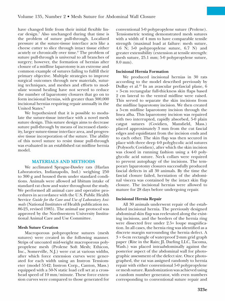
initial laparotomy procedure). Within 30 min-utes of killing the animal, we circumferentially dissected the skin free of the abdominal wall. We then excised the thin-wall hernia sac from the sur-rounding hernia ring, which at this point was well developed and clearly identified with 2.5× loupe magnification. Suture pull-through was recorded if the suture was no longer in contact with both sides of the abdominal wall. Two 4-cm transverse incisions were made 1 cm from the rostral and caudal ends of the hernia defect, through which a 4 × 8-cm sheet of waterproof 2-mm grid graph paper was passed. The graph paper was positioned in contact with the posterior aspect of the abdomi-nal wall and the postrepair hernia defect was then photographed. With the 2-mm grid graph paper serving as a calibration reference for each sample, all photographs were recorded in Tagged Image File Format. To minimize the amount of investi-gator bias introduced in defining and measuring the hernia defect size, edge-detection software provided by the open-source image processing package, Fiji, was used to define the border of the hernia defect and calculate the defect area. All area calculations were calibrated based on the 2-mm grid present in each photograph (Fig. 1).
Fig. 1. Original hernia defect, defect closure, and postrepair hernia defect following mesh suture (above) and conventional suture (below) repair. Defect area measurement by means of edge-detection technique is demonstrated.
Volume 135, Number 2 • Mesh Suture for Abdominal Wall Closure
325e
HistologyOnce the hernia defect had been photo-
graphed, biopsy specimens were taken of the rostral mesh suture and 1 cm of surrounding abdominal wall in all animals that had under-gone mesh suture repair. These tissue samples were fixed in formalin, embedded in paraffin, sectioned, and stained with hematoxylin and eosin. Five nonoverlapping fields per specimen were evaluated at 100× magnification and graded according to a four-point scale adapted from the American Society for Testing and Materials guide-lines, as described previously by Melman et al.11 Higher scores correlate with a greater degree of tissue ingrowth.
Mechanical TestingTensiometric analysis had been intended,
but a useful comparison between the suture and mesh repairs was precluded by the frequency with which the conventional suture repair pulled com-pletely through the contralateral abdominal wall, thus failing to leave a spanning tissue segment on which to perform testing. Likewise, tensiometry of the strength of the mesh suture-tissue interface failed to yield interpretable data, as mechanical tissue disruption lateral to the suture-tissue inter-face occurred with each trial. This mechanical disruption resulted from tearing of the speci-men along the pneumatic grip rather than the intended pull-through phenomenon and thus did not provide useful data with respect to the tissue-mesh interaction.
Statistical AnalysisData were analyzed with the use of GraphPad
Prism 6.0 software (GraphPad Software, Inc., La
Jolla, Calif.). An unpaired t test with Welch’s cor-rection was used to determine the difference in prerepair and postrepair defect area between the mesh ribbon and conventional suture groups. Val-ues were reported as mean ± SEM. Values of p < 0.05 were considered significant.
RESULTS
Hernia Area MeasurementsAll 30 rats developed an incisional hernia
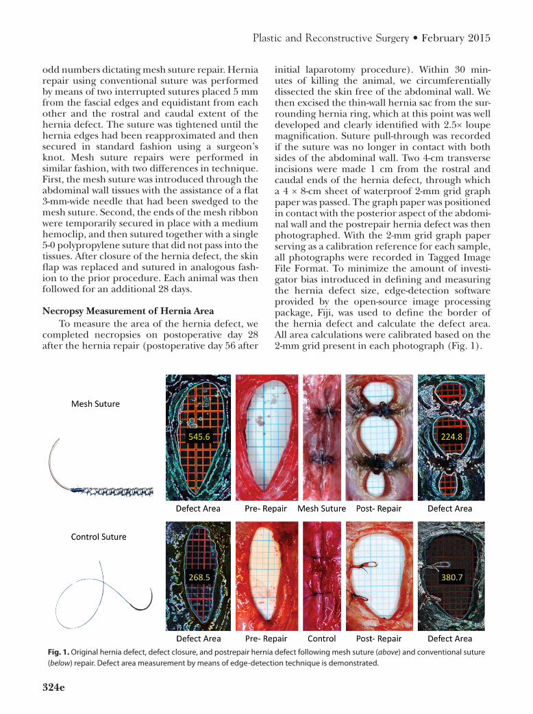
by postoperative day 28. After randomization to repair type, 17 hernias were repaired with mesh suture and 13 hernias were repaired with conven-tional 5-0 polypropylene suture. The mean area of the hernia defects repaired with mesh suture was 391.9 ± 33.4 mm2, whereas that of the hernia defects repaired with conventional 5-0 polypropyl-ene suture was 255.4 ± 23.3 mm2. Despite a rigid randomization protocol, the defects repaired with mesh ribbon were significantly larger than those undergoing conventional suture repair (p < 0.0025). The mean area of the recurrent defect following repair with the mesh ribbon suture was 177.8 ± 27.1 mm2, compared with 267.3 ± 34.1 mm2 following conventional suture repair (Fig. 2, left). When compared with the ini-tial defect area, the mesh suture repairs produced recurrent defects with a mean area that was 42.6 percent that of the original defect, whereas the conventional suture repairs resulted in a recurrent defect area that averaged 110.1 percent that of the original defect (p < 0.0001). This correlated to a 57.4 percent reduction in defect area after mesh suture repair, whereas conventional suture repairs yielded a 10.1 percent increase in postrepair defect area (p < 0.0007) (Fig. 2, right). Notably, none (zero of 34)
Fig. 2. (Left) Comparison of prerepair and postrepair hernia defect area. (Right) Percentage change in hernia defect area following repair.
326e
Plastic and Reconstructive Surgery • February 2015
of the mesh sutures pulled through the surround-ing abdominal wall tissue, whereas 65 percent (17 of 26) of the conventional sutures used for repair demonstrated complete pull-through.
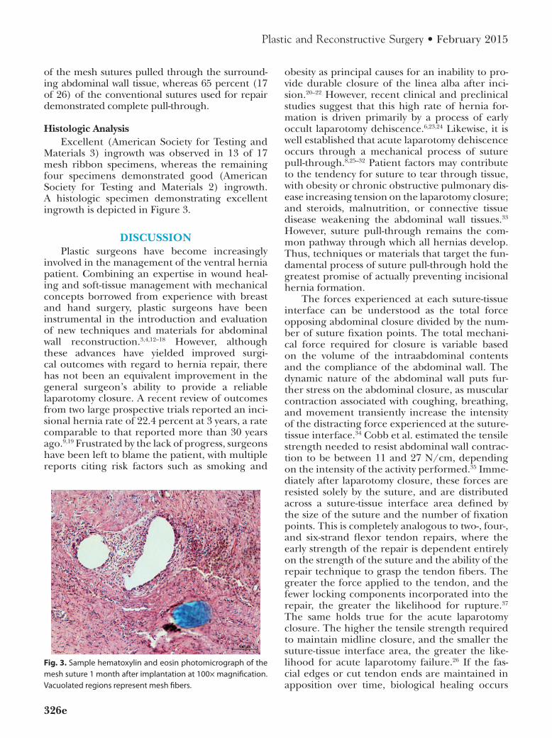
Histologic AnalysisExcellent (American Society for Testing and
Materials 3) ingrowth was observed in 13 of 17 mesh ribbon specimens, whereas the remaining four specimens demonstrated good (American Society for Testing and Materials 2) ingrowth. A histologic specimen demonstrating excellent ingrowth is depicted in Figure 3.
DISCUSSIONPlastic surgeons have become increasingly
involved in the management of the ventral hernia patient. Combining an expertise in wound heal-ing and soft-tissue management with mechanical concepts borrowed from experience with breast and hand surgery, plastic surgeons have been instrumental in the introduction and evaluation of new techniques and materials for abdominal wall reconstruction.3,4,12–18 However, although these advances have yielded improved surgi-cal outcomes with regard to hernia repair, there has not been an equivalent improvement in the general surgeon’s ability to provide a reliable laparotomy closure. A recent review of outcomes from two large prospective trials reported an inci-sional hernia rate of 22.4 percent at 3 years, a rate comparable to that reported more than 30 years ago.9,19 Frustrated by the lack of progress, surgeons have been left to blame the patient, with multiple reports citing risk factors such as smoking and
obesity as principal causes for an inability to pro-vide durable closure of the linea alba after inci-sion.20–22 However, recent clinical and preclinical studies suggest that this high rate of hernia for-mation is driven primarily by a process of early occult laparotomy dehiscence.6,23,24 Likewise, it is well established that acute laparotomy dehiscence occurs through a mechanical process of suture pull-through.8,25–32 Patient factors may contribute to the tendency for suture to tear through tissue, with obesity or chronic obstructive pulmonary dis-ease increasing tension on the laparotomy closure; and steroids, malnutrition, or connective tissue disease weakening the abdominal wall tissues.33 However, suture pull-through remains the com-mon pathway through which all hernias develop. Thus, techniques or materials that target the fun-damental process of suture pull-through hold the greatest promise of actually preventing incisional hernia formation.
The forces experienced at each suture-tissue interface can be understood as the total force opposing abdominal closure divided by the num-ber of suture fixation points. The total mechani-cal force required for closure is variable based on the volume of the intraabdominal contents and the compliance of the abdominal wall. The dynamic nature of the abdominal wall puts fur-ther stress on the abdominal closure, as muscular contraction associated with coughing, breathing, and movement transiently increase the intensity of the distracting force experienced at the suture-tissue interface.34 Cobb et al. estimated the tensile strength needed to resist abdominal wall contrac-tion to be between 11 and 27 N/cm, depending on the intensity of the activity performed.35 Imme-diately after laparotomy closure, these forces are resisted solely by the suture, and are distributed across a suture-tissue interface area defined by the size of the suture and the number of fixation points. This is completely analogous to two-, four-, and six-strand flexor tendon repairs, where the early strength of the repair is dependent entirely on the strength of the suture and the ability of the repair technique to grasp the tendon fibers. The greater the force applied to the tendon, and the fewer locking components incorporated into the repair, the greater the likelihood for rupture.37 The same holds true for the acute laparotomy closure. The higher the tensile strength required to maintain midline closure, and the smaller the suture-tissue interface area, the greater the like-lihood for acute laparotomy failure.26 If the fas-cial edges or cut tendon ends are maintained in apposition over time, biological healing occurs
Fig. 3. Sample hematoxylin and eosin photomicrograph of the mesh suture 1 month after implantation at 100× magnification. Vacuolated regions represent mesh fibers.
Volume 135, Number 2 • Mesh Suture for Abdominal Wall Closure
327e
to add intrinsic support to the extrinsic strength provided by the suture. In essence, fascial heal-ing serves to offload the mechanical construct by increasing the area across which this tension is distributed.
However, biological healing, and any therapy or intervention aimed to optimize it, is predicated on the ability of the repair to maintain apposition of the involved tissue edges. Just as gapping of the tendon ends is known to predispose a tendon repair to rupture, early laparotomy dehiscence predicts eventual hernia formation.23,24 Although splinting and positioning can be used to offload a flexor tendon repair to minimize the forces experienced at the suture-tissue interface, no such adjuncts exist to protect an abdominal wall closure. Viewed along the lines of an unprotected flexor tendon repair, it is easier to appreciate the mechanical challenge posed by laparotomy clo-sure, and to understand why incisional hernia formation remains the most common operative complication following abdominal surgery.6
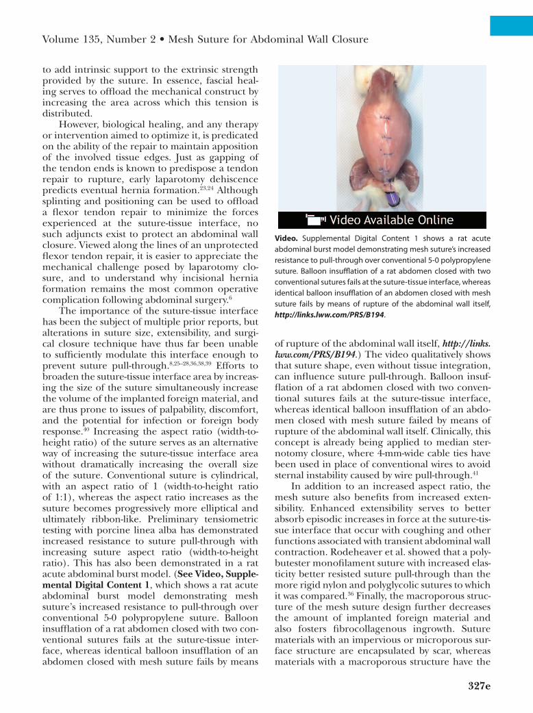
The importance of the suture-tissue interface has been the subject of multiple prior reports, but alterations in suture size, extensibility, and surgi-cal closure technique have thus far been unable to sufficiently modulate this interface enough to prevent suture pull-through.8,25–28,36,38,39 Efforts to broaden the suture-tissue interface area by increas-ing the size of the suture simultaneously increase the volume of the implanted foreign material, and are thus prone to issues of palpability, discomfort, and the potential for infection or foreign body response.40 Increasing the aspect ratio (width-to-height ratio) of the suture serves as an alternative way of increasing the suture-tissue interface area without dramatically increasing the overall size of the suture. Conventional suture is cylindrical, with an aspect ratio of 1 (width-to-height ratio of 1:1), whereas the aspect ratio increases as the suture becomes progressively more elliptical and ultimately ribbon-like. Preliminary tensiometric testing with porcine linea alba has demonstrated increased resistance to suture pull-through with increasing suture aspect ratio (width-to-height ratio). This has also been demonstrated in a rat acute abdominal burst model. (See Video, Supple-mental Digital Content 1, which shows a rat acute abdominal burst model demonstrating mesh suture’s increased resistance to pull-through over conventional 5-0 polypropylene suture. Balloon insufflation of a rat abdomen closed with two con-ventional sutures fails at the suture-tissue inter-face, whereas identical balloon insufflation of an abdomen closed with mesh suture fails by means
of rupture of the abdominal wall itself, http://links.lww.com/PRS/B194.) The video qualitatively shows that suture shape, even without tissue integration, can influence suture pull-through. Balloon insuf-flation of a rat abdomen closed with two conven-tional sutures fails at the suture-tissue interface, whereas identical balloon insufflation of an abdo-men closed with mesh suture failed by means of rupture of the abdominal wall itself. Clinically, this concept is already being applied to median ster-notomy closure, where 4-mm-wide cable ties have been used in place of conventional wires to avoid sternal instability caused by wire pull-through.41
In addition to an increased aspect ratio, the mesh suture also benefits from increased exten-sibility. Enhanced extensibility serves to better absorb episodic increases in force at the suture-tis-sue interface that occur with coughing and other functions associated with transient abdominal wall contraction. Rodeheaver et al. showed that a poly-butester monofilament suture with increased elas-ticity better resisted suture pull-through than the more rigid nylon and polyglycolic sutures to which it was compared.36 Finally, the macroporous struc-ture of the mesh suture design further decreases the amount of implanted foreign material and also fosters fibrocollagenous ingrowth. Suture materials with an impervious or microporous sur-face structure are encapsulated by scar, whereas materials with a macroporous structure have the
Video. Supplemental Digital Content 1 shows a rat acute abdominal burst model demonstrating mesh suture’s increased resistance to pull-through over conventional 5-0 polypropylene suture. Balloon insufflation of a rat abdomen closed with two conventional sutures fails at the suture-tissue interface, whereas identical balloon insufflation of an abdomen closed with mesh suture fails by means of rupture of the abdominal wall itself, http://links.lww.com/PRS/B194.
328e
Plastic and Reconstructive Surgery • February 2015
potential to become incorporated into the sur-rounding tissue.42 Although cellular integration may not necessarily impart greater strength on the implanted suture itself, limiting the foreign body response should make it less prone to the chronic suture pull-through that occurs by means of prote-ase-mediated breakdown of adjacent scar tissue.6,25 As anticipated based on clinical experience with the use of macroporous polypropylene mesh, the majority of the mesh suture specimens exhibited excellent ingrowth on histologic evaluation, with the few remaining specimens demonstrating good tissue integration. More in-depth histologic evalu-ation was not performed, as the biocompatibility of macroporous midweight polypropylene has been previously well defined.42
We hypothesized that a mesh suture design featuring increased elasticity and a larger suture-tissue interface area would better distribute force across the repair and thus be less prone to acute suture pull-through. Furthermore, we anticipated that the macroporous mesh construct would also better resist a chronic pull-through mechanism. Improved distribution of forces should minimize the extent to which pressure-induced necrosis occurs, and any tendency toward chronic cheese-wiring should be offset by fibrocollagenous ingrowth within the suture itself. Although the relative importance of each of these mechanisms is still unknown, the ability of the overall construct to resist suture pull-through was clearly demon-strated by the rat hernia model presented here.
Given the intent for mesh suture to be used for primary laparotomy closure rather than to be applied as a means of repairing an established hernia, one may question why a hernia model was used in place of a laparotomy model. Unfor-tunately, acute laparotomy failure is a uniquely human phenomenon.6 As a result, we elected to use an established ventral hernia model as a “chal-lenged” laparotomy, one in which the fascial edges possess the healing characteristics of a chronic wound and thus contribute little tissue bridging to offset the forces experienced at the suture-tissue interface. This model is known to produce predictable hernia recurrence when repaired with conventional polypropylene suture.10 The suture pull-through mechanism by which this hernia recurrence occurs has been shown to be similar in nature to that which occurs clinically.10 Thus, this “stressed” model was ideally suited to test suture pull-through rather than the efficacy of the her-nia repair. Given the limitations of the model, her-nia recurrence was expected in all animals, with the size of the recurrent hernia defect intended
to serve as a proxy for the extent of suture pull-through. To accentuate this, only two sutures were used for repair, rather than the five or six that would have been required to mimic a clinical lap-arotomy closure. Overall, the data clearly suggest that the mesh suture better resisted cheese-wiring, supporting the primary hypothesis that it is pos-sible to modify the suture-tissue interface with use of an unconventional suture design.
We recognize that these findings are pre-liminary in nature. In addition to the limitations discussed previously, our rat model is further restrained by the issue of scale, which prohib-its immediate translation of our findings to the clinical setting. In addition, planned tensiometric testing aimed at quantifying the tensile strength of the suture-tissue interface was limited by tech-nical constraints. Because of the frequency with which the conventional sutures pulled completely through the contralateral abdominal wall, there were insufficient specimens for testing. In addi-tion, evaluation of the tensile strength at the suture-tissue interface of the mesh suture speci-mens was prevented by tissue tearing elsewhere in the specimen. Beyond these scientific limitations, there exist practical challenges to incorporating these design characteristics into a clinically useful suture. The ribbon-like shape of the mesh suture used for investigation is hampered by poor han-dling characteristics, as it requires precise place-ment to ensure that the broad portion of the suture serves as the suture-tissue interface. Efforts are underway to harness these design concepts in the form of a hollow, macroporous, three-dimensional suture capable of flattening against the tissue when exposed to distracting forces. Such a design would eliminate the need to ori-ent the suture and would dramatically improve its handling characteristics. Testing is currently underway of a mesh suture for human use in a large-animal model.
CONCLUSIONSIn conclusion, macroporous mesh suture
demonstrated an improved ability to approximate tissues under tension over time. Although encour-aged by these results, we recognize the limitations of the described model. Nevertheless, the con-cepts introduced represent an entirely novel and seemingly effective approach to the challenging problem of suture pull-through. Although this problem is perhaps most apparent in the form of laparotomy failure, it is certainly not limited to this clinical entity. From tendon repairs to
Volume 135, Number 2 • Mesh Suture for Abdominal Wall Closure
329e
rhytidectomies, all surgical procedures that aim to join tissues under tension are plagued by suture pull-through. In an era of molecular medicine and rational drug design, it is only reasonable to expect surgical suture to exhibit design character-istics better suited to its intended purpose.
Jason M. Souza, M.D.Division of Plastic Surgery
Northwestern University Feinberg School of Medicine675 North St. Clair, Suite 19-250
Chicago, Ill. [email protected]
REFERENCES 1. Saulis AS, Dumanian GA. Periumbilical rectus abdominis
perforator preservation significantly reduces superficial wound complications in “separation of parts” hernia repairs. Plast Reconstr Surg. 2002;109:2275–2280; discussion 2281.
2. Ghali S, Turza KC, Baumann DP, Butler CE. Minimally inva-sive component separation results in fewer wound-healing complications than open component separation for large ventral hernia repairs. J Am Coll Surg. 2012;214:981–989.
3. Patel KM, Bhanot P, Franklin B, Albino F, Nahabedian MY. Use of intraoperative indocyanine-green angiography to minimize wound healing complications in abdominal wall reconstruction. J Plast Surg Hand Surg. 2013;47:476–480.
4. Souza JM, Dumanian GA. Routine use of bioprosthetic mesh is not necessary: A retrospective review of 100 consecutive cases of intra-abdominal midweight polypropylene mesh for ventral hernia repair. Surgery 2013;153:393–399.
5. Koltz PF, Frey JD, Bell DE, et al. Evolution of abdominal wall reconstruction: Development of a unified algorithm with improved outcomes. Ann Plast Surg. 2013;47:476–480.
6. Xing L, Culbertson EJ, Wen Y, Franz MG. Early laparotomy wound failure as the mechanism for incisional hernia forma-tion. J Surg Res. 2013;182:e35–e42.
7. Majno G. The Healing Hand: Man and Wound in the Ancient World. Cambridge, Mass; Harvard University Press; 1975.
8. Höer J, Fischer L, Schachtrupp A. Laparotomy closure and incisional hernia prevention: What are the surgical require-ments? (in German). Zentralbl Chir. 2011;136:42–49.
9. Fink C, Baumann P, Wente MN, et al. Incisional hernia rate 3 years after midline laparotomy. Br J Surg. 2014;101:51–54.
10. DuBay DA, Wang X, Adamson B, Kuzon WM Jr, Dennis RG, Franz MG. Progressive fascial wound failure impairs subse-quent abdominal wall repairs: A new animal model of inci-sional hernia formation. Surgery 2005;137:463–471.
11. Melman L, Jenkins ED, Hamilton NA, et al. Histologic and biomechanical evaluation of a novel macroporous polytet-rafluoroethylene knit mesh compared to lightweight and heavyweight polypropylene mesh in a porcine model of ven-tral incisional hernia repair. Hernia 2011;15:423–431.
12. Ramirez OM, Ruas E, Dellon AL. “Components separation” method for closure of abdominal-wall defects: An anatomic and clinical study. Plast Reconstr Surg. 1990;86:519–526.
13. Butler CE, Campbell KT. Minimally invasive component separation with inlay bioprosthetic mesh (MICSIB) for complex abdominal wall reconstruction. Plast Reconstr Surg. 2011;128:698–709.
14. Ko JH, Wang EC, Salvay DM, et al. Abdominal wall recon-struction lessons learned from 200 “components separation” procedures. Arch Surg. 2009;144:1047–1055.
15. Ko JH, Salvay DM, Paul BC, Wang EC, Dumanian GA. Soft polypropylene mesh, but not cadaveric dermis, signifi-cantly improves outcomes in midline hernia repairs using the components separation technique. Plast Reconstr Surg. 2009;124:836–847.
16. Janis JE. Use of progressive tension sutures in compo-nents separation: Merging cosmetic surgery techniques with reconstructive surgery outcomes. Plast Reconstr Surg. 2012;130:851–855.
17. Breuing K, Butler CE, Ferzoco S, et al. Incisional ventral hernias: Review of the literature and recommendations regarding the grading and technique of repair. Surgery 2010;148:544–558.
18. Souza JM, Dumanian GA. An evidence-based approach to abdominal wall reconstruction. Plast Reconstr Surg. 2012;130:116–124.
19. Wissing J, van Vroonhoven TJ, Schattenkerk ME, Veen HF, Ponsen RJ, Jeekel J. Fascia closure after midline laparotomy: Results of a randomized trial. Br J Surg. 1987;74:738–741.
20. Sørensen LT, Hemmingsen UB, Kirkeby LT, Kallehave F, Jørgensen LN. Smoking is a risk factor for incisional hernia. Arch Surg. 2005;140:119–123.
21. Antoniou GA, Georgiadis GS, Antoniou SA, Granderath FA, Giannoukas AD, Lazarides MK. Abdominal aortic aneurysm and abdominal wall hernia as manifestations of a connective tissue disorder. J Vasc Surg. 2011;54:1175–1181.
22. Yahchouchy-Chouillard E, Aura T, Picone O, Etienne JC, Fingerhut A. Incisional hernias: I. Related risk factors. Dig Surg. 2003;20:3–9.
23. Pollock AV, Evans M. Early prediction of late incisional her-nias. Br J Surg. 1989;76:953–954.
24. Burger JW, Lange JF, Halm JA, Kleinrensink GJ, Jeekel H. Incisional hernia: Early complication of abdominal surgery. World J Surg. 2005;29:1608–1613.
25. Carlson MA. Acute wound failure. Surg Clin North Am. 1997;77:607–636.
26. Alexander HC, Prudden JF. The causes of abdominal wound disruption. Surg Gynecol Obstet. 1966;122:1223–1229.
27. Gislason H, Grønbech JE, Søreide O. Burst abdomen and incisional hernia after major gastrointestinal opera-tions: Comparison of three closure techniques. Eur J Surg. 1995;161:349–354.
28. Greenburg AG, Saik RP, Peskin GW. Wound dehiscence: Pathophysiology and prevention. Arch Surg. 1979;114:143–146.
29. Lythgoe JP. Burst abdomen. Postgrad Med J. 1960;36:388–391. 30. Niggebrugge AH, Hansen BE, Trimbos JB, van de Velde CJ,
Zwaveling A. Mechanical factors influencing the incidence of burst abdomen. Eur J Surg. 1995;161:655–661.
31. Sanders RJ, DiClementi D. Principles of abdominal wound closure: II. Prevention of wound dehiscence. Arch Surg. 1977;112:1188–1191.
32. Poole GV Jr, Meredith JW, Kon ND, Martin MB, Kawamoto EH, Myers RT. Suture technique and wound-bursting strength. Am Surg. 1984;50:569–572.
33. Franz MG. The biology of hernia formation. Surg Clin North Am. 2008;88:1–15.
34. Dumanian GA. The physics of abdominal wall recon-struction. Nahabedian MY, Bhanot P, eds. Abdominal Wall Reconstruction. Woodbury, Conn: Cinemed; (in press).
35. Cobb WS, Kercher KW, Heniford BT. The argument for lightweight polypropylene mesh in hernia repair. Surg Innov. 2005;12:63–69.
36. Rodeheaver GT, Nesbit WS, Edlich RF. Novafil: A dynamic suture for wound closure. Ann Surg. 1986;204:193–199.
37. Strickland JW. Development of flexor tendon surgery: Twenty-five years of progress. J Hand Surg Am. 2000;25:214–235.
330e
Plastic and Reconstructive Surgery • February 2015
38. Dudley HA. Layered and mass closure of the abdominal wall. Br J Surg. 1970;57:664–667.
39. Israelsson LA, Millbourn D. Closing midline abdominal inci-sions. Langenbecks Arch Surg. 2012;397:1201–1207.
40. McLeod RS, Brenneman FD, Rotstein OD, Bhanot P; Members of the Evidence-Based Reviews in Surgery Group. Does the size of the stitch length affect surgical site infec-tion? J Am Coll Surg. 2013;217:556–559.
41. Grapow MT, Melly LF, Eckstein FS, Reuthebuch OT. A new cable-tie based sternal closure system: Description of the device, technique of implantation and first clinical evalua-tion. J Cardiothorac Surg. 2012;7:59.
42. Orenstein SB, Saberski ER, Kreutzer DL, Novitsky YW. Comparative analysis of histopathologic effects of synthetic meshes based on material, weight, and pore size in mice. J Surg Res. 2012;176:423–429.