exosomes derived from bm-mscs mitigate the development
TRANSCRIPT
Citation: Alasmari, W.A.;
Abdelfattah-Hassan, A.;
El-Ghazali, H.M.; Abdo, S.A.;
Ibrahim, D.; ElSawy, N.A.;
El-Shetry, E.S.; Saleh, A.A.;
Abourehab, M.A.S.; Mahfouz, H.
Exosomes Derived from BM-MSCs
Mitigate the Development of Chronic
Kidney Damage Post-Menopause via
Interfering with Fibrosis and
Apoptosis. Biomolecules 2022, 12, 663.
https://doi.org/10.3390/
biom12050663
Academic Editors: Daria Ilatovskaya
and Krisztian Stadler
Received: 24 February 2022
Accepted: 22 April 2022
Published: 2 May 2022
Publisher’s Note: MDPI stays neutral
with regard to jurisdictional claims in
published maps and institutional affil-
iations.
Copyright: © 2022 by the authors.
Licensee MDPI, Basel, Switzerland.
This article is an open access article
distributed under the terms and
conditions of the Creative Commons
Attribution (CC BY) license (https://
creativecommons.org/licenses/by/
4.0/).
biomolecules
Article
Exosomes Derived from BM-MSCs Mitigate the Developmentof Chronic Kidney Damage Post-Menopause via Interferingwith Fibrosis and ApoptosisWardah A. Alasmari 1,*,† , Ahmed Abdelfattah-Hassan 2,3,*,† , Hanaa M. El-Ghazali 2, Samar A. Abdo 4,Doaa Ibrahim 5 , Naser A. ElSawy 6 , Eman S. El-Shetry 6, Ayman A. Saleh 7, Mohammed A. S. Abourehab 8
and Hala Mahfouz 9
1 Department of Anatomy, Faculty of Medicine, Umm Al-Qura University, Makkah 24230, Saudi Arabia2 Department of Anatomy and Embryology, Faculty of Veterinary Medicine, Zagazig University,
Zagazig 44519, Egypt; [email protected] Biomedical Sciences Program, University of Science and Technology, Zewail City of Science and Technology,
Giza 12578, Egypt4 Department of Biochemistry, Faculty of Veterinary Medicine, Zagazig University, Zagazig 44519, Egypt;
[email protected] Department of Nutrition and Clinical Nutrition, Faculty of Veterinary Medicine, Zagazig University,
Zagazig 44511, Egypt; [email protected] Department of Human Anatomy and Embryology, Faculty of Medicine, Zagazig University,
Zagazig 44511, Egypt; [email protected] (N.A.E.); [email protected] (E.S.E.-S.)7 Department of Animal Wealth Development, Genetics & Genetic Engineering, Faculty of Veterinary Medicine,
Zagazig University, Zagazig 44519, Egypt; [email protected] Department of Pharmaceutics, College of Pharmacy, Umm Al-Qura University, Makkah 21955, Saudi Arabia;
[email protected] Department of Medical Biochemistry and Molecular Biology, Faculty of Medicine, Kafrelsheikh University,
Kafrelsheikh 33516, Egypt; [email protected]* Correspondence: [email protected] (W.A.A.); [email protected] or
[email protected] (A.A.-H.)† These authors contributed equally to this work.
Abstract: The rate of chronic kidney disease (CKD) is increasing globally, and it is caused by contin-uous damage to kidney tissue. With time the renal damage becomes irreversible, leading to CKDdevelopment. In females, post-menopause lack of estrogen supply has been described as a risk factorfor CKD development, and studies targeting post-menopause CKD are scarce. In the present study,we used exosomes isolated from bone marrow mesenchymal stem/stromal cells (BM-MSCs) to testtheir therapeutic potential against the development of CKD. At first, the menopause model wasachieved by surgical bilateral ovariectomy in female albino rats. After that, 100 µg of exosomes wasgiven to ovariectomized rats, and the study continued for 2 months. Changes in urine volume, urineprotein content, kidney function biochemical parameters (creatinine and BUN), kidney antioxidantparameters (SOD, GPx and CAT), histological changes, immunohistochemical levels of caspase 3,and the gene expression of NGAL (related to kidney damage), TGFβ1 and αSMA (related to fibrosisand EMT), and caspase 3 (related to apoptosis) were studied. After the ovariectomy, the occurrenceof CKD was confirmed in the rats by the drastic reduction of serum estrogen and progesteronelevels, reduced urine output, increased urinary protein excretion, elevated serum creatinine and BUN,reduced GPx SOD, and CAT in kidney tissue, degenerative and fibrotic lesions in the histopatho-logical examination, higher immunohistochemical expression of caspase 3 and increased expressionof all studied genes. After exosomes administration, the entire chronic inflammatory picture in thekidney was corrected, and a near-normal kidney structure and function were attained. This studyshows for the first time that BM-MSCs exosomes are potent for reducing apoptosis and fibrosis levelsand, thus, can reduce the chronic damage of the kidneys in females that are in their menopauseperiod. Therefore, MSCs-derived exosomes should be considered a valuable therapy for preservingpostmenopausal kidney structure and function and, subsequently, could improve the quality offemales’ life during menopause.
Biomolecules 2022, 12, 663. https://doi.org/10.3390/biom12050663 https://www.mdpi.com/journal/biomolecules
Biomolecules 2022, 12, 663 2 of 16
Keywords: exosome therapy; renoprotective; nephroprotective; chronic renal injury; caspase 3;fibrosis
1. Introduction
Chronic kidney disease (CKD) is characterized by the gradual loss of kidney func-tions and an increased risk for death. The global prevalence of CKD is estimated tobe 11–13%, and most affected patients are in stage 3 [1]. The available data also re-vealed that females are more prone to CKD compared to males [1]. Seven percent ofpostmenopausal females with normal kidney function will develop CKD within 10 years(https://www.mayoclinicproceedings.org/article/S0025-6196(20)30374-8/fulltext, last ac-cessed on 5 April 2022). CKD will gradually progress toward end-stage renal failure, whichis fatal and requires dialysis or kidney transplantation and causes life-threatening and finan-cial problems for patients and health systems. Various factors contribute to the occurrenceof CKD (such as chemicals, medications, genetics, immunity, infections, environment anddiet). The association between menopause and CKD was given less attention comparedto other conditions, and limited data can be found related to CKD and menopause [2]. Infact, the role of menopause in the development of CKD is complex, and associated withvarious predisposing factors, such as aging, cardiovascular diseases, abnormal mineralmetabolism and increased oxidative stress in the body, most of these conditions are relatedto the lack of estrogen during post-menopause (reviewed in detail in [2]). The role of renalfibrosis and the involvement of TGF-β in the development of CKD has been extensivelystudied [3,4], and the inhibition of TGF-β can lead to favorable outcomes in terms of theprevention of fibrosis in CKD [5]. Another important aspect of CKD development is theapoptosis of renal glomerular and tubular cells [6,7], with caspases, and essentially caspase3, playing a pivotal role in the progression of CKD [8]. Therefore, therapeutic approachesthat target both TGF-β and caspase 3 should be beneficial in preventing the developmentof CKD in post-menopause females.
It has been investigated that decreased estrogen levels during menopause lead tosignificant effects on the female body. In addition, it predisposes them to other significantdiseases, such as bone mineralization and cardiovascular diseases, which are under heavyinvestigation lately. However, no data exists on the targeting of post-menopause CKD. Asa result of the lack of estrogen, it is widely accepted that hormone replacement therapycan be effective in menopausal females; however, hormone therapy results, in general,were described to be controversial [9]. In addition, in vivo studies showed that supplyingestrogen can lead to reduced renal damage and oxidative stress in ovariectomized rats [10].However, several reports showed that hormonal replacements are not recommended forprolonged use, as long treatment was proven to cause adverse effects, including kidneydamage [11]. Consequently, post-menopause CKD is still lacking appropriate therapeuticapproaches to reduce the rate of apoptosis and fibrosis, which occur in the kidneys, leadingto their chronic damage, and there are currently no known treatment regimens.
Recently, studies have elucidated that mesenchymal stem cells (MSCs) possess thera-peutic capabilities for many diseases, mainly through their paracrine mechanisms [12–14].These paracrine actions are mediated by the release of soluble factors and encapsulatedfactors (inside microvesicles and exosomes). The latter is of special importance sinceexosomes released from MSCs are responsible for the horizontal transfer of importantmRNA, microRNA and proteins and are now being recognized as an integral component ofintercellular communication [15–17]. Exosomes are ideal therapeutic agents because theircomplex content of proteins and genetic materials has the potential to treat complex dis-eases, such as CKD. In addition, exosome-based therapy circumvents some of the concernsand limitations of using viable replicating cells, including mesenchymal stem cells [18].One of the main contributors to kidney regeneration is stem cells that reside in the kidneyitself (reviewed in [19]). However, isolated stem cells from animals suffering from CKD

Biomolecules 2022, 12, 663 3 of 16
showed decreased regenerative potential and premature senescence [20]. The latter studyshows that during CKD, the kidney regenerative capacity was not functioning properly andcontributes to advancing chronic kidney damage. Very recently, adipose MSCs and theirsecreted vesicles were found to be beneficial in protecting against ischemia-reperfusionacute kidney failure [21]. However, there is a global concern about using viable replicat-ing cells, and much clinical investigation is still needed; therefore, stem cells’ secretedexosomes come as a promising cure for diseases with complex etiology, such as CKD inpostmenopausal females. In this regard, exosomes used for treating acute kidney injuryhave drawn recent attention [22–24]. In addition, few studies reported the use of exosomesfor treating chronic kidney injury (reviewed in [25]).
In the literature, there is a lack of studies that focus on the use of exosomes derivedfrom BM-MSCs for reducing the progression of CKD during menopause, and there islimited information on the use of exosomes or their renoprotective effects during post-menopause. Therefore, the current study intended to investigate the therapeutic effi-cacy of exosomes released from MSCs and their use as a novel therapeutic approach toprevent/reduce apoptosis and fibrosis associated with chronic kidney damage in post-menopausal females.
2. Materials and Methods2.1. Study Animals and Experimental Design
For this study, a group of healthy female albino rats (n = 28) of similar age andweight (approx. 7 months old and their weight averaged 300 g approx.). The rats wereobtained from the animal house of the Faculty of Medicine, Zagazig University, andwere housed in a temperature/humidity-controlled (24 ± 2 ◦C and 50 ± 10% relativehumidity) and light-controlled room (12 h light/dark cycle) with free access to a standardrat chow diet and filtered tap water. The rats were permitted to acclimatize for 2 weeksbefore starting the experiment. All experimental procedures followed the guidelines of theInstitutional Animal Care and Use Committee of Zagazig University (protocol number:ZU-IACUC/2/F/164/2020), and the study was conducted in strict accordance with therecommendations in the Guide for the Care and Use of Laboratory Animals of the NationalInstitutes of Health (NRC, Washington D.C., USA). The control group contained 7 healthyfemale rats, while the remaining rats were randomly assigned to one of the followinggroups: Sham group, included rats that underwent the same operation as the ovariectomygroup but with no ovariectomy (Sham, n = 7), post-menopausal chronic kidney damagegroup (CKD, n = 7), and, finally, CKD rats that received exosomes derived from BM-MSCs(CKD + Exosomes, n = 7). The administration of MSCs-derived exosomes was performedwithin 24 h after ovariectomy or sham surgical procedures, and the study continued for2 months to assure the occurrence of CKD in operated rats.
2.2. Isolation and Characterization of Bone Marrow MSCs
Rat bone marrow was collected from the long bones of 4–6 week-old, apparentlyhealthy rats, as previously reported by others and us [26,27]. The process included, briefly,collecting long bones and the flushing of their bone marrow using a syringe containing10,000 IU heparin to prevent coagulation. The collected bone marrow was pooled andwas cultured on tissue culture plates. With frequent medium changes, the non-adherentcells were washed off; the medium was changed after 3 h from initial plating and thenevery 8 h during the succeeding 72 h. The remaining cells were only adherent cells that areconsidered to be mesenchymal stem cells (MSCs). The characterization of cultured MSCswas performed to confirm that these cells were MSCs; this was done by flow cytometricanalysis of FITC-labeled anti-CD34 (Cat. No: 555821, BD Pharmingen, San Diego, CA,USA), anti-CD45 (ab33916, abcam, Boston, MA, USA), anti-CD90 (ab226, abcam, USA) andanti-CD105 (ab184667, abcam, USA) antibodies, as we previously reported [28]. MSCs usedfor exosomes isolation were from passages 4 to 7.
Biomolecules 2022, 12, 663 4 of 16
2.3. Isolation and Characterization of Exosomes from BM-MSCs
Exosome isolation, characterization (by TEM and western blotting) and storage wereperformed as previously reported by us and others [27,29]. In brief, starting from passagethree, the MSCs culture medium was removed when the cells reached 80% confluency,and the serum-free medium was replenished and left for 24 h; it was collected and nowcalled conditioned medium (CM). The collected CM was filtered through a 0.22 µm filter(to remove debris and dead cells) and was centrifuged at 10,000 g for 30 min at 4 ◦C andthe supernatant was collected. The supernatant was then subjected to ultracentrifugationat 100,000 g for 60 min at 4 ◦C to pellet the exosomes. The pellet was washed twicewith ice-cold PBS, and ultracentrifugation was performed for each wash to re-pellet theisolated exosomes. To quantify the amount of isolated exosomes, the protein content wasmeasured by standard Bradford assay, and then the isolated exosomes were adjusted to100 µg protein in 200 µL aliquots and stored at −80 ◦C. The characterization of isolatedexosomes was performed using the detection of exosomes’ markers, CD63 (sc-5275, SantaCruz Biotechnology, USA) and CD81 (sc-166029, Santa Cruz Biotechnology, Dallas, Texas,USA), by western blotting and by transmission electron microscopic (TEM) detection oftheir shape and size.
2.4. Post-Menopause Chronic Kidney Disease (CKD) Model
The establishment of the post-menopause CKD model followed previously publishedprotocols [30,31]. Briefly, strict aseptic surgical procedures were performed; after that, theassigned rats were anesthetized using intraperitoneal injection of a mixture of Ketamine(80 mg/kg) and Xylazine (12.5 mg/kg). A 20 mm dorsal skin incision was cut in the midlineto allow access for two 10mm flank muscle incisions to excise both ovaries from both sides.After the removal of the ovaries, the incisions were sutured and the rats were observed forcomplete recovery. Administration of analgesia (midazolam 1 mg/kg s.c.) and antibiotics(gentamicin 5 mg/kg i.m.) was performed for 5 days post-surgery. The sham-operated ratsunderwent the same surgical procedures except for the ovaries, which were not removed.The success of the operation was confirmed by the absence of monthly cycles and the abruptdecrease in serum levels of estrogen and progesterone. Other signs were reduced uterinemass observed during sample collection at the end of the study, as reported previously [32].The rats were humanely euthanized by an overdose of inhalation general anesthesia at theend of the study.
2.5. Urine Analyses
Urine was collected in metabolic cages for a total period of 24 h at the end of thestudy (after 2 months of CKD induction). The urine analysis included the estimatedglomerular filtration rate, calculated as previously reported [11], and the evaluation of urineprotein content by standard Bradford assay kit (Pierce™ Coomassie (Bradford) ProteinAssay Kit, ThermoFisher Scientific Inc., Waltham, MA, USA), following the manufacturer’sinstructions.
2.6. Administration of Exosomes Derived from Bone Marrow MSCs
Exosomes purified from step 2.3 were administered intravenously through the tailvein in the CKD + Exosomes group; the rats received one-time 100 µg protein-equivalentof MSCs-derived exosomes in 200 µL. The injections were performed after the rats werecompletely recovered following CKD induction [24].
2.7. Serum Biochemical Assays and Kidney Tissue Antioxidants Detection
Samples from the rat’s blood were collected for serum separation. Serum sampleswere used to measure the levels of creatinine (Creatinine ELISA Kit, Catalog #: E4370,BioVision, Inc., USA), BUN (BUN ELISA Kit, Catalog #: MBS2611085, MyBioSource, USA),estrogen (Estrogen ELISA Kit, Catalog #: K4266, BioVision, Inc., USA) and progesterone(Progesterone ELISA Kit, Catalog #: K7416, BioVision, Inc., USA), this was performed by
Biomolecules 2022, 12, 663 5 of 16
commercial kits and according to the kit’s recommendations. Moreover, kidney sampleswere collected immediately after sacrifice, washed twice in warm PBS, and then subjectedto homogenization, as we previously reported [33]. The antioxidant markers evaluatedin kidney tissue were the levels of superoxide dismutase (SOD), glutathione peroxidase(GPx) and catalase (CAT); their measurement was conducted using commercial kits andfollowing the kit’s recommendations (Bio-diagnostics Co., Cairo, Egypt and BioVision, Inc.,Milpitas, CA, USA).
2.8. Histopathologic Examination
After sacrificing the study rats, immediately, a kidney was collected and longitudinallyexcised and immersed in a fixative (10% neutral buffered formalin) solution. After 24–48 h,the fixed samples of kidney tissue were paraffin-embedded and histologically prepared byconventional histological technique, as previously described [33]. After that, tissue sections(4 µm) were stained using the standard H&E technique. The extent of renal damage wasblindly evaluated by an independent pathologist.
2.9. Immunohistochemistry of Caspase 3
For the evaluation of caspase 3 expression in kidney tissue, paraffin-embedded kidneytissue was cut into 4 µm sections, and the sections were exposed to the standard heat-antigen retrieval procedure. Then, blocking serum was added (to block non-specificbinding), followed by the addition of H2O2 (to block endogenous peroxidases). The anti-caspase 3 primary antibody was added (1:250, rabbit caspase 3 antibody, ab13,847, abcam,USA), and the slides were incubated overnight at 4 ◦C. The secondary antibody was addedto the slides (1:500, HRP-labeled goat anti-rabbit antibody, ab6721, abcam, USA) andincubated for 30 min at room temperature. Finally, the DAB substrate was used for thevisualization of positive reactions (Mouse and Rabbit Specific HRP/DAB Detection kit,ab64264, abcam, USA). The slides were examined under light microscopy, and stainingintensity was determined by ImageJ software (ImageJ v 1.53, National Institutes of Health,Bethesda, MD, USA), as previously reported [28].
2.10. Real Time qPCR
Kidney tissue samples were immediately collected after sacrifice and were kept inQiazol Lysis Reagent (Cat. No.: 79306, Qiagen, Cairo, Egypt) and stored at −20 ◦C. TheRNA extraction from kidney tissue samples was performed using an RNA extraction kit(RNeasy Mini Kit, Cat. No.: 74106, Qiagen, Cairo, Egypt), following the manufacturer’srecommendations. The extracted RNA was checked for concentration and purity using nan-odrop at 260 and 280 nm wavelengths (Quawell Q5000, Quawell Technology, Inc., San Jose,CA, USA). After that, reverse transcription into cDNA was performed using a RevertAidFirst Strand cDNA Synthesis Kit (Thermo Scientific, Cat. No. K1621, Cairo, Egypt). Theexpression of study genes (the used primers’ sequences are listed in Table 1) was performedusing StepOnePlus™ Real-Time PCR system (Applied Biosystems, Waltham, MA, USA)by using QuantiTect SYBR® Green PCR Kit (Qiagen, Cat. No. 204141, Cairo, Egypt). Theresulting gene expression data were normalized against GABDH (as a housekeeping gene),and the amount of amplified products was relatively quantified using the −2∆∆Ct method,as previously described.
2.11. Statistical Analyses
The obtained data from this study were analyzed using one-way ANOVA, followedby Tukey’s HSD between-group analysis using PASW statistical package (SPSS v18, SPSSInc., Chicago, IL, USA). The gene expression data were analyzed by one-way ANOVA andvisualized using GraphPad Prism 5 (GraphPad Software Inc., La Jolla, CA, USA). Statisticalsignificance was considered when the p-value ≤ 0.05, and the data were presented asmeans ± SD.
Biomolecules 2022, 12, 663 6 of 16
Table 1. Primers used for qRT-PCR.
Gene Name Sequence (5′-3′) Accession No. Ref.
NGAL F- TTGGGACAGGGAAGACGAR- TCACGCTGGGCAACATTA XM_032901803.1 [34]
TGFβ1 F- CAGGAGCGCACAATCATGTTR- CTTTAGGAAGGACCTGGGTT XM_032894155.1 [35]
αSMA F- GTCCCAGACATCAGGGAGTAAR- TCGGATACTTCAGCGTCAGGA XM_032891814.1 [35]
Caspase 3 F- CTCGGTCTGGTACAGATGTCGATGR- GGTTAACCCGGGTAAGAATGTGCA XM_032896303.1 [36]
GAPDH F- AGACAGCCGCATCTTCTTGTR- TTCCCATTCTCAGCCTTGAC NM_017008.4 [28]
3. Results3.1. BM-MSCs Isolation and Collection of Their Exosomes
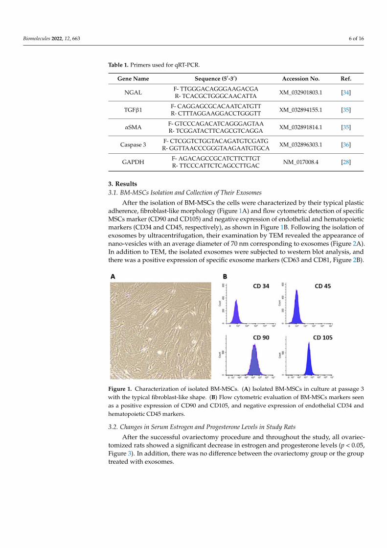
After the isolation of BM-MSCs the cells were characterized by their typical plasticadherence, fibroblast-like morphology (Figure 1A) and flow cytometric detection of specificMSCs marker (CD90 and CD105) and negative expression of endothelial and hematopoieticmarkers (CD34 and CD45, respectively), as shown in Figure 1B. Following the isolation ofexosomes by ultracentrifugation, their examination by TEM revealed the appearance ofnano-vesicles with an average diameter of 70 nm corresponding to exosomes (Figure 2A).In addition to TEM, the isolated exosomes were subjected to western blot analysis, andthere was a positive expression of specific exosome markers (CD63 and CD81, Figure 2B).
Biomolecules 2022, 12, x FOR PEER REVIEW 6 of 17
NGAL F‐ TTGGGACAGGGAAGACGA
R‐ TCACGCTGGGCAACATTA XM_032901803.1 [34]
TGFβ1 F‐ CAGGAGCGCACAATCATGTT
R‐ CTTTAGGAAGGACCTGGGTT XM_032894155.1 [35]
αSMA F‐ GTCCCAGACATCAGGGAGTAA
R‐ TCGGATACTTCAGCGTCAGGA XM_032891814.1 [35]
Caspase 3 F‐ CTCGGTCTGGTACAGATGTCGATG
R‐ GGTTAACCCGGGTAAGAATGTGCA XM_032896303.1 [36]
GAPDH F‐ AGACAGCCGCATCTTCTTGT
R‐ TTCCCATTCTCAGCCTTGAC NM_017008.4 [28]
2.11. Statistical Analyses
The obtained data from this study were analyzed using one‐way ANOVA, followed
by Tukey’s HSD between‐group analysis using PASW statistical package (SPSS v18, SPSS
Inc., Chicago, IL, USA). The gene expression data were analyzed by one‐way ANOVA
and visualized using GraphPad Prism 5 (GraphPad Software Inc., La Jolla, CA, USA). Sta‐
tistical significance was considered when the p‐value ≤ 0.05, and the data were presented
as means ± SD.
3. Results
3.1. BM‐MSCs Isolation and Collection of Their Exosomes
After the isolation of BM‐MSCs the cells were characterized by their typical plastic
adherence, fibroblast‐like morphology (Figure 1A) and flow cytometric detection of spe‐
cific MSCs marker (CD90 and CD105) and negative expression of endothelial and hema‐
topoietic markers (CD34 and CD45, respectively), as shown in Figure 1B. Following the
isolation of exosomes by ultracentrifugation, their examination by TEM revealed the ap‐
pearance of nano‐vesicles with an average diameter of 70 nm corresponding to exosomes
(Figure 2A). In addition to TEM, the isolated exosomes were subjected to western blot
analysis, and there was a positive expression of specific exosome markers (CD63 and
CD81, Figure 2B).
Figure 1. Characterization of isolated BM‐MSCs. (A) Isolated BM‐MSCs in culture at passage 3 with
the typical fibroblast‐like shape. (B) Flow cytometric evaluation of BM‐MSCs markers seen as a pos‐
itive expression of CD90 and CD105, and negative expression of endothelial CD34 and hematopoi‐
etic CD45 markers.
Figure 1. Characterization of isolated BM-MSCs. (A) Isolated BM-MSCs in culture at passage 3with the typical fibroblast-like shape. (B) Flow cytometric evaluation of BM-MSCs markers seenas a positive expression of CD90 and CD105, and negative expression of endothelial CD34 andhematopoietic CD45 markers.
3.2. Changes in Serum Estrogen and Progesterone Levels in Study Rats
After the successful ovariectomy procedure and throughout the study, all ovariec-tomized rats showed a significant decrease in estrogen and progesterone levels (p < 0.05,Figure 3). In addition, there was no difference between the ovariectomy group or the grouptreated with exosomes.

Biomolecules 2022, 12, 663 7 of 16Biomolecules 2022, 12, x FOR PEER REVIEW 7 of 17
Figure 2. Characterization of exosomes obtained from BM‐MSCs. (A) TEM examination showing
the presence of exosomes and their diameter is less than 100 nm. (B) Western blot analysis of exo‐
somes’ markers CD63 and CD81. (C) Scheme outlining the experimental setup of the study.
3.2. Changes in Serum Estrogen and Progesterone Levels in Study Rats
After the successful ovariectomy procedure and throughout the study, all ovariecto‐
mized rats showed a significant decrease in estrogen and progesterone levels (p < 0.05,
Figure 3). In addition, there was no difference between the ovariectomy group or the
group treated with exosomes.
Figure 3. Changes in serum estrogen and progesterone levels in study rats. CKD + Exosomes group
received an intravenous injection of 100 μg protein‐equivalent of exosomes in their tail vein. Differ‐
ent letters denote statistical significance (p < 0.05, using Tukey’s HSD post‐hoc test).
3.3. Changes in Serum/Tissue Biochemical Parameters
The serum levels of BUN and creatinine in ovariectomized rats were significantly
increased (p < 0.01) compared to control and sham groups (Figure 4). In addition, the kid‐
ney‐tissue levels of antioxidant parameters, SOD, GPx and CAT were significantly de‐
creased (p < 0.01, Figure 5) in ovariectomized albino rats compared to the control or sham
Figure 2. Characterization of exosomes obtained from BM-MSCs. (A) TEM examination showing thepresence of exosomes and their diameter is less than 100 nm. (B) Western blot analysis of exosomes’markers CD63 and CD81. (C) Scheme outlining the experimental setup of the study.
Biomolecules 2022, 12, x FOR PEER REVIEW 7 of 17
Figure 2. Characterization of exosomes obtained from BM‐MSCs. (A) TEM examination showing
the presence of exosomes and their diameter is less than 100 nm. (B) Western blot analysis of exo‐
somes’ markers CD63 and CD81. (C) Scheme outlining the experimental setup of the study.
3.2. Changes in Serum Estrogen and Progesterone Levels in Study Rats
After the successful ovariectomy procedure and throughout the study, all ovariecto‐
mized rats showed a significant decrease in estrogen and progesterone levels (p < 0.05,
Figure 3). In addition, there was no difference between the ovariectomy group or the
group treated with exosomes.
Figure 3. Changes in serum estrogen and progesterone levels in study rats. CKD + Exosomes group
received an intravenous injection of 100 μg protein‐equivalent of exosomes in their tail vein. Differ‐
ent letters denote statistical significance (p < 0.05, using Tukey’s HSD post‐hoc test).
3.3. Changes in Serum/Tissue Biochemical Parameters
The serum levels of BUN and creatinine in ovariectomized rats were significantly
increased (p < 0.01) compared to control and sham groups (Figure 4). In addition, the kid‐
ney‐tissue levels of antioxidant parameters, SOD, GPx and CAT were significantly de‐
creased (p < 0.01, Figure 5) in ovariectomized albino rats compared to the control or sham
Figure 3. Changes in serum estrogen and progesterone levels in study rats. CKD + Exosomes groupreceived an intravenous injection of 100 µg protein-equivalent of exosomes in their tail vein. Differentletters denote statistical significance (p < 0.05, using Tukey’s HSD post-hoc test).
3.3. Changes in Serum/Tissue Biochemical Parameters
The serum levels of BUN and creatinine in ovariectomized rats were significantlyincreased (p < 0.01) compared to control and sham groups (Figure 4). In addition, thekidney-tissue levels of antioxidant parameters, SOD, GPx and CAT were significantlydecreased (p < 0.01, Figure 5) in ovariectomized albino rats compared to the control or shamgroups. Following the administration of exosomes isolated from BM-MSCs, serum levels ofBUN and creatinine were significantly decreased (p < 0.01) compared to the ovariectomizedgroup, but their levels were still higher than the control and sham groups. Moreover,
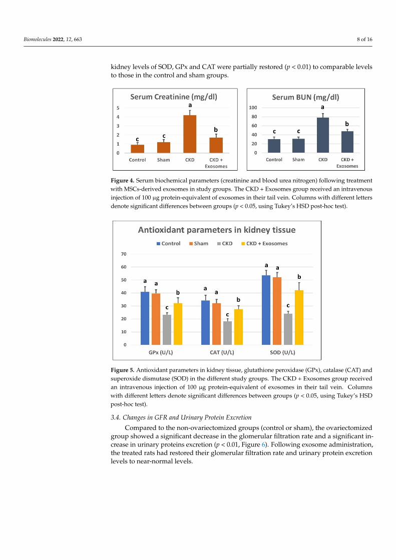
Biomolecules 2022, 12, 663 8 of 16
kidney levels of SOD, GPx and CAT were partially restored (p < 0.01) to comparable levelsto those in the control and sham groups.
Biomolecules 2022, 12, x FOR PEER REVIEW 8 of 17
groups. Following the administration of exosomes isolated from BM‐MSCs, serum levels
of BUN and creatinine were significantly decreased (p < 0.01) compared to the ovariecto‐
mized group, but their levels were still higher than the control and sham groups. Moreo‐
ver, kidney levels of SOD, GPx and CAT were partially restored (p < 0.01) to comparable
levels to those in the control and sham groups.
Figure 4. Serum biochemical parameters (creatinine and blood urea nitrogen) following treatment
with MSCs‐derived exosomes in study groups. The CKD + Exosomes group received an intravenous
injection of 100 μg protein‐equivalent of exosomes in their tail vein. Columns with different letters
denote significant differences between groups (p < 0.05, using Tukey’s HSD post‐hoc test).
Figure 5. Antioxidant parameters in kidney tissue, glutathione peroxidase (GPx), catalase (CAT)
and superoxide dismutase (SOD) in the different study groups. The CKD + Exosomes group re‐
ceived an intravenous injection of 100 μg protein‐equivalent of exosomes in their tail vein. Columns
with different letters denote significant differences between groups (p < 0.05, using Tukey’s HSD
post‐hoc test).
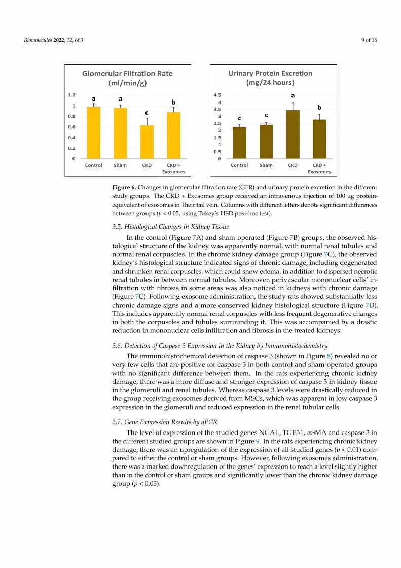
3.4. Changes in GFR and Urinary Protein Excretion
Compared to the non‐ovariectomized groups (control or sham), the ovariectomized
group showed a significant decrease in the glomerular filtration rate and a significant in‐
crease in urinary proteins excretion (p < 0.01, Figure 6). Following exosome administra‐
tion, the treated rats had restored their glomerular filtration rate and urinary protein ex‐
cretion levels to near‐normal levels.
Figure 4. Serum biochemical parameters (creatinine and blood urea nitrogen) following treatmentwith MSCs-derived exosomes in study groups. The CKD + Exosomes group received an intravenousinjection of 100 µg protein-equivalent of exosomes in their tail vein. Columns with different lettersdenote significant differences between groups (p < 0.05, using Tukey’s HSD post-hoc test).
Biomolecules 2022, 12, x FOR PEER REVIEW 8 of 17
groups. Following the administration of exosomes isolated from BM‐MSCs, serum levels
of BUN and creatinine were significantly decreased (p < 0.01) compared to the ovariecto‐
mized group, but their levels were still higher than the control and sham groups. Moreo‐
ver, kidney levels of SOD, GPx and CAT were partially restored (p < 0.01) to comparable
levels to those in the control and sham groups.
Figure 4. Serum biochemical parameters (creatinine and blood urea nitrogen) following treatment
with MSCs‐derived exosomes in study groups. The CKD + Exosomes group received an intravenous
injection of 100 μg protein‐equivalent of exosomes in their tail vein. Columns with different letters
denote significant differences between groups (p < 0.05, using Tukey’s HSD post‐hoc test).
Figure 5. Antioxidant parameters in kidney tissue, glutathione peroxidase (GPx), catalase (CAT)
and superoxide dismutase (SOD) in the different study groups. The CKD + Exosomes group re‐
ceived an intravenous injection of 100 μg protein‐equivalent of exosomes in their tail vein. Columns
with different letters denote significant differences between groups (p < 0.05, using Tukey’s HSD
post‐hoc test).
3.4. Changes in GFR and Urinary Protein Excretion
Compared to the non‐ovariectomized groups (control or sham), the ovariectomized
group showed a significant decrease in the glomerular filtration rate and a significant in‐
crease in urinary proteins excretion (p < 0.01, Figure 6). Following exosome administra‐
tion, the treated rats had restored their glomerular filtration rate and urinary protein ex‐
cretion levels to near‐normal levels.
Figure 5. Antioxidant parameters in kidney tissue, glutathione peroxidase (GPx), catalase (CAT) andsuperoxide dismutase (SOD) in the different study groups. The CKD + Exosomes group receivedan intravenous injection of 100 µg protein-equivalent of exosomes in their tail vein. Columnswith different letters denote significant differences between groups (p < 0.05, using Tukey’s HSDpost-hoc test).
3.4. Changes in GFR and Urinary Protein Excretion
Compared to the non-ovariectomized groups (control or sham), the ovariectomizedgroup showed a significant decrease in the glomerular filtration rate and a significant in-crease in urinary proteins excretion (p < 0.01, Figure 6). Following exosome administration,the treated rats had restored their glomerular filtration rate and urinary protein excretionlevels to near-normal levels.
Biomolecules 2022, 12, 663 9 of 16Biomolecules 2022, 12, x FOR PEER REVIEW 9 of 17
Figure 6. Changes in glomerular filtration rate (GFR) and urinary protein excretion in the different
study groups. The CKD + Exosomes group received an intravenous injection of 100 μg protein‐
equivalent of exosomes in Their tail vein. Columns with different letters denote significant differ‐
ences between groups (p < 0.05, using Tukey’s HSD post‐hoc test).
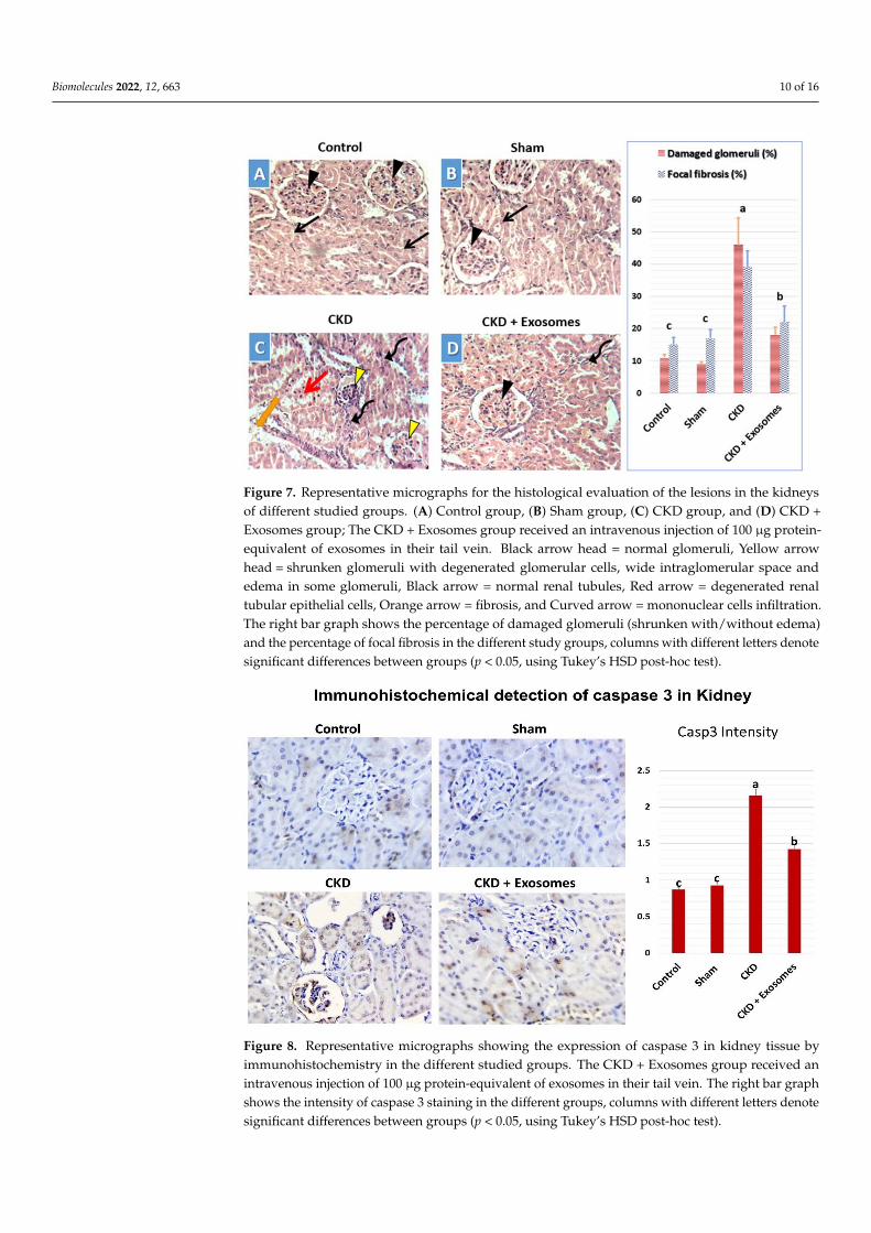
3.5. Histological Changes in Kidney Tissue
In the control (Figure 7A) and sham‐operated (Figure 7B) groups, the observed his‐
tological structure of the kidney was apparently normal, with normal renal tubules and
normal renal corpuscles. In the chronic kidney damage group (Figure 7C), the observed
kidney’s histological structure indicated signs of chronic damage, including degenerated
and shrunken renal corpuscles, which could show edema, in addition to dispersed ne‐
crotic renal tubules in between normal tubules. Moreover, perivascular mononuclear
cells’ infiltration with fibrosis in some areas was also noticed in kidneys with chronic dam‐
age (Figure 7C). Following exosome administration, the study rats showed substantially
less chronic damage signs and a more conserved kidney histological structure (Figure 7D).
This includes apparently normal renal corpuscles with less frequent degenerative changes
in both the corpuscles and tubules surrounding it. This was accompanied by a drastic
reduction in mononuclear cells infiltration and fibrosis in the treated kidneys.
Figure 6. Changes in glomerular filtration rate (GFR) and urinary protein excretion in the differentstudy groups. The CKD + Exosomes group received an intravenous injection of 100 µg protein-equivalent of exosomes in Their tail vein. Columns with different letters denote significant differencesbetween groups (p < 0.05, using Tukey’s HSD post-hoc test).
3.5. Histological Changes in Kidney Tissue
In the control (Figure 7A) and sham-operated (Figure 7B) groups, the observed his-tological structure of the kidney was apparently normal, with normal renal tubules andnormal renal corpuscles. In the chronic kidney damage group (Figure 7C), the observedkidney’s histological structure indicated signs of chronic damage, including degeneratedand shrunken renal corpuscles, which could show edema, in addition to dispersed necroticrenal tubules in between normal tubules. Moreover, perivascular mononuclear cells’ in-filtration with fibrosis in some areas was also noticed in kidneys with chronic damage(Figure 7C). Following exosome administration, the study rats showed substantially lesschronic damage signs and a more conserved kidney histological structure (Figure 7D).This includes apparently normal renal corpuscles with less frequent degenerative changesin both the corpuscles and tubules surrounding it. This was accompanied by a drasticreduction in mononuclear cells infiltration and fibrosis in the treated kidneys.
3.6. Detection of Caspase 3 Expression in the Kidney by Immunohistochemistry
The immunohistochemical detection of caspase 3 (shown in Figure 8) revealed no orvery few cells that are positive for caspase 3 in both control and sham-operated groupswith no significant difference between them. In the rats experiencing chronic kidneydamage, there was a more diffuse and stronger expression of caspase 3 in kidney tissuein the glomeruli and renal tubules. Whereas caspase 3 levels were drastically reduced inthe group receiving exosomes derived from MSCs, which was apparent in low caspase 3expression in the glomeruli and reduced expression in the renal tubular cells.
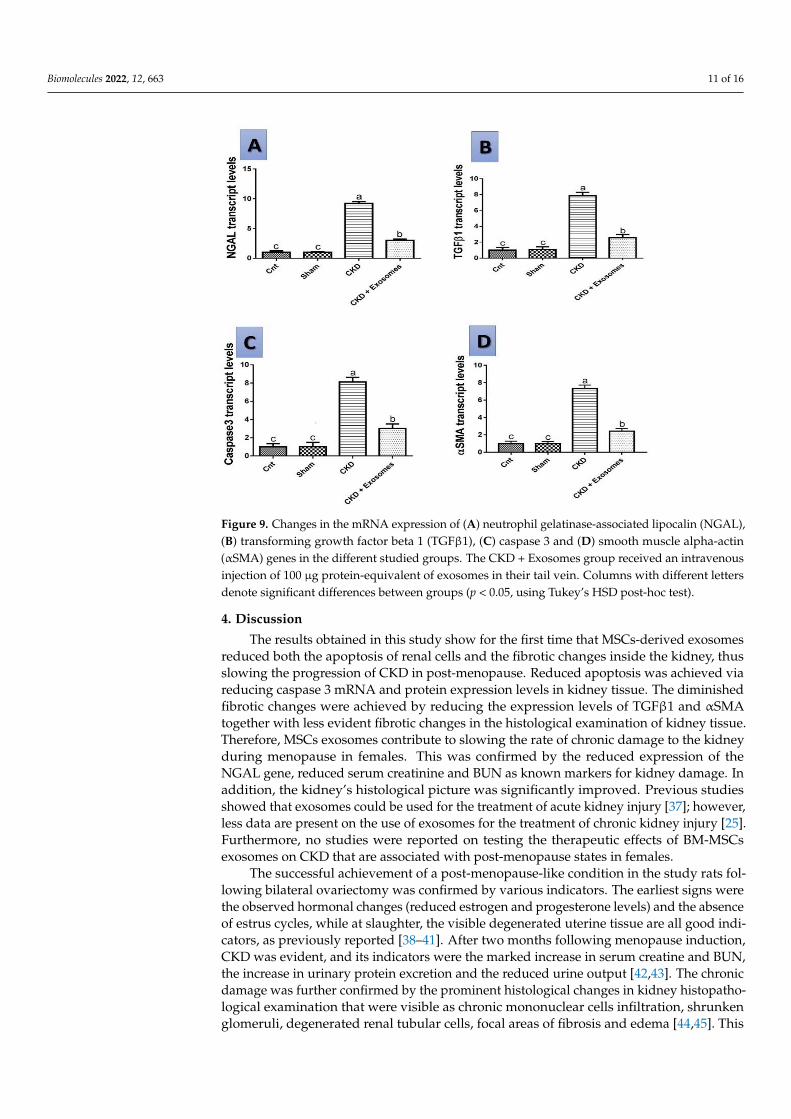
3.7. Gene Expression Results by qPCR
The level of expression of the studied genes NGAL, TGFβ1, αSMA and caspase 3 inthe different studied groups are shown in Figure 9. In the rats experiencing chronic kidneydamage, there was an upregulation of the expression of all studied genes (p < 0.01) com-pared to either the control or sham groups. However, following exosomes administration,there was a marked downregulation of the genes’ expression to reach a level slightly higherthan in the control or sham groups and significantly lower than the chronic kidney damagegroup (p < 0.05).
Biomolecules 2022, 12, 663 10 of 16Biomolecules 2022, 12, x FOR PEER REVIEW 10 of 17
Figure 7. Representative micrographs for the histological evaluation of the lesions in the kidneys of
different studied groups. (A) Control group, (B) Sham group, (C) CKD group, and (D) CKD + Exo‐
somes group; The CKD + Exosomes group received an intravenous injection of 100 μg protein‐
equivalent of exosomes in their tail vein. Black arrow head = normal glomeruli, Yellow arrow head
= shrunken glomeruli with degenerated glomerular cells, wide intraglomerular space and edema in
some glomeruli, Black arrow = normal renal tubules, Red arrow = degenerated renal tubular epithe‐
lial cells, Orange arrow = fibrosis, and Curved arrow = mononuclear cells infiltration. The right bar
graph shows the percentage of damaged glomeruli (shrunken with/without edema) and the per‐
centage of focal fibrosis in the different study groups, columns with different letters denote signifi‐
cant differences between groups (p < 0.05, using Tukey’s HSD post‐hoc test).
3.6. Detection of Caspase 3 Expression in the Kidney by Immunohistochemistry
The immunohistochemical detection of caspase 3 (shown in Figure 8) revealed no or
very few cells that are positive for caspase 3 in both control and sham‐operated groups
with no significant difference between them. In the rats experiencing chronic kidney dam‐
age, there was a more diffuse and stronger expression of caspase 3 in kidney tissue in the
glomeruli and renal tubules. Whereas caspase 3 levels were drastically reduced in the
group receiving exosomes derived from MSCs, which was apparent in low caspase 3 ex‐
pression in the glomeruli and reduced expression in the renal tubular cells.
Figure 7. Representative micrographs for the histological evaluation of the lesions in the kidneysof different studied groups. (A) Control group, (B) Sham group, (C) CKD group, and (D) CKD +Exosomes group; The CKD + Exosomes group received an intravenous injection of 100 µg protein-equivalent of exosomes in their tail vein. Black arrow head = normal glomeruli, Yellow arrowhead = shrunken glomeruli with degenerated glomerular cells, wide intraglomerular space andedema in some glomeruli, Black arrow = normal renal tubules, Red arrow = degenerated renaltubular epithelial cells, Orange arrow = fibrosis, and Curved arrow = mononuclear cells infiltration.The right bar graph shows the percentage of damaged glomeruli (shrunken with/without edema)and the percentage of focal fibrosis in the different study groups, columns with different letters denotesignificant differences between groups (p < 0.05, using Tukey’s HSD post-hoc test).
Biomolecules 2022, 12, x FOR PEER REVIEW 11 of 17
Figure 8. Representative micrographs showing the expression of caspase 3 in kidney tissue by im‐
munohistochemistry in the different studied groups. The CKD + Exosomes group received an intra‐
venous injection of 100 μg protein‐equivalent of exosomes in their tail vein. The right bar graph
shows the intensity of caspase 3 staining in the different groups, columns with different letters de‐
note significant differences between groups (p < 0.05, using Tukey’s HSD post‐hoc test).
3.7. Gene Expression Results by qPCR
The level of expression of the studied genes NGAL, TGFβ1, αSMA and caspase 3 in
the different studied groups are shown in Figure 9. In the rats experiencing chronic kidney
damage, there was an upregulation of the expression of all studied genes (p < 0.01) com‐
pared to either the control or sham groups. However, following exosomes administration,
there was a marked downregulation of the genes’ expression to reach a level slightly
higher than in the control or sham groups and significantly lower than the chronic kidney
damage group (p < 0.05).
Figure 8. Representative micrographs showing the expression of caspase 3 in kidney tissue byimmunohistochemistry in the different studied groups. The CKD + Exosomes group received anintravenous injection of 100 µg protein-equivalent of exosomes in their tail vein. The right bar graphshows the intensity of caspase 3 staining in the different groups, columns with different letters denotesignificant differences between groups (p < 0.05, using Tukey’s HSD post-hoc test).
Biomolecules 2022, 12, 663 11 of 16Biomolecules 2022, 12, x FOR PEER REVIEW 12 of 17
Figure 9. Changes in the mRNA expression of (A) neutrophil gelatinase‐associated lipocalin
(NGAL), (B) transforming growth factor beta 1 (TGFβ1), (C) caspase 3 and (D) smooth muscle alpha‐
actin (αSMA) genes in the different studied groups. The CKD + Exosomes group received an intra‐
venous injection of 100 μg protein‐equivalent of exosomes in their tail vein. Columns with different
letters denote significant differences between groups (p < 0.05, using Tukey’s HSD post‐hoc test).
4. Discussion
The results obtained in this study show for the first time that MSCs‐derived exosomes
reduced both the apoptosis of renal cells and the fibrotic changes inside the kidney, thus
slowing the progression of CKD in post‐menopause. Reduced apoptosis was achieved via
reducing caspase 3 mRNA and protein expression levels in kidney tissue. The diminished
fibrotic changes were achieved by reducing the expression levels of TGFβ1 and αSMA
together with less evident fibrotic changes in the histological examination of kidney tissue.
Therefore, MSCs exosomes contribute to slowing the rate of chronic damage to the kidney
during menopause in females. This was confirmed by the reduced expression of the
NGAL gene, reduced serum creatinine and BUN as known markers for kidney damage.
In addition, the kidney’s histological picture was significantly improved. Previous studies
showed that exosomes could be used for the treatment of acute kidney injury [37]; how‐
ever, less data are present on the use of exosomes for the treatment of chronic kidney
injury [25]. Furthermore, no studies were reported on testing the therapeutic effects of
BM‐MSCs exosomes on CKD that are associated with post‐menopause states in females.
The successful achievement of a post‐menopause‐like condition in the study rats fol‐
lowing bilateral ovariectomy was confirmed by various indicators. The earliest signs were
the observed hormonal changes (reduced estrogen and progesterone levels) and the ab‐
sence of estrus cycles, while at slaughter, the visible degenerated uterine tissue are all
good indicators, as previously reported [38–41]. After two months following menopause
induction, CKD was evident, and its indicators were the marked increase in serum crea‐
tine and BUN, the increase in urinary protein excretion and the reduced urine output
[42,43]. The chronic damage was further confirmed by the prominent histological changes
in kidney histopathological examination that were visible as chronic mononuclear cells
infiltration, shrunken glomeruli, degenerated renal tubular cells, focal areas of fibrosis and
Figure 9. Changes in the mRNA expression of (A) neutrophil gelatinase-associated lipocalin (NGAL),(B) transforming growth factor beta 1 (TGFβ1), (C) caspase 3 and (D) smooth muscle alpha-actin(αSMA) genes in the different studied groups. The CKD + Exosomes group received an intravenousinjection of 100 µg protein-equivalent of exosomes in their tail vein. Columns with different lettersdenote significant differences between groups (p < 0.05, using Tukey’s HSD post-hoc test).
4. Discussion
The results obtained in this study show for the first time that MSCs-derived exosomesreduced both the apoptosis of renal cells and the fibrotic changes inside the kidney, thusslowing the progression of CKD in post-menopause. Reduced apoptosis was achieved viareducing caspase 3 mRNA and protein expression levels in kidney tissue. The diminishedfibrotic changes were achieved by reducing the expression levels of TGFβ1 and αSMAtogether with less evident fibrotic changes in the histological examination of kidney tissue.Therefore, MSCs exosomes contribute to slowing the rate of chronic damage to the kidneyduring menopause in females. This was confirmed by the reduced expression of theNGAL gene, reduced serum creatinine and BUN as known markers for kidney damage. Inaddition, the kidney’s histological picture was significantly improved. Previous studiesshowed that exosomes could be used for the treatment of acute kidney injury [37]; however,less data are present on the use of exosomes for the treatment of chronic kidney injury [25].Furthermore, no studies were reported on testing the therapeutic effects of BM-MSCsexosomes on CKD that are associated with post-menopause states in females.
The successful achievement of a post-menopause-like condition in the study rats fol-lowing bilateral ovariectomy was confirmed by various indicators. The earliest signs werethe observed hormonal changes (reduced estrogen and progesterone levels) and the absenceof estrus cycles, while at slaughter, the visible degenerated uterine tissue are all good indi-cators, as previously reported [38–41]. After two months following menopause induction,CKD was evident, and its indicators were the marked increase in serum creatine and BUN,the increase in urinary protein excretion and the reduced urine output [42,43]. The chronicdamage was further confirmed by the prominent histological changes in kidney histopatho-logical examination that were visible as chronic mononuclear cells infiltration, shrunkenglomeruli, degenerated renal tubular cells, focal areas of fibrosis and edema [44,45]. This
Biomolecules 2022, 12, 663 12 of 16
was accompanied by reduced antioxidant reserve (GPx, SOD and CAT) in kidney tissueand altered gene expression of NGAL, TGFβ1, αSMA and caspase 3 genes that are relatedto CKD [35,46–48].
Neutrophil gelatinase-associated lipocalin (NGAL) is among the earliest markers thatwere found important to indicate renal damage [49]. In addition, serum NGAL levels werefound to be positively correlated with the severity of kidney damage and are considered tobe promising next-generation biomarkers for kidney diseases (reviewed in detail in [50]).In CKD in particular, NGAL was correlated with levels of serum creatinine, and glomerularfiltration rate, and all three indicators reflected the extent of renal damage [51]. In thepresent study, the expression of the NGAL gene was increased in the rats of the CKDgroup, agreeing with previous studies. After the treatment with exosomes, the expressionlevels of NGAL were greatly reduced and approached the normal levels in non-CKDrats. These results clearly indicate a reduction in renal damage, which was confirmedby the biochemical and histological results of the current study. In addition, NGAL wasconsidered to have a protective role against apoptosis by preventing the overexpressionof caspase 3 in the kidney [52], as the suppression of NGAL expression by siRNA led toincreased expression of caspase 3 and increased apoptosis of kidney cells.
Caspase 3 is one member of the apoptosis-associated proteases that degrade intra-cellular components and lead to cell death. It is one of the final proteases to be activatedbefore cell death and the formation of apoptotic bodies [53]. In addition, the inhibition ofcaspase 3 activation led to a reduction in both the rate of apoptosis and kidney damageand protected against acute renal failure [54]. In the present study, in the CKD group,the gene expression of caspase 3 was upregulated, as was the immunostaining of caspase3, which was localized to both the tubules as well as the glomerular cells. Our resultsare in accordance with previous studies that showed that caspase 3 levels are associatedwith apoptosis, fibrosis and chronic renal scarring (i.e., fibrosis) and were detected in thetubular cells and glomeruli [8]. Following exosome administration, in the CKD + exosomesgroup, the expression levels of both caspase 3 mRNA and protein in kidney tissue weregreatly reduced and approached the normal levels observed in the control and sham groups.The association between apoptosis, fibrosis and the progression of CKD was previouslydemonstrated where apoptosis of renal cells increased together with the rate of fibrosis inkidney tissue [7].
The fibrosis in kidney tissue, as well as in other tissues, is greatly mediated by theTGF-β pathway, and the inhibition of TGF-β is a major target for therapeutic approachesagainst chronic kidney disease associated with fibrosis [3,5] and other fibrotic diseasesin general [55]. In the kidney, the inhibition of TGF-β signaling is also associated withdecreased epithelial mesenchymal transition (EMT) and the accumulation of myofibroblastsin the kidney, thus reducing renal fibrosis and the progression of chronic kidney injury [56].EMT is also implicated in the pathogenesis of CKD and is characterized by the expressionof both TGF-β and αSMA [57], and the prevention of EMT is important for protectingrenal tissue and reducing kidney damage. In our study, the expression of TGFβ1 wasincreased in the CKD group and was associated with the increased expression of αSMA(which is an EMT and myofibroblast marker) in kidney tissue and also associated with theprominent pathological lesions observed in the kidney histological examination, whichincluded degeneration and fibrotic reactions. However, in the exosome-treated group,the expression levels of TGFβ1 and αSMA were significantly reduced and approachedthe control and sham rates; this indicates that the administered exosomes reduced theprogression of CKD in these rats, and this was also evident in the prominent histologicalimprovements in the kidneys of these rats. Other studies targeting TGFβ1 and αSMA inkidney diseases are in accordance with our results [58,59]; however, no studies have usedexosomes for the treatment of fibrosis associated with CKD during the menopause period.
MSCs were previously studied in treating diabetic nephropathy, and their favorableeffects were attributed to their paracrine factors (either soluble factors or inside extracellularvesicles; as exosomes) [60]. The role of MSCs in repairing kidney injury and fibrosis has
Biomolecules 2022, 12, 663 13 of 16
been addressed elsewhere [61,62]. However, the use of exosomes for relieving apoptosisand fibrosis in CKD has received less attention in the literature. For example, exosomesisolated from human umbilical cord blood MSCs were found to reduce renal fibrosisinduced by mechanical stress through regulating Yes-associated proteins (YAP) in kidneytissue [63] or through ROS-mediated MAPK/ERK signaling [64]. Other studies showedthat exosomes isolated from MSCs were able to protect against acute kidney injury [24,65]and reduced autophagy in diabetic nephropathy [66]. In our study, exosomes isolated frombone marrow MSCs were efficient at protecting against post-menopause CKD and theirtherapeutic use was associated with reduced kidney damage.
5. Conclusions
Our study shows for the first time that exosomes isolated from MSCs have a renalprotective role on the kidneys of menopausal rats (used as a model for post-menopauseCKD). Exosomes were found to prevent CKD progression by reducing the gene expressionlevels of NGAL, TGFβ1 and αSMA. In addition, they preserved kidney antioxidant defensemechanisms (GPx, CAT and SOD) and reduced the rate of apoptosis in kidney tissue viareducing the expression of caspase 3 mRNA genes and protein in kidney tissues. Thisled to stabilizing serum creatinine and BUN levels, reducing urine protein excretion andreducing the damaged histologic picture of the kidneys in exosome-treated rats. Therefore,the current study provides evidence for the utility of exosome therapy in preserving kidneyhealth in post-menopause females and preventing their rapid progression towards CKD.
Author Contributions: All authors (W.A.A., A.A.-H., H.M.E.-G., S.A.A., D.I., N.A.E., E.S.E.-S., A.A.S.,M.A.S.A. and H.M.) contributed equally to this manuscript (conceptualization; funding acquisition;data curation; formal analysis; investigation; methodology; resources; supervision; visualization;roles/Writing—original draft; writing—review & editing). All authors have read and agreed to thepublished version of the manuscript.
Funding: The authors would like to thank the Deanship of Scientific Research at Umm Al-QuraUniversity, for supporting this work through project number 19-MED-1-03-0014.
Institutional Review Board Statement: The study was conducted in accordance with the Declarationof Helsinki, and approved by the Institutional Animal Care and Use Committee of Zagazig University(protocol number: ZU-IACUC/2/F/164/2020).
Data Availability Statement: The data presented in this study are available in the article.
Conflicts of Interest: The authors declare no conflict of interest.
References1. Hill, N.R.; Fatoba, S.T.; Oke, J.L.; Hirst, J.A.; O’Callaghan, C.A.; Lasserson, D.S.; Hobbs, F.D.R. Global prevalence of chronic
kidney disease—A systematic review and meta-analysis. PLoS ONE 2016, 11, e0158765. [CrossRef] [PubMed]2. Suzuki, H.; Kondo, K. Chronic kidney disease in postmenopausal women. Hypertens. Res. 2012, 35, 142–147. [CrossRef] [PubMed]3. Meng, X.M.; Tang, P.M.K.; Li, J.; Lan, H.Y. TGF-ß/Smad signaling in renal fibrosis. Front. Physiol. 2015, 6, 82. [CrossRef] [PubMed]4. Chen, L.; Yang, T.; Lu, D.W.; Zhao, H.; Feng, Y.L.; Chen, H.; Chen, D.Q.; Vaziri, N.D.; Zhao, Y.Y. Central role of dysregulation of
TGF-β/Smad in CKD progression and potential targets of its treatment. Biomed. Pharm. 2018, 101, 670–681. [CrossRef] [PubMed]5. Yanagita, M. Inhibitors/antagonists of TGF-β system in kidney fibrosis. Nephrol. Dial. Transpl. 2012, 27, 3686–3691. [CrossRef]
[PubMed]6. Sugiyama, H.; Kashihara, N.; Makino, H.; Yamasaki, Y.; Ota, Z. Apoptosis in glomerular sclerosis. Kidney Int. 1996, 49, 103–111.
[CrossRef]7. Thomas, G.L.; Yang, B.; Wagner, B.E.; Savill, J.; el Nahas, A.M. Cellular apoptosis and proliferation in experimental renal fibrosis.
Nephrol. Dial. Transpl. 1998, 13, 2216–2226. [CrossRef]8. Yang, B.; el Nahas, A.M.; Thomas, G.L.; Haylor, J.L.; Watson, P.F.; Wagner, B.; Johnson, T.S. Caspase-3 and apoptosis in
experimental chronic renal scarring. Kidney Int. 2001, 60, 1765–1776. [CrossRef]9. Michels, K.B.; Manson, J.E. Postmenopausal Hormone Therapy in the 21st Century: Reconciling Findings from Observational Studies and
Randomized Clinical Trials, 3rd ed.; Lobo, R.A., Ed.; Treat. Postmenopausal Woman; Academic Press: New York, NY, USA, 2007;pp. 619–626. [CrossRef]
10. El-Gendy, A.A.; Elsaed, W.M.; Abdallah, H.I. Potential role of estradiol in ovariectomy-induced derangement of renal endocrinefunctions. Ren. Fail. 2019, 41, 507–520. [CrossRef]
Biomolecules 2022, 12, 663 14 of 16
11. Zimmerman, M.A.; Hutson, D.D.; Trimmer, E.H.; Kashyap, S.N.; Duong, J.L.; Murphy, B.; Grissom, E.M.; Daniel, J.M.; Lindsey, S.H.Long- but not short-term estradiol treatment induces renal damage in midlife ovariectomized Long-Evans rats. Am. J. Physiol.Ren. Physiol. 2017, 312, F305–F311. [CrossRef]
12. Chen, J.S.; Wong, V.W.; Gurtner, G.C. Therapeutic potential of bone marrow-derived mesenchymal stem cells for cutaneouswound healing. Front. Immunol. 2012, 3, 192. [CrossRef] [PubMed]
13. Biancone, L.; Bruno, S.; Deregibus, M.C.; Tetta, C.; Camussi, G. Therapeutic potential of mesenchymal stem cell-derivedmicrovesicles. Nephrol. Dial. Transpl. 2012, 27, 3037–3042. [CrossRef] [PubMed]
14. Patel, D.M.; Shah, J.; Srivastava, A.S. Therapeutic potential of mesenchymal stem cells in regenerative medicine. Stem Cells Int.2013, 2013, 496218. [CrossRef] [PubMed]
15. Simons, M.; Raposo, G. Exosomes—Vesicular carriers for intercellular communication. Curr. Opin. Cell Biol. 2009, 21, 575–581.[CrossRef] [PubMed]
16. Ludwig, A.K.; Giebel, B. Exosomes: Small vesicles participating in intercellular communication. Int. J. Biochem. Cell Biol. 2012, 44,11–15. [CrossRef] [PubMed]
17. Murray, L.M.A.; Krasnodembskaya, A.D. Concise Review: Intercellular Communication Via Organelle Transfer in the Biologyand Therapeutic Applications of Stem Cells. Stem Cells 2019, 37, 14–25. [CrossRef]
18. Lee, Y.; el Andaloussi, S.; Wood, M.J.A. Exosomes and microvesicles: Extracellular vesicles for genetic information transfer andgene therapy. Hum. Mol. Genet. 2012, 21, R125–R134. [CrossRef]
19. Bruno, S.; Chiabotto, G.; Camussi, G. Concise Review: Different Mesenchymal Stromal/Stem Cell Populations Reside in theAdult Kidney. Stem Cells Transl. Med. 2014, 3, 1451–1455. [CrossRef]
20. Klinkhammer, B.M.; Kramann, R.; Mallau, M.; Makowska, A.; van Roeyen, C.R.; Rong, S.; Buecher, E.B.; Boor, P.; Kovacova, K.;Zok, S.; et al. Mesenchymal Stem Cells from Rats with Chronic Kidney Disease Exhibit Premature Senescence and Loss ofRegenerative Potential. PLoS ONE 2014, 9, e92115. [CrossRef]
21. Lin, K.C.; Yip, H.K.; Shao, P.L.; Wu, S.C.; Chen, K.H.; Chen, Y.T.; Yang, C.C.; Sun, C.K.; Kao, G.S.; Chen, S.Y.; et al. Combination ofadipose-derived mesenchymal stem cells (ADMSC) and ADMSC-derived exosomes for protecting kidney from acute ischemia-reperfusion injury. Int. J. Cardiol. 2016, 216, 173–185. [CrossRef]
22. Cao, J.; Wang, B.; Tang, T.; Lv, L.; Ding, Z.; Li, Z.; Hu, R.; Wei, Q.; Shen, A.; Fu, Y.; et al. Three-dimensional culture of MSCsproduces exosomes with improved yield and enhanced therapeutic efficacy for cisplatin-induced acute kidney injury. Stem CellRes. Ther. 2020, 11, 206. [CrossRef] [PubMed]
23. Lee, J.H.; Ha, D.H.; Go, H.K.; Youn, J.; Kim, H.K.; Jin, R.C.; Miller, R.B.; Kim, D.H.; Cho, B.S.; Yi, Y.W. Reproducible Large-ScaleIsolation of Exosomes from Adipose Tissue-Derived Mesenchymal Stem/Stromal Cells and Their Application in Acute KidneyInjury. Int. J. Mol. Sci. 2020, 21, 4774. [CrossRef] [PubMed]
24. Gao, F.; Zuo, B.; Wang, Y.; Li, S.; Yang, J.; Sun, D. Protective function of exosomes from adipose tissue-derived mesenchymal stemcells in acute kidney injury through SIRT1 pathway. Life Sci. 2020, 255, 117719. [CrossRef] [PubMed]
25. Nowak, N.; Yamanouchi, M.; Satake, E. The Nephroprotective Properties of Extracellular Vesicles in Experimental Models ofChronic Kidney Disease: A Systematic Review. Stem Cell Rev. Rep. 2022, 18, 902–932. [CrossRef] [PubMed]
26. Soleimani, M.; Nadri, S. A protocol for isolation and culture of mesenchymal stem cells from mouse bone marrow. Nat. Protoc.2009, 4, 102–106. [CrossRef] [PubMed]
27. Alzahrani, F.A.; El-Magd, M.A.; Abdelfattah-Hassan, A.; Saleh, A.A.; Saadeldin, I.M.; El-Shetry, E.S.; Badawy, A.A.; Alkarim, S.Potential Effect of Exosomes Derived from Cancer Stem Cells and MSCs on Progression of DEN-Induced HCC in Rats. Stem CellsInt. 2018, 2018, 8058979. [CrossRef]
28. el Nashar, E.M.; Alghamdi, M.A.; Alasmari, W.A.; Hussein, M.M.A.; Hamza, E.; Taha, R.I.; Ahmed, M.M.; Al-Khater, K.M.;Abdelfattah-Hassan, A. Autophagy Promotes the Survival of Adipose Mesenchymal Stem/Stromal Cells and Enhances TheirTherapeutic Effects in Cisplatin-Induced Liver Injury via Modulating TGF-β1/Smad and PI3K/AKT Signaling Pathways. Cells2021, 10, 2475. [CrossRef]
29. Lobb, R.J.; Becker, M.; Wen, S.W.; Wong, C.S.F.; Wiegmans, A.P.; Leimgruber, A.; Möller, A. Optimized exosome isolation protocolfor cell culture supernatant and human plasma. J. Extracell. Vesicles 2015, 4, 27031. [CrossRef]
30. Steele, M.S.; Bennett, R.A. Clinical Technique: Dorsal Ovariectomy in Rodents. J. Exot. Pet Med. 2011, 20, 222–226. [CrossRef]31. Vorland, C.J.; Lachcik, P.J.; Swallow, E.A.; Metzger, C.E.; Allen, M.R.; Chen, N.X.; Moe, S.M.; Gallant, K.M.H. Effect of ovariectomy
on the progression of chronic kidney disease-mineral bone disorder (CKD-MBD) in female Cy/+ rats. Sci. Rep. 2019, 9, 7936.[CrossRef]
32. Ke, H.Z.; Chen, H.K.; Simmons, H.A.; Qi, H.; Crawford, D.T.; Pirie, C.M.; Chidsey-Frink, K.L.; Ma, Y.F.; Jee, W.S.S.; Thompson, D.D.Comparative effects of droloxifene, tamoxifen, and estrogen on bone, serum cholesterol, and uterine histology in the ovariec-tomized rat model. Bone 1997, 20, 31–39. [CrossRef]
33. Abdelfattah-Hassan, A.; Shalaby, S.I.; Khater, S.I.; El-Shetry, E.S.; el Fadil, H.A.; Elsayed, S.A. Panax ginseng is superior to vitaminE as a hepatoprotector against cyclophosphamide-induced liver damage. Complement. Ther. Med. 2019, 46, 95–102. [CrossRef][PubMed]
34. Han, M.; Li, Y.; Liu, M.; Li, Y.; Cong, B. Renal neutrophil gelatinase associated lipocalin expression in lipopolysaccharide-inducedacute kidney injury in the rat. BMC Nephrol. 2012, 13, 25. [CrossRef] [PubMed]
Biomolecules 2022, 12, 663 15 of 16
35. Hwang, I.; Uddin, M.J.; Lee, G.; Jiang, S.; Pak, E.S.; Ha, H. Peroxiredoxin 3 deficiency accelerates chronic kidney injury in micethrough interactions between macrophages and tubular epithelial cells. Free Radic. Biol. Med. 2019, 131, 162–172. [CrossRef][PubMed]
36. Adil, M.; Kandhare, A.D.; Visnagri, A.; Bodhankar, S.L. Naringin ameliorates sodium arsenite-induced renal and hepatic toxicityin rats: Decisive role of KIM-1, Caspase-3, TGF-β, and TNF-α. Ren. Fail. 2015, 37, 1396–1407. [CrossRef] [PubMed]
37. Thongboonkerd, V. Roles for exosome in various kidney diseases and disorders. Front. Pharm. 2020, 10, 1655. [CrossRef][PubMed]
38. Wise, P.M.; Rainer, A. Effect of Ovariectomy on Plasma LH, FSH, Estradiol, and Progesterone and Medial Basal HypothalamicLHRH Concentrations in Old and Young Rats. Neuroendocrinology 1980, 30, 15–19. [CrossRef]
39. Wronski, T.J.; Schenk, P.A.; Cintrón, M.; Walsh, C.C. Effect of body weight on osteopenia in ovariectomized rats. Calcif. Tissue Int.1987, 40, 155–159. [CrossRef]
40. Li, L.H.; Wang, Z.C.; Yu, J.; Zhang, Y.Q. Ovariectomy Results in Variable Changes in Nociception, Mood and Depression in AdultFemale Rats. PLoS ONE 2014, 9, e94312. [CrossRef]
41. Hao, F.; Gu, Y.; Tan, X.; Deng, Y.; Wu, Z.T.; Xu, M.J.; Wang, W.Z. Estrogen Replacement Reduces Oxidative Stress in the RostralVentrolateral Medulla of Ovariectomized Rats. Oxid. Med. Cell. Longev. 2016, 2016, 2158971. [CrossRef]
42. Diwan, V.; Small, D.; Kauter, K.; Gobe, G.C.; Brown, L. Gender differences in adenine-induced chronic kidney disease andcardiovascular complications in rats. Am. J. Physiol. Ren. Physiol. 2014, 307, F1169–F1178. [CrossRef] [PubMed]
43. Li, T.; Gua, C.; Wu, B.; Chen, Y. Increased circulating trimethylamine N-oxide contributes to endothelial dysfunction in a ratmodel of chronic kidney disease. Biochem. Biophys. Res. Commun. 2018, 495, 2071–2077. [CrossRef] [PubMed]
44. Nangaku, M.; Pippin, J.; Couser, W.G. C6 Mediates Chronic Progression of Tubulointerstitial Damage in Rats with RemnantKidneys. J. Am. Soc. Nephrol. 2002, 13, 928–936. [CrossRef]
45. Al Za’abi, M.; Al Salam, S.; Al Suleimani, Y.; Ashique, M.; Manoj, P.; Nemmar, A.; Ali, B.H. Effects of repeated increasing doses ofcisplatin as models of acute kidney injury and chronic kidney disease in rats. Naunyn. Schmiedebergs. Arch. Pharm. 2021, 394,249–259. [CrossRef]
46. Chow, J.; Hartley, R.B.; Jagger, C.; Dilly, S.A. ICAM-1 expression in renal disease. J. Clin. Pathol. 1992, 45, 880. [CrossRef][PubMed]
47. Cho, K.H.; Kim, H.J.; Rodriguez-Iturbe, B.; Vaziri, N.D. Niacin ameliorates oxidative stress, inflammation, proteinuria, andhypertension in rats with chronic renal failure. Am. J. Physiol. Ren. Physiol. 2009, 297, 106–113. [CrossRef]
48. Karadeniz, A.; Simsek, N.; Karakus, E.; Yildirim, S.; Kara, A.; Can, I.; Kisa, F.; Emre, H.; Turkeli, M. Royal jelly modulates oxidativestress and apoptosis in liver and kidneys of rats treated with cisplatin. Oxid. Med. Cell. Longev. 2011, 2011, 981793. [CrossRef]
49. Supavekin, S.; Zhang, W.; Kucherlapati, R.; Kaskel, F.J.; Moore, L.C.; Devarajan, P. Differential gene expression following earlyrenal ischemia/reperfusion. Kidney Int. 2003, 63, 1714–1724. [CrossRef]
50. Bolignano, D.; Donato, V.; Coppolino, G.; Campo, S.; Buemi, A.; Lacquaniti, A.; Buemi, M. Neutrophil Gelatinase–AssociatedLipocalin (NGAL) as a Marker of Kidney Damage. Am. J. Kidney Dis. 2008, 52, 595–605. [CrossRef]
51. Mori, K.; Nakao, K. Neutrophil gelatinase-associated lipocalin as the real-time indicator of active kidney damage. Kidney Int.2007, 71, 967–970. [CrossRef]
52. Han, M.; Li, Y.; Wen, D.; Liu, M.; Ma, Y.; Cong, B. NGAL protects against endotoxin-induced renal tubular cell damage bysuppressing apoptosis. BMC Nephrol. 2018, 19, 168. [CrossRef] [PubMed]
53. Han, Z.; Hendrickson, E.A.; Bremner, T.A.; Wyche, J.H. A Sequential Two-Step Mechanism for the Production of the Maturep17:p12 Form of Caspase-3 In Vitro. J. Biol. Chem. 1997, 272, 13432–13436. [CrossRef] [PubMed]
54. Guo, R.; Wang, Y.; Minto, A.W.; Quigg, R.J.; Cunningham, P.N. Acute Renal Failure in Endotoxemia is Dependent on CaspaseActivation. J. Am. Soc. Nephrol. 2004, 15, 3093–3102. [CrossRef] [PubMed]
55. Sureshbabu, A.; Muhsin, S.A.; Choi, M.E. TGF-β signaling in the kidney: Profibrotic and protective effects. Am. J. Physiol. Ren.Physiol. 2016, 310, F596. [CrossRef] [PubMed]
56. Zeisberg, M.; Hanai, J.I.; Sugimoto, H.; Mammoto, T.; Charytan, D.; Strutz, F.; Kalluri, R. BMP-7 counteracts TGF-β1–inducedepithelial-to-mesenchymal transition and reverses chronic renal injury. Nat. Med. 2003, 9, 964–968. [CrossRef]
57. Bolati, D.; Shimizu, H.; Higashiyama, Y.; Nishijima, F.; Niwa, T. Indoxyl Sulfate Induces Epithelial-to-Mesenchymal Transition inRat Kidneys and Human Proximal Tubular Cells. Am. J. Nephrol. 2011, 34, 318–323. [CrossRef]
58. Cho, J.H.; Ryu, H.M.; Oh, E.J.; Yook, J.M.; Ahn, J.S.; Jung, H.Y.; Choi, J.Y.; Park, S.H.; Kim, Y.L.; Kwak, I.S.; et al. Alpha1-AntitrypsinAttenuates Renal Fibrosis by Inhibiting TGF-β1-Induced Epithelial Mesenchymal Transition. PLoS ONE 2016, 11, e0162186.[CrossRef]
59. Xiao, Q.; Guan, Y.; Li, C.; Liu, L.; Zhao, D.; Wang, H. Decreased expression of transforming growth factor-β1 and α-smoothmuscle actin contributes to the protection of lotensin against chronic renal failure in rats. Ren. Fail. 2018, 40, 583. [CrossRef]
60. Nagaishi, K.; Mizue, Y.; Chikenji, T.; Otani, M.; Nakano, M.; Konari, N.; Fujimiya, M. Mesenchymal stem cell therapy amelioratesdiabetic nephropathy via the paracrine effect of renal trophic factors including exosomes. Sci. Rep. 2016, 6, 34842. [CrossRef]
61. Kuppe, C.; Kramann, R. Role of mesenchymal stem cells in kidney injury and fibrosis. Curr. Opin. Nephrol. Hypertens. 2016, 25,372–377. [CrossRef]
62. Zhuang, Q.; Ma, R.; Yin, Y.; Lan, T.; Yu, M.; Ming, Y. Mesenchymal Stem Cells in Renal Fibrosis: The Flame of Cytotherapy. StemCells Int. 2019, 2019, 8387350. [CrossRef]

Biomolecules 2022, 12, 663 16 of 16
63. Ji, C.; Zhang, J.; Zhu, Y.; Shi, H.; Yin, S.; Sun, F.; Wang, Q.; Zhang, L.; Yan, Y.; Zhang, X.; et al. Exosomes derived from hucMSCattenuate renal fibrosis through CK1δ/β-TRCP-mediated YAP degradation. Cell Death Dis. 2020, 11, 327. [CrossRef] [PubMed]
64. Liu, B.; Hu, D.; Zhou, Y.; Yu, Y.; Shen, L.; Long, C.; Butnaru, D.; Timashev, P.; He, D.; Lin, T.; et al. Exosomes released by humanumbilical cord mesenchymal stem cells protect against renal interstitial fibrosis through ROS-mediated P38MAPK/ERK signalingpathway. Am. J. Transl. Res. 2020, 12, 4998. [PubMed]
65. Ji, C.; Zhang, J.; Zhou, Z.; Shi, H.; Liu, W.; Sun, F.; Zhang, C.; Zhang, L.; Sun, Z.; Qian, H. Platelet-rich plasma promotes MSCsexosomes paracrine to repair acute kidney injury via AKT/Rab27 pathway. Am. J. Transl. Res. 2021, 13, 1445. [PubMed]
66. Ebrahim, N.; Ahmed, I.A.; Hussien, N.I.; Dessouky, A.A.; Farid, A.S.; Elshazly, A.M.; Mostafa, O.; El Gazzar, W.B.; Sorour, S.M.;Seleem, Y.; et al. Mesenchymal Stem Cell-Derived Exosomes Ameliorated Diabetic Nephropathy by Autophagy Inductionthrough the mTOR Signaling Pathway. Cells 2018, 7, 226. [CrossRef]