exosomes: emerging players of intercellular communication in tumor microenvironment
TRANSCRIPT
Exosome mediated intercellular crosstalk in TME
www.discoveriesjournals.org 1
PERSPECTIVE Article
Exosomes: Emerging Players of Intercellular Communication
in Tumor Microenvironment Shrankhla Maheshwari
#,1,2, Anup Kumar Singh
#,1, Rakesh Kumar Arya
#,1, Deepti Pandey
1,
Akhilesh Singh1, Dipak Datta
1,2,*
1Biochemistry Division, CSIR-Central Drug Research Institute (CDRI), Lucknow-226031, India
2Academy of Scientific and Innovative Research, New Delhi, India
# These authors contributed equally to this work;
*Corresponding author: Dipak Datta, Biochemistry Division, CSIR-CDRI, B.S. 10/1, Sector 10, Jankipuram
Extension, Sitapur Road, Lucknow- 226031, India, Tel: 91-522-2772450 (Extn-4347/48), Fax: 191-522-
2771941, E-mail: [email protected]
Submitted: August 1, 2014; Revised: Sept. 24, 2014; Accepted: Sept. 25, 2014; Published: Sept. 25, 2014;
Citation: Maheshwari S, Singh AK, Arya RK, Pandey D, Singh A, Datta D. Exosomes: Emerging Players of
Intercellular Communication in Tumor Microenvironment. Discoveries 2014, Jul-Sep; 2(3): e26. DOI:
10.15190/d.2014.18
ABSTRACT
Seminal discoveries have established the role of
complex tumor microenvironment (TME) in cancer
progression; and later on also uncovered that
vesiculation is an integral part of intercellular
communication among various cell types in
coordinating the tumor assembly in a dynamic
manner. Exosomes are small membrane bound
endosomal vesicles, which are classically known
for their role in discarding cellular wastes; however,
recent reports underlined their novel role in
malignancy by their release from cells into the
TME. Since then, the role of exosomes have been a
subject of increasing interest, as exosome mediated
intercellular communications offer a novel
reciprocal relationship between cancer and stromal
cells within the TME and modulate the fate and
function of the recipient cells to finally shape the
tumor progression. Exosomes are characterised by
different features including size, content and mode
of delivery; and its cargo delivers interesting
bioactive components in the form of proteins,
miRNAs or other molecules to the target cell. In the
pursuit of further study of exosomes, it was found
that with the help of its distinct bioactive
components, exosomes specifically regulate tumor
growth, angiogenesis, metastasis as well as drug
resistance properties. In fact, it acts as a bridge
between different signaling networks, present inside
the spatially distant cells of the heterogeneous
tumor population. In the current endeavour, we
have highlighted the role of exosomes in
modulating the intercellular crosstalk during tumor
growth and progression, and proposed certain novel
roles of exosomes to address the few enigmatic
questions of cancer cell biology.
Keywords:
Exosome, Tumor Microenvironment, Angiogenesis,
Metastasis, Drug Resistance
Abbreviations: Extracellular vesicles (EV); Tumor Microenvironment
(TME); Carcinoma-associated fibroblasts (CAFs);
Tumor-associated macrophages (TAM); Transforming
growth factor-beta (TGF-β); Vascular endothelial growth
factor (VEGF); Endothelial cell (EC); Matrix
metalloproteinases (MMPs); Heat shock proteins (HSPs);
Cancer stem cells (CSCs); Microvesicles (MVs);
Epithelial-mesenchymal transition (EMT); Multi drug
resistance-1 (MDR-1); Multidrug resistance associated
protein (MRP); ATP-binding cassette transporter (ABC
transporter)
DISCOVERIES 2014, Jul-Sep, 2(3): e26 DOI: 10.15190/d.2014.18
Exosome mediated intercellular crosstalk in TME
www.discoveriesjournals.org 2
♦ Exosomes comprise of bioactive components such as proteins, RNAs, miRNAs
or lipids, and play a pivotal role in modulating tumor niche by controlling
intercellular crosstalk.
♦ Exosomes mediated intercellular communications and signals within TME
regulate tumor growth, angiogenesis, metastasis and drug resistance.
1. Introduction
Cell secretion is a widely accepted phenomenon
and a fundamental process for the maintenance of
physiological functions of a cell.1 It is accompanied
mostly by microvesicles or exosome like vesicles
which were previously known by many names
depending on their cellular origin: ectosome,
oncosome, texosome, prostasome, epididymosome
and dexosome.2, 3
The concept of extracellular
vesicles (EV) being introduced, in 1980s by
Johnstone’s and Stahl’s groups, for secretory
vesicles originating from endosome in
multivesicular bodies with a function of clearing
transferrin receptor during reticulocyte maturation.
However, the word ‘exosome’ was proposed for the
first time for EV of endosomal origin in 1987.4-6
Exosomes are small lipid bilayer vesicles secreted
by most but not all types of cells in their
microenvironment. Besides their well known role in
discarding waste material from the cells, exosomes
play an important function in maintaining normal as
well as pathological processes.7 In case of tumor
development and progression, exosome has
emerged as an important mediator of cellular
communication and opened up a window that
extends our understanding about how certain
secretory vesicles perform a critical function of
transferring genetic material, induce epigenetic
changes, modulate immune response to manipulate
the local and systemic tumor environment to
regulate cancer growth and dissemination. Current
endeavour is an attempt to shed light on the
emerging role of tumor-secreted exosomes as a
novel player to modulate the tumor
microenvironment during carcinogenesis.
2. Exosomes: Isolation and Characterization
Cells secrete different kinds of vesicles in the body
fluids, which vary in their biogenesis as well as in
biophysical properties. Exosomes have certain
characteristic features including their size (40 to
100 nm) and morphology that distinguish them
from other EVs.8 Though the role of exosomes in
regulating the fate of other cells within the tissue is
now widely accepted, controversies exist for the
terminology in the field of EV research and they are
sometimes misleading per se in the context of their
functionality. As an attempt to address such
controversies, releasing mechanism of EVs in the
extracellular environment has been used as the
criteria for its nomenclature. On this basis, EVs can
be categorized into (i) exosomes: 40-100 nm
diameter membranous vesicles of endocytic origin
released by exocytic fusion of multivesicular bodies
to plasma membrane, (ii) ectosomes (also referred
to as shedding microvesicles): large membranous
vesicles of 50-1000 nm diameter that are shed
directly from the plasma membrane, and (iii)
apoptotic blebs (50-5000 nm diameter): released by
dying cells.9-13
Similar to other vesicles, exosomes
contain integrins, selectins but unlike others, they
also harbour annexins, Rab proteins, SNAREs (v-
SNARE and t-SNARE), tetraspanins like CD9,
CD82, CD81, CD63 etc. and enrichment in lipids
like cholesterol, sphingolipids, ceramides and
glycerophospholipids with long saturated fatty acyl
chains.8, 14-17
Recent studies have shown that exosomes can
be isolated in vivo from body fluids such as blood,
urine, breast milk, amniotic fluid, malignant ascites,
bronchoalveolar lavage fluid and synovial fluid
etc.18-25
These vesicles can be isolated by various
techniques including differential ultracentrifugation,
which can be sometimes combined with 0.1 µm to
0.22 µm filtration in order to separate the nano-
sized particles from larger particles and cellular
debris.26
Immuno-affinity technique using beads
against tetraspanins, CD63 or CD82, or other origin
specific exosomal markers is also used to isolate
these vesicles.27
Unfortunately, the purification
methods are often confusing in distinguishing
exosomes from other EVs present as cross
Exosome mediated intercellular crosstalk in TME
www.discoveriesjournals.org 3
contaminations.9 Microscopy based estimation of
exosomes has its own limitation of fixation and
dehydration, that may result in shrinkage and hence
underestimation of actual size. However, certain
modern techniques like nanoparticle based tracking
analysis can be used to overcome these problems by
analyzing them in PBS based on their Brownian
motion in suspension to avoid shrinkage.28, 29
As
conventional flow cytometers cannot differentiate
between vesicles that are less than 300 nm, a novel
high resolution flow cytometry–based method has
been discovered for a quantitative high throughput
analysis of individual (immuno-labelled) nano-sized
vesicles.30-32
Cargo material of an exosome may include
lipids, proteins (oncoproteins, tumor suppressor
proteins and functional transmembrane proteins),
nucleic acids (mRNA, miRNA, DNA), growth
receptors, and soluble factors.33-40
Various
techniques including western blot, mass
spectrometry, fluorescence activated cell sorting
and immuno-electron microscopy can be used to
analyse the exosomal content.41
At a given time
point, exosomes do not represent the snapshot of
transcriptome of parental cells but exhibit a
selective cargo profiles.42-44
For example, exosomes
from Glioma represent a different repertoire of
microRNAs compared to their cells of origin, with
abundance of unusual or novel non-coding RNAs of
unknown functions.45
Furthermore, it has been
found that there is a low correlation between
exosomal miRNA content of metastatic SKOV-3
cells versus non-metastatic OVCAR-3 cells. This
dictates that specific export of miRNAs may have
some potential tumor-specific modulatory role.46
3. Exosomes: Release and Uptake It is well established that exosomes are released by
the cells after fusion of multivesicular bodies with
the plasma membrane, but the molecular
mechanism of their release to the microenvironment
is still poorly understood.47
It has been found that a
number of Rab family proteins, such as Rab22a,
Rab27a, Rab27b and Rab35, R-SNARE protein
YKT6, glyco-sphingolipids and flotillins, as well as
ceramides, are involved in the regulation of their
secretion.17, 48-52
Nevertheless, the involvement of
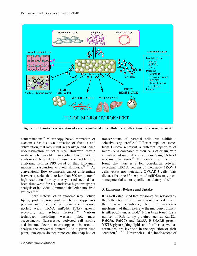
Figure 1: Schematic representation of exosome mediated intercellular crosstalk in tumor microenvironment
Exosome mediated intercellular crosstalk in TME
www.discoveriesjournals.org 4
these proteins in exosome formation is dependent
on cell types. Exosomal release has been found to
be dependent on several physico-chemical factors
like calcium, hypoxia, chemotherapeutic drug
exposure, temperature and oxidative stress etc.44, 53-
55 Recently, a group of scientists explored a novel
feedback mechanism to control the exosomal
release in a cell type specific manner. They found
that exosomes from normal mammary epithelial
cells at a similar concentration had a dramatic
inhibitory effect on exosome production by breast cancer cells compared to exosomes from bladder
cancer cells. They also proposed a dynamic
equilibrium between the release and uptake of
exosomes from and to the surrounding medium,
depending on their concentration in extracellular
environment (Figure 1).56
The exosomal uptake is carried out by either
endocytosis or phagocytosis or micro-pinocytosis.57-
59 In 2009, Parolini et al. demonstrated another way
of exosome import to receiving cells by lipid
dependant membrane fusion through "lipid rafts" of
exosomal membrane. They also suggested that low
pH microenvironment increases the uptake of
exosomes as well as their release from the cancer
cells, probably because of the intrinsically endowed
negative charge due to high lipid content in
exosomes. This could be the possible reason behind
the cancer cells to secrete higher amount of
exosomes compared to their normal counterpart and
their preferential delivery to metastatic tumor cells
rather than primary tumor site.60
4. Exosomes and Tumor Microenvironment
(TME)
TME is composed of various cells which include
stromal cells like endothelial cells, carcinoma-
associated fibroblasts (CAFs), adipocytes,
mesenchymal cells, immune cells (tumor-associated
macrophages or TAM, T-cells etc.), as well as
extracellular matrix which encircle all tumor cells,
and soluble factors such as growth factors,
chemokines and cytokines.61
Besides genetic and
epigenetic control, cells of the TME also possess a
remodelling influence on the tumor growth and its
transformation from benign to malignant. For tumor
propagation, cellular communication is not only
required between the tumor cells but also between
cancer cells and the other neighbouring cells in
TME. However, such intercellular communication
can occur with or without cell-to-cell contact.
Contact independent communication may take
place via endocrine signaling or as an alternative;
exosomes represent another way of distant cellular
communication. This exosomal cross talk between
resident cells imparts strong influence on inherent
complexity of TME by regulating and moulding
various oncogenic signaling pathways.
4.1 Role in Tumor Growth
In a normal cellular homeostatic condition, cells
maintain a competitive environment for deleterious
cells to eradicate them from the local niche but
during tumor initiation, transformed cells dominate
over normal cells. Recent experimental evidence
suggest an important role of exosome mediated
intercellular communication in this tug of war of
normal versus tumorigenic cells (Figure 1).62
As for
example, exosomal miR-143 derived from non-
cancerous cells have the ability to suppress the
growth of cancer cells both in vitro and in vivo.63
In
contrast, breast cancer cell (MDA-MB-231,
T47D:A18 and MCF-7) derived exosomes
manipulate epithelial cells of the mammary duct to
facilitate tumor development.64
During tumor growth, Wnt signaling play a
pivotal role where Wnt ligands disperse spatially,
however, the mechanism of their dispersal remained
enigmatic. Recently, exosome mediated transfer of
proteins resolve the riddle of how the hydrophobic
Wnts traffic over long distances. In this context,
Wnt3A has been found to be present on the surface
of exosomes which explains a putative mechanism
of Wnt transport to the distant recipient cells.51
Tumor suppressor Phosphatase and tensin homolog
(PTEN) is a well established intracellular signaling
molecule, but, interestingly, it can also be
transported outside the cells via exosomal cargo and
impart growth inhibitory function.35, 65
Tumor derived exosomes can also influence
the immune system by triggering the
immunosuppressive responses, in order to favor
tumor progression. These exosomes can stimulate
the expansion of regulatory T-cells, which in turn
impair the function of anti-tumorigenic T-cells in
TME.66
It has also been found that Transforming
growth factor-beta (TGF-β) present on the surface
of exosomes influences the immunosuppressive
effects of regulatory T-cells.67
Tumor derived
microvesicles (MVs) also carry Fas ligands and
Exosome mediated intercellular crosstalk in TME
www.discoveriesjournals.org 5
TRAIL on their surface, which indicates their
apoptosis inducing effect on activated T-cells.68, 69
Moreover, enzymes of exosomes negatively
regulate T-cell activation by hydrolysis of ATP into
adenosine.70
Secretory exosomes also reduce the
proliferation of Natural Killer (NK) cells or block
the Interleukin 2-mediated activation of NK cells.71,
72
4.2 Role in Angiogenesis
The process of angiogenesis requires a balance
between pro- and anti-angiogenic factors in order to
disseminate the endothelial progenitor cells to the
vasculogenic site. Among the various factors,
vascular endothelial growth factor (VEGF) and
their receptors play a crucial role in maintaining the
vascular homeostasis. During tumorigenesis, TAMs
secrete VEGF, which in turn leads to
vascularization to sustain tumor growth.
Interestingly, microvesicle- delivery of antisense
miR-150 into the mice decreased the secretion of
VEGF by TAMs via targeting ING4.73
Although the
expression of EGFR on endothelial cells has been
controversial, recently it has been found that
exosomes facilitate a path for the expression of
EGFR on the HUVEC as well as tumor associated
endothelial cells, through phosphotidylserine
mediated fusion between endothelial cell membrane
and exosomes.74
The process of angiogenesis also
requires the activation of matrix metalloproteinases
(MMPs) for the degradation of basement
membrane, liberation of angiogenic factors, and
sprouting of the capillaries. Tumor cells
overexpress membrane bound molecule CD147,
which is an extracellular MMPs inducer. Studies
have shown that cancer cells (lung carcinoma, colon
carcinoma, and pancreatic cancers) produce large
amounts of CD147-positive MVs75, 76
that can
interact with the cells of TME and stimulate the
production of MMPs. It is also observed that
CD147-positive secretory vesicles of ovarian cancer
cells promote angiogenic phenotype in endothelial
cells (HUVECs) and pre-treatment of siRNA
against CD147 suppressed their angiogenic
potential.77
Induction of endothelial cell (EC)
migration and angiogenesis is mediated by the
CCR1, CCL20, CXCL5, and MIF expression in
ECs as evident from co-culturing them with
ASTspan8 carrying exosomes.78
Interestingly, it has
also been found that D6.1A (tetraspanin Tspan8)-
expressing tumor cells’ supernatant as well as
D6.1A-containing exosomes, strongly induce
angiogenesis.79
Additionally, it is also reported that hypoxic
condition enhances the release of exosomes, which
further promote microvascular endothelial cell
migration and vasculogenesis. For example,
hypoxia triggers the release of TF/VIIa bearing
exosomes, which in turn increase the pro-
angiogenic growth factor HB-EGF in ECs via
ERK1/2-PAR2 dependent pathway.80
As an explanation of its capability to induce
angiogenesis, exosomes has been found to modulate
the many signaling pathways in a surprisingly novel
mode. For instance, Notch signaling is an
evolutionary conserved pathway that requires cell-
to-cell contact for ligand-receptor interaction and
further induction of cascade of its characteristics
events. It has been shown that exosomes have
signaling potential by transferring Dll-4 (ligand of
notch receptor) to the neighbouring cells and
incorporate it into the plasma membrane in vitro
and in vivo. They have the ability to inhibit Notch
signaling in vitro and appear to switch the
endothelial cell phenotype toward tip cells
phenotype, which in turn enhance vessel density.29
Interestingly, not every cell within the
heterogenous tumor population secretes similar
types of MVs, rather specific stem like cells; called
cancer stem cells (CSCs) profoundly secrete
specific MVs. For example, in the case of renal
cancer, CD105 positive cells (CSCs) secrete MVs
containing higher CD105. On co-culturing with
HUVEC, only those MVs which are secreted by
CSCs are able to form capillary like structures on
matrigel and are able to enhance invasiveness of
ECs. Furthermore, RNase pre-treatment of MVs
reduced the above capabilities, which indicates that
the content of these vesicles are mostly RNA
molecules including miRNAs.81
In this direction, it
was further found that exogenous miR-9, previously
reported to promote tumor cell motility and
metastasis by repressing E-cadherin expression and
increase VEGF transcription,82
enhances EC
migration and angiogenesis when exosomaly
transferred to HUVECs. This exogenous miR-9
effectively reduced SOCS5 levels, leading to
activation of JAK-STAT pathway.83
Exosomes
from colon cancer cells are able to induce mitosis
after incorporation into endothelial cell (HUVECs)
cytoplasm, which has been shown by

Exosome mediated intercellular crosstalk in TME
www.discoveriesjournals.org 6
immunostaining of both phospho-histone H3
(mitosis marker) and α-tubulin (mitotic spindle
marker) in MV-treated HUVECs compared to
control HUVECs.84
4.3 Role in Metastasis
Cancer metastasis involves the “leak”, or “spill” of
potential cancer cells from the primary tumor, and
settle down to other tissues in the body to establish
a full blown secondary tumor.85
It requires a
cooperative interaction of tumor cells with non-
tumor cells in the TME. Exosome mediated
intercellular crosstalk plays a critical role in this
coordinated process (Figure 1). Different tumor
cells have distinct metastatic potential due to their
genetic instability and exosomes are found to
influence this genetic instability among tumor cells
by transferring oncogenic sequences.34
Exosomes
are also found to enhance the metastatic potential of
less metastatic melanoma cells B16-F1cells by
transferring a metastasis marker (Met 72 tumor
antigen) from highly metastatic B16 melanoma
cells BL6-10.86
For successful evasion through extracellular
matrix, tumor cells secrete MMPs or activators of
MMPs like heat shock proteins (HSPs) for
extracellular matrix remodelling. HSPs are also
secreted through exosomes along with other
proteins. It has been found that HSP90α together
with annexin II in exosomes impart cell motility to
their cancer cells via an interaction with an
extracellular tissue plasminogen activator (tPA),
which in turn activates protease plasmin.87
Similar
to extracellular matrix remodelling, vascular
destabilization at the pre-metastatic niche is
indispensable for cancer cell dissemination from the
primary tumor site. Recently, exosomal miR-105
has been found to enhance tumor migration through
enhancing vascular permeability by targeting tight
junction protein ZO-1 (zonula occludens-1) in
vascular endothelial cells.88
The role of tumor
derived exosomes in promoting metastatic potential
of cancer cells is also supported by another elegant
study in which, poorly metastasizing ASML-
CD44vkd (CD44v-knockdown rat pancreatic
adenocarcinoma BSp73ASML (ASMLwt) cells)
cells regain metastatic capacity, when pre-treated
with conditioned medium of ASMLwt cells
containing exosomes. These exosomes target
stromal and other cells of pre-metastatic organs to
prepare metastatic niche to thrive tumor cells
predominantly by transferring miRNAs (exosomal
miR-494 and miR-542-3p which target cadherin-
17).89
Furthermore Jung et al. also showed that
conditioned media collected from ASMLwt, but not
ASML-CD44vkd tumor cells promoted lymph node
and lung metastasis of pancreatic cancer.
Fractionation of conditioned media revealed that
exosomes are the key factors which require CD44v
for assembling soluble matrix.90
CAFs, an abundant stromal cell in the TME,
support tumor growth by secreting several growth
factors. They are found to stimulate breast cancer
cells’ protrusive activity by exosomally transferring
their endogenous Wnt11 to activate Wnt-planar cell
polarity (PCP) in breast cancer cells.91
These
fibroblasts can be converted into myofibroblastic
cells to support tumor growth, vascularization and
metastasis. It has been shown that exosomal TGF-β
was able to develop myofibroblastic phenotype in
fibroblasts, through TGF-β-SMAD dependent
signalling.92
However, such differentiation was also
found to occur in adipose derived mesenchymal
stem cells via exosome mediated pathway in breast
and ovarian cancer cells.93, 94
Recently, in an in vitro
study, it has been demonstrated that 786-0 renal
cancer cell derived exosomes increased migration
and invasion capacity of these cells by decreasing
the adhesion ability and increasing the expression
levels of CXCR4 and MMP-9.95
Moreover,
metastasis promoting epithelial-mesenchymal
transition (EMT) related factors, such as vimentin,
hepatoma-derived growth factor (HDGF) were
found in the plasma membrane and annexin 2,
CK2α, and moesin in the lumen of exosomes of
bladder cancer respectively, suggesting their crucial
involvement in metastatic process.96
4.4 Role in Drug Resistance
In the growing area of cancer research and
treatment, development of chemoresistance is a
decisive challenge for chemotherapy. Decreased
drug uptake, increased drug efflux, activation of
detoxifying systems, activation of DNA repair
mechanisms and evasion of drug-induced apoptosis
etc. are several defence mechanisms, which cancer
cells can develop against the chemotherapy.
However, the underlying molecular mechanism for
chemoresistance still remains unclear. Thus, a more
efficient strategy is required to target cancer cells,
Exosome mediated intercellular crosstalk in TME
www.discoveriesjournals.org 7
♦ Exosomal biogenesis, sorting, secretion and discriminative uptake from the
microenvironmental exosomal pool by recipient cells are emerging areas and
future directions of research.
which are smarter than originally believed. Genetic
and molecular studies have shown that most of the
malignant cancer cells have amplified multi drug
resistance (MDR) 1 and multidrug resistance
associated protein (MRP) genes, which are the
members of ATP-binding cassette transporter (ABC
transporter) superfamily.97
Now, a series of studies
dictate that tumor derived exosomes added the layer
of complexity in the complex nature of cancer by
transmission of resistance from resistant cells to
sensitive ones (Figure 1). Bebawy et al., showed the
transmission of functional P-glycoprotein (P-gp)
(MDR1) from drug resistant cancer cells (VLB100)
to drug sensitive cancer cells (CCRF–CEM) over a
co-culture period of 4 hours. They suggested that
the expression of P-gp in recipient cell is not
transcriptionally induced thus establishing a ‘non-
genetic’ mechanism whereby MVs serve as a vector
in the acquisition and spreading of MDR.98
Recently, Wei-xian Chen et al. added another piece
of evidence in transferring drug resistance via
targeting MAPK pathway (putative target) through
exosomal miRNAs of docetaxel resistant MCF-7
cells.99
In a study with pulse chase and flow
cytometric experimentation, vesicle shedding was
represented as a potential mechanism of drug
(doxorubicin) expulsion, which is found to be
proportional to dose concentration.100
Similarly,
cisplatin (CDDP) was also found to expel out via
exosomal pathway by CDDP-resistant cells, which
possessed more putative CDDP transporters like
ATP7A, ATP7B, and MRP2 (ABCB2) in order to
escape from chemotherapeutic pressure. However,
the routing of intercellular CDDP to exosomal
pathway is not clear yet.101
It has been shown that
adriamycin-resistant MCF-7 cells’ derived MVs
aided the resistance in recipient cells by transferring
Ca2+
permeable channel TrpC5, which after
incorporation induced the expression of P-gp.102
Exosome-antibody interaction is another way
by which cancer cells evade chemotherapeutic
pressure. It has been found that exosome antibody
sequestration reduces the antibody-dependent
cytotoxicity in cancer cells by immune effector
cells.103
Moreover, HER2-overexpressing breast
cancer cell lines express a full-length HER2
molecule on exosomes, which can bind to the
HER2 antibody Trastuzumab to nullify its effect on
tumor proliferation.104
Thus, it seems that
exosomes, besides their permissive role in tumor
growth, metastasis and angiogenesis, also program
the tumor cells towards resistance to chemotherapy.
5. Conclusion and Future Direction
Exosomes are not just cellular debris but have a
functional importance in cancer biology. In a pliant
TME, where every cell communicates with each
other through various ways, exosomes represent a
new pathway to transport the information from
donor to recipient cells. As an intercellular player,
exosomes exhibit their ability to promote tumor
growth, metastasis niche formation and provoking
angiogenesis by carrying their specific cargo.
Although, extensive researches on exosomes from
the past few years have revealed their various new
roles in cancer progression, a deep understanding of
their biogenesis, sorting, secretion and uptake are
still in its infancy. Moreover, few questions also
remain obscured including “how recipient cells
discriminate between the exosomes they need to
take from a cohort of exosomes in a
microenvironmental exosomal pool”. Thus, a better
understanding is warranted for selective exosomal
uptake by recipient cells, in order to exert a specific
stimulation.
Conflict of interest
Authors have no potential conflict of interest.
Acknowledgement
This work was supported by different CSIR
network projects and fellowships grants from CSIR,
UGC, and MOES.
References:
1. Jena BP. Cell secretion and membrane fusion.
Domestic animal endocrinology 2005; 29(1): 145-
165.

Exosome mediated intercellular crosstalk in TME
www.discoveriesjournals.org 8
2. Simpson RJ, Jensen SS, Lim JW. Proteomic
profiling of exosomes: current perspectives.
Proteomics 2008; 8(19): 4083-4099.
3. Dinkla S, Brock R, Joosten I, Bosman GJ. Gateway
to understanding microparticles: standardized
isolation and identification of plasma membrane-
derived vesicles. Nanomedicine (Lond) 2013; 8(10):
1657-1668.
4. Pan BT, Johnstone RM. Fate of the transferrin
receptor during maturation of sheep reticulocytes in
vitro: selective externalization of the receptor. Cell
1983; 33(3): 967-978.
5. Harding C, Heuser J, Stahl P. Endocytosis and
intracellular processing of transferrin and colloidal
gold-transferrin in rat reticulocytes: demonstration
of a pathway for receptor shedding. Eur J Cell Biol
1984; 35(2): 256-263.
6. Johnstone RM, Adam M, Hammond JR, Orr L,
Turbide C. Vesicle formation during reticulocyte
maturation. Association of plasma membrane
activities with released vesicles (exosomes). J Biol
Chem 1987; 262(19): 9412-9420.
7. Lee Y, Andaloussi SE, Wood MJ. Exosomes and
microvesicles: extracellular vesicles for genetic
information transfer and gene therapy. Human
molecular genetics 2012; 21(R1): R125-R134.
8. Simons M, Raposo G. Exosomes–vesicular carriers
for intercellular communication. Current Opinion in
Cell Biology 2009; 21(4): 575-581.
9. Simpson R, Mathivanan S. Extracellular
microvesicles: the need for internationally
recognised nomenclature and stringent purification
criteria. J Proteomics Bioinform 2012; 5(2).
10. Mathivanan S. Quest for cancer biomarkers:
assaying mutant proteins and RNA that provides the
much needed specificity. J Proteomics Bioinform
2012; 5, xiii-xvii. DOI: 10.4172/jpb.10000e16
11. Mathivanan S, Ji H, Simpson RJ. Exosomes:
extracellular organelles important in intercellular
communication. Journal of Proteomics 2010;
73(10): 1907-1920.
12. Hess C, Sadallah S, Hefti A, Landmann R, Schifferli
J-A. Ectosomes released by human neutrophils are
specialized functional units. The Journal of
Immunology 1999; 163(8): 4564-4573.
13. Heijnen HF, Schiel AE, Fijnheer R, Geuze HJ,
Sixma JJ. Activated Platelets Release Two Types of
Membrane Vesicles: Microvesicles by Surface
Shedding and Exosomes Derived From Exocytosis
of Multivesicular Bodies and alpha-Granules. Blood
1999; 94(11): 3791-3799.
14. Théry C, Zitvogel L, Amigorena S. Exosomes:
composition, biogenesis and function. Nature
Reviews Immunology 2002; 2(8): 569-579.
15. Wubbolts R, Leckie RS, Veenhuizen PT,
Schwarzmann G, Möbius W, Hoernschemeyer J, et
al. Proteomic and biochemical analyses of human B
cell-derived exosomes. Potential implications for
their function and multivesicular body formation. J
Biol Chem 2003; 278(13): 10963-10972.
16. Subra C, Laulagnier K, Perret B, Record M.
Exosome lipidomics unravels lipid sorting at the
level of multivesicular bodies. Biochimie 2007;
89(2): 205-212.
17. Trajkovic K, Hsu C, Chiantia S, Rajendran L,
Wenzel D, Wieland F, et al. Ceramide triggers
budding of exosome vesicles into multivesicular
endosomes. Science 2008; 319(5867): 1244-1247.
18. Mathivanan S, Fahner CJ, Reid GE, Simpson RJ.
ExoCarta 2012: database of exosomal proteins,
RNA and lipids. Nucleic acids research 2012;
40(D1): D1241-D1244.
19. Caby M-P, Lankar D, Vincendeau-Scherrer C,
Raposo G, Bonnerot C. Exosomal-like vesicles are
present in human blood plasma. International
Immunology 2005; 17(7): 879-887.
20. Pisitkun T, Shen R-F, Knepper MA. Identification
and proteomic profiling of exosomes in human
urine. Proceedings of the National Academy of
Sciences of the United States of America 2004;
101(36): 13368-13373.
21. Admyre C, Johansson SM, Qazi KR, Filén J-J,
Lahesmaa R, Norman M, et al. Exosomes with
immune modulatory features are present in human
breast milk. The Journal of Immunology 2007;
179(3): 1969-1978.
22. Taylor DD, Akyol S, Gercel-Taylor C. Pregnancy-
associated exosomes and their modulation of T cell
signaling. The Journal of Immunology 2006; 176(3):
1534-1542.
23. Bard MP, Hegmans JP, Hemmes A, Luider TM,
Willemsen R, Severijnen L-AA, et al. Proteomic
analysis of exosomes isolated from human
malignant pleural effusions. American Journal of
Respiratory Cell and Molecular Biology 2004;
31(1): 114-121.
24. Admyre C, Grunewald J, Thyberg J, Gripenbäck S,
Tornling G, Eklund A, et al. Exosomes with major
histocompatibility complex class II and co-
stimulatory molecules are present in human BAL
fluid. European Respiratory Journal 2003; 22(4):
578-583.
25. Skriner K, Adolph K, Jungblut P, Burmester G.
Association of citrullinated proteins with synovial
exosomes. Arthritis & Rheumatism 2006; 54(12):
3809-3814.
26. Théry C, Amigorena S, Raposo G, Clayton A.
Isolation and characterization of exosomes from cell
culture supernatants and biological fluids. Current
Protocols in Cell Biology 2006: Chapter 3:Unit 3.22.
DOI: 10.1002/0471143030.cb0322s30.
Exosome mediated intercellular crosstalk in TME
www.discoveriesjournals.org 9
27. Hannafon BN, Ding W-Q. Intercellular
communication by exosome-derived microRNAs in
cancer. International Journal of Molecular Sciences
2013; 14(7): 14240-14269.
28. Kanno T, Yamada T, Iwabuki H, Tanaka H, Kuroda
Si, Tanizawa K, et al. Size distribution measurement
of vesicles by atomic force microscopy. Analytical
Biochemistry 2002; 309(2): 196-199.
29. Sheldon H, Heikamp E, Turley H, Dragovic R,
Thomas P, Oon CE, et al. New mechanism for
Notch signaling to endothelium at a distance by
Delta-like 4 incorporation into exosomes. Blood
2010; 116(13): 2385-2394.
30. Raposo G, Stoorvogel W. Extracellular vesicles:
exosomes, microvesicles, and friends. The Journal
of Cell Biology 2013; 200(4): 373-383.
31. Hoen EN, van der Vlist EJ, Aalberts M, Mertens
HC, Bosch BJ, Bartelink W, et al. Quantitative and
qualitative flow cytometric analysis of nanosized
cell-derived membrane vesicles. Nanomedicine:
Nanotechnology, Biology and Medicine 2012; 8(5):
712-720.
32. van der Vlist EJ, Nolte EN, Stoorvogel W,
Arkesteijn GJ, Wauben MH. Fluorescent labeling of
nano-sized vesicles released by cells and subsequent
quantitative and qualitative analysis by high-
resolution flow cytometry. Nature Protocols 2012;
7(7): 1311-1326.
33. Record M, Carayon K, Poirot M, Silvente-Poirot S.
Exosomes as new vesicular lipid transporters
involved in cell–cell communication and various
pathophysiologies. Biochimica et Biophysica Acta
(BBA)-Molecular and Cell Biology of Lipids 2014;
1841(1): 108-120.
34. Balaj L, Lessard R, Dai L, Cho Y-J, Pomeroy SL,
Breakefield XO, et al. Tumour microvesicles
contain retrotransposon elements and amplified
oncogene sequences. Nature Communications 2011;
2: 180.
35. Putz U, Howitt J, Doan A, Goh C-P, Low L-H, Silke
J, et al. The tumor suppressor PTEN is exported in
exosomes and has phosphatase activity in recipient
cells. Science Signaling 2012; 5(243): ra70.
36. Valadi H, Ekström K, Bossios A, Sjöstrand M, Lee
JJ, Lötvall JO. Exosome-mediated transfer of
mRNAs and microRNAs is a novel mechanism of
genetic exchange between cells. Nat Cell Biol 2007;
9(6): 654-659.
37. Mack M, Kleinschmidt A, Brühl H, Klier C, Nelson
PJ, Cihak J, et al. Transfer of the chemokine
receptor CCR5 between cells by membrane-derived
microparticles: a mechanism for cellular human
immunodeficiency virus 1 infection. Nat Med 2000;
6(7): 769-775.
38. Sanderson MP, Keller S, Alonso A, Riedle S,
Dempsey PJ, Altevogt P. Generation of novel,
secreted epidermal growth factor receptor
(EGFR/ErbB1) isoforms via metalloprotease-
dependent ectodomain shedding and exosome
secretion. Journal of Cellular Biochemistry 2008;
103(6): 1783-1797.
39. Esser J, Gehrmann U, D'Alexandri FL, Hidalgo-
Estévez AM, Wheelock CE, Scheynius A, et al.
Exosomes from human macrophages and dendritic
cells contain enzymes for leukotriene biosynthesis
and promote granulocyte migration. Journal of
Allergy and Clinical Immunology 2010; 126(5):
1032-1040. e1034.
40. Yu J, Rak J. Shedding of tissue factor (TF)-
containing microparticles rather than alternatively
spliced TF is the main source of TF activity released
from human cancer cells. Journal of Thrombosis and
Haemostasis 2004; 2(11): 2065-2067.
41. Taylor DD, Gercel-Taylor C. The origin, function,
and diagnostic potential of RNA within extracellular
vesicles present in human biological fluids.
Frontiers in Genetics 2013; 4.
42. Hessvik NP, Phuyal S, Brech A, Sandvig K,
Llorente A. Profiling of microRNAs in exosomes
released from PC-3 prostate cancer cells. Biochim
Biophys Acta 2012; 1819(11-12): 1154-1163.
43. Ohshima K, Inoue K, Fujiwara A, Hatakeyama K,
Kanto K, Watanabe Y, et al. Let-7 microRNA
family is selectively secreted into the extracellular
environment via exosomes in a metastatic gastric
cancer cell line. PLoS One 2010; 5(10): e13247.
44. King HW, Michael MZ, Gleadle JM. Hypoxic
enhancement of exosome release by breast cancer
cells. BMC Cancer 2012; 12: 421.
45. Li CC, Eaton SA, Young PE, Lee M, Shuttleworth
R, Humphreys DT, et al. Glioma microvesicles carry
selectively packaged coding and non-coding RNAs
which alter gene expression in recipient cells. RNA
Biology 2013; 10(8): 1333.
46. Kobayashi M, Salomon C, Tapia J, Illanes SE,
Mitchell MD, Rice GE. Ovarian cancer cell
invasiveness is associated with discordant exosomal
sequestration of Let-7 miRNA and miR-200. J
Transl Med 2014; 12: 4.
47. van Niel G, Porto-Carreiro I, Simoes S, Raposo G.
Exosomes: a common pathway for a specialized
function. Journal of Biochemistry 2006; 140(1): 13-
21.
48. Wang T, Gilkes DM, Takano N, Xiang L, Luo W,
Bishop CJ, et al. Hypoxia-inducible factors and
RAB22A mediate formation of microvesicles that
stimulate breast cancer invasion and metastasis.
Proceedings of the National Academy of Sciences
2014: 201410041.
49. Ostrowski M, Carmo NB, Krumeich S, Fanget I,
Raposo G, Savina A, et al. Rab27a and Rab27b
Exosome mediated intercellular crosstalk in TME
www.discoveriesjournals.org 10
control different steps of the exosome secretion
pathway. Nature Cell Biology 2009; 12(1): 19-30.
50. Hsu C, Morohashi Y, Yoshimura S-i, Manrique-
Hoyos N, Jung S, Lauterbach MA, et al. Regulation
of exosome secretion by Rab35 and its GTPase-
activating proteins TBC1D10A–C. The Journal of
Cell Biology 2010; 189(2): 223-232.
51. Gross JC, Chaudhary V, Bartscherer K, Boutros M.
Active Wnt proteins are secreted on exosomes.
Nature Cell Biology 2012; 14(10): 1036-1045.
52. Phuyal S, Hessvik NP, Skotland T, Sandvig K,
Llorente A. Regulation of exosome release by
glycosphingolipids and flotillins. FEBS Journal
2014; 281(9): 2214-2227.
53. Savina A, Furlán M, Vidal M, Colombo MI.
Exosome release is regulated by a calcium-
dependent mechanism in K562 cells. Journal of
Biological Chemistry 2003; 278(22): 20083-20090.
54. Lv L-H, Wan Y-L, Lin Y, Zhang W, Yang M, Li G-
L, et al. Anticancer drugs cause release of exosomes
with heat shock proteins from human hepatocellular
carcinoma cells that elicit effective natural killer cell
antitumor responses in vitro. Journal of Biological
Chemistry 2012; 287(19): 15874-15885.
55. Hedlund M, Nagaeva O, Kargl D, Baranov V,
Mincheva-Nilsson L. Thermal-and oxidative stress
causes enhanced release of NKG2D ligand-bearing
immunosuppressive exosomes in
leukemia/lymphoma T and B cells. PloS One 2011;
6(2): e16899.
56. Riches A, Campbell E, Borger E, Powis S.
Regulation of exosome release from mammary
epithelial and breast cancer cells–A new regulatory
pathway. European Journal of Cancer 2014; 50(5):
1025-1034.
57. Morelli AE, Larregina AT, Shufesky WJ, Sullivan
ML, Stolz DB, Papworth GD, et al. Endocytosis,
intracellular sorting, and processing of exosomes by
dendritic cells. Blood 2004; 104(10): 3257-3266.
58. Feng D, Zhao WL, Ye YY, Bai XC, Liu RQ, Chang
LF, et al. Cellular internalization of exosomes
occurs through phagocytosis. Traffic 2010; 11(5):
675-687.
59. Fitzner D, Schnaars M, van Rossum D,
Krishnamoorthy G, Dibaj P, Bakhti M, et al.
Selective transfer of exosomes from
oligodendrocytes to microglia by macropinocytosis.
J Cell Sci 2011; 124(Pt 3): 447-458.
60. Parolini I, Federici C, Raggi C, Lugini L, Palleschi
S, De Milito A, et al. Microenvironmental pH is a
key factor for exosome traffic in tumor cells.
Journal of Biological Chemistry 2009; 284(49):
34211-34222.
61. Castells M, Thibault B, Delord J-P, Couderc B.
Implication of tumor microenvironment in
chemoresistance: tumor-associated stromal cells
protect tumor cells from cell death. International
Journal of Molecular Sciences 2012; 13(8): 9545-
9571.
62. Wagstaff L, Kolahgar G, Piddini E. Competitive cell
interactions in cancer: a cellular tug of war. Trends
in cell biology 2013; 23(4): 160-167.
63. Kosaka N, Iguchi H, Yoshioka Y, Hagiwara K,
Takeshita F, Ochiya T. Competitive interactions of
cancer cells and normal cells via secretory
microRNAs. Journal of Biological Chemistry 2012;
287(2): 1397-1405.
64. Dutta S, Warshall C, Bandyopadhyay C, Dutta D,
Chandran B. Interactions between Exosomes from
Breast Cancer Cells and Primary Mammary
Epithelial Cells Leads to Generation of Reactive
Oxygen Species Which Induce DNA Damage
Response, Stabilization of p53 and Autophagy in
Epithelial Cells. PloS One 2014; 9(5): e97580.
65. Dumitrascu GR, Bucur O. Critical physiological and
pathological functions of Forkhead Box O tumor
suppressors. Discoveries 2013; 1(1): e5.
66. Szajnik M, Czystowska M, Szczepanski MJ,
Mandapathil M, Whiteside TL. Tumor-derived
microvesicles induce, expand and up-regulate
biological activities of human regulatory T cells
(Treg). PloS One 2010; 5(7): e11469.
67. WADA J, Onishi H, Suzuki H, Yamasaki A, Nagai
S, Morisaki T, et al. Surface-bound TGF-β1 on
effusion-derived exosomes participates in
maintenance of number and suppressive function of
regulatory T-cells in malignant effusions. Anticancer
Research 2010; 30(9): 3747-3757.
68. Andreola G, Rivoltini L, Castelli C, Huber V,
Perego P, Deho P, et al. Induction of lymphocyte
apoptosis by tumor cell secretion of FasL-bearing
microvesicles. The Journal of Experimental
Medicine 2002; 195(10): 1303-1316.
69. Huber V, Fais S, Iero M, Lugini L, Canese P,
Squarcina P, et al. Human colorectal cancer cells
induce T-cell death through release of proapoptotic
microvesicles: role in immune escape.
Gastroenterology 2005; 128(7): 1796-1804.
70. Clayton A, Al-Taei S, Webber J, Mason MD, Tabi
Z. Cancer exosomes express CD39 and CD73,
which suppress T cells through adenosine
production. The Journal of Immunology 2011;
187(2): 676-683.
71. Whiteside TL. Immune modulation of T-cell and
NK (natural killer) cell activities by TEXs (tumour-
derived exosomes). Biochemical Society
transactions 2013; 41(1): 245.
72. Liu C, Yu S, Zinn K, Wang J, Zhang L, Jia Y, et al.
Murine mammary carcinoma exosomes promote
tumor growth by suppression of NK cell function.
The Journal of Immunology 2006; 176(3): 1375-
1385.
Exosome mediated intercellular crosstalk in TME
www.discoveriesjournals.org 11
73. Liu Y, Zhao L, Li D, Yin Y, Zhang CY, Li J, et al.
Microvesicle-delivery miR-150 promotes
tumorigenesis by up-regulating VEGF, and the
neutralization of miR-150 attenuate tumor
development. Protein Cell 2013; 4(12): 932-941.
74. Al-Nedawi K, Meehan B, Kerbel RS, Allison AC,
Rak J. Endothelial expression of autocrine VEGF
upon the uptake of tumor-derived microvesicles
containing oncogenic EGFR. Proceedings of the
National Academy of Sciences 2009; 106(10): 3794-
3799.
75. Sidhu SS, Mengistab AT, Tauscher AN, LaVail J,
Basbaum C. The microvesicle as a vehicle for
EMMPRIN in tumor–stromal interactions.
Oncogene 2004; 23(4): 956-963.
76. Baj-Krzyworzeka M, Szatanek R, Węglarczyk K,
Baran J, Urbanowicz B, Brański P, et al. Tumour-
derived microvesicles carry several surface
determinants and mRNA of tumour cells and
transfer some of these determinants to monocytes.
Cancer Immunology, Immunotherapy 2006; 55(7):
808-818.
77. Millimaggi D, Mari M, D'Ascenzo S, Carosa E,
Jannini EA, Zucker S, et al. Tumor vesicle-
associated CD147 modulates the angiogenic
capability of endothelial cells. Neoplasia 2007; 9(4):
349-357..
78. Nazarenko I, Rana S, Baumann A, McAlear J,
Hellwig A, Trendelenburg M, et al. Cell surface
tetraspanin Tspan8 contributes to molecular
pathways of exosome-induced endothelial cell
activation. Cancer Research 2010; 70(4): 1668-
1678.
79. Gesierich S, Berezovskiy I, Ryschich E, Zöller M.
Systemic induction of the angiogenesis switch by
the tetraspanin D6. 1A/CO-029. Cancer Research
2006; 66(14): 7083-7094.
80. Svensson KJ, Kucharzewska P, Christianson HC,
Sköld S, Löfstedt T, Johansson MC, et al. Hypoxia
triggers a proangiogenic pathway involving cancer
cell microvesicles and PAR-2–mediated heparin-
binding EGF signaling in endothelial cells.
Proceedings of the National Academy of Sciences
2011; 108(32): 13147-13152.
81. Grange C, Tapparo M, Collino F, Vitillo L,
Damasco C, Deregibus MC, et al. Microvesicles
released from human renal cancer stem cells
stimulate angiogenesis and formation of lung
premetastatic niche. Cancer Research 2011; 71(15):
5346-5356.
82. Ma L, Young J, Prabhala H, Pan E, Mestdagh P,
Muth D, et al. miR-9, a MYC/MYCN-activated
microRNA, regulates E-cadherin and cancer
metastasis. Nature Cell Biology 2010; 12(3): 247-
256.
83. Zhuang G, Wu X, Jiang Z, Kasman I, Yao J, Guan
Y, et al. Tumour-secreted miR-9 promotes
endothelial cell migration and angiogenesis by
activating the JAK-STAT pathway. EMBO J 2012;
31(17): 3513-3523.
84. Hong BS, Cho J-H, Kim H, Choi E-J, Rho S, Kim J,
et al. Colorectal cancer cell-derived microvesicles
are enriched in cell cycle-related mRNAs that
promote proliferation of endothelial cells. BMC
Genomics 2009; 10(1): 556.
85. Kedrin D, van Rheenen J, Hernandez L, Condeelis J,
Segall JE. Cell motility and cytoskeletal regulation
in invasion and metastasis. Journal of Mammary
Gland Biology and Neoplasia 2007; 12(2-3): 143-
152.
86. Hao S, Ye Z, Li F, Meng Q, Qureshi M, Yang J, et
al. Epigenetic transfer of metastatic activity by
uptake of highly metastatic B16 melanoma cell-
released exosomes. Exp Oncol 2006; 28(2): 126-
131.
87. McCready J, Sims JD, Chan D, Jay DG. Secretion of
extracellular hsp90α via exosomes increases cancer
cell motility: a role for plasminogen activation. BMC
Cancer 2010; 10(1): 294.
88. Zhou W, Fong MY, Min Y, Somlo G, Liu L,
Palomares MR, et al. Cancer-Secreted miR-105
Destroys Vascular Endothelial Barriers to Promote
Metastasis. Cancer Cell 2014; 25(4): 501-515.
89. Rana S, Malinowska K, Zöller M. Exosomal tumor
microRNA modulates premetastatic organ cells.
Neoplasia 2013; 15(3): 281-IN231.
90. Jung T, Castellana D, Klingbeil P, Hernández IC,
Vitacolonna M, Orlicky DJ, et al. CD44v6
dependence of premetastatic niche preparation by
exosomes. Neoplasia 2009; 11(10): 1093-IN1017.
91. Luga V, Zhang L, Viloria-Petit AM, Ogunjimi AA,
Inanlou MR, Chiu E, et al. Exosomes mediate
stromal mobilization of autocrine Wnt-PCP
signaling in breast cancer cell migration. Cell 2012;
151(7): 1542-1556.
92. Webber J, Steadman R, Mason MD, Tabi Z, Clayton
A. Cancer exosomes trigger fibroblast to
myofibroblast differentiation. Cancer Research
2010; 70(23): 9621-9630.
93. Cho JA, Park H, Lim EH, Lee KW. Exosomes from
breast cancer cells can convert adipose tissue-
derived mesenchymal stem cells into myofibroblast-
like cells. International Journal of Oncology 2012;
40(1): 130-138.
94. Cho JA, Park H, Lim EH, Kim KH, Choi JS, Lee
JH, et al. Exosomes from ovarian cancer cells
induce adipose tissue-derived mesenchymal stem
cells to acquire the physical and functional
characteristics of tumor-supporting myofibroblasts.
Gynecologic Oncology 2011; 123(2): 379-386.
Exosome mediated intercellular crosstalk in TME
www.discoveriesjournals.org 12
95. Chen G, Zhang Y, Wu X. 786-0 Renal cancer cell
line-derived exosomes promote 786-0 cell migration
and invasion in vitro. Oncology Letters 2014; 7(5):
1576.
96. Jeppesen DK, Nawrocki A, Jensen SG, Thorsen K,
Whitehead B, Howard KA, et al. Quantitative
proteomics of fractionated membrane and lumen
exosome proteins from isogenic metastatic and
nonmetastatic bladder cancer cells reveal differential
expression of EMT factors. Proteomics 2014; 14(6):
699-712.
97. Gillet J-P, Gottesman MM. Mechanisms of
multidrug resistance in cancer. Springer, 2010.
98. Bebawy M, Combes V, Lee E, Jaiswal R, Gong J,
Bonhoure A, et al. Membrane microparticles
mediate transfer of P-glycoprotein to drug sensitive
cancer cells. Leukemia 2009; 23(9): 1643-1649.
99. Chen W-x, Cai Y-q, Lv M-m, Chen L, Zhong S-l,
Ma T-f, et al. Exosomes from docetaxel-resistant
breast cancer cells alter chemosensitivity by
delivering microRNAs. Tumor Biology 2014: 1-11.
100. Shedden K, Xie XT, Chandaroy P, Chang YT,
Rosania GR. Expulsion of small molecules in
vesicles shed by cancer cells association with gene
expression and chemosensitivity profiles. Cancer
Research 2003; 63(15): 4331-4337.
101. Safaei R, Larson BJ, Cheng TC, Gibson MA, Otani
S, Naerdemann W, et al. Abnormal lysosomal
trafficking and enhanced exosomal export of
cisplatin in drug-resistant human ovarian carcinoma
cells. Molecular Cancer Therapeutics 2005; 4(10):
1595-1604.
102. Ma X, Chen Z, Hua D, He D, Wang L, Zhang P, et
al. Essential role for TrpC5-containing extracellular
vesicles in breast cancer with chemotherapeutic
resistance. Proceedings of the National Academy of
Sciences 2014; 111(17): 6389-6394.
103. Battke C, Ruiss R, Welsch U, Wimberger P, Lang S,
Jochum S, et al. Tumour exosomes inhibit binding
of tumour-reactive antibodies to tumour cells and
reduce ADCC. Cancer Immunology, Immunotherapy
2011; 60(5): 639-648.
104.Ciravolo V, Huber V, Ghedini GC, Venturelli E,
Bianchi F, Campiglio M, et al. Potential role of
HER2-overexpressing exosomes in countering
trastuzumab-based therapy. Journal of Cellular
Physiology 2012; 227(2): 658-667.
DISCOVERIES is a peer-reviewed, open access,
online, multidisciplinary and integrative journal,
publishing high impact and innovative manuscripts
from all areas related to MEDICINE, BIOLOGY and
CHEMISTRY; © 2014, Applied Systems