an assay for intercellular adhesive specificity
TRANSCRIPT
AN ASSAY FOR INTERCELLULAR
ADHESIVE SPECIFICITY
STEPHEN ROTH, EDWARD J . McGUIRE, and SAUL ROSEMAN
From the Department of Biology and the McCollum-Pratt Institute, The Johns Hopkins University,Baltimore, Maryland 21213
ABSTRACT
A modification of an assay for intercellular adhesive specificity is described . The methodinvolves the collection of radioactively labeled cells by aggregates of the same (isotypicaggregates) or different (heterotypic aggregates) types of tissue and determination of thenumber of cells collected by liquid scintillation counting. The use of 32P to label the tissuespermitted a much more rapid estimation of cell collection than was obtained previously.With the use of chick embryo neural retina, liver, forebrain, pectoral muscle, and heartventricle tissue, it was shown that isotypic was always greater than heterotypic collection .
Labeled neural retina cell collection by neural retina aggregates was studied as a func-tion of time, cell suspension density, aggregate diameter, temperature, and aggregatenumber. Neural retina aggregates were treated with certain enzymes in an attempt todetermine whether specific changes on the surface of the aggregates would interfere withlabeled neural retina cell collection . Of the various proteases and glycosidases tested, onlyß-galactosidase rendered the surface more nonspecific .
Intercellular adhesion apparently plays a key rolein diverse biological phenomena (14) . Studiesfrom a number of laboratories have defined manybiological parameters of the adhesive process :cells can adhere to glass or other nonbiologicalsubstrata (5), to each other in a specific or in anonspecific manner (10), and can "sort out"when mixed with different cell types (13) .
Neither the specific nor the nonspecific ad-hesive process has been explained in chemicalterms, although we have recently shown thatcomplex carbohydrates are involved in the inter-cellular adhesion of mouse teratoma cells (8) .The present studies were aimed at defining thesubstance(s) on the surfaces of chicken embryonicneural retina cells required for the specific adhe-sion of these cells to each other. An obvious pre-requisite for biochemical studies of this type is anassay method for adhesion. While a rapid and
THE JOURNAL OF CELL BIOLOGY . VOLUME 51, 1971 • pages 515-535
reproducible quantitative method using theCoulter counter was developed in this laboratory(9), it could not discriminate between specificand nonspecific adhesion . A method whichmeasures specific adhesion has been reportedby Roth and Weston (11); labeled cells adheringto homologous and to heterologous collectingaggregates were counted by radioautographicmethods. In the present studies, the specific ad-hesion assay of Roth and Weston was modifiedto permit more rapid estimation of the numbers ofcells adhering to the aggregates. The method wasthen applied to studies on the specific intercellularadhesion of embryonic chicken neural retinacells . The available evidence suggests that terminalß-n-galactopyranosyl groups, presumably at theends of the oligosaccharide chains of glycoproteins,mucins, and/or glycolipids, are involved in thespecific adhesion of these cells .
52 5
on February 13, 2015
jcb.rupress.orgD
ownloaded from
Published November 1, 1971
MATERIALS
The following materials were obtained from theindicated commercial sources : HEPES buffer' (N-2-hydroxyethylpiperazine-N'-2-ethane sulfonic acid),Calbiochem, Los Angeles, Calif. ; chicken serum(heated at 56 ° C for 30 min), Medium 199, penicillinG, and streptomycin sulfate, Gibco Inc ., GrandIsland, New York ; crude trypsin, 1 :250, DifcoLaboratories, Inc ., Detroit, Mich . ; crude collagenase,Worthington Biochemical Corp., Freehold ., N . J . ;Fungizone (amphotericin B), E . R. Squibb & Sons,New York ; carrier-free phosphoric acid- 32P in0.9% NaCl, New England Nuclear Corp., Boston,Mass .
All media were buffered to pH 7 .2-7 .4 with 0 .01M HEPES .
Nutrient medium, prepared aseptically, containedthe following components (v/v) : Medium 199, 60% ;Hanks' basic salts solution, 25% ; heat-inactivatedchicken serum, 15%.
Labeling medium contained the following com-ponents : 2 mCi of carrier-free 32 P ; in 0.1 ml of 0.9%NaCl; nutrient medium, 1 .9 ml ; penicillin, 200 units ;streptomycin sulfate, 200 jug; and Fungizone, 1 µg.
Dissociating medium contained the following com-ponents: heat-inactivated chicken serum, 10% v/v ;Ca++- and Mg++-free Hanks' basic salts solution, 90%v/v; Difco 1 :250 trypsin, 0.1% w/v; and colla-genase, 0.1 % w/v. The medium was filter-sterilizedbefore use.
METHODS AND RESULTS
Assay for Intercellular Adhesive Specificity
The assay used in these studies, which is amodification of that reported by Roth andWeston (11), is designed to compare intercellularadhesion between cells of the same type (isotypic),with adhesion between cells of different types(heterotypic) . In the original assay, "collectingaggregates" were circulated in a thymidine 3H-labeled cell suspension. The aggregates were com-posed either of cells of the same type as thosewhich were labeled, or of a different type. Thenumber of labeled cells which adhered to thecollecting aggregates was determined by radio-autographic methods . When eight different typesof tissue from mouse and chick embryos weretested, isotypic adhesions were found to be greatlyfavored in nearly every case . However, the method
1 Abbreviations used in this paper : DNP, dinitrophenolHEPES buffer, N-2-hydroxyethylpiperazine-N'-2-ethane sulfonic acid; POPOP, 1,4 bis[2-(5-phen-yloxazolyl) ]benzene ; PPO, 2,5-diphenyloxazole .
5 2 6 THE JOURNAL OF CELL BIOLOGY - VOLUME 51, 1971
was tedious, requiring several weeks for analysisof each experiment.
In the present studies, the cells were labeledto a much higher degree by incubating wholetissue in phosphoric acid-32P-containing media .Two to three intact livers or neural retinas wereremoved aseptically from 8-day old White Leg-horn chicken embryos, washed briefly in nutrientmedium, and incubated in the labeling mediumfor 16-20 hr at 37 °C on a gyratory water bathshaker (New Brunswick Scientific Company,New Brunswick, N. J ., model G-76) at 75 rpm.After incubation, the tissues were removed fromthe medium and washed six times in 10-ml por-tions of glucose-free Hanks' solution. Labelingwas more efficient when the labeling mediumhad been used at least once; therefore the mediumwas first incubated with undissociated neuralretina as described, the tissue was discarded, andthe medium was then employed for labelingfresh tissue. The labeling medium could be sub-sequently used repeatedly and efficiently forperiods up to 2 wk . The labeled tissues were dis-sociated by incubating them for 10 min in 4 mlof dissociating medium at 37 °C. After this incu-bation, the tissue fragments were aspirated severaltimes through a small-bore Pasteur pipette, andonce again incubated for 10 min in the dissociatingmedium. After further aspiration to completethe dissociation of the cells, the suspension wascentrifuged at 200 g for 4 min at 4 °C, and thecells were washed twice with 4 ml of either nutrientmedium or glucose-free Hanks' solution, andmaintained at 0 °C until used (never more than15 min). In general, this procedure gave betweenI and 4 cpm per cell .Unlabeled aggregates were prepared by dis-
sociation of the appropriate tissue as describedabove, and subsequent reaggregation on a gyra-tory shaker (6) . The unlabeled single cell sus-pensions were diluted with nutrient medium to afinal concentration of 2.2 X 10 6 cells/ml, andportions containing 4 ml of suspension wereplaced in 25-ml DeLong culture flasks and ro-tated at 75 rpm, 37 °C, for 16-20 hr to promoteaggregation. At this time, aggregates with di-
ameters of 0.8 f 0.1 mm were selected under adissecting microscope with a stage micrometer .
The preparation of labeled single cell suspen-sions and unlabeled aggregates was generally
conducted at the same time ; it is important tonote that while 8-day old chicken embryonic
on February 13, 2015
jcb.rupress.orgD
ownloaded from
Published November 1, 1971
tissue was employed for these studies, the tissueshad been incubated for an additional day duringthe labeling experiments, and may perhaps beequivalent to 9-day old rather than 8-day oldcells in terms of their development .
Collection of labeled single cells by unlabeledaggregates was initiated by adding a specifiednumber of aggregates to 4 ml of single cell sus-pension (in either nutrient medium or glucose-free Hanks' solution) in a 25 ml DeLong cultureflask, and placing the mixture on a reciprocatingwater-bath shaker; the shaker operated at 60oscillations/min, with a 1 .5-inch stroke per oscilla-tion. Specific conditions, such as time of incu-bation, cell density, and number of aggregates,are given for each experiment .
After the aggregates had been circulated in thelabeled cell suspensions for the specified time,they were removed from each DeLong flaskwith a Pasteur pipette and washed by droppingthem into a 500 ml separatory funnel containingglucose-free Hanks' solution . The passage of theaggregates from the top to the bottom of thefunnel separated them from most of the nonadher-ing cells. The stopcock (Teflon, 2 mm bore) wasthen opened and the aggregates were quicklycollected in a Petri dish containing 30 ml ofglucose-free Hanks' solution. The aggregateswere then deposited directly on Whatman No . 1chromatography paper (1 X 2 inches) and driedat room temperature for 1 hr . The papers, eachcontaining all the aggregates from a single flask,were then counted in a toluene solution containing4 g of 2, 5-diphenyloxazole (PPO) and 0 .1 g of 1, 4bis[2-(5-phenyloxazolyl)]benzene (POPOP)/1, ina Packard Tri-Carb liquid scintillation spectrom-eter.The number of counts per minute per cell
was estimated in each experiment by determiningthe cell density with a hemocytometer and bycounting 0.1 ml of the labeled suspension byliquid scintillation techniques . Viability was de-termined by dye exclusion criteria with trypanblue. From 5 to 10% of freshly dissociated, un-labeled cells did not exclude the dye, whereas thenumber of stained cells in the dissociated, labeledcells ranged from 15 to 20 % .
In the original assay procedure (11), the labelwhich appeared in the aggregates at the end ofeach experiment was shown by radioautographicmethods to result from the adhesion of labeledsingle cells to the aggregates . In the present case,
where such techniques were not employed, it waspossible that some radioactivity in the aggregatescould have been obtained from label leaked intothe medium from the labeled single cells, and wasnot the result of cell adhesion to the aggregate .In fact, a large number of experiments indicatedthat between 10 and 30% of the label was lostto the medium from the single cells over a 2 hrincubation period under standard assay condi-tions. The following control was therefore em-ployed for each experiment : Labeled cell sus-pensions were shaken without aggregates for 2 hrunder standard conditions . The cells were thenremoved by centrifugation, and the labeled super-natant was incubated for an additional 2 hrunder standard conditions with aggregates of thesame type as employed in each experiment . Theaggregates were then collected and counted asdescribed above . In all cases, the control aggre-gates exhibited less than 30 cpm per aggregate(above background) and these values were sub-tracted from the experimental values.LABELING OF LIVER AND NEURAL RET-
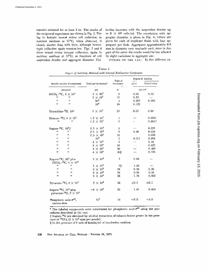
INA CELLS BY DIFFERENT SUBSTRATES : Alarge number of isotopic substances were tested todetermine optimal conditions for labeling the cellsused for collection . The results obtained withthe different compounds are shown in Table I .32P consistently gave higher labeling and wasroutinely used .VARIABILITY OF THE ASSAY : The speed
and convenience of the present method providethe major advantages over the radioautographicprocedure previously described (11), and permitthe analysis of a large number of samples. Therewas considerable variability in replicate flasks, sothat all of the experimental values in each experi-ment are given in the tables, as well as their meanvalues. The tables show that the assay method maybe considered semiquantitative, but provides re-sults sufficiently quantitative and reproducible todraw certain conclusions concerning the natureof specific intercellular adhesion .
Specificity and Kinetic Parameters
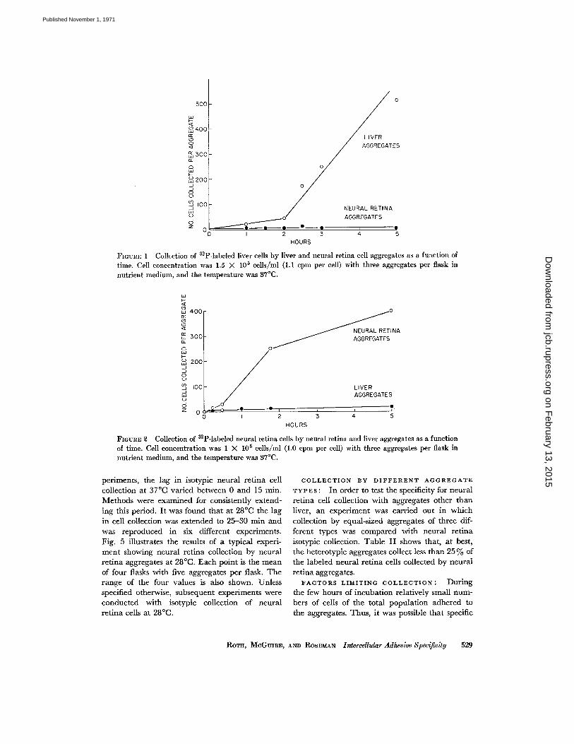
Fig. 1 shows the extent of collection of labeledliver cells by 8-day old liver and neural retinacell aggregates as a function of time . Collectionwas carried out in nutrient medium at 37 °C withthree aggregates per flask. After a lag of about 2hr, isotypic collection begins and proceeds at arate far greater than heterotypic collection which
RoTH, MCGmRE, AND ROSEMAN Intercellular Adhesive Specificity
527
on February 13, 2015
jcb.rupress.orgD
ownloaded from
Published November 1, 1971
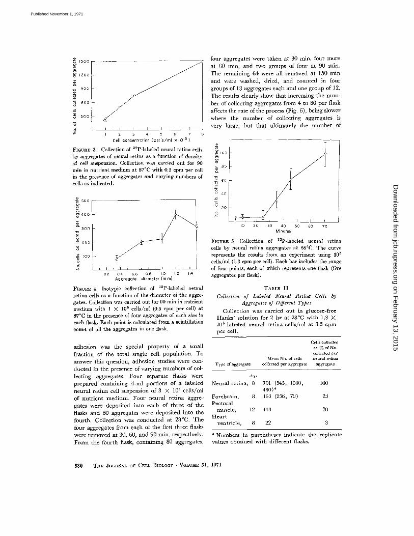
remains minimal for at least 5 hr . The results ofthe reciprocal experiment are shown in Fig . 2 . Thelag in isotypic neural retina cell collection innutrient medium at 37 °C, when observed, isclearly shorter than with liver, although hetero-typic collection again remains low. Figs . 3 and 4show neural retina isotypic collection, again innutrient medium at 37 °C, as functions of cellsuspension density and aggregate diameter . Col-
528
TABLE I
Degree of Labeling Attained with Selected Radioactive Compounds
THE JOURNAL OF CELL BIOLOGY . VOLUME 51, 1971
lection increases with the suspension density upto 8 X 10 5 cells/ml . The correlation with ag-gregate diameter is given in Fig. 4; values aregiven for each of duplicate flasks with four ag-gregates per flask . Aggregates approximately 0 .8mm in diameter were routinely used, since in thispart of the curve the results would be less affectedby slight variations in aggregate size .STUDIES ON THE LAG : In five different ex-
* The labeled compounds were substituted for phosphoric acid-P 32 using the pro-cedures described in the text .$ Sugars- 14C are obtained by alcohol extraction of tobacco leaves grown in the pres-ence of 14C02 (2 X 10 8 cpm per µmole) .§ In the presence of I unit of insulin/ml of incubation medium .
Degree of labeling
Specific activity of compounds Total per incubationTime of
incubation Liver Neural retina
cpm/i mote rpm hr cpm/cell
HCO3-14C, 4 X 10 8 5 X 10⁷ 2 0 .05 0 .032X107 15 0 .10 -
10 9 5 0 .207 0 .09310 9 24 0 .122
Thymidine3H, 1010 5 X 10 7 29 0.05 0.02
Glucose- 14 C, 6 X 10⁵ 1 .2 X 10 7 1 0 .0021((
f( 1 .2 X 10 7 3 0.0043
Sugars-14C, 10 8$ 2.5 X 10 8 3 0 .0332.5 X 10 8 5 0 .08 0 .058((
((
(( 2.5 X 10 8 10 0 .07810 9 5 0 .211 0 .094
4 X 10 9 5 0 .18((
K 4 X 10 9 24 0 .422u
u 4 X 10⁹ 30 0.380u
u 4 X 10⁹ 30§ 0.374
Sugars- 14 C, 10 8 plus 5 X 10⁹ 7 0 .88HCO3-14C, 4 X 10 8
5 X 10⁹ 7§ 1 .06((
((K
(( 5X10⁹ 24 0 .98 0 .36(0
(i 5 X 10 9 24 0 .56 0 .35(i
« 5X10⁹ 26 1 .76 0 .402
Pyruvate- 14C, 2 X 10 7 2 X 10⁸ 24 <0.1 <0.1
Sugars- 14C, 10 8 plus -4 X 10 9 24 1 .41 0.410pyruvate- 14 C, 2 X 10 7
16 -2 .0 -2 .0Phosphoric acid-P32 , 10 °carrier-free
on February 13, 2015
jcb.rupress.orgD
ownloaded from
Published November 1, 1971
500
wQw 400
aw 300a0ww 200JJOUJ 100JwC)0Z 0
0
periments, the lag in isotypic neural retina cellcollection at 37 °C varied between 0 and 15 min .Methods were examined for consistently extend-ing this period . It was found that at 28 °C the lagin cell collection was extended to 25-30 min andwas reproduced in six different experiments .Fig. 5 illustrates the results of a typical experi-ment showing neural retina collection by neuralretina aggregates at 28 °C. Each point is the meanof four flasks with five aggregates per flask . Therange of the four values is also shown. Unlessspecified otherwise, subsequent experiments wereconducted with isotypic collection of neuralretina cells at 28 °C .
I
2
3HOURS
FIGURE 1 Collection of 32 P-labeled liver cells by liver and neural retina cell aggregates as a function oftime . Cell concentration was 1 .5 X 10 5 cells/ml (1 .1 cpm per cell) with three aggregates per flask innutrient medium, and the temperature was 37 °C .
HOURS
FIGURE 9 Collection of 32 P-labeled neural retina cells by neural retina and liver aggregates as a functionof time. Cell concentration was 1 X 10 5 cells/ml (1 .0 cpm per cell) with three aggregates per flask innutrient medium, and the temperature was 37 °C .
4 5
COLLECTION BY DIFFERENT AGGREGATETYPES : In order to test the specificity for neuralretina cell collection with aggregates other thanliver, an experiment was carried out in whichcollection by equal-sized aggregates of three dif-ferent types was compared with neural retinaisotypic collection . Table II shows that, at best,the heterotypic aggregates collect less than 25% ofthe labeled neural retina cells collected by neuralretina aggregates .FACTORS LIMITING COLLECTION : During
the few hours of incubation relatively small num-bers of cells of the total population adhered tothe aggregates. Thus, it was possible that specific
ROTH, MCGUIRE, AND ROSEMAN Intercellular Adhesive Specificity
529
on February 13, 2015
jcb.rupress.orgD
ownloaded from
Published November 1, 1971
â1200
Z
900
600
â 500N
Ô 400
I 1 II
2
3
4
5
6
7
8
Cell concentration (cells/ml x 10-5 )
FIGURE 3 Collection of 32P-labeled neural retina cellsby aggregates of neural retina as a function of densityof cell suspension . Collection was carried out for 90
min in nutrient medium at 37 °C with 0 .5 cpm per cellin the presence of aggregates and varying numbers ofcells as indicated .
I
0 .2
04
0.6
0.8
1 .0
1 .2
1 .4Aggregate diameter (mm)
FIGURE 4 Isotypic collection of 32P-labeled neuralretina cells as a function of the diameter of the aggre-gates . Collection was carried out for 90 min in nutrientmedium with 1 X 10 5 cells/m1 (0.5 cpm per cell) at37°C in the presence of four aggregates of each size ineach flask . Each point is calculated from a scintillationcount of all the aggregates in one flask .
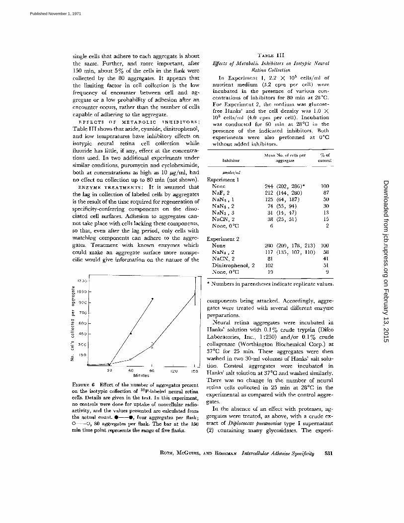
adhesion was the special property of a smallfraction of the total single cell population. Toanswer this question, adhesion studies were con-ducted in the presence of varying numbers of col-lecting aggregates . Four separate flasks wereprepared containing 4-ml portions of a labeledneural retina cell suspension of 3 X 10 5 cells/mlof nutrient medium. Four neural retina aggre-gates were deposited into each of three of theflasks and 80 aggregates were deposited into thefourth. Collection was conducted at 28 °C. Thefour aggregates from each of the first three flaskswere removed at 30, 60, and 90 min, respectively.From the fourth flask, containing 80 aggregates,
I1I11
four aggregates were taken at 30 min, four moreat 60 min, and two groups of four at 90 min .The remaining 64 were all removed at 150 minand were washed, dried, and counted in fourgroups of 13 aggregates each and one group of 12 .The results clearly show that increasing the num-ber of collecting aggregates from 4 to 80 per flaskaffects the rate of the process (Fig . 6), being slowerwhere the number of collecting aggregates isvery large, but that ultimately the number of
ôloo
o'80
60
40
20
5 3 0
THE JOURNAL OF CELL BIOLOGY . VOLUME 51, 1971
10
FIGURE 5 Collection of 32 P-labeled neural retinacells by neural retina aggregates at 28 °C. The curverepresents the results from an experiment using 10 5cells/ml (1 .3 cpm per cell) . Each bar includes the rangeof four points, each of which represents one flask (fiveaggregates per flask) .
TABLE II
Collection of Labeled Neural Retina Cells byAggregates of Different Types
Collection was carried out in glucose-freeHanks' solution for 2 hr at 28 °C with 1 .3 X10 5 labeled neural retina cells/ml at 3.3 cpmper cell .
Type of aggregate
20 30
40
50Minutes
60 70
Cells collectedas % of No .collected per
Mean No . of cells
neural retinacollected per aggregate
aggregate
* Numbers in parentheses indicate the replicatevalues obtained with different flasks .
days
Neural retina, 8 701 (545, 1080, 100480)*
Forebrain, 8 163 (256, 70) 23Pectoral
muscle, 12 143 20Heart
ventricle, 8 22 3
on February 13, 2015
jcb.rupress.orgD
ownloaded from
Published November 1, 1971
single cells that adhere to each aggregate is about
the same. Further, and more important, after150 min, about 5% of the cells in the flask were
collected by the 80 aggregates . It appears that
the limiting factor in cell collection is the lowfrequency of encounter between cell and ag-gregate or a low probability of adhesion after anencounter occurs, rather than the number of cells
capable of adhering to the aggregate .EFFECTS OF METABOLIC INHIBITORS :
Table III shows that azide, cyanide, dinitrophenol,and low temperatures have inhibitory effects onisotypic neural retina cell collection whilefluoride has little, if any, effect at the concentra-tions used. In two additional experiments undersimilar conditions, puromycin and cycloheximide,
both at concentrations as high as 10 µg/ml, hadno effect on collection up to 80 min (not shown) .
ENZYME TREATMENTS : It is assumed thatthe lag in collection of labeled cells by aggregatesis the result of the time required for regeneration ofspecificity-conferring components on the disso-
ciated cell surfaces . Adhesion to aggregates can-not take place with cells lacking these components,so that, even after the lag period, only cells withmatching components can adhere to the aggre-gates. Treatment with known enzymes whichcould make an aggregate surface more nonspe-
cific would give information on the nature of the
1200d
1050d
900o
600
150
30 60
90Minutes
120 150
FIOuRE 6 Effect of the number of aggregates presenton the isotypic collection of 32 P-labeled neural retinacells. Details are given in the text . In this experiment,no controls were done for uptake of noncellular radio-activity, and the values presented are calculated fromthe actual count.*--*, four aggregates per flask ;O-O, 80 aggregates per flask. The bar at the 150min time point represents the range of five flasks.
TABLE III
Effects of Metabolic Inhibitors on Isotypic NeuralRetina Collection
In Experiment 1, 2.2 X 10 5 cells/ml ofnutrient medium (3 .2 cpm per cell) wereincubated in the presence of various con-centrations of inhibitors for 80 min at 28 °C .For Experiment 2, the medium was glucose-free Hanks' and the cell density was 1 .0 X10 5 cells/ml (4 .0 cpm per cell) . Incubationwas conducted for 60 min at 28 °C in thepresence of the indicated inhibitors . Bothexperiments were also performed at 0 °Cwithout added inhibitors .
* Numbers in parentheses indicate replicate values .
components being attacked . Accordingly, aggre-gates were treated with several different enzyme
preparations .Neural retina aggregates were incubated in
Hanks' solution with 0.1 % crude trypsin (DifcoLaboratories, Inc ., 1 :250) and/or 0 .1 % crudecollagenase (Worthington Biochemical Corp.) at37 °C for 25 min. These aggregates were thenwashed in two 30-ml volumes of Hanks' salt solu-tion. Control aggregates were incubated inHanks' salt solution at 37 °C and washed similarly .There was no change in the number of neuralretina cells collected in 25 min at 28 °C in theexperimental as compared with the control aggre-gates .
In the absence of an effect with proteases, ag-gregates were treated, as above, with a crude ex-tract of Diplococcus pnewnoniae type I supernatant(2) containing many glycosidases . The experi-
Rarx, MCGUIRE, AND RosEMAN Intercellular Adhesive Specificity
531
InhibitorMean No. of cells per
aggregate% of
control
,mates/ml
Experiment 1None 244 (202, 286)* 100NaF, 2 212 (144, 280) 87NaN3,1 125 (64, 187) 50NaN3,2 74 (55, 94) 30NaN3, 3 31 (14, 47) 13NaCN, 2 38 (25, 51) 15None, 0 °C 6 2
Experiment 2None 200 (209, 178, 213) 100NaN3, 2 117 (135, 107, 110) 58NaCN, 2 81 41Dinitrophenol, 2 102 51None, 0 °C 19 9 on F
ebruary 13, 2015jcb.rupress.org
Dow
nloaded from
Published November 1, 1971
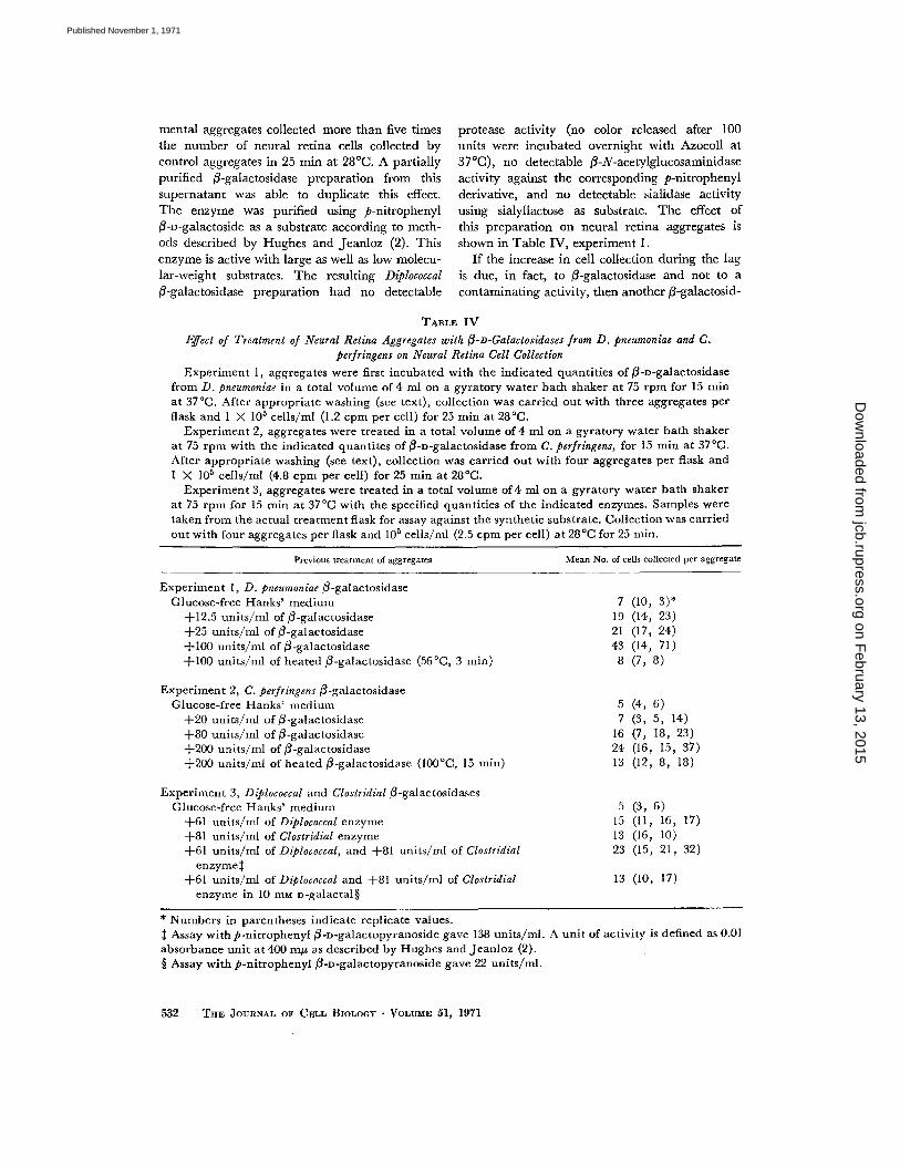
mental aggregates collected more than five timesthe number of neural retina cells collected bycontrol aggregates in 25 min at 28 °C. A partiallypurified ß-galactosidase preparation from thissupernatant was able to duplicate this effect .The enzyme was purified using p-nitrophenylß-n-galactoside as a substrate according to meth-ods described by Hughes and Jeanloz (2) . Thisenzyme is active with large as well as low molecu-lar-weight substrates . The resulting Diplococcalß-galactosidase preparation had no detectable
protease activity (no color released after 100units were incubated overnight with Azocoll at37°C), no detectable ß-N-acetylglucosaminidaseactivity against the corresponding p-nitrophenylderivative, and no detectable sialidase activityusing sialyllactose as substrate . The effect ofthis preparation on neural retina aggregates isshown in Table IV, experiment 1 .
If the increase in cell collection during the lagis due, in fact, to ß-galactosidase and not to acontaminating activity, then another ß-galactosid-
TABLE IV
Effect of Treatment of Neural Retina Aggregates with ß-n-Galactosidases from D . pneumoniae and C .perfringens on Neural Retina Cell Collection
Experiment 1, aggregates were first incubated with the indicated quantities of ß-n-galactosidasefrom D. pneumoniae in a total volume of 4 ml on a gyratory water bath shaker at 75 rpm for 15 minat 37 °C. After appropriate washing (see text), collection was carried out with three aggregates perflask and 1 X 10⁵ cells/ml (1 .2 cpm per cell) for 25 min at 28°C .
Experiment 2, aggregates were treated in a total volume of 4 ml on a gyratory water bath shakerat 75 rpm with the indicated quantites of ß-n-galactosidase from C. perfringens, for 15 min at 37 °C .After appropriate washing (see text), collection was carried out with four aggregates per flask andI X 105 cells/ml (4.8 cpm per cell) for 25 min at 28°C .
Experiment 3, aggregates were treated in a total volume of 4 ml on a gyratory water bath shakerat 75 rpm for 15 min at 37 °C with the specified quantities of the indicated enzymes . Samples weretaken from the actual treatment flask for assay against the synthetic substrate . Collection was carriedout with four aggregates per flask and 105 cells/ml (2 .5 cpm per cell) at 28 °C for 25 min .
* Numbers in parentheses indicate replicate values .$ Assay with p-nitrophenyl ß-n-galactopyranoside gave 138 units/mi . A unit of activity is defined as 0 .01absorbance unit at 400 mg as described by Hughes and Jeanloz (2) .§ Assay with p-nitrophenyl ß-n-galactopyranoside gave 22 units/ml .
532
THE JOURNAL OF CELL BIOLOGY . VOLUME 51, 1971
Previous treatment of aggregates Mean No. of cells collected per aggregate
ExperimentGlucose-free
1, D. pneumoniae ß-galactosidaseHanks' medium 7 (10, 3)*
+12.5 units/ml of ß-galactosidase 19 (14, 23)+25 units/ml of ß-galactosidase 21 (17, 24)+100 units/ml of ß-galactosidase 43 (14, 71)+100 units/ml of heated ß-galactosidase (56 °C, 3 min) 8 (7, 8)
ExperimentGlucose-free
2, C . perfringens ß-galactosidaseHanks' medium 5 (4, 6)
+20 units/ml of ß-galactosidase 7 (3, 5, 14)+80 units/ml of 0-galactosidase 16 (7, 18, 23)+200 units/ml of ß-galactosidase 24 (16, 15, 37)+200 units/ml of heated ß-galactosidase (100 °C, 15 min) 13 (12, 8, 18)
ExperimentGlucose-free
3, Diplococcal and Clostridial ß-galactosidasesHanks' medium 5 (3, 6)
+61 units/ml of Diplococcal enzyme 15 (11, 16, 17)+81 units/ml of Clostridial enzyme 13 (16, 10)+61 units/ml of Diplococcal, and +81 units/ml of Clostridial 23 (15, 21, 32)enzymes
13 (10, 17)+61 units/ml of Diplococcal and +81 units/ml of Clostridialenzyme in 10 MM n-galactal§
on February 13, 2015
jcb.rupress.orgD
ownloaded from
Published November 1, 1971
ase isolated from a different source should give asimilar result . Experiment 2 in Table IV showsthe results obtained when aggregates were treatedwith ß-D-galactosidase purified from a Clostridiumperfringens supernatant (1) . Although this prepara-tion had ß-N-acetylglucosaminidase activity,ß-galactosidase-free (3-N-acetylglucosaminidasefrom the same organism had no effect on isotypicneural retina collection . (Both of the Clostridialenzyme preparations were kind gifts of Mr .Stefan Chipowsky .) The combination of Clostridialand Diplococcal ß-galactosidases (Table IV, ex-periment 3) had an additive effect on neuralretina cell collection and this effect was sensitiveto the ß-galactosidase inhibitor, D-galactal (3),although the extent of inhibition of the enzymesusing the p-nitrophenyl ß-D-galactopyranosidesubstrate was much greater than the inhibitionof the enzymatic activities in the cell collectingassays .
Table IV presents the results of three of eightsimilar experiments. While considerable variabilityis apparent in each experiment, all eight experi-ments showed essentially the same results .
If ß-galactosidase treatment does render neuralretina aggregate surfaces less specific, then treatedaggregates should collect more heterotypic cellsthan untreated neural retina aggregates . Table Vshows that, in fact, this is the case. Treatmentwith both ß-galactosidases, however, had noeffect on the collection of liver or neural retinacells by liver aggregates .EFFECT OF AZIDE AND ß-GALACTOSIDASE
ON SPECIFIC ADHESION : When untreated
TABLE VEffect of Clostridial ß-Galactosidase-Treated
Neural Retina Aggregates on Collection ofLabeled Liver Cells
Aggregates were first treated in a totalvolume of 4 ml on a gyratory water bath shakerat 75 rpm for 30 min at 37 °C. Collection wascarried out with five neural retina aggregatesper flask with cell concentration of 2 X 10 5 /ml (2 .1 cpm per cell) for 30 min at 28 °C .
* Numbers in parentheses indicate replicate values . were detectable . However, specificity could not
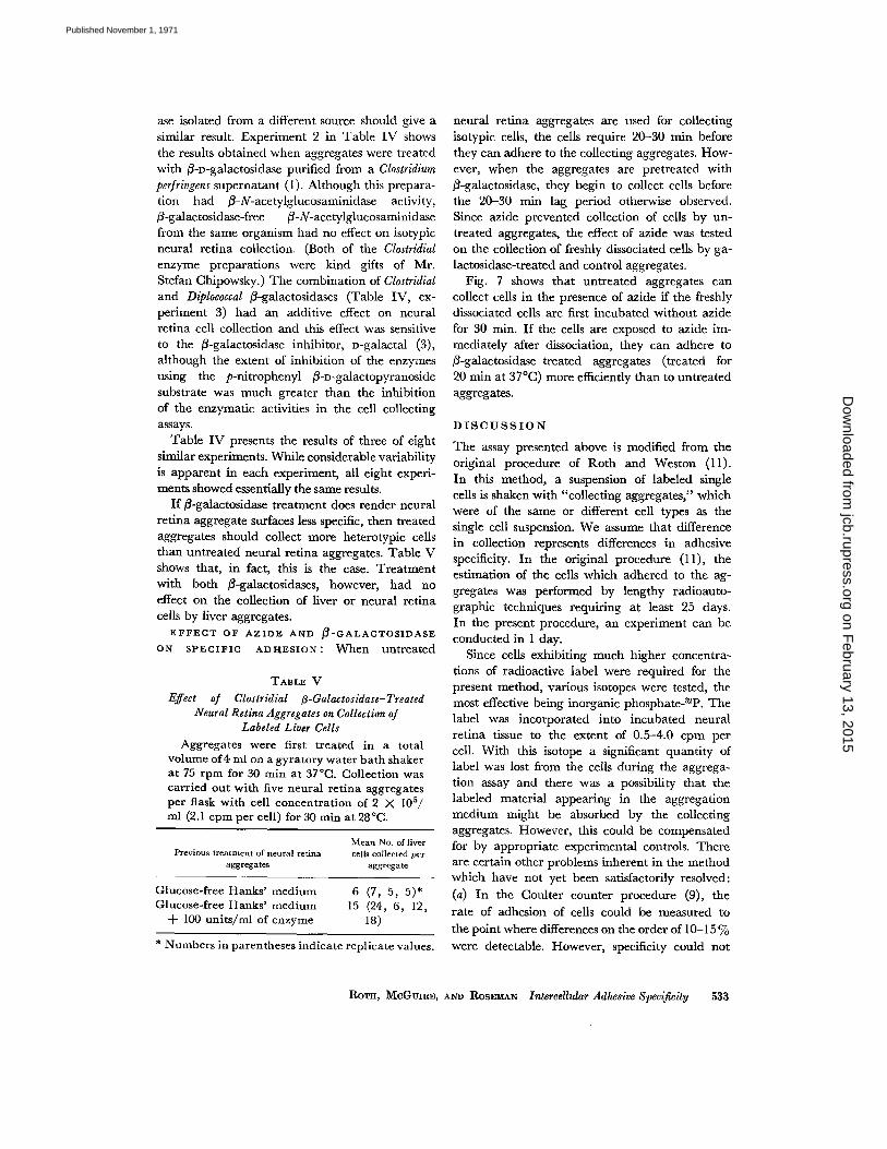
neural retina aggregates are used for collectingisotypic cells, the cells require 20-30 min beforethey can adhere to the collecting aggregates . How-ever, when the aggregates are pretreated withß-galactosidase, they begin to collect cells beforethe 20-30 min lag period otherwise observed .Since azide prevented collection of cells by un-treated aggregates, the effect of azide was testedon the collection of freshly dissociated cells by ga-lactosidase-treated and control aggregates .
Fig. 7 shows that untreated aggregates cancollect cells in the presence of azide if the freshlydissociated cells are first incubated without azidefor 30 min. If the cells are exposed to azide im-mediately after dissociation, they can adhere toß-galactosidase-treated aggregates (treated for20 min at 37 °C) more efficiently than to untreatedaggregates .
DISCUSSION
The assay presented above is modified from theoriginal procedure of Roth and Weston (11) .In this method, a suspension of labeled singlecells is shaken with "collecting aggregates," whichwere of the same or different cell types as thesingle cell suspension . We assume that differencein collection represents differences in adhesivespecificity . In the original procedure (11), theestimation of the cells which adhered to the ag-gregates was performed by lengthy radioauto-graphic techniques requiring at least 25 days .In the present procedure, an experiment can beconducted in I day.
Since cells exhibiting much higher concentra-tions of radioactive label were required for thepresent method, various isotopes were tested, themost effective being inorganic phosphate- 32P. Thelabel was incorporated into incubated neuralretina tissue to the extent of 0.5-4.0 cpm percell . With this isotope a significant quantity oflabel was lost from the cells during the aggrega-tion assay and there was a possibility that thelabeled material appearing in the aggregationmedium might be absorbed by the collectingaggregates . However, this could be compensatedfor by appropriate experimental controls. Thereare certain other problems inherent in the methodwhich have not yet been satisfactorily resolved :(a) In the Coulter counter procedure (9), therate of adhesion of cells could be measured tothe point where differences on the order of 10-15
ROTH, McGuim, AND RosEMAN Intercellular Adhesive Specificity
533
Previous treatment of neural retinaaggregates
Mean No. of livercells collected per
aggregate
Glucose-free Hanks' medium 6 (7, 5, 5)*Glucose-free Hanks' medium 15 (24, 6, 12,+ 100 units/ml of enzyme 18)
on February 13, 2015
jcb.rupress.orgD
ownloaded from
Published November 1, 1971
5 3 4
O
O4ii
Io
20
40
50
60
MINUTES
FIGURE 7 Effect of azide and ß-galactosidase on specific adhesion . Collection was carried outwith a 32 P-labeled neural retina suspension of 1 X 10 5 cells/ml (1 .7 cpm per cell) in the presence of4 µmoles/ml of NaN3 . Three systems were used : OO, neural retina suspension with un-treated aggregates ; o-s, neural retina suspension incubated at 28 °C in glucose-free Hanks' solu-tion for 30 min without azide before addition to flask containing untreated aggregates ; and • •,neural retina suspension with aggregates which had been incubated with 150 units/ml of Diplo-coccal ß-galactosidase in glucose-free Hanks' for 20 min at 37°C before collection.
be distinguished by this method . The presentprocedure can demonstrate adhesive specificity, inspite of the fact that replicate samples in differentflasks occasionally show variations of as much as1007o , although the usual range is 25%. We be-lieve that the major cause is that small groups ofcells (as well as single cells) may adhere to thecollecting aggregates . (b) Another inherent diffi-culty in this method lies in the small numbers ofcells which are collected by the aggregates . Anytissue which is dissociated to a suspension of singlecells contains a variety of cell types . Do the singlecells collected by the aggregates represent a majoror a minor fraction of the cell types in the totalpopulation? This question cannot be answeredwith certainty, but the experiment described inFig. 6 shows that when a large number of col-lecting aggregates is used per flask, about 5%of the total single cell population adhere to theaggregates. On the other hand, the rate of adhe-sion per aggregate in the presence of large num-bers of collecting aggregates was slower than therate in the presence of small numbers of collectingaggregates . This could be due to the gradual ac-quisition of specificity-conferring components byall the cells or to the fact that a special class ofcells was being collected.
In spite of these limitations, the procedureclearly manifests an adhesive recognition andappears to be a useful method for measuring thisimportant process.
Results of studies on the collection of labeled
THE JOURNAL OF CELL BIOLOGY . VOLUME 51, 1971
neural retina cells by neural retina aggregatesindicate that collection begins after a variable lagat 37 °C and after a definite 25-30 min lag at 28 °C .They show, as well, that collection is specific ;liver, forebrain, muscle, and heart aggregatescollect fewer neural retina cells than do neuralretina aggregates under the same conditions .It was also demonstrated that neural retina iso-typic collection is sensitive to several metabolicinhibitors.
In contrast to these results, other investigatorsworking with simple reaggregation of neuralretina cells after dissociation have shown that(a) there is no detectable lag in single cell disap-pearance at 37 °C or 25°C (4, 9), (b) after disso-ciation, neural retina cells will adhere to severalother cell types nonspecifically (6, 12), and (c)aggregation of neural retina cells is not sensitiveto azide, cyanide, or dinitrophenol (DNP) forat least the first 2 hr after dissociation (9) .
The simplest explanation for these differences isthat cells dissociated from each other by treat-ment with crude trypsin lack surface componentsrequired for selective adhesion . These cells arecapable of nonspecific adhesion to each other, butadhere poorly to collecting aggregates whichcontain regenerated or "complete" cell surfaces .During the lag period, the single cells regeneratethe necessary components which permit them toadhere to the intact collecting aggregates . Totest this hypothesis, the surfaces of the cells in thecollecting aggregates were modified by treatment
on February 13, 2015
jcb.rupress.orgD
ownloaded from
Published November 1, 1971
with specific enzymes. It was found that neuralretina aggregates treated with two partially puri-fied ß-galactosidases collected more liver cellsthan did untreated aggregates ; in other words,treatment of the neural retina aggregates withß-galactosidase made them less specific . Further-more, if single neural retina cells are poisonedwith azide, they are unable to regenerate thesurface components required for specific adhesion .Such cells adhered more rapidly to ß-galactosidase-treated aggregates than they did to untreatedaggregates, again indicating that ß-galactosidasetreatment of the neural retina aggregates de-creased their specificity. Finally, when single cellswere permitted to recover from the trypsin treat-ment by incubation for 30 min at 28 °C in theabsence of azide, and were then suspended in amedium containing azide, the treated aggregatescollected fewer cells than did the untreated ag-gregates.
The results of these experiments showed thattreatment with ß-galactosidase changed the ad-hesive properties of the collecting aggregates,making them more, but not totally, nonspecific .These results might be expected since it appearsreasonable to suppose that many sites on the cellsurface are responsible for specific adhesion, andnot all of them are sensitive to ß-galactosidase .Thus, ß-galactosidase treatment would probablyyield a "mixed" cell surface with respect to ad-hesive properties rather than a complete change .Nevertheless, it appears reasonable to concludethat terminal ß-galactosyl residues on the surfacesof neural retina cells are at least partly responsiblefor the adhesive selectivity in these cells .
This investigation was supported in part by researchgrants from the National Institute of Arthritis andMetabolic Diseases (AM 09851) and the AmericanCancer Society (P544) . This is Contribution No. 640from the McCollum-Pratt Institute.
During this study, Dr. Roth was a PostdoctoralFellow of the American Cancer Society (PF 455) .Received for publication 15 March 1971, and in revisedform 8 June 1971 .
BIBLIOGRAPHY
1 . CHIPOWSKY, S., and E. J . McGuIRE . 1969 .Hydrolysis of glycosides in glycoproteins bypurified glycosidases. Fed. Proc . 28:606.
2. HUGHES, R . C ., and R . W. JEANLOZ . 1964. Theextracellular glycosidases of Diplococcus pneu-moniae. I . Purification and properties of aneuraminidase and a ß-galactosidase. Actionon the a,-acid glycoprotein of human plasma .Biochemistry . 3:1535.
3. LEE, Y. C. 1969. Inhibition of ß-D-galactosidasesby D-galactal. Biochem . Biophys . Res . Commun .35:161 .
4. LILIEN, J. E . 1968 . Specific enhancement of cellaggregation in vitro. Develop . Biol. 17:657 .
5. MANLY, R. S ., editor . 1970 . Adhesion inBiological Systems. Academic Press Inc ., NewYork.
6. MOSCONA, A . A . 1961 . Rotation mediated histo-genetic aggregation of dissociated cells .Exp. Cell Res. 22:455 .
7. MoscoeA, A. A. 1965 . In Cells and Tissues inCulture . E. N. Willmer, editor. AcademicPress Inc., New York . 1 :489 .
8. OPPENHEIMER, S . B ., M . EDIDIN, C . W. ORR, andS. ROSEMAN . 1969. An L-glutamine require-ment for intercellular adhesion . Proc. Nat.Acad.Sci. U. S. A. 63:1395.
9 . ORR, C. W., and S. ROSEMAN. 1969 . Intercellularadhesion . I . A quantitative assay for measuringthe rate of adhesion. J. Membrane Biol . 1 :124.
10. ROTH, S . A . 1968 . Studies on intercellular adhe-sive selectivity . Develop . Biol. 18:602 .
11. ROTH, S . A ., and J . A. WESTON . 1967. The meas-urement of intercellular adhesion . Proc. Nat.Acad. Sci. U. S. A . 58 :974.
12 . STEINBERG, M. S. 1964. In Cellular Membranesin Development. M . Locke, editor. AcademicPress Inc ., New York . 213.
13 . STEINBERG, M. S. 1970 . Does differential adhe-sion govern self assembly processes in histo-genesis? Equilibrium configurations and theemergence of a hierarchy among populationsof embryonic cells. J. Exp . Zool . 173 :395 .
14 . TRINKAUS, J. P. 1969 . Cells into Organs . Prentice-Hall Inc., Englewood Cliffs, N . J .
ROTH, MCGUIRE, AND ROSEMAN Intercellular Adhesive Specificity 535
on February 13, 2015
jcb.rupress.orgD
ownloaded from
Published November 1, 1971