combinatorial engineering of dextransucrase specificity
TRANSCRIPT
Combinatorial Engineering of Dextransucrase SpecificityRomain Irague1,2,3, Laurence Tarquis1,2,3, Isabelle André1,2,3, Claire Moulis1,2,3, Sandrine Morel1,2,3, PierreMonsan1,2,3, Gabrielle Potocki-Véronèse1,2,3, Magali Remaud-Siméon1,2,3*
1 Université de Toulouse; INSA, UPS, INP, LISBP, Toulouse, France, 2 CNRS, UMR5504, Toulouse, France, 3 INRA, UMR792 Ingénierie des SystèmesBiologiques et des Procédés, Toulouse, France
Abstract
We used combinatorial engineering to investigate the relationships between structure and linkage specificity of thedextransucrase DSR-S from Leuconostoc mesenteroides NRRL B-512F, and to generate variants with alteredspecificity. Sequence and structural analysis of glycoside-hydrolase family 70 enzymes led to eight amino acids(D306, F353, N404, W440, D460, H463, T464 and S512) being targeted, randomized by saturation mutagenesis andsimultaneously recombined. Screening of two libraries totaling 3.6.104 clones allowed the isolation of a toolboxcomprising 81 variants which synthesize high molecular weight α-glucans with different proportions of α(1→3)linkages ranging from 3 to 20 %. Mutant sequence analysis, biochemical characterization and molecular modellingstudies revealed the previously unknown role of peptide 460DYVHT464 in DSR-S linkage specificity. This peptidesequence together with residue S512 contribute to defining +2 subsite topology, which may be critical for the enzymeregiospecificity.
Citation: Irague R, Tarquis L, André I, Moulis C, Morel S, et al. (2013) Combinatorial Engineering of Dextransucrase Specificity. PLoS ONE 8(10): e77837.doi:10.1371/journal.pone.0077837
Editor: Pratul K. Agarwal, Oak Ridge National Laboratory, United States of America
Received July 1, 2013; Accepted September 10, 2013; Published October 18, 2013
Copyright: © 2013 Irague et al. This is an open-access article distributed under the terms of the Creative Commons Attribution License, which permitsunrestricted use, distribution, and reproduction in any medium, provided the original author and source are credited.
Funding: This work was funded by the French National Institute for Agricultural Research (INRA) and the French National Center for Scientific Research(CNRS). Grant number : CEPIA-AIC2007-2010, http://www.inra.fr/, http://www.cnrs.fr/. ICEO is supported by grants from the Region Midi-Pyrénées,France, the European Regional Development Fund, and INRA. The funders had no role in study design, data collection and analysis, decision to publish, orpreparation of the manuscript.
Competing interests: The authors have declared that no competing interests exist.
* E-mail: [email protected]
Introduction
Bacterial glucansucrases (EC. 2.4.1.) are transglucosidasesthat synthesize high molecular weight α-glucans,oligosaccharides or glucoconjugates from sucrose, a low costagroresource, as glucosyl donor. Depending on theirspecificity, glucansucrases (GS) catalyze the formation of bothlinear and branched α-D-glucans with various types of osidiclinkages, namely α(1→2); α(1→3); α(1→4) and/or α(1→6)glucosidic bonds. These enzymes are thus attractive tools forglycodiversification due to their ability to producecarbohydrates of diverse size, structure and physico-chemicalproperties [1].
The GS produced by lactic acid bacteria of the generaLeuconostoc, Streptococcus, Lactobacillus, Exigobacteriumand Weissella are classified into the family 70 of Glycoside-Hydrolases (GH70) [1–3]. To date, 57 GS enzymes have beenbiochemically characterized and three-dimensional structuresare available for only four glucansucrases [4–9]. Thesestructures were obtained by crystallization of recombinanttruncated forms of GS from Lb. reuteri 180 (GTF180-ΔN; PDB:3LK), S. mutans (GTF-SI; PDB: 3AIE), Ln. mesenteroidesNRRL B-1299 (ΔN123-GBD-CD2; PDB: 3TTQ) and Lb. reuteri
121 (GTFA-ΔN; PDB: 4AMC). These four enzymes presentdifferent linkage specificity but share a common U-type fold,organized into five domains (A, B, C, IV and V).
All the domains except domain C, are built up fromdiscontinuous segments of the polypeptide chain. The catalyticdomain A in GH70 enzymes adopts a (β/α)8 barrel fold, whichis circularly permuted relative to the related (β/α)8 barrel of GHfamily 13 and 77 enzymes (belonging to the same GH-H clanas family GH70). The active site is shaped as a groove inwhich a pocket accommodates the glucosyl unit of sucrose insubsite -1 (according to Davies’s subsite numbering [10]).There are no -2 or -3 subsites in GH70 family enzymes, and ithas been suggested that they catalyze glucosyl transferthrough an α-retaining double displacement mechanismcomparable to that of GH family 13 enzymes. The availablecrystal structures are consistent with this mechanism in whichthe amino acids D400, E438 and D511 (DRS-S vardel Δ4Nnumbering, Figure 1) may play the role of the nucleophile, theacid/base catalyst and the transition-state stabilizer,respectively [11,12]. Structural analyses and site-directedmutagenesis experiments also indicate that linkage specificityis probably controlled by the topology of the acceptor subsites,in particular the +1 and +2 subsites. Indeed, several studies
PLOS ONE | www.plosone.org 1 October 2013 | Volume 8 | Issue 10 | e77837

highlight the critical role of residues in the conserved regionssurrounding the catalytic residues. In particular, mutations ofthe amino acids downstream from the transition state stabilizermodify the linkage specificity of dextransucrase, mutansucrase,reuteransucrase and alternansucrase [11,13–20]. Numerouschimeric glucansucrase structures have been produced, anddisplay specificities different from those of their parentenzymes, indicating that other regions, for example theextremities of the B-domain, may also contribute to linkagespecificity [21,22].
The determinants of GS specificity have thus not beencompletely described. We therefore used an approach basedon the construction of a structurally guided library ofglucansucrases, derived from one single parent enzyme, toisolate mutants synthesizing high molecular weight α-glucanswith various proportions of α(1→3) and α(1→6) linkages [21].We previously developed a straightforward, sensitive andquantitative NMR-based method for detecting mutants showingnew linkage specificity at a throughput of 480 enzyme mutantsscreened per day [23]. A library of 3.6.104 E. coli clonesexpressing mutants of dextransucrase DSR-S vardel Δ4N, aGS highly specific for the formation α(1→6) glucosidic linkages,have been screened and 303 clones producing enzymes withaltered specificity were identified. Seven of these mutantsproducing dextran polymers with a degree of α(1→3) linkagesranging from 3 to 20 % have been studied in more detail. Thedextran products were characterized and differences in size,conformation, as well as ability to form film were described [24].
To continue this study, we investigated the structuralfeatures of the DSR-S vardel Δ4N mutants that may governlinkage specificity. The semi-rational libraries, from whichmutants able to produce dextrans with various proportions ofα(1→3) linkages were isolated, was therefore analysed. Thetolerance of the targeted amino acid positions to mutations wasevaluated. The kinetic properties of the seven mutantsproducing innovative α(1→3) branched dextran structures werecharacterized. Combined with their three-dimensional structuremodels, these findings provide insight into the structuraldeterminants involved in GS substrate recognition and linkagespecificity.
Materials and Methods
Sequence alignmentAlignX (Vector NTI Advance 10, Invitrogen) was used to
align the sequences of the glucansucrases from Lb. reuteri 180GTF-180 (GenBank accession no. AAU08001.1), Ln. citreumNRRL B-1299 DSR-B (GenBank accession no. AAB95453.1),Ln. mesenteroides NRRL B-1299CB4 DSR-BCB4 (GenBankaccession no. ABF85832.1), Ln. citreum B-1355 DSR-C(GenBank accession no. CAB76565.1), Ln. mesenteroidesLcc4 DSR-D (GenBank accession no. AAG61158.1), Ln.citreumB/110-1-2 DSR-F (GenBank accession no.ACY92456.2), W. cibaria LBAE-k39 DSR-k39 (GenBankaccession no. ADB43097.3), Ln. mesenteroides IBT-PQ DSR-P (GenBank accession no. AAS79426.1), W. cibaria CMUDSR-wC (GenBank accession no. ACK38203.1), Ln. citreumKM20 DexT (GenBank accession no. ACA83218.1), Ln.
mesenteroides 0326 DexYG (GenBank accession no.ABC75033.1), Lb. parabuchneri 33 GTF-33 (GenBankaccession no. AAU08006.1), S. mutans GS 5 GTF-D(GenBank accession no. AAA26895.1), S. gordonii str. Challissubstr. CH 1 GTF-G (GenBank accession no. AAC43483.1), S.salivarius ATCC 25975 GTF-K (GenBank accession no.AAA26896.1), Lb. fermentum Kg3 GTF-kg3 (GenBankaccession no. AAU08008.1), Lb. sakei Kg15 GTF-kg15(GenBank accession no. AAU08011.1), S. salivarius ATCC25975 GTF-M (GenBank accession no. AAC41413.1), S.sanguinis ATCC 10556 GTF-P (GenBank accession no.BAF43788.1), S. downei MFE 28 GTF-S (GenBank accessionno. AAA26898.1), S. oralis ATCC 10557 GTF-R (GenBankaccession no. BAA95201.1), S. sobrinus B13N /OMZ176 GTF-T (GenBank accession no. AAX76986.1), S. sobrinus B13NGTF-U (GenBank accession no. BAC07265.1), Ln. citreum HJ-P4 LcDS (GenBank accession no. BAF96719.1), S. mutansGS5 GTF-ISmGS (GenBank accession no. AAA88588.1), S.mutans UA159 GTF-ISmUA (GenBank accession no.AAN58705.1), S. criceti GTC242/ HS-6 GTF-ISc (GenBankaccession no. BAF62338.1), S. downei Mfe 28 GTF-ISd
(GenBank accession no. AAC63063.1), S. sobrinus ATCC33478 / OMZ176 GTF-ISsob (GenBank accession no.BAA02976.1), S. salivarius ATCC 25975 GTF-ISsal (GenBankaccession no. AAA26896.1), S. salivarius ATCC 25975 GTF-L(GenBank accession no. AAC41412.1), Lb. reuteri ML1 GTF-ML1 (GenBank accession no. AAU08004.1), Lb. reuteri 121GTF-A (GenBank accession no. AAU08015.1), Lb. reuteriATCC 55730 GTF-O (GenBank accession no. AAY86923.1),Ln. citreum B-1355 ASR (GenBank accession no.CAB65910.2), Ln. citreum NRRL B-1299 DSR-E (GenBankaccession no. CAD22883.1) and DSR-S vardel Δ4N.
Bacterial strains, plasmids and growth conditionsOne shot® E. coli TOP 10 (Invitrogen) was used for mutant
library construction. One shot® E. coli BL21 AI™ (Invitrogen)was used for library selection, screening and larger-scaleproduction of the selected variants. Plasmid pBAD harboring agene encoding DSR-S vardel Δ4N fused to thioredoxin andpolyhistidine tags [25] was used as the template for libraryconstruction. Bacterial strains were grown on LB mediumsupplemented with 100 µg.ml-1 ampicillin at 37°C.
DNA manipulationsRestriction endonucleases and DNA-modifying enzymes
were purchased from New England Biolabs and usedaccording to the manufacturer’s instructions. Plasmid DNA wasisolated with Qiaprep® Spin miniprep kits (Qiagen). PCRproducts were extracted from agarose gel with Quiquick® gelextraction kits (Qiagen). DNA sequencing was carried out usingthe dideoxy chain termination procedure [26] by GATC BiotechSARL (Mulhouse, France).
Generation of DSR-S vardel ∆4N mutant librariesAn AatII restriction site was first incorporated into the dsr-s
vardel Δ4N gene, at position 1881, by inverse PCR introducinga silent mutation using pBAD Thio-dsrs vardel Δ4N-His DNA asthe template (Table S1). Then, an 1130 bp cassette,
Engineering of Dextransucrase Specificity
PLOS ONE | www.plosone.org 2 October 2013 | Volume 8 | Issue 10 | e77837

Figure 1. Alignment of GH70 amino acid sequences in the regions chosen for the combinatorial site-directed mutagenesisof DSR-S vardel Δ4N. Sequences are clustered according to the specificity of the glucansucrases. Catalytic (D400 and E438,DSR-S vardel Δ4N numbering) and transition state stabilizer (D511, DSR-S vardel Δ4N numbering) amino acids are highlighted inred; amino acids chosen for mutagenesis are highlighted in green.doi: 10.1371/journal.pone.0077837.g001
Engineering of Dextransucrase Specificity
PLOS ONE | www.plosone.org 3 October 2013 | Volume 8 | Issue 10 | e77837

corresponding to positions 829 to 1958 of the dsr-s vardel Δ4Ngene, was amplified by PCR with primers for_DSRS_Alpha3and rev_DSR-S_Alpha8 (Table S1). Five µg of purified PCRproduct was digested with 1 U of DNaseI in the supplied bufferat 20°C in a final volume of 50 µL. The reaction was stoppedafter 3 min by adding 15 µL of 0.5 M EDTA and heating for 10min at 75°C. Fragments were separated on 2 % agarose geland those of 50-100 bp were extracted. Gene reassembly wascarried out with approximately 100 ng of purified fragments and2 µM of degenerated oligonucleotides (Table S1), to mutatepositions D306, F353, N404 and W440 for library LibA, andD460, H463, T464, S512 for library LibB. The reaction mixture(30 µL), containing 1 U Phusion® High-Fidelity DNAPolymerase (Finnzyme) in the appropriate buffer and 0.4 mMof each dNTP, was thermocycled according to the followingprogram: one denaturation step at 98°C for 30 s; 40 cyclescomposed of a denaturation step at 98°C for 10 s, sixsuccessive hybridization steps separated by 4°C each, from 65to 41°C for 10 s each, and an elongation step at 72°C for 20 s;and finally a 2 min step at 72°C. The fully recombinedcassettes were isolated from the reassembly products in a lastamplification by nested PCR using the primers for_K7a3_ntedand rev_K7a8_nted. The purified nested PCR products weredigested with the restriction enzymes SpeI and AatII andligated into the pBAD Thio-dsrs vardel Δ4N-His to replace theparental cassette. The ligation products were precipitated byadding 5 volumes of absolute ethanol and the DNA pellet wasrinsed twice with 70 % ethanol. The resuspended plasmidswere used to transform E. coli TOP 10 electrocompetent cellsand plated on LB agar supplemented with ampicillin (100µg.ml-1). The transformants were grown overnight at 37°C andthe colonies were scraped from the plates for plasmidextraction to constitute the DNA libraries.
Selection of active glucansucrase mutantsThe DNA libraries were used to transform chemically
competent E. coli BL21 AI cells and plated onto LB agarsupplemented with ampicillin (100 µg.ml-1). After overnightgrowth at 37°C, colonies were scraped off, resuspended inphysiological water and diluted to an OD600nm of 5.10-5. Theclones were subjected to selection pressure by plating thesuspensions on 22x22 cm plates containing solid M9 mineralmedium (42 mM Na2HPO4, 22 mM KH2PO4, 18.7 mM NH4Cl,8.5 mM NaCl, 2.5.10-2 mM CaCl2, 1 mM MgSO4) supplementedwith ampicillin (100 µg.ml-1), 0.02 % arabinose (wt/vol) andsucrose (50 g.L-1) as the sole carbon source. The plates wereincubated for 7 days at 20°C to allow sufficient expression ofrecombinant genes encoding active glucansucrases and thuscell growth. This high-throughput selection was carried out atthe Laboratoire d’Ingénierie des Systèmes Biologiques et desProcédés (Toulouse, France) with the equipment of the ICEOfacility, devoted to the engineering and screening of new andoriginal enzymes.
Screening for glucansucrase mutants specificityActive polymerase-positive mutants (identified as colonies
covered by a polymer bubble) were picked and transferred into96-well microplates (Nunc™ Brands Products, Roskilde,
Denmark) containing 250 µL LB per well supplemented withampicillin (100 µg.ml-1) using a Biomek 2000 pipettor (BeckmanCoulter, Brea, CA). After overnight growth at 30°C underhorizontal shaking at 250 rpm, 50 µL of each of these startercultures was used to inoculate 96-deep-well plates (ABgene,Epsom, UK) containing 500 µL auto-inducing media ZYM-5052[27] supplemented with ampicillin (100 µg.ml-1) and 0.1 %arabinose (w/v). The cultures were grown for 48 h at 20°C in anincubator-shaker (INFORS HT, Bottmingen, Switzerland). Theplates were centrifuged (5 min, 3 700 g, 4°C) and thesupernatants removed. Bacterial cell pellets were resuspendedin 200 µL of lysozyme solution (0.5 mg.ml-1), incubated for 20min at 37°C and frozen at -80°C for 12 h. After thawing for 1 hat room temperature, 800 µL of reaction mixture containing 292mM sucrose and 50 mM isomalto-oligosaccharides (1 kDa onaverage, Pharmacosmos, Denmark) in buffered deuteriumwater (NaAc 50 mM, CaCl2 0.05 g.L-1, pH 5.2) was added toeach well. Enzymatic reactions were allowed to proceed for 48h at 25°C under agitation at 700 rpm. The primary structures ofthe products synthesized were determined by the NMR-basedscreening method described previously using the flow injectionsystem (Bruker BioSpin GmbH) combining a Bruker Avance600 MHz spectrometer with a Gilson Liquid Handler [23].
Production and purification of the wild-type enzymeand mutants with altered specificities
E. coli BL21 AI cells carrying the pBAD plasmids encodingthe Thio-DSR-S vardel ∆4N-His and selected tagged variantswere grown for 24 h at 20°C, in flasks containing ZYM-5052medium supplemented with ampicillin (100 µg.ml-1) and 0.1 %arabinose (w/v). The cultures were centrifuged (4 500 g, 15min, 4°C) and the cell pellets were resuspended to a finalOD600nm of 80 in 50 mM sodium acetate buffer, pH 5.2,containing 0.05 g.L-1 of CaCl2. The suspensions were sonicatedand centrifuged (15 000 g, 30 to 60 min, 4°C), and thesupernatants were harvested for subsequent purification of theenzymes. The enzymes were purified by affinitychromatography on Probond™ Nickel-Chelating resin(Invitrogen) according the protocol previously described forThio-DSR-S vardel Δ4N-His purification [25]. Proteinconcentrations were determined by spectrometry using aNanoDrop® ND-1000 spectrophotometer (Thermo FisherScientific, Waltham, MA).
Glucansucrase activity assaysOne unit of glucansucrase activity corresponds to the
amount of enzyme that catalyzes the formation of 1 µmol offructose per minute at 25°C in 50 mM sodium acetate buffer,pH 5.2, containing 0.05 g.L-1 of CaCl2 and 100 g.L-1 sucrose.The concentration of reducing sugars was determined usingthe dinitrosalicylic acid method [28], using fructose as thestandard.
Kinetic studies were performed using various initial sucroseconcentrations from 10 to 600 mM. The reactions were initiatedby addition of the enzyme to a final concentration of 0.25U.mL-1. At regular time intervals, samples were withdrawn andimmediately heated at 95°C for 5 min to stop the reaction. Theconcentration of released fructose was determined by ion
Engineering of Dextransucrase Specificity
PLOS ONE | www.plosone.org 4 October 2013 | Volume 8 | Issue 10 | e77837

exchange chromatography, using a Dionex system equippedwith an Aminex HPX-87C Carbohydrate column (300 x 7.7 mm;Bio-rad, Hercules, CA, USA), with ultrapure water as eluent ata flow rate of 0.6 mL.min-1. The temperature of the columnoven was set at 80°C. The initial velocities were calculateddirectly from fructose production rates and are expressed inµmol of fructose produced per minute and per gram of enzyme.The SigmaPlot “Enzyme kinetics” module (version 3.1) wasused for curve fitting of the data and determination of kineticparameters.
α-glucan synthesisCell-free extract preparations of the parental Thio-DSR-S
vardel Δ4N-His enzyme and selected tagged variants wereincubated in sodium acetate buffer 50 mM, pH 5.2,supplemented with 0.05 g.L-1 of CaCl2 and 292 mM sucrose.Polymer syntheses were carried out at 25°C using 1 U.mL-1 ofenzyme. Sucrose depletion was monitored by HPLC (seeabove). Reactions were stopped by a 5 min incubation at 95°C.
HPLC analysisα-glucan concentrations in the reaction media were
determined by High-Performance Size ExclusionChromatography (HPSEC) using Shodex OH-Pack SB-805 andSB-802.5 columns in series. Carbohydrates were eluted usinga 0.45 M NaNO3 and 1 % (v/v) ethylene glycol solution as theeluent at a flow rate of 0.3 mL.min-1[25]. The column oventemperature was set at 70°C.
Glucose, fructose and leucrose concentrations weredetermined by ion exchange chromatography using an AminexHPX-87C Carbohydrate column as described above. Thepercentages of glucosyl moieties incorporated into free glucose(%Gglucose) and leucrose (%Gleucrose), when all sucrose wasconsumed, were calculated as follows:
%Gglucose= [glucosetf]/([sucroset0]x180/342) x 100 and%Gleucrose = [leucrosetf]/[sucroset0] x 100 where [sucroset0] is theinitial massic substrate concentration, and [glucosetf] and[leucrosetf] to the final massic concentration of glucose andleucrose at the end of the reaction.
The percentages of glucosyl moieties incorporated intoHigh Molecular Weight (HMW) α-glucans weredetermined as follows
%GHMW glucans= [HMW glucanstf]/([sucroset0]x162/342) x 100where [HMW glucanstf] correspond to the concentration ofHMW α-glucans synthesized at the end of the reaction, anddetermined on HPSEC chromatogram from the RI signalintegration of the peak corresponding to HMW α-glucans.
The proportion of glucose incorporated into IntermediateMolecular Weight (IMW) α-glucans (gluco-oligosaccharides ofDP from 3 to 37) cannot be quantified directly bychromatography due to the weak and disperse signals.Consequently, it was determined as follows:
%GIMW glucans= 100 - %GHMW glucans - %Gglucose -%Gleucrose
NMR spectroscopyFreeze-dried polymer samples (15 mg) were exchanged
twice with 99.9 atom % D2O, lyophilized and dissolved in 600μL of D2O. 1D 1H NMR spectra were recorded on aBrukerAvance 500 MHz spectrometer using a 5 mm z-gradientTBI probe at 298 K, an acquisition frequency of 500.13 MHzand a spectral width of 8012.82 Hz. The 1H-signal from D2Owas used for automatic lock and a gradient shimming wasperformed with each sample. Before Fourier transformation,the FIDs were multiplied by an exponential function with a linebroadening of 0.3 Hz. Spectra were processed with a 64 k zerofilling, baseline correction and were referenced using the TSP-d4 signal at 0 ppm. All NMR data were acquired and processedusing TopSpin 2.1 software. The various signals were assignedas previously described [15,16,29–31]. The percentages ofα(1→3) and α(1→6) linkages in α-glucans were calculated fromthe relative intensities of the corresponding anomeric protonsignals by integration of peak areas.
Three-dimensional molecular modelingThe sequence of DSR-S vardel Δ4N catalytic domain (region
N119 to K973) was submitted to the I-TASSER server [32–34],an online service for automated protein structure and functionprediction, using as restraint the structure of the Lactobacillusreuteri N-terminally truncated glucansucrase GTF180 (PDBaccession code: 3KLK). The 3D model of DSR-S vardel Δ4Npredicted by I-TASSER was then further refined using theCFF91 force-field implemented in the DISCOVER module ofthe Insight II software suite (Accelrys, San Diego, CA, USA).For the minimization, the CFF91 cross terms, a harmonic bondpotential, and a dielectric of 1.0 were used. An initialminimization with a restraint on the protein backbone wasperformed using a steepest descent algorithm followed byconjugated gradient minimization steps until the maximumRMS was less than 0.5. In a subsequent step, the system wasfully relaxed.
A 3D model of the D460M:H463Y:T464M:S512C mutant wasconstructed by introducing, in silico, the divergent amino acidsin this mutant into the corresponding positions in the parentalenzyme, using the Biopolymer module of the Insight II softwarepackage (Accelrys). The conformations of the mutated residueside chains were optimized by manually selecting a low-energyconformation from a side-chain rotamer library. Steric clashes(van der Waals overlap) and non-bonded interaction energies(Coulombic and Lennard-Jones) were evaluated for the variousside-chain conformations. The mutant model was subsequentlyminimized following the same energy minimization procedureas described for the parental enzyme.
Sucrose was manually docked into the active site of DSR-Svardel Δ4N and the D460M:H463Y:T464M:S512C mutantusing the structure of the Lactobacillus reuteri N-terminallytruncated glucansucrase GTF180 in complex with sucrose(PDB code: 1HZ3) as a model. Complexes were thenoptimized using the procedure above described.
One of the putative products of theD460M:H463Y:T464M:S512C mutant, the α-D-Glcp-(1→6)[α-D-Glcp-(1→3)]α-D-Glcp-(1→6)-D-Glcp tetrasaccharide, wasdocked into the mutant enzyme active site using the automated
Engineering of Dextransucrase Specificity
PLOS ONE | www.plosone.org 5 October 2013 | Volume 8 | Issue 10 | e77837

flexible docking program FlexX (Biosolveit) [35,36]. Thetetrasaccharide molecule was covalently docked into the activesite using covalent constraint with catalytic residue D400. Allparameters were set to the standard values as implemented inVersion 3.1.1. The docking region was defined to encompassall amino acids in the protein for which at least one heavy atomwas located within a sphere of 6.5 Å radius around the centerof mass of catalytic D400. For each docking, the top 30solutions, those with the best FlexX scores, were retained. Alldrawings were performed using PyMOL software (DeLanoScientific).
Results and Discussion
Selection of amino acid targetsA semi rational approach was implemented to alter DSR-S
vardel Δ4N linkage specificity. Amino acids presumed to beinvolved in linkage specificity were first identified by sequencealignment of 37 GS catalytic domains (Figure 1). In thisalignment were included 25 dextransucrases exhibiting mainlyα(1→6) linkage specificity, eight mutansucrases specific forα(1→3) linkages, two reuteransucrases catalyzing both α(1→4)and α(1→6) linkage synthesis [37,38], one alternansucrasegenerating alternated α(1→3)/α(1→6) linkages [39], and thesequence of the second catalytic domain of DSR-E (DSR-ECD2), specific for α(1→2) glucosidic linkage synthesis [39–41].Five highly conserved segments emerged from the alignment,three of them corresponding to the signature regions II, III andIV of the GH13 family, and which are close to the catalyticresidues. Arbitrarily, sequence divergences in these highlyconserved sequences were considered to be relevant tolinkage specificity and the corresponding positions wereselected for mutation.
Within segment S304-T314 (DSR-S vardel Δ4N numbering,Figure 1), residue D306 is well conserved amongstdextransucrases. However, this position is occupied by aglutamine in ASR and an alanine in DSR-E CD2. Inreuteransucrases, leucine or isoleucine are found. Interestingly,a leucine residue is also found at this position in DSR-F, adextransucrase synthesizing 1 % of α(1→4) linkages [42].Downstream from this region, in the segment G352-N374, allthe dextransucrases that are highly specific for α(1→6)linkages, except DSR-F and GTF-180, contain an aromaticresidue (phenylalanine or tyrosine) at position 353. DSR-F,GTF-180, reuteransucrases and alternansucrase have analanine, a serine or a glutamine residue at this position. In thesignature regions II and III, residues N404, downstream fromthe catalytic nucleophile D400, and W440, upstream from thegeneral acid/base catalyst E438, are strictly conserved amongGH70 glucansucrases, except in DSR-E CD2 which displaysα(1→2) linkage specificity. The residues corresponding toN404 and W440 in GH13 enzymes interact with the substrateaglycon [43]. In GTF-180 and GTF-A, they are parts of thesubsites +1 and +2 for substrate (sucrose) and acceptor(maltose) molecules [5,9]. Consequently, we considered theresidues D306, F353, N404, and W440 likely to be importantfor linkage specificity. We also included residue S512 in theanalysis, because as a part of the tripeptide512SEV514, it is
located immediately downstream from the second aspartic acidof the catalytic triad in the motif IV and has been reported toaffect linkage specificity [11,15–17,19,20].
Three other positions, located in a non-conserved region ofGH70 enzymes, emerged as possibly significant from structuralanalysis of GTF180-ΔN glucansucrase in complex with maltose(PDB code: 3KLL) [5]. In this structure, four residuescorresponding to D460, H463, T464 and S512 of DSR-S vardelΔ4N interact with maltose in subsite +2, either directly orthrough water molecule mediation. It has been suggested thatthese residues participate in the correct positioning of theacceptor for driving the formation of α(1→6) linkage [5]. Byanalogy, these residues were assumed to participate in linkagespecificity and were thus selected as targets for mutation.
Consequently, engineering efforts were focused on eightresidues: D306, F353, N404, W440, D460, H463, T464 andS512. The ISOR method [44] was used to generate twolibraries named “LibA”, targeting amino acids D306, F353,N404, W440, and "LibB", targeting amino acids D460, H463,T464 and S512. The synthetic oligonucleotides used forsaturation mutagenesis were designed to contain theconventional degenerated NNS codons at the targetedpositions and to give theoretically access to the 20 possibleamino acid residues (Table S1).
Analysis of initial libraryA total of 2.5.105 and 3.8.104 clones were obtained for LibA
and LibB, respectively. 24 and 51 clones were randomlyisolated from LibA and LibB, respectively, and sequenced(Figure 2). All the sequences corresponded to mutantenzymes. In addition, all the 24 sequences of LibA mutantsdiffered from one another. In LibB, only three of the 51 mutantswere found duplicates. Thus, both libraries exhibited a very lowlevel of redundancy. For both libraries, an average of 3mutations per protein sequence was observed. Mutations wereincorporated at all targeted positions and with generally similarfrequencies (Figure 2). This provides evidence that the ISORmethod was effective for both generating and combiningmutations between distant or close residues, such as thosefound in LibA and LibB, respectively.
Analysis of the mutant libraries after primary screeningDNA was isolated from libraries LibA and LibB and used to
transform E. coli BL21 AI cells. A total of 1.5.104 clones forLibA and 2.1.104 clones for LibB were plated on selectivesucrose medium [45]. Only clones producing activeglucansucrase mutants can grow on this medium, and form apolymer bubble if the expressed mutant retains polymeraseactivity. This step, consisting of positive selection coupled tovisual screening, led to the isolation of 216 clones from LibA,fewer than 2 % of the clones selected, expressing GS mutantswith polymerase activity (Figure 2). Sequence analysis of the66 mutants revealed that 80 % of the mutations affectedposition 353, the other 20 % being equally distributed betweenthe three other positions. This indicates that positions 306, 404and 440 are less tolerant to mutations than position 353.Possibly, there is more stringency at these positions forsubstrate recognition and/or transglucosylating activity.
Engineering of Dextransucrase Specificity
PLOS ONE | www.plosone.org 6 October 2013 | Volume 8 | Issue 10 | e77837
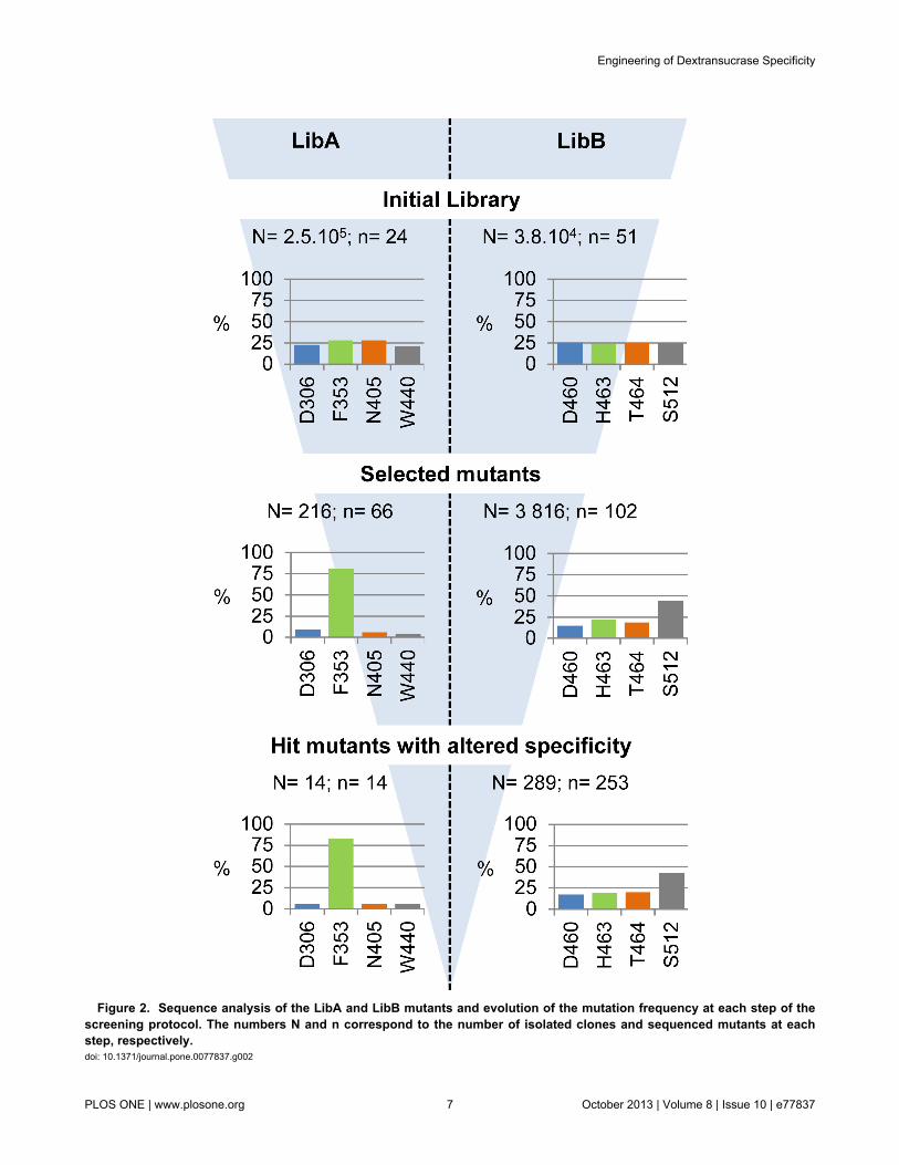
Figure 2. Sequence analysis of the LibA and LibB mutants and evolution of the mutation frequency at each step of thescreening protocol. The numbers N and n correspond to the number of isolated clones and sequenced mutants at eachstep, respectively. doi: 10.1371/journal.pone.0077837.g002
Engineering of Dextransucrase Specificity
PLOS ONE | www.plosone.org 7 October 2013 | Volume 8 | Issue 10 | e77837

From LibB, 3,816 clones exhibiting polymerase activity weredetected (20 % of the mutants selected). Mutational spectrumanalysis (Figure 2) revealed that 44 % of the mutations were atposition 512, showing that residue S512 is more permissive tomutations than residues D460, H463 and T464.
Analysis of the mutants with altered linkage specificityThe 4,032 clones expressing mutants with polymerase
activity were subjected to the high-throughput NMR-basedscreening method previously described [23]. Briefly, themethod is based on the 1D 1H NMR detection of the α(1→3)and α(1→6) linkages in the gluco-oligosaccharides (GOS)synthesized by each mutant, in the presence of sucrose as aglucosyl donor and isomalto-oligosaccharides (IMOS) asacceptor. This IMOS corresponds to a population of smalldextrans with an average molecular mass of 1 kDa and anaverage degree of polymerization of 7. This step allowed us toidentify 14 clones from LibA and 289 from LibB expressing GSwith osidic linkage specificity that was different to that of theparental enzyme. Sequencing of these 303 mutants revealedthat the distribution of mutations was similar to that determinedfrom sequencing of the polymerase-active mutants afterprimary screening (Figure 2). Substantial sequenceredundancy was observed as only 3 and 78 different mutants(at the protein sequence level) were sorted out from LibA andLibB, respectively (Figure 3). Furthermore, all three mutantsfrom LibA displayed mutations at position 353, highlighting therole of this position in subsite +1 for positioning acceptorsduring transglucosylation. Mutants synthesizing GOS with thehighest level of α(1→3) linkage were retrieved from LibB(Figure 3). Almost all the LibB mutants synthesizing GOS withmore than 5 % of α(1→3) linkages exhibited a mutation atposition 512. This is consistent with the role previouslyattributed to the residues downstream from the transition statestabilizer (D511) in the control of linkage specificity[11,15–17,19,20]. Mutant D460M:H463Y:T464M:S512C wasthe “best hit”, with an ability to incorporate more than 8 % ofα(1→3) linkages in the synthesized GOS. The linkagespecificity of the single mutant S512C was not substantiallydifferent from that of the wild type enzyme. In contrast, 10 triplemutants (12 % of the mutants with altered specificity),displaying mutations at positions 460, 463 and 464,synthesized GOS with 4 times more α(1→3) linkages than thewild type enzyme (Figure 3). These findings demonstrate theimportance of recombination in remodeling the active sitetopology and controlling linkage specificity.
This preliminary analysis led us to study seven mutants(F353T; S512C; F353W; H463R:T464D:S512T;H463R:T464V:S512T; D460A:H463S:T464L andD460M:H463Y:T464M:S512C) in more detail. These mutantswere used to produce α-glucans from sucrose only. MostlyHMW α-glucans are synthesized in these reaction conditions.1D 1H NMR analyses of the various products showed that theproportion of α(1→3) linkages was comprised between 3 to 20% (Table 1). The physico-chemical properties of thesepolymers have been described elsewhere and, here, we reportfurther analysis of the relations between the mutations and thecatalytic properties of these mutants.
Relations between mutations, linkage specificity andcatalytic properties
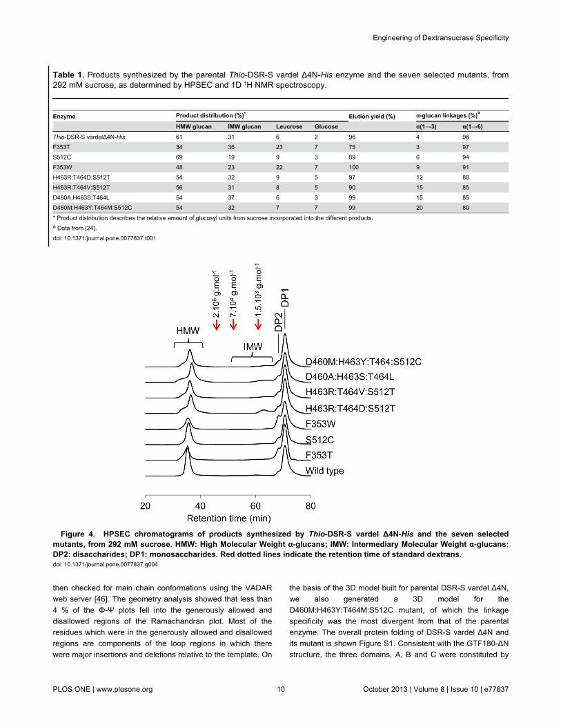
The lowest α(1→3)/α(1→6) linkage ratio was for HMW α-glucans synthesized by mutants exhibiting only a singlemutation. The mutants that synthesized HMW α-glucans withmore than 10 % of α(1→3) linkages harbored three or fourmutations, and the highest proportion of α(1→3) linkages (20%) being obtained by the quadruple mutantD460M:H463Y:T464:S512C. To our knowledge, the role ofthese residues has not previously been explored byglucansucrase engineering. The reaction products were alsosubjected to HPSEC analysis (Figure 4). Like parental enzyme,all the mutants produced two populations of α-glucans referredas HMW α-glucans (molar mass higher than 107 g.mol-1) andIMW glucans (molar mass in the range 0.5.103 to 5.7.103
g.mol-1). The HMW α-glucans produced by these sevenmutants were previously shown to have various structures [24].The seven mutants also catalyzed the formation of leucrose,glucose and fructose (Table 1). Except for the two singlemutants F353T and F353W, the product yields for the mutantsare only slightly affected, with respect to the parental enzyme,suggesting that the mutations at positions D460, H463, T464,and S512 do not greatly alter acceptor binding ability. However,mutants F353T and F353W displayed a 3 to 4-fold higherleucrose yield, suggesting that the mutations may favorfructose binding and thereby result in greater leucroseformation.
Mutants D460M:H463Y:T464M:S512C,D460A:H463S:T464L, H463R:T464D:S512T and F353Tdisplayed a 10 to 30-fold reduction of catalytic efficiency (Table2). For the first three of them, this was mainly due to adecrease of the kcat values whereas for the F353T mutant, theloss of catalytic efficiency is mainly due to a drastic increase ofthe Km value, about 15-times higher than that of the parentalenzyme. Interestingly, F353T mutant also exhibits a 76-foldincrease of Km compared to the F353W mutant. The aromaticring of phenylalanine or tryptophan residues at position 353 islikely to be critical for binding the fructosyl ring in subsite +1.Kinetic analyses revealed that, as previously shown for theparental enzyme [11], five variants —F353T, S512C,H463R:T464D:S512T, D460A:H463S:T464L andD460M:H463Y:T464M:S512C— were inhibited by sucroseexcess. By contrast, there was no such inhibition of mutantsF353W and H463R:T464V:S512T. Notably, mutantH463R:T464V:S512T, that synthesizes HMW α-glucans with15 % α(1→3) linkages, is slightly more efficient than theparental enzyme. Moreover, at a substrate concentration of600 mM, the initial velocity of H463R:T464V:S512T is 3 timeshigher than that of DSR-S vardel Δ4N. In contrast, mutantH463R:T464D:S512T, differing only by the nature of the aminoacid substitution at position 464, is eight times less efficientthan the parental enzyme. This reveals a critical role for thisposition.
3D- modeling of DSR-S catalytic domainA 3D-model of the DSR-S vardel Δ4N catalytic domain was
generated from the structure of Lb. reuteri GTF180-ΔN andsequence alignment of these two enzymes. The model was
Engineering of Dextransucrase Specificity
PLOS ONE | www.plosone.org 8 October 2013 | Volume 8 | Issue 10 | e77837

Figure 3. Mutants with altered specificity identified by NMR-based screening. doi: 10.1371/journal.pone.0077837.g003
Engineering of Dextransucrase Specificity
PLOS ONE | www.plosone.org 9 October 2013 | Volume 8 | Issue 10 | e77837
then checked for main chain conformations using the VADARweb server [46]. The geometry analysis showed that less than4 % of the Φ-Ψ plots fell into the generously allowed anddisallowed regions of the Ramachandran plot. Most of theresidues which were in the generously allowed and disallowedregions are components of the loop regions in which therewere major insertions and deletions relative to the template. On
the basis of the 3D model built for parental DSR-S vardel Δ4N,we also generated a 3D model for theD460M:H463Y:T464M:S512C mutant, of which the linkagespecificity was the most divergent from that of the parentalenzyme. The overall protein folding of DSR-S vardel Δ4N andits mutant is shown Figure S1. Consistent with the GTF180-ΔNstructure, the three domains, A, B and C were constituted by
Table 1. Products synthesized by the parental Thio-DSR-S vardel Δ4N-His enzyme and the seven selected mutants, from292 mM sucrose, as determined by HPSEC and 1D 1H NMR spectroscopy.
Enzyme Product distribution (%)* Elution yield (%) α-glucan linkages (%)a
HMW glucan IMW glucan Leucrose Glucose α(1→3) α(1→6)Thio-DSR-S vardelΔ4N-His 61 31 6 2 96 4 96F353T 34 36 23 7 75 3 97S512C 69 19 9 3 89 6 94F353W 48 23 22 7 100 9 91H463R:T464D:S512T 54 32 9 5 97 12 88H463R:T464V:S512T 56 31 8 5 90 15 85D460A:H463S:T464L 54 37 6 3 99 15 85D460M:H463Y:T464M:S512C 54 32 7 7 99 20 80
* Product distribution describes the relative amount of glucosyl units from sucrose incorporated into the different products.a Data from [24].doi: 10.1371/journal.pone.0077837.t001
Figure 4. HPSEC chromatograms of products synthesized by Thio-DSR-S vardel Δ4N-His and the seven selectedmutants, from 292 mM sucrose. HMW: High Molecular Weight α-glucans; IMW: Intermediary Molecular Weight α-glucans;DP2: disaccharides; DP1: monosaccharides. Red dotted lines indicate the retention time of standard dextrans. doi: 10.1371/journal.pone.0077837.g004
Engineering of Dextransucrase Specificity
PLOS ONE | www.plosone.org 10 October 2013 | Volume 8 | Issue 10 | e77837

non-continuous sequence regions. In domain A, the (β/α)8-barrel catalytic domain is composed of three stretches of aminoacids (364-614, 745-880 and 917-959). Domain B is insertedbetween helix α1 and strand β8 and also consists of threestretches (296-364, 880-917 and 959-973) and domain C iscomposed by residues 614-745 and forms the central part ofthe U-shaped protein.
Description of the catalytic site, docking of substrate,and putative products
The 3D-model of DSR-S vardel Δ4N with sucrose bound inthe catalytic site was used to describe binding interactions withthe glucosyl ring at subsite -1 and the fructosyl ring at subsite+1 (Figure 5A). The topology of the enzyme active site appears
Table 2. Kinetic properties of seven mutants with alteredspecificity, and of the wt Thio-DSR-S vardel Δ4N-His(values are means of three independent assays).
Enzyme Km (mM) Ks (mM) kcat (s-1)kcat/Km(s-1.mM-1)
Thio-DSR-S vardelΔ4N-His 7.75* 326* 584* 75.4F353T 115.8±nd 46.6±nd 338.5±nd 2.9S512C 3.9±1.0 405.8±65.9 207.1±9.7 53.1F353W 1.5±0.1 - 86±1.2 57.3H463R:T464D:S512T 15.5±1.4 201.7±17.7 172.1±6.7 11.1H463R:T464V:S512T 6.9±0.2 - 610.2±2.7 88.4D460A:H463S:T464L 5.3±0.5 552.7±54.8 43.9±0.9 8.3D460M:H463Y:T464M:S512C 15.5±2.8 175.1±36.7 38.2±3.2 2.5
* Data from [25].doi: 10.1371/journal.pone.0077837.t002
to be a shallow pocket with the catalytic residues, namely theacid/base E438, the nucleophile D400 and the transition statestabilizer D511 (E1063, D1025 and D1136 in GTF180-ΔN,respectively), located at the bottom. The glucosyl moiety stacksonto Y834 in subsite -1 and establishes hydrogen bondinginteractions with H510, D511 and Y880, while the β-D-fructosylmoiety interacts mostly with D511 in subsite +1. Theseresidues are conserved in GTF180-ΔN.
The 3D-model of DSR-S vardel Δ4N was used to locate theeight amino acid positions targeted for randomization (Figure5B). All belong to the first shell of amino acid residues less than20 Å away from catalytic residues, residue S512 being thenearest as it is adjacent to catalytic D511. All the targetedpositions are thus appropriately located to be involved in thebinding of sucrose and oligosaccharides in the catalytic site.This is in agreement with the data obtained from the libraryanalysis and kinetic characterization of the seven mutants withaltered specificity.
To investigate the influence of the mutations on acceptorrecognition, we performed docking studies of thetetrasaccharide α-D-Glcp-(1→6)[α-D-Glcp-(1→3)] α-D-Glcp-(1→6)-D-Glcp in the 3D model ofD460M:H463Y:T464M:S512C. This tetrasaccharide is aputative product of glucosyl transfer onto isomaltotriose withthe formation of an α(1→3) linkage. The favored binding modeof this tetrasaccharide is shown in Figure 6. The glucosyl ringat subsite -1 binds in a very similar way to the glucosyl unit ofsucrose. The glucosyl unit at subsite +1 was also found in theproductive conformation with respect to the acid/base E438 forthe formation of an α(1→3) linkage. The +2 subsiteaccommodates the non-reducing end of the isomaltotriosethrough van der Waals interactions with W440T.
Figure 5. View of substrate binding in the active site of DSR-S vardel Δ4N. (A) Molecular docking of sucrose in theactive site of DSR-S vardel Δ4N. (B) Overview of the positions of mutations affecting DSR-S vardel Δ4N linkage specificity.A docked sucrose molecule is shown for reference. Targeted residues are in orange (LibA) and green (LibB). doi: 10.1371/journal.pone.0077837.g005
Engineering of Dextransucrase Specificity
PLOS ONE | www.plosone.org 11 October 2013 | Volume 8 | Issue 10 | e77837

The +2’ subsite is formed by a small pocket between loopscarrying the catalytic residues D400 and E438. The topology ofthis pocket is also determined by amino acids F353 and N404,targeted in LibA. It is therefore plausible that the mutations atthese two positions, F353W and N404G, affect binding pockettopology and thus acceptor recognition. The positions at whichmutations affect the linkage specificity, as in theD460M:H463Y:T464M:S512C mutant, are located in closespatial proximity to W440 (+2 subsite). In particular, the H463Ymutation could improve the stacking platform allowinginteraction with longer oligosaccharide chains and therebyfavor their binding. The presence of a polar side chain atposition 512 (serine in the parental enzyme or cysteine in themutant) also appears to be required for establishing hydrogenbonding interactions with hydroxyl groups of the acceptorglucosyl unit bound at +2 subsite. This is consistent with theputative role of S512 in oligosaccharide binding, as suggestedpreviously [11].
The mutations introduced at subsite +2 thus seem topromote productive binding of α(1→6) linkedglucooligosaccharides in the active site and favor the α(1→3)branching transglucosylation of glucan chains. However, thebranching pattern of products synthesized by mutantD460M:H463Y:T464M:S512C will have to be characterized inmore details to allow a better understanding of how the protein
accommodates oligosaccharide molecules in the catalyticpocket, and how this differs from the parental DSR-S vardelΔ4N.
Conclusions
Herein, we demonstrate the value of combinatorialengineering for investigating the relationships betweenstructure and specificity of glucansucrases. Focusing on thecatalytic domain of DSR-S vardel Δ4N dextransucrase, weidentified key positions involved in the control of its specificity.In particular, this is the first time that the role of the peptidesequence 460DYVHT464 was investigated and demonstrated tobe essential for α(1→3) linkage formation. It appears thatmodifications of acceptor binding at subsite +2 are responsiblefor the promotion of α(1→3) linkage formation. We alsodescribe the design of catalytic pocket-directed libraries ofmutants, combined with efficient screening protocols. Thisallowed the isolation of eighty-two variants producing HMW α-glucans with various proportions of α(1→3) linkages. Theselibraries of mutants are novel enzymatic tools forglycodiversification.
Figure 6. Molecular docking of α-D-Glcp-(1→6)[α-D-Glcp-(1→3)]α-D-Glcp-(1→6)-D-Glcp tetrasaccharide in the active siteof the D460M:H463Y:T464M:S512C mutant. doi: 10.1371/journal.pone.0077837.g006
Engineering of Dextransucrase Specificity
PLOS ONE | www.plosone.org 12 October 2013 | Volume 8 | Issue 10 | e77837
Supporting Information
Table S1. Primers and degenerated oligonucleotides usedfor combinatorial site-directed mutagenesis of dsr-s vardelΔ4N. Underlined nucleotides correspond to the introducedrestriction sites.(DOCX)
Figure S1. Overall structure of GS catalytic domain. (A)3D-model of DSR-S vardel Δ4N catalytic domain. (B)Catalytic domain of Lactobacillus reuteri N-terminallytruncated glucansucrase GTF180 (PDB accession code:3KLK).
(DOCX)
Acknowledgements
We cordially thank Joana Lima and Marta Leal for technicalsupport. We thank S. Bozonnet and S. Pizzut-Serin forassistance with the use of the ICEO automated facility.
Author Contributions
Conceived and designed the experiments: RI IA PM GPVMRS. Performed the experiments: RI LT IA. Analyzed the data:RI IA GPV MRS. Contributed reagents/materials/analysis tools:RI CM SM. Wrote the manuscript: RI IA GPV MRS.
References
1. André I, Potocki-Véronèse G, Morel S, Monsan P, Remaud-Siméon M(2010) Sucrose-utilizing transglucosidases for biocatalysis. Top CurrChem 294: 25–48. doi:10.1007/128_2010_52. PubMed: 21626747.
2. Van Hijum SAFT, Kralj S, Ozimek LK, Dijkhuizen L, van Geel-SchuttenIGH (2006) Structure-function relationships of glucansucrase andfructansucrase enzymes from lactic acid bacteria. Microbiol Mol BiolRev 70: 157–176. doi:10.1128/MMBR.70.1.157-176.2006. PubMed:16524921.
3. Cantarel BL, Coutinho PM, Rancurel C, Bernard T, Lombard V et al.(2009) The Carbohydrate-Active EnZymes database (CAZy): an expertresource for Glycogenomics. Nucleic Acids Res 37: 233–238. doi:10.1093/nar/gkn663. PubMed: 18838391.
4. Pijning T, Vujičić-Žagar A, Kralj S, Eeuwema W, Dijkhuizen L et al.(2008) Biochemical and crystallographic characterization of aglucansucrase from Lactobacillus reuteri 180. Biocatal Biotransform 26:12–17. doi:10.1080/10242420701789163.
5. Vujicic-Zagar A, Pijning T, Kralj S, López CA, Eeuwema W et al. (2010)Crystal structure of a 117 kDa glucansucrase fragment provides insightinto evolution and product specificity of GH70 enzymes. Proc Natl AcadSci USA 107: 21406–21411. doi:10.1073/pnas.1007531107. PubMed:21118988.
6. Ito K, Ito S, Shimamura T, Kawarasaki Y, Abe K et al. (2010)Crystallization and preliminary X-ray analysis of a glucansucrase fromthe dental caries pathogen Streptococcus mutans. Acta CrystallogrSect Struct 66: 1086–1088. doi:10.1107/S1600536810031028.PubMed: 20823533.
7. Ito K, Ito S, Shimamura T, Weyand S, Kawarasaki Y et al. (2011)Crystal structure of glucansucrase from the dental caries pathogenStreptococcus mutans. J Mol Biol 408: 177–186. doi:10.1016/j.jmb.2011.02.028. PubMed: 21354427.
8. Brison Y, Pijning T, Malbert Y, Fabre É, Mourey L et al. (2012)Functional and structural characterization of α-(1→2) branchingsucrase derived from DSR-E glucansucrase. J Biol Chem 287: 7915–7924. doi:10.1074/jbc.M111.305078. PubMed: 22262856.
9. Pijning T, Vujičić-Žagar A, Kralj S, Dijkhuizen L, Dijkstra BW (2012)Structure of the α-1,6/α-1,4-specific glucansucrase GTFA fromLactobacillus reuteri 121. Acta Crystallogr F 68: 1448–1454. doi:10.1107/S1744309112044168.
10. Davies GJ, Wilson KS, Henrissat B (1997) Nomenclature for sugar-binding subsites in glycosyl hydrolases. Biochem J 321 (2): 557–559.PubMed: 9020895.
11. Moulis C, Joucla G, Harrison D, Fabre E, Potocki-Veronese G et al.(2006) Understanding the polymerization mechanism of glycoside-hydrolase family 70 glucansucrases. J Biol Chem 281: 31254–31267.doi:10.1074/jbc.M604850200. PubMed: 16864576.
12. Leemhuis H, Pijning T, Dobruchowska JM, van Leeuwen SS, Kralj S etal. (2013) Glucansucrases: Three-dimensional structures, reactions,mechanism, α-glucan analysis and their implications in biotechnologyand food applications. Gene Prod Gene Prod 163: 250–272. PubMed:22796091.
13. Shimamura A, Nakano YJ, Mukasa H, Kuramitsu HK (1994)Identification of amino acids residues in Streptococcus mutansglucosyltransferases influencing the structure of the glucan product. JBacteriol 176: 4845–4850. PubMed: 8050997.
14. Hellmuth H, Wittrock S, Kralj S, Dijkhuizen L, Hofer B et al. (2008)Engineering the glucansucrase GTFR enzyme reaction and glycosidic
bond specificity: toward tailor-made polymer and oligosaccharideproducts. Biochemistry (Mosc) 47: 6678–6684. doi:10.1021/bi800563r.PubMed: 18512955.
15. Van Leeuwen SS, Kralj S, Eeuwema W, Gerwig GJ, Dijkhuizen L et al.(2009) Structural characterization of bioengineered α-D-glucansproduced by mutant glucansucrase GTF180 enzymes of Lactobacillusreuteri strain 180. Biomacromolecules 10: 580–588. doi:10.1021/bm801240r. PubMed: 19186951.
16. Van Leeuwen SS, Kralj S, Gerwig GJ, Dijkhuizen L, Kamerling JP(2008) Structural analysis of bioengineered α-D-glucan produced by atriple mutant of the glucansucrase GTF180 enzyme from Lactobacillusreuteri strain 180: generation of (α1→4) linkages in a native (1→3)(1→6)-α-D-glucan. Biomacromolecules 9: 2251–2258. doi:10.1021/bm800410w. PubMed: 18616317.
17. Kang HK, Kimura A, Kim D (2011) Bioengineering of Leuconostocmesenteroides glucansucrases that gives selected bond formation forglucan snthesis and/or acceptor-product synthesis. J Agric Food Chem59: 4148–4155. doi:10.1021/jf104629g. PubMed: 21391600.
18. Monchois V, Vignon M, Russell RR (2000) Mutagenesis of asp-569 ofglucosyltransferase I glucansucrase modulates glucan andoligosaccharide synthesis. Appl Environ Microbiol 66: 1923–1927. doi:10.1128/AEM.66.5.1923-1927.2000. PubMed: 10788361.
19. Kralj S, van Geel-Schutten IG, Faber EJ, van der Maarel MJ,Dijkhuizen L (2005) Rational transformation of Lactobacillus reuteri 121reuteransucrase into a dextransucrase. Biochemistry (Mosc) 44: 9206–9216. doi:10.1021/bi050447q. PubMed: 15966745.
20. Kralj S, Eeuwema W, Eckhardt TH, Dijkhuizen L (2006) Role ofasparagine 1134 in glucosidic bond and transglycosylation specificity ofreuteransucrase from Lactobacillus reuteri 121. FEBS J 273: 3735–3742. doi:10.1111/j.1742-4658.2006.05376.x. PubMed: 16911522.
21. Kralj S, Van Leeuwen SS, Valk V, Eeuwema W, Kamerling JP et al.(2008) Hybrid reuteransucrase enzymes reveal regions important forglucosidic linkage specificity and the transglucosylation/hydrolysis ratio.FEBS J 275: 6002–6010. doi:10.1111/j.1742-4658.2008.06729.x.PubMed: 19016850.
22. Funane K, Ishii T, Terasawa K, Yamamoto T, Kobayashi M (2004)Construction of chimeric glucansucrases for analyzing substrate-binding regions that affect the structure of glucan products. BiosciBiotechnol Biochem 68: 1912–1920. doi:10.1271/bbb.68.1912.PubMed: 15388967.
23. Irague R, Massou S, Moulis C, Saurel O, Milon A et al. (2011) NMR-based structural glycomics for high-throughput screening ofcarbohydrate-active enzyme specificity. Anal Chem 83: 1202–1206.doi:10.1021/ac1032148. PubMed: 21271685.
24. Irague R, Rolland-Sabaté A, Tarquis L, Doublier JL, Moulis C et al.(2011) Structure and property engineering of α-D-glucans synthesizedby dextransucrase mutants. Biomacromolecules 13: 187–195. PubMed:22098057.
25. Moulis C, Arcache A, Escalier PC, Rinaudo M, Monsan P et al. (2006)High-level production and purification of a fully active recombinantdextransucrase from Leuconostoc mesenteroides NRRL B-512F.FEMS Microbiol Lett 261: 203–210. doi:10.1111/j.1574-6968.2006.00347.x. PubMed: 16907721.
26. Sanger F, Nicklen S, Coulson AR (1977) DNA sequencing with chain-terminating inhibitors. Proc Natl Acad Sci USA 74: 5463–5467. doi:10.1073/pnas.74.12.5463. PubMed: 271968.
Engineering of Dextransucrase Specificity
PLOS ONE | www.plosone.org 13 October 2013 | Volume 8 | Issue 10 | e77837

27. Studier FW (2005) Protein production by auto-induction in high densityshaking cultures. Protein Expr Purif 41: 207–234. doi:10.1016/j.pep.2005.01.016. PubMed: 15915565.
28. Sumner JB, Howell SF (1935) A method for determination ofsaccharase activity. J Biol Chem 108: 51–54.
29. Seymour FR, Knapp RD, Bishop SH (1979) Correlation of the structureof dextrans to their 1H-N.M.R. spectra. Carbohydr Res 74: 77–92. doi:10.1016/S0008-6215(00)84766-7.
30. Van Leeuwen SS, Kralj S, Van Geel-Schutten IH, Gerwig GJ,Dijkhuizen L et al. (2008) Structural analysis of the α-D-glucan(EPS180) produced by the Lactobacillus reuteri strain 180glucansucrase GTF180 enzyme. Carbohydr Res 343: 1237–1250. doi:10.1016/j.carres.2008.01.042. PubMed: 18313038.
31. Maina NH, Tenkanen M, Maaheimo H, Juvonen R, Virkki L (2008) NMRspectroscopic analysis of exopolysaccharides produced byLeuconostoc citreum and Weissella confusa. Carbohydr Res 343:1446–1455. doi:10.1016/j.carres.2008.04.012. PubMed: 18452899.
32. Roy A, Kucukural A, Zhang Y (2010) I-TASSER: a unified platform forautomated protein structure and function prediction. Nat Protoc 5: 725–738. doi:10.1038/nprot.2010.5. PubMed: 20360767.
33. Zhang Y (2008) I-TASSER server for protein 3D structure prediction.BMC Bioinformatics 9: 40. doi:10.1186/1471-2105-9-40. PubMed:18215316.
34. Zhang Y (2009) I-TASSER: fully automated protein structure predictionin CASP8. Proteins 77 Suppl 9: 100–113. doi:10.1002/prot.22588.PubMed: 19768687.
35. Kramer B, Rarey M, Lengauer T (1999) Evaluation of the FLEXXincremental construction algorithm for protein–ligand docking. ProteinsStruct Funct Bioinf 37: 228. doi:10.1002/(SICI)1097-0134(19991101)37:2.
36. Rarey M, Kramer B, Lengauer T, Klebe G (1996) A fast flexible dockingmethod using an incremental construction algorithm. J Mol Biol 261:470–489. doi:10.1006/jmbi.1996.0477. PubMed: 8780787.
37. Kralj S, Stripling E, Sanders P, van Geel-Schutten GH, Dijkhuizen L(2005) Highly hydrolytic reuteransucrase from probiotic Lactobacillusreuteri strain ATCC 55730. Appl Environ Microbiol 71: 3942–3950. doi:10.1128/AEM.71.7.3942-3950.2005. PubMed: 16000808.
38. Kralj S, van Geel-Schutten GH, van der Maarel MJ, Dijkhuizen L (2004)Biochemical and molecular characterization of Lactobacillus reuteri 121
reuteransucrase. Microbiology 150: 2099–2112. doi:10.1099/mic.0.27105-0. PubMed: 15256553.
39. Côté GL, Robyt JF (1982) Isolation and partial characterization of anextracellular glucansucrase from Leuconostoc mesenteroides NRRLB-1355 that synthesizes an alternating (1-6), (1-3)-α-D-glucan.Carbohydr Res 101: 57–74. doi:10.1016/S0008-6215(00)80795-8.PubMed: 7060056.
40. Argüello-Morales MA, Remaud-Simeon M, Pizzut S, Sarçabal P,Willemot RM et al. (2000) Sequence analysis of the gene encodingalternansucrase, a sucrose glucosyltransferase from Leuconostocmesenteroides NRRL B-1355. FEMS Microbiol Lett 182: 81–85. doi:10.1111/j.1574-6968.2000.tb08878.x. PubMed: 10612736.
41. Fabre E, Bozonnet S, Arcache A, Willemot RM, Vignon M et al. (2005)Role of the two catalytic domains of DSR-E dextransucrase and theirinvolvement in the formation of highly alpha-1,2 branched dextran. JBacteriol 187: 296–303. doi:10.1128/JB.187.1.296-303.2005. PubMed:15601714.
42. Fraga Vidal R, Moulis C, Escalier P, Remaud-Siméon M, Monsan P(2011) Isolation of a gene from Leuconostoc citreum B/110-1-2encoding a novel dextransucrase enzyme. Curr Microbiol 62: 1260–1266. doi:10.1007/s00284-010-9851-7. PubMed: 21229247.
43. MacGregor EA, Jespersen HM, Svensson B (1996) A circularlypermuted alpha-amylase-type alpha/beta-barrel structure in glucan-synthesizing glucosyltransferases. FEBS Lett 378: 263–266. doi:10.1016/0014-5793(95)01428-4. PubMed: 8557114.
44. Herman A, Tawfik DS (2007) Incorporating synthetic oligonucleotidesvia gene reassembly (ISOR): a versatile tool for generating targetedlibraries. Proteins Eng Sel 20: 219–226. doi:10.3934/dcds.2008.20.219.PubMed: 17483523.
45. Van Der Veen BA, Potocki-Véronèse G, Albenne C, Joucla G, MonsanP et al. (2004) Combinatorial engineering to enhance amylosucraseperformance: construction, selection, and screening of variant librariesfor increased activity. FEBS Lett 560: 91–97. doi:10.1016/S0014-5793(04)00077-8. PubMed: 14988004.
46. Willard L, Ranjan A, Zhang H, Monzavi H, Boyko RF et al. (2003)VADAR: a web server for quantitative evaluation of protein structurequality. Nucleic Acids Res 31: 3316–3319. doi:10.1093/nar/gkg565.PubMed: 12824316.
Engineering of Dextransucrase Specificity
PLOS ONE | www.plosone.org 14 October 2013 | Volume 8 | Issue 10 | e77837