a combinatorial regulatory signature controls terminal
TRANSCRIPT
A combinatorial regulatory signaturecontrols terminal differentiationof the dopaminergic nervous systemin C. elegans
Maria Doitsidou,1,2,3,4,8,9 Nuria Flames,1,2,5,8,9 Irini Topalidou,6,7 Namiko Abe,1 Terry Felton,1,2
Laura Remesal,1,2,5 Tatiana Popovitchenko,1,2,3,4 Richard Mann,1 Martin Chalfie,6 and Oliver Hobert1,2
1Department of Biochemistry and Molecular Biophysics, 2Howard Hughes Medical Institute, Columbia University MedicalCenter, New York, New York 10032, USA; 3Norwegian Center for Movement Disorders, Stavanger University Hospital,Stavanger N-4068, Norway; 4Center for Organelle Research, University of Stavanger, Stavanger N-4036, Norway; 5Instituto deBiomedicina de Valencia-Consejo Superior de Investigaciones Cientıficas (IBV-CSIC), 46010 Valencia, Spain; 6Department ofBiological Sciences, Columbia University, New York, New York 10027, USA; 7Department of Biochemistry, University ofWashington, Seattle, Washington 98195, USA
Terminal differentiation programs in the nervous system are encoded by cis-regulatory elements that control theexpression of terminal features of individual neuron types. We decoded the regulatory information that controlsthe expression of five enzymes and transporters that define the terminal identity of all eight dopaminergic neuronsin the nervous system of the Caenorhabditis elegans hermaphrodite. We show that the tightly coordinated, robustexpression of these dopaminergic enzymes and transporters (‘‘dopamine pathway’’) is ensured through a combi-natorial cis-regulatory signature that is shared by all dopamine pathway genes. This signature is composed of anEts domain-binding site, recognized by the previously described AST-1 Ets domain factor, and two distinct types ofhomeodomain-binding sites that act in a partially redundant manner. Through genetic screens, we identified thesole C. elegans Distalless/Dlx ortholog, ceh-43, as a factor that acts through one of the homeodomain sites tocontrol both induction and maintenance of terminal dopaminergic fate. The second type of homeodomain site isa Pbx-type site, which is recognized in a partially redundant and neuron subtype-specific manner by two Pbxfactors, ceh-20 and ceh-40, revealing novel roles of Pbx factors in the context of terminal neuron differentiation.Taken together, we revealed a specific regulatory signature and cognate, terminal selector-type transcriptionfactors that define the entire dopaminergic nervous system of an animal. Dopaminergic neurons in the mouseolfactory bulb express a similar combinatorial transcription factor collective of Ets/Dlx/Pbx factors, suggestingdeep phylogenetic conservation of dopaminergic regulatory programs.
[Keywords: cis-regulatory motif; differentiation; dopamine; elegans; homeodomain; neuron]
Supplemental material is available for this article.
Received May 6, 2013; revised version accepted May 16, 2013.
The underlying basis of the functional and anatomicaldiversity of cell types in the nervous system is the differ-ential expression of neuron-type-specific gene batteries,which are composed of terminal differentiation geneswhose products define the specific properties of a matureneuron throughout its lifetime (Hobert et al. 2010). These‘‘terminal differentiation’’ or ‘‘effector’’ genes include, forexample, neurotransmitter-synthesizing enzymes, neu-
rotransmitter receptors, transporters, the many ion chan-nels that tune the electric properties of a neuron, specificsynaptic adhesion molecules, and many others. With thisnotion in mind, one approach to understand the genera-tion of neuronal diversity is a ‘‘bottom-up’’ approach thatfocuses on studying the cis-regulatory principles by whichthe expression of terminal gene batteries is controlled. Inspite of the conceptual promise of such an approach, thecis-regulatory architecture of neuronal gene batteries isgenerally poorly described.
The neurotransmitter dopamine controls a number ofdiverse behaviors across the animal kingdom (Iversen andIversen 2007). Dopaminergic neurons are molecularlydefined through the coordinated expression of five genes
8These authors contributed equally.9Corresponding authorsE-mail [email protected] [email protected] is online at http://www.genesdev.org/cgi/doi/10.1101/gad.217224.113.
GENES & DEVELOPMENT 27:1391–1405 � 2013 by Cold Spring Harbor Laboratory Press ISSN 0890-9369/13; www.genesdev.org 1391
Cold Spring Harbor Laboratory Press on January 26, 2022 - Published by genesdev.cshlp.orgDownloaded from
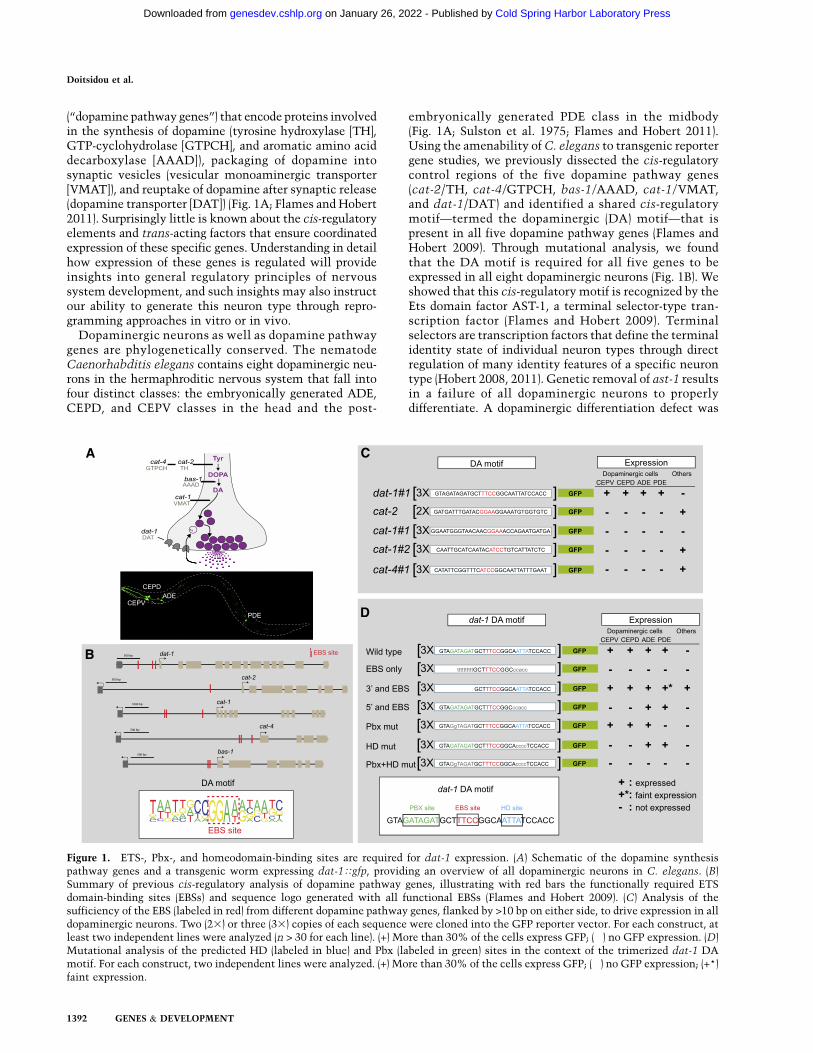
(‘‘dopamine pathway genes’’) that encode proteins involvedin the synthesis of dopamine (tyrosine hydroxylase [TH],GTP-cyclohydrolase [GTPCH], and aromatic amino aciddecarboxylase [AAAD]), packaging of dopamine intosynaptic vesicles (vesicular monoaminergic transporter[VMAT]), and reuptake of dopamine after synaptic release(dopamine transporter [DAT]) (Fig. 1A; Flames and Hobert2011). Surprisingly little is known about the cis-regulatoryelements and trans-acting factors that ensure coordinatedexpression of these specific genes. Understanding in detailhow expression of these genes is regulated will provideinsights into general regulatory principles of nervoussystem development, and such insights may also instructour ability to generate this neuron type through repro-gramming approaches in vitro or in vivo.
Dopaminergic neurons as well as dopamine pathwaygenes are phylogenetically conserved. The nematodeCaenorhabditis elegans contains eight dopaminergic neu-rons in the hermaphroditic nervous system that fall intofour distinct classes: the embryonically generated ADE,CEPD, and CEPV classes in the head and the post-
embryonically generated PDE class in the midbody(Fig. 1A; Sulston et al. 1975; Flames and Hobert 2011).Using the amenability of C. elegans to transgenic reportergene studies, we previously dissected the cis-regulatorycontrol regions of the five dopamine pathway genes(cat-2/TH, cat-4/GTPCH, bas-1/AAAD, cat-1/VMAT,and dat-1/DAT) and identified a shared cis-regulatorymotif—termed the dopaminergic (DA) motif—that ispresent in all five dopamine pathway genes (Flames andHobert 2009). Through mutational analysis, we foundthat the DA motif is required for all five genes to beexpressed in all eight dopaminergic neurons (Fig. 1B). Weshowed that this cis-regulatory motif is recognized by theEts domain factor AST-1, a terminal selector-type tran-scription factor (Flames and Hobert 2009). Terminalselectors are transcription factors that define the terminalidentity state of individual neuron types through directregulation of many identity features of a specific neurontype (Hobert 2008, 2011). Genetic removal of ast-1 resultsin a failure of all dopaminergic neurons to properlydifferentiate. A dopaminergic differentiation defect was
Figure 1. ETS-, Pbx-, and homeodomain-binding sites are required for dat-1 expression. (A) Schematic of the dopamine synthesispathway genes and a transgenic worm expressing dat-1Tgfp, providing an overview of all dopaminergic neurons in C. elegans. (B)Summary of previous cis-regulatory analysis of dopamine pathway genes, illustrating with red bars the functionally required ETSdomain-binding sites (EBSs) and sequence logo generated with all functional EBSs (Flames and Hobert 2009). (C) Analysis of thesufficiency of the EBS (labeled in red) from different dopamine pathway genes, flanked by >10 bp on either side, to drive expression in alldopaminergic neurons. Two (23) or three (33) copies of each sequence were cloned into the GFP reporter vector. For each construct, atleast two independent lines were analyzed (n > 30 for each line). (+) More than 30% of the cells express GFP; (�) no GFP expression. (D)Mutational analysis of the predicted HD (labeled in blue) and Pbx (labeled in green) sites in the context of the trimerized dat-1 DAmotif. For each construct, two independent lines were analyzed. (+) More than 30% of the cells express GFP; (�) no GFP expression; (+*)faint expression.
Doitsidou et al.
1392 GENES & DEVELOPMENT
Cold Spring Harbor Laboratory Press on January 26, 2022 - Published by genesdev.cshlp.orgDownloaded from
also observed in olfactory bulb dopaminergic neurons ofmice lacking the AST-1 homolog Etv1 (Flames and Hobert2009). One key issue that remained unresolved by ourprevious studies is the question of specificity. While theEts domain transcription factor AST-1 and mouse Etv1are required to generate dopaminergic neurons, they arenot sufficient to do so, since both genes are expressed inmultiple other, nondopaminergic neuron types. Similarspecificity issues apply to many other terminal selector-type transcription factors that are required to define theidentity of specific neuron types in vertebrate and in-vertebrate nervous systems but are often expressed inmany other cell types as well (Hobert 2011; Holmberg andPerlmann 2012).
Here we investigated this issue of specificity using acombination of cis-regulatory mutational analysis andgenetic screening approaches. We found that ast-1 indeeddoes not act in isolation but rather through a combinato-rial cis-regulatory signature that is present in the cis-regulatory regions of all dopamine pathways genes. Weidentified three distinct types of trans-acting factors thatrecognize this cis-regulatory signature and show thatthese factors act together as a ‘‘transcription factorcollective’’ (Junion et al. 2012) to ensure robust executionof the terminal differentiation program of dopaminergicneurons. Mouse homologs of this transcription factorcollective are also expressed in a specific population ofdopaminergic neurons in vertebrates, suggesting that ourfindings may also apply to mammals.
Results
A combinational cis-regulatory signature requiredfor gene expression in dopaminergic neurons
We previously showed that a construct containing intriplicate a 31-base-pair (bp) element from the dat-1promoter is sufficient to drive expression of a reportergene in all eight dopaminergic neurons of the C. eleganshermaphrodite (Fig. 1C; Flames and Hobert 2009). Thiselement contains an Ets domain-binding site (EBS), whichwe showed through deletion analysis to be essential forexpression of dat-1 in dopaminergic neurons. We foundfunctional EBSs in all of the dopamine pathway genes andtherefore called the EBS the ‘‘DA motif’’ (Fig. 1B; Flamesand Hobert 2009). Unexpectedly, when we tested the DAmotifs from other dopamine pathway genes in a mannersimilar to our testing of the DA motif from dat-1 (i.e., EBSplus 13–14 bp of flanking sequences), we found that,unlike in the dat-1 case, DA motifs from none of theother four dopamine pathway genes were sufficient todrive expression in dopaminergic neurons (Fig. 1C) eventhough each of the DA motifs is required to drive expres-sion in all dopaminergic neurons (Flames and Hobert2009).
We therefore examined the 31-bp DA motif from thedat-1 promoter in more detail. Using MatInspector soft-ware analysis, we noted the presence of a predicted Pbx-type homeodomain (HD)-binding site [‘‘GAT(N)1-2GAT’’]and a canonical HD-binding site (‘‘TAAT’’) flanking the
EBS. Mutating either site alone had partial effects on theexpression of the reporter gene in dopaminergic neurons,while mutating both sites simultaneously—leaving, at thesame time, the EBS intact—completely abolished expres-sion of dat-1 in all dopaminergic neuron types (Fig. 1D).
The DA motif-containing 30- to 32-bp elements fromeach of the other four dopamine pathway genes do notcontain a combination of predicted Pbx- and canonicalHD-binding sites, thereby providing a potential explana-tion for their insufficiency to drive expression in dopa-minergic neurons. However, each of the minimal regionsfrom all other dopamine pathway genes driving dopami-nergic neuron expression contained a set of predictedPbx- and HD-binding sites similar to those observed inthe dat-1 promoter. We systematically mutated thesepredicted Pbx and HD sites in four of the five dopaminepathway genes and found that they are required for do-paminergic neuron expression (Fig. 2A–D). Unlike theEBS, which is essential for expression of all dopaminepathway genes (Flames and Hobert 2009), the Pbx and HDsites act in a partially redundant manner, as detailed below.
dat-1/DAT The minimal dat-1 cis-regulatory elementthat drives expression in all dopaminergic neurons (400bp) contains two predicted HD sites, one predicted Pbxsite, and three functional EBSs (previously shown to beessential for expression) (Fig. 2A). Mutating a single HDsite has no noticeable effect on dat-1 reporter geneexpression in transgenic animals (data not shown), andmutating both HD sites has partial effects, similar to theeffect of the HD mutation in the dat-1 DA motif construct(Figs. 1D, 2A; Supplemental Fig. 1). Mutating the one Pbxsite has no effect (data not shown). However, mutatingboth HD sites and the one Pbx site together completelyeliminates reporter gene expression in all dopaminergicneurons (even though the Ets sites are still present) (Fig.2A; Supplemental Fig. 1), again in concordance with theeffect seen in the dat-1 DA motif construct analysis.
cat-2/TH The minimal cat-2 cis-regulatory elementthat drives expression in all dopaminergic neurons (153bp) also contains two potential canonical HD sites, onepotential Pbx site, and one functional EBS (previouslyshown to be essential) (Fig. 2B). The effects of the mutationof individual sites are similar to the dat-1 case. Mutationof the first HD site results in partial effects, whereasmutation of the second HD site does not have any effect.The combined mutation of both sites did not significantlyincrease the loss of expression (Fig. 2B). Interestingly, thesecond HD site that shows no effect when mutated didnot match the ATAAT consensus sequence found for thedat-1 HD sites (Supplemental Fig. 1). Similarly, mutatingthe single Pbx site has a very mild effect. However,mutating the ATAAT HD site concomitantly with thePbx site completely eliminates reporter gene expressionin all dopaminergic neurons (even though the Ets site isstill present) (Fig. 2B; Supplemental Fig. 1).
cat-4/GTPCH In addition to the functionally requiredEBS, the minimal cat-4 cis-regulatory element that drives
Regulation of dopaminergic neuron identity
GENES & DEVELOPMENT 1393
Cold Spring Harbor Laboratory Press on January 26, 2022 - Published by genesdev.cshlp.orgDownloaded from
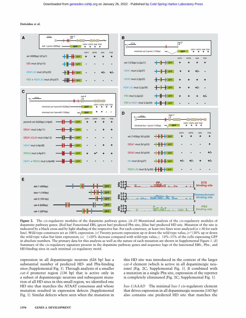
expression in all dopaminergic neurons (626 bp) has asubstantial number of predicted HD- and Pbx-bindingsites (Supplemental Fig. 1). Through analysis of a smallercat-4 promoter region (136 bp) that is active only ina subset of dopaminergic neurons and subsequent muta-tion of all HD sites in this small region, we identified oneHD site that matches the ATAAT consensus and whosemutation resulted in expression defects (SupplementalFig. 1). Similar defects where seen when the mutation in
this HD site was introduced in the context of the largercat-4 element (which is active in all dopaminergic neu-rons) (Fig. 2C; Supplemental Fig. 1). If combined witha mutation in a single Pbx site, expression of the reporteris completely eliminated (Fig. 2C; Supplemental Fig. 1).
bas-1/AAAD The minimal bas-1 cis-regulatory elementthat drives expression in all dopaminergic neurons (143 bp)also contains one predicted HD site that matches the
Figure 2. The cis-regulatory modules of the dopamine pathway genes. (A–D) Mutational analysis of the cis-regulatory modules ofdopamine pathway genes. (Red bar) Functional EBS; (green bar) predicted Pbx site; (blue bar) predicted HD site. Mutation of the site isindicated by a black cross and by light shading of the respective bar. For each construct, at least two lines were analyzed (n > 30 for eachline). Wild-type constructs set as 100% expression. (+) Twenty percent expression up or down the wild-type value; (+*) 20% up or downthe wild-type value but faint expression; (+/�) >20% decrease compared with wild-type value; (�) 0%–15% of the cells expressing GFPin absolute numbers. The primary data for this analysis as well as the nature of each mutation are shown in Supplemental Figure 1. (E)Summary of the cis-regulatory signature present in the dopamine pathway genes and sequence logo of the functional EBS-, Pbx-, andHD-binding sites in each minimal cis-regulatory motif.
Doitsidou et al.
1394 GENES & DEVELOPMENT
Cold Spring Harbor Laboratory Press on January 26, 2022 - Published by genesdev.cshlp.orgDownloaded from
ATAAT consensus, two predicted Pbx sites, and two func-tional EBSs (previously shown to be essential) (Fig. 2D).Mutating the single HD site has partial effects on bas-1reporter gene expression in transgenic animals. Mutatingeither Pbx site alone has no or just partial effects, but ifboth Pbx sites are mutated, reporter gene expression iseliminated in all dopaminergic neurons (even though theEBS is still present) (Fig. 2D; Supplemental Fig. 1).
In conclusion, members of the dopamine pathway arecontrolled by a common cis-regulatory signature com-posed of one or multiple essential EBSs and redundantlyoperating HD- and Pbx-binding sites (Fig. 2E). Impor-tantly, the combination of these three motifs is sufficientto drive specific expression in all dopaminergic neurons(Fig. 1D). Notably, there is no defined ‘‘motif grammar’’(Spitz and Furlong 2012); that is, no specific number,relative orientation, or spacing of the three motifs definesthis cis-regulatory signature.
The sole C. elegans Distalless/Dlx ortholog, ceh-43,controls dopaminergic neuron differentiation
To identify the trans-acting factors that act through theATAAT HD site, we turned to a collection of mutantanimals that show abnormal expression of the dat-1Tgfpdopaminergic fate marker and that we isolated previouslythrough automated sorting of EMS-mutagenized animals(Doitsidou et al. 2008). One locus that we identified inthis screen is the previously uncloned dopy-2 gene, rep-resented by four mutant alleles: ot340, ot479, ot406, andot345 (Doitsidou et al. 2008). We mapped this mutant to asmall interval on chromosome III by SNP linkage analysis
and three-factor mapping (see Fig. 4A, below). Througha combination of transformation rescue analysis (Table 1;see Fig. 4A, below), whole-genome sequencing (Supple-mental Table 1), RNAi phenocopy (Table 1), and analysisof a genetic deletion in the locus (kindly provided by theC. elegans knockout facility at Tokyo Women’s MedicalSchool) (Fig. 3B,D), we found that dopy-2 corresponds tothe ceh-43 locus (Fig. 4A,B). Mutations in the ceh-43locus had not previously been described. From here on,we refer to dopy-2 as ceh-43.
ceh-43 encodes the sole C. elegans ortholog of the flyDistalless and vertebrate Dlx homeobox genes (Aspockand Burglin 2001). In vertebrates, Dlx genes have beenimplicated in neuronal patterning (Panganiban andRubenstein 2002), but their roles in terminal neurondifferentiation have not previously been described. Theviability of the four alleles isolated from our screen (Table 1)and the nature of the mutations (Fig. 4B) suggest that theyare hypomorphic mutations. Thus, we further analyzedthe involvement of ceh-43 in dopaminergic neuron dif-ferentiation using the tm480 deletion allele, a likely nullallele. Since animals carrying this allele die as embryos,we analyzed mosaic animals that specifically lost ceh-43expression in dopaminergic neurons (see the Material andMethods for details on mosaic analysis). These animalsshow defects in the expression of all dopamine pathwaygenes and in all dopaminergic neuron types in both thehermaphrodite (Fig. 3B,D) and the male (data not shown).
The function of ceh-43 is not restricted to controllingthe dopamine pathway genes—it also controls the ex-pression of other terminal identity markers of dopami-nergic neurons; namely, the trp-4 and asic-1 ion channel
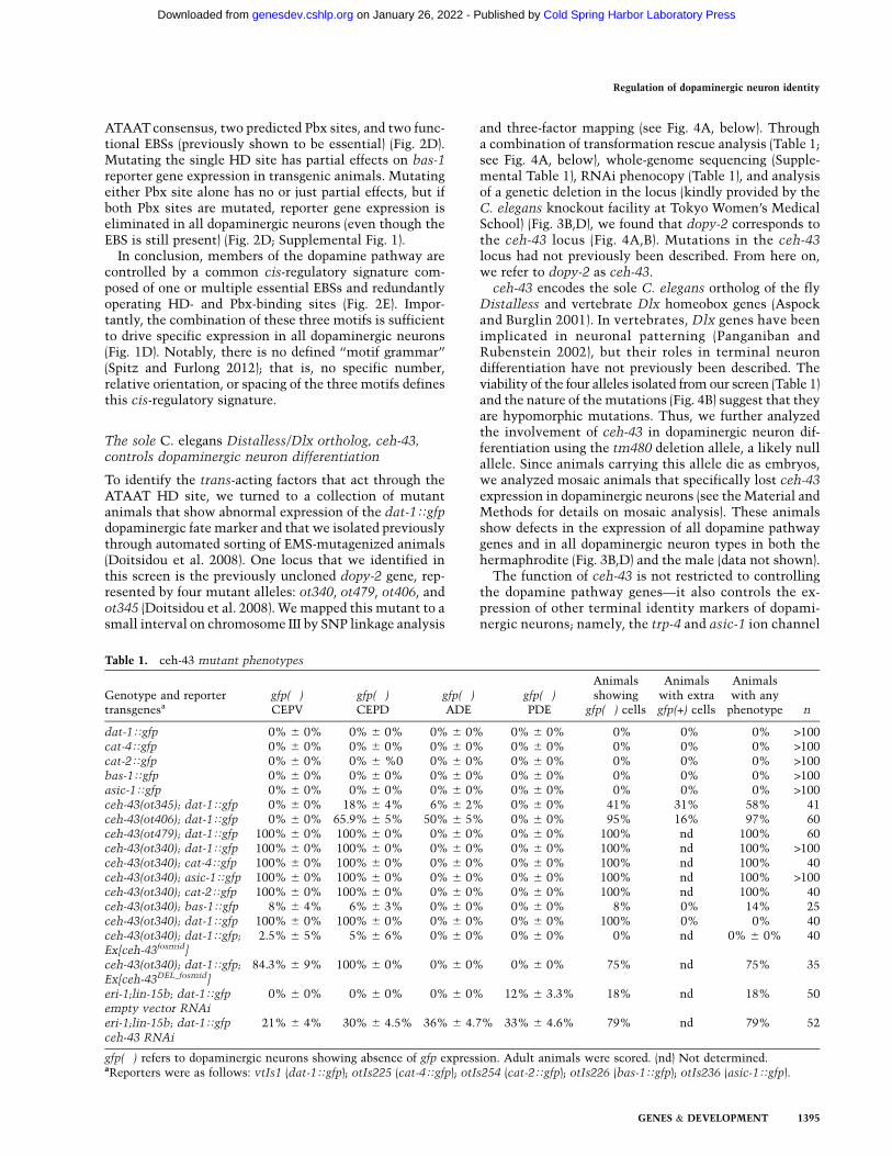
Table 1. ceh-43 mutant phenotypes
Genotype and reportertransgenesa
gfp(�)CEPV
gfp(�)CEPD
gfp(�)ADE
gfp(�)PDE
Animalsshowing
gfp(�) cells
Animalswith extragfp(+) cells
Animalswith any
phenotype n
dat-1Tgfp 0% 6 0% 0% 6 0% 0% 6 0% 0% 6 0% 0% 0% 0% >100cat-4Tgfp 0% 6 0% 0% 6 0% 0% 6 0% 0% 6 0% 0% 0% 0% >100cat-2Tgfp 0% 6 0% 0% 6 %0 0% 6 0% 0% 6 0% 0% 0% 0% >100bas-1Tgfp 0% 6 0% 0% 6 0% 0% 6 0% 0% 6 0% 0% 0% 0% >100asic-1Tgfp 0% 6 0% 0% 6 0% 0% 6 0% 0% 6 0% 0% 0% 0% >100ceh-43(ot345); dat-1Tgfp 0% 6 0% 18% 6 4% 6% 6 2% 0% 6 0% 41% 31% 58% 41ceh-43(ot406); dat-1Tgfp 0% 6 0% 65.9% 6 5% 50% 6 5% 0% 6 0% 95% 16% 97% 60ceh-43(ot479); dat-1Tgfp 100% 6 0% 100% 6 0% 0% 6 0% 0% 6 0% 100% nd 100% 60ceh-43(ot340); dat-1Tgfp 100% 6 0% 100% 6 0% 0% 6 0% 0% 6 0% 100% nd 100% >100ceh-43(ot340); cat-4Tgfp 100% 6 0% 100% 6 0% 0% 6 0% 0% 6 0% 100% nd 100% 40ceh-43(ot340); asic-1Tgfp 100% 6 0% 100% 6 0% 0% 6 0% 0% 6 0% 100% nd 100% >100ceh-43(ot340); cat-2Tgfp 100% 6 0% 100% 6 0% 0% 6 0% 0% 6 0% 100% nd 100% 40ceh-43(ot340); bas-1Tgfp 8% 6 4% 6% 6 3% 0% 6 0% 0% 6 0% 8% 0% 14% 25ceh-43(ot340); dat-1Tgfp 100% 6 0% 100% 6 0% 0% 6 0% 0% 6 0% 100% 0% 0% 40ceh-43(ot340); dat-1Tgfp; 2.5% 6 5% 5% 6 6% 0% 6 0% 0% 6 0% 0% nd 0% 6 0% 40Ex[ceh-43fosmid]
ceh-43(ot340); dat-1Tgfp; 84.3% 6 9% 100% 6 0% 0% 6 0% 0% 6 0% 75% nd 75% 35Ex[ceh-43DEL_fosmid]
eri-1;lin-15b; dat-1Tgfp 0% 6 0% 0% 6 0% 0% 6 0% 12% 6 3.3% 18% nd 18% 50empty vector RNAi
eri-1;lin-15b; dat-1Tgfp 21% 6 4% 30% 6 4.5% 36% 6 4.7% 33% 6 4.6% 79% nd 79% 52ceh-43 RNAi
gfp(�) refers to dopaminergic neurons showing absence of gfp expression. Adult animals were scored. (nd) Not determined.aReporters were as follows: vtIs1 (dat-1Tgfp); otIs225 (cat-4Tgfp); otIs254 (cat-2Tgfp); otIs226 (bas-1Tgfp); otIs236 (asic-1Tgfp).
Regulation of dopaminergic neuron identity
GENES & DEVELOPMENT 1395
Cold Spring Harbor Laboratory Press on January 26, 2022 - Published by genesdev.cshlp.orgDownloaded from
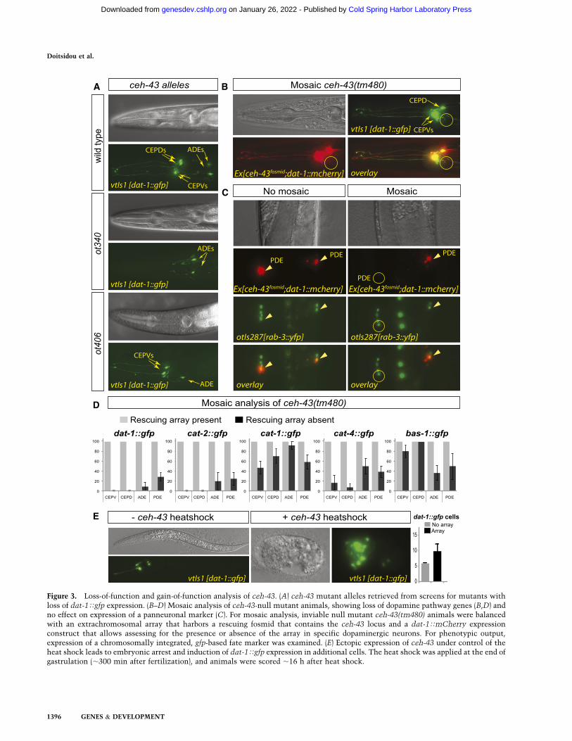
Figure 3. Loss-of-function and gain-of-function analysis of ceh-43. (A) ceh-43 mutant alleles retrieved from screens for mutants withloss of dat-1Tgfp expression. (B–D) Mosaic analysis of ceh-43-null mutant animals, showing loss of dopamine pathway genes (B,D) andno effect on expression of a panneuronal marker (C). For mosaic analysis, inviable null mutant ceh-43(tm480) animals were balancedwith an extrachromosomal array that harbors a rescuing fosmid that contains the ceh-43 locus and a dat-1TmCherry expressionconstruct that allows assessing for the presence or absence of the array in specific dopaminergic neurons. For phenotypic output,expression of a chromosomally integrated, gfp-based fate marker was examined. (E) Ectopic expression of ceh-43 under control of theheat shock leads to embryonic arrest and induction of dat-1Tgfp expression in additional cells. The heat shock was applied at the end ofgastrulation (;300 min after fertilization), and animals were scored ;16 h after heat shock.
Doitsidou et al.
1396 GENES & DEVELOPMENT
Cold Spring Harbor Laboratory Press on January 26, 2022 - Published by genesdev.cshlp.orgDownloaded from
genes (Table 1; data not shown). The expression of apanneuronal marker, rab-3, is not affected (Fig. 3C), in-dicating that ceh-43 does not affect the generation ofdopaminergic neurons but affects the adoption of a spe-cific neuronal identity. This phenotype is similar to theloss of the trans-acting factor for the Ets-binding site,AST-1 (Flames and Hobert 2009).
Two missense mutations in the ceh-43 locus, identifiedthrough our EMS screen, revealed weaker defects (Figs. 3A,4B; Table 1). Notably, two other alleles (ot479 and ot340)are partially overlapping, <2-kb deletions that reside >7 kbupstream of the ceh-43 locus (Fig. 4B; Supplemental Table 1).
Both alleles fail to complement the missense alleles(Doitsidou et al. 2008). In these deletion alleles, a highlypenetrant loss of dopaminergic neuron fate is restrictedentirely to the CEPD and CEPV neuron types, while ADEand PDE neuron types are unaffected (Fig. 3A; Table 1). Aswe show below, both deletions eliminate cis-regulatoryelements required for expression of ceh-43 in the CEPDand CEPV neurons.
We further corroborated the notion that ceh-43 actsthrough the ATAAT motifs by using an in vitro approachin which we tested whether bacterially produced CEH-43protein can bind to regulatory elements from four of the
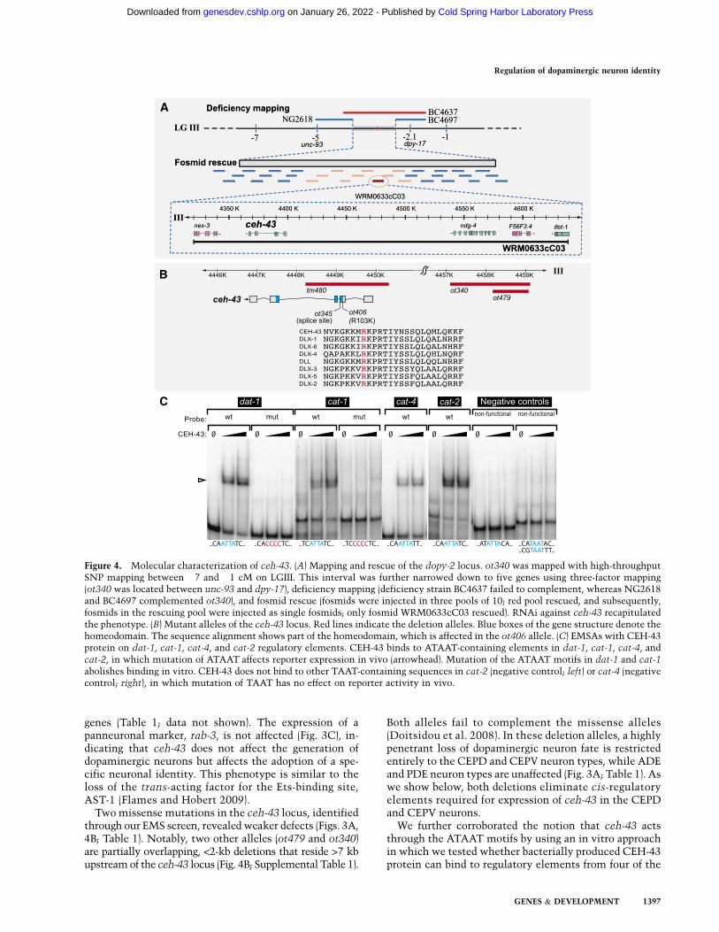
Figure 4. Molecular characterization of ceh-43. (A) Mapping and rescue of the dopy-2 locus. ot340 was mapped with high-throughputSNP mapping between �7 and �1 cM on LGIII. This interval was further narrowed down to five genes using three-factor mapping(ot340 was located between unc-93 and dpy-17), deficiency mapping (deficiency strain BC4637 failed to complement, whereas NG2618and BC4697 complemented ot340), and fosmid rescue (fosmids were injected in three pools of 10; red pool rescued, and subsequently,fosmids in the rescuing pool were injected as single fosmids; only fosmid WRM0633cC03 rescued). RNAi against ceh-43 recapitulatedthe phenotype. (B) Mutant alleles of the ceh-43 locus. Red lines indicate the deletion alleles. Blue boxes of the gene structure denote thehomeodomain. The sequence alignment shows part of the homeodomain, which is affected in the ot406 allele. (C) EMSAs with CEH-43protein on dat-1, cat-1, cat-4, and cat-2 regulatory elements. CEH-43 binds to ATAAT-containing elements in dat-1, cat-1, cat-4, andcat-2, in which mutation of ATAAT affects reporter expression in vivo (arrowhead). Mutation of the ATAAT motifs in dat-1 and cat-1
abolishes binding in vitro. CEH-43 does not bind to other TAAT-containing sequences in cat-2 (negative control; left) or cat-4 (negativecontrol; right), in which mutation of TAAT has no effect on reporter activity in vivo.
Regulation of dopaminergic neuron identity
GENES & DEVELOPMENT 1397
Cold Spring Harbor Laboratory Press on January 26, 2022 - Published by genesdev.cshlp.orgDownloaded from
dopamine pathway genes. We readily detected suchbinding in gel shift assays (Fig. 4C). This in vitro bindingwas abrogated when the same ATAAT motifs that dis-rupted activity in the in vivo reporter gene assay weremutated (Fig. 4C). This binding is not the result of un-specific binding to the TAAT core motif, since otherTAAT motifs in the minimal regulatory regions that donot match to the ATAAT consensus and showed no ac-tivity in the in vivo reporter assay do not bind CEH-43(Fig. 4C, negative controls). Two other ceh-43 targets thatwe identified in other cellular contexts also contain func-tional ATAAT-binding sites and bind CEH-43 in vitro(L Cochella, J Etchberger, N Abe, and O Hobert, unpubl.data).
ceh-43 not only is required to express terminal fatemarkers of dopaminergic neurons, but is sufficient to doso, at least in some cellular contexts. When misexpressedunder control of a ubiquitous and inducible heat-shockpromoter, up to twice as many dopamine marker-positivecells can be generated (Fig. 3E). The extent of ectopicdopaminergic neuron induction upon heat-shocked-induced ceh-43 expression is similar to the extent ofectopic dopaminergic neuron induction upon heat-shockedinduction of ast-1 (Flames and Hobert 2009).
ceh-43 is expressed in dopaminergic neurons,and its function is continuously required to maintainthe differentiated state of dopaminergic neurons
To examine ceh-43 expression, we generated a fosmid-based reporter in which we recombineered gfp at theC-terminal end of the ceh-43 locus in the context of an;32-kb fosmid that contains several genes upstream ofand downstream from ceh-43 (Fig. 5A). Through cola-beling with a dopaminergic neuron-specific marker, wefound the fosmid reporter to be expressed in all dopami-nergic neurons throughout the life of the neurons (Fig. 5B).Expression can also be observed in some additional headand body neurons as well as nonneuronal cells (Supple-mental Fig. 2a). This expression was corroborated withimmunostaining of endogenous CEH-43 protein using apan-species anti-Distalless antibody (data not shown).As assessed with immunostaining for CEH-43 and re-porter transgene for ast-1 expression, despite the broadneuronal expression of both ceh-43 and ast-1, theyuniquely overlap in dopaminergic neurons plus one addi-tional pair of nondopaminergic neurons in the head andone additional neuron in the midbody region (Supplemen-tal Fig. 2b).
Introducing the smallest of the overlapping deletionspresent in the ot340 and ot479 alleles in the context of thefosmid reporter results in the loss of expression of thereporter specifically in the CEPD/V neuron types, demon-strating that these deletions affect relevant cis-regulatoryelements (Fig. 5C). Moreover, we found that ot340 mu-tants can be rescued with a wild-type fosmid containingthe ceh-43 locus but not by a fosmid in which these reg-ulatory elements are deleted (Fig. 5D), corroborating thenotion that ceh-43 acts autonomously at least in the CEPneurons to affect their terminal differentiation.
We noted that animals carrying a missense mutationin the homeobox of ceh-43 (ot406 allele) show a signifi-cantly more pronounced defect in the expression ofdopamine markers in adults compared with younglarvae (Fig. 5E). This progressive loss of dopaminergicneuron identity indicates that ceh-43, like ast-1 (Flamesand Hobert 2009), is continuously required to maintainthe differentiated state of dopaminergic neurons, a typi-cal feature of terminal selector-type transcription fac-tors (Hobert 2008).
Cell type- and target gene-specific interactionsof ceh-43 and ast-1
The coexpression of CEH-43 and AST-1 in mature dopa-minergic neurons as well as the presence of cognate andfunctionally required CEH-43- and AST-1-binding sites indopamine pathway genes suggest that both proteins co-operate to activate dopamine pathway genes. The level ofcooperation may differ in distinct dopaminergic neurontypes, as suggested by the observation that in ast-1-nullmutant animals (gk463 allele), the dat-1 gene is stillnormally expressed in one dopaminergic neuron type: theCEPV neurons (Table 2). In ceh-43(ot406) hypomorphicanimals, dat-1 expression in CEPV is also unaffected. Inast-1(gk463); ceh-43(ot406) double mutants, expressionof dat-1 is now strongly affected in the CEPV neurons(Table 2). This finding suggests that, in the absence ofast-1, correct dat-1 expression in the CEPV neurons canstill be ensured by ceh-43, but under such circumstances,even weak disruptions of ceh-43 activity will severelyaffect dat-1 expression. The genetic synergism betweenceh-43 and ast-1 was further confirmed by the analysis ofdouble-hypomorph mutants (Table 2). In ceh-43(ot406)mutants, dat-1 expression is only partially affected in theADE and CEPD neurons, but this phenotype is greatlyenhanced when the ceh-43(ot406) allele is combined withthe ast-1(hd1) hypomorphic allele, which alone has nodefects in dat-1 expression (Table 2).
These results not only corroborate the genetic interac-tion of these two factors, but also make the point thatindividual dopamine pathway genes may display a differ-entially tuned requirement for individual trans-actingfactors in specific cell types. In CEPVs, the loss of ast-1can be tolerated in regard to dat-1 expression becauseceh-43 can ensure robust dat-1 expression, but for otherdopamine pathway genes, loss of ast-1 alone results instrong defects in expression. Moreover, this is not a gen-eral compensatory role for ceh-43 because in the otherdopaminergic neurons, the presence of ceh-43 cannotcompensate for a complete ast-1 loss in regard to dat-1expression.
Two distinct Pbx genes constitute the third componentof the dopamine regulatory signature
To identify the factors that operate through the thirdcis-regulatory motif required for dopaminergic neuronexpression, the predicted Pbx HD site, we turned to acandidate gene approach. The C. elegans genome codesfor three Pbx genes: ceh-20, ceh-40, and ceh-60 (Van Auken
Doitsidou et al.
1398 GENES & DEVELOPMENT
Cold Spring Harbor Laboratory Press on January 26, 2022 - Published by genesdev.cshlp.orgDownloaded from
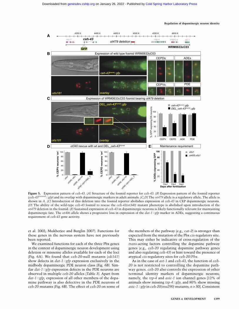
et al. 2002; Mukherjee and Burglin 2007). Functions forthese genes in the nervous system have not previouslybeen reported.
We examined functions for each of the three Pbx genesin the context of dopaminergic neuron development usingdeletion or missense alleles available for each of the loci(Fig. 6A). We found that ceh-20-null mutants (ok541)show defects in dat-1Tgfp expression exclusively in themidbody dopaminergic PDE neuron class (Fig. 6B). Sim-ilar dat-1Tgfp expression defects in the PDE neurons areobserved in multiple ceh-20 alleles (Table 3). Apart fromdat-1Tgfp, expression of all other members of the dopa-mine pathway is also defective in the PDE neurons ofceh-20 mutants (Fig. 6B). The effect of ceh-20 on some of
the members of the pathway (e.g., cat-2) is stronger thanexpected from the mutation of the Pbx cis-regulatory site.This may either be indicative of cross-regulation of thetrans-acting factors controlling the dopamine pathwaygenes (e.g., ceh-20 regulating dopamine pathway genesand also regulating ceh-43) or hint toward the presence ofatypical cis-regulatory sites for ceh-20/Pbx.
As in the case of ast-1 and ceh-43, the function of ceh-20 is not restricted to controlling the dopamine path-way genes. ceh-20 also controls the expression of otherterminal identity markers of dopaminergic neurons;namely, the trp-4 and asic-1 ion channel genes [12% ofanimals show missing trp-4Tgfp, and 80% show missingasic-1Tgfp in ceh-20(mu290) mutants; n > 30]. Consistent
Figure 5. Expression pattern of ceh-43. (A) Structure of the fosmid reporter for ceh-43. (B) Expression pattern of the fosmid reporter(ceh-43fosmidTgfp) and its overlap with dopaminergic markers in adult animals. (C,D) The ot479 allele is a regulatory allele. The allele isshown in A. (C) Introduction of this deletion into the fosmid reporter abolishes expression of ceh-43 in CEP dopaminergic neurons.(D) The ability of the wild-type ceh-43 fosmid to rescue the ceh-43(ot340) mutant phenotype is abolished upon introduction of theot479 deletion in the fosmid. (E) Sustained expression of ceh-43 in dopaminergic neurons is likely functionally relevant for maintainingdopaminergic fate. The ot406 allele shows a progressive loss in expression of the dat-1Tgfp marker in ADEs, suggesting a continuousrequirement of ceh-43 gene activity.
Regulation of dopaminergic neuron identity
GENES & DEVELOPMENT 1399
Cold Spring Harbor Laboratory Press on January 26, 2022 - Published by genesdev.cshlp.orgDownloaded from
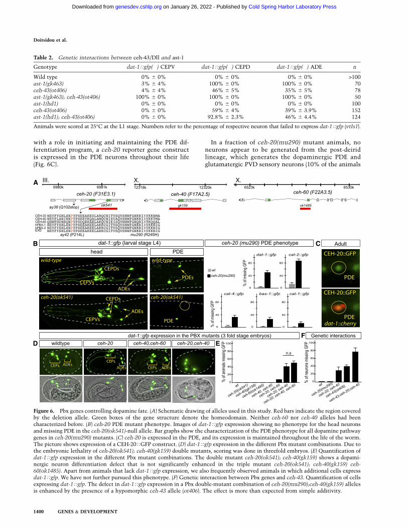
with a role in initiating and maintaining the PDE dif-ferentiation program, a ceh-20 reporter gene constructis expressed in the PDE neurons throughout their life(Fig. 6C).
In a fraction of ceh-20(mu290) mutant animals, noneurons appear to be generated from the post-deiridlineage, which generates the dopaminergic PDE andglutamatergic PVD sensory neurons (10% of the animals
Figure 6. Pbx genes controlling dopamine fate. (A) Schematic drawing of alleles used in this study. Red bars indicate the region coveredby the deletion allele. Green boxes of the gene structure denote the homeodomain. Neither ceh-60 nor ceh-40 alleles had beencharacterized before. (B) ceh-20 PDE mutant phenotype. Images of dat-1Tgfp expression showing no phenotype for the head neuronsand missing PDE in the ceh-20(ok541)-null allele. Bar graphs show the characterization of the PDE phenotype for all dopamine pathwaygenes in ceh-20(mu290) mutants. (C) ceh-20 is expressed in the PDE, and its expression is maintained throughout the life of the worm.The picture shows expression of a CEH-20TGFP construct. (D) dat-1Tgfp expression in the different Pbx mutant combinations. Due tothe embryonic lethality of ceh-20(ok541); ceh-40(gk159) double mutants, scoring was done in threefold embryos. (E) Quantification ofdat-1Tgfp expression in the different Pbx mutant combinations. The double mutant ceh-20(ok541); ceh-40(gk159) shows a dopami-nergic neuron differentiation defect that is not significantly enhanced in the triple mutant ceh-20(ok541); ceh-40(gk159) ceh-60(ok1485). Apart from animals that lack dat-1Tgfp expression, we also frequently observed animals in which additional cells expressdat-1Tgfp. We have not further pursued this phenotype. (F) Genetic interaction between Pbx genes and ceh-43. Quantification of cellsexpressing dat-1Tgfp. The defect in dat-1Tgfp expression in a Pbx double-mutant combination of ceh-20(mu290);ceh-40(gk159) allelesis enhanced by the presence of a hypomorphic ceh-43 allele (ot406). The effect is more than expected from simple additivity.
Table 2. Genetic interactions between ceh-43/Dll and ast-1
Genotype dat-1Tgfp(�) CEPV dat-1Tgfp(�) CEPD dat-1Tgfp(�) ADE n
Wild type 0% 6 0% 0% 6 0% 0% 6 0% >100ast-1(gk463) 3% 6 4% 100% 6 0% 100% 6 0% 70ceh-43(ot406) 4% 6 4% 46% 6 5% 35% 6 5% 78ast-1(gk463); ceh-43(ot406) 100% 6 0% 100% 6 0% 100% 6 0% 50ast-1(hd1) 0% 6 0% 0% 6 0% 0% 6 0% 100ceh-43(ot406) 0% 6 0% 59% 6 4% 39% 6 3.9% 152ast-1(hd1); ceh-43(ot406) 0% 6 0% 92.8% 6 2.3% 46% 6 4.4% 124
Animals were scored at 25°C at the L1 stage. Numbers refer to the percentage of respective neuron that failed to express dat-1Tgfp (vtIs1).
Doitsidou et al.
1400 GENES & DEVELOPMENT
Cold Spring Harbor Laboratory Press on January 26, 2022 - Published by genesdev.cshlp.orgDownloaded from
generate no neurons in the post-deirid, 14% generate onlyone neuron, 37% generate the normal number of twoneurons, and 39% generate more than two neurons; n =49), suggesting lineage defects. However, the PDE differ-entiation defects in ceh-20 mutants are not merely due toPDE lineage defects because dat-1 expression is also lostin animals with an unaffected PDE lineage, as assessed bycorrect expression of the pan-neuronal rab-3 marker inPDE (Supplemental Fig. 3).
In contrast to ast-1 and ceh-43 (Fig. 3E), ceh-20 aloneis not able to induce the production of additional dopa-minergic neurons upon heat-shock promoter-mediatedmisexpression (as assessed by ectopic dat-1Tgfp expres-sion) (data not shown). Animals that coexpress heat-shockpromoter-driven ceh-43, ast-1, and ceh-20 constructsshow no more ectopic dopaminergic neuron productionthan animals expressing ceh-43 or ast-1 alone (data notshown).
The effect of ceh-20 loss of function is restricted to thePDE neurons, since the expression of several dopaminepathway genes is completely unaffected in head dopami-nergic neurons (Fig. 6B; data not shown). Since our cis-regulatory analysis indicates that putative Pbx-bindingsites are required for the expression of dopamine pathwaygenes in not just PDE neurons but also all dopaminergichead neurons, we tested whether removal of other Pbxgenes affected dopaminergic head neurons. ceh-40-nullmutants, ceh-60-null mutants, and ceh-40 ceh-60 double-null mutants did not show defects in dat-1Tgfp expres-sion in any dopaminergic neuron type (Fig. 6D; Table 3).However, removal of ceh-40 in a ceh-20 mutant back-ground, which alone has no effect on head dopaminergicneurons, results in defects in dat-1Tgfp expression inhead dopaminergic neurons (Fig. 6D; Table 3). In contrast,combining the ceh-60 mutation with the ceh-20 muta-tion only shows very mild (<10% penetrant) differentia-tion defects of head dopaminergic neurons. The defectof the ceh-20; ceh-40 double mutants are not further
enhanced in ceh-20; ceh-40 ceh-60 triple-null mutants(Fig. 6E; Table 3). However, the ceh-20; ceh-40 double-mutant defects are significantly enhanced by removal ofceh-43/Distalless (ceh-43; ceh-20; ceh-40 triple mutants)(Fig. 6F), which corroborates the genetic interactionsamong distinct members of the dopaminergic regulatoryensemble. Taken together, ceh-20 functions nonredun-dantly in the PDE neurons to control their differentiationbut acts redundantly with ceh-40 in head dopaminergicneurons. Consistent with this notion, we could not detectexpression of ceh-40 in the midbody PDE neuron butdetected expression of both ceh-20 and ceh-40 in the headdopaminergic neurons (Supplemental Fig. 4).
We examined the overlap of ast-1, ceh-43, and Pbx geneexpression to assess whether their coexpression uniquelydefines dopaminergic neurons. We detected coexpressionof ast-1, ceh-43, and ceh-20 in at least one nondopami-nergic neuron (SDQL). The two most likely possibilitiesthat could explain this lack of specificity are that eitherthere are still other components of the dopaminergictranscription factor collective that remain to be identifiedand are not expressed in SDQL (those are unlikely to beDNA-binding factors, since the ETS, HD, and Pbx sitesare sufficient to exclusive drive gene expression indopaminergic neurons) or, alternatively, repressive mech-anisms could operate in SDQL to inhibit the expressionof dopaminergic neuron identity.
We also investigated whether ast-1, ceh-43, and ceh-20cross-regulate each other’s expression. Focusing on thePDE neuron class, we found that ast-1 expression is un-affected in ceh-43 and ceh-20 mutants (data not shown).However, ast-1 is required for ceh-43 expression (Supple-mental Fig. 5) but is not required for ceh-20 expression(data not shown). These results indicate that these factorsact to some extent independently of one another.
Taken together, our reverse genetic analysis identifieda third component of the regulatory signature of dopa-minergic neuron terminal differentiation programs. As
Table 3. Pbx genes affecting dopaminergic neuron differentiation
Loss of dat-1Tgfp expressionin all or a subset of head neurons
(ADE and CEPs)Loss of dat-1Tgfp expression
of midbody neurons (PDE) n
Wild type 0% 6 0% 3% 6 2% 55ceh-20(ok541) 0% 6 0% 55% 6 7%a 56ceh-20(ay42) 0% 6 0% 54% 6 8% 39ceh-20(ay38) 0% 6 0% 40% 6 8%b 39ceh-20(mu290) 0% 6 0% 50% 6 7%c 56ceh-40(gk159) 0% 6 0% 0% 6 0% 50ceh-60(ok1485) 0% 6 0% 0% 6 0% 50ceh-40(gk159), ceh-60(ok1485) 0% 6 0% 0% 6 0% 52ceh-20(ok541); ceh-40(gk159) 38% 6 5%d Not scorable due to embryonic lethality 86ceh-20(ok541); ceh-60(ok1485) 9% 6 4%e 50% 6 6% 66ceh-20(ok541); ceh-40(gk159), ceh-60(ok1485) 53% 6 4%f Not scorable due to embryonic lethality 126
aok541 allele 5% of the escapers show extra GFP cells in PDE regionbay38 allele 11% of the escapers show extra GFP cells in PDE regioncmu290 allele 5% of animals show extra GFP cells in PDE regiondThirty-six percent of the embryos show extra GFP cellseFourteen percent of the embryos show extra GFP cellsfTwenty-four percent of the embryos show extra GFP cells
Regulation of dopaminergic neuron identity
GENES & DEVELOPMENT 1401
Cold Spring Harbor Laboratory Press on January 26, 2022 - Published by genesdev.cshlp.orgDownloaded from
predicted by the cis-regulatory analysis, this third com-ponent is a Pbx gene. In one dopaminergic neuron type,only one Pbx gene is required; in other dopaminergicneuron types, a combination of Pbx genes act redundantly.
AST-1, CEH-43, and CEH-20 cooperate to activatedopamine pathway genes in a heterologous, cell-basedcis-activation assay
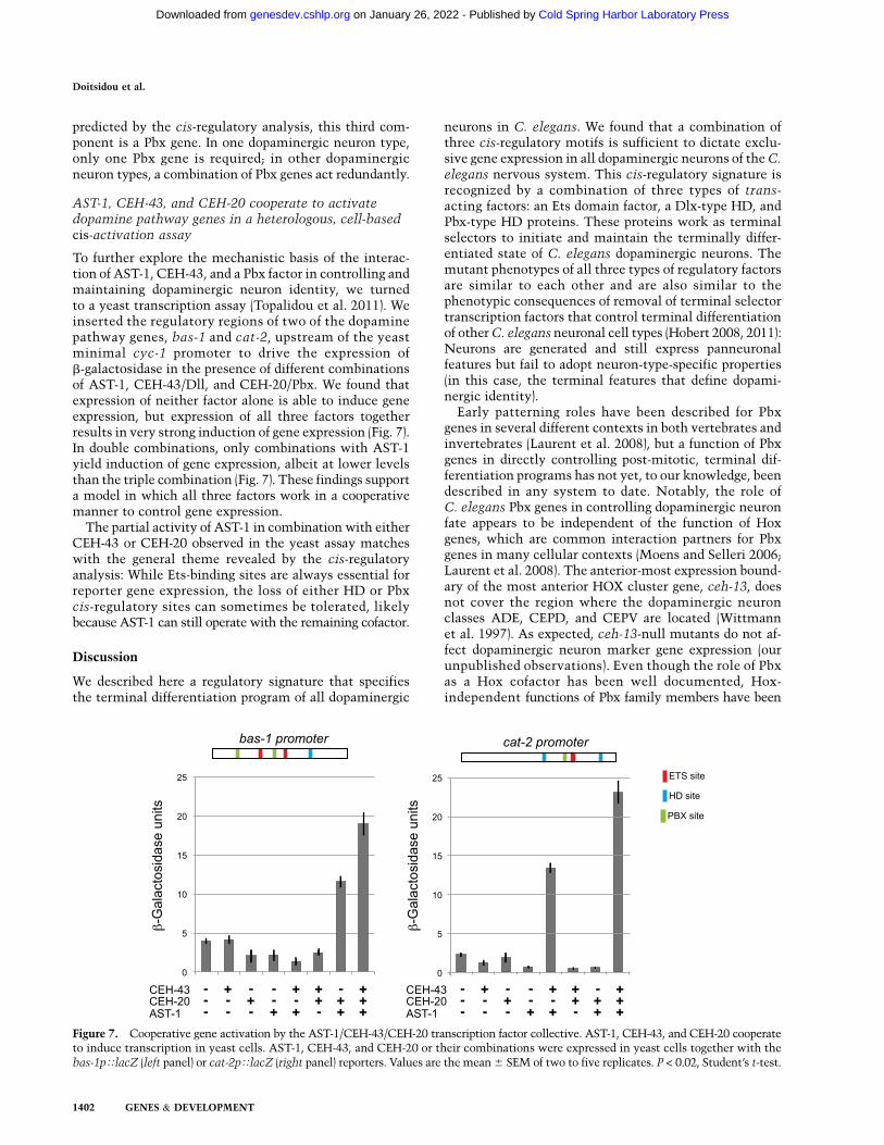
To further explore the mechanistic basis of the interac-tion of AST-1, CEH-43, and a Pbx factor in controlling andmaintaining dopaminergic neuron identity, we turnedto a yeast transcription assay (Topalidou et al. 2011). Weinserted the regulatory regions of two of the dopaminepathway genes, bas-1 and cat-2, upstream of the yeastminimal cyc-1 promoter to drive the expression ofb-galactosidase in the presence of different combinationsof AST-1, CEH-43/Dll, and CEH-20/Pbx. We found thatexpression of neither factor alone is able to induce geneexpression, but expression of all three factors togetherresults in very strong induction of gene expression (Fig. 7).In double combinations, only combinations with AST-1yield induction of gene expression, albeit at lower levelsthan the triple combination (Fig. 7). These findings supporta model in which all three factors work in a cooperativemanner to control gene expression.
The partial activity of AST-1 in combination with eitherCEH-43 or CEH-20 observed in the yeast assay matcheswith the general theme revealed by the cis-regulatoryanalysis: While Ets-binding sites are always essential forreporter gene expression, the loss of either HD or Pbxcis-regulatory sites can sometimes be tolerated, likelybecause AST-1 can still operate with the remaining cofactor.
Discussion
We described here a regulatory signature that specifiesthe terminal differentiation program of all dopaminergic
neurons in C. elegans. We found that a combination ofthree cis-regulatory motifs is sufficient to dictate exclu-sive gene expression in all dopaminergic neurons of the C.elegans nervous system. This cis-regulatory signature isrecognized by a combination of three types of trans-acting factors: an Ets domain factor, a Dlx-type HD, andPbx-type HD proteins. These proteins work as terminalselectors to initiate and maintain the terminally differ-entiated state of C. elegans dopaminergic neurons. Themutant phenotypes of all three types of regulatory factorsare similar to each other and are also similar to thephenotypic consequences of removal of terminal selectortranscription factors that control terminal differentiationof other C. elegans neuronal cell types (Hobert 2008, 2011):Neurons are generated and still express panneuronalfeatures but fail to adopt neuron-type-specific properties(in this case, the terminal features that define dopami-nergic identity).
Early patterning roles have been described for Pbxgenes in several different contexts in both vertebrates andinvertebrates (Laurent et al. 2008), but a function of Pbxgenes in directly controlling post-mitotic, terminal dif-ferentiation programs has not yet, to our knowledge, beendescribed in any system to date. Notably, the role ofC. elegans Pbx genes in controlling dopaminergic neuronfate appears to be independent of the function of Hoxgenes, which are common interaction partners for Pbxgenes in many cellular contexts (Moens and Selleri 2006;Laurent et al. 2008). The anterior-most expression bound-ary of the most anterior HOX cluster gene, ceh-13, doesnot cover the region where the dopaminergic neuronclasses ADE, CEPD, and CEPV are located (Wittmannet al. 1997). As expected, ceh-13-null mutants do not af-fect dopaminergic neuron marker gene expression (ourunpublished observations). Even though the role of Pbxas a Hox cofactor has been well documented, Hox-independent functions of Pbx family members have been
Figure 7. Cooperative gene activation by the AST-1/CEH-43/CEH-20 transcription factor collective. AST-1, CEH-43, and CEH-20 cooperateto induce transcription in yeast cells. AST-1, CEH-43, and CEH-20 or their combinations were expressed in yeast cells together with thebas-1pTlacZ (left panel) or cat-2pTlacZ (right panel) reporters. Values are the mean 6 SEM of two to five replicates. P < 0.02, Student’s t-test.
Doitsidou et al.
1402 GENES & DEVELOPMENT
Cold Spring Harbor Laboratory Press on January 26, 2022 - Published by genesdev.cshlp.orgDownloaded from
described in several species, including C. elegans (Yang et al.2005), vertebrates (Ferretti et al. 2011), and flies (e.g., Casaresand Mann 2001; Bessa et al. 2002). Intriguingly, one study inDrosophila has shown that the ceh-43 ortholog Distallessgenetically interacts with the fly Pbx homolog Exd (Donget al. 2000). The function of worm Pbx genes in controllingdopaminergic neuron differentiation is also independentof another common cofactor of Pbx gene, the Meis-typehomeobox gene. There is a single canonical Meis homologin the C. elegans, encoded by the unc-62 loucs (Van Aukenet al. 2002), but animals carrying a null mutant allele of unc-62 show no reduction in the production of dopaminergicneurons (our unpublished observations).
As revealed through the use of a heterologous yeasttranscriptional assay system, the Ets/Dlx/Pbx regula-tory ensemble acts in a cooperative manner to activategene expression. Cooperativity is also suggested by ge-netic interaction tests that show synergy (rather thanadditivity) in loss-of-function scenarios. The cooperativ-ity is reminiscent of the terminal differentiation programexecuted by the cholinergic AIY interneuron class in C.elegans. Here again, two factors (the HD transcriptionfactors ttx-3 and ceh-10), each expressed in a number ofdistinct neuron types, uniquely overlap in their expres-sion in the AIY interneurons, where they cooperativelyactivate scores of terminal differentiation genes thatdefine AIY interneuron identity (Wenick and Hobert2004). The key conceptual and mechanistic differencebetween the cooperativity of ttx-3/ceh-10 and ast-1/ceh-43/Pbx lies in what generally is referred to as the‘‘grammar’’ of cis-regulatory logic; that is, the overallorganization of the individual cis-regulatory motifs. Inthe case of the AIY-expressed, ttx-3/ceh-10-dependent reg-ulatory elements, the two binding sites for TTX-3 andCEH-10 are precisely spaced and oriented to allow forcooperative binding. Contrasting such fixed grammar, thecis-regulatory regions that control dopaminergic neuronexpression are composed of cis-regulatory sites (ETSdomain, HD, and Pbx-binding site) that display no fixedspacing, relative orientation, or overall number of in-dividual sites, yet the activity of the cis-acting factors isnevertheless cooperative, as determined by mutant anal-ysis and heterologous transcription assays in yeast. Arecently published study described a similar regulatoryarchitecture for cardiac gene expression in Drosophilaand proposed the term ‘‘transcription factor collective’’(Junion et al. 2012). In that case, five cardiogenic tran-scriptional regulators cooperatively activate target genesin the absence of a defined motif grammar, and in sometarget cases, only a subset of the these factors are bound.Our studies broaden this concept to a different cell typeand organism, suggesting universality of this regulatorymechanism.
We further extend the transcription factor collectivemodel by our ability to examine its activity in four distinctneuronal subclasses: ADE, CEPD, CEPV, and PDE. Eventhough these four dopaminergic classes share many mo-lecular and functional features, these neurons have distinctlineage histories and distinct axo/dendritic projectionsand are located in different parts of the nervous system.
Even though our cis-regulatory analysis in combinationwith our genetic analysis clearly shows that all fourneuron classes employ this combination of regulatoryfactors, the extent of the involvement of individualfactors differs for each individual dopamine pathway genein individual dopaminergic neuron subtypes. For exam-ple, dat-1 expression critically depends on ast-1 expres-sion in all neurons except for the CEPV neurons, in whichast-1 function can be compensated for by ceh-43 geneactivity. Head dopaminergic neuron types rely on multiplePbx genes, as a mutant phenotype is only evident in double-mutant combinations, while midbody dopaminergic neu-rons critically depend on only one Pbx gene (ceh-20). Theunderlying common theme of all of these interactionsmay be that the presence of multiple factors ensures robustexpression of the target genes of these transcription factorsand that each cell and target gene may use slightly differentalthough related means to ensure this robustness.
The same Ets/Dlx/Pbx regulatory signature that wedescribe here may also function in dopaminergic neuronsin vertebrates. We showed previously that Etv1, a mousehomolog of ast-1, is required for the appropriate differen-tiation of olfactory dopaminergic neurons (Flames andHobert 2009). The defects of Etv1 mutants, however, arenot as pronounced as ast-1 mutant defects are; for exam-ple, while TH expression is affected by Etv1, DAT ex-pression is not (Cave et al. 2010). One potential expla-nation for this partial effect can be seen in the regulatorylogic described here: Other factors that cooperate withEtv1 may partially compensate for its loss. In fact, thefailure to see a loss of Dat expression in Etv1 mousemutants (Cave et al. 2010) may precisely mirror theabsence of a phenotype of dat-1 expression in the CEPVneurons of ast-1-null mutants; that is, the ast-1 pheno-type is only revealed if a cooperating factor (in this case,ceh-43) is disabled.
The factors that cooperate with vertebrate Etv1 toensure dopaminergic neuron differentiation in the olfac-tory bulb could be the same as we defined here in C.elegans. A null mutation of Dlx2 results in early speci-fication defects of olfactory bulb neurons and a loss ofTH-positive neurons (Qiu et al. 1995). Dlx2 expression ismaintained in adult dopaminergic neurons, and laterfunction of Dlx2 in terminal differentiation specificallyof the dopamine olfactory bulb neurons is suggestedthrough expression of a dominant-negative form of Dlx2,which resulted in dopaminergic neuron specificationdefects (Brill et al. 2008). To assess whether the thirdcomponent of the worm Ets/Dlx/Pbx regulatory signa-ture is also expressed in mouse olfactory bulb dopami-nergic neurons, we stained mouse olfactory bulbs witha Pbx1/2/3 antibody and observed Pbx immunoreactivityin TH-positive neurons (Supplemental Fig. 6). Intrigu-ingly, Pbx1 is expressed in the midbrain dopaminergicneurons, and Pbx1 mutants show dopaminergic axonpathfinding defects (Sgado et al. 2012). These observa-tions suggest that the regulatory code between olfactorybulb dopaminergic neuron specification and worm dopa-minergic neuron specification could, at least in part, bephylogenetically conserved.
Regulation of dopaminergic neuron identity
GENES & DEVELOPMENT 1403
Cold Spring Harbor Laboratory Press on January 26, 2022 - Published by genesdev.cshlp.orgDownloaded from
Materials and methods
DNA constructs and cis-regulatory analysis
All gfp-based reporter constructs were generated using thepPD95.75 vector as backbone and by subcloning into the multiplecloning site. Mutagenesis reactions were performed using theQuickChangeII XL site-directed mutagenesis kit (Stratagene).Constructs were injected or crossed into otIs181 (dat-1Tcherry;
ttx-3Tcherry) to allow easy identification of dopaminergic neu-rons. All reporter constructs were injected at 50 ng/mL usingrol-6(su1006) as a coinjection marker (100 ng/mL). For eachconstruct, two or three independent lines were scored (at least 30animals per line). Sequences of the wild-type minimal constructsare in the Supplemental Material, and the nature of the muta-tions introduced is indicated in Supplemental Figure 1.
Cloning of dopy-2/ceh-43
ot340 was mapped with high-throughput SNP mapping (Daviset al. 2005) between �7 and �1 cM on LGIII. Three-factormapping mapped ot340 between unc-93 and dpy-17. Deficiencymapping placed the ot340 locus within deficiency BC4637 butoutside deficiencies NG2618 and BC4697. Fosmid rescue ofpools of fosmids were injected, followed by single-fosmid injec-tions of the fosmids included in the rescuing pool. Single-fosmidinjection of WRM0633cC03 rescued the ot340 phenotype. Onlyfive genes were included in the rescuing fosmid. RNAi againsteach of these genes was performed using a bacterial feedingprotocol (Kamath and Ahringer 2003) in an eri-1;lin-15b;vtIs1
mutant background (Kennedy et al. 2004). RNAi against ceh-43
recapitulated the phenotype. Sanger sequencing revealed pointmutations in the ceh-43 locus for ot406 and ot345 alleles butno mutation in alleles ot340 and ot479. Whole-genome sequenc-ing using an Illumina platform followed by data analysis usingMAQGene (Bigelow et al. 2009) of ot340 and ot479 revealed twooverlapping deletions >7 kb upstream of ceh-43 CDS (Supple-mental Table 1). Deletions were confirmed by Sanger sequenc-ing to span genomic regions on LGIII from 4457789 to 4459806(2018 bp) for ot340 and from 4458889 to 4459754 (866 bp) forot479.
Fosmid recombineering and ceh-43 expression analysis
ceh-43-containing fosmid WRM0633cC03 was tagged asdescribed before (Tursun et al. 2009) to generate OH9993(otEx4439[ceh-43fosmidTgfp;ttx-3Tdsred;rol-6]) and integrated togenerate OH10447 (otIs339[ceh-43fosmidTgfp;ttx-3Tdsred;rol-6]).The ot479 deletion was engineered in the recombineeredfosmid as described before (Tursun et al. 2009) to generateDEL_ceh-43fosmidTgfp. For determining the neuronal expressionof ceh-43, strain MDH33 (otIs339[ceh-43fosmidTgfp;ttx-3Tdsred;rol-6];otIs355[rab-3TNLSTtagRFP]) was used. To determine ceh-43
expression in ast-1 mutants, strain MDH38 {ast-1(gk463) bli-2(e768)
unc-4(e120);Ex[ast-1cosmid;ttx-3Tgfp;dat-1TmCherry];otIs339;
otIs355} was used.The ability of the recombineered fosmid with and without the
deletion to rescue ceh-43(ot340) mutants was assessed usingstrains OH10412 {ceh-43(ot340);vtIs1; otEx4439[ceh-43fosmidTgfp;ttx-3Trfp;rol-6]} and OH10424 {ceh-43(ot340); vtIs1;[ExDEL_ceh-43fosmidTgfp;ttx-3Trfp;rol-6]}, respectively.
Mosaic analysis
The strain CH1890 {ceh-43(tm480)/qC1 e1259q339[gIs26]}was injected with ceh-43 fosmid WRM0633cC03 (15 ng/mL)
and dat-1TmCherry (40 ng/mL) to generate the strain ceh-43(tm480);Ex[ceh-43fosmid;dat-1TmCherry]. This balanced strainwas then crossed with animals bearing chromosomally in-tegrated, gfp-based transgenes that monitor expression of dopa-mine pathway genes. Mosaic animals were identified by the lossof dat-1TmCherry in one or more dopaminergic neurons, andthe fate of these neurons was assessed by scoring the expressionof the integrated transgene. Note that the experimental design ofthis mosaic analysis does not allow distinguishing between lossof the array and inability of the array to rescue the mutantphenotype; that is, animals that fail to show dat-1TmCherry
may carry the array containing dat-1Tmcherry and the ceh-43fosmid, but ceh-43 fails to rescue the dat-1 expression defect, andthis is why no dat-1TmCherry expression is observed in the cell.This very failure of ceh-43 to rescue the expression of the dat-1TmCherry phenotype implies that ceh-43 mutation has an effecton dat-1 expression to begin with. Thus, any dopaminergicphenotype in ceh-43-rescued mutant worms attests to a role ofche-43 in dopaminergic fate whether this phenotype is caused byloss of the array per se or its inability to rescue with 100%penetrance.
Ectopic expression of ceh-43
ceh-43 under control of the heat-shock promoter was injectedinto vtIs1[dat-1Tgfp] to generate the strain Ex[hsp-16.2Tceh-43;hsp-16.2TNLS-mCherry;ttx-3Tds-red];vtIs1. The heat shockwas applied at the end of gastrulation (;300 min after fertiliza-tion), and animals were scored ;16 h after heat shock.
Protein purification, EMSA, and yeast transcription assay
Full-length His-tagged CEH-43 was expressed in BL21 cells andpurified using Co2+ chromatography. EMSAs were performedas previously described (Gebelein et al. 2002). For all EMSAs,CEH-43 was tested at two concentrations: 60 nM and 180 nM.Probe sequences are listed in the Supplemental Material.
For the yeast assays, the reporter plasmids bas-1pTlacZ andcat-2pTlacZ were constructed by cloning the 279 bp and 354 bpupstream of the ATG of the bas-1 and cat-2 genes, respectively,into the HindIII/XmaI sites of plasmid pXCZ55 (a gift from MarkPtashne). Yeast strains expressing bas-1pT lacZ and cat-2pTlacZ
were constructed by digesting plasmids with these DNAs withApaI and integrating them in the URA locus of the yeast strainYPH499 (Stratagene). Positive colonies were identified usingsingle-colony PCR. Yeast transformation was performed usingthe Liac/SS carrier DNA/PEG method (Gietz and Woods 2002).Induction was achieved as described previously (Topalidouet al. 2011), and cells were harvested when OD600 = 1.5. Liquidb-galactosidase assays were performed as described by Reynoldset al. (2001).
Acknowledgments
We thank Qi Chen for expert assistance in generating transgenicstrains, Alexander Boyanov for expert assistance in whole-genome sequencing, Shohei Mitani at Tokyo Women’s Med-ical University School of Medicine for the tm480 allele, theOklahoma and Vancouver knockout consortia for gk and okalleles, Bob Waterston for the ceh-40 fosmid reporter stIs11399,and the Servei Central de Suport a la Investigacio Experimental(SCSIE) from Universidad de Valencia for microscope assistance.This work was funded by EMBO post-doctoral fellowships andMarie Curie Funds (to M.D. and N.F.), the New York Stem CellFoundation Fellowships and the Spanish Government (SAF2011-
Doitsidou et al.
1404 GENES & DEVELOPMENT
Cold Spring Harbor Laboratory Press on January 26, 2022 - Published by genesdev.cshlp.orgDownloaded from
26273) (to N.F), the NIH (R01NS039996-05; R01NS050266-03to O.H.; R01GM30997 to M.C.; R01GM054510 to R.S.M.; andF32GM099160 to N.A.), and the Stavanger University Hospital(to M.D.). N.F is a NARSAD Young Investigator. O.H. is anInvestigator of the Howard Hughes Medical Institute.
References
Aspock G, Burglin TR. 2001. The Caenorhabditis elegans distal-less ortholog ceh-43 is required for development of theanterior hypodermis. Dev Dyn 222: 403–409.
Bessa J, Gebelein B, Pichaud F, Casares F, Mann RS. 2002.Combinatorial control of Drosophila eye development byeyeless, homothorax, and teashirt. Genes Dev 16: 2415–2427.
Bigelow H, Doitsidou M, Sarin S, Hobert O. 2009. MAQGene:Software to facilitate C. elegans mutant genome sequenceanalysis. Nat Methods 6: 549.
Brill MS, Snapyan M, Wohlfrom H, Ninkovic J, Jawerka M,Mastick GS, Ashery-Padan R, Saghatelyan A, Berninger B,Gotz M. 2008. A dlx2- and pax6-dependent transcriptionalcode for periglomerular neuron specification in the adultolfactory bulb. J Neurosci 28: 6439–6452.
Casares F, Mann RS. 2001. The ground state of the ventral ap-pendage in Drosophila. Science 293: 1477–1480.
Cave JW, Akiba Y, Banerjee K, Bhosle S, Berlin R, Baker H. 2010.Differential regulation of dopaminergic gene expression byEr81. J Neurosci 30: 4717–4724.
Davis MW, Hammarlund M, Harrach T, Hullett P, Olsen S,Jorgensen EM. 2005. Rapid single nucleotide polymorphismmapping in C. elegans. BMC Genomics 6: 118.
Doitsidou M, Flames N, Lee AC, Boyanov A, Hobert O. 2008.Automated screening for mutants affecting dopaminergic-neuron specification in C. elegans. Nat Methods 5: 869–872.
Dong PD, Chu J, Panganiban G. 2000. Coexpression of thehomeobox genes Distal-less and homothorax determinesDrosophila antennal identity. Development 127: 209–216.
Ferretti E, Li B, Zewdu R, Wells V, Hebert JM, Karner C,Anderson MJ, Williams T, Dixon J, Dixon MJ, et al. 2011.A conserved Pbx–Wnt–p63–Irf6 regulatory module controlsface morphogenesis by promoting epithelial apoptosis. Dev
Cell 21: 627–641.Flames N, Hobert O. 2009. Gene regulatory logic of dopamine
neuron differentiation. Nature 458: 885–889.Flames N, Hobert O. 2011. Transcriptional control of the ter-
minal fate of monoaminergic neurons. Annu Rev Neurosci34: 153–184.
Gebelein B, Culi J, Ryoo HD, Zhang W, Mann RS. 2002. Specificityof Distalless repression and limb primordia development byabdominal Hox proteins. Dev Cell 3: 487–498.
Gietz RD, Woods RA. 2002. Transformation of yeast by lithiumacetate/single-stranded carrier DNA/polyethylene glycolmethod. Methods Enzymol 350: 87–96.
Hobert O. 2008. Regulatory logic of neuronal diversity: Termi-nal selector genes and selector motifs. Proc Natl Acad Sci
105: 20067–20071.Hobert O. 2011. Regulation of terminal differentiation programs
in the nervous system. Annu Rev Cell Dev Biol 27: 681–696.Hobert O, Carrera I, Stefanakis N. 2010. The molecular and gene
regulatory signature of a neuron. Trends Neurosci 33: 435–445.Holmberg J, Perlmann T. 2012. Maintaining differentiated
cellular identity. Nat Rev Genet 13: 429–439.Iversen SD, Iversen LL. 2007. Dopamine: 50 years in perspective.
Trends Neurosci 30: 188–193.Junion G, Spivakov M, Girardot C, Braun M, Gustafson EH,
Birney E, Furlong EE. 2012. A transcription factor collective
defines cardiac cell fate and reflects lineage history. Cell 148:473–486.
Kamath RS, Ahringer J. 2003. Genome-wide RNAi screening inCaenorhabditis elegans. Methods 30: 313–321.
Kennedy S, Wang D, Ruvkun G. 2004. A conserved siRNA-degrading RNase negatively regulates RNA interference inC. elegans. Nature 427: 645–649.
Laurent A, Bihan R, Omilli F, Deschamps S, Pellerin I. 2008.PBX proteins: Much more than Hox cofactors. Int J Dev Biol52: 9–20.
Moens CB, Selleri L. 2006. Hox cofactors in vertebrate devel-opment. Dev Biol 291: 193–206.
Mukherjee K, Burglin TR. 2007. Comprehensive analysis ofanimal TALE homeobox genes: New conserved motifs andcases of accelerated evolution. J Mol Evol 65: 137–153.
Panganiban G, Rubenstein JL. 2002. Developmental functionsof the Distal-less/Dlx homeobox genes. Development 129:4371–4386.
Qiu M, Bulfone A, Martinez S, Meneses JJ, Shimamura K,Pedersen RA, Rubenstein JL. 1995. Null mutation of Dlx-2results in abnormal morphogenesis of proximal first andsecond branchial arch derivatives and abnormal differentia-tion in the forebrain. Genes Dev 9: 2523–2538.
Reynolds A, Lundblad V, Dorris D, Keaveney M. 2001. Yeastvectors and assays for expression of cloned genes. Curr Protoc
Mol Biol 39: 13.6.1–13.6.6.Sgado P, Ferretti E, Grbec D, Bozzi Y, Simon HH. 2012. The
atypical homeoprotein Pbx1a participates in the axonalpathfinding of mesencephalic dopaminergic neurons. Neural
Dev 7: 24.Spitz F, Furlong EE. 2012. Transcription factors: From enhancer
binding to developmental control. Nat Rev Genet 13: 613–626.
Sulston J, Dew M, Brenner S. 1975. Dopaminergic neurons inthe nematode Caenorhabditis elegans. J Comp Neurol 163:215–226.
Topalidou I, van Oudenaarden A, Chalfie M. 2011. Caenorhab-
ditis elegans aristaless/Arx gene alr-1 restricts variable geneexpression. Proc Natl Acad Sci 108: 4063–4068.
Tursun B, Cochella L, Carrera I, Hobert O. 2009. A toolkit androbust pipeline for the generation of fosmid-based reportergenes in C. elegans. PLoS ONE 4: e4625.
Van Auken K, Weaver D, Robertson B, Sundaram M, Saldi T,Edgar L, Elling U, Lee M, Boese Q, Wood WB. 2002. Roles ofthe Homothorax/Meis/Prep homolog UNC-62 and the Exd/Pbx homologs CEH-20 and CEH-40 in C. elegans embryo-genesis. Development 129: 5255–5268.
Wenick AS, Hobert O. 2004. Genomic cis-regulatory architec-ture and trans-acting regulators of a single interneuron-specific gene battery in C. elegans. Dev Cell 6: 757–770.
Wittmann C, Bossinger O, Goldstein B, Fleischmann M, KohlerR, Brunschwig K, Tobler H, Muller F. 1997. The expressionof the C. elegans labial-like Hox gene ceh-13 during earlyembryogenesis relies on cell fate and on anteroposterior cellpolarity. Development 124: 4193–4200.
Yang L, Sym M, Kenyon C. 2005. The roles of two C. elegans
HOX co-factor orthologs in cell migration and vulva devel-opment. Development 132: 1413–1428.
Regulation of dopaminergic neuron identity
GENES & DEVELOPMENT 1405
Cold Spring Harbor Laboratory Press on January 26, 2022 - Published by genesdev.cshlp.orgDownloaded from
10.1101/gad.217224.113Access the most recent version at doi: 27:2013, Genes Dev.
Maria Doitsidou, Nuria Flames, Irini Topalidou, et al.
C. elegansof the dopaminergic nervous system in A combinatorial regulatory signature controls terminal differentiation
Material
Supplemental
http://genesdev.cshlp.org/content/suppl/2013/06/20/27.12.1391.DC1
References
http://genesdev.cshlp.org/content/27/12/1391.full.html#ref-list-1
This article cites 37 articles, 11 of which can be accessed free at:
License
ServiceEmail Alerting
click here.right corner of the article or
Receive free email alerts when new articles cite this article - sign up in the box at the top
Copyright © 2013 by Cold Spring Harbor Laboratory Press
Cold Spring Harbor Laboratory Press on January 26, 2022 - Published by genesdev.cshlp.orgDownloaded from