fluorescence-quenched solid phase combinatorial libraries in the characterization of cysteine...
TRANSCRIPT
Fluorescence-Quenched Solid Phase Combinatorial Libraries in theCharacterization of Cysteine Protease Substrate Specificity†
Phaedria M. St. Hilaire,*,‡ Marianne Willert,‡ Maria Aparecida Juliano,§
Luiz Juliano,§ and Morten Meldal*,‡
Department of Chemistry, Carlsberg Laboratory, Gamle Carlsberg Vej 10, DK-2500 Valby-Copenhagen,Denmark, and Department of Biophysics, Escola Paulista de Medicina, Rua Tres de Maio 100,
04044-20 Sa˜o Paulo, Brazil
ReceiVed June 16, 1999
To map the substrate specificity of cysteine proteases, two combinatorial peptide libraries were synthesizedand screened using the archetypal protease, papain. The use of PEGA resin as the solid support for librarysynthesis facilitated the application of an on-resin fluorescence-quenched assay. Results from the screeningof library 2 indicated a preference for Pro or Val in the S3 subsite and hydrophobic residues in S2; the mostprevalent residue not being Phe but Val. The S1 subsite exhibited a dual specificity for both small, nonpolarresidues, Ala or Gly, as well as larger, Gln, and charged residues, Arg. Small residues predominated in theS1′-S4′ subsites. Active peptides from the libraries and variations thereof were resynthesized and their kineticsof hydrolysis by papain assessed in solution phase assays. Generally, there was a good correlation betweenthe extent of substrate cleavage on solid phase and thekcat/KM’s obtained in solution phase assays. Severalgood substrates for papain were obtained, the best substrates being Y(NO2)PMPPLCTSMK(Abz) (kcat/KM
) 2109 (mM s)-1), Y(NO2)PYAVQSPQK(Abz) (kcat/KM ) 1524 (mM s)-1), and Y(NO2)PVLRQQRSK-(Abz) (kcat/KM ) 1450 (mM s)-1). These results were interpreted in structural terms by the use of moleculardynamics (MD). These MD calculations indicated two different modes for the binding of substrates in thenarrow enzyme cleft.
Introduction
Combinatorial chemistry has revolutionized both drugdiscovery and fundamental approaches to the elucidation ofvarious processes in the biological as well as the physicalsciences. The development of the portion-mixing (split/mixor divide and combine) methodology1,2 has made possiblethe rapid generation of millions of compounds for highthroughput screening. An important advance in the rapidscreening of these vast libraries has been the developmentof solid supports such as TentaGel,3 PEGA,4 POEPOP,5
POEPS-3,6 and SPOCC7 that allow the screening of thelibraries to be performed directly on resin-bound compounds.However, while solid phase assays are expedient, hitsobtained from the screening process may not necessarily lead
to highly active ligands for the particular receptor beingstudied. A thorough understanding of the system beingstudied as well as the advantages and limitations of the solidphase assay is necessary in order to correctly interpret theresults.
One of our research goals is the application of the solidphase combinatorial methodology to characterize proteolyticenzymes, in particular cysteine proteases as a preface to thedesign of their specific inhibitors. To this end, we havedeveloped an intramolecular fluorescence quenching assayfor the determination of the substrate8-11 as well as inhibitorspecificity12,13of these enzymes. The conventional methodsof characterizing the substrate specificity of proteolyticenzymes involve the systematic yet tedious synthesis ofseveral substrates usually containing a chromophore (AMCor pNA) at one end. This approach is limited because it ispractically impossible to synthesize all the possible differentsubstrates for testing, and furthermore, the use of chromoge-nic substrates gives rise to information about the requirementsof the nonprimed (S) or primed (S′) subsites independently.In addition, it has been demonstrated that subsite-substrateinteraction is generally dependent on the substrate structureand is not necessarily additive.14 The use of an internallyquenched fluorescent substrate, on the other hand, givesinformation about both the nonprimed and primed subsitessimultaneously, making the use of a fluorescence-quenchedpeptide library ideal for the investigation of the substratespecificity of proteolytic enzymes.15,16
† 1X, set as standards100 times dilution of enzyme (8.34µM); 20X, 20times dilution of 1X enzyme solution; Abz, 2-amino-benzoyl; AMC,7-amino-4-methylcoumarin; Boc,tert-butyloxycarbonyl; CHC,R-cyano-4-hydroxycinnamic acid; DhBt-OH, 3,4-dihydro-3-hydroxy-4-oxo-1,2,3-ben-zotriazine; DMF, dimethylformamide; eddnp, E′, ethylenediamine-N-(2,4-dinitrophenyl);Fmoc,NR-fluoren-9-ylmethyloxycarbonyl;HMBA,hydroxymethylbenzoic acid; MeIm,N-methyl imidazole; MSNT, 1-(mesitylene-2-sulfonyl)-3-nitro-1H-1,2,4-triazole; NEM,N-ethyl morpholine; PEGA, poly(ethyleneglycol) acrylamide copolymer; Pfp, pentafluorophenyl; Pmc, 2,2,5,7,8-pentamethyl chroman-6-sulfonyl; PNA,p-nitroanilide; TBTU,O-(benzo-triazol-1-yl)-N,N,N′,N′-tetramethyluronium tetrafluoroborate; TEA, triethylamine; TFA, trifluoroacetic acid.
* Correspondence: Dr. Phaedria M. St. Hilaire or Dr. Morten Meldal,Department of Chemistry, Carlsberg Laboratory, Gamle Carlsberg Vej 10,DK-2500 Valby, Denmark. E-mail: [email protected] or [email protected].
‡ Carlsberg Laboratory.§ Escola Paulista de Medicina.
509J. Comb. Chem.1999,1, 509-523
10.1021/cc990031u CCC: $18.00 © 1999 American Chemical SocietyPublished on Web 09/21/1999

Cysteine proteases comprise a wide class of proteolyticenzymes in plants and animals that serve important functionssuch as the degradation of muscular protein, the processingof propeptides, prohormones, and zymogens, and the pro-cessing of foreign antigens for immunological responses.17
Defects in the regulation of cysteine protease activity havealso been reported in connection with several diseased statesincluding osteoporosis, cancer metastasis, muscular dystro-phy, viral replication, and parasitic infection.18 Cysteineproteases are divided into about 20 families; the best knownfamily being that of papain. The papain family containspeptidases with a wide range of activities including endopep-tidases with broad or narrow specificities, amino peptidases,dipeptidylpeptidases, and peptidases containing both endo-and exopeptidase activity. Although papain has been selectedas the archetypal cysteine protease and other members ofthe family are described as papain-like based on theirsubstrate specificity, there has been surprisingly little sys-tematic characterization of the substrate specificity of papain.The specificity is still primarily based on the pioneering workof Schechter and Berger which utilized diastereomericmixtures ofD and L Ala and a Phe scan to determine thenumber of subsites required and the specificity of theenzyme.19 Results from that study suggested that the subsiteof papain, hence most cysteine proteases, is seven aminoacids long and that there is a preference for an aromaticresidue in the S2 subsite. At least one other study examiningthe hydrolysis of tripeptide esters and amides also concludedthat Phe is best in S2 compared to Leu or Ala20 while othershave shown that AMC substrates containing Leu in S2 arealso fairly well hydrolyzed.21 A few studies have attemptedto explore the specificity of the S1′, S2′, and S3′ subsitesthrough the synthesis of a limited number of fluorescentquenched substrates containing Phe at P2.22-25 Recently,however, studies utilizing internally quenched substratesbased on a partial sequence of a protein inhibitor, cystatin,have shown that an extremely good substrate may beobtained with the general sequence: QxVxG.17,26-28 Thesesubstrates do not contain the usual aromatic residue in theS2 subsite, lending doubt to the suggested strict requirementof a bulky aromatic residue at S2. More recently, there hasbeen an attempt to investigate the substrate specificity ofpapain by parallel synthesis of different resin-bound (Dansyl-GGGFX1X2GGGG-linker resin) substrates having Phe in P2
and varying the amino acids at P1 and P1′ with 20 differentamino acids.29
In the present work, we assess the effectiveness andlimitations of the solid phase fluorescence-quenched assayas a tool for determining the substrate specificity of pro-teolytic enzymes. By screening combinatorial libraries, theenzyme itself is allowed to select the best substrate fromthousands of peptides. Thus, through a combination of thelibrary methodology and molecular dynamics calculations,we present herein the first systematic characterization of thesubstrate specificity of papain, the archetypal cysteineprotease.
ResultsDesign and Construction of Solid Phase Libraries.The
general structure of the internally quenched fluorescent
library is shown in Figure 1. PEGA resin was the solidsupport of choice because of its excellent swelling in bothorganic and aqueous media, thus allowing library synthesisas well as on-bead screening. Furthermore, in contrast toother commercially available PEG-based resins, the gellikematrix of PEGA facilitates rapid diffusion of enzymes intothe bead where they maintain optimal enzymatic activity.8-15,29
The library was synthesized on PEGA4000 resin30 whichcontains a larger pore size than PEGA1900
31 or TentaGelthereby allowing large macromolecules (50 000 to 90 000Da) to freely diffuse in to the interior of the bead.30 Sevenpositions (X1-X7) were randomized using all 20 geneticallyencoded amino acids. This length was selected in order tomap the seven subsites of cysteine proteases as proposed bySchechter and Berger.19 The peptide sequences were flankedby the fluorescence donor, 2-aminobenzoic acid (Abz),attached to the side chain of lysine (K(Abz)) and by thefluorescent quencher, 3-nitrotyrosine (Y(NO2)). Two librarieswere synthesized: the first contained the 20 amino acids inthe seven randomized positions, while in the second, acidicresidues (Asp and Glu) were excluded from the library. Inthe second library, a proline residue was inserted at position8 at the C-terminal end of Y(NO2) in order to direct Y(NO2)away from the S2 site of the enzyme. The Lys-Lys sequencewas incorporated in order to double the loading of the resin;initial loading was 0.13 mmol/g and final loading was 0.21mmol/g (loading was calculated by measuring the absorbanceat 290 nm of the piperidene-dibenzofulvene adduct obtainedfrom the cleavage of the Fmoc protecting group by piperi-dine). The base labile HMBA linker was included to facilitatesubstrate cleavage for analysis of the purity of the library.
Each library was synthesized on 1.7 g of PEGA4000 resincontaining about 270 000 beads (200-800µm; average beadsize was 300-400 µm). The large variation in bead size isa consequence of the difficult polymerization of long PEGchains during resin synthesis and does not influence thescreening process given the long time scale of the enzymaticreaction compared to differing rates of enzyme diffusionwithin beads of varying size. Since the theoretical numberof compounds (207) exceeds the number of particles, all thepossible compounds were not present. However, a largenumber (270 000) of compounds were obtained. The peptideswere synthesized using the Fmoc/OPfp ester methodology32
except for the incorporation of K(Abz) and Y(NO2) whichwere incorporated using TBTU/NEM activation.33 Uponcompletion of synthesis and deprotection protocols, 35randomly chosen beads were cleaved and the product purity
Figure 1. Peptide library construct. The libraries which containfree N-termini were synthesized on PEGA4000resin, and the loadingwas doubled by incorporation of the sequence of two Lys residues.X1-X7 are randomized positions while X8 is omitted in library 1and is Pro in library 2. Library 1 was synthesized without theHMBA linker.
510 Journal of Combinatorial Chemistry, 1999, Vol. 1, No. 6 St. Hilaire et al.

was analyzed by MALDI-TOF-MS.34 The mass spectraindicated that most of the peptides were at least 90-95%pure.
Screening of Peptide Libraries.After the synthesis andcomplete deprotection of library 1, 300 mg (ca. 50 000 beads)of the beads were incubated with papain (5.0 nM) for 2.5 hat 25°C. Beads were then examined under UV light for thepresence of fluorescent beads, which were transferred toTFA-treated cartridge filters for sequencing by Edmandegradation. Because the enzyme reaction was terminatedbefore complete hydrolysis of all the peptide attached to asingle bead, each bead contained both the complete peptidesequence as well as the sequences of the remaining portionof the cleaved peptide (S′ amino acids). Thus, from Edmansequencing, it was possible to obtain the peptide sequence,the cleavage point and the approximate extent of cleavage.Beads (115) with varying degrees of brightness were isolatedfrom the first library, and the amino acid subsite frequenciesobtained from peptides attached to 40 of them are presentedin Figure 2. The results from the sequencing showed that
roughly 30% of the peptides were cleaved at two positions,and the alignments resulting from both cleavages wereutilized. From these data, the nature of the amino acid inthe S2 subsite is important for directing substrate cleavage(consistent with literature results).19,35 Surprisingly, by farthe most preferred amino acid in S2 was the fluorescencequencher Y(NO2), making it difficult to get reasonable datadescribing the residue specificity of subsites S3 and S4 whichmay also be important for substrate specificity. This resultis in agreement with recent results which suggest that betterbinding of substrates of the type Dansyl-X-R-A-P-W isobtained when amino acids with aromatic side chainscontaining electron withdrawing groups occupy the S2 subsite(X).36 In the event that S2 was not occupied by Y(NO2),hydrophobic residues, primarily Val, Leu, and Phe occupiedthat position. There was insufficient data to obtain a clearpicture of the amino acid preference for positions S4 and S3.The S1 subsite was occupied by small amino acids, Gln, Ala,Ser, or Gly and to a lesser extent, Arg. The S1′ subsitedemonstrated a preference for Ser, and the S2′ was occupiedby small amino acids Ser or Ala or by acidic residues Gluand Asp. The S3′ site demonstrated little specificity and wasoccupied by hydrophobic residues, Tyr, Leu, or Val as wellas Pro and Gln. There was an exceedingly high preponder-ance of acidic residues (Glu and Asp) in the P4′, P5′, and P6′positions, suggesting a substrate unrelated bias in the results.A series of test peptides were then synthesized to determinethe best way to prevent substrate cleavage such that Y(NO2)occupied the S2 position. It was found that placement of aPro residue C-terminally to the Y(NO2) prevented such acleavage (data not shown). Interestingly, placement of twoproline residues N-terminally to the Y(NO2) enhanced andaccelerated the cleavage (data not shown) in agreement withthe substrate specificity found in library 2 (vide infra). Anew library, library 2, was thus synthesized incorporating aPro C-terminally to the Y(NO2) and excluding acidicresidues.
Library 2, 300 mg (ca. 50 000 beads) of the beads, wasincubated with papain (5.0 nM) for 3.5 h at 25°C, and 34bright beads were selected for Edman degradation. In twobeads, the peptide was completely cleaved by the enzyme,providing the amino acids in only the primed subsites whilein two other beads only K(Abz) was detected. In three cases,it was impossible to determine the sequence and cleavagepoint from the data. Consequently, the results from theEdman degradation of 29 beads are summarized in Table 1and the amino acid subsite frequencies are shown in Figure3. Again, about 30% of the peptides were cleaved in twopositions. There was, in this case, a lack of preference forY(NO2) in the S2 subsite thus making it possible to obtaininformation about the occupancy of the S4 and S3 subsites.These subsites were occupied by small hydrophobic residuessuch as Pro, Val, Ala, and Gln, but with an overall preferencefor Pro (data does not include Pro residues fixed in theC-terminal position adjacent to Y(NO2)). There was a clearpreference for a hydrophobic residue in S2 with Val beingthe preferred amino acid followed by Phe and Leu. S1 wasoccupied by Arg, Gln, Ala, or Gly. When the small aminoacids occupied S1, Val occurred most often in S2; whereas
Figure 2. Amino acid frequency in enzyme subsites obtained fromscreening of library 1. It should be noted that an erroneouslysuperficial impression of specificity can be obtained from thisrepresentation since there are three effects at once: the bias ofY(NO2) in S2 subsite, the preference for peptides containing acidicresidues, and to a much lesser extent, the specificity free from bothbiases.
Fluorescence-Quenched Solid Phase Libraries Journal of Combinatorial Chemistry, 1999, Vol. 1, No. 6511
when Arg or Lys occupied S1, Phe or Val were present at S2
with almost equal frequency. Ala, Thr, and Ser occupied S1′,while small amino acids Ser, Gln, and Ala occupied S2′.Small amino acids particularly Pro, Gln, and Gly alsopredominated in S3′, S4′, and S5′ although in a less specificmanner.
Kinetic Characterization of Selected Peptides. 1. Selec-tion of Peptides.Lead peptides were selected for solutionphase kinetics only from library 2 because the subsiteoccupancy trend obtained from the two libraries was similarand, additionally, peptides from library 2 were more centrallycleaved thus allowing amino acid substitutions from P3 toP4′. Peptide sequences with a high degree of cleavage asdetermined from amino acid sequencing data (Table 1: beadnumber 1, 26, 11, 5, 7, 10, 25, and 20 corresponding tocompounds1, 18, 19, 20, 21, 23, 24, 26 in Table 2) andwhich contained only one cleavage point were selected aslead peptides. These and variations thereof were resynthe-sized and used for solution phase kinetic studies (Table 2).Peptide1 was selected as the lead substrate since it wascleaved to a high degree (31%) and its sequence reflectedthe general enzyme subsite preferences determined from the
library screen. Amino acid substitutions at each subsite werebased on the statistical occurrences of particular amino acidsin those subsites. To test the efficiency of the screening meth-od, substrates with intermediate to low degree of cleavage(Table 1: bead number 9, 22, and 21 corresponding to com-pounds29, 30, and31 in Table 2) were also synthesized tosee if they were indeed poor substrates. Because acidic resi-dues had been excluded from the second library, these wereincorporated into a few sequences (Table 2:14, 15, and16)to investigate the effect they may have on the binding andcatalysis of the substrate. Since the best known substrate forpapain is the internally quenched substrate AbzQVVAGA-eddnp (36),26 this substrate was also tested, and in order todirectly compare with the current results, the substrate wasalso synthesized using K(Abz) and Y(NO2) as the fluores-cence donor/quencher pair (32, 33, 34, and35).
2. Synthesis of Peptides.Peptides (except for AbzAV-VAGAE′26) were synthesized on PEGA800 resin (0.26 mmol/g; 4 µM peptide) using standard Fmoc/OPfp ester method-ology except for the incorporation of linker, K(Abz) and Y-(NO2) were coupled using the Fmoc/TBTU/NEM methodol-ogy. After deprotection, peptides were purified using reverse
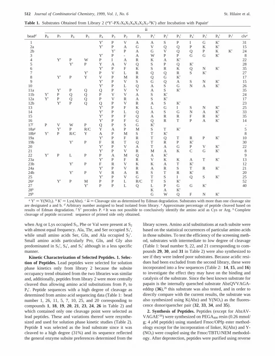
Table 1. Substrates Obtained from Library 2 (aY′-PX7X6X5X4X3X2X1-bK′) after Incubation with Papainc
VV
beadd P8 P7 P6 P5 P4 P3 P2 P1 P1′ P2′ P3′ P4′ P5′ P6′ P7′ clve
1 Y′ P V A A S P I G K′ 312a Y′ P A G V Q Q P K K′ 152b Y′ P A G V Q Q P K K′ 243 Y′ P - A W P P G G K′ 84 Y′ P W P I A R K A K′ 225 Y′ P Y A V Q S P Q K′ 286 Y′ P F K S R K Q N K′ 357 Y′ P V L R Q Q R S K′ 278 Y′ P Y V P M R Q G K′ 179 Y′ P V S G Q A S N K′ 15
10 Y′ P L Q A S G N A K′ 2611a Y′ P Q Q P V V A S K′ 3511b Y′ P Q Q P V V A S K′ 2412a Y′ P Q Q P V R A S K′ 1712b Y′ P Q Q P V R A S K′ 2313 Y′ P F K L G I S N K′ 2514 Y′ P L Q A S G N A K′ 3315 Y′ P F Q A R R F R K′ 3516 Y′ P F G Q R T P A K′ 1417f P V W P Q P S G K′ -18ag Y′ P R/C Y A P M S T K′ 518bg Y′ P R/C Y A P M S T K′ 819a Y′ P F R T Q T R P K′ 1019b Y′ P F R T Q T R P K′ 3020 Y′ P V A T A G P V K′ 2221 Y′ P V R M A K C G K′ 822 Y′ P L P P A M Q A K′ 923a Y′ P F R V K K A T K′ 1323b Y′ P F R V K K A T K′ 1224a Y′ P V R A R S T R K′ 2124b Y′ P V R A R S T R K′ 2025 Y′ P V G T S I Q S K′ 2526g Y′ P M P P L R/C T S K′ 5227 Y′ P P L Q L P G G K′ 4028h K A K ′29h G W Q F N K′a Y′ ) Y(NO2). b K′ ) Lys(Abz). c VV ) Cleavage site as determined by Edman degradation. Substrates with more than one cleavage site
are designated a and b.d Arbitrary number assigned to bead isolated from library.e Approximate percentage of peptide cleaved based onresults of Edman degradation.f Y′ precedes P.g It was not possible to conclusively identify the amino acid as Cys or Arg.h Completecleavage of peptide occurred: sequence of primed side only obtained.
512 Journal of Combinatorial Chemistry, 1999, Vol. 1, No. 6 St. Hilaire et al.

phase HPLC and characterized by the determination of themass using MALDI-TOF-MS or ES-MS (Table 2). Thesynthesis of all peptides proceeded smoothly in high yield.
3. Kinetics of Hydrolysis. The kinetics of substratehydrolysis using papain was first determined under pseudo-first-order conditions at 25°C for the determination of thesecond-order rate constant,kcat/KM. The cleavage points weredetermined using MALDI-TOF-MS as described in theExperimental Section (Figure 4a,b). The results shown inTable 3 indicate that while most substrates had identicalcleavage points both in solution and on solid phase, a fewwere cleaved in different or multiple positions compared towhen they were resin-bound. For example, peptides1 and30 were cleaved in one position on solid phase but cleavedat two positions in the solution phase assay. Conversely,peptide19 that showed two cleavage points on solid phase
had only one cleavage point in solution. Substitution of leadpeptides at various positions with different amino acids alsolead to variations in the cleavage points (e.g. peptides1 and2, 21 and22). For peptides showing significant hydrolysisat a second site, it can be shown that the measuredkcat/KM
corresponds to the sum of individual values ofkcat/KM foreach cleavage site and that the ratio of the resulting productsfrom each site corresponds to the ratio of the individualkcat/KM values.37 This ratio was determined from the MALDI-TOF-MS experiments and the measuredkcat/KM corrected toreflect solely the specificity constant for hydrolysis at theP1-P1′ position deemed the main cleavage point. MALDI-TOF-MS is not a generally quantitative technique; however,under certain circumstances the method can be used quan-titatively.38 Since the enzyme cleavage products have similarstructures, it is expected that their ionization and flightproperties may be similar.39 Therefore, the relative productsignal intensities may be a good measure of their relativequantities in the product mixture.
4. Effect of Substitution in the Various Subsites onLead Peptide 1.The amino acids in subsites P3 to P4′ weresubstituted by various amino acids primarily based on thelibrary results (Table 2,5-17). Most of the modified peptidesalso manifested major and minor cleavage points although
Table 2. Mass Characterization and Kinetic Parameters(kcat/KM) for Papain Hydrolysis of Internally QuenchedFluorogenic Substrates Derived from Library Results
no. substrateamassexp
massfoundb
clv(%)c
kcat/KM
(mM s)-1
1 dY′PVAVVAVSPIGK′ 1166.3 1166.7 31 760( 152 eK ′PVAVAVVSPIGY′ 1166.3 1165.8 815( 383 K′VA VVASPIGY′ 1069.2 1069.6 83( 334 AbzVA VVAVSPIGY′ 940.0 940.9 190( 85 Y′AVA VVAVSPIGK′ 1140.3 1140.3 735( 326 Y′PFAVVAVSPIGK′ 1214.6 1214.1 620( 247 Y′PLAVVAVSPIGK′ 1180.3 1180.8 540( 278 Y′PYAVVASPIGK′ 1230.6 1230.3 749( 79 Y′PVGVVASPIGK′ 1152.3 1152.4 1825( 19
10 Y′PVAVVSVSPIGK′ 1181.3 1181.9 469( 2611 Y′PVAVVAPPIGK′ 1176.4 1177.0 259( 1712 Y′PVAVVAVSQIGK′ 1197.3 1196.7 446( 713 Y′PVAVVASPNGK′ 1167.3 1168.1 315( 714 Y′PVAVVAVSPEGK′ 1182.3 1182.5 397( 2215 Y′PVGVVASPEGK′ 1168.2 1168.8f 1017( 4416 Y′PVEVVASGIGK′ 1184.3 1184.3 90( 717 Y′SPVAVVASPIGK′ 1253.4 1255.5 1095( 5318 Y′PMPPLCVVTSMK′ g 1431.7 1431.6f 52 2109( 10119 Y′PQQPVVAVVSK′ 1280.4 1280.3 35/24 190( 3420 Y′PYAVQVVSPQK′ 1344.5 1344.0 28 1524( 7921 Y′PVLRVVQQRSK′ 1438.6 1437.7 27 1450( 4122 Y′PVGVVRQQRSK′ 1382.5 1382.0 639( 6023 Y′PLQVVASGNAK′ 1212.3 1212.7 26 242( 2324 Y′PVGVVTSIQSK′ 1243.4 1243.7 25 800( 8225 Y′PVGVVGVSIQSK′ 1199.3 1199.2 254( 1426 Y′PVAVVTAGPVK′ 1166.3 1167.4 22 920( 12727 Y′PVAVVGVAGPVK′ 1122.3 1122.8 674( 2728 Y′PVAVVTAGK′ 970.1 970.0 168( 1629 Y′PVSVVGQASNK′ 1214.3 1211.9f 15 79( 430 Y′PLPPAVVMVVQAK′ 1279.5 1279.4 9 40( 331 Y′PVRVVMAKCGK ′ 1316.6 1315.3 8 557( 4532 Y′YQVVA VVGAK ′ 1162.3 1163.8 460( 4433 Y′PQVVAVVGAK ′ 1092.2 1096.6 88( 1734 Y′QVVA VVGAK ′ 999.1 999.9f 149( 435 AbzQVVA VVGAGY′ 928.0 928.3f 918( 12736 AbzQVVA VVGAE′ h 870.9 893.9 9351( 38237 Y′FRVVQQK′ i 1032.1 1032.6 84( 1338 Y′PFRVVQQK′ 1129.2 1130.6 84( 4
a Amino acid substitutions are shown in bold;VV and V denotemajor and minor cleavage points of substrates in solution, respec-tively. b Mass determined by MALDI-TOF-MS unless otherwisenoted.c Approximate cleavage of resin-bound peptide based onresults of Edman degradation.d Y′ ) Y(NO2). e K′ ) Lys(Abz).f Mass determined by ES-MS.g Original sequence obtained fromlibrary was Y′PMPPLCVVTSK′. h E′ ) eddnp.i Substrate for barleyendoprotease A and B.
Figure 3. Amino acid frequency in enzyme subsites obtained fromscreening of library 2. Peptide sequences are shown in Table 1.
Fluorescence-Quenched Solid Phase Libraries Journal of Combinatorial Chemistry, 1999, Vol. 1, No. 6513

some had one cleavage point as in the original solid phaseresults and others had up to three (data not shown). Substrate1 already possessed the preferred Pro residue at P3, andsubstitution by another small hydrophobic residue, Val,resulted in cleavage of the substrate at three different siteswhich made direct comparison to1 difficult (data not shown).Substitution of Ala (5) for Pro at P3 had almost no effect onthe specificity constant. Replacement of the hydrophobicresidue in P2 by other hydrophobic or aromatic residues Phe,Leu, or Tyr (6, 7, 8), led to a slight reduction inkcat/KM inthe case of Phe and Leu but not for Tyr. This effect reflectsthe natural tendency of the enzyme to cleave substrates withY(NO2) in the P2 position (screening of library 1) and thosewith amino acids containing nitro- and chloro-substitutedaromatic side chains in P2.36 Replacement of the Ala in theP1 position by Gly almost doubled thekcat/KM, providing oneof the best substrates (9) for papain. This large increase isprobably due to the insertion of the more flexible Gly into
the S1 binding pocket in a manner that allows optimizedcontacts of the other residues with the enzyme. Substitutionby Ser at P1 led to a substrate with multiple cleavage points,making a direct comparison difficult (data not shown).Substitution of Ala in P1′ by Ser (10) resulted in a reductionof the specificity constant by about a half. The P2′ positionwas already occupied by the favored amino acid, Ser; thusa substitution by a Pro residue was performed to see the effectof Pro in P2′ since, in autohydrolysis of propapain, cleavageoccurs such that Pro is present in the P2′ position (11).40 Thekcat/KM was reduced to about one-third its original value.Substitution of the Pro residue in P3′ with the most abundantamino acid Gln (12), resulted in a reduction ofkcat/KM byabout one-third. A substitution of Asn in P4′ (13) caused alarge reduction of catalytic efficiency. Since acidic residueswere excluded from the library, they were incorporated atsites where they had been observed in the first library farfrom (14 and15) and near (16) the active site, in order toevaluate their contribution to substrate hydrolysis by papain.Insertion of Glu at P1 or P4′ both resulted in a large reductionof the specificity constant.
5. Other Trends. Other sequences obtained from library2 and variations thereof (18-31) were synthesized in anattempt to investigate the correlation between solid phaseand solution phase hydrolysis. The results suggest that thereis not necessarily a direct correlation between the solid phaseand solution phase enzymatic hydrolysis of the samesubstrate. Some sequences that should result in goodsubstrates according to the degree of cleavage on solid phasedo not (e.g.19 and 23) while others perform as expected(e.g.18, 20, and21). Most sequences that were expected tobe poor substrates (30 and 31) had low kcat/KM’s. Toinvestigate the number of subsites required, peptide26 wasshortened by two amino acids (28) on the primed side andpeptide1 was shortened by one amino acid (3 and4) andlengthened by one amino acid on the nonprimed side (17).Results indicate that lengthening the peptide on the non-primed side slightly increases the specificity constant whereasshortening the peptide on either side lowerskcat/KM. Shortpeptides (37 and 38) that were good substrates for barleyendoprotease A and B41 were poorly hydrolyzed by papain.These results suggest that seven to eight subsites are indeedthe minimum required for optimal catalytic activity of papain-like cysteine proteases.
6. Influence of Donor/Quencher Pair.The influence ofthe position and nature of the donor/quencher pair wasinvestigated using two series of peptides:1-4 and32-36.The results suggest that the placement and nature of thegroups can greatly influencekcat/KM and the cleavage pointof the substrate, particularly if the cleavage point is relativelyclose to the reporter group. Changing the position of K(Abz)and Y(NO2) in 1 and2 resulted in a slight change inkcat/Km,probably due to a change in cleavage site. The effect is morepronounced in3 and 4 when substitution of K(Abz) withAbz in P3 halves the specificity constant. It appears that incertain substrates using eddnp as the quencher instead ofY(NO2) greatly enhances thekcat/KM of the substrate (35and36), probably due to interaction of the eddnp group witharomatic residues W177 and W181 (Figure 6f).
Figure 4. Representative mass spectra showing cleavage site ofsubstrates. (a) Cleavage of substrate1 in two positions. Often, di-sodiated species are also observed (538.09, 665.15, and 736.5). Peakat 657.91 arises from the buffer used in the assay. (b) Cleavage ofsubstrate9 at a single position. The peak at 479.13 represents theprotonated species, those at 524.31 and 547.26 represent the di-and tri-sodiated species, respectively, while the peak at 656.99comes from the buffer.
514 Journal of Combinatorial Chemistry, 1999, Vol. 1, No. 6 St. Hilaire et al.

To determine whether a high or low specificity constantwas a result of changes in binding or in catalytic rate,KM’sandkcat’s for selected substrates were determined from Hanesplots using substrate concentrations ranging from 2 to 150µM (Table 3). Examination of theKM of the best substratesobtained from the library revealed that a good substrate wasobtained either by tight binding to the enzyme (18, kcat )10.1 s-1; KM ) 6.8 µM) or by having a high catalytic ratecompensating for less effective binding (21,kcat ) 33.0 s-1;KM ) 22.6µM). On shortening the substrate at the primedside, the rate of catalysis remained the same but the bindingwas reduced by a factor of 3 (26 and28). In the series ofpeptides3, 9, 11, 15, 16 and 17, the rate of hydrolysis ofthe G-A or A-A amide bond was essentially the sameexcept in peptides11 and16 where having two prolines inP2′ and P3′ or a Glu in P1 results in marked reductions of the
catalytic rate. All these substrates had roughly the sameKM
except for17 which was 3-4-fold better and for16 whichwas reduced by about 25%. Peptides34-36 explored theeffect of the donor and quencher groups and it is clear thata higher specificity constant arose from lowerKM’s.
7. Modeling. The crystal structure of papain complexedwith the inhibitor Suc-QVVAA-pNA with a resolution of1.7 Å was used as the starting point for all calculations.42
To rationalize the results obtained from screening of thesubstrate libraries, seven representative substrates (1, 18, 21,26, 28, 35, 36) were docked into the active site of papain toinvestigate the following: multiple cleavages (1), the bindingof amino acids with large side chains (21), additional, lessspecific interaction with P4 and P5 (18), the importance ofP3′ and P4′ (26 and28), and the importance of fluorescentquencher for binding (35 and 36). The peptide substrates
Table 3. Kinetic Parameters for the Hydrolysis of Selected Internally Quenched Fluorogenic Substrates by Papain
[S] , KM1/8Km < [S] < 8 KM
no. substratea kcat/KM (mM s)-1 KM (µM) kcat(s)-1 kcat/KM (mM s)-1
3 bK ′VA VVASPIGY′ 583( 33 35.8( 8.2 23.8( 2.4 6659 cY′PVGVVASPIGK′ 1825( 19 37.6( 8.6 21.6( 1.6 575
11 Y′PVAVVAPPIGK′ 259( 17 40( 3.6 11( 0.42 27515 Y′PVGVVASPEGK′ 1017( 44 22.9( 11.8 19.1( 1.6 83416 Y′PVEVVASGIGK′ 90 ( 7 50.5( 47.1 5( 1.8 9917 Y′SPVAVVASPIGK′ 1095( 53 11.4( 2.9 10.6( 0.77 93018 Y′PMPPLCVVTSMK′ 2109( 101 6.8( 1.9 10.1( 0.25 147221 Y′PVLRVVQQRSK′ 1450( 41 22.6( 10.3 33.0( 4.6 146026 Y′PVAVVTAGPVK′ 920( 127 16.9( 10.9 13( 1.1 76928 Y′PVAVVTAGK′ 168( 16 52.9( 20.3 10.2( 1.5 19334 Y′QVVA VVGAK ′ 149( 4 37.9( 14.7 6.4( 1.2 16935 AbzQVVA VVGAGY′ 918( 127 17.4( 4.4 12.7( 0.60 73036 AbzQVVA VVGAE′ d 9351( 382
31 000e 1.6e 46e 29 000e
a Amino acid substitutions are shown in bold;VV denotes cleavage point of substrates in solution.b K′ ) Lys(Abz). c Y′ ) Y(NO2). d E′) eddnp.e Kinetic parameters determined at 30°C.26
Figure 5. The detailed structure of the papain binding site with peptide26 displayed as a thin black stick model. The structure is in thesame orientation as shown with the Connolly surfaces in Figure 6a and Figure 6b-f. Only residues forming the active site are shown asa thick stick model with light gray side chains and darker gray backbone atoms. The residues are indexed as numbered from the N-terminusof papain. Residues C25 and H159 form part of the catalytic triad.
Fluorescence-Quenched Solid Phase Libraries Journal of Combinatorial Chemistry, 1999, Vol. 1, No. 6515
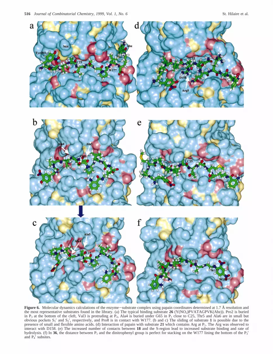
Figure 6. Molecular dynamics calculations of the enzyme-substrate complex using papain coordinates determined at 1.7 Å resolution andthe most representative substrates found in the library. (a) The typical binding substrate26 (Y(NO2)PVATAGPVK(Abz)). Pro2 is buriedin P3 at the bottom of the cleft, Val3 is protruding at P2, Ala4 is buried under G65 in P1 close to C25, Thr5 and Ala6 are in small butobvious pockets S1′ and S2′, respectively, and Pro8 is in contact with W177. (b and c) The sliding of substrate1 is possible due to thepresence of small and flexible amino acids. (d) Interaction of papain with substrate21 which contains Arg at P1. The Arg was observed tointeract with D158. (e) The increased number of contacts between18 and the S-region lead to increased substrate binding and rate ofhydrolysis. (f) In36, the distance between P1 and the dinitrophenyl group is perfect for stacking on the W177 lining the bottom of the P3′and P4′ subsites.
516 Journal of Combinatorial Chemistry, 1999, Vol. 1, No. 6 St. Hilaire et al.

were aligned in the direction inferred from most availablecrystal structures of papain complexed with peptide inhibi-tors; that is, the orientation with S3 between V133 and Y67forward to S3′-S5′ at W181 and W177 (Figure 5). Thepossibility that peptide substrates can be aligned in thereverse direction has been discussed.43 Our attempts, how-ever, to also model substrates in that orientation wereunsuccessful since the cooperative orientation of the substrateside chains in the S and S′ sites did not allow close proximityof the active sulfhydryl to the scissile bond (i.e.<4 Å).
The results of the calculations are best illustrated withsubstrate26 (kcat/KM ) 920 (mM s)-1; intermediate bindingenergyKM ) 17 µM and intermediate catalytic efficiencykcat ) 13 s-1) which binds in the classical way predictedfrom small peptide-like inhibitor complexes with papain, i.e.in an extended conformation and with very close contactsto the enzyme active site (Figure 5). Several startingconformations particularly with different orientations of theP1 side chains were attempted, and for the most part, thesame ending conformation with the Ala methyl group boundin a small pocket under N64 and G65 and pointing towardW26 was attained at equilibrium. Other contact residues forP1 are G66, C25, and Ala 160. The P2 Val side chain isoriented away from the cleft and interacts with residues Y61,G65, and D158. The orientation of Pro in P3 was with thepyrrolidine ring pointing toward the cavity with P68 at thebottom of the cleft. Other P3 contact residues are V150,V133, and Y67. P1′ is to the left of the binding cleftcontacting D158, A136, H159, A137, and Q142, while P2′points to the right to interact with G23, C22, G20, and Q19(when viewing along the substrate cleft in the Nf Cterminal direction). The positions of S3′ (W177 and Q147),S4′ (W177, W181, Q147, and L143), and S5′ (G20, N18,and S21) were less obvious, and various binding modes wereobtained with different substrates (vide infra). According tothe current model, there may even be an S5′ binding subsitecomprising residues W181, G178, T179, and G180. Interest-ingly, there was a stacking of the Pro ring at P4′ onto thearomatic side chain of W177.
The docking of substrate1 (Y(NO2)PVAVVAVSPIGK(Abz))with a major and a minor cleavage site examined the abilityof papain to cleave substrates with small amino acids suchas Ala-Ala or Gly-Ala at P1-P1′ or Ser-Thr at P1′-P2′ intwo different positions (Table 2). The substrate bound in aconformation which allowed “sliding” of the amino acid inP1-P1′ along the cleft to optimize interactions either at S3,S2, or at S1′, S2′, S3′. The “sliding” actually occurred afterrelease of constraints to the sulfhydryl group during thecalculation going from the complex in Figure 6b to the onein Figure 6c and was possible because in papain there is asmall cavity beneath the S2 subsite that can accommodatethe small Ala methyl group. When the equilibrium confor-mation obtained for26 was used as the starting point for1and the constraints on the sulfhydryl group were maintained,the calculation converged to a single equilibrium conforma-tion similar to that in Figure 6b.
Other substrates obtained from the library screening werethose with Arg in P1 and with relatively large amino acidside chains in P2-P3′ as typified by21 (Y(NO2)PVLRVVQ-
QRSK(Abz)). Modeling of complexes with these types ofsubstrates was considerably more difficult since the motionof the more bulky peptides in the narrow cleft was limited.The result was an orientation of the P3 and P2 side chains asfor 26; however, the P1 Arg side chain bent upward to interactwith the carboxylate of D158 (Figure 6d). During thecalculation, the distance between the guanidyl nitrogen andthe carboxyl oxygen varied from 2 to 5 Å. The larger residuesin P1′ and P2′ (QQ) were accommodated by the backboneforming a kink in the chain allowing the P2′ Gln to interactwith S1′ and the P1′ Gln to interact with S2′ with subsitesdefined as previously reported43 and by the docking ofsubstrate26.
The addition of residues in P4 and P5 as in substrate18(Y(NO2)PMPPLCVVTSMK(Abz)) gave an increased bindingto the enzyme. From the molecular modeling calculations,the Pro rich sequence results in a turn structure protrudingfrom the Pro buried in P3 that allows all three amino acidsin P4-P6 to interact in a less specific manner with the shallownonprimed part of the substrate cleft (Figure 6e). Thisincreased enzyme substrate interaction leads to lowerKM’s.
Modeling of28, the shortened form of26 lacking Gly andPro in P3′ and P4′, demonstrated the absence of stackinginteractions between the Pro at P4′ on the aromatic side chainof W177 (data not shown). A similar stacking withπ-πinteractions was observed with substrate36 (Figure 6f),which had an almost ideal orientation and distance betweenthe Ala in P1 and the dinitrophenyl group of eddnp tofacilitate stacking on W177 (and W181). Interestingly, whena change inø1 of W177 occurred during one annealing,perfect intercalation of the dinitrophenyl group between theindole rings of W177 and W181 was obtained, suggestingthat such an intercalation may also be at play during enzymecleavage.
Discussion
Library Screening. The combinatorial library approachencompasses three steps: the design and construction oflibraries of chemical diversity, followed by the applicationof screening and assay techniques, and finally the identifica-tion of the active compounds. Screening of libraries andidentification of active compounds depend on the mode oflibrary construction and the type of receptor being investi-gated. Synthetic peptide libraries have been used to charac-terize the substrate specificity of various proteolytic enzymesboth in solution44-46 and on solid phase.8,10,30 But howeffective are these methods? Do the hits obtained from thelibrary screen truly represent the best possible substrate orinhibitor for that enzyme? In the screening of solution phaselibraries (i.e. screening of mixtures of compounds), thatquestion cannot be answered since the precise identity ofthe active compounds in the mixture is usually not knownand the identity of the substrate or inhibitor is obtained froma statistical analysis of the preferred residue at variouspositions. Despite the tedium of the process, good substrates/inhibitors are nonetheless obtained.45,47In the one-bead-one-compound libraries, good lead compounds can be obtainedboth by direct identification of a single active compound aswell as from a statistical analysis of favored residues in the
Fluorescence-Quenched Solid Phase Libraries Journal of Combinatorial Chemistry, 1999, Vol. 1, No. 6517

various enzyme subsites. Furthermore, different families ofsubstrates may be identified and it is possible to correlatethe results of the solid phase screening to the subsequentsolution phase assays.
In the present work, we endeavor to assess the efficiencyof the solid phase fluorescence-quenched library screeningas a means of characterizing the substrate specificity ofproteolytic enzymes, in particular cysteine proteases. To thisend, we have used papain, the archetypal cysteine protease,as the model enzyme and have attempted to explain thesubstrate specificity using molecular modeling. Screening oflibrary 1 randomly generated from all 20 natural amino acidsyielded substrates with a preponderance of acidic residuesprimarily in subsites away from the cleavage site, i.e. S4′,S5′, and S6′. These results are artifactual and do notnecessarily arise from preferential selection of these sub-strates by the enzyme. The apparent preference for acidicresidues is most likely due to an increase in the effectivelocal concentration of the enzyme in beads with a highconcentration of negative charges (acidic residues) becauseat pH 6.8 papain is an overall basic molecule with a pI of8.75. Replacement of particular residues with acidic residuesin substrates found from the screening of library 2 (withoutacidic residues) did not yield better substrates (Table 2:14and15) with increasedkcat/KM values, suggesting that theyare not preferred in those subsites. This occurrence alsosuggests that the nature of the residue P4′-P6′ is lessimportant for the cleavage of the substrate. Interestingly, theamino acid subsite preferences for the P2-P2′ obtained fromscreening library 2 were similar to the results obtained fromscreening library 1, after disregarding the preponderance ofacidic residues and the preference for Y(NO2) in S2. Thisfinding suggests that in the event that the enzyme concentra-tion is locally increased due to additional unrelated affinity,the substrate preference must still be satisfied in order toachieve enzymatic hydrolysis.
The intramolecular fluorescence-quenched assay has theadvantage that information about the amino acid preferencein both the nonprimed and the primed subsites can beobtained in a single experiment. However, the methoddemands that the peptide be flanked by fluorescent donorand quencher groups that inevitably interact with the enzyme,thus influencing the cleavage points and the absolute valueof the kinetic parameters. In the present library, 2-aminobenzoic acid (Abz), attached to the side chain of Lys, wasused as the fluorescent donor and 3-nitrotyrosine (Y(NO2))as the fluorescent quencher. These residues are relativelysmall compared to other fluorescent donor/quencher pairsthat are considerably more hydrophobic (e.g. DABCYL/EDANS23-25,48) and have the additional advantage that theyare readily incorporated into the peptide during synthesis.The results obtained from the screening of library 1 (Figure2) illustrate the situation wherein the fluorescence quencher(Y(NO2)) strongly influences the cleavage site of thesubstrates and subsequently biases the screening results.However, the results from the kinetics of hydrolysis of leadsubstrates (Table 2) obtained from library 2 suggest thatwhile the positioning of Y(NO2) and K(Abz) at either endof the peptide does not significantly influence thekcat/KM
values, they may influence the cleavage point of the substrate(Table 2: 1-4). In contrast, other fluorescent donors andquenchers may heavily influence the value of the specificityconstant in a way that depends on the nature of the substrateand the distance of the reporter molecule from the cleavagepoint. This influence is particularly evident when the reportergroup is situated at important subsites, e.g. Y(NO2) in P2 oreddnp in P3′-P4′. It can be seen that the use of eddnp insteadof Y(NO2) as the quencher residue enhances 1 order ofmagnitude thekcat/KM value in the shorter substrates analyzed(Table 2: 35 and36).
Of fundamental importance is the question of correlationbetween enzymatic activity on solid phase compared to thatin solution. Is the best substrate obtained from the solid phaseassay the best in solution? Based on the data in Table 2, theanswer is not so straightforward and there does not appearto be a direct, linear correlation between activity on solidphase compared to that in solution, particularly in terms ofranking of the substrates. Generally, a substrate that wasgreater than 20% cleaved on solid phase was a good orexcellent substrate in solution phase assays (1, 18, 20, 21,24, 26) with two exceptions (19 and 23). The converse isalso true; substrates with a low degree (10-15%) of solidphase cleavage were generally poor substrates in solutionphase assays (29, 20, 31). The ranking of the substrates inincreasing order of degree of cleavage on solid phase, wasnot the same as the ranking based on increasingkcat/KM
obtained in the solution phase assays. Furthermore, thenumber and position of the cleavage site of the peptidediffered on solid phase compared to in solution. Themolecular modeling and the amino acid substitutions shownin Table 2 suggest that subtle changes in the substrate arerequired for changes in the cleavage site in certain substrates.The reasons for these differences are not immediatelyobvious but are related to the microenvironment and dynam-ics of the substrate on the resin. Furthermore, the active siteof papain is a narrow cleft without significant subsite“binding pockets” and the preference for small amino acidsin most subsites (Figures 2 and 3) allows the substrate toeasily slide thus resulting in alternative modes of bindingbecause of minor changes in the substrate. The resin linker(HMBA) is situated in the extended primed subsites and maythus influence the binding conformation of the substrate.Another possibility for lack of correlation between degreeof cleavage on solid phase and solution phasekcat/KM valuescould be due to higher local enzyme concentration in thebeads which contain peptides that may bind tightly to anoncatalytic binding site of the enzyme. Thus, a peptide maybe cleaved to greater extent than another because there wasa higher concentration of enzyme present. Such local effectsare not at play in the solution phase assay. These liabilitiesnotwithstanding, overall there is a high probability that agood substrate on solid phase will be a good one in solutionand vice versa: the peptide with the highest degree ofcleavage on solid phase was one of the best cleaved insolution (18) while the one with one of the lowest degreesof cleavage was the poorest substrate (30).
Characterization of the Substrate Specificity of Papain.A secondary goal of this investigation was to characterize
518 Journal of Combinatorial Chemistry, 1999, Vol. 1, No. 6 St. Hilaire et al.
the substrate specificity of papain by implementation of amethodology that provides information on the specificity ofboth the nonprimed and primed sites simultaneously. Usingan intramolecular fluorescence-quenched solid phase com-binatorial library approach, we were able to obtain very goodsubstrates for papain and to characterize the subsite specific-ity from S4 to S4′. The papain active site, a narrow cleftbetween the two tightly interacting domains, is linedpredominantly by protein backbone of limited flexibility, andmost of the 16 contact residues are small amino acids, i.e.five Gly, three Ala, two Cys, and one Ser. Interaction of thesubstrate with the active cleft of the enzyme is thereforedominated by contacts to the protein backbone. In order forthe substrate to arrive at the short distance required forcleavage, the rigid cavity demands flexibility of the substrateduring binding and this was clearly reflected by the relativeease of docking substrates with small amino acid side chaingroups into the active site and by the preponderance ofsubstrates composed of small amino acids naturally selectedfrom the library screening. There is a general absence ofdistinct binding pockets that can accommodate large aminoacid side chains in the active site.
Subsites S4 and S3 showed a preference for the smallnonpolar amino acids Val or Ala or Pro. While there is somedebate over the actual existence of well-defined subsitesbeyond S3,49 results from the modeling support the lack ofwell-defined subsites. The active site broadens into ahydrophobic patch from the P3 subsite onward, making itrelatively easy for hydrophobic residues of some substratesto orient themselves in ways that optimize enzyme-substrateinteractions. For example, Y(NO2)PMPPLCTSMK(Abz) (18)with Met and Pro in P5 and P4 has the best specificityconstant of all the identified substrates due to increasedbinding energy (Table 3).
In substrates1, 26, and28, there is a small, but significantpreference for Pro in P3 due to the perfect fit of the Câ-Cδof the pyrrolidine ring into a small cavity with P68 at thebottom. A similar interaction is observed with21 which hasthe other preferred residue, Val, in P3. In fact, all substrateshad the same orientation at P3/P2. Consistent with literatureresults, a hydrophobic residue was best at S2; however, thatresidue could be Val, Phe, Leu, Tyr, or Y(NO2). From boththe statistical analysis of the library results (Figure 3) andthe specificity constants for selected substrates (Table 2), itappears that Val is slightly favored over Phe, Tyr, and Leuin substrates containing a small residue at P1 but an equalfrequency in those containing Arg or Lys at P1. The P2 Valside chain is oriented away from the binding cleft. Theapparent lack of structural requirement in S2 and strictrequirement for a hydrophobic residue at P2 indicate that theside chain does not fit into a specific pocket but ratherinteracts with side chains of hydrophobic residues (e.g. V150,Y61) at the rim of the cleft. Interestingly, the S1 subsiteexhibited a dual specificity for small, nonpolar amino acidsas well as the larger, charged Arg. For small residues, theorientation of the P1 side chain was into a small pocket underG65 at the left side of the cleft. Larger substrates clearlybind in a different mode since there is no space for a largeside chain in the small P1 pocket, and in the case of substrate
21 the salt bridge formed between the Arg in P1 and D158may result in the extra force needed to get sufficientproximity of the carbonyl and the sulfhydryl group to givefast rate of hydrolysis (kcat ) 33.0 s-1) and poorer binding(KM ) 22.6 µM) (Table 3).
The presence of small residues at P1-P2′ causes somesubstrates to be cleaved in more than one position becauseof “sliding” of the small amino acid side chains into smallcavities in the enzyme at S1-S2′. The remaining subsites(S1′-S4′) all preferred small nonpolar residues with apreference for Ala and Ser. These results are consistent withprevious studies in solution23,24 and on solid phase29 whichdetermined that small and/or hydrophobic residues such asSer, Val, Ala, and Leu, were best at P1′-P2′. Most goodsubstrates, i.e. those with akcat/KM greater than 1000 (Table2: 9, 15, 17, 18, and20) contained primarily small aminoacids in P3-P3′ although exceptional substrates (Tables 2and 3: 21) also contained larger residues in these positions.Although S1′ and S2′ are well defined for substrates withsmall amino acids, other substrates with larger amino acidsin those positions bind with the side chains away from thecleft toward solvent. Substrate28 which lacks residues Glyand Pro in P3′and P4′ had a similarkcat and a higherKM (Table3) than26, underscoring the importance of the stacking ofPro at P4′ on the aromatic side chain of W177. This stackinginteraction plays a significant role in the specificity of theS3′-S4′ subsite of papain and could be the reason for thevery highKM (Table 3) found for36 in contrast to substrate35 for which stacking interactions of the Y(NO2) with W177(and W181) are not optimal.
Several good substrates for papain were obtained directlyfrom the library screening and from modification of leadpeptides. The sequences of the substrates vary significantlybut can be loosely classified into two groups: thosecontaining part of the cystatin-like sequence xVxA(T)x26 orthe C hordein-like xQQx sequence (C hordein is a substratefor endoproteases from barley).41 The best substrates obtainedwere Y′PMPPLCTSMK′ (kcat/KM ) 2109 (mM s)-1),Y′PYAVQSPQK′ (kcat/KM ) 1524 (mM s)-1), and Y′PVL-RQQRSK′ (kcat/KM ) 1450 (mM s)-1) and not thosecontaining cystatin-like sequences. These substrates hadhigherkcat/KM values than Y′QVVA VVGAK′ (kcat/KM ) 149(mM s)-1), the equivalent of AbzQVVAVVGAeddnp (kcat/KM
) 9351 (mM s)-1). Interestingly, a substrate Y′PQQPVV-AVVSK′ (kcat/KM ) 190 (mM s)-1 very similar to theQVVAGA substrate was obtained directly from the libraryscreen, and thekcat/KM is on the same order of magnitude asY′QVVA VVGAK′. The QVVA motif was only superior inthe case where the eddnp moiety is stacked above W177/W181, creating additional favorable binding interactions.
Conclusions
A new methodology which combines the screening ofcombinatorial libraries of substrates with molecular modelingof hits from the library has been developed for the completecharacterization of proteolytic enzyme specificity. The ap-proach combines the natural selection of substrates by theenzyme from a large library with the modeling of theinteraction of the enzyme with these substrates to allow
Fluorescence-Quenched Solid Phase Libraries Journal of Combinatorial Chemistry, 1999, Vol. 1, No. 6519

rational design of ligands with desired properties. Of keyimportance is the ability to perform high throughput screen-ing of the resin-bound substrates using a solid support thatdoes not greatly influence the outcome of the screen. Anawareness of factors such as the nature and placement ofreporter groups and linker and properties of the receptor thatcan skew the screening results should be maintained. Thismethodology once properly applied is quite effective, provid-ing a fairly good correlation between activity on solid phaseand in solution and has generated, in this case, very goodsubstrates for papain. In addition, we have mapped the subsitespecificities of papain and in contrast to previous results, itappears that the nature of the residue in S2 alone does notdominate the specificity requirements. Rather, it is thesynergistic relationship between the residues in subsites S2,S1, and S1′ that govern the specificity of the enzyme. Wealso suggest that although the S′ interactions are veryimportant for the substrate binding and cleavage, multiplebinding modes in S′ subsites are possible. Furthermore,comparison of the calculations performed on the varioussubstrates demonstrated that the enzyme, even with the quiterestricted motion in the active site observed during calcula-tions, reaches complex structures with quite different topolo-gies to accommodate the different substrates. This is probablya prerequisite for the binding and cleavage of such diversefamilies of substrates by general proteases such as papain.
Experimental Section
Materials And Methods. All solvents were purchasedfrom Labscan Ltd. (Dublin, Ireland). Dichloromethane wasdistilled from P2O5 and was stored over 3 Å molecular sievesunder argon. Fluoren-9-ylmethoxycarbonyl (Fmoc) aminoacids and their pentafluorophenyl (Pfp) ester derivatives werepurchased from Bachem and NovaBiochem. Fmoc-Lys(Boc-Abz)-OH and Fmoc-Tyr(NO2)-OH were prepared as previ-ously described.50 The substitution of the resins was deter-mined by spectrophotometric analysis at 290 nm of thedibenzofulvene-piperidine adduct formed upon deprotectionof the amino terminal using a Perkin-Elmer Lambda 7 UV/vis spectrophotometer. Purification of peptides was per-formed by preparative reverse phase HPLC on a WatersHPLC system with a delta pak C-18 column (200× 25 mm)and a linear gradient of A (0.1% TFA in water) and B (0.1%TFA in 90% aqueous MeCN) at a flow rate of 20 mL/min.Amino acid analyses were carried out in a Beckman 600amino acid analyzer following hydrolysis with 6 M HCl with5% (v/v) phenol at 110°C for 48 h. Amino acid sequencingwas performed on resin-bound substrates using an AppliedBiosystems Sequencer models 477A or 470A equipped withan on-line phenylthiohydantoin analyzer (model 120A)according to the protocol of the manufacturer. MALDI-TOF-MS of synthetic peptides was performed on a FinniganLasermat 2000 with a matrix ofR-cyano-4-hydroxycinnamicacid while peptides from library beads were analyzed in thereflectron mode on a Bruker Reflex III using the same matrix.ES mass spectra were recorded with a VG-Quatro instrumentfrom Fisons. Enzyme kinetics were performed using atemperature-controlled Perkin-Elmer luminescence Spec-trometer (LS 50B).
Enzyme.Papain, twice recrystallized from papaya latex,was purchased from Sigma (Lot # 84H7220) and usedwithout further purification. The molar concentration wasdetermined by active site titration with E-64 [trans-ep-oxysuccinyl-L-leucylamido-(4-guanidino)butane] (Sigma; Lot# 77H0328) as previously described using Abz-CRQQY-(NO2)D-OH as the substrate.41 The concentration of enzymein a 100× diluted solution (1X) was 8.34µM. For assays,the 1X solution was diluted 20-fold giving rise to the 20Xsolution. Papain was activated as the 1X solution byincubation in activation buffer (50 mM phosphate buffer,pH 7.2, augmented with 10 mM cysteine, 1 mM EDTA, and0.08% Brij 35) for 10 min at room temperature. A freshsolution of enzyme was activated every 3 h for use in solutionphase assays.
General Methods for Solid Phase Peptide Synthesis.Syntheses of peptides and libraries were carried out manuallyby MCPS.51,52 on PEGA resin respectively in a 20 columnTeflon synthesis block. In general, NR-Fmoc amino acidOPfp esters with the following side chain protecting groupssO-tBu for Asp and Glu,tBu for Tyr, Ser and Thr, Trt forCys, Asn and Gln, Boc for His, Lys and Trp and Pmc forArgswere used for the synthesis. Each coupling step wascarried out using the amino acid (3 equiv) in DMF alongwith Dhbt-OH (1 equiv) as an acylation catalyst as well asan indicator of the reaction completeness.53 In certain cases,reaction completion was also assessed using the Kaiser test.To coupling reactions that were incomplete was added anadditional 1-2 equiv of the amino acid ester. Each couplingstep was followed by washing with DMF (6×), removal ofthe Fmoc group by treatment with 20% piperidine in DMF(4 + 16 min), and then another DMF washing step (6×). Atthe end of the synthesis, the resin was washed with CH2Cl2(6×) and dried by air suction over a period of 1 h. The sidechain protecting groups were removed by treatment with amixture of TFA:thioanisole:ethanedithiol:water (87.5:5:2.5:5) initially for 10 min and then for 2.5 h. The resin wasthen washed with 95% acetic acid (4×), DMF (2×), 5%DIPEA (2×), DMF (2×), and CH2Cl2 (6×) then dried invacuo. Cleavage of peptides from the resin was effected bytreatment with 0.1 M NaOH for 2 h followed by washingwith water (7×). The combined filtrate was neutralized with0.1 N HCl, and the crude peptides were purified bypreparative HPLC.
Solid Phase Substrate Library Synthesis.The librariesof the structure Y(NO2)X8X7X6X5X4X3X2X1K(Abz) (Figure1) containing all 20 genetically encoded amino acids wereprepared on PEGA4000 resin.11 The capacity of the resin wasdoubled (final loading 0.21 mmol/g) by synthesizing thepeptide on the amino group of the side chains of two lysines(Figure 1). Using the syringe technology54 two FmocLys-(Boc)OPfp were coupled to the PEGA4000 resin (1,7 g, 0.12mmol/g; 200-800µm beads), and after removal of the Fmocgroup, the N-terminus was acetylated and the Boc side chainprotecting groups were removed by treatment with 50% TFAin CH2Cl2 for 30 min. After the appropriate washing protocol,HMBA (3 equiv) in DMF was coupled under TBTU (2.9equiv)-NEM (4.5 equiv) activation. The resin was washedand dried by lyophilization before the coupling of Fmoc-
520 Journal of Combinatorial Chemistry, 1999, Vol. 1, No. 6 St. Hilaire et al.

Lys(BocAbz)-OH (2.5 equiv) in dry CH2Cl2 under the agencyof MSNT (2 equiv) and MeIm (2.5 equiv).55 The reactionwas allowed to proceed for 45 min, after which the resinwas washed with DMF (2×) and CH2Cl2 (1×). The couplingprocedure was repeated for another 45 min and the resinwashed with DMF (7×). The resin was evenly distributedin the 20 wells of a Teflon synthesis block, and the Fmocgroup was removed. An Fmoc amino acid OPfp ester wascoupled into each of the 20 wells. After completion of thecoupling, the block was filled with DMF up to 1 cm abovethe top of the wells and inverted, and the resin was mixedvigorously for 30 min in the mixing chamber. The blockwas once more inverted, evenly distributing the resins oncemore into the wells for washing and Fmoc deprotectionprotocols. This procedure was repeated for the incorporationof the first seven amino acids of the library in the case oflibrary 1. In the case of library 2, after the coupling of theseven randomized positions, FmocProOPfp was added to allthe wells. In both libraries, Fmoc(Y(NO2))-OH was incor-porated using TBTU/NEM preactivation for 7 min. Thelibrary was deprotected and washed as described in generalSPPS methods. The mass and purity of a random samplingof compounds attached to 36 beads (ca. 100 pmol/bead) wereanalyzed by high resolution MALDI-TOF-MS. For thispurpose, 36 resin beads were randomly chosen and thepeptides cleaved off by treatment with 10% TEA in MeOH.For some of those compounds, the sequences were alsodetermined by Edman degradation and correlated with themasses obtained.
Solid Phase Multiple Column Peptide Synthesis.Leadpeptides from the library screen were synthesized onPEGA800 resin (0.21 mmol/g, 4µM/well, 150-300 µmbeads). The HMBA linker was attached as described for thelibrary synthesis with the exception that a glycine was firstcoupled to the resin before coupling of the linker. Peptidesynthesis was carried out as described for library synthesiswith the omission of the mixing step. After deprotection andwashing, the peptides were cleaved with 0.1 M NaOH (350µL/well) for 2 h and the products collected in tubes placedbeneath the wells. The solutions were neutralized with 0.1M HCl, and the peptides were purified by reverse phaseHPLC. The peptides were characterized by MALDI-TOF-MS or ES-MS, and their purity was assessed by analyticalHPLC at 215 and 320 nm.
Solid Phase Library Screening. The library beads (300mg) were washed with assay buffer (50 mM phosphatebuffer, pH 7.2, augmented with 2 mM cysteine, 1 mMEDTA, and 0.08% Brij 35; 3×) and then incubated withactivated papain (135µL of 20X in 10 mL buffer; 5 nM) at25 °C in a small glass Petri dish (4 cm diameter). Thefluorescence intensity of the beads was monitored with afluorescent microscope every 30 min for indications ofhydrolysis. After 2.5 h for library 1 and 3.5 h for library 2,several beads showed a fluorescent “ring”, indicating hy-drolysis of some of the peptides on the beads. The reactionwas then stopped by treatment with 2% aqueous TFAsolution and washed with water (2×), 2% NaHCO3 (2×),and then water (3×). The fluorescence intensity of the beadswas assessed by inspection with a fluorescent microscope,
and bright beads were collected and transferred to a TFA-treated cartridge filter for on-resin sequence analysis. Theamino acid sequence and the cleavage point of the peptidesubstrates were determined by Edman degradation. Theextent of cleavage was determined by a comparison of thepicomoles of an amino acid in both the noncleaved andcleaved peptide in different cycles of the degradation.
Enzymatic Hydrolysis of Fluorescence-Quenched Sub-strates in Solution. Substrates were dissolved in water,DMF, or combinations thereof to a final concentration ofapproximately 5 mM. Hydrolysis of substrates was carriedout at 25°C in 50 mM phosphate buffer, pH 6.8, augmentedwith 2 mM cysteine, 1 mM EDTA, and 0.08% Brij 35.Hydrolysis was followed by measurement of the increase inintensity of the Abz fluorescence (λEX ) 320 nm,λEM )420 nm, slit width: 10 nm). Under pseudo first-orderconditions, the rate of initial hydrolysis was measuredcontinuously for 5 min for four concentrations of eachsubstrate. Total hydrolysis was effected by the addition of50 µL of 1X enzyme (final concentration of 0.2µM), andfinal fluorescence was measured after 24 h.kcat/KM valueswere calculated from the following equation:kcat/KM )slope/([E]o(I f - Io)) where slope) ν ) ∆I/∆t, [E]o is initialenzyme concentration, and (I f - Io) ≡ [S] assuming [S],KM. For determinations ofkcat andKM, assays were carriedout under similar conditions in a 100µL flow cell using sixdifferent substrate concentrations ranging from 2 to 150µM.The slit width was reduced to 2.5 nm to compensate for thehigher fluorescent intensities of more concentrated solutions.The slope was converted into moles of substrate hydrolyzedper second by use of a standard curve. Substrate concentra-tions were also correct using the standard curve. The standardcurve was obtained from measurement of the fluorescenceintensity of AbzGAGAAF-OH derived from the total hy-drolysis of AbzGAGAAFFA-Y(NO2)D-OH by subtilisin atdifferent concentrations (20-180 µM). The kinetic param-eterskcat and KM for each substrate were then determinedfrom Hanes plots ([S]/ν vs [S]) of the measured values.
Determination of Cleavage Point in Solution PhaseAssays. The cleavage point was determined in separatemicroassays on more concentrated solutions but maintaininga similar enzyme-to-substrate ratio as in the solution assaysabove. Substrates (6µL of 5 mM stock) were incubated with2 µL of 1X enzyme (activated with activation buffer withoutBrij 35 since peaks from Brij occur in the mass range ofinterest) in 12µL assay buffer (without Brij 35) for 30 min.The hydrolysis mixture (2µL) was mixed with CHC matrix(1 µL), and the MALDI-TOF spectra were acquired andanalyzed (Figure 4).
Modeling. Molecular dynamics calculations were carriedout on a Silicon Graphics Octane workstation using theInsightII/Discover program. The coordinates from the crystalstructure of papain complexed with peptide inhibitor (Suc-QVVAA-pNA) determined at 1.7 Å resolution42 were usedas input for the calculations after removal of the inhibitor.The calculations were performed with seven differentsubstrates1, 18, 21, 26, 28, 35, and36 in order to explainvariations in the substrate preferences found in the two “one-bead-one-substrate” libraries. During all calculations, most
Fluorescence-Quenched Solid Phase Libraries Journal of Combinatorial Chemistry, 1999, Vol. 1, No. 6521

of the amide backbone of papain was fixed, while all theside chains were allowed to move. Each calculation wascarried out as annealing at decreasing temperatures, 650 andthen 500 and 300 K. At 500 and 300 K, the papain residuesin contact with the substrate were allowed to move freely insequence ranges of five successive amino acids or less. Thesubstrates were initially energy-minimized and added inextended conformations approximately 15 Å away from thebinding site. A weak constraint of∼3 kcal/mol (boundaries3 and 4 Å) was added between the scissile bond and theactive thiol, and this was maintained throughout all calcula-tions. Initially, in the first calculations a weak constraint of∼3 kcal/mol (boundaries 4 and 8 Å) between the terminalresidues and papain residues at either end of the active cleftwere added; however, this constraint was later omitted sinceno significantly different results were obtained with theseextra constraints. Additional constraints of∼3 kcal/mol(boundaries 2 and 3 Å) were applied for distances betweenthe sulfhydryl hydrogen andπ-nitrogen of H159 and betweenthe τ-NH of the H159 and the N175 side chain carbonyl.The calculations were initiated with 10 steps of minimizationand then 30 000 to 60 000 steps were calculated at eachtemperature at 1 fs intervals. The development of eachcalculation was monitored, and when persistent obstructionsto the progress (i.e. distance of>5 Å between the sulfuratom and the scissile carbonyl carbon) of the calculation wereobserved, a new starting substrate conformation was used.Only theæ-ψ angles allowed in theæ-ψ space were used asstarting conformations. As many as five independent suc-cessful calculations were performed for each substrate, andwhen the calculations were allowed to reach equilibrium,the results obtained with different starting conformationsconverged to one or two related bound conformations.Constraints were completely removed from the final structureand the complex was subjected to extended calculations, inorder to determine whether the substrate remained bound tothe papain molecule in a stable conformation.
Acknowledgment. This work was supported by theINCO-DC program (EU contract number ERBIC18CT97-0225), the Danish National Research Foundation, and theBrazilian Research Foundations FAPESP and PADCT. Dr.Ib Svendsen, Bodil Corneliussen, and Lone Sørensen areacknowledged for peptide sequencing. Dr. David Simpsonis thanked for many helpful discussions about enzymekinetics and Hanne Christiansen for help with peptidesynthesis. High resolution MALDI-TOF-MS and ES-MSwere carried out by Pia Breddam and Anita Jansson.
References and Notes(1) Furka, A.; Sebestyen, F.; Asgedom, M.; Dibo, G. General method
for rapid synthesis of multicomponent peptide mixtures.Int. J. Pept.Protein Res.1991, 37, 487-493.
(2) Lam, K. S.; Salmon, S. E.; Hersh, E. M.; Hruby, V. J.; Kazmierski,W. M.; Knapp, R. J. A new type of synthetic peptide library foridentifying ligand-binding activity.Nature1991, 354, 82-84.
(3) Rapp, W.; Zhang, L.; Ha¨bish, R.; Bayer, E. Polystyrene-Polyoxy-ethylene graftcopolymers for high-speed peptide synthesis. InPep-tides 1988, Proc. Eur. Pept. Symp., 1 ed.; Jung, G., Bayer, E., Eds.;Walter de Gruyter: Berlin, 1989; pp 199-201.
(4) Meldal, M. PEGA: A flow stable poly(ethylene glycol) dimethylacrylamide copolymer for solid phase synthesis.Tetrahedron Lett.1992, 33, 3077-3080.
(5) Renil, M.; Meldal, M. POEPOP and POEPS: Inert poly(ethyleneglycol) cross-linked polymeric supports for solid phase synthesis.Tetrahedron Lett.1996, 37, 6185-6188.
(6) Buchardt, J.; Meldal, M. A chemically inert hydrophilic resin forsolid phase organic synthesis.Tetrahedron Lett.1998, 39, 8695-8698.
(7) Rademann, J.; Gro¨tli, M.; Meldal, M.; Bock, K. SPOCC: Resin forsolid phase organic chemistry and enzyme reactions.J. Am. Chem.Soc.1999, 121, 5459-5466.
(8) Breddam, K.; Meldal, M. Substrate preferences of glutamic-acid-specific endopeptidases assessed by peptide substrates based onintramolecular fluorescence quenching.Eur. J. Biochem.1992, 206,103-107.
(9) Grøn, H.; Meldal, M.; Breddam, K. Extensive comparison of substratepreferences of two subtilisins as determined with peptide substrateswhich are based on the principle of intramolecular quenching.Biochemistry1992, 31, 6011-6018.
(10) Juliano, M. A.; Nery, E. D.; Scharfstein, J.; Meldal, M.; Svendsen,I.; Walmsley, A.; Juliano, L. Characterization of substrate specificityof the major cysteine protease (Cruzipain) fromTrypanosoma cruzi.Biochem. J.1997, 323, 427-433.
(11) Renil, M.; Meldal, M.; Delaisse, J.-M.; Foged, N. T. Fluorescentquenched peptide libraries as a tool for identification of enzymesubstrates for matrix metalloproteinase (MMP)-9 from osteoclasts.In Peptides 1996, Proc. Eur. Pept. Symp.1 ed.; Ramage, R.; Epton,R., Eds.; Mayflower Scientific: Kingswindford, 1997; pp 753-754.
(12) Meldal, M.; Svendsen, I. Direct visualization of enzyme inhibitorsusing a portion mixing inhibitor library containing a quenchedfluorogenic peptide substrate. 1: Inhibitors for subtilisin Carlsberg.J. Chem. Soc., Perkin Trans. 11995, 1591-1596.
(13) Meldal, M.; Svendsen, I.; Juliano, L.; Juliano, M. A.; Del Nery, E.;Scharfstein, J. Inhibition of Cruzipain visualized in a fluorescencequenched solid phase inhibitor library. D-Amino acid inhibitors forCruzipain, cathepsin B and cathepsin L.J. Pept. Sci.1998, 4, 83-91.
(14) Grøn, H.; Breddam, K. Interdependency of the binding subsites insubtilisin.Biochemistry1992, 31, 8967-8971.
(15) Meldal, M.; Svendsen, I.; Breddam, K.; Auzanneau, F.-I. Portion-mixing peptide libraries of quenched fluorogenic substrates forcomplete subsite mapping of endoprotease specificity.Proc. Natl.Acad. Sci. U.S.A.1994, 91, 3314-3318.
(16) Meldal, M. Combinatorial solid phase assay for enzyme activity andinhibition. In Combinatorial Peptide Libraries, 1 ed.; Schmuel, C.,Ed.; Humana Press: Totowa, NJ, 1998; pp 51-82.
(17) Serveau, C.; Juliano, L.; Bernard, P.; Moreau, T.; Mayer, R.; Gauthier,F. New substrates of papain based on the conserved sequence ofnatural inhibitors of the cystatin family.Biochemie1994, 76, 153-158.
(18) Harrison, M. J.; Burton, N. A.; Hillier, I. H. Catalytic mechanism ofthe enzyme papain: predictions with a hybrid quantum mechanical/molecular mechanical potential.J. Am. Chem. Soc.1997, 119,12285-12291.
(19) Schechter, I.; Berger, A. On the size of the active site of proteasesI. Papain.Biochem. Biophys. Res. Commun.1967, 27, 157-162.
(20) Brubacher, L. J.; Zaher, M. R. A kinetic study of hydrophobicinteractions at the S1 and S2 sites of papain.Can. J. Biochem.1979,57, 1064-1072.
(21) Alves, L. C.; Almeida, P. C.; Franzoni, L.; Juliano, L.; Juliano, M.A. Synthesis of NR-protected aminoacyl 7-amino-4-methylcoumarinamide by phosphorous oxychloride and preparation of specificfluorogenic substrates for papain.Pept. Res.1996, 9, 92-96.
(22) Menard, M.; Carmona, E.; Plouffe, C.; Bromme, D.; Konishi, Y.;Lefebvre, J.; Storer, A. C. The specificity of the S1′ subsite of cysteineproteases.FEBS Lett.1993, 328, 107-110.
(23) Garcia-Echeverria, C.; Rich, D. H. New intramolecularly quenchedfluorogenic peptide substrates for the kinetic specificity of papain.FEBS Lett.1992, 297, 100-102.
(24) Garcia-Echeverria, C.; Rich, D. H. Effect of P2′ substituents on kineticconstants for hydrolysis by cysteine proteinases.Biochem. Biophys.Res. Commun.1993, 187, 615-619.
(25) Garcia-Echeverria, C.; Rich, D. H. Kinetic studies of papain: Effectof P3′ substituents and donor/acceptor pairs of intramolecularlyquenched fluorogenic substrates.Lett. Pept. Sci.1995, 2, 77-82.
(26) Gauthier, F.; Moreau, T.; Lalmanach, G.; Brillard-Bourdet, M.; Ferrer-Di Martino, M.; Juliano, L. A new, sensitive fluorogenic substratefor papain based on the sequence of the cystatin inhibitory site.Arch.Biochem. Biophys.1993, 306, 304-308.
522 Journal of Combinatorial Chemistry, 1999, Vol. 1, No. 6 St. Hilaire et al.

(27) Lalmanach, G.; Hoebeke, J.; Moreau, T.; Brillard-Bourdet, M.; Ferrer-Di Martino, M.; Borras-Cuesta, F.; Gauthier, F. Interaction betweencystatin-derived peptides and papain.J. Protein Chem.1993, 12, 23-31.
(28) Lalmanach, G.; Serveau, C.; Brillard-Bourdet, M.; Chagas, J. R.;Mayer, R.; Juliano, L.; Gauthier, F. Conserved cystatin segments asmodels for designing specific substrates and inhibitors of cysteineproteinases.J. Protein Chem.1995, 14, 645-653.
(29) Leon, S.; Quarrell, R.; Lowe, G. Evaluation of resins for on-beadscreening: a study of papain and chymotrypsin specificity using pega-bound combinatorial peptide libraries.Bioorg. Med. Chem. Lett.1998,8, 2997-3002.
(30) Renil, M.; Ferreras, M.; Delaisse, J. M.; Foged, N. T.; Meldal, M.PEGA supports for combinatorial peptide synthesis and solid phaseenzymatic library assays.J. Pept. Sci.1998, 4, 195-210.
(31) Auzanneau, F. I.; Meldal, M.; Bock, K. Synthesis, characterizationand biocompatibility of PEGA resins.J. Pept. Sci.1994, 1, 31-44.
(32) Atherton, E.; Sheppard, R. C. Solid phase peptide synthesis usingN-R-fluorenylmethyloxycarbonylamino acid pentafluorophenyl esters.J. Chem. Soc., Chem. Commun.1985, 165-166.
(33) Knorr, R.; Trzeciak, A.; Bannwarth, W.; Gillessen, D. New couplingreagents in peptide synthesis.Tetrahedron Lett.1989, 30, 1927-1930.
(34) Smart, S. S.; Mason, T. J.; Bennell, P. S.; Maeji, N. J.; Geysen, H.M. High-throughput purity estimation and characterisation of syn-thetic peptides by electrospray mass spectrometry.Int. J. Pept. ProteinRes.1998, 47, 47-55.
(35) Schechter, I.; Berger, A. On the active site of proteases. III. Mappingthe active site of papain and specific peptide inhibitors of papain.Biochem. Biophys. Res. Commun.1968, 32, 898-902.
(36) Lecaille, F.; Serveau, C.; Gauthier, F.; Lalmanach, G. Revisiting theS2 specificity of papain by structural analogues of Phe.FEBS Lett.1999, 445, 311-314.
(37) Berti, P. J.; Faerman, C. H.; Storer, A. C. Cooperativity of papain-substrate interaction energies in the S2 to S2′ subsites.Biochemistry1991, 30, 1394-1402.
(38) Schmidt, J.; Wermann, M.; Rosche, F.; Demuth, H.-U. The use ofMALDI-TOF mass spectrometry in quantification of the stability ofprolyl endopeptidase.Protein Pept. Lett.1996, 3, 385-392.
(39) Valero, M.-L.; Giralt, E.; Andreu, D. An investigation of residue-specific contributions to peptide desorption in MALDI-TOF massspectrometry.Lett. Pept. Sci.1999, 6, 109-115.
(40) Groves, M.; Coulombe, R.; Jenkins, J.; Cygler, M. Structural basisfor specificity of papain-like cysteine protease proregions towardstheir cognate enzymes.Proteins: Struct. Funct. Genet.1998, 32,504-514.
(41) Davy, A.; Svendsen, I.; Sørensen, S. O.; Sørensen, M. B.; Rouster,J.; Meldal, M.; Simpson, D. J.; Cameron-Mills, V. Substratespecificity of two barley cysteine endoproteases EP-A and EP-B.Plant Physiol.1998, 117, 255-261.
(42) Yamamoto, A.; Tomoo, K.; Doi, M.; Ohishi, H.; Inoue, M.; Ishida,T.; Yamamoto, D.; Tsuboi, S.; Okamoto, H.; Okada, Y. Crystalstructure of papain-succinyl-Gln-Val-Val-Ala-Ala-p-nitroanilide com-plex at 1.7-Å resolution: noncovalent binding mode of a commonsequence of endogenous thiol protease inhibitors.Biochemistry1992,31, 11305-11309.
(43) Lalonde, J. M.; Zhao, B.; Smith, W. W.; Janson, C. A.; DesJarlais,R. L.; Tomaszek, T. A.; Carr, T. J.; Thompson, S. K.; Oh, H.-J.;Yamashita, D. S.; Veber, D. F.; Abdel-Meguid, S. Use of papain asa model for the structure-based design of Cathepsin K inhibitors:crystal structures of two papain-inhibitor complexes demonstratebinding to S′-subsites.J. Med. Chem.1998, 41, 4567-4576.
(44) Thornberry, N. A.; Rano, T. A.; Peterson, E. P.; Rasper, D. M.;Timkey, T.; Garcia-Calco, M.; Houtzager, V. M.; Nordstrom, P. A.;Roy, S.; Vaillancourt, J. P.; Chapman, K. T.; Nicholson, D. W. Acombinatorial approach defines specificities of members of the capasefamily and granzyme B.J. Biol. Chem1997, 272, 17907-17911.
(45) Rano, T. A.; Timkey, T.; Peterson, E. P.; Rotonda, J.; Nicholson, D.W.; Becker, J. W.; Chapman, K. T.; Thornberry, N. A. A combina-torial approach for determining protease specificities: applicationto interleukin-1â converting enzyme (ICE).Chem. Biol.1997, 4,149-155.
(46) Arnold, D.; Keilholz, W.; Schild, H.; Dumrese, T.; Stevanovic, S.;Rammensee, H.-G. Substrate specificity of cathepsins D and Edetermined by N-terminal and C-terminal sequencing of peptidepools.Eur. J. Biochem.1997, 249, 171-179.
(47) Eichler, J.; Lucka, A. W.; Houghten, R. A. Cyclic peptide templatecombinatorial libraries: synthesis and identification of chymotrypsininhibitors.Pept. Res.1994, 7, 300-307.
(48) Wang, G. T.; Matayoshi, E.; Huffaker, H. J.; Krafft, G. A. Designand synthesis of new fluorogenic HIV protease substrates based onresonance energy transfer.Tetrahedron Lett.1990, 31, 6493-6496.
(49) Turk, D.; Guncar, G.; Podobnik, M.; Turk, B. Revised definition ofSubstrate binding sites of papain-like cysteine proteases.Biol. Chem.1998, 379, 137-147.
(50) Meldal, M.; Breddam, K. Anthranilamide and nitrotyrosine as a donoracceptor pair in internally quenched fluorescent substrates forendopeptidases- Multicolumn peptide synthesis of enzyme sub-strates for subtilisin carlsberg and pepsin.Anal. Biochem.1991, 195,141-147.
(51) Holm, A.; Meldal, M. Multiple Column Peptide Synthesis. InPeptides1988, Proc. Eur. Pept. Symp., 20th, 1 ed.; Jung, G., Bayer, E., Eds.;Berlin: Walter de Gruyter, 1989; pp 208-210.
(52) Meldal, M.; Holm, C. B.; Bogginess, G.; Jacobean, M. H.; Holm,A. Multiple column peptide synthesis, Part 2(1,2).Int. J. Pept. ProteinRes.1993, 41, 250-260.
(53) Atherton, E.; Cameron, L.; Meldal, M.; Sheppard, R. C. Self-indicating activated esters for use in solid phase peptide synthesis.Fluorenylmethoxycarbonyl amino acid derivatives of 3- hydroxy-4-oxodihydrobenzotriazine.J. Chem. Soc., Chem. Commun.1986,1763-1765.
(54) Christensen, M. K.; Meldal, M.; Bock, K. Synthesis of mannose6-phosphate-containing disaccharide threonine building blocks andtheir use in solid phase glycopeptide synthesis.J. Chem. Soc., PerkinTrans. 11993, 1453-1460.
(55) Blankemeyer-Menge, B.; Nimtz, M.; Frank, R. An Efficient methodfor anchoring Fmoc-amino acids to hydroxyl-functionalised solidsupports.Tetrahedron Lett.1990, 31, 1701-1704.
CC990031U
Fluorescence-Quenched Solid Phase Libraries Journal of Combinatorial Chemistry, 1999, Vol. 1, No. 6523