ambidextrous magnetic nanovectors for synchronous gene transfection and labeling of human mscs
TRANSCRIPT
lable at ScienceDirect
Biomaterials 32 (2011) 6174e6182
Contents lists avai
Biomaterials
journal homepage: www.elsevier .com/locate/biomateria ls
Ambidextrous magnetic nanovectors for synchronous gene transfection andlabeling of human MSCs
Jaemoon Yang a,b,c, Eun-Sook Lee a, Min-Young Noh d, Seong-Ho Koh d, Eun-Kyung Lim e, A-Rum Yoo d,Kwangyeol Lee b,f, Jin-Suck Suh a,b,g,h, Seung Hyun Kimd, Seungjoo Haamb,e,*, Yong-Min Huh a,b,g,h,**
aDepartment of Radiology, Yonsei University College of Medicine, Seoul 120-752, Republic of Koreab YUHS-KRIBB Medical Convergence Research Institute, Seoul 120-752, Republic of Koreac Severance Integrative Research Institute for Cerebral & Cadiovascular Diseases, Yonsei University Health System, Seoul 120-752, Republic of KoreadDepartment of Neurology, Hanyang University College of Medicine, Seoul 120-791, Republic of KoreaeDepartment of Chemical and Biomolecular Engineering, College of Engineering, Yonsei University, Seoul 120-749, Republic of KoreafDepartment of Chemistry, Korea University, Seoul 136-701, Republic of Koreag Severance Biomedical Science Institute(SBSI), Seoul 120-752, Republic of KoreahBrain Korea 21 Project for Medical Science, Yonsei University College of Medicine, Seoul 120-752, Republic of Korea
a r t i c l e i n f o
Article history:Received 16 February 2011Accepted 5 April 2011Available online 21 June 2011
Keywords:MagneticNanovectorsGene deliveryStem cellSolvent shifting
* Corresponding author. Department of Chemical aCollege of Engineering, Yonsei University, Seoul 1Tel.: þ82 2 2123 2751; fax: þ82 2 312 6401.** Corresponding author. Department of Radiology,University, Seoul 120-752, Republic of Korea. Tel.: þ825041.
E-mail addresses: [email protected] (S. Haam), y
0142-9612/$ e see front matter � 2011 Elsevier Ltd.doi:10.1016/j.biomaterials.2011.04.007
a b s t r a c t
The synchronization of gene expression and cell trafficking in transfected stem cells is crucial foraugmentation of stem cell functions (differentiation and neurotropic factor secretion) and real timein vivo monitoring. We report a magnetic nanoparticle-based gene delivery system that can ensuresimultaneous gene delivery and in vivo cell trafficking by high resolution MR imaging. The polar aproticsolvent soluble MnFe2O4 nanoparticles were enveloped using cationic polymers (branched poly-ethyleneimine, PEI) by the solvent shifting method for a gene loading. Using our magnetic nanovectorsystem (PEI-coated MnFe2O4 nanoparticles), thus, we synchronized stem cell migration and its geneexpression in a rat stroke model.
� 2011 Elsevier Ltd. All rights reserved.
1. Introduction
Utilization of multipotent stem cells has been actively pursued inregeneration therapy for reconstruction of the disordered nervoussystems caused by cerebral infarction, spinal cord injury, and Par-kinson’s disease [1e4]. While healthy allogenic stem cells arefrequently employed in clinical trials, gene delivery to autologousstem cells can also be a good strategy to augment therapeutic efficacyby a controlling migration and differentiation of transplanted cells, aswell as a neurotropic factor secretion [5e7]. In particular, variousmethods, stable or transient, have been developed to enhance theefficiency of gene transfection without affecting normal cellularbehaviors [8], and gene transfected cells are often subsequently
nd Biomolecular Engineering,20-749, Republic of Korea.
College of Medicine, Yonsei2 2228 1680; fax: þ82 2 312
[email protected] (Y.-M. Huh).
All rights reserved.
labeled for in vivo real time monitoring by various methods suchas MR imaging, PET, or optical imaging [9e11]. However, a lack ofsynchronization between gene transfection and cell labeling cannotguarantee accurate in vivo cellular monitoring, which is important forfollow-up of stem cell differentiation or neurotropic factor secretions.Thus, we here report a highly versatile magnetic nanovectors (MNVs,the combination of a magnetic labeler and a non-viral vector withinone nanoparticle) that can ensure simultaneous in vitro gene deliveryto human mesenchymal stem cells (hMSCs) and their magnetic celllabeling for in vivo tracking after the transplantation at the rat brainischemia model by high resolution MR imaging. In especial, weintroduce the novel strategy to formulate water-stable magneticnanoparticles coated by cationic polymers as MNVs by a solventshifting process.
For the fabrication of MNVs, MnFe2O4 nanocrystals (MNCs) asmagnetic labeling agents were synthesized by the thermal decom-position method for sensitive in vivo tracking of transplanted hMSCsvia MR imaging [12,13]. To fabricate water-soluble MNCs, in ourprevious works, the ligand addition method using various amphi-philic ligands has been involved the formation of a nanoemulsion(volatile non-polar organic solvent emulsions in an aqueous

Fig. 1. Synthetic scheme for polar aprotic solvent soluble MNCs (PASSMNCs) and magnetic nanovectors (MNVs, PEI-coated PASSMNCs) for synchronous gene transfection and in vivotracking of hMSCs via MR imaging.
J. Yang et al. / Biomaterials 32 (2011) 6174e6182 6175
medium) by a high energy inducing process like as ultrasonicationand subsequent removal of the organic solvent [12,14]. Because thehydrophobic ligands (i.e. fatty acids) on the MNCs should be coatedby the hydrophilic parts of amphiphilic ligans to be solubilized in anaqueous medium. On the other hand, in this study, hydrophobicMNCs were solubilized in the aqueous phase by the solvent shiftingprocess. The solvent shifting process is very simple process for thedispersion of hydrophobic molecules or nanoparticles in aqueousmedia without any high energy inducing steps [15,16]. Herein, thehydrophobic MNCs were dispersed in water (polar protic)-miscible
Fig. 2. FT-IR spectrum of PASSMNCs (PLA-coated MNCs). The arrows show character-istic bands at 590 and 1748 cm�1, indicating the presence of FeeO in MnFe2O4 andC]O of ester bonds from PLA, respectively.
polar aprotic solvents for the solvent displacement. In general,however, the solubility of fatty acids on the MNCs is very poor inmost polar aprotic solvents. Thus, we synthesize the polar aproticsolvent soluble MNCs (PASSMNCs) by the coating of MnFe2O4
nanoparticles using the biodegradable poly lactic acid (PLA) insteadof fatty acids for the water-solubilization. Subsequently, PASSMNCs(PLA-coated MNCs) were enveloped using cationic polymers(branched polyethyleneimine, PEI) for a gene loading at the outerlayer of PASSMNCs via the solvent shifting method. Because highmolecular weight PEI (25 K in this work) can interact very stronglywith negatively charged DNA, leading to the development of variousPEI-based DNA delivery systems [17]. The procedure for the prepa-ration of DNA-loaded magnetic nanovectors (MNVs, PEI-coatedPASSMNCs) for in vitro gene delivery to hMSCs and their in vivotrafficking via MR imaging after the transplantation to the ischemicbrain of rats is depicted in Fig. 1.
Fig. 3. Chemical structure of PLA from PASSMNCs. 1H NMR spectrum of PLA compo-nent of PASSMNCs after the magnetic components was removed by HCl.

Fig. 4. a) Photographs for PASMNCs dispersed in acetone (left) and their magnetic sensitivity test by the NdeFeeB magnet (right). (b) Photographs for water solubility test of theMNVs (PEI-coated PASSMNCs). (c) TEM image of MNVs. Scale bar, 20 nm. (d) The size distribution of MNVs by the dynamic laser scattering.
J. Yang et al. / Biomaterials 32 (2011) 6174e61826176
2. Materials and methods
2.1. Materials
Iron (III) acetylacetonate, manganese (II) acetylacetonate, 1,2-hexadecanediol,dodecanoic acid, and dodecylamine, benzyl ether, lactide (3,6-dimethyl-1,4-dioxane-2,5-dione), stannous octoate and branched polyethyleneimine (PEI,Mw: 25,000 Da) werepurchased from SigmaeAldrich. pEGFP-C1 (4.7 kb) was purchased from ClontechLaboratories Inc. andpurifiedusing aQiagenmaxi-prep kit (QIAGEN,Germany). All otherchemicals and reagents were of analytical grade.
2.2. Synthesis and characterization of polar aprotic solvent soluble MNCs(PASSMNCs)
MnFe2O4 nanoparticles (MNCs) were synthesized as previously described inRef. [12]. Iron (III) acetylacetonate (2 mmol), manganese (II) acetylacetonate(1 mmol), 1,2-hexadecanediol (10 mmol), dodecanoic acid (6 mmol), and dodecyl-amine (6 mmol) were dissolved in benzyl ether (20 mL) under nitrogen atmosphere.The mixture was heated to 150 �C for 30 min then refluxed at 300 �C for 30 min.After cooling to room temperature, the products were precipitated with an excess ofpure ethanol. Approximately 12 nm of MNCs were synthesized using the seed-mediated growth method. For synthesis of polar aprotic solvent soluble MNCs(PASSMNCs), MNCs (100mg), lactide (3,6-dimethyl-1,4-dioxane-2,5-dione, 250mg),and stannous octoate (50 mg) were dissolved in dry toluene (40 mL). The solutionwas heated to 120 �C for 10 h under nitrogen atmosphere. After cooling to roomtemperature, the products were precipitated with cold ethyl ether. The precipitateswere gathered using filter paper. For analysis of chemical structure if synthesizedpolymers, the magnetic substances were eliminated using excess hydrochloridesolution and cold methanol. The organic compounds were characterized at 400MHzon a Varian INOVA400 NMR spectrometer. The characteristic bands of PASSMNCswere confirmed by Fourier transform infrared spectroscopy (FT-IR, Varian,Excalibur�, USA). All materials were analyzed using attenuated total reflectance
Fig. 5. Magnetic properties of MNVs. (a) Saturation of magnetization curve of MNVsconcentration.
(ATR). The solubility of MNCs and PASSMNCs was investigated against non-polar(hexane, toluene, ethyl ether, ethyl acetate, chloroform and methylene chloride),polar aprotic (dioxane, tetrahydrofuran, acetone, dimethylformamide and dime-thylsulfoxide), and polar protic (water, ethanol and methanol) solvents. After MNCscontents were removed by dissolution in HCl, the molecular weight of PLA fromPASSMNCs was determined by using gel permeation chromatography (Acme 9200GPC, Young Lin Instrument Co., Korea).
2.3. Preparation of magnetic nanovectors (MNVs)
PASSMNCs (20 mg) were dissolved in acetone (1 mL) and the solution wasrapidly injected into PEI-containing phosphate buffer saline (50 mL, concentration:1 mg/mL) [15,18]. The solvent was eliminated at reduced pressure, under magnet-ically stirring, for 6 h. The suspension was then purified with three cycles ofcentrifugation at 20,000 rpm to remove free PEI. The precipitated nanoparticleswere dispersed in sodium phosphate buffer (2 mL, pH 7.4 and 10 mM) and the sizedistribution of MNVs was analyzed by laser scattering (ELS-Z, Otsuka electronics).Themorphology and presence of MNCs were evaluated with a transmission electronmicroscope (JEOL-2100, JEOL). The saturation of magnetization was evaluated usinga vibrating-sample magnetometer (VSM, MODEL-7300, Lakeshore) and the quantityof magnetic substances in MNVs was analyzed with a thermo-gravimetric analyzer(SDT-Q600, TA instrument).
2.4. Preparation of hMSCs
Normal human bone marrow MSCs (hMSCs), originating from the same source,were purchase from Cambrex (USA). According to the supplier’s recommendations,hMSCs were cultivated in hMSC-culture medium (Cambrex, USA) containingmesenchymal cell growth supplement, L-glutamine, penicillin, and streptomycinwithout supplements or vitamins. The cells were cultured in T75 flasks (Nunc, USA)in a humidified incubator at 37 �C, using a standard mixture of 95% air and 5% CO2.hMSCs of passages 5 were used in all experiments (6).
measured by vibration sample magnetometer and (b) Relaxivity varied with MNV
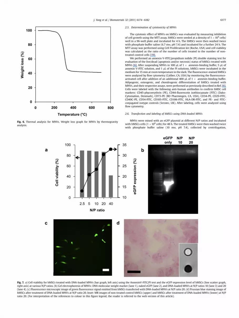
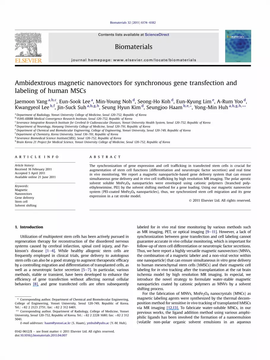
Fig. 6. Thermal analysis for MNVs. Weight loss graph for MNVs by thermogravityanalysis.
Fig. 7. a) Cell viability for hMSCs treated with DNA-loaded MNVs (bar graph, left axis) usingright axis) at various N/P ratios. (b) Gel electrophoresis of MNVs: DNA molecular weight mark(lane 4). (c) Fluorescence microscopic image of green fluorescence signal emitted from hMSChMSCs after treatment of DNA-loaded MNVs at N/P ratio 20. Inset: MR images of non-treatedratio 20. (For interpretation of the references to colour in this figure legend, the reader is r
J. Yang et al. / Biomaterials 32 (2011) 6174e6182 6177
2.5. Determination of cytotoxicity of MNVs
The cytotoxic effect of MNVs on hMSCs was evaluated by measuring inhibitionof cell growth using the MTT assay. hMSCs were seeded at a density of 1 �104 cells/well in a 96-well plate and incubated for 4 h. The hMSCs were then washed twicewith phosphate buffer saline (6.7 mM, pH 7.4) and incubated for a further 24 h. TheMTT assay was performed using Cell Proliferation kit (Roche, USA) and cell viabilitywas calculated as the ratio of the number of cells treated to the number of non-treated control cells [19].
We performed an annexin V-FITC/propidium iodide (PI) double staining test forevaluation of the live/dead (apoptosis and/or necrosis) status of hMSCs treated withMNVs [6]. After suspending MNVs in 100 mL of 1 � annexin-binding buffer, 5 mL ofannexin V-FITC solution, and 1 mL of the PI solutions, hMSCs were incubated in themedium for 15min at room temperature in the dark. The fluorescence-stained hMSCswere analyzed by flow cytometry (Caliber, CA, USA) by monitoring the fluorescence-activated cell after addition of an additional 400 mL of 1 � annexin-binding buffer.Adipogenic, osteogenic, and chondrogenic differentiation of hMSCs treated withMNVs, and their respective assays, were performed as previously described in Ref. [6].Cells were labeled with the following anti-human antibodies to confirm hMSC cellmarkers: CD45-phycoerythrin (PE), CD44-fluorescein isothiocyanate (FITC) (Dako-Cytomation, Denmark), CD73-PE (BD Pharmingen, CA, USA), CD34-PE, CD29-FITC,CD49C-PE, CD54-FITC, CD105-FITC, CD106-FITC, HLA-DR-FITC, and PE- and FITC-conjugated isotype controls (Serotec, UK). After labeling, cells were analyzed usingflow cytometry.
2.6. Transfection and labeling of hMSCs using DNA-loaded MNVs
MNVs were mixed with an eGFP plasmid at different N/P ratios and incubatedwith hMSCs cells (1�106 cells) for 48 h. The treated hMSCs were thenwashed twicewith phosphate buffer saline (10 mM, pH 7.4), collected by centrifugation,
the AnnexinV-FITC/PI test and the eGFP expression level of hMSCs (line scatter graph,er (lane 1), naked eGFP (lane 2), and DNA-loaded MNVs at N/P ratios 10 (lane 3) and 20s transfected with DNA-loaded MNVs at N/P ratio 20. (d) Prussian blue staining image ofcontrol hMSCs (upper) and hMSCs after treatment of DNA-loaded MNVs (lower) at N/Peferred to the web version of this article).
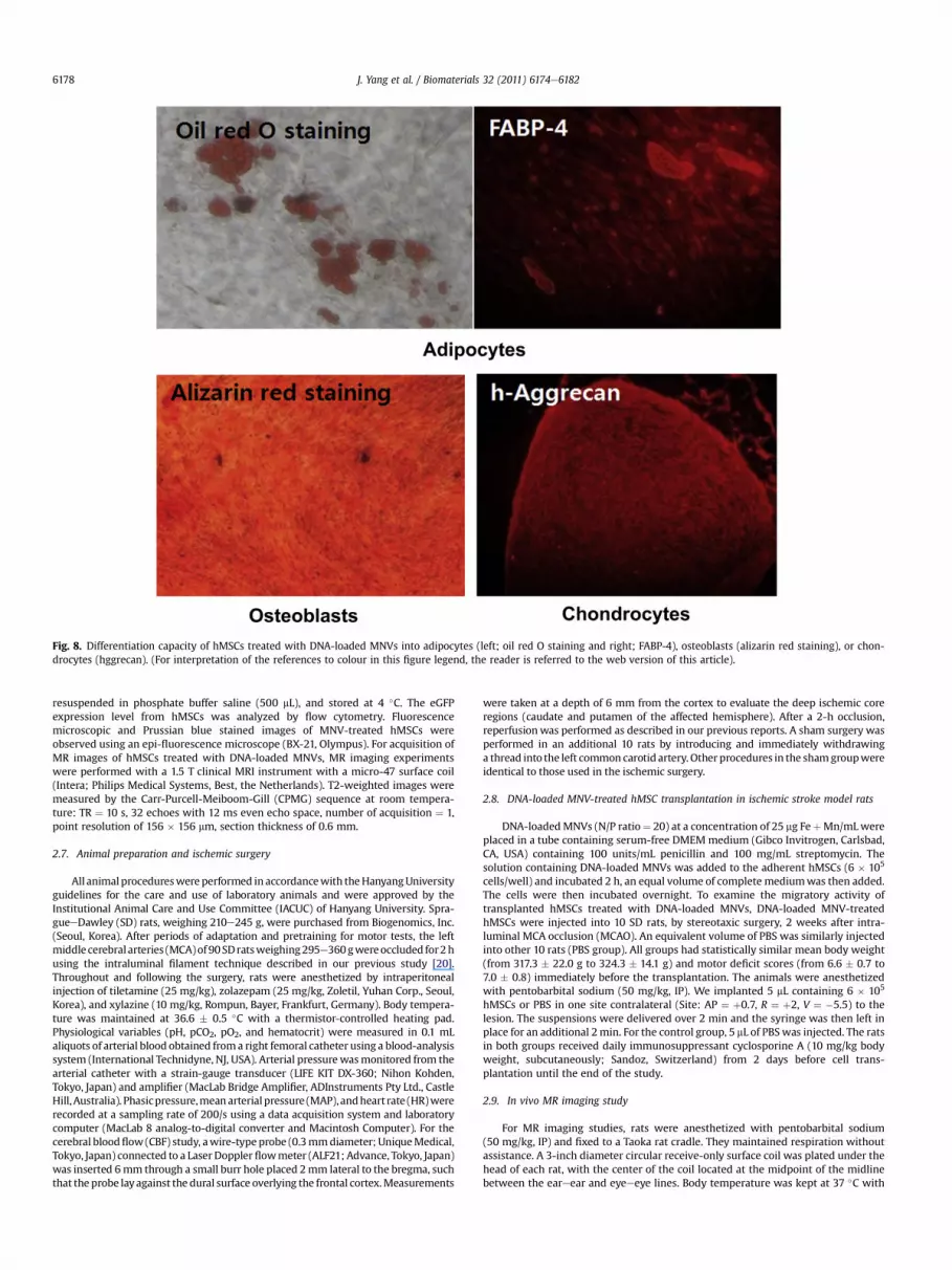
Fig. 8. Differentiation capacity of hMSCs treated with DNA-loaded MNVs into adipocytes (left; oil red O staining and right; FABP-4), osteoblasts (alizarin red staining), or chon-drocytes (hggrecan). (For interpretation of the references to colour in this figure legend, the reader is referred to the web version of this article).
J. Yang et al. / Biomaterials 32 (2011) 6174e61826178
resuspended in phosphate buffer saline (500 mL), and stored at 4 �C. The eGFPexpression level from hMSCs was analyzed by flow cytometry. Fluorescencemicroscopic and Prussian blue stained images of MNV-treated hMSCs wereobserved using an epi-fluorescence microscope (BX-21, Olympus). For acquisition ofMR images of hMSCs treated with DNA-loaded MNVs, MR imaging experimentswere performed with a 1.5 T clinical MRI instrument with a micro-47 surface coil(Intera; Philips Medical Systems, Best, the Netherlands). T2-weighted images weremeasured by the Carr-Purcell-Meiboom-Gill (CPMG) sequence at room tempera-ture: TR ¼ 10 s, 32 echoes with 12 ms even echo space, number of acquisition ¼ 1,point resolution of 156 � 156 mm, section thickness of 0.6 mm.
2.7. Animal preparation and ischemic surgery
All animal procedureswereperformed in accordancewith theHanyangUniversityguidelines for the care and use of laboratory animals and were approved by theInstitutional Animal Care and Use Committee (IACUC) of Hanyang University. Spra-gueeDawley (SD) rats, weighing 210e245 g, were purchased from Biogenomics, Inc.(Seoul, Korea). After periods of adaptation and pretraining for motor tests, the leftmiddle cerebral arteries (MCA)of90SDratsweighing295e360gwereoccluded for2husing the intraluminal filament technique described in our previous study [20].Throughout and following the surgery, rats were anesthetized by intraperitonealinjection of tiletamine (25 mg/kg), zolazepam (25 mg/kg, Zoletil, Yuhan Corp., Seoul,Korea), and xylazine (10 mg/kg, Rompun, Bayer, Frankfurt, Germany). Body tempera-ture was maintained at 36.6 � 0.5 �C with a thermistor-controlled heating pad.Physiological variables (pH, pCO2, pO2, and hematocrit) were measured in 0.1 mLaliquots of arterial blood obtained froma right femoral catheter using a blood-analysissystem (International Technidyne, NJ, USA). Arterial pressurewasmonitored from thearterial catheter with a strain-gauge transducer (LIFE KIT DX-360; Nihon Kohden,Tokyo, Japan) and amplifier (MacLab Bridge Amplifier, ADInstruments Pty Ltd., CastleHill,Australia). Phasicpressure,meanarterial pressure (MAP), andheart rate (HR)wererecorded at a sampling rate of 200/s using a data acquisition system and laboratorycomputer (MacLab 8 analog-to-digital converter and Macintosh Computer). For thecerebral bloodflow (CBF) study, awire-typeprobe (0.3mmdiameter; UniqueMedical,Tokyo, Japan) connected to a Laser Dopplerflowmeter (ALF21; Advance, Tokyo, Japan)was inserted 6mm through a small burr hole placed 2mm lateral to the bregma, suchthat the probe layagainst the dural surface overlying the frontal cortex.Measurements
were taken at a depth of 6 mm from the cortex to evaluate the deep ischemic coreregions (caudate and putamen of the affected hemisphere). After a 2-h occlusion,reperfusionwas performed as described in our previous reports. A sham surgery wasperformed in an additional 10 rats by introducing and immediately withdrawinga thread into the left common carotid artery. Other procedures in the shamgroupwereidentical to those used in the ischemic surgery.
2.8. DNA-loaded MNV-treated hMSC transplantation in ischemic stroke model rats
DNA-loadedMNVs (N/P ratio¼ 20) at a concentration of 25 mg FeþMn/mLwereplaced in a tube containing serum-free DMEM medium (Gibco Invitrogen, Carlsbad,CA, USA) containing 100 units/mL penicillin and 100 mg/mL streptomycin. Thesolution containing DNA-loaded MNVs was added to the adherent hMSCs (6 � 105
cells/well) and incubated 2 h, an equal volume of complete mediumwas then added.The cells were then incubated overnight. To examine the migratory activity oftransplanted hMSCs treated with DNA-loaded MNVs, DNA-loaded MNV-treatedhMSCs were injected into 10 SD rats, by stereotaxic surgery, 2 weeks after intra-luminal MCA occlusion (MCAO). An equivalent volume of PBS was similarly injectedinto other 10 rats (PBS group). All groups had statistically similar mean body weight(from 317.3 � 22.0 g to 324.3 � 14.1 g) and motor deficit scores (from 6.6 � 0.7 to7.0 � 0.8) immediately before the transplantation. The animals were anesthetizedwith pentobarbital sodium (50 mg/kg, IP). We implanted 5 mL containing 6 � 105
hMSCs or PBS in one site contralateral (Site: AP ¼ þ0.7, R ¼ þ2, V ¼ �5.5) to thelesion. The suspensions were delivered over 2 min and the syringe was then left inplace for an additional 2min. For the control group, 5 mL of PBSwas injected. The ratsin both groups received daily immunosuppressant cyclosporine A (10 mg/kg bodyweight, subcutaneously; Sandoz, Switzerland) from 2 days before cell trans-plantation until the end of the study.
2.9. In vivo MR imaging study
For MR imaging studies, rats were anesthetized with pentobarbital sodium(50 mg/kg, IP) and fixed to a Taoka rat cradle. They maintained respiration withoutassistance. A 3-inch diameter circular receive-only surface coil was plated under thehead of each rat, with the center of the coil located at the midpoint of the midlinebetween the eareear and eyeeeye lines. Body temperature was kept at 37 �C with

Fig. 9. Flow cytometry data of hMSCs treated with or without MNVs. Both hMSCs treated with or without MNVs demonstrated a CD34�CD45�CD54�CD29þCD44þCD49Cþ
CD73þCD105þCD106þHLA-DR� phenotype. Filled purple areas show the profile of the negative control. (For interpretation of the references to color in this figure legend, the reader isreferred to the web version of this article.)
J. Yang et al. / Biomaterials 32 (2011) 6174e6182 6179
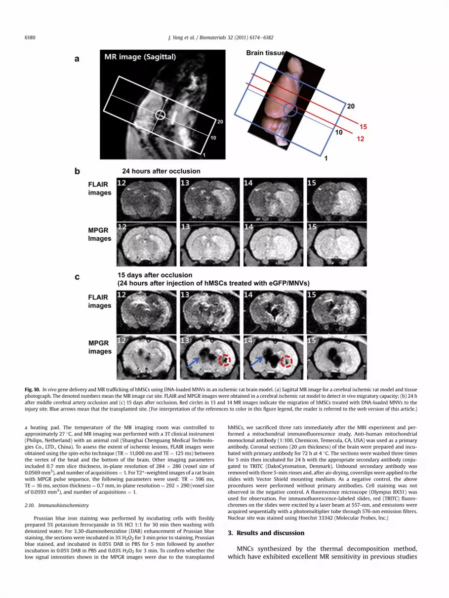
Fig. 10. In vivo gene delivery and MR trafficking of hMSCs using DNA-loaded MNVs in an ischemic rat brain model. (a) Sagittal MR image for a cerebral ischemic rat model and tissuephotograph. The denoted numbers mean the MR image cut site. FLAIR and MPGR images were obtained in a cerebral ischemic rat model to detect in vivomigratory capacity; (b) 24 hafter middle cerebral artery occlusion and (c) 15 days after occlusion. Red circles in 13 and 14 MR images indicate the migration of hMSCs treated with DNA-loaded MNVs to theinjury site. Blue arrows mean that the transplanted site. (For interpretation of the references to color in this figure legend, the reader is referred to the web version of this article.)
J. Yang et al. / Biomaterials 32 (2011) 6174e61826180
a heating pad. The temperature of the MR imaging room was controlled toapproximately 27 �C, and MR imaging was performed with a 3T clinical instrument(Philips, Netherland) with an animal coil (Shanghai Chenguang Medical Technolo-gies Co., LTD., China). To assess the extent of ischemic lesions, FLAIR images wereobtained using the spin-echo technique (TR ¼ 11,000 ms and TE ¼ 125 ms) betweenthe vertex of the head and the bottom of the brain. Other imaging parametersincluded 0.7 mm slice thickness, in-plane resolution of 284 � 286 (voxel size of0.0569mm3), and number of acquisitions¼ 1. For T2*-weighted images of a rat brainwith MPGR pulse sequence, the following parameters were used: TR ¼ 596 ms,TE¼ 16 ms, section thickness¼ 0.7 mm, in-plane resolution¼ 292 � 290 (voxel sizeof 0.0593 mm3), and number of acquisitions ¼ 1.
2.10. Immunohistochemistry
Prussian blue iron staining was performed by incubating cells with freshlyprepared 5% potassium ferrocyanide in 5% HCl 1:1 for 30 min then washing withdeionized water. For 3,30-diaminobenzidine (DAB) enhancement of Prussian bluestaining, the sections were incubated in 3% H2O2 for 3 min prior to staining, Prussianblue stained, and incubated in 0.05% DAB in PBS for 5 min followed by anotherincubation in 0.05% DAB in PBS and 0.03% H2O2 for 3 min. To confirm whether thelow signal intensities shown in the MPGR images were due to the transplanted
hMSCs, we sacrificed three rats immediately after the MRI experiment and per-formed a mitochondrial immunofluorescence study. Anti-human mitochondrialmonoclonal antibody (1:100, Chemicon, Temecula, CA, USA) was used as a primaryantibody. Coronal sections (20 mm thickness) of the brain were prepared and incu-bated with primary antibody for 72 h at 4 �C. The sections were washed three timesfor 5 min then incubated for 24 h with the appropriate secondary antibody conju-gated to TRITC (DakoCytomation, Denmark). Unbound secondary antibody wasremovedwith three 5-min rinses and, after air-drying, coverslips were applied to theslides with Vector Shield mounting medium. As a negative control, the aboveprocedures were performed without primary antibodies. Cell staining was notobserved in the negative control. A fluorescence microscope (Olympus BX51) wasused for observation. For immunofluorescence-labeled slides, red (TRITC) fluoro-chromes on the slides were excited by a laser beam at 557-nm, and emissions wereacquired sequentially with a photomultiplier tube through 576-nm emission filters.Nuclear site was stained using Hoechst 33342 (Molecular Probes, Inc.)
3. Results and discussion
MNCs synthesized by the thermal decomposition method,which have exhibited excellent MR sensitivity in previous studies

Fig. 11. a) Prussian blue staining of brain tissue reveals the presence of blue spots, indicative of migratory behavior of transplanted DNA-loaded MNV-treated hMSCs (blue arrow).The region confined by a circle (blue dotted) indicates ischemic parts of brain. (b) Fluorescence microscopic images of hMSCs treated with DNA-loaded MNVs in the migration site(blue arrow region in Fig. 3c): eGFP gene expression (green), mitochondria of hMSCs (red, TRITC), nuclear site of hMSCs (blue, Hoechst 33342), and their merged image. (Forinterpretation of the references to color in this figure legend, the reader is referred to the web version of this article.)
J. Yang et al. / Biomaterials 32 (2011) 6174e6182 6181
[12,13], were coated with PLA molecules by polymerization of lac-tide dimers in the presence of Sn(Oct)2 in toluene. The precipitatescollected by a centrifugation were then transferred to various non-polar, polar aprotic and polar protic solvents to assess the solubilityof PASSMNCs. The solubility of PASSMNCs was analogous with PLAdue to the presence of PLA molecules on the surface of MNCs afterthe polymerization process. Moreover, the polar eCOOe functionalgroup containing the PLA-coating makes the MNCs readily solublein all polar aprotic solvents. The FT-IR spectrum of PASSMNCsshowed characteristic bands at 590 and 1748 cm�1 indicating thepresence of FeeO in MNCs and C]O of ester bonds from PLA,respectively (Fig. 2). Furthermore, the molecular weight of PLAfrom PASSMNCs was determined to be 2.4 kDa (polydispersityindex: 1.42) by using gel permeation chromatography and thepresence of PLA was corroborated by 1H NMR: d 3.62 (hydrogen ofPEG back bone), 1.54 (methyl group of lactide chain), 5.21(hydrogen of lactide back bone), 4.83 (hydrogen of glycolide chain)(Fig. 3). In particular, the acetone-soluble MnFe2O4 nanoparticles asPASSMNCs were stably and reversibly reacted to an externalmagnetic field (Fig. 4a). For the preparation this solution was thenadded drop-wise to PEI-containing phosphate buffer saline (pH 7.4,10 mM). The PLA and PEI could interact through a strong hydro-phobic and electrostatic interaction during the solvent displace-ment [19]. The nanoparticle, sequentially coated by PLA then PEI,possessed the desired water solubility (Fig. 4b). The morphology ofMNVs was examined by transmission electron microscopy (TEM)(Fig. 4c). The colloidal size of MNVs was 73.7 � 8.7 nm by laserscattering that was larger than baer MnFe2O4 nanoparticle due toswelled PEI molecules on the surface of MNVs (Fig. 4d). Zeta-potential of MNVs was 23.1 � 3.2 mV, consistent with the pres-ence of positively charged PEI on the outermost surface and anionicDNA could then be loaded onto the positively charged MNVs.Furthermore, the colloidal stability of MNV nanoparticles preparedby the solvent shifting method in the aqueous medium, could bemaintained for over 3 months without aggregations. The magnetichysteresis loop, using a vibration sample magnetometer, showedthat MNVs exhibit superparamagnetic behavior without magnetichysteresis, with a magnetization saturation of approximately1 emu g�1 at 1.5 T and 300 K (Fig. 5a) and a linear relaxivity coef-ficient increase with concentration of MNVs and the relaxivitycoefficient was 350.4 mM
�1s�1 (Fig. 5b). The amount of MNCs inMNVs was determined to be 41.7 wt.% with a thermo-gravimetricanalyzer (Fig. 6). Thus, MNVs demonstrated the excellentcolloidal stability in the biological media and gene-loadable
potentials for synchronous gene transfection and magneticlabeling of hMSCs for in vivo MR imaging tracking.
We investigated the cytotoxicity and transfection efficiency ofMNVs against hMSCs. The transfection efficiency and viability wereoptimized by controlling N/P ratio of DNA-loaded MNVs. We foundthat a gene delivery vehicle with an N/P ratio >10, providing anincreased positive charge to enhance transfection, showed genetransfection efficiency of>40% (Fig. 7a, line scatter graph). However,an N/P ratio over 40 showed severe cytotoxicity (Fig. 7a, bar graph)due to too strong positive charge of PEI well documented [17]. At N/Pratios 10 and 20, very strong DNA-MNV complexes were shown bythe absence of free DNA (Fig. 7b), and successful gene delivery andexpressionwas demonstrated by the fluorescence of the transfectedeGFP cells (Fig. 7c). Following these observations, we determinedthat an N/P ratio of 20 provided optimal conditions for the genetransfection to hMSCs. Prussian blue staining demonstrated thepresence of iron species (Fig. 7d). Furthermore, the enhanced signalintensity in the T2-weighted MR image of hMSCs treated DNA-loaded MNVs clearly indicated the presence of magnetic compo-nents (Fig. 7d, inset). For the investigation of the cellular behaviorinhibition effects from MNVs, differentiation of hMSCs transfectedwith DNA-loaded MNVs has been studied following establishedprotocols [6]. These cells successfully differentiated into adipocytes,osteoblasts, and chondrocytes, consistent with the normal behav-iors of non-treated control cells (Fig. 8). Furthermore, the identitiesof hMSCs examined by flow cytometry with 11 hMSC markers, areunaffected by DNA-loaded MNVs-treatment (Fig. 9). These resultsdemonstrated that the prepared MNVs exhibited the excellent geneloading and magnetic labeling efficiencies without the cellularbehavior inhibition of hMSCs.
To demonstrate the potential applicability of our gene deliveryto hMSCs and their in vivo monitoring system based on MNVs, wechose an ischemic cerebral infarction rat model. 24 h after themiddle cerebral artery occlusion of rat, the cerebral infarction areawas observed by both Fluid Attenuated Inversion Recovery (FLAIR)and Multiplanar Gradient-Recalled (MPGR) images (Fig. 10a and b).Direct gene transfer to the interface between the infarction site andthe undamaged brain is considered to be very important in treatingstroke with regenerative stem cell-based gene therapy. For theproof of our concept, we delivered the reporter gene, eGFP, tohMSCs using MNVs. These cells were injected into the contralateralside of the cerebral hemisphere with ischemic cerebral infarction 2weeks aftermiddle cerebral artery occlusion in the rat.We followedhMSCs migrationwith T2*-weighted MR imaging, and showed that

J. Yang et al. / Biomaterials 32 (2011) 6174e61826182
these hMSCs migrated to the ischemic cerebral infarction sitewithin 2 days, as evidenced by the low signal intensity spots in T2*-weighted MR image (Fig. 10c). Furthermore, the presence of ironspecies from MNVs in both the injection and migration sites wasconfirmed by the Prussian blue staining of the extracted braintissue and both sites exhibited green fluorescence due to eGFP geneexpressions (Fig. 11a and b). Human mitochondria-specific immu-nofluorescent staining was conducted to show red fluorescence.Considerable overlap of green and red fluorescencewas observed inthe merged images, confirming that hMSCs were the source of thered fluorescence (Fig. 11b).
4. Conclusions
We have prepared a simple MR-responsive magneticnanoparticle-based gene delivery system (MNVs) for synchroniza-tion of gene delivery and in vivo hMSCs trafficking events. EachMNV with positive surface charge can serve as an individual genecarrier, enabling easy cell trafficking and high gene loadingcapacity. The nanoparticle system, highly stable and soluble inphysiological condition, demonstrated high gene transfection effi-ciency in hMSCs. In addition, transfected and labeled hMSCsexhibited normal differentiation behaviors, which provides a newopportunity for stem cell-based gene therapy and systemic genedelivery.
Acknowledgments
This work was supported by the National Research Foundationof Korea Grant funded by the Korean Government (MEST) (2010-0020587) and a grantof the Korea Healthcare technology R&DProject, Ministry for Health & Welfare Affairs, Republic of Korea(A085136).
References
[1] Kameda M, Shingo T, Takahashi K, Muraoka K, Kurozumi K, Yasuhara T, et al.Adult neural stem and progenitor cells modified to secrete GDNF can protect,migrate and integrate after intracerebral transplantation in rats with transientforebrain ischemia. Eur J Neurosci 2007;26(6):1462e78.
[2] Shi Y, Hu G, Su J, Li W, Chen Q, Shou P, et al. Mesenchymal stem cells: a newstrategy for immunosuppression and tissue repair. Cell Res 2010;20(5):510e8.
[3] Stroh A, Boltze J, Sieland K, Hild K, Gutzeit C, Jung T, et al. Impact of magneticlabeling on human and mouse stem cells and their long-term magneticresonance tracking in a rat model of Parkinson disease. Mol Imaging 2009;8(3):166e78.
[4] Sykova E, Jendelova P, John TW, Andrew IRM. In vivo tracking of stem cells inbrain and spinal cord injury. Prog Brain Res 2007;161:367e83.
[5] Ke Y-Q, Hu C-C, Jiang X-D, Yang Z-J, Zhang H-W, Ji H-M, et al. In vivo magneticresonance tracking of Feridex-labeled bone marrow-derived neural stem cellsafter autologous transplantation in rhesus monkey. J Neurosci Meth 2009;179(1):45e50.
[6] Koh S-H, Noh MY, Cho GW, Kim KS, Kim SH. Erythropoietin increases themotility of human bone marrow-multipotent stromal cells (hBM-MSCs) andenhances the production of neurotrophic factors from hBM-MSCs. Stem CellsDev 2009;18(3):441e521.
[7] Sykova E, Jendelova P. Migration, fate and in vivo imaging of adult stem cells inthe CNS. Cell Death Differ 2007;14(7):1336e42.
[8] Tinsley RB, Faijerson J, Eriksson PS. Efficient non-viral transfection of adultneural stem/progenitor cells, without affecting viability, proliferation ordifferentiation. J Gene Med 2006;8(1):72e81.
[9] Daadi MM, Li Z, Arac A, Grueter BA, Sofilos M, Malenka RC, et al. Molecular andmagnetic resonance imaging of human embryonic stem cell-derived neuralstem cell grafts in ischemic rat brain. Mol Ther 2009;17(7):1282e91.
[10] Higuchi T, Anton M, Dumler K, Seidl S, Pelisek J, Saraste A, et al. Combinedreporter gene PET and iron oxide MRI for monitoring survival and localizationof transplanted cells in the rat heart. J Nucl Med 2009;50(7):1088e94.
[11] Wu JC, Chen IY, Sundaresan G, Min J-J, De A, Qiao J-H, et al. Molecular imagingof cardiac cell transplantation in living animals using optical bioluminescenceand positron emission tomography. Circulation 2003;108(11):1302e5.
[12] Yang J, Lee C-H, Ko H-J, Suh J-S, Yoon H-G, Lee K, et al. Multifunctionalmagneto-polymeric nanohybrids for targeted detection and synergistic ther-apeutic effects on breast cancer. Angew Chem-Int Edit 2007;46(46):8836e9.
[13] Lee J-H, Huh Y-M, Jun Y-W, Seo J-W, Jang J-T, Song H-T, et al. Artificiallyengineered magnetic nanoparticles for ultra-sensitive molecular imaging. NatMed 2007;13(1):95e9.
[14] Yang J, Lim E-K, Lee HJ, Park J, Lee SC, Lee K, et al. Fluorescent magneticnanohybrids as multimodal imaging agents for human epithelial cancerdetection. Biomaterials 2008;29(16):2548e55.
[15] Bilati U, Allémann E, Doelker E. Development of a nanoprecipitation methodintended for the entrapment of hydrophilic drugs into nanoparticles. Eur JPharm Sci 2005;24(1):67e75.
[16] Aubry J, Ganachaud F, Cohen Addad J-P, Cabane B. Nanoprecipitation of pol-ymethylmethacrylate by solvent shifting:1. boundaries. Langmuir 2009;25(4):1970e9.
[17] Zhang C, Yadava P, Hughes J. Polyethylenimine strategies for plasmid deliveryto brain-derived cells. Methods 2004;33(2):144e50.
[18] Legrand P, Lesieur S, Ai Bochot, Gref R, Raatjes W, Barratt G, et al. Influence ofpolymer behaviour in organic solution on the production of polylactidenanoparticles by nanoprecipitation. Int J Pharm 2007;344(1e2):33e43.
[19] Seo S-B, Yang J, Lee E-S, Jung Y, Kim K, Lee S-Y, et al. Nanohybrids via a pol-ycation-based nanoemulsion method for dual-mode detection of humanmesenchymal stem cells. J Mater Chem 2008;18(37):4402e7.
[20] Koh S-H, Kim KS, Choi MR, Jung KH, Park KS, Chai YG, et al. Implantation ofhuman umbilical cord-derived mesenchymal stem cells as a neuroprotectivetherapy for ischemic stroke in rats. Brain Res 2008;1229:233e48.