mscs: delivery routes and engraftment, cell-targeting strategies, and immune modulation
TRANSCRIPT
Hindawi Publishing CorporationStem Cells InternationalVolume 2013, Article ID 732742, 13 pageshttp://dx.doi.org/10.1155/2013/732742
Review ArticleMSCs: Delivery Routes and Engraftment, Cell-TargetingStrategies, and Immune Modulation
Thomas J. Kean,1 Paul Lin,2 Arnold I. Caplan,2 and James E. Dennis1
1 Benaroya Research Institute, Seattle, WA 98101, USA2 Skeletal Research Center, Department of Biology, Case Western Reserve University, Cleveland, OH 44106, USA
Correspondence should be addressed toThomas J. Kean; [email protected]
Received 30 April 2013; Revised 27 June 2013; Accepted 1 July 2013
Academic Editor: Donald G. Phinney
Copyright © 2013 Thomas J. Kean et al.This is an open access article distributed under the Creative Commons Attribution License,which permits unrestricted use, distribution, and reproduction in any medium, provided the original work is properly cited.
Mesenchymal stem cells (MSCs) are currently being widely investigated both in the lab and in clinical trials for multiple diseasestates.The differentiation, trophic, and immunomodulatory characteristics ofMSCs contribute to their therapeutic effects. Anotheroften overlooked factor related to efficacy is the degree of engraftment. When reported, engraftment is generally low and transientin nature. MSC delivery methods should be tailored to the lesion being treated, which may be local or systemic, and customized tothemechanism of action of theMSCs, which can also be local or systemic. Engraftment efficiency is enhanced by using intra-arterialdelivery instead of intravenous delivery, thus avoiding the “first-pass” accumulation of MSCs in the lung. Several methodologiesto target MSCs to specific organs are being developed. These cell targeting methodologies focus on the modification of cell surfacemolecules through chemical, genetic, and coating techniques to promote selective adherence to particular organs or tissues. Futureimprovements in targeting and delivery methodologies to improve engraftment are expected to improve therapeutic results, extendthe duration of efficacy, and reduce the effective (MSC) therapeutic dose.
1. Introduction
Mesenchymal stem cells (MSCs) are multipotential adultprogenitor cells that have the capacity to differentiate alongseveral mesenchymal lineages, including cartilage, adipose,marrow stroma, and bone tissue [1–3]. Studies have beenconducted on the use of MSCs as a therapeutic basedon this capacity to differentiate directly into these end-stage phenotypes, including the use of MSCs to promote oraugment bone repair [4] and for the repair of cartilage defects[4, 5]. In addition to direct differentiation into end-stagephenotypes, MSCs have also been shown to have a positivetherapeutic effect in many repair situations because of theircapacity to secrete trophic factors (reviewed in [6]) thatcontribute to repair via the promotion of vascularization andthe inhibition of cell death as well as through the modulationof the immune response. Currently, there are over 160 openstudies and 116 closed clinical trials (results retrieved (3rdJune 2013) in a search of www.clinicaltrials.gov on the searchterm “mesenchymal stem cells” and excluding trials with
an unknown status and those that were conducted in vitro)that use MSCs to treat a variety of conditions that range fromdirect formation of bone tissue to treatments for graft versushost disease (GvHD) [7–9], myocardial infarction, braintrauma, and multiple sclerosis [10, 11] (reviewed by Millardand Fisk [12]). Indeed, MSCs have been well characterizedwith respect to their ability to produce a range of growthfactors and cytokines, which inspired the designation ofthese cells as a kind of “injury drugstore” [13]. An interestingsubset of this factory of cytokines is the factors that havebeen shown to have a profound effect on modulating theimmune system.These immunemodulatory factors are beingtested for their effect on immune disorders such as GvHD,rheumatoid arthritis [14, 15], multiple sclerosis [16, 17], type Idiabetes [18, 19], inflammatory bowel disease (IBD) [20–23],and transplant tolerance [24].
Of particular relevance to the therapeutic applicationof MSCs is their fate post-implantation. Ambiguity seen inthe efficacy of MSCs, in both animal studies and clinicaltrials, with therapies being ineffective or only temporarily
2 Stem Cells International
effective could be due to suboptimal application of MSCs.Whether systemically injected or injected directly into atissue or organ, there is the issue of where the cells go andwhether the cells can bind, engraft, and, in many instances,survive. Very few studies have quantified the efficiency ofMSC transplantation, and those that have quantified MSCengraftment have shown poor engraftment efficiency. Com-plicating this determination, as noted in Karp and LengTeo [25], are the details of the quantification methodology.The techniques for assessing biodistribution of MSCs canbe categorized into in vivo and ex vivo methods. Examplesof in vivo methods include bioluminescence, whereby cellsare transduced to express luciferase and can then be imagedthrough their metabolism of luciferin resulting in lightemission [26]; fluorescence, whereby cells are either loadedwith a fluorescent dye or transduced to express a fluorescentreporter which can then be imaged; radionuclide labeling,where cells are loaded with radionuclides and localized withscintigraphy [27], positron emission tomography (PET) orsingle photon emission computed tomography (SPECT); andmagnetic resonance imaging (MRI), wherein cells loadedwith paramagnetic compounds (e.g., iron oxide nanoparti-cles) are traced with an MRI scanner. For further reviewof these imaging modalities and their clinical applicationsee Srinivas et al. [28] and Reagan and Kaplan [29]. Exvivo methods to assess biodistribution include quantitativePCR, flow cytometry, and histological methods. Histologicalmethods include tracking fluorescently labeled cells; in situhybridization, for example, for Y-chromosomes and forhuman-specific ALU sequences; and histochemical stainingfor species-specific or genetically introduced proteins suchas bacterial 𝛽-galactosidase. These immunohistochemicalmethods are useful for discerning engraftment location butnecessitate the excision of tissue. They are, however, proneto errors for quantification due to sampling, the possibilityof false positives, and the loss of signal in studies whereMSCs are tagged with fluorescent probes that lose signal witheach cell division. With the use of genetic markers, suchas luciferase or green fluorescent protein (GFP), the fate ofMSCs in whole animals can be tracked without losing signalafter cell division. However, luciferase and GFP-like opticalprobes suffer from limited penetration and 3D localizationissues due to tissue shielding; thus, they are most applica-ble in small animal models, while combinational trackingapproaches make it feasible to more accurately track cell fatesystemically and over extended periods of time. Additionally,care must be taken in how the cells are labeled, as certainmethods of labelingMSCswith reporter genes can affect theirproliferation [26] or their differentiation potential [30]. Withthe use of these newer tracking methods, researchers nowhave the ability to address the long-term fate of transplantedMSCs, although there is still a paucity of data related to thisissue. This review will analyze factors that may influence thetherapeutic efficacy of MSCs, including an overview of theimmune status of MSCs, “intrinsic” MSC activity, optimalMSC delivery methods, and targeting methods to improvecell engraftment and survival.
2. MSC Immune Modulation
Several characteristics of MSCs are purported to impartimmune privilege, thereby allowing MSCs to avoid immunerejection in certain situations, which may facilitate the clin-ical use of allogenic MSCs. MSCs do not express class IIMajor Histocompatibility Complex (MHC) or costimulatormolecules and express low levels of class I MHC [31]. One ofthe first indicators of the role ofMSCs inmodulating immunereactions was in studies showing that activated MSCs inhibitT-cell expansion in mixed lymphocyte reactions [32–34].MSCs have been shown to influence the immune systemthrough the secretion of a variety of soluble factors includ-ing indoleamine 2,3-dioxygenase [35], nitric oxide [9],transforming growth factor beta (TGF𝛽), prostaglandin E2(PGE2) [36, 37], and tumor necrosis factor stimulated gene-6 protein (TSG-6) [38]. Di Nicola et al. [39] showed thatMSCs could impede T-cell expansion in transwell culturesand that this inhibitory effect was abolished by the additionof antibodies to TGF𝛽 and hepatocyte growth factor (HGF),while Tse et al. [33] showed a similar inhibitory effect that wasa soluble factor but not due to TGF𝛽, PGE2, or interleukin-10. In still another study, this time using adipose-derivedMSCs, it was shown that the inhibition of T-cell expansion inmixed lymphocyte reactions was abrogated by the addition ofindomethacin, a PGE2 inhibitor, but was not affected by anti-bodies to TGF𝛽 or HGF [40]. It has also been demonstratedthat proinflammatory factors, such as interferon-𝛾 or tumornecrosis factor alpha (TNF𝛼), upregulate the expression ofthese important regulatory factors [9, 35, 41]. This MSC anti-inflammatory response to proinflammatory stimulation isreferred to as “licensing” [35]. For a detailed review on themechanism of action of each of these factors, see English(2013) [42].
While many studies have shown that soluble factors playa significant role in the immunomodulatory characteristics ofMSCs, several other studies indicate that cell-to-cell contactmay also be important. In one study, MSCs were shownto inhibit proliferation of memory T-cells but, in this case,direct cell contact was required as the MSCs were ineffectivein transwell experiments [35]. Another study showed thatMSCs interact with T-cells via Notch signaling and thatinactivation of Toll-3 and Toll-4 receptors downregulatedJagged-1 expression inMSCs, thus impeding interaction withNotch on T-cells and resulting in the loss of MSC inhibitionof T-cell proliferation [43]. Indeed, several more recentstudies indicate that cell-to-cell contact between MSCs andimmune cells may be very important in specific situations.For example, it was shown that tissue-resident MSCs areable to promote epithelial cell repair through the secretionof PGE2 after the MSCs had been exposed to microbesof the GI-tract, while mice deficient in Toll-like receptorsignaling (myeloid differentiation primary response gene 88knockouts; Myd88−/−) were ineffective at epithelial repair[44]. Additional studies have shown that MSCs may alsoregulate dendritic cells via Notch signaling [45, 46].
The mechanism of the immunomodulation character-istics of MSCs is critical when considering the potentialtherapeutic effects of MSC transplantation. If MSCs need
Stem Cells International 3
cell-to-cell contact or need to secrete factors at a high localconcentration to impart their regulatory role, thenMSCs willneed to be delivered in close proximity to the target tissueor organ. Alternatively, MSCs may act distally through thesecretion of sufficient amounts of soluble factors with anadequate half-life to reach the target lesion. For example,TSG-6 was shown to play a key role in MSC-mediatedinflammatory regulation in postmyocardial infarction. Inthis study, MSCs or TSG-6 alone, but not MSCs treatedwith TSG-6 siRNA, were able to decrease postmyocardialinfarction inflammation and improve cardiac function [38].A similar anti-inflammatory effect of MSC-produced TSG-6was shown in a cornea transplant model [47], and anotherstudy in a peritonitis model showed that MSCs stimulated bymacrophage-produced TNF𝛼 produced TSG-6 which actedas a negative feedback loop on macrophage inflammatorysignaling [48].
These immunomodulatory characteristics are likely theunderlying mechanism(s) of the anti-inflammatory roleMSCs play in many of the aforementioned clinical trials.However, this purported ability to avoid rejection remainscontroversial, with some studies showing rejection in allo-genic settings and others showing tolerance, and a selectionshowing rejection when MSCs begin to differentiate (seereview by Griffin et al. [49]). In one study, it was shownthat when allogeneic rat MSCs were combined with ceramicsand implanted subcutaneously to promote osteoblast differ-entiation, the MSCs elicited an immune response and wererejected [50], but the MSCs were able to form bone in theceramic if the host rats were administered an immunosup-pressive drug (FK-506). In a rat cardiac study, it was alsoshown that transplanted MSCs expressing a cardiac pheno-type were responsible for the induction of a host immuneresponse and subsequent rejection [51]. In another study, itwas also shown that MSCs are unable to avoid detection bythe innate immune system and that cell culture conditionsalone may impart a change in MSCs that allows innaterecognition of autotransplanted MSCs [52]. This rejectionissue is an important aspect of developing effective therapiesbecause if most or all of the MSCs are rejected, then thetreatment may not be as effective or long lasting. However,if allogeneic MSCs are well tolerated, it would be easier andless expensive to use banked allogeneic MSCs than it wouldbe to use autologous MSCs. If MSCs are rejected after theyexpress class II MHC markers, it does not preclude the useof allogeneic MSCs in cases where MSCs are not expected todifferentiate. In fact, many studies have shown that allogeneicMSCs are well tolerated in people, and many of the positiveresults observed in various clinical trials have come frompatients transplanted with allogeneic MSCs (reviewed byMillard and Fisk [12]). However, it may be that, in manystudies, the observed therapeutic effects do not requirelong-term engraftment. In a study examining the long-termengraftment of allotransplanted MSCs in deceased patientshaving received MSC infusions, it was shown that only smallnumbers, if any, of the infused MSCs were detectable [53].This observation has led to the proposal that some MSCtherapeutic effects are essentially “hit and run” phenomena.The fact that MSCs often do not engraft seems a likely
explanation for why many MSC therapies are marginally oronly transiently effective. Indeed, it could be speculated thatthe initial improvement in cardiac function seen at a 6-monthtime point in the BOOST clinical trial, that had disappearedby the 18-month time point, could have been extended withimproved engraftment [54]. Clinically, if MSCs were to beused in conditions where they might need to differentiate inorder to achieve a therapeutic effect or need to be engrafted,the use of autologous MSCs is a preferred option. WhetherMSCs are immunomodulatory or used as an autograft, thereis the critical issue of effectively delivering them to the site ofaction and, ideally, enabling engraftment.
3. MSCs, Pericytes, and Tissue Repair
At sites of injury, it has been established that most, if notall, MSCs are derived from perivascular mesenchymal cells,pericytes, that are on the tissue side of blood vessels andsinusoids [55, 56]. This location has been identified in MSCsderived from bone marrow and dental pulp [57], brain[58], umbilical cord [59], adipose tissue [60, 61], and liver[62]. Whether all pericytes are MSCs or whether MSCs arethe bone marrow-derived subset of pericytes is debatablesince “pericytes” or “MSCs” isolated from different organsof the body can display markedly different differentiationpotentials, such as pulp-derived pericytes/MSCs displayingodontoblastic potential [63], while marrow-derived peri-cyte/MSCs have not shown an odontoblastic phenotype.The argument is that if all pericytes were MSCs, then allpericytes should have equivalent differentiation potentials.Alternatively, it has been proposed that pericytes constitutea reservoir of tissue-specific progenitor cells [64], of whichclassically defined MSCs may only be a subset.
Regardless of their designation as either pericytes orMSCs, it has been proposed that, during injury, blood vesselsbreak or become inflamed and the pericytes are liberatedfrom their perivascular tethers [65]. These released pericytesbecome activated and function tomodulate the local immuneenvironment and establish a zone of tissue regeneration. Itis apparent, in some cases, that this release is a local effectand there is little cell recruitment from uninjured tissue [66].There are, however, a number of publications that provideevidence that MSCs home to sites of inflammation or tissueinjury when infused into living organisms. Thus, it can beinferred that in some injured tissues, MSCs may have anintrinsic affinity for the sites of tissue injury. Several studieshave shown thatMSCs respond to chemokines, such as SDF-1[67], MCP-3 [68], CXCL9, CXCL16, CCL20, CCL25 [69], andHGF [70], which can either act locally or recruit MSCs fromthe bloodstream (the issue of systemicMSCmobilization willnot be discussed further here).
The injected or naturally released MSCs are activated bythe local microenvironment and respond to these cues bysecreting a site-specific array of bioactive molecules. Thesemolecules act to immunomodulate the MSC microenvi-ronment, reduce inflammation, and establish a regenerativemilieu.Wewould further assert that theseMSCs are geared tofunction in the repair of these compromised vessels. Indeed,MSCs are capable not only of stimulating angiogenesis
4 Stem Cells International
in injured muscle by secreting large amounts of vascularendothelial growth factor that attracts vascular progenitorsand is cytoprotective [71], but also of stabilizing newly formedcapillaries [72] by assuming perivascular positions embeddedin the basement membranes of newly forming blood vessels.
4. MSC Delivery and Biodistribution
There are two principal methods to introduce cells into thebody: local delivery into the tissue and systemic delivery.Local delivery can be further defined by the specific type ofdelivery, either cells embedded in a scaffold (for review seeArthur et al. [73]) or local injection, for example intraperi-toneal (IP), intramuscular, or intracardiac injection. Systemicdelivery can be further defined by the vascular route, venous(IV) or arterial (IA). The optimal method of delivery willdepend on which mechanism of action of the MSC is beingutilized. This review will concentrate on the distribution ofinjected cells.
Having defined some of the characteristics and functionsof MSCs, the question then becomes whether or not MSCsperform best when present at the site of injury/inflammation.If MSCs can exert their effect distally, for example, by secret-ing cytokines into the circulation [6, 13, 38], then itmay not benecessary for the cells to be located at the specific injury siteand systemic effects could be achieved using a cell reservoir.For example, when MSCs were injected IP, they were ableto prevent the damage caused by collagen-induced arthritis,despite the lack of a detectable presence in the arthritic joints[74]. If, however,MSCs need to be present at the site of injury,for example, by differentiating into replacement cells, as inthe treatment of osteogenesis imperfecta (OI) [75] or by thelocal production of antiapoptotic or angiogenic factors [6],then the delivery systemmust place cells at, or allowMSCs tomigrate to, the site of injury.
Arguments for local injection include not only deliveringMSCs to the site of the lesion, but also the possibility that theMSCs have the capacity to migrate toward injured tissue. Forexample, when MSCs were injected IP, they were found tomigrate toward an inflamed colon [76]. While IP injectionis a common systemic delivery route for small molecules,IP injected MSCs [76] and neuroprogenitor cells [77] werenot found to migrate to other tissues. However, it is possiblethat lymphoid access could be achieved through this route,leading to widespread distribution of a subset of the injectedcells detected by endpoint PCR [78]. In another example,MSCs were found to migrate toward an ischemic lesion ofthe brain when injected near the location of the ischemiaand appeared,morphologically, to differentiate intomicroglia[79]. MSCs have also been found to migrate toward tumorsin vivo; it is hypothesized that tumors mimic a chronicwound [80]. When MSCs were directly injected into thebrain, they were found to migrate to glioma cells located onthe contralateral hemisphere [81]. The disadvantage of localinjection is that it can lead to further tissue damage from theinjection bolus [69]. It has also been shownwith bonemarrowmononuclear cells that, although direct injection increasedlocalization, it did not increase engraftment or survival [82].Furthermore, there has been little evidence that MSCs can
migrate out of local tissues and into the circulatory system,which is problematic if MSCs need to be present in multiplebody compartments, or if the injury is systemic.
The alternative method is to deliver the cells intravascu-larly.With this systemic injectionmethodology, there remainseveral hurdles to overcome in order to deliver cells to thetarget tissue and have them engraft. Intravascular injectionhas the advantage of being minimally invasive, and it allowsfor wide distribution of cells throughout the body. The mostcommon method for accessing the circulatory system is IVand is historically the most common method for deliveringMSCs. However, cells delivered IV have to first pass throughthe lungs before they can distribute throughout the body.This presents a major problem with what has been termedthe pulmonary “first-pass” effect, which results in significantentrapment of cells [83]. This problem arises because MSCshave an estimated diameter of 20–30𝜇m [27, 83, 84] andexperiments withmicrospheres have demonstrated that mostparticles of this size are filtered out by the lungs [83, 85].While this may be an overestimation of the amount ofentrapment, as microspheres are rigid andMSCs can deform,experimental data supports that a large proportion of MSCsare entrapped in the lung following IV administration [27](Figure 1). Furthermore, it was observed that the numberof trapped cells decreased with the administration of avasodilator [27], lending support to the hypothesis that MSCsize is a major contributor to the first-pass effect. In additionto size, it is possible that endothelial cell adhesion moleculescontribute to lung entrapment. This hypothesis is supportedby evidence showing that when the CD49d receptor wasblocked, there was a small, but significant, decrease in thenumber of cells trapped in the lungs [83]. In a comparisonbetween umbilical cord blood-derived MSCs (UCB-MSCs)and bone marrow-derived MSCs (BM-MSCs), a significantdifference was found in the cell surface expression of adhe-sion molecules (significantly higher CD49f, CD49d, andcMET in UCB-MSCs) and glycolipid carbohydrate epitopes(significantly lower GD2 and SSEA4 inUCB-MSCs), and thiscell surface profile correlated with lung clearance rates, withUCB-MSCs exiting the lungs faster than BM-MSCs [86].
While the previous examples were from animal models,it is likely that lung entrapment is also a hindrance toMSCs administered to humans. For example, when MSCswere administered IV into OI patients, less than 1% of thecells were detected in the target organ by PCR [87, 88].Similarly low MSC engraftment levels were found with IVadministration of MSCs to treat GvHD [53], or when co-infused with hematopoietic stem cells (HSCs) to promoteHSC engraftment [89]. However, it is difficult to ascertainfrom these two studies whether lung entrapment, or someother mechanism such as cell death, was the cause of thelow engraftment efficiency since the injected cells were nottracked in real time.
There has been some suggestion that MSC entrapment inthe lungs itself can potentially be therapeutic. In experimentsby Lee et al. [38], MSCs trapped in the lungs were still ableto improve cardiac function after a myocardial infarctionthrough the release of the anti-inflammatory protein TSG-6. However, experiments with OI patients were not 100%
Stem Cells International 5
Tail
vein
250
200
100
150
50
Color barMin = 2000
Max = 2.5e+05
×10
3
p/s
/cm
2
/sr
(a)
Aort
ic ar
ch
250
200
100
150
50
Color barMin = 2000
Max = 2.5e+05
×10
3
p/s
/cm
2
/sr
(b)
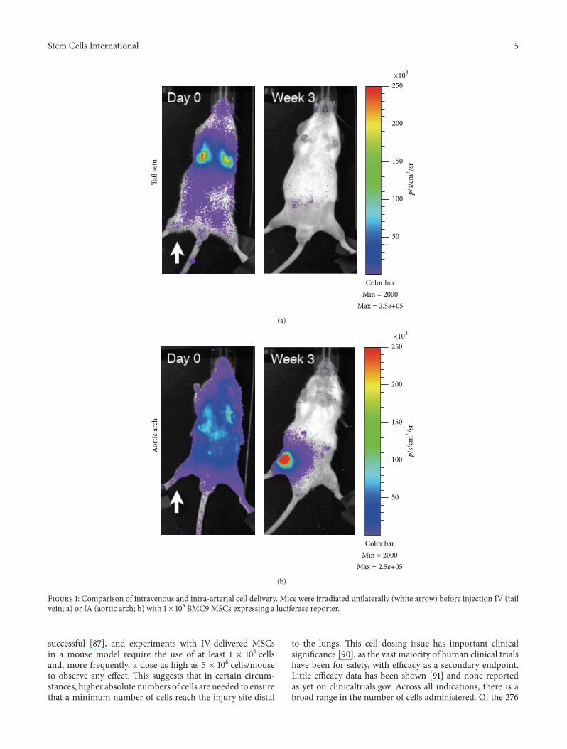
Figure 1: Comparison of intravenous and intra-arterial cell delivery. Mice were irradiated unilaterally (white arrow) before injection IV (tailvein; a) or IA (aortic arch; b) with 1 × 10⁶ BMC9 MSCs expressing a luciferase reporter.
successful [87], and experiments with IV-delivered MSCsin a mouse model require the use of at least 1 × 10⁶ cellsand, more frequently, a dose as high as 5 × 10⁶ cells/mouseto observe any effect. This suggests that in certain circum-stances, higher absolute numbers of cells are needed to ensurethat a minimum number of cells reach the injury site distal
to the lungs. This cell dosing issue has important clinicalsignificance [90], as the vast majority of human clinical trialshave been for safety, with efficacy as a secondary endpoint.Little efficacy data has been shown [91] and none reportedas yet on clinicaltrials.gov. Across all indications, there is abroad range in the number of cells administered. Of the 276
6 Stem Cells International
clinical trials mentioned in our introduction, 59 give a doseper injection per personwith amean of 2.16× 10⁸ cells/person(1 × 106, 5 × 10⁷, 5 × 10⁹; minimum, median, and maximum,resp.) and 75 give a dose/kg with a mean of 7.24 × 10⁶ cells/kg(1 × 105, 2 × 10⁶, 2 × 10⁸; minimum, median, and maximum,resp.). For instance, Horwitz et al. used a cell dose of 1–5 ×10⁶/kg in treatingOI pediatric patients [87], andLeBlanc et al.used a cell dose of 1-2× 10⁶/kg in treating patients withGvHD[92]. Similar to pharmacological studies, in which there is aneffective dose (ED) for drug treatment, there is an effectivecell dose (ECD) equivalent for cell therapy, which is definedas the minimum cell dose required to discern a significanttherapeutic effect. To put this in perspective, a commonlyused cell dose of 1 × 10⁶/30 gmouse would be equivalent to 33× 106/kg or approximately 2.3 billion cells for a 70 kg human.This number is technically and operationally challenging,and most MSC therapies, as part of ongoing clinical trials inhumans, use significantly lower cell doses.
Arterial delivery has the potential to alleviate some ofthese dosing limitations. In theory, arterial delivery allowsthe cells to bypass the lungs at least once and thus avoidthe pulmonary first-pass effect. This venous versus arterialeffect has long been recognized in other model systems.For example, to obtain bone engraftment of melanoma cells,tumor cells were injected into the arterial system [93]. Ifdelivered IV, tumor engraftment was primarily seen only inthe lungs. Other groups have attempted a similar tactic todeliver MSCs with similar results. For example, Togel et al.[94, 95] found increased numbers of IA-infused MSCs inkidneys in an acute kidney injury model after 24 h. This wasalso true in another study: when MSCs were infused IA in amodel of acute stroke, it resulted in enhanced cerebral MSCengraftment [96].
Our experiments with delivering MSCs into the arterialsystem by injection into the aortic arch or tail vein sup-port this “first-pass” cell delivery hypothesis. In this studydesigned to test for homing to injured tissue, mice wereinjured prior to injection by irradiating only one leg andwere then injected with MSCs transduced with a luciferasereporter gene and the signal tracked over time. As expected,MSCs showed significant entrapment in the lungs whendelivered IV into the tail vein. However, when delivered IAthrough the aortic arch, the cells weremore evenly distributedthroughout the entire animal (Figure 1). Over time, there wasevidence of engraftment of theMSCs at the injury site and notat the contralateral, noninjured leg in the arterial-deliveredgroups. In the tail vein-injected group, the cells dissipatedfrom the animal after a few days and were undetectable oneweek after injection.
In a separate set of studies, we were able to decreasethe ECD to 2.5 × 10⁵ cells/200𝜇L/mouse (fourfold ≤ orig-inal dose), by simply switching from IV to IA delivery(unpublished results). This has important consequences.First, there is concern with cell therapies of creating a cellembolus, even with IA delivery, that could potentially leadto increased mortality [96–98]. These concerns depend onboth the concentration and rate of cell delivery [97]. We,and others [99], have found pulmonary emboli and increased
mortality in IV-delivered animals at higher cell dose/flowrate. In particular, concentrations above 1.0 × 10⁷ cells/mLby IV administration in mice lead to insufficient dilutionof the cells in the blood, resulting in occlusion in the firstcapillary bed that is encountered (i.e., pulmonary embolism)and a significant increase in mortality. IA delivery is capableof decreasing the dose necessary, administers into a higherflow rate causing greater dilution, and thus decreases thepotential risk of cellular embolism. Furthermore, for clinicalapplications, this reduction in cell dosage translates into acell dose of ∼8 × 10⁶/kg for an average human which ismuch more feasible than the 33 × 106/kg ECD calculatedpreviously. It should be noted that, while this dose is withinthe realm of possibility for pediatric patients, this still equatesto ∼560 × 106MSCs/70 kg adult, which remains a challengeclinically. As a consequence, further improvements to MSCengraftment are needed.
5. Cell Targeting Strategies
Although there are numerous reports of stem cells homingto injured tissue, the mechanisms have yet to be elucidatedand could be heavily reliant on the leaky vasculature found ininjured [100, 101] or tumor tissue [102] resulting in passiveentrapment in the interstitial space. It is also evident thatany endogenous homing is insufficient, with less than 1% ofdelivered cells being found in target tissues [103]. With thisin mind, we now review various methods that have beenemployed to enhance site-specific delivery using cell surfaceligands.
5.1. Targeting Approaches. As with the application of ligand-directed techniques to drug therapies [104], there are a varietyof mechanisms which are being investigated to enhanceendogenous cell homing. In contrast to synthetic molecules,the cell represents a much more complex and dynamicenvironment, conferring both advantages and disadvantages.On the positive side, cells are a self-contained manufacturingplant capable of synthesizing therapeutic molecules, sensingthe environment, and responding to signals orchestratingregeneration or repair. On the negative side, cells are adifficult product to characterize; more expensive to produce;have nonspecific or unwanted cell-cell interactions; and caninternalize targeting ligands applied to the cell surface. Thetargeting approaches described in the following section arecategorized as antibody-, genetically-, selectin-, and peptide-directed cell therapies. For further review of cell surfacemodification strategies see Stephan and Irvine [105].
5.2. Antibody-Directed Cell Therapy. Antibodies are highlyspecific ligands with excellent affinity for their antigen.Antibodies have, therefore, found application in producingthe first approved ligand-targeted therapeutic, gemtuzumabozogamicin, an anti-CD33monoclonal drug conjugate for thetreatment of myeloid leukemia [106, 107]. It should be notedthat the FDA withdrew approval due to safety concerns in2010, but clinical trials continue, and the EuropeanMedicinesAgency has given it an orphan designation (EU/3/00/005).
Stem Cells International 7
Isolation and (optional) expansion of therapeutic cells, e.g., MSCs, T-regs, etc.
Cell painting
Two step One step
Antibody
Protein G
Cell membrane
Lipid
Biotin
Targetingsequence
or
Painted cells
Antibody Peptide
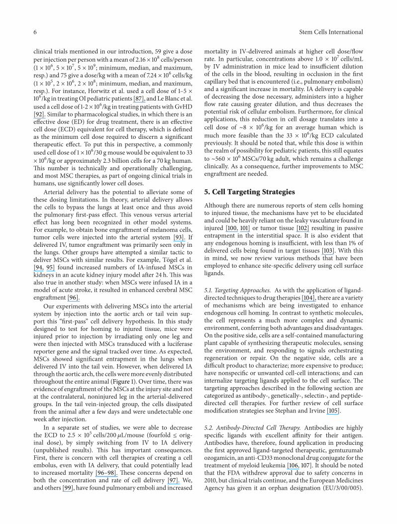
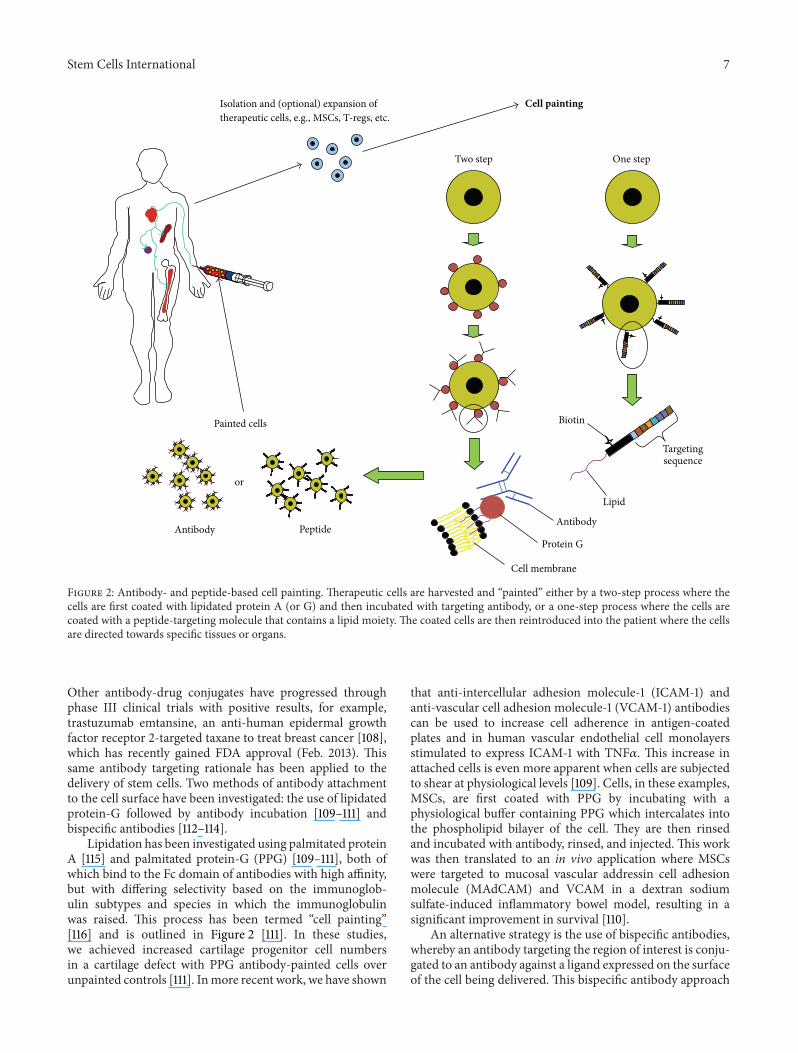
Figure 2: Antibody- and peptide-based cell painting. Therapeutic cells are harvested and “painted” either by a two-step process where thecells are first coated with lipidated protein A (or G) and then incubated with targeting antibody, or a one-step process where the cells arecoated with a peptide-targeting molecule that contains a lipid moiety. The coated cells are then reintroduced into the patient where the cellsare directed towards specific tissues or organs.
Other antibody-drug conjugates have progressed throughphase III clinical trials with positive results, for example,trastuzumab emtansine, an anti-human epidermal growthfactor receptor 2-targeted taxane to treat breast cancer [108],which has recently gained FDA approval (Feb. 2013). Thissame antibody targeting rationale has been applied to thedelivery of stem cells. Two methods of antibody attachmentto the cell surface have been investigated: the use of lipidatedprotein-G followed by antibody incubation [109–111] andbispecific antibodies [112–114].
Lipidation has been investigated using palmitated proteinA [115] and palmitated protein-G (PPG) [109–111], both ofwhich bind to the Fc domain of antibodies with high affinity,but with differing selectivity based on the immunoglob-ulin subtypes and species in which the immunoglobulinwas raised. This process has been termed “cell painting”[116] and is outlined in Figure 2 [111]. In these studies,we achieved increased cartilage progenitor cell numbersin a cartilage defect with PPG antibody-painted cells overunpainted controls [111]. Inmore recent work, we have shown
that anti-intercellular adhesion molecule-1 (ICAM-1) andanti-vascular cell adhesion molecule-1 (VCAM-1) antibodiescan be used to increase cell adherence in antigen-coatedplates and in human vascular endothelial cell monolayersstimulated to express ICAM-1 with TNF𝛼. This increase inattached cells is even more apparent when cells are subjectedto shear at physiological levels [109]. Cells, in these examples,MSCs, are first coated with PPG by incubating with aphysiological buffer containing PPG which intercalates intothe phospholipid bilayer of the cell. They are then rinsedand incubated with antibody, rinsed, and injected. This workwas then translated to an in vivo application where MSCswere targeted to mucosal vascular addressin cell adhesionmolecule (MAdCAM) and VCAM in a dextran sodiumsulfate-induced inflammatory bowel model, resulting in asignificant improvement in survival [110].
An alternative strategy is the use of bispecific antibodies,whereby an antibody targeting the region of interest is conju-gated to an antibody against a ligand expressed on the surfaceof the cell being delivered. This bispecific antibody approach
8 Stem Cells International
has been used to target HSCs to damaged vasculature inthe heart, using either anti-VCAM-1 conjugated to anti-c-kit[113] or anti-CD45-anti-myosin light chain kinase [112]. Thismethod has also been used in the field of cancer therapy todirect activated T-cells to human epidermal growth factorreceptor-2+ tumor cells in vitro [117]. This cancer treatmentwas tested in vivo, yielding reduced tumor size and a signifi-cantly increased rate of survival [118]. In vitro, a recent reportdescribes the use of an anti-CD90/anti-myosin light chainkinase to bind MSCs and increase their resistance to shearin a parallel plate assay [119].
Of the two antibody-directed cell therapy methods,lipidated protein-G followed by antibody incubation offersthe more versatile approach and has been used successfullywith MSCs. The same coating technique could, in theory, beapplied to any cell type. It is also possible to use multipleantibodies at once [111] and at a higher cell coating densitythan that achievable with bispecific antibodies because of thelimited number of cell surface ligands. Lipidated proteins canalso be used to bind other Fc-conjugated ligands; Chen etal. [116] conjugated B7-1 to the Fc portion of human IgGand used it as a co-stimulator. To our knowledge, bispecificantibodies have not been used with an in vivo MSC therapy,perhaps because MSCs do not have a cell-specific antibodymarker, although any surface-expressed antigen could beutilized. Bispecific antibodies would also have a high costfactor due to difficulties in manufacture and poor stability[120].
5.3. Genetically-Directed Cell Therapy. Genetically-directedcell therapy is defined as the introduction of genetic materialinto a cell, either DNA- or RNA-based, to express a ligandon the cell surface that will increase its localization in thetarget tissue. Genetically overexpressing receptor ligandshas great potential, but has the additional hurdles of celltransfection (efficiency, timing, stability, immunogenicity,deleterious effects on cell viability, oncogenic integrationsites, and consistent activity), and the regulatory problemsassociated with delivering both a gene- and cell-therapy. Ifthese issues are overcome, it could be a powerful tool toachieve higher efficacy in cell therapies. Within the MSCtargeting field, there have been positive results in cardiac-targeted cells directed using the CXCR4 ligand to induceMSCs to home toward the chemokine SDF1 [121, 122]. BothCheng et al. [121] and Zhang et al. [122] found increasedhoming of CXCR4-transduced MSCs to infarcted hearts andimproved cardiac output, with Zhang et al. also reportingincreased angiogenesis within the infarcted area. Cho et al.found that increasing the expression of CXCR4 also aidedin the prevention of bone loss in an ovariectomized mousemodel, potentiating the effect of an increased expressionof RANK ligand [123]. Another study showed increasedhoming of CXCR4-transduced C3H10T1/2 cells toward bonemarrow along with an increased bone mass in steroid-induced osteoporotic mice, along with complete restorationof bone mass in CXCR4- Cbfa1(RUNX2)-cotransduced cells[124]. Outside of MSC therapy, T-cells have been targeted foranticancer therapy using CD19 [125]. Genetically-modified
T-cells have progressed to the clinic with mixed results (forreview see [126]).
5.4. Selectin-Directed Cell Therapy. Selectin-directed cell tar-geting was pioneered by Xia et al. [127] who, using 𝛼-1,3-fucosyltransferase,modified the surface of CD34+ cord bloodcells and found increased engraftment in NOD/SCID mice.Sackstein et al. [128] applied this technique to modify thecell surface of MSCs, to form HCELL, an E-selectin andL-selectin binding ligand. The use of selectins mimics thenatural process of endogenous lymphocyte extravasation. Inthis process, activated endothelial cells express ligands forselectins, which are endogenously present on lymphocytes,causing them to bind to the endothelium and roll, followedby firm adherence and extravasation [129]. When CD44 onhuman MSCs was modified to produce the HCELL moietyand injected into immunocompromisedmice, theMSCswereobserved by intravital microscopy in the calvaria of micewhere they extravasated [128]. Sarkar et al. avoided enzymaticmodification of the cell surface through two methods: one,biotinylation of cell surface proteins followed by incubationwith streptavidin and biotin-conjugated sialyl lewis x (slex),a P-selectin ligand [130, 131], and two, liposome surfacemodification with biotin-conjugated lipids then incubationwith streptavidin and biotin-conjugated slex [132]. It isunclear what benefit Sarkar et al. gained from the morerecent liposome method, but it is unlikely that either methodwould have the same intracellular signaling cascade [133] asthat achieved with the enzymatic modification method ofSackstein et al. [128]. Although in vivo imaging of selectin-targeted MSCs has been demonstrated by both groups,neither has demonstrated efficacy in a disease model.
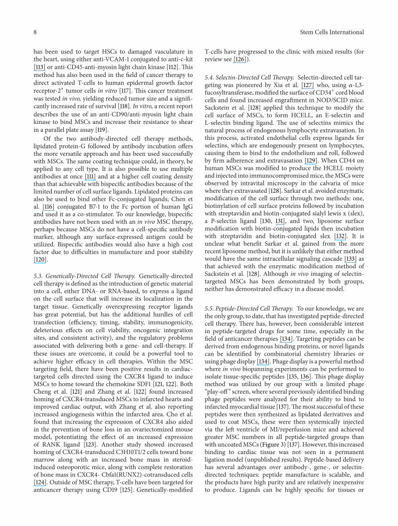
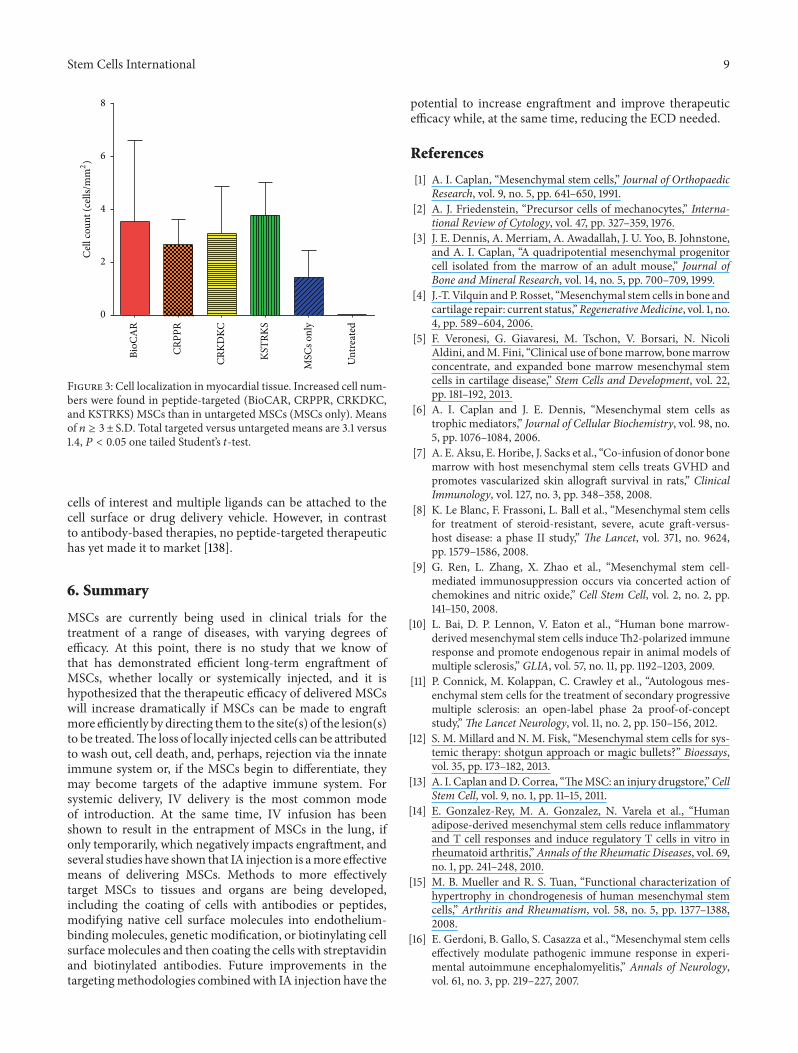
5.5. Peptide-Directed Cell Therapy. To our knowledge, we arethe only group, to date, that has investigated peptide-directedcell therapy. There has, however, been considerable interestin peptide-targeted drugs for some time, especially in thefield of anticancer therapies [134]. Targeting peptides can bederived from endogenous binding proteins, or novel ligandscan be identified by combinatorial chemistry libraries orusing phage display [134]. Phage display is a powerfulmethodwhere in vivo biopanning experiments can be performed toisolate tissue-specific peptides [135, 136]. This phage displaymethod was utilized by our group with a limited phage“play-off” screen, where several previously identified bindingphage peptides were analyzed for their ability to bind toinfarctedmyocardial tissue [137].Themost successful of thesepeptides were then synthesized as lipidated derivatives andused to coat MSCs, these were then systemically injectedvia the left ventricle of MI/reperfusion mice and achievedgreater MSC numbers in all peptide-targeted groups thanwith uncoatedMSCs (Figure 3) [137].However, this increasedbinding to cardiac tissue was not seen in a permanentligation model (unpublished results). Peptide-based deliveryhas several advantages over antibody-, gene-, or selectin-directed techniques: peptide manufacture is scalable, andthe products have high purity and are relatively inexpensiveto produce. Ligands can be highly specific for tissues or
Stem Cells International 9
0
2
4
6
8
BioC
AR
CRPP
R
CRKD
KC
KSTR
KS
MSC
s onl
y
Unt
reat
ed
Cell
coun
t (ce
lls/m
m2
)
Figure 3: Cell localization inmyocardial tissue. Increased cell num-bers were found in peptide-targeted (BioCAR, CRPPR, CRKDKC,and KSTRKS) MSCs than in untargeted MSCs (MSCs only). Meansof 𝑛 ≥ 3 ± S.D. Total targeted versus untargeted means are 3.1 versus1.4, 𝑃 < 0.05 one tailed Student’s 𝑡-test.
cells of interest and multiple ligands can be attached to thecell surface or drug delivery vehicle. However, in contrastto antibody-based therapies, no peptide-targeted therapeutichas yet made it to market [138].
6. Summary
MSCs are currently being used in clinical trials for thetreatment of a range of diseases, with varying degrees ofefficacy. At this point, there is no study that we know ofthat has demonstrated efficient long-term engraftment ofMSCs, whether locally or systemically injected, and it ishypothesized that the therapeutic efficacy of delivered MSCswill increase dramatically if MSCs can be made to engraftmore efficiently by directing them to the site(s) of the lesion(s)to be treated.The loss of locally injected cells can be attributedto wash out, cell death, and, perhaps, rejection via the innateimmune system or, if the MSCs begin to differentiate, theymay become targets of the adaptive immune system. Forsystemic delivery, IV delivery is the most common modeof introduction. At the same time, IV infusion has beenshown to result in the entrapment of MSCs in the lung, ifonly temporarily, which negatively impacts engraftment, andseveral studies have shown that IA injection is amore effectivemeans of delivering MSCs. Methods to more effectivelytarget MSCs to tissues and organs are being developed,including the coating of cells with antibodies or peptides,modifying native cell surface molecules into endothelium-binding molecules, genetic modification, or biotinylating cellsurfacemolecules and then coating the cells with streptavidinand biotinylated antibodies. Future improvements in thetargetingmethodologies combinedwith IA injection have the
potential to increase engraftment and improve therapeuticefficacy while, at the same time, reducing the ECD needed.
References
[1] A. I. Caplan, “Mesenchymal stem cells,” Journal of OrthopaedicResearch, vol. 9, no. 5, pp. 641–650, 1991.
[2] A. J. Friedenstein, “Precursor cells of mechanocytes,” Interna-tional Review of Cytology, vol. 47, pp. 327–359, 1976.
[3] J. E. Dennis, A. Merriam, A. Awadallah, J. U. Yoo, B. Johnstone,and A. I. Caplan, “A quadripotential mesenchymal progenitorcell isolated from the marrow of an adult mouse,” Journal ofBone and Mineral Research, vol. 14, no. 5, pp. 700–709, 1999.
[4] J.-T. Vilquin andP. Rosset, “Mesenchymal stem cells in bone andcartilage repair: current status,”RegenerativeMedicine, vol. 1, no.4, pp. 589–604, 2006.
[5] F. Veronesi, G. Giavaresi, M. Tschon, V. Borsari, N. NicoliAldini, andM. Fini, “Clinical use of bonemarrow, bonemarrowconcentrate, and expanded bone marrow mesenchymal stemcells in cartilage disease,” Stem Cells and Development, vol. 22,pp. 181–192, 2013.
[6] A. I. Caplan and J. E. Dennis, “Mesenchymal stem cells astrophic mediators,” Journal of Cellular Biochemistry, vol. 98, no.5, pp. 1076–1084, 2006.
[7] A. E. Aksu, E. Horibe, J. Sacks et al., “Co-infusion of donor bonemarrow with host mesenchymal stem cells treats GVHD andpromotes vascularized skin allograft survival in rats,” ClinicalImmunology, vol. 127, no. 3, pp. 348–358, 2008.
[8] K. Le Blanc, F. Frassoni, L. Ball et al., “Mesenchymal stem cellsfor treatment of steroid-resistant, severe, acute graft-versus-host disease: a phase II study,” The Lancet, vol. 371, no. 9624,pp. 1579–1586, 2008.
[9] G. Ren, L. Zhang, X. Zhao et al., “Mesenchymal stem cell-mediated immunosuppression occurs via concerted action ofchemokines and nitric oxide,” Cell Stem Cell, vol. 2, no. 2, pp.141–150, 2008.
[10] L. Bai, D. P. Lennon, V. Eaton et al., “Human bone marrow-derived mesenchymal stem cells induceTh2-polarized immuneresponse and promote endogenous repair in animal models ofmultiple sclerosis,” GLIA, vol. 57, no. 11, pp. 1192–1203, 2009.
[11] P. Connick, M. Kolappan, C. Crawley et al., “Autologous mes-enchymal stem cells for the treatment of secondary progressivemultiple sclerosis: an open-label phase 2a proof-of-conceptstudy,”The Lancet Neurology, vol. 11, no. 2, pp. 150–156, 2012.
[12] S. M. Millard and N. M. Fisk, “Mesenchymal stem cells for sys-temic therapy: shotgun approach or magic bullets?” Bioessays,vol. 35, pp. 173–182, 2013.
[13] A. I. Caplan andD.Correa, “TheMSC: an injury drugstore,”CellStem Cell, vol. 9, no. 1, pp. 11–15, 2011.
[14] E. Gonzalez-Rey, M. A. Gonzalez, N. Varela et al., “Humanadipose-derived mesenchymal stem cells reduce inflammatoryand T cell responses and induce regulatory T cells in vitro inrheumatoid arthritis,”Annals of the Rheumatic Diseases, vol. 69,no. 1, pp. 241–248, 2010.
[15] M. B. Mueller and R. S. Tuan, “Functional characterization ofhypertrophy in chondrogenesis of human mesenchymal stemcells,” Arthritis and Rheumatism, vol. 58, no. 5, pp. 1377–1388,2008.
[16] E. Gerdoni, B. Gallo, S. Casazza et al., “Mesenchymal stem cellseffectively modulate pathogenic immune response in experi-mental autoimmune encephalomyelitis,” Annals of Neurology,vol. 61, no. 3, pp. 219–227, 2007.
10 Stem Cells International
[17] E. Zappia, S. Casazza, E. Pedemonte et al., “Mesenchymal stemcells ameliorate experimental autoimmune encephalomyelitisinducing T-cell anergy,” Blood, vol. 106, no. 5, pp. 1755–1761,2005.
[18] R. Abdi, P. Fiorina, C. N. Adra, M. Atkinson, andM. H. Sayegh,“Immunomodulation by mesenchymal stem cells: a potentialtherapeutic strategy for type 1 diabetes,” Diabetes, vol. 57, no. 7,pp. 1759–1767, 2008.
[19] V. S. Urban, J. Kiss, J. Kovacs et al., “Mesenchymal stem cellscooperate with bone marrow cells in therapy of diabetes,” StemCells, vol. 26, no. 1, pp. 244–253, 2008.
[20] D. Garcıa-Olmo, M. Garcıa-Arranz, L. Gomez Garcıa et al.,“Autologous stem cell transplantation for treatment of recto-vaginal fistula in perinatal Crohn’s disease: a new cell-basedtherapy,” International Journal of Colorectal Disease, vol. 18, no.5, pp. 451–454, 2003.
[21] E. Gonzalez-Rey, P. Anderson, M. A. Gonzalez, L. Rico, D.Buscher, and M. Delgado, “Human adult stem cells derivedfrom adipose tissue protect against experimental colitis andsepsis,” Gut, vol. 58, no. 7, pp. 929–939, 2009.
[22] Y. Hayashi, S. Tsuji, M. Tsujii et al., “Topical implantationof mesenchymal stem cells has beneficial effects on healingof experimental colitis in rats,” Journal of Pharmacology andExperimental Therapeutics, vol. 326, no. 2, pp. 523–531, 2008.
[23] T. Yabana, Y. Arimura, H. Tanaka et al., “Enhancing epithelialengraftment of rat mesenchymal stem cells restores epithelialbarrier integrity,” Journal of Pathology, vol. 218, no. 3, pp. 350–359, 2009.
[24] X. Zhang, C. Jiao, and S. Zhao, “Role of mesenchymal stem cellsin immunological rejection of organ transplantation,” Stem CellReviews and Reports, vol. 5, no. 4, pp. 402–409, 2010.
[25] J. M. Karp and G. S. Leng Teo, “Mesenchymal stem cell homing:the devil is in the details,” Cell Stem Cell, vol. 4, no. 3, pp. 206–216, 2009.
[26] P. Lin, Y. Lin, D. P. Lennon, D. Correa, M. Schluchter, and A. I.Caplan, “Efficient lentiviral transduction of human mesenchy-mal stem cells that preserves proliferation and differentiationcapabilities,” Stem Cells Translational Medicine, vol. 1, pp. 886–897, 2012.
[27] J. Gao, J. E. Dennis, R. F. Muzic, M. Lundberg, and A. I. Caplan,“The dynamic in vivo distribution of bone marrow-derivedmesenchymal stem cells after infusion,” Cells Tissues Organs,vol. 169, no. 1, pp. 12–20, 2001.
[28] M. Srinivas, E. H. J. G. Aarntzen, J. W. M. Bulte et al., “Imagingof cellular therapies,” Advanced Drug Delivery Reviews, vol. 62,no. 11, pp. 1080–1093, 2010.
[29] M. R. Reagan and D. L. Kaplan, “Concise review: mesenchymalstem cell tumor-homing: detection methods in disease modelsystems,” Stem Cells, vol. 29, no. 6, pp. 920–927, 2011.
[30] L. Kostura, D. L. Kraitchman, A. M. Mackay, M. F. Pittenger,and J. M.W. Bulte, “Feridex labeling of mesenchymal stem cellsinhibits chondrogenesis but not adipogenesis or osteogenesis,”NMR in Biomedicine, vol. 17, no. 7, pp. 513–517, 2004.
[31] K. Le Blanc, C. Tammik, K. Rosendahl, E. Zetterberg, andO. Ringden, “HLA expression and immunologic properties ofdifferentiated and undifferentiated mesenchymal stem cells,”Experimental Hematology, vol. 31, no. 10, pp. 890–896, 2003.
[32] B. Maitra, E. Szekely, K. Gjini et al., “Human mesenchymalstem cells support unrelated donor hematopoietic stem cellsand suppress T-cell activation,” Bone Marrow Transplantation,vol. 33, no. 6, pp. 597–604, 2004.
[33] W. T. Tse, J. D. Pendleton, W. M. Beyer, M. C. Egalka, and E.C. Guinan, “Suppression of allogeneic T-cell proliferation byhuman marrow stromal cells: implications in transplantation,”Transplantation, vol. 75, no. 3, pp. 389–397, 2003.
[34] A. Bartholomew, C. Sturgeon, M. Siatskas et al., “Mesenchymalstem cells suppress lymphocyte proliferation in vitro andprolong skin graft survival in vivo,” Experimental Hematology,vol. 30, no. 1, pp. 42–48, 2002.
[35] M. Krampera, L. Cosmi, R. Angeli et al., “Role for interferon-𝛾 in the immunomodulatory activity of human bone marrowmesenchymal stem cells,” Stem Cells, vol. 24, no. 2, pp. 386–398,2006.
[36] K. English, F. P. Barry, C. P. Field-Corbett, and B. P. Mahon,“IFN-𝛾 and TNF-𝛼 differentially regulate immunomodulationby murine mesenchymal stem cells,” Immunology Letters, vol.110, no. 2, pp. 91–100, 2007.
[37] J.M. Ryan, F. Barry, J.M.Murphy, and B. P.Mahon, “Interferon-𝛾 does not break, but promotes the immunosuppressive capac-ity of adult human mesenchymal stem cells,” Clinical andExperimental Immunology, vol. 149, no. 2, pp. 353–363, 2007.
[38] R. H. Lee, A. A. Pulin, M. J. Seo et al., “Intravenous hMSCsimprove myocardial infarction in mice because cells embolizedin lung are activated to secrete the anti-inflammatory proteinTSG-6,” Cell Stem Cell, vol. 5, no. 1, pp. 54–63, 2009.
[39] M. Di Nicola, C. Carlo-Stella, M. Magni et al., “Humanbonemarrow stromal cells suppress T-lymphocyte proliferationinduced by cellular or nonspecificmitogenic stimuli,”Blood, vol.99, no. 10, pp. 3838–3843, 2002.
[40] L. Cui, Y. Shuo, W. Liu, N. Li, W. Zhang, and Y. Cao, “Expandedadipose-derived stem cells suppressmixed lymphocyte reactionby secretion of prostaglandin E2,”Tissue Engineering, vol. 13, no.6, pp. 1185–1195, 2007.
[41] G. Ren, J. Su, L. Zhang et al., “Species variation in themechanisms of mesenchymal stem cell-mediated immunosup-pression,” Stem Cells, vol. 27, no. 8, pp. 1954–1962, 2009.
[42] K. English, “Mechanisms of mesenchymal stromal cellimmunomodulation,” Immunology and Cell Biology, vol. 91, pp.19–26, 2013.
[43] F. Liotta, R. Angeli, L. Cosmi et al., “Toll-like receptors 3 and4 are expressed by human bone marrow-derived mesenchymalstem cells and can inhibit their T-cell modulatory activity byimpairing notch signaling,” Stem Cells, vol. 26, no. 1, pp. 279–289, 2008.
[44] S. L. Brown, T. E. Riehl, M. R. Walker et al., “Myd88-dependentpositioning of Ptgs2-expressing stromal cells maintains colonicepithelial proliferation during injury,” Journal of Clinical Inves-tigation, vol. 117, no. 1, pp. 258–269, 2007.
[45] Y.-P. Li, S. Paczesny, E. Lauret et al., “Human mesenchymalstem cells license adult CD34+ hemopoietic progenitor cells todifferentiate into regulatory dendritic cells through activation ofthe notch pathway,” Journal of Immunology, vol. 180, no. 3, pp.1598–1608, 2008.
[46] B. Zhang, R. Liu, D. Shi et al., “Mesenchymal stem cellsinduce mature dendritic cells into a novel Jagged-2 dependentregulatory dendritic cell population,” Blood, vol. 113, no. 1, pp.46–57, 2009.
[47] G. W. Roddy, J. Y. Oh, R. H. Lee et al., “Action at a dis-tance: systemically administered adult stem/progenitor cells(MSCs) reduce inflammatory damage to the cornea withoutengraftment and primarily by secretion of TNF-𝛼 stimulatedgene/protein 6,” Stem Cells, vol. 29, no. 10, pp. 1572–1579, 2011.
Stem Cells International 11
[48] H. Choi, R. H. Lee, N. Bazhanov, J. Y. Oh, and D. J. Prockop,“Anti-inflammatory protein TSG-6 secreted by activated MSCsattenuates zymosan-induced mouse peritonitis by decreasingTLR2/NF-𝜅B signaling in resident macrophages,” Blood, vol.118, no. 2, pp. 330–338, 2011.
[49] M. D. Griffin, A. E. Ryan, S. Alagesan, P. Lohan, O. Treacy,and T. Ritter, “Anti-donor immune responses elicited by allo-geneic mesenchymal stem cells: what have we learned so far?”Immunology and Cell Biology, vol. 91, pp. 40–51, 2013.
[50] N. Kotobuki, Y. Katsube, Y. Katou,M. Tadokoro,M. Hirose, andH. Ohgushi, “In vivo survival and osteogenic differentiation ofallogeneic rat bone marrow mesenchymal stem cells (MSCs),”Cell Transplantation, vol. 17, no. 6, pp. 705–712, 2008.
[51] X.-P. Huang, Z. Sun, Y. Miyagi et al., “Differentiation ofallogeneic mesenchymal stem cells induces immunogenicityand limits their long-term benefits for myocardial repair,”Circulation, vol. 122, no. 23, pp. 2419–2429, 2010.
[52] Y. Li and F. Lin, “Mesenchymal stem cells are injured bycomplement after their contact with serum,” Blood, vol. 120, pp.3436–3443, 2012.
[53] L. von Bahr, I. Batsis, G.Moll et al., “Analysis of tissues followingmesenchymal stromal cell therapy in humans indicates limitedlong-term engraftment and no ectopic tissue formation,” StemCells, vol. 30, pp. 1575–1578, 2012.
[54] G. P. Meyer, K. C. Wollert, J. Lotz et al., “Intracoronary bonemarrow cell transfer after myocardial infarction: 5-year follow-up from the randomized-controlled BOOST trial,” EuropeanHeart Journal, vol. 30, no. 24, pp. 2978–2984, 2009.
[55] M. Abedin, Y. Tintut, and L. L. Demer, “Mesenchymal stem cellsand the artery wall,” Circulation Research, vol. 95, no. 7, pp. 671–676, 2004.
[56] M. Crisan, S. Yap, L. Casteilla et al., “A perivascular origin formesenchymal stem cells in multiple human organs,” Cell StemCell, vol. 3, no. 3, pp. 301–313, 2008.
[57] S. Shi and S. Gronthos, “Perivascular niche of postnatal mes-enchymal stem cells in human bone marrow and dental pulp,”Journal of Bone andMineral Research, vol. 18, no. 4, pp. 696–704,2003.
[58] S.-G. Kang, N. Shinojima, A. Hossain et al., “Isolation andperivascular localization of mesenchymal stem cells frommouse brain,” Neurosurgery, vol. 67, no. 3, pp. 711–720, 2010.
[59] N. Zebardast, D. Lickorish, and J. E. Davies, “Human umbilicalcord perivascular cells (HUCPVC): a mesenchymal cell sourcefor dermal wound healing,”Organogenesis, vol. 6, no. 4, pp. 197–203, 2010.
[60] X. Cai, Y. Lin, P. V. Hauschka, and B. E. Grottkau, “Adipose stemcells originate from perivascular cells,” Biology of the Cell, vol.103, no. 9, pp. 435–447, 2011.
[61] A. W. James, J. N. Zara, M. Corselli et al., “An abundantperivascular source of stem cells for bone tissue engineering,”Stem Cells Translational Medicine, vol. 1, pp. 673–684, 2012.
[62] J. C. Gerlach, P. Over, M. E. Turner et al., “Perivascularmesenchymal progenitors in human fetal and adult liver,” StemCells and Development, vol. 21, pp. 3258–3269, 2012.
[63] S. Gronthos, J. Brahim, W. Li et al., “Stem cell properties ofhuman dental pulp stem cells,” Journal of Dental Research, vol.81, no. 8, pp. 531–535, 2002.
[64] P. Bianco, P. G. Robey, and P. J. Simmons, “Mesenchymal stemcells: revisiting history, concepts, and assays,”Cell StemCell, vol.2, no. 4, pp. 313–319, 2008.
[65] P. Dore-Duffy, C. Owen, R. Balabanov, S.Murphy, T. Beaumont,and J. A. Rafols, “Pericyte migration from the vascular wall inresponse to traumatic brain injury,”Microvascular Research, vol.60, no. 1, pp. 55–69, 2000.
[66] M. A. Maloney, R. A. Lamela, and H. M. Patt, “The question ofbone marrow stromal fibroblast traffic,” Annals of the New YorkAcademy of Sciences, vol. 459, pp. 190–197, 1985.
[67] T. Kitaori, H. Ito, E. M. Schwarz et al., “Stromal cell-derivedfactor 1/CXCR4 signaling is critical for the recruitment ofmesenchymal stem cells to the fracture site during skeletalrepair in a mouse model,” Arthritis and Rheumatism, vol. 60,no. 3, pp. 813–823, 2009.
[68] S. Schenk, N. Mal, A. Finan et al., “Monocyte chemotacticprotein-3 is a myocardial mesenchymal stem cell homingfactor,” Stem Cells, vol. 25, no. 1, pp. 245–251, 2007.
[69] G. Chamberlain, H. Smith, G. E. Rainger, and J. Middleton,“Mesenchymal stem cells exhibit firm adhesion, crawling,spreading and transmigration across aortic endothelial cells:effects of chemokines and shear,”PLoSONE, vol. 6, no. 9, ArticleID e25663, 2011.
[70] M. Iwasaki, M. Koyanagi, H. Kossmann et al., “Hepatocytegrowth factor mobilizes non-bone marrow-derived circulatingmesoangioblasts,” European Heart Journal, vol. 32, no. 5, pp.627–636, 2011.
[71] T. Kinnaird, E. S. Burnett, M. Shou et al., “Local delivery ofmarrow-derived stromal cells augments collateral perfusionthrough paracrinemechanisms,”Circulation, vol. 109, no. 12, pp.1543–1549, 2004.
[72] M. T. Valarmathi, J. M. Davis, M. J. Yost, R. L. Goodwin,and J. D. Potts, “A three-dimensional model of vasculogenesis,”Biomaterials, vol. 30, no. 6, pp. 1098–1112, 2009.
[73] A. Arthur, A. Zannettino, and S. Gronthos, “The therapeuticapplications of multipotential mesenchymal/stromal stem cellsin skeletal tissue repair,” Journal of Cellular Physiology, vol. 218,no. 2, pp. 237–245, 2009.
[74] A. Augello, R. Tasso, S. M. Negrini, R. Cancedda, and G. Pen-nesi, “Cell therapy using allogeneic bone marrowmesenchymalstem cells prevents tissue damage in collagen-induced arthritis,”Arthritis and Rheumatism, vol. 56, no. 4, pp. 1175–1186, 2007.
[75] A. I. Caplan, “Osteogenesis imperfecta, rehabilitationmedicine,fundamental research andmesenchymal stem cells,”ConnectiveTissue Research, vol. 31, no. 4, pp. S9–S14, 1995.
[76] M. T. L. Castelo-Branco, I. D. P. Soares, D. V. Lopes et al.,“Intraperitoneal but not intravenous cryopreserved mesenchy-mal stromal cells home to the inflamed colon and ameliorateexperimental colitis,” PLoS ONE, vol. 7, no. 3, Article ID e33360,2012.
[77] Y. Tang, K. Shah, S.M.Messerli, E. Snyder, X. Breakefield, andR.Weissleder, “In vivo tracking of neural progenitor cell migrationto glioblastomas,”HumanGeneTherapy, vol. 14, no. 13, pp. 1247–1254, 2003.
[78] T. Wilson, C. Stark, J. Holmbom et al., “Fate of bone marrow-derived stromal cells after intraperitoneal infusion or implan-tation into femoral bone defects in the host animal,” Journal ofTissue Engineering, vol. 2010, Article ID 345806, 2010.
[79] A. T. Dinh, N. Kubis, Y. Tomita et al., “In vivo imaging withcellular resolution of bone marrow cells transplanted into theischemic brain of a mouse,” NeuroImage, vol. 31, no. 3, pp. 958–967, 2006.
[80] H. F. Dvorak, “Tumors: wounds that do not heal: similaritiesbetween tumor stroma generation and wound healing,” New
12 Stem Cells International
England Journal of Medicine, vol. 315, no. 26, pp. 1650–1659,1986.
[81] K. Nakamura, Y. Ito, Y. Kawano et al., “Antitumor effect ofgenetically engineered mesenchymal stem cells in a rat gliomamodel,” Gene Therapy, vol. 11, no. 14, pp. 1155–1164, 2004.
[82] K. T. Chabner, G. B. Adams, J. Qiu et al., “Direct vasculardelivery of primitive hematopoietic cells to bone marrowimproves localization but not engraftment,” Blood, vol. 103, no.12, pp. 4685–4686, 2004.
[83] U. M. Fischer, M. T. Harting, F. Jimenez et al., “Pulmonarypassage is a major obstacle for intravenous stem cell delivery:the pulmonary first-pass effect,” Stem Cells and Development,vol. 18, no. 5, pp. 683–691, 2009.
[84] I. Sekiya, B. L. Larson, J. R. Smith, R. Pochampally, J.-G. Cui, andD. J. Prockop, “Expansion of human adult stem cells from bonemarrow stroma: conditions that maximize the yields of earlyprogenitors and evaluate their quality,” Stem Cells, vol. 20, no.6, pp. 530–541, 2002.
[85] S. Schrepfer, T. Deuse, H. Reichenspurner,M. P. Fischbein, R. C.Robbins, andM. P. Pelletier, “Stem cell transplantation: the lungbarrier,” Transplantation Proceedings, vol. 39, no. 2, pp. 573–576,2007.
[86] J. Nystedt, H. Anderson, J. Tikkanen et al., “Cell surfacestructures influence lung clearance rate of systemically infusedmesenchymal stromal cells,” Stem Cells, vol. 31, pp. 317–326,2013.
[87] E. M. Horwitz, P. L. Gordon, W. K. K. Koo et al., “Isolatedallogeneic bone marrow-derived mesenchymal cells engraftand stimulate growth in children with osteogenesis imper-fecta: implications for cell therapy of bone,” Proceedings of theNational Academy of Sciences of the United States of America,vol. 99, no. 13, pp. 8932–8937, 2002.
[88] E. M. Horwitz, D. J. Prockop, L. A. Fitzpatrick et al., “Trans-plantability and therapeutic effects of bone marrow-derivedmesenchymal cells in children with osteogenesis imperfecta,”Nature Medicine, vol. 5, no. 3, pp. 309–313, 1999.
[89] L. Fouillard, A. Chapel, D. Bories et al., “Infusion of allogeneic-related HLA mismatched mesenchymal stem cells for thetreatment of incomplete engraftment following autologoushaematopoietic stem cell transplantation,” Leukemia, vol. 21, no.3, pp. 568–570, 2007.
[90] J. Muller-Ehmsen, “The problem is obvious, the solution is not:numbers do matter in cardiac cell therapy!,” CardiovascularResearch, vol. 96, pp. 208–209, 2012.
[91] J. Ankrum and J.M. Karp, “Mesenchymal stem cell therapy: twosteps forward, one step back,”Trends inMolecularMedicine, vol.16, no. 5, pp. 203–209, 2010.
[92] K. Le Blanc, I. Rasmusson, B. Sundberg et al., “Treatment ofsevere acute graft-versus-host disease with third party hap-loidentical mesenchymal stem cells,” The Lancet, vol. 363, no.9419, pp. 1439–1441, 2004.
[93] F. Arguello, R. B. Baggs, and C. N. Frantz, “A murine modelof experimental metastasis to bone and bone marrow,” CancerResearch, vol. 48, no. 23, pp. 6876–6881, 1988.
[94] F. Togel, Y. Yang, P. Zhang, Z. Hu, and C. Westenfelder,“Bioluminescence imaging to monitor the in vivo distributionof administeredmesenchymal stem cells in acute kidney injury,”American Journal of Physiology, vol. 295, no. 1, pp. F315–F321,2008.
[95] F. Togel, Z. Hu, K.Weiss, J. Isaac, C. Lange, and C.Westenfelder,“Administeredmesenchymal stem cells protect against ischemic
acute renal failure through differentiation-independent mecha-nisms,” American Journal of Physiology, vol. 289, no. 1, pp. F31–F42, 2005.
[96] P. Walczak, J. Zhang, A. A. Gilad et al., “Dual-modalitymonitoring of targeted intraarterial delivery of mesenchymalstem cells after transient ischemia,” Stroke, vol. 39, no. 5, pp.1569–1574, 2008.
[97] M. Janowski, A. Lyczek, C. Engels et al., “Cell size and velocityof injection are major determinants of the safety of intracarotidstem cell transplantation,” Journal of Cerebral Blood Flow andMetabolism, vol. 33, pp. 921–927, 2013.
[98] L. Li, Q. Jiang, G. Ding et al., “Effects of administrationroute on migration and distribution of neural progenitor cellstransplanted into rats with focal cerebral ischemia, an MRIstudy,” Journal of Cerebral Blood Flow and Metabolism, vol. 30,no. 3, pp. 653–662, 2010.
[99] C. Kyriakou, N. Rabin, A. Pizzey, A. Nathwani, and K. Yong,“Factors that influence short-term homing of human bonemarrow-derived mesenchymal stem cells in a xenogeneic ani-mal model,”Haematologica, vol. 93, no. 10, pp. 1457–1465, 2008.
[100] A. Fleck, G. Raines, F. Hawker et al., “Increased vascularpermeability: a major cause of hypoalbuminaemia in diseaseand injury,”The Lancet, vol. 1, no. 8432, pp. 781–784, 1985.
[101] L. F. Brown, K.-T. Yeo, B. Berse et al., “Expression of vascularpermeability factor (vascular endothelial growth factor) byepidermal keratinocytes during wound healing,” Journal ofExperimental Medicine, vol. 176, no. 5, pp. 1375–1379, 1992.
[102] H. Maeda, J. Wu, T. Sawa, Y. Matsumura, and K. Hori, “Tumorvascular permeability and the EPR effect in macromoleculartherapeutics: a review,” Journal of Controlled Release, vol. 65, no.1-2, pp. 271–284, 2000.
[103] D. J. Prockop, “Repair of tissues by adult stem/progenitorcells (MSCs): controversies, myths, and changing paradigms,”Molecular Therapy, vol. 17, no. 6, pp. 939–946, 2009.
[104] V. P. Torchilin, “Multifunctional nanocarriers,” Advanced DrugDelivery Reviews, vol. 58, no. 14, pp. 1532–1555, 2006.
[105] M. T. Stephan and D. J. Irvine, “Enhancing cell therapiesfrom the outside in: cell surface engineering using syntheticnanomaterials,” Nano Today, vol. 6, no. 3, pp. 309–325, 2011.
[106] A. K. Burnett, “Treatment of acute myeloid leukemia: are wemaking progress?” Hematology/The Education Program of theAmerican Society of Hematology, vol. 2012, pp. 1–6, 2012.
[107] R. A. Larson, M. Boogaerts, E. Estey et al., “Antibody-targetedchemotherapy of older patients with acute myeloid leukemiain first relapse using Mylotarg (gemtuzumab ozogamicin),”Leukemia, vol. 16, no. 9, pp. 1627–1636, 2002.
[108] S. Verma, D.Miles, L. Gianni et al., “Trastuzumab emtansine forHER2-positive advanced breast cancer,”New England Journal ofMedicine, vol. 367, pp. 1783–1791, 2012.
[109] I. K. Ko, T. J. Kean, and J. E. Dennis, “Targeting mesenchymalstem cells to activated endothelial cells,” Biomaterials, vol. 30,no. 22, pp. 3702–3710, 2009.
[110] I. K. Ko, B.-G. Kim, A. Awadallah et al., “Targeting improvesMSC treatment of inflammatory bowel disease,” MolecularTherapy, vol. 18, no. 7, pp. 1365–1372, 2010.
[111] J. E. Dennis, N. Cohen, V. M. Goldberg, and A. I. Caplan,“Targeted delivery of progenitor cells for cartilage repair,”Journal of Orthopaedic Research, vol. 22, no. 4, pp. 735–741,2004.
[112] R. J. Lee, Q. Fang, P. A. Davol et al., “Antibody targeting of stemcells to infarctedmyocardium,” StemCells, vol. 25, no. 3, pp. 712–717, 2007.
Stem Cells International 13
[113] L. G. Lum, H. Fok, R. Sievers, M. Abedi, P. J. Quesenberry,and R. J. Lee, “Targeting of Lin-Sca+ hematopoietic stem cellswith bispecific antibodies to injured myocardium,” Blood Cells,Molecules, and Diseases, vol. 32, no. 1, pp. 82–87, 2004.
[114] T. C. Zhao, A. Tseng, N. Yano et al., “Targeting humanCD34+ hematopoietic stem cells with anti-CD45X anti-myosinlight-chain bispecific antibody preserves cardiac function inmyocardial infarction,” Journal of Applied Physiology, vol. 104,no. 6, pp. 1793–1800, 2008.
[115] S. A. Kim and J. S. Peacock, “The use of palmitate-conjugatedprotein A for coating cells with artificial receptors whichfacilitate intercellular interactions,” Journal of ImmunologicalMethods, vol. 158, no. 1, pp. 57–65, 1993.
[116] A. Chen, G. Zheng, andM. L. Tykocinski, “Hierarchical costim-ulator thresholds for distinct immune responses: application ofa novel two-step Fc fusion protein transfer method,” Journal ofImmunology, vol. 164, no. 2, pp. 705–711, 2000.
[117] M. Sen,D.M.Wankowski, N. K.Garlie et al., “Use of anti-CD3×anti-HER2/neu bispecific antibody for redirecting cytotoxicityof activated T cells toward HER2/neu+ tumors,” Journal ofHematotherapy and Stem Cell Research, vol. 10, no. 2, pp. 247–260, 2001.
[118] J. K. Chan, C. A. Hamilton, M. K. Cheung et al., “Enhancedkilling of primary ovarian cancer by retargeting autologouscytokine-induced killer cells with bispecific antibodies: a pre-clinical study,” Clinical Cancer Research, vol. 12, no. 6, pp. 1859–1867, 2006.
[119] C. W. Gundlach IV, A. Caivano, M. Da Graca Cabreira-Hansenet al., “Synthesis and evaluation of an anti-MLC1 × anti-CD90bispecific antibody for targeting and retaining bone-marrow-derived multipotent stromal cells in infarcted myocardium,”Bioconjugate Chemistry, vol. 22, no. 8, pp. 1706–1714, 2011.
[120] V. R. Doppalapudi, J. Huang, D. Liu et al., “Chemical generationof bispecific antibodies,” Proceedings of the National Academyof Sciences of the United States of America, vol. 107, no. 52, pp.22611–22616, 2010.
[121] Z. Cheng, L. Ou, X. Zhou et al., “Targeted migration of mes-enchymal stem cells modified with CXCR4 gene to infarctedmyocardium improves cardiac performance,” Molecular Ther-apy, vol. 16, no. 3, pp. 571–579, 2008.
[122] D. Zhang, G.-C. Fan, X. Zhou et al., “Over-expression ofCXCR4 onmesenchymal stem cells augmentsmyoangiogenesisin the infarctedmyocardium,” Journal of Molecular and CellularCardiology, vol. 44, no. 2, pp. 281–292, 2008.
[123] S. W. Cho, H. J. Sun, J.-Y. Yang et al., “Transplantation ofmesenchymal stem cells overexpressing RANK-Fc or CXCR4prevents bone loss in ovariectomized mice,”MolecularTherapy,vol. 17, no. 11, pp. 1979–1987, 2009.
[124] C.-Y. Lien, K. C.-Y. Ho, O. K. Lee, G. W. Blunn, and Y. Su,“Restoration of bone mass and strength in glucocorticoid-treated mice by systemic transplantation of CXCR4 and Cbfa-1 co-expressing mesenchymal stem cells,” Journal of Bone andMineral Research, vol. 24, no. 5, pp. 837–848, 2009.
[125] J. N. Kochenderfer, W. H. Wilson, J. E. Janik et al., “Eradicationof B-lineage cells and regression of lymphoma in a patienttreated with autologous T cells genetically engineered to rec-ognize CD19,” Blood, vol. 116, no. 20, pp. 4099–4102, 2010.
[126] T. S. Park, S. A. Rosenberg, and R. A. Morgan, “Treating cancerwith genetically engineered T cells,” Trends in Biotechnology,vol. 29, no. 11, pp. 550–557, 2011.
[127] L. Xia, J. M. McDaniel, T. Yago, A. Doeden, and R. P. McEver,“Surface fucosylation of human cord blood cells augments
binding to P-selectin and E-selectin and enhances engraftmentin bone marrow,” Blood, vol. 104, no. 10, pp. 3091–3096, 2004.
[128] R. Sackstein, J. S. Merzaban, D. W. Cain et al., “Ex vivoglycan engineering of CD44 programs human multipotentmesenchymal stromal cell trafficking to bone,”NatureMedicine,vol. 14, no. 2, pp. 181–187, 2008.
[129] A. E. Aplin, A. Howe, S. K. Alahari, and R. L. Juliano, “Signaltransduction and signal modulation by cell adhesion receptors:the role of integrins, cadherins, immunoglobulin-cell adhesionmolecules, and selectins,” Pharmacological Reviews, vol. 50, no.2, pp. 197–263, 1998.
[130] D. Sarkar, P. K. Vemula, G. S. L. Teo et al., “Chemical engineer-ing of mesenchymal stem cells to induce a cell rolling response,”Bioconjugate Chemistry, vol. 19, no. 11, pp. 2105–2109, 2008.
[131] D. Sarkar, J. A. Spencer, J. A. Phillips et al., “Engineered cellhoming,” Blood, vol. 118, no. 25, pp. e184–e191, 2011.
[132] D. Sarkar, P. K. Vemula, W. Zhao, A. Gupta, R. Karnik, and J. M.Karp, “Engineeredmesenchymal stem cells with self-assembledvesicles for systemic cell targeting,” Biomaterials, vol. 31, no. 19,pp. 5266–5274, 2010.
[133] E. Crockett-Torabi, “Selectins and mechanisms of signal trans-duction,” Journal of Leukocyte Biology, vol. 63, no. 1, pp. 1–14,1998.
[134] M. Shadidi and M. Sioud, “Selective targeting of cancer cellsusing synthetic peptides,” Drug Resistance Updates, vol. 6, no.6, pp. 363–371, 2003.
[135] E. Ruoslahti, “Vascular zip codes in angiogenesis and metasta-sis,” Biochemical Society Transactions, vol. 32, no. 3, pp. 397–402,2004.
[136] E. Ruoslahti, S. N. Bhatia, and M. J. Sailor, “Targeting of drugsand nanoparticles to tumors,” Journal of Cell Biology, vol. 188,no. 6, pp. 759–768, 2010.
[137] T. J. Kean, L. Duesler, R. G. Young et al., “Development of apeptide-targeted, myocardial ischemia-homing, mesenchymalstem cell,” Journal of Drug Targeting, vol. 20, no. 1, pp. 23–32,2012.
[138] S.Majumdar andT. J. Siahaan, “Peptide-mediated targeted drugdelivery,”Medicinal Research Reviews, vol. 32, no. 3, pp. 637–658,2012.

Submit your manuscripts athttp://www.hindawi.com
Hindawi Publishing Corporation http://www.hindawi.com Volume 2013Hindawi Publishing Corporation http://www.hindawi.com Volume 2013
The Scientific World Journal
Hindawi Publishing Corporationhttp://www.hindawi.com
Nucleic AcidsJournal of
Volume 2013
ArchaeaHindawi Publishing Corporationhttp://www.hindawi.com Volume 2013
ISRN Biotechnology
Hindawi Publishing Corporationhttp://www.hindawi.com Volume 2013
Hindawi Publishing Corporationhttp://www.hindawi.com
GenomicsInternational Journal of
Volume 2013
Evolutionary BiologyInternational Journal of
Hindawi Publishing Corporationhttp://www.hindawi.com Volume 2013
Hindawi Publishing Corporationhttp://www.hindawi.com Volume 2013
Advances in
Virolog y
ISRN Microbiology
Hindawi Publishing Corporationhttp://www.hindawi.com Volume 2013
Marine BiologyJournal of
Hindawi Publishing Corporationhttp://www.hindawi.com Volume 2013
BioMed Research International
Hindawi Publishing Corporationhttp://www.hindawi.com Volume 2013
ISRN Zoology
Hindawi Publishing Corporationhttp://www.hindawi.com Volume 2013
Hindawi Publishing Corporationhttp://www.hindawi.com Volume 2013
Signal TransductionJournal of
ISRN Cell Biology
Hindawi Publishing Corporationhttp://www.hindawi.com Volume 2013
Hindawi Publishing Corporationhttp://www.hindawi.com Volume 2013
BioinformaticsAdvances in
PeptidesInternational Journal of
Hindawi Publishing Corporationhttp://www.hindawi.com Volume 2013
Hindawi Publishing Corporationhttp://www.hindawi.com Volume 2013
Enzyme Research
Hindawi Publishing Corporationhttp://www.hindawi.com Volume 2013
Biochemistry Research International
ISRN Molecular Biology
Hindawi Publishing Corporationhttp://www.hindawi.com Volume 2013
Stem CellsInternational
Hindawi Publishing Corporationhttp://www.hindawi.com Volume 2013