essential microbiology
TRANSCRIPT


JWBK011-FM JWBK011-Hogg August 12, 2005 19:44 Char Count= 0
EssentialMicrobiology
i

JWBK011-FM JWBK011-Hogg August 12, 2005 19:44 Char Count= 0
ii

JWBK011-FM JWBK011-Hogg August 12, 2005 19:44 Char Count= 0
EssentialMicrobiology
Stuart HoggThe University of Glamorgan, UK
iii
JWBK011-FM JWBK011-Hogg August 12, 2005 19:44 Char Count= 0
Copyright C© 2005 John Wiley & Sons Ltd, The Atrium, Southern Gate, Chichester,West Sussex PO19 8SQ, England
Telephone (+44) 1243 779777
Email (for orders and customer service enquiries): [email protected] our Home Page on www.wileyeurope.com or www.wiley.com
Reprinted with corrections September 2005
All Rights Reserved. No part of this publication may be reproduced, stored in a retrievalsystem or transmitted in any form or by any means, electronic, mechanical, photocopying,recording, scanning or otherwise, except under the terms of the Copyright, Designs andPatents Act 1988 or under the terms of a licence issued by the Copyright Licensing AgencyLtd, 90 Tottenham Court Road, London W1T 4LP, UK, without the permission in writing ofthe Publisher. Requests to the Publisher should be addressed to the Permissions Department,John Wiley & Sons Ltd, The Atrium, Southern Gate, Chichester, West Sussex PO19 8SQ,England, or emailed to [email protected], or faxed to (+44) 1243 770620.
Designations used by companies to distinguish their products are often claimed as trademarks.All brand names and product names used in this book are trade names, service marks,trademarks or registered trademarks of their respective owners. The Publisher is notassociated with any product or vendor mentioned in this book.
This publication is designed to provide accurate and authoritative information in regard tothe subject matter covered. It is sold on the understanding that the Publisher is not engagedin rendering professional services. If professional advice or other expert assistance isrequired, the services of a competent professional should be sought.
Other Wiley Editorial Offices
John Wiley & Sons Inc., 111 River Street, Hoboken, NJ 07030, USA
Jossey-Bass, 989 Market Street, San Francisco, CA 94103-1741, USA
Wiley-VCH Verlag GmbH, Boschstr. 12, D-69469 Weinheim, Germany
John Wiley & Sons Australia Ltd, 33 Park Road, Milton, Queensland 4064, Australia
John Wiley & Sons (Asia) Pte Ltd, 2 Clementi Loop #02-01, Jin Xing Distripark,Singapore 129809
John Wiley & Sons Canada Ltd, 22 Worcester Road, Etobicoke, Ontario, Canada M9W 1L1
Wiley also publishes its books in a variety of electronic formats. Some content that appearsin print may not be available in electronic books.
Library of Congress Cataloguing-in-Publication Data
British Library Cataloguing in Publication Data
A catalogue record for this book is available from the British Library
ISBN 0 471 49753 3 (hbk)0 471 49754 1 (pbk)
Typeset in 10/12pt Sabon by TechBooks, New Delhi, IndiaPrinted and bound in Great Britain by Antony Rowe, Ltd, Chippenham, WiltshireThis book is printed on acid-free paper responsibly manufactured from sustainable forestryin which at least two trees are planted for each one used for paper production.
iv
JWBK011-FM JWBK011-Hogg August 12, 2005 19:44 Char Count= 0
Contents
Preface ix
Acknowledgements xi
Part I Introduction 1
1 Microbiology: What, Why and How? 3What is microbiology? 3Why is microbiology important? 3How do we know? Microbiology in perspective: to the ‘golden age’ and beyond 4Light microscopy 10Electron microscopy 15
2 Biochemical Principles 17Atomic structure 17Acids, bases, and pH 25Biomacromolecules 27Test yourself 48
3 Cell Structure and Organisation 51The procaryotic cell 54The eucaryotic cell 65Cell division in procaryotes and eucaryotes 72Test yourself 75
Part II Microbial Nutrition, Growth and Metabolism 77
4 Microbial Nutrition and Cultivation 79Nutritional categories 81How do nutrients get into the microbial cell? 83Laboratory cultivation of microorganisms 84Test yourself 89
5 Microbial Growth 91Estimation of microbial numbers 91Factors affecting microbial growth 96The kinetics of microbial growth 101
v
JWBK011-FM JWBK011-Hogg August 12, 2005 19:44 Char Count= 0
vi CONTENTS
Growth in multicellular microorganisms 105Test yourself 106
6 Microbial Metabolism 109Why is energy needed? 109Enzymes 110Principles of energy generation 118Anabolic reactions 148The regulation of metabolism 154Test yourself 155
Part III Microbial Diversity 157A few words about classification 158
7 Procaryote Diversity 163Domain: Archaea 164Domain: Bacteria 169Bacteria and human disease 192Test yourself 195
8 The Fungi 197General biology of the Fungi 198Classification of the Fungi 199Fungi and disease 208Test yourself 209
9 The Protista 211‘The Algae’ 211‘The Protozoa’ 224The slime moulds and water moulds (the fungus-like protists) 230Protistan taxonomy: a modern view 234Test yourself 234
10 Viruses 237What are viruses? 237Viral structure 238Classification of viruses 243Viral replication cycles 244Viroids 255Prions 256Cultivating viruses 256Viral diseases in humans 259Test yourself 264
Part IV Microbial Genetics 267
11 Microbial Genetics 269How do we know genes are made of DNA? 269DNA replication 271
JWBK011-FM JWBK011-Hogg August 12, 2005 19:44 Char Count= 0
CONTENTS vii
What exactly do genes do? 275Regulation of gene expression 285The molecular basis of mutations 288Genetic transfer in microorganisms 299Test yourself 312
12 Microorganisms in Genetic Engineering 315Introduction 315Plasmid cloning vectors 319Bacteriophages as cloning vectors 323Expression vectors 326Eucaryotic cloning vectors 328Polymerase chain reaction (PCR) 333Test yourself 335
Part V Control of Microorganisms 337
13 The Control of Microorganisms 339Sterilisation 339Disinfection 344The kinetics of cell death 347Test yourself 351
14 Antimicrobial Agents 353Antibiotics 355Resistance to antibiotics 364Antibiotic susceptibility testing 367Antifungal and antiviral agents 368The future 371Test yourself 372
Part VI Microorganisms in the Environment 375
15 Microbial Associations 377Microbial associations with animals 377Microbial associations with plants 379Microbial associations with other microorganisms 383Test yourself 386
16 Microorganisms in the Environment 389The carbon cycle 390The nitrogen cycle 390The sulphur cycle 393Phosphorus 394The microbiology of soil 394The microbiology of freshwater 396The microbiology of seawater 397Detection and isolation of microorganisms in the environment 398Beneficial effects of microorganisms in the environment 399
JWBK011-FM JWBK011-Hogg August 12, 2005 19:44 Char Count= 0
viii CONTENTS
Harmful effects of microorganisms in the environment 402Test yourself 403
Part VII Microorganisms in Industry 405
17 Industrial and Food Microbiology 407Microorganisms and food 407Microorganisms as food 413The microbial spoilage of food 414Microorganisms in the production of biochemicals 414Products derived from genetically engineered microorganisms 418Microorganisms in wastewater treatment and bioremediation 420Microorganisms in the mining industry 420Test yourself 422
Glossary 425
Appendix 447
Further Reading 449
Index 454
JWBK011-FM JWBK011-Hogg August 12, 2005 19:44 Char Count= 0
Preface
Every year, in UK universities alone, many hundreds of students study microbiologyas part of an undergraduate course. For some, the subject will form the major partof their studies, leading to a BSc degree in Microbiology, or a related subject such asBacteriology or Biotechnology. For the majority, however, the study of microbiologywill be a brief encounter, forming only a minor part of their course content.
A number of excellent and well-established textbooks are available to support thestudy of microbiology; such titles are mostly over 1000 pages in length, beautifullyillustrated in colour, and rather expensive. This book in no way seeks to replace orcompete with such texts, which will serve specialist students well throughout their threeyears of study, and represent a sound investment. It is directed rather towards the secondgroup of students, who require a text that is less detailed, less comprehensive, and lessexpensive! The majority of the students in my own classes are enrolled on BSc degreesin Biology, Human Biology and Forensic Science; I have felt increasingly uncomfortableabout recommending that they invest a substantial sum of money on a book muchof whose content is irrelevant to their needs. Alternative recommendations, however,are not thick on the ground. This, then, was my initial stimulus to write a book of‘microbiology for the non-microbiologist’.
The facts and principles you will find here are no different from those describedelsewhere, but I have tried to select those topics that one might expect to encounter inyears 1 and 2 of a typical non-specialist degree in the life sciences or related disciplines.Above all, I have tried to explain concepts or mechanisms; one thing my research forthis book has taught me is that textbooks are not always right, and they certainly don’talways explain things as clearly as they might. It is my wish that the present text will givethe attentive reader a clear understanding of sometimes complex issues, whilst avoidingover-simplification.
The book is arranged into seven sections, the fourth of which, Microbial Genetics,acts as a pivot, leading from principles to applications of microbiology. Depending ontheir starting knowledge, readers may ‘dip into’ the book at specific topics, but thosewhose biological and chemical knowledge is limited are strongly recommended to readChapters 2 and 3 for the foundation necessary for the understanding of later chapters.Occasional boxes are inserted into the text, which provide some further enlightenmenton the topic being discussed, or offer supplementary information for the inquisitivereader. As far as possible, diagrams are limited to simple line drawings, most of whichcould be memorised for reproduction in an examination setting. Although a Glossary isprovided at the end of the book, new words are also defined in the text at the point of
ix

JWBK011-FM JWBK011-Hogg August 12, 2005 19:44 Char Count= 0
x PREFACE
their first introduction, to facilitate uninterrupted reading. All chapters except the firstare followed by a self-test section in which readers may review their knowledge andunderstanding by ‘filling in the gaps’ in incomplete sentences; the answers are all to befound in the text, and so are not provided separately. The only exceptions to this aretwo numerical questions, the solutions to which are to be found at the back of the book.By completing the self-test questions, the reader effectively provides a summary for thechapter.
A book such as this stands or falls by the reception it receives from its target reader-ship. I should be pleased to receive any comments on the content and style of EssentialMicrobiology from students and their tutors, all of which will be given serious consid-eration for inclusion in any further editions.
Stuart HoggJanuary 2005

JWBK011-FM JWBK011-Hogg August 12, 2005 19:44 Char Count= 0
Acknowledgements
I would like to thank those colleagues who took the time to read over individual chaptersof this book, and those who reviewed the entire manuscript. Their comments have beengratefully received, and in some cases spared me from the embarrassment of seeing mymistakes perpetuated in print.
Thanks are also due to my editorial team at John Wiley, Rachael Ballard and AndySlade, and production editor Robert Hambrook for ensuring smooth production of thisbook.
I am grateful to those publishers and individuals who have granted permission toreproduce diagrams. Every effort has been made to trace holders of copyright; anyinadvertent omissions will gladly be rectified in any future editions of this book.
Finally, I would like to express my gratitude to my family for allowing me to devoteso many weekends to ‘the book’.
xi

JWBK011-FM JWBK011-Hogg August 12, 2005 19:44 Char Count= 0
xii

JWBK011-01 JWBK011-Hogg August 12, 2005 15:39 Char Count= 0
Part IIntroduction
1

JWBK011-01 JWBK011-Hogg August 12, 2005 15:39 Char Count= 0
2
JWBK011-01 JWBK011-Hogg August 12, 2005 15:39 Char Count= 0
1Microbiology: What, Whyand How?
As you begin to explore the world of microorganisms, one of the first things you’llnotice is their extraordinary diversity – of structure, function, habitat and applications.Microorganisms (or microbes) inhabit every corner of the globe, are indispensable tolife on Earth, are responsible for some of the most deadly human diseases and form thebasis of many industrial processes. Yet until a few hundred years ago, nobody knewthey existed!
In this opening chapter, we offer some answers to three questions:
� What is microbiology?
� Why is it such an important subject?
� How have we gained our present knowledge of microbiology?
What is microbiology?
Things aren’t always the way they seem. On the face of it, ‘microbiology’ should be aneasy word to define: the science (logos) of small (micro) life (bios), or to put it anotherway, the study of living things so small that they cannot be seen with the naked eye.Bacteria neatly fit this definition, but what about fungi and algae? These two groups eachcontain members that are far from microscopic. On the other hand, certain animals,such as nematode worms, can be microscopic, yet are not considered to be the domainof the microbiologist. Viruses represent another special case; they are most certainlymicroscopic (indeed, most are submicroscopic), but by most accepted definitions theyare not living. Nevertheless, these too fall within the remit of the microbiologist.
In the central section of this book you can read about the thorny issue of microbialclassification and gain some understanding of just what is and what is not regarded asa microorganism.
Why is microbiology important?
To the lay person, microbiology means the study of sinister, invisible ‘bugs’ that causedisease. As a subject, it generally only impinges on the popular consciousness in news
3
JWBK011-01 JWBK011-Hogg August 12, 2005 15:39 Char Count= 0
4 MICROBIOLOGY: WHAT, WHY AND HOW?
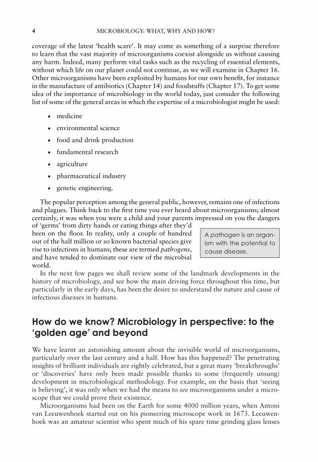
coverage of the latest ‘health scare’. It may come as something of a surprise thereforeto learn that the vast majority of microorganisms coexist alongside us without causingany harm. Indeed, many perform vital tasks such as the recycling of essential elements,without which life on our planet could not continue, as we will examine in Chapter 16.Other microorganisms have been exploited by humans for our own benefit, for instancein the manufacture of antibiotics (Chapter 14) and foodstuffs (Chapter 17). To get someidea of the importance of microbiology in the world today, just consider the followinglist of some of the general areas in which the expertise of a microbiologist might be used:
� medicine
� environmental science
� food and drink production
� fundamental research
� agriculture
� pharmaceutical industry
� genetic engineering.
The popular perception among the general public, however, remains one of infectionsand plagues. Think back to the first time you ever heard about microorganisms; almostcertainly, it was when you were a child and your parents impressed on you the dangersof ‘germs’ from dirty hands or eating things after they’dbeen on the floor. In reality, only a couple of hundredout of the half million or so known bacterial species giverise to infections in humans; these are termed pathogens,and have tended to dominate our view of the microbialworld.
A pathogen is an organ-ism with the potential tocause disease.
In the next few pages we shall review some of the landmark developments in thehistory of microbiology, and see how the main driving force throughout this time, butparticularly in the early days, has been the desire to understand the nature and cause ofinfectious diseases in humans.
How do we know? Microbiology in perspective: to the‘golden age’ and beyond
We have learnt an astonishing amount about the invisible world of microorganisms,particularly over the last century and a half. How has this happened? The penetratinginsights of brilliant individuals are rightly celebrated, but a great many ‘breakthroughs’or ‘discoveries’ have only been made possible thanks to some (frequently unsung)development in microbiological methodology. For example, on the basis that ‘seeingis believing’, it was only when we had the means to see microorganisms under a micro-scope that we could prove their existence.
Microorganisms had been on the Earth for some 4000 million years, when Antonivan Leeuwenhoek started out on his pioneering microscope work in 1673. Leeuwen-hoek was an amateur scientist who spent much of his spare time grinding glass lenses
JWBK011-01 JWBK011-Hogg August 12, 2005 15:39 Char Count= 0
HOW DO WE KNOW? MICROBIOLOGY IN PERSPECTIVE 5
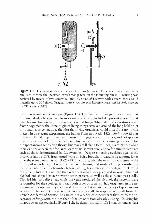
Figure 1.1 Leeuwenhoek’s microscope. The lens (a) was held between two brass platesand used to view the specimen, which was placed on the mounting pin (b). Focusing wasachieved by means of two screws (c) and (d). Some of Leeuwenhoek’s microscopes couldmagnify up to 300 times. Original source: Antony van Leeuwenhoek and his little animalsby CE Dobell (1932)
to produce simple microscopes (Figure 1.1). His detailed drawings make it clear thatthe ‘animalcules’ he observed from a variety of sources included representatives of whatlater became known as protozoa, bacteria and fungi. Where did these creatures comefrom? Arguments about the origin of living things revolved around the long held beliefin spontaneous generation, the idea that living organisms could arise from non-livingmatter. In an elegant experiment, the Italian Francesco Redi (1626–1697) showed thatthe larvae found on putrefying meat arose from eggs deposited by flies, and not sponta-neously as a result of the decay process. This can be seen as the beginning of the end forthe spontaneous generation theory, but many still clung to the idea, claiming that whileit may not have been true for larger organisms, it must surely be so for minute creaturessuch as those demonstrated by Leeuwenhoek. Despite mounting evidence against thetheory, as late as 1859, fresh ‘proof’ was still being brought forward in its support. Enteronto the scene Louis Pasteur (1822–1895), still arguably the most famous figure in thehistory of microbiology. Pasteur trained as a chemist, and made a lasting contributionto the science of stereochemistry before turning his attention to spoilage problems inthe wine industry. He noticed that when lactic acid was produced in wine instead ofalcohol, rod-shaped bacteria were always present, as well as the expected yeast cells.This led him to believe that while the yeast produced the alcohol, the bacteria wereresponsible for the spoilage, and that both types of organism had originated in the en-vironment. Exasperated by continued efforts to substantiate the theory of spontaneousgeneration, he set out to disprove it once and for all. In response to a call from theFrench Academy of Science, he carried out a series of experiments that led to the ac-ceptance of biogenesis, the idea that life arises only from already existing life. Using hisfamous swan-necked flasks (Figure 1.2), he demonstrated in 1861 that as long as dust

JWBK011-01 JWBK011-Hogg August 12, 2005 15:39 Char Count= 0
6 MICROBIOLOGY: WHAT, WHY AND HOW?
Liquid sterilisedby boiling.
Liquid allowedto cool.
Flask tilted, allowingliquid to come intocontact with deposit in neck.
Liquid turns cloudydue to microbial growth.
Dust and microrganismssettle in bend of flask neck.Liquid remains sterile.
Left formonths/year
Figure 1.2 Pasteur’s swan-necked flasks. Broth solutions rich in nutrients were placed inflasks and boiled. The necks of the flasks were heated and drawn out into a curve, butkept open to the atmosphere. Pasteur showed that the broth remained sterile because anycontaminating dust and microorganisms remained trapped in the neck of the flask as longas it remained upright
particles (and the microorganisms carried on them) were excluded, the contents wouldremain sterile. This also disproved the idea held by many that there was some element inthe air itself that was capable of initiating microbial growth. In Pasteur’s words ‘. . . . thedoctrine of spontaneous generation will never recover from this mortal blow. Thereis no known circumstance in which it can be affirmed that microscopic beings cameinto the world without germs, without parents similar to themselves.’ Pasteur’s findingson wine contamination led inevitably to the idea that microorganisms may be also beresponsible for diseases in humans, animals and plants.
The notion that some invisible (and therefore, presumably, extremely small) livingcreatures were responsible for certain diseases was not a new one. Long before micro-organisms had been shown to exist, the Roman philosopher Lucretius (∼98–55 bc) andmuch later the physician Girolamo Fracastoro (1478–1553) had supported the idea.Fracastoro wrote ‘Contagion is an infection that passes from one thing to another’ andrecognised three forms of transmission: by direct contact, through inanimate objectsand via the air. We still class transmissibility of infectious disease in much the same waytoday. The prevailing belief at the time, however, was that an infectious disease was due
JWBK011-01 JWBK011-Hogg August 12, 2005 15:39 Char Count= 0
HOW DO WE KNOW? MICROBIOLOGY IN PERSPECTIVE 7
to something called a miasma, a poisonous vapour arising from dead or diseased bodies,or to an imbalance between the four humours of the body (blood, phlegm, yellow bileand black bile). During the 19th century, many diseases were shown, one by one, to becaused by microorganisms. In 1835, Agostino Bassi showed that a disease of silkwormswas due to a fungal infection, and 10 years later, Miles Berkeley demonstrated that a fun-gus was also responsible for the great Irish potato blight. Joseph Lister’s pioneering workon antiseptic surgery provided strong, albeit indirect, evidence of the involvement of mi-croorganisms in infections of humans. The use of heat-treated instruments and of phenolboth on dressings and actually sprayed in a mist over the surgical area, was found greatlyto reduce the number of fatalities following surgery. Around the same time, in the 1860s,the indefatigable Pasteur had shown that a parasitic protozoan was the cause of anotherdisease of silkworms called pebrine, which had devastated the French silk industry.
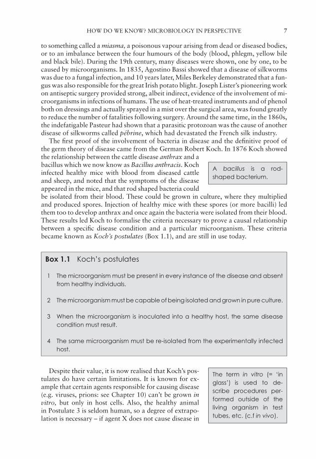
The first proof of the involvement of bacteria in disease and the definitive proof ofthe germ theory of disease came from the German Robert Koch. In 1876 Koch showed
A bacillus is a rod-shaped bacterium.
the relationship between the cattle disease anthrax and abacillus which we now know as Bacillus anthracis. Kochinfected healthy mice with blood from diseased cattleand sheep, and noted that the symptoms of the diseaseappeared in the mice, and that rod shaped bacteria couldbe isolated from their blood. These could be grown in culture, where they multipliedand produced spores. Injection of healthy mice with these spores (or more bacilli) ledthem too to develop anthrax and once again the bacteria were isolated from their blood.These results led Koch to formalise the criteria necessary to prove a causal relationshipbetween a specific disease condition and a particular microorganism. These criteriabecame known as Koch’s postulates (Box 1.1), and are still in use today.
Box 1.1 Koch’s postulates
1 The microorganism must be present in every instance of the disease and absentfrom healthy individuals.
2 The microorganism must be capable of being isolated and grown in pure culture.
3 When the microorganism is inoculated into a healthy host, the same diseasecondition must result.
4 The same microorganism must be re-isolated from the experimentally infectedhost.
The term in vitro (= ‘inglass’) is used to de-scribe procedures per-formed outside of theliving organism in testtubes, etc. (c.f in vivo).
Despite their value, it is now realised that Koch’s pos-tulates do have certain limitations. It is known for ex-ample that certain agents responsible for causing disease(e.g. viruses, prions: see Chapter 10) can’t be grown invitro, but only in host cells. Also, the healthy animalin Postulate 3 is seldom human, so a degree of extrapo-lation is necessary – if agent X does not cause disease in
JWBK011-01 JWBK011-Hogg August 12, 2005 15:39 Char Count= 0
8 MICROBIOLOGY: WHAT, WHY AND HOW?
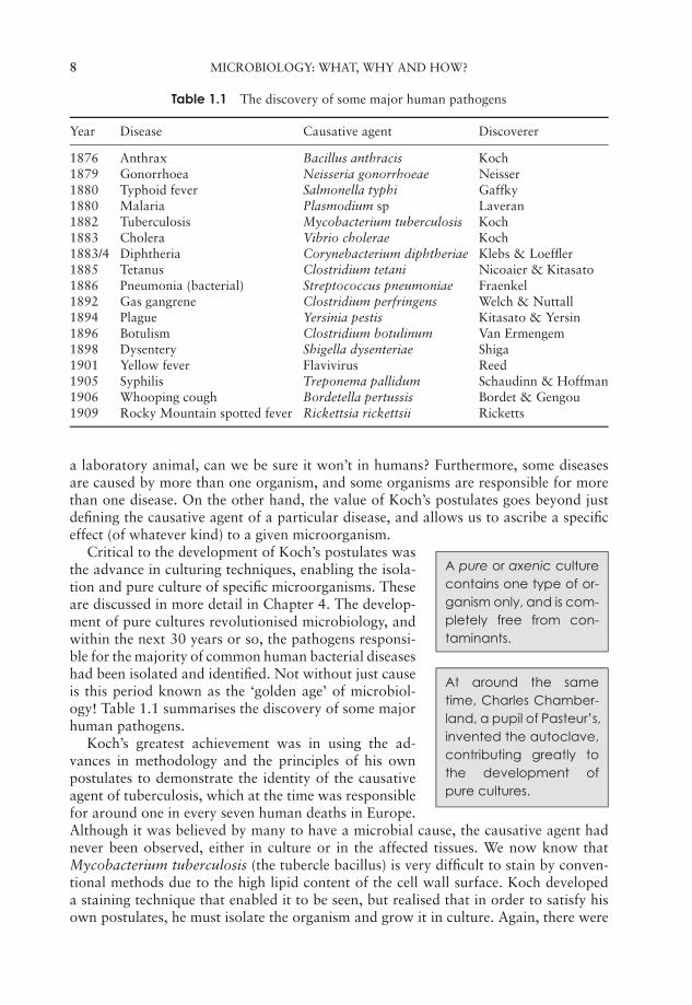
Table 1.1 The discovery of some major human pathogens
Year Disease Causative agent Discoverer
1876 Anthrax Bacillus anthracis Koch1879 Gonorrhoea Neisseria gonorrhoeae Neisser1880 Typhoid fever Salmonella typhi Gaffky1880 Malaria Plasmodium sp Laveran1882 Tuberculosis Mycobacterium tuberculosis Koch1883 Cholera Vibrio cholerae Koch1883/4 Diphtheria Corynebacterium diphtheriae Klebs & Loeffler1885 Tetanus Clostridium tetani Nicoaier & Kitasato1886 Pneumonia (bacterial) Streptococcus pneumoniae Fraenkel1892 Gas gangrene Clostridium perfringens Welch & Nuttall1894 Plague Yersinia pestis Kitasato & Yersin1896 Botulism Clostridium botulinum Van Ermengem1898 Dysentery Shigella dysenteriae Shiga1901 Yellow fever Flavivirus Reed1905 Syphilis Treponema pallidum Schaudinn & Hoffman1906 Whooping cough Bordetella pertussis Bordet & Gengou1909 Rocky Mountain spotted fever Rickettsia rickettsii Ricketts
a laboratory animal, can we be sure it won’t in humans? Furthermore, some diseasesare caused by more than one organism, and some organisms are responsible for morethan one disease. On the other hand, the value of Koch’s postulates goes beyond justdefining the causative agent of a particular disease, and allows us to ascribe a specificeffect (of whatever kind) to a given microorganism.
A pure or axenic culturecontains one type of or-ganism only, and is com-pletely free from con-taminants.
Critical to the development of Koch’s postulates wasthe advance in culturing techniques, enabling the isola-tion and pure culture of specific microorganisms. Theseare discussed in more detail in Chapter 4. The develop-ment of pure cultures revolutionised microbiology, andwithin the next 30 years or so, the pathogens responsi-ble for the majority of common human bacterial diseaseshad been isolated and identified. Not without just causeis this period known as the ‘golden age’ of microbiol-ogy! Table 1.1 summarises the discovery of some majorhuman pathogens.
At around the sametime, Charles Chamber-land, a pupil of Pasteur’s,invented the autoclave,contributing greatly tothe development ofpure cultures.
Koch’s greatest achievement was in using the ad-vances in methodology and the principles of his ownpostulates to demonstrate the identity of the causativeagent of tuberculosis, which at the time was responsiblefor around one in every seven human deaths in Europe.Although it was believed by many to have a microbial cause, the causative agent hadnever been observed, either in culture or in the affected tissues. We now know thatMycobacterium tuberculosis (the tubercle bacillus) is very difficult to stain by conven-tional methods due to the high lipid content of the cell wall surface. Koch developeda staining technique that enabled it to be seen, but realised that in order to satisfy hisown postulates, he must isolate the organism and grow it in culture. Again, there were
JWBK011-01 JWBK011-Hogg August 12, 2005 15:39 Char Count= 0
HOW DO WE KNOW? MICROBIOLOGY IN PERSPECTIVE 9
technical difficulties, since even under favourable conditions, M. tuberculosis growsslowly, but eventually Koch was able to demonstrate the infectivity of the culturedorganisms towards guinea pigs. He was then able to isolate them again from the dis-eased animal and use them to cause disease in uninfected animals, thus satisfying theremainder of his postulates.
Aetiology is the cause ororigin of a disease.
Although most bacterial diseases of humans and theiraetiological agents have now been identified, importantvariants continue to evolve and emerge. Notable exam-ples in recent times include Legionnaires’ disease, anacute respiratory infection caused by the previously unrecognised genus, Legionella,and Lyme disease, a tickborne infection first described in Connecticut, USA in the mid-1970s. Also, a newly recognised pathogen, Helicobacter pylori, has been shown to playan important (and previously unsuspected) role in the development of peptic ulcers.There still remain a few diseases that some investigators suspect are caused by bacteria,but for which no pathogen has been identified.
Following the discovery of viruses during the last decade of the 19th century (seeChapter 10), it was soon established that many diseases of plants, animals and humanswere caused by these minute, non-cellular agents.
The major achievement of the first half of the 20th century was the development ofantibiotics and other antimicrobial agents, a topic discussed in some detail in Chapter 14.Infectious diseases that previously accounted for millions of deaths became treatable bya simple course of therapy, at least in the affluent West, where such medications werereadily available.
The Human GenomeProject is an interna-tional effort to map andsequence all the DNA inthe human genome. Theproject has also involvedsequencing the geno-mes of several other or-ganisms.
If the decades either side of 1900 have become knownas the golden age of microbiology, the second half ofthe twentieth century will surely be remembered as thegolden age of molecular genetics. Following on fromthe achievements of others such as Griffith and Avery, thepublication of Watson and Crick’s structure for DNA in1953 heralded an extraordinary 50 years of achievementin this area, culminating at the turn of the 21st centuryin the completion of the Human Genome Project.
What, you might ask, has this genetic revolution todo with microbiology? Well, all the early work in molec-ular genetics was carried out on bacteria and viruses, asyou’ll learn in Chapter 11, and microbial systems have also been absolutely central tothe development of genetic engineering over the last three decades (Chapter 12). Also, aspart of the Human Genome Project, the genomes of several microorganisms have beendecoded, and it will become increasingly easy to do the same for others in the future,thanks to methodological advances made during the project. Having this informationwill help us to understand in greater detail the disease strategies of microorganisms, andto devise ways of countering them.
As we have seen, a recurring theme in the history of microbiology has been the waythat advances in knowledge have followed on from methodological or technologicaldevelopments, and we shall refer to a number of such developments during the courseof this book. To conclude this introduction to microbiology, we shall return to theinstrument that, in some respects, started it all. In any microbiology course, you are sureto spend some time looking down a microscope, and to get the most out of the instrument
JWBK011-01 JWBK011-Hogg August 12, 2005 15:39 Char Count= 0
10 MICROBIOLOGY: WHAT, WHY AND HOW?
it is essential that you understand the principles of how it works. The following pagesattempt to explain these principles.
The refractive index ofa substance is the ra-tio between the veloc-ity of light as it passesthrough that substanceand its velocity in avacuum. It is a mea-sure of how much thesubstance slows downand therefore refractsthe light.
Light microscopy
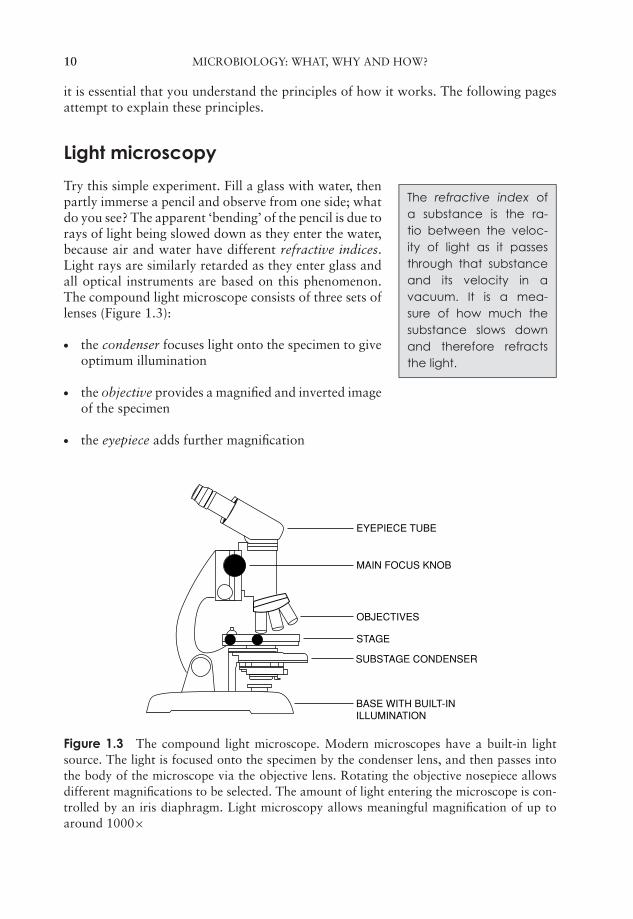
Try this simple experiment. Fill a glass with water, thenpartly immerse a pencil and observe from one side; whatdo you see? The apparent ‘bending’ of the pencil is due torays of light being slowed down as they enter the water,because air and water have different refractive indices.Light rays are similarly retarded as they enter glass andall optical instruments are based on this phenomenon.The compound light microscope consists of three sets oflenses (Figure 1.3):
� the condenser focuses light onto the specimen to giveoptimum illumination
� the objective provides a magnified and inverted imageof the specimen
� the eyepiece adds further magnification
EYEPIECE TUBE
MAIN FOCUS KNOB
OBJECTIVES
STAGE
BASE WITH BUILT-INILLUMINATION
SUBSTAGE CONDENSER
Figure 1.3 The compound light microscope. Modern microscopes have a built-in lightsource. The light is focused onto the specimen by the condenser lens, and then passes intothe body of the microscope via the objective lens. Rotating the objective nosepiece allowsdifferent magnifications to be selected. The amount of light entering the microscope is con-trolled by an iris diaphragm. Light microscopy allows meaningful magnification of up toaround 1000×
JWBK011-01 JWBK011-Hogg August 12, 2005 15:39 Char Count= 0
LIGHT MICROSCOPY 11
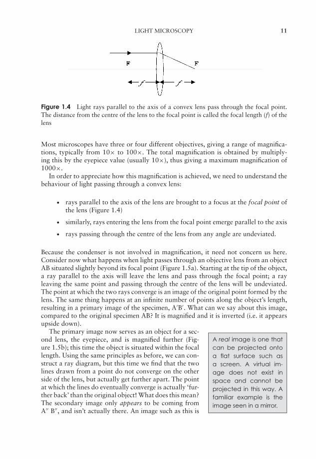
Figure 1.4 Light rays parallel to the axis of a convex lens pass through the focal point.The distance from the centre of the lens to the focal point is called the focal length (f) of thelens
Most microscopes have three or four different objectives, giving a range of magnifica-tions, typically from 10× to 100×. The total magnification is obtained by multiply-ing this by the eyepiece value (usually 10×), thus giving a maximum magnification of1000×.
In order to appreciate how this magnification is achieved, we need to understand thebehaviour of light passing through a convex lens:
� rays parallel to the axis of the lens are brought to a focus at the focal point ofthe lens (Figure 1.4)
� similarly, rays entering the lens from the focal point emerge parallel to the axis
� rays passing through the centre of the lens from any angle are undeviated.
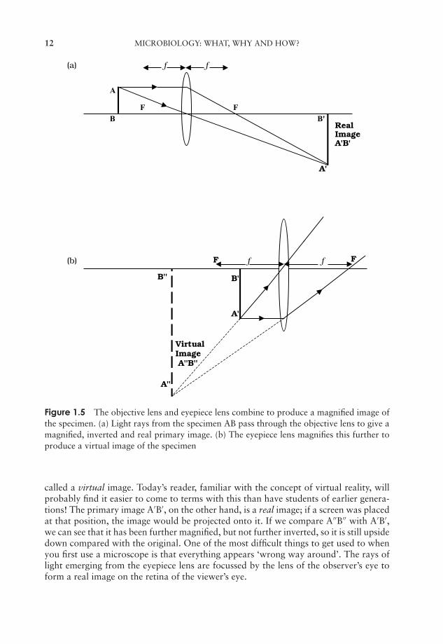
Because the condenser is not involved in magnification, it need not concern us here.Consider now what happens when light passes through an objective lens from an objectAB situated slightly beyond its focal point (Figure 1.5a). Starting at the tip of the object,a ray parallel to the axis will leave the lens and pass through the focal point; a rayleaving the same point and passing through the centre of the lens will be undeviated.The point at which the two rays converge is an image of the original point formed by thelens. The same thing happens at an infinite number of points along the object’s length,resulting in a primary image of the specimen, A′B′. What can we say about this image,compared to the original specimen AB? It is magnified and it is inverted (i.e. it appearsupside down).
A real image is one thatcan be projected ontoa flat surface such asa screen. A virtual im-age does not exist inspace and cannot beprojected in this way. Afamiliar example is theimage seen in a mirror.
The primary image now serves as an object for a sec-ond lens, the eyepiece, and is magnified further (Fig-ure 1.5b); this time the object is situated within the focallength. Using the same principles as before, we can con-struct a ray diagram, but this time we find that the twolines drawn from a point do not converge on the otherside of the lens, but actually get further apart. The pointat which the lines do eventually converge is actually ‘fur-ther back’ than the original object! What does this mean?The secondary image only appears to be coming fromA′′ B′′, and isn’t actually there. An image such as this is
JWBK011-01 JWBK011-Hogg August 12, 2005 15:39 Char Count= 0
12 MICROBIOLOGY: WHAT, WHY AND HOW?
(a) f f
A
F FB B'
RealImageA'B'
A'
(b) F f f F
B'' B'
A'
VirtualImage A''B''
A''
Figure 1.5 The objective lens and eyepiece lens combine to produce a magnified image ofthe specimen. (a) Light rays from the specimen AB pass through the objective lens to give amagnified, inverted and real primary image. (b) The eyepiece lens magnifies this further toproduce a virtual image of the specimen
called a virtual image. Today’s reader, familiar with the concept of virtual reality, willprobably find it easier to come to terms with this than have students of earlier genera-tions! The primary image A′B′, on the other hand, is a real image; if a screen was placedat that position, the image would be projected onto it. If we compare A′′B′′ with A′B′,we can see that it has been further magnified, but not further inverted, so it is still upsidedown compared with the original. One of the most difficult things to get used to whenyou first use a microscope is that everything appears ‘wrong way around’. The rays oflight emerging from the eyepiece lens are focussed by the lens of the observer’s eye toform a real image on the retina of the viewer’s eye.
JWBK011-01 JWBK011-Hogg August 12, 2005 15:39 Char Count= 0
LIGHT MICROSCOPY 13
(a)
(b)
(c)
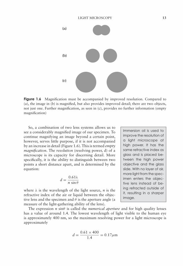
Figure 1.6 Magnification must be accompanied by improved resolution. Compared to(a), the image in (b) is magnified, but also provides improved detail; there are two objects,not just one. Further magnification, as seen in (c), provides no further information (emptymagnification)
Immersion oil is used toimprove the resolution ofa light microscope athigh power. It has thesame refractive index asglass and is placed be-tween the high powerobjective and the glassslide. With no layer of air,more light from the spec-imen enters the objec-tive lens instead of be-ing refracted outside ofit, resulting in a sharperimage.
So, a combination of two lens systems allows us tosee a considerably magnified image of our specimen. Tocontinue magnifying an image beyond a certain point,however, serves little purpose, if it is not accompaniedby an increase in detail (Figure 1.6). This is termed emptymagnification. The resolution (resolving power, d) of amicroscope is its capacity for discerning detail. Morespecifically, it is the ability to distinguish between twopoints a short distance apart, and is determined by theequation:
d = 0.61λ
n sin θ
where λ is the wavelength of the light source, n is therefractive index of the air or liquid between the objec-tive lens and the specimen and θ is the aperture angle (ameasure of the light-gathering ability of the lens).
The expression n sinθ is called the numerical aperture and for high quality lenseshas a value of around 1.4. The lowest wavelength of light visible to the human eyeis approximately 400 nm, so the maximum resolving power for a light microscope isapproximately
d = 0.61 × 4001.4
= 0.17µm
JWBK011-01 JWBK011-Hogg August 12, 2005 15:39 Char Count= 0
14 MICROBIOLOGY: WHAT, WHY AND HOW?
that is, it cannot distinguish between two points closertogether than about 0.2 µm. For comparison, the nakedeye is unable to resolve two points more than about0.2 mm apart.
A nanometre (nm) is 1millionth of a millimetre.There are 1000 nm inone micron (µm), whichis therefore one thou-sandth of a millimetre.1 mm = 10−3 m1 µm = 10−6 m1 nm = 10−9 m
For us to be able to discern detail in a specimen, itmust have contrast; most biological specimens, however,are more or less colourless, so unless a structure is ap-preciably denser than its surroundings, it will not standout. This is why preparations are commonly subjectedto staining procedures prior to viewing. The introduc-tion of coloured dyes, which bind to certain structures,enables the viewer to discern more detail.
Since staining procedures involve the addition and washing off of liquid stains, thesample must clearly be immobilised or fixed to the slide if it is not to end up down thesink. The commonest way of doing this is to make a heat-fixed smear; this kills andattaches the cells to the glass microscope slide. A thin aqueous suspension of the cells isspread across the slide, allowed to dry, then passed (sample side up!) through a flame afew times. Excessive heating must be avoided, as it would distort the natural structureof the cells.
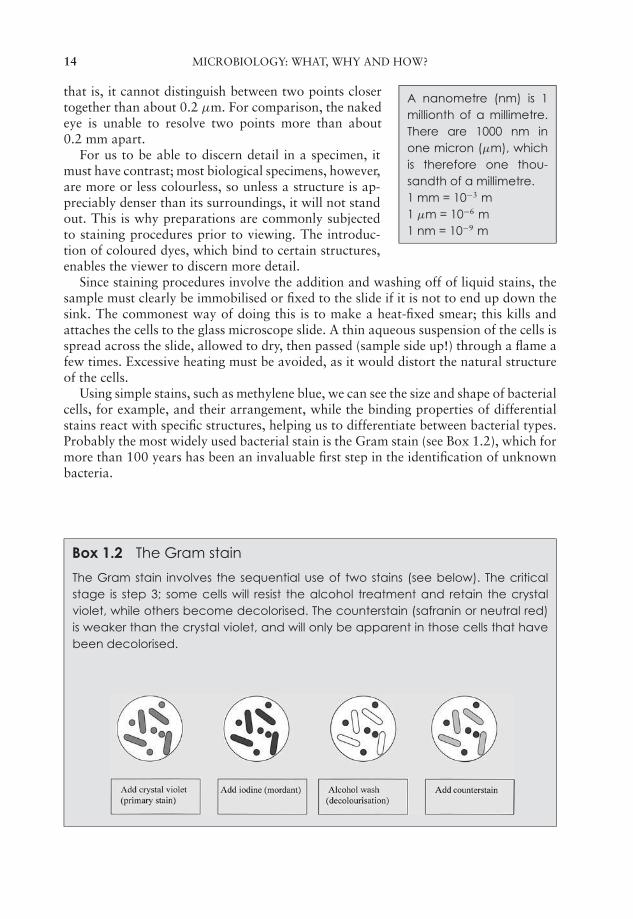
Using simple stains, such as methylene blue, we can see the size and shape of bacterialcells, for example, and their arrangement, while the binding properties of differentialstains react with specific structures, helping us to differentiate between bacterial types.Probably the most widely used bacterial stain is the Gram stain (see Box 1.2), which formore than 100 years has been an invaluable first step in the identification of unknownbacteria.
Box 1.2 The Gram stain
The Gram stain involves the sequential use of two stains (see below). The criticalstage is step 3; some cells will resist the alcohol treatment and retain the crystalviolet, while others become decolorised. The counterstain (safranin or neutral red)is weaker than the crystal violet, and will only be apparent in those cells that havebeen decolorised.

JWBK011-01 JWBK011-Hogg August 12, 2005 15:39 Char Count= 0
ELECTRON MICROSCOPY 15
Phase contrast micro-scopy exploits differ-ences in thickness andrefractive index of trans-parent objects such asliving cells to give im-proved contrast.
Dark field microscopyemploys a modifiedcondenser. It works byblocking out direct light,and viewing the ob-ject only by the light itdiffracts.
The Gram stain is a differential stain, which only takesa few minutes to carry out, and which enables us toplace a bacterial specimen into one of two groups, Gram-positive or Gram-negative. The reason for this differen-tial reaction to the stain was not understood for manyyears, but is now seen to be a reflection of differences incell wall structure, discussed in more detail in Chapter 3.
Specialised forms of microscopy have been developedto allow the viewer to discern detail in living, unstainedspecimens; these include phase contrast and dark-fieldmicroscopy. We can also gain an estimate of the numberof microorganisms in a sample by directly counting themunder the microscope. This is discussed along with otherenumeration methods in Chapter 5.
Electron microscopy
From the equation shown above, you can see that if it were possible to use a shorter wave-length of light, we could improve the resolving power of a microscope. However, becausewe are limited by the wavelength of light visible to the human eye, we are not able todo this with the light microscope. The electron microscope is able to achieve greatermagnification and resolution because it uses a high voltage beam of electrons, whosewavelength is very much shorter than that of visible light. Consequently we are able toresolve points that are much closer together than is possible even with the very best lightmicroscope. The resolving power of an electron microscope may be as low as 1–2 nm,enabling us to see viruses, for example, and the internal structure of cells. The greatly im-proved resolution means that specimens can be meaningfully magnified over 100 000×.
Electron microscopes, which were first developed in the 1930s and 1940s, use ring-shaped electromagnets as ‘lenses’ to focus the beam of electrons onto the specimen.Because the electrons would collide with, and be deflected by, molecules in the air,electron microscopes require a pump to maintain a vacuum in the column of the instru-ment. There are two principal types of electron microscope, the transmission electronmicroscope (TEM) and the scanning electron microscope (SEM).
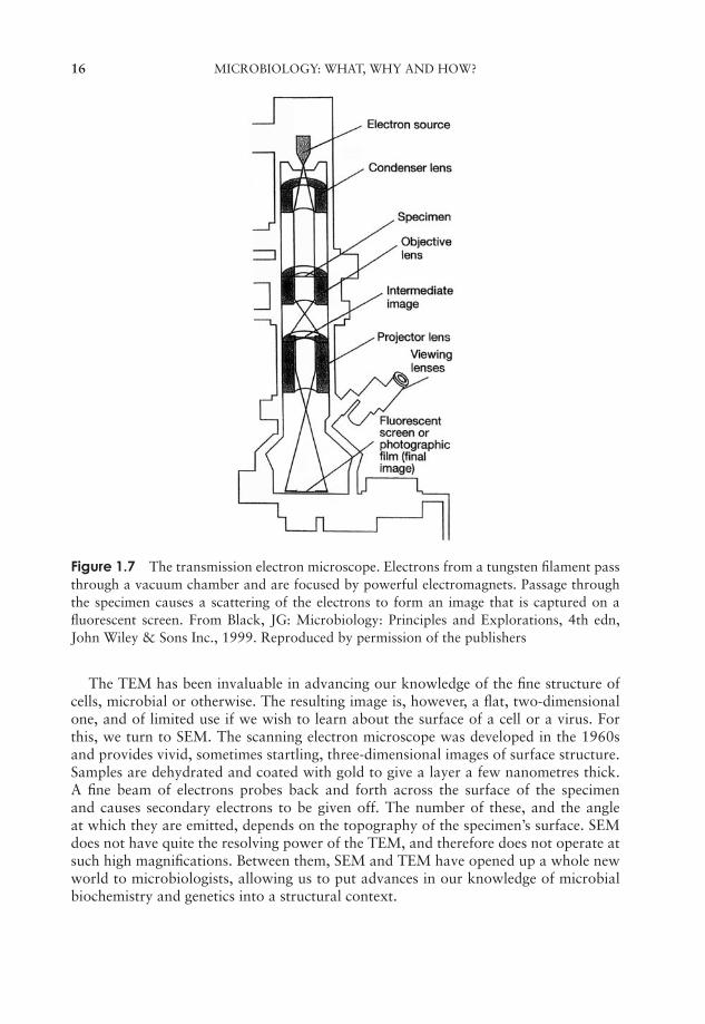
Figure 1.7 shows the main features of a TEM. As the name suggests, the electron beampasses through the specimen and is scattered according to the density of the differentparts. Due to the limited penetrating power of the electrons, extremely thin sections(<100 nm, or less than one-tenth of the diameter of a bacterial cell) must be cut, usinga diamond knife. To allow this, the specimen must be fixed and dehydrated, a processthat can introduce shrinkage and distortion to its structure if not correctly performed.
After being magnified by an objective ‘lens’, an image of the specimen is projectedonto a fluorescent screen or photographic plate. More dense areas, which scatter thebeam, appear dark, and those where it has passed through are light. It is often necessaryto enhance contrast artificially, by means of ‘staining’ techniques that involve coatingthe specimen with a thin layer of a compound containing a heavy metal, such as osmiumor palladium. It will be evident from the foregoing description of sample preparationand use of a vacuum that electron microscopy cannot be used to study living specimens.
JWBK011-01 JWBK011-Hogg August 12, 2005 15:39 Char Count= 0
16 MICROBIOLOGY: WHAT, WHY AND HOW?
Figure 1.7 The transmission electron microscope. Electrons from a tungsten filament passthrough a vacuum chamber and are focused by powerful electromagnets. Passage throughthe specimen causes a scattering of the electrons to form an image that is captured on afluorescent screen. From Black, JG: Microbiology: Principles and Explorations, 4th edn,John Wiley & Sons Inc., 1999. Reproduced by permission of the publishers
The TEM has been invaluable in advancing our knowledge of the fine structure ofcells, microbial or otherwise. The resulting image is, however, a flat, two-dimensionalone, and of limited use if we wish to learn about the surface of a cell or a virus. Forthis, we turn to SEM. The scanning electron microscope was developed in the 1960sand provides vivid, sometimes startling, three-dimensional images of surface structure.Samples are dehydrated and coated with gold to give a layer a few nanometres thick.A fine beam of electrons probes back and forth across the surface of the specimenand causes secondary electrons to be given off. The number of these, and the angleat which they are emitted, depends on the topography of the specimen’s surface. SEMdoes not have quite the resolving power of the TEM, and therefore does not operate atsuch high magnifications. Between them, SEM and TEM have opened up a whole newworld to microbiologists, allowing us to put advances in our knowledge of microbialbiochemistry and genetics into a structural context.
JWBK011-02 JWBK011-Hogg August 12, 2005 19:26 Char Count= 0
2Biochemical Principles
All matter, whether living or non-living, is made up of atoms; the atom is the smallestunit of matter capable of entering into a chemical reaction. Atoms can combine togetherby bonding, to form molecules, which range from the small and simple to the large andcomplex. The latter are known as macromolecules; major cellular constituents such ascarbohydrates and proteins belong to this group and it is with these that this chapter ismainly concerned (Table 2.1). In order to appreciate how these macromolecules operatein the structure and function of microbial cells however, we need to review the basicprinciples of how atoms are constructed and how they interact with one other.
Atomic structure
All atoms have a central, positively charged nucleus, which is very dense, and makesup most of the mass of the atom. The nucleus is made up of two types of particle,protons and neutrons. Protons carry a positive charge, and neutrons are uncharged,hence the nucleus overall is positively charged. It is surrounded by much lighter, andrapidly orbiting, electrons (Figure 2.1). These are negatively charged, the charge beingequal (but of course opposite) to that of the protons, but they have only 1/1840 of themass of either protons or neutrons. The attractive force between the positively chargedprotons and the negatively charged electrons holds the atom together.
The number of protons in the nucleus is called the atomic number, and ranges from 1to over 100. The combined total of protons and neutrons is known as the mass number.All atoms have an equal number of protons and electrons, so regardless of the atomicnumber, the overall charge on the atom will always be zero.
Atoms having the same atomic number have the same chemical properties; suchatoms all belong to the same element. An element is made up of one type of atom onlyand cannot be chemically broken down into simpler substances; thus pure copper forexample is made up entirely of copper atoms. There are 92 of these elements occurringnaturally, 26 of which commonly occur in living things. Each element has been givena universally agreed symbol; examples which we shall encounter in biological macro-molecules include carbon (C), hydrogen (H) and oxygen (O). The atomic numbers ofselected elements are shown in Table 2.2.
The relationship between neutrons, protons, atomic number, and mass number isillustrated in Table 2.3, using carbon as an example, since all living matter is based
17
JWBK011-02 JWBK011-Hogg August 12, 2005 19:26 Char Count= 0
18 BIOCHEMICAL PRINCIPLES
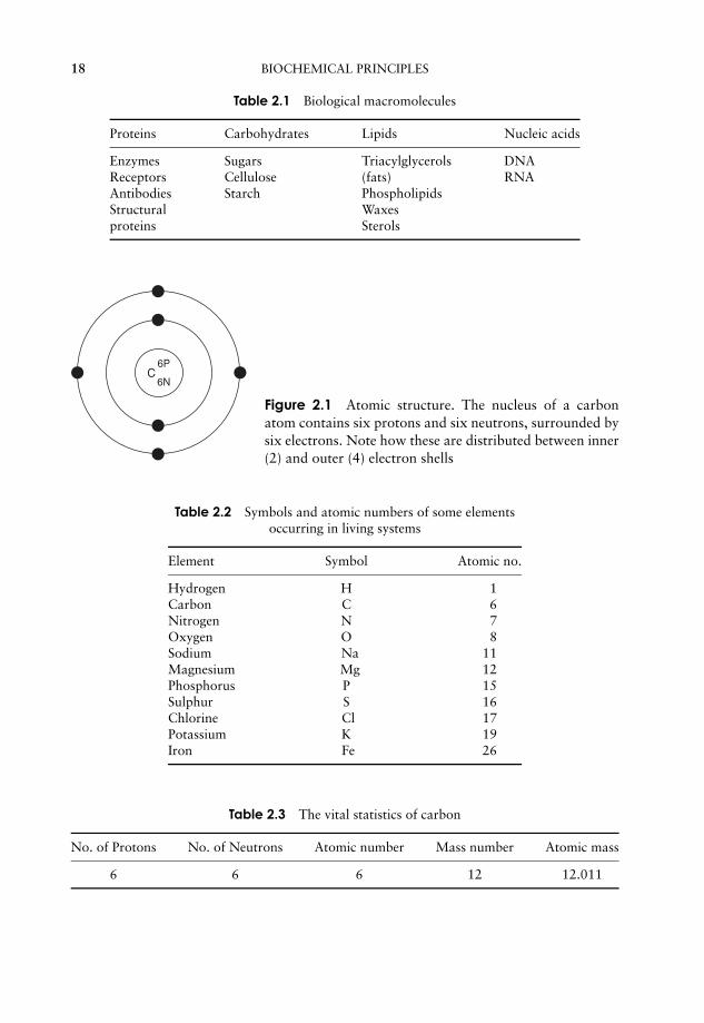
Table 2.1 Biological macromolecules
Proteins Carbohydrates Lipids Nucleic acids
Enzymes Sugars Triacylglycerols DNAReceptors Cellulose (fats) RNAAntibodies Starch PhospholipidsStructural Waxesproteins Sterols
C6P
6N
Figure 2.1 Atomic structure. The nucleus of a carbonatom contains six protons and six neutrons, surrounded bysix electrons. Note how these are distributed between inner(2) and outer (4) electron shells
Table 2.2 Symbols and atomic numbers of some elementsoccurring in living systems
Element Symbol Atomic no.
Hydrogen H 1Carbon C 6Nitrogen N 7Oxygen O 8Sodium Na 11Magnesium Mg 12Phosphorus P 15Sulphur S 16Chlorine Cl 17Potassium K 19Iron Fe 26
Table 2.3 The vital statistics of carbon
No. of Protons No. of Neutrons Atomic number Mass number Atomic mass
6 6 6 12 12.011

JWBK011-02 JWBK011-Hogg August 12, 2005 19:26 Char Count= 0
ATOMIC STRUCTURE 19
upon this element. The carbon represented can be expressed in the form:
12 (mass number)
C = (Element symbol)6 (atomic number)
The number of neutrons in an atom can be deduced by subtracting the atomic numberfrom the mass number. In the case of carbon, this is the same as the number of protons(6), but this is not always so. Phosphorus for example has 15 protons and 16 neutrons,giving it an atomic number of 15 and a mass number of 31.
Isotopes
Although the number of protons in the nucleus of a given element is always the same,the number of neutrons can vary, to give different forms or isotopes of that element.Carbon-14 (14C) is a naturally occurring but rare isotope of carbon that has eight neu-trons instead of six, hence the atomic mass of 14. Carbon-13 (13C) is a rather more com-mon isotope, making up around 1 per cent of naturally occurring carbon; it has sevenneutrons per atomic nucleus. The atomic mass (or atomic weight) of an element is theaverage of the mass numbers of an element’s different isotopes, taking into accountthe proportions in which they occur. (Box 2.1 shows how atomic weight is used toquantify amounts of compounds using moles.) Carbon-12 is by far the predominantform of the element in nature, but the existence of small amounts of the other formsmeans that the atomic mass is 12.011. Some isotopes are stable, while others decay spon-taneously, with the release of subatomic particles. The latter are called radioisotopes; 14Cis a radioisotope, while the other two forms of carbon are stable isotopes. Radioisotopeshave been an extremely useful research tool in a number of areas of molecular biology.
The electrons that orbit around the nucleus do not do so randomly, but are arrangedin a series of electron shells, radiating out from the nucleus (Figure 2.1). These layerscorrespond to different energy levels, with the highest energy levels being located furthestaway from the nucleus. Each shell can accommodate a maximum number of electrons,and electrons always fill up the shells starting at the innermost one, that is, the onewith the lowest energy level. In our example, carbon has filled the first shell with twoelectrons, and occupied four of the eight available spaces on the second.
The chemical properties of atoms are determined by the number of electrons in theoutermost occupied shell. Neon, one of the ‘noble’ gases, has an atomic number of10, completely filling the first two shells, and is chemically unreactive or inert. Atomsthat do not achieve a similar configuration are unstable, or reactive. Reactions takeplace between atoms that attempt to achieve stability by attaining a full outer shell.These reactions may involve atoms of the same element or ones of different elements;the result in either case is a molecule or ion (see below). Figure 2.2 shows how atomscombine to form a molecule. A substance made up of molecules containing two or moredifferent elements is called a compound. In each example, the product of the reactionhas a full outer electron shell; note that some atoms are donating electrons, while othersare accepting them.
The number of unfilled spaces in the outermost electron shell determines the reactivityof an atom. If most of the spaces in the outermost shell are full, or if most are empty,atoms tend to strive for stability by gaining or losing electrons, as shown in Figure 2.3.
JWBK011-02 JWBK011-Hogg August 12, 2005 19:26 Char Count= 0
20 BIOCHEMICAL PRINCIPLES
Box 2.1 How heavy is a mole?
When you work in a laboratory, something you’llneed to come to grips with sooneror later is the matter of quantifying the amounts and concentrations of substancesused. Central to this is the mole, so before we go any further, let’sdefine this:
A mole is the molecular mass of a compound expressed in grams.(The molecular mass is simply the sum of the atomic mass of all the atoms in a
compound.)
So, to take sodium chloride as an example:
Molecular formula = NaCl (one atom each of sodiumand chlorine)
Atomic mass of sodium = 22.99Atomic mass of chlorine = 35.45∴ Molecular mass = 58.44
Thus one mole of sodium chloride equals 58.44 grams (58.44 g)
Concentrations are expressed in terms of mass per volume, so here we introducethe idea of the molar solution. This is a solution containing one mole dissolved in afinal volume of 1 litre of an appropriate solvent (usually water).
Molar solution = one mole per litre
A one molar (1 M) solution of sodium chloride therefore contains 58.44 g dissolved inwater and made up to 1 litre. A 2 M solution would contain 116.88 g in a litre, and soon.
In biological systems, a molar solution of anything is actually rather concentrated,so we tend to deal in solutions which are so many millimolar (mM, one thousandthof a mole per litre) or micromolar (µM, one millionth of a mole per litre).
Why bother with moles?
So far, so good, but why can’t we just deal in grams, or grams per litre? Considerthe following example. You’vebeen let loose in the laboratory, and been asked tocompare the effects of supplementing the growth medium of a bacterial culturewith several different amino acids. ‘Easy’,you think. ‘Add X milligrams of each to thenormal growth medium, and see which stimulates growth the most’. The problem isthat although you may be adding the same weight of each amino acid, you’re notadding the same number of molecules, because each has a different molecularmass. If you add the same number of moles (or millimoles or micromoles) of eachinstead, you would be comparing the effect of the same number of molecules ofeach, and thus obtain a much more meaningful comparison. This is because 1 moleof one compound contains the same number of molecules as a mole of any othercompound. This number is called Avogadro’sNumber, and is 6.023 × 10²³ moleculesper mole.
JWBK011-02 JWBK011-Hogg August 12, 2005 19:26 Char Count= 0
ATOMIC STRUCTURE 21
C H
H
H
H
HH HH
CH H
H
H
Hydrogen atom
Carbon atom
Hydrogen atom
Hydrogen atoms Methane molecule
Hydrogen molecule
+
+
(a)
(b)
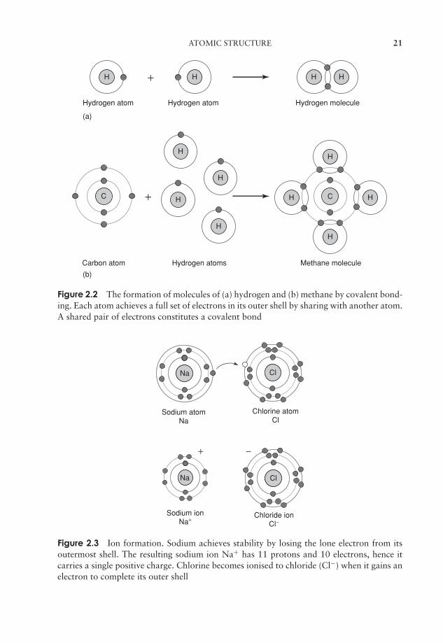
Figure 2.2 The formation of molecules of (a) hydrogen and (b) methane by covalent bond-ing. Each atom achieves a full set of electrons in its outer shell by sharing with another atom.A shared pair of electrons constitutes a covalent bond
NaNa NaCl
NaCl
Sodium atom Na
Sodium ion Na+
Chloride ion Cl−
Chlorine atom Cl
NaNa
+ −
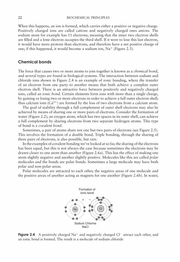
Figure 2.3 Ion formation. Sodium achieves stability by losing the lone electron from itsoutermost shell. The resulting sodium ion Na+ has 11 protons and 10 electrons, hence itcarries a single positive charge. Chlorine becomes ionised to chloride (Cl−) when it gains anelectron to complete its outer shell
JWBK011-02 JWBK011-Hogg August 12, 2005 19:26 Char Count= 0
22 BIOCHEMICAL PRINCIPLES
When this happens, an ion is formed, which carries either a positive or negative charge.Positively charged ions are called cations and negatively charged ones anions. Thesodium atom for example has 11 electrons, meaning that the inner two electron shellsare filled and a lone electron occupies the third shell. If it were to lose this last electron,it would have more protons than electrons, and therefore have a net positive charge ofone; if this happened, it would become a sodium ion, Na+ (Figure 2.3).
Chemical bonds
The force that causes two or more atoms to join together is known as a chemical bond,and several types are found in biological systems. The interaction between sodium andchloride ions shown in Figure 2.4 is an example of ionic bonding, where the transferof an electron from one party to another means that both achieve a complete outerelectron shell. There is an attractive force between positively and negatively chargedions, called an ionic bond. Certain elements form ions with more than a single charge,by gaining or losing two or more electrons in order to achieve a full outer electron shell;thus calcium ions (Ca2+) are formed by the loss of two electrons from a calcium atom.
The goal of stability through a full complement of outer shell electrons may also beachieved by means of sharing one or more pairs of electrons. Consider the formation ofwater (Figure 2.2); an oxygen atom, which has two spaces in its outer shell, can achievea full complement by sharing electrons from two separate hydrogen atoms. This typeof bond is a covalent bond.
Sometimes, a pair of atoms share not one but two pairs of electrons (see Figure 2.5).This involves the formation of a double bond. Triple bonding, through the sharing ofthree pairs of electrons, is also possible, but rare.
In the examples of covalent bonding we’ve looked at so far, the sharing of the electronshas been equal, but this is not always the case because sometimes the electrons may bedrawn closer to one atom than another (Figure 2.6a). This has the effect of making oneatom slightly negative and another slightly positive. Molecules like this are called polarmolecules and the bonds are polar bonds. Sometimes a large molecule may have bothpolar and non-polar areas.
Polar molecules are attracted to each other, the negative areas of one molecule andthe positive areas of another acting as magnets for one another (Figure 2.6b). In water,
Na NaCl
Sodium Chlorine NaCl
Formation of ionic bond
+ −
Figure 2.4 A positively charged Na+ and negatively charged Cl− attract each other, andan ionic bond is formed. The result is a molecule of sodium chloride
JWBK011-02 JWBK011-Hogg August 12, 2005 19:26 Char Count= 0
ATOMIC STRUCTURE 23
O C O (Carbon dioxide)
O C O
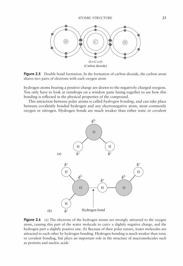
Figure 2.5 Double bond formation. In the formation of carbon dioxide, the carbon atomshares two pairs of electrons with each oxygen atom
hydrogen atoms bearing a positive charge are drawn to the negatively charged oxygens.You only have to look at raindrops on a window pane fusing together to see how thisbonding is reflected in the physical properties of the compound.
This attraction between polar atoms is called hydrogen bonding, and can take placebetween covalently bonded hydrogen and any electronegative atom, most commonlyoxygen or nitrogen. Hydrogen bonds are much weaker than either ionic or covalent
O
O
H H
H
H
H O
H
δ2–
δ2– δ2–
δ+
δ+ δ+
δ+
δ+
δ+(a)
(b) Hydrogen bond
Figure 2.6 (a) The electrons of the hydrogen atoms are strongly attracted to the oxygenatom, causing this part of the water molecule to carry a slightly negative charge, and thehydrogen part a slightly positive one. (b) Because of their polar nature, water molecules areattracted to each other by hydrogen bonding. Hydrogen bonding is much weaker than ionicor covalent bonding, but plays an important role in the structure of macromolecules suchas proteins and nucleic acids

JWBK011-02 JWBK011-Hogg August 12, 2005 19:26 Char Count= 0
24 BIOCHEMICAL PRINCIPLES
bonds; however, if sufficient of them form in a compound the overall bonding force canbe appreciable. Each water molecule can form hydrogen bonds with others of its kindin four places (Figure 2.6b). In order to break all these bonds, a large input of energy isrequired, explaining why water has such a relatively high boiling point, and why mostof the water on the planet is in liquid form.
Another weak form of interaction is brought about by Van der Waals forces, whichoccur briefly when two non-polar molecules (or parts of molecules) come into very closecontact with one another. Although transient, and generally even weaker than hydrogenbonds, they occur in great numbers in certain macromolecules and play an importantrole in holding proteins together (see below).
Water is essential for living things, both in the composition of their cells and in theenvironment surrounding them. Organisms are made up of between 60 and 95 per centwater by weight, and even inert, dormant forms like spores and seeds have a significantwater component. This dependence on water is a function of its unique properties,which in turn derive from its polar nature.
Water is the medium in which most biochemical reactions take place; it is a highlyefficient solvent, indeed more substances will dissolve in water than in any other sol-vent. Substances held together by ionic bonds tend to dissociate into anions and cationsin water, because as individual solute molecules become surrounded by molecules ofwater, hydration shells are formed, in which the negatively charged parts of the soluteattract the positive region of the water molecule, and the positive parts the negativeregion (Figure 2.7). The attractive forces that allow the solute to dissolve are called hy-drophilic forces, and substances which are water-soluble are hydrophilic (water-loving).Other polar substances such as sugars and proteins are also soluble in water by forminghydrophilic interactions.
O
OH H
H Hδ2–
δ+ δ+
Na+
Cl–
Figure 2.7 An ionic compound such as sodium chloride dissociates in water to its con-stituent ions. Water molecules form hydration shells around both Na+ and Cl− ions
Molecules such as oils and fats are non-polar, and because of their non-reactivitywith water are termed hydrophobic (‘water-fearing’). If such a molecule is mixed withwater, it will be excluded, as water molecules ‘stick together’. This very exclusion bywater can act as a cohesive force among hydrophobic molecules (or hydrophobic areasof large molecules). This is often called hydrophobic bonding, but is not really bondingas such, rather a shared avoidance of water. All living cells have a hydrophilic interiorsurrounded by a hydrophobic membrane, as we will see in Chapter 3.
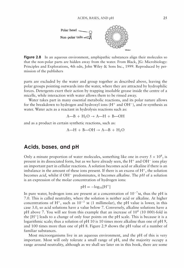
An amphipathic substance is one which is part polar and part non-polar. Whensuch a substance is mixed with water, micelles are formed (Figure 2.8); the non-polar
JWBK011-02 JWBK011-Hogg August 12, 2005 19:26 Char Count= 0
ACIDS, BASES, AND pH 25
Figure 2.8 In an aqueous environment, amphipathic substances align their molecules sothat the non-polar parts are hidden away from the water. From Black, JG: Microbiology:Principles and Explorations, 4th edn, John Wiley & Sons Inc., 1999. Reproduced by per-mission of the publishers
parts are excluded by the water and group together as described above, leaving thepolar groups pointing outwards into the water, where they are attracted by hydrophilicforces. Detergents exert their action by trapping insoluble grease inside the centre of amicelle, while interaction with water allows them to be rinsed away.
Water takes part in many essential metabolic reactions, and its polar nature allowsfor the breakdown to hydrogen and hydroxyl ions (H+ and OH−), and re-synthesis aswater. Water acts as a reactant in hydrolysis reactions such as:
A—B + H2O → A—H + B—OH
and as a product in certain synthetic reactions, such as:
A—H + B—OH → A—B + H2O
Acids, bases, and pH
Only a minute proportion of water molecules, something like one in every 5 × 10⁸, ispresent in its dissociated form, but as we have already seen, the H+ and OH− ions playan important part in cellular reactions. A solution becomes acid or alkaline if there is animbalance in the amount of these ions present. If there is an excess of H+, the solutionbecomes acid, whilst if OH− predominates, it becomes alkaline. The pH of a solutionis an expression of the molar concentration of hydrogen ions:
pH = −log10[H+]
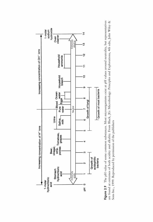
In pure water, hydrogen ions are present at a concentration of 10−7m, thus the pH is7.0. This is called neutrality, where the solution is neither acid or alkaline. At higherconcentrations of H+, such as 10−3 m (1 millimolar), the pH value is lower, in thiscase 3.0, so acid solutions have a value below 7. Conversely, alkaline solutions have apH above 7. You will see from this example that an increase of 104 (10 000)-fold inthe [H+] leads to a change of only four points on the pH scale. This is because it is alogarithmic scale; thus a solution of pH 10 is 10 times more alkaline than one of pH 9,and 100 times more than one of pH 8. Figure 2.9 shows the pH value of a number offamiliar substances.
Most microorganisms live in an aqueous environment, and the pH of this is veryimportant. Most will only tolerate a small range of pH, and the majority occupy arange around neutrality, although as we shall see later on in this book, there are some
JWBK011-02 JWBK011-Hogg August 12, 2005 19:26 Char Count= 0
Ext
rem
ely
acid
icE
xtre
mel
yal
kalin
eN
eutr
al
Fig
ure
2.9
The
pHva
lue
ofso
me
com
mon
subs
tanc
es.M
ost
mic
roor
gani
sms
exis
tat
pHva
lues
arou
ndne
utra
lity,
but
repr
esen
tati
ves
are
foun
dat
extr
emes
ofbo
thac
idit
yan
dal
kalit
y.Fr
omB
lack
,JG
:M
icro
biol
ogy:
Prin
cipl
esan
dE
xplo
rati
ons,
4th
edn,
John
Wile
y&
Sons
Inc.
,199
9.R
epro
duce
dby
perm
issi
onof
the
publ
ishe
rs
26

JWBK011-02 JWBK011-Hogg August 12, 2005 19:26 Char Count= 0
BIOMACROMOLECULES 27
Table 2.4 Occurrence and characteristics of some functional groups
Functional Type ofGroup Formula molecule Found in: Remarks
Hydroxyl -OH Alcohols Sugars Polar group, makingorganic moleculesmore water soluble
Carbonyl Aldehydes Sugars Carbonyl at end ofchain
Ketones Sugars Carbonyl elsewhere inchain
Carboxyl -COOH Carboxylic acids Sugars, fats,amino acids
Amino -NH2 Amines Amino acids,proteins
Can gain H+to becomeNH+
3Sulphhydryl -SH Thiols Amino acids,
proteinsOxidises to give
S=S bondsPhosphate Phospholipids
nucleic acidsInvolved in energy
transfer
startling exceptions to this. Most of the important molecules involved in the chemistryof living cells are organic, that is, they are based on a skeleton of covalently linkedcarbon atoms. Biological molecules have one or more functional groups attached tothis skeleton; these are groupings of atoms with distinctive reactive properties, and areresponsible for many of the chemical properties of the organic molecule. The possessionof a functional group(s) frequently makes an organic molecule more polar and thereforemore soluble in water.
Some of the most common functional groups are shown in Table 2.4. It can beseen that the functional groups occur in simpler organic molecules as well as in themacromolecules we consider below.
Biomacromolecules
Many of the most important molecules in biological systems are polymers, that is, largemolecules made up of smaller subunits joined together by covalent bonds, and in somecases in a specific order.
Carbohydrates
The suffix -ose always de-notes a carbohydrate.
Carbohydrates are made up of just three different ele-ments, carbon, hydrogen and oxygen. The simplest car-bohydrates are monosaccharides, or simple sugars; these

JWBK011-02 JWBK011-Hogg August 12, 2005 19:26 Char Count= 0
28 BIOCHEMICAL PRINCIPLES
Figure 2.10 Monosaccharides may be aldoses or ketoses. The three carbon sugars (a) glyc-eraldehyde and (b) dihydroxyacetone share the same molecular formula, but have differentfunctional groups. The two molecules are isomers (see Box 2.2)
have the general formula (CH2O)n. They are classed as either aldoses or ketoses, ac-cording to whether they contain an aldehyde group or a ketone group (Figure 2.10).Monosaccharides can further be classified on the basis of the number of carbon atomsthey contain. The simplest are trioses (three carbons) and the most important biologi-cally are hexoses (six carbons) (see Boxes 2.2 and 2.3).
Monosaccharides are generally crystalline solids which are soluble in water and havea sweet taste. They all reducing sugars, so called because they are able to reduce alkalinesolutions of cupric ions (Cu2+) to cuprous ions (Cu+).
A disaccharide is formed when two monosaccharides (which may be of the sametype or different), join together with a concomitant loss of a water molecule (Fig-ure 2.11). Further monosaccharides can be added, giving chains of three, four, five
H
CH2OH
H
OH
OH OH
H
OH
O
HO
H
H
HO
CH2OH
H
OH
HOH
O
H
H
H
H
CH2OH
H
OH
OH
H
OH
O
H
H
H
CH2OH
H
OH
OH
H
OH
O
H2OH
H
HO
CH2OH
H
OH
HOH
O
H
H
H
H
CH2OH
H
OH
OH
H
OH
O
O
H
H
O
H
CH2OH
H
OH
H
OH
O
HO
H
H
H
CH2OH
H
OH
OH
H
OH
O
H
H
Glucose Glucose Maltose+ =
Galactose Glucose Lactose+ =
(a)
(b)
OH HO
H2O
Figure 2.11 Monosaccharides such as two glucose molecules can be joined by a glycosidiclinkage to form a disaccharide. The reaction is a condensation reaction, in which a moleculeof water is lost. α- (a) and β-linkages (b) result in different orientations in space
JWBK011-02 JWBK011-Hogg August 12, 2005 19:26 Char Count= 0
BIOMACROMOLECULES 29
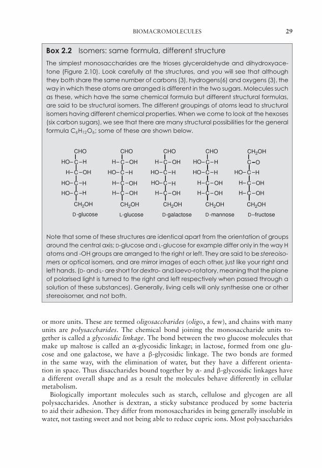
Box 2.2 Isomers: same formula, different structure
The simplest monosaccharides are the trioses glyceraldehyde and dihydroxyace-tone (Figure 2.10). Look carefully at the structures, and you will see that althoughthey both share the same number of carbons (3), hydrogens(6) and oxygens (3), theway in which these atoms are arranged is different in the two sugars. Molecules suchas these, which have the same chemical formula but different structural formulas,are said to be structural isomers. The different groupings of atoms lead to structuralisomers having different chemical properties. When we come to look at the hexoses(six carbon sugars), we see that there are many structural possibilities for the generalformula C6H12O6; some of these are shown below.
CHO
C HHO
H OH
HO
HO
H
H
CH2OH
C
C
C
D-glucose
CHO
C OHH
HO H
H
H
OH
OH
CH2OH
C
C
C
L-glucose
CHO
C OHH
HO H
HO
H
H
OH
CH2OH
C
C
C
D-galactose
CHO
C HHO
HO H
H
H
OH
OH
CH2OH
C
C
C
D-mannose
C O
HO H
H
H
OH
OH
CH2OH
C
C
C
D–fructose
CH2OH
Note that some of these structures are identical apart from the orientation of groupsaround the central axis; D-glucose and L-glucose for example differ only in the way Hatoms and -OH groups are arranged to the right or left. They are said to be stereoiso-mers or optical isomers, and are mirror images of each other, just like your right andleft hands. (D- and L- are short for dextro- and laevo-rotatory, meaning that the planeof polarised light is turned to the right and left respectively when passed through asolution of these substances). Generally, living cells will only synthesise one or otherstereoisomer, and not both.
or more units. These are termed oligosaccharides (oligo, a few), and chains with manyunits are polysaccharides. The chemical bond joining the monosaccharide units to-gether is called a glycosidic linkage. The bond between the two glucose molecules thatmake up maltose is called an α-glycosidic linkage; in lactose, formed from one glu-cose and one galactose, we have a β-glycosidic linkage. The two bonds are formedin the same way, with the elimination of water, but they have a different orienta-tion in space. Thus disaccharides bound together by α- and β-glycosidic linkages havea different overall shape and as a result the molecules behave differently in cellularmetabolism.
Biologically important molecules such as starch, cellulose and glycogen are allpolysaccharides. Another is dextran, a sticky substance produced by some bacteriato aid their adhesion. They differ from monosaccharides in being generally insoluble inwater, not tasting sweet and not being able to reduce cupric ions. Most polysaccharides
JWBK011-02 JWBK011-Hogg August 12, 2005 19:26 Char Count= 0
30 BIOCHEMICAL PRINCIPLES
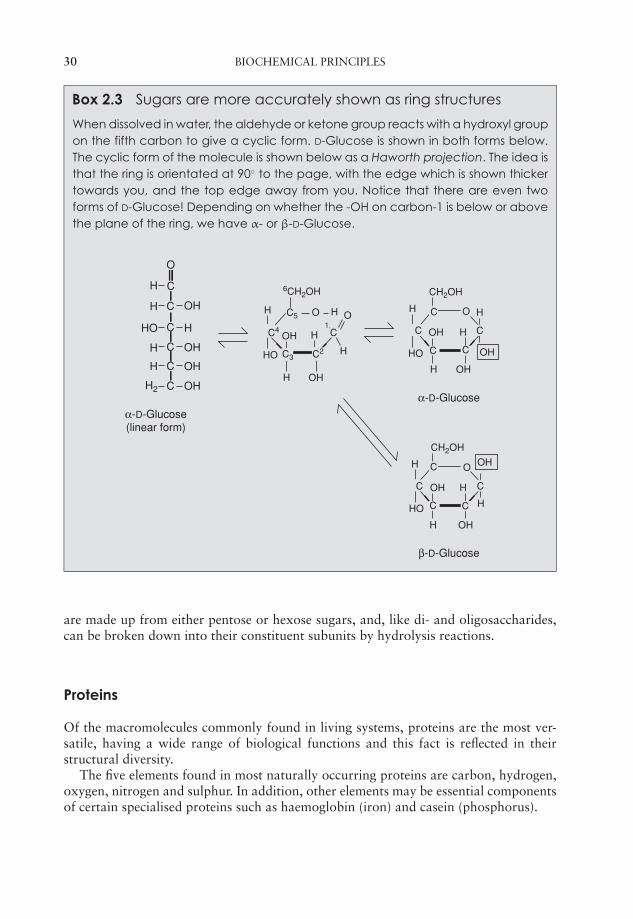
Box 2.3 Sugars are more accurately shown as ring structures
When dissolved in water, the aldehyde or ketone group reacts with a hydroxyl groupon the fifth carbon to give a cyclic form. D-Glucose is shown in both forms below.The cyclic form of the molecule is shown below as a Haworth projection. The idea isthat the ring is orientated at 90◦ to the page, with the edge which is shown thickertowards you, and the top edge away from you. Notice that there are even twoforms of D-Glucose! Depending on whether the -OH on carbon-1 is below or abovethe plane of the ring, we have α- or β-D-Glucose.
O
CH
H
HO
OH
H
OH
OH
OH
H
H2
H
C
C
C
C
C
H
HO OH
OH
α-D-Glucose(linear form)
α-D-Glucose
6CH2OH
H O
HO
C5 O H1
H
H OH
HOH C
C2
C4
CH2OH
OH
H
OH
O
H
H
C
C
C C
C
H
HO H
β-D-Glucose
CH2OH
OH
OH
O
H
H
C
C
C C
C
C3
are made up from either pentose or hexose sugars, and, like di- and oligosaccharides,can be broken down into their constituent subunits by hydrolysis reactions.
Proteins
Of the macromolecules commonly found in living systems, proteins are the most ver-satile, having a wide range of biological functions and this fact is reflected in theirstructural diversity.
The five elements found in most naturally occurring proteins are carbon, hydrogen,oxygen, nitrogen and sulphur. In addition, other elements may be essential componentsof certain specialised proteins such as haemoglobin (iron) and casein (phosphorus).
JWBK011-02 JWBK011-Hogg August 12, 2005 19:26 Char Count= 0
BIOMACROMOLECULES 31
C
R
H
H2N COOH C
H
R
H3N+ COO−
(a) (b)
Figure 2.12 Amino acid structure. (a) The basic structure of an amino acid. (b) In solution,the amino and carboxyl groups become ionised, giving rise to a zwitterion (a molecule withspatially separated positive and negative charges). All the 20 amino acids commonly foundin proteins are based on a common structure, differing only in the nature of their ‘R’ group(see Figure 2.13)
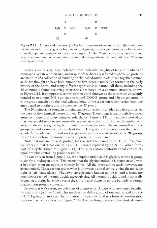
Proteins can be very large molecules, with molecular weights of tens or hundreds ofthousands. Whatever their size, and in spite of the diversity referred to above, all proteinsare made up of a collection of ‘building bricks’ called amino acids joined together. Aminoacids are thought to have been among the first organic molecules formed in the earlyhistory of the Earth, and many different types exist in nature. All these, including the20 commonly found occurring in proteins, are based on a common structure, shownin Figure 2.12. It comprises a central carbon atom (known as the α-carbon) covalentlybonded to an amino (NH2) group, a carboxyl (COOH) group and a hydrogen atom. Itis the group attached to the final valency bond of the α-carbon which varies from oneamino acid to another; this is known as the ‘R’-group.
The 20 amino acids found in proteins can be conveniently divided into five groups, onthe basis of the chemical nature of their ‘R’-group. These range from a single hydrogenatom to a variety of quite complex side chains (Figure 2.13). It is unlikely nowadaysthat you would need to memorise the precise structure of all 20, as the author wasasked to do in days gone by, but it would be advisable to familiarise yourself with thegroupings and examples from each of them. The groups differentiate on the basis ofa polar/non-polar nature and on the presence or absence of an ionisable ‘R’-group.Box 2.4 shows how we normally refer to proteins in shorthand.
Note that one amino acid, proline, falls outside the main groups. This differs fromthe others in that it has one of its N—H linkages replaced by an N—C, which formspart of a cyclic structure (Figure 2.13). This puts certain conformational constraintsupon proteins containing proline residues.
As can be seen from Figure 2.13, the simplest amino acid is glycine, whose R-groupis simply a hydrogen atom. This means that the glycine molecule is symmetrical, witha hydrogen atom on opposite valency bonds. All the other amino acids however, areasymmetrical. The α-carbon acts as what is known as a chiral centre, giving the moleculeright or left ‘handedness’. Thus two stereoisomers known as the d- and l-forms arepossible for each of the amino acids except glycine. All the amino acids found in naturallyoccurring proteins have the l-form; the d-form also occurs in nature but only in certainspecific, non-protein contexts.
Proteins, as we’ve seen, are polymers of amino acids. Amino acids are joined togetherby means of a peptide bond. This involves the -NH2 group of one amino acid and the-COOH group of another. The formation of a peptide bind is a form of condensationreaction in which water is lost (Figure 2.14). The resulting structure of two linked amino
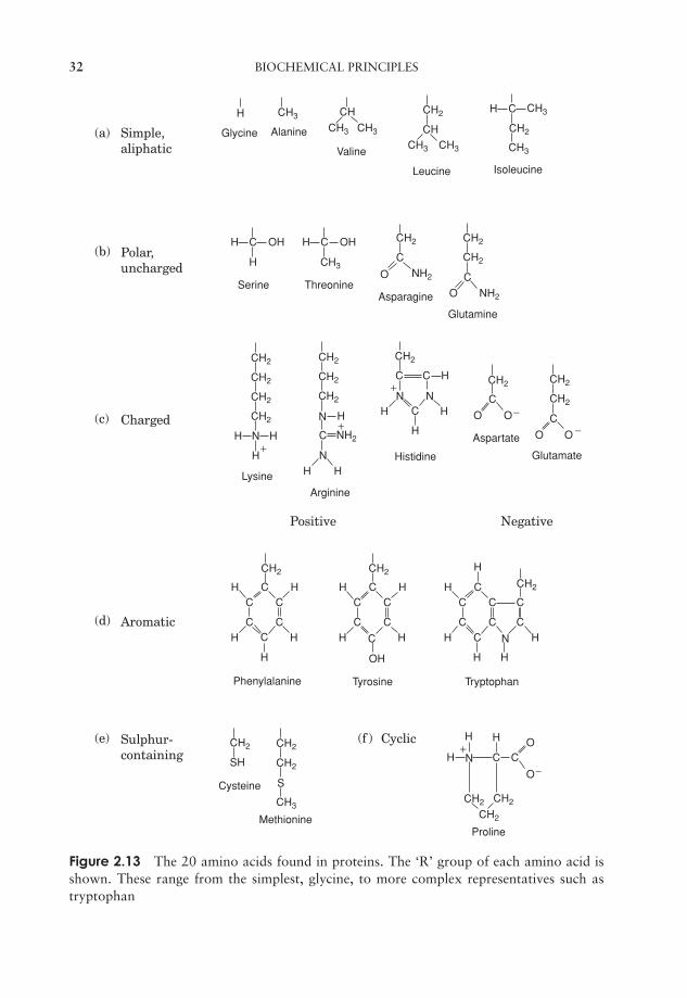
JWBK011-02 JWBK011-Hogg August 12, 2005 19:26 Char Count= 0
32 BIOCHEMICAL PRINCIPLES
H
H C
C
C
C
C
C
CO
O O
O
C H
CO
O
H
H H
H
H H
H
HH
N
N
N N
NH
H H
H
H
H
CC C
C CC
H H
H
H H
H
H
OH
CC C
N
N
C C
C
H
H H
CC C
C C
C
CC
C CO
O
OH H C OH
CH
CH3 CH3
CH3
CH2
CH2
NH2
NH2
NH2
CH3
CH3CH3
CH2 CH3
CH3
CH2
CH2
CH2
CH2
CH2
CH2 CH2
CH2 CH2
CH2
CH2
CH2
CH2
CH2
CH2 CH2
CH2
CH2 CH2
CH2
S
H
H
H
CH3
SH
CH2
CH2
CH
H C
Glycine Alanine
Valine
Leucine Isoleucine
Serine ThreonineAsparagine
Glutamine
Lysine
Arginine
Histidine
Aspartate
Glutamate
Phenylalanine Tyrosine Tryptophan
Cysteine
MethionineProline
−
+
−
−
+
+
+
(a)
(b)
(c)
(d)
(e) (f )
Simple,aliphatic
Polar,uncharged
Charged
Positive
Cyclic
Negative
Aromatic
Sulphur-containing
Figure 2.13 The 20 amino acids found in proteins. The ‘R’ group of each amino acid isshown. These range from the simplest, glycine, to more complex representatives such astryptophan
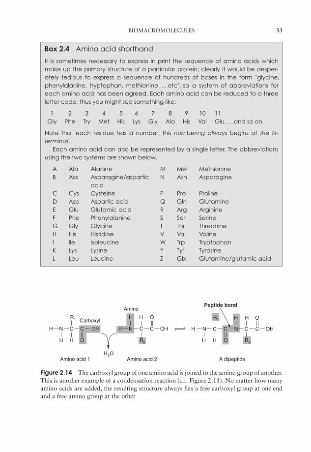
JWBK011-02 JWBK011-Hogg August 12, 2005 19:26 Char Count= 0
BIOMACROMOLECULES 33
Box 2.4 Amino acid shorthand
It is sometimes necessary to express in print the sequence of amino acids whichmake up the primary structure of a particular protein; clearly it would be desper-ately tedious to express a sequence of hundreds of bases in the form ’glycine,phenylalanine, tryptophan, methionine . . . etc’, so a system of abbreviations foreach amino acid has been agreed. Each amino acid can be reduced to a threeletter code, thus you might see something like:
1 2 3 4 5 6 7 8 9 10 11Gly Phe Try Met His Lys Gly Ala His Val Glu. . . .and so on.
Note that each residue has a number; this numbering always begins at the N-terminus.
Each amino acid can also be represented by a single letter. The abbreviationsusing the two systems are shown below.
A Ala Alanine M Met MethionineB Asx Asparagine/aspartic N Asn Asparagine
acidC Cys Cysteine P Pro ProlineD Asp Aspartic acid Q Gln GlutamineE Glu Glutamic acid R Arg ArginineF Phe Phenylalanine S Ser SerineG Gly Glycine T Thr ThreonineH His Histidine V Val ValineI Ile Isoleucine W Trp TryptophanK Lys Lysine Y Tyr TyrosineL Leu Leucine Z Glx Glutamine/glutamic acid
H
H
H
H H
H H O
O
N C C N C C OH
H O
H H O
N C
R1
R2 R2
R1
C CN C OHOH H
Carboxyl
Amino
H2OAmino acid 1 Amino acid 2 A dipeptide
Peptide bond
Figure 2.14 The carboxyl group of one amino acid is joined to the amino group of another.This is another example of a condensation reaction (c.f. Figure 2.11). No matter how manyamino acids are added, the resulting structure always has a free carboxyl group at one endand a free amino group at the other
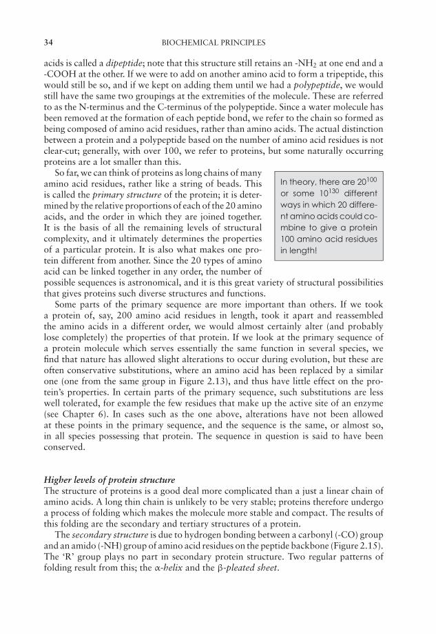
JWBK011-02 JWBK011-Hogg August 12, 2005 19:26 Char Count= 0
34 BIOCHEMICAL PRINCIPLES
acids is called a dipeptide; note that this structure still retains an -NH2 at one end and a-COOH at the other. If we were to add on another amino acid to form a tripeptide, thiswould still be so, and if we kept on adding them until we had a polypeptide, we wouldstill have the same two groupings at the extremities of the molecule. These are referredto as the N-terminus and the C-terminus of the polypeptide. Since a water molecule hasbeen removed at the formation of each peptide bond, we refer to the chain so formed asbeing composed of amino acid residues, rather than amino acids. The actual distinctionbetween a protein and a polypeptide based on the number of amino acid residues is notclear-cut; generally, with over 100, we refer to proteins, but some naturally occurringproteins are a lot smaller than this.
In theory, there are 20100
or some 10130 differentways in which 20 differe-nt amino acids could co-mbine to give a protein100 amino acid residuesin length!
So far, we can think of proteins as long chains of manyamino acid residues, rather like a string of beads. Thisis called the primary structure of the protein; it is deter-mined by the relative proportions of each of the 20 aminoacids, and the order in which they are joined together.It is the basis of all the remaining levels of structuralcomplexity, and it ultimately determines the propertiesof a particular protein. It is also what makes one pro-tein different from another. Since the 20 types of aminoacid can be linked together in any order, the number ofpossible sequences is astronomical, and it is this great variety of structural possibilitiesthat gives proteins such diverse structures and functions.
Some parts of the primary sequence are more important than others. If we tooka protein of, say, 200 amino acid residues in length, took it apart and reassembledthe amino acids in a different order, we would almost certainly alter (and probablylose completely) the properties of that protein. If we look at the primary sequence ofa protein molecule which serves essentially the same function in several species, wefind that nature has allowed slight alterations to occur during evolution, but these areoften conservative substitutions, where an amino acid has been replaced by a similarone (one from the same group in Figure 2.13), and thus have little effect on the pro-tein’s properties. In certain parts of the primary sequence, such substitutions are lesswell tolerated, for example the few residues that make up the active site of an enzyme(see Chapter 6). In cases such as the one above, alterations have not been allowedat these points in the primary sequence, and the sequence is the same, or almost so,in all species possessing that protein. The sequence in question is said to have beenconserved.
Higher levels of protein structureThe structure of proteins is a good deal more complicated than a just a linear chain ofamino acids. A long thin chain is unlikely to be very stable; proteins therefore undergoa process of folding which makes the molecule more stable and compact. The results ofthis folding are the secondary and tertiary structures of a protein.
The secondary structure is due to hydrogen bonding between a carbonyl (-CO) groupand an amido (-NH) group of amino acid residues on the peptide backbone (Figure 2.15).The ‘R’ group plays no part in secondary protein structure. Two regular patterns offolding result from this; the α-helix and the β-pleated sheet.

JWBK011-02 JWBK011-Hogg August 12, 2005 19:26 Char Count= 0
BIOMACROMOLECULES 35
Figure 2.15 Secondary structure in proteins. Hydrogen bonding occurs between the -COand -NH groups of amino acids on the backbone of a polypeptide chain. The two aminoacids may be on the same or different chains
Very small distanceswithin molecules are me-asured in Angstrom units(A). One Angstrom unit isequal to one tenbillionth(10−10) of a metre.
The α-helix occurs when hydrogen bonding takesplace between amino acids close together in the primarystructure. A stable helix is formed by the -NH group ofan amino acid bonding to the -CO group of the aminoacid four residues further along the chain (Figure 2.16a).This causes the chain to twist into the characteristic he-lical shape. One turn of the helix occurs every 3.6 aminoacid residues, and results in a rise of 5.4 A (0.54 nm);this is called the pitch height of the helix. The ability toform a helix like this is dependent on the component amino acids; if there are too manywith large R-groups, or R-groups carrying the same charge, a stable helix will not beformed. Because of its rigid structure, proline (Figure 2.13) cannot be accommodated inan α-helix. Naturally occurring α-helices are always right-handed, that is, the chain ofamino acids coils round the central axis in a clockwise direction. This is a much morestable configuration than a left-handed helix, due to the fact that there is less sterichindrance (overlapping of electron clouds) between the R-groups and the C==O groupon the peptide backbone. Note that if proteins were made up of the d-form of aminoacids, we would have the reverse situation, with a left-handed form favoured. In theβ-pleated sheet, the hydrogen bonding occurs between amino acids either on separatepolypeptide chains or on residues far apart in the primary structure (Figure 2.16b). Thechains in a β-pleated sheet are fully extended, with 3.5 A (0.35 nm) between adjacentamino acid residues (c.f. α-helix, 1.5 A). When two or more of these chains lie next toeach other, extensive hydrogen bonding occurs between the chains. Adjacent strands ina β-pleated sheet can either run in the same direction (e.g. N→C), giving rise to a par-allel β-pleated sheet, or in opposite directions (antiparallel β-pleated sheet, as shown inFigure 2.16b).
A common structural element in the secondary structure of proteins is the β-turn.This occurs when a chain doubles back on itself, such as in an antiparallel β-pleatedsheet. The -CO group of one amino acid is hydrogen bonded to the -NH group of the
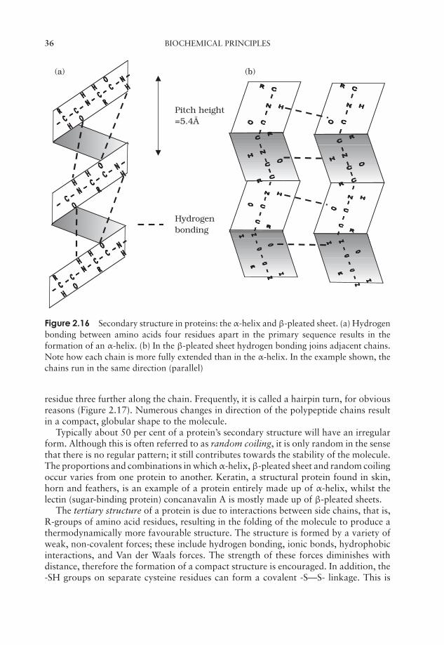
JWBK011-02 JWBK011-Hogg August 12, 2005 19:26 Char Count= 0
36 BIOCHEMICAL PRINCIPLES
=5.4Å
Hydrogenbonding
(a) (b)
Pitch height
Figure 2.16 Secondary structure in proteins: the α-helix and β-pleated sheet. (a) Hydrogenbonding between amino acids four residues apart in the primary sequence results in theformation of an α-helix. (b) In the β-pleated sheet hydrogen bonding joins adjacent chains.Note how each chain is more fully extended than in the α-helix. In the example shown, thechains run in the same direction (parallel)
residue three further along the chain. Frequently, it is called a hairpin turn, for obviousreasons (Figure 2.17). Numerous changes in direction of the polypeptide chains resultin a compact, globular shape to the molecule.
Typically about 50 per cent of a protein’s secondary structure will have an irregularform. Although this is often referred to as random coiling, it is only random in the sensethat there is no regular pattern; it still contributes towards the stability of the molecule.The proportions and combinations in which α-helix, β-pleated sheet and random coilingoccur varies from one protein to another. Keratin, a structural protein found in skin,horn and feathers, is an example of a protein entirely made up of α-helix, whilst thelectin (sugar-binding protein) concanavalin A is mostly made up of β-pleated sheets.
The tertiary structure of a protein is due to interactions between side chains, that is,R-groups of amino acid residues, resulting in the folding of the molecule to produce athermodynamically more favourable structure. The structure is formed by a variety ofweak, non-covalent forces; these include hydrogen bonding, ionic bonds, hydrophobicinteractions, and Van der Waals forces. The strength of these forces diminishes withdistance, therefore the formation of a compact structure is encouraged. In addition, the-SH groups on separate cysteine residues can form a covalent -S—S- linkage. This is
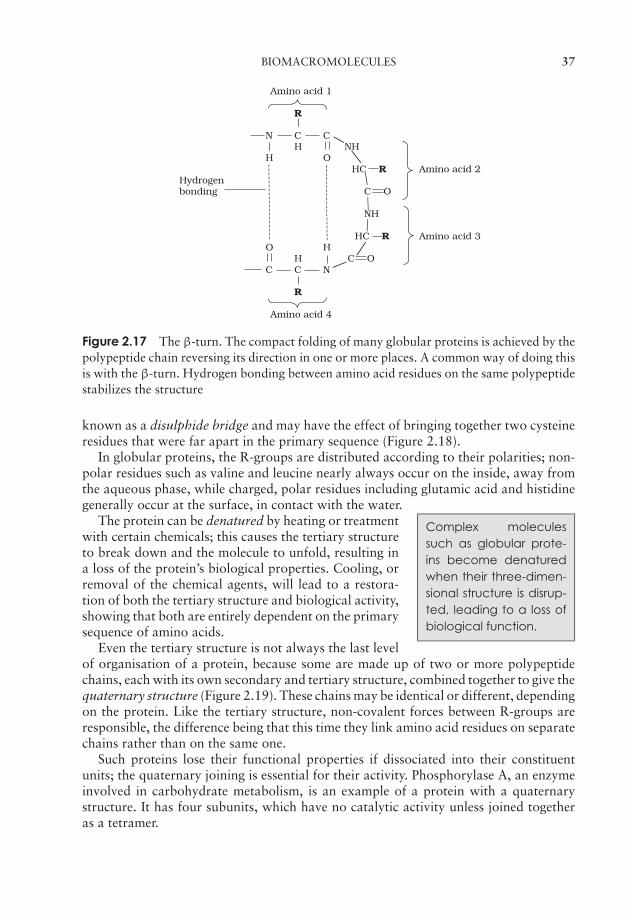
JWBK011-02 JWBK011-Hogg August 12, 2005 19:26 Char Count= 0
BIOMACROMOLECULES 37
Amino acid 1
R
N C CH NH
H OHC R Amino acid 2
Hydrogenbonding C O
NH
HC R Amino acid 3O H
H C OC C N
R
Amino acid 4
Figure 2.17 The β-turn. The compact folding of many globular proteins is achieved by thepolypeptide chain reversing its direction in one or more places. A common way of doing thisis with the β-turn. Hydrogen bonding between amino acid residues on the same polypeptidestabilizes the structure
known as a disulphide bridge and may have the effect of bringing together two cysteineresidues that were far apart in the primary sequence (Figure 2.18).
In globular proteins, the R-groups are distributed according to their polarities; non-polar residues such as valine and leucine nearly always occur on the inside, away fromthe aqueous phase, while charged, polar residues including glutamic acid and histidinegenerally occur at the surface, in contact with the water.
Complex moleculessuch as globular prote-ins become denaturedwhen their three-dimen-sional structure is disrup-ted, leading to a loss ofbiological function.
The protein can be denatured by heating or treatmentwith certain chemicals; this causes the tertiary structureto break down and the molecule to unfold, resulting ina loss of the protein’s biological properties. Cooling, orremoval of the chemical agents, will lead to a restora-tion of both the tertiary structure and biological activity,showing that both are entirely dependent on the primarysequence of amino acids.
Even the tertiary structure is not always the last levelof organisation of a protein, because some are made up of two or more polypeptidechains, each with its own secondary and tertiary structure, combined together to give thequaternary structure (Figure 2.19). These chains may be identical or different, dependingon the protein. Like the tertiary structure, non-covalent forces between R-groups areresponsible, the difference being that this time they link amino acid residues on separatechains rather than on the same one.
Such proteins lose their functional properties if dissociated into their constituentunits; the quaternary joining is essential for their activity. Phosphorylase A, an enzymeinvolved in carbohydrate metabolism, is an example of a protein with a quaternarystructure. It has four subunits, which have no catalytic activity unless joined togetheras a tetramer.
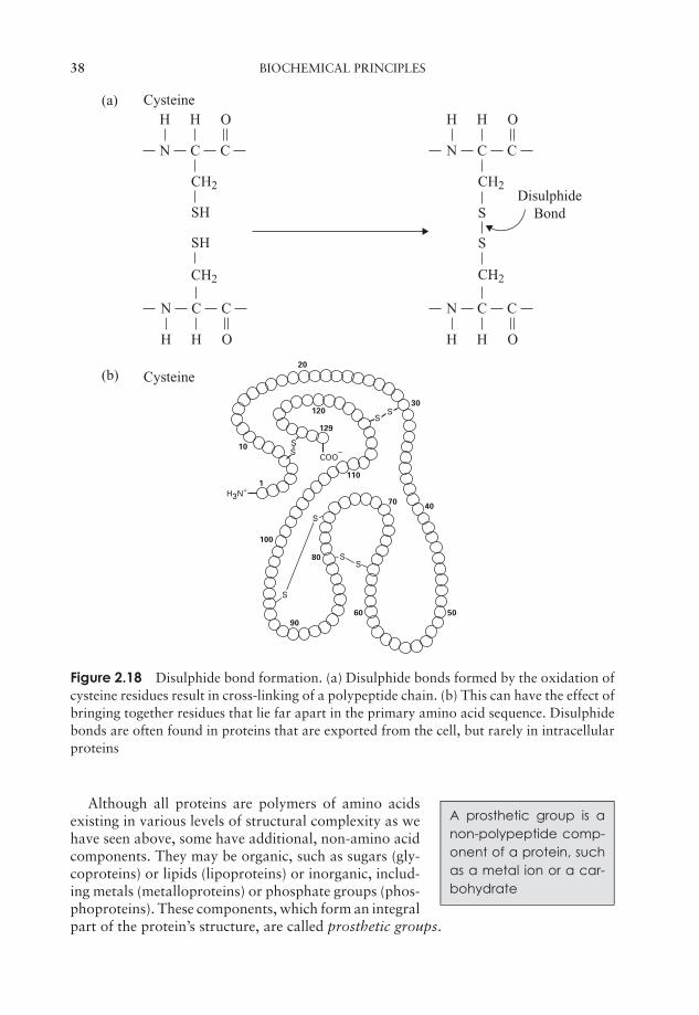
JWBK011-02 JWBK011-Hogg August 12, 2005 19:26 Char Count= 0
38 BIOCHEMICAL PRINCIPLES
CN
CH2
CH2
SH
SH
H OH
C CN
CH2
CH2
S
S
H OH
H OH
C
CN C
H OH
CN C
DisulphideBond
Cysteine
Cysteine
(a)
COO−
H3N+
SSSSSS
20
120
10
1
129
30
40
5060
70
80
90
100
110
SSSSSS
SSS
SS
(b)
S
SS
Figure 2.18 Disulphide bond formation. (a) Disulphide bonds formed by the oxidation ofcysteine residues result in cross-linking of a polypeptide chain. (b) This can have the effect ofbringing together residues that lie far apart in the primary amino acid sequence. Disulphidebonds are often found in proteins that are exported from the cell, but rarely in intracellularproteins
A prosthetic group is anon-polypeptide comp-onent of a protein, suchas a metal ion or a car-bohydrate
Although all proteins are polymers of amino acidsexisting in various levels of structural complexity as wehave seen above, some have additional, non-amino acidcomponents. They may be organic, such as sugars (gly-coproteins) or lipids (lipoproteins) or inorganic, includ-ing metals (metalloproteins) or phosphate groups (phos-phoproteins). These components, which form an integralpart of the protein’s structure, are called prosthetic groups.

JWBK011-02 JWBK011-Hogg August 12, 2005 19:26 Char Count= 0
BIOMACROMOLECULES 39
Figure 2.19 Polypeptide chains may join to form quaternary structure. The example showncomprises two identical polypeptide subunits. Coils indicate α-helical sequences, arrows areβ-pleated sheets. From Bolsover, SR , Hyams, JS, Jones, S, Shepherd, EA & White, HA:From Genes to Cells, John Wiley & Sons, 1997. Reproduced by permission of the publishers
Nucleic acids
The third class of polymeric macromolecules are the nucleic acids. These are deoxyri-bonucleic acid (DNA) and ribonucleic acid (RNA), and both are polymers of smallermolecules called nucleotides. As we shall see, there are important differences both inthe overall structures of RNA and DNA and in the nucleotides they contain, so we shallconsider each of them in turn.
The structure of DNAThe composition of a DNA nucleotide is shown in Figure 2.20(a). It has three parts, afive-carbon sugar called deoxyribose, a phosphate group and a base. This base can beany one of four molecules; as can be seen in Figure 2.21, these are all based on a cyclicstructure containing nitrogen. Two of the bases, cytosine and thymine, have a singlering and are called pyrimidines. The other two, guanine and adenine, have a doublering structure; these are the purines. The four bases are often referred to by their initialletter only, thus we have A, C, G and T.
One nucleotide differs from another by the identity of the base it contains; the rest ofthe molecule (sugar and phosphate) is identical. You will recall from the previous sectionthat the properties of a protein depend on the order in which its constituent amino acids
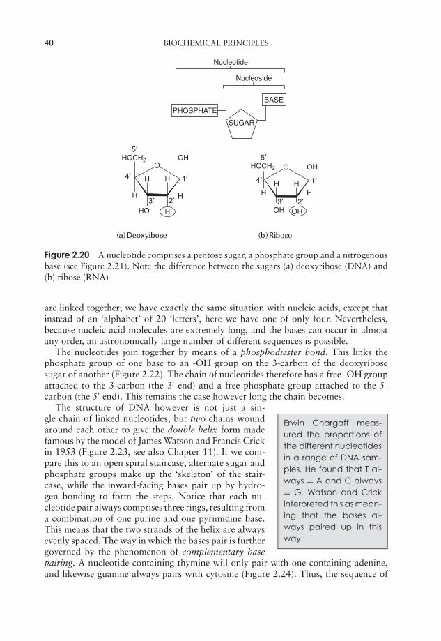
JWBK011-02 JWBK011-Hogg August 12, 2005 19:26 Char Count= 0
40 BIOCHEMICAL PRINCIPLES
Nucleotide
Nucleoside
PHOSPHATE
SUGAR
BASE
OOHHOCH2
H
HH
H
HHO
4′
5′
1′
2′3′
O OHHOCH2
HHH
H
OH OH
4′
5′
1′
2′3′
(a) Deoxyibose (b) Ribose
Figure 2.20 A nucleotide comprises a pentose sugar, a phosphate group and a nitrogenousbase (see Figure 2.21). Note the difference between the sugars (a) deoxyribose (DNA) and(b) ribose (RNA)
are linked together; we have exactly the same situation with nucleic acids, except thatinstead of an ‘alphabet’ of 20 ‘letters’, here we have one of only four. Nevertheless,because nucleic acid molecules are extremely long, and the bases can occur in almostany order, an astronomically large number of different sequences is possible.
The nucleotides join together by means of a phosphodiester bond. This links thephosphate group of one base to an -OH group on the 3-carbon of the deoxyribosesugar of another (Figure 2.22). The chain of nucleotides therefore has a free -OH groupattached to the 3-carbon (the 3′ end) and a free phosphate group attached to the 5-carbon (the 5′ end). This remains the case however long the chain becomes.
Erwin Chargaff meas-ured the proportions ofthe different nucleotidesin a range of DNA sam-ples. He found that T al-ways = A and C always= G. Watson and Crickinterpreted this as mean-ing that the bases al-ways paired up in thisway.
The structure of DNA however is not just a sin-gle chain of linked nucleotides, but two chains woundaround each other to give the double helix form madefamous by the model of James Watson and Francis Crickin 1953 (Figure 2.23, see also Chapter 11). If we com-pare this to an open spiral staircase, alternate sugar andphosphate groups make up the ‘skeleton’ of the stair-case, while the inward-facing bases pair up by hydro-gen bonding to form the steps. Notice that each nu-cleotide pair always comprises three rings, resulting froma combination of one purine and one pyrimidine base.This means that the two strands of the helix are alwaysevenly spaced. The way in which the bases pair is furthergoverned by the phenomenon of complementary basepairing. A nucleotide containing thymine will only pair with one containing adenine,and likewise guanine always pairs with cytosine (Figure 2.24). Thus, the sequence of
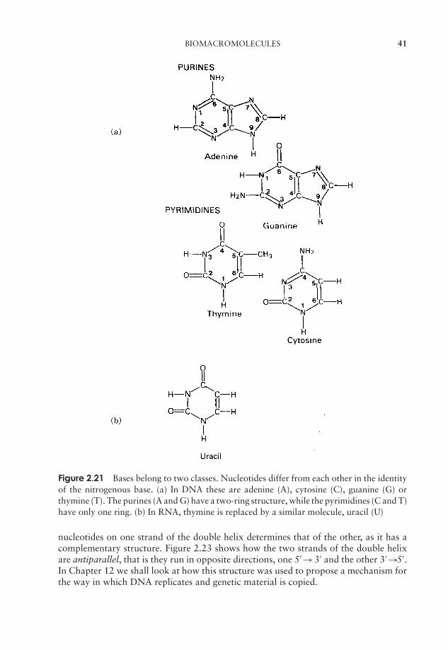
JWBK011-02 JWBK011-Hogg August 12, 2005 19:26 Char Count= 0
BIOMACROMOLECULES 41
Figure 2.21 Bases belong to two classes. Nucleotides differ from each other in the identityof the nitrogenous base. (a) In DNA these are adenine (A), cytosine (C), guanine (G) orthymine (T). The purines (A and G) have a two-ring structure, while the pyrimidines (C and T)have only one ring. (b) In RNA, thymine is replaced by a similar molecule, uracil (U)
nucleotides on one strand of the double helix determines that of the other, as it has acomplementary structure. Figure 2.23 shows how the two strands of the double helixare antiparallel, that is they run in opposite directions, one 5′ →3′ and the other 3′ →5′.In Chapter 12 we shall look at how this structure was used to propose a mechanism forthe way in which DNA replicates and genetic material is copied.
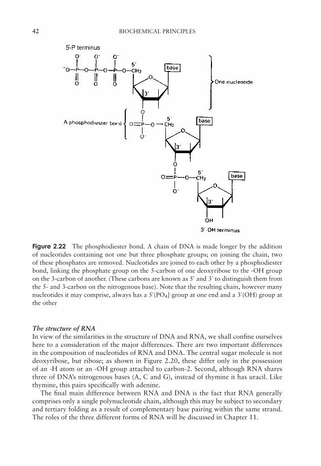
JWBK011-02 JWBK011-Hogg August 12, 2005 19:26 Char Count= 0
42 BIOCHEMICAL PRINCIPLES
Figure 2.22 The phosphodiester bond. A chain of DNA is made longer by the additionof nucleotides containing not one but three phosphate groups; on joining the chain, twoof these phosphates are removed. Nucleotides are joined to each other by a phosphodiesterbond, linking the phosphate group on the 5-carbon of one deoxyribose to the -OH groupon the 3-carbon of another. (These carbons are known as 5′ and 3′ to distinguish them fromthe 5- and 3-carbon on the nitrogenous base). Note that the resulting chain, however manynucleotides it may comprise, always has a 5′(PO4) group at one end and a 3′(OH) group atthe other
The structure of RNAIn view of the similarities in the structure of DNA and RNA, we shall confine ourselveshere to a consideration of the major differences. There are two important differencesin the composition of nucleotides of RNA and DNA. The central sugar molecule is notdeoxyribose, but ribose; as shown in Figure 2.20, these differ only in the possessionof an -H atom or an -OH group attached to carbon-2. Second, although RNA sharesthree of DNA’s nitrogenous bases (A, C and G), instead of thymine it has uracil. Likethymine, this pairs specifically with adenine.
The final main difference between RNA and DNA is the fact that RNA generallycomprises only a single polynucleotide chain, although this may be subject to secondaryand tertiary folding as a result of complementary base pairing within the same strand.The roles of the three different forms of RNA will be discussed in Chapter 11.
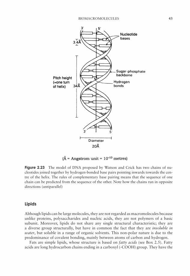
JWBK011-02 JWBK011-Hogg August 12, 2005 19:26 Char Count= 0
BIOMACROMOLECULES 43
Figure 2.23 The model of DNA proposed by Watson and Crick has two chains of nu-cleotides joined together by hydrogen-bonded base pairs pointing inwards towards the cen-tre of the helix. The rules of complementary base pairing means that the sequence of onechain can be predicted from the sequence of the other. Note how the chains run in oppositedirections (antiparallel)
Lipids
Although lipids can be large molecules, they are not regarded as macromolecules becauseunlike proteins, polysaccharides and nucleic acids, they are not polymers of a basicsubunit. Moreover, lipids do not share any single structural characteristic; they area diverse group structurally, but have in common the fact that they are insoluble inwater, but soluble in a range of organic solvents. This non-polar nature is due to thepredominance of covalent bonding, mainly between atoms of carbon and hydrogen.
Fats are simple lipids, whose structure is based on fatty acids (see Box 2.5). Fattyacids are long hydrocarbon chains ending in a carboxyl (-COOH) group. They have the

JWBK011-02 JWBK011-Hogg August 12, 2005 19:26 Char Count= 0
44 BIOCHEMICAL PRINCIPLES
Figure 2.24 Adenine pairs only with thymine, and guanine with cytosine, thus if the se-quence of bases in one strand of a DNA molecule is known, that of the other can be predicted.This critical feature of Watson and Crick’s model offers an explanation for how DNA is ableto replicate itself. Note that GC pairs are held together by three hydrogen bonds, while ATpairs only have two
general formula:
CH3—(CH2)n—COOH
where n is usually an even number. They combine with glycerol according to the basicreaction:
Alcohol + Acid → Ester
The bond so formed is called an ester linkage, and the result is an acylglycerol (Figure2.25). One, two or all three of the -OH groups may be esterified with a fatty acid, to giverespectively mono-, di- and triacylglycerols. Natural fats generally contain a mixtureof two or three different fatty acids substituted at the three positions; consequently,a considerable diversity is possible among fats. Fats serve as energy stores; a higherproportion of C—C and C—H bonds in comparison with proteins or carbohydratesresults in a higher energy-storing capacity.
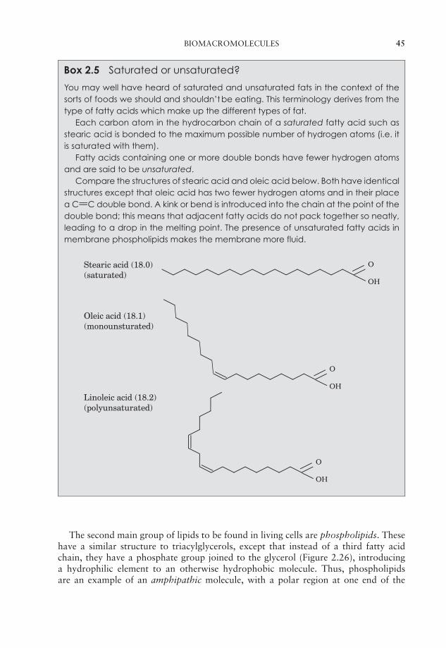
JWBK011-02 JWBK011-Hogg August 12, 2005 19:26 Char Count= 0
BIOMACROMOLECULES 45
Box 2.5 Saturated or unsaturated?
You may well have heard of saturated and unsaturated fats in the context of thesorts of foods we should and shouldn’t be eating. This terminology derives from thetype of fatty acids which make up the different types of fat.
Each carbon atom in the hydrocarbon chain of a saturated fatty acid such asstearic acid is bonded to the maximum possible number of hydrogen atoms (i.e. itis saturated with them).
Fatty acids containing one or more double bonds have fewer hydrogen atomsand are said to be unsaturated.
Compare the structures of stearic acid and oleic acid below. Both have identicalstructures except that oleic acid has two fewer hydrogen atoms and in their placea C==C double bond. A kink or bend is introduced into the chain at the point of thedouble bond; this means that adjacent fatty acids do not pack together so neatly,leading to a drop in the melting point. The presence of unsaturated fatty acids inmembrane phospholipids makes the membrane more fluid.
O
OH
O
OH
O
OH
Stearic acid (18.0) (saturated)
Oleic acid (18.1) (monounsturated)
Linoleic acid (18.2) (polyunsaturated)
The second main group of lipids to be found in living cells are phospholipids. Thesehave a similar structure to triacylglycerols, except that instead of a third fatty acidchain, they have a phosphate group joined to the glycerol (Figure 2.26), introducinga hydrophilic element to an otherwise hydrophobic molecule. Thus, phospholipidsare an example of an amphipathic molecule, with a polar region at one end of the
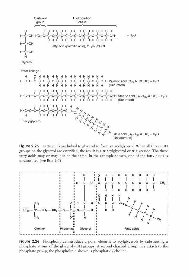
JWBK011-02 JWBK011-Hogg August 12, 2005 19:26 Char Count= 0
C
C
C
H
H
H
H
H H H H H H H H H H H H H H H HH
H H H H H H H H H H H H H H HOH HO
OH
OH
O
C C C C C C C C C C C C C C C C
HH
HH H H H H H H H H H H H H H
HH H H H H H H H H H H H H H H
O
O CC
H OC
H
H
OC
C C C C C C C C C C C C C C C
H H H H H H H H H H H H H H HH
H H H H H H H H H H H H H H H
O
C C C C C C C C
H H H H H H H H HH
HH
HH
HH
H
HH
HH
HH
HH H
H H H H H H H
O
C C C C C C C C C CC
CC
CC
CC
C
C C C C C C C CH H
H HC C
Carboxyl group
Hydrocarbonchain
Fatty acid (palmitic acid), C15H31COOH
Glycerol
Ester linkage
Triacylglycerol
Palmitic acid (C15H31COOH) + H2O(Saturated)
Stearic acid (C17H35COOH) + H2O(Saturated)
Oleic acid (C17H33COOH) + H2O(Unsaturated)
+ H2O
Figure 2.25 Fatty acids are linked to glycerol to form an acylglycerol. When all three -OHgroups on the glycerol are esterified, the result is a triacylglycerol or triglyceride. The threefatty acids may or may not be the same. In the example shown, one of the fatty acids isunsaturated (see Box 2.5)
CH3
CH3
CH3
CH3
CH3
CH2 CH2 O
O H
H
H H
H
C
C
C
H
H
O
O
O
O
O
PN+
C C
H H HH
H H
C C
Fatty acidsGlycerolCholine
C CC
CC
C
O
C
H
H
C
H
H
C
H
H
C
H
H
C
H
H
C
H
H
C
H
H
C
H
HH
HH
HH
H
Phosphate
Figure 2.26 Phospholipids introduce a polar element to acylglycerols by substituting aphosphate at one of the glycerol -OH groups. A second charged group may attach to thephosphate group; the phospholipid shown is phosphatidylcholine
46

JWBK011-02 JWBK011-Hogg August 12, 2005 19:26 Char Count= 0
BIOMACROMOLECULES 47
H2O
H2O
Hydrophilichead groups
Hydrophobictail groups
Figure 2.27 Phospholipids can form a bilayer in aqueous surroundings. A ‘sandwich’ ar-rangement is achieved by the polar phosphate groups facing outwards and burying the fattyacid chains within. Water is thus excluded from the hydrophobic region, a key property ofbiological membranes (see Figure 3.5)
molecule and a non-polar region at the other. This fact is essential for the forma-tion of a bilayer when the phospholipid is introduced into an aqueous environment;the hydrophilic phosphate groups point outwards towards the water, while the hy-drophobic hydrocarbon chains ‘hide’ inside (Figure 2.27, and c.f. Figure 2.8, micelleformation).
This bilayer structure forms the basis of all biological membranes (see Chapter 3),forming a barrier around cells and certain organelles. Phospholipids generally have an-other polar group attached to the phosphate; Figure 2.25 shows the effect of substitutingserine.
The structural diversity of lipids can be illustrated by comparing fats and phospho-lipids with the final group of lipids we need to consider, the steroids. As can be seenfrom Figure 2.28, these have a completely different form, but still share in common theproperty of hydrophobicity. The four ring planar structure is common to all steroids,with the substitution of different side groups producing great differences in function.Cholesterol is an important component of many membranes.
It would be wrong to gain the impression that living cells contain only moleculesof the four groups outlined above. Smaller organic molecules play important roles asprecursors or intermediates in metabolic pathways (see Chapter 6), and several inorganicions such as potassium, sodium and chloride play essential roles in maintaining theliving cell. Finally, some macromolecules comprise elements of more than one group,for example, lipopolysaccharides (carbohydrate and lipid) and glycoproteins (proteinand carbohydrate).
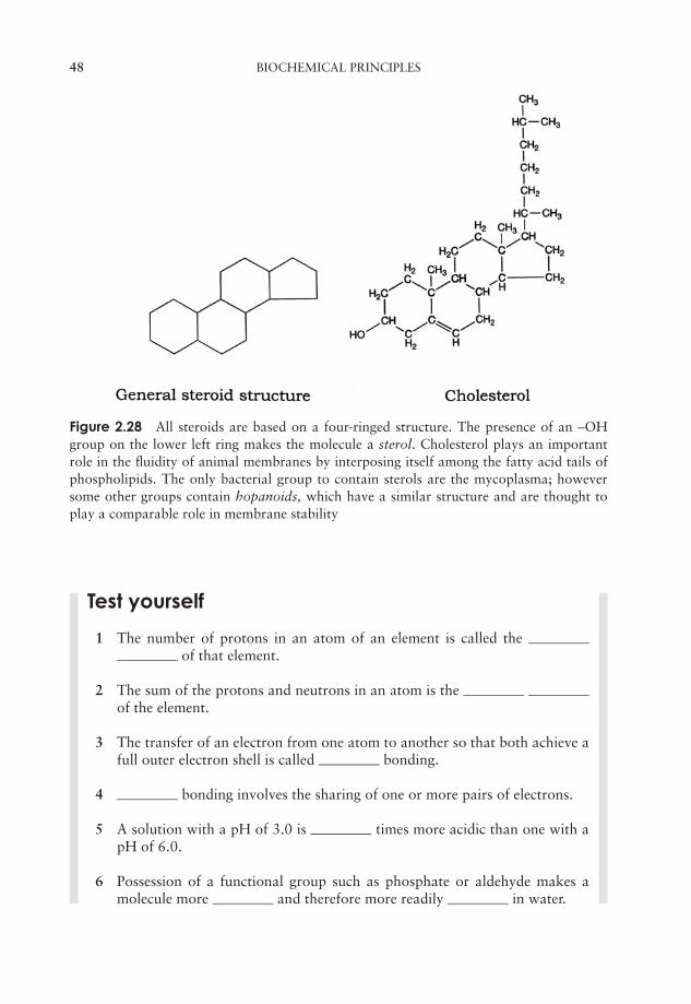
JWBK011-02 JWBK011-Hogg August 12, 2005 19:26 Char Count= 0
48 BIOCHEMICAL PRINCIPLES
Figure 2.28 All steroids are based on a four-ringed structure. The presence of an –OHgroup on the lower left ring makes the molecule a sterol. Cholesterol plays an importantrole in the fluidity of animal membranes by interposing itself among the fatty acid tails ofphospholipids. The only bacterial group to contain sterols are the mycoplasma; howeversome other groups contain hopanoids, which have a similar structure and are thought toplay a comparable role in membrane stability
Test yourself
1 The number of protons in an atom of an element is called theof that element.
2 The sum of the protons and neutrons in an atom is theof the element.
3 The transfer of an electron from one atom to another so that both achieve afull outer electron shell is called bonding.
4 bonding involves the sharing of one or more pairs of electrons.
5 A solution with a pH of 3.0 is times more acidic than one with apH of 6.0.
6 Possession of a functional group such as phosphate or aldehyde makes amolecule more and therefore more readily in water.
JWBK011-02 JWBK011-Hogg August 12, 2005 19:26 Char Count= 0
TEST YOURSELF 49
7 Simple carbohydrates may be classed on the basis of whether they have aor group, or according to how many atoms
they possess.
8 Sugars are joined together by linkages.
9 No matter how long a peptide chain grows, it always has an groupat one end and a group at the other.
10 An example of a negatively charged amino acid is .
11 Disulphide bonds are formed between residues of the amino acid .
12 Secondary protein structure is brought about by the formation ofbonds between a group and a group.
13 Naturally occurring amino acids are all the -isomer. This results inan α-helix taking on a configuration for maximum stability.
14 Heating or treatment with certain chemicals cause proteins to lose their three-dimensional structure and become .
15 A short stretch of double stranded DNA has 52 adenine residues and 61guanine residues. There are therefore cytosine residues and a totalof hydrogen bonds joining the two strands.
16 The arrangement of the two strands of a DNA molecule is described as.
17 The nucleotides of RNA contain instead of thymine.
18 Lipids are a diverse group of molecules, sharing the common property ofin .
19 Phospholipids are described as because their molecules have bothpolar and non-polar regions.
20 In fats, fatty acids are joined to glycerol via linkages.

JWBK011-02 JWBK011-Hogg August 12, 2005 19:26 Char Count= 0
50
JWBK011-03 JWBK011-Hogg August 12, 2005 15:52 Char Count= 0
3Cell Structure and Organisation
The basic unit of all living things is the cell. The cell theory is one of the fundamentalconcepts of biology; it states that:
� all organisms are made up of cells,
and that
� all cells derive from other, pre-existing cells.
As we shall see in this chapter, there may exist within a cell many smaller, subcellularstructures, each with its own characteristics and function, but these are not capable ofindependent life.
An organism may comprise just a single cell (unicellular), a collection of cellsthat are not morphologically or functionally differentiated (colonial), or several dis-tinct cell types with specialised functions (multicellular). Among microorganisms, all
The names given to thetwo cell types derivefrom Greek words:Procaryotic = ‘beforenucleus’Eucaryotic = ‘truenucleus’
bacteria and protozoans are unicellular; fungi may beunicellular or multicellular, while algae may exist in allthree forms. There is, however, one way that organismscan be differentiated from each other that is even morefundamental than whether they are uni- or multicellular.It is a difference that is greater than that between a lionand a mushroom or between an earthworm and an oaktree, and it exists at the level of the individual cell. All or-ganisms are made up of one or other (definitely not both!)of two very distinct cell types, which we call procaryoticand eucaryotic cells, both of which exist in the microbial world. These differ from eachother in many ways, including size, structural complexity and organisation of geneticmaterial (Table 3.1).
The most fundamental difference between procaryotic and eucaryotic cells is reflectedin their names; eucaryotic cells possess a true nucleus, and several other distinct subcel-lular organelles that are bounded by a membrane. Procaryotes have no such organelles.Most of these differences only became apparent after the development of electron mi-croscopy techniques.
As can be seen from Table 3.2, the procaryotes comprise the simpler and more prim-itive types of microorganisms; they are generally single celled, and arose much earlierin evolutionary history than the eucaryotes. Indeed, as discussed later in this chapter, it
51

JWBK011-03 JWBK011-Hogg August 12, 2005 15:52 Char Count= 0
52 CELL STRUCTURE AND ORGANISATION
Table 3.1 Similarities and differences between procaryotic and eucaryotic cell structure
SimilaritiesCell contents bounded by a plasma
membraneGenetic information encoded on DNARibosomes act as site of protein synthesis
Differences
Procaryotic Eucaryotic
SizeTypically 1–5 µm Typically 10–100 µm
Genetic materialFree in cytoplasm Contained within a membrane-bound
nucleusSingle circular chromosome or
nucleoidMultiple chromosomes, generally in pairs
Histones absent. DNA complexed with histone proteinsInternal features
Membrane-bound organelles absent Several membrane-bound organellespresent, including mitochondria, Golgibody, endoplasmic reticulum and (inplants & algae) chloroplasts
Ribosomes smaller (70S), free incytoplasm
Ribosomes larger (80S), free in cytoplasmor attached to membranes
Respiratory enzymes bound to plasmamembrane
Respiratory enzymes located inmitochondria
Cell wallUsually based on peptidoglycan (not
Archaea)When present, based on cellulose or chitin
External featuresCilia absent Cilia may be presentFlagella, if present, composed of
flagellin. Provide rotating motilityFlagella, if present, have complex (9 + 2)
structure. Provide ‘whiplash’ motilityPili may be present Pili absentOutside layer (slime layer, capsule,
glycocalyx) present in some typesPellicle or test present in some types
is widely accepted that eucaryotic cells actually arose from their more primitive coun-terparts. Note that the viruses do not appear in Table 3.2, because they do not have acellular structure at all, and are not therefore considered to be living organisms. (SeeChapter 10 for further discussion of the viruses).
Phylogenetic: pertainingto the evolutionary rela-tionship between organ-isms.
The use of DNA sequencing methods to determinephylogenetic relationships between organisms has re-vealed that within the procaryotes there is another fun-damental division. One group of bacteria were shown todiffer greatly from all the others; we now call these theArchaea, to differentiate them from the true Bacteria.
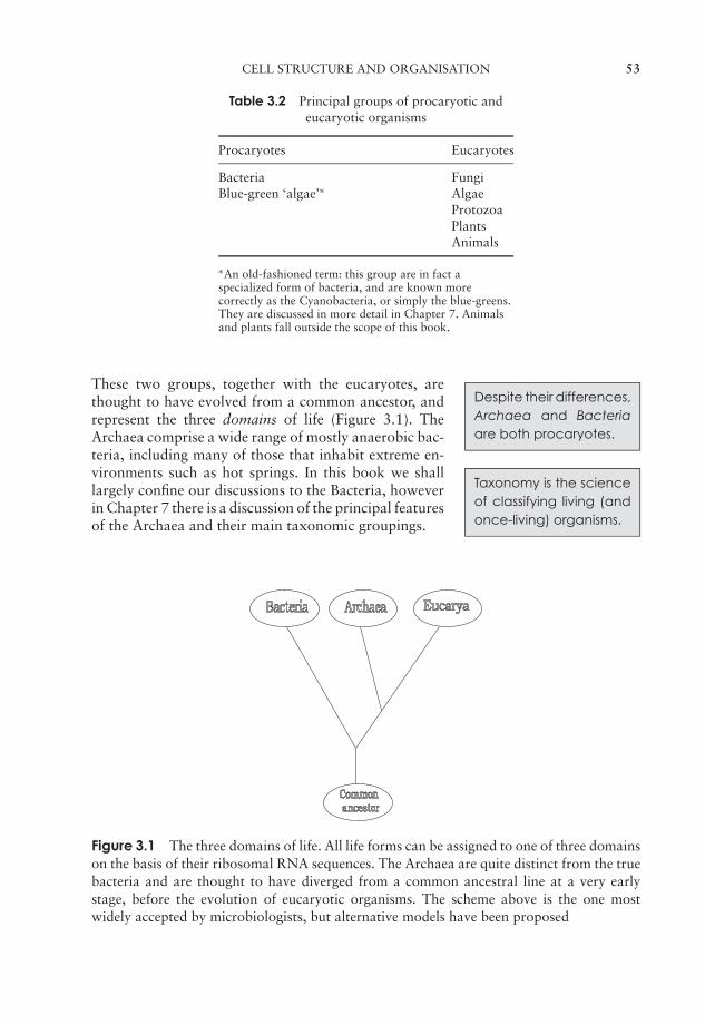
JWBK011-03 JWBK011-Hogg August 12, 2005 15:52 Char Count= 0
CELL STRUCTURE AND ORGANISATION 53
Table 3.2 Principal groups of procaryotic andeucaryotic organisms
Procaryotes Eucaryotes
Bacteria FungiBlue-green ‘algae’∗ Algae
ProtozoaPlantsAnimals
∗An old-fashioned term: this group are in fact aspecialized form of bacteria, and are known morecorrectly as the Cyanobacteria, or simply the blue-greens.They are discussed in more detail in Chapter 7. Animalsand plants fall outside the scope of this book.
Despite their differences,Archaea and Bacteriaare both procaryotes.
These two groups, together with the eucaryotes, arethought to have evolved from a common ancestor, andrepresent the three domains of life (Figure 3.1). TheArchaea comprise a wide range of mostly anaerobic bac-teria, including many of those that inhabit extreme en-vironments such as hot springs. In this book we shalllargely confine our discussions to the Bacteria, howeverin Chapter 7 there is a discussion of the principal featuresof the Archaea and their main taxonomic groupings.
Taxonomy is the scienceof classifying living (andonce-living) organisms.
Figure 3.1 The three domains of life. All life forms can be assigned to one of three domainson the basis of their ribosomal RNA sequences. The Archaea are quite distinct from the truebacteria and are thought to have diverged from a common ancestral line at a very earlystage, before the evolution of eucaryotic organisms. The scheme above is the one mostwidely accepted by microbiologists, but alternative models have been proposed
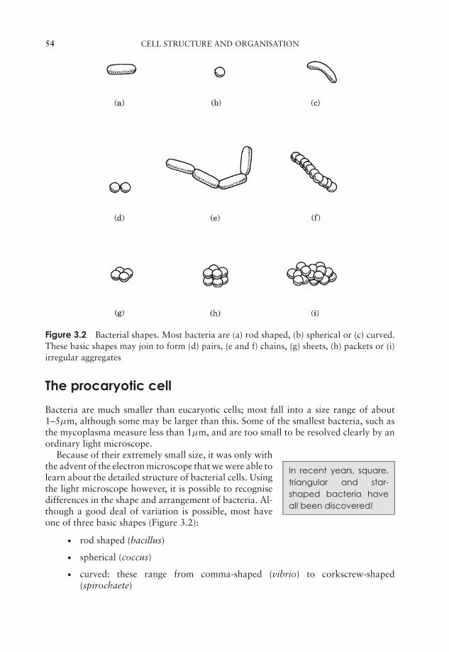
JWBK011-03 JWBK011-Hogg August 12, 2005 15:52 Char Count= 0
54 CELL STRUCTURE AND ORGANISATION
Figure 3.2 Bacterial shapes. Most bacteria are (a) rod shaped, (b) spherical or (c) curved.These basic shapes may join to form (d) pairs, (e and f) chains, (g) sheets, (h) packets or (i)irregular aggregates
The procaryotic cell
Bacteria are much smaller than eucaryotic cells; most fall into a size range of about1–5µm, although some may be larger than this. Some of the smallest bacteria, such asthe mycoplasma measure less than 1µm, and are too small to be resolved clearly by anordinary light microscope.
In recent years, square,triangular and star-shaped bacteria haveall been discovered!
Because of their extremely small size, it was only withthe advent of the electron microscope that we were able tolearn about the detailed structure of bacterial cells. Usingthe light microscope however, it is possible to recognisedifferences in the shape and arrangement of bacteria. Al-though a good deal of variation is possible, most haveone of three basic shapes (Figure 3.2):
� rod shaped (bacillus)
� spherical (coccus)
� curved: these range from comma-shaped (vibrio) to corkscrew-shaped(spirochaete)
JWBK011-03 JWBK011-Hogg August 12, 2005 15:52 Char Count= 0
THE PROCARYOTIC CELL 55
All these shapes confer certain advantages to their owners; rods, with a large surfacearea are better able to take up nutrients from the environment, while the cocci are lessprone to drying out. The spiral forms are usually motile; their shape aids their movementthrough an aqueous medium.
As well as these characteristic cell shapes, bacteria may also be found grouped to-gether in particular formations. When they divide, they may remain attached to oneanother, and the shape the groups of cells assume reflects the way the cell divides. Cocci,for example, are frequently found as chains of cells, a reflection of repeated division inone plane (Figure 3.2(f)). Other cocci may form regular sheets or packets of cells, as aresult of division in two or three planes. Yet others, such as the staphylococci, dividein several planes, producing the irregular and characteristic ‘bunch of grapes’ appear-ance. Rod-shaped bacteria only divide in a single plane and may therefore be foundin chains, while spiral forms also divide in one plane, but tend not to stick together.Blue–greens form filaments; these are regarded as truly multicellular rather than as aloose association of individuals.
Procaryotic cell structure
When compared with the profusion of elaborate organelles encountered inside a typicaleucaryotic cell, the interior of a typical bacterium looks rather empty. The only internalstructural features are:
� a bacterial chromosome or nucleoid, comprising a closed loop of double stranded,supercoiled DNA. In addition, there may be additional DNA in the form of a plasmid
� thousands of granular ribosomes
� a variety of granular inclusions associated with nutrient storage.
All of these are contained in a thick aqueous soup of carbohydrates, proteins, lipids andinorganic salts known as the cytoplasm, which is surrounded by a plasma membrane.This in turn is wrapped in a cell wall, whose rigidity gives the bacterial cell its charac-teristic shape. Depending on the type of bacterium, there may be a further surroundinglayer such as a capsule or slime layer and/or structures external to the cell associatedwith motility (flagella) or attachment (pili/fimbriae). Figure 3.3 shows these featuresin a generalised bacterial cell. In the following pages we shall examine these features
Not all bacteria con-form to the model of asingle circular chromo-some; some have beenshown to possess twowith genes shared be-tween them, while ex-amples of linear chromo-somes are also known.
in a little more detail, noting how each has a crucial roleto play in the survival or reproduction of the cell.
Genetic materialAlthough it occupies a well defined area within the cell,the genetic material of procaryotes is not present as atrue nucleus, as it lacks a surrounding nuclear membrane(c.f. the eucaryotic nucleus, Figure 3.12). The nucleoid orbacterial chromosome comprises a closed circle of dou-ble stranded DNA, many times the length of the cell andhighly folded and compacted. (The common laboratory

JWBK011-03 JWBK011-Hogg August 12, 2005 15:52 Char Count= 0
56 CELL STRUCTURE AND ORGANISATION
Plasma membrane
Outer membraneMesosome
Inclusion body Cell wall
PlasmidNucleoid
PiliFlagellum
EndosporeRibosome
Figure 3.3 Structure of a generalised procaryotic cell. Note the lack of complex internalorganelles (c.f. Figure 3.12). Gram-positive and Gram-negative bacteria differ in the detailsof their cell wall structure (see Figures 3.7 & 3.8)
Plasmids are small loopsof DNA independent ofthe chromosome. Theyare capable of directingtheir own replication.
bacterium Escherichia coli is around 3–4 µm in length,but contains a DNA molecule some 1400 µm in length!)The DNA may be associated with certain bacterial pro-teins, but these are not the same as the histones foundin eucaryotic chromosomes. Some bacteria contain ad-ditional DNA in the form of small, self-replicating ex-trachromosomal elements called plasmids. These do notcarry any genes essential for growth and reproduction,and thus the cell may survive without them. They can be very important however, asthey may include genes encoding toxins or resistance to antibiotics, and can be passedfrom cell to cell (see Chapter 12).
RibosomesApart from the nucleoid, the principal internal structures of procaryotic cells are theribosomes. These are the site of protein synthesis, and there may be many thousands ofthese in an active cell, lending a speckled appearance to the cytoplasm. Ribosomes arecomposed of a complex of protein and RNA, and are the site of protein synthesis in thecell.
Although they carry out a similar function, the ribosomes of procaryotic cellsare smaller and lighter than their eucaryotic counterparts. Ribosomes are mea-sured in Svedberg units (S), a function of their size and shape, and determined bytheir rate of sedimentation in a centrifuge; procaryotic ribosomes are 70S, whilethose of eucaryotes are 80S. Some types of antibiotic exploit this difference by
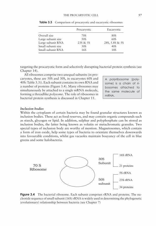
JWBK011-03 JWBK011-Hogg August 12, 2005 15:52 Char Count= 0
THE PROCARYOTIC CELL 57
Table 3.3 Comparison of procaryotic and eucaryotic ribosomes
Procaryotic Eucaryotic
Overall size 70S 80SLarge subunit size 50S 60SLarge subunit RNA 23S & 5S 28S, 5.8S & 5SSmall subunit size 30S 40SSmall subunit RNA 16S 18S
targeting the procaryotic form and selectively disrupting bacterial protein synthesis (seeChapter 14).
A polyribosome (poly-some) is a chain of ri-bosomes attached tothe same molecule ofmRNA.
All ribosomes comprise two unequal subunits (in pro-caryotes, these are 50S and 30S, in eucaryotes 60S and40S: Table 3.3)). Each subunit contains its own RNA anda number of proteins (Figure 3.4). Many ribosomes maysimultaneously be attached to a single mRNA molecule,forming a threadlike polysome. The role of ribosomes inbacterial protein synthesis is discussed in Chapter 11.
Inclusion bodiesWithin the cytoplasm of certain bacteria may be found granular structures known asinclusion bodies. These act as food reserves, and may contain organic compounds suchas starch, glycogen or lipid. In addition, sulphur and polyphosphate can be stored asinclusion bodies, the latter being known as volutin or metachromatic granules. Twospecial types of inclusion body are worthy of mention. Magnetosomes, which containa form of iron oxide, help some types of bacteria to orientate themselves downwardsinto favourable conditions, whilst gas vacuoles maintain bouyancy of the cell in bluegreens and some halobacteria.
70 SRibosome
30SSubunit
50Ssubunit
16S rRNA
21 proteins
5S rRNA
23S rRNA
34 proteins
Figure 3.4 The bacterial ribosome. Each subunit comprises rRNA and proteins. The nu-cleotide sequence of small subunit (16S) rRNA is widely used in determining the phylogenetic(evolutionary) relationship between bacteria (see Chapter 7)
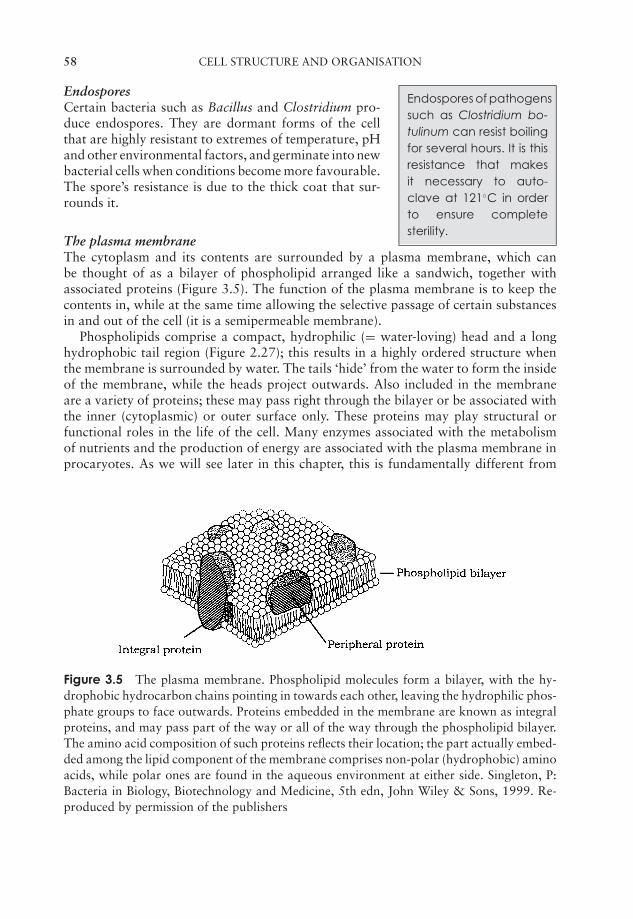
JWBK011-03 JWBK011-Hogg August 12, 2005 15:52 Char Count= 0
58 CELL STRUCTURE AND ORGANISATION
Endospores Endospores of pathogenssuch as Clostridium bo-tulinum can resist boilingfor several hours. It is thisresistance that makesit necessary to auto-clave at 121◦C in orderto ensure completesterility.
Certain bacteria such as Bacillus and Clostridium pro-duce endospores. They are dormant forms of the cellthat are highly resistant to extremes of temperature, pHand other environmental factors, and germinate into newbacterial cells when conditions become more favourable.The spore’s resistance is due to the thick coat that sur-rounds it.
The plasma membraneThe cytoplasm and its contents are surrounded by a plasma membrane, which canbe thought of as a bilayer of phospholipid arranged like a sandwich, together withassociated proteins (Figure 3.5). The function of the plasma membrane is to keep thecontents in, while at the same time allowing the selective passage of certain substancesin and out of the cell (it is a semipermeable membrane).
Phospholipids comprise a compact, hydrophilic (= water-loving) head and a longhydrophobic tail region (Figure 2.27); this results in a highly ordered structure whenthe membrane is surrounded by water. The tails ‘hide’ from the water to form the insideof the membrane, while the heads project outwards. Also included in the membraneare a variety of proteins; these may pass right through the bilayer or be associated withthe inner (cytoplasmic) or outer surface only. These proteins may play structural orfunctional roles in the life of the cell. Many enzymes associated with the metabolismof nutrients and the production of energy are associated with the plasma membrane inprocaryotes. As we will see later in this chapter, this is fundamentally different from
Figure 3.5 The plasma membrane. Phospholipid molecules form a bilayer, with the hy-drophobic hydrocarbon chains pointing in towards each other, leaving the hydrophilic phos-phate groups to face outwards. Proteins embedded in the membrane are known as integralproteins, and may pass part of the way or all of the way through the phospholipid bilayer.The amino acid composition of such proteins reflects their location; the part actually embed-ded among the lipid component of the membrane comprises non-polar (hydrophobic) aminoacids, while polar ones are found in the aqueous environment at either side. Singleton, P:Bacteria in Biology, Biotechnology and Medicine, 5th edn, John Wiley & Sons, 1999. Re-produced by permission of the publishers
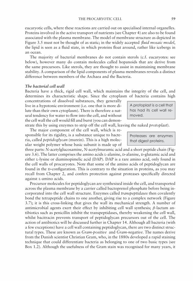
JWBK011-03 JWBK011-Hogg August 12, 2005 15:52 Char Count= 0
THE PROCARYOTIC CELL 59
eucaryotic cells, where these reactions are carried out on specialised internal organelles.Proteins involved in the active transport of nutrients (see Chapter 4) are also to be foundassociated with the plasma membrane. The model of membrane structure as depicted inFigure 3.5 must not be thought of as static; in the widely accepted fluid mosaic model,the lipid is seen as a fluid state, in which proteins float around, rather like icebergs inan ocean.
The majority of bacterial membranes do not contain sterols (c.f. eucaryotes: seebelow), however many do contain molecules called hopanoids that are derive fromthe same precursors. Like sterols, they are thought to assist in maintaining membranestability. A comparison of the lipid components of plasma membranes reveals a distinctdifference between members of the Archaea and the Bacteria.
The bacterial cell wallBacteria have a thick, rigid cell wall, which maintains the integrity of the cell, anddetermines its characteristic shape. Since the cytoplasm of bacteria contains high
A protoplast is a cell thathas had its cell wall re-moved.
concentrations of dissolved substances, they generallylive in a hypotonic environment (i.e. one that is more di-lute than their own cytoplasm). There is therefore a nat-ural tendency for water to flow into the cell, and withoutthe cell wall the cell would fill and burst (you can demon-strate this by using enzymes to strip off the cell wall, leaving the naked protoplast).
Proteases are enzymesthat digest proteins.
The major component of the cell wall, which is re-sponsible for its rigidity, is a substance unique to bacte-ria, called peptidoglycan (murein). This is a high molec-ular weight polymer whose basic subunit is made up ofthree parts: N-acetylglucosamine, N-acetylmuramic acid and a short peptide chain (Fig-ure 3.6). The latter comprises the amino acids l-alanine, d-alanine, d-glutamic acid andeither l-lysine or diaminopimelic acid (DAP). DAP is a rare amino acid, only found inthe cell walls of procaryotes. Note that some of the amino acids of peptidoglycan arefound in the d-configuration. This is contrary to the situation in proteins, as you mayrecall from Chapter 2, and confers protection against proteases specifically directedagainst l-amino acids.
Precursor molecules for peptidoglycan are synthesised inside the cell, and transportedacross the plasma membrane by a carrier called bactoprenol phosphate before being in-corporated into the cell wall structure. Enzymes called transpeptidases then covalentlybond the tetrapeptide chains to one another, giving rise to a complex network (Figure3.7); it is this cross-linking that gives the wall its mechanical strength. A number ofantimicrobial agents exert their effect by inhibiting cell wall synthesis; β-lactam an-tibiotics such as penicillin inhibit the transpeptidases, thereby weakening the cell wall,whilst bacitracin prevents transport of peptidoglycan precursors out of the cell. Theaction of antibiotics will be discussed further in Chapter 14. Although all bacteria (witha few exceptions) have a cell wall containing peptidoglycan, there are two distinct struc-tural types. These are known as Gram-positive and Gram-negative. The names derivefrom the Danish scientist Christian Gram, who, in the 1880s developed a rapid stainingtechnique that could differentiate bacteria as belonging to one of two basic types (seeBox 1.2). Although the usefulness of the Gram stain was recognised for many years, it
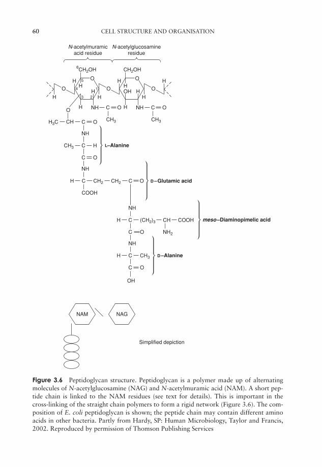
JWBK011-03 JWBK011-Hogg August 12, 2005 15:52 Char Count= 0
60 CELL STRUCTURE AND ORGANISATION
CH2OH
HH
H
NH
H
H
H
O
O
O
CH2OH
HH
HOH
NHH
H
O
O H
O
C
COOH
NH
H CH2 CH2 C O
C
C NH2
NH
H (CH2)3 CH COOH
C
C O
O
NH
OH
H CH3
C
C
C O
O
NH
CH3
C O
CH3 CH3
C O
CHH3C
H
N-acetylmuramicacid residue
N-acetylglucosamineresidue
Simplified depiction
–Alanine
–Glutamic acid
meso–Diaminopimelic acid
D
–AlanineD
L
NAM NAG
1
23
4
5
6
Figure 3.6 Peptidoglycan structure. Peptidoglycan is a polymer made up of alternatingmolecules of N-acetylglucosamine (NAG) and N-acetylmuramic acid (NAM). A short pep-tide chain is linked to the NAM residues (see text for details). This is important in thecross-linking of the straight chain polymers to form a rigid network (Figure 3.6). The com-position of E. coli peptidoglycan is shown; the peptide chain may contain different aminoacids in other bacteria. Partly from Hardy, SP: Human Microbiology, Taylor and Francis,2002. Reproduced by permission of Thomson Publishing Services
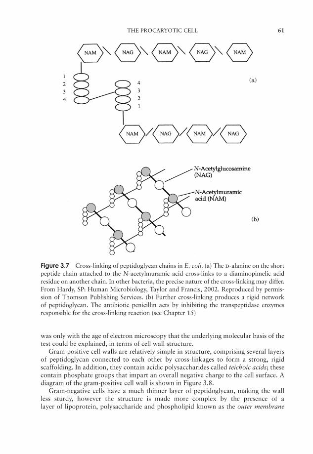
JWBK011-03 JWBK011-Hogg August 12, 2005 15:52 Char Count= 0
THE PROCARYOTIC CELL 61
Figure 3.7 Cross-linking of peptidoglycan chains in E. coli. (a) The d-alanine on the shortpeptide chain attached to the N-acetylmuramic acid cross-links to a diaminopimelic acidresidue on another chain. In other bacteria, the precise nature of the cross-linking may differ.From Hardy, SP: Human Microbiology, Taylor and Francis, 2002. Reproduced by permis-sion of Thomson Publishing Services. (b) Further cross-linking produces a rigid networkof peptidoglycan. The antibiotic penicillin acts by inhibiting the transpeptidase enzymesresponsible for the cross-linking reaction (see Chapter 15)
was only with the age of electron microscopy that the underlying molecular basis of thetest could be explained, in terms of cell wall structure.
Gram-positive cell walls are relatively simple in structure, comprising several layersof peptidoglycan connected to each other by cross-linkages to form a strong, rigidscaffolding. In addition, they contain acidic polysaccharides called teichoic acids; thesecontain phosphate groups that impart an overall negative charge to the cell surface. Adiagram of the gram-positive cell wall is shown in Figure 3.8.
Gram-negative cells have a much thinner layer of peptidoglycan, making the wallless sturdy, however the structure is made more complex by the presence of alayer of lipoprotein, polysaccharide and phospholipid known as the outer membrane

JWBK011-03 JWBK011-Hogg August 12, 2005 15:52 Char Count= 0
62 CELL STRUCTURE AND ORGANISATION
Figure 3.8 The Gram-positive cell wall. Peptidoglycan is many layers thick in the Gram-positive cell wall and may account for 30–70% of its dry weight. Teichoic acids are neg-atively charged polysaccharides; they are polymers of ribitol phosphate and cross-link topeptidoglycan. Lipoteichoic acids are teichoic acids found in association with glycolipids.From Henderson, B, Wilson, M, McNab, R & Lax, AJ: Cellular Microbiology: Bacteria-Host Interactions in Health and Disease, John Wiley & Sons Inc., 1999. Reproduced bypermission of the publishers
(Figure 3.9). This misleading name derives from the fact that it superficially resemblesthe bilayer of the plasma membrane; however, instead of two layers of phospholipid, ithas only one, the outer layer being made up of lipopolysaccharide. This has three parts:lipid A, core polysaccharide and an O-specific side chain. The lipid A component mayact as an endotoxin, which, if released into the bloodstream, can lead to serious con-ditions such as fever and toxic shock. The O-specific antigens are carbohydrate chainswhose composition often varies between strains of the same species. Serological meth-ods can distinguish between these, a valuable tool in the investigation, for example, ofthe origin of an outbreak of an infectious disease. Proteins incorporated into the outermembrane and penetrating its entire thickness form channels that allow the passage ofwater and small molecules to enter the cell. Unlike the plasma membrane, the outermembrane plays no part in cellular respiration.
Box 3.1 Mesosomes − the structures that never were?
When looked at under the electron microscope, Gram-positive bacteria often con-tained localised in-foldings of the plasma membrane. These were given the namemesosomes, and were thought by some to act as attachment points for DNA dur-ing cell division, or to play a role in the formation of cross-walls. Others thoughtthey were nothing more than artefacts produced by the rather elaborate samplepreparation procedures necessary for electron microscopy. Nowadays, most micro-biologists support the latter view.
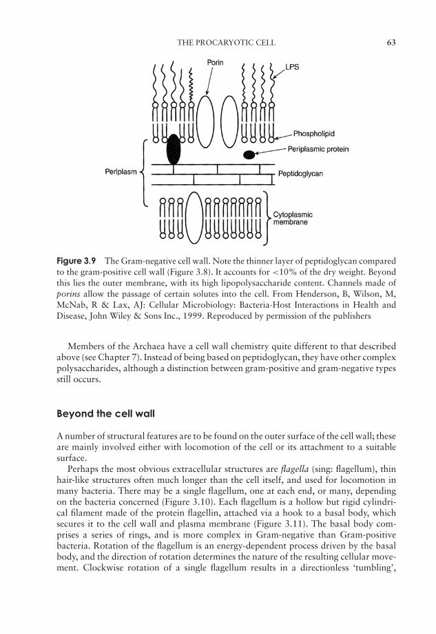
JWBK011-03 JWBK011-Hogg August 12, 2005 15:52 Char Count= 0
THE PROCARYOTIC CELL 63
Figure 3.9 The Gram-negative cell wall. Note the thinner layer of peptidoglycan comparedto the gram-positive cell wall (Figure 3.8). It accounts for <10% of the dry weight. Beyondthis lies the outer membrane, with its high lipopolysaccharide content. Channels made ofporins allow the passage of certain solutes into the cell. From Henderson, B, Wilson, M,McNab, R & Lax, AJ: Cellular Microbiology: Bacteria-Host Interactions in Health andDisease, John Wiley & Sons Inc., 1999. Reproduced by permission of the publishers
Members of the Archaea have a cell wall chemistry quite different to that describedabove (see Chapter 7). Instead of being based on peptidoglycan, they have other complexpolysaccharides, although a distinction between gram-positive and gram-negative typesstill occurs.
Beyond the cell wall
A number of structural features are to be found on the outer surface of the cell wall; theseare mainly involved either with locomotion of the cell or its attachment to a suitablesurface.
Perhaps the most obvious extracellular structures are flagella (sing: flagellum), thinhair-like structures often much longer than the cell itself, and used for locomotion inmany bacteria. There may be a single flagellum, one at each end, or many, dependingon the bacteria concerned (Figure 3.10). Each flagellum is a hollow but rigid cylindri-cal filament made of the protein flagellin, attached via a hook to a basal body, whichsecures it to the cell wall and plasma membrane (Figure 3.11). The basal body com-prises a series of rings, and is more complex in Gram-negative than Gram-positivebacteria. Rotation of the flagellum is an energy-dependent process driven by the basalbody, and the direction of rotation determines the nature of the resulting cellular move-ment. Clockwise rotation of a single flagellum results in a directionless ‘tumbling’,
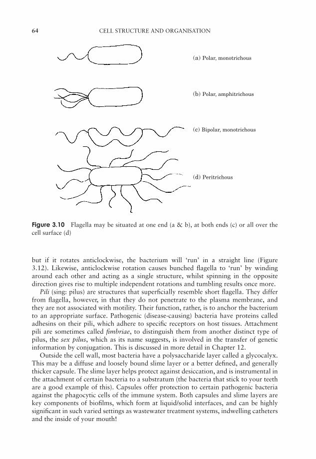
JWBK011-03 JWBK011-Hogg August 12, 2005 15:52 Char Count= 0
64 CELL STRUCTURE AND ORGANISATION
(a) Polar, monotrichous
(b) Polar, amphitrichous
(c) Bipolar, monotrichous
(d) Peritrichous
Figure 3.10 Flagella may be situated at one end (a & b), at both ends (c) or all over thecell surface (d)
but if it rotates anticlockwise, the bacterium will ‘run’ in a straight line (Figure3.12). Likewise, anticlockwise rotation causes bunched flagella to ‘run’ by windingaround each other and acting as a single structure, whilst spinning in the oppositedirection gives rise to multiple independent rotations and tumbling results once more.
Pili (sing: pilus) are structures that superficially resemble short flagella. They differfrom flagella, however, in that they do not penetrate to the plasma membrane, andthey are not associated with motility. Their function, rather, is to anchor the bacteriumto an appropriate surface. Pathogenic (disease-causing) bacteria have proteins calledadhesins on their pili, which adhere to specific receptors on host tissues. Attachmentpili are sometimes called fimbriae, to distinguish them from another distinct type ofpilus, the sex pilus, which as its name suggests, is involved in the transfer of geneticinformation by conjugation. This is discussed in more detail in Chapter 12.
Outside the cell wall, most bacteria have a polysaccharide layer called a glycocalyx.This may be a diffuse and loosely bound slime layer or a better defined, and generallythicker capsule. The slime layer helps protect against desiccation, and is instrumental inthe attachment of certain bacteria to a substratum (the bacteria that stick to your teethare a good example of this). Capsules offer protection to certain pathogenic bacteriaagainst the phagocytic cells of the immune system. Both capsules and slime layers arekey components of biofilms, which form at liquid/solid interfaces, and can be highlysignificant in such varied settings as wastewater treatment systems, indwelling cathetersand the inside of your mouth!
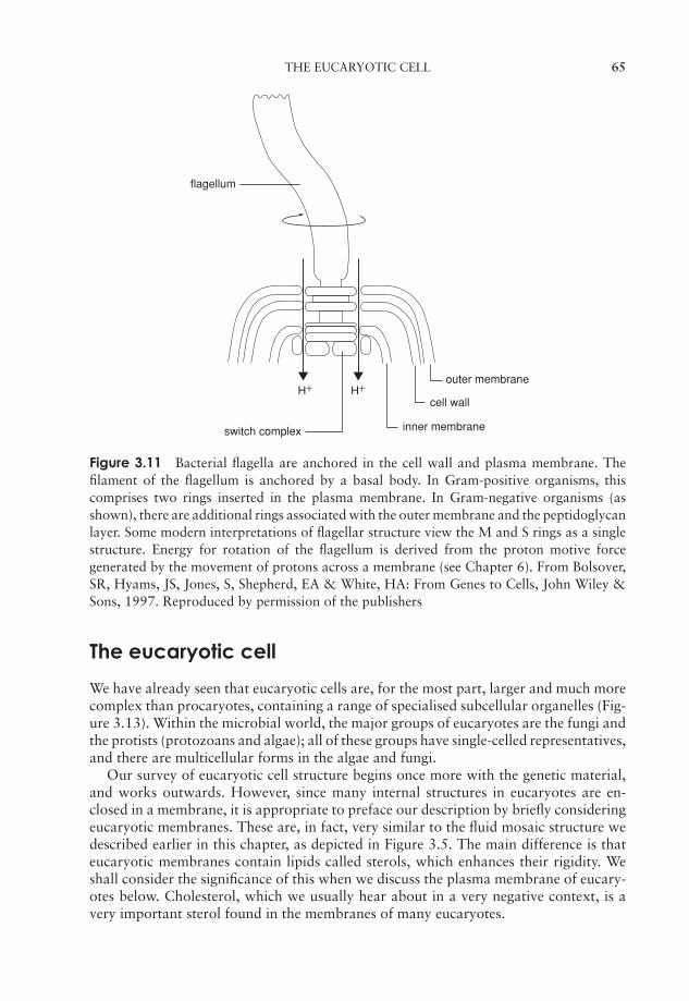
JWBK011-03 JWBK011-Hogg August 12, 2005 15:52 Char Count= 0
THE EUCARYOTIC CELL 65
flagellum
outer membrane
inner membraneswitch complex
cell wall H+ H+
Figure 3.11 Bacterial flagella are anchored in the cell wall and plasma membrane. Thefilament of the flagellum is anchored by a basal body. In Gram-positive organisms, thiscomprises two rings inserted in the plasma membrane. In Gram-negative organisms (asshown), there are additional rings associated with the outer membrane and the peptidoglycanlayer. Some modern interpretations of flagellar structure view the M and S rings as a singlestructure. Energy for rotation of the flagellum is derived from the proton motive forcegenerated by the movement of protons across a membrane (see Chapter 6). From Bolsover,SR, Hyams, JS, Jones, S, Shepherd, EA & White, HA: From Genes to Cells, John Wiley &Sons, 1997. Reproduced by permission of the publishers
The eucaryotic cell
We have already seen that eucaryotic cells are, for the most part, larger and much morecomplex than procaryotes, containing a range of specialised subcellular organelles (Fig-ure 3.13). Within the microbial world, the major groups of eucaryotes are the fungi andthe protists (protozoans and algae); all of these groups have single-celled representatives,and there are multicellular forms in the algae and fungi.
Our survey of eucaryotic cell structure begins once more with the genetic material,and works outwards. However, since many internal structures in eucaryotes are en-closed in a membrane, it is appropriate to preface our description by briefly consideringeucaryotic membranes. These are, in fact, very similar to the fluid mosaic structure wedescribed earlier in this chapter, as depicted in Figure 3.5. The main difference is thateucaryotic membranes contain lipids called sterols, which enhances their rigidity. Weshall consider the significance of this when we discuss the plasma membrane of eucary-otes below. Cholesterol, which we usually hear about in a very negative context, is avery important sterol found in the membranes of many eucaryotes.
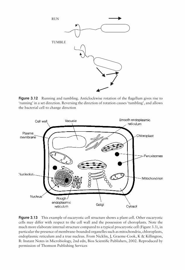
JWBK011-03 JWBK011-Hogg August 12, 2005 15:52 Char Count= 0
RUN
TUMBLE
Figure 3.12 Running and tumbling. Anticlockwise rotation of the flagellum gives rise to‘running’ in a set direction. Reversing the direction of rotation causes ‘tumbling’, and allowsthe bacterial cell to change direction
Figure 3.13 This example of eucaryotic cell structure shows a plant cell. Other eucaryoticcells may differ with respect to the cell wall and the possession of choroplasts. Note themuch more elaborate internal structure compared to a typical procaryotic cell (Figure 3.3), inparticular the presence of membrane-bounded organelles such as mitochondria, chloroplasts,endoplasmic reticulum and a true nucleus. From Nicklin, J, Graeme-Cook, K & Killington,R: Instant Notes in Microbiology, 2nd edn, Bios Scientific Publishers, 2002. Reproduced bypermission of Thomson Publishing Services
66

JWBK011-03 JWBK011-Hogg August 12, 2005 15:52 Char Count= 0
THE EUCARYOTIC CELL 67
The nucleus
The principal difference between procaryotic and eucaryotic cells, and the one thatgives the two forms their names, lies in the accommodation of their genetic material.Eucaryotic cells have a true nucleus, surrounded by a nuclear membrane. This is in fact adouble membrane; it contains pores, through which messenger RNA leaves the nucleuson its way to the ribosomes during protein synthesis (see Chapter 11).
A cell containing onlyone copy of each chro-mosome is said to behaploid. The term is alsoapplied to organismsmade up of such cellsThe haploid state is of-ten denoted as N. (c.f.diploid (2N): contain-ing two copies of eachchromosome)
The organisation of genetic material in eucaryotes isvery different from that in procaryotes. Instead of ex-isting as a single closed loop, the DNA of eucaryotes isorganised into one or more pairs of chromosomes. Thefact that they occur in pairs highlights another importantdifference from procaryotes: eucaryotes are geneticallydiploid in at least some part of their life cycle, while pro-caryotes are haploid. The DNA of eucaryotic chromo-somes is linear in the sense that it has free ends; however,because there is so much of it, it is highly condensed andwound around proteins called histones. These carry astrong positive charge and associate with the negativelycharged phosphate groups on the DNA.
As well as the chromosomes, the nucleus also containsthe nucleolus, a discrete structure rich in RNA, where ri-bosomes are assembled. The ribosomes themselves havethe same function as their procaryotic counterparts;the differences in size have already been discussed (see
A histone is a basic pro-tein found associatedwith DNA in eucaryoticchromosomes.
Table 3.3). They may be found free in the cytoplasm orassociated with the endoplasmic reticulum (see below),depending on the type of protein they synthesise.
Endoplasmic reticulum
Running throughout the cell and taking up much of its volume, the endoplasmic retic-ulum (ER) is a complex membrane system of tubes and flattened sacs. The presence ofnumerous ribosomes on their surface gives those parts of the ER involved in protein syn-thesis a granular appearance when seen under the electron microscope, giving rise to thename rough ER. Areas of the ER not associated with ribosomes are known as smoothER; this is where the synthesis of membrane lipids takes place. The ER also serves as acommunications network, allowing the transport of materials between different partsof the cell.
Golgi apparatus
The Golgi apparatus is another membranous organelle, comprising a set of flattenedvesicles, usually arranged in a stack called a dictyosome. The function of the Golgi

JWBK011-03 JWBK011-Hogg August 12, 2005 15:52 Char Count= 0
68 CELL STRUCTURE AND ORGANISATION
apparatus is to package newly synthesised substances such as proteins and assist intheir transport away from the cell. The substances are contained in vesicles that arereleased from the main part of the complex, and fuse with the cytoplasmic membrane.The Golgi apparatus is poorly defined in certain fungi and protozoans.
Lysosomes
Another function of the Golgi apparatus is to package certain hydrolytic (digestive)enzymes into membrane-bound packets called lysosomes. The enzymes are needed todigest nutrient molecules that enter the cell by endocytosis (Figure 3.14), and wouldbreak down the fabric of the cell itself if they were not contained within the lyso-somes. Peroxisomes are similar to lysosomes, but smaller, and also contain degradativeenzymes. They contain the enzyme catalase, which breaks down the potentially toxichydrogen peroxide generated by other breakdown reactions within the peroxisome.
Figure 3.14 Endocytosis. Membrane-bound vacuoles surround a food particle and inter-nalise it in the form of a phagosome. This fuses with a lysosome, which releases digestiveenzymes, resulting in the breakdown of the contents. The process of endocytosis is unique toeucaryotic cells. From Black, JG: Microbiology: Principles and Explorations, 4th edn, JohnWiley & Sons Inc., 1999. Reproduced by permission of the publishers

JWBK011-03 JWBK011-Hogg August 12, 2005 15:52 Char Count= 0
THE EUCARYOTIC CELL 69
Outer membraneInner membrane
Intermembrane space
Crista
Matrix
Figure 3.15 Mitochondrial structure. The inner membrane, the location of the electrontransport chain in aerobic respiration, is formed by the invagination of the more permeableouter membrane. Mitochondria have similar dimensions to many bacteria (approx. 1–3µm),but may vary in shape due to the plasticity of their membranes
Mitochondria
Whereas in procaryotes the enzymes involved in adenosine triphosphate generation (seeChapter 6) are associated with the plasma membrane, in eucaryotes they are foundin specialised organelles called mitochondria. These are generally rod-shaped and maybe present in large numbers. They are enclosed by a double membrane, the inner sur-face of which is folded into finger-like projections called cristae. Respiratory enzymesare located on the increased surface area this provides, while other metabolic reactionstake place in the semi-fluid matrix (Figure 3.15) (see also Chapter 6).
The mitochondrial cristae of algae, fungi and protozoans each have their own char-acteristic shapes. Until very recently, a few primitive protozoans, such as Giardia, ap-peared to lack mitochondria completely, and were thought to represent an intermediatestage in the evolution of the eucaryotic condition. Recent research, however, has shownthem to possess highly reduced remnants of mitochondria, which have been given thename mitosomes. It seems that such organisms did, after all, once possess mitochondria,but have subsequently lost much of their function – an example of so-called reductiveevolution.
Chloroplasts
Chloroplasts are specialised organelles involved in the process of photosynthesis, theconversion of light into cellular energy. As such, they are characteristic of green plantsand algae. Like mitochondria, chloroplasts are surrounded by a double membrane,and serve as the location for energy-generating reactions. Inside the chloroplast are
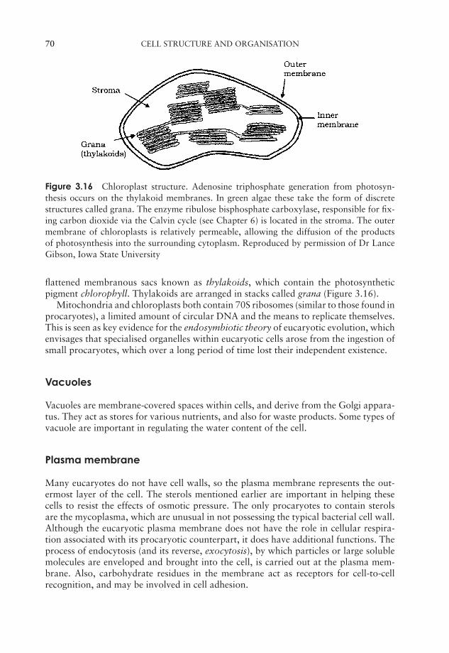
JWBK011-03 JWBK011-Hogg August 12, 2005 15:52 Char Count= 0
70 CELL STRUCTURE AND ORGANISATION
Figure 3.16 Chloroplast structure. Adenosine triphosphate generation from photosyn-thesis occurs on the thylakoid membranes. In green algae these take the form of discretestructures called grana. The enzyme ribulose bisphosphate carboxylase, responsible for fix-ing carbon dioxide via the Calvin cycle (see Chapter 6) is located in the stroma. The outermembrane of chloroplasts is relatively permeable, allowing the diffusion of the productsof photosynthesis into the surrounding cytoplasm. Reproduced by permission of Dr LanceGibson, Iowa State University
flattened membranous sacs known as thylakoids, which contain the photosyntheticpigment chlorophyll. Thylakoids are arranged in stacks called grana (Figure 3.16).
Mitochondria and chloroplasts both contain 70S ribosomes (similar to those found inprocaryotes), a limited amount of circular DNA and the means to replicate themselves.This is seen as key evidence for the endosymbiotic theory of eucaryotic evolution, whichenvisages that specialised organelles within eucaryotic cells arose from the ingestion ofsmall procaryotes, which over a long period of time lost their independent existence.
Vacuoles
Vacuoles are membrane-covered spaces within cells, and derive from the Golgi appara-tus. They act as stores for various nutrients, and also for waste products. Some types ofvacuole are important in regulating the water content of the cell.
Plasma membrane
Many eucaryotes do not have cell walls, so the plasma membrane represents the out-ermost layer of the cell. The sterols mentioned earlier are important in helping thesecells to resist the effects of osmotic pressure. The only procaryotes to contain sterolsare the mycoplasma, which are unusual in not possessing the typical bacterial cell wall.Although the eucaryotic plasma membrane does not have the role in cellular respira-tion associated with its procaryotic counterpart, it does have additional functions. Theprocess of endocytosis (and its reverse, exocytosis), by which particles or large solublemolecules are enveloped and brought into the cell, is carried out at the plasma mem-brane. Also, carbohydrate residues in the membrane act as receptors for cell-to-cellrecognition, and may be involved in cell adhesion.
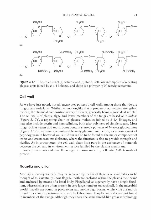
JWBK011-03 JWBK011-Hogg August 12, 2005 15:52 Char Count= 0
THE EUCARYOTIC CELL 71
CH2OH
CH2OH
OH OH
O
(a)
(b)
O
O
O
CH2OH
OH
O
O
CH2OH NHCOCH3
CH2OH
CH2OH
OH OHO
O
O
O
CH2OHNHCOCH3 NHCOCH3 NHCOCH3
OH OH
O
O
O
O
CH2OH
OH
O
O
CH2OH
CH2OH
OH OH
O
O
O
O
NHCOCH3
O O
O O
Figure 3.17 The structures of (a) cellulose and (b) chitin. Cellulose is composed of repeatingglucose units joined by β-1,4 linkages, and chitin is a polymer of N-acetylglucosamine
Cell wall
As we have just noted, not all eucaryotes possess a cell wall; among those that do arefungi, algae and plants. Whilst the function, like that of procaryotes, is to give strength tothe cell, the chemical composition is very different, generally being a good deal simpler.The cell walls of plants, algae and lower members of the fungi are based on cellulose(Figure 3.17a), a repeating chain of glucose molecules joined by β-1,4 linkages, andmay also include pectin and hemicellulose, both also polymers of simple sugars. Mostfungi such as yeasts and mushrooms contain chitin, a polymer of N-acetylglucosamine(Figure 3.17b: we have encountered N-acetylglucosamine before, as a component ofpeptidoglycan in bacterial walls.) Chitin is also to be found as the major component ofinsect and crustacean exoskeletons, where the function is also to provide strength andrigidity. As in procaryotes, the cell wall plays little part in the exchange of materialsbetween the cell and its environment, a role fulfilled by the plasma membrane.
Some protozoans and unicellular algae are surrounded by a flexible pellicle made ofprotein.
Flagella and cilia
Motility in eucaryotic cells may be achieved by means of flagella or cilia; cilia can bethought of as, essentially, short flagella. Both are enclosed within the plasma membraneand anchored by means of a basal body. Flagellated cells generally have a single flagel-lum, whereas cilia are often present in very large numbers on each cell. In the microbialworld, flagella are found in protozoans and motile algal forms, whilst cilia are mostlyfound in a class of protozoans called the Ciliophora. Flagella and cilia are not foundin members of the Fungi. Although they share the same thread-like gross morphology,
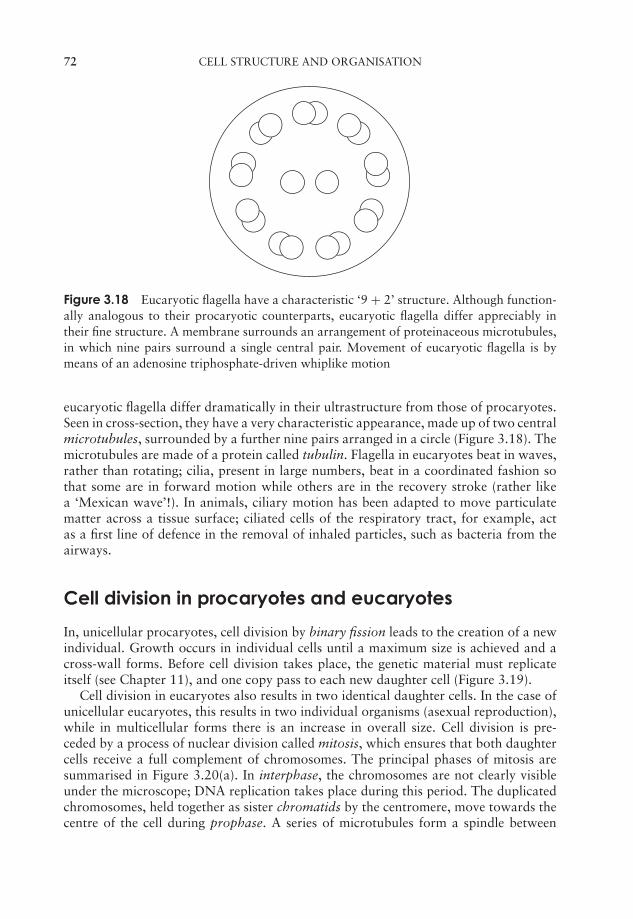
JWBK011-03 JWBK011-Hogg August 12, 2005 15:52 Char Count= 0
72 CELL STRUCTURE AND ORGANISATION
Figure 3.18 Eucaryotic flagella have a characteristic ‘9 + 2’ structure. Although function-ally analogous to their procaryotic counterparts, eucaryotic flagella differ appreciably intheir fine structure. A membrane surrounds an arrangement of proteinaceous microtubules,in which nine pairs surround a single central pair. Movement of eucaryotic flagella is bymeans of an adenosine triphosphate-driven whiplike motion
eucaryotic flagella differ dramatically in their ultrastructure from those of procaryotes.Seen in cross-section, they have a very characteristic appearance, made up of two centralmicrotubules, surrounded by a further nine pairs arranged in a circle (Figure 3.18). Themicrotubules are made of a protein called tubulin. Flagella in eucaryotes beat in waves,rather than rotating; cilia, present in large numbers, beat in a coordinated fashion sothat some are in forward motion while others are in the recovery stroke (rather likea ‘Mexican wave’!). In animals, ciliary motion has been adapted to move particulatematter across a tissue surface; ciliated cells of the respiratory tract, for example, actas a first line of defence in the removal of inhaled particles, such as bacteria from theairways.
Cell division in procaryotes and eucaryotes
In, unicellular procaryotes, cell division by binary fission leads to the creation of a newindividual. Growth occurs in individual cells until a maximum size is achieved and across-wall forms. Before cell division takes place, the genetic material must replicateitself (see Chapter 11), and one copy pass to each new daughter cell (Figure 3.19).
Cell division in eucaryotes also results in two identical daughter cells. In the case ofunicellular eucaryotes, this results in two individual organisms (asexual reproduction),while in multicellular forms there is an increase in overall size. Cell division is pre-ceded by a process of nuclear division called mitosis, which ensures that both daughtercells receive a full complement of chromosomes. The principal phases of mitosis aresummarised in Figure 3.20(a). In interphase, the chromosomes are not clearly visibleunder the microscope; DNA replication takes place during this period. The duplicatedchromosomes, held together as sister chromatids by the centromere, move towards thecentre of the cell during prophase. A series of microtubules form a spindle between
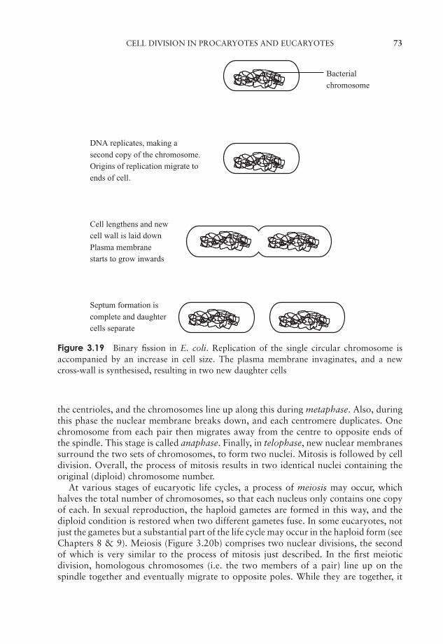
JWBK011-03 JWBK011-Hogg August 12, 2005 15:52 Char Count= 0
CELL DIVISION IN PROCARYOTES AND EUCARYOTES 73
Bacterial
chromosome
DNA replicates, making a
second copy of the chromosome.
Origins of replication migrate to
ends of cell.
Cell lengthens and new
cell wall is laid down
Plasma membrane
starts to grow inwards
Septum formation is
complete and daughter
cells separate
Figure 3.19 Binary fission in E. coli. Replication of the single circular chromosome isaccompanied by an increase in cell size. The plasma membrane invaginates, and a newcross-wall is synthesised, resulting in two new daughter cells
the centrioles, and the chromosomes line up along this during metaphase. Also, duringthis phase the nuclear membrane breaks down, and each centromere duplicates. Onechromosome from each pair then migrates away from the centre to opposite ends ofthe spindle. This stage is called anaphase. Finally, in telophase, new nuclear membranessurround the two sets of chromosomes, to form two nuclei. Mitosis is followed by celldivision. Overall, the process of mitosis results in two identical nuclei containing theoriginal (diploid) chromosome number.
At various stages of eucaryotic life cycles, a process of meiosis may occur, whichhalves the total number of chromosomes, so that each nucleus only contains one copyof each. In sexual reproduction, the haploid gametes are formed in this way, and thediploid condition is restored when two different gametes fuse. In some eucaryotes, notjust the gametes but a substantial part of the life cycle may occur in the haploid form (seeChapters 8 & 9). Meiosis (Figure 3.20b) comprises two nuclear divisions, the secondof which is very similar to the process of mitosis just described. In the first meioticdivision, homologous chromosomes (i.e. the two members of a pair) line up on thespindle together and eventually migrate to opposite poles. While they are together, it
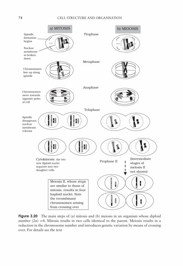
JWBK011-03 JWBK011-Hogg August 12, 2005 15:52 Char Count= 0
74 CELL STRUCTURE AND ORGANISATION
Cytokinesis: the twonew diploid nucleiseparate into twodaughter cells
Spindledisappears,nuclearmembranereforms
Telophase
Chromosomesmove towardsopposite polesof cell
Chromosomesline up alongspindle
Spindleformationbegins
Nuclearmembraneis brokendown
Prophase
Metaphase
Anaphase
Prophase II
a) MITOSIS b) MEIOSIS
(Intermediatestages ofmeiosis IInot shown)
Meiosis II, whose stepsare similar to those ofmitosis, results in fourhaploid nuclei. Notethe recombinantchromosomes arisingfrom crossing over
Figure 3.20 The main steps of (a) mitosis and (b) meiosis in an organism whose diploidnumber (2n) =4. Mitosis results in two cells identical to the parent. Meiosis results in areduction in the chromosome number and introduces genetic variation by means of crossingover. For details see the text
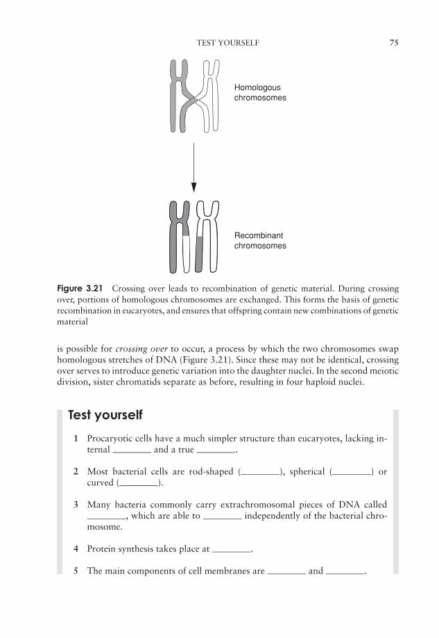
JWBK011-03 JWBK011-Hogg August 12, 2005 15:52 Char Count= 0
TEST YOURSELF 75
Homologouschromosomes
Recombinantchromosomes
Figure 3.21 Crossing over leads to recombination of genetic material. During crossingover, portions of homologous chromosomes are exchanged. This forms the basis of geneticrecombination in eucaryotes, and ensures that offspring contain new combinations of geneticmaterial
is possible for crossing over to occur, a process by which the two chromosomes swaphomologous stretches of DNA (Figure 3.21). Since these may not be identical, crossingover serves to introduce genetic variation into the daughter nuclei. In the second meioticdivision, sister chromatids separate as before, resulting in four haploid nuclei.
Test yourself
1 Procaryotic cells have a much simpler structure than eucaryotes, lacking in-ternal and a true .
2 Most bacterial cells are rod-shaped ( ), spherical ( ) orcurved ( ).
3 Many bacteria commonly carry extrachromosomal pieces of DNA called, which are able to independently of the bacterial chro-
mosome.
4 Protein synthesis takes place at .
5 The main components of cell membranes are and .

JWBK011-03 JWBK011-Hogg August 12, 2005 15:52 Char Count= 0
76 CELL STRUCTURE AND ORGANISATION
6 Gram-positive cell walls contain a higher percentage of than thoseof Gram-negative cells.
7 Many bacteria have long, hair-like structures called projecting fromthe cell wall. These are used for .
8 The DNA of eucaryotes is organized into chromosomes and associated withproteins called .
9 In eucaryotic cells, extranuclear DNA is also found in and.
10 Eucaryotic ribosomes may be found associated with theor free in the cytoplasm.
11 The Golgi apparatus and newly synthesised substances.
12 are the site of energy generation in eucaryotic cells. In procaryoticcells, some of these reactions take place at the .
13 The photosynthetic membranes of chloroplasts are called .
14 The cell walls of algae are mostly made up of .
15 The structure of eucaryotic flagella is more complex than that of procaryotes,comprising an arrangement of made of .

JWBK011-04 JWBK011-Hogg August 12, 2005 15:53 Char Count= 0
Part IIMicrobial Nutrition, Growthand Metabolism
77

JWBK011-04 JWBK011-Hogg August 12, 2005 15:53 Char Count= 0
78

JWBK011-04 JWBK011-Hogg August 12, 2005 15:53 Char Count= 0
4Microbial Nutritionand Cultivation
In Chapter 2 we introduced the major groups of macromolecules found in living cells;the raw materials from which these are synthesised are ultimately derived from theorganism’s environment in the form of nutrients (Table 4.1). These can be convenientlydivided into those required in large quantities* (macronutrients) and those which areneeded only in trace amounts (micronutrients or trace elements).
You will recall that carbon forms the central component of proteins, carbohydrates,nucleic acids and lipids; indeed, the living world is based on carbon, so it should come asno surprise that this is the most abundant element in all living cells, microbial or other-wise. Of the other macronutrients, nitrogen, oxygen, hydrogen, sulphur and phosphorusare also constituents of biological macromolecules, while the remainder (magnesium,potassium, sodium, calcium and iron in their ionised forms) are required in lesser quan-tities for a range of functions that will be described in due course. Micronutrients areall metal ions, and frequently serve as cofactors for enzymes.
All microorganisms must have a supply of the nutrients described above, but theyshow great versatility in the means they use to satisfy these requirements.
The metabolic processes by which microorganisms assimilate nutrients to make cel-lular material and derive energy will be reviewed in Chapter 6. In the following sec-tion we briefly describe the role of each element, and the form in which it may beacquired.
Carbon is the central component of the biological macromolecules we discussedin Chapter 2. Carbon incorporated into biosynthetic pathways may be derived fromorganic or inorganic sources (see below); some organisms can derive it from CO2, whileothers require their carbon in ‘ready-made’, organic form.
Hydrogen is also a key component of macromolecules, and participates in energy gen-eration processes in most microorganisms. In autotrophs (see ‘Nutritional categories’ be-low), hydrogen is required to reduce carbon dioxide in the synthesis of macromolecules.
Oxygen is of central importance to the respiration of many microorganisms, but inits molecular form (O2), it can be toxic to some forms (see Chapter 5). These obtain theoxygen they need for the synthesis of macromolecules from water.
* Everything is relative in the microbial world; a typical bacterial cell weighs around three tenmillionmillionths (3 × 10⁻¹³) of a gram!
79
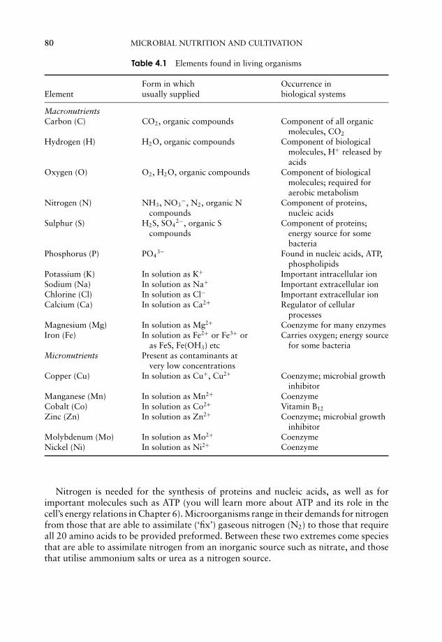
JWBK011-04 JWBK011-Hogg August 12, 2005 15:53 Char Count= 0
80 MICROBIAL NUTRITION AND CULTIVATION
Table 4.1 Elements found in living organisms
Form in which Occurrence inElement usually supplied biological systems
MacronutrientsCarbon (C) CO2, organic compounds Component of all organic
molecules, CO2
Hydrogen (H) H2O, organic compounds Component of biologicalmolecules, H+ released byacids
Oxygen (O) O2, H2O, organic compounds Component of biologicalmolecules; required foraerobic metabolism
Nitrogen (N) NH3, NO3−, N2, organic N
compoundsComponent of proteins,
nucleic acidsSulphur (S) H2S, SO4
2−, organic Scompounds
Component of proteins;energy source for somebacteria
Phosphorus (P) PO43− Found in nucleic acids, ATP,
phospholipidsPotassium (K) In solution as K+ Important intracellular ionSodium (Na) In solution as Na+ Important extracellular ionChlorine (Cl) In solution as Cl− Important extracellular ionCalcium (Ca) In solution as Ca2+ Regulator of cellular
processesMagnesium (Mg) In solution as Mg2+ Coenzyme for many enzymesIron (Fe) In solution as Fe2+ or Fe3+ or
as FeS, Fe(OH3) etcCarries oxygen; energy source
for some bacteriaMicronutrients Present as contaminants at
very low concentrationsCopper (Cu) In solution as Cu+, Cu2+ Coenzyme; microbial growth
inhibitorManganese (Mn) In solution as Mn2+ CoenzymeCobalt (Co) In solution as Co2+ Vitamin B12
Zinc (Zn) In solution as Zn2+ Coenzyme; microbial growthinhibitor
Molybdenum (Mo) In solution as Mo2+ CoenzymeNickel (Ni) In solution as Ni2+ Coenzyme
Nitrogen is needed for the synthesis of proteins and nucleic acids, as well as forimportant molecules such as ATP (you will learn more about ATP and its role in thecell’s energy relations in Chapter 6). Microorganisms range in their demands for nitrogenfrom those that are able to assimilate (‘fix’) gaseous nitrogen (N2) to those that requireall 20 amino acids to be provided preformed. Between these two extremes come speciesthat are able to assimilate nitrogen from an inorganic source such as nitrate, and thosethat utilise ammonium salts or urea as a nitrogen source.
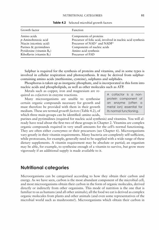
JWBK011-04 JWBK011-Hogg August 12, 2005 15:53 Char Count= 0
NUTRITIONAL CATEGORIES 81
Table 4.2 Selected microbial growth factors
Growth factor Function
Amino acids Components of proteinsp-Aminobenzoic acid Precursor of folic acid, involved in nucleic acid synthesisNiacin (nicotinic acid) Precursor of NAD+ and NADP+
Purines & pyrimidines Components of nucleic acidsPyridoxine (vitamin B6) Amino acid synthesisRiboflavin (vitamin B2) Precursor of FAD
Sulphur is required for the synthesis of proteins and vitamins, and in some types isinvolved in cellular respiration and photosynthesis. It may be derived from sulphur-containing amino acids (methionine, cysteine), sulphates and sulphides.
Phosphorus is taken up as inorganic phosphate, and is incorporated in this form intonucleic acids and phospholipids, as well as other molecules such as ATP.
A cofactor is a non-protein component ofan enzyme (often ametal ion) essential forits normal functioning.
Metals such as copper, iron and magnesium are re-quired as cofactors in enzyme reactions.
Many microorganisms are unable to synthesisecertain organic compounds necessary for growth andmust therefore be provided with them in their growthmedium. These are termed growth factors (Table 4.2), ofwhich three main groups can be identified: amino acids,purines and pyrimidines (required for nucleic acid synthesis) and vitamins. You will al-ready have read about the first two of these groups in Chapter 2. Vitamins are complexorganic compounds required in very small amounts for the cell’s normal functioning.They are often either coenzymes or their precursors (see Chapter 6). Microorganismsvary greatly in their vitamin requirements. Many bacteria are completely self-sufficient,while protozoans, for example, generally need to be supplied with a wide range of thesedietary supplements. A vitamin requirement may be absolute or partial; an organismmay be able, for example, to synthesise enough of a vitamin to survive, but grow morevigorously if an additional supply is made available to it.
Nutritional categories
Microorganisms can be categorised according to how they obtain their carbon andenergy. As we have seen, carbon is the most abundant component of the microbial cell,and most microorganisms obtain their carbon in the form of organic molecules, deriveddirectly or indirectly from other organisms. This mode of nutrition is the one that isfamiliar to us as humans (and all other animals); all the food we eat is derived as complexorganic molecules from plants and other animals (and even some representatives of themicrobial world such as mushrooms!). Microorganisms which obtain their carbon in
JWBK011-04 JWBK011-Hogg August 12, 2005 15:53 Char Count= 0
82 MICROBIAL NUTRITION AND CULTIVATION
A heterotroph must useone or more organiccompounds as its sourceof carbon.
this way are described as heterotrophs, and include allthe fungi and protozoans as well as most types of bac-teria. Microorganisms as a group are able to incorpo-rate the carbon from an incredibly wide range of or-ganic compounds into cellular material. In fact thereis hardly any such compound occurring in nature thatcannot be metabolised by some microorganism or other,explaining in part why microbial life is to be found thriving in the most unlikely habitats.Many synthetic materials can also serve as carbon sources for some microorganisms,which can have considerable economic significance.
An autotroph can deriveits carbon from carbondioxide.
A significant number of bacteria and all of the algaedo not, however, take up their carbon preformed as or-ganic molecules in this way, but derive it instead fromcarbon dioxide. These organisms are called autotrophs,and again we can draw a parallel with higher organ-isms, where all members of the plant kingdom obtaintheir carbon in a similar fashion.
A chemotroph obtainsits energy from chemi-cal compounds. A pho-totroph uses light as itssource of energy.
We can also categorise microorganisms nutritionallyby the way they derive the energy they require to carryout essential cellular reactions. Autotrophs thus fall intotwo categories. Chemoautotrophs obtain their energyas well as their carbon from inorganic sources; theydo this by the oxidation of inorganic molecules suchas sulphur or nitrite. Photoautotrophs have photosyn-thetic pigments enabling them to convert light energyinto chemical energy. The mechanisms by which this is achieved will be discussed inChapter 6.
The great majority of heterotrophs obtain energy as well as carbon from thesame organic source. Such organisms release energy by the chemical oxidation of or-ganic nutrient molecules, and are therefore termed chemoheterotrophs. Those few het-erotrophs which do not follow this mode of nutrition include the green and purplenon-sulphur bacteria. These are able to carry out photosynthesis and are known asphotoheterotrophs.
There is one final subdivision of nutritional categories in microorganisms! Whetherorganisms are chemotrophs or phototrophs, they need a molecule to act as a source
A lithotroph is an organ-ism that uses inorganicmolecules as a sourceof electrons. An organ-otroph uses organicmolecules for the samepurpose.
of electrons (reducing power) to drive their energy-generating systems (see Chapter 6). Those able to use aninorganic electron donor such as H2O, H2S or ammoniaare called lithotrophs, while those requiring an organicmolecule to fulfil the role are organotrophs. Most (butnot all) microorganisms are either photolithotrophic au-totrophs (algae, blue-greens) or chemo-organotrophicheterotrophs (most bacteria). For the latter category, asingle organic compound can often act as the provider ofcarbon, energy and reducing power. The substance usedby chemotrophs as an energy source may be organic (chemoorganotrophs) or inorganic(chemolithotrophs).

JWBK011-04 JWBK011-Hogg August 12, 2005 15:53 Char Count= 0
HOW DO NUTRIENTS GET INTO THE MICROBIAL CELL? 83
How do nutrients get into the microbial cell?
Having found a source of a given nutrient, a microorganism must:
� have some means of taking it up from the environment
� possess the appropriate enzyme systems to utilise it.
The plasma membrane represents a selective barrier, allowing into the cell only thosesubstances it is able to utilise. This selectivity is due in large part to the hydrophobicnature of the lipid bilayer. A substance can be transported across the cell membrane inone of three ways, known as simple diffusion, facilitated diffusion and active transport.
In simple diffusion, small molecules move across the membrane in response to aconcentration gradient (from high to low), until concentrations on either side of themembrane are in equilibrium. The ability to do this depends on being small (H2O,Na+, Cl−) or soluble in the lipid component of the membrane (non-polar gases such asO2 and CO2).
Larger polar molecules such as glucose and amino acids are unable to enter the cellunless assisted by membrane-spanning transport proteins by the process of facilitateddiffusion (Figure 4.1). Like enzymes, these proteins are specific for a single/small numberof related solutes; another parallel is that they too can become saturated by too much‘substrate’. As with simple diffusion, there is no expenditure of cellular energy, andan inward concentration gradient is required. The transported substance tends to bemetabolised rapidly once inside the cell, thus maintaining the concentration gradientfrom outside to inside.
Diffusion is only an effective method of internalising substances when their concen-trations are greater outside the cell than inside. Generally, however, microorganismsfind themselves in very dilute environments; hence the concentration gradient runs inthe other direction, and diffusion into the cell is not possible. Active transport enablesthe cell to overcome this unfavourable gradient. Here, regardless of the direction of thegradient, transport takes place in one direction only, into the cell. Energy, derived from
Figure 4.1 In facilitated diffusion, substances can move across the plasma membrane bybinding to an embedded transport protein (shown shaded). No energy input is required, butthe diffusion can only occur from an area of high concentration to one of low concentration.From Thomas, G: Medicinal Chemistry, an Introduction, John Wiley & Sons Inc., 2000.Reproduced by permission of the publishers
JWBK011-04 JWBK011-Hogg August 12, 2005 15:53 Char Count= 0
84 MICROBIAL NUTRITION AND CULTIVATION
hydrolysis of adenosine triphosphate (see Chapter 6) is required to achieve this, andagain specific transmembrane proteins are involved. They bind the solute molecule withhigh affinity outside of the cell, and then undergo a conformational change that causesthem to be released into the interior. Procaryotic cells can carry out a specialised formof active transport called group translocation, whereby the solute is chemically modi-fied as it crosses the membrane, preventing its escape. A well-studied example of thisis the phosphorylation of glucose in E. coli by the phosphotransferase system. Glucosepresent in very low concentrations outside the cell can be concentrated within it by thismechanism. Glucose is unable to pass back across the membrane in its phosphorylatedform (glucose-6-phosphate), however it can be utilised in metabolic pathways in thisform.
Often it may be necessary to employ extracellular enzymes to break down largemolecules before any of these mechanisms can be used to transport nutrients into thecell.
Laboratory cultivation of microorganisms
Critical to the development of microbiology during its ‘golden age’ was the advance inculturing techniques, enabling the isolation and pure culture of specific microorganisms.The study of pure cultures made it possible to determine the properties of a specificorganism such as its metabolic characteristics or its ability to cause a particular disease.It also opened up the possibility of classifying microorganisms, on the basis of thecharacteristics they display in pure culture.
The artificial culture of any organism requires a supply of the necessary nutrients,together with the provision of appropriate conditions such as temperature, pH andoxygen concentration. The nutrients and conditions provided in the laboratory are usu-ally a reflection of those found in the organism’s natural habitat. It is also essential thatappropriate steps are taken to avoid contamination (Box 4.1). In the next section weshall describe the techniques used to isolate and propagate microorganisms in the labo-ratory. The section refers specifically to the culture of bacteria; laboratory propagationof algae, fungi and viruses will be referred to in the chapters devoted to those groups.
Box 4.1 Aseptic technique
Most commonly used culture media will support the growth of a number of differentbacteria. It is therefore essential when working in the microbiology laboratory thatsuitable precautions are taken to prevent the growth of unwanted contaminants inour cultures. These simple practical measures are termed aseptic technique, and itis essential to master them if reliable experimental results are to be obtained. Anyglassware and equipment used is sterilised before work begins. Containers such astubes, flasks and plates are kept open for the minimum amount of time, and thenecks of bottles and tubes are passed through a flame to maintain their sterility.The wire loops and needles used to transfer small volumes of microbial culturesare sterilised by heating them to redness in a flame. Your instructor will normallydemonstrate aseptic technique to you in an early practical session.
JWBK011-04 JWBK011-Hogg August 12, 2005 15:53 Char Count= 0
LABORATORY CULTIVATION OF MICROORGANISMS 85
Obtaining a pure culture Bacteria may be cul-tured using either liquidor solid media. Solid me-dia are particularly use-ful in the isolation of bac-teria; they are also usedfor their long-term stor-age. Liquid (broth) cul-tures are used for rapidand large-scale produc-tion of bacteria.
Microorganisms in the natural world do not live in purecultures; they exist as part of complex ecosystems com-prising numerous other organisms. The first step in thecultivation of microorganisms is therefore the creation ofa pure culture. A key development for the production ofpure cultures was the ability to grow microorganisms ona solid medium. Koch had noticed that when a nutrientsurface such as cut potato was exposed to air, individualmicrobial colonies grew up, and he inferred from thisthat these had each arisen from the numerous divisionsof single cells.
It soon became apparent that a number of organisms would not grow on potatoes, soKoch and his colleagues turned to gelatin as a means of solidifying a synthetic nutrient
A culture consisting en-tirely of one strain of or-ganism is called a pureor axenic culture. In the-ory, such a culture repre-sents the descendants ofa single cell.
growth medium. Horizontal slabs were cut, and coveredto help keep them free from atmospheric contaminants.Gelatin was a convenient means of solidifying media, asit could be boiled and then allowed to set in the desiredvessel. There were two main drawbacks to its use, how-ever; many organisms needed to be incubated at aroundbody temperature (37 ◦C), and gelatin melted before thistemperature was reached. Also, it was found that a num-ber of bacteria were capable of utilising gelatin as a nu-trient source, resulting in the liquefaction of the gel.
A more suitable alternative was soon found in the form of agar. This is a complexpolysaccharide derived from seaweeds, and was suggested by the wife of one of Koch’scolleagues, who had used it as a setting agent in jam making. Agar does not meltuntil near boiling point; this means that cultures can be incubated at 37 ◦C or abovewithout the medium melting. Moreover, when it cools, agar remains molten until just
A petri dish is the stan-dard vessel for short-term growth of solidmedium cultures in thelaboratory. It comprisesa circular dish with anoverlapping lid.
over 40 ◦C, allowing heat-sensitive media componentssuch as blood to be added. In addition, most bacteria cantolerate a short exposure to temperatures in this range,so they too can be inoculated into molten agar (see pourplate method below). Crucially, agar is more or less in-ert nutritionally; only a very few organisms are knownthat are able to use agar as a food source; consequently,it is the near ideal setting agent, resisting both thermaland microbial breakdown. Agar soon became the settingagent of choice, and has remained so ever since; shortlyafterwards, Richard Petri developed the two-part culture dish that was named afterhim, and which could be sterilised separately from the medium and provided protectionfrom contamination by means of its lid,. This again is still standard equipment today,although the original glass has been largely replaced by presterilised, disposable plastic.
The standard method of obtaining a pure bacterial culture is the creation of a streakplate (Figure 4.2). A wire inoculating loop is used to spread out a drop of bacterialsuspension on an agar plate in such a way that it becomes progressively more dilute;
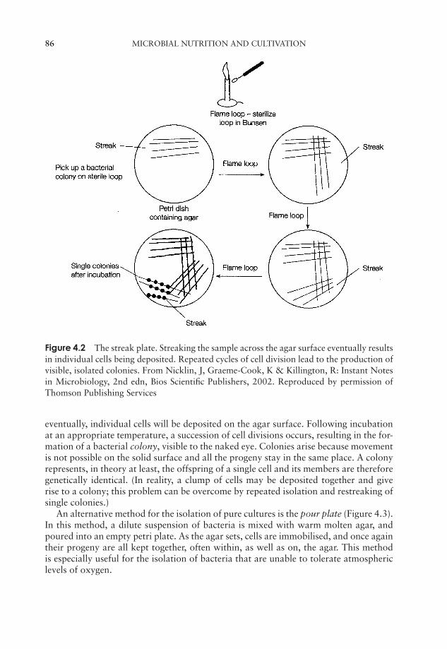
JWBK011-04 JWBK011-Hogg August 12, 2005 15:53 Char Count= 0
86 MICROBIAL NUTRITION AND CULTIVATION
Figure 4.2 The streak plate. Streaking the sample across the agar surface eventually resultsin individual cells being deposited. Repeated cycles of cell division lead to the production ofvisible, isolated colonies. From Nicklin, J, Graeme-Cook, K & Killington, R: Instant Notesin Microbiology, 2nd edn, Bios Scientific Publishers, 2002. Reproduced by permission ofThomson Publishing Services
eventually, individual cells will be deposited on the agar surface. Following incubationat an appropriate temperature, a succession of cell divisions occurs, resulting in the for-mation of a bacterial colony, visible to the naked eye. Colonies arise because movementis not possible on the solid surface and all the progeny stay in the same place. A colonyrepresents, in theory at least, the offspring of a single cell and its members are thereforegenetically identical. (In reality, a clump of cells may be deposited together and giverise to a colony; this problem can be overcome by repeated isolation and restreaking ofsingle colonies.)
An alternative method for the isolation of pure cultures is the pour plate (Figure 4.3).In this method, a dilute suspension of bacteria is mixed with warm molten agar, andpoured into an empty petri plate. As the agar sets, cells are immobilised, and once againtheir progeny are all kept together, often within, as well as on, the agar. This methodis especially useful for the isolation of bacteria that are unable to tolerate atmosphericlevels of oxygen.

JWBK011-04 JWBK011-Hogg August 12, 2005 15:53 Char Count= 0
LABORATORY CULTIVATION OF MICROORGANISMS 87
Moltenagar
at 45–50°
Colonies grow onsurface and within
agar
Bacterial suspension
Mix, incubate
Figure 4.3 The pour plate. A sample of diluted bacterial suspension is mixed with moltenagar and poured into a petri dish. Most bacteria can tolerate a short exposure to the agar,which is held at a temperature just above its setting point
Growth media for the cultivation of bacteria
A defined medium is onewhose precise chemicalcomposition is known.
A synthetic growth medium may be defined, that is, itsexact chemical composition is known, or undefined. Adefined growth medium may have few or many con-stituents, depending on the nutritional requirements of
An undefined or com-plex medium is onewhose precise chemi-cal composition is notknown.
the organism in question. Examples of each are givenin Table 4.3. An undefined or complex medium mayhave a variable composition due to the inclusion ofa component such as blood, yeast extract or tap wa-ter (Table 4.4). Peptones are also commonly found incomplex media; these are the products of partially di-gesting protein sources such as beef or casein. Theexact composition of a complex medium is neither
A fastidious organism isunable to synthesise arange of nutrients andtherefore has complexrequirements in culture.
known nor critically important. A medium of thistype would generally be chosen for the cultivation offastidious bacteria such as Neisseria gonorrhoeae (thecausative agent of gonorrhoea); it is easier and less ex-pensive to supply the many nutrients required by such anorganism in this form rather than supplying them all in-dividually. Bacteria whose specific nutrient requirementsare not known are also grown on complex media.
A selective medium isone that favours thegrowth of a particular or-ganism or group of or-ganisms, often by sup-pressing the growth ofothers.
Whilst media such as nutrient agar are used to sup-port the growth of a wide range of organisms, others arespecifically designed for the isolation and identificationof particular types. Selective media such as bismuth sul-phite medium preferentially support the growth of par-ticular bacteria. The bismuth ion inhibits the growth ofGram-positive organisms as well as many Gram-negativetypes; this medium is used for the isolation of the
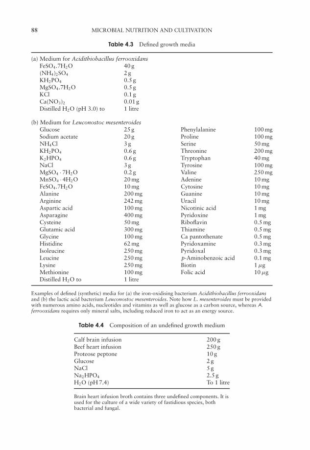
JWBK011-04 JWBK011-Hogg August 12, 2005 15:53 Char Count= 0
88 MICROBIAL NUTRITION AND CULTIVATION
Table 4.3 Defined growth media
(a) Medium for Acidithiobacillus ferrooxidansFeSO4.7H2O 40 g(NH4)2SO4 2 gKH2PO4 0.5 gMgSO4.7H2O 0.5 gKCl 0.1 gCa(NO3)2 0.01 gDistilled H2O (pH 3.0) to 1 litre
(b) Medium for Leuconostoc mesenteroidesGlucose 25 g Phenylalanine 100 mgSodium acetate 20 g Proline 100 mgNH4Cl 3 g Serine 50 mgKH2PO4 0.6 g Threonine 200 mgK2HPO4 0.6 g Tryptophan 40 mgNaCl 3 g Tyrosine 100 mgMgSO4 · 7H2O 0.2 g Valine 250 mgMnSO4 · 4H2O 20 mg Adenine 10 mgFeSO4.7H2O 10 mg Cytosine 10 mgAlanine 200 mg Guanine 10 mgArginine 242 mg Uracil 10 mgAspartic acid 100 mg Nicotinic acid 1 mgAsparagine 400 mg Pyridoxine 1 mgCysteine 50 mg Riboflavin 0.5 mgGlutamic acid 300 mg Thiamine 0.5 mgGlycine 100 mg Ca pantothenate 0.5 mgHistidine 62 mg Pyridoxamine 0.3 mgIsoleucine 250 mg Pyridoxal 0.3 mgLeucine 250 mg p-Aminobenzoic acid 0.1 mgLysine 250 mg Biotin 1 µgMethionine 100 mg Folic acid 10 µgDistilled H2O to 1 litre
Examples of defined (synthetic) media for (a) the iron-oxidising bacterium Acidithiobacillus ferrooxidansand (b) the lactic acid bacterium Leuconostoc mesenteroides. Note how L. mesenteroides must be providedwith numerous amino acids, nucleotides and vitamins as well as glucose as a carbon source, whereas A.
ferrooxidans requires only mineral salts, including reduced iron to act as an energy source.
Table 4.4 Composition of an undefined growth medium
Calf brain infusion 200 gBeef heart infusion 250 gProteose peptone 10 gGlucose 2 gNaCl 5 gNa2HPO4 2.5 gH2O (pH 7.4) To 1 litre
Brain heart infusion broth contains three undefined components. It isused for the culture of a wide variety of fastidious species, bothbacterial and fungal.
JWBK011-04 JWBK011-Hogg August 12, 2005 15:53 Char Count= 0
TEST YOURSELF 89
pathogenic bacterium Salmonella typhi, one of the few organisms that can toleratethe bismuth. Specific media called differential media can be used to distinguish be-tween organisms whose growth they support, usually by means of a coloured indica-tor. MacConkey agar contains lactose and a pH indicator, allowing the differentiation
A differential medium al-lows colonies of a partic-ular organism to be dif-ferentiated from othersgrowing in the same cul-ture.
between lactose fermenters (red colonies) and non-lactose fermenters (white/pale pink colonies). Many me-dia act both selectively and differentially; MacConkeyagar, for example, also contains bile salts and the dyecrystal violet, both of which serve to inhibit the growthof unwanted Gram-positive bacteria. Mannitol salt agaris also both selective and differential. The high (7.5 percent) salt content suppresses growth of most bacteria,whilst a combination of mannitol and an indicator permits the detection of mannitol fer-menters in a similar fashion to that just described. Sometimes, it is desirable to isolate an
An enrichment cultureuses a selective mediumto encourage thegrowth of an organismpresent in low numbers.
organism that is present in small numbers in a largemixed population (e.g. faeces or soil). Enrichment mediaprovide conditions that selectively encourage the growthof these organisms; the use of blood agar in the isolationof streptococci provides an example of such a medium.Blood agar can act as a differential medium, in allow-ing the user to distinguish between haemolytic and non-haemolytic bacteria (see Chapter 7).
If we are to culture microorganisms successfully in the laboratory, we must provideappropriate physical conditions as well as providing an appropriate nutrient medium.In the next chapter, we shall examine how physical factors such as pH and temperatureinfluence the growth of microorganisms, and describe how these conditions are providedin the laboratory.
Preservation of microbial cultures
Microbial cultures are preserved by storage at low temperatures, in order to suspendgrowth processes. For short periods, most organisms can be kept at refrigerator temper-ature (around 4 ◦C), but for longer-term storage, more specialised treatment is necessary.Using deep freezing or freeze-drying, cultures can be kept for many years, and then resur-rected and re-cultured. Deep freezing requires rapid freezing to −70 ◦C to −95 ◦C, whilefreeze-drying (lyophilisation) involves freezing at slightly less extreme temperatures andremoving the water content under vacuum. Long-term storage may be desirable to avoidthe development of mutations or loss of cell viability.
Test yourself
1 Heterotrophic organisms acquire their carbon in an form, whilstautotrophic organisms acquire theirs in an form.
2 Some autotrophs can derive energy from the Sun; these are termed .
JWBK011-04 JWBK011-Hogg August 12, 2005 15:53 Char Count= 0
90 MICROBIAL NUTRITION AND CULTIVATION
3 The passage of solutes into a cell across a concentration gradient is knownas .
4 Agar has a high point and a relatively low point.
5 The use of agar as a setting agent was a crucial step in the development oftechniques.
6 Successive divisions of a single cell lead to the formation of a on asolid medium.
7 The chemical composition of an undefined medium is .
8 organisms must have a wide range of organic nutrients supplied.
9 media encourage the growth of chosen species, whilstmedia prevent the growth of unwanted forms.
10 (removal of the water content at low temperatures) is used in thepreservation of microbial cultures.
JWBK011-05 JWBK011-Hogg August 12, 2005 15:56 Char Count= 0
5Microbial Growth
When we consider growth as applied to a multicellular organism such as a tree, a fishor a human being, we think in terms of an ordered increase in the size of an individual.Growth in unicellular microorganisms such as bacteria, yeasts and protozoans, however,is more properly defined in terms of an increase in the size of a given population.
Biomass is the total amo-unt of cellular material ina system.
This may be expressed as an increase in either the numberof individuals or the total amount of biomass. Methodsemployed in the measurement of growth of unicellularmicroorganisms may be based on either of these. In thischapter we shall describe some of these methods, beforeconsidering the dynamics of microbial growth and some of the factors that affect it.
Estimation of microbial numbers
Several methods exist for the measurement of bacterial numbers, most of which are alsoapplicable to the enumeration of other unicellular forms such as yeasts. Such methodsfall into two main categories: those that count total cell numbers, and those that countviable cells only.
Total cell counts are generally done by direct microscopic examination. A specialisedglass slide is employed, which carries an etched grid of known area (Figure 5.1). Thedepth of the liquid sample is also known, so by counting the number of cells visible inthe field of view, the number of cells per unit volume can be determined. The methodmay be made more accurate by the use of a fluorescent dye such as acridine orange,which binds to DNA, and hence avoids confusion with non-cellular debris. However,such methods cannot differentiate between living and non-living cells. Their usefulnessis further limited by the fact that the smallest bacteria are difficult to resolve as indi-vidual cells by light microscopy. Other total cell count methods use cell-sorting devices,originally developed for separating blood cells in medical research. These pass the cellsuspension through an extremely fine nozzle, and a detector registers the conductivitychange each time a particle passes it. Again, no distinction can be made between viableand non-viable cells.
A viable cell count, on the other hand, is a measure of the number of living cells in asample, or more specifically those capable of multiplying and producing a visible colonyof cells. It is most commonly estimated by spreading a known volume of cell suspensiononto an agar plate, and counting the number of colonies that arise after a period of
91
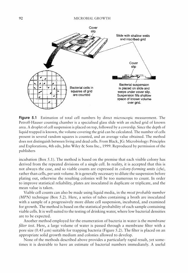
JWBK011-05 JWBK011-Hogg August 12, 2005 15:56 Char Count= 0
92 MICROBIAL GROWTH
Figure 5.1 Estimation of total cell numbers by direct microscopic measurement. ThePetroff-Hauser counting chamber is a specialised glass slide with an etched grid of knownarea. A droplet of cell suspension is placed on top, followed by a coverslip. Since the depth ofliquid trapped is known, the volume covering the grid can be calculated. The number of cellspresent in several random squares is counted, and an average value obtained. The methoddoes not distinguish between living and dead cells. From Black, JG: Microbiology: Principlesand Explorations, 4th edn, John Wiley & Sons Inc., 1999. Reproduced by permission of thepublishers
incubation (Box 5.1). The method is based on the premise that each visible colony hasderived from the repeated divisions of a single cell. In reality, it is accepted that this isnot always the case, and so viable counts are expressed in colony-forming units (cfu),rather than cells, per unit volume. It is generally necessary to dilute the suspension beforeplating out, otherwise the resulting colonies will be too numerous to count. In orderto improve statistical reliability, plates are inoculated in duplicate or triplicate, and themean value is taken.
Viable cell counts can also be made using liquid media, in the most probable number(MPN) technique (Box 5.2). Here, a series of tubes containing a broth are inoculatedwith a sample of a progressively more dilute cell suspension, incubated, and examinedfor growth. The method is based on the statistical probability of each sample containingviable cells. It is well suited to the testing of drinking water, where low bacterial densitiesare to be expected.
Another method employed for the enumeration of bacteria in water is the membranefilter test. Here, a large volume of water is passed through a membrane filter with apore size (0.45 µm) suitable for trapping bacteria (Figure 5.2). The filter is placed on anappropriate solid growth medium and colonies allowed to develop.
None of the methods described above provides a particularly rapid result, yet some-times it is desirable to have an estimate of bacterial numbers immediately. A useful
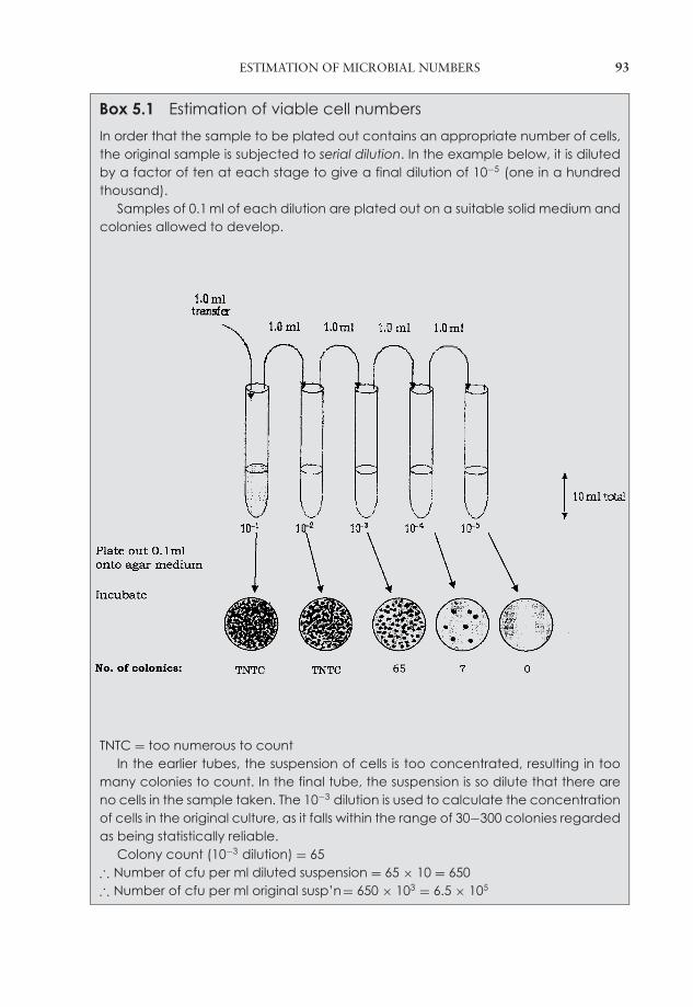
JWBK011-05 JWBK011-Hogg August 12, 2005 15:56 Char Count= 0
ESTIMATION OF MICROBIAL NUMBERS 93
Box 5.1 Estimation of viable cell numbers
In order that the sample to be plated out contains an appropriate number of cells,the original sample is subjected to serial dilution. In the example below, it is dilutedby a factor of ten at each stage to give a final dilution of 10−5 (one in a hundredthousand).
Samples of 0.1 ml of each dilution are plated out on a suitable solid medium andcolonies allowed to develop.
TNTC = too numerous to countIn the earlier tubes, the suspension of cells is too concentrated, resulting in too
many colonies to count. In the final tube, the suspension is so dilute that there areno cells in the sample taken. The 10−3 dilution is used to calculate the concentrationof cells in the original culture, as it falls within the range of 30−300 colonies regardedas being statistically reliable.
Colony count (10−3 dilution) = 65∴ Number of cfu per ml diluted suspension = 65 × 10 = 650∴ Number of cfu per ml original susp’n= 650 × 103 = 6.5 × 10⁵
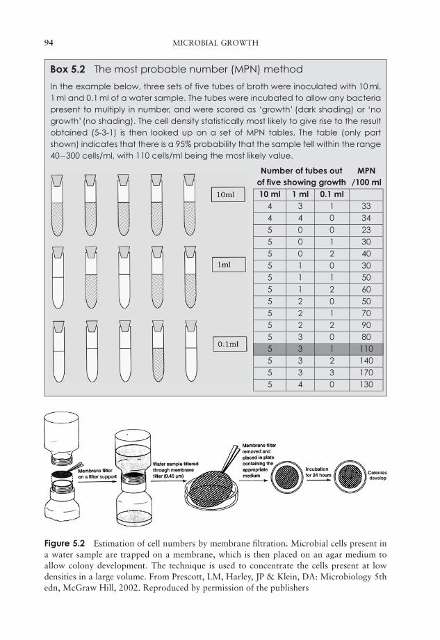
JWBK011-05 JWBK011-Hogg August 12, 2005 15:56 Char Count= 0
94 MICROBIAL GROWTH
Box 5.2 The most probable number (MPN) method
In the example below, three sets of five tubes of broth were inoculated with 10 ml,1 ml and 0.1 ml of a water sample. The tubes were incubated to allow any bacteriapresent to multiply in number, and were scored as ‘growth’ (dark shading) or ‘nogrowth’ (no shading). The cell density statistically most likely to give rise to the resultobtained (5-3-1) is then looked up on a set of MPN tables. The table (only partshown) indicates that there is a 95% probability that the sample fell within the range40−300 cells/ml, with 110 cells/ml being the most likely value.
Number of tubes out MPNof five showing growth /100 ml10 ml 1 ml 0.1 ml
4 3 1 334 4 0 345 0 0 235 0 1 305 0 2 405 1 0 305 1 1 505 1 2 605 2 0 505 2 1 705 2 2 905 3 0 805 3 1 1105 3 2 1405 3 3 1705 4 0 130
Figure 5.2 Estimation of cell numbers by membrane filtration. Microbial cells present ina water sample are trapped on a membrane, which is then placed on an agar medium toallow colony development. The technique is used to concentrate the cells present at lowdensities in a large volume. From Prescott, LM, Harley, JP & Klein, DA: Microbiology 5thedn, McGraw Hill, 2002. Reproduced by permission of the publishers
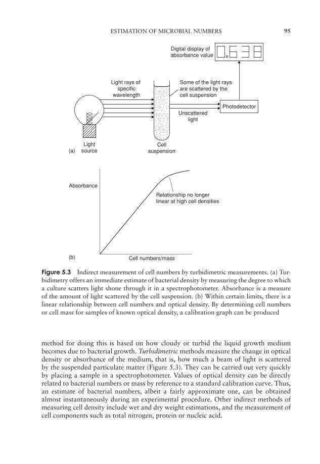
JWBK011-05 JWBK011-Hogg August 12, 2005 15:56 Char Count= 0
ESTIMATION OF MICROBIAL NUMBERS 95
Absorbance
Cell numbers/mass
Relationship no longerlinear at high cell densities
(a)
(b)
Light rays of specific
wavelength
Lightsource
Cellsuspension
Unscatteredlight
Some of the light raysare scattered by thecell suspension
Photodetector
Digital display ofabsorbance value
Figure 5.3 Indirect measurement of cell numbers by turbidimetric measurements. (a) Tur-bidimetry offers an immediate estimate of bacterial density by measuring the degree to whicha culture scatters light shone through it in a spectrophotometer. Absorbance is a measureof the amount of light scattered by the cell suspension. (b) Within certain limits, there is alinear relationship between cell numbers and optical density. By determining cell numbersor cell mass for samples of known optical density, a calibration graph can be produced
method for doing this is based on how cloudy or turbid the liquid growth mediumbecomes due to bacterial growth. Turbidimetric methods measure the change in opticaldensity or absorbance of the medium, that is, how much a beam of light is scatteredby the suspended particulate matter (Figure 5.3). They can be carried out very quicklyby placing a sample in a spectrophotometer. Values of optical density can be directlyrelated to bacterial numbers or mass by reference to a standard calibration curve. Thus,an estimate of bacterial numbers, albeit a fairly approximate one, can be obtainedalmost instantaneously during an experimental procedure. Other indirect methods ofmeasuring cell density include wet and dry weight estimations, and the measurement ofcell components such as total nitrogen, protein or nucleic acid.
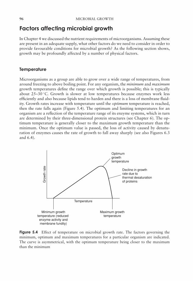
JWBK011-05 JWBK011-Hogg August 12, 2005 15:56 Char Count= 0
96 MICROBIAL GROWTH
Factors affecting microbial growth
In Chapter 4 we discussed the nutrient requirements of microorganisms. Assuming theseare present in an adequate supply, what other factors do we need to consider in order toprovide favourable conditions for microbial growth? As the following section shows,growth may be profoundly affected by a number of physical factors.
Temperature
Microorganisms as a group are able to grow over a wide range of temperatures, fromaround freezing to above boiling point. For any organism, the minimum and maximumgrowth temperatures define the range over which growth is possible; this is typicallyabout 25–30 ◦C. Growth is slower at low temperatures because enzymes work lessefficiently and also because lipids tend to harden and there is a loss of membrane fluid-ity. Growth rates increase with temperature until the optimum temperature is reached,then the rate falls again (Figure 5.4). The optimum and limiting temperatures for anorganism are a reflection of the temperature range of its enzyme systems, which in turnare determined by their three-dimensional protein structures (see Chapter 6). The op-timum temperature is generally closer to the maximum growth temperature than theminimum. Once the optimum value is passed, the loss of activity caused by denatu-ration of enzymes causes the rate of growth to fall away sharply (see also Figures 6.3and 6.4).
Optimumgrowthtemperature
Decline in growthrate due tothermal desaturationof proteinsG
row
th r
ate
Minimum growthtemperature (reduced enzyme activity andmembrane fundity)
Maximum growthtemperature
Temperature
Figure 5.4 Effect of temperature on microbial growth rate. The factors governing theminimum, optimum and maximum temperatures for a particular organism are indicated.The curve is asymmetrical, with the optimum temperature being closer to the maximumthan the minimum

JWBK011-05 JWBK011-Hogg August 12, 2005 15:56 Char Count= 0
FACTORS AFFECTING MICROBIAL GROWTH 97
Gro
wth
Rat
e Mesophile
ThermophilePsychrophile
Extreme thermophile
Temperature(°c)
Figure 5.5 Microorganisms can be categorised according to the temperature range at whichthey grow
The majority of microorganisms achieve optimal growth at ‘middling’ temperaturesof around 20–45 ◦C; these are called mesophiles (Figure 5.5). Contrast these with ther-mophiles, which have become adapted to not only surviving, but thriving at much highertemperatures. Typically, these would be capable of growth within a range of about40–80 ◦C, with an optimum around 50–65 ◦C. Extreme thermophiles have optimumvalues in excess of this, and can tolerate temperatures in excess of 100 ◦C. In 2003,a member of the primitive bacterial group called the Archaea (see Chapter 7) wasreported as growing at a temperature of 121 ◦C, a new world record! Psychrophilesoccupy the other extreme of the temperature range; they can grow at 0◦C, with optimalgrowth occurring at 15 ◦C or below. Such organisms are not able to grow at tem-peratures above 25 ◦C or so. Psychrotrophs, on the other hand, although they canalso grow at 0 ◦C, have much higher temperature optima (20–30 ◦C). Members of thisgroup are often economically significant due to their ability to grow on refrigeratedfoodstuffs.
In the laboratory, appropriate temperatures for growth are provided by culturingin an appropriate incubator. These come in a variety of shapes and sizes, but all arethermostatically controlled and generally hold the temperature within a degree or twoof the desired value.
pH
Microorganisms are strongly influenced by the prevailing pH of their surroundings. Aswith temperature, we can define minimum, optimum and maximum values for growthof a particular type (Figure 5.6). The pH range (between minimum and maximumvalues) is greater in fungi than it is in bacteria. Most microorganisms grow best aroundneutrality (pH 7). Many bacteria prefer slightly alkaline conditions but relatively few
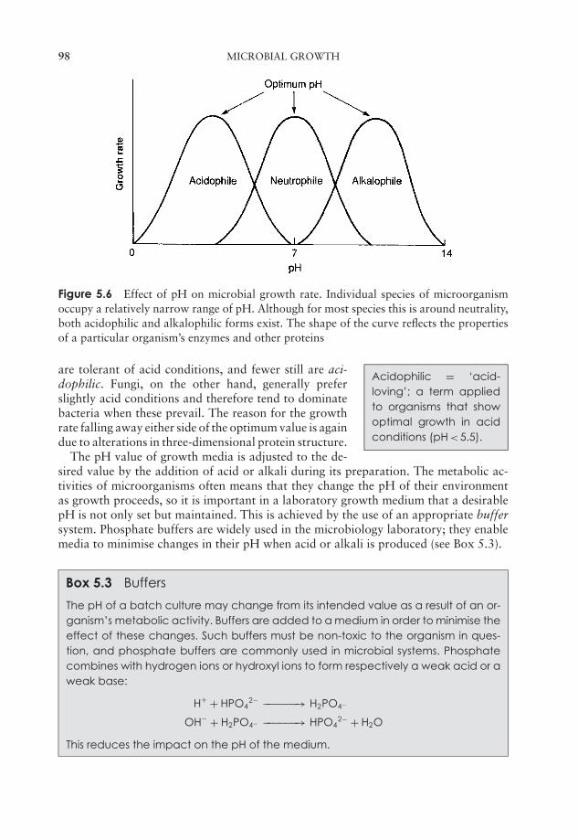
JWBK011-05 JWBK011-Hogg August 12, 2005 15:56 Char Count= 0
98 MICROBIAL GROWTH
Figure 5.6 Effect of pH on microbial growth rate. Individual species of microorganismoccupy a relatively narrow range of pH. Although for most species this is around neutrality,both acidophilic and alkalophilic forms exist. The shape of the curve reflects the propertiesof a particular organism’s enzymes and other proteins
Acidophilic = ‘acid-loving’; a term appliedto organisms that showoptimal growth in acidconditions (pH < 5.5).
are tolerant of acid conditions, and fewer still are aci-dophilic. Fungi, on the other hand, generally preferslightly acid conditions and therefore tend to dominatebacteria when these prevail. The reason for the growthrate falling away either side of the optimum value is againdue to alterations in three-dimensional protein structure.
The pH value of growth media is adjusted to the de-sired value by the addition of acid or alkali during its preparation. The metabolic ac-tivities of microorganisms often means that they change the pH of their environmentas growth proceeds, so it is important in a laboratory growth medium that a desirablepH is not only set but maintained. This is achieved by the use of an appropriate buffersystem. Phosphate buffers are widely used in the microbiology laboratory; they enablemedia to minimise changes in their pH when acid or alkali is produced (see Box 5.3).
Box 5.3 Buffers
The pH of a batch culture may change from its intended value as a result of an or-ganism’s metabolic activity. Buffers are added to a medium in order to minimise theeffect of these changes. Such buffers must be non-toxic to the organism in ques-tion, and phosphate buffers are commonly used in microbial systems. Phosphatecombines with hydrogen ions or hydroxyl ions to form respectively a weak acid or aweak base:
H+ + HPO42− −−−−−−→ H2PO4−
OH− + H2PO4− −−−−−−→ HPO42− + H2O
This reduces the impact on the pH of the medium.
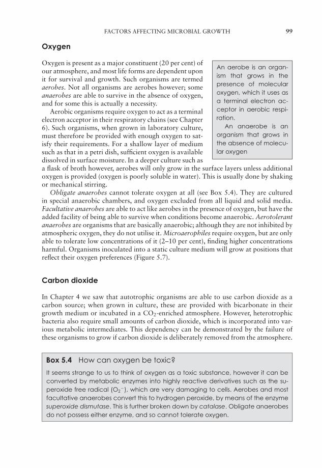
JWBK011-05 JWBK011-Hogg August 12, 2005 15:56 Char Count= 0
FACTORS AFFECTING MICROBIAL GROWTH 99
Oxygen
An aerobe is an organ-ism that grows in thepresence of molecularoxygen, which it uses asa terminal electron ac-ceptor in aerobic respi-ration.
An anaerobe is anorganism that grows inthe absence of molecu-lar oxygen
Oxygen is present as a major constituent (20 per cent) ofour atmosphere, and most life forms are dependent uponit for survival and growth. Such organisms are termedaerobes. Not all organisms are aerobes however; someanaerobes are able to survive in the absence of oxygen,and for some this is actually a necessity.
Aerobic organisms require oxygen to act as a terminalelectron acceptor in their respiratory chains (see Chapter6). Such organisms, when grown in laboratory culture,must therefore be provided with enough oxygen to sat-isfy their requirements. For a shallow layer of mediumsuch as that in a petri dish, sufficient oxygen is availabledissolved in surface moisture. In a deeper culture such asa flask of broth however, aerobes will only grow in the surface layers unless additionaloxygen is provided (oxygen is poorly soluble in water). This is usually done by shakingor mechanical stirring.
Obligate anaerobes cannot tolerate oxygen at all (see Box 5.4). They are culturedin special anaerobic chambers, and oxygen excluded from all liquid and solid media.Facultative anaerobes are able to act like aerobes in the presence of oxygen, but have theadded facility of being able to survive when conditions become anaerobic. Aerotolerantanaerobes are organisms that are basically anaerobic; although they are not inhibited byatmospheric oxygen, they do not utilise it. Microaerophiles require oxygen, but are onlyable to tolerate low concentrations of it (2–10 per cent), finding higher concentrationsharmful. Organisms inoculated into a static culture medium will grow at positions thatreflect their oxygen preferences (Figure 5.7).
Carbon dioxide
In Chapter 4 we saw that autotrophic organisms are able to use carbon dioxide as acarbon source; when grown in culture, these are provided with bicarbonate in theirgrowth medium or incubated in a CO2-enriched atmosphere. However, heterotrophicbacteria also require small amounts of carbon dioxide, which is incorporated into var-ious metabolic intermediates. This dependency can be demonstrated by the failure ofthese organisms to grow if carbon dioxide is deliberately removed from the atmosphere.
Box 5.4 How can oxygen be toxic?
It seems strange to us to think of oxygen as a toxic substance, however it can beconverted by metabolic enzymes into highly reactive derivatives such as the su-peroxide free radical (O2
−), which are very damaging to cells. Aerobes and mostfacultative anaerobes convert this to hydrogen peroxide, by means of the enzymesuperoxide dismutase. This is further broken down by catalase. Obligate anaerobesdo not possess either enzyme, and so cannot tolerate oxygen.
JWBK011-05 JWBK011-Hogg August 12, 2005 15:56 Char Count= 0
100 MICROBIAL GROWTH
(a) Obligate aerobes
(c) Obligate anaerobes
(d) Aerotolerant anaerobes
(b) Facultative anaerobes
(e) Microaerophiles
Figure 5.7 Microorganisms have different oxygen requirements. In a static culture, mi-croorganisms occupy different regions of the medium, reflecting their pattern of oxygenusage. (a) Obligate aerobes must grow at or near the surface, where oxygen is able to dif-fuse. (b) Facultative anaerobes are able to adjust their metabolism to the prevailing oxygenconditions. (c) Obligate anaerobes, in contrast, occupy those zones where no oxygen ispresent at all. (d) Aerotolerant anaerobes do not use oxygen, but neither are they inhibitedby it. (e) Microaerophiles have specific oxygen requirements, and can only grow within anarrow range of oxygen tensions
Osmotic pressure
Osmosis is the diffusion of water across a semipermeable membrane from a less con-centrated solution to a more concentrated one, equalising concentrations. The pressurerequired to make this happen is called the osmotic pressure. If a cell were placed in a
Plasmolysis is the shrink-age of the plasma mem-brane away from thecell wall, due to osmoticloss of water from thecell.
hypertonic solution (one whose solute concentration ishigher), osmosis would lead to a loss of water from thecell (plasmolysis). This is the basis of using high con-centrations of salt or other solutes in preserving foodsagainst microbial attack. In the opposite situation, wa-ter would pass from a dilute (hypotonic) solution into thecell, causing it to swell and burst. The rigid cell walls ofbacteria prevent them from bursting; this, together withtheir minute size, makes them less sensitive to variationsin osmotic pressure than other types of cell. They are generally able to tolerate NaCl con-centrations of between 0.5 and 3.0 per cent. Haloduric (‘salt-tolerant’) bacteria are ableto tolerate concentrations ten times as high, but prefer lower concentrations, whereashalophilic (‘salt-loving’) forms are adapted to grow best in conditions of high salinitysuch as those that prevail in the Dead Sea in the Middle East. In order to do this withoutplasmolysis occurring, they must build up a higher internal solute concentration, whichthey do by actively concentrating potassium ions inside the cell.
Light
Phototrophic organisms require light in order to carry out photosynthesis. In the lab-oratory, care must be taken that light of the correct wavelength is used, and that thesource used does not also act as a heat source. Fluorescent light produces little heat,
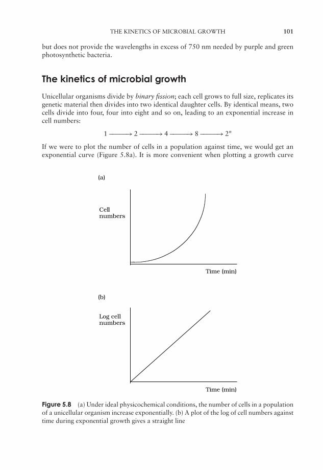
JWBK011-05 JWBK011-Hogg August 12, 2005 15:56 Char Count= 0
THE KINETICS OF MICROBIAL GROWTH 101
but does not provide the wavelengths in excess of 750 nm needed by purple and greenphotosynthetic bacteria.
The kinetics of microbial growth
Unicellular organisms divide by binary fission; each cell grows to full size, replicates itsgenetic material then divides into two identical daughter cells. By identical means, twocells divide into four, four into eight and so on, leading to an exponential increase incell numbers:
1 −−−−−→ 2 −−−−−→ 4 −−−−−→ 8 −−−−−→ 2n
If we were to plot the number of cells in a population against time, we would get anexponential curve (Figure 5.8a). It is more convenient when plotting a growth curve
(a)
Cellnumbers
Time (min)
(b)
Log cellnumbers
Time (min)
Figure 5.8 (a) Under ideal physicochemical conditions, the number of cells in a populationof a unicellular organism increase exponentially. (b) A plot of the log of cell numbers againsttime during exponential growth gives a straight line
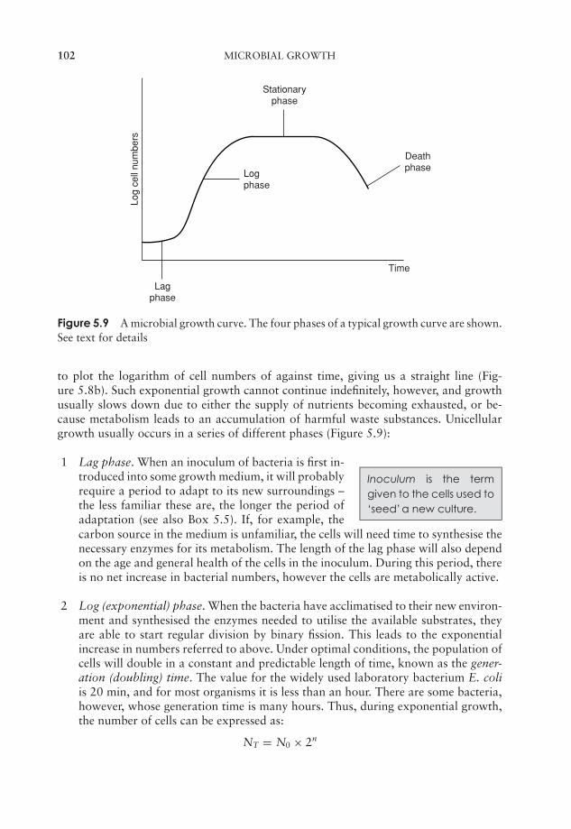
JWBK011-05 JWBK011-Hogg August 12, 2005 15:56 Char Count= 0
102 MICROBIAL GROWTH
Log
cell
num
bers
Logphase
Stationaryphase
Deathphase
Time
Lagphase
Figure 5.9 A microbial growth curve. The four phases of a typical growth curve are shown.See text for details
to plot the logarithm of cell numbers of against time, giving us a straight line (Fig-ure 5.8b). Such exponential growth cannot continue indefinitely, however, and growthusually slows down due to either the supply of nutrients becoming exhausted, or be-cause metabolism leads to an accumulation of harmful waste substances. Unicellulargrowth usually occurs in a series of different phases (Figure 5.9):
Inoculum is the termgiven to the cells used to‘seed’a new culture.
1 Lag phase. When an inoculum of bacteria is first in-troduced into some growth medium, it will probablyrequire a period to adapt to its new surroundings –the less familiar these are, the longer the period ofadaptation (see also Box 5.5). If, for example, thecarbon source in the medium is unfamiliar, the cells will need time to synthesise thenecessary enzymes for its metabolism. The length of the lag phase will also dependon the age and general health of the cells in the inoculum. During this period, thereis no net increase in bacterial numbers, however the cells are metabolically active.
2 Log (exponential) phase. When the bacteria have acclimatised to their new environ-ment and synthesised the enzymes needed to utilise the available substrates, theyare able to start regular division by binary fission. This leads to the exponentialincrease in numbers referred to above. Under optimal conditions, the population ofcells will double in a constant and predictable length of time, known as the gener-ation (doubling) time. The value for the widely used laboratory bacterium E. coliis 20 min, and for most organisms it is less than an hour. There are some bacteria,however, whose generation time is many hours. Thus, during exponential growth,the number of cells can be expressed as:
NT = N0 × 2n
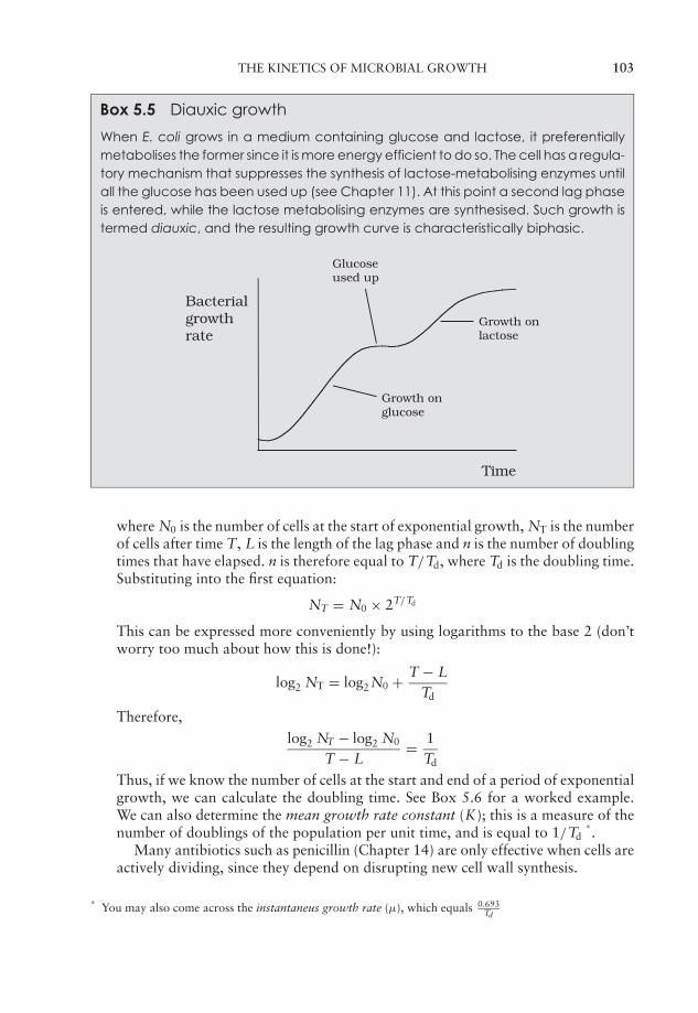
JWBK011-05 JWBK011-Hogg August 12, 2005 15:56 Char Count= 0
THE KINETICS OF MICROBIAL GROWTH 103
Box 5.5 Diauxic growth
When E. coli grows in a medium containing glucose and lactose, it preferentiallymetabolises the former since it is more energy efficient to do so. The cell has a regula-tory mechanism that suppresses the synthesis of lactose-metabolising enzymes untilall the glucose has been used up (see Chapter 11). At this point a second lag phaseis entered, while the lactose metabolising enzymes are synthesised. Such growth istermed diauxic, and the resulting growth curve is characteristically biphasic.
Bacterialgrowthrate
Time
Glucoseused up
Growth on glucose
Growth on lactose
where N0 is the number of cells at the start of exponential growth, NT is the numberof cells after time T, L is the length of the lag phase and n is the number of doublingtimes that have elapsed. n is therefore equal to T/Td, where Td is the doubling time.Substituting into the first equation:
NT = N0 × 2T/Td
This can be expressed more conveniently by using logarithms to the base 2 (don’tworry too much about how this is done!):
log2 NT = log2N0 + T − LTd
Therefore,
log2 NT − log2 N0
T − L= 1
Td
Thus, if we know the number of cells at the start and end of a period of exponentialgrowth, we can calculate the doubling time. See Box 5.6 for a worked example.We can also determine the mean growth rate constant (K); this is a measure of thenumber of doublings of the population per unit time, and is equal to 1/Td
∗.
Many antibiotics such as penicillin (Chapter 14) are only effective when cells areactively dividing, since they depend on disrupting new cell wall synthesis.
∗You may also come across the instantaneus growth rate (µ), which equals 0.693
Td
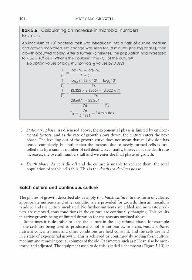
JWBK011-05 JWBK011-Hogg August 12, 2005 15:56 Char Count= 0
104 MICROBIAL GROWTH
Box 5.6 Calculating an increase in microbial numbersExample:
An inoculum of 107 bacterial cells was introduced into a flask of culture mediumand growth monitored. No change was seen for 18 minutes (the lag phase), thengrowth occurred rapidly. After a further 76 minutes, the population had increasedto 4.32 × 10⁸ cells. What is the doubling time (Td) of the culture?
(To obtain values of log2, multiply log10 values by 3.322)
1Td
= log2 NT − log2 No
T − L1Td
= log2 (4.32 × 10⁸) − log2 107
761Td
= (3.322 × 8.6355) − (3.322 × 7)76
28.6871 − 23.25476
= 1Td
Td = 765.4331
= 14 minutes
3 Stationary phase. As discussed above, the exponential phase is limited by environ-mental factors, and as the rate of growth slows down, the culture enters the nextphase. The levelling out of the growth curve does not mean that cell division hasceased completely, but rather that the increase due to newly formed cells is can-celled out by a similar number of cell deaths. Eventually, however, as the death rateincreases, the overall numbers fall and we enter the final phase of growth.
4 Death phase. As cells die off and the culture is unable to replace them, the totalpopulation of viable cells falls. This is the death (or decline) phase.
Batch culture and continuous culture
The phases of growth described above apply to a batch culture. In this form of culture,appropriate nutrients and other conditions are provided for growth, then an inoculumis added and the culture incubated. No further nutrients are added and no waste prod-ucts are removed, thus conditions in the culture are continually changing. This resultsin active growth being of limited duration for the reasons outlined above.
Sometimes it is desirable to keep the culture in the logarithmic phase, for exampleif the cells are being used to produce alcohol or antibiotics. In a continuous culture,nutrient concentrations and other conditions are held constant, and the cells are heldin a state of exponential growth. This is achieved by continuously adding fresh culturemedium and removing equal volumes of the old. Parameters such as pH can also be mon-itored and adjusted. The equipment used to do this is called a chemostat (Figure 5.10); it

JWBK011-05 JWBK011-Hogg August 12, 2005 15:56 Char Count= 0
GROWTH IN MULTICELLULAR MICROORGANISMS 105
Air supply
Nutrient
Overflow
pHelectrode
Stirrer
Acid/alkaliaddition toadjust pH
pHcontroller
Figure 5.10 Continuous culture of microorganisms: the chemostat. By the continuousaddition of new nutrients and removal of waste, the culture is kept in a steady state, withboth cell numbers and nutrient composition remaining constant
produces a steady-state culture whose population size is kept constant by careful controlof flow rates and nutrient concentrations.
Growth in multicellular microorganisms
If uninterrupted, growth in fungi proceeds radially outwards from the initiating spore(Box 5.7), allowing the fungal colony to colonise new regions potentially rich in nutrients(Box 5.8). Actual growth occurs solely at the hyphal tip; as this happens, the terminalcell grows longer, until eventually a new cross wall or septum is formed. Cells away
Box 5.7 See for yourself!
You can observe growth at a hyphal tip for yourself with a petri dish culture of afungus such as Mucor and a light microscope fitted with an eyepiece graticule.Focus on the outer visible edge of a colony, then identify the tip of one of theouwardly radiating hyphae. Align it with the graticule scale and note the positionof the tip. Return after half an hour and note the new value, being careful not todisturb the microscope in the meantime. You may be surprised at how far the hyphahas travelled in such a short time!
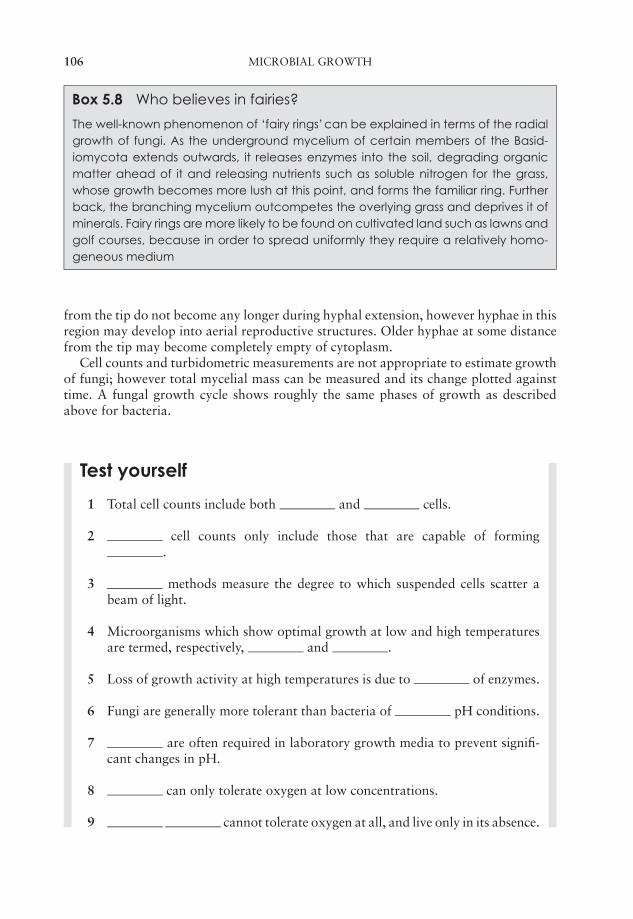
JWBK011-05 JWBK011-Hogg August 12, 2005 15:56 Char Count= 0
106 MICROBIAL GROWTH
Box 5.8 Who believes in fairies?
The well-known phenomenon of ‘fairy rings’can be explained in terms of the radialgrowth of fungi. As the underground mycelium of certain members of the Basid-iomycota extends outwards, it releases enzymes into the soil, degrading organicmatter ahead of it and releasing nutrients such as soluble nitrogen for the grass,whose growth becomes more lush at this point, and forms the familiar ring. Furtherback, the branching mycelium outcompetes the overlying grass and deprives it ofminerals. Fairy rings are more likely to be found on cultivated land such as lawns andgolf courses, because in order to spread uniformly they require a relatively homo-geneous medium
from the tip do not become any longer during hyphal extension, however hyphae in thisregion may develop into aerial reproductive structures. Older hyphae at some distancefrom the tip may become completely empty of cytoplasm.
Cell counts and turbidometric measurements are not appropriate to estimate growthof fungi; however total mycelial mass can be measured and its change plotted againsttime. A fungal growth cycle shows roughly the same phases of growth as describedabove for bacteria.
Test yourself
1 Total cell counts include both and cells.
2 cell counts only include those that are capable of forming.
3 methods measure the degree to which suspended cells scatter abeam of light.
4 Microorganisms which show optimal growth at low and high temperaturesare termed, respectively, and .
5 Loss of growth activity at high temperatures is due to of enzymes.
6 Fungi are generally more tolerant than bacteria of pH conditions.
7 are often required in laboratory growth media to prevent signifi-cant changes in pH.
8 can only tolerate oxygen at low concentrations.
9 cannot tolerate oxygen at all, and live only in its absence.
JWBK011-05 JWBK011-Hogg August 12, 2005 15:56 Char Count= 0
TEST YOURSELF 107
10 bacteria have evolved to thrive in conditions of extremely highsalinity.
11 Bacteria produce two identical daughter cells by a process of.
12 Given ideal growth conditions, the size of a population of bacteria wouldincrease in a fashion.
13 The four phases of a typical growth curve are , ,and .
14 The small sample of cells used to start off a culture is called an .
15 A maintains a population of cells constantly in phase,by carefully controlling nutrients and flow rates.

JWBK011-05 JWBK011-Hogg August 12, 2005 15:56 Char Count= 0
108
JWBK011-06 JWBK011-Hogg August 12, 2005 16:0 Char Count= 0
6Microbial Metabolism
You may have wondered why it was necessary to learn all that biochemistry in Chapter 2;well, you are about to find out! In the following pages, you will learn about the processesby which microorganisms obtain and use energy.
Why is energy needed?
Like all other living things, microorganisms need to acquire energy in order to survive.Energy is required:
� to maintain the structural integrity of the cell by repairing any damage to itsconstituents
� to synthesise new cellular components such as nucleic acids, polysaccharidesand enzymes
� to transport certain substances into the cell from its surroundings
� for the cell to grow and multiply
� for cellular movement.
Catabolism is the termused to describe reac-tions that break downlarge molecules, usuallycoupled to a release ofenergy.
Anabolism is the termused to describe reac-tions involved in thesynthesis of macromole-cules, usually requiringan input of energy
Metabolism is the term used to describe all the biochemi-cal reactions that take place inside a cell; it includes thosereactions that release energy into the cell, and those thatmake use of that energy. Figure 6.1 summarises theseprocesses.
As we saw in Chapter 4, most microorganisms ob-tain their energy from the nutrients they take into thecell; these may come from an organic or an inorganicsource. Once inside the cell, these nutrients must thenbe biochemically processed by reactions that trap someof their chemical energy, at the same time breakingthem down into smaller molecules. These then serve asbuilding blocks for the synthesis of new cellular com-ponents. Chemical compounds contain potential energywithin their molecular structure, and some of this can be
109
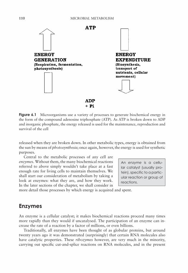
JWBK011-06 JWBK011-Hogg August 12, 2005 16:0 Char Count= 0
110 MICROBIAL METABOLISM
Figure 6.1 Microorganisms use a variety of processes to generate biochemical energy inthe form of the compound adenosine triphosphate (ATP). As ATP is broken down to ADPand inorganic phosphate, the energy released is used for the maintenance, reproduction andsurvival of the cell
released when they are broken down. In other metabolic types, energy is obtained fromthe sun by means of photosynthesis; once again, however, the energy is used for syntheticpurposes.
An enzyme is a cellu-lar catalyst (usually pro-tein), specific to a partic-ular reaction or group ofreactions.
Central to the metabolic processes of any cell areenzymes. Without them, the many biochemical reactionsreferred to above simply wouldn’t take place at a fastenough rate for living cells to maintain themselves. Weshall start our consideration of metabolism by taking alook at enzymes: what they are, and how they work.In the later sections of the chapter, we shall consider inmore detail those processes by which energy is acquired and spent.
Enzymes
An enzyme is a cellular catalyst; it makes biochemical reactions proceed many timesmore rapidly than they would if uncatalysed. The participation of an enzyme can in-crease the rate of a reaction by a factor of millions, or even billions.
Traditionally, all enzymes have been thought of as globular proteins, but aroundtwenty years ago it was demonstrated (surprisingly) that certain RNA molecules alsohave catalytic properties. These ribozymes however, are very much in the minority,carrying out specific cut-and-splice reactions on RNA molecules, and in the present

JWBK011-06 JWBK011-Hogg August 12, 2005 16:0 Char Count= 0
ENZYMES 111
Enzyme
Active sites
Substrate
Enzyme-substratecomplex
Product
Figure 6.2 Enzyme–substrate interaction. An enzyme interacts with its substrate(s) to forman enzyme–substrate complex, leading to the formation of a product. In the example shown,two substrate molecules are held in position by the enzyme and joined together. From Black,JG: Microbiology: Principles and Explorations, 4th edn, John Wiley & Sons Inc., 1999.Reproduced by permission of the publishers
context can be ignored. In this book we shall confine our discussion of enzymes to theprotein type.
Like any other catalyst, an enzyme remains unchanged at the end of a reaction. Itmust, however, at some point during the reaction bind to its substrate (the substanceupon which it acts) to form an enzyme–substrate complex (Figure 6.2) by multipleweak forces such as electrostatic forces and hydrogen bonding. Only a small part ofthe enzyme’s three-dimensional structure is involved in this binding; these few aminoacids make up the active site, which forms a groove or dent in the enzyme’s surface,into which the appropriate part of the substrate molecule fits (Figure 6.3). The amino
HIS 95 GLU 97
GLU 165
LYS 12
Substrate-binding cleft
Figure 6.3 Catalytic activity occurs at the active site of an enzyme. Four amino acids playan important role in the active site of triose phosphate isomerase; note how they are farapart in the primary sequence, but are brought together by subsequent protein folding
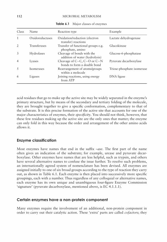
JWBK011-06 JWBK011-Hogg August 12, 2005 16:0 Char Count= 0
112 MICROBIAL METABOLISM
Table 6.1 Major classes of enzymes
Class Name Reaction type Example
1 Oxidoreductases Oxidation/reduction (electrontransfer) reactions
Lactate dehydrogenase
2 Transferases Transfer of functional groups e.g.phosphate, amino
Glucokinase
3 Hydrolases Cleavage of bonds with theaddition of water (hydrolysis)
Glucose-6-phosphatase
4 Lyases Cleavage of C−C, C−O or C−Nbonds to form a double bond
Pyruvate decarboxylase
5 Isomerases Rearrangement of atoms/groupswithin a molecule
Triose-phosphate isomerase
6 Ligases Joining reactions, using energyfrom ATP
DNA ligase
acid residues that go to make up the active site may be widely separated in the enzyme’sprimary structure, but by means of the secondary and tertiary folding of the molecule,they are brought together to give a specific conformation, complementary to that ofthe substrate. It is this precise formation of the active site that accounts for one of themajor characteristics of enzymes, their specificity. You should not think, however, thatthese few residues making up the active site are the only ones that matter; the enzymecan only fold in this way because the order and arrangement of the other amino acidsallows it.
Enzyme classification
Most enzymes have names that end in the suffix –ase. The first part of the nameoften gives an indication of the substrate; for example, urease and pyruvate decar-boxylase. Other enzymes have names that are less helpful, such as trypsin, and othershave several alternative names to confuse the issue further. To resolve such problems,an internationally agreed system of nomenclature has been devised. All enzymes areassigned initially to one of six broad groups according to the type of reaction they carryout, as shown in Table 6.1. Each enzyme is then placed into successively more specificgroupings, each with a number. Thus regardless of any colloquial or alternative names,each enzyme has its own unique and unambiguous four-figure Enzyme Commission‘signature’ (pyruvate decarboxylase, mentioned above, is EC 4.1.1.1).
Certain enzymes have a non-protein component
Many enzymes require the involvement of an additional, non-protein component inorder to carry out their catalytic action. These ‘extra’ parts are called cofactors; they
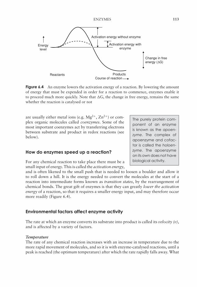
JWBK011-06 JWBK011-Hogg August 12, 2005 16:0 Char Count= 0
ENZYMES 113
Activation energy without enzyme
Activation energy withenzyme
Change in freeenergy (∆G)
ReactantsCourse of reaction
Energylevel
Products
Figure 6.4 An enzyme lowers the activation energy of a reaction. By lowering the amountof energy that must be expended in order for a reaction to commence, enzymes enable itto proceed much more quickly. Note that �G, the change in free energy, remains the samewhether the reaction is catalysed or not
The purely protein com-ponent of an enzymeis known as the apoen-zyme. The complex ofapoenzyme and cofac-tor is called the holoen-zyme. The apoenzymeon its own does not havebiological activity.
are usually either metal ions (e.g. Mg2+, Zn2+) or com-plex organic molecules called coenzymes. Some of themost important coenzymes act by transferring electronsbetween substrate and product in redox reactions (seebelow).
How do enzymes speed up a reaction?
For any chemical reaction to take place there must be asmall input of energy. This is called the activation energy,and is often likened to the small push that is needed to loosen a boulder and allow itto roll down a hill. It is the energy needed to convert the molecules at the start of areaction into intermediate forms known as transition states, by the rearrangement ofchemical bonds. The great gift of enzymes is that they can greatly lower the activationenergy of a reaction, so that it requires a smaller energy input, and may therefore occurmore readily (Figure 6.4).
Environmental factors affect enzyme activity
The rate at which an enzyme converts its substrate into product is called its velocity (v),and is affected by a variety of factors.
TemperatureThe rate of any chemical reaction increases with an increase in temperature due to themore rapid movement of molecules, and so it is with enzyme-catalysed reactions, until apeak is reached (the optimum temperature) after which the rate rapidly falls away. What
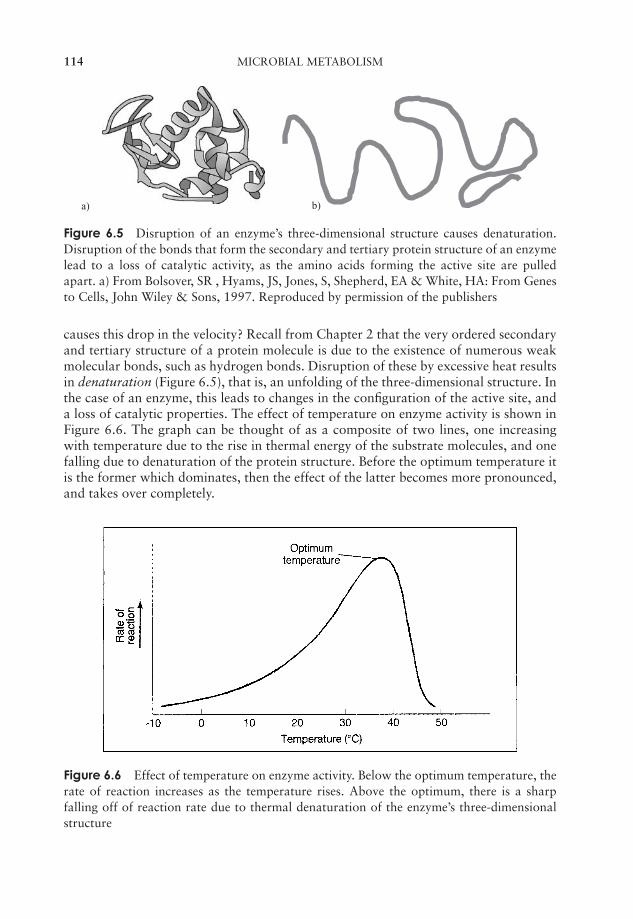
JWBK011-06 JWBK011-Hogg August 12, 2005 16:0 Char Count= 0
114 MICROBIAL METABOLISM
a) b)
Figure 6.5 Disruption of an enzyme’s three-dimensional structure causes denaturation.Disruption of the bonds that form the secondary and tertiary protein structure of an enzymelead to a loss of catalytic activity, as the amino acids forming the active site are pulledapart. a) From Bolsover, SR , Hyams, JS, Jones, S, Shepherd, EA & White, HA: From Genesto Cells, John Wiley & Sons, 1997. Reproduced by permission of the publishers
causes this drop in the velocity? Recall from Chapter 2 that the very ordered secondaryand tertiary structure of a protein molecule is due to the existence of numerous weakmolecular bonds, such as hydrogen bonds. Disruption of these by excessive heat resultsin denaturation (Figure 6.5), that is, an unfolding of the three-dimensional structure. Inthe case of an enzyme, this leads to changes in the configuration of the active site, anda loss of catalytic properties. The effect of temperature on enzyme activity is shown inFigure 6.6. The graph can be thought of as a composite of two lines, one increasingwith temperature due to the rise in thermal energy of the substrate molecules, and onefalling due to denaturation of the protein structure. Before the optimum temperature itis the former which dominates, then the effect of the latter becomes more pronounced,and takes over completely.
Figure 6.6 Effect of temperature on enzyme activity. Below the optimum temperature, therate of reaction increases as the temperature rises. Above the optimum, there is a sharpfalling off of reaction rate due to thermal denaturation of the enzyme’s three-dimensionalstructure
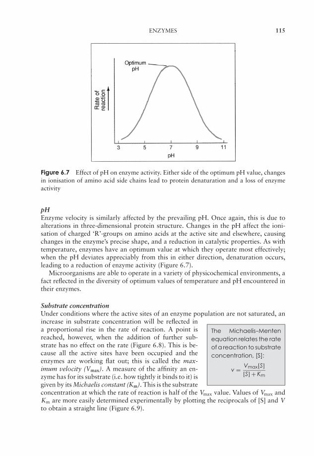
JWBK011-06 JWBK011-Hogg August 12, 2005 16:0 Char Count= 0
ENZYMES 115
Figure 6.7 Effect of pH on enzyme activity. Either side of the optimum pH value, changesin ionisation of amino acid side chains lead to protein denaturation and a loss of enzymeactivity
pHEnzyme velocity is similarly affected by the prevailing pH. Once again, this is due toalterations in three-dimensional protein structure. Changes in the pH affect the ioni-sation of charged ‘R’-groups on amino acids at the active site and elsewhere, causingchanges in the enzyme’s precise shape, and a reduction in catalytic properties. As withtemperature, enzymes have an optimum value at which they operate most effectively;when the pH deviates appreciably from this in either direction, denaturation occurs,leading to a reduction of enzyme activity (Figure 6.7).
Microorganisms are able to operate in a variety of physicochemical environments, afact reflected in the diversity of optimum values of temperature and pH encountered intheir enzymes.
Substrate concentrationUnder conditions where the active sites of an enzyme population are not saturated, an
The Michaelis--Mentenequation relates the rateof a reaction to substrateconcentration, [S]:
v = Vmax[S][S] + Km
increase in substrate concentration will be reflected ina proportional rise in the rate of reaction. A point isreached, however, when the addition of further sub-strate has no effect on the rate (Figure 6.8). This is be-cause all the active sites have been occupied and theenzymes are working flat out; this is called the max-imum velocity (Vmax). A measure of the affinity an en-zyme has for its substrate (i.e. how tightly it binds to it) isgiven by its Michaelis constant (Km). This is the substrateconcentration at which the rate of reaction is half of the Vmax value. Values of Vmax andKm are more easily determined experimentally by plotting the reciprocals of [S] and Vto obtain a straight line (Figure 6.9).

JWBK011-06 JWBK011-Hogg August 12, 2005 16:0 Char Count= 0
116 MICROBIAL METABOLISM
max
max /2
v
0 Km
[S]
Figure 6.8 Enzyme activity is influenced by substrate concentration. The initial rate ofreaction (vo) is proportional to substrate concentration at low values of [S]. However, whenthe active sites of the enzyme molecules become saturated with substrate, a maximum rateof reaction (Vmax) is reached. This cannot be exceeded, no matter how much the value of[S] increases. The curve of the graph fits the Michaelis–Menten equation. Km is the value of[S] where v = Vmax
2 .
Figure 6.9 The Lineweaver–Burk plot. Plotting the reciprocal values of Vo and [S] enablesvalues of Km and Vmax to be derived from the intercepts on a straight-line graph
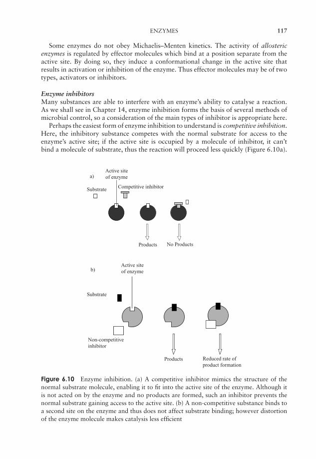
JWBK011-06 JWBK011-Hogg August 12, 2005 16:0 Char Count= 0
ENZYMES 117
Some enzymes do not obey Michaelis–Menten kinetics. The activity of allostericenzymes is regulated by effector molecules which bind at a position separate from theactive site. By doing so, they induce a conformational change in the active site thatresults in activation or inhibition of the enzyme. Thus effector molecules may be of twotypes, activators or inhibitors.
Enzyme inhibitorsMany substances are able to interfere with an enzyme’s ability to catalyse a reaction.As we shall see in Chapter 14, enzyme inhibition forms the basis of several methods ofmicrobial control, so a consideration of the main types of inhibitor is appropriate here.
Perhaps the easiest form of enzyme inhibition to understand is competitive inhibition.Here, the inhibitory substance competes with the normal substrate for access to theenzyme’s active site; if the active site is occupied by a molecule of inhibitor, it can’tbind a molecule of substrate, thus the reaction will proceed less quickly (Figure 6.10a).
Active siteof enzyme
Substrate Competitive inhibitor
Products No Products
a)
b)Active siteof enzyme
Substrate
Products Reduced rate ofproduct formation
Non-competitiveinhibitor
Figure 6.10 Enzyme inhibition. (a) A competitive inhibitor mimics the structure of thenormal substrate molecule, enabling it to fit into the active site of the enzyme. Although itis not acted on by the enzyme and no products are formed, such an inhibitor prevents thenormal substrate gaining access to the active site. (b) A non-competitive substance binds toa second site on the enzyme and thus does not affect substrate binding; however distortionof the enzyme molecule makes catalysis less efficient
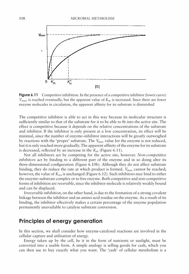
JWBK011-06 JWBK011-Hogg August 12, 2005 16:0 Char Count= 0
118 MICROBIAL METABOLISM
Figure 6.11 Competitive inhibition. In the presence of a competitive inhibitor (lower curve)Vmax is reached eventually, but the apparent value of Km is increased. Since there are fewerenzyme molecules in circulation, the apparent affinity for its substrate is diminished
The competitive inhibitor is able to act in this way because its molecular structure issufficiently similar to that of the substrate for it to be able to fit into the active site. Theeffect is competitive because it depends on the relative concentrations of the substrateand inhibitor. If the inhibitor is only present at a low concentration, its effect will beminimal, since the number of enzyme–inhibitor interactions will be greatly outweighedby reactions with the ‘proper’ substrate. The Vmax value for the enzyme is not reduced,but it is only reached more gradually. The apparent affinity of the enzyme for its substrateis decreased, reflected by an increase in the Km (Figure 6.11).
Not all inhibitors act by competing for the active site, however. Non-competitiveinhibitors act by binding to a different part of the enzyme and in so doing alter itsthree-dimensional configuration (Figure 6.10b). Although they do not affect substratebinding, they do reduce the rate at which product is formed. Vmax cannot be reached;however, the value of Km, is unchanged (Figure 6.12). Such inhibitors may bind to eitherthe enzyme–substrate complex or to free enzyme. Both competitive and non-competitiveforms of inhibition are reversible, since the inhibitor molecule is relatively weakly boundand can be displaced.
Irreversible inhibition, on the other hand, is due to the formation of a strong covalentlinkage between the inhibitor and an amino acid residue on the enzyme. As a result of itsbinding, the inhibitor effectively makes a certain percentage of the enzyme populationpermanently unavailable to catalyse substrate conversion.
Principles of energy generation
In this section, we shall consider how enzyme-catalysed reactions are involved in thecellular capture and utilisation of energy.
Energy taken up by the cell, be it in the form of nutrients or sunlight, must beconverted into a usable form. A simple analogy is selling goods for cash, which youcan then use to buy exactly what you want. The ‘cash’ of cellular metabolism is a
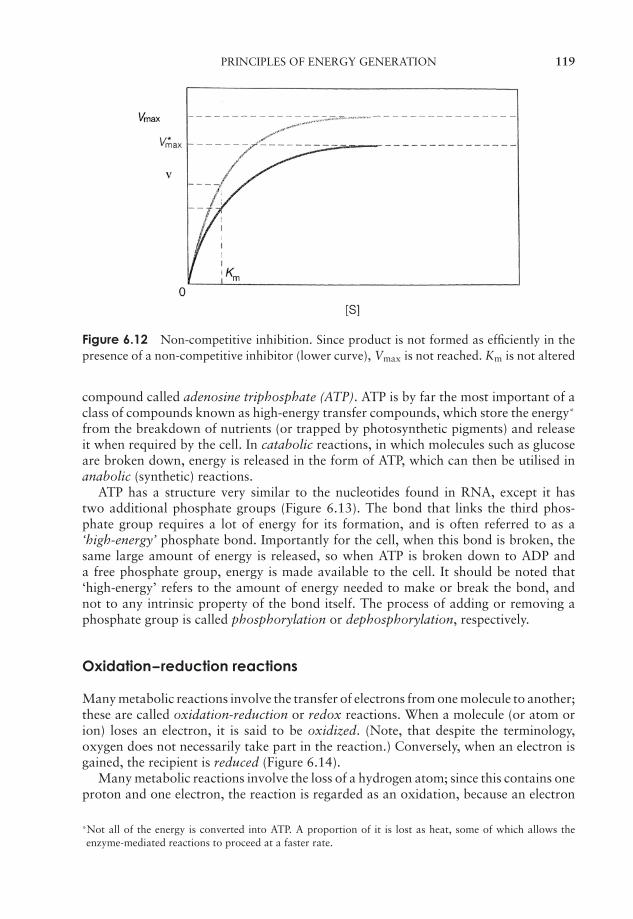
JWBK011-06 JWBK011-Hogg August 12, 2005 16:0 Char Count= 0
PRINCIPLES OF ENERGY GENERATION 119
Figure 6.12 Non-competitive inhibition. Since product is not formed as efficiently in thepresence of a non-competitive inhibitor (lower curve), Vmax is not reached. Km is not altered
compound called adenosine triphosphate (ATP). ATP is by far the most important of aclass of compounds known as high-energy transfer compounds, which store the energy*
from the breakdown of nutrients (or trapped by photosynthetic pigments) and releaseit when required by the cell. In catabolic reactions, in which molecules such as glucoseare broken down, energy is released in the form of ATP, which can then be utilised inanabolic (synthetic) reactions.
ATP has a structure very similar to the nucleotides found in RNA, except it hastwo additional phosphate groups (Figure 6.13). The bond that links the third phos-phate group requires a lot of energy for its formation, and is often referred to as a‘high-energy’ phosphate bond. Importantly for the cell, when this bond is broken, thesame large amount of energy is released, so when ATP is broken down to ADP anda free phosphate group, energy is made available to the cell. It should be noted that‘high-energy’ refers to the amount of energy needed to make or break the bond, andnot to any intrinsic property of the bond itself. The process of adding or removing aphosphate group is called phosphorylation or dephosphorylation, respectively.
Oxidation--reduction reactions
Many metabolic reactions involve the transfer of electrons from one molecule to another;these are called oxidation-reduction or redox reactions. When a molecule (or atom orion) loses an electron, it is said to be oxidized. (Note, that despite the terminology,oxygen does not necessarily take part in the reaction.) Conversely, when an electron isgained, the recipient is reduced (Figure 6.14).
Many metabolic reactions involve the loss of a hydrogen atom; since this contains oneproton and one electron, the reaction is regarded as an oxidation, because an electron
*Not all of the energy is converted into ATP. A proportion of it is lost as heat, some of which allows theenzyme-mediated reactions to proceed at a faster rate.
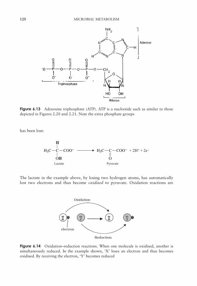
JWBK011-06 JWBK011-Hogg August 12, 2005 16:0 Char Count= 0
120 MICROBIAL METABOLISM
Figure 6.13 Adenosine triphosphate (ATP). ATP is a nucleotide such as similar to thosedepicted in Figures 2.20 and 2.21. Note the extra phosphate groups
has been lost:
C
OH
H
H3C COO– C
O
H3C COO– + 2H+ + 2e–
Lactate Pyruvate
The lactate in the example above, by losing two hydrogen atoms, has automaticallylost two electrons and thus become oxidised to pyruvate. Oxidation reactions are
Reduction
electron
Oxidation
Figure 6.14 Oxidation–reduction reactions. When one molecule is oxidised, another issimultaneously reduced. In the example shown, ‘X’ loses an electron and thus becomesoxidised. By receiving the electron, ‘Y’ becomes reduced
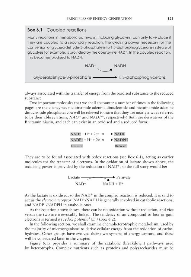
JWBK011-06 JWBK011-Hogg August 12, 2005 16:0 Char Count= 0
PRINCIPLES OF ENERGY GENERATION 121
Box 6.1 Coupled reactions
Many reactions in metabolic pathways, including glycolysis, can only take place ifthey are coupled to a secondary reaction. The oxidising power necessary for theconversion of glyceraldehyde-3-phosphate into 1,3-diphosphoglycerate in step 6 ofglycolysis for example, is provided by the coenzyme NAD+. In the coupled reaction,this becomes oxidised to NADH:
NAD+ NADH
Glyceraldehyde-3-phosphate 1, 3-diphosphoglycerate
always associated with the transfer of energy from the oxidised substance to the reducedsubstance.
Two important molecules that we shall encounter a number of times in the followingpages are the coenzymes nicotinamide adenine dinucleotide and nicotinamide adeninedinucleotide phosphate; you will be relieved to learn that they are nearly always referredto by their abbreviations, NAD+ and NADP+, respectively! Both are derivatives of theB vitamin niacin, and each can exist in an oxidised and a reduced form:
NAD+ + H+ + 2e– NADH
NADP+ + H+ + 2e– NADPH
Oxidised Reduced
They are to be found associated with redox reactions (see Box 6.1), acting as carriermolecules for the transfer of electrons. In the oxidation of lactate shown above, theoxidising power is provided by the reduction of NAD+, so the full story would be:
Lactate Pyruvate
NAD+ NADH + H+
As the lactate is oxidised, so the NAD+ in the coupled reaction is reduced. It is said toact as the electron acceptor. NAD+/NADH is generally involved in catabolic reactions,and NADP+/NADPH in anabolic ones.
As the equation above shows, there can be no oxidation without reduction, and viceversa; the two are irrevocably linked. The tendency of an compound to lose or gainelectrons is termed its redox potential (Eo) (Box 6.2).
In the following section, we shall examine chemoheterotrophic metabolism, used bythe majority of microorganisms to derive cellular energy from the oxidation of carbo-hydrates. Other groups have evolved their own systems of energy capture, and thesewill be considered later in the chapter.
Figure 6.15 provides a summary of the catabolic (breakdown) pathways usedby heterotrophs. Complex nutrients such as proteins and polysaccharides must be
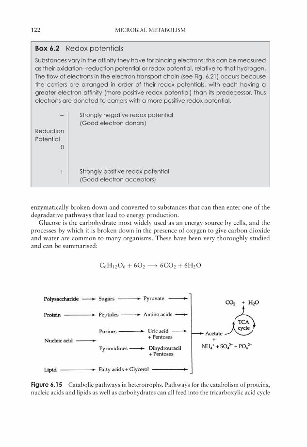
JWBK011-06 JWBK011-Hogg August 12, 2005 16:0 Char Count= 0
122 MICROBIAL METABOLISM
Box 6.2 Redox potentials
Substances vary in the affinity they have for binding electrons; this can be measuredas their oxidation--reduction potential or redox potential, relative to that hydrogen.The flow of electrons in the electron transport chain (see Fig. 6.21) occurs becausethe carriers are arranged in order of their redox potentials, with each having agreater electron affinity (more positive redox potential) than its predecessor. Thuselectrons are donated to carriers with a more positive redox potential.
− Strongly negative redox potential(Good electron donors)
ReductionPotential
0
+ Strongly positive redox potential(Good electron acceptors)
enzymatically broken down and converted to substances that can then enter one of thedegradative pathways that lead to energy production.
Glucose is the carbohydrate most widely used as an energy source by cells, and theprocesses by which it is broken down in the presence of oxygen to give carbon dioxideand water are common to many organisms. These have been very thoroughly studiedand can be summarised:
C6H12O6 + 6O2 −→ 6CO2 + 6H2O
Figure 6.15 Catabolic pathways in heterotrophs. Pathways for the catabolism of proteins,nucleic acids and lipids as well as carbohydrates can all feed into the tricarboxylic acid cycle
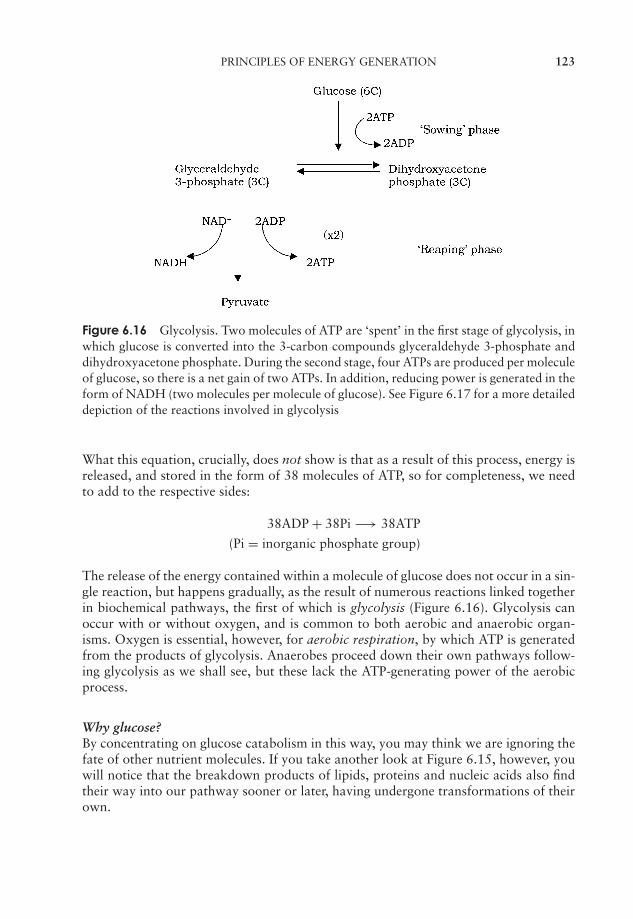
JWBK011-06 JWBK011-Hogg August 12, 2005 16:0 Char Count= 0
PRINCIPLES OF ENERGY GENERATION 123
Figure 6.16 Glycolysis. Two molecules of ATP are ‘spent’ in the first stage of glycolysis, inwhich glucose is converted into the 3-carbon compounds glyceraldehyde 3-phosphate anddihydroxyacetone phosphate. During the second stage, four ATPs are produced per moleculeof glucose, so there is a net gain of two ATPs. In addition, reducing power is generated in theform of NADH (two molecules per molecule of glucose). See Figure 6.17 for a more detaileddepiction of the reactions involved in glycolysis
What this equation, crucially, does not show is that as a result of this process, energy isreleased, and stored in the form of 38 molecules of ATP, so for completeness, we needto add to the respective sides:
38ADP + 38Pi −→ 38ATP
(Pi = inorganic phosphate group)
The release of the energy contained within a molecule of glucose does not occur in a sin-gle reaction, but happens gradually, as the result of numerous reactions linked togetherin biochemical pathways, the first of which is glycolysis (Figure 6.16). Glycolysis canoccur with or without oxygen, and is common to both aerobic and anaerobic organ-isms. Oxygen is essential, however, for aerobic respiration, by which ATP is generatedfrom the products of glycolysis. Anaerobes proceed down their own pathways follow-ing glycolysis as we shall see, but these lack the ATP-generating power of the aerobicprocess.
Why glucose?By concentrating on glucose catabolism in this way, you may think we are ignoring thefate of other nutrient molecules. If you take another look at Figure 6.15, however, youwill notice that the breakdown products of lipids, proteins and nucleic acids also findtheir way into our pathway sooner or later, having undergone transformations of theirown.
JWBK011-06 JWBK011-Hogg August 12, 2005 16:0 Char Count= 0
124 MICROBIAL METABOLISM
Glycolysis
The initial sequence of reactions, in which a molecule of glucose is converted to twomolecules of pyruvate
∗, is called glycolysis (Figures 6.16 and 6.17). In the first phase
of glycolysis, glucose is phosphorylated and its six-carbon ring structure rearranged,before being cleaved into two three-carbon molecules. In the second phase, each ofthese undergoes oxidation, resulting in pyruvate.
Also known as the Embden–Meyerhof pathway, glycolysis is used for the metabolismof simple sugars not just by microorganisms, but by most living cells. The pathway,which takes place in the cytoplasm, comprises a series of 10 linked reactions, in whicheach molecule of the six-carbon glucose is converted to two molecules of the three-carbon pyruvate, with a net gain of two molecules of ATP.
The full pathway of reactions is shown in Figure 6.17. Note how, in terms of energy,glycolysis can be divided conveniently into a ‘sowing’ phase, in which two molecules ofATP are expended per molecule of glucose, followed by a ‘reaping’ phase which yieldsfour molecules of ATP. The overall energy balance is therefore a gain of two moleculesof ATP for each molecule of glucose oxidised to pyruvate. In addition, the second phasefeatures the conversion of two molecules of NAD+ to NADH, which, as we will see,act as an important source of reducing power in subsequent pathways.
The reactions by which ATP is generated from ADP in the second phase of glycolysisare examples of substrate-level phosphorylation, so-called because the phosphate groupis transferred directly from a donor molecule.
What happens next to the pyruvate produced by glycolysis depends on the organismconcerned, and on whether the environment is aerobic or anaerobic; we shall look atthese possibilities in due course.
Glycolysis is not the only way to metabolise glucoseAlthough glycolysis is widespread in both the microbial and nonmicrobial worlds, sev-eral bacterial types use alternative pathways to oxidise glucose. For certain Gram-negative groups, notably the pseudomonads (see Chapter 7), the main route used isthe Entner–Doudoroff pathway, producing a mixture of pyruvate and glyceraldehyde-3-phosphate (Figure 6.18). The former, like that produced in glycolysis, can enter anumber of pathways, while the latter can feed into the later stages of glycolysis. Thenet result of catabolism by the Entner–Doudoroff pathway is the production of onemolecule each of ATP, NADH and NADPH per molecule of glucose degraded.
A secondary pathway, which may operate in tandem with glycolysis or the Entner–Doudoroff pathway, is the pentose phosphate pathway, sometimes known as the hexosemonophosphate shunt (Figure 6.19). Like glycolysis, the pathway can operate in thepresence or absence of oxygen. Although glyceraldehyde-3-phosphate can once againenter the glycolytic pathway and lead to ATP generation, for most organisms the path-way has a mainly anabolic (biosynthetic) function, acting as a source of precursormolecules for other metabolic pathways. The pentose phosphate pathway is a useful
∗At physiological pH, carboxylic acids such as pyruvic acid and citric acid are found in their ionised form
(pyruvate, citrate); however long-established traditions persist, and you may well find reference elsewhere tothe ‘-ic acid’ forms.
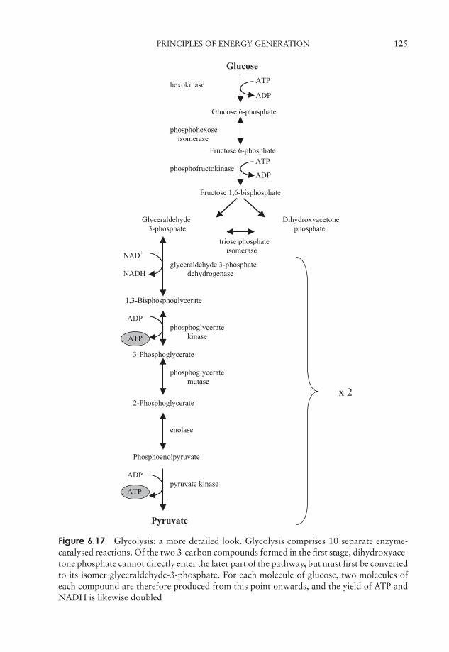
JWBK011-06 JWBK011-Hogg August 12, 2005 16:0 Char Count= 0
PRINCIPLES OF ENERGY GENERATION 125
Glucose
ATPhexokinase
ADP
Glucose 6-phosphate
phosphohexose isomerase
Fructose 6-phosphate
ATPphosphofructokinase
ADP
Fructose 1,6-bisphosphate
Glyceraldehyde Dihydroxyacetone 3-phosphate phosphate
triose phosphateisomerase
NAD+
glyceraldehyde 3-phosphate NADH dehydrogenase
1,3-Bisphosphoglycerate
ADPphosphoglycerate
ATP kinase
3-Phosphoglycerate
phosphoglycerate mutase
x 2 2-Phosphoglycerate
enolase
Phosphoenolpyruvate
ADPpyruvate kinase
ATP
Pyruvate
Figure 6.17 Glycolysis: a more detailed look. Glycolysis comprises 10 separate enzyme-catalysed reactions. Of the two 3-carbon compounds formed in the first stage, dihydroxyace-tone phosphate cannot directly enter the later part of the pathway, but must first be convertedto its isomer glyceraldehyde-3-phosphate. For each molecule of glucose, two molecules ofeach compound are therefore produced from this point onwards, and the yield of ATP andNADH is likewise doubled
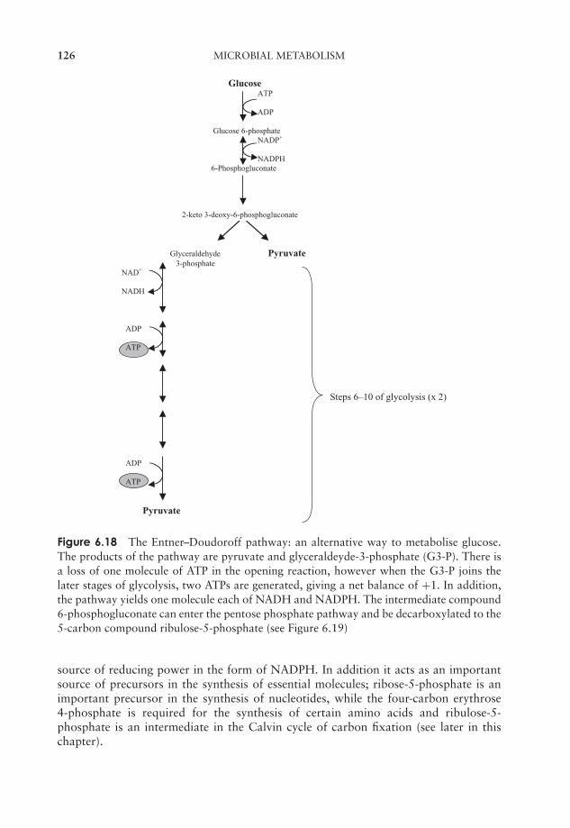
JWBK011-06 JWBK011-Hogg August 12, 2005 16:0 Char Count= 0
126 MICROBIAL METABOLISM
GlucoseATP
ADP
Glucose 6-phosphateNADP+
NADPH 6-Phosphogluconate
2-keto 3-deoxy-6-phosphogluconate
Glyceraldehyde Pyruvate 3-phosphate
NAD+
NADH
ADP
ATP
Steps 6–10 of glycolysis (x 2)
ADP
ATP
Pyruvate
Figure 6.18 The Entner–Doudoroff pathway: an alternative way to metabolise glucose.The products of the pathway are pyruvate and glyceraldeyde-3-phosphate (G3-P). There isa loss of one molecule of ATP in the opening reaction, however when the G3-P joins thelater stages of glycolysis, two ATPs are generated, giving a net balance of +1. In addition,the pathway yields one molecule each of NADH and NADPH. The intermediate compound6-phosphogluconate can enter the pentose phosphate pathway and be decarboxylated to the5-carbon compound ribulose-5-phosphate (see Figure 6.19)
source of reducing power in the form of NADPH. In addition it acts as an importantsource of precursors in the synthesis of essential molecules; ribose-5-phosphate is animportant precursor in the synthesis of nucleotides, while the four-carbon erythrose4-phosphate is required for the synthesis of certain amino acids and ribulose-5-phosphate is an intermediate in the Calvin cycle of carbon fixation (see later in thischapter).
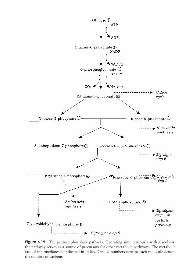
JWBK011-06 JWBK011-Hogg August 12, 2005 16:0 Char Count= 0
Figure 6.19 The pentose phosphate pathway. Operating simultaneously with glycolysis,the pathway serves as a source of precursors for other metabolic pathways. The metabolicfate of intermediates is indicated in italics. Circled numbers next to each molecule denotethe number of carbons
127
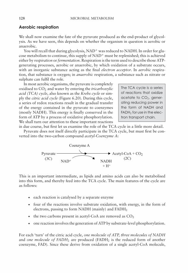
JWBK011-06 JWBK011-Hogg August 12, 2005 16:0 Char Count= 0
128 MICROBIAL METABOLISM
Aerobic respiration
We shall now examine the fate of the pyruvate produced as the end-product of glycol-ysis. As we have seen, this depends on whether the organism in question is aerobic oranaerobic.
You will recall that during glycolysis, NAD+ was reduced to NADH. In order for glu-cose metabolism to continue, this supply of NAD+ must be replenished; this is achievedeither by respiration or fermentation. Respiration is the term used to describe those ATP-generating processes, aerobic or anaerobic, by which oxidation of a substrate occurs,with an inorganic substance acting as the final electron acceptor. In aerobic respira-tion, that substance is oxygen; in anaerobic respiration, a substance such as nitrate orsulphate can fulfil the role.
The TCA cycle is a seriesof reactions that oxidizeacetate to CO2, gener-ating reducing power inthe form of NADH andFADH2 for use in the elec-tron transport chain.
In most aerobic organisms, the pyruvate is completelyoxidised to CO2 and water by entering the tricarboxylicacid (TCA) cycle, also known as the Krebs cycle or sim-ply the citric acid cycle (Figure 6.20). During this cycle,a series of redox reactions result in the gradual transferof the energy contained in the pyruvate to coenzymes(mostly NADH). This energy is finally conserved in theform of ATP by a process of oxidative phosphorylation.We shall turn our attention to these important reactionsin due course, but first let us examine the role of the TCA cycle in a little more detail.
Pyruvate does not itself directly participate in the TCA cycle, but must first be con-verted into the two-carbon compound acetyl-Coenzyme A:
Pyruvate Acetyl-CoA + CO2(2C)
NAD+ NADH + H+
(3C)
Coenzyme A
This is an important intermediate, as lipids and amino acids can also be metabolisedinto this form, and thereby feed into the TCA cycle. The main features of the cycle areas follows:
� each reaction is catalysed by a separate enzyme
� four of the reactions involve substrate oxidation, with energy, in the form ofelectrons, passing to form NADH (mainly) and FADH2
� the two carbons present in acetyl-CoA are removed as CO2
� one reaction involves the generation of ATP by substrate-level phosphorylation.
For each ‘turn’ of the citric acid cycle, one molecule of ATP, three molecules of NADHand one molecule of FADH2 are produced (FADH2 is the reduced form of anothercoenzyme, FAD). Since these derive from oxidation of a single acetyl-CoA molecule,
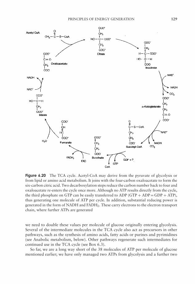
JWBK011-06 JWBK011-Hogg August 12, 2005 16:0 Char Count= 0
PRINCIPLES OF ENERGY GENERATION 129
Figure 6.20 The TCA cycle. Acetyl-CoA may derive from the pyruvate of glycolysis orfrom lipid or amino acid metabolism. It joins with the four-carbon oxaloacetate to form thesix-carbon citric acid. Two decarboxylation steps reduce the carbon number back to four andoxaloacetate re-enters the cycle once more. Although no ATP results directly from the cycle,the third phosphate on GTP can be easily transferred to ADP (GTP + ADP = GDP + ATP),thus generating one molecule of ATP per cycle. In addition, substantial reducing power isgenerated in the form of NADH and FADH2. These carry electrons to the electron transportchain, where further ATPs are generated
we need to double these values per molecule of glucose originally entering glycolysis.Several of the intermediate molecules in the TCA cycle also act as precursors in otherpathways, such as the synthesis of amino acids, fatty acids or purines and pyrimidines(see Anabolic metabolism, below). Other pathways regenerate such intermediates forcontinued use in the TCA cycle (see Box 6.3).
So far, we are a long way short of the 38 molecules of ATP per molecule of glucosementioned earlier; we have only managed two ATPs from glycolysis and a further two
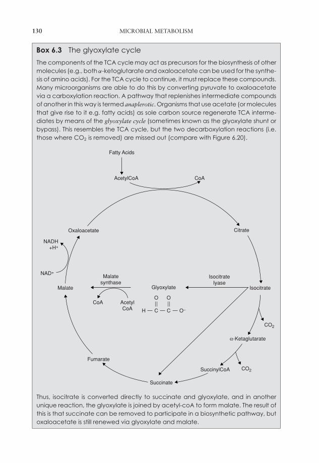
JWBK011-06 JWBK011-Hogg August 12, 2005 16:0 Char Count= 0
130 MICROBIAL METABOLISM
Box 6.3 The glyoxylate cycle
The components of the TCA cycle may act as precursors for the biosynthesis of othermolecules (e.g., both α-ketoglutarate and oxaloacetate can be used for the synthe-sis of amino acids). For the TCA cycle to continue, it must replace these compounds.Many microorganisms are able to do this by converting pyruvate to oxaloacetatevia a carboxylation reaction. A pathway that replenishes intermediate compoundsof another in this way is termed anaplerotic. Organisms that use acetate (or moleculesthat give rise to it e.g. fatty acids) as sole carbon source regenerate TCA interme-diates by means of the glyoxylate cycle (sometimes known as the glyoxylate shunt orbypass). This resembles the TCA cycle, but the two decarboxylation reactions (i.e.those where CO2 is removed) are missed out (compare with Figure 6.20).
Succinate
α-Ketaglutarate
Isocitrate
Citrate
Fumarate
Malate
Oxaloacetate
SuccinylCoA
CO2
CO2
AcetylCoA CoA
Fatty Acids
CoA AcetylCoA
Malatesynthase
IsocitrateIyase
Glyoxylate
O O
CH C O–
NADH+H+
NAD+
Thus, isocitrate is converted directly to succinate and glyoxylate, and in anotherunique reaction, the glyoxylate is joined by acetyl-coA to form malate. The result ofthis is that succinate can be removed to participate in a biosynthetic pathway, butoxaloacetate is still renewed via glyoxylate and malate.

JWBK011-06 JWBK011-Hogg August 12, 2005 16:0 Char Count= 0
PRINCIPLES OF ENERGY GENERATION 131
NADH NAD+
FADH2
FMN FAD
Q
Cyt b
Energy Cyt c1
Cyt c
Cyt a
Cyt a 3
O221
2H + H2O
Figure 6.21 The electron transport chain. Electrons from NADH and FADH2 pass fromone electron carrier to another, with a gradual release of energy as ATP by chemiosmosis (seeFigure 6.22). The electron carriers are arranged in order of their reduction potential (tendencyto gain electrons) and oscillate between the oxidised and the reduced state. FMN = flavinmononucleotide, Q = coenzyne Q.
from the TCA cycle. Where do all the rest come from? Most of the energy originallystored in the glucose molecule is now held in the form of the reduced coenzymes (NADHand FADH2) produced during glycolysis and the TCA cycle. This is now converted tono less than 34 molecules of ATP per glucose molecule by oxidative phosphorylationin the remaining steps in aerobic respiration (three from each molecule of NADH andtwo from each of FADH2).
The electron transportchain is a series of donor/acceptor moleculesthat transfer electronsfrom donors (e.g. NADH)to a terminal electronacceptor (e.g. O2).
In the final phase of aerobic respiration, electronsare transferred from NADH and FADH2, via a seriesof carrier molecules known collectively as the electrontransport (or respiratory) chain to oxygen, the terminalelectron acceptor (Figure 6.21). This in turn is reducedto the molecules of water you will remember from ouroverall equation on page 122. In procaryotes, this elec-tron transfer occurs at the plasma membrane, while ineucaryotes it takes place on the inner membrane of mi-tochondria. Table 6.2 summarises the locations of the reactions in the different phasesof carbohydrate metabolism.
JWBK011-06 JWBK011-Hogg August 12, 2005 16:0 Char Count= 0
132 MICROBIAL METABOLISM
Table 6.2 Location of respiratory enzymes
Reaction Procaryotes Eucaryotes
Glycolysis Cytoplasm CytoplasmTCA cycle Cytoplasm Mitochondrial matrixElectron Transport Plasma membrane Mitochondrial inner
membrane
Oxidative phosphorylation and the electron transport chain
The components of the electron transport chain differ between procaryotes and eu-caryotes, and even among bacterial systems, thus details may differ from the exampleoutlined below. The purpose of the electron transport is the same for all systems, how-ever, that is, the transfer of electrons from NADH/FADH2 via a series of carriers to,ultimately, oxygen. Around half of the energy released during this process is conservedas ATP.
The carrier molecules, which act alternately as acceptors and donors of electrons,are mostly complex modified proteins such as flavoproteins and cytochromes, togetherwith a class of lipid-soluble molecules called ubiquinones (also called coenzyme Q). Thecarriers are arranged in the chain such that each one has a more positive redox potentialthan the previous one. In the first step in the chain, NADH passes electrons to flavinmononucleotide (FMN), and in so doing becomes converted back to NAD+, therebyensuring a ready supply of the latter for the continuation of glycolysis (Figure 6.21).From FMN, the electrons are transferred to coenzyme Q, and thence to a series ofcytochromes; at each transfer of electrons the donor reverts back to its oxidised form,ready to pick up more electrons. You may recall that FADH2 yields only two, rather thanthree molecules of ATP per molecule; this is because it enters the electron transport chainat a later point than NADH, thereby missing one of the points where export of protonsoccurs. The final cytochrome in the chain transfers its electrons to molecular oxygen,which, as we have seen, acts as the terminal oxygen acceptor. The negatively chargedoxygen combines with protons from its surroundings to form water. Four electrons andprotons are required for the formation of each water molecule:
O2 + 4e− + 4H+ −→ 2H2O
Since two electrons are released by the oxidation of each NADH, it follows that twoNADH are needed for the oxidation of each oxygen.
How does this transfer of electrons lead to the formation of ATP? The chemiosmotictheory proposed by Peter Mitchell in 1961 offers an explanation. Although it was notimmediately accepted, the validity of the chemiosmotic model is now widely recognised,and in 1978 Mitchell received a Nobel Prize for his work. As envisaged by Mitchell,sufficient energy is released at three points in the electron transport chain for the transferof protons to the outside of the membrane, resulting in a gradient of both concentrationand charge (proton motive force). The protons are able to return across the membraneand achieve an equilibrium through specific protein channels within the enzyme ATP
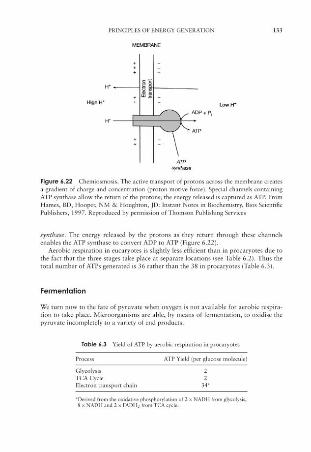
JWBK011-06 JWBK011-Hogg August 12, 2005 16:0 Char Count= 0
PRINCIPLES OF ENERGY GENERATION 133
Figure 6.22 Chemiosmosis. The active transport of protons across the membrane createsa gradient of charge and concentration (proton motive force). Special channels containingATP synthase allow the return of the protons; the energy released is captured as ATP. FromHames, BD, Hooper, NM & Houghton, JD: Instant Notes in Biochemistry, Bios ScientificPublishers, 1997. Reproduced by permission of Thomson Publishing Services
synthase. The energy released by the protons as they return through these channelsenables the ATP synthase to convert ADP to ATP (Figure 6.22).
Aerobic respiration in eucaryotes is slightly less efficient than in procaryotes due tothe fact that the three stages take place at separate locations (see Table 6.2). Thus thetotal number of ATPs generated is 36 rather than the 38 in procaryotes (Table 6.3).
Fermentation
We turn now to the fate of pyruvate when oxygen is not available for aerobic respira-tion to take place. Microorganisms are able, by means of fermentation, to oxidise thepyruvate incompletely to a variety of end products.
Table 6.3 Yield of ATP by aerobic respiration in procaryotes
Process ATP Yield (per glucose molecule)
Glycolysis 2TCA Cycle 2Electron transport chain 34∗
∗Derived from the oxidative phosphorylation of 2 × NADH from glycolysis,8 × NADH and 2 × FADH2 from TCA cycle.
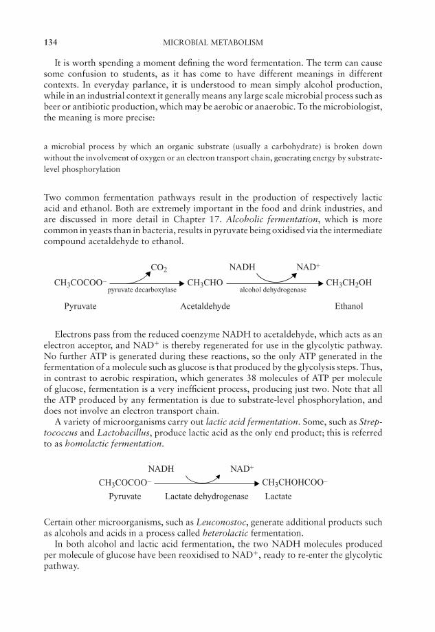
JWBK011-06 JWBK011-Hogg August 12, 2005 16:0 Char Count= 0
134 MICROBIAL METABOLISM
It is worth spending a moment defining the word fermentation. The term can causesome confusion to students, as it has come to have different meanings in differentcontexts. In everyday parlance, it is understood to mean simply alcohol production,while in an industrial context it generally means any large scale microbial process such asbeer or antibiotic production, which may be aerobic or anaerobic. To the microbiologist,the meaning is more precise:
a microbial process by which an organic substrate (usually a carbohydrate) is broken downwithout the involvement of oxygen or an electron transport chain, generating energy by substrate-level phosphorylation
Two common fermentation pathways result in the production of respectively lacticacid and ethanol. Both are extremely important in the food and drink industries, andare discussed in more detail in Chapter 17. Alcoholic fermentation, which is morecommon in yeasts than in bacteria, results in pyruvate being oxidised via the intermediatecompound acetaldehyde to ethanol.
CH3COCOO– CH3CHO CH3CH2OH
CO2
Pyruvate Acetaldehyde Ethanol
pyruvate decarboxylase alcohol dehydrogenase
NAD+NADH
Electrons pass from the reduced coenzyme NADH to acetaldehyde, which acts as anelectron acceptor, and NAD+ is thereby regenerated for use in the glycolytic pathway.No further ATP is generated during these reactions, so the only ATP generated in thefermentation of a molecule such as glucose is that produced by the glycolysis steps. Thus,in contrast to aerobic respiration, which generates 38 molecules of ATP per moleculeof glucose, fermentation is a very inefficient process, producing just two. Note that allthe ATP produced by any fermentation is due to substrate-level phosphorylation, anddoes not involve an electron transport chain.
A variety of microorganisms carry out lactic acid fermentation. Some, such as Strep-tococcus and Lactobacillus, produce lactic acid as the only end product; this is referredto as homolactic fermentation.
CH3COCOO– CH3CHOHCOO–
Pyruvate Lactate dehydrogenase
NAD+NADH
Lactate
Certain other microorganisms, such as Leuconostoc, generate additional products suchas alcohols and acids in a process called heterolactic fermentation.
In both alcohol and lactic acid fermentation, the two NADH molecules producedper molecule of glucose have been reoxidised to NAD+, ready to re-enter the glycolyticpathway.
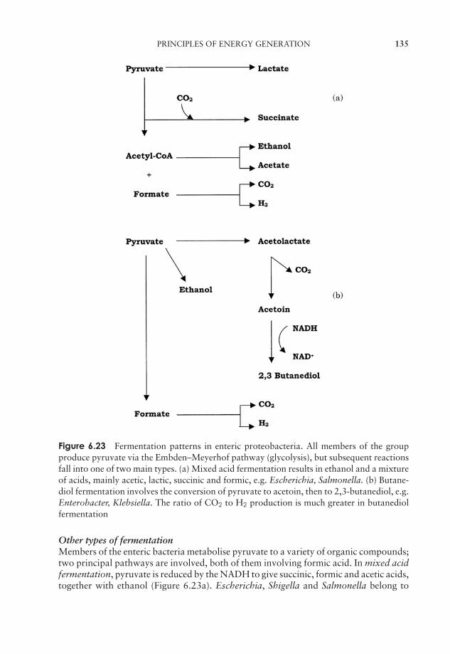
JWBK011-06 JWBK011-Hogg August 12, 2005 16:0 Char Count= 0
PRINCIPLES OF ENERGY GENERATION 135
Figure 6.23 Fermentation patterns in enteric proteobacteria. All members of the groupproduce pyruvate via the Embden–Meyerhof pathway (glycolysis), but subsequent reactionsfall into one of two main types. (a) Mixed acid fermentation results in ethanol and a mixtureof acids, mainly acetic, lactic, succinic and formic, e.g. Escherichia, Salmonella. (b) Butane-diol fermentation involves the conversion of pyruvate to acetoin, then to 2,3-butanediol, e.g.Enterobacter, Klebsiella. The ratio of CO2 to H2 production is much greater in butanediolfermentation
Other types of fermentationMembers of the enteric bacteria metabolise pyruvate to a variety of organic compounds;two principal pathways are involved, both of them involving formic acid. In mixed acidfermentation, pyruvate is reduced by the NADH to give succinic, formic and acetic acids,together with ethanol (Figure 6.23a). Escherichia, Shigella and Salmonella belong to

JWBK011-06 JWBK011-Hogg August 12, 2005 16:0 Char Count= 0
136 MICROBIAL METABOLISM
this group (see Chapter 7). Other enteric bacteria, such as Klebsiella and Enterobacter,carry out 2,3-butanediol fermentation. In this, the products are not acidic, and includean intermediate called acetoin. Much more CO2 is produced per molecule of glucosethan in mixed acid fermentation (Figure 6.23b). The end products of the two types offermentation provide a useful diagnostic test for the identification of unknown entericbacteria.
Metabolism of lipids and proteins
We have concentrated so far on the metabolism of carbohydrates, but both lipids andproteins may also act as energy sources. Both are converted by a series of reactions toan intermediate compound that can then enter the pathways of metabolism we havediscussed above.
Lipids often form an important energy source for microorganisms; they are plentifulin nature, as they form the major component of cell membranes, and may also exist ascellular storage structures. Lipids are hydrolysed by a class of enzymes called lipases totheir constituent parts; the fatty acids so produced enter the cyclic β-oxidation pathway.In this, fatty acids are joined to coenzyme A to form an acyl-CoA, and shortened by twocarbons in a series of reactions (Figure 6.24). Molecules of NADH and FADH2 derivedfrom β-oxidation can enter the electron transport chain to produce ATP. Acetyl-CoA,you will recall from earlier in this chapter, serves as the entry point into the TCA cycle.When you consider that a single turn of the TCA cycle gives rise to the production of 14molecules of ATP, you can appreciate what a rich source of energy a 16- or 18-carbonfatty acid represents. The glycerol component of a lipid requires only slight modificationin order to enter the glycolytic pathway as dihydroxyacetone phosphate (see ‘Glycolysis’above).
Proteins are a less useful source of energy than lipids or carbohydrates, but may beutilised when these are in short supply. Like lipids, they are initially hydrolysed to theirconstituent ‘building blocks’, in this case, amino acids. These then undergo the loss ofan amino group (deamination), resulting in a compound that is able to enter, eitherdirectly or indirectly, the TCA cycle.
CH3 CH3 CCH COOH COO– + NH4+
Alanine Pyruvate
NH2 O
NH2 O
−−−−−−−−−−−−−−−−−−−−−−−−−−−−−−−−−−−−−−−−−−−−−−−−−−−−−−−−−−−−−−−−−−→Figure 6.24 β-oxidation of lipids. β–Oxidation comprises a series of four reactions,repeated for the removal of each two-carbon unit. Formation of the acyl CoA esterrequires the expenditure of ATP, but there is a net gain in reducing power (NADH + FADH2),which can feed into the electron transport chain. The shortened acyl chain at the end of theprocess can re-enter the cycle and become further shortened, while acetyl CoA can enterdirectly into the TCA cycle (CoA-SH = coenzyme A)
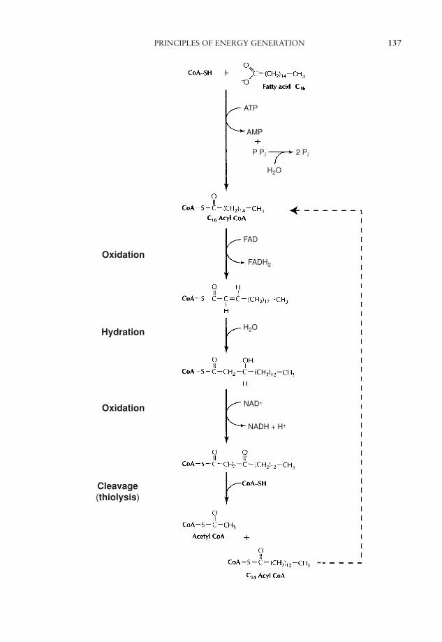
JWBK011-06 JWBK011-Hogg August 12, 2005 16:0 Char Count= 0
PRINCIPLES OF ENERGY GENERATION 137
Oxidation
Oxidation
Cleavage(thiolysis)
Hydration
ATP
AMP
P P 2 P
H2O
H2O
NAD+
NADH + H+
FAD
FADH2
JWBK011-06 JWBK011-Hogg August 12, 2005 16:0 Char Count= 0
138 MICROBIAL METABOLISM
Anaerobic respiration
In the process of anaerobic respiration, carbohydrate can be metabolised by a processthat utilises oxidative phosphorylation via an electron transport chain, but instead ofoxygen serving as the terminal electron acceptor a (usually) inorganic molecule such asnitrate or sulphate is used. These processes are referred to, respectively, as dissimilatorynitrate or sulphate reduction. Obligate anaerobes carry out this process, as they areunable to utilise oxygen; in addition, other organisms may turn to this form of res-piration if oxygen is unavailable (facultative anaerobes). Other examples of inorganicelectron acceptors for anaerobic respiration include Fe3+, CO2 and Mn4+. In certaincircumstances, an organic molecule such as fumarate may be used instead.
Anaerobic respiration is not as productive as its aerobic counterpart in terms of ATPproduction, because electron acceptors such as nitrate or sulphate have less positiveredox potentials than oxygen. Anaerobic respiration tends to occur in oxygen-depletedenvironments such as waterlogged soils.
It must be stressed that anaerobic respiration is not the same as fermentation. Thelatter process does not involve the components of the electron transport chain (i.e. thereis no oxidative phosphorylation), and much smaller amounts of energy are generated.
Energy may be generated by the oxidation of inorganic molecules
In the previous pages, we have seen how electrons derived from a variety of organicsources can be channelled into the glycolytic pathway (or one of its alternatives), andhow energy is generated by either oxygen or an organic/inorganic molecule acting asan electron acceptor. Certain bacteria, however, are able to derive their energy from theoxidation of inorganic substrates; these are termed chemolithotrophs (see Chapter 4).Molecules such as NH4
+, NO2−, Fe2+ and S0 can be oxidised, with the concomitant
generation of ATP.
NH4+ −→NO2
−
NO2− −→ NO3
−
Fe2+ −→ Fe3+
S0 −→ SO42−
The �G (change in free energy) for each of these reactions is much smaller than that foraerobic respiration. The value of �G is a measure of how much energy is released by a
The Calvin cycle is apathway for the fixa-tion of carbon dioxideused by photosyntheticorganisms and somechemolithotrophs.
reaction. Thus bacteria using this form of metabolismneed to oxidise a larger amount of their substrate inorder to synthesise the same amount of cellular mate-rial. In most cases, such bacteria are autotrophs, fix-ing carbon from carbon dioxide via the Calvin cycle.This is also used by phototrophic organisms, and is de-scribed at greater length in the section on photosynthe-sis below. If organic carbon is available, however, some
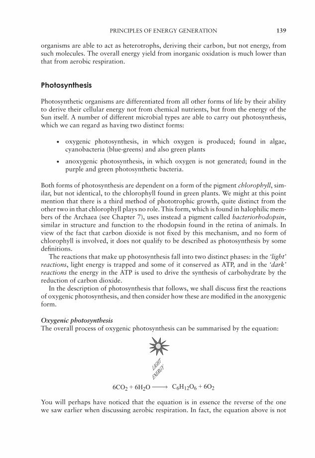
JWBK011-06 JWBK011-Hogg August 12, 2005 16:0 Char Count= 0
PRINCIPLES OF ENERGY GENERATION 139
organisms are able to act as heterotrophs, deriving their carbon, but not energy, fromsuch molecules. The overall energy yield from inorganic oxidation is much lower thanthat from aerobic respiration.
Photosynthesis
Photosynthetic organisms are differentiated from all other forms of life by their abilityto derive their cellular energy not from chemical nutrients, but from the energy of theSun itself. A number of different microbial types are able to carry out photosynthesis,which we can regard as having two distinct forms:
� oxygenic photosynthesis, in which oxygen is produced; found in algae,cyanobacteria (blue-greens) and also green plants
� anoxygenic photosynthesis, in which oxygen is not generated; found in thepurple and green photosynthetic bacteria.
Both forms of photosynthesis are dependent on a form of the pigment chlorophyll, sim-ilar, but not identical, to the chlorophyll found in green plants. We might at this pointmention that there is a third method of phototrophic growth, quite distinct from theother two in that chlorophyll plays no role. This form, which is found in halophilic mem-bers of the Archaea (see Chapter 7), uses instead a pigment called bacteriorhodopsin,similar in structure and function to the rhodopsin found in the retina of animals. Inview of the fact that carbon dioxide is not fixed by this mechanism, and no form ofchlorophyll is involved, it does not qualify to be described as photosynthesis by somedefinitions.
The reactions that make up photosynthesis fall into two distinct phases: in the ‘light’reactions, light energy is trapped and some of it conserved as ATP, and in the ‘dark’reactions the energy in the ATP is used to drive the synthesis of carbohydrate by thereduction of carbon dioxide.
In the description of photosynthesis that follows, we shall discuss first the reactionsof oxygenic photosynthesis, and then consider how these are modified in the anoxygenicform.
Oxygenic photosynthesisThe overall process of oxygenic photosynthesis can be summarised by the equation:
6CO2 + 6H2O C6H12O6 + 6O2
You will perhaps have noticed that the equation is in essence the reverse of the onewe saw earlier when discussing aerobic respiration. In fact, the equation above is not
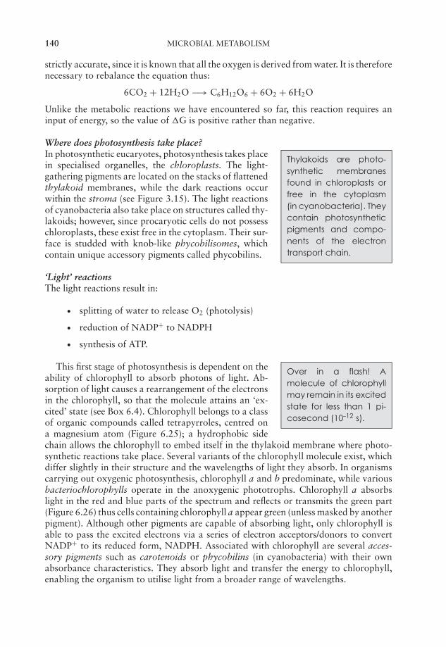
JWBK011-06 JWBK011-Hogg August 12, 2005 16:0 Char Count= 0
140 MICROBIAL METABOLISM
strictly accurate, since it is known that all the oxygen is derived from water. It is thereforenecessary to rebalance the equation thus:
6CO2 + 12H2O −→ C6H12O6 + 6O2 + 6H2O
Unlike the metabolic reactions we have encountered so far, this reaction requires aninput of energy, so the value of �G is positive rather than negative.
Where does photosynthesis take place?
Thylakoids are photo-synthetic membranesfound in chloroplasts orfree in the cytoplasm(in cyanobacteria). Theycontain photosyntheticpigments and compo-nents of the electrontransport chain.
In photosynthetic eucaryotes, photosynthesis takes placein specialised organelles, the chloroplasts. The light-gathering pigments are located on the stacks of flattenedthylakoid membranes, while the dark reactions occurwithin the stroma (see Figure 3.15). The light reactionsof cyanobacteria also take place on structures called thy-lakoids; however, since procaryotic cells do not possesschloroplasts, these exist free in the cytoplasm. Their sur-face is studded with knob-like phycobilisomes, whichcontain unique accessory pigments called phycobilins.
‘Light’ reactionsThe light reactions result in:
� splitting of water to release O2 (photolysis)
� reduction of NADP+ to NADPH
� synthesis of ATP.
Over in a flash! Amolecule of chlorophyllmay remain in its excitedstate for less than 1 pi-cosecond (10–12 s).
This first stage of photosynthesis is dependent on theability of chlorophyll to absorb photons of light. Ab-sorption of light causes a rearrangement of the electronsin the chlorophyll, so that the molecule attains an ‘ex-cited’ state (see Box 6.4). Chlorophyll belongs to a classof organic compounds called tetrapyrroles, centred ona magnesium atom (Figure 6.25); a hydrophobic sidechain allows the chlorophyll to embed itself in the thylakoid membrane where photo-synthetic reactions take place. Several variants of the chlorophyll molecule exist, whichdiffer slightly in their structure and the wavelengths of light they absorb. In organismscarrying out oxygenic photosynthesis, chlorophyll a and b predominate, while variousbacteriochlorophylls operate in the anoxygenic phototrophs. Chlorophyll a absorbslight in the red and blue parts of the spectrum and reflects or transmits the green part(Figure 6.26) thus cells containing chlorophyll a appear green (unless masked by anotherpigment). Although other pigments are capable of absorbing light, only chlorophyll isable to pass the excited electrons via a series of electron acceptors/donors to convertNADP+ to its reduced form, NADPH. Associated with chlorophyll are several acces-sory pigments such as carotenoids or phycobilins (in cyanobacteria) with their ownabsorbance characteristics. They absorb light and transfer the energy to chlorophyll,enabling the organism to utilise light from a broader range of wavelengths.
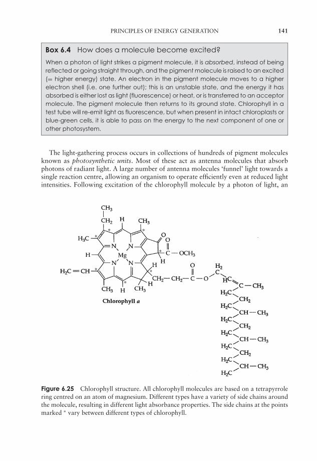
JWBK011-06 JWBK011-Hogg August 12, 2005 16:0 Char Count= 0
PRINCIPLES OF ENERGY GENERATION 141
Box 6.4 How does a molecule become excited?
When a photon of light strikes a pigment molecule, it is absorbed, instead of beingreflected or going straight through, and the pigment molecule is raised to an excited(= higher energy) state. An electron in the pigment molecule moves to a higherelectron shell (i.e. one further out); this is an unstable state, and the energy it hasabsorbed is either lost as light (fluorescence) or heat, or is transferred to an acceptormolecule. The pigment molecule then returns to its ground state. Chlorophyll in atest tube will re-emit light as fluorescence, but when present in intact chloroplasts orblue-green cells, it is able to pass on the energy to the next component of one orother photosystem.
The light-gathering process occurs in collections of hundreds of pigment moleculesknown as photosynthetic units. Most of these act as antenna molecules that absorbphotons of radiant light. A large number of antenna molecules ‘funnel’ light towards asingle reaction centre, allowing an organism to operate efficiently even at reduced lightintensities. Following excitation of the chlorophyll molecule by a photon of light, an
Figure 6.25 Chlorophyll structure. All chlorophyll molecules are based on a tetrapyrrolering centred on an atom of magnesium. Different types have a variety of side chains aroundthe molecule, resulting in different light absorbance properties. The side chains at the pointsmarked ∗ vary between different types of chlorophyll.
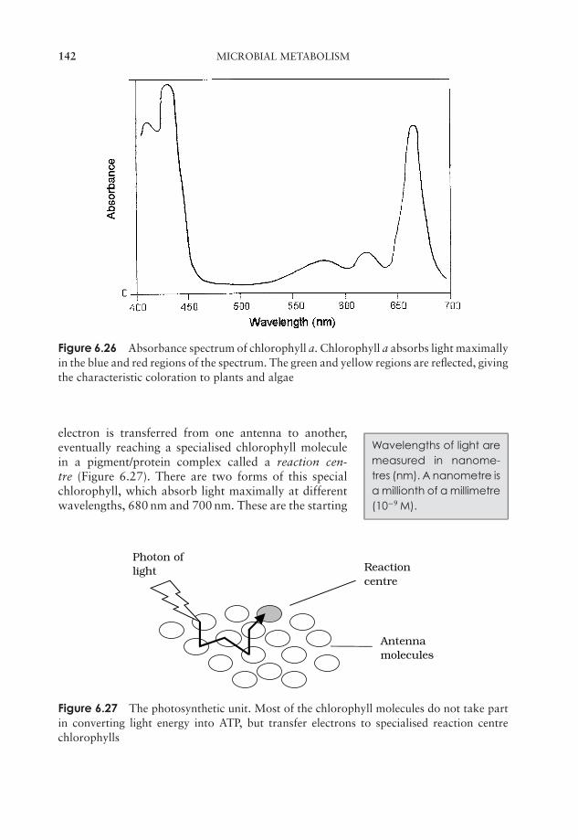
JWBK011-06 JWBK011-Hogg August 12, 2005 16:0 Char Count= 0
142 MICROBIAL METABOLISM
Figure 6.26 Absorbance spectrum of chlorophyll a. Chlorophyll a absorbs light maximallyin the blue and red regions of the spectrum. The green and yellow regions are reflected, givingthe characteristic coloration to plants and algae
Wavelengths of light aremeasured in nanome-tres (nm). A nanometre isa millionth of a millimetre(10−9 M).
electron is transferred from one antenna to another,eventually reaching a specialised chlorophyll moleculein a pigment/protein complex called a reaction cen-tre (Figure 6.27). There are two forms of this specialchlorophyll, which absorb light maximally at differentwavelengths, 680 nm and 700 nm. These are the starting
Photon oflight
Antennamolecules
Reactioncentre
Figure 6.27 The photosynthetic unit. Most of the chlorophyll molecules do not take partin converting light energy into ATP, but transfer electrons to specialised reaction centrechlorophylls
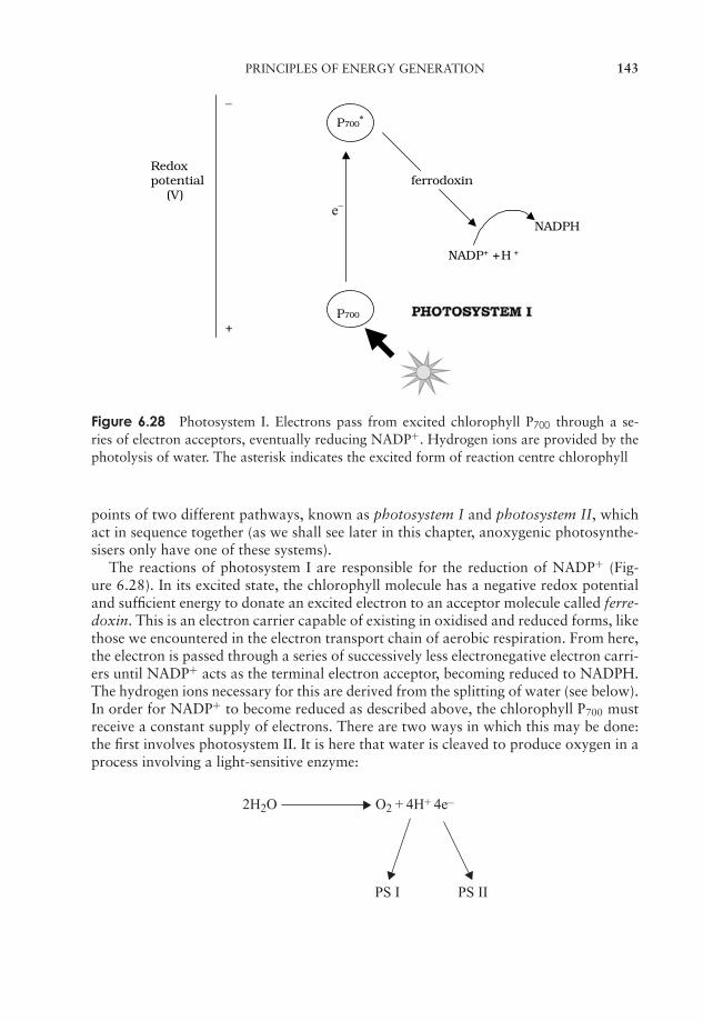
JWBK011-06 JWBK011-Hogg August 12, 2005 16:0 Char Count= 0
PRINCIPLES OF ENERGY GENERATION 143
− P700*
Redoxpotential ferrodoxin (V)
e−
NADPH
NADP+ + H +
P700
+
Figure 6.28 Photosystem I. Electrons pass from excited chlorophyll P700 through a se-ries of electron acceptors, eventually reducing NADP+. Hydrogen ions are provided by thephotolysis of water. The asterisk indicates the excited form of reaction centre chlorophyll
points of two different pathways, known as photosystem I and photosystem II, whichact in sequence together (as we shall see later in this chapter, anoxygenic photosynthe-sisers only have one of these systems).
The reactions of photosystem I are responsible for the reduction of NADP+ (Fig-ure 6.28). In its excited state, the chlorophyll molecule has a negative redox potentialand sufficient energy to donate an excited electron to an acceptor molecule called ferre-doxin. This is an electron carrier capable of existing in oxidised and reduced forms, likethose we encountered in the electron transport chain of aerobic respiration. From here,the electron is passed through a series of successively less electronegative electron carri-ers until NADP+ acts as the terminal electron acceptor, becoming reduced to NADPH.The hydrogen ions necessary for this are derived from the splitting of water (see below).In order for NADP+ to become reduced as described above, the chlorophyll P700 mustreceive a constant supply of electrons. There are two ways in which this may be done:the first involves photosystem II. It is here that water is cleaved to produce oxygen in aprocess involving a light-sensitive enzyme:
2H2O O2 + 4H+ 4e–
PS I PS II
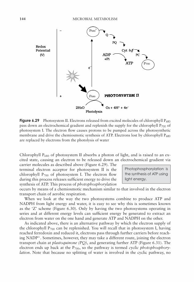
JWBK011-06 JWBK011-Hogg August 12, 2005 16:0 Char Count= 0
144 MICROBIAL METABOLISM
− P680*
Q
PQ RedoxPotential Cyt b/f (V) ADP PC
+Pi ATP P700
e−
+ P680
2H2O O2 + 4H+ + 4e−
Photolysis
− P680*
Q
PQ RedoxPotential Cyt b/f (V) ADP PC
+Pi ATP P700
e−
+ P680
2H2O O2 + 4H+ + 4e−
Photolysis
Figure 6.29 Photosystem II. Electrons released from excited molecules of chlorophyll P680
pass down an electrochemical gradient and replenish the supply for the chlorophyll P700 ofphotosystem I. The electron flow causes protons to be pumped across the photosyntheticmembrane and drive the chemiosmotic synthesis of ATP. Electrons lost by chlorophyll P680
are replaced by electrons from the photolysis of water
Chlorophyll P680 of photosystem II absorbs a photon of light, and is raised to an ex-cited state, causing an electron to be released down an electrochemical gradient via
Photophosphorylation isthe synthesis of ATP usinglight energy.
carrier molecules as described above (Figure 6.29). Theterminal electron acceptor for photosystem II is thechlorophyll P700 of photosystem I. The electron flowduring this process releases sufficient energy to drive thesynthesis of ATP. This process of photophosphorylationoccurs by means of a chemiosmotic mechanism similar to that involved in the electrontransport chain of aerobic respiration.
When we look at the way the two photosystems combine to produce ATP andNADPH from light energy and water, it is easy to see why this is sometimes knownas the ‘Z’ scheme (Figure 6.30). Only by having the two photosystems operating inseries and at different energy levels can sufficient energy be generated to extract anelectron from water on the one hand and generate ATP and NADPH on the other.
As indicated above, there is an alternative pathway by which the electron supply ofthe chlorophyll P700 can be replenished. You will recall that in photosystem I, havingreached ferredoxin and reduced it, electrons pass through further carriers before reach-ing NADP+. Sometimes, however, they may take a different route, joining the electrontransport chain at plastoquinone (PQ), and generating further ATP (Figure 6.31). Theelectron ends up back at the P700, so the pathway is termed cyclic photophosphory-lation. Note that because no splitting of water is involved in the cyclic pathway, no

JWBK011-06 JWBK011-Hogg August 12, 2005 16:0 Char Count= 0
PRINCIPLES OF ENERGY GENERATION 145
Figure 6.30 Non-cyclic photophosphorylation: the ‘Z’ scheme. Photosystems I and II in-teract to convert water and light energy into ATP and NADPH. P680 and P700 indicate thewavelength of light maximally absorbed by the two photosystems
Figure 6.31 Cyclic photophosphorylation. Photosystem I electrons may enter the electrontransport chain at plastoquinone and return to reaction centre chlorophyll P700, generatinga proton motive force for use in ATP production
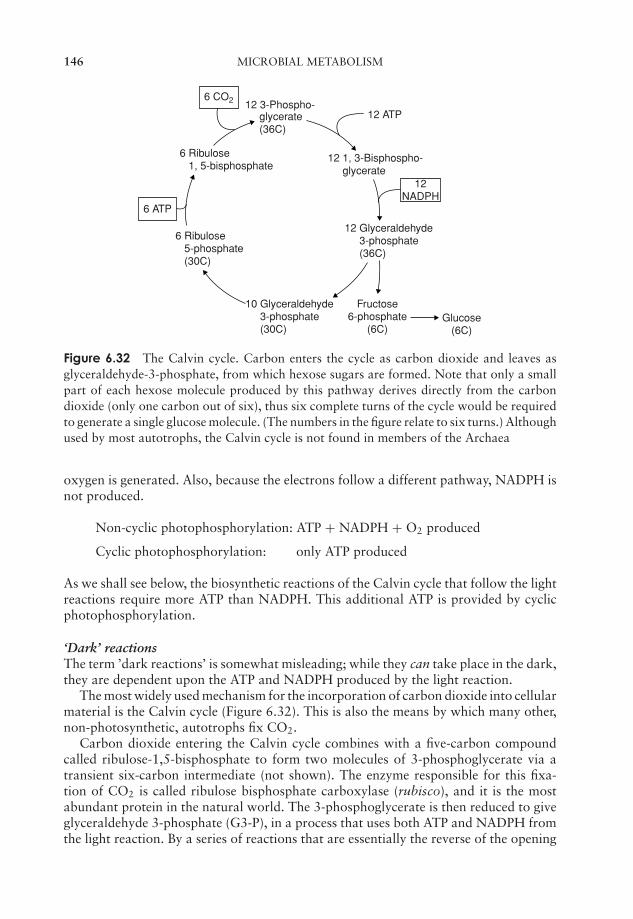
JWBK011-06 JWBK011-Hogg August 12, 2005 16:0 Char Count= 0
146 MICROBIAL METABOLISM
6 CO2 12 3-Phospho-glycerate(36C)
6 Ribulose 1, 5-bisphosphate
6 Ribulose 5-phosphate (30C)
10 Glyceraldehyde 3-phosphate (30C)
12 Glyceraldehyde 3-phosphate (36C)
Fructose6-phosphate
(6C)
12 1, 3-Bisphospho- glycerate
12 ATP
12NADPH
6 ATP
Glucose(6C)
Figure 6.32 The Calvin cycle. Carbon enters the cycle as carbon dioxide and leaves asglyceraldehyde-3-phosphate, from which hexose sugars are formed. Note that only a smallpart of each hexose molecule produced by this pathway derives directly from the carbondioxide (only one carbon out of six), thus six complete turns of the cycle would be requiredto generate a single glucose molecule. (The numbers in the figure relate to six turns.) Althoughused by most autotrophs, the Calvin cycle is not found in members of the Archaea
oxygen is generated. Also, because the electrons follow a different pathway, NADPH isnot produced.
Non-cyclic photophosphorylation: ATP + NADPH + O2 produced
Cyclic photophosphorylation: only ATP produced
As we shall see below, the biosynthetic reactions of the Calvin cycle that follow the lightreactions require more ATP than NADPH. This additional ATP is provided by cyclicphotophosphorylation.
‘Dark’ reactionsThe term ’dark reactions’ is somewhat misleading; while they can take place in the dark,they are dependent upon the ATP and NADPH produced by the light reaction.
The most widely used mechanism for the incorporation of carbon dioxide into cellularmaterial is the Calvin cycle (Figure 6.32). This is also the means by which many other,non-photosynthetic, autotrophs fix CO2.
Carbon dioxide entering the Calvin cycle combines with a five-carbon compoundcalled ribulose-1,5-bisphosphate to form two molecules of 3-phosphoglycerate via atransient six-carbon intermediate (not shown). The enzyme responsible for this fixa-tion of CO2 is called ribulose bisphosphate carboxylase (rubisco), and it is the mostabundant protein in the natural world. The 3-phosphoglycerate is then reduced to giveglyceraldehyde 3-phosphate (G3-P), in a process that uses both ATP and NADPH fromthe light reaction. By a series of reactions that are essentially the reverse of the opening

JWBK011-06 JWBK011-Hogg August 12, 2005 16:0 Char Count= 0
PRINCIPLES OF ENERGY GENERATION 147
steps of glycolysis, some of this G3-P is converted to glucose. The remaining steps ofthe Calvin cycle are concerned with regenerating the five-carbon ribulose-5-phosphate.We can summarise the incorporation of CO2 into glucose as:
6CO2 + 18ATP + 12NADPH + 2H+ + 12H2O → C6H12O6 + 18ADP
+18Pi + 12NADP+
Anoxygenic photosynthesis
Photosynthesis as carried out in the purple and green bacteria is different to the processjust described in a number of respects. Some of the main differences are summarisedbelow:
� no oxygen is generated during this type of photosynthesis, and the bacteria involvedare growing anaerobically
� bacteriochlophylls absorb light maximally at longer wavelengths than chlorophylla and b, allowing them more effectively to utilise the light available in their ownparticular habitat
� purple and green bacteria are not able to utilise water as a donor of electrons, andmust instead use a compound that is oxidised more easily, such as hydrogen sulphideor succinate
� only a single photosystem is involved in the light reactions of anoxygenic photosyn-thesis. In the green bacteria this is similar to photosystem I, while in the purples itmore closely resembles photosystem II
� thylakoid membranes are not found in the green and purple bacteria; light reactionstake place in lamellar invaginations of the cytoplasmic membrane in the purples andin vesicles called chlorosomes in the greens.
In the generation of ATP, a form of cyclic photophosporylation is employed; the bac-teriochlorophyll acts as both donor and acceptor of electrons (Figure 6.33). In orderto generate reducing power in the form of reduced coenzymes, an external electronsource is necessary, since this process is non-cyclic. In the green and purple sulphurbacteria, this role is served by sulphur or reduced sulphur compounds such as sulphideor thiosulphate. The non-sulphur bacteria utilise an organic molecule such as succi-nate as an electron donor. In some cases, the electron donor has a more positive redoxpotential than the NADP+, so in order to reduce the coenzyme, electrons would haveto flow against the electrochemical gradient. An input of energy in the form of ATP isneeded to make this possible, in a process known as reverse electron flow. This alsohappens in many chemolithotrophs such as Acidithiobacillus and Nitrobacter. Table6.4 compares the different groups of anoxygenic photosynthesisers. A special form ofanoxygenic photosynthesis occurs in the Heliobacteria (see Chapter 7); these carry outphotoheterotrophy using a unique form of bacteriochlorophyll called bacteriochloro-phyll b. They are unable to use carbon dioxide as a carbon source.
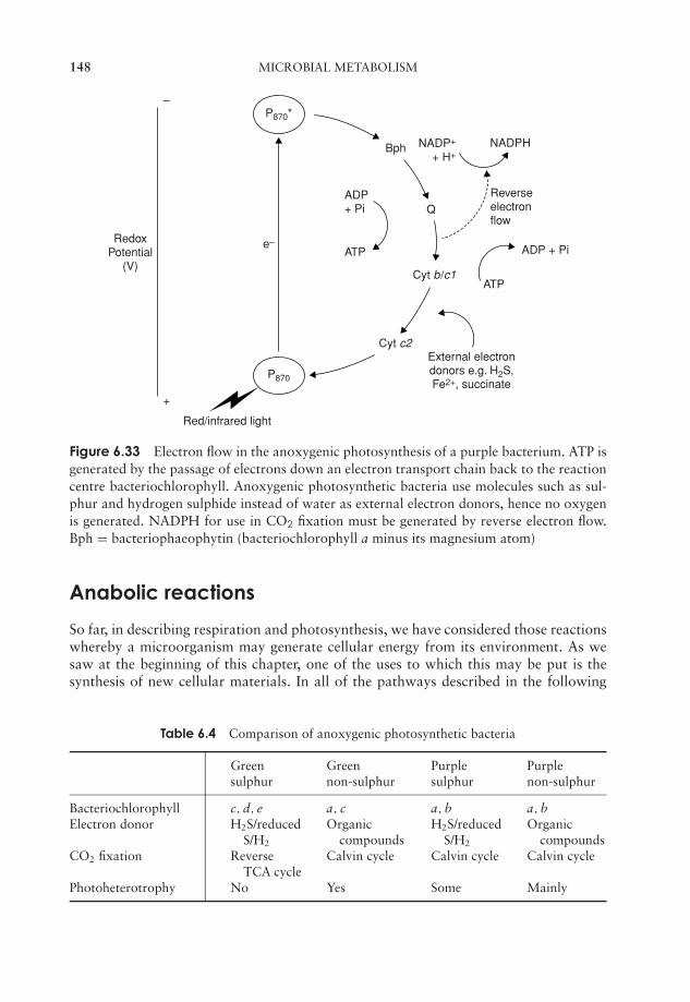
JWBK011-06 JWBK011-Hogg August 12, 2005 16:0 Char Count= 0
148 MICROBIAL METABOLISM
–
+
e–
P870*
P870
Red/infrared light
ADP+ Pi
ATP
NADP+
+ H+NADPH
External electrondonors e.g. H2S,Fe2+, succinate
ADP + Pi
ATPCyt b/c1
Cyt c2
Q
Bph
RedoxPotential
(V)
Reverseelectronflow
Figure 6.33 Electron flow in the anoxygenic photosynthesis of a purple bacterium. ATP isgenerated by the passage of electrons down an electron transport chain back to the reactioncentre bacteriochlorophyll. Anoxygenic photosynthetic bacteria use molecules such as sul-phur and hydrogen sulphide instead of water as external electron donors, hence no oxygenis generated. NADPH for use in CO2 fixation must be generated by reverse electron flow.Bph = bacteriophaeophytin (bacteriochlorophyll a minus its magnesium atom)
Anabolic reactions
So far, in describing respiration and photosynthesis, we have considered those reactionswhereby a microorganism may generate cellular energy from its environment. As wesaw at the beginning of this chapter, one of the uses to which this may be put is thesynthesis of new cellular materials. In all of the pathways described in the following
Table 6.4 Comparison of anoxygenic photosynthetic bacteria
Green Green Purple Purplesulphur non-sulphur sulphur non-sulphur
Bacteriochlorophyll c, d, e a, c a, b a, bElectron donor H2S/reduced
S/H2
Organiccompounds
H2S/reducedS/H2
Organiccompounds
CO2 fixation ReverseTCA cycle
Calvin cycle Calvin cycle Calvin cycle
Photoheterotrophy No Yes Some Mainly
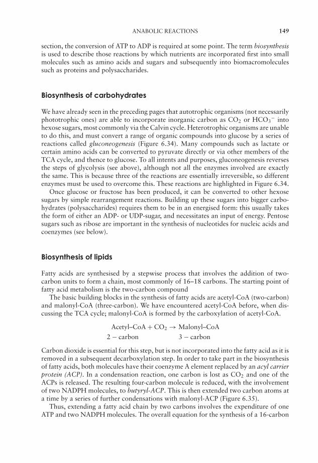
JWBK011-06 JWBK011-Hogg August 12, 2005 16:0 Char Count= 0
ANABOLIC REACTIONS 149
section, the conversion of ATP to ADP is required at some point. The term biosynthesisis used to describe those reactions by which nutrients are incorporated first into smallmolecules such as amino acids and sugars and subsequently into biomacromoleculessuch as proteins and polysaccharides.
Biosynthesis of carbohydrates
We have already seen in the preceding pages that autotrophic organisms (not necessarilyphototrophic ones) are able to incorporate inorganic carbon as CO2 or HCO3
− intohexose sugars, most commonly via the Calvin cycle. Heterotrophic organisms are unableto do this, and must convert a range of organic compounds into glucose by a series ofreactions called gluconeogenesis (Figure 6.34). Many compounds such as lactate orcertain amino acids can be converted to pyruvate directly or via other members of theTCA cycle, and thence to glucose. To all intents and purposes, gluconeogenesis reversesthe steps of glycolysis (see above), although not all the enzymes involved are exactlythe same. This is because three of the reactions are essentially irreversible, so differentenzymes must be used to overcome this. These reactions are highlighted in Figure 6.34.
Once glucose or fructose has been produced, it can be converted to other hexosesugars by simple rearrangement reactions. Building up these sugars into bigger carbo-hydrates (polysaccharides) requires them to be in an energised form: this usually takesthe form of either an ADP- or UDP-sugar, and necessitates an input of energy. Pentosesugars such as ribose are important in the synthesis of nucleotides for nucleic acids andcoenzymes (see below).
Biosynthesis of lipids
Fatty acids are synthesised by a stepwise process that involves the addition of two-carbon units to form a chain, most commonly of 16–18 carbons. The starting point offatty acid metabolism is the two-carbon compound
The basic building blocks in the synthesis of fatty acids are acetyl-CoA (two-carbon)and malonyl-CoA (three-carbon). We have encountered acetyl-CoA before, when dis-cussing the TCA cycle; malonyl-CoA is formed by the carboxylation of acetyl-CoA.
Acetyl–CoA + CO2 → Malonyl–CoA
2 − carbon 3 − carbon
Carbon dioxide is essential for this step, but is not incorporated into the fatty acid as it isremoved in a subsequent decarboxylation step. In order to take part in the biosynthesisof fatty acids, both molecules have their coenzyme A element replaced by an acyl carrierprotein (ACP). In a condensation reaction, one carbon is lost as CO2 and one of theACPs is released. The resulting four-carbon molecule is reduced, with the involvementof two NADPH molecules, to butyryl-ACP. This is then extended two carbon atoms ata time by a series of further condensations with malonyl-ACP (Figure 6.35).
Thus, extending a fatty acid chain by two carbons involves the expenditure of oneATP and two NADPH molecules. The overall equation for the synthesis of a 16-carbon
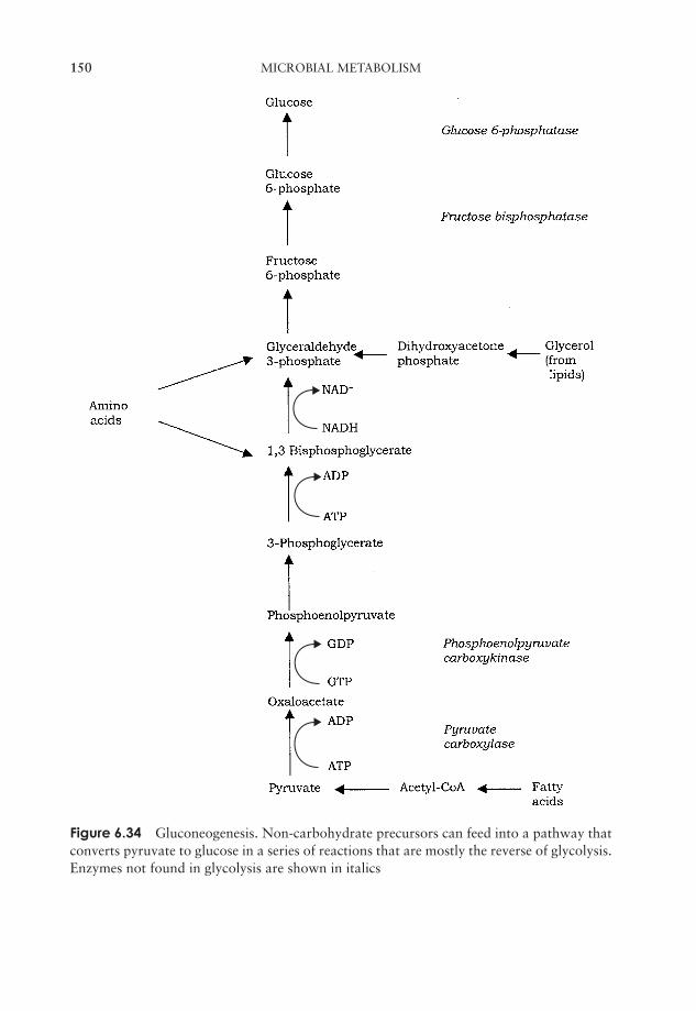
JWBK011-06 JWBK011-Hogg August 12, 2005 16:0 Char Count= 0
150 MICROBIAL METABOLISM
Figure 6.34 Gluconeogenesis. Non-carbohydrate precursors can feed into a pathway thatconverts pyruvate to glucose in a series of reactions that are mostly the reverse of glycolysis.Enzymes not found in glycolysis are shown in italics
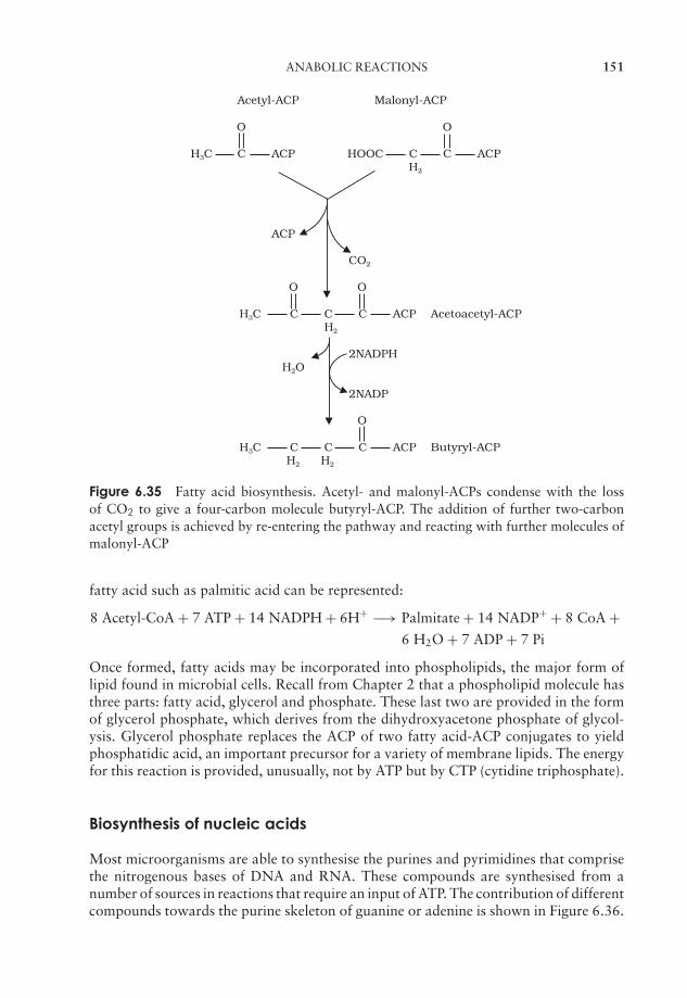
JWBK011-06 JWBK011-Hogg August 12, 2005 16:0 Char Count= 0
ANABOLIC REACTIONS 151
Acetyl-ACP Malonyl-ACP
O O
H3C C ACP HOOC C C ACPH2
ACP
CO2
O O
H3C C C C ACP Acetoacetyl-ACP H2
2NADPHH2O
2NADP
O
H3C C C C ACP Butyryl-ACPH2 H2
Figure 6.35 Fatty acid biosynthesis. Acetyl- and malonyl-ACPs condense with the lossof CO2 to give a four-carbon molecule butyryl-ACP. The addition of further two-carbonacetyl groups is achieved by re-entering the pathway and reacting with further molecules ofmalonyl-ACP
fatty acid such as palmitic acid can be represented:
8 Acetyl-CoA + 7 ATP + 14 NADPH + 6H+ −→ Palmitate + 14 NADP+ + 8 CoA +6 H2O + 7 ADP + 7 Pi
Once formed, fatty acids may be incorporated into phospholipids, the major form oflipid found in microbial cells. Recall from Chapter 2 that a phospholipid molecule hasthree parts: fatty acid, glycerol and phosphate. These last two are provided in the formof glycerol phosphate, which derives from the dihydroxyacetone phosphate of glycol-ysis. Glycerol phosphate replaces the ACP of two fatty acid-ACP conjugates to yieldphosphatidic acid, an important precursor for a variety of membrane lipids. The energyfor this reaction is provided, unusually, not by ATP but by CTP (cytidine triphosphate).
Biosynthesis of nucleic acids
Most microorganisms are able to synthesise the purines and pyrimidines that comprisethe nitrogenous bases of DNA and RNA. These compounds are synthesised from anumber of sources in reactions that require an input of ATP. The contribution of differentcompounds towards the purine skeleton of guanine or adenine is shown in Figure 6.36.
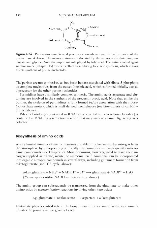
JWBK011-06 JWBK011-Hogg August 12, 2005 16:0 Char Count= 0
152 MICROBIAL METABOLISM
Figure 6.36 Purine structure. Several precursors contribute towards the formation of thepurine base skeleton. The nitrogen atoms are donated by the amino acids glutamine, as-partate and glycine. Note the important role played by folic acid. The antimicrobial agentsulphonamide (Chapter 15) exerts its effect by inhibiting folic acid synthesis, which in turnaffects synthesis of purine nucleotides
The purines are not synthesised as free bases but are associated with ribose-5-phosphateas complete nucleotides from the outset. Inosinic acid, which is formed initially, acts asa precursor for the other purine nucleotides.
Pyrimidines have a similarly complex synthesis. The amino acids aspartate and glu-tamine are involved in the synthesis of the precursor orotic acid. Note that unlike thepurines, the skeleton of pyrimidines is fully formed before association with the ribose-5-phosphate moiety, which is itself derived from glucose (see biosynthesis of carbohy-drates, above).
Ribonucleotides (as contained in RNA) are converted to deoxyribonucleotides (ascontained in DNA) by a reduction reaction that may involve vitamin B12 acting as acofactor.
Biosynthesis of amino acids
A very limited number of microorganisms are able to utilise molecular nitrogen fromthe atmosphere by incorporating it initially into ammonia and subsequently into or-ganic compounds (see Chapter 7). Most organisms, however, need to have their ni-trogen supplied as nitrate, nitrite, or ammonia itself. Ammonia can be incorporatedinto organic nitrogen compounds in several ways, including glutamate formation fromα-ketoglutarate (see TCA cycle, above):
α-ketoglutarate + NH4+ + NADPH* + H+ −→ glutamate + NADP+ + H2O
(*Some species utilise NADH as their electron donor)
The amino group can subsequently be transferred from the glutamate to make otheramino acids by transamination reactions involving other keto acids:
e.g. glutamate + oxaloacetate −→ aspartate + α-ketoglutarate
Glutamate plays a central role in the biosynthesis of other amino acids, as it usuallydonates the primary amino group of each:
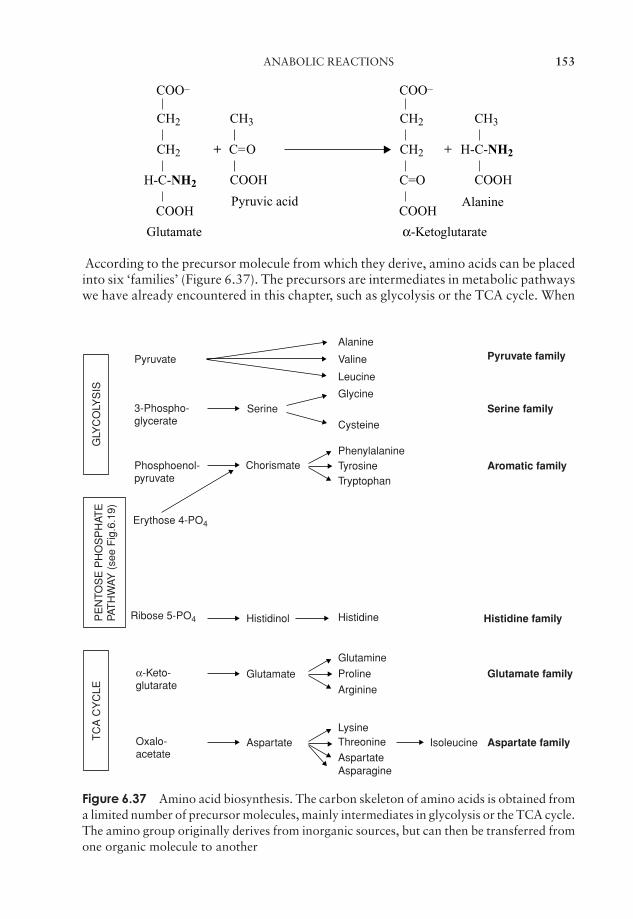
JWBK011-06 JWBK011-Hogg August 12, 2005 16:0 Char Count= 0
ANABOLIC REACTIONS 153
Glutamate
Pyruvic acid
CH3
C=O
COOH
CH2
COO–
CH2
H-C-NH2
COOH
+
Alanine
H-C-NH2
CH3
COOH
+
α-Ketoglutarate
CH2
COO–
CH2
C=O
COOH
According to the precursor molecule from which they derive, amino acids can be placedinto six ‘families’ (Figure 6.37). The precursors are intermediates in metabolic pathwayswe have already encountered in this chapter, such as glycolysis or the TCA cycle. When
Pyruvate familyPyruvate
3-Phospho-glycerate
Alanine
Valine
Leucine
Serine Serine family
Aromatic familyChorismate
Glycine
Cysteine
PhenylalanineTyrosineTryptophan
Phosphoenol-pyruvate
Erythose 4-PO4
Ribose 5-PO4
GLY
CO
LYS
IST
CA
CY
CLE
Histidinol Histidine
LysineThreonine Isoleucine
Histidine family
Glutamate family
Glutamine
Arginine
ProlineGlutamate
AspartateOxalo-acetate
α-Keto-glutarate
AspartateAspartate family
Asparagine
PE
NTO
SE
PH
OS
PH
ATE
PAT
HW
AY (
see
Fig
.6.1
9)
Figure 6.37 Amino acid biosynthesis. The carbon skeleton of amino acids is obtained froma limited number of precursor molecules, mainly intermediates in glycolysis or the TCA cycle.The amino group originally derives from inorganic sources, but can then be transferred fromone organic molecule to another
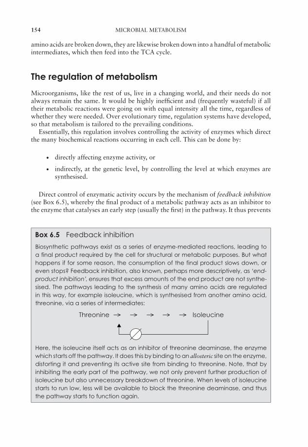
JWBK011-06 JWBK011-Hogg August 12, 2005 16:0 Char Count= 0
154 MICROBIAL METABOLISM
amino acids are broken down, they are likewise broken down into a handful of metabolicintermediates, which then feed into the TCA cycle.
The regulation of metabolism
Microorganisms, like the rest of us, live in a changing world, and their needs do notalways remain the same. It would be highly inefficient and (frequently wasteful) if alltheir metabolic reactions were going on with equal intensity all the time, regardless ofwhether they were needed. Over evolutionary time, regulation systems have developed,so that metabolism is tailored to the prevailing conditions.
Essentially, this regulation involves controlling the activity of enzymes which directthe many biochemical reactions occurring in each cell. This can be done by:
� directly affecting enzyme activity, or
� indirectly, at the genetic level, by controlling the level at which enzymes aresynthesised.
Direct control of enzymatic activity occurs by the mechanism of feedback inhibition(see Box 6.5), whereby the final product of a metabolic pathway acts as an inhibitor tothe enzyme that catalyses an early step (usually the first) in the pathway. It thus prevents
Box 6.5 Feedback inhibition
Biosynthetic pathways exist as a series of enzyme-mediated reactions, leading toa final product required by the cell for structural or metabolic purposes. But whathappens if for some reason, the consumption of the final product slows down, oreven stops? Feedback inhibition, also known, perhaps more descriptively, as ‘end-product inhibition’, ensures that excess amounts of the end product are not synthe-sised. The pathways leading to the synthesis of many amino acids are regulatedin this way, for example isoleucine, which is synthesised from another amino acid,threonine, via a series of intermediates:
Threonine Isoleucine
Here, the isoleucine itself acts as an inhibitor of threonine deaminase, the enzymewhich starts off the pathway. It does this by binding to an allosteric site on the enzyme,distorting it and preventing its active site from binding to threonine. Note, that byinhibiting the early part of the pathway, we not only prevent further production ofisoleucine but also unnecessary breakdown of threonine. When levels of isoleucinestarts to run low, less will be available to block the threonine deaminase, and thusthe pathway starts to function again.
JWBK011-06 JWBK011-Hogg August 12, 2005 16:0 Char Count= 0
TEST YOURSELF 155
more of its own formation. When the concentration of the product subsequently fallsbelow a certain level, it is no longer inhibitory, and biosynthesis resumes.
Regulation of metabolic pathways can also be achieved by controlling whether ornot an enzyme is synthesised in the first place, and if so, the rate at which it is produced.This is done at the DNA level, by one of two mechanisms, induction and repression,which respectively ‘switch on’ and ‘switch off’ the machinery of protein synthesis dis-cussed earlier in this chapter. These are discussed under the heading ‘Regulation of geneexpression’ in Chapter 11
Test yourself
1 Reactions which involve the breakdown of compounds are collectivelytermed and synthetic (building up) ones are termed .
2 Enzymes lower the of a reaction.
3 The of an enzyme is the part that is actually involved incatalysis.
4 At pH values away from the optimum, of ‘R’ groups causes denat-uration of proteins.
5 When a molecule an electron, it is said to be oxidised.
6 Compounds which are good electron donors have a strongly redoxpotential.
7 The energy ‘currency’ of cellular metabolism is.
8 The first step in glucose metabolism is , which can occur with orwithout .
9 ATP may be formed from ADP by phosphorylation,phosphorylation or phosphorylation.
10 Before entering the TCA cycle, pyruvate must be converted to the two-carboncompound . This type of reaction, in which a molecule of carbondioxide is removed, is called a .
11 The main product of the TCA cycle is reducing power, in the form ofand .
12 The role of oxygen in aerobic respiration is to act as thein the electron transport chain.
JWBK011-06 JWBK011-Hogg August 12, 2005 16:0 Char Count= 0
156 MICROBIAL METABOLISM
13 As envisaged by the chemiosmotic theory of Peter Mitchell, protons arepumped out across a membrane, generating a .
14 anaerobes are able to utilise anaerobic respiration if oxygen isunavailable.
15 The two commonest products of fermentation reactions are and.
16 Fermentation yields far less ATP than aerobic respiration because it does notinvolve an .
17 Fatty acids are broken down carbons at a time by the process of.
18 are organisms that can derive their energy from the oxidation ofinorganic compounds such as ammonia or reduced iron.
19 Photosynthesis carried out by green and purple bacteria differs from thatcarried out by algae and cyanobacteria in that no is generated.
20 In the light reactions of photosynthesis, light energy and an electron donorare used to generate and .
21 In the dark reactions that make up the cycle, carbon dioxide isassimilated into glyceraldehyde-3-phosphate, which is then converted to glu-cose in a series of steps that are the reverse of .
22 Anoxygenic photosynthetic bacteria are unable to use as an elec-tron donor, and therefore use compounds such as hydrogen sulphide or suc-cinate.
23 Organisms unable to synthesise glucose from inorganic carbon must deriveit from other organic compound by the process of .
24 In amino acid synthesis, -NH2 groups may be transferred between com-pounds in reactions.
25 The inhibition of a metabolic pathway by its final product is termed end-product or inhibition.

JWBK011-07 JWBK011-Hogg August 12, 2005 16:8 Char Count= 0
Part IIIMicrobial Diversity
157
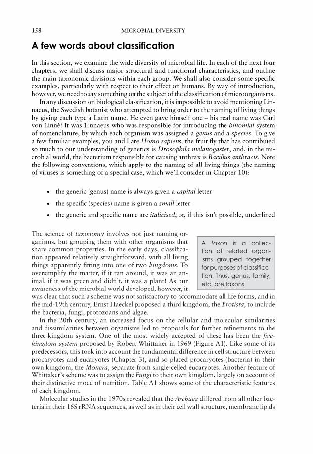
JWBK011-07 JWBK011-Hogg August 12, 2005 16:8 Char Count= 0
158 MICROBIAL DIVERSITY
A few words about classification
In this section, we examine the wide diversity of microbial life. In each of the next fourchapters, we shall discuss major structural and functional characteristics, and outlinethe main taxonomic divisions within each group. We shall also consider some specificexamples, particularly with respect to their effect on humans. By way of introduction,however, we need to say something on the subject of the classification of microorganisms.
In any discussion on biological classification, it is impossible to avoid mentioning Lin-naeus, the Swedish botanist who attempted to bring order to the naming of living thingsby giving each type a Latin name. He even gave himself one – his real name was Carlvon Linne! It was Linnaeus who was responsible for introducing the binomial systemof nomenclature, by which each organism was assigned a genus and a species. To givea few familiar examples, you and I are Homo sapiens, the fruit fly that has contributedso much to our understanding of genetics is Drosophila melanogaster, and, in the mi-crobial world, the bacterium responsible for causing anthrax is Bacillus anthracis. Notethe following conventions, which apply to the naming of all living things (the namingof viruses is something of a special case, which we’ll consider in Chapter 10):
� the generic (genus) name is always given a capital letter
� the specific (species) name is given a small letter
� the generic and specific name are italicised, or, if this isn’t possible, underlined
A taxon is a collec-tion of related organ-isms grouped togetherfor purposes of classifica-tion. Thus, genus, family,etc. are taxons.
The science of taxonomy involves not just naming or-ganisms, but grouping them with other organisms thatshare common properties. In the early days, classifica-tion appeared relatively straightforward, with all livingthings apparently fitting into one of two kingdoms. Tooversimplify the matter, if it ran around, it was an an-imal, if it was green and didn’t, it was a plant! As ourawareness of the microbial world developed, however, itwas clear that such a scheme was not satisfactory to accommodate all life forms, and inthe mid-19th century, Ernst Haeckel proposed a third kingdom, the Protista, to includethe bacteria, fungi, protozoans and algae.
In the 20th century, an increased focus on the cellular and molecular similaritiesand dissimilarities between organisms led to proposals for further refinements to thethree-kingdom system. One of the most widely accepted of these has been the five-kingdom system proposed by Robert Whittaker in 1969 (Figure A1). Like some of itspredecessors, this took into account the fundamental difference in cell structure betweenprocaryotes and eucaryotes (Chapter 3), and so placed procaryotes (bacteria) in theirown kingdom, the Monera, separate from single-celled eucaryotes. Another feature ofWhittaker’s scheme was to assign the Fungi to their own kingdom, largely on account oftheir distinctive mode of nutrition. Table A1 shows some of the characteristic featuresof each kingdom.
Molecular studies in the 1970s revealed that the Archaea differed from all other bac-teria in their 16S rRNA sequences, as well as in their cell wall structure, membrane lipids
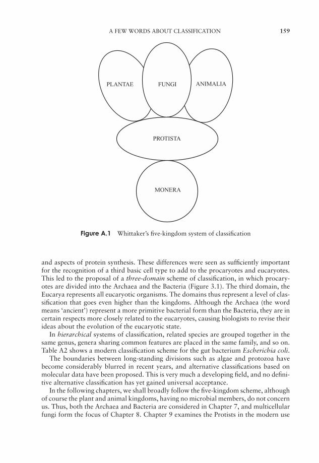
JWBK011-07 JWBK011-Hogg August 12, 2005 16:8 Char Count= 0
A FEW WORDS ABOUT CLASSIFICATION 159
FUNGIPLANTAE
PROTISTA
MONERA
ANIMALIA
Figure A.1 Whittaker’s five-kingdom system of classification
and aspects of protein synthesis. These differences were seen as sufficiently importantfor the recognition of a third basic cell type to add to the procaryotes and eucaryotes.This led to the proposal of a three-domain scheme of classification, in which procary-otes are divided into the Archaea and the Bacteria (Figure 3.1). The third domain, theEucarya represents all eucaryotic organisms. The domains thus represent a level of clas-sification that goes even higher than the kingdoms. Although the Archaea (the wordmeans ‘ancient’) represent a more primitive bacterial form than the Bacteria, they are incertain respects more closely related to the eucaryotes, causing biologists to revise theirideas about the evolution of the eucaryotic state.
In hierarchical systems of classification, related species are grouped together in thesame genus, genera sharing common features are placed in the same family, and so on.Table A2 shows a modern classification scheme for the gut bacterium Escherichia coli.
The boundaries between long-standing divisions such as algae and protozoa havebecome considerably blurred in recent years, and alternative classifications based onmolecular data have been proposed. This is very much a developing field, and no defini-tive alternative classification has yet gained universal acceptance.
In the following chapters, we shall broadly follow the five-kingdom scheme, althoughof course the plant and animal kingdoms, having no microbial members, do not concernus. Thus, both the Archaea and Bacteria are considered in Chapter 7, and multicellularfungi form the focus of Chapter 8. Chapter 9 examines the Protists in the modern use
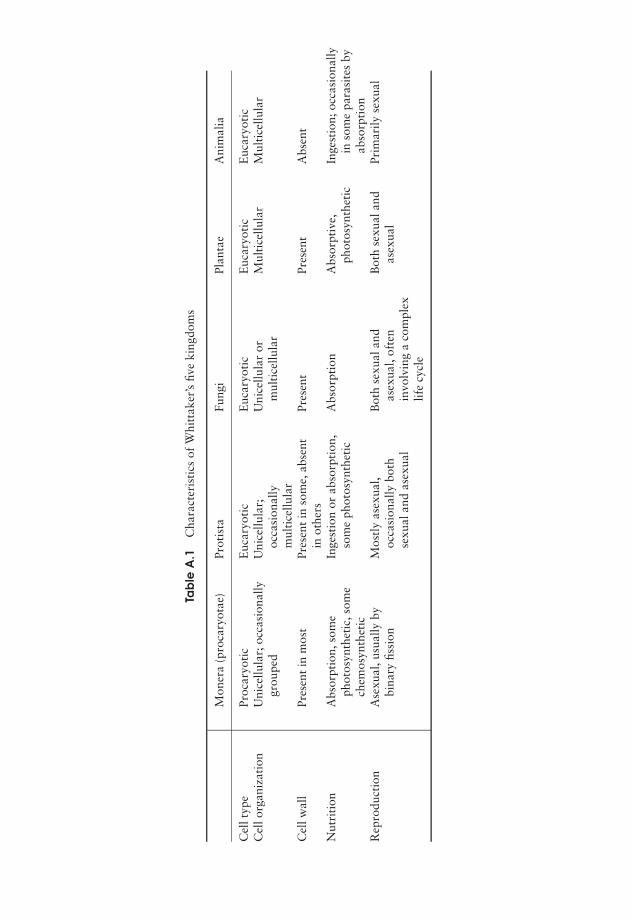
JWBK011-07 JWBK011-Hogg August 12, 2005 16:8 Char Count= 0
Tab
leA
.1C
hara
cter
isti
csof
Whi
ttak
er’s
five
king
dom
s
Mon
era
(pro
cary
otae
)Pr
otis
taFu
ngi
Plan
tae
Ani
mal
ia
Cel
ltyp
ePr
ocar
yoti
cE
ucar
yoti
cE
ucar
yoti
cE
ucar
yoti
cE
ucar
yoti
cC
ello
rgan
izat
ion
Uni
cellu
lar;
occa
sion
ally
grou
ped
Uni
cellu
lar;
occa
sion
ally
mul
tice
llula
r
Uni
cellu
lar
orm
ulti
cellu
lar
Mul
tice
llula
rM
ulti
cellu
lar
Cel
lwal
lPr
esen
tin
mos
tPr
esen
tin
som
e,ab
sent
inot
hers
Pres
ent
Pres
ent
Abs
ent
Nut
riti
onA
bsor
ptio
n,so
me
phot
osyn
thet
ic,s
ome
chem
osyn
thet
ic
Inge
stio
nor
abso
rpti
on,
som
eph
otos
ynth
etic
Abs
orpt
ion
Abs
orpt
ive,
phot
osyn
thet
icIn
gest
ion;
occa
sion
ally
inso
me
para
site
sby
abso
rpti
onR
epro
duct
ion
Ase
xual
,usu
ally
bybi
nary
fissi
onM
ostl
yas
exua
l,oc
casi
onal
lybo
thse
xual
and
asex
ual
Bot
hse
xual
and
asex
ual,
ofte
nin
volv
ing
aco
mpl
exlif
ecy
cle
Bot
hse
xual
and
asex
ual
Prim
arily
sexu
al
160
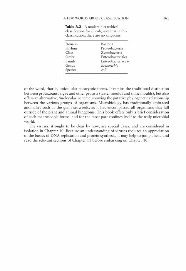
JWBK011-07 JWBK011-Hogg August 12, 2005 16:8 Char Count= 0
A FEW WORDS ABOUT CLASSIFICATION 161
Table A.2 A modern hierarchicalclassification for E. coli; note that in thisclassification, there are no kingdoms
Domain BacteriaPhylum ProteobacteriaClass ZymobacteriaOrder EnterobacterialesFamily EnterobacteriaceaeGenus EscherichiaSpecies coli
of the word, that is, unicellular eucaryotic forms. It retains the traditional distinctionbetween protozoans, algae and other protists (water moulds and slime moulds), but alsooffers an alternative, ‘molecular’ scheme, showing the putative phylogenetic relationshipbetween the various groups of organisms. Microbiology has traditionally embracedanomalies such as the giant seaweeds, as it has encompassed all organisms that falloutside of the plant and animal kingdoms. This book offers only a brief considerationof such macroscopic forms, and for the most part confines itself to the truly microbialworld.
The viruses, it ought to be clear by now, are special cases, and are considered inisolation in Chapter 10. Because an understanding of viruses requires an appreciationof the basics of DNA replication and protein synthesis, it may help to jump ahead andread the relevant sections of Chapter 11 before embarking on Chapter 10.

JWBK011-07 JWBK011-Hogg August 12, 2005 16:8 Char Count= 0
162
JWBK011-07 JWBK011-Hogg August 12, 2005 16:8 Char Count= 0
7Procaryote Diversity
The phenotype of an or-ganism refers to its ob-servable characteristics.
The genotype of an or-ganism refers to its ge-netic make-up.
Ever since bacteria were first identified, microbiologists have attempted to bring orderto the way they are named and classified. The range of morphological features use-ful in the differentiation of bacteria is fairly limited (compared, say, to animals andplants), so other characteristics have also been employed. These include metabolicproperties, pathogenicity, nutritional requirements, staining reactions and antigenicproperties. The first edition of Bergey’s Manual of Systematic Bacteriology (henceforthreferred to as ‘Bergey’), published in the mid-1980s, mainly used phenotypic character-istics such as these to classify bacteria. The result placed bacteria into taxonomic groupsthat may or may not reflect their evolutionary rela-tionship to one another. In the years since the firstedition of Bergey, the remarkable advances made inmolecular genetics have led to a radical reappraisalof the classification of bacteria. Comparison of nu-cleic acid sequences, notably those of 16S riboso-mal RNA genes, has led to a new, phylogeneticallybased scheme of classification, that is, one based onhow closely different groups of bacteria are thoughtto be related, rather than what morphological orphysiological features they may share. Ribosomal RNA occurs in all organisms, andserves a similar function, thus to a large extent these sequences are conserved (remainsimilar) in all organisms. The nature and extent of any differences that have crept induring evolution will, therefore, be an indication of the relatedness of different organ-isms.
The second edition of Bergey aims to reflect this change of approach and reassignmany bacteria according to their phylogenetic relationship, as deduced from molecularevidence. Due to be issued in five volumes over a number of years, the first volume waspublished in 2001. As an example, the genus Pseudomonas previously contained some70 species on the basis of phenotypic similarities, but in the second edition of Bergey,taking into account 16S rRNA information, many of these are assigned to newly createdgenera.
It must be stressed that Bergey (second edition) does not represent the definitivefinal word on the subject, and that the classification of bacteria is very much a de-veloping science, in a constant process of evolution. Indeed, microbiologists are byno means unanimous in their acceptance of the ‘molecular’ interpretation of bacte-rial taxonomy. Some point to perceived inadequacies in the collection of data for the
163
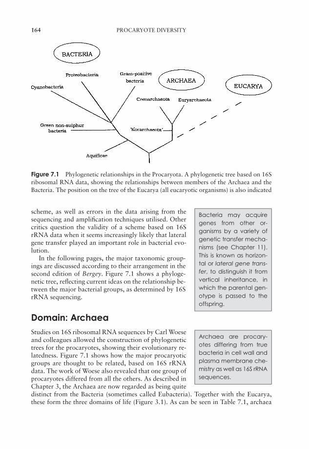
JWBK011-07 JWBK011-Hogg August 12, 2005 16:8 Char Count= 0
164 PROCARYOTE DIVERSITY
Figure 7.1 Phylogenetic relationships in the Procaryota. A phylogenetic tree based on 16Sribosomal RNA data, showing the relationships between members of the Archaea and theBacteria. The position on the tree of the Eucarya (all eucaryotic organisms) is also indicated
scheme, as well as errors in the data arising from thesequencing and amplification techniques utilised. Othercritics question the validity of a scheme based on 16SrRNA data when it seems increasingly likely that lateralgene transfer played an important role in bacterial evo-lution.
Bacteria may acquiregenes from other or-ganisms by a variety ofgenetic transfer mecha-nisms (see Chapter 11).This is known as horizon-tal or lateral gene trans-fer, to distinguish it fromvertical inheritance, inwhich the parental gen-otype is passed to theoffspring.
In the following pages, the major taxonomic group-ings are discussed according to their arrangement in thesecond edition of Bergey. Figure 7.1 shows a phyloge-netic tree, reflecting current ideas on the relationship be-tween the major bacterial groups, as determined by 16SrRNA sequencing.
Archaea are procary-otes differing from truebacteria in cell wall andplasma membrane che-mistry as well as 16S rRNAsequences.
Domain: ArchaeaStudies on 16S ribosomal RNA sequences by Carl Woeseand colleagues allowed the construction of phylogenetictrees for the procaryotes, showing their evolutionary re-latedness. Figure 7.1 shows how the major procaryoticgroups are thought to be related, based on 16S rRNAdata. The work of Woese also revealed that one group ofprocaryotes differed from all the others. As described inChapter 3, the Archaea are now regarded as being quitedistinct from the Bacteria (sometimes called Eubacteria). Together with the Eucarya,these form the three domains of life (Figure 3.1). As can be seen in Table 7.1, archaea
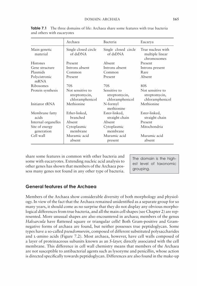
JWBK011-07 JWBK011-Hogg August 12, 2005 16:8 Char Count= 0
DOMAIN: ARCHAEA 165
Table 7.1 The three domains of life: Archaea share some features with true bacteriaand others with eucaryotes
Archaea Bacteria Eucarya
Main geneticmaterial
Single closed circleof dsDNA
Single closed circleof dsDNA
True nucleus withmultiple linearchromosomes
Histones Present Absent PresentGene structure Introns absent Introns absent Introns presentPlasmids Common Common RarePolycistronic
mRNAPresent Present Absent
Ribosomes 70S 70S 80SProtein synthesis Not sensitive to
streptomycin,chloramphenicol
Sensitive tostreptomycin,chloramphenicol
Not sensitive tostreptomycin,chloramphenicol
Initiator tRNA Methionine N-formylmethionine
Methionine
Membrane fattyacids
Ether-linked,branched
Ester-linked,straight chain
Ester-linked,straight chain
Internal organelles Absent Absent PresentSite of energy
generationCytoplasmic
membraneCytoplasmic
membraneMitochondria
Cell wall Muramic acidabsent
Muramic acidpresent
Muramic acidabsent
The domain is the high-est level of taxonomicgrouping.
share some features in common with other bacteria andsome with eucaryotes. Extending nucleic acid analysis toother genes has shown that members of the Archaea pos-sess many genes not found in any other type of bacteria.
General features of the Archaea
Members of the Archaea show considerable diversity of both morphology and physiol-ogy. In view of the fact that the Archaea remained unidentified as a separate group for somany years, it should come as no surprise that they do not display any obvious morpho-logical differences from true bacteria, and all the main cell shapes (see Chapter 2) are rep-resented. More unusual shapes are also encountered in archaea; members of the genusHaloarcula have flattened square or triangular cells! Both Gram-positive and Gram-negative forms of archaea are found, but neither possesses true peptidoglycan. Sometypes have a so-called pseudomurein, composed of different substituted polysaccharidesand l-amino acids (Figure 7.2). Most archaea, however, have cell walls composed ofa layer of proteinaceous subunits known as an S-layer, directly associated with the cellmembrane. This difference in cell wall chemistry means that members of the Archaeaare not susceptible to antibacterial agents such as lysozyme and penicillin, whose actionis directed specifically towards peptidoglycan. Differences are also found in the make-up
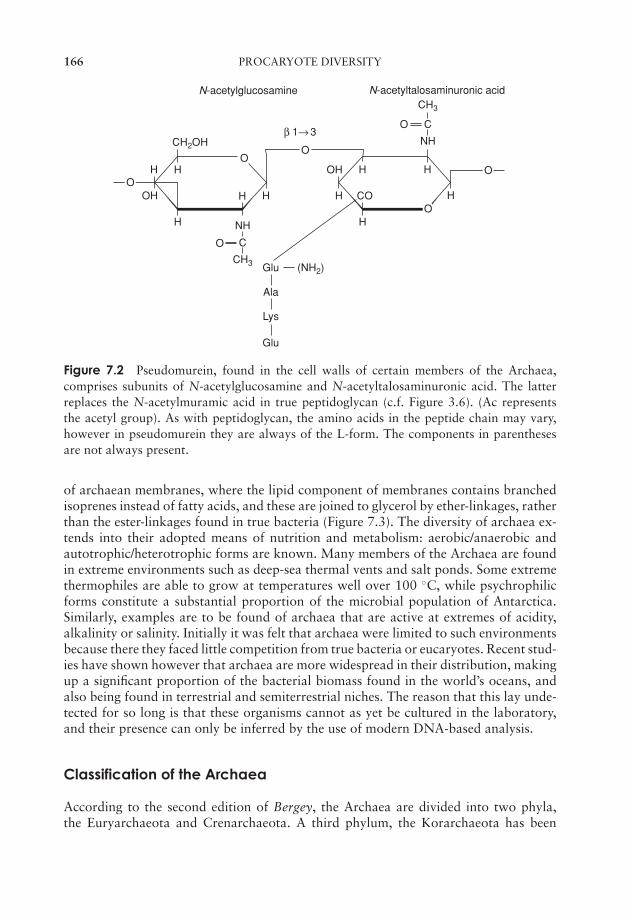
JWBK011-07 JWBK011-Hogg August 12, 2005 16:8 Char Count= 0
166 PROCARYOTE DIVERSITY
H H
β 1→ 3
H
O
N-acetylglucosamine
OH
H
H
OH H H
HCOH
H
Glu
Ala
Lys
Glu (NH2)
CH3
CO
NH
OO
O
CH3
CH2OH
CO
NH
N-acetyltalosaminuronic acid
O
Figure 7.2 Pseudomurein, found in the cell walls of certain members of the Archaea,comprises subunits of N-acetylglucosamine and N-acetyltalosaminuronic acid. The latterreplaces the N-acetylmuramic acid in true peptidoglycan (c.f. Figure 3.6). (Ac representsthe acetyl group). As with peptidoglycan, the amino acids in the peptide chain may vary,however in pseudomurein they are always of the L-form. The components in parenthesesare not always present.
of archaean membranes, where the lipid component of membranes contains branchedisoprenes instead of fatty acids, and these are joined to glycerol by ether-linkages, ratherthan the ester-linkages found in true bacteria (Figure 7.3). The diversity of archaea ex-tends into their adopted means of nutrition and metabolism: aerobic/anaerobic andautotrophic/heterotrophic forms are known. Many members of the Archaea are foundin extreme environments such as deep-sea thermal vents and salt ponds. Some extremethermophiles are able to grow at temperatures well over 100 ◦C, while psychrophilicforms constitute a substantial proportion of the microbial population of Antarctica.Similarly, examples are to be found of archaea that are active at extremes of acidity,alkalinity or salinity. Initially it was felt that archaea were limited to such environmentsbecause there they faced little competition from true bacteria or eucaryotes. Recent stud-ies have shown however that archaea are more widespread in their distribution, makingup a significant proportion of the bacterial biomass found in the world’s oceans, andalso being found in terrestrial and semiterrestrial niches. The reason that this lay unde-tected for so long is that these organisms cannot as yet be cultured in the laboratory,and their presence can only be inferred by the use of modern DNA-based analysis.
Classification of the Archaea
According to the second edition of Bergey, the Archaea are divided into two phyla,the Euryarchaeota and Crenarchaeota. A third phylum, the Korarchaeota has been
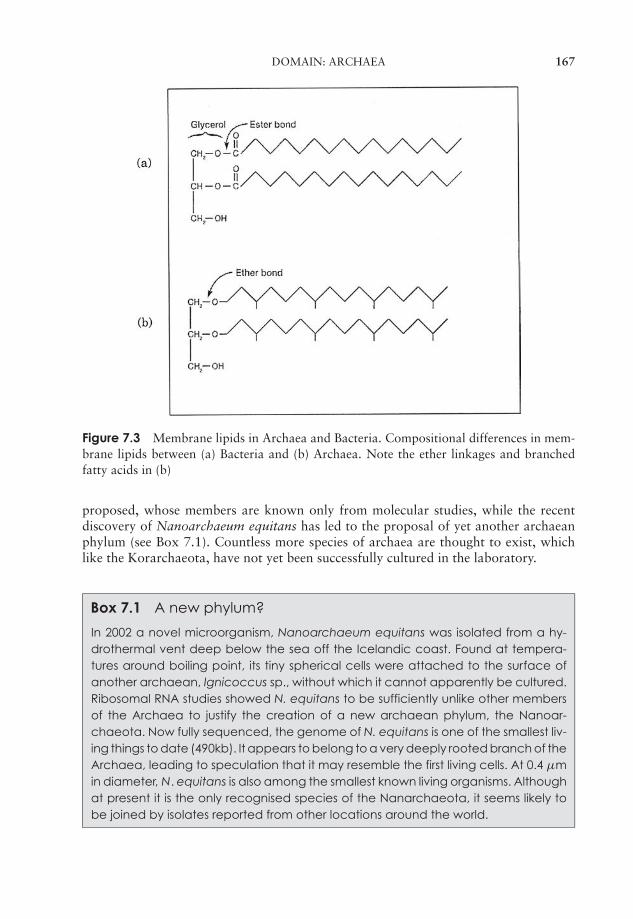
JWBK011-07 JWBK011-Hogg August 12, 2005 16:8 Char Count= 0
DOMAIN: ARCHAEA 167
Figure 7.3 Membrane lipids in Archaea and Bacteria. Compositional differences in mem-brane lipids between (a) Bacteria and (b) Archaea. Note the ether linkages and branchedfatty acids in (b)
proposed, whose members are known only from molecular studies, while the recentdiscovery of Nanoarchaeum equitans has led to the proposal of yet another archaeanphylum (see Box 7.1). Countless more species of archaea are thought to exist, whichlike the Korarchaeota, have not yet been successfully cultured in the laboratory.
Box 7.1 A new phylum?
In 2002 a novel microorganism, Nanoarchaeum equitans was isolated from a hy-drothermal vent deep below the sea off the Icelandic coast. Found at tempera-tures around boiling point, its tiny spherical cells were attached to the surface ofanother archaean, Ignicoccus sp., without which it cannot apparently be cultured.Ribosomal RNA studies showed N. equitans to be sufficiently unlike other membersof the Archaea to justify the creation of a new archaean phylum, the Nanoar-chaeota. Now fully sequenced, the genome of N. equitans is one of the smallest liv-ing things to date (490kb). It appears to belong to a very deeply rooted branch of theArchaea, leading to speculation that it may resemble the first living cells. At 0.4 µmin diameter, N. equitans is also among the smallest known living organisms. Althoughat present it is the only recognised species of the Nanarchaeota, it seems likely tobe joined by isolates reported from other locations around the world.
JWBK011-07 JWBK011-Hogg August 12, 2005 16:8 Char Count= 0
168 PROCARYOTE DIVERSITY
The phylum Eurychaeota is a bigger group than the Crenarchaeota, and includeshalophilic and methanogenic forms. The former are aerobic heterotrophs, requiring achloride concentration of at least 1.5 m (generally 2.0–4.0 m) for growth. One species,Halobacterium salinarum, is able to carry out a unique form of photosynthesis usingthe bacterial pigment bacteriorhodopsin, and uses the ATP so generated for the activetransport into the cell of the chloride ions it requires.
Members of the Euryarchaeota such as Methanococcus and Methanobacterium areunique among all life forms in their ability to generate methane from simple carbon com-pounds. They are strict anaerobes found in environments such as hot springs, marshesand the gut of ruminant mammals. The methane is derived from the metabolism of var-ious simple carbon compounds such as carbon dioxide or methanol in reactions linkedto the production of ATP. e.g.
CO2 + 4H2 −−−−−−−→ CH4 + 2H2O
CH3OH + H2 −−−−−−−→ CH4 + H2O
In addition, a few species can cleave acetate to produce methane:
CH3COO− + H2O −−−−−−−→ CH4 + HCO3−
This acetotrophic reaction is responsible for the much of the methane production insewage sludges. Although sharing the unique facility to generate methane, some of themethanogenic genera are quite distantly related to one another.
Pleomorphic meanslacking a regular shape.
Other representatives of the Eurychaeota includethe Thermoplasmata and the Thermococci. Thermo-plasmata are highly acidophilic and moderately ther-mophilic; they completely lack a cell wall, and are pleo-morphic. A unique membrane lipid composition allows them to withstand temperaturesof well over 50 ◦C. Thermococci are anaerobic extreme thermophiles found in anoxicthermal waters at temperatures as high as 95 ◦C. Enzymes isolated from thermococcihave found a variety of applications. A thermostable DNA polymerase from Pyrococcusfuriosus is used as an alternative to Taq polymerase (see Phylum ‘Deinococcus-Thermus’later in this chapter) in the polymerase chain reaction (PCR).
Representative genera: Methanobacterium, Halobacterium
Members of the Crenarchaeota are nearly all extreme thermophiles, many of themcapable of growth at temperatures in excess of 100 ◦C, including Pyrolobus fumarii,which has an optimum growth temperature of 106 ◦C, and can survive autoclaving at121 ◦C.
Many utilise inorganic sulphur compounds as either a source or acceptor of electrons(respectively, oxidation to H2SO4 or reduction to H2S).
Crenarchaeotes are mostly anaerobic, and are thought by many to resemble thecommon ancestors of all bacteria.
Representative genera: Thermoproteus, Sulfolobus
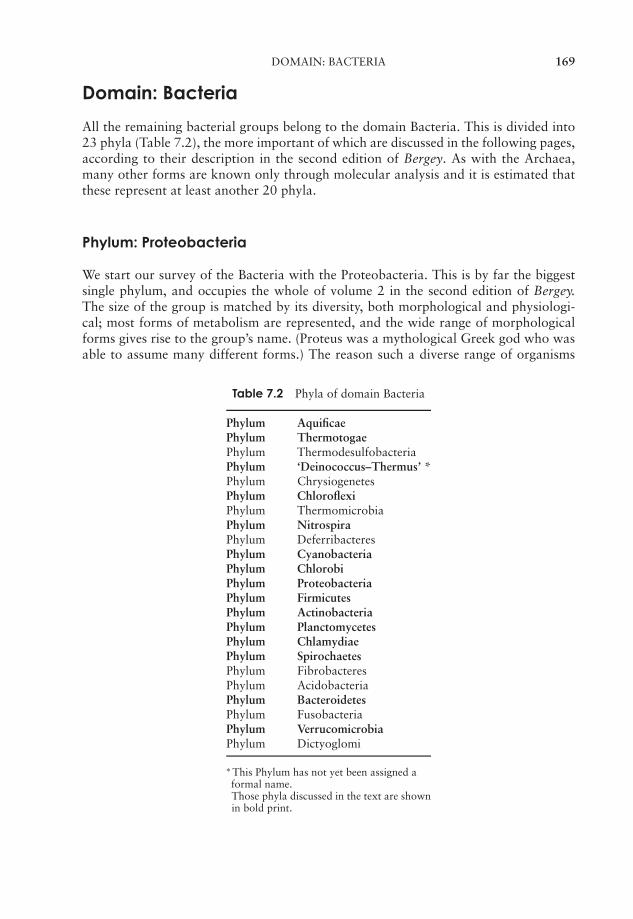
JWBK011-07 JWBK011-Hogg August 12, 2005 16:8 Char Count= 0
DOMAIN: BACTERIA 169
Domain: Bacteria
All the remaining bacterial groups belong to the domain Bacteria. This is divided into23 phyla (Table 7.2), the more important of which are discussed in the following pages,according to their description in the second edition of Bergey. As with the Archaea,many other forms are known only through molecular analysis and it is estimated thatthese represent at least another 20 phyla.
Phylum: Proteobacteria
We start our survey of the Bacteria with the Proteobacteria. This is by far the biggestsingle phylum, and occupies the whole of volume 2 in the second edition of Bergey.The size of the group is matched by its diversity, both morphological and physiologi-cal; most forms of metabolism are represented, and the wide range of morphologicalforms gives rise to the group’s name. (Proteus was a mythological Greek god who wasable to assume many different forms.) The reason such a diverse range of organisms
Table 7.2 Phyla of domain Bacteria
Phylum AquificaePhylum ThermotogaePhylum ThermodesulfobacteriaPhylum ‘Deinococcus–Thermus’ *Phylum ChrysiogenetesPhylum ChloroflexiPhylum ThermomicrobiaPhylum NitrospiraPhylum DeferribacteresPhylum CyanobacteriaPhylum ChlorobiPhylum ProteobacteriaPhylum FirmicutesPhylum ActinobacteriaPhylum PlanctomycetesPhylum ChlamydiaePhylum SpirochaetesPhylum FibrobacteresPhylum AcidobacteriaPhylum BacteroidetesPhylum FusobacteriaPhylum VerrucomicrobiaPhylum Dictyoglomi
∗ This Phylum has not yet been assigned aformal name.Those phyla discussed in the text are shownin bold print.

JWBK011-07 JWBK011-Hogg August 12, 2005 16:8 Char Count= 0
170 PROCARYOTE DIVERSITY
have been assigned to a single taxonomic grouping is that their 16S rRNA indicates acommon ancestor (thought to be photosynthetic, though few members now retain thisability). At the time of writing more than 460 genera and 1600 species had been iden-tified, all of them Gram-negative and representing almost half of all accepted bacterialgenera. These include many of the best known Gram-negative bacteria of medical, in-dustrial and agricultural importance. For taxonomic purposes, the Proteobacteria havebeen divided into five classes reflecting their presumed lines of descent and termed theAlphaproteobacteria, Betaproteobacteria, Gammaproteobacteria, Deltaproteobacteriaand Epsilonproteobacteria (Figure 7.4). It should be stressed that because classificationis based on molecular relatedness rather than shared phenotypic traits, few if any mor-phological or physiological properties can be said to be characteristic of all members ofeach class. Equally, organisms united by a particular feature may be found in more thanone of the proteobacterial classes, for example nitrifying bacteria are to be found in theα, β and γ Protobacteria. For this reason, in the following paragraphs we describe theProteobacteria in terms of their phenotypic characteristics rather than attempt to groupthem phylogenetically.
Photosynthetic ProteobacteriaThe purple sulphur and purple non-sulphur bacteria are the only members of the Pro-teobacteria to have retained the photosynthetic ability of their presumed ancestor. Thetype of photosynthesis they carry out, however, is quite distinct from that carried outby plants, algae and cyanobacteria (see later in this chapter), differing in two importantrespects:
� it is anoxygenic – no oxygen is produced by the process
� it utilizes bacteriochlorophyll a and/or b, which have different absorbance prop-erties to chlorophylls a and b.
Like organisms that carry out green photosynthesis, however, they incorporate CO2 intocarbohydrate by means of the Calvin cycle (see Chapter 6). All are at least facultatively
Figure 7.4 The phylogenetic relationships of the Proteobacteria, based on 16S rRNAsequences
JWBK011-07 JWBK011-Hogg August 12, 2005 16:8 Char Count= 0
DOMAIN: BACTERIA 171
anaerobic, and are typically found in sediments of stagnant lakes and salt marsh pools,where they may form extensive coloured blooms. Because the absorption spectrum ofbacteriochlorophylls lies mostly in the infrared part of the spectrum, they are able toutilise light energy that penetrates beyond the surface layers of water.
The coloration, ranging from orange/brown to purple, is due to the presence ofcarotenoid pigments such as lycopene and spirillixanthin, which mask the blue/greencolour of the bacteriochlorophylls. The photosynthetic pigments are located on highlyfolded extensions of the plasma membrane. Photosynthetic proteobacteria include rods,cocci and spiral forms.
Under anaerobic conditions, the purple sulphur bacteria typically utilise hydrogensulphide or elemental sulphur as an electron donor for the reduction of CO2.
H2S + CO2 −−−−−−−→ (CH2O)n + S0
S0 + CO2 + H2O −−−−−−−→ (CH2O)n + H2SO4
Many store sulphur in the form of intracellular granules. The purple sulphur bacteriaall belong to the γ -Protobacteria. They are typically found in surface muds, and sulphursprings, habitats that provide the right combination of light and anaerobic conditions.
Representative genera: Thiospirillum, Chromatium
The purple non-sulphur bacteria were distinguished from the above group because oftheir apparent inability to use H2S as an electron donor. It is now known, however,that the majority can do this, but are able to tolerate much lower concentrations incomparison with the purple sulphur bacteria. The purple non-sulphur bacteria are fac-ultative anaerobes able to grow as photoheterotrophs, that is, with light as an energysource and a range of organic molecules such as carbohydrates and organic acids assources of both carbon and electrons. In addition, many are able to grow aerobically aschemoheterotrophs in the absence of light. Under present classification systems, purplenonsulphur bacteria are divided between the α- and β-Proteobacteria.
Representative genera: Rhodospirillum, Rhodopseudomonas
Nitrifying ProteobacteriaThis group comprises aerobic Gram-negative chemolithoautotrophs that derive theirenergy from the oxidation of inorganic nitrogen compounds (either ammonia or nitrite),and their carbon from CO2. The nitrifying bacteria are represented in both the α- andβ-Proteobacteria.
The oxidation of ammonia through to nitrate is a two-stage process, with specificbacteria carrying out each stage (ammonia to nitrite and nitrite to nitrate). This isreflected in the generic names of the bacteria, bearing the prefix Nitroso- or Nitro-according to whether they carry out the first or second reaction. Nitrifying bacteria playan essential role in the cycling of nitrogen in terrestrial, marine and freshwater habitats.Nitrite, which is toxic to many forms of life, rarely accumulates in the environment,due to the activity of the Nitrobacteria. As was the case with the purple photosyntheticbacteria, several cell forms are represented among the nitrifiers.
JWBK011-07 JWBK011-Hogg August 12, 2005 16:8 Char Count= 0
172 PROCARYOTE DIVERSITY
Representative genera: Nitrosomonas (NH4+ −−−−−→ NO2
−)Nitrobacter (NO2
− −−−−−→ NO3−)
Iron- and sulphur-oxidising ProteobacteriaTwo further groups of environmentally significant chemolithoautotrophs derive theirenergy through the oxidation of reduced iron and sulphur respectively.
Among the sulphur oxidisers, perhaps the best studied are members of the genusAcidithiobacillus*, which includes extreme acidophiles such as A. thiooxidans that arecapable of growth at a pH as low as 1.0! These may utilise sulphur in its elementalform, as H2S, metal sulphides, or other forms of reduced sulphur such as thiosulphate:
2S0 + 3O2 + 2H2O −−−−−−−→ 2H2SO4
S2− + 4O2 −−−−−−−→ 2SO42−
S2O32− + 2O2 + H2O −−−−−−−→ 2SO4
2− + 2H+
The result of all these reactions is the production of sulphuric acid and a lowering of theenvironmental pH. Bacteria such as these are responsible for the phenomenon of acidmine drainage. Their environmental impact is discussed in Chapter 16, whilst Chapter17 describes their use in the extraction of valuable metals from intractable mineral ores.A particularly valuable organism in the latter context is A. ferrooxidans, due to itsability to use not only reduced sulphur compounds as energy sources, but also reducediron (see below).
A second group of sulphur oxidisers are bacteria that exist not as single cells, butjoin to form filaments, the best known of which is Beggiotoa. These are typically foundin sulphur springs, marine sediments and hydrothermal vents at the bottom of thesea.
Acidithiobacillus ferrooxidans is also an example of an iron oxidiser. At normalphysiological pH values and in the presence of oxygen, reduced iron (iron II, Fe2+) isspontaneously oxidised to the oxidised form (iron III, Fe3+). Under very acidic con-ditions, the iron remains in its reduced form, unless acted on by certain bacteria. A.ferrooxidans; is an obligate aerobe able to use iron II as an energy source, converting itto iron III at an optimum pH range of around 2:
2Fe2+ + 1/2O2 + 2H+−−−−−−−→ Fe3+ + H2O
Gallionella ferruginea, on the other hand, grows around neutrality in oxygen-poorenvironments such as bogs and iron springs. Ferric hydroxide is excreted from thecell and deposited on a stalk-like structure projecting from, and much bigger than,the cell itself. This gives the macroscopic impression of a mass of red/brown twistedfilaments.
Representative genera: Acidithiobacillus, Beggiotoa (sulphur oxidisers)Leptospirillum, Gallionella (iron oxidisers)
∗ Note: A. thiooxidans and A. ferrooxidans formerly belonged to the genus Thiobacillus. In 2000, severalspecies where assigned to new genera, however you may still find them referred to by their old names.
JWBK011-07 JWBK011-Hogg August 12, 2005 16:8 Char Count= 0
DOMAIN: BACTERIA 173
Hydrogen-oxidising ProteobacteriaThis diverse group of bacteria are united by their ability to derive energy by usinghydrogen gas as a donor of electrons, and oxygen as an acceptor:
2H2 + O2 −−−−−−−→ 2H2O
Nearly all the members of this group are facultative chemolithotrophs, i.e. they canalso grow as heterotrophs, utilising organic compounds instead of CO2 as their carbonsource, and indeed most grow more efficiently in this way.
Representative genera: Alcaligenes, Ralstonia
Nitrogen-fixing Proteobacteria Nitrogen fixation is lim-ited to a few speciesof bacteria and cyanob-acteria. No eucaryotesare known to have thisproperty.
The α-Proteobacteria includes certain genera ofnitrogen-fixing bacteria. These are able to fix (reduce)atmospheric N2 as NH4
+ for subsequent incorporationinto cellular materials, a process that requires a consid-erable input of energy in the form of ATP:
N2 + 8e− + 8H+ + 16ATP −−−−−−−→, 2NH4+ + 16ADP + 16Pi
Nitrogen-fixing bacteria may be free-living in the soil (e.g. Azotobacter), or form asymbiotic relationship with cells on the root hairs of leguminous plants such as peas,beans and clover (e.g. Rhizobium). The nitrogenase responsible for the reaction (actuallya complex of two enzymes) is highly sensitive to oxygen; many nitrogen fixers areanaerobes, while others have devised ways of keeping the cell interior oxygen-free.Nitrogen fixation is discussed further in Chapters 15 and 16.
Closely related to Rhizobium, but unable to fix nitrogen, are members of the genusAgrobacterium. Like Rhizobium, these enter the tissues of plants, but instead of forminga mutually beneficial association, cause cell proliferation and tumour formation. A.tumefaciens has proved to be a valuable tool in the genetic engineering of plants, andis discussed further in Chapter 12.
Representative genera: Rhizobium, Azotobacter
Methanotrophic Proteobacteria
Methane is one of the so-called greenhouse gas-es, responsible for thephenomenon of globalwarming. Its effectswould be much more pr-onounced if it were notfor the activity of metha-notrophic bacteria.
In discussing the Archaea earlier in this chapter, we en-countered species capable of the production of methane,a gas found widely in such diverse locations as marshes,sewage sludge and animal intestines. Certain proteobac-teria are able to utilise this methane as a carbon andenergy source and are known as methanotrophs.
Methanotrophs are strict aerobes, requiring oxy-gen for the oxidation of methane. The methane-generating bacteria, however, as we’ve seen are anaer-obes; methanotrophs are consequently to be found ataerobic/anaerobic interfaces such as topsoil, where they
JWBK011-07 JWBK011-Hogg August 12, 2005 16:8 Char Count= 0
174 PROCARYOTE DIVERSITY
The methylotroph Meth-ylophilus methylotrophuswas once produced inhuge quantities as asource of ‘single cellprotein’for use as animalfeed, until the low priceof alternatives suchas soya and fish mealmade it commerciallyunviable.
can find both the oxygen and the methane they require.The methane is firstly oxidised to methanol, then toformaldehyde, by means of separate enzyme systems.Some of the carbon in formaldehyde is assimilated intoorganic cellular material, while some is further oxidisedto carbon dioxide.
Bacteria able to utilise other single-carbon com-pounds such as methanol (CH3OH) or methylamine(CH3NH2) are termed methylotrophs. Depending onwhether they possess the enzyme methane monooxyge-nase (MMO), they may also be methanotrophs.
Representative genera: Methylomonas, Methylococcus
Sulphate- and sulphur-reducing ProteobacteriaSome 20 genera of anaerobic δ-Proteobacteria reduce either elemental sulphur or oxi-dised forms of sulphur such as sulphate to hydrogen sulphide. Organic compounds suchas pyruvate, lactate or certain fatty acids act as electron donors:
Desulfovibrio2CH3CHOHCOO− + SO4
2− −−−−−−→ 2CH3COO− + H2S + 2HCO3−
Lactate Acetate
Sulphate- and sulphur-reducers are found in anaerobic muds and play an important rolein the global sulphur cycle.
Representative genera: Desulfovibrio (sulphate), Desulfuromonas (sulphur)
Enteric ProteobacteriaThis is a large group of rod-shaped bacteria, mostly motile by means of peritrichousflagella, which all belong to the γ -Proteobacteria. They are facultative aerobes, charac-terised by their ability in anaerobic conditions to carry out fermentation of glucose andother sugars to give a variety of products. The nature of these products allows divisioninto two principal groups, the mixed acid fermenters and the butanediol fermenters(Figure 6.23). All the enteric bacteria test negative for cytochrome c oxidase (see Vibrioand related genera below). In view of their similar appearance, members of the groupare distinguished from one another largely by means of their biochemical characteris-tics. An unknown isolate is subjected to a series of tests including its ability to utilisesubstrates such as lactose and citrate, convert tryptophan to indole, and hydrolyse urea.On the basis of its response to each test, a characteristic profile can be built up for theisolate, and matched against those of known species (see Table 7.3).
The most thoroughly studied of all bacteria, Escherichia coli (E. coli) is a memberof this group, as are a number of important pathogens of humans such as Salmonella,Shigella and Yersinia (the causative agent of plague).
Representative genera: Escherichia, Enterobacter
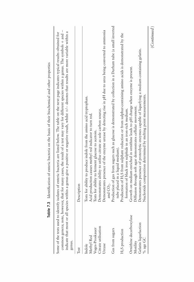
JWBK011-07 JWBK011-Hogg August 12, 2005 16:8 Char Count= 0
Tab
le7.
3Id
enti
ficat
ion
ofen
teri
cba
cter
iaon
the
basi
sof
thei
rbi
oche
mic
alan
dot
her
prop
erti
es
Som
eof
the
test
sus
edto
iden
tify
isol
ates
ofen
teri
cba
cter
iaar
elis
ted
belo
w.T
heta
ble
onth
ene
xtpa
gein
dica
tes
typi
calr
esul
tsob
tain
edfo
rco
mm
onge
nera
;not
e,ho
wev
er,t
hat
for
man
yca
ses,
the
resu
ltof
ate
stm
ayva
ryfo
rdi
ffer
ent
spec
ies
wit
hin
age
nus.
The
sym
bols
+an
d−
indi
cate
that
mos
tor
alls
peci
esw
ithi
na
genu
sgi
vea
posi
tive
orne
gati
vere
sult
,whi
lst+/
−de
note
sth
atre
sult
sar
em
ore
vari
able
wit
hin
age
nus.
Test
Des
crip
tion
Indo
leTe
sts
for
abili
tyto
prod
uce
indo
lefr
omth
eam
ino
acid
tryp
toph
an.
Met
hylR
edA
cid
prod
ucti
onca
uses
met
hylr
edin
dica
tor
totu
rnre
d.V
oges
-Pro
skau
erTe
sts
for
abili
tyto
ferm
ent
gluc
ose
toac
etoi
n.C
itra
teut
ilisa
tion
Dem
onst
rate
sab
ility
tout
ilise
citr
ate
asso
leca
rbon
sour
ce.
Ure
ase
Dem
onst
rate
spr
esen
ceof
the
enzy
me
urea
seby
dete
ctin
gri
sein
pHdu
eto
urea
bein
gco
nver
ted
toam
mon
iaan
dC
O2.
Gas
from
suga
rsPr
oduc
tion
ofga
sfr
omsu
gars
such
asgl
ucos
eis
dem
onst
rate
dby
colle
ctio
nin
aD
urha
mtu
be(a
smal
linv
erte
dtu
bepl
aced
ina
liqui
dm
ediu
m).
H2S
prod
ucti
onPr
oduc
tion
ofH
2S
from
sulp
hate
redu
ctio
nor
from
sulp
hur-
cont
aini
ngam
ino
acid
sis
dem
onst
rate
dby
the
form
atio
nof
blac
kir
onsu
lphi
dein
anir
on-r
ich
med
ium
.O
rnit
hine
deca
rbox
ylas
eG
row
thon
med
ium
enri
ched
inor
nith
ine
lead
sto
pHch
ange
whe
nen
zym
eis
pres
ent.
Mot
ility
Dif
fusi
onth
roug
hso
ftag
arde
mon
stra
tes
cellu
lar
mov
emen
t.G
elat
inliq
uefa
ctio
nD
emon
stra
tes
pres
ence
ofpr
oteo
lyti
cen
zym
esca
pabl
eof
lique
fyin
ga
med
ium
cont
aini
ngge
lati
n.%
age
GC
Nuc
leot
ide
com
posi
tion
dete
rmin
edby
mel
ting
poin
tm
easu
rem
ents
.
(Con
tinu
ed)
175
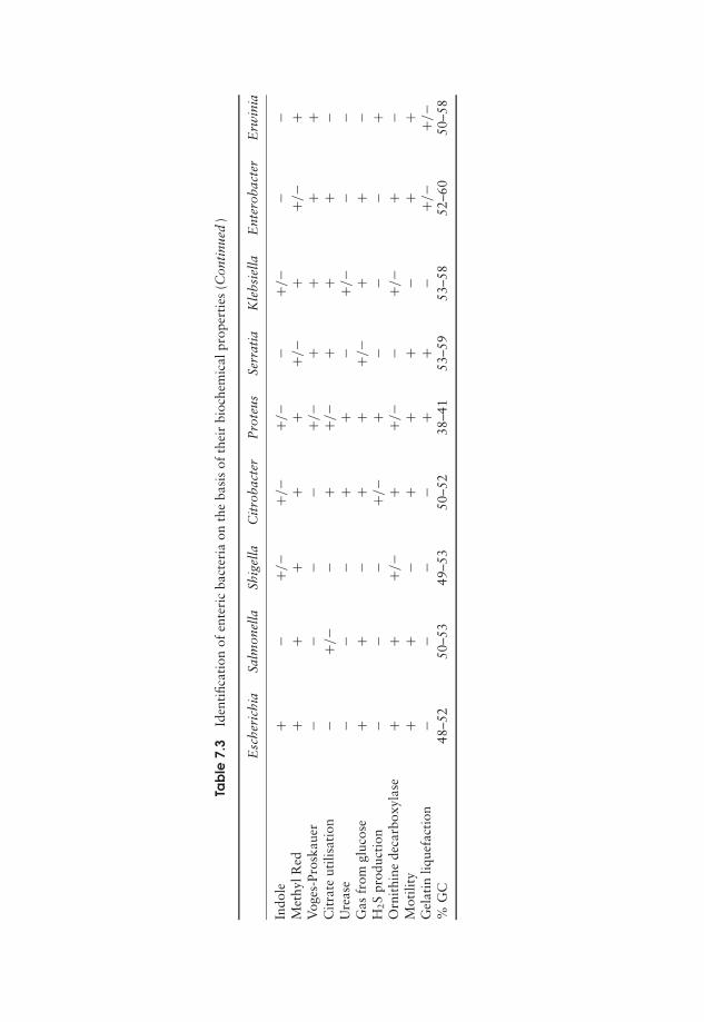
JWBK011-07 JWBK011-Hogg August 12, 2005 16:8 Char Count= 0
Tab
le7.
3Id
enti
ficat
ion
ofen
teri
cba
cter
iaon
the
basi
sof
thei
rbi
oche
mic
alpr
oper
ties
(Con
tinu
ed)
Esc
heri
chia
Salm
onel
laSh
igel
laC
itro
bact
erP
rote
usSe
rrat
iaK
lebs
iella
Ent
erob
acte
rE
rwin
ia
Indo
le+
−+/
−+/
−+/
−−
+/−
−−
Met
hylR
ed+
++
++
+/−
++/
−+
Vog
es-P
rosk
auer
−−
−−
+/−
++
++
Cit
rate
utili
sati
on−
+/−
−+
+/−
++
+−
Ure
ase
−−
−+
+−
+/−
−−
Gas
from
gluc
ose
++
−+
++/
−+
+−
H2S
prod
ucti
on−
−−
+/−
+−
−−
+O
rnit
hine
deca
rbox
ylas
e+
++/
−+
+/−
−+/
−+
−M
otili
ty+
+−
++
+−
++
Gel
atin
lique
fact
ion
−−
−−
++
−+/
−+/
−%
GC
48–5
250
–53
49–5
350
–52
38–4
153
–59
53–5
852
–60
50–5
8
176
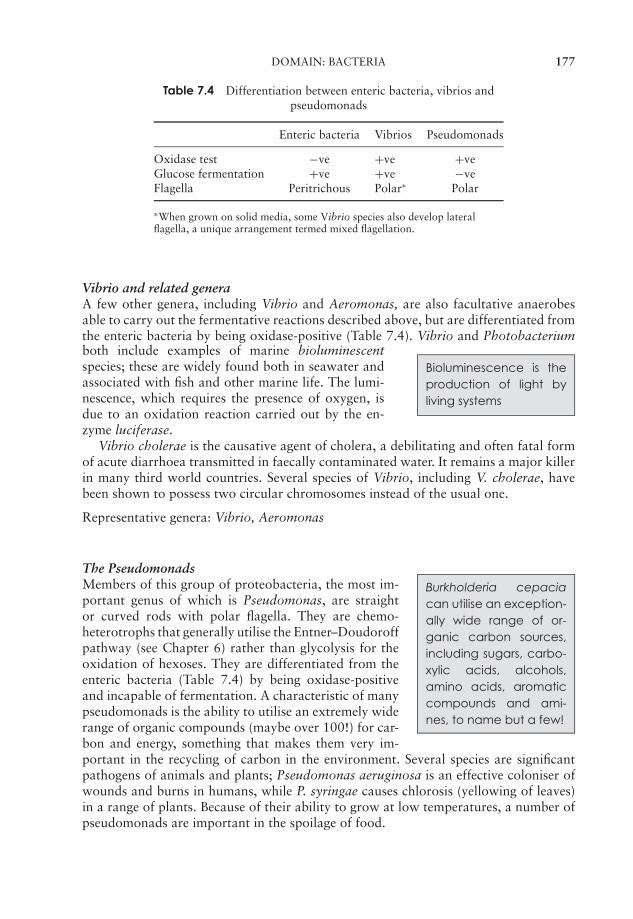
JWBK011-07 JWBK011-Hogg August 12, 2005 16:8 Char Count= 0
DOMAIN: BACTERIA 177
Table 7.4 Differentiation between enteric bacteria, vibrios andpseudomonads
Enteric bacteria Vibrios Pseudomonads
Oxidase test −ve +ve +veGlucose fermentation +ve +ve −veFlagella Peritrichous Polar∗ Polar
∗When grown on solid media, some Vibrio species also develop lateralflagella, a unique arrangement termed mixed flagellation.
Vibrio and related generaA few other genera, including Vibrio and Aeromonas, are also facultative anaerobesable to carry out the fermentative reactions described above, but are differentiated fromthe enteric bacteria by being oxidase-positive (Table 7.4). Vibrio and Photobacteriumboth include examples of marine bioluminescentspecies; these are widely found both in seawater andassociated with fish and other marine life. The lumi-nescence, which requires the presence of oxygen, isdue to an oxidation reaction carried out by the en-zyme luciferase.
Bioluminescence is theproduction of light byliving systems
Vibrio cholerae is the causative agent of cholera, a debilitating and often fatal formof acute diarrhoea transmitted in faecally contaminated water. It remains a major killerin many third world countries. Several species of Vibrio, including V. cholerae, havebeen shown to possess two circular chromosomes instead of the usual one.
Representative genera: Vibrio, Aeromonas
The PseudomonadsBurkholderia cepaciacan utilise an exception-ally wide range of or-ganic carbon sources,including sugars, carbo-xylic acids, alcohols,amino acids, aromaticcompounds and ami-nes, to name but a few!
Members of this group of proteobacteria, the most im-portant genus of which is Pseudomonas, are straightor curved rods with polar flagella. They are chemo-heterotrophs that generally utilise the Entner–Doudoroffpathway (see Chapter 6) rather than glycolysis for theoxidation of hexoses. They are differentiated from theenteric bacteria (Table 7.4) by being oxidase-positiveand incapable of fermentation. A characteristic of manypseudomonads is the ability to utilise an extremely widerange of organic compounds (maybe over 100!) for car-bon and energy, something that makes them very im-portant in the recycling of carbon in the environment. Several species are significantpathogens of animals and plants; Pseudomonas aeruginosa is an effective coloniser ofwounds and burns in humans, while P. syringae causes chlorosis (yellowing of leaves)in a range of plants. Because of their ability to grow at low temperatures, a number ofpseudomonads are important in the spoilage of food.
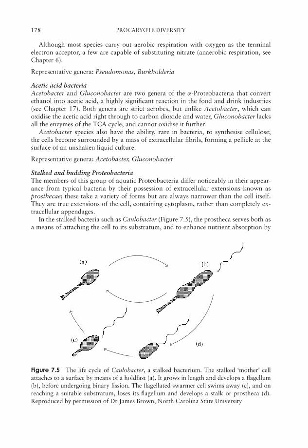
JWBK011-07 JWBK011-Hogg August 12, 2005 16:8 Char Count= 0
178 PROCARYOTE DIVERSITY
Although most species carry out aerobic respiration with oxygen as the terminalelectron acceptor, a few are capable of substituting nitrate (anaerobic respiration, seeChapter 6).
Representative genera: Pseudomonas, Burkholderia
Acetic acid bacteriaAcetobacter and Gluconobacter are two genera of the α-Proteobacteria that convertethanol into acetic acid, a highly significant reaction in the food and drink industries(see Chapter 17). Both genera are strict aerobes, but unlike Acetobacter, which canoxidise the acetic acid right through to carbon dioxide and water, Gluconobacter lacksall the enzymes of the TCA cycle, and cannot oxidise it further.
Acetobacter species also have the ability, rare in bacteria, to synthesise cellulose;the cells become surrounded by a mass of extracellular fibrils, forming a pellicle at thesurface of an unshaken liquid culture.
Representative genera: Acetobacter, Gluconobacter
Stalked and budding ProteobacteriaThe members of this group of aquatic Proteobacteria differ noticeably in their appear-ance from typical bacteria by their possession of extracellular extensions known asprosthecae; these take a variety of forms but are always narrower than the cell itself.They are true extensions of the cell, containing cytoplasm, rather than completely ex-tracellular appendages.
In the stalked bacteria such as Caulobacter (Figure 7.5), the prostheca serves both asa means of attaching the cell to its substratum, and to enhance nutrient absorption by
Figure 7.5 The life cycle of Caulobacter, a stalked bacterium. The stalked ‘mother’ cellattaches to a surface by means of a holdfast (a). It grows in length and develops a flagellum(b), before undergoing binary fission. The flagellated swarmer cell swims away (c), and onreaching a suitable substratum, loses its flagellum and develops a stalk or prostheca (d).Reproduced by permission of Dr James Brown, North Carolina State University
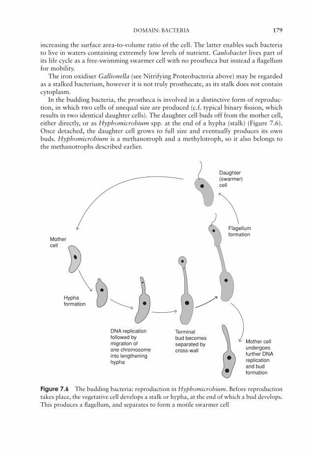
JWBK011-07 JWBK011-Hogg August 12, 2005 16:8 Char Count= 0
DOMAIN: BACTERIA 179
increasing the surface area-to-volume ratio of the cell. The latter enables such bacteriato live in waters containing extremely low levels of nutrient. Caulobacter lives part ofits life cycle as a free-swimming swarmer cell with no prostheca but instead a flagellumfor mobility.
The iron oxidiser Gallionella (see Nitrifying Proteobacteria above) may be regardedas a stalked bacterium, however it is not truly prosthecate, as its stalk does not containcytoplasm.
In the budding bacteria, the prostheca is involved in a distinctive form of reproduc-tion, in which two cells of unequal size are produced (c.f. typical binary fission, whichresults in two identical daughter cells). The daughter cell buds off from the mother cell,either directly, or as Hyphomicrobium spp. at the end of a hypha (stalk) (Figure 7.6).Once detached, the daughter cell grows to full size and eventually produces its ownbuds. Hyphomicrobium is a methanotroph and a methylotroph, so it also belongs tothe methanotrophs described earlier.
Mother cellundergoesfurther DNAreplicationand budformation
Terminalbud becomesseparated bycross-wall
DNA replicationfollowed bymigration ofone chromosomeinto lengtheninghypha
Hyphaformation
Mothercell
Daughter(swarmer)cell
Flagellumformation
Figure 7.6 The budding bacteria: reproduction in Hyphomicrobium. Before reproductiontakes place, the vegetative cell develops a stalk or hypha, at the end of which a bud develops.This produces a flagellum, and separates to form a motile swarmer cell
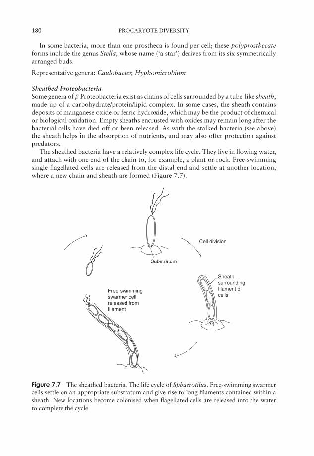
JWBK011-07 JWBK011-Hogg August 12, 2005 16:8 Char Count= 0
180 PROCARYOTE DIVERSITY
In some bacteria, more than one prostheca is found per cell; these polyprosthecateforms include the genus Stella, whose name (‘a star’) derives from its six symmetricallyarranged buds.
Representative genera: Caulobacter, Hyphomicrobium
Sheathed ProteobacteriaSome genera of β Proteobacteria exist as chains of cells surrounded by a tube-like sheath,made up of a carbohydrate/protein/lipid complex. In some cases, the sheath containsdeposits of manganese oxide or ferric hydroxide, which may be the product of chemicalor biological oxidation. Empty sheaths encrusted with oxides may remain long after thebacterial cells have died off or been released. As with the stalked bacteria (see above)the sheath helps in the absorption of nutrients, and may also offer protection againstpredators.
The sheathed bacteria have a relatively complex life cycle. They live in flowing water,and attach with one end of the chain to, for example, a plant or rock. Free-swimmingsingle flagellated cells are released from the distal end and settle at another location,where a new chain and sheath are formed (Figure 7.7).
Free-swimmingswarmer cellreleased fromfilament
Sheathsurroundingfilament ofcells
Substratum
Cell division
Figure 7.7 The sheathed bacteria. The life cycle of Sphaerotilus. Free-swimming swarmercells settle on an appropriate substratum and give rise to long filaments contained within asheath. New locations become colonised when flagellated cells are released into the waterto complete the cycle
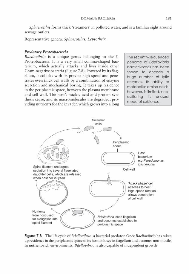
JWBK011-07 JWBK011-Hogg August 12, 2005 16:8 Char Count= 0
DOMAIN: BACTERIA 181
Sphaerotilus forms thick ‘streamers’ in polluted water, and is a familiar sight aroundsewage outlets.
Representative genera: Sphaerotilus, Leptothrix
Predatory ProteobacteriaThe recently-sequencedgenome of Bdellovibriobacterivorans has beenshown to encode ahuge number of lyticenzymes. Its ability tometabolise amino acids,however, is limited, nec-essitating its unusualmode of existence.
Bdellovibrio is a unique genus belonging to the δ-Proteobacteria. It is a very small comma-shaped bac-terium, which actually attacks and lives inside otherGram-negative bacteria (Figure 7.8). Powered by its flag-ellum, it collides with its prey at high speed and pene-trates even thick cell walls by a combination of enzymesecretion and mechanical boring. It takes up residencein the periplasmic space, between the plasma membraneand cell wall. The host’s nucleic acid and protein syn-thesis cease, and its macromolecules are degraded, pro-viding nutrients for the invader, which grows into a long
Cell wall
Swarmer cells
Periplasmicspace
Hostbacterium e.g PseudomonasEscherichia
Spiral filament undergoes septation into several flagellated daughter cells, which are released when host cell is lysed
Nutrients from host used for elongation into spiral filament
Bdellovibrio loses flagellum and becomes established in periplasmic space
'Attack phase' cell attaches to host. High-speed rotation allows penetration of cell wall.
Figure 7.8 The life cycle of Bdellovibrio, a bacterial predator. Once Bdellovibrio has takenup residence in the periplasmic space of its host, it loses its flagellum and becomes non-motile.In nutrient-rich environments, Bdellovibrio is also capable of independent growth

JWBK011-07 JWBK011-Hogg August 12, 2005 16:8 Char Count= 0
182 PROCARYOTE DIVERSITY
Myxospores
Germination
Cells formaggregates
Vegetativegrowth
a)
b)
c)d)
Fruitingbody
Figure 7.9 The Myxobacteria: a complex bacterial life cycle. When nutrients are in plen-tiful supply, myxobacteria divide by binary fission (a). On depletion of nutrients, they formaggregates of cells, which leads to the formation of a fruiting body (b). Within the fruit-ing body, some cells form myxospores, enclosed within a sporangium (c). Myxospores re-main dormant until environmental conditions are favourable, then germinate into vegetativecells (d)
helical cell. This eventually divides into several motile progeny cells, which are thenreleased.
Representative genus: Bdellovibrio
Another group of bacteria that may be regarded as predatory are the Myxobacteria(Figure 7.9). These are rod-shaped bacteria lacking flagella, which yet are motile bygliding along a solid surface, aided by the excretion of extracellular polysaccharides. Forthis reason they are sometimes referred to as the gliding bacteria. They are heterotrophs,typically requiring complex organic nutrients, which they obtain by the lysis of othertypes of bacteria. Thus, unlike Bdellovibrio, they digest their prey before they ingest it.When a rich supply of nutrients is not available, many thousands of cells may aggregateto form fruiting bodies, inside which myxospores develop. These are able to resistdrought and lack of nutrients for many years. Myxobacteria exhibit the most complexlife cycles of any procaryote so far studied.
Representative genera: Myxococcus, Chondromyces
SpirillaCollected together under this heading are several genera of aerobic (mostly mi-croaerophilic) spiral-shaped bacteria with polar flagella. These include free-living, sym-biotic and parasitic types.
JWBK011-07 JWBK011-Hogg August 12, 2005 16:8 Char Count= 0
DOMAIN: BACTERIA 183
Spirilla such as Aquaspirillum and Magnetospirillum contain magnetosomes, intra-cellular particles of iron oxide (magnetite, Fe3O4). Such magnetotactic bacteria havethe remarkable ability to orientate themselves with respect to the earth’s magnetic field(magnetotaxis).
Two important pathogens of humans are included in the spirilla; Campylobacterjejuni is responsible for foodborne gastroenteritis, while Helicobacter pylori has in recenttimes been identified as the cause of many cases of peptic ulcers.
Representative genera: Magnetospirillum, Campylobacter
RickettsiaThis group comprises arthropod-borne intracellular parasites of vertebrates, and in-cludes the causative agents of human diseases such as typhus and Rocky Mountainspotted fever. The bacteria are taken up by host phagocytic cells, where they multiplyand eventually cause lysis.
The Rickettsia are aerobic organotrophs, but some possess an unusual mode of en-ergy metabolism, only being able to oxidise intermediate metabolites such as glutamateand succinate, which they obtain from their host. Rickettsia and Coxiella, the twomain genera, are not closely related phylogenetically and are placed in the α- and γ -Proteobacteria, respectively.
Representative genera: Rickettsia, Coxiella
Neisseria and related ProteobacteriaAll members of this loose collection of bacteria are aerobic non-motile cocci, typicallyseen as pairs, with flattened sides where they join. Some however only assume thismorphology during stationary growth phase. Many are found in warm-blooded animals,and some species are pathogenic. The genus Neisseria includes species responsible forgonorrhoea and meningitis in humans.
Other Gram-negative phyla
The following section considers those Gram-negative bacteria not included in the Pro-teobacteria. These phyla are not closely related in the phylogenetic sense, either to eachother or to the Proteobacteria.
Phylum Cyanobacteria: the blue-green bacteria
The Cyanobacteria are placed in volume 1 of the second edition of Bergey, along withthe Archaea (see above), the deeply branching bacteria, the ‘Deinococcus–Thermus’group, and the green sulphur and green non-sulphur bacteria (see below). See alsoBox 7.2.
Members of the Cyanobacteria were once known as blue-green algae becausethey carry out the same kind of oxygenic photosynthesis as algae and green plants
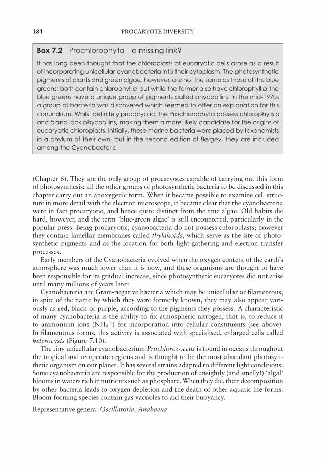
JWBK011-07 JWBK011-Hogg August 12, 2005 16:8 Char Count= 0
184 PROCARYOTE DIVERSITY
Box 7.2 Prochlorophyta -- a missing link?
It has long been thought that the chloroplasts of eucaryotic cells arose as a resultof incorporating unicellular cyanobacteria into their cytoplasm. The photosyntheticpigments of plants and green algae, however, are not the same as those of the bluegreens; both contain chlorophyll a, but while the former also have chlorophyll b, theblue greens have a unique group of pigments called phycobilins. In the mid-1970sa group of bacteria was discovered which seemed to offer an explanation for thisconundrum. Whilst definitely procaryotic, the Prochlorophyta possess chlorophylls aand band lack phycobilins, making them a more likely candidate for the origins ofeucaryotic chloroplasts. Initially, these marine bacteria were placed by taxonomistsin a phylum of their own, but in the second edition of Bergey, they are includedamong the Cyanobacteria.
(Chapter 6). They are the only group of procaryotes capable of carrying out this formof photosynthesis; all the other groups of photosynthetic bacteria to be discussed in thischapter carry out an anoxygenic form. When it became possible to examine cell struc-ture in more detail with the electron microscope, it became clear that the cyanobacteriawere in fact procaryotic, and hence quite distinct from the true algae. Old habits diehard, however, and the term ‘blue-green algae’ is still encountered, particularly in thepopular press. Being procaryotic, cyanobacteria do not possess chloroplasts; howeverthey contain lamellar membranes called thylakoids, which serve as the site of photo-synthetic pigments and as the location for both light-gathering and electron transferprocesses.
Early members of the Cyanobacteria evolved when the oxygen content of the earth’satmosphere was much lower than it is now, and these organisms are thought to havebeen responsible for its gradual increase, since photosynthetic eucaryotes did not ariseuntil many millions of years later.
Cyanobacteria are Gram-negative bacteria which may be unicellular or filamentous;in spite of the name by which they were formerly known, they may also appear vari-ously as red, black or purple, according to the pigments they possess. A characteristicof many cyanobacteria is the ability to fix atmospheric nitrogen, that is, to reduce itto ammonium ions (NH4
+) for incorporation into cellular constituents (see above).In filamentous forms, this activity is associated with specialised, enlarged cells calledheterocysts (Figure 7.10).
The tiny unicellular cyanobacterium Prochlorococcus is found in oceans throughoutthe tropical and temperate regions and is thought to be the most abundant photosyn-thetic organism on our planet. It has several strains adapted to different light conditions.Some cyanobacteria are responsible for the production of unsightly (and smelly!) ‘algal’blooms in waters rich in nutrients such as phosphate. When they die, their decompositionby other bacteria leads to oxygen depletion and the death of other aquatic life forms.Bloom-forming species contain gas vacuoles to aid their buoyancy.
Representative genera: Oscillatoria, Anabaena
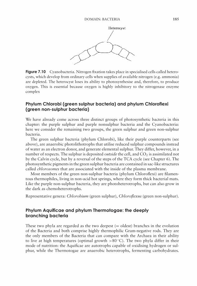
JWBK011-07 JWBK011-Hogg August 12, 2005 16:8 Char Count= 0
DOMAIN: BACTERIA 185
Figure 7.10 Cyanobacteria. Nitrogen fixation takes place in specialised cells called hetero-cysts, which develop from ordinary cells when supplies of available nitrogen (e.g. ammonia)are depleted. The heterocyst loses its ability to photosynthesise and, therefore, to produceoxygen. This is essential because oxygen is highly inhibitory to the nitrogenase enzymecomplex
Phylum Chlorobi (green sulphur bacteria) and phylum Chloroflexi(green non-sulphur bacteria)
We have already come across three distinct groups of photosynthetic bacteria in thischapter: the purple sulphur and purple nonsulphur bacteria and the Cyanobacteria:here we consider the remaining two groups, the green sulphur and green non-sulphurbacteria.
The green sulphur bacteria (phylum Chlorobi), like their purple counterparts (seeabove), are anaerobic photolithotrophs that utilise reduced sulphur compounds insteadof water as an electron donor, and generate elemental sulphur. They differ, however, in anumber of respects. The sulphur is deposited outside the cell, and CO2 is assimilated notby the Calvin cycle, but by a reversal of the steps of the TCA cycle (see Chapter 6). Thephotosynthetic pigments in the green sulphur bacteria are contained in sac-like structurescalled chlorosomes that are associated with the inside of the plasma membrane.
Most members of the green non-sulphur bacteria (phylum Chloroflexi) are filamen-tous thermophiles, living in non-acid hot springs, where they form thick bacterial mats.Like the purple non-sulphur bacteria, they are photoheterotrophs, but can also grow inthe dark as chemoheterotrophs.
Representative genera: Chlorobium (green sulphur), Chloroflexus (green non-sulphur).
Phylum Aquificae and phylum Thermotogae: the deeplybranching bacteria
These two phyla are regarded as the two deepest (= oldest) branches in the evolutionof the Bacteria and both comprise highly thermophilic Gram-negative rods. They arethe only members of the Bacteria that can compare with the Archaea in their abilityto live at high temperatures (optimal growth >80 ◦C). The two phyla differ in theirmode of nutrition: the Aquificae are autotrophs capable of oxidising hydrogen or sul-phur, while the Thermotogae are anaerobic heterotrophs, fermenting carbohydrates.
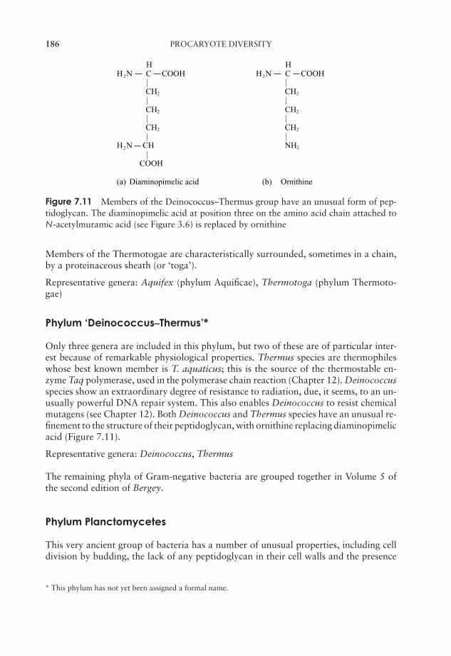
JWBK011-07 JWBK011-Hogg August 12, 2005 16:8 Char Count= 0
186 PROCARYOTE DIVERSITY
H HH2N C COOH H2N C COOH
| | CH2 CH2
| | CH2 CH2
| | CH2 CH2
| |H2N CH NH2
| COOH
(a) Diaminopimelic acid (b) Ornithine
Figure 7.11 Members of the Deinococcus–Thermus group have an unusual form of pep-tidoglycan. The diaminopimelic acid at position three on the amino acid chain attached toN-acetylmuramic acid (see Figure 3.6) is replaced by ornithine
Members of the Thermotogae are characteristically surrounded, sometimes in a chain,by a proteinaceous sheath (or ‘toga’).
Representative genera: Aquifex (phylum Aquificae), Thermotoga (phylum Thermoto-gae)
Phylum ‘Deinococcus–Thermus’*
Only three genera are included in this phylum, but two of these are of particular inter-est because of remarkable physiological properties. Thermus species are thermophileswhose best known member is T. aquaticus; this is the source of the thermostable en-zyme Taq polymerase, used in the polymerase chain reaction (Chapter 12). Deinococcusspecies show an extraordinary degree of resistance to radiation, due, it seems, to an un-usually powerful DNA repair system. This also enables Deinococcus to resist chemicalmutagens (see Chapter 12). Both Deinococcus and Thermus species have an unusual re-finement to the structure of their peptidoglycan, with ornithine replacing diaminopimelicacid (Figure 7.11).
Representative genera: Deinococcus, Thermus
The remaining phyla of Gram-negative bacteria are grouped together in Volume 5 ofthe second edition of Bergey.
Phylum Planctomycetes
This very ancient group of bacteria has a number of unusual properties, including celldivision by budding, the lack of any peptidoglycan in their cell walls and the presence
∗ This phylum has not yet been assigned a formal name.
JWBK011-07 JWBK011-Hogg August 12, 2005 16:8 Char Count= 0
DOMAIN: BACTERIA 187
of a degree of internal compartmentalisation. (Recall from Chapter 3 that membrane-bound compartments are regarded as a quintessentially eucaryotic feature.)
The recently discovered reaction known as anammox (anaerobic ammonium oxida-tion), whereby ammonium and nitrite are converted to nitrogen gas:
NH4+ + NO2
− = N2 + 2H2O
has been attributed to certain members of the Planctomycetes. It is thought that thisreaction may be responsible for much of the nitrogen cycling in the world’s oceans. Thebacteria concerned have only been identified by means of their rRNA gene sequences,and have as yet been assigned only provisional generic and specific names. They areanaerobic chemolithoautotrophs, however this is not typical of the Planctomycetes,most of which are aerobic chemoorganoheterotrophs.
Recent studies propose that the Planctomycetes should be placed much closer to theroot of any proposed phylogenetic tree than had previously been proposed.
Representative genera: Planctomyces, Pirellula
Phylum Chlamydiae
Formerly grouped with the Rickettsia (see above), these non-motile obligate parasites ofbirds and mammals are now assigned a separate phylum comprising only five genera, ofwhich Chlamydia is the most important. Like the Rickettsia, members of the Chlamydiaehave extremely small cells, and very limited metabolic capacities, and depend on thehost cell for energy generation. Unlike that group, however, they are not dependent onan arthropod vector for transmission from host to host.
Chlamydia trachomatis is the causative agent of trachoma, a major cause of blindnessin humans. Different strains of this same species are responsible for one of the mostcommon forms of sexually transmitted disease. C. psittaci causes the avian diseasepsittacosis, and C. pneumoniae causes chlamydial pneumonia in humans as well asbeing linked to some cases of coronary artery disease.
Representative genus: Chlamydia
Phylum Spirochaetes
The Spirochaetes are distinguished from all other bacteria by their slender helical mor-phology and corkscrew-like movement. This is made possible by endoflagella (axialfilaments), so-called because they are enclosed in the space between the cell and a flexi-ble sheath that surrounds it.
Spirochaetes comprise both aerobic and anaerobic bacteria that inhabit a wide rangeof habitats, including water and soil as well as the gut and oral cavities of both vertebrateand invertebrate animals. Some species are important pathogens of humans, includingTreponema pallidum (syphilis) and Leptospira interrogans (leptospirosis).
Representative genera: Treponema, Leptospira
JWBK011-07 JWBK011-Hogg August 12, 2005 16:8 Char Count= 0
188 PROCARYOTE DIVERSITY
Phylum Bacteroidetes
No unifying phenotypic feature characterises this diverse group, but their phylogeneticcloseness causes them to be placed together. In light of this, we can only consider ex-amples, without claiming them to be in any way representative.
The genus Flavobacterium takes its name from the yellow carotenoid pigments se-creted by its members. These are aerobic, free-living, aquatic forms, although they arealso associated with food spoilage.
In contrast, Bacteroides species are obligate anaerobes found in the human gut,where they ferment undigested food to acetate or lactate. Here they outnumber allother microbial forms, and are responsible for a significant percentage of the weight ofhuman faeces. Some species can also be pathogenic, and may cause peritonitis in caseswhere the large intestine or appendix has become perforated.
Representative genera: Bacteroides, Flavobacterium
Phylum: Verrucomicrobia
Members of the Verrucomicrobia form several prosthecae per cell; these are similar tothose described for certain Proteobacteria (see above). Although widespread in terres-trial, freshwater and marine environments, only a handful of representatives have beenisolated in pure culture.
Representative genera: Verrucomicrobium, Prosthecobacter
The Gram-positive bacteria: phylum Firmicutesand phylum Actinobacteria
In the second edition of Bergey, the Gram-positive bacteria are divided into two largephyla, the Firmicutes and the Actinobacteria. Some 2500 species are known, but asubstantial proportion of these belong to just a handful of genera. Gram-positivebacteria mostly have a chemoheterotrophic mode of nutrition and include amongtheir number several important human pathogens, as well as industrially significantforms.
The base composition of an organism’s DNA can be expressed as the percentage ofcytosine and guanine residues (per cent GC content); the technique is used widely inmicrobial taxonomy, and the Gram-positive bacteria are divided into those whose GCcontent is significantly over or under 50 per cent. It is convenient to consider groupingswithin the high GC and low GC forms as follows:
Phylum Firmicutes (low GC): spore-formingnon-spore formingmycoplasma
Phylum Actinobacteria (high GC): actinomycetescoryneform bacteria
JWBK011-07 JWBK011-Hogg August 12, 2005 16:8 Char Count= 0
DOMAIN: BACTERIA 189
Phylum Firmicutes: The low GC Gram-positive bacteria
The low GC Gram-positive bacteria form volume 3 of the second edition of Bergey.The spore-forming Gram-positive bacteria include two large genera, Clostridium and
Bacillus. Although not particularly close in phylogenetic terms, they are both capableof propagation by endospores.
Clostridium species are obligate anaerobes, and common inhabitants of soil. Sug-ars are fermented to various end-products such as butyric acid, acetone or butanol.Lacking an electron transport system, they obtain all their ATP from substrate-levelphosphorylation.
So-called ‘Botox’ injec-tions, much in voguein certain circles as acosmetic treatment, in-volve low doses of C. bo-tulinum exotoxin. By act-ing as a muscle relaxant,they are intended to re-duce the facial wrinklesthat develop with thepassing of time! The toxinis also used to treat med-ical conditions in whichabnormal muscle con-tractions make it impos-sible for patients to opentheir eyes properly.
Several species of Clostridium are serious humanpathogens including C. botulinum (botulism) and C.tetani (tetanus). C. perfringens causes gas gangrene, andif ingested, can also result in gastroenteritis. All theseconditions are due to the production of bacterial exo-toxins. The resistance of spores to heating is thus highlyrelevant both in medicine and in the food industry. Re-lated to Clostridium are the heliobacteria, two generaof anaerobic photoheterotrophic rods, some of whichproduce endospores. They are the only known photo-synthetic Gram-positive bacteria.
Bacillus species are aerobes or facultative anaerobes.They are chemoheterotrophs and usually motile bymeans of peritrichous flagella. Only a few species ofBacillus are pathogenic in humans, notably B. anthracis,the causative agent of anthrax. This is seen by manyas a potential agent of bioterrorism, and here again therelative indestructibility of its spores is a crucial factor.Other species, conversely, are positively beneficial to hu-mans; antibiotics such as bacitracin and polymixin areproduced by Bacillus species, whilst the toxin from B. thuringiensis has been used as anatural insecticide (see Chapter 12).
Representative genera: Bacillus, Clostridium
The non-spore-forming low GC Gram-positive bacteria include a number of medicallyand industrially significant genera, a few of which are discussed below.
The lactic acid bacteria are a taxonomically diverse group containing both rods (Lac-tobacillus) and cocci (Streptococcus, Lactococcus), all characterised by their fermenta-tive metabolism with lactic acid as end-product. Although they are able to tolerateoxygen, these bacteria do not use it in respiration. They are said to be aerotolerant.
Probiotics are living or-ganisms that are de-liberately ingested byhumans with the aim ofpromoting health.
Like the clostridia, they lack cytochromes, and are there-fore unable to carry out electron transport phosphory-lation. The lactic acid bacteria have limited synthetic ca-pabilities, so they are dependent on a supply of nutrientssuch as amino acids, purines/pyrimidines and vitamins.There has been growing interest in recent years in theuse of certain lactic acid bacteria as probiotics.
JWBK011-07 JWBK011-Hogg August 12, 2005 16:8 Char Count= 0
190 PROCARYOTE DIVERSITY
The genus Streptococcus remains a large one, although some members have beenassigned to new genera in recent years, e.g. Enterococcus, Lactococcus. Streptococci
Haemolysis is the lysis(bursting) of red bloodcells. It may be broughtabout by bacterial toxinscalled haemolysins.
are classified in a number of ways on the basis of phe-notypic characteristics, but these do not correspondto phylogenetic relationships. Many species producehaemolysis when grown on blood agar, due to the pro-duction of toxins called haemolysins. In α-haemolysis,haemoglobin is reduced to methaemoglobin, resultingin a partial clearance of the medium and a characteristicgreen colour. β-Haemolysis causes a complete lysis of thered blood cells, leaving an area of clearing in the agar. A few species are non-haemolytic.Streptococci are also classified on the basis of carbohydrate antigens found in the cellwall; this system, which assigns each organism to a lettered group, is named after itsdevisor, Rebecca Lancefield.
Pathogenic species of Streptococcus include S. pyogenes (‘strep’ sore throat, as wellas the more serious rheumatic fever), S. pneumoniae (pneumococcal pneumonia) andS. mutans (tooth decay). Cells of Streptococcus exist mostly in chains, but in S. pneu-moniae they are characteristically paired.
Lactobacillus is used very widely in the food and drink industry in the production ofsuch diverse foodstuffs as yoghurt, cheeses, pickled foods (e.g. sauerkraut) and certainbeers. This is discussed further in Chapter 17.
The cells of staphylococci occur in irregular bunches rather than ordered chains. Theyalso produce lactic acid but can additionally carry out aerobic respiration involvingcytochromes, and lack the complex nutritional requirements of the lactic acid bacteria.They are resistant to drying and able to tolerate relatively high concentrations of salt.These properties allow Staphylococcus aureus to be a normal inhabitant of the humanskin, where it can sometimes give rise to dermatological conditions such as acne, boilsand impetigo. It is also found in the respiratory tract of many healthy individuals, towhom it poses no threat, but in people whose immune system has been in some waycompromised, it can cause serious respiratory infections. S. aureus can also cause atype of food poisoning and is the causative agent of toxic shock syndrome. Widespreadantibiotic use has been largely responsible for the development of resistant forms ofS. aureus, which have become ubiquitous inhabitants of hospitals (methicillin-resistantStaphylococcus aureus: MRSA). The problem of antibiotic resistance is discussed atgreater length in Chapter 14.
Representative genera: Streptococcus, Staphylococcus
The Mycoplasma (Class Mollicutes) lack a cell wall and hence have a fluid shape(pleomorphic). Since the Gram test is based on the peptidoglycan content of a cell wall,why are these organisms grouped with the Gram-positive bacteria? The answer is thatalthough they do not give a positive Gram test, they are clearly related at the genetic levelto other members of the low GC Gram-positive group. The membranes of mycoplasmacontain sterols; these help in resisting osmotic lysis, and are often essential as a growthrequirement. Saprophytic, commensal and parasitic forms are known, and some speciesare associated with respiratory diseases in animals. Mycoplasma frequently occur ascontaminants in the culture of animal cells, because their small size allows them topass through filters, and they are resistant to antibiotics directed at cell wall synthesis.
JWBK011-07 JWBK011-Hogg August 12, 2005 16:8 Char Count= 0
DOMAIN: BACTERIA 191
Members of the Mycoplasma are among the smallest of all known cells and have someof the smallest genomes (just over half a million base pairs).
Representative genera: Mycoplasma, Ureoplasma
Phylum Actinobacteria: The high GC Gram-positive bacteria
The high GC gram-positive bacteria make up volume 4 of the second edition of Bergey.The actinomycetes are aerobic, filamentous bacteria that form branching mycelia
superficially similar to those of the Fungi (Chapter 8). Remember, however, that theactinomycetes are procaryotes and the fungi are eucaryotes, so the mycelia formed bythe former are appreciably smaller. In some cases, the mycelium extends clear of the
It has been suggestedthat camels are able tolocate water sources inthe desert by detect-ing the smell of geosminsgiven off by Strepto-myces spp. in the dampearth!
substratum and bears asexual conidiospores at the hy-phal tips. These are produced by the formation ofcross-walls and pinching off of spores, which are oftencoloured. The best known actinomycete genus is Strepto-myces, which contains some 500 species, all with a char-acteristically high GC content (69–73 per cent). Strepto-myces are very prevalent in soil, where they saprobicallydegrade a wide range of complex organic substrates bymeans of extracellular enzymes. Indeed, the characteris-tic musty smell of many soils is due to the productionof a volatile organic compound called geosmin. A highproportion of therapeutically useful antibiotics derive from Streptomyces species, in-cluding well-known examples such as streptomycin, erythromycin and tetracycline (seeChapter 14).
Most actinomycetes, including Streptomyces, are aerobic; however, members of thegenus Actinomyces are facultative anaerobes.
Representative genus: Streptomyces
The coryneform bacteria are morphologically half way between single celled bacilliand the branching filamentous actinomycetes. They are rods that show rudimentarybranching, giving rise to characteristic ‘V’ and ‘Y’ shapes. Among the genera in thisgroup are Corynebacterium, Mycobacterium, Propionibacterium and Nocardia.
Corynebacterium species are common in soil, and are also found in the mouths of avariety of animals. C. diphtheriae is the causative agent of diphtheria; it only becomespathogenic when it has been infected by a bacteriophage that carries the gene for thediphtheria exotoxin.
Members of the genus Mycobacterium are characterised by their unusual cell wall
The acid-fast test as-sesses the ability of an or-ganism to retain hot car-bol fuchsin stain whenrinsed with acidic alco-hol.
structure; they include unusual complex lipids calledmycolic acids. This causes the cells to be positive forthe acid-fast staining technique, a useful way of identi-fying the presence of these bacteria. Mycobacteria arerod shaped, sometimes becoming filamentous; when fil-aments are formed, propagation is by means of fragmen-tation. M. leprae and M. tuberculosis cause, respectively,leprosy and tuberculosis in humans.
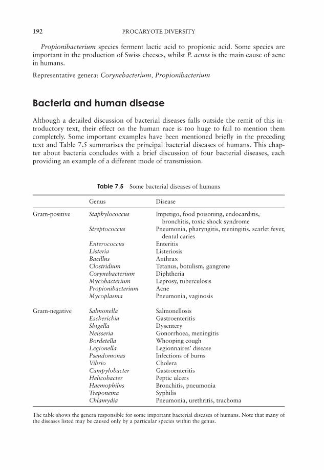
JWBK011-07 JWBK011-Hogg August 12, 2005 16:8 Char Count= 0
192 PROCARYOTE DIVERSITY
Propionibacterium species ferment lactic acid to propionic acid. Some species areimportant in the production of Swiss cheeses, whilst P. acnes is the main cause of acnein humans.
Representative genera: Corynebacterium, Propionibacterium
Bacteria and human disease
Although a detailed discussion of bacterial diseases falls outside the remit of this in-troductory text, their effect on the human race is too huge to fail to mention themcompletely. Some important examples have been mentioned briefly in the precedingtext and Table 7.5 summarises the principal bacterial diseases of humans. This chap-ter about bacteria concludes with a brief discussion of four bacterial diseases, eachproviding an example of a different mode of transmission.
Table 7.5 Some bacterial diseases of humans
Genus Disease
Gram-positive Staphylococcus Impetigo, food poisoning, endocarditis,bronchitis, toxic shock syndrome
Streptococcus Pneumonia, pharyngitis, meningitis, scarlet fever,dental caries
Enterococcus EnteritisListeria ListeriosisBacillus AnthraxClostridium Tetanus, botulism, gangreneCorynebacterium DiphtheriaMycobacterium Leprosy, tuberculosisPropionibacterium AcneMycoplasma Pneumonia, vaginosis
Gram-negative Salmonella SalmonellosisEscherichia GastroenteritisShigella DysenteryNeisseria Gonorrhoea, meningitisBordetella Whooping coughLegionella Legionnaires’ diseasePseudomonas Infections of burnsVibrio CholeraCampylobacter GastroenteritisHelicobacter Peptic ulcersHaemophilus Bronchitis, pneumoniaTreponema SyphilisChlamydia Pneumonia, urethritis, trachoma
The table shows the genera responsible for some important bacterial diseases of humans. Note that many ofthe diseases listed may be caused only by a particular species within the genus.

JWBK011-07 JWBK011-Hogg August 12, 2005 16:8 Char Count= 0
BACTERIA AND HUMAN DISEASE 193
Waterborne transmission: cholera
The causative agent Vibrio cholerae is ingested in faecally contaminated water or food.The bacteria attach by means of adhesins to the intestinal mucosa, where, withoutactually penetrating the cells, they release the cholera exotoxin. This comprises an ‘A’and several ‘B’ subunits; the former is the active ingredient, while the latter attachto epithelial cells by binding to a specific glycolipid in the membrane. This allows thepassage of the ‘A’ subunit into the cell, where it causes the activation of an enzyme calledadenylate cyclase (Figure 7.12). This results in uncontrolled production of cyclic AMP,causing active secretion of chloride and water into the intestinal lumen. The outcome ofthis is huge fluid loss (10 l or more per day) through profuse and debilitating diarrhoea.In the young, old and sick, death through dehydration and salt depletion can followwithin a very short time. If proper liquid and electrolyte replacement therapy is available,recovery rates can be very high. Although it is now very rare in the developed world(in 2002, only two cases were reported in the USA), cholera is a major killer in thethird world. It is easily preventable by means of clean water supplies and improved
AC
cAMP
Epithelialcells
Cl− H2OLumen ofintestine
Cholera toxinattaches toepithelial membrane
cAMP activatesother enzymes,resulting in theloss of chlorideions and waterfrom the cell
ATP
'A' subunit enters celland activates adenylatecyclase (AC), whichconverts ATP to cAMP
Figure 7.12 The action of the cholera exotoxin. Activation of the enzyme adenylate cyclaseresults in elevated levels of cyclic AMP and the secretion of electrolytes out of the epithelialcells lining the intestine and into the lumen. This is followed by water loss, resulting indebilitating dehydration
JWBK011-07 JWBK011-Hogg August 12, 2005 16:8 Char Count= 0
194 PROCARYOTE DIVERSITY
sanitation, however when these services break down, for example during war or afteran earthquake, cholera outbreaks quickly follow.
Airborne transmission: ‘strep’ throat
Streptococcal pharyngitis, commonly known as strep throat, is one of the commonestbacterial diseases of humans, being particularly common in children of school age. Theprimary means of transmission is by the inhalation from coughs and sneezes of respi-ratory droplets containing Streptococcus pyogenes (β-haemolytic type A streptococci),although other routes (kissing, infected handkerchiefs) are possible. The primary symp-toms are a red and raw throat (and/or tonsils), accompanied by headaches and fever.S. pyogenes attaches to the throat mucosa, stimulating an inflammatory response andsecreting virulence factors that destroy host blood cells. Although self-limiting withina week or so, strep throat should be treated with penicillin or erythromycin as moreserious streptococcal diseases such as scarlet fever and rheumatic fever may follow if itis left untreated.
Contact transmission: syphilis
Causative organisms of sexually transmitted diseases such as Neisseria gonorrhoeae(gonorrhoea) and Treponema pallidum (syphilis) are extremely sensitive to the effectsof environmental factors such as UV light and desiccation. They are therefore unable tolive outside of their human host, and rely for transmission on intimate human contact.
The spirochaete T. pallidum enters the body through minor abrasions, generally onthe genitalia or mouth, where a characteristic lesion called a chancre develops. Thedisease may proceed no further than this, but if T. pallidum enters the bloodstream andpasses around the body, the more serious secondary stage develops, lasting some weeks.Following a latent period of several years, around half of secondary syphilis cases go onto develop into the tertiary stage of the disease, whose symptoms may include mentalretardation, paralysis and blindness. Congenital syphilis is caused by T. pallidum beingpassed from a mother to her unborn child.
The primary and secondary stages of syphilis are readily treated by penicillin; how-ever, the tertiary stage is much less responsive to such therapy.
Vector-borne transmission: plague
A limited number of bacterial diseases reach their human hosts via an insect intermediaryfrom their main host, usually another species of mammal.
Plague (bubonic plague, the Black Death) has been responsible for the deaths ofuntold millions of people in terrible epidemics such as the ones that wiped out as muchas one-third of the population of Europe in the Middle Ages. It is caused by the Gram-negative bacterium Yersinia pestis, whose normal host is a rat, but can be spread tohumans by fleas. The bacteria pass to the lymph nodes, where they multiply, causingthe swellings known as bubos. Y. pestis produces an exotoxin, which prevents it frombeing destroyed by the host’s macrophages; instead, it is able to multiply inside them.
JWBK011-07 JWBK011-Hogg August 12, 2005 16:8 Char Count= 0
TEST YOURSELF 195
From the lymph nodes, the bacteria spread via the bloodstream to other tissues such asthe liver and lungs.
Once established in the lungs (pneumonic plague), plague can spread from humanto human by airborne transmission in respiratory droplets. Untreated, plague has ahigh rate of fatality, particularly for the pneumonic form of the disease. Early treatmentwith streptomycin or tetracycline, however, is largely successful. Improved public healthmeasures and the awareness of the dangers of rats and other rodents have meant thatconfirmed cases of plague are now relatively few.
Test yourself
1 The second edition of Bergey’s Manual of Systematic Bacteriology is basedon the relationships between microorganisms, as determined by acomparison of their sequences.
2 The three domains into which all living things are divided are the ,the and the .
3 Archaean membrane lipids contain branched instead of fatty acids.
4 Certain members of the Euryarchaeota have the unique ability to producemethane. Such organisms are termed .
5 Members of the Crenarchaeota are mostly extreme .
6 The biggest single phylum of bacteria is the , which includes manyof the best-known Gram-negative species.
7 The form of photosynthesis carried out by bacteria such as the purple sulphurbacteria is fundamentally different to that carried out by plants and algaebecause it is .
8 The genus Nitrosomonas carries out the first step of the nitrification process,converting to .
9 Acid mine drainage is largely due to the activities of bacteria that are able tooxidise compounds.
10 Bacteria such as Rhizobium are able to atmosphericnitrogen in association with the roots of plants.
11 The genus Desulfovibrio is found in conditions, where itsulphur compounds to .
12 Two main forms of fermentation are found in the enteric bacteria:fermentation and fermentation.
JWBK011-07 JWBK011-Hogg August 12, 2005 16:8 Char Count= 0
196 PROCARYOTE DIVERSITY
13 The pseudomonads can be distinguished from the enteric bacteria becausethey are -positive and incapable of carrying out .
14 The extracellular extension possessed by members of the stalked bacteria iscalled a .
15 Coxiella is a member of the , arthropod-borne intracellular para-sites of vertebrates.
16 The Cyanobacteria are the only procaryotes capable of carrying outphotosynthesis.
17 Some members of the Planctomycetes can carry out reactions, inwhich ammonia and nitrite are converted to nitrogen gas.
18 Treponema pallidum, the causative agent of syphilis, belongs to the, a group characterized by their shape and movement.
19 is an obligate anaerobe, and the most numerous microorganism inthe human gut.
20 Bacillus and Clostridium are Gram-positive genera characterised by theirability to form .
21 The Lancefield system is used to classify the on the basis of cellsurface antigens.
22 The are exceptionally small bacteria, which lack a cell wall.
23 The are branching, filamentous bacteria. One genus, , isthe source of many useful antibiotics.
24 Cell walls of the genus Mycobacterium contain acids.
25 The diphtheria exotoxin is encoded by a of Corynebacteriumdiphtheriae.

JWBK011-08 JWBK011-Hogg August 12, 2005 16:11 Char Count= 0
8The Fungi
As we saw in the introduction to this section on microbial diversity, fungi were formany years classified along with bacteria, algae and the slime moulds (Chapter 9) asmembers of the Kingdom Plantae. As recently as the 1960s it was possible to find fungibeing discussed under this heading, but in more recent times there has been universalagreement that they should be assigned their own kingdom. This is because fungi differfrom plants in two quite fundamental respects:
� plants obtain energy from the sun, fungi do not
� plants utilise CO2 as a carbon source, fungi do not.
To use the terminology we introduced in Chapter 4, plants are photoautotrophs, whereasfungi are chemoheterotrophs. In fact it now seems on the basis of molecular evidencethat fungi are more closely related to animals than they are to plants!
We may define true fungi as primarily terrestrial, spore-bearing organisms, lackingchlorophyll and having a heterotrophic, absorptive mode of nutrition. Some 80 000species are known and it is thought possible that at least a million more remain to bedescribed! True fungi are a monophyletic group; that is, they are all thought to descendfrom a common ancestor, some 550 million years ago.
Fungi are of great importance economically and socially, and may have beneficial ordetrimental effects. Many fungi, particularly yeasts, are involved in industrial fermen-tation processes (Chapter 17). These include, for example, the production of bread andalcohol, while other fungi are essential to the cheese-making process. Many antibiotics,including penicillin, derive from fungi, as does the immunosuppressive drug cyclosporin.Along with bacteria, fungi are responsible for the decomposition and reprocessing ofvast amounts of complex organic matter; some of this is recycled to the atmosphereas CO2, while much is rendered into a form that can be utilised by other organisms(Chapter 16). The other side of this coin is seen in the activity of fungi that degradeand destroy materials of economic importance such as wood, paper and leather, em-ploying essentially the same biochemical processes. Additionally, some fungi may causedisease; huge damage is caused to crops and other commercially valuable plants, whilea number of human diseases, particularly of the skin and scalp, are also caused byfungi.
197
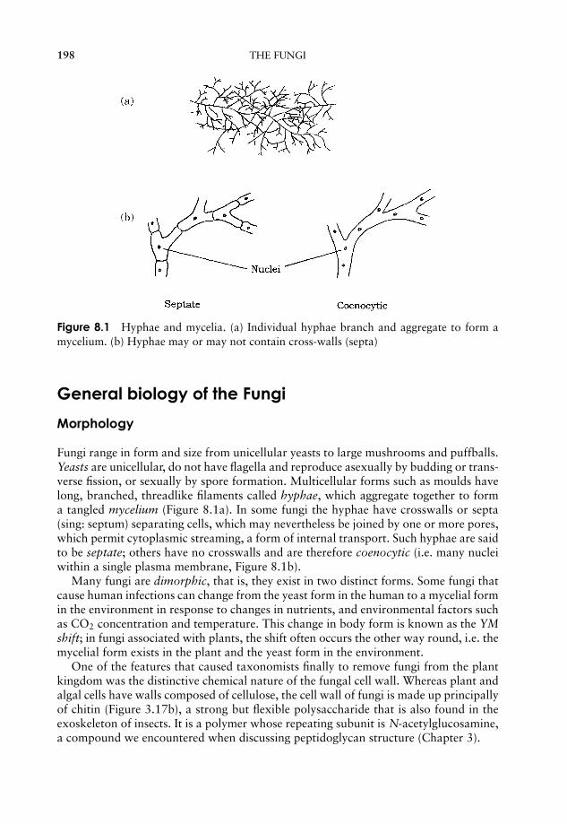
JWBK011-08 JWBK011-Hogg August 12, 2005 16:11 Char Count= 0
198 THE FUNGI
Figure 8.1 Hyphae and mycelia. (a) Individual hyphae branch and aggregate to form amycelium. (b) Hyphae may or may not contain cross-walls (septa)
General biology of the Fungi
Morphology
Fungi range in form and size from unicellular yeasts to large mushrooms and puffballs.Yeasts are unicellular, do not have flagella and reproduce asexually by budding or trans-verse fission, or sexually by spore formation. Multicellular forms such as moulds havelong, branched, threadlike filaments called hyphae, which aggregate together to forma tangled mycelium (Figure 8.1a). In some fungi the hyphae have crosswalls or septa(sing: septum) separating cells, which may nevertheless be joined by one or more pores,which permit cytoplasmic streaming, a form of internal transport. Such hyphae are saidto be septate; others have no crosswalls and are therefore coenocytic (i.e. many nucleiwithin a single plasma membrane, Figure 8.1b).
Many fungi are dimorphic, that is, they exist in two distinct forms. Some fungi thatcause human infections can change from the yeast form in the human to a mycelial formin the environment in response to changes in nutrients, and environmental factors suchas CO2 concentration and temperature. This change in body form is known as the YMshift; in fungi associated with plants, the shift often occurs the other way round, i.e. themycelial form exists in the plant and the yeast form in the environment.
One of the features that caused taxonomists finally to remove fungi from the plantkingdom was the distinctive chemical nature of the fungal cell wall. Whereas plant andalgal cells have walls composed of cellulose, the cell wall of fungi is made up principallyof chitin (Figure 3.17b), a strong but flexible polysaccharide that is also found in theexoskeleton of insects. It is a polymer whose repeating subunit is N-acetylglucosamine,a compound we encountered when discussing peptidoglycan structure (Chapter 3).
JWBK011-08 JWBK011-Hogg August 12, 2005 16:11 Char Count= 0
CLASSIFICATION OF THE FUNGI 199
Nutrition
The term saprobe de-scribes an organism thatfeeds on dead and de-caying organic materi-als. The older term sapro-phyte is no longer used,since the name perpet-uates the idea of fungibeing plants (phyton =a plant). Saprobes con-tribute greatly to the re-cycling of carbon andother elements.
Most fungi are saprobic (although some have othermodes of nutrition), that is, they obtain their nu-trients from decaying matter, which they grow overand through, frequently secreting enzymes extracellu-larly to break down complex molecules to simplerforms that can then be absorbed by the hyphae. Mostfungi are able to synthesise their own amino acidsand proteins from carbohydrates and simple nitroge-nous compounds. Although fungi are unable to move,they can swiftly colonise new territory as a result ofthe rapid rate at which their hyphae grow. All energyis concentrated on adding length rather than thick-ness; this growth pattern leads to an increase in sur-face area and is an adaptation to an absorptive wayof life. Carbohydrates are stored mainly in the formof glycogen (c.f. starch in higher plants, green al-gae). Metabolism is generally aerobic, but some yeasts can function as facultativeanaerobes.
Reproduction
Although, as we will see, there is a good deal of variety among the patterns of reproduc-tion among the fungi, all share in common the feature of reproducing by spores; theseare non-motile reproductive cells that rely on being carried by animals or the wind fortheir dispersal. The hyphae that bear the spores usually project up into the air, aidingtheir dispersal. One of the main reasons that we have to practise aseptic techniques inthe laboratory is that fungal spores are pretty well ubiquitous, and will germinate andgrow if they find a suitable growth medium. Spores of the common black bread mould,Rhizopus, (see below) have been found in the air over the North Pole, and hundreds ofmiles out to sea. In some fungi the aerial spore-bearing hyphae are developed into largecomplex structures called fruiting bodies. The most familiar example of a fruiting bodyis the mushroom. Many people think that the mushroom itself is the whole fungus butit only represents a part of it; most is buried away out of sight below the surface of thesoil or rotting material, a network of nearly invisible hyphae.
Classification of the Fungi
The Fungi are arranged into four major phyla on the basis of differences in their sexualreproduction. These are:
Zygomycota
Chytridiomycota
}‘Lower Fungi’
JWBK011-08 JWBK011-Hogg August 12, 2005 16:11 Char Count= 0
200 THE FUNGI
Ascomycota
Basidiomycota
}‘Higher Fungi’
Members of the differentfungal phyla are giventhe suffix -cetes, e.g. as-comycetes.
The designation ‘higher’ and ‘lower’ Fungi, is an unof-ficial distinction based on the fact that the latter two(much larger) groups possess septate hyphae. In somebooks, you may come across references to a group calledthe Deuteromycota or Fungi Imperfecti. This is not ataxonomic grouping, but a ‘holding area’ for species inwhich only an asexual stage has been recognised, and which cannot therefore be as-signed to any of the above groups. It is now possible, however, by means of DNAanalysis, to place such species with their nearest relatives, mostly in the Ascomycota orBasidiomycota.
In the following pages, we shall look at each of the phyla in turn, concentratingparticularly on their life cycles.
Zygomycota
The Zygomycota is a relatively small phylum, comprising less than a thousand species.Its members are typically found in soil, or on decaying organic matter, including animaldroppings. Some members of the group are of great importance in the formation of amutualistic association with plant roots known as a mycorrhiza. This will be discussedin more detail in Chapter 15.
Members of the Zygomycota are characterised by the formation of a dormant form,the zygospore, which is resistant to unfavourable environmental conditions. Hyphaeare coenocytic, with numerous haploid nuclei, but few dividing walls or septa.
A sporangium is a struc-ture inside which spo-res develop. It is heldaloft on an aerial hy-pha called a sporango-phore.
Familiar examples of this group are Mucor and theblack bread mould Rhizopus. The life cycle of Rhizopusis shown in Figure 8.2. Hyphae spread rapidly over thesurface of the substrate (bread, fruit etc.) and penetrateit, absorbing soluble nutrients such as sugars. Uprighthyphae develop, carrying at their tip sporangia, full ofblack haploid spores. The spores give the characteristiccolour to the mould; they are the asexual reproductivestructures, and are released when the thin wall of the sporangium ruptures. Whenconditions are favourable, Rhizopus reproduces in this way; each spore, upon find-ing a suitable substratum for growth, is capable of germinating and initiating a newmycelium.
A dicaryon is a struc-ture formed by two cellswhose contents, but notnuclei, have fused.
Sexual reproduction occurs when environmental con-ditions are unfavourable. Most species of Rhizopus areheterothallic; that is, there exist two distinct matingstrains known as + and −. Sexual reproduction is onlypossible between a member of the + -strain and a mem-ber of the − -strain. Although reproductively distinct,
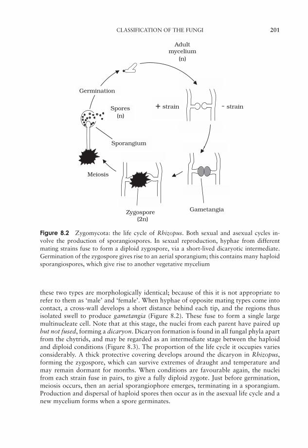
JWBK011-08 JWBK011-Hogg August 12, 2005 16:11 Char Count= 0
CLASSIFICATION OF THE FUNGI 201
Meiosis
Spores(n)
+ strain - strain
Zygospore(2n)
Sporangium
Adultmycelium
(n)
Gametangia
Germination
Figure 8.2 Zygomycota: the life cycle of Rhizopus. Both sexual and asexual cycles in-volve the production of sporangiospores. In sexual reproduction, hyphae from differentmating strains fuse to form a diploid zygospore, via a short-lived dicaryotic intermediate.Germination of the zygospore gives rise to an aerial sporangium; this contains many haploidsporangiospores, which give rise to another vegetative mycelium
these two types are morphologically identical; because of this it is not appropriate torefer to them as ‘male’ and ‘female’. When hyphae of opposite mating types come intocontact, a cross-wall develops a short distance behind each tip, and the regions thusisolated swell to produce gametangia (Figure 8.2). These fuse to form a single largemultinucleate cell. Note that at this stage, the nuclei from each parent have paired upbut not fused, forming a dicaryon. Dicaryon formation is found in all fungal phyla apartfrom the chytrids, and may be regarded as an intermediate stage between the haploidand diploid conditions (Figure 8.3). The proportion of the life cycle it occupies variesconsiderably. A thick protective covering develops around the dicaryon in Rhizopus,forming the zygospore, which can survive extremes of draught and temperature andmay remain dormant for months. When conditions are favourable again, the nucleifrom each strain fuse in pairs, to give a fully diploid zygote. Just before germination,meiosis occurs, then an aerial sporangiophore emerges, terminating in a sporangium.Production and dispersal of haploid spores then occur as in the asexual life cycle and anew mycelium forms when a spore germinates.
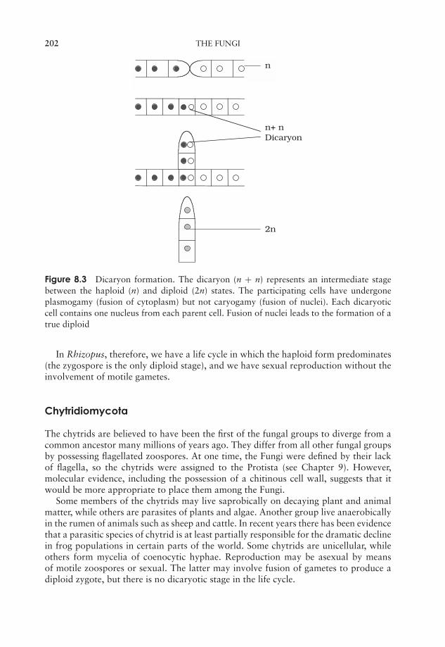
JWBK011-08 JWBK011-Hogg August 12, 2005 16:11 Char Count= 0
202 THE FUNGI
n
n+ n Dicaryon
2n
Figure 8.3 Dicaryon formation. The dicaryon (n + n) represents an intermediate stagebetween the haploid (n) and diploid (2n) states. The participating cells have undergoneplasmogamy (fusion of cytoplasm) but not caryogamy (fusion of nuclei). Each dicaryoticcell contains one nucleus from each parent cell. Fusion of nuclei leads to the formation of atrue diploid
In Rhizopus, therefore, we have a life cycle in which the haploid form predominates(the zygospore is the only diploid stage), and we have sexual reproduction without theinvolvement of motile gametes.
Chytridiomycota
The chytrids are believed to have been the first of the fungal groups to diverge from acommon ancestor many millions of years ago. They differ from all other fungal groupsby possessing flagellated zoospores. At one time, the Fungi were defined by their lackof flagella, so the chytrids were assigned to the Protista (see Chapter 9). However,molecular evidence, including the possession of a chitinous cell wall, suggests that itwould be more appropriate to place them among the Fungi.
Some members of the chytrids may live saprobically on decaying plant and animalmatter, while others are parasites of plants and algae. Another group live anaerobicallyin the rumen of animals such as sheep and cattle. In recent years there has been evidencethat a parasitic species of chytrid is at least partially responsible for the dramatic declinein frog populations in certain parts of the world. Some chytrids are unicellular, whileothers form mycelia of coenocytic hyphae. Reproduction may be asexual by meansof motile zoospores or sexual. The latter may involve fusion of gametes to produce adiploid zygote, but there is no dicaryotic stage in the life cycle.

JWBK011-08 JWBK011-Hogg August 12, 2005 16:11 Char Count= 0
CLASSIFICATION OF THE FUNGI 203
Ascomycota
A lichen is formed by thesymbiotic association ofa fungus (usually an as-comycete) and an algaor cyanophyte.
The Ascomycota are characterised by the production ofhaploid ascospores through the meiosis of a diploid nu-cleus in a small sac called an ascus. For this reason theyare sometimes called the sac fungi or cap fungi. Many ofthe fungi that cause serious plant diseases such as Dutchelm disease and powdery mildew belong to this group.They include some 30 000 species, among them yeasts,food spoilage moulds, brown fruit rotting fungi and truffles. Note that the latter, of-ten regarded as the most prized type of mushrooms by gourmets, are assigned to acompletely different group to the true mushrooms, which belong to the Basidiomycota.Around half of ascomycote species exist in associations with algae to form lichens; thesewill be discussed more fully in Chapter 15. Most ascomycetes produce mycelia that su-perficially resemble those of zygomycetes, but differ in that they have distinct, albeitperforated cross walls (septa) separating each cell.
Asexual reproduction in most ascomycetes involves the production of airborne sporescalled conidia. These are carried on the ends of specialised hyphae called conidiophores,where they may be pinched off as chains or clusters (Figure 8.4). Note that the conidiaare not contained within sporangia; they may be naked or protected by a flask-likestructure called the pycnidium. Asexual reproduction by conidia formation is a meansof rapid propagation for the fungus in favourable conditions. The characteristic green,
Figure 8.4 Asexual reproduction in the Ascomycota. Chains of conidia develop at the endof specialised hyphae called conidiophores

JWBK011-08 JWBK011-Hogg August 12, 2005 16:11 Char Count= 0
204 THE FUNGI
Figure 8.5 Yeast cells in various stages of budding. A protuberance or bud develops on theparent yeast; the nucleus undergoes division and one copy passes into the bud. Eventuallythe bud is walled off and separated to form a new cell
pink or brown colour of many moulds is due to the pigmentation of the conidia, whichare produced in huge numbers and dispersed by air currents. The conidia germinate toform another mycelium (haploid).
In the case of the unicellular yeasts, asexual reproduction occurs as the result ofbudding, a pinching off of a protuberance from the cell, which eventually grows to fullsize (Figure 8.5).
Plasmogamy is the fu-sion of the cytoplasmiccontent of two cells.Caryogamy is the fu-sion of nuclei from twodifferent cells.
Sexual reproduction in some ascomycetes involvesseparate + and − mating strains similar to those seen inzygomycetes, whilst in other cases an individual will beself-fertile, and thus able to mate with itself.When thereare separate strains, the hyphae involved in reproductionare termed the ascogonium (− -strain) and antheridium(+ -strain). In either case, two hyphae grow together andthere is a fusion of their cytoplasm (Figure 8.6). Withinthis fused structure, rather like the zygospore of Rhizopus, nuclei pair, but do not fuse;thus the resulting structure is a dicaryon. Following cytoplasmic fusion (plasmogamy),branching hyphae develop. These hyphae are septate, i.e. partitioned off into separatecells, but each cell is dicaryotic, having a nucleus from each parental type.
As we have seen, a key characteristic of ascomycetes is the production of sexual sporesin sac-like structures called asci. These develop in distinct macroscopic fruiting bodiescalled ascocarps, which arise from the aggregation of dicaryotic hyphae (Figure 8.6).At the tip of each dicaryotic hypha, pairs of nuclei fuse to give a diploid zygote; this isfollowed by one meiotic and one mitotic division, giving rise to eight haploid ascospores.An ascocarp may contain thousands of asci, each with eight ascospores. Tetrad analysisof ascospores has proved a valuable technique in genetic mapping.
When the ascus is mature, it splits open at its tip and the ascospores are released.They are dispersed, often over long distances, by air currents. If a mature ascocarpis disturbed, it may release smoke-like puffs containing thousands of ascospores. Thegerminating ascospore forms a new mycelium. Frequently there are many rounds ofasexual reproduction between successive rounds of sexual reproduction by ascosporeproduction.
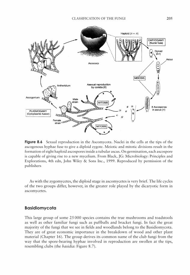
JWBK011-08 JWBK011-Hogg August 12, 2005 16:11 Char Count= 0
CLASSIFICATION OF THE FUNGI 205
Figure 8.6 Sexual reproduction in the Ascomycota. Nuclei in the cells at the tips of theascogenous hyphae fuse to give a diploid zygote. Meiotic and mitotic divisions result in theformation of eight haploid ascospores inside a tubular ascus. On germination, each ascosporeis capable of giving rise to a new mycelium. From Black, JG: Microbiology: Principles andExplorations, 4th edn, John Wiley & Sons Inc., 1999. Reproduced by permission of thepublishers
As with the zygomycetes, the diploid stage in ascomycetes is very brief. The life cyclesof the two groups differ, however, in the greater role played by the dicaryotic form inascomycetes.
Basidiomycota
This large group of some 25 000 species contains the true mushrooms and toadstoolsas well as other familiar fungi such as puffballs and bracket fungi. In fact the greatmajority of the fungi that we see in fields and woodlands belong to the Basidiomycota.They are of great economic importance in the breakdown of wood and other plantmaterial (Chapter 16). The group derives its common name of the club fungi from theway that the spore-bearing hyphae involved in reproduction are swollen at the tips,resembling clubs (the basidia: Figure 8.7).
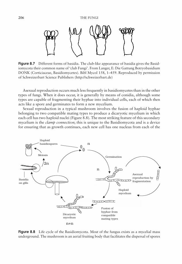
JWBK011-08 JWBK011-Hogg August 12, 2005 16:11 Char Count= 0
206 THE FUNGI
Figure 8.7 Different forms of basidia. The club-like appearance of basidia gives the Basid-iomycota their common name of ‘club Fungi’. From Langer, E: Die Gattung BotryobasidiumDONK (Corticiaceae, Basidiomycetes). Bibl Mycol 158, 1–459. Reproduced by permissionof Schweizerbart Science Publishers (http://schweizerbart.de)
Asexual reproduction occurs much less frequently in basidiomycetes than in the othertypes of fungi. When it does occur, it is generally by means of conidia, although sometypes are capable of fragmenting their hyphae into individual cells, each of which thenacts like a spore and germinates to form a new mycelium.
Sexual reproduction in a typical mushroom involves the fusion of haploid hyphaebelonging to two compatible mating types to produce a dicaryotic mycelium in whicheach cell has two haploid nuclei (Figure 8.8). The most striking feature of this secondarymycelium is the clamp connection; this is unique to the Basidiomycota and is a devicefor ensuring that as growth continues, each new cell has one nucleus from each of the
Asexualreproduction byfragmentation
Dicaryoticmycelium
Haploidmycelium
Fusion ofhyphae fromcompatiblemating types
n
n+n
2n
MeiosisGermination
Haploidbasidiospores
Basidiaon gills
n
Figure 8.8 Life cycle of the Basidiomycota. Most of the fungus exists as a mycelial massunderground. The mushroom is an aerial fruiting body that facilitates the dispersal of spores
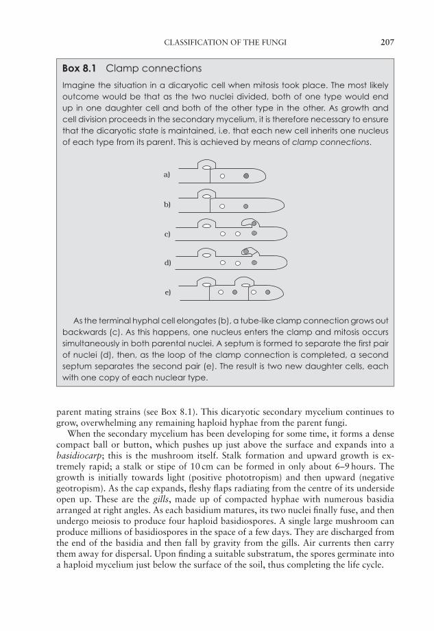
JWBK011-08 JWBK011-Hogg August 12, 2005 16:11 Char Count= 0
CLASSIFICATION OF THE FUNGI 207
Box 8.1 Clamp connections
Imagine the situation in a dicaryotic cell when mitosis took place. The most likelyoutcome would be that as the two nuclei divided, both of one type would endup in one daughter cell and both of the other type in the other. As growth andcell division proceeds in the secondary mycelium, it is therefore necessary to ensurethat the dicaryotic state is maintained, i.e. that each new cell inherits one nucleusof each type from its parent. This is achieved by means of clamp connections.
d)
c)
b)
a)
e)
As the terminal hyphal cell elongates (b), a tube-like clamp connection grows outbackwards (c). As this happens, one nucleus enters the clamp and mitosis occurssimultaneously in both parental nuclei. A septum is formed to separate the first pairof nuclei (d), then, as the loop of the clamp connection is completed, a secondseptum separates the second pair (e). The result is two new daughter cells, eachwith one copy of each nuclear type.
parent mating strains (see Box 8.1). This dicaryotic secondary mycelium continues togrow, overwhelming any remaining haploid hyphae from the parent fungi.
When the secondary mycelium has been developing for some time, it forms a densecompact ball or button, which pushes up just above the surface and expands into abasidiocarp; this is the mushroom itself. Stalk formation and upward growth is ex-tremely rapid; a stalk or stipe of 10 cm can be formed in only about 6–9 hours. Thegrowth is initially towards light (positive phototropism) and then upward (negativegeotropism). As the cap expands, fleshy flaps radiating from the centre of its undersideopen up. These are the gills, made up of compacted hyphae with numerous basidiaarranged at right angles. As each basidium matures, its two nuclei finally fuse, and thenundergo meiosis to produce four haploid basidiospores. A single large mushroom canproduce millions of basidiospores in the space of a few days. They are discharged fromthe end of the basidia and then fall by gravity from the gills. Air currents then carrythem away for dispersal. Upon finding a suitable substratum, the spores germinate intoa haploid mycelium just below the surface of the soil, thus completing the life cycle.
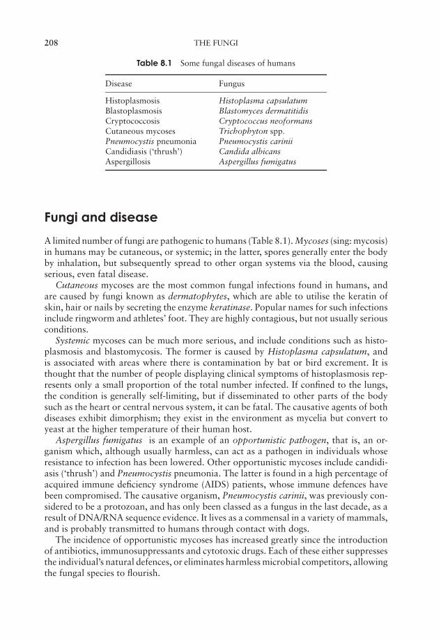
JWBK011-08 JWBK011-Hogg August 12, 2005 16:11 Char Count= 0
208 THE FUNGI
Table 8.1 Some fungal diseases of humans
Disease Fungus
Histoplasmosis Histoplasma capsulatumBlastoplasmosis Blastomyces dermatitidisCryptococcosis Cryptococcus neoformansCutaneous mycoses Trichophyton spp.
Pneumocystis pneumonia Pneumocystis cariniiCandidiasis (‘thrush’) Candida albicansAspergillosis Aspergillus fumigatus
Fungi and disease
A limited number of fungi are pathogenic to humans (Table 8.1). Mycoses (sing: mycosis)in humans may be cutaneous, or systemic; in the latter, spores generally enter the bodyby inhalation, but subsequently spread to other organ systems via the blood, causingserious, even fatal disease.
Cutaneous mycoses are the most common fungal infections found in humans, andare caused by fungi known as dermatophytes, which are able to utilise the keratin ofskin, hair or nails by secreting the enzyme keratinase. Popular names for such infectionsinclude ringworm and athletes’ foot. They are highly contagious, but not usually seriousconditions.
Systemic mycoses can be much more serious, and include conditions such as histo-plasmosis and blastomycosis. The former is caused by Histoplasma capsulatum, andis associated with areas where there is contamination by bat or bird excrement. It isthought that the number of people displaying clinical symptoms of histoplasmosis rep-resents only a small proportion of the total number infected. If confined to the lungs,the condition is generally self-limiting, but if disseminated to other parts of the bodysuch as the heart or central nervous system, it can be fatal. The causative agents of bothdiseases exhibit dimorphism; they exist in the environment as mycelia but convert toyeast at the higher temperature of their human host.
Aspergillus fumigatus is an example of an opportunistic pathogen, that is, an or-ganism which, although usually harmless, can act as a pathogen in individuals whoseresistance to infection has been lowered. Other opportunistic mycoses include candidi-asis (‘thrush’) and Pneumocystis pneumonia. The latter is found in a high percentage ofacquired immune deficiency syndrome (AIDS) patients, whose immune defences havebeen compromised. The causative organism, Pneumocystis carinii, was previously con-sidered to be a protozoan, and has only been classed as a fungus in the last decade, as aresult of DNA/RNA sequence evidence. It lives as a commensal in a variety of mammals,and is probably transmitted to humans through contact with dogs.
The incidence of opportunistic mycoses has increased greatly since the introductionof antibiotics, immunosuppressants and cytotoxic drugs. Each of these either suppressesthe individual’s natural defences, or eliminates harmless microbial competitors, allowingthe fungal species to flourish.
JWBK011-08 JWBK011-Hogg August 12, 2005 16:11 Char Count= 0
TEST YOURSELF 209
Box 8.2 Ergot
Members of the genus Claviceps may infect a variety of grains, particularly rye,when they come into flower, giving rise to the condition called ergot. No greatdamage is caused to the crop, but as the fungus develops in the maturing grain,powerful hallucinatory compounds are produced, which cause ergotism in thosewho consume bread made from the affected grain. This was relatively common inthe Middle Ages, when it was known as St Anthony’s Fire. The hallucinatory effectsof ergotism have been put forward by some as an explanation for outbreaks ofmass hysteria such as witch hunts and also for the cause of the abandonment ofthe ship, the Mary Celeste. The effects can go beyond the psychological causingconvulsions and even death. In small controlled amounts, the drugs derived fromergot can be medically useful in certain situations such as the induction of childbirthand the relief of migraine headaches.
Secondary metabolitesare produced by amicroorganism after thephase of active growthhas ceased. Such sub-stances are usuallynot required for essen-tial metabolic or cellmaintenance purposes.Examples include toxinspigments and most an-tibiotics. Carcinogenicmeans cancer-causing.
Many fungi produce natural mycotoxins; these aresecondary metabolites, which, if consumed by humans,can cause food poisoning that can sometimes be fatal.Certain species of mushrooms (‘toadstools’) includingthe genus Amanita contain substances that are highlypoisonous to humans. Other examples of mycotoxin ill-nesses include ergotism (see Box 8.2) and aflatoxin poi-soning. Aflatoxins are carcinogenic toxins produced byAspergillus flavus that grows on stored peanuts. In theearly 1960s, the turkey industry in the UK was almostcrippled by ‘Turkey X disease’, caused by the consump-tion of feed contaminated by A. flavus.
It is thought likely that all animals are parasitisedby one fungus or another. Extraordinary though it mayseem, there are even fungi that act as predators on smallsoil animals such as nematode worms, producing constrictive hyphal loops that tighten,immobilising the prey.
Fungi also cause disease in plants, and can have a devastating effect on crops ofeconomic importance, either on the living plant or in storage subsequent to harvest-ing. Rusts, smuts and mildews are all examples of common plant diseases caused byfungi. The effects of fungi on materials such as wood and textiles will be considered inChapter 16.
Test yourself
1 Categorised according to their carbon and energy sources, all fungi are.
2 Fungi whose cells are separated by cross-walls are described as .
JWBK011-08 JWBK011-Hogg August 12, 2005 16:11 Char Count= 0
210 THE FUNGI
3 Fungi whose cells lack cross-walls are described as .
4 The YM shift describes an alternation between and .
5 The principal component of cell walls is .
6 The (or Deuteromycota) is a term used to describe thosespecies in which no sexual reproductive stage has been observed.
7 The Chytridiomycota and the Zygomycota are sometimes termed thefungi.
8 The Ascomycota and the Basidiomycota are sometimes termed thefungi.
9 A resistant form, the , enables members of the Zygomycota to over-come adverse environmental conditions.
10 An aerial hypha that gives rise to spore formation is termed a .
11 The chytrids are thought to be the most primitive fungal group. Unlike otherfungi, they possess .
12 Members of the Ascomycota reproduce asexually by means of spores called.
13 Unicellular yeasts reproduce asexually by the process of .
14 In sexual reproduction in the Ascomycota, haploid ascospores develop infruiting bodies called .
15 cells contain two nuclei, one inherited from each parent.
16 The appearance of spore-bearing hyphae called basidia gives the Basidiomy-cota their common name: the .
17 The connection is a morphological feature unique to basid-iomycetes.
18 A mushroom is a fruiting body of a member of the Basidiomycota; its scientificname is a .
19 A is a fungal disease of humans. It may be or .
20 Aspergillus flavus produces carcinogenic toxins called .

JWBK011-09 JWBK011-Hogg August 12, 2005 19:58 Char Count= 0
9The Protista
Although not such an all-embracing a term as originally envisaged by Haeckel, theProtista represents a very diverse group of organisms, united by their possession ofeucaryotic characteristics, and failure to fit satisfactorily into the animal, plant or fungalkingdoms. Some scientists limit use of the name to unicellular organisms, while othersalso include organisms such as the macroscopic algae, which are not accommodatedconveniently elsewhere.
The Protista is a groupingof convenience, con-taining organisms noteasily accommodatedelsewhere. It includes allunicellular and colonialeucaryotic organisms,but is often expandedto include multicellularalgae.
It has become clear from molecular studies thatsome members of the Protista bear only a very dis-tant relationship to each other, making it an unsatis-factory grouping in many respects. In an introductorytext such as this, however, it is convenient and, it ishoped, less confusing, to retain the name Protista asa chapter heading, as long as the student understandsthat it does not represent a coherent taxonomic group-ing of phylogenetically related organisms. At the endof the chapter, we shall look at how members of theProtista are placed in modern, phylogenetic taxonomicschemes.
It has been found helpful in the past to think of Protists as being divided intothose with characteristics that are plant–like (the Algae), animal-like (the Protozoa) andfungus-like (the water moulds and slime moulds). We shall discuss each of these groupsin turn in the following pages. It should be borne in mind, however, that molecular evi-dence suggests such a division to be artificial; on the basis of molecular and cytologicalcomparison, the ‘animal-like’ protozoan Trypanosoma, for example, is closely relatedto the photosynthetic (and therefore ‘plant-like’) Euglena.
‘The Algae’
The Algae is a collective name traditionally given to several phyla of primitive, andmostly aquatic plants, making up a highly diverse group of over 30 000 species. Theydisplay a wide variety of structure, habitat and life-cycle, ranging from single-celledforms to massive seaweeds tens of metres in length. Most algae share a number ofcommon features which caused them to be grouped together. Among these are:
211

JWBK011-09 JWBK011-Hogg August 12, 2005 19:58 Char Count= 0
212 THE PROTISTA
� possession of the pigment chlorophyll
� deriving energy from the sun by means of oxygenic photosynthesis
� fixing carbon from CO2 or dissolved bicarbonate (see Chapter 6).
Modern taxonomy attempts to reflect more accurately the relationship between organ-isms with an assumed common ancestor. Thus, in the following pages, the unicellular‘algae’ are discussed in relation to other unicellular eucaryotes. Multicellular forms,including the Phaeophyta (brown algae) and Rhodophyta (red algae), are not discussedat great length and are included for the sake of completeness.
Structural characteristics of algal protists
All algal types are eucaryotic, and therefore contain the internal organelles we encoun-tered in Chapter 3, that is, nuclei, mitochondria, endoplasmic reticulum, ribosomes,Golgi body, and in most instances, chloroplasts. With the exception of one group (theEuglenophyta), all have a cellulose cell wall, which is frequently modified with otherpolysaccharides, including pectin and alginic acids. In some cases, the cell wall maybe fortified with deposits of calcium carbonate or silica. This is permeable to smallmolecules and ions, but impermeable to macromolecules. To the exterior of the cellmay be one or two flagella, with the typical eucaryotic 9 + 2 microstructure (see Figure3.18), which may allow unicellular types to move through the water; cilia are notfound in any algae.
The characteristics used to place algal protists into different taxa include the typeof chlorophyll present, the form in which carbohydrate is stored, and the structure ofthe cell wall (Table 9.1). A group not considered here are the cyanophytes, previouslyknown as the blue-green algae; although they carry out oxygenic photosynthesis, theyare procaryotes, and as such are more closely related to certain bacteria. They aretherefore discussed in Chapter 7.
Euglenophyta
A pellicle is a semi-rigidstructure composed ofprotein strips found sur-rounding the cell ofmany unicellular proto-zoans and algae
This is a group of unicellular flagellated organisms,which probably represent the most ancient group of al-gal protists. Individuals range in size from 10−500 µm.Euglenophytes are commonly found in fresh water, par-ticularly that with a high organic content, and to a lesserextent, in soil, brackish water and salt water. Membersof this group have a well-defined nucleus, and chloro-plasts containing chlorophylls a and b (Figure 9.1). Thestorage product of photosynthesis is a β-1,3-linked glucan called paramylon, found al-most exclusively in this group. Euglenophytes lack a cellulose cell wall but have instead,situated within the plasma membrane, a flexible pellicle made up of interlocking proteinstrips, a characteristic which links them to certain protozoan species. A further similarityis the way in which locomotion is achieved by the undulation of a terminal flagellum.Movement towards a light source is facilitated in many euglenids by two structures
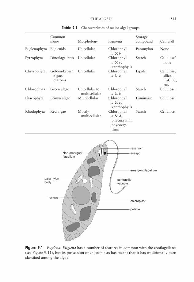
JWBK011-09 JWBK011-Hogg August 12, 2005 19:58 Char Count= 0
‘THE ALGAE’ 213
Table 9.1 Characteristics of major algal groups
Common Storagename Morphology Pigments compound Cell wall
Euglenophyta Euglenids Unicellular Chlorophylla & b
Paramylon None
Pyrrophyta Dinoflagellates Unicellular Chlorophylla & c,xanthophylls
Starch Cellulose/none
Chrysophyta Golden-brownalgae,diatoms
Unicellular Chlorophylla & c
Lipids Cellulose,silica,CaCO3,etc.
Chlorophyta Green algae Unicellular tomulticellular
Chlorophylla & b
Starch Cellulose
Phaeophyta Brown algae Multicellular Chlorophylla & c,xanthophylls
Laminarin Cellulose
Rhodophyta Red algae Mostlymulticellular
Chlorophylla & d,phycocyanin,phycoery-thrin
Starch Cellulose
reservoir
Non-emergentflagellum
eyespot
nucleuschloroplast
pellicle
paramylonbody
emergent flagellum
contractilevacuole
Figure 9.1 Euglena. Euglena has a number of features in common with the zooflagellates(see Figure 9.11), but its possession of chloroplasts has meant that it has traditionally beenclassified among the algae

JWBK011-09 JWBK011-Hogg August 12, 2005 19:58 Char Count= 0
214 THE PROTISTA
situated near the base of the flagellum; these are the paraflagellar body and the stigmaor eyespot. The latter is particularly conspicuous, as it is typically an orange-red colour,and relatively large.
Reproduction is by binary fission (i.e. by asexual means only). Division starts atthe anterior end, and proceeds longitudinally down the length of the cell, giving thecell a characteristic ‘two-headed’ appearance. During mitosis, the chromosomes withinthe nucleus replicate, forming pairs that split longitudinally. Since the euglenophyte isusually haploid, it thus becomes diploid for a short period. As fission proceeds, onedaughter cell retains the old flagellum, while the other one generates a new one later.As in the binary fission of bacteria, the progeny are genetically identical, i.e. clones.When conditions are unfavourable for survival due to failing nutrient supplies, the cellsround up to form cysts surrounded by a gelatinous covering; these have an increasedcomplement of paramylon granules, but no flagella. An important respect in whicheuglenids may be at variance with the notion of ‘plant-like protists’ is their abilityto exist as heterotrophs under certain conditions. When this happens, they lose theirphotosynthetic pigments and feed saprobically on dead organic material in the water.
Dinoflagellata
The dinoflagellates (also known variously as Pyrrophyta, or ‘fire algae’) are chiefly ma-rine planktonic types, comprising some 2000 species. This is another unicellular group,but one whose cells are often covered with armoured plates known as thecae (sing:theca). They are generally biflagellate, with the two dissimilar flagella lying in part withinthe longitudinal and lateral grooves that run around the cell (Figure 9.2). The beatingof the flagella causes the cell to spin like a top as it moves through the water (the grouptakes its name from the Greek word ‘to whirl’). Although many non-photosynthetic(chemoheterotrophic) types exist, most dinoflagellates are photosynthetic, containing
Theca
Longitudinalflagellum
Transverseflagellum
Figure 9.2 A dinoflagellate. Note the two flagella in perpendicular grooves. Each plays itspart in the organism’s locomotion

JWBK011-09 JWBK011-Hogg August 12, 2005 19:58 Char Count= 0
‘THE ALGAE’ 215
Box 9.1 Why would an alga want to glow in the dark?
The production of bioluminescence by several dinoflagellate species is thought tohave a protective function. Such algae are the natural prey of copepods, tiny crus-taceans found in astronomical numbers as part of the zooplankton. The biolumines-cence could have an effect directly, by acting as a warning signal to the copepods,or indirectly, by making those crustaceans which had consumed glowing algaemuch more conspicuous to their own predators.
chlorophyll a and c plus certain carotenoids and xanthophylls, which give them ared/golden appearance. As a group, they are second only to the diatoms (see below)as the primary photosynthetic producers in the marine environment. Some dinoflag-ellates form endosymbiotic relationships with marine animals such as corals and seaanemones; these are termed zooxanthellae. An unusual feature of dinoflagellate ultra-structure is that the chromosomes contain little, if any, histone protein, and exist almostpermanently in the condensed form.
Some tropical species of dinoflagellate emit light, the only algae to do so (see Box 9.1).Due to an enzyme–substrate (luciferin–luciferase) interaction, this can cause a spectac-ular glow in the water at night, especially when the water is disturbed, for example bya ship. Bioluminescence of this kind has proved to be a useful ‘tagging’ system for cellsin biological research. Other marine dinoflagellates can produce metabolites that act asnerve toxins to higher animals. Shellfish such as mussels and oysters can concentratethese with no harm to themselves, but they can be fatal to humans who consume them.Sometimes, when conditions are highly favourable, an explosion of growth results inthe development of huge ‘red tides’ of dinoflagellates in coastal waters. This produces abuild-up of toxins, and may lead to the death of massive numbers of fish and other ma-rine life. The greatly increased incidence of these blooms in recent decades is probablydue to pollution by fertilisers containing nitrates and phosphates.
Reproduction by asexual means involves binary fission. In armoured forms, the thecamay be shed before cell division, or split along suture lines; in either case, daughter cellsmust regenerate the missing sections. Sexual reproduction is known to occur in somedinoflagellates, and is probably more widespread. Gametes produced by mitosis fuse toproduce a diploid zygote; this undergoes meiosis to reinstate the haploid condition in theoffspring. In some species we see isogamy, the fusion of identical, motile gametes, whilein others, anisogamy occurs, in which gametes of dissimilar size fuse. Fusion may occurbetween genetically identical gametes, or only when the gametes come from geneticallydistinct populations.
Diatoms
The diatoms, which belong to the division Chrysophyta (the golden-brown algae), makeup the majority of phytoplankton in marine food chains, and as such are the mostimportant group of algal protists in terms of photosynthetic production. Over 10 000species of diatom are recognised, but some experts feel that the real number is manytimes greater than this.

JWBK011-09 JWBK011-Hogg August 12, 2005 19:58 Char Count= 0
216 THE PROTISTA
Figure 9.3 Diatoms are covered by an intricate two-part silicaceous shell called a frustule,whose often highly striking appearance make them among the most beautiful of microor-ganisms
As with the dinoflagellates, chlorophylls a and c are present, but not chlorophyll b.Their colour is due to carotenoids and xanthophylls (particularly fucoxanthin) maskingthe chlorophyll.
Diatoms have their cells surrounded by a silica-based shell known as a frustule,composed of two overlapping halves (the epitheca and the hypotheca). Microbiologistsare rarely able to resist the temptation to liken this structure to that of a petri dish,and with good reason. With the electron microscope it can be seen that the frustule isperforated with numerous tiny pores that connect the protoplast of the cell with theoutside environment. Diatom classification is based almost entirely on the shape andpattern of these shells, which are uniform for a particular species, and often have avery striking appearance (Figure 9.3). When diatoms die, their shells fall to the bottomof the sea, and can accumulate in thick layers where they represent a valuable mineralresource. This fine, light material (diatomaceous earth) has a number of applications,for example in filtration systems, and also as a light abrasive in products such as silverpolish or toothpaste.
Reproduction is usually asexual by binary fission, but a sexual phase with the produc-tion of haploid gametes can occur. Chrysophytes are unusual among the three primitivegroups of algae in that they are diploid. In diatoms, asexual reproduction involves mi-totic cell division, with each daughter cell receiving one half of the parental frustule, andsynthesizing a new one to complement it. The newly formed half, however, always actsas the hypotheca (lower half) of the new cell; consequently, one in two daughter cellswill be slightly smaller than the parent, an effect which is heightened over a number
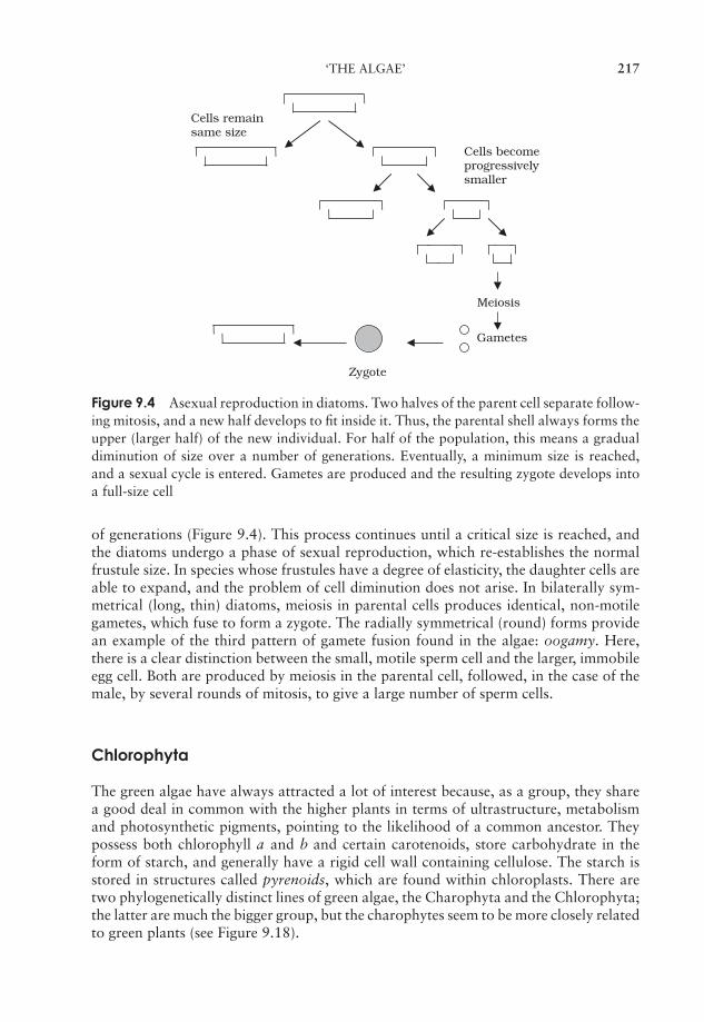
JWBK011-09 JWBK011-Hogg August 12, 2005 19:58 Char Count= 0
‘THE ALGAE’ 217
Meiosis
Gametes
Cells become progressivelysmaller
Cells remain same size
Zygote
Figure 9.4 Asexual reproduction in diatoms. Two halves of the parent cell separate follow-ing mitosis, and a new half develops to fit inside it. Thus, the parental shell always forms theupper (larger half) of the new individual. For half of the population, this means a gradualdiminution of size over a number of generations. Eventually, a minimum size is reached,and a sexual cycle is entered. Gametes are produced and the resulting zygote develops intoa full-size cell
of generations (Figure 9.4). This process continues until a critical size is reached, andthe diatoms undergo a phase of sexual reproduction, which re-establishes the normalfrustule size. In species whose frustules have a degree of elasticity, the daughter cells areable to expand, and the problem of cell diminution does not arise. In bilaterally sym-metrical (long, thin) diatoms, meiosis in parental cells produces identical, non-motilegametes, which fuse to form a zygote. The radially symmetrical (round) forms providean example of the third pattern of gamete fusion found in the algae: oogamy. Here,there is a clear distinction between the small, motile sperm cell and the larger, immobileegg cell. Both are produced by meiosis in the parental cell, followed, in the case of themale, by several rounds of mitosis, to give a large number of sperm cells.
Chlorophyta
The green algae have always attracted a lot of interest because, as a group, they sharea good deal in common with the higher plants in terms of ultrastructure, metabolismand photosynthetic pigments, pointing to the likelihood of a common ancestor. Theypossess both chlorophyll a and b and certain carotenoids, store carbohydrate in theform of starch, and generally have a rigid cell wall containing cellulose. The starch isstored in structures called pyrenoids, which are found within chloroplasts. There aretwo phylogenetically distinct lines of green algae, the Charophyta and the Chlorophyta;the latter are much the bigger group, but the charophytes seem to be more closely relatedto green plants (see Figure 9.18).
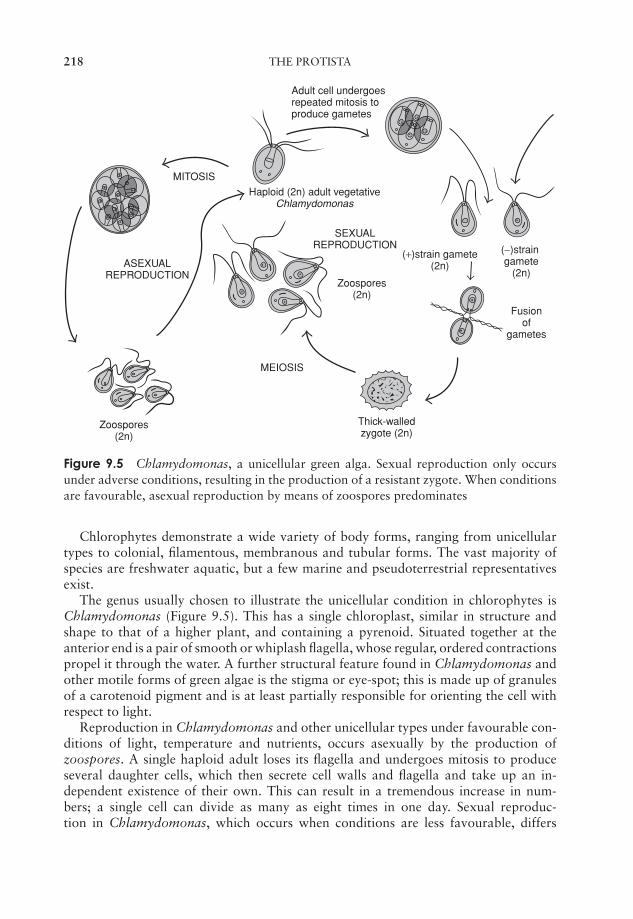
JWBK011-09 JWBK011-Hogg August 12, 2005 19:58 Char Count= 0
218 THE PROTISTA
MEIOSIS
MITOSIS
Haploid (2n) adult vegetative Chlamydomonas
Zoospores(2n)
Zoospores(2n)
Adult cell undergoes repeated mitosis to produce gametes
(+)strain gamete (2n)
(−)strain gamete
(2n)
SEXUAL REPRODUCTION
ASEXUAL REPRODUCTION
Fusionof
gametes
Thick-walled zygote (2n)
Figure 9.5 Chlamydomonas, a unicellular green alga. Sexual reproduction only occursunder adverse conditions, resulting in the production of a resistant zygote. When conditionsare favourable, asexual reproduction by means of zoospores predominates
Chlorophytes demonstrate a wide variety of body forms, ranging from unicellulartypes to colonial, filamentous, membranous and tubular forms. The vast majority ofspecies are freshwater aquatic, but a few marine and pseudoterrestrial representativesexist.
The genus usually chosen to illustrate the unicellular condition in chlorophytes isChlamydomonas (Figure 9.5). This has a single chloroplast, similar in structure andshape to that of a higher plant, and containing a pyrenoid. Situated together at theanterior end is a pair of smooth or whiplash flagella, whose regular, ordered contractionspropel it through the water. A further structural feature found in Chlamydomonas andother motile forms of green algae is the stigma or eye-spot; this is made up of granulesof a carotenoid pigment and is at least partially responsible for orienting the cell withrespect to light.
Reproduction in Chlamydomonas and other unicellular types under favourable con-ditions of light, temperature and nutrients, occurs asexually by the production ofzoospores. A single haploid adult loses its flagella and undergoes mitosis to produceseveral daughter cells, which then secrete cell walls and flagella and take up an in-dependent existence of their own. This can result in a tremendous increase in num-bers; a single cell can divide as many as eight times in one day. Sexual reproduc-tion in Chlamydomonas, which occurs when conditions are less favourable, differs

JWBK011-09 JWBK011-Hogg August 12, 2005 19:58 Char Count= 0
‘THE ALGAE’ 219
Daughter colony
Figure 9.6 Volvox, a colonial green alga. The colony comprises thousands of biflagellatedcells embedded in mucilage. Note the presence of daughter colonies, produced by asexualreproduction; these are eventually liberated and assume an independent existence
in detail according to the species (Figure 9.5). Any one of the three variants of ga-mete production seen in the algae may be seen: isogamy, anisogamy and oogamy. Inall cases, two haploid gametes undergo a fusion of both cytoplasm and nuclei to give adiploid zygote. The gametes may simply be unmodified haploid adult cells, or they mayarise through mitotic cleavage of the adult, depending on the species. The process ofisogamy, where the two gametes are morphologically alike and cannot be differentiatedvisually, only occurs in relatively lowly organisms such as Chlamydomonas. In somespecies we see the beginnings of sexual differentiation – there are two mating strains,designated + and −, and fusion will only take place between individuals of oppositestrains. The diploid zygote, once formed, often develops into a tough-walled protectivespore called a zygospore, which tides the organism over conditions of cold or drought.At an appropriate time the zygospore is stimulated to recommence the life-cycle, andmeiosis occurs, to produce haploid cells, which then mature into adult individuals.
In C. braunii, sexual reproduction is anisogamous; a + strain produces eight mi-crogametes and a − strain produces four macrogametes. In C. coccifera, simple oogamyoccurs, in which a vegetative cell loses its flagella, rounds off and enlarges; this acts asthe female gamete or ovum, and is fertilised by male gametes formed by other cells.
The next level of organization in the green algae is seen in the colonial types, typifiedby Volvox. These, like the unicellular types, are motile by means of flagella, and existas a number of cells embedded in a jelly-like matrix (Figure 9.6). Both the number ofcells and the way they are arranged is fixed and characteristic of a particular species.During growth, the number of cells does not increase. In simpler types, all cells seem tobe identical but in more complex forms there are distinct anterior and posterior ends,with the stigma more prominent at the anterior, and the posterior cells becoming larger.Reproduction can occur asexually or sexually.
The diversity of body forms in multicellular chlorophytes referred to earlier ismatched by that of their life cycles. Two examples are described here.
Oedogonium is a filamentous type. When young it attaches to the substratum bya basal holdfast, but unless it lives in flowing water the adult form is free-floating.Asexual reproduction occurs by means of motile zoospores, which swim free for aroundan hour before becoming fixed to a substratum and developing into a new filament. Insexual reproduction, the process of sexual differentiation is carried a step further thanwe have seen so far, with two separate filaments producing gametes from specialisedcells called gametangia (Figure 9.7). These are morphologically distinct, with the malebeing termed an antheridium and the female an oogonium. Gametes (morphologically
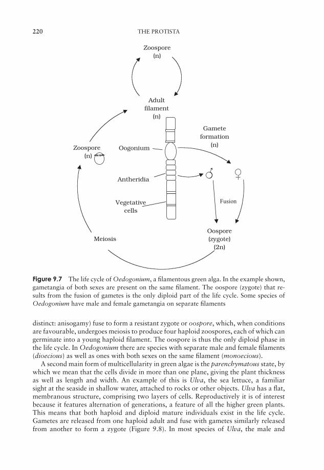
JWBK011-09 JWBK011-Hogg August 12, 2005 19:58 Char Count= 0
220 THE PROTISTA
Zoospore(n)
Fusion
Adultfilament
(n)
Gameteformation
(n)
Oospore(zygote)
(2n)Meiosis
Zoospore(n)
Antheridia
Oogonium
Vegetativecells
Figure 9.7 The life cycle of Oedogonium, a filamentous green alga. In the example shown,gametangia of both sexes are present on the same filament. The oospore (zygote) that re-sults from the fusion of gametes is the only diploid part of the life cycle. Some species ofOedogonium have male and female gametangia on separate filaments
distinct: anisogamy) fuse to form a resistant zygote or oospore, which, when conditionsare favourable, undergoes meiosis to produce four haploid zoospores, each of which cangerminate into a young haploid filament. The oospore is thus the only diploid phase inthe life cycle. In Oedogonium there are species with separate male and female filaments(dioecious) as well as ones with both sexes on the same filament (monoecious).
A second main form of multicellularity in green algae is the parenchymatous state, bywhich we mean that the cells divide in more than one plane, giving the plant thicknessas well as length and width. An example of this is Ulva, the sea lettuce, a familiarsight at the seaside in shallow water, attached to rocks or other objects. Ulva has a flat,membranous structure, comprising two layers of cells. Reproductively it is of interestbecause it features alternation of generations, a feature of all the higher green plants.This means that both haploid and diploid mature individuals exist in the life cycle.Gametes are released from one haploid adult and fuse with gametes similarly releasedfrom another to form a zygote (Figure 9.8). In most species of Ulva, the male and
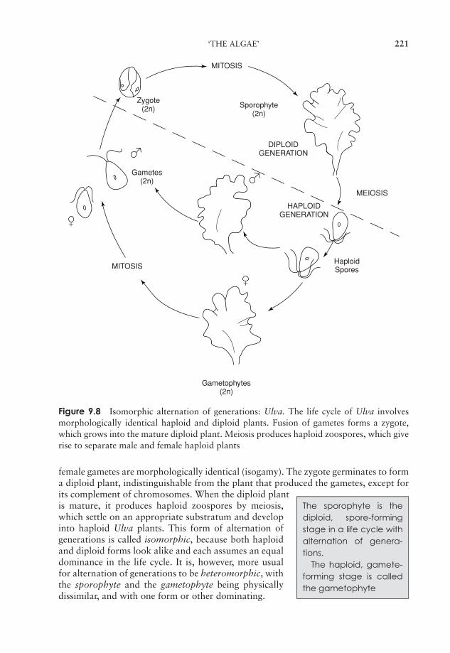
JWBK011-09 JWBK011-Hogg August 12, 2005 19:58 Char Count= 0
‘THE ALGAE’ 221
HAPLOIDGENERATION
DIPLOIDGENERATION
HaploidSpores
Gametophytes(2n)
Zygote(2n) Sporophyte
(2n)
MITOSIS
MEIOSIS
MITOSIS
Gametes(2n)
+
+
Figure 9.8 Isomorphic alternation of generations: Ulva. The life cycle of Ulva involvesmorphologically identical haploid and diploid plants. Fusion of gametes forms a zygote,which grows into the mature diploid plant. Meiosis produces haploid zoospores, which giverise to separate male and female haploid plants
female gametes are morphologically identical (isogamy). The zygote germinates to forma diploid plant, indistinguishable from the plant that produced the gametes, except forits complement of chromosomes. When the diploid plant
The sporophyte is thediploid, spore-formingstage in a life cycle withalternation of genera-tions.
The haploid, gamete-forming stage is calledthe gametophyte
is mature, it produces haploid zoospores by meiosis,which settle on an appropriate substratum and developinto haploid Ulva plants. This form of alternation ofgenerations is called isomorphic, because both haploidand diploid forms look alike and each assumes an equaldominance in the life cycle. It is, however, more usualfor alternation of generations to be heteromorphic, withthe sporophyte and the gametophyte being physicallydissimilar, and with one form or other dominating.
JWBK011-09 JWBK011-Hogg August 12, 2005 19:58 Char Count= 0
222 THE PROTISTA
Phaeophyta
The brown algae are multicellular, large and complex seaweeds, which dominate rockyshores in temperate and polar regions. Apart from one or two freshwater types, theyare all marine. The presence of fucoxanthin masks the presence of chlorophylls a andc. (In this context it must be stated here that not all ‘brown’ seaweeds look brown, norindeed do all the ‘red’ ones look red). Unlike the higher plants and green algae, whichuse starch as a food reserve, the phaeophytes use an unusual polysaccharide calledlaminarin (β-1,3-glucan).
Thallus is the word usedto describe a simplevegetative plant bodyshowing no differentia-tion into root, stem andleaf.
The level of tissue organisation in the brown algae isgreatly in advance of any of the types we’ve discussed sofar. The simplest thallus of a brown alga resembles themost complex found in the greens.
The phaeophytes also represent an advance in termsof sexual reproduction; here oogamy is the usual state ofaffairs and alternation of generations has developed tosuch an extent that diploid and haploid stages frequentlyassume separate morphological forms. Again, we shall use two examples to illustratelife cycle diversity in the brown algae.
Laminaria is one of the kelps, the largest group of brown algae. It grows attachedto underwater rocks or other objects by means of holdfasts, root-like structures whichanchor the plant. The thallus is further subdivided into a stalk-like stipe and a broader,blade-like lamina. Reproduction in Laminaria involves sporophyte and gametophyteplants that are morphologically quite distinct; (heteromorphic alternation of genera-tions). Reproductive areas called sori develop on the blade of the diploid sporophyte atcertain times of year (Figure 9.9). These consist of many sporangia, interspersed withthick protective hairs called paraphyses. As the sori develop, meiosis occurs, leading tothe production of haploid zoospores. These in turn develop into haploid filamentousgametophyte plants, much smaller and quite different in morphology from the morehighly organised sporophyte. Indeed, in contrast to the large sporophyte the gameto-phyte is a microscopic structure. The gametophytes are dioecious, that is the male andfemale reproductive structures are borne on separate individuals. The female plant bearsa number of oogonia, each of which produces a single egg, which escapes through apore at the apex of the oogonium, but remains attached in a sort of cup, formed by thesurrounds of the pore. In similar fashion the male plant bears several antheridia, eachliberating a single antherozoid; this however is motile by means of flagella and fertilisesthe egg. The diploid zygote so produced grows immediately into a new sporophyteplant.
In our second example of a phaeophyte life cycle, there is no alternation of generationsat all, the gametophyte generation having been completely lost. The wracks are familiarseaweeds found in the intertidal zone, and Fucus vesiculosus, known commonly as thebladder wrack, is one of the best known (Figure 9.10). It gets its name from the airbladders distributed on its surface, which assist buoyancy.
The adult has reproductive structures called receptacles, slight swellings situated atthe tip of the thallus; within these are flask-like invaginations called conceptacles whichcontain the male or female gametangia, again interspersed with sterile paraphyses. F.vesiculosus is monoecious but some other Fucus species are dioecious. Each antheridium
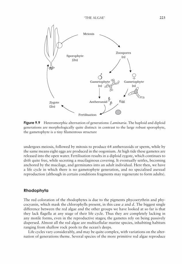
JWBK011-09 JWBK011-Hogg August 12, 2005 19:58 Char Count= 0
‘THE ALGAE’ 223
Sporophyte(2n)
Zoospores(n)
Meiosis
Gametophyte(n)
Gametophyte(n)
Antherozoid EggZygote(2n)
Fertilisation
Figure 9.9 Heteromorphic alternation of generations: Laminaria. The haploid and diploidgenerations are morphologically quite distinct: in contrast to the large robust sporophyte,the gametophyte is a tiny filamentous structure
undergoes meiosis, followed by mitosis to produce 64 antherozoids or sperm, while bythe same means eight eggs are produced in the oogonium. At high tide these gametes arereleased into the open water. Fertilisation results in a diploid zygote, which continues todrift quite free, while secreting a mucilaginous covering. It eventually settles, becominganchored by the mucilage, and germinates into an adult individual. Here then, we havea life cycle in which there is no gametophyte generation, and no specialised asexualreproduction (although in certain conditions fragments may regenerate to form adults).
Rhodophyta
The red coloration of the rhodophytes is due to the pigments phycoerythrin and phy-cocyanin, which mask the chlorophylls present, in this case a and d. The biggest singledifference between the red algae and the other groups we have looked at so far is thatthey lack flagella at any stage of their life cycle. Thus they are completely lacking inany motile forms, even in the reproductive stages; the gametes rely on being passivelydispersed. Almost all the red algae are multicellular marine species, inhabiting habitatsranging from shallow rock pools to the ocean’s deeps.
Life cycles vary considerably, and may be quite complex, with variations on the alter-nation of generations theme. Several species of the more primitive red algae reproduce
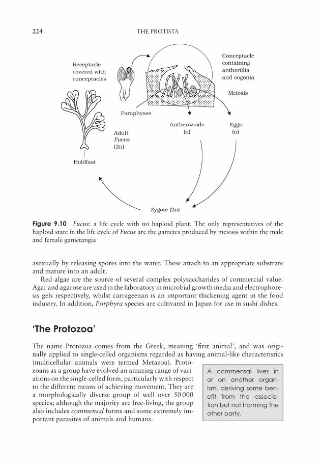
JWBK011-09 JWBK011-Hogg August 12, 2005 19:58 Char Count= 0
224 THE PROTISTA
Antherozoids(n)
Zygote (2n)
Eggs(n)Adult
Fucus(2n)
Conceptaclecontainingantheridiaand oogonia
Receptaclecovered withconceptacles
Holdfast
Paraphyses
Meiosis
Figure 9.10 Fucus: a life cycle with no haploid plant. The only representatives of thehaploid state in the life cycle of Fucus are the gametes produced by meiosis within the maleand female gametangia
asexually by releasing spores into the water. These attach to an appropriate substrateand mature into an adult.
Red algae are the source of several complex polysaccharides of commercial value.Agar and agarose are used in the laboratory in microbial growth media and electrophore-sis gels respectively, whilst carrageenan is an important thickening agent in the foodindustry. In addition, Porphyra species are cultivated in Japan for use in sushi dishes.
‘The Protozoa’
The name Protozoa comes from the Greek, meaning ‘first animal’, and was origi-nally applied to single-celled organisms regarded as having animal-like characteristics
A commensal lives inor on another organ-ism, deriving some ben-efit from the associa-tion but not harming theother party.
(multicellular animals were termed Metazoa). Proto-zoans as a group have evolved an amazing range of vari-ations on the single-celled form, particularly with respectto the different means of achieving movement. They area morphologically diverse group of well over 50 000species; although the majority are free-living, the groupalso includes commensal forms and some extremely im-portant parasites of animals and humans.
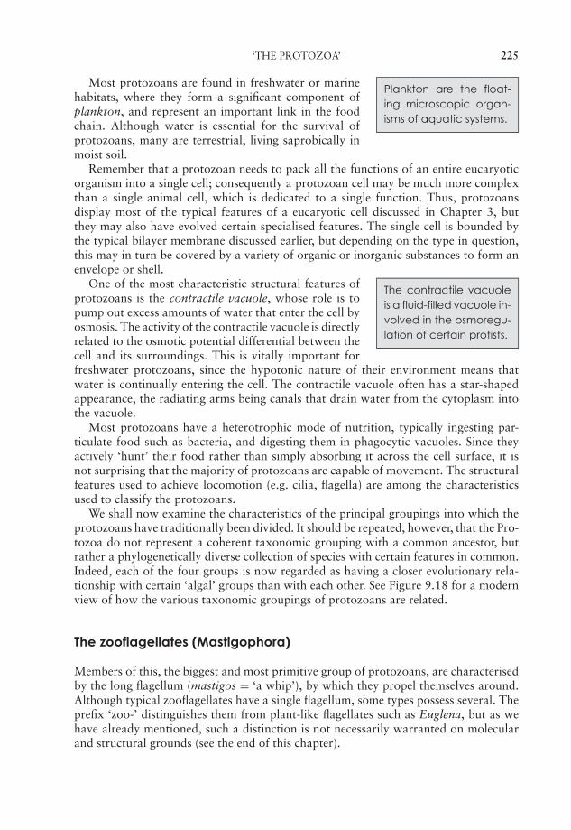
JWBK011-09 JWBK011-Hogg August 12, 2005 19:58 Char Count= 0
‘THE PROTOZOA’ 225
Plankton are the float-ing microscopic organ-isms of aquatic systems.
Most protozoans are found in freshwater or marinehabitats, where they form a significant component ofplankton, and represent an important link in the foodchain. Although water is essential for the survival ofprotozoans, many are terrestrial, living saprobically inmoist soil.
Remember that a protozoan needs to pack all the functions of an entire eucaryoticorganism into a single cell; consequently a protozoan cell may be much more complexthan a single animal cell, which is dedicated to a single function. Thus, protozoansdisplay most of the typical features of a eucaryotic cell discussed in Chapter 3, butthey may also have evolved certain specialised features. The single cell is bounded bythe typical bilayer membrane discussed earlier, but depending on the type in question,this may in turn be covered by a variety of organic or inorganic substances to form anenvelope or shell.
The contractile vacuoleis a fluid-filled vacuole in-volved in the osmoregu-lation of certain protists.
One of the most characteristic structural features ofprotozoans is the contractile vacuole, whose role is topump out excess amounts of water that enter the cell byosmosis. The activity of the contractile vacuole is directlyrelated to the osmotic potential differential between thecell and its surroundings. This is vitally important forfreshwater protozoans, since the hypotonic nature of their environment means thatwater is continually entering the cell. The contractile vacuole often has a star-shapedappearance, the radiating arms being canals that drain water from the cytoplasm intothe vacuole.
Most protozoans have a heterotrophic mode of nutrition, typically ingesting par-ticulate food such as bacteria, and digesting them in phagocytic vacuoles. Since theyactively ‘hunt’ their food rather than simply absorbing it across the cell surface, it isnot surprising that the majority of protozoans are capable of movement. The structuralfeatures used to achieve locomotion (e.g. cilia, flagella) are among the characteristicsused to classify the protozoans.
We shall now examine the characteristics of the principal groupings into which theprotozoans have traditionally been divided. It should be repeated, however, that the Pro-tozoa do not represent a coherent taxonomic grouping with a common ancestor, butrather a phylogenetically diverse collection of species with certain features in common.Indeed, each of the four groups is now regarded as having a closer evolutionary rela-tionship with certain ‘algal’ groups than with each other. See Figure 9.18 for a modernview of how the various taxonomic groupings of protozoans are related.
The zooflagellates (Mastigophora)
Members of this, the biggest and most primitive group of protozoans, are characterisedby the long flagellum (mastigos = ‘a whip’), by which they propel themselves around.Although typical zooflagellates have a single flagellum, some types possess several. Theprefix ‘zoo-’ distinguishes them from plant-like flagellates such as Euglena, but as wehave already mentioned, such a distinction is not necessarily warranted on molecularand structural grounds (see the end of this chapter).
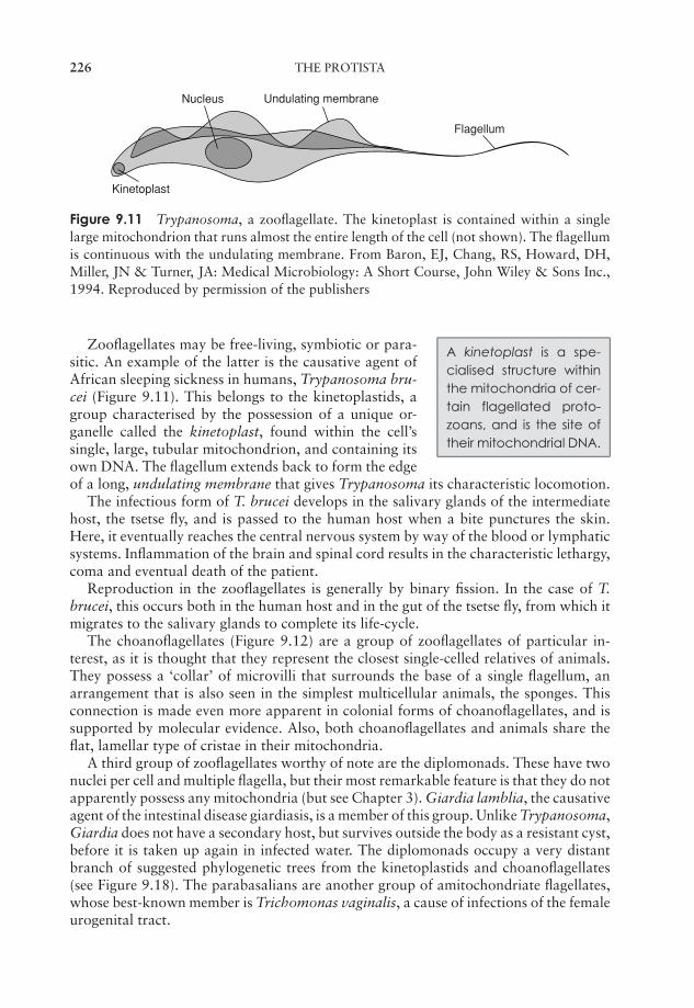
JWBK011-09 JWBK011-Hogg August 12, 2005 19:58 Char Count= 0
226 THE PROTISTA
Kinetoplast
Nucleus Undulating membrane
Flagellum
Figure 9.11 Trypanosoma, a zooflagellate. The kinetoplast is contained within a singlelarge mitochondrion that runs almost the entire length of the cell (not shown). The flagellumis continuous with the undulating membrane. From Baron, EJ, Chang, RS, Howard, DH,Miller, JN & Turner, JA: Medical Microbiology: A Short Course, John Wiley & Sons Inc.,1994. Reproduced by permission of the publishers
A kinetoplast is a spe-cialised structure withinthe mitochondria of cer-tain flagellated proto-zoans, and is the site oftheir mitochondrial DNA.
Zooflagellates may be free-living, symbiotic or para-sitic. An example of the latter is the causative agent ofAfrican sleeping sickness in humans, Trypanosoma bru-cei (Figure 9.11). This belongs to the kinetoplastids, agroup characterised by the possession of a unique or-ganelle called the kinetoplast, found within the cell’ssingle, large, tubular mitochondrion, and containing itsown DNA. The flagellum extends back to form the edgeof a long, undulating membrane that gives Trypanosoma its characteristic locomotion.
The infectious form of T. brucei develops in the salivary glands of the intermediatehost, the tsetse fly, and is passed to the human host when a bite punctures the skin.Here, it eventually reaches the central nervous system by way of the blood or lymphaticsystems. Inflammation of the brain and spinal cord results in the characteristic lethargy,coma and eventual death of the patient.
Reproduction in the zooflagellates is generally by binary fission. In the case of T.brucei, this occurs both in the human host and in the gut of the tsetse fly, from which itmigrates to the salivary glands to complete its life-cycle.
The choanoflagellates (Figure 9.12) are a group of zooflagellates of particular in-terest, as it is thought that they represent the closest single-celled relatives of animals.They possess a ‘collar’ of microvilli that surrounds the base of a single flagellum, anarrangement that is also seen in the simplest multicellular animals, the sponges. Thisconnection is made even more apparent in colonial forms of choanoflagellates, and issupported by molecular evidence. Also, both choanoflagellates and animals share theflat, lamellar type of cristae in their mitochondria.
A third group of zooflagellates worthy of note are the diplomonads. These have twonuclei per cell and multiple flagella, but their most remarkable feature is that they do notapparently possess any mitochondria (but see Chapter 3). Giardia lamblia, the causativeagent of the intestinal disease giardiasis, is a member of this group. Unlike Trypanosoma,Giardia does not have a secondary host, but survives outside the body as a resistant cyst,before it is taken up again in infected water. The diplomonads occupy a very distantbranch of suggested phylogenetic trees from the kinetoplastids and choanoflagellates(see Figure 9.18). The parabasalians are another group of amitochondriate flagellates,whose best-known member is Trichomonas vaginalis, a cause of infections of the femaleurogenital tract.
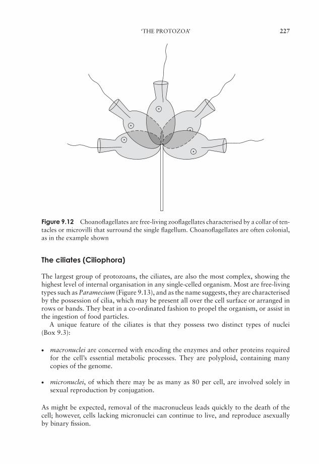
JWBK011-09 JWBK011-Hogg August 12, 2005 19:58 Char Count= 0
‘THE PROTOZOA’ 227
Figure 9.12 Choanoflagellates are free-living zooflagellates characterised by a collar of ten-tacles or microvilli that surround the single flagellum. Choanoflagellates are often colonial,as in the example shown
The ciliates (Ciliophora)
The largest group of protozoans, the ciliates, are also the most complex, showing thehighest level of internal organisation in any single-celled organism. Most are free-livingtypes such as Paramecium (Figure 9.13), and as the name suggests, they are characterisedby the possession of cilia, which may be present all over the cell surface or arranged inrows or bands. They beat in a co-ordinated fashion to propel the organism, or assist inthe ingestion of food particles.
A unique feature of the ciliates is that they possess two distinct types of nuclei(Box 9.3):
� macronuclei are concerned with encoding the enzymes and other proteins requiredfor the cell’s essential metabolic processes. They are polyploid, containing manycopies of the genome.
� micronuclei, of which there may be as many as 80 per cell, are involved solely insexual reproduction by conjugation.
As might be expected, removal of the macronucleus leads quickly to the death of thecell; however, cells lacking micronuclei can continue to live, and reproduce asexuallyby binary fission.
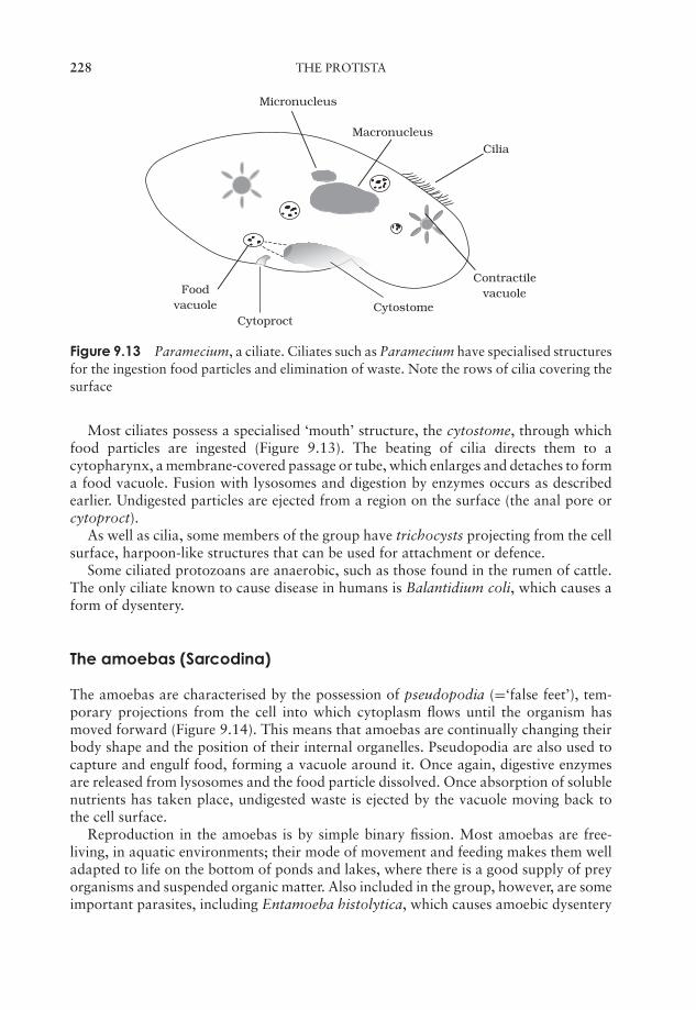
JWBK011-09 JWBK011-Hogg August 12, 2005 19:58 Char Count= 0
228 THE PROTISTA
ContractilevacuoleFood
vacuole
Macronucleus
Micronucleus
Cilia
CytoproctCytostome
Figure 9.13 Paramecium, a ciliate. Ciliates such as Paramecium have specialised structuresfor the ingestion food particles and elimination of waste. Note the rows of cilia covering thesurface
Most ciliates possess a specialised ‘mouth’ structure, the cytostome, through whichfood particles are ingested (Figure 9.13). The beating of cilia directs them to acytopharynx, a membrane-covered passage or tube, which enlarges and detaches to forma food vacuole. Fusion with lysosomes and digestion by enzymes occurs as describedearlier. Undigested particles are ejected from a region on the surface (the anal pore orcytoproct).
As well as cilia, some members of the group have trichocysts projecting from the cellsurface, harpoon-like structures that can be used for attachment or defence.
Some ciliated protozoans are anaerobic, such as those found in the rumen of cattle.The only ciliate known to cause disease in humans is Balantidium coli, which causes aform of dysentery.
The amoebas (Sarcodina)
The amoebas are characterised by the possession of pseudopodia (=‘false feet’), tem-porary projections from the cell into which cytoplasm flows until the organism hasmoved forward (Figure 9.14). This means that amoebas are continually changing theirbody shape and the position of their internal organelles. Pseudopodia are also used tocapture and engulf food, forming a vacuole around it. Once again, digestive enzymesare released from lysosomes and the food particle dissolved. Once absorption of solublenutrients has taken place, undigested waste is ejected by the vacuole moving back tothe cell surface.
Reproduction in the amoebas is by simple binary fission. Most amoebas are free-living, in aquatic environments; their mode of movement and feeding makes them welladapted to life on the bottom of ponds and lakes, where there is a good supply of preyorganisms and suspended organic matter. Also included in the group, however, are someimportant parasites, including Entamoeba histolytica, which causes amoebic dysentery
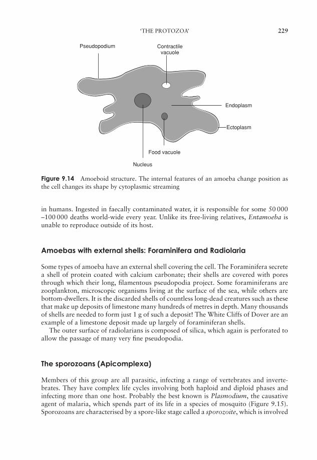
JWBK011-09 JWBK011-Hogg August 12, 2005 19:58 Char Count= 0
‘THE PROTOZOA’ 229
Pseudopodium
Endoplasm
Ectoplasm
Food vacuole
Nucleus
Contractilevacuole
Figure 9.14 Amoeboid structure. The internal features of an amoeba change position asthe cell changes its shape by cytoplasmic streaming
in humans. Ingested in faecally contaminated water, it is responsible for some 50 000–100 000 deaths world-wide every year. Unlike its free-living relatives, Entamoeba isunable to reproduce outside of its host.
Amoebas with external shells: Foraminifera and Radiolaria
Some types of amoeba have an external shell covering the cell. The Foraminifera secretea shell of protein coated with calcium carbonate; their shells are covered with poresthrough which their long, filamentous pseudopodia project. Some foraminiferans arezooplankton, microscopic organisms living at the surface of the sea, while others arebottom-dwellers. It is the discarded shells of countless long-dead creatures such as thesethat make up deposits of limestone many hundreds of metres in depth. Many thousandsof shells are needed to form just 1 g of such a deposit! The White Cliffs of Dover are anexample of a limestone deposit made up largely of foraminiferan shells.
The outer surface of radiolarians is composed of silica, which again is perforated toallow the passage of many very fine pseudopodia.
The sporozoans (Apicomplexa)
Members of this group are all parasitic, infecting a range of vertebrates and inverte-brates. They have complex life cycles involving both haploid and diploid phases andinfecting more than one host. Probably the best known is Plasmodium, the causativeagent of malaria, which spends part of its life in a species of mosquito (Figure 9.15).Sporozoans are characterised by a spore-like stage called a sporozoite, which is involved
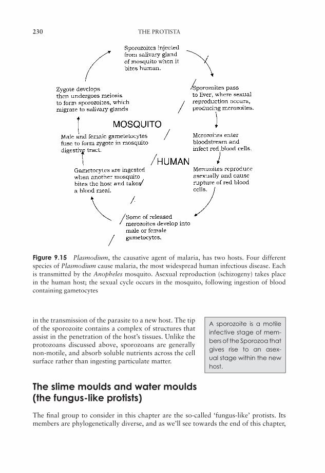
JWBK011-09 JWBK011-Hogg August 12, 2005 19:58 Char Count= 0
230 THE PROTISTA
Figure 9.15 Plasmodium, the causative agent of malaria, has two hosts. Four differentspecies of Plasmodium cause malaria, the most widespread human infectious disease. Eachis transmitted by the Anopheles mosquito. Asexual reproduction (schizogeny) takes placein the human host; the sexual cycle occurs in the mosquito, following ingestion of bloodcontaining gametocytes
A sporozoite is a motileinfective stage of mem-bers of the Sporozoa thatgives rise to an asex-ual stage within the newhost.
in the transmission of the parasite to a new host. The tipof the sporozoite contains a complex of structures thatassist in the penetration of the host’s tissues. Unlike theprotozoans discussed above, sporozoans are generallynon-motile, and absorb soluble nutrients across the cellsurface rather than ingesting particulate matter.
The slime moulds and water moulds(the fungus-like protists)
The final group to consider in this chapter are the so-called ‘fungus-like’ protists. Itsmembers are phylogenetically diverse, and as we’ll see towards the end of this chapter,
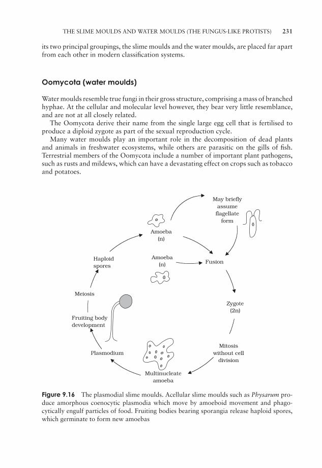
JWBK011-09 JWBK011-Hogg August 12, 2005 19:58 Char Count= 0
THE SLIME MOULDS AND WATER MOULDS (THE FUNGUS-LIKE PROTISTS) 231
its two principal groupings, the slime moulds and the water moulds, are placed far apartfrom each other in modern classification systems.
Oomycota (water moulds)
Water moulds resemble true fungi in their gross structure, comprising a mass of branchedhyphae. At the cellular and molecular level however, they bear very little resemblance,and are not at all closely related.
The Oomycota derive their name from the single large egg cell that is fertilised toproduce a diploid zygote as part of the sexual reproduction cycle.
Many water moulds play an important role in the decomposition of dead plantsand animals in freshwater ecosystems, while others are parasitic on the gills of fish.Terrestrial members of the Oomycota include a number of important plant pathogens,such as rusts and mildews, which can have a devastating effect on crops such as tobaccoand potatoes.
Fruiting bodydevelopment
Zygote(2n)
Fusion
Amoeba(n)
Amoeba(n)
May brieflyassumeflagellate
form
Meiosis
Multinucleateamoeba
Mitosiswithout cell
division
Haploidspores
Plasmodium
Figure 9.16 The plasmodial slime moulds. Acellular slime moulds such as Physarum pro-duce amorphous coenocytic plasmodia which move by amoeboid movement and phago-cytically engulf particles of food. Fruiting bodies bearing sporangia release haploid spores,which germinate to form new amoebas
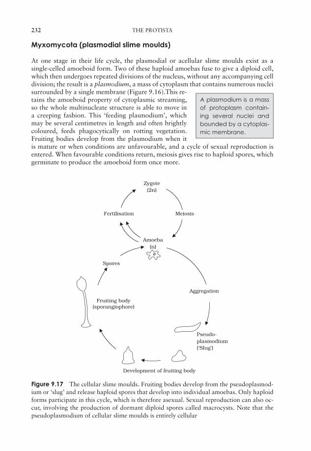
JWBK011-09 JWBK011-Hogg August 12, 2005 19:58 Char Count= 0
232 THE PROTISTA
Myxomycota (plasmodial slime moulds)
At one stage in their life cycle, the plasmodial or acellular slime moulds exist as asingle-celled amoeboid form. Two of these haploid amoebas fuse to give a diploid cell,which then undergoes repeated divisions of the nucleus, without any accompanying celldivision; the result is a plasmodium, a mass of cytoplasm that contains numerous nuclei
A plasmodium is a massof protoplasm contain-ing several nuclei andbounded by a cytoplas-mic membrane.
surrounded by a single membrane (Figure 9.16).This re-tains the amoeboid property of cytoplasmic streaming,so the whole multinucleate structure is able to move ina creeping fashion. This ‘feeding plasmodium’, whichmay be several centimetres in length and often brightlycoloured, feeds phagocytically on rotting vegetation.Fruiting bodies develop from the plasmodium when itis mature or when conditions are unfavourable, and a cycle of sexual reproduction isentered. When favourable conditions return, meiosis gives rise to haploid spores, whichgerminate to produce the amoeboid form once more.
Fertilisation
Zygote(2n)
Meiosis
Amoeba(n)
Aggregation
Pseudo-plasmodium(‘Slug’)
Development of fruiting body
Fruiting body (sporangiophore)
Spores
Figure 9.17 The cellular slime moulds. Fruiting bodies develop from the pseudoplasmod-ium or ‘slug’ and release haploid spores that develop into individual amoebas. Only haploidforms participate in this cycle, which is therefore asexual. Sexual reproduction can also oc-cur, involving the production of dormant diploid spores called macrocysts. Note that thepseudoplasmodium of cellular slime moulds is entirely cellular
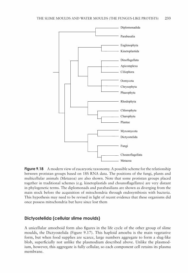
JWBK011-09 JWBK011-Hogg August 12, 2005 19:58 Char Count= 0
THE SLIME MOULDS AND WATER MOULDS (THE FUNGUS-LIKE PROTISTS) 233
Diplomonadida
Parabasalia
Euglenophyta
Kinetoplastida
Dinoflagellata
Apicomplexa
Ciliophora
Oomycota
Chrysophyta
Phaeophyta
Rhodophyta
Chlorophyta
Charophyta
Plantae
Myxomycota
Dictyostelida
Fungi
Choanoflagellata
Metazoa
Figure 9.18 A modern view of eucaryotic taxonomy. A possible scheme for the relationshipbetween protistan groups based on 18S RNA data. The positions of the fungi, plants andmulticellular animals (Metazoa) are also shown. Note that some protistan groups placedtogether in traditional schemes (e.g. kinetoplastids and choanoflagellates) are very distantin phylogenetic terms. The diplomonads and parabasilians are shown as diverging from themain stock before the acquisition of mitochondria through endosymbiosis with bacteria.This hypothesis may need to be revised in light of recent evidence that these organisms didonce possess mitochondria but have since lost them
Dictyostelida (cellular slime moulds)
A unicellular amoeboid form also figures in the life cycle of the other group of slimemoulds, the Dictyostelida (Figure 9.17). This haploid amoeba is the main vegetativeform, but when food supplies are scarce, large numbers aggregate to form a slug-likeblob, superficially not unlike the plasmodium described above. Unlike the plasmod-ium, however, this aggregate is fully cellular, so each component cell retains its plasmamembrane.
JWBK011-09 JWBK011-Hogg August 12, 2005 19:58 Char Count= 0
234 THE PROTISTA
Compare the life cycle of cellular slime moulds (Figure 9.17) with that of the plas-modial kind (Figure 9.16). Fruiting bodies again develop, giving rise to spores thatgerminate into new amoebas. No meiosis step is required, however, because the wholecycle comprises haploid forms, and this is therefore a form of asexual reproduction. Asimple sexual cycle may also occur, when haploid amoebas fuse to give a diploid zygote.
Protistan taxonomy: a modern view
Traditional classification of the protists was on the basis of physical features such asthe possession of flagella, chloroplasts and other structures. This had the effect of plac-ing into separate groups organisms that, at the molecular level, are closely related. Thekinetoplastids and the euglenophytes, for example, although both flagellate forms, weredistantly separated because the latter contained chloroplasts. They are now known tobe closely related in phylogenetic terms (Figure 9.18). On the other hand, the kineto-plastids were previously placed together with choanoflagellates as zooflagellates, butmolecular analysis now shows the two groups to have little in common in evolutionaryterms.
Test yourself
1 In nutritional terms, all algae are .
2 Although the Cyanophyta carry out oxygenic photosynthesis, they are notclassified with the algae, because they are ———.
3 Euglenophytes store their carbohydrate in an unusual form called .
4 The is a reddish structure situated near the flagellum,and is responsible for reacting to light stimuli.
5 Members of the Pyrrophyta are more commonly known as the .
6 Diatoms are surrounded by a made of silica, which is perforatedby numerous .
7 During asexual reproduction in the diatoms, one daughter cell at each divisiongets progressively smaller. This is stopped by entering a phase of
.
8 The group of algae thought to be phylogenetically the closest to the greenplants is the .
9 Multicellular algae are not differentiated into root, stem and leaf; their veg-etative body form is called a .
JWBK011-09 JWBK011-Hogg August 12, 2005 19:58 Char Count= 0
TEST YOURSELF 235
10 Sexual reproduction involving clearly differentiated sperm and egg cells istermed .
11 Species which have reproductive structures of the two sexes on separate in-dividuals are said to be .
12 A alternation of generations involves morphologically distinctsporophyte and gametophyte adult phases.
13 The coloration of the red algae is due to and , whichmask the presence of .
14 Protozoans are a diverse group, which do not share a.
15 Osmoregulation in protozoans is carried out by the .
16 The kinetoplastids include , the causative agent of African sleepingsickness. They show little evolutionary relationship to the other group offlagellated protozoans, the .
17 such as Giardia lack conventional mitochondria.
18 Ciliates possess two kinds of .
19 The are amoebas that have a calcium-based shell.
20 The acellular slime moulds are characterised by the formation of a ,a mass of cytoplasm containing numerous nuclei.

JWBK011-09 JWBK011-Hogg August 12, 2005 19:58 Char Count= 0
236
JWBK011-10 JWBK011-Hogg August 12, 2005 19:20 Char Count= 0
10Viruses
In Chapter 1, we saw how, in the late 19th century, one disease after another, in plantsas well as in animals, was shown to have a bacterial cause. In 1892, however, the Rus-sian Dimitri Iwanowsky made a surprising discovery concerning the condition knownas tobacco mosaic disease. He showed that an extract from an infected leaf retainedthe ability to transmit the disease to another plant even after being passed through aporcelain filter. This recently developed device was believed to remove even the smallestbacteria, and it was therefore proposed that perhaps the cause of the disease was not anorganism, but a filterable toxin. The work of the Dutch botanist Martinus Beijerinckand others around the turn of the century, however, (see Table 10.1) led to the idea ofviruses, filterable entities much smaller than bacteria, that were responsible for a widerange of diseases in plants, animals and members of the microbial world.
What are viruses?
All viruses are obligate intracellular parasites; they inhabit a no-man’s-land betweenthe living and the non-living worlds, and possess characteristics of both. They are nowknown to differ radically from the simplest true organisms, bacteria, in a number ofrespects:
� they cannot be observed using a light microscope
� they have no internal cellular structure
� they contain either DNA or RNA, but not both*
� they are incapable of replication unless occupying an appropriate living hostcell
� they are incapable of metabolism
� individuals show no increase in size.
When inside a host cell, viruses show some of the features of a living organism, suchas the ability to replicate themselves, but outside the cell they are just inert chemical
* Some viruses have DNA and RNA at different phases of their growth cycle. See p. 253
237
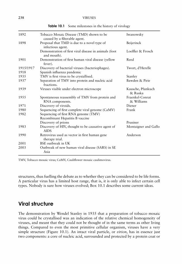
JWBK011-10 JWBK011-Hogg August 12, 2005 19:20 Char Count= 0
238 VIRUSES
Table 10.1 Some milestones in the history of virology
1892 Tobacco Mosaic Disease (TMD) shown to becaused by a filterable agent.
Iwanowsky
1898 Proposal that TMD is due to a novel type ofinfectious agent.
Beijerinck
Demonstration of first viral disease in animals (footand mouth).
Loeffler & Frosch
1901 Demonstration of first human viral disease (yellowfever).
Reed
1915/1917 Discovery of bacterial viruses (bacteriophages). Twort, d’Herelle1918 Spanish influenza pandemic1935 TMV is first virus to be crystallised. Stanley1937 Separation of TMV into protein and nucleic acid
fractions.Bawden & Pirie
1939 Viruses visible under electron microscope Kausche, Pfankuch& Ruska
1955 Spontaneous reassembly of TMV from protein andRNA components.
Fraenkel-Conrat& Williams
1971 Discovery of viroids. Diener1980 Sequencing of first complete viral genome (CaMV) Frank1982 Sequencing of first RNA genome (TMV)
Recombinant Hepatitis B vaccineDiscovery of prions Prusiner
1983 Discovery of HIV, thought to be causative agent ofAIDS
Montaigner and Gallo
1990 Retrovirus used as vector in first human genetherapy trial.
Anderson
2001 BSE outbreak in UK2003 Outbreak of new human viral disease (SARS) in SE
Asia
TMV, Tobacco mosaic virus; CaMV, Cauliflower mosaic caulimovirus.
structures, thus fuelling the debate as to whether they can be considered to be life forms.A particular virus has a limited host range, that is, it is only able to infect certain celltypes. Nobody is sure how viruses evolved; Box 10.1 describes some current ideas.
Viral structure
The demonstration by Wendel Stanley in 1935 that a preparation of tobacco mosaicvirus could be crystallised was an indication of the relative chemical homogeneity ofviruses, and meant that they could not be thought of in the same terms as other livingthings. Compared to even the most primitive cellular organism, viruses have a verysimple structure (Figure 10.1). An intact viral particle, or virion, has in essence justtwo components: a core of nucleic acid, surrounded and protected by a protein coat or
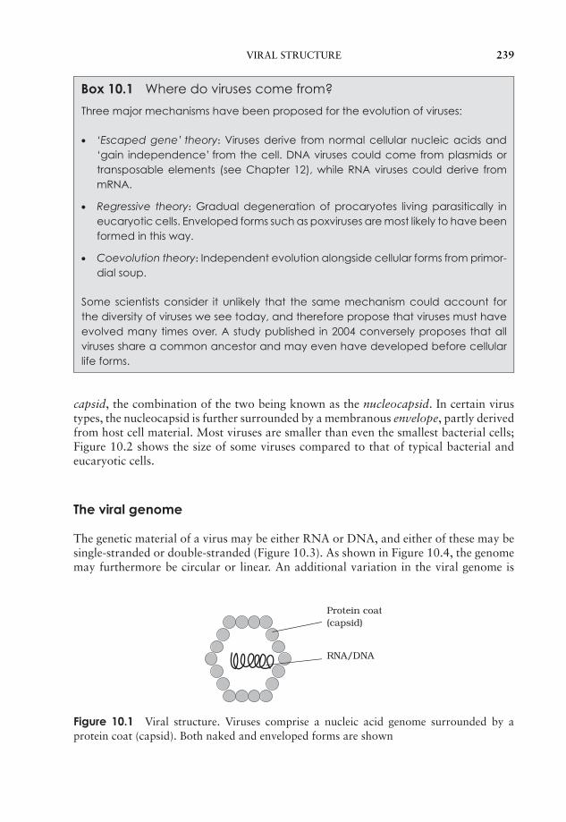
JWBK011-10 JWBK011-Hogg August 12, 2005 19:20 Char Count= 0
VIRAL STRUCTURE 239
Box 10.1 Where do viruses come from?
Three major mechanisms have been proposed for the evolution of viruses:
� ‘Escaped gene’ theory: Viruses derive from normal cellular nucleic acids and‘gain independence’ from the cell. DNA viruses could come from plasmids ortransposable elements (see Chapter 12), while RNA viruses could derive frommRNA.
� Regressive theory: Gradual degeneration of procaryotes living parasitically ineucaryotic cells. Enveloped forms such as poxviruses are most likely to have beenformed in this way.
� Coevolution theory: Independent evolution alongside cellular forms from primor-dial soup.
Some scientists consider it unlikely that the same mechanism could account forthe diversity of viruses we see today, and therefore propose that viruses must haveevolved many times over. A study published in 2004 conversely proposes that allviruses share a common ancestor and may even have developed before cellularlife forms.
capsid, the combination of the two being known as the nucleocapsid. In certain virustypes, the nucleocapsid is further surrounded by a membranous envelope, partly derivedfrom host cell material. Most viruses are smaller than even the smallest bacterial cells;Figure 10.2 shows the size of some viruses compared to that of typical bacterial andeucaryotic cells.
The viral genome
The genetic material of a virus may be either RNA or DNA, and either of these may besingle-stranded or double-stranded (Figure 10.3). As shown in Figure 10.4, the genomemay furthermore be circular or linear. An additional variation in the viral genome is
RNA/DNA
Protein coat(capsid)
Figure 10.1 Viral structure. Viruses comprise a nucleic acid genome surrounded by aprotein coat (capsid). Both naked and enveloped forms are shown
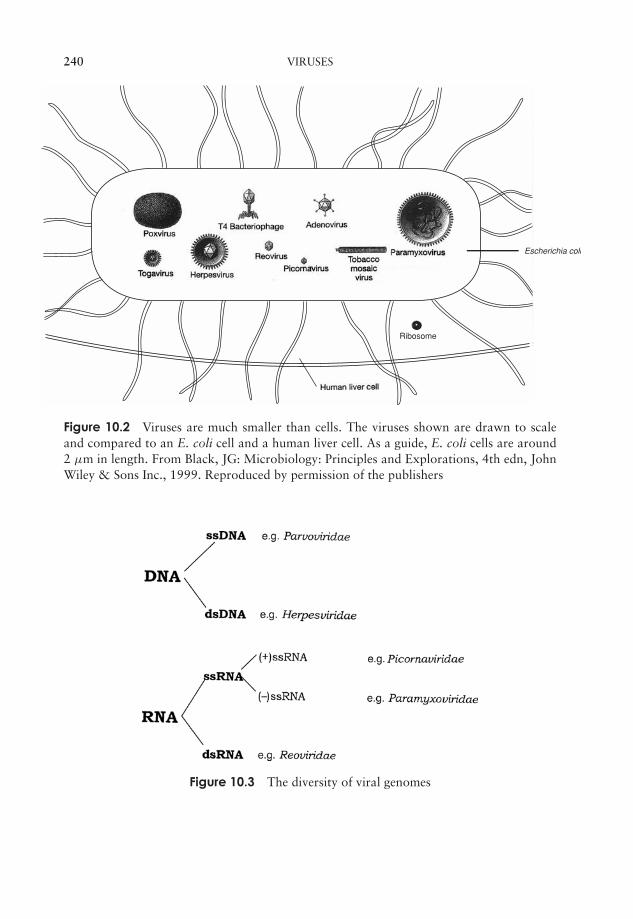
JWBK011-10 JWBK011-Hogg August 12, 2005 19:20 Char Count= 0
240 VIRUSES
Escherichia coli
Ribosome
Figure 10.2 Viruses are much smaller than cells. The viruses shown are drawn to scaleand compared to an E. coli cell and a human liver cell. As a guide, E. coli cells are around2 µm in length. From Black, JG: Microbiology: Principles and Explorations, 4th edn, JohnWiley & Sons Inc., 1999. Reproduced by permission of the publishers
Figure 10.3 The diversity of viral genomes
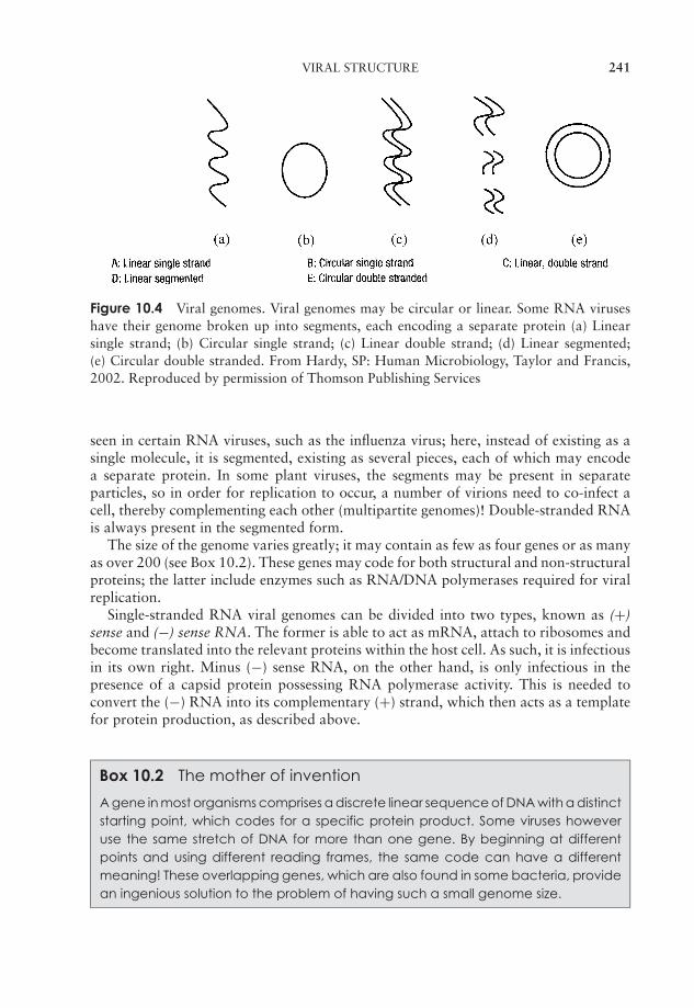
JWBK011-10 JWBK011-Hogg August 12, 2005 19:20 Char Count= 0
VIRAL STRUCTURE 241
Figure 10.4 Viral genomes. Viral genomes may be circular or linear. Some RNA viruseshave their genome broken up into segments, each encoding a separate protein (a) Linearsingle strand; (b) Circular single strand; (c) Linear double strand; (d) Linear segmented;(e) Circular double stranded. From Hardy, SP: Human Microbiology, Taylor and Francis,2002. Reproduced by permission of Thomson Publishing Services
seen in certain RNA viruses, such as the influenza virus; here, instead of existing as asingle molecule, it is segmented, existing as several pieces, each of which may encodea separate protein. In some plant viruses, the segments may be present in separateparticles, so in order for replication to occur, a number of virions need to co-infect acell, thereby complementing each other (multipartite genomes)! Double-stranded RNAis always present in the segmented form.
The size of the genome varies greatly; it may contain as few as four genes or as manyas over 200 (see Box 10.2). These genes may code for both structural and non-structuralproteins; the latter include enzymes such as RNA/DNA polymerases required for viralreplication.
Single-stranded RNA viral genomes can be divided into two types, known as (+)sense and (−) sense RNA. The former is able to act as mRNA, attach to ribosomes andbecome translated into the relevant proteins within the host cell. As such, it is infectiousin its own right. Minus (−) sense RNA, on the other hand, is only infectious in thepresence of a capsid protein possessing RNA polymerase activity. This is needed toconvert the (−) RNA into its complementary (+) strand, which then acts as a templatefor protein production, as described above.
Box 10.2 The mother of invention
A gene in most organisms comprises a discrete linear sequence of DNA with a distinctstarting point, which codes for a specific protein product. Some viruses howeveruse the same stretch of DNA for more than one gene. By beginning at differentpoints and using different reading frames, the same code can have a differentmeaning! These overlapping genes, which are also found in some bacteria, providean ingenious solution to the problem of having such a small genome size.
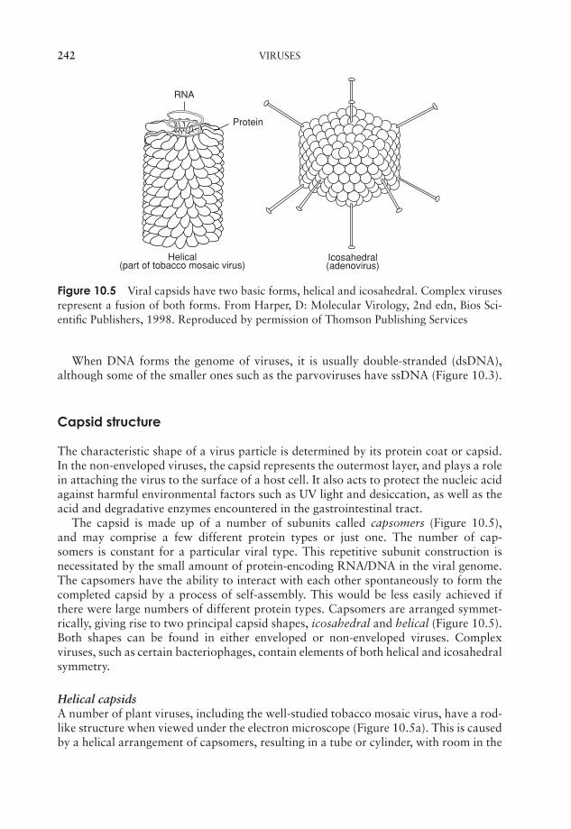
JWBK011-10 JWBK011-Hogg August 12, 2005 19:20 Char Count= 0
242 VIRUSES
Icosahedral (adenovirus)
Helical(part of tobacco mosaic virus)
RNA
Protein
Figure 10.5 Viral capsids have two basic forms, helical and icosahedral. Complex virusesrepresent a fusion of both forms. From Harper, D: Molecular Virology, 2nd edn, Bios Sci-entific Publishers, 1998. Reproduced by permission of Thomson Publishing Services
When DNA forms the genome of viruses, it is usually double-stranded (dsDNA),although some of the smaller ones such as the parvoviruses have ssDNA (Figure 10.3).
Capsid structure
The characteristic shape of a virus particle is determined by its protein coat or capsid.In the non-enveloped viruses, the capsid represents the outermost layer, and plays a rolein attaching the virus to the surface of a host cell. It also acts to protect the nucleic acidagainst harmful environmental factors such as UV light and desiccation, as well as theacid and degradative enzymes encountered in the gastrointestinal tract.
The capsid is made up of a number of subunits called capsomers (Figure 10.5),and may comprise a few different protein types or just one. The number of cap-somers is constant for a particular viral type. This repetitive subunit construction isnecessitated by the small amount of protein-encoding RNA/DNA in the viral genome.The capsomers have the ability to interact with each other spontaneously to form thecompleted capsid by a process of self-assembly. This would be less easily achieved ifthere were large numbers of different protein types. Capsomers are arranged symmet-rically, giving rise to two principal capsid shapes, icosahedral and helical (Figure 10.5).Both shapes can be found in either enveloped or non-enveloped viruses. Complexviruses, such as certain bacteriophages, contain elements of both helical and icosahedralsymmetry.
Helical capsidsA number of plant viruses, including the well-studied tobacco mosaic virus, have a rod-like structure when viewed under the electron microscope (Figure 10.5a). This is causedby a helical arrangement of capsomers, resulting in a tube or cylinder, with room in the

JWBK011-10 JWBK011-Hogg August 12, 2005 19:20 Char Count= 0
CLASSIFICATION OF VIRUSES 243
centre for the nucleic acid element, which fits into a groove on the inside. The diameterof the helix is determined by the nature of the protein(s) making up the capsomers; itslength depends on the size of the nucleic acid core.
Icosahedral capsids The icosahedron has alow surface-area to vol-ume ratio, allowing forthe maximum amount ofnucleic acid to be pack-aged.
An icosahedron is a regular three-dimensional shapewith 20 triangular faces, and 12 points or corners (Fig-ure 10.5). The overall effect is of a roughly sphericalstructure.
The viral envelope
Envelopes are much more common in animal viruses than in those of plants. The lipidbilayer covering an enveloped virus is derived from the nuclear or cytoplasmic membraneof a previous host. Embedded in this, however, are proteins (usually glycoproteins)encoded by the virus’s own genome. These may project from the surface of the virion asspikes, which may be instrumental in allowing the virus to bind to or penetrate its hostcell (Figure 10.6). The envelope is more susceptible than the capsid to environmentalpressures, and the virus needs to remain moist in order to survive. Consequently, suchviruses are transmitted by means of body fluids such as blood (e.g. hepatitis B virus) orrespiratory secretions (e.g. influenza virus).
Classification of viruses
As we saw at the beginning of this chapter, viruses are not considered to be strictlyliving, and their classification is a complex issue. As with true organisms we havespecies, genera, families and orders of viruses, but none of the higher groupings (class,phylum, kingdom). Latin binomials (e.g. Homo sapiens, Escherichia coli), familiar
RNA/DNA Envelope
Spike
Protein coat(capsid)
Figure 10.6 An enveloped virus. The envelope derives from the host cell’s membrane, andincludes virus-encoded proteins. Some viral envelopes contain projecting spikes, which mayassist in attachment to the host
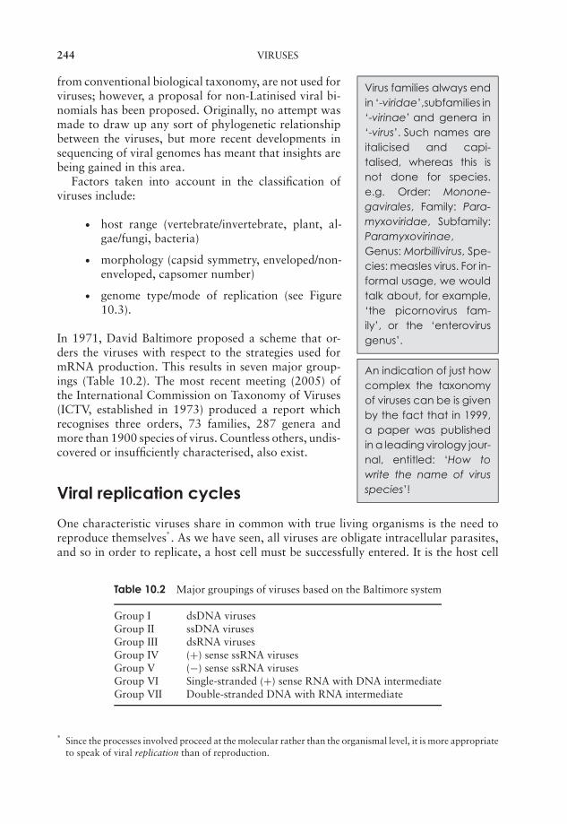
JWBK011-10 JWBK011-Hogg August 12, 2005 19:20 Char Count= 0
244 VIRUSES
Virus families always endin ‘-viridae’,subfamilies in‘-virinae’ and genera in‘-virus’. Such names areitalicised and capi-talised, whereas this isnot done for species.e.g. Order: Monone-gavirales, Family: Para-myxoviridae, Subfamily:Paramyxovirinae,Genus: Morbillivirus, Spe-cies: measles virus. For in-formal usage, we wouldtalk about, for example,‘the picornovirus fam-ily’, or the ‘enterovirusgenus’.
from conventional biological taxonomy, are not used forviruses; however, a proposal for non-Latinised viral bi-nomials has been proposed. Originally, no attempt wasmade to draw up any sort of phylogenetic relationshipbetween the viruses, but more recent developments insequencing of viral genomes has meant that insights arebeing gained in this area.
Factors taken into account in the classification ofviruses include:
� host range (vertebrate/invertebrate, plant, al-gae/fungi, bacteria)
� morphology (capsid symmetry, enveloped/non-enveloped, capsomer number)
� genome type/mode of replication (see Figure10.3).
An indication of just howcomplex the taxonomyof viruses can be is givenby the fact that in 1999,a paper was publishedin a leading virology jour-nal, entitled: ‘How towrite the name of virusspecies’!
In 1971, David Baltimore proposed a scheme that or-ders the viruses with respect to the strategies used formRNA production. This results in seven major group-ings (Table 10.2). The most recent meeting (2005) ofthe International Commission on Taxonomy of Viruses(ICTV, established in 1973) produced a report whichrecognises three orders, 73 families, 287 genera andmore than 1900 species of virus. Countless others, undis-covered or insufficiently characterised, also exist.
Viral replication cycles
One characteristic viruses share in common with true living organisms is the need toreproduce themselves
∗. As we have seen, all viruses are obligate intracellular parasites,
and so in order to replicate, a host cell must be successfully entered. It is the host cell
Table 10.2 Major groupings of viruses based on the Baltimore system
Group I dsDNA virusesGroup II ssDNA virusesGroup III dsRNA virusesGroup IV (+) sense ssRNA virusesGroup V (−) sense ssRNA virusesGroup VI Single-stranded (+) sense RNA with DNA intermediateGroup VII Double-stranded DNA with RNA intermediate
∗Since the processes involved proceed at the molecular rather than the organismal level, it is more appropriateto speak of viral replication than of reproduction.
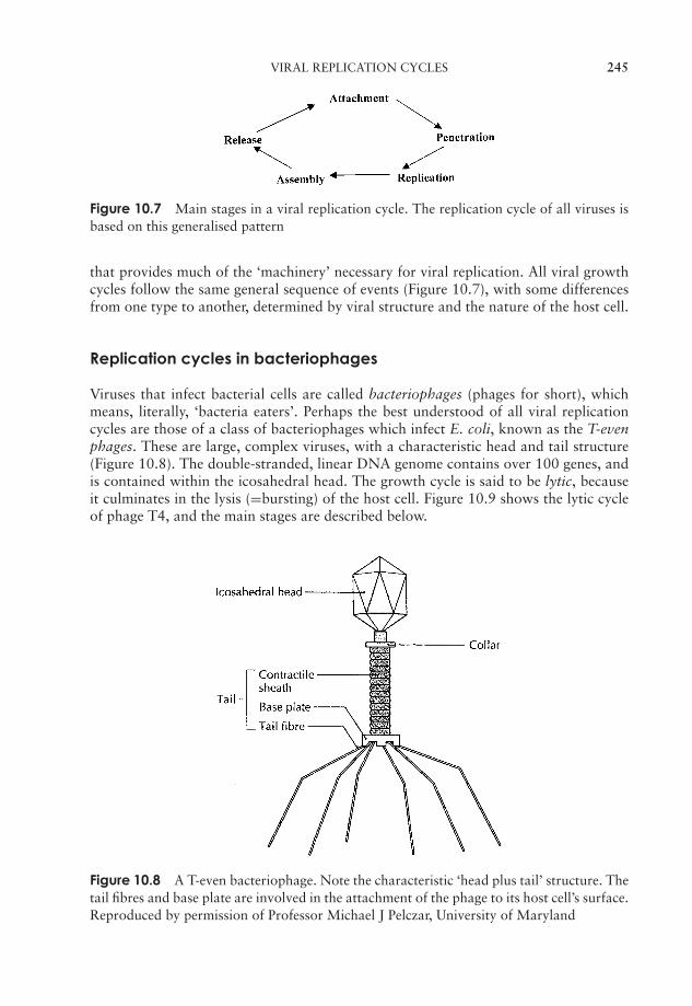
JWBK011-10 JWBK011-Hogg August 12, 2005 19:20 Char Count= 0
VIRAL REPLICATION CYCLES 245
Figure 10.7 Main stages in a viral replication cycle. The replication cycle of all viruses isbased on this generalised pattern
that provides much of the ‘machinery’ necessary for viral replication. All viral growthcycles follow the same general sequence of events (Figure 10.7), with some differencesfrom one type to another, determined by viral structure and the nature of the host cell.
Replication cycles in bacteriophages
Viruses that infect bacterial cells are called bacteriophages (phages for short), whichmeans, literally, ‘bacteria eaters’. Perhaps the best understood of all viral replicationcycles are those of a class of bacteriophages which infect E. coli, known as the T-evenphages. These are large, complex viruses, with a characteristic head and tail structure(Figure 10.8). The double-stranded, linear DNA genome contains over 100 genes, andis contained within the icosahedral head. The growth cycle is said to be lytic, becauseit culminates in the lysis (=bursting) of the host cell. Figure 10.9 shows the lytic cycleof phage T4, and the main stages are described below.
Figure 10.8 A T-even bacteriophage. Note the characteristic ‘head plus tail’ structure. Thetail fibres and base plate are involved in the attachment of the phage to its host cell’s surface.Reproduced by permission of Professor Michael J Pelczar, University of Maryland

JWBK011-10 JWBK011-Hogg August 12, 2005 19:20 Char Count= 0
246 VIRUSES
‘Early’proteinsynthesis(phageenzymes)
Host DNAdegrades
3. ReplicationSynthesis of‘late’ structuralproteins
4. Assembly
1. Attachment
2. Penetration
5. Release
Release of newphage particles
Injection ofphage DNA
PhageDNA
Host chromosome
Lysis of host
Figure 10.9 The lytic cycle of phage T4. The cycle comprises the five main stages describedin the text; from injection of phage of DNA to cell lysis takes 22 minutes. The number ofphage particles released per cell is called the burst size, and for T4 it ranges from 50 to 200
You might reasonablyask yourself why cellswould evolve recep-tor molecules for viruses,when the outcome isclearly not in theirinterests. The answer is, ofcourse, that theyhaven’t; the receptorshave other biologicalproperties, but theviruses have ‘taken themover’.
1 Adsorption (attachment): T4 attaches by means ofspecific tail fibre proteins to complementary recep-tors on the host cell’s surface. The nature of thesereceptors is one of the main factors in determining avirus’s host specificity.
2 Penetration: The enzyme lysozyme, present in thetail of the phage, weakens the cell wall at the pointof attachment, and a contraction of the tail sheathof the phage causes the core to be pushed downinto the cell, releasing the viral DNA into the in-terior of the bacterium. The capsid remains en-tirely outside the cell, as elegantly demonstrated inthe famous experiment by Hershey and Chase (seeChapter 11).
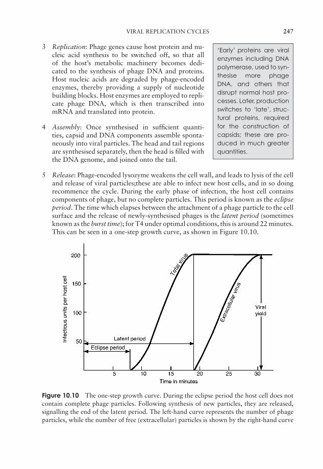
JWBK011-10 JWBK011-Hogg August 12, 2005 19:20 Char Count= 0
VIRAL REPLICATION CYCLES 247
‘Early’ proteins are viralenzymes including DNApolymerase, used to syn-thesise more phageDNA, and others thatdisrupt normal host pro-cesses. Later, productionswitches to ‘late’, struc-tural proteins, requiredfor the construction ofcapsids; these are pro-duced in much greaterquantities.
3 Replication: Phage genes cause host protein and nu-cleic acid synthesis to be switched off, so that allof the host’s metabolic machinery becomes dedi-cated to the synthesis of phage DNA and proteins.Host nucleic acids are degraded by phage-encodedenzymes, thereby providing a supply of nucleotidebuilding blocks. Host enzymes are employed to repli-cate phage DNA, which is then transcribed intomRNA and translated into protein.
4 Assembly: Once synthesised in sufficient quanti-ties, capsid and DNA components assemble sponta-neously into viral particles. The head and tail regionsare synthesised separately, then the head is filled withthe DNA genome, and joined onto the tail.
5 Release: Phage-encoded lysozyme weakens the cell wall, and leads to lysis of the celland release of viral particles;these are able to infect new host cells, and in so doingrecommence the cycle. During the early phase of infection, the host cell containscomponents of phage, but no complete particles. This period is known as the eclipseperiod. The time which elapses between the attachment of a phage particle to the cellsurface and the release of newly-synthesised phages is the latent period (sometimesknown as the burst time); for T4 under optimal conditions, this is around 22 minutes.This can be seen in a one-step growth curve, as shown in Figure 10.10.
Figure 10.10 The one-step growth curve. During the eclipse period the host cell does notcontain complete phage particles. Following synthesis of new particles, they are released,signalling the end of the latent period. The left-hand curve represents the number of phageparticles, while the number of free (extracellular) particles is shown by the right-hand curve
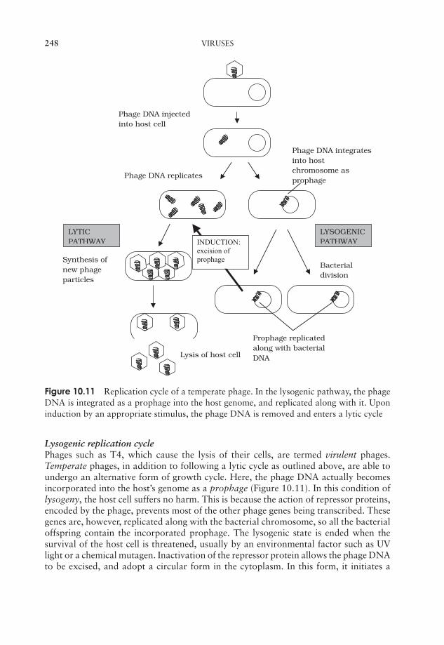
JWBK011-10 JWBK011-Hogg August 12, 2005 19:20 Char Count= 0
248 VIRUSES
Bacterialdivision
Prophage replicatedalong with bacterialDNA
Phage DNA injectedinto host cell
LYSOGENICPATHWAY
Phage DNA integratesinto hostchromosome asprophage
LYTICPATHWAY
Phage DNA replicates
Lysis of host cell
Synthesis ofnew phageparticles
INDUCTION:excision ofprophage
Figure 10.11 Replication cycle of a temperate phage. In the lysogenic pathway, the phageDNA is integrated as a prophage into the host genome, and replicated along with it. Uponinduction by an appropriate stimulus, the phage DNA is removed and enters a lytic cycle
Lysogenic replication cyclePhages such as T4, which cause the lysis of their cells, are termed virulent phages.Temperate phages, in addition to following a lytic cycle as outlined above, are able toundergo an alternative form of growth cycle. Here, the phage DNA actually becomesincorporated into the host’s genome as a prophage (Figure 10.11). In this condition oflysogeny, the host cell suffers no harm. This is because the action of repressor proteins,encoded by the phage, prevents most of the other phage genes being transcribed. Thesegenes are, however, replicated along with the bacterial chromosome, so all the bacterialoffspring contain the incorporated prophage. The lysogenic state is ended when thesurvival of the host cell is threatened, usually by an environmental factor such as UVlight or a chemical mutagen. Inactivation of the repressor protein allows the phage DNAto be excised, and adopt a circular form in the cytoplasm. In this form, it initiates a
JWBK011-10 JWBK011-Hogg August 12, 2005 19:20 Char Count= 0
VIRAL REPLICATION CYCLES 249
lytic cycle, resulting in destruction of the host cell. An example of a temperate phage isbacteriophage λ (Lambda), which infects certain strains of E. coli. Bacterial strains thatcan incorporate phage DNA in this way are termed lysogens.
Replication cycles in animal viruses
Viruses that infect multicellular organisms such as animals may be specific not only toa particular organism, but also to a particular cell or tissue type. This is known as thetissue tropism of the virus, and is due to the fact that attachment occurs via specificreceptors on the host cell surface.
The growth cycles of animal viruses have the same main stages as described forbacteriophages (Figure 10.7), but may differ a good deal in some of the details. Most ofthese variations are a reflection of differences in structure between bacterial and animalhost cells.
Adsorption and penetrationAnimal viruses do not have the head and tail structure of phages, so it follows thattheir method of attachment is different. The specific interaction with a host receptor ismade via some component of the capsid, or, in the case of enveloped viruses, by specialstructures such as spikes (peplomers). Viral attachment sites can frequently be blockedby host antibody molecules; however some viruses (e.g. the rhinoviruses) have overcomethis by having their sites situated in deep depressions, inaccessible to the antibodies.
Whereas bacteriophages inject their nucleic acid component from the outside, theprocess in animal viruses is more complex, a fact reflected in the time taken for com-pletion of the process. Animal viruses do not have to cope with a thick cell wall, andin many such cases the entire virion is internalised. This necessitates the extra step ofuncoating, a process carried out by host enzymes. Many animal viruses possess an en-velope; such viruses are taken into the cell either by fusion with the cell membrane, orby endocytosis (Figure 10.12). While some non-enveloped types release only their nu-cleic acid component into the cytoplasm, others require additionally that virus-encodedenzymes be introduced to ensure successful replication.
Replication (DNA viruses)The DNA of animal cells, unlike that of bacteria, is compartmentalised within a nucleus,and it is here that replication and transcription of viral DNA generally occur
∗. Messenger
RNA then passes to ribosomes in the cytoplasm for translation (Figure 10.13). In thecase of viruses with a ssDNA genome, a double-stranded intermediate is formed, whichserves as a template for mRNA synthesis.
AssemblyTranslation products are finally returned to the nucleus for assembly into new virusparticles.
∗Poxviruses are an exception. Both replication and assembly occur in the cytoplasm.
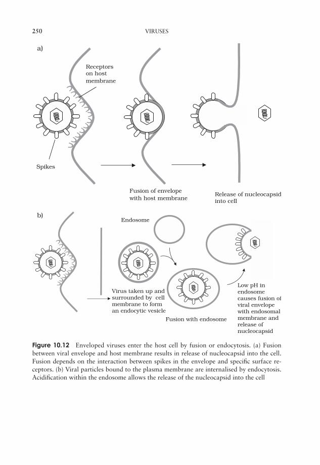
JWBK011-10 JWBK011-Hogg August 12, 2005 19:20 Char Count= 0
250 VIRUSES
Receptorson hostmembrane
Spikes
Fusion of envelopewith host membrane
Release of nucleocapsidinto cell
a)
Endosome
Fusion with endosome
Low pH in endosomecauses fusion ofviral envelope with endosomal membrane and release of nucleocapsid
Virus taken up and surrounded by cell membrane to form an endocytic vesicle
b)
Figure 10.12 Enveloped viruses enter the host cell by fusion or endocytosis. (a) Fusionbetween viral envelope and host membrane results in release of nucleocapsid into the cell.Fusion depends on the interaction between spikes in the envelope and specific surface re-ceptors. (b) Viral particles bound to the plasma membrane are internalised by endocytosis.Acidification within the endosome allows the release of the nucleocapsid into the cell
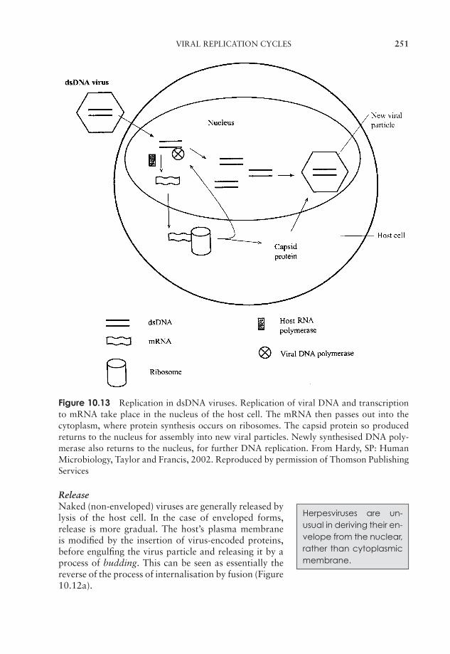
JWBK011-10 JWBK011-Hogg August 12, 2005 19:20 Char Count= 0
VIRAL REPLICATION CYCLES 251
Figure 10.13 Replication in dsDNA viruses. Replication of viral DNA and transcriptionto mRNA take place in the nucleus of the host cell. The mRNA then passes out into thecytoplasm, where protein synthesis occurs on ribosomes. The capsid protein so producedreturns to the nucleus for assembly into new viral particles. Newly synthesised DNA poly-merase also returns to the nucleus, for further DNA replication. From Hardy, SP: HumanMicrobiology, Taylor and Francis, 2002. Reproduced by permission of Thomson PublishingServices
Release
Herpesviruses are un-usual in deriving their en-velope from the nuclear,rather than cytoplasmicmembrane.
Naked (non-enveloped) viruses are generally released bylysis of the host cell. In the case of enveloped forms,release is more gradual. The host’s plasma membraneis modified by the insertion of virus-encoded proteins,before engulfing the virus particle and releasing it by aprocess of budding. This can be seen as essentially thereverse of the process of internalisation by fusion (Figure10.12a).
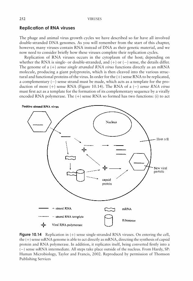
JWBK011-10 JWBK011-Hogg August 12, 2005 19:20 Char Count= 0
252 VIRUSES
Replication of RNA viruses
The phage and animal virus growth cycles we have described so far have all involveddouble-stranded DNA genomes. As you will remember from the start of this chapter,however, many viruses contain RNA instead of DNA as their genetic material, and wenow need to consider briefly how these viruses complete their replication cycles.
Replication of RNA viruses occurs in the cytoplasm of the host; depending onwhether the RNA is single- or double-stranded, and (+) or (−) sense, the details differ.The genome of a (+) sense single stranded RNA virus functions directly as an mRNAmolecule, producing a giant polyprotein, which is then cleaved into the various struc-tural and functional proteins of the virus. In order for the (+) sense RNA to be replicated,a complementary (−) sense strand must be made, which acts as a template for the pro-duction of more (+) sense RNA (Figure 10.14). The RNA of a (−) sense RNA virusmust first act as a template for the formation of its complementary sequence by a virallyencoded RNA polymerase. The (+) sense RNA so formed has two functions: (i) to act
Figure 10.14 Replication in (+) sense single-stranded RNA viruses. On entering the cell,the (+) sense ssRNA genome is able to act directly as mRNA, directing the synthesis of capsidprotein and RNA polymerase. In addition, it replicates itself, being converted firstly into a(−) sense ssRNA intermediate. All steps take place outside of the nucleus. From Hardy, SP:Human Microbiology, Taylor and Francis, 2002. Reproduced by permission of ThomsonPublishing Services
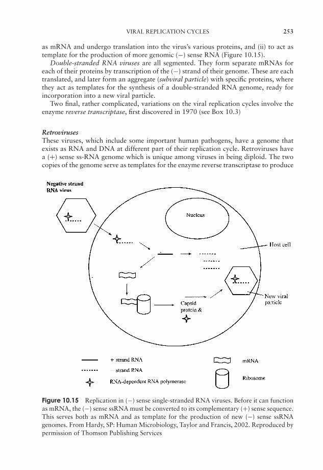
JWBK011-10 JWBK011-Hogg August 12, 2005 19:20 Char Count= 0
VIRAL REPLICATION CYCLES 253
as mRNA and undergo translation into the virus’s various proteins, and (ii) to act astemplate for the production of more genomic (−) sense RNA (Figure 10.15).
Double-stranded RNA viruses are all segmented. They form separate mRNAs foreach of their proteins by transcription of the (−) strand of their genome. These are eachtranslated, and later form an aggregate (subviral particle) with specific proteins, wherethey act as templates for the synthesis of a double-stranded RNA genome, ready forincorporation into a new viral particle.
Two final, rather complicated, variations on the viral replication cycles involve theenzyme reverse transcriptase, first discovered in 1970 (see Box 10.3)
RetrovirusesThese viruses, which include some important human pathogens, have a genome thatexists as RNA and DNA at different part of their replication cycle. Retroviruses havea (+) sense ss-RNA genome which is unique among viruses in being diploid. The twocopies of the genome serve as templates for the enzyme reverse transcriptase to produce
Figure 10.15 Replication in (−) sense single-stranded RNA viruses. Before it can functionas mRNA, the (−) sense ssRNA must be converted to its complementary (+) sense sequence.This serves both as mRNA and as template for the production of new (−) sense ssRNAgenomes. From Hardy, SP: Human Microbiology, Taylor and Francis, 2002. Reproduced bypermission of Thomson Publishing Services
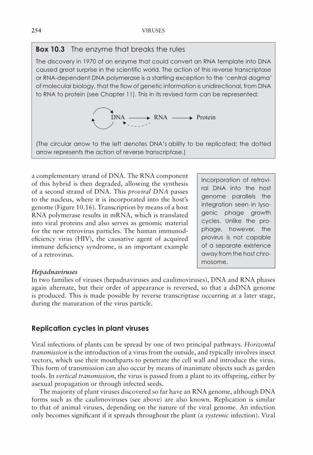
JWBK011-10 JWBK011-Hogg August 12, 2005 19:20 Char Count= 0
254 VIRUSES
Box 10.3 The enzyme that breaks the rules
The discovery in 1970 of an enzyme that could convert an RNA template into DNAcaused great surprise in the scientific world. The action of this reverse transcriptaseor RNA-dependent DNA polymerase is a startling exception to the ‘central dogma’of molecular biology, that the flow of genetic information is unidirectional, from DNAto RNA to protein (see Chapter 11). This in its revised form can be represented:
DNA RNA Protein
(The circular arrow to the left denotes DNA’s ability to be replicated; the dottedarrow represents the action of reverse transcriptase.)
Incorporation of retrovi-ral DNA into the hostgenome parallels theintegration seen in lyso-genic phage growthcycles. Unlike the pro-phage, however, theprovirus is not capableof a separate existenceaway from the host chro-mosome.
a complementary strand of DNA. The RNA componentof this hybrid is then degraded, allowing the synthesisof a second strand of DNA. This proviral DNA passesto the nucleus, where it is incorporated into the host’sgenome (Figure 10.16). Transcription by means of a hostRNA polymerase results in mRNA, which is translatedinto viral proteins and also serves as genomic materialfor the new retrovirus particles. The human immunod-eficiency virus (HIV), the causative agent of acquiredimmune deficiency syndrome, is an important exampleof a retrovirus.
HepadnavirusesIn two families of viruses (hepadnaviruses and caulimoviruses), DNA and RNA phasesagain alternate, but their order of appearance is reversed, so that a dsDNA genomeis produced. This is made possible by reverse transcriptase occurring at a later stage,during the maturation of the virus particle.
Replication cycles in plant viruses
Viral infections of plants can be spread by one of two principal pathways. Horizontaltransmission is the introduction of a virus from the outside, and typically involves insectvectors, which use their mouthparts to penetrate the cell wall and introduce the virus.This form of transmission can also occur by means of inanimate objects such as gardentools. In vertical transmission, the virus is passed from a plant to its offspring, either byasexual propagation or through infected seeds.
The majority of plant viruses discovered so far have an RNA genome, although DNAforms such as the caulimoviruses (see above) are also known. Replication is similarto that of animal viruses, depending on the nature of the viral genome. An infectiononly becomes significant if it spreads throughout the plant (a systemic infection). Viral
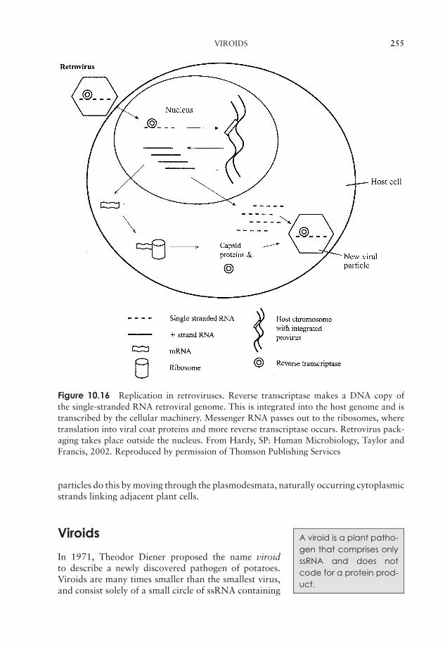
JWBK011-10 JWBK011-Hogg August 12, 2005 19:20 Char Count= 0
VIROIDS 255
Figure 10.16 Replication in retroviruses. Reverse transcriptase makes a DNA copy ofthe single-stranded RNA retroviral genome. This is integrated into the host genome and istranscribed by the cellular machinery. Messenger RNA passes out to the ribosomes, wheretranslation into viral coat proteins and more reverse transcriptase occurs. Retrovirus pack-aging takes place outside the nucleus. From Hardy, SP: Human Microbiology, Taylor andFrancis, 2002. Reproduced by permission of Thomson Publishing Services
particles do this by moving through the plasmodesmata, naturally occurring cytoplasmicstrands linking adjacent plant cells.
Viroids A viroid is a plant patho-gen that comprises onlyssRNA and does notcode for a protein prod-uct.
In 1971, Theodor Diener proposed the name viroidto describe a newly discovered pathogen of potatoes.Viroids are many times smaller than the smallest virus,and consist solely of a small circle of ssRNA containing
JWBK011-10 JWBK011-Hogg August 12, 2005 19:20 Char Count= 0
256 VIRUSES
some 300–400 nucleotide bases and no protein coat. Enzymes in the host’s nucleus areused to replicate the RNA, which does not appear to be translated into protein. Ap-preciable sequence homology suggests that viroids arose from transposable elements(see Chapter 11), segments of DNA capable of movement within or between DNAmolecules. To date, viroids have only been found in plants, where they cause a varietyof diseases.
Prions
A prion (=proteinaceousinfectious particle) is aself-replicating proteinresponsible for a rangeof neurodegenerativedisorders in humans andmammals.
A decade after the discovery of viroids, Stanley Prusinermade the startling claim that scrapie, a neurodegener-ative disease of sheep, was caused by a self-replicatingagent composed solely of protein. He called this type ofentity a prion, and in the years which followed, other,related, diseases of humans and animals were shown tohave a similar cause. These include bovine spongiformencephalopathy (BSE, ‘mad cow disease’) and its humanequivalent, Creutzfeldt–Jakob disease.
How could something that contains no nucleic acid be capable of replicating itself? –Prusiner’s idea seemed to go against the basic rules of biology. It appears that prions maybe altered versions of normal animal proteins, and somehow have the ability to causethe normal version to refold itself into the mutant form. Thus the prion propagates itself.All prion diseases described thus far are similar conditions, involving a degeneration ofbrain tissue.
Cultivating viruses
Whilst the growth of bacteria in the laboratory generally demands only a supply ofthe relevant nutrients and appropriate environmental conditions, maintaining virusespresents special challenges. Think back to the start of this chapter, and you will realisewhy this is so; all viruses are obligate intracellular parasites, and therefore need anappropriate host cell if they are to replicate.
Bacteriophages, for example, are grown in culture with their bacterial hosts. Stockcultures of phages are prepared by allowing them to infect a broth culture of the appro-priate bacterium. Successful propagation of phages results in a clearing of the culture’sturbidity; centrifugation removes any remaining bacteria, leaving the phage particles inthe supernatant. A quantitative measure of phages, known as the titre, can be obtainedby mixing them with a much greater number of bacteria and immobilising them in agar.Due to their numbers, the bacteria grow as a confluent lawn. Some become infectedby phage, and when new viral particles are released following lysis of their host, theyinfect more host cells. Because they are immobilised in agar, the phages are only able toinfect cells in the immediate vicinity. As more and more cells in the same area are lysed,an area of clearing called a plaque appears in the lawn of bacteria (Figure 10.17).Quantification is based on the assumption that each visible plaque arises from
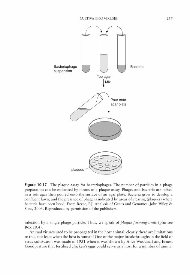
JWBK011-10 JWBK011-Hogg August 12, 2005 19:20 Char Count= 0
CULTIVATING VIRUSES 257
Top agar
Bacteria
plaques
Mix
Pour ontoagar plate
Bacteriophagesuspension
Figure 10.17 The plaque assay for bacteriophages. The number of particles in a phagepreparation can be estimated by means of a plaque assay. Phages and bacteria are mixedin a soft agar then poured onto the surface of an agar plate. Bacteria grow to develop aconfluent lawn, and the presence of phage is indicated by areas of clearing (plaques) wherebacteria have been lysed. From Reece, RJ: Analysis of Genes and Genomes, John Wiley &Sons, 2003. Reproduced by permission of the publishers
infection by a single phage particle. Thus, we speak of plaque-forming units (pfu: seeBox 10.4).
Animal viruses used to be propagated in the host animal; clearly there are limitationsto this, not least when the host is human! One of the major breakthroughs in the field ofvirus cultivation was made in 1931 when it was shown by Alice Woodruff and ErnestGoodpasture that fertilised chicken’s eggs could serve as a host for a number of animal
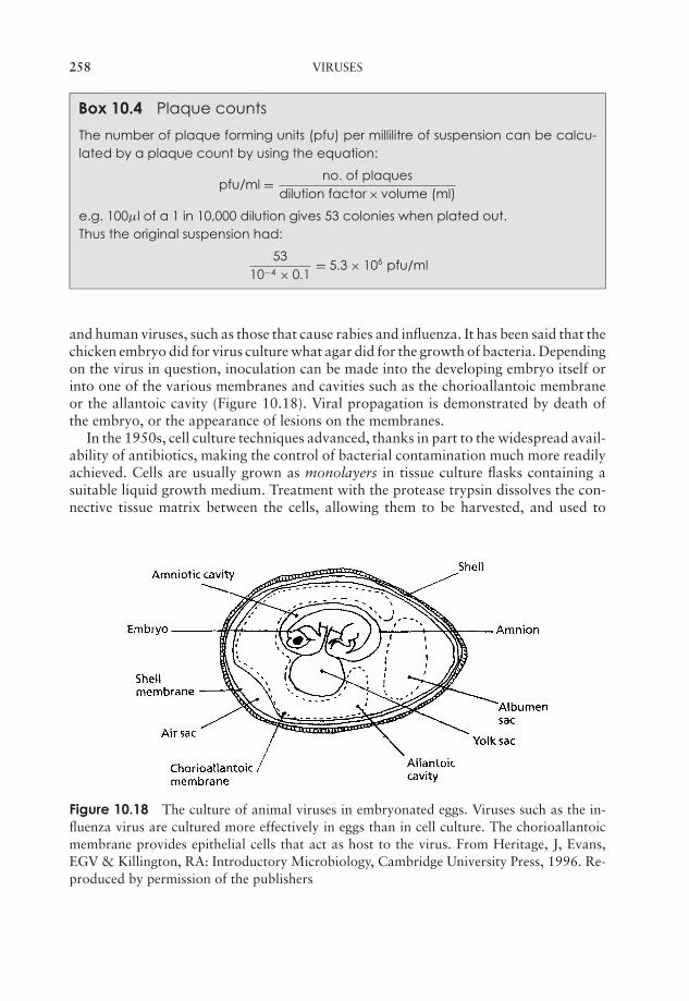
JWBK011-10 JWBK011-Hogg August 12, 2005 19:20 Char Count= 0
258 VIRUSES
Box 10.4 Plaque counts
The number of plaque forming units (pfu) per millilitre of suspension can be calcu-lated by a plaque count by using the equation:
pfu/ml = no. of plaquesdilution factor × volume (ml)
e.g. 100µl of a 1 in 10,000 dilution gives 53 colonies when plated out.Thus the original suspension had:
5310−4 × 0.1
= 5.3 × 10⁶ pfu/ml
and human viruses, such as those that cause rabies and influenza. It has been said that thechicken embryo did for virus culture what agar did for the growth of bacteria. Dependingon the virus in question, inoculation can be made into the developing embryo itself orinto one of the various membranes and cavities such as the chorioallantoic membraneor the allantoic cavity (Figure 10.18). Viral propagation is demonstrated by death ofthe embryo, or the appearance of lesions on the membranes.
In the 1950s, cell culture techniques advanced, thanks in part to the widespread avail-ability of antibiotics, making the control of bacterial contamination much more readilyachieved. Cells are usually grown as monolayers in tissue culture flasks containing asuitable liquid growth medium. Treatment with the protease trypsin dissolves the con-nective tissue matrix between the cells, allowing them to be harvested, and used to
Figure 10.18 The culture of animal viruses in embryonated eggs. Viruses such as the in-fluenza virus are cultured more effectively in eggs than in cell culture. The chorioallantoicmembrane provides epithelial cells that act as host to the virus. From Heritage, J, Evans,EGV & Killington, RA: Introductory Microbiology, Cambridge University Press, 1996. Re-produced by permission of the publishers

JWBK011-10 JWBK011-Hogg August 12, 2005 19:20 Char Count= 0
VIRAL DISEASES IN HUMANS 259
seed new cultures. Changes in cell morphology, known generically as cytopathic effects,are indicators of viral infection, and may be used diagnostically in the identification ofspecific viral types.
Plant viruses need to overcome the barrier presented by the cellulose cell wall of theplant; in nature this is often achieved by the piercing mouthparts of an insect vector orby entering areas of damaged tissue. Experimentally, viruses can be introduced into anappropriate host by rubbing the surface of a leaf with the virus together with a mildabrasive to create a minor wound.
Viral diseases in humans
Viruses are responsible for causing some of the most serious infectious diseases to affecthumans. Some important examples are listed in Table 10.3, and some are discussed ina little more detail below.
Airborne transmission: influenza
Influenza is a disease of the respiratory tract caused by members of the Orthomyx-oviridae. Transmission occurs as a result of inhaling airborne respiratory droplets froman infected individual. Infection by the influenza virus results in the destruction ofepithelial cells of the respiratory tract, leaving the host open to secondary infectionsfrom bacteria such as Haemophilus influenzae and Staphylococcus aureus. It is thesesecondary infections that are responsible for the great majority of fatalities caused byinfluenza. Generally, sufferers from influenza recover completely within 10–14 days, butsome people, notably the elderly and those with chronic health problems, may developcomplications such as pneumonia.
The influenza virus has an envelope, and a segmented (−) sense ssRNA genome(Figure 10.19). The envelope contains two types of protein spike, each of which playsa crucial role in the virus’s infectivity:
� Neuraminidase is an enzyme which hydrolyses sialic acid, thereby assisting inthe release of viral particles.
� Haemagglutinin enables the virus to attach to host cells by binding to epithelialsialic acid residues. It also helps in the fusion of the viral envelope with the cellmembrane.
Both types of spike act as antigens, proteins that stimulate the production of antibodiesin a host. One of the reasons that influenza is such a successful virus is that the ‘N’and ‘H’ antigens are prone to undergoing changes (antigenic shift) so that the antigenic‘signature’ of the virus becomes altered, and host immunity is evaded. Different strainsof the influenza virus are given a code denoting which variants of the antigens theycarry; the strain that caused the 1918 pandemic, for example, was N1H1, while the oneresponsible for the outbreak of ‘bird flu’ in SE Asia in 2003/4 was H5N1.
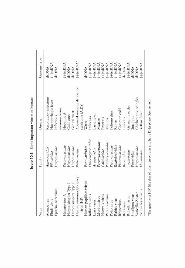
JWBK011-10 JWBK011-Hogg August 12, 2005 19:20 Char Count= 0
Tab
le10
.3So
me
impo
rtan
tvi
ruse
sof
hum
ans
Vir
usFa
mily
Dis
ease
Gen
ome
type
Ade
novi
rus
Ade
novi
rida
eR
espi
rato
ryin
fect
ions
dsD
NA
Ebo
lavi
rus
Filo
viri
dae
Hae
mor
rhag
icfe
ver
(−)s
sRN
AE
pste
in–B
arr
viru
sH
erpe
svir
idae
Infe
ctio
usds
DN
Am
onon
ucle
osis
Hep
atov
irus
AP
icor
navi
rida
eH
epat
itis
A(+
)ssR
NA
Her
pes
sim
plex
Type
IH
erpe
svir
idae
Col
dso
res
dsD
NA
Her
pes
sim
plex
Type
IIH
erpe
svir
idae
Gen
ital
war
tsds
DN
AH
uman
imm
unod
efici
ency
Ret
rovi
rida
eA
cqui
red
imm
une
defic
ienc
y(+
)ssR
NA
*vi
rus
(HIV
)sy
ndro
me
(AID
S)H
uman
papi
llom
avir
usP
apov
avir
idae
War
tsds
DN
AIn
fluen
zavi
rus
Ort
hom
yxov
irid
aeIn
fluen
za(−
)ssR
NA
Las
savi
rus
Are
navi
rida
eL
assa
feve
r(−
)ssR
NA
Mor
billi
viru
sP
aram
yxov
irid
aeM
easl
es(−
)ssR
NA
Nor
wal
kvi
rus
Cal
civi
rida
eE
nter
itis
(+)s
sRN
APa
ram
yxov
irus
Par
amyx
ovir
idae
Mum
ps(−
)ssR
NA
Polio
viru
sP
icor
navi
rida
ePo
liom
yelit
is(+
)ssR
NA
Rab
ies
viru
sR
habd
ovir
idae
Rab
ies
(−)s
sRN
AR
hino
viru
sP
icor
navi
rida
eC
omm
onco
ld(+
)ssR
NA
Rot
avir
usR
eovi
rida
eE
nter
itis
dsR
NA
Rub
ella
viru
sT
ogav
irid
aeG
erm
anm
easl
es(+
)ssR
NA
Smal
lpox
viru
sP
oxvi
rida
eSm
allp
oxds
DN
AV
aric
ella
-Zos
ter
Her
pesv
irid
aeC
hick
enpo
x,sh
ingl
esds
DN
AY
ello
wfe
ver
viru
sFl
aviv
irid
aeY
ello
wfe
ver
(+)s
sRN
A
∗ The
geno
me
ofH
IV,l
ike
that
ofot
her
retr
ovir
uses
also
has
aD
NA
phas
e.Se
eth
ete
xt.
260

JWBK011-10 JWBK011-Hogg August 12, 2005 19:20 Char Count= 0
VIRAL DISEASES IN HUMANS 261
Neuraminidase
Haemagglutinin
Segmented ssRNA genome
Envelope
Figure 10.19 The influenza virus. The RNA segments are bound to protein, forming anucleocapsid, and are surrounded by further protein. The two types of spike assist in theattachment and penetration of the virus into its host
Transmission by water or food: viral gastroenteritis
rota = ‘a wheel’,describ-ing the distinctive ap-pearance of this type ofvirus.
Everybody must surely be familiar with the symptomsof gastroenteritis – sickness, diarrhoea, headaches andfever. The cause of this gastroenteritis may be bacterial(e.g. Salmonella) or viral. The major cause of the vi-ral form is the human rotavirus , which, together withthe Norwalk virus, is responsible for the majority of re-ported cases. The rotavirus has a segmented, dsRNA genome, and is a non-envelopedvirus.
The virus damages the villi in the upper part of the intestinal tract, affecting normalion transport, and resulting in the characteristic water loss. Transmission of gastroen-teritis is via the faecal–oral route, that is, by the ingestion of faecally contaminated foodor water. Poor hygiene practice or contaminated water supplies are usually to blamefor the perpetuation of the cycle. Normally, the condition is self-limiting, lasting only acouple of days; the normal treatment is fluid replacement therapy. In areas where cleanwater supplies are not available, however, the outcome can be much more serious. In theThird World, the condition is a major killer; it is the principal cause of infant mortality,and the cause of some five to ten million deaths per year.
Latent and slow (persistent) viral infections
After an infection has passed, a virus may sometimes remain in the body for longperiods, causing no harm. It may be reactivated, however, by stress or some changein the individual’s health, and initiate a disease state. Well known examples of latentviral infections are cold sores and shingles, both caused by members of the herpesvirusfamily. A virus of this sort will remain with an individual throughout their lifetime.
Whereas latent virus infections are characterised by a sudden increase in virus pro-duction, in persistent (slow) infections the increase is more gradual, building up over
JWBK011-10 JWBK011-Hogg August 12, 2005 19:20 Char Count= 0
262 VIRUSES
several years. Such infections have a serious effect on the target cells, and are generallyfatal. An example is the measles virus, which can re-manifest itself after many years ina rare condition called subacute sclerosing panencephalitis.
Viruses and cancer
A number of chemical and physical agents are known to trigger the uncontrolled prolif-eration of cells that characterise cancers, but in the last two decades it has become clearthat at least six types of human cancer can be virally induced. How do cells lose controlof their division, and how are viruses able to bring this about? It is now known thatcells contain genes called protooncogenes, involved in normal cell replication. They arenormally under the control of other, tumour-suppressor genes, but these can be blockedby proteins encoded by certain DNA viruses. When this happens, the protooncogenefunctions as an oncogene, and cell division is allowed to proceed uncontrolled. Retro-viruses have a different mechanism; they carry their own, altered, version of the cellular
An oncogene is a geneassociated with the con-version of a cell to a can-cerous form.
oncogene, which becomes integrated into the host’sgenome and leads to uncontrolled cell growth. Retro-virus oncogenes are thought to have been acquired orig-inally from human (or animal) genomes, with the RNAtranscript becoming incorporated into the retrovirusparticle.
Emerging and re-emerging viral diseases
A zoonosis is a diseasenormally found in an-imals, but transmissibleto humans under certaincircumstances.
As a result of changes in the pathogen or in the host pop-ulation, completely new infectious diseases may arise, orwe may experience the reappearance of diseases previ-ously considered to be under control. These are knownas, respectively, emerging and re-emerging infections.Changed patterns of human population movement areoften responsible for the development of such infections,with the spread of smallpox to the New World by European colonisers being a famousexample. Frequently emerging virus infections are zoonotic in origin, that is, they aretransferred to humans from animal reservoirs. HIV, for example, is thought to havedeveloped from a similar virus found in monkeys.
While this book was in preparation, there was a sharp reminder of the ever-presentthreat of emerging viral diseases, in the form of a new viral disease called severe acute res-piratory syndrome (SARS). The outbreak of this disease began in Guangdong provincein southern China in November 2002. The Chinese authorities were heavily criticisedfor not reporting the extent of the outbreak until some 3 months later, by which timecases were appearing in many parts of the world, illustrating the role of increased inter-continental travel in the spread of such a disease. At its peak in April 2003, over 1000new cases of SARS were being reported per week. The cause of SARS was quickly iden-tified as a member of the Coronaviridae (single-stranded RNA viruses). Transmitted
JWBK011-10 JWBK011-Hogg August 12, 2005 19:20 Char Count= 0
VIRAL DISEASES IN HUMANS 263
by droplets from coughs and sneezes, it produces flu-like symptoms, but has a mor-tality rate of around 4 percent. Strict public health measures were brought into force,including restrictions in flights to and from affected areas, and the number of reportedcases began to subside. In July 2003, the World Health Organisation announced thatthe final country, Taiwan, had been removed from its list of SARS-infected countries.By this time, SARS had claimed over 800 lives, mostly in China and Hong Kong, butwith a number of deaths occurring as far afield as Canada and South Africa. Althoughapparently under control, isolated cases of SARS infections emerged in late 2003, andearly 2004, most of which could be linked to laboratory workers. In January 2004 theChinese authorities announced that a SARS vaccine was about to enter human clinicaltrials.
Virus vaccines
Smallpox, once the scourge of millions, was in 1979 the first infectious disease to bedeclared successfully eradicated. This followed a worldwide campaign of vaccinationby the World Health Organisation over the previous decade, and was made feasible bythe fact that humans are the only reservoir for the virus. Vaccination is a preventativestrategy that aims to stimulate the host immune system, by exposing it to the infectiousagent in question in an inactivated or incomplete form.
There are four main classes of virus vaccines:
� Attenuated (=‘weakened’) vaccines contain ‘live’ viruses, but ones whose pathogenic-ity has been greatly reduced. The aim is to mimic an infection in order to stimulate animmune response, but without bringing about the disease itself. A famous exampleof this type of vaccine is the polio vaccine developed by Albert Sabin in the 1960s.The cowpox virus used by Edward Jenner in his pioneering vaccination work in thelate 18th century was a naturally occurring attenuated version of the smallpox virus.
� Inactivated vaccines contain viruses which have been exposed to a denaturing agentsuch as formalin. This has the effect of rendering them non-infectious, while at thesame time retaining their ability to stimulate an immune response. Vaccines directedagainst influenza are of this type.
� Subunit vaccines depend on the stimulation of an immune response by just a part ofthe virus. Since the complete virus is not introduced, there is no chance of infection,so vaccines of this type have the attraction of being very safe. Subunit vaccines areoften made using recombinant DNA technology (Chapter 12); the first example tobe approved for human use was the hepatitis B vaccine, which consists of part of theprotein coat of the virus produced in specially engineered yeast cells.
� DNA vaccines are also the product of modern molecular biology techniques. DNAcoding for virus antigens is directly injected into the host, where it is expressed andtriggers a response by the immune system. Vaccines of this type have not so far beenapproved for use in humans.
JWBK011-10 JWBK011-Hogg August 12, 2005 19:20 Char Count= 0
264 VIRUSES
Test yourself
1 Unlike bacteria, viruses are able to pass through a .
2 Viruses are incapable of except when occupying a host cell.
3 A is another name for an intact virus particle.
4 The nucleic acid of a virus is surrounded by a protein coat or . Insome forms, this may in turn be surrounded by an .
5 In some types of virus, the genome is segmented.
6 Parvoviruses are unusual among DNA viruses in having agenome.
7 Capsids may be either or in shape.
8 The scheme of classification orders viruses in terms of their methodof mRNA production.
9 The replication cycle of the T-even phages is described as becauseit results in the of the host cell.
10 The time between infection of a host cell and the release of newly-synthesisedviral particles is called the .
11 The DNA of phages becomes integrated into the host genome,where it is known as a .
12 Enveloped viruses are taken into the host cell either by with thecell membrane or by a process of .
13 The viral envelope is formed by the viral particle being engulfed in the host, into which proteins encoded by the are in-
serted.
14 In order for the genome of ss-RNA virus to be replicated, a strandmust be formed to act as a template.
15 Retroviruses contain the enzyme reverse , which can synthesisefrom an template.
16 The genome of most plant viruses is made of .
17 Subviral particles found in plants and comprising RNA but no areknown as .

JWBK011-10 JWBK011-Hogg August 12, 2005 19:20 Char Count= 0
TEST YOURSELF 265
18 When bacteriophages infect bacterial cells, their presence can be detected ason an agar plate.
19 The envelope of the influenza virus has two types of protein spike made ofand that play a part in the infectious process.
20 The surface antigens of viruses often undergo a gradual modification knownas , which helps to protect them against the host immunesystem.
21 Most cases of viral gastroenteritis in humans are caused by the .
22 Shingles and cold sores, caused by herpesviruses, are examples ofviral infections.
23 infections of humans are transmitted from an animal source.
24 vaccines contain ‘live’ viruses whose has been reduced.
25 The hepatitis B vaccine is an example of a vaccine, which dependson stimulating the immune response without introducing the whole virusparticle.

JWBK011-10 JWBK011-Hogg August 12, 2005 19:20 Char Count= 0
266
JWBK011-11 JWBK011-Hogg August 12, 2005 19:21 Char Count= 0
Part IVMicrobial Genetics
267

JWBK011-11 JWBK011-Hogg August 12, 2005 19:21 Char Count= 0
268
JWBK011-11 JWBK011-Hogg August 12, 2005 19:21 Char Count= 0
11Microbial Genetics
A gene is a sequenceof DNA that usually en-codes a polypeptide.
When any living organism reproduces, it passes ongenetic information to its offspring. This informationtakes the form of genes, linear sequences of DNA thatcan be thought of as the basic units of heredity. The totalcomplement of an organism’s genetic material is calledits genome.
How do we know genesare made of DNA?
The concept of the gene as an inherited physical entity determining some aspect ofan organism’s phenotype dates back to the earliest days of genetics. The question ofwhat genes are actually made of was a major concern of molecular biologists (not thatthey would have described themselves as such!) in the first half of the 20th century.Since it was recognised by this time that genes must be located on chromosomes, andthat chromosomes (in eucaryotes) comprised largely protein and DNA, the reasonableassumption was made that genes must be made up of one of these substances. In theearly years, protein was regarded as the more likely candidate, since, from what wasknown of molecular structure at the time, it offered far more scope for the variationwhich would be essential to account for the thousands of genes that any organism mustpossess. The road to proving that DNA is in fact the ‘stuff of life’ was a long and hardone, which can be read about elsewhere; we shall mention below just some of the keyexperiments which provided crucial evidence.
In 1928 the Englishman Fred Griffith carried out a seminal series of experimentswhich not only demonstrated for the first time the phenomenon of genetic transferin bacteria (a subject we shall consider in more detail later in this chapter), but alsoacted as the first step towards proving that DNA was the genetic material. As we shallsee, Griffith showed that it was possible for heritable characteristics to be transferredfrom one type of bacterium to another, but the cellular component responsible for thisphenomenon was not known at this time.
Attempts were made throughout the 1930s to isolate and identify the transform-ing principle, as it became known, and in 1944 Avery, MacLeod and McCarty pub-lished a paper, which for the first time, proposed DNA as the genetic material. Averyand his colleagues demonstrated that when DNA was rendered inactive by enzy-matic treatment, transforming ability was lost from a cell extract, but if proteins,
269
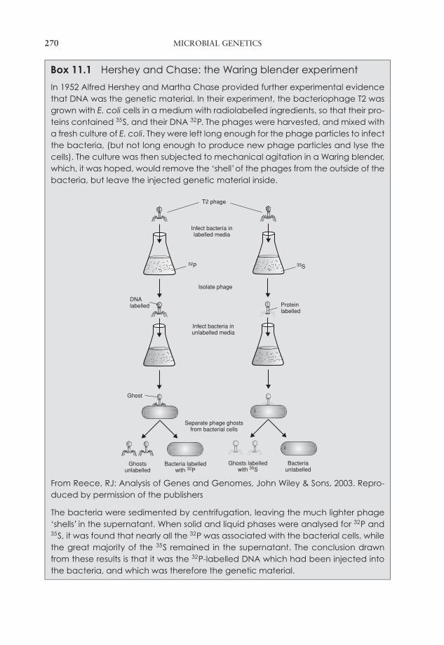
JWBK011-11 JWBK011-Hogg August 12, 2005 19:21 Char Count= 0
270 MICROBIAL GENETICS
Box 11.1 Hershey and Chase: the Waring blender experiment
In 1952 Alfred Hershey and Martha Chase provided further experimental evidencethat DNA was the genetic material. In their experiment, the bacteriophage T2 wasgrown with E. coli cells in a medium with radiolabelled ingredients, so that their pro-teins contained 35S, and their DNA 32P. The phages were harvested, and mixed witha fresh culture of E. coli. They were left long enough for the phage particles to infectthe bacteria, (but not long enough to produce new phage particles and lyse thecells). The culture was then subjected to mechanical agitation in a Waring blender,which, it was hoped, would remove the ‘shell’of the phages from the outside of thebacteria, but leave the injected genetic material inside.
T2 phage
32P 35S
DNAlabelled Protein
labelled
Infect bacteria inlabelled media
Isolate phage
Infect bacteria inunlabelled media
Separate phage ghostsfrom bacterial cells
Ghost
Ghostsunlabelled
Bacteria labelledwith 32P
Ghosts labelledwith 35S
Bacteriaunlabelled
From Reece, RJ: Analysis of Genes and Genomes, John Wiley & Sons, 2003. Repro-duced by permission of the publishers
The bacteria were sedimented by centrifugation, leaving the much lighter phage‘shells’ in the supernatant. When solid and liquid phases were analysed for 32P and35S, it was found that nearly all the 32P was associated with the bacterial cells, whilethe great majority of the 35S remained in the supernatant. The conclusion drawnfrom these results is that it was the 32P-labelled DNA which had been injected intothe bacteria, and which was therefore the genetic material.
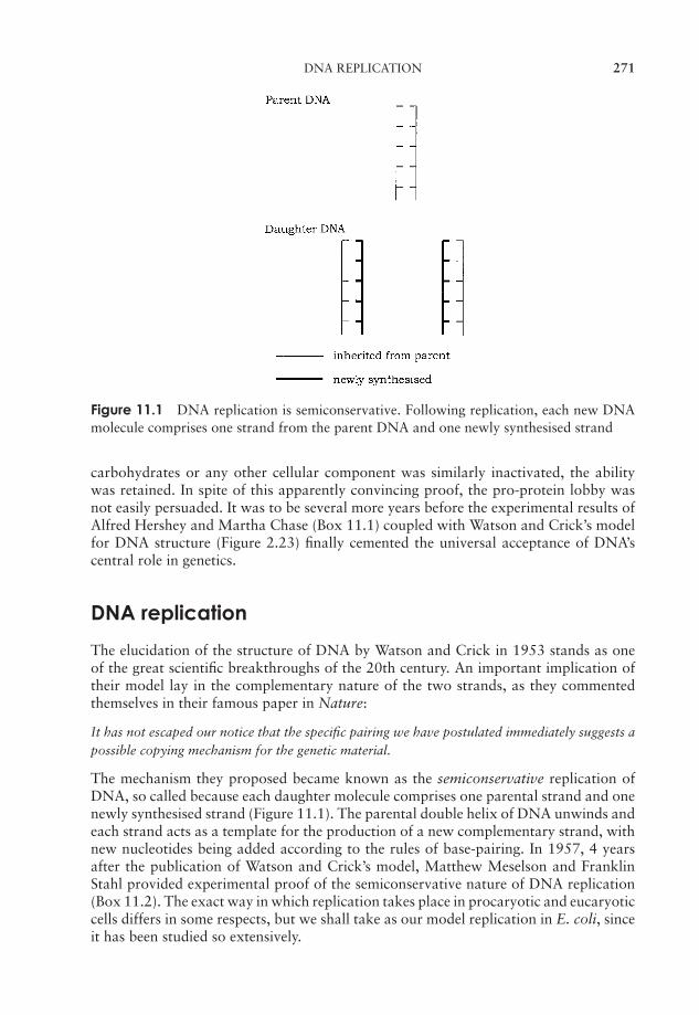
JWBK011-11 JWBK011-Hogg August 12, 2005 19:21 Char Count= 0
DNA REPLICATION 271
Figure 11.1 DNA replication is semiconservative. Following replication, each new DNAmolecule comprises one strand from the parent DNA and one newly synthesised strand
carbohydrates or any other cellular component was similarly inactivated, the abilitywas retained. In spite of this apparently convincing proof, the pro-protein lobby wasnot easily persuaded. It was to be several more years before the experimental results ofAlfred Hershey and Martha Chase (Box 11.1) coupled with Watson and Crick’s modelfor DNA structure (Figure 2.23) finally cemented the universal acceptance of DNA’scentral role in genetics.
DNA replication
The elucidation of the structure of DNA by Watson and Crick in 1953 stands as oneof the great scientific breakthroughs of the 20th century. An important implication oftheir model lay in the complementary nature of the two strands, as they commentedthemselves in their famous paper in Nature:
It has not escaped our notice that the specific pairing we have postulated immediately suggests apossible copying mechanism for the genetic material.
The mechanism they proposed became known as the semiconservative replication ofDNA, so called because each daughter molecule comprises one parental strand and onenewly synthesised strand (Figure 11.1). The parental double helix of DNA unwinds andeach strand acts as a template for the production of a new complementary strand, withnew nucleotides being added according to the rules of base-pairing. In 1957, 4 yearsafter the publication of Watson and Crick’s model, Matthew Meselson and FranklinStahl provided experimental proof of the semiconservative nature of DNA replication(Box 11.2). The exact way in which replication takes place in procaryotic and eucaryoticcells differs in some respects, but we shall take as our model replication in E. coli, sinceit has been studied so extensively.
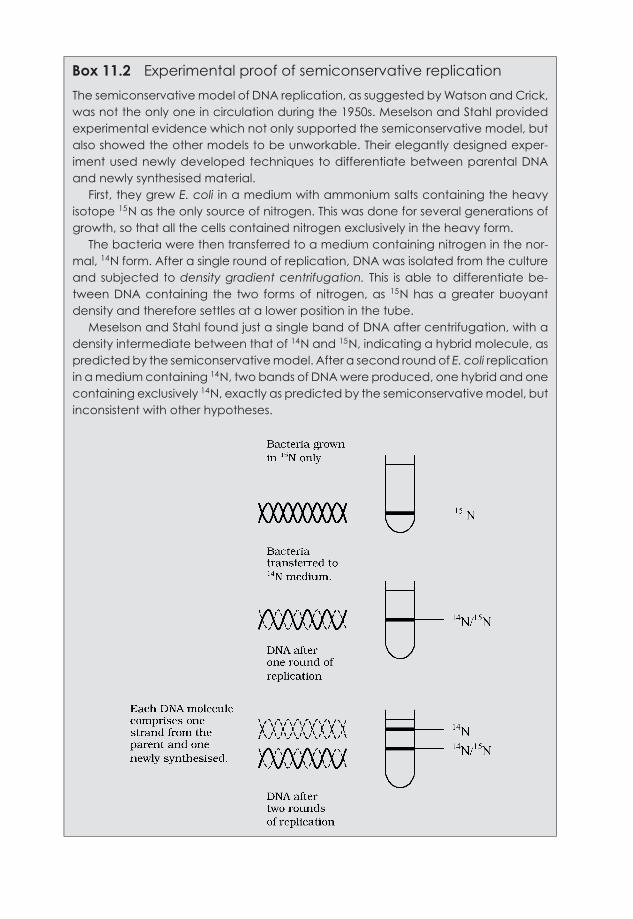
JWBK011-11 JWBK011-Hogg August 12, 2005 19:21 Char Count= 0
Box 11.2 Experimental proof of semiconservative replication
The semiconservative model of DNA replication, as suggested by Watson and Crick,was not the only one in circulation during the 1950s. Meselson and Stahl providedexperimental evidence which not only supported the semiconservative model, butalso showed the other models to be unworkable. Their elegantly designed exper-iment used newly developed techniques to differentiate between parental DNAand newly synthesised material.
First, they grew E. coli in a medium with ammonium salts containing the heavyisotope 15N as the only source of nitrogen. This was done for several generations ofgrowth, so that all the cells contained nitrogen exclusively in the heavy form.
The bacteria were then transferred to a medium containing nitrogen in the nor-mal, 14N form. After a single round of replication, DNA was isolated from the cultureand subjected to density gradient centrifugation. This is able to differentiate be-tween DNA containing the two forms of nitrogen, as 15N has a greater buoyantdensity and therefore settles at a lower position in the tube.
Meselson and Stahl found just a single band of DNA after centrifugation, with adensity intermediate between that of 14N and 15N, indicating a hybrid molecule, aspredicted by the semiconservative model. After a second round of E. coli replicationin a medium containing 14N, two bands of DNA were produced, one hybrid and onecontaining exclusively 14N, exactly as predicted by the semiconservative model, butinconsistent with other hypotheses.
272
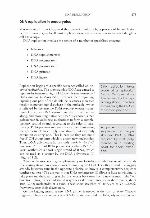
JWBK011-11 JWBK011-Hogg August 12, 2005 19:21 Char Count= 0
DNA REPLICATION 273
DNA replication in procaryotes
You may recall from Chapter 4 that bacteria multiply by a process of binary fission;before this occurs, each cell must duplicate its genetic information so that each daughtercell has a copy.
DNA replication involves the action of a number of specialised enzymes:
� helicases
� DNA topoisomerases
� DNA polymerase I
� DNA polymerase III
� DNA primase
� DNA ligase.
DNA replication takesplace at a replicationfork, a Y-shaped struc-ture formed by the sep-arating strands. The forkmoves along the DNA asreplication proceeds.
Replication begins at a specific sequence called an ori-gin of replication. The two strands of DNA are caused toseparate by helicases (Figure 11.2), while single-strandedDNA binding proteins (SSB) prevents them rejoining.Opening out part of the double helix causes increasedtension (supercoiling) elsewhere in the molecule, whichis relieved by the enzyme DNA topoiosomerase (some-times known as DNA gyrase). As the ‘zipper’ movesalong, and more single stranded DNA is exposed, DNApolymerase III adds new nucleotides to form a comple-mentary second strand, according to the rules of base-pairing. DNA polymerases are not capable of initiatingthe synthesis of an entirely new strand, but can onlyextend an existing one. This is because they require afree 3′-OH group onto which to attach new nucleotides.Thus, DNA polymerase III can only work in the 5′–3′
direction. A form of RNA polymerase called DNA pri-mase synthesises a short single strand of RNA, whichcan be used as a primer by the DNA polymerase III.(Figure 11.2).
A primer is a shortsequence of single-stranded DNA or RNArequired by DNA poly-merase as a startingpoint for chain exten-sion.
When replication occurs, complementary nucleotides are added to one of the strands(the leading strand) in a continuous fashion (Figure 11.2). The other strand (the laggingstrand), however, runs in the opposite polarity; so how is a complementary sequencesynthesised here? The answer is that DNA polymerase III allows a little unwinding totake place and then, starting at the fork, works back over from a new primer, in the 5′–3′
direction. Thus, the second strand is synthesised discontinuously, in short bursts, about1000–2000 nucleotides at a time. These short stretches of DNA are called Okazakifragments, after their discoverers.
On the lagging strand, a new RNA primer is needed at the start of every Okazakifragment. These short sequences of RNA are later removed by DNA polymerase I, which

JWBK011-11 JWBK011-Hogg August 12, 2005 19:21 Char Count= 0
274 MICROBIAL GENETICS
single-strandedbinding protein
5′
5′
3′
3′5′
3′5′
5′
3′
3′ 5′
helicase
helicase
RNA primer
leading strand
DNA polymerase III
Okazakifragment
lagging strand
DNAprimase
DNApolymerase
Figure 11.2 DNA replication takes place at a replication fork. Strands of DNA are sep-arated and unwound by helicase and DNA gyrase, and prevented from rejoining by theattachment of single-strand binding proteins. Starting from an RNA primer, DNA poly-merase III adds the complementary nucleotides to form a second strand. On the leadingstrand, a single primer is set down (not shown), and replication proceeds uninterrupted inthe same direction as the fork. Because DNA polymerase III only works in the 5′ → 3′ di-rection, on the lagging strand a new primer must be added periodically as the strands openup. Replication here is thus discontinuous, as a series of Okazaki fragments (see the text).From Bolsover, SR, Hyams, JS, Jones, S, Shepherd, EA & White, HA: From Genes to Cells,John Wiley & Sons, 1997. Reproduced by permission of the publishers
DNA ligase repairsbreaks in DNA by re-establishing phosphodi-ester bonds in the sugar-phosphate backbone.
then replaces them with DNA nucleotides. Finally, thefragments are joined together by the action of DNAligase. Replication is bidirectional (Figure 11.3), withtwo forks moving in opposite directions; when theymeet, the whole chromosome is copied and replicationis complete*.
What happens when replication goes wrong?
It is clearly important that synthesis of a complementary second strand of DNA shouldoccur with complete accuracy, but occasionally, a non-complementary nucleotide isinserted; this may happen as frequently as once in every 10 000 nucleotides. Cells,however, are able to have a second attempt to incorporate the correct base because ofthe proofreading activity of the enzymes DNA polymerase I and III. These are able to
* This rather long-winded description hardly does justice to a process that incorporates new nucleotides at arate of around 1000 per second!
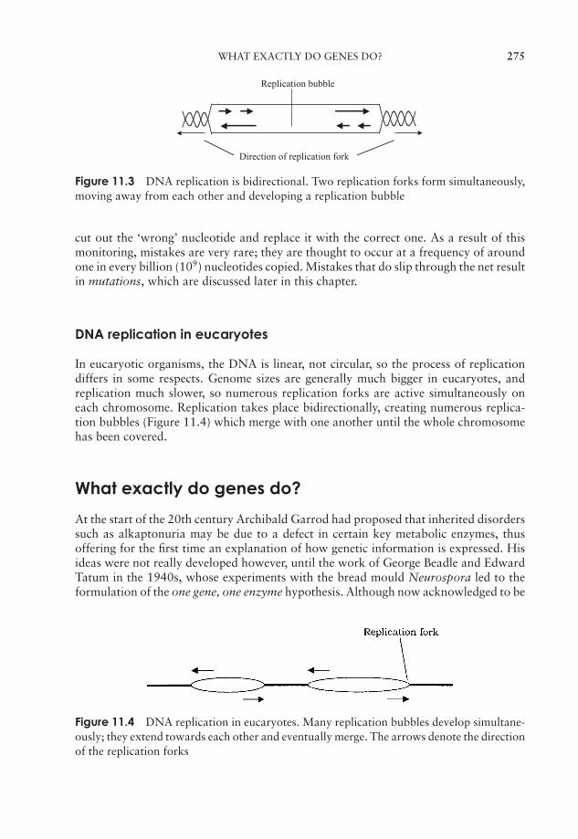
JWBK011-11 JWBK011-Hogg August 12, 2005 19:21 Char Count= 0
WHAT EXACTLY DO GENES DO? 275
Direction of replication fork
Replication bubble
Figure 11.3 DNA replication is bidirectional. Two replication forks form simultaneously,moving away from each other and developing a replication bubble
cut out the ‘wrong’ nucleotide and replace it with the correct one. As a result of thismonitoring, mistakes are very rare; they are thought to occur at a frequency of aroundone in every billion (109) nucleotides copied. Mistakes that do slip through the net resultin mutations, which are discussed later in this chapter.
DNA replication in eucaryotes
In eucaryotic organisms, the DNA is linear, not circular, so the process of replicationdiffers in some respects. Genome sizes are generally much bigger in eucaryotes, andreplication much slower, so numerous replication forks are active simultaneously oneach chromosome. Replication takes place bidirectionally, creating numerous replica-tion bubbles (Figure 11.4) which merge with one another until the whole chromosomehas been covered.
What exactly do genes do?
At the start of the 20th century Archibald Garrod had proposed that inherited disorderssuch as alkaptonuria may be due to a defect in certain key metabolic enzymes, thusoffering for the first time an explanation of how genetic information is expressed. Hisideas were not really developed however, until the work of George Beadle and EdwardTatum in the 1940s, whose experiments with the bread mould Neurospora led to theformulation of the one gene, one enzyme hypothesis. Although now acknowledged to be
Figure 11.4 DNA replication in eucaryotes. Many replication bubbles develop simultane-ously; they extend towards each other and eventually merge. The arrows denote the directionof the replication forks
JWBK011-11 JWBK011-Hogg August 12, 2005 19:21 Char Count= 0
276 MICROBIAL GENETICS
Box 11.3 One gene, one enzyme: not quite true
Beadle and Tatum proposed that each gene was responsible for the production ofa specific enzyme. However all proteins, not just enzymes, are encoded by DNA,and furthermore, some have a quaternary structure (see Chapter 2) with differentpolypeptide subunits being encoded by different genes. The hypothesis was there-fore modified to one gene, one polypeptide. Later still, it emerged that even thisis not always the case, as some genes do not encode proteins at all, but forms ofRNA.
somewhat over-generalised (see Box 11.3), this model proved useful in the years whenthe molecular basis of gene action was being elucidated.
Having established that genes are made of DNA, and having a model for the structureof DNA that explained how it was able to copy itself, the way was open in the 1950sfor scientists to work out the mechanism by which the information encoded in a DNAsequence was converted into a specific protein.
How does a gene direct the synthesis of a protein?
You may recall from Chapter 2 that both DNA and proteins are polymers whose ‘build-ing blocks’ (nucleotides and amino acids respectively) can be put together in an almostinfinite number of sequences. The sequence of amino acids making up the primarystructure of a protein is determined by the sequence of nucleotides in the particulargene responsible for its production. It does this not directly, but through an intermedi-ary molecule, now known to be a form of RNA called messenger RNA (mRNA). It is thisintermediary that carries out the crucial task of passing the information encoded in theDNA sequence to the site of protein synthesis. This unidirectional flow of informationcan be summarised:
DNA mRNA
DNA replication
Protein
and is often referred to as the Central Dogma of biology, because of its applicability toall forms of life. Proposed by Crick in the late 1950s, this is still accepted as being atrue model of the basic events in protein synthesis. Sometimes the message encoded inDNA is transcribed into either ribosomal RNA (rRNA) or transfer RNA (tRNA); thesetypes of RNA are not translated into proteins but represent end-products in themselves.(In the last chapter, we saw that retroviruses have proved an exception to one part ofthe Central Dogma, since they possess enzymes capable of forming DNA from an RNAtemplate.)
The conversion of information encoded as DNA into the synthesis of a polypeptidechain occurs in two distinct phases (Figure 11.5); first the ‘message’ encoded in the

JWBK011-11 JWBK011-Hogg August 12, 2005 19:21 Char Count= 0
WHAT EXACTLY DO GENES DO? 277
Figure 11.5 Transcription and translation. The sequence on one strand of DNA is tran-scribed as a molecule of mRNA (with uracil replacing thymine). The triplet code on themRNA is then translated at ribosomes as a series of amino acids
Messenger RNA (mRNA)is formed from a DNAtemplate (transcription)and carries its encodedmessage to the ribo-some, where it directssynthesis of a polypep-tide (translation).
DNA sequence of a gene is converted to mRNA by tran-scription, then this directs the assembly of a specific se-quence of amino acids during translation. We shall dis-cuss how this happens shortly, but first we need to con-sider the question: how does the sequence of nucleotidesin a gene serve as an instruction in the synthesis of pro-teins?
The genetic code
Messenger RNA carries information (copied from a DNA template) in the form of agenetic code that directs the synthesis of a particular protein. The nature of this codewas worked out in the early 1960s by Marshall Nirenberg, Har Gobind Khorana andothers (Box 11.4). The message encoded in a gene takes the form of a series of triplets(codons). Of the 64 possible three-letter combinations of A, C, G and U, 61 correspondto specific amino acids, while the remaining three act as ‘stop’ messages, indicating thatreading of the message should cease at that point (Figure 11.6). It is essential that thereading of the message starts at the correct place, otherwise the reading frame (groups of
Box 11.4 The genetic code is (almost) universal
The genetic code was first worked out using E. coli, but soon found to apply to otherorganisms too. It seemed reasonable to assume that the code was universal, i.e.applicable to all life forms. It has been shown, however, that certain genes suchas those found in the mitochondria of some eucaryotes employ a slight variation ofthe code. Mitochondria have their own transcription enzymes, ribosomes and tRNAsand so are able to use a modified system.
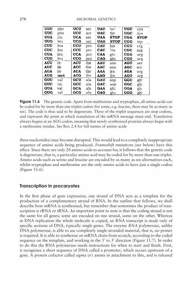
JWBK011-11 JWBK011-Hogg August 12, 2005 19:21 Char Count= 0
278 MICROBIAL GENETICS
Figure 11.6 The genetic code. Apart from methionine and tryptophan, all amino acids canbe coded for by more than one triplet codon (for some, e.g. leucine, there may be as many assix). The code is thus said to be degenerate. Three of the triplet sequences are stop codons,and represent the point at which translation of the mRNA message must end. Translationalways begins at an AUG codon, meaning that newly-synthesised proteins always begin witha methionine residue. See Box 2.4 for full names of amino acids
three nucleotides) may become disrupted. This would lead to a completely inappropriatesequence of amino acids being produced. Frameshift mutations (see below) have thiseffect. Since there are only 20 amino acids to account for, it follows that the genetic codeis degenerate, that is, a particular amino acid may be coded for by more than one triplet.Amino acids such as serine and leucine are encoded by as many as six alternatives each,whilst tryptophan and methionine are the only amino acids to have just a single codon(Figure 11.6).
Transcription in procaryotes
In the first phase of gene expression, one strand of DNA acts as a template for theproduction of a complementary strand of RNA. In the outline that follows, we shalldescribe how mRNA is synthesised, but remember that sometimes the product of tran-scription is rRNA or tRNA. An important point to note is that the coding strand is notthe same for all genes; some are encoded on one strand, some on the other. Whereasin DNA replication the whole molecule is copied, an RNA transcript is made only ofspecific sections of DNA, typically single genes. The enzyme RNA polymerase, unlikeDNA polymerase, is able to use completely single-stranded material, that is, no primeris required. It is able to synthesise an mRNA chain from scratch, according to the codedsequence on the template, and working in the 5′ to 3′ direction (Figure 11.7). In orderto do this the RNA polymerase needs instructions for when to start and finish. First,it recognises a short sequence of DNA called a promoter, which occurs upstream of agene. A protein cofactor called sigma (σ ) assists in attachment to this, and is released
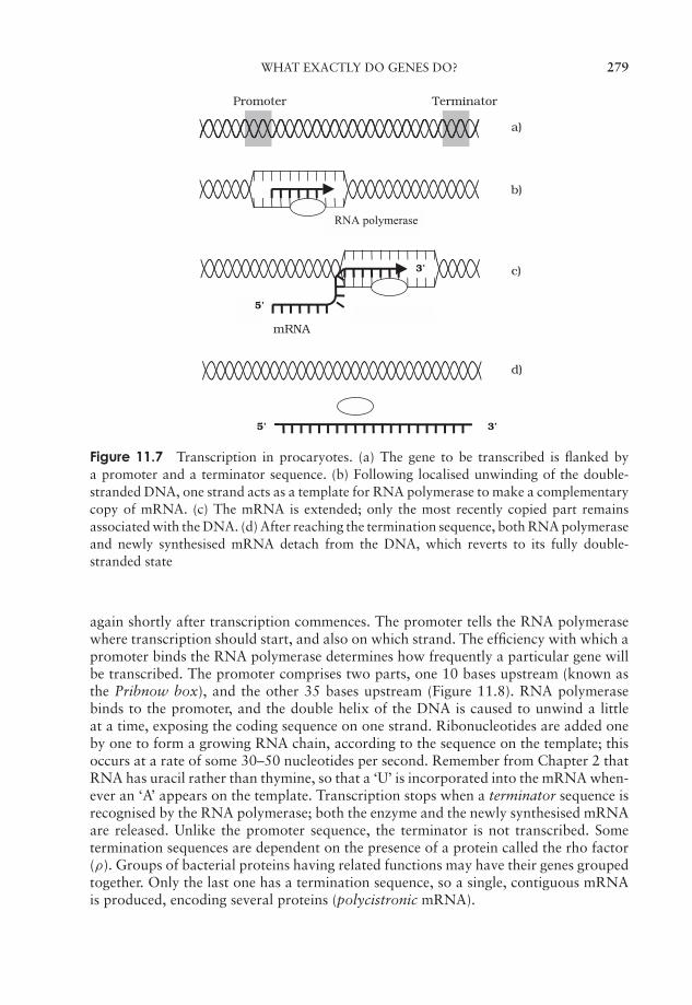
JWBK011-11 JWBK011-Hogg August 12, 2005 19:21 Char Count= 0
WHAT EXACTLY DO GENES DO? 279
Promoter Terminator
RNA polymerase
a)
b)
3’
5’
5’
mRNA
c)
d)
3’
Figure 11.7 Transcription in procaryotes. (a) The gene to be transcribed is flanked bya promoter and a terminator sequence. (b) Following localised unwinding of the double-stranded DNA, one strand acts as a template for RNA polymerase to make a complementarycopy of mRNA. (c) The mRNA is extended; only the most recently copied part remainsassociated with the DNA. (d) After reaching the termination sequence, both RNA polymeraseand newly synthesised mRNA detach from the DNA, which reverts to its fully double-stranded state
again shortly after transcription commences. The promoter tells the RNA polymerasewhere transcription should start, and also on which strand. The efficiency with which apromoter binds the RNA polymerase determines how frequently a particular gene willbe transcribed. The promoter comprises two parts, one 10 bases upstream (known asthe Pribnow box), and the other 35 bases upstream (Figure 11.8). RNA polymerasebinds to the promoter, and the double helix of the DNA is caused to unwind a littleat a time, exposing the coding sequence on one strand. Ribonucleotides are added oneby one to form a growing RNA chain, according to the sequence on the template; thisoccurs at a rate of some 30–50 nucleotides per second. Remember from Chapter 2 thatRNA has uracil rather than thymine, so that a ‘U’ is incorporated into the mRNA when-ever an ‘A’ appears on the template. Transcription stops when a terminator sequence isrecognised by the RNA polymerase; both the enzyme and the newly synthesised mRNAare released. Unlike the promoter sequence, the terminator is not transcribed. Sometermination sequences are dependent on the presence of a protein called the rho factor(ρ). Groups of bacterial proteins having related functions may have their genes groupedtogether. Only the last one has a termination sequence, so a single, contiguous mRNAis produced, encoding several proteins (polycistronic mRNA).
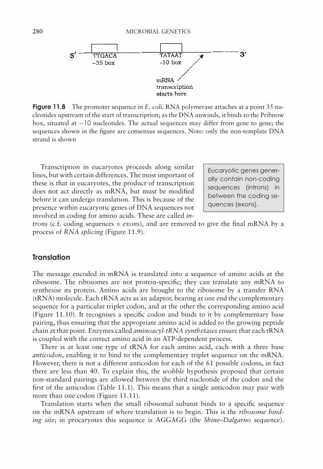
JWBK011-11 JWBK011-Hogg August 12, 2005 19:21 Char Count= 0
280 MICROBIAL GENETICS
Figure 11.8 The promoter sequence in E. coli. RNA polymerase attaches at a point 35 nu-cleotides upstream of the start of transcription; as the DNA unwinds, it binds to the Pribnowbox, situated at −10 nucleotides. The actual sequences may differ from gene to gene; thesequences shown in the figure are consensus sequences. Note: only the non-template DNAstrand is shown
Eucaryotic genes gener-ally contain non-codingsequences (introns) inbetween the coding se-quences (exons).
Transcription in eucaryotes proceeds along similarlines, but with certain differences. The most important ofthese is that in eucaryotes, the product of transcriptiondoes not act directly as mRNA, but must be modifiedbefore it can undergo translation. This is because of thepresence within eucaryotic genes of DNA sequences notinvolved in coding for amino acids. These are called in-trons (c.f. coding sequences = exons), and are removed to give the final mRNA by aprocess of RNA splicing (Figure 11.9).
Translation
The message encoded in mRNA is translated into a sequence of amino acids at theribosome. The ribosomes are not protein-specific; they can translate any mRNA tosynthesise its protein. Amino acids are brought to the ribosome by a transfer RNA(tRNA) molecule. Each tRNA acts as an adaptor, bearing at one end the complementarysequence for a particular triplet codon, and at the other the corresponding amino acid(Figure 11.10). It recognises a specific codon and binds to it by complementary basepairing, thus ensuring that the appropriate amino acid is added to the growing peptidechain at that point. Enzymes called aminoacyl-tRNA synthetases ensure that each tRNAis coupled with the correct amino acid in an ATP-dependent process.
There is at least one type of tRNA for each amino acid, each with a three baseanticodon, enabling it to bind to the complementary triplet sequence on the mRNA.However, there is not a different anticodon for each of the 61 possible codons, in factthere are less than 40. To explain this, the wobble hypothesis proposed that certainnon-standard pairings are allowed between the third nucleotide of the codon and thefirst of the anticodon (Table 11.1). This means that a single anticodon may pair withmore than one codon (Figure 11.11).
Translation starts when the small ribosomal subunit binds to a specific sequenceon the mRNA upstream of where translation is to begin. This is the ribosome bind-ing site; in procaryotes this sequence is AGGAGG (the Shine–Dalgarno sequence).

JWBK011-11 JWBK011-Hogg August 12, 2005 19:21 Char Count= 0
WHAT EXACTLY DO GENES DO? 281
E I I IE E E
E I I IE E E
E
E E E E
I I
III
I
E E E
mRNAtranscript
EukaryoticDNA
Entire gene (both introns and exons) istranscribed to RNA by RNA polymerase enzymes
Processing enzymes remove introns
Exons are spliced together, withoutintrons, forming mRNA that can passthrough the nuclear membrane intothe cytoplasm and be translated.Note that while introns were transcribed,they will not be translated.
Excisedintrons
Nucleus
Cytoplasm
Nuclearmembrane
Pore innuclear
membrane
C
B
A
Figure 11.9 Eucaryotic genes contain non-coding sequences. The product of transcription(primary RNA transcript) cannot act as mRNA because it contains sequences that do notcode for the final polypeptide. These introns must be removed and the remaining codingsequences (exons) spliced together to give the mature mRNA. From Black, JG: Microbiol-ogy: Principles and Explorations, 4th edn, John Wiley & Sons Inc., 1999. Reproduced bypermission of the publishers
The initial amino acid inthe chain is always amethionine, correspon-ding to the AUG startcodon. In procaryotesthere is a modified formcalled formylmethionine(fMet); a special tRNAcarries it to the initiationsite.
This sets the ribosome in the correct reading frame toread the message encoded on the mRNA. A tRNA car-rying a formylmethionine then binds to the AUG startcodon on the mRNA. The large ribosomal subunit joins,and the initiation complex is complete (Figure 11.12).Proteins called initiation factors help to assemble the ini-tiation complex, with energy provided by GTP.
The positioning of the large subunit means that theinitiation codon (AUG) fits into the P-site, and the nexttriplet on the mRNA is aligned with the A-site. Elon-gation of the peptide chain (Figure 11.13) starts whena second tRNA carrying an amino acid is added at theA-site. Peptidyl transferase activity breaks the link between the first amino acid and itstRNA, and forms a peptide bond with the second amino acid. The catalytic action is duepartly to the ribozyme activity of the large subunit rRNA. The ribosome moves along
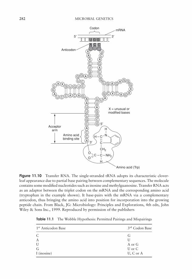
JWBK011-11 JWBK011-Hogg August 12, 2005 19:21 Char Count= 0
282 MICROBIAL GENETICS
U G G
X
U C
CGA
XG
C
U G U
A C A G
CCU
G G A GGUCX
A
A C C
X UC
AGACCG
C G A G
C X C
A
A
G GGX
XGG
AUUA
UAAUGGCG
UCGCACCA
Acceptorarm
Amino acidbinding site
H
H NC
C
CCO
OH
CH2
NH2
3′
5′
X = unusual ormodified bases
Amino acid (Trp)
5′ 3′
Anticodon
mRNACodon
Figure 11.10 Transfer RNA. The single-stranded tRNA adopts its characteristic clover-leaf appearance due to partial base pairing between complementary sequences. The moleculecontains some modified nucleotides such as inosine and methylguanosine. Transfer RNA actsas an adaptor between the triplet codon on the mRNA and the corresponding amino acid(tryptophan in the example shown). It base-pairs with the mRNA via a complementaryanticodon, thus bringing the amino acid into position for incorporation into the growingpeptide chain. From Black, JG: Microbiology: Principles and Explorations, 4th edn, JohnWiley & Sons Inc., 1999. Reproduced by permission of the publishers
Table 11.1 The Wobble Hypothesis: Permitted Pairings and Mispairings
1st Anticodon Base 3rd Codon Base
C GA UU A or GG U or CI (inosine) U, C or A
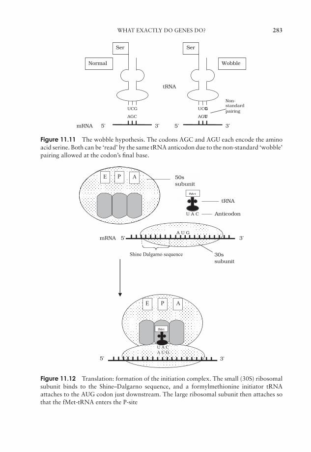
JWBK011-11 JWBK011-Hogg August 12, 2005 19:21 Char Count= 0
WHAT EXACTLY DO GENES DO? 283
Ser
mRNA 5’
UCG
3’
AGC
UCG
AGU
3’5’
Ser
Non-standardpairing
Normal Wobble
tRNA
Figure 11.11 The wobble hypothesis. The codons AGC and AGU each encode the aminoacid serine. Both can be ‘read’ by the same tRNA anticodon due to the non-standard ‘wobble’pairing allowed at the codon’s final base.
U A C A U G
30ssubunit
A U G
fMet
mRNA 5’
tRNA
E P A 50ssubunit
Shine Dalgarno sequence
3’
U A C Anticodon
PE A
3’5’
fMet
Figure 11.12 Translation: formation of the initiation complex. The small (30S) ribosomalsubunit binds to the Shine–Dalgarno sequence, and a formylmethionine initiator tRNAattaches to the AUG codon just downstream. The large ribosomal subunit then attaches sothat the fMet-tRNA enters the P-site
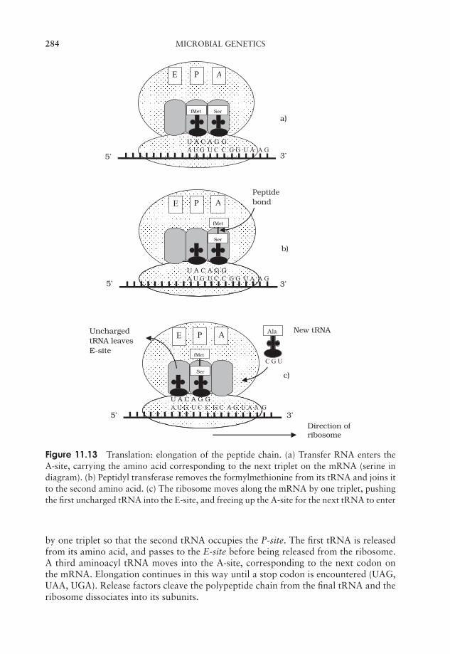
JWBK011-11 JWBK011-Hogg August 12, 2005 19:21 Char Count= 0
284 MICROBIAL GENETICS
U A C A G GA U G U C C G G U A A G
U A C A G GA U G U C C G G U A A G
tRNA leavesE-site
5’
5’
5’
Peptidebond
PE A
E AP
3’
Direction ofribosome
3’
a)
b)
fMet
C G U
U A C A G G A U G U C C G C A G U A A G
Uncharged
fMet
E AP
3’
Ala New tRNA
c)
fMet Ser
Ser
Ser
Figure 11.13 Translation: elongation of the peptide chain. (a) Transfer RNA enters theA-site, carrying the amino acid corresponding to the next triplet on the mRNA (serine indiagram). (b) Peptidyl transferase removes the formylmethionine from its tRNA and joins itto the second amino acid. (c) The ribosome moves along the mRNA by one triplet, pushingthe first uncharged tRNA into the E-site, and freeing up the A-site for the next tRNA to enter
by one triplet so that the second tRNA occupies the P-site. The first tRNA is releasedfrom its amino acid, and passes to the E-site before being released from the ribosome.A third aminoacyl tRNA moves into the A-site, corresponding to the next codon onthe mRNA. Elongation continues in this way until a stop codon is encountered (UAG,UAA, UGA). Release factors cleave the polypeptide chain from the final tRNA and theribosome dissociates into its subunits.
JWBK011-11 JWBK011-Hogg August 12, 2005 19:21 Char Count= 0
REGULATION OF GENE EXPRESSION 285
Regulation of gene expression
The proteins synthesised by microorganisms may broadly be divided into structuralproteins required for the fabric of the cell, and enzymes, used to maintain the essentialmetabolic processes of the cell. It would be terribly wasteful (and possibly harmful) toproduce all these proteins incessantly, regardless of whether or not the cell actually re-quired them, so microorganisms, in common with other living things, have mechanismsof gene regulation, whereby genes can be switched on and off according to the cell’srequirements.
Induction of gene expression
Enzymes whose produc-tion can be switchedon and off are calledinducible enzymes. Thisdistinguishes them fromconstitutive enzymes,which are always pro-duced regardless of pre-vailing conditions.
The synthesis of many enzymes required for thecatabolism (breakdown) of a substrate is regulated by en-zyme induction. If the substrate molecule is not presentin the environment, there is little point in synthesisingthe enzyme needed to break it down. In terms of conser-vation of cellular energy, it makes more sense for suchenzymes to be produced only when they are needed, thatis, when the appropriate substrate molecule is present.The substrate itself therefore acts as the inducer of the en-zyme’s synthesis. An example of this that has been stud-ied in great depth concerns the enzyme β-galactosidase,used by E. coli to convert the disaccharide lactose intoits constituent sugars:
Lactose −−−−−−−−→ Glucose + Galactose
β -galactosidase
A group of functionallyrelated genes involvedin the regulation of en-zyme synthesis and po-sitioned together at thesame locus is called anoperon. It contains bothstructural and regulatorygenes.
β-galactosidase has been intensively studied as part ofthe lac operon of E. coli. This is made up of threestructural genes designated Z, Y and A, which are clus-tered together and share a common promoter and ter-minator (Figure 11.14). The genes code for, respectively,β-galactosidase, a permease and a transacetylase. Thepermease is necessary for the transport of lactose intothe cell, while the role of the transacetylase is not en-tirely clear, although it is essential for the metabolism oflactose. Grouping the three genes together in this way en-sures an ‘all-or-nothing’ expression of the three proteins.
A repressor protein regu-lates the transcription ofa gene by binding to itsoperator sequence. It isencoded by a regula-tory gene.
Transcription of these structural genes into their re-spective mRNAs is initiated by the enzyme RNA poly-merase binding to the promoter sequence. However, thisis only possible in the presence of lactose; in its ab-sence, a repressor protein binds to an operator site, adja-cent to the promoter, preventing RNA polymerase bind-ing to the promoter, and therefore preventing mRNA
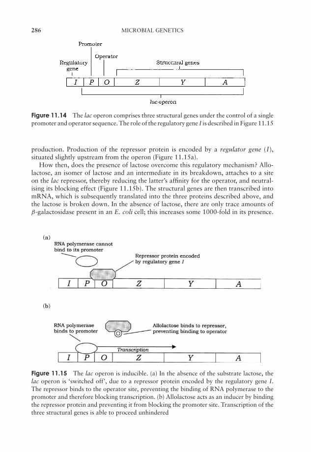
JWBK011-11 JWBK011-Hogg August 12, 2005 19:21 Char Count= 0
286 MICROBIAL GENETICS
Figure 11.14 The lac operon comprises three structural genes under the control of a singlepromoter and operator sequence. The role of the regulatory gene I is described in Figure 11.15
production. Production of the repressor protein is encoded by a regulator gene (I),situated slightly upstream from the operon (Figure 11.15a).
How then, does the presence of lactose overcome this regulatory mechanism? Allo-lactose, an isomer of lactose and an intermediate in its breakdown, attaches to a siteon the lac repressor, thereby reducing the latter’s affinity for the operator, and neutral-ising its blocking effect (Figure 11.15b). The structural genes are then transcribed intomRNA, which is subsequently translated into the three proteins described above, andthe lactose is broken down. In the absence of lactose, there are only trace amounts ofβ-galactosidase present in an E. coli cell; this increases some 1000-fold in its presence.
Figure 11.15 The lac operon is inducible. (a) In the absence of the substrate lactose, thelac operon is ‘switched off’, due to a repressor protein encoded by the regulatory gene I.The repressor binds to the operator site, preventing the binding of RNA polymerase to thepromoter and therefore blocking transcription. (b) Allolactose acts as an inducer by bindingthe repressor protein and preventing it from blocking the promoter site. Transcription of thethree structural genes is able to proceed unhindered

JWBK011-11 JWBK011-Hogg August 12, 2005 19:21 Char Count= 0
REGULATION OF GENE EXPRESSION 287
When all the lactose has been consumed, the repressor protein is free to block theoperator gene once more, and the needless synthesis of further β-galactosidase ceases.
The lac operon can also be induced by isopropyl β-thiogalactoside (IPTG); E. coliis not able to break this down, so the genes remain permanently switched on. IPTG isutilised as an inducer in cloning systems involving the expression of the lacZ gene onpUC plasmids (Chapter 12).
The lac operon is subject to control by positive as well as negative regulator proteins.Transcription of the operon only occurs if another regulatory protein called cataboliteactivator protein (CAP) is bound to the promoter sequence (see Box 11.5). This isdependent on a relatively high concentration of the nucleotide cAMP which only occurswhen glucose is scarce. The activation of the lac operon thus occurs only if lactose ispresent and glucose is (almost) absent.
Repression of gene expression
The induction of gene expression, such as we have just described for the lac operon,generally relates to catabolic (breakdown) reactions. Anabolic (synthetic) reactions,such as those leading to the production of specific amino acids, by contrast, are oftencontrolled by the repression of key genes.
Enzyme repression mechanisms operate along similar lines to induction mechanisms,but the determining factor here is not the substrate of the enzymes in question (lactosein our example), but the end-product of their action. The trp operon contains a clusterof genes encoding five enzymes involved in the synthesis of the amino acid tryptophan.(Figure 11.16) In the presence of tryptophan, the cell has no need to synthesise its
Box 11.5 A choice of substrates
Glucose is central to the reactions of glycolysis (Chapter 6), and is utilised by E. coliwith high efficiency, because the enzymes involved are permanently switched onor constitutive. The β-galactosidase required for lactose breakdown, however, mustbe induced. What happens then, when E . coli is presented with a mixture of bothglucose and lactose? It would be more efficient to metabolise the glucose, with theready-to-use enzymes, but from what you have learnt elsewhere in this section (seeFigure 11.15b), the presence of lactose would induce formation of β-galactosidaseand subsequent lactose breakdown, a less energy-efficient way of going aboutthings. In fact, E. coli has a way of making sure that while the readily utilised glu-cose is present, it takes precedence. It does this by repressing the formation ofβ-galactosidase, a phenomenon known as catabolite repression. Thus, the pres-ence of a ‘preferred’ nutrient prevents the synthesis of enzymes needed tometabolise a less favoured one.
This is because glucose inhibits the formation of cAMP, which is required for thebinding of the CAP to its site on the lac promoter. When glucose levels drop, morecAMP forms and causes CAP to bind to the CAP binding site. Thus, after a delay, theenzymes needed for lactose catabolism are synthesised, and the lactose is utilised,leading to a diauxic growth curve (see Chapter 5).
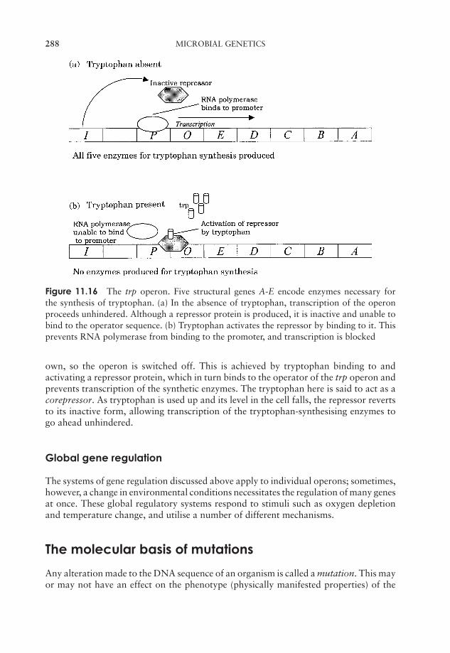
JWBK011-11 JWBK011-Hogg August 12, 2005 19:21 Char Count= 0
288 MICROBIAL GENETICS
Figure 11.16 The trp operon. Five structural genes A-E encode enzymes necessary forthe synthesis of tryptophan. (a) In the absence of tryptophan, transcription of the operonproceeds unhindered. Although a repressor protein is produced, it is inactive and unable tobind to the operator sequence. (b) Tryptophan activates the repressor by binding to it. Thisprevents RNA polymerase from binding to the promoter, and transcription is blocked
own, so the operon is switched off. This is achieved by tryptophan binding to andactivating a repressor protein, which in turn binds to the operator of the trp operon andprevents transcription of the synthetic enzymes. The tryptophan here is said to act as acorepressor. As tryptophan is used up and its level in the cell falls, the repressor revertsto its inactive form, allowing transcription of the tryptophan-synthesising enzymes togo ahead unhindered.
Global gene regulation
The systems of gene regulation discussed above apply to individual operons; sometimes,however, a change in environmental conditions necessitates the regulation of many genesat once. These global regulatory systems respond to stimuli such as oxygen depletionand temperature change, and utilise a number of different mechanisms.
The molecular basis of mutations
Any alteration made to the DNA sequence of an organism is called a mutation. This mayor may not have an effect on the phenotype (physically manifested properties) of the
JWBK011-11 JWBK011-Hogg August 12, 2005 19:21 Char Count= 0
THE MOLECULAR BASIS OF MUTATIONS 289
organism. It may for example enable bacteria to grow without the need for a particulargrowth supplement, or confer resistance to an antibiotic. Just how this happened wasfor many years a source of debate. Since the mutant forms only became apparent afterthe change in conditions (e.g. withdrawal of a nutrient, addition of antibiotic), havesome of the bacteria been induced to adapt to the new conditions, or are mutant formsarising all the time at a very low frequency, and merely selected by the environmentalchange? In 1943, Salvador Luria and Max Delbruck devised the fluctuation test to settlethe matter (see Box 11.6). Results of the fluctuation test together with other evidenceled to the understanding that mutations occur spontaneously in nature at a very lowfrequency. As we shall see later on in this section, however, they can also be inducedby a variety of chemical and physical agents. Any change to the DNA sequence isheritable, thus mutations represent a major source of evolutionary variation. Bacteriamake marvellous tools for the study of mutations because of their huge numbers andvery short generation times.
Since the DNA sequence of a gene represents highly ordered coded information,most mutations have a neutral or detrimental effect on the organism’s phenotype, butoccasionally a mutation occurs which confers an advantage to an organism, makingit better able to survive and reproduce in a particular environment. Mutants that arefavoured in this way may eventually become the dominant type in a population, and,by steps like this, evolution gradually takes place.
Mutations occur spontaneously in any part of an organism’s genome. Spontaneousmutations causing an inactivation of gene function occur in bacteria at the rate of aboutone in a million for a given gene at each round of cell division. Most genes within agiven organism show similar rates of mutation, relative to their gene size; clearly a larger‘target’ will be ‘hit’ more often than a small one.
How do mutations occur?
Figure 11.17(a) reminds us how the code in DNA is transcribed into messenger RNAand then translated into a sequence of amino acids. Each time the DNA undergoes repli-cation, this same sequence will be passed on, coding for the same sequence of aminoacids. Occasionally, mistakes occur during replication. Cells have repair mechanismsto minimise these errors, but what happens if a mistake still slips through? In Figure11.17(b), we can see the effect of one nucleotide being inserted into the strand insteadof another. When the next round of replication occurs, the modified DNA will act asa template for a newly synthesised strand, which at this position will be made comple-mentary to the new, ‘wrong’ base, instead of the original one, and thus the mistake willbe perpetuated.
A missense mutation al-ters the sense of the mes-sage encoded in theDNA, and results in an in-correct amino acid be-ing produced at thepoint where it occurs.
This is an example of the simplest type of mutation,a point mutation, where one nucleotide has been substi-tuted by another. The example shown is a missense muta-tion, which has resulted in the affected triplet coding fora different amino acid; this may or may not have an ef-fect on the phenotype of the organism. RNA polymerase,which transcribes the DNA sequence into mRNA, is un-able to tell that an error has occurred, and faithfully
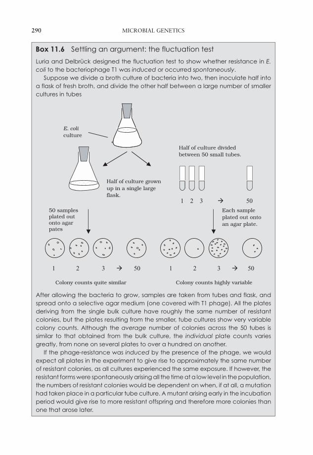
JWBK011-11 JWBK011-Hogg August 12, 2005 19:21 Char Count= 0
290 MICROBIAL GENETICS
Box 11.6 Settling an argument: the fluctuation test
Luria and Delbruck designed the fluctuation test to show whether resistance in E.coli to the bacteriophage T1 was induced or occurred spontaneously.
Suppose we divide a broth culture of bacteria into two, then inoculate half intoa flask of fresh broth, and divide the other half between a large number of smallercultures in tubes
1 2 3 50 1 2 3 50
Half of culture grownup in a single largeflask.
Half of culture dividedbetween 50 small tubes.
1 2 3 50
E. coliculture
50 samples plated out onto agar pates
Each sampleplated out ontoan agar plate.
Colony counts quite similar Colony counts highly variable
After allowing the bacteria to grow, samples are taken from tubes and flask, andspread onto a selective agar medium (one covered with T1 phage). All the platesderiving from the single bulk culture have roughly the same number of resistantcolonies, but the plates resulting from the smaller, tube cultures show very variablecolony counts. Although the average number of colonies across the 50 tubes issimilar to that obtained from the bulk culture, the individual plate counts variesgreatly, from none on several plates to over a hundred on another.
If the phage-resistance was induced by the presence of the phage, we wouldexpect all plates in the experiment to give rise to approximately the same numberof resistant colonies, as all cultures experienced the same exposure. If however, theresistant forms were spontaneously arising all the time at a low level in the population,the numbers of resistant colonies would be dependent on when, if at all, a mutationhad taken place in a particular tube culture. A mutant arising early in the incubationperiod would give rise to more resistant offspring and therefore more colonies thanone that arose later.
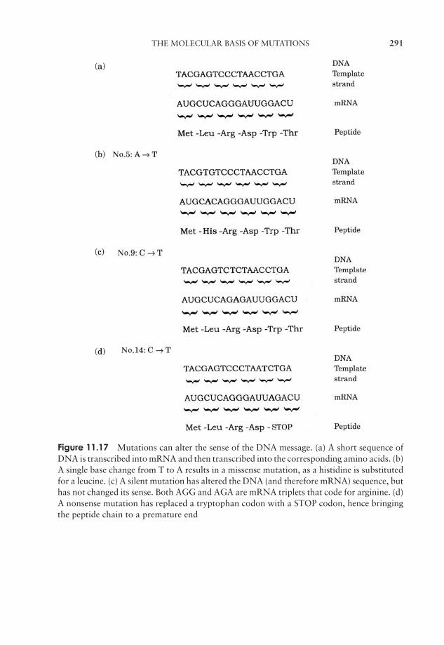
JWBK011-11 JWBK011-Hogg August 12, 2005 19:21 Char Count= 0
THE MOLECULAR BASIS OF MUTATIONS 291
Figure 11.17 Mutations can alter the sense of the DNA message. (a) A short sequence ofDNA is transcribed into mRNA and then transcribed into the corresponding amino acids. (b)A single base change from T to A results in a missense mutation, as a histidine is substitutedfor a leucine. (c) A silent mutation has altered the DNA (and therefore mRNA) sequence, buthas not changed its sense. Both AGG and AGA are mRNA triplets that code for arginine. (d)A nonsense mutation has replaced a tryptophan codon with a STOP codon, hence bringingthe peptide chain to a premature end
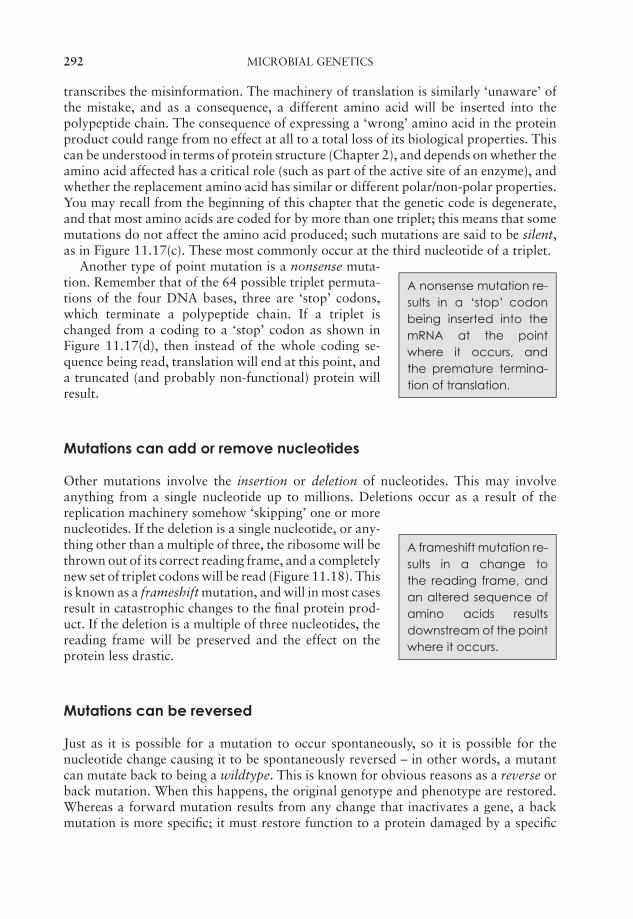
JWBK011-11 JWBK011-Hogg August 12, 2005 19:21 Char Count= 0
292 MICROBIAL GENETICS
transcribes the misinformation. The machinery of translation is similarly ‘unaware’ ofthe mistake, and as a consequence, a different amino acid will be inserted into thepolypeptide chain. The consequence of expressing a ‘wrong’ amino acid in the proteinproduct could range from no effect at all to a total loss of its biological properties. Thiscan be understood in terms of protein structure (Chapter 2), and depends on whether theamino acid affected has a critical role (such as part of the active site of an enzyme), andwhether the replacement amino acid has similar or different polar/non-polar properties.You may recall from the beginning of this chapter that the genetic code is degenerate,and that most amino acids are coded for by more than one triplet; this means that somemutations do not affect the amino acid produced; such mutations are said to be silent,as in Figure 11.17(c). These most commonly occur at the third nucleotide of a triplet.
A nonsense mutation re-sults in a ‘stop’ codonbeing inserted into themRNA at the pointwhere it occurs, andthe premature termina-tion of translation.
Another type of point mutation is a nonsense muta-tion. Remember that of the 64 possible triplet permuta-tions of the four DNA bases, three are ‘stop’ codons,which terminate a polypeptide chain. If a triplet ischanged from a coding to a ‘stop’ codon as shown inFigure 11.17(d), then instead of the whole coding se-quence being read, translation will end at this point, anda truncated (and probably non-functional) protein willresult.
Mutations can add or remove nucleotides
Other mutations involve the insertion or deletion of nucleotides. This may involveanything from a single nucleotide up to millions. Deletions occur as a result of the
A frameshift mutation re-sults in a change tothe reading frame, andan altered sequence ofamino acids resultsdownstream of the pointwhere it occurs.
replication machinery somehow ‘skipping’ one or morenucleotides. If the deletion is a single nucleotide, or any-thing other than a multiple of three, the ribosome will bethrown out of its correct reading frame, and a completelynew set of triplet codons will be read (Figure 11.18). Thisis known as a frameshift mutation, and will in most casesresult in catastrophic changes to the final protein prod-uct. If the deletion is a multiple of three nucleotides, thereading frame will be preserved and the effect on theprotein less drastic.
Mutations can be reversed
Just as it is possible for a mutation to occur spontaneously, so it is possible for thenucleotide change causing it to be spontaneously reversed – in other words, a mutantcan mutate back to being a wildtype. This is known for obvious reasons as a reverse orback mutation. When this happens, the original genotype and phenotype are restored.Whereas a forward mutation results from any change that inactivates a gene, a backmutation is more specific; it must restore function to a protein damaged by a specific

JWBK011-11 JWBK011-Hogg August 12, 2005 19:21 Char Count= 0
THE MOLECULAR BASIS OF MUTATIONS 293
Figure 11.18 Frameshift mutations. (a) A short sequence of DNA is transcribed intomRNA and then transcribed into the corresponding amino acids. (b) The fourth nucleotidein the sequence is deleted, upsetting the groups of triplets or reading frame. This alters thesense of the remainder of the message, leading to a completely different sequence of aminoacids
mutation. Not surprisingly, given this specificity, the rate of back mutations is much lessfrequent.
It is possible for the wildtype phenotype to be restored, not through a reversal of theoriginal base change, but due to a second mutation at a different location. The effect ofthis second mutation is to suppress the effects of the first one. These are called suppressoror second site mutations. They are double mutants that produce a pseudowildtype; thephenotype appears to be wildtype, but the genotype differs.
Mutations have a variety of mechanisms
Why are mistakes made every so often during DNA replication? One source of erro-neous base incorporation is a phenomenon called tautomerism. Some of the nucleotidebases occur in rare alternative forms, which have different base-pairing properties. Forexample, as you will recall from Chapter 2, cytosine normally has an amino group thatprovides a hydrogen atom for bonding with the complementary keto group of gua-nine. Occasionally, however (once in about every 104–105 molecules), the cytosine mayundergo a rearrangement called a tautomeric shift, which results in the amino groupchanging to an imino group (==NH), and this now behaves in pairing terms as if it werethymine, and therefore pairs with adenine (Figure 11.19a). Similarly, thymine mightundergo a tautomeric shift, changing its usual keto form (C==O) to the rare enol form(COH). This then takes on the pairing properties of cytosine and pairs with guanine (Fig-ure 11.19b). The result of such mispairing is that at subsequent rounds of replication,
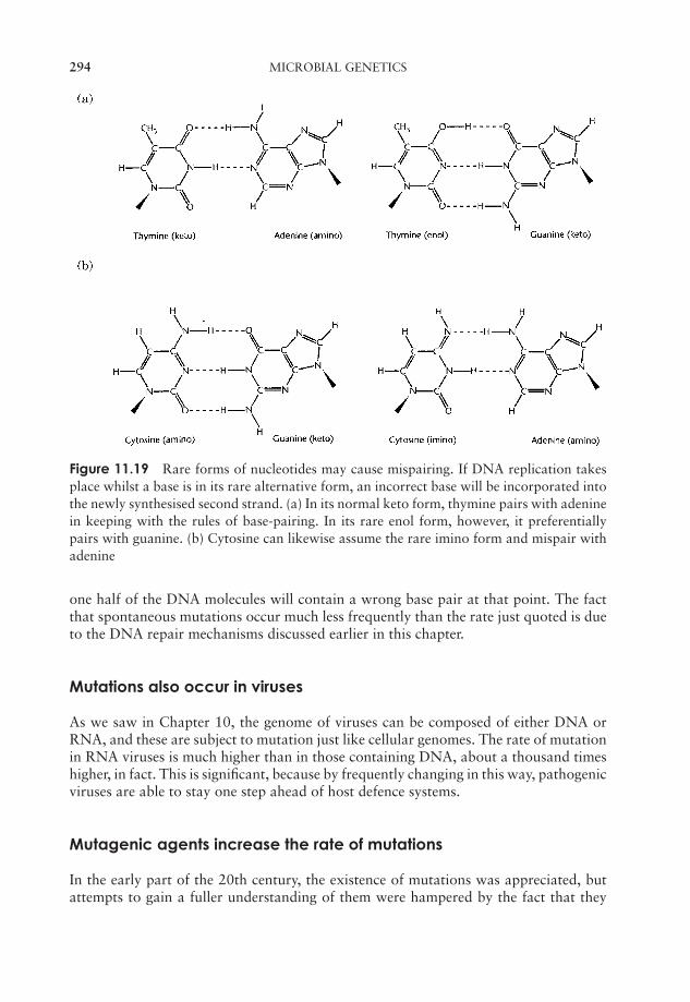
JWBK011-11 JWBK011-Hogg August 12, 2005 19:21 Char Count= 0
294 MICROBIAL GENETICS
Figure 11.19 Rare forms of nucleotides may cause mispairing. If DNA replication takesplace whilst a base is in its rare alternative form, an incorrect base will be incorporated intothe newly synthesised second strand. (a) In its normal keto form, thymine pairs with adeninein keeping with the rules of base-pairing. In its rare enol form, however, it preferentiallypairs with guanine. (b) Cytosine can likewise assume the rare imino form and mispair withadenine
one half of the DNA molecules will contain a wrong base pair at that point. The factthat spontaneous mutations occur much less frequently than the rate just quoted is dueto the DNA repair mechanisms discussed earlier in this chapter.
Mutations also occur in viruses
As we saw in Chapter 10, the genome of viruses can be composed of either DNA orRNA, and these are subject to mutation just like cellular genomes. The rate of mutationin RNA viruses is much higher than in those containing DNA, about a thousand timeshigher, in fact. This is significant, because by frequently changing in this way, pathogenicviruses are able to stay one step ahead of host defence systems.
Mutagenic agents increase the rate of mutations
In the early part of the 20th century, the existence of mutations was appreciated, butattempts to gain a fuller understanding of them were hampered by the fact that they
JWBK011-11 JWBK011-Hogg August 12, 2005 19:21 Char Count= 0
THE MOLECULAR BASIS OF MUTATIONS 295
occurred so rarely. In the 1920s, however, it was shown that the mutation rate in bothbarley plants and the fruit fly Drosophila was greatly increased as a result of exposure toX-rays. In the following decades, a number of chemical and physical agents were shown
Chemical or physicalagents capable of in-ducing mutations aretermed mutagens.
also to cause mutations. Mutagens such as these raisethe general level of mutations in a population, ratherthan the incidence of mutation in at particular locationin the genome. They are extremely useful tools for themicrobial geneticist, but need to be treated with greatcare because they are also mutagenic (and in most casescarcinogenic) towards humans.
Chemical mutagens can be divided into five classes:
Base analoguesA base analogue is able to ‘mimic’ one of the four normal DNA bases by having achemical structure sufficiently similar for it to be incorporated into a DNA moleculeinstead of that base during replication. One such base is 5-bromouracil (5-BU); in itsusual keto form, it acts like thymine (which it closely resembles – see Figure 11.20), andtherefore pairs with adenine, and is not mutagenic. However 5-BU is capable of tau-tomerising to the enol form, which, remember, pairs with G, not A. Whereas for thyminethe enol form is a rarity, for 5-BU it is more common, hence mispairing with guanineoccurs more frequently. So, we get the same outcome as with spontaneous mutation –the TA has been replaced by a CG – but its frequency is much increased. Several otherbase analogues, such as 2-aminopurine, which mimics adenine, have similar effects to5-BU. Mutations brought about by base analogues all result in a purine being replacedby another purine, or a pyrimidine being replaced by another pyrimidine; this type ofmutation is called a transition.
Alkylating agentsAs the name suggests, this group of mutagens act by adding an alkyl group (e.g. methyl,ethyl) at various positions on DNA bases. Ethylethanesulphonate (EES) and ethyl-methanesulphonate (EMS) are alkylating agents, used as laboratory mutagens. Theyact by alkylating guanine or thymine at the oxygen atom involved in hydrogen bond-ing. This leads to an impairment of the normal base-pairing properties, and causesguanine for example to mispair with thymine.
Substitutions brought about by alkylating agents can be either transitions or trans-versions.
Deaminating agentsNitrous acid is a potent mutagen, which alters the base-pairing affinities of cytosine andadenine by replacing an amino group with an oxygen atom. Transitions occur in bothdirections, AT → GC and GC → AT. Note that although guanine is deaminated, itsbase pairing properties are not affected.
Intercalating agentsThese mutagens exert their effect by inserting themselves between adjacent nucleotidesin a single strand of DNA. They distort the strand at this site and cause either the additionor, less commonly, deletion of a nucleotide. This leads to frameshift mutations, and often

JWBK011-11 JWBK011-Hogg August 12, 2005 19:21 Char Count= 0
296 MICROBIAL GENETICS
Figure 11.20 Base analogues mimic DNA base structure. In its keto form, the base analogue5-bromouracil (5-BU) forms base pairs with adenine, but in the enol form it mispairs withguanine. 5-BU differs from thymine only in the substitution of a bromine atom in place of amethyl group. This makes the occurrence of a tautomeric shift more likely
during replication the bound mutagen interferes with new strand synthesis, leading to agross deletion of nucleotides. Ethidium bromide, commonly used for the visualisation ofsmall amounts of DNA in the molecular biology laboratory, is an intercalating agent; ithas a planar structure with roughly the same dimensions as a purine–pyrimidine pairing.
Hydroxylating agentsHydroxylamine has a specific mutagenic effect, hydroxylating the amino group of cy-tosine to cause the transition of GC → AT.
Base analogues and certain intercalating agents can only exert their mutagenic effectsif they are incorporated into DNA while it is replicating. Others, which depend onaltering base pairing by modifying the structure of DNA bases, are effective on bothreplicating and non-replicating DNA.
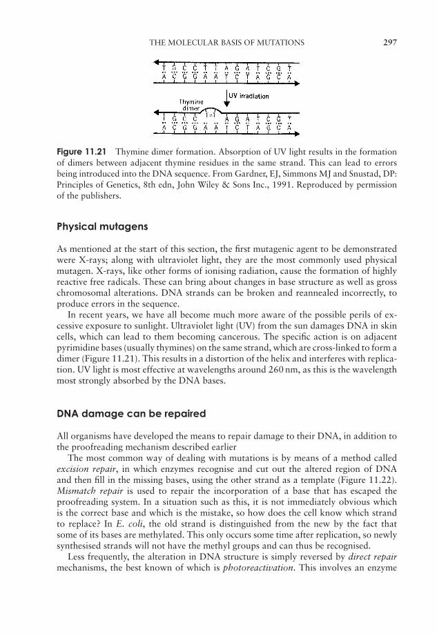
JWBK011-11 JWBK011-Hogg August 12, 2005 19:21 Char Count= 0
THE MOLECULAR BASIS OF MUTATIONS 297
Figure 11.21 Thymine dimer formation. Absorption of UV light results in the formationof dimers between adjacent thymine residues in the same strand. This can lead to errorsbeing introduced into the DNA sequence. From Gardner, EJ, Simmons MJ and Snustad, DP:Principles of Genetics, 8th edn, John Wiley & Sons Inc., 1991. Reproduced by permissionof the publishers.
Physical mutagens
As mentioned at the start of this section, the first mutagenic agent to be demonstratedwere X-rays; along with ultraviolet light, they are the most commonly used physicalmutagen. X-rays, like other forms of ionising radiation, cause the formation of highlyreactive free radicals. These can bring about changes in base structure as well as grosschromosomal alterations. DNA strands can be broken and reannealed incorrectly, toproduce errors in the sequence.
In recent years, we have all become much more aware of the possible perils of ex-cessive exposure to sunlight. Ultraviolet light (UV) from the sun damages DNA in skincells, which can lead to them becoming cancerous. The specific action is on adjacentpyrimidine bases (usually thymines) on the same strand, which are cross-linked to form adimer (Figure 11.21). This results in a distortion of the helix and interferes with replica-tion. UV light is most effective at wavelengths around 260 nm, as this is the wavelengthmost strongly absorbed by the DNA bases.
DNA damage can be repaired
All organisms have developed the means to repair damage to their DNA, in addition tothe proofreading mechanism described earlier
The most common way of dealing with mutations is by means of a method calledexcision repair, in which enzymes recognise and cut out the altered region of DNAand then fill in the missing bases, using the other strand as a template (Figure 11.22).Mismatch repair is used to repair the incorporation of a base that has escaped theproofreading system. In a situation such as this, it is not immediately obvious whichis the correct base and which is the mistake, so how does the cell know which strandto replace? In E. coli, the old strand is distinguished from the new by the fact thatsome of its bases are methylated. This only occurs some time after replication, so newlysynthesised strands will not have the methyl groups and can thus be recognised.
Less frequently, the alteration in DNA structure is simply reversed by direct repairmechanisms, the best known of which is photoreactivation. This involves an enzyme
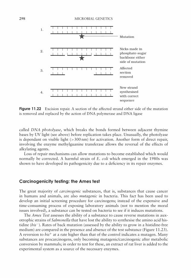
JWBK011-11 JWBK011-Hogg August 12, 2005 19:21 Char Count= 0
298 MICROBIAL GENETICS
Affectedsectionremoved
3.
Nicks made inphosphate-sugarbackbone eitherside of mutation
2.
Mutation
1.
New strandsynthesisedwith correctsequence
4.
Figure 11.22 Excision repair. A section of the affected strand either side of the mutationis removed and replaced by the action of DNA polymerase and DNA ligase
called DNA photolyase, which breaks the bonds formed between adjacent thyminebases by UV light (see above) before replication takes place. Unusually, the photolyaseis dependant on visible light (>300 nm) for activation. Another form of direct repair,involving the enzyme methylguanine transferase allows the reversal of the effects ofalkylating agents.
Loss of repair mechanisms can allow mutations to become established which wouldnormally be corrected. A harmful strain of E. coli which emerged in the 1980s wasshown to have developed its pathogenicity due to a deficiency in its repair enzymes.
Carcinogenicity testing: the Ames test
The great majority of carcinogenic substances, that is, substances that cause cancerin humans and animals, are also mutagenic in bacteria. This fact has been used todevelop an initial screening procedure for carcinogens; instead of the expensive andtime-consuming process of exposing laboratory animals (not to mention the moralissues involved), a substance can be tested on bacteria to see if it induces mutations.
The Ames Test assesses the ability of a substance to cause reverse mutations in aux-otrophic strains of Salmonella that have lost the ability to synthesise the amino acid his-tidine (his−). Rates of back mutation (assessed by the ability to grow in a histidine-freemedium) are compared in the presence and absence of the test substance (Figure 11.23).A reversion to his+ at a rate higher than that of the control indicates a mutagen. Manysubstances are procarcinogens, only becoming mutagenic/carcinogenic after metabolicconversion by mammals; in order to test for these, an extract of rat liver is added to theexperimental system as a source of the necessary enzymes.
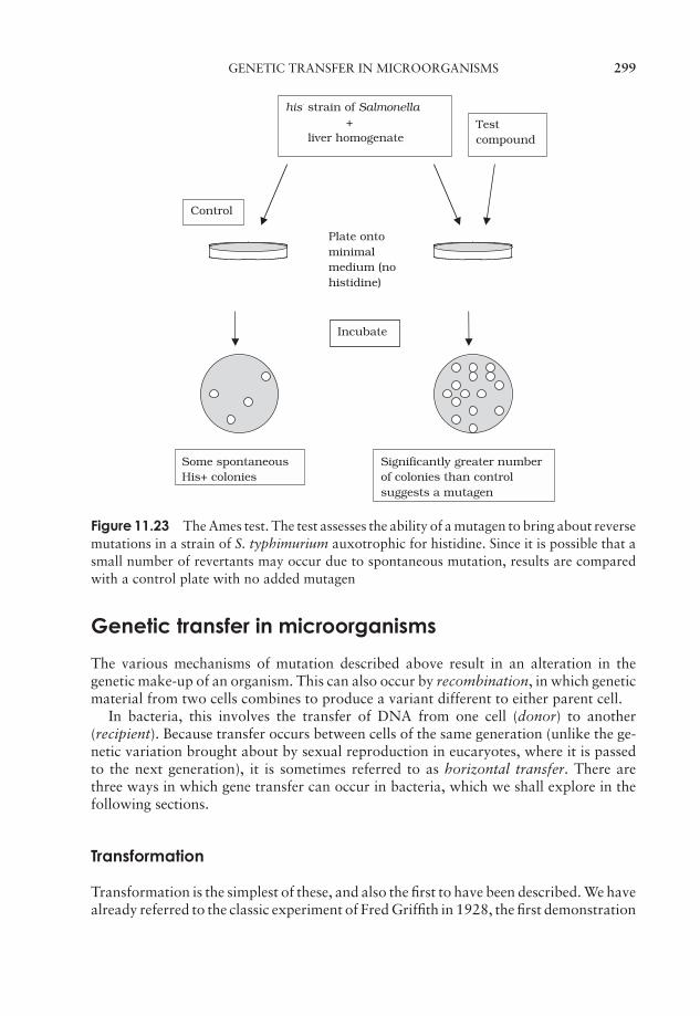
JWBK011-11 JWBK011-Hogg August 12, 2005 19:21 Char Count= 0
GENETIC TRANSFER IN MICROORGANISMS 299
his- strain of Salmonella +
liver homogenate
Plate ontominimalmedium (nohistidine)
Testcompound
Control
Some spontaneousHis+ colonies
Significantly greater numberof colonies than controlsuggests a mutagen
Incubate
Figure 11.23 The Ames test. The test assesses the ability of a mutagen to bring about reversemutations in a strain of S. typhimurium auxotrophic for histidine. Since it is possible that asmall number of revertants may occur due to spontaneous mutation, results are comparedwith a control plate with no added mutagen
Genetic transfer in microorganisms
The various mechanisms of mutation described above result in an alteration in thegenetic make-up of an organism. This can also occur by recombination, in which geneticmaterial from two cells combines to produce a variant different to either parent cell.
In bacteria, this involves the transfer of DNA from one cell (donor) to another(recipient). Because transfer occurs between cells of the same generation (unlike the ge-netic variation brought about by sexual reproduction in eucaryotes, where it is passedto the next generation), it is sometimes referred to as horizontal transfer. There arethree ways in which gene transfer can occur in bacteria, which we shall explore in thefollowing sections.
Transformation
Transformation is the simplest of these, and also the first to have been described. We havealready referred to the classic experiment of Fred Griffith in 1928, the first demonstration
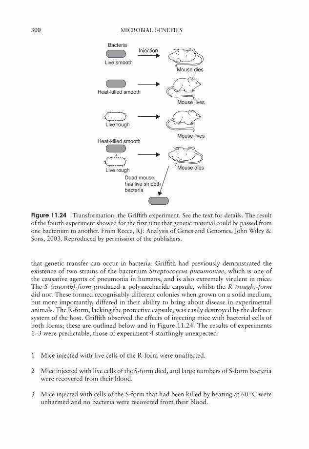
JWBK011-11 JWBK011-Hogg August 12, 2005 19:21 Char Count= 0
300 MICROBIAL GENETICS
InjectionBacteria
Live smooth
Heat-killed smooth
Heat-killed smooth
Live rough
Live rough
+
Dead mouse has live smooth bacteria
Mouse dies
Mouse lives
Mouse lives
Mouse dies
Figure 11.24 Transformation: the Griffith experiment. See the text for details. The resultof the fourth experiment showed for the first time that genetic material could be passed fromone bacterium to another. From Reece, RJ: Analysis of Genes and Genomes, John Wiley &Sons, 2003. Reproduced by permission of the publishers.
that genetic transfer can occur in bacteria. Griffith had previously demonstrated theexistence of two strains of the bacterium Streptococcus pneumoniae, which is one ofthe causative agents of pneumonia in humans, and is also extremely virulent in mice.The S (smooth)-form produced a polysaccharide capsule, whilst the R (rough)-formdid not. These formed recognisably different colonies when grown on a solid medium,but more importantly, differed in their ability to bring about disease in experimentalanimals. The R-form, lacking the protective capsule, was easily destroyed by the defencesystem of the host. Griffith observed the effects of injecting mice with bacterial cells ofboth forms; these are outlined below and in Figure 11.24. The results of experiments1–3 were predictable, those of experiment 4 startlingly unexpected:
1 Mice injected with live cells of the R-form were unaffected.
2 Mice injected with live cells of the S-form died, and large numbers of S-form bacteriawere recovered from their blood.
3 Mice injected with cells of the S-form that had been killed by heating at 60 ◦C wereunharmed and no bacteria were recovered from their blood.
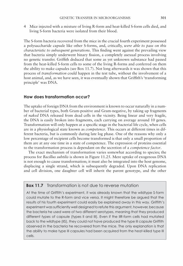
JWBK011-11 JWBK011-Hogg August 12, 2005 19:21 Char Count= 0
GENETIC TRANSFER IN MICROORGANISMS 301
4 Mice injected with a mixture of living R-form and heat-killed S-form cells died, andliving S-form bacteria were isolated from their blood.
The S-form bacteria recovered from the mice in the crucial fourth experiment possesseda polysaccharide capsule like other S-forms, and, critically, were able to pass on thischaracteristic to subsequent generations. This finding went against the prevailing viewthat bacteria simply underwent binary fission, a completely asexual process involvingno genetic transfer. Griffith deduced that some as yet unknown substance had passedfrom the heat-killed S-form cells to some of the living R-forms and conferred on themthe ability to make capsules (see Box 11.7). Not long afterwards it was shown that thisprocess of transformation could happen in the test tube, without the involvement of ahost animal, and, as we have seen, it was eventually shown that Griffith’s ‘transformingprinciple’ was DNA.
How does transformation occur?
The uptake of foreign DNA from the environment is known to occur naturally in a num-ber of bacterial types, both Gram-positive and Gram-negative, by taking up fragmentsof naked DNA released from dead cells in the vicinity. Being linear and very fragile,the DNA is easily broken into fragments, each carrying on average around 10 genes.Transformation will only happen at a specific stage in the bacterial life cycle, when cellsare in a physiological state known as competence. This occurs at different times in dif-ferent bacteria, but is commonly during late log phase. One of the reasons why only alow percentage of recipient cells become transformed is that only a small proportion ofthem are at any one time in a state of competence. The expression of proteins essentialto the transformation process is dependant on the secretion of a competence factor.
The exact mechanism of transformation varies somewhat according to species; theprocess for Bacillus subtilis is shown in Figure 11.25. Mere uptake of exogenous DNAis not enough to cause transformation; it must also be integrated into the host genome,displacing a single strand, which is subsequently degraded. Upon DNA replicationand cell division, one daughter cell will inherit the parent genotype, and the other
Box 11.7 Transformation is not due to reverse mutation
At the time of Griffith’s experiment, it was already known that the wildtype S-formcould mutate to the R-form and vice versa. It might therefore be argued that theresults of his fourth experiment could easily be explained away in this way. Griffith’sexperiment was sufficiently well designed to refute this argument, however, becausethe bacteria he used were of two different serotypes, meaning that they produceddifferent types of capsule (types II and III). Even if the IIR-form cells had mutatedback to the wildtype (IIS), they could not have produced the type III capsule Griffithobserved in the bacteria he recovered from the mice. The only explanation is thatthe ability to make type III capsules had been acquired from the heat-killed type IIIcells.
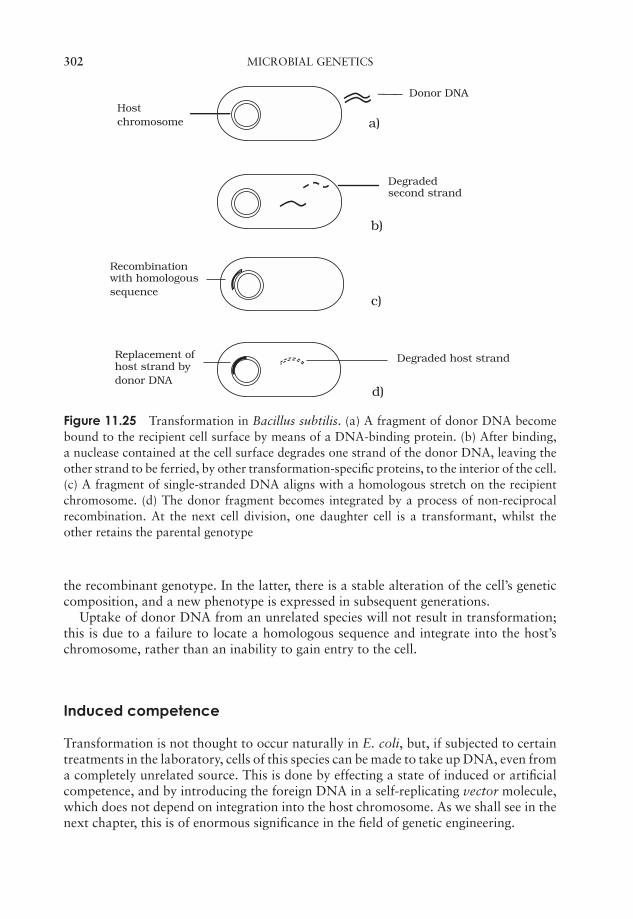
JWBK011-11 JWBK011-Hogg August 12, 2005 19:21 Char Count= 0
302 MICROBIAL GENETICS
Donor DNAHostchromosome a)
b)
c)
d)
Replacement ofhost strand bydonor DNA
Degraded host strand
Degradedsecond strand
Recombinationwith homologoussequence
Figure 11.25 Transformation in Bacillus subtilis. (a) A fragment of donor DNA becomebound to the recipient cell surface by means of a DNA-binding protein. (b) After binding,a nuclease contained at the cell surface degrades one strand of the donor DNA, leaving theother strand to be ferried, by other transformation-specific proteins, to the interior of the cell.(c) A fragment of single-stranded DNA aligns with a homologous stretch on the recipientchromosome. (d) The donor fragment becomes integrated by a process of non-reciprocalrecombination. At the next cell division, one daughter cell is a transformant, whilst theother retains the parental genotype
the recombinant genotype. In the latter, there is a stable alteration of the cell’s geneticcomposition, and a new phenotype is expressed in subsequent generations.
Uptake of donor DNA from an unrelated species will not result in transformation;this is due to a failure to locate a homologous sequence and integrate into the host’schromosome, rather than an inability to gain entry to the cell.
Induced competence
Transformation is not thought to occur naturally in E. coli, but, if subjected to certaintreatments in the laboratory, cells of this species can be made to take up DNA, even froma completely unrelated source. This is done by effecting a state of induced or artificialcompetence, and by introducing the foreign DNA in a self-replicating vector molecule,which does not depend on integration into the host chromosome. As we shall see in thenext chapter, this is of enormous significance in the field of genetic engineering.
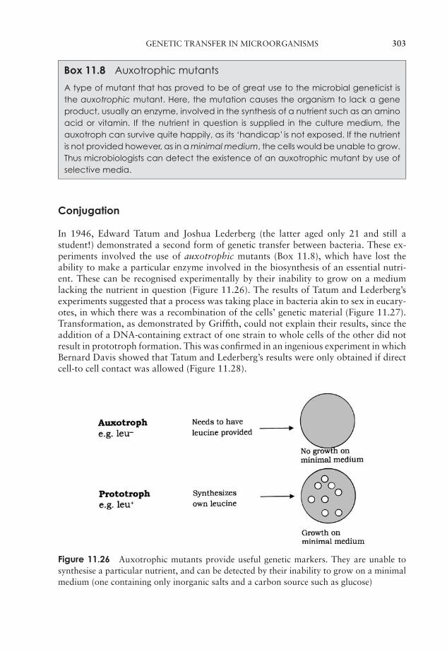
JWBK011-11 JWBK011-Hogg August 12, 2005 19:21 Char Count= 0
GENETIC TRANSFER IN MICROORGANISMS 303
Box 11.8 Auxotrophic mutants
A type of mutant that has proved to be of great use to the microbial geneticist isthe auxotrophic mutant. Here, the mutation causes the organism to lack a geneproduct, usually an enzyme, involved in the synthesis of a nutrient such as an aminoacid or vitamin. If the nutrient in question is supplied in the culture medium, theauxotroph can survive quite happily, as its ‘handicap’ is not exposed. If the nutrientis not provided however, as in a minimal medium, the cells would be unable to grow.Thus microbiologists can detect the existence of an auxotrophic mutant by use ofselective media.
Conjugation
In 1946, Edward Tatum and Joshua Lederberg (the latter aged only 21 and still astudent!) demonstrated a second form of genetic transfer between bacteria. These ex-periments involved the use of auxotrophic mutants (Box 11.8), which have lost theability to make a particular enzyme involved in the biosynthesis of an essential nutri-ent. These can be recognised experimentally by their inability to grow on a mediumlacking the nutrient in question (Figure 11.26). The results of Tatum and Lederberg’sexperiments suggested that a process was taking place in bacteria akin to sex in eucary-otes, in which there was a recombination of the cells’ genetic material (Figure 11.27).Transformation, as demonstrated by Griffith, could not explain their results, since theaddition of a DNA-containing extract of one strain to whole cells of the other did notresult in prototroph formation. This was confirmed in an ingenious experiment in whichBernard Davis showed that Tatum and Lederberg’s results were only obtained if directcell-to cell contact was allowed (Figure 11.28).
Figure 11.26 Auxotrophic mutants provide useful genetic markers. They are unable tosynthesise a particular nutrient, and can be detected by their inability to grow on a minimalmedium (one containing only inorganic salts and a carbon source such as glucose)

JWBK011-11 JWBK011-Hogg August 12, 2005 19:21 Char Count= 0
304 MICROBIAL GENETICS
Figure 11.27 Tatum and Lederberg: sexual recombination in bacteria. Two strains withcomplementary genotypes (auxotrophic for certain genes, prototrophic for others) are eachunable to grow on a minimal medium. When the two are mixed, however, colonies areformed, indicating that recombinant bacteria prototrophic for all the genes have beenformed. The bacteria are washed before plating out to avoid the carry-over of nutrientsfrom the initial broth to the minimal medium. From Black, JG: Microbiology: Principlesand Explorations, 4th edn, John Wiley & Sons Inc., 1999. Reproduced by permission of thepublishers
Gene transfer in conjugation is one way only
The process of conjugation was initially envisaged as a fusion of the two partner cells togive a diploid zygote, which subsequently underwent meiosis to give haploid offspringwith modified genotypes. The work of William Hayes, however, showed that the devel-opment of colonies of recombinant cells was dependant on the survival of only one ofthe participating strains, the other strain being required only as a donor of DNA.
It became apparent that in E. coli, there are two distinct mating types, which becameknown as F+ and F−, depending on whether or not they possessed a plasmid calledthe F (fertility) plasmid. This contains some 30 or 40 genes responsible for its ownreplication and for the synthesis of a thread-like structure expressed on the cell surfacecalled a sex pilus. In a mixture of F+ and F− cells, the sex pilus contacts an F− cell, then
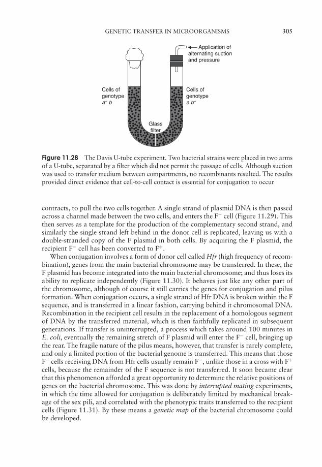
JWBK011-11 JWBK011-Hogg August 12, 2005 19:21 Char Count= 0
GENETIC TRANSFER IN MICROORGANISMS 305
Application of alternating suction and pressure
Cells of genotypea b+
Cells of genotypea+ b
Glassfilter
Figure 11.28 The Davis U-tube experiment. Two bacterial strains were placed in two armsof a U-tube, separated by a filter which did not permit the passage of cells. Although suctionwas used to transfer medium between compartments, no recombinants resulted. The resultsprovided direct evidence that cell-to-cell contact is essential for conjugation to occur
contracts, to pull the two cells together. A single strand of plasmid DNA is then passedacross a channel made between the two cells, and enters the F− cell (Figure 11.29). Thisthen serves as a template for the production of the complementary second strand, andsimilarly the single strand left behind in the donor cell is replicated, leaving us with adouble-stranded copy of the F plasmid in both cells. By acquiring the F plasmid, therecipient F− cell has been converted to F+.
When conjugation involves a form of donor cell called Hfr (high frequency of recom-bination), genes from the main bacterial chromosome may be transferred. In these, theF plasmid has become integrated into the main bacterial chromosome; and thus loses itsability to replicate independently (Figure 11.30). It behaves just like any other part ofthe chromosome, although of course it still carries the genes for conjugation and pilusformation. When conjugation occurs, a single strand of Hfr DNA is broken within the Fsequence, and is transferred in a linear fashion, carrying behind it chromosomal DNA.Recombination in the recipient cell results in the replacement of a homologous segmentof DNA by the transferred material, which is then faithfully replicated in subsequentgenerations. If transfer is uninterrupted, a process which takes around 100 minutes inE. coli, eventually the remaining stretch of F plasmid will enter the F− cell, bringing upthe rear. The fragile nature of the pilus means, however, that transfer is rarely complete,and only a limited portion of the bacterial genome is transferred. This means that thoseF− cells receiving DNA from Hfr cells usually remain F−, unlike those in a cross with F+
cells, because the remainder of the F sequence is not transferred. It soon became clearthat this phenomenon afforded a great opportunity to determine the relative positions ofgenes on the bacterial chromosome. This was done by interrupted mating experiments,in which the time allowed for conjugation is deliberately limited by mechanical break-age of the sex pili, and correlated with the phenotypic traits transferred to the recipientcells (Figure 11.31). By these means a genetic map of the bacterial chromosome couldbe developed.
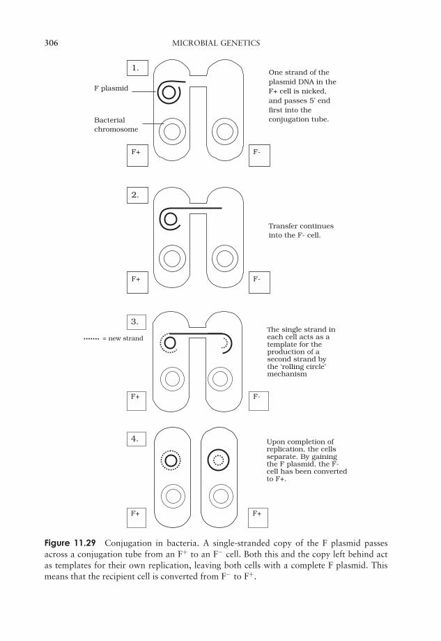
JWBK011-11 JWBK011-Hogg August 12, 2005 19:21 Char Count= 0
306 MICROBIAL GENETICS
F+ F-
Transfer continuesinto the F- cell.
2.
F+ F-
One strand of theplasmid DNA in theF+ cell is nicked,and passes 5’ endfirst into theconjugation tube.
F plasmid
Bacterialchromosome
1.
F+ F+
Upon completion ofreplication, the cellsseparate. By gainingthe F plasmid, the F-cell has been convertedto F+.
4.
= new strand
F+ F-
The single strand ineach cell acts as atemplate for theproduction of asecond strand bythe ‘rolling circle’mechanism
3.
Figure 11.29 Conjugation in bacteria. A single-stranded copy of the F plasmid passesacross a conjugation tube from an F+ to an F− cell. Both this and the copy left behind actas templates for their own replication, leaving both cells with a complete F plasmid. Thismeans that the recipient cell is converted from F− to F+.
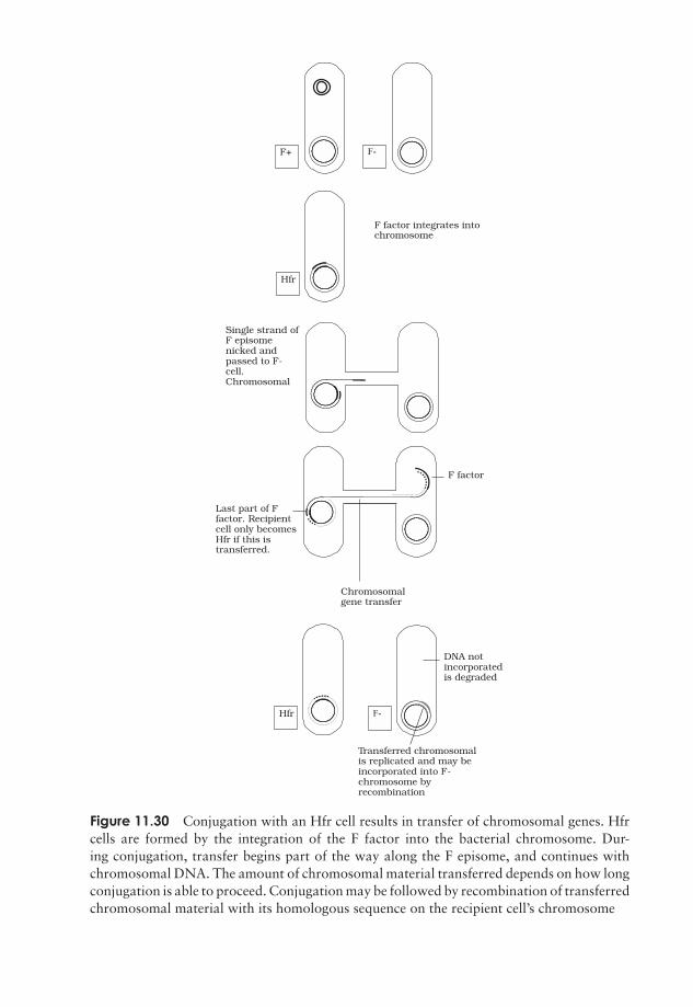
JWBK011-11 JWBK011-Hogg August 12, 2005 19:21 Char Count= 0
F+ F-
Hfr
F factor integrates intochromosome
Single strand ofF episomenicked andpassed to F-cell.Chromosomal
DNA notincorporatedis degraded
F factor
Chromosomalgene transfer
Last part of Ffactor. Recipientcell only becomesHfr if this istransferred.
Hfr F-
Transferred chromosomalis replicated and may beincorporated into F-chromosome byrecombination
Figure 11.30 Conjugation with an Hfr cell results in transfer of chromosomal genes. Hfrcells are formed by the integration of the F factor into the bacterial chromosome. Dur-ing conjugation, transfer begins part of the way along the F episome, and continues withchromosomal DNA. The amount of chromosomal material transferred depends on how longconjugation is able to proceed. Conjugation may be followed by recombination of transferredchromosomal material with its homologous sequence on the recipient cell’s chromosome
307
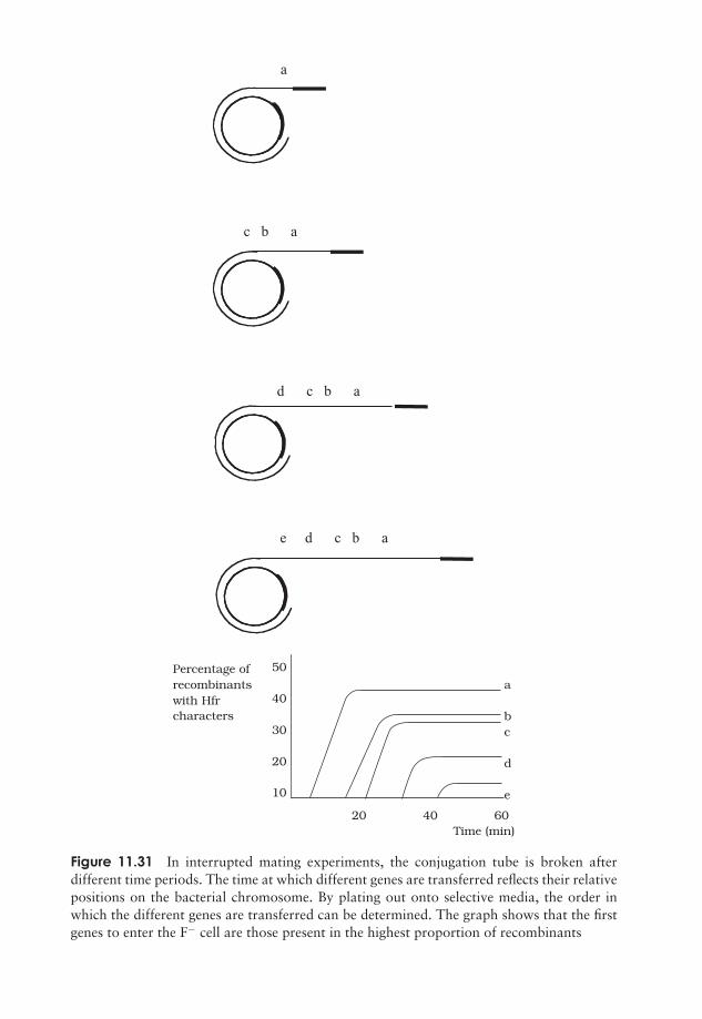
JWBK011-11 JWBK011-Hogg August 12, 2005 19:21 Char Count= 0
a
c b a
d c b a
e d c b a
50
40
30
20
10
a
bc
d
e
20 40 60 Time (min)
Percentage ofrecombinantswith Hfrcharacters
Figure 11.31 In interrupted mating experiments, the conjugation tube is broken afterdifferent time periods. The time at which different genes are transferred reflects their relativepositions on the bacterial chromosome. By plating out onto selective media, the order inwhich the different genes are transferred can be determined. The graph shows that the firstgenes to enter the F− cell are those present in the highest proportion of recombinants
308
JWBK011-11 JWBK011-Hogg August 12, 2005 19:21 Char Count= 0
GENETIC TRANSFER IN MICROORGANISMS 309
Box 11.9 It’s that man again!
Not content with going down in history as the man who first demonstrated conju-gation in bacteria, Joshua Lederberg was also, in 1952, one of the co-discoverersof transduction, along with Norton Zinder. Lederberg was to become a dominantfigure in microbial genetics for over half a century. He was even responsible forcoining the term ‘plasmid’!
The integration of the F plasmid into the bacterial chromosome is reversible; thusHfr cells can revert to F+. Excision of the integrated plasmid is not always precise, andsometimes a little chromosomal DNA is removed too. When this happens, the plasmid,and the cell containing it, are called F′ (‘F prime’); transfer of the plasmid to an F− celltakes with it the extra DNA from the host chromosome. The recipient genome thusbecomes partially diploid (merodiploid), because it has its own copy, plus the ‘guest’copy of certain genes.
Transduction (Box 11.9)
In the third form of genetic transfer in bacteria, bacteriophages act as carriers of DNAfrom one cell to another. In order to appreciate the way in which this is done, it isnecessary to recall the sequence of events in phage replication cycles discussed in theprevious chapter (see Figure 10.11).
Generalised transduction occurs in virulent phages, that is, those with a lytic lifecycle. Sometimes, the enzymes responsible for packaging phage DNA into its proteincoat package instead similarly sized fragments of degraded chromosomal DNA (Figure11.32). Despite containing the wrong DNA, this transducing phage particle is still infec-tive, since this is dependant on its protein element. Thus following infection of anotherbacterial cell, the DNA can be incorporated by recombining with the homologous seg-ment in the recipient cell. Since any chromosomal fragment can be mistakenly packagedin this way (as long as it finds an area of homology), all genes are transferred at a similar(low) frequency.
Specialised transduction results in a much higher efficiency of transfer for specificgenes, however it is limited to genes having a particular chromosomal location. Recallfrom Chapter 10 that in lysogenic life cycles, the phage DNA is integrated into thehost chromosome, and later, perhaps after many rounds of cell division, excised againbefore re-entering a lytic cycle. If this excision does not happen precisely, some of theadjoining chromosomal DNA, carrying a gene or two, may be incorporated into thephage particle (we saw a similar mechanism in the case of F′ plasmid formation). Uponinfecting another cell, the transduced genes would undergo recombination and becomeincorporated into the recipient’s chromosome (Figure 11.33). Although limited to genesin the vicinity of the lysogenic phage’s integration, this is a highly efficient form oftransfer, since the genes become stably integrated into the host cell.
Transduction experiments, like those involving conjugation, can be used to determinethe relative positions of genes on a bacterial chromosome.

JWBK011-11 JWBK011-Hogg August 12, 2005 19:21 Char Count= 0
310 MICROBIAL GENETICS
a) b) c)
f ) e) d)
Figure 11.32 Generalised transduction. During the lytic cycle of a phage, the host DNA isdegraded (b), and a fragment may be mistakenly packaged into a newly synthesised phageparticle (c). Upon infecting a new host cell, the transducing phage releases its DNA (e);although unable to replicate, this can undergo recombination with a homologous sequenceon the host chromosome (f)
Transposable elements
An unusual type of genetic transfer which takes place within an individual cell involvessequences of DNA called transposable elements. One type is known as an insertionsequence (IS), a relatively short piece of chromosomal or plasmid DNA which containsa gene for the enzyme transposase (Figure 11.34). This recognises, cuts and re-ligatesthe insertion sequence anywhere in the bacterial genome. In so doing, it may interrupt a
Transposable elementsthat also carry genesother than those re-quired for transposition,such as genes for an-tibiotic resistance or tox-ins, are known as trans-posons.
gene sequence, and thereby cause a mutation. Unlike re-combination events, no homology is required betweenthe transposable element and the point at which it in-serts. This relocation of a transposable element from oneplace in the genome to another is termed conservativetransposition. In replicative transposition, the elementremains in its original position and a copy is made andinserted elsewhere in the genome. Insertion sequences areflanked by inverted sequences some 9–41bp in length,which are thought to be essential for the recognition ofthe sequence by the transposase.
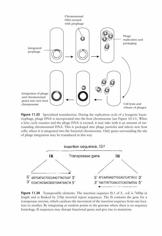
JWBK011-11 JWBK011-Hogg August 12, 2005 19:21 Char Count= 0
Integration of phageand chromosomalgenes into new hostchromosome
ChromosomalDNA excisedwith prophage
Integratedprophage
Phagereplication andpackaging
Cell lysis andrelease of phages
Figure 11.33 Specialised transduction. During the replication cycle of a lysogenic bacte-riophage, phage DNA is incorporated into the host chromosome (see Figure 10.11). Whena lytic cycle resumes and the phage DNA is excised, it may take with it an amount of sur-rounding chromosomal DNA. This is packaged into phage particles and infects new hostcells, where it is integrated into the bacterial chromosome. Only genes surrounding the siteof phage integration may be transduced in this way
Figure 11.34 Transposable elements. The insertion sequence IS 1 of E. coli is 768bp inlength and is flanked by 23bp inverted repeat sequences. The IS contains the gene for atransposase enzyme, which catalyses the movement of the insertion sequence from one loca-tion to another. By integrating at random points in the genome where there is no sequencehomology, IS sequences may disrupt functional genes and give rise to mutations
311
JWBK011-11 JWBK011-Hogg August 12, 2005 19:21 Char Count= 0
312 MICROBIAL GENETICS
Test yourself
1 In the - model of DNA replication, each strand serves asa for the synthesis of a stand. Each daughter moleculecomprises one strand and one newly synthesised strand.
2 During DNA replication, the point where the two strands become separatedis called the .
3 III forms a second strand by adding complementarynucleotides in the → direction.
4 During DNA replication, the must be synthesised dis-continuously, as a series of fragments.
5 Mistakes in DNA replication are largely corrected by the cell’senzymes. Any errors that persist may lead to .
6 The function of genes was expressed in the one , one,hypothesis.
7 The flow of information: DNA → mRNA → protein is often referred to asthe of biology.
8 In the genetic code, many amino acids are encoded by more than one tripletsequence; the code is therefore said to be . Three of thetriplet combinations do not code for an amino acid, but instead serve as
codons.
9 The enzyme RNA polymerase uses a single-stranded template tosynthesise a complementary strand of .
10 Transcription begins at a sequence, situated of the gene.
11 In bacteria, proteins with related functions may be encoded together; theresult of transcription is a mRNA.
12 In eukaryotes, genes are usually discontinuous; coding regions calledare interspersed with non-coding .
13 Molecules of tRNA act as adapter molecules during translation; at one endthey have a three-base complementary to a triplet codon, and atthe other end carry the corresponding .
14 Lactose acts as an for the three genes that make up the lac operon.It neutralises the effects of a protein encoded by the I gene.
JWBK011-11 JWBK011-Hogg August 12, 2005 19:21 Char Count= 0
TEST YOURSELF 313
15 The trp operon contains five genes involved in the synthesis of . Thepresence of this substance activates a , which prevents transcriptionof the operon by binding to the sequence.
16 A mutation alters the of a gene and will changethe sense of the encoded message. Such a mutation arises throughor of DNA.
17 A mutation changes a normal codon into a codon, andresults in the premature termination of translation.
18 such as 5-bromouracil mimic the structure of normalnucleotide bases and become into the DNA structure.
19 The test is used to assess the mutagenicity of a substance.
20 In Griffith’s famous experiment, cells of the virulentS-strain appeared to pass on the ability to synthesise a capsule to the non-virulent R-strain. Griffith coined the term for the factorresponsible.
21 Transformation only occurs between related cells as it depends on the donorDNA finding a sequence on the host chromosome with which to
.
22 experiments can be used to map the order in which geneson a bacterial chromosome are transferred by .
23 In transduction, chromosomal genes close to the point of integra-tion of the may be excised along with it and be transferred toanother host cell.
24 In transduction, fragments of DNA are mistakenly pack-aged into phage coats and can be transferred to another bacterial cell.
25 are sequences of DNA that can move from one locationon a chromosome to another.

JWBK011-11 JWBK011-Hogg August 12, 2005 19:21 Char Count= 0
314
JWBK011-12 JWBK011-Hogg August 12, 2005 20:21 Char Count= 0
12Microorganisms in GeneticEngineering
Introduction
In the last 30 years or so, there has been a revolution in the field of genetics, which hashad a profound effect on virtually every other area of biology. This has been due to thedevelopment of new techniques that have enabled scientists to analyse and manipulateDNA in a quite unprecedented way. Genetically modified crops, DNA ‘fingerprinting’and gene therapy are just three of the many applications made possible by these ad-vances. The subject of ‘genetic engineering’ is too huge to be discussed here in detail,and indeed it extends into areas far beyond the remit of this book. In this chapter, how-ever, we shall examine some of the ways that microorganisms have contributed to thegenetic revolution. As we shall see, their role in the development of new techniques ofDNA manipulation since the 1970s has been just as important as their earlier contri-bution to the elucidation of the structure, role and replication of DNA several decadesearlier.
Restriction endonucle-ases do not destroy thehost bacterium’s ownDNA, because certainnucleotides in the re-cognition sequence aremodified by methyla-tion. The REs are unableto cleave the DNA atmethylated sites.
The beginnings of genetic engineering can be said todate from the discovery, in the late 1960s, of a class ofbacterial enzymes called restriction endonucleases (REs).These are enzymes that cleave DNA into pieces by mak-ing breaks in the sugar-phosphate backbone; in nature,they serve to destroy any foreign DNA that may enterthe cell. They do not cut the DNA in a random fash-ion, however; their unique usefulness to the molecu-lar biologist lies in the fact that they break the DNAin a precise and reproducible manner. They do this bycutting only at specific recognition sites, sequences oftypically four to six nucleotides (Figure 12.1). Thus,under favourable conditions, a particular RE will di-gest a given piece of DNA into an identical collection of fragments, time after time.In the ensuing years, many hundreds of restriction endonucleases have been discov-ered, many of which recognise different specific sequences, providing biologists witha hugely versatile tool for the manipulation of DNA, often likened to a pair ofmolecular ‘scissors’. Not long after REs were first isolated, they were used to createthe first man-made recombinant DNA molecule (Figure 12.2). This involved cutting
315
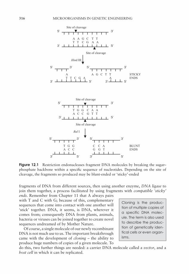
JWBK011-12 JWBK011-Hogg August 12, 2005 20:21 Char Count= 0
316 MICROORGANISMS IN GENETIC ENGINEERING
Site of cleavage5’ 3’
A A G C T T T T C G A A
3’ 5’
Site of cleavage
Hind III
5’ 3’ 5’ 3’
A A G C T T STICKY T T C G A A ENDS
3’ 5’ 3’ 5’
Site of cleavage5’ 3’
T G G C A A A C C G T T
3’ 5’
Site of cleavage
Bal I
5’ 3’ 5’ 3’
T G G C C A BLUNT A C C G G T ENDS
3’ 5’ 3’ 5’
Figure 12.1 Restriction endonucleases fragment DNA molecules by breaking the sugar–phosphate backbone within a specific sequence of nucleotides. Depending on the site ofcleavage, the fragments so produced may be blunt-ended or ‘sticky’-ended
fragments of DNA from different sources, then using another enzyme, DNA ligase tojoin them together, a process facilitated by using fragments with compatible ‘sticky’
Cloning is the produc-tion of multiple copies ofa specific DNA molec-ule. The term is also usedto describe the produc-tion of genetically iden-tical cells or even organ-isms.
ends. Remember from Chapter 11 that A always pairswith T and C with G; because of this, complementarysequences that come into contact with one another will‘stick’ together. DNA, it seems, is DNA, wherever itcomes from; consequently DNA from plants, animals,bacteria or viruses can be joined together to create novelsequences undreamed of by Mother Nature.
Of course, a single molecule of our newly recombinantDNA is not much use to us. The important breakthroughcame with the development of cloning – the ability toproduce huge numbers of copies of a given molecule. Todo this, two further things are needed: a carrier DNA molecule called a vector, and ahost cell in which it can be replicated.
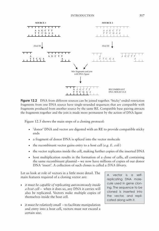
JWBK011-12 JWBK011-Hogg August 12, 2005 20:21 Char Count= 0
INTRODUCTION 317
SOURCE 1 SOURCE 2
A A G C T T A A G C T T T T C G A A T T C G A A
Hind III Hind III
A A G C T T A A G C T T T T C G A A T T C G A A
Mix fragments and join with DNA ligase
A A G C T T RECOMBINANTT T C G A A DNA MOLECULE
Figure 12.2 DNA from different sources can be joined together. ‘Sticky’-ended restrictionfragments from one DNA source have single-stranded sequences that are compatible withfragments produced from another source by the same RE. Compatible base pairing attractsthe fragments together and the join is made more permanent by the action of DNA ligase
Figure 12.3 shows the main steps of a cloning protocol:
� ‘donor’ DNA and vector are digested with an RE to provide compatible stickyends
� a fragment of donor DNA is spliced into the vector molecule
� the recombinant vector gains entry to a host cell (e.g. E. coli )
� the vector replicates inside the cell, making further copies of the inserted DNA
� host multiplication results in the formation of a clone of cells, all containingthe same recombinant plasmid – we now have millions of copies of our donorDNA ‘insert’. A collection of such clones is called a DNA library.
A vector is a self-replicating DNA mole-cule used in gene clon-ing. The sequence to becloned is inserted intothe vector, and repli-cated along with it.
Let us look at role of vectors in a little more detail. Themain features required of a cloning vector are:
� it must be capable of replicating autonomously insidea host cell – when it does so, any DNA it carries willalso be replicated. Vectors make multiple copies ofthemselves inside the host cell.
� it must be relatively small – to facilitate manipulationand entry into a host cell, vectors must not exceed acertain size.

JWBK011-12 JWBK011-Hogg August 12, 2005 20:21 Char Count= 0
318 MICROORGANISMS IN GENETIC ENGINEERING
Vector
Donor DNA
RE digestion
Recombinant vector
Host bacterial cell Vector replication
Bacterial replication
Clone of identical cells
Figure 12.3 Gene cloning. DNA fragments obtained by restriction digestion can be splicedinto a similarly digested vector molecule and transformed into a host bacterial cell. See thetext for details
A selectable marker isa gene that allows cellscontaining it to be identi-fied by the expression ofa recognisable charac-teristic.
� it must carry a selectable marker – since only a pro-portion of host cells will take up the vector, there mustbe a means of differentiating them from those that donot. A common way to do this is to use a vector thatcarries a gene that confers resistance to an antibioticsuch as ampicillin. When bacterial cells are plated outon a medium containing the antibiotic, only those thathave taken up the vector will be able to form colonies.(The host strain must, of course, normally be suscep-tible to the antibiotic.)
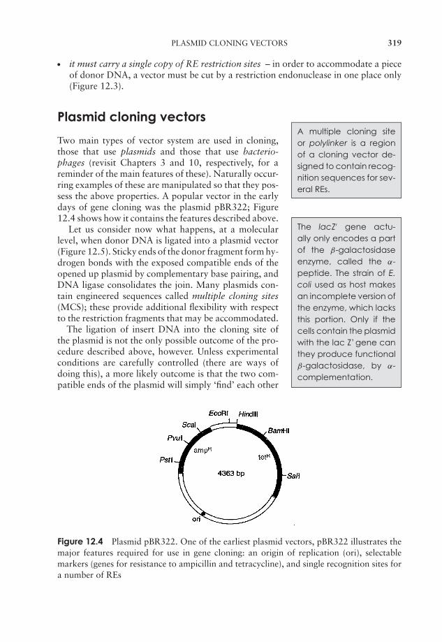
JWBK011-12 JWBK011-Hogg August 12, 2005 20:21 Char Count= 0
PLASMID CLONING VECTORS 319
� it must carry a single copy of RE restriction sites – in order to accommodate a pieceof donor DNA, a vector must be cut by a restriction endonuclease in one place only(Figure 12.3).
Plasmid cloning vectorsA multiple cloning siteor polylinker is a regionof a cloning vector de-signed to contain recog-nition sequences for sev-eral REs.
Two main types of vector system are used in cloning,those that use plasmids and those that use bacterio-phages (revisit Chapters 3 and 10, respectively, for areminder of the main features of these). Naturally occur-ring examples of these are manipulated so that they pos-sess the above properties. A popular vector in the earlydays of gene cloning was the plasmid pBR322; Figure12.4 shows how it contains the features described above.
The lacZ′ gene actu-ally only encodes a partof the β-galactosidaseenzyme, called the α-peptide. The strain of E.coli used as host makesan incomplete version ofthe enzyme, which lacksthis portion. Only if thecells contain the plasmidwith the lac Z’ gene canthey produce functionalβ-galactosidase, by α-complementation.
Let us consider now what happens, at a molecularlevel, when donor DNA is ligated into a plasmid vector(Figure 12.5). Sticky ends of the donor fragment form hy-drogen bonds with the exposed compatible ends of theopened up plasmid by complementary base pairing, andDNA ligase consolidates the join. Many plasmids con-tain engineered sequences called multiple cloning sites(MCS); these provide additional flexibility with respectto the restriction fragments that may be accommodated.
The ligation of insert DNA into the cloning site ofthe plasmid is not the only possible outcome of the pro-cedure described above, however. Unless experimentalconditions are carefully controlled (there are ways ofdoing this), a more likely outcome is that the two com-patible ends of the plasmid will simply ‘find’ each other
Figure 12.4 Plasmid pBR322. One of the earliest plasmid vectors, pBR322 illustrates themajor features required for use in gene cloning: an origin of replication (ori), selectablemarkers (genes for resistance to ampicillin and tetracycline), and single recognition sites fora number of REs
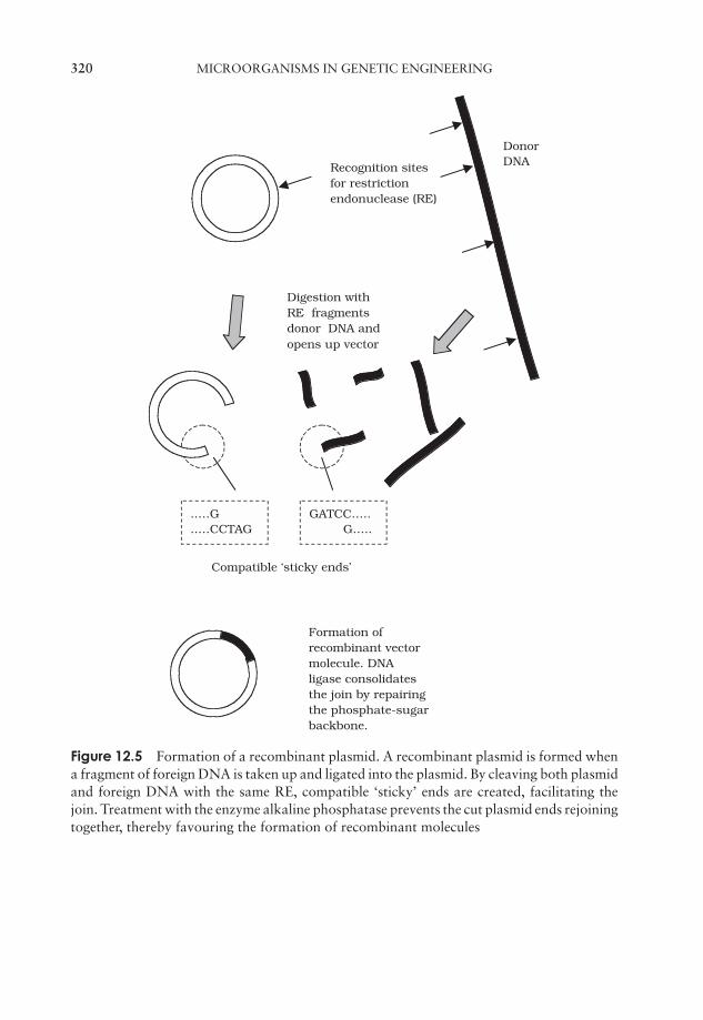
JWBK011-12 JWBK011-Hogg August 12, 2005 20:21 Char Count= 0
320 MICROORGANISMS IN GENETIC ENGINEERING
Recognition sitesfor restrictionendonuclease (RE)
Digestion withRE fragmentsdonor DNA andopens up vector
DonorDNA
Formation ofrecombinant vectormolecule. DNAligase consolidatesthe join by repairingthe phosphate-sugarbackbone.
..…G
..…CCTAGGATCC….. G…..
Compatible ‘sticky ends’
Figure 12.5 Formation of a recombinant plasmid. A recombinant plasmid is formed whena fragment of foreign DNA is taken up and ligated into the plasmid. By cleaving both plasmidand foreign DNA with the same RE, compatible ‘sticky’ ends are created, facilitating thejoin. Treatment with the enzyme alkaline phosphatase prevents the cut plasmid ends rejoiningtogether, thereby favouring the formation of recombinant molecules
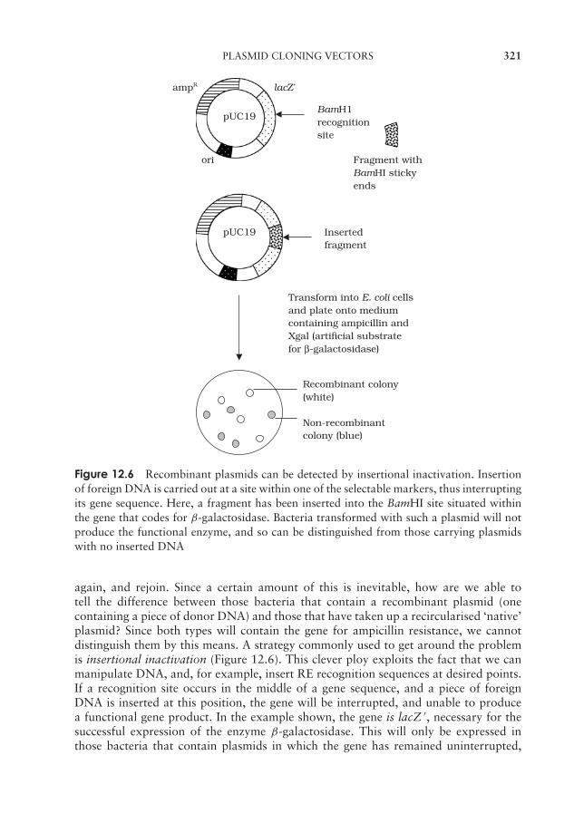
JWBK011-12 JWBK011-Hogg August 12, 2005 20:21 Char Count= 0
PLASMID CLONING VECTORS 321
pUC19
Fragment withBamHI stickyends
ori
lacZ’ampR
pUC19BamH1recognitionsite
Insertedfragment
Transform into E. coli cellsand plate onto mediumcontaining ampicillin andXgal (artificial substratefor β-galactosidase)
Recombinant colony(white)
Non-recombinantcolony (blue)
Figure 12.6 Recombinant plasmids can be detected by insertional inactivation. Insertionof foreign DNA is carried out at a site within one of the selectable markers, thus interruptingits gene sequence. Here, a fragment has been inserted into the BamHI site situated withinthe gene that codes for β-galactosidase. Bacteria transformed with such a plasmid will notproduce the functional enzyme, and so can be distinguished from those carrying plasmidswith no inserted DNA
again, and rejoin. Since a certain amount of this is inevitable, how are we able totell the difference between those bacteria that contain a recombinant plasmid (onecontaining a piece of donor DNA) and those that have taken up a recircularised ‘native’plasmid? Since both types will contain the gene for ampicillin resistance, we cannotdistinguish them by this means. A strategy commonly used to get around the problemis insertional inactivation (Figure 12.6). This clever ploy exploits the fact that we canmanipulate DNA, and, for example, insert RE recognition sequences at desired points.If a recognition site occurs in the middle of a gene sequence, and a piece of foreignDNA is inserted at this position, the gene will be interrupted, and unable to producea functional gene product. In the example shown, the gene is lacZ ′, necessary for thesuccessful expression of the enzyme β-galactosidase. This will only be expressed inthose bacteria that contain plasmids in which the gene has remained uninterrupted,
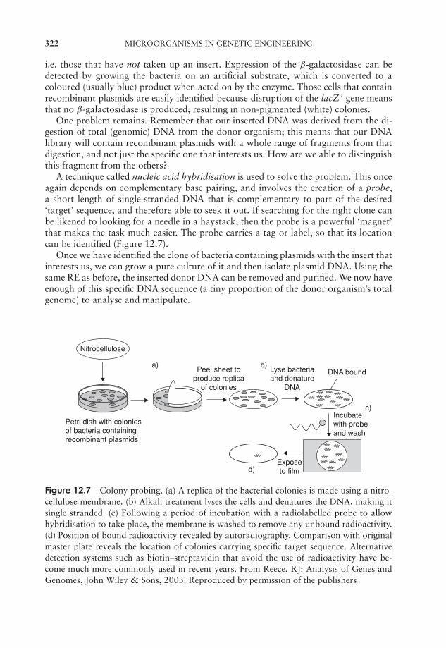
JWBK011-12 JWBK011-Hogg August 12, 2005 20:21 Char Count= 0
322 MICROORGANISMS IN GENETIC ENGINEERING
i.e. those that have not taken up an insert. Expression of the β-galactosidase can bedetected by growing the bacteria on an artificial substrate, which is converted to acoloured (usually blue) product when acted on by the enzyme. Those cells that containrecombinant plasmids are easily identified because disruption of the lacZ ′ gene meansthat no β-galactosidase is produced, resulting in non-pigmented (white) colonies.
One problem remains. Remember that our inserted DNA was derived from the di-gestion of total (genomic) DNA from the donor organism; this means that our DNAlibrary will contain recombinant plasmids with a whole range of fragments from thatdigestion, and not just the specific one that interests us. How are we able to distinguishthis fragment from the others?
A technique called nucleic acid hybridisation is used to solve the problem. This onceagain depends on complementary base pairing, and involves the creation of a probe,a short length of single-stranded DNA that is complementary to part of the desired‘target’ sequence, and therefore able to seek it out. If searching for the right clone canbe likened to looking for a needle in a haystack, then the probe is a powerful ‘magnet’that makes the task much easier. The probe carries a tag or label, so that its locationcan be identified (Figure 12.7).
Once we have identified the clone of bacteria containing plasmids with the insert thatinterests us, we can grow a pure culture of it and then isolate plasmid DNA. Using thesame RE as before, the inserted donor DNA can be removed and purified. We now haveenough of this specific DNA sequence (a tiny proportion of the donor organism’s totalgenome) to analyse and manipulate.
Nitrocellulose
Petri dish with colonies of bacteria containing recombinant plasmids
Peel sheet toproduce replica
of colonies
Lyse bacteria and denature
DNA
DNA bound
Incubatewith probe and wash
Exposeto film
a) b)
c)
d)
Figure 12.7 Colony probing. (a) A replica of the bacterial colonies is made using a nitro-cellulose membrane. (b) Alkali treatment lyses the cells and denatures the DNA, making itsingle stranded. (c) Following a period of incubation with a radiolabelled probe to allowhybridisation to take place, the membrane is washed to remove any unbound radioactivity.(d) Position of bound radioactivity revealed by autoradiography. Comparison with originalmaster plate reveals the location of colonies carrying specific target sequence. Alternativedetection systems such as biotin–streptavidin that avoid the use of radioactivity have be-come much more commonly used in recent years. From Reece, RJ: Analysis of Genes andGenomes, John Wiley & Sons, 2003. Reproduced by permission of the publishers
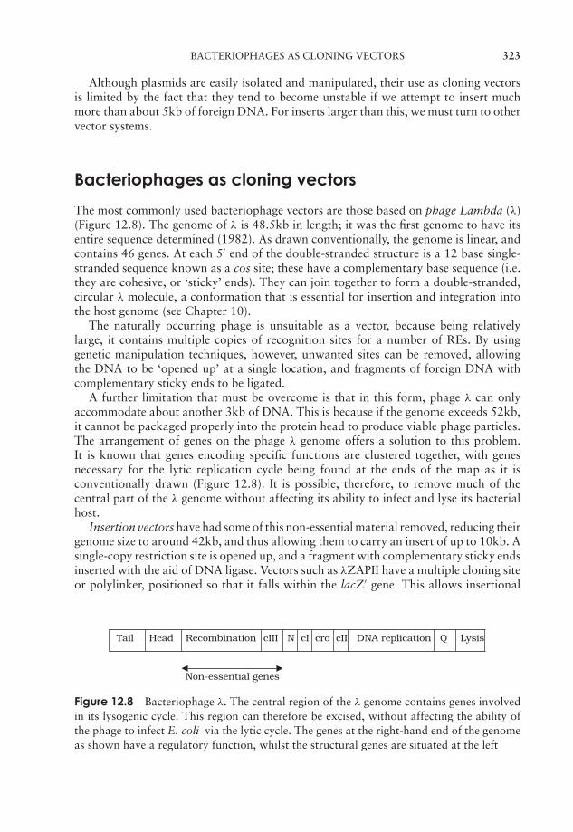
JWBK011-12 JWBK011-Hogg August 12, 2005 20:21 Char Count= 0
BACTERIOPHAGES AS CLONING VECTORS 323
Although plasmids are easily isolated and manipulated, their use as cloning vectorsis limited by the fact that they tend to become unstable if we attempt to insert muchmore than about 5kb of foreign DNA. For inserts larger than this, we must turn to othervector systems.
Bacteriophages as cloning vectors
The most commonly used bacteriophage vectors are those based on phage Lambda (λ)(Figure 12.8). The genome of λ is 48.5kb in length; it was the first genome to have itsentire sequence determined (1982). As drawn conventionally, the genome is linear, andcontains 46 genes. At each 5′ end of the double-stranded structure is a 12 base single-stranded sequence known as a cos site; these have a complementary base sequence (i.e.they are cohesive, or ‘sticky’ ends). They can join together to form a double-stranded,circular λ molecule, a conformation that is essential for insertion and integration intothe host genome (see Chapter 10).
The naturally occurring phage is unsuitable as a vector, because being relativelylarge, it contains multiple copies of recognition sites for a number of REs. By usinggenetic manipulation techniques, however, unwanted sites can be removed, allowingthe DNA to be ‘opened up’ at a single location, and fragments of foreign DNA withcomplementary sticky ends to be ligated.
A further limitation that must be overcome is that in this form, phage λ can onlyaccommodate about another 3kb of DNA. This is because if the genome exceeds 52kb,it cannot be packaged properly into the protein head to produce viable phage particles.The arrangement of genes on the phage λ genome offers a solution to this problem.It is known that genes encoding specific functions are clustered together, with genesnecessary for the lytic replication cycle being found at the ends of the map as it isconventionally drawn (Figure 12.8). It is possible, therefore, to remove much of thecentral part of the λ genome without affecting its ability to infect and lyse its bacterialhost.
Insertion vectors have had some of this non-essential material removed, reducing theirgenome size to around 42kb, and thus allowing them to carry an insert of up to 10kb. Asingle-copy restriction site is opened up, and a fragment with complementary sticky endsinserted with the aid of DNA ligase. Vectors such as λZAPII have a multiple cloning siteor polylinker, positioned so that it falls within the lacZ′ gene. This allows insertional
Tail Head Recombination cIII N cI cro cII DNA replication Q Lysis
Non-essential genes
Figure 12.8 Bacteriophage λ. The central region of the λ genome contains genes involvedin its lysogenic cycle. This region can therefore be excised, without affecting the ability ofthe phage to infect E. coli via the lytic cycle. The genes at the right-hand end of the genomeas shown have a regulatory function, whilst the structural genes are situated at the left
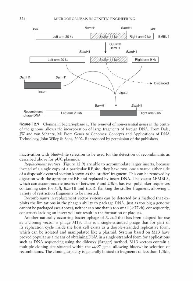
JWBK011-12 JWBK011-Hogg August 12, 2005 20:21 Char Count= 0
324 MICROORGANISMS IN GENETIC ENGINEERING
cos cosBamH1
Cut with BamH1
BamH1
BamH1 BamH1
BamH1 BamH1
BamH1
BamH1
Left arm 20 kb
Left arm 20 kb Right arm 9 kb
Left arm 20 kb
Insert
Recombinantphage DNA
Ligate
Discarded
EMBL4Right arm 9 kb
Right arm 9 kb
Stuffer 14 kb
Stuffer 14 kb
Figure 12.9 Cloning in bacteriophage λ. The removal of non-essential genes in the centreof the genome allows the incorporation of large fragments of foreign DNA. From Dale,JW and von Schantz, M: From Genes to Genomes: Concepts and Applications of DNATechnology, John Wiley & Sons, 2002. Reproduced by permission of the publishers
inactivation with blue/white selection to be used for the detection of recombinants asdescribed above for pUC plasmids.
Replacement vectors (Figure 12.9) are able to accommodate larger inserts, becauseinstead of a single copy of a particular RE site, they have two, one situated either sideof a disposable central section known as the ‘stuffer’ fragment. This can be removed bydigestion with the appropriate RE and replaced by insert DNA. The vector λEMBL3,which can accommodate inserts of between 9 and 23kb, has two polylinker sequencescontaining sites for SalI, BamHI and EcoRI flanking the stuffer fragment, allowing avariety of restriction fragments to be inserted.
Recombinants in replacement vector systems can be detected by a method that ex-ploits the limitations in the phage’s ability to package DNA. Just as too big a genomecannot be packaged (see above), neither can one that is too small (<37kb); consequently,constructs lacking an insert will not result in the formation of plaques.
Another naturally occurring bacteriophage of E. coli that has been adapted for useas a cloning vector is phage M13. This is a single-stranded phage that for part ofits replication cycle inside the host cell exists as a double-stranded replicative form,which can be isolated and manipulated like a plasmid. Systems based on M13 haveproved popular as a means of obtaining DNA in a single-stranded form for applicationssuch as DNA sequencing using the dideoxy (Sanger) method. M13 vectors contain amultiple cloning site situated within the lacZ′ gene, allowing blue/white selection ofrecombinants. The cloning capacity is generally limited to fragments of less than 1.5kb,
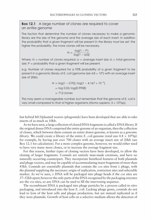
JWBK011-12 JWBK011-Hogg August 12, 2005 20:21 Char Count= 0
BACTERIOPHAGES AS CLONING VECTORS 325
Box 12.1 A large number of clones are required to coveran entire genome
The factors that determine the number of clones necessary to make a genomiclibrary are the size of the genome and the average size of each insert. In additionthe probability that a given fragment will be present in the library must be set: thehigher the probability, the more clones will be necessary.
N = log(1 − P)log(1 − a/b)
Where: N = number of clones required; a = average insert size; b = total genomesize; P = probability that a given fragment will be present.
e.g. Number of clones required for a 95% probability of a given fragment to bepresent in a genomic library of E. coli (genome size 4.8 × 10⁶) with an average insertsize of 20kb:
N = log(1 − 0.95)/ log(1 − 4.167 × 10⁻³)
= log 0.05/ log(0.9958)
= 712 clones
This may seem a manageable number, but remember that the genome of E. coli isvery small compared to that of higher organisms (Homo sapiens: 3 × 10⁹bp).
but hybrid M13/plasmid vectors (phagemids) have been developed that are able to takeinserts of as much as 10kb.
As we have seen, a large collection of cloned DNA fragments is called a DNA library. Ifthe original donor DNA comprised the entire genome of an organism, then the collectionof clones, which between them contain an entire donor genome, is known as a genomiclibrary. We could create a library of the entire E. coli genome (total size 4.8 × 10⁶bp)for example, by having just over 700 clones with an average insert size of 20kb (seeBox 12.1 for calculation). For a more complex genome, however, we would either needto have very many more clones, or to increase the average fragment size.
For this reason, further types of cloning vectors have been developed, to allow thecloning of larger fragments. Cosmids are entirely man-made creations, and have nonaturally occurring counterpart. They incorporate beneficial features of both plasmidsand phage vectors, and may be capable of accommodating insert fragments of more than45kb. Cosmids are essentially plasmids that contain the cos sites from λ phage, withthe plasmid supplying the necessary origin of replication, restriction sites and selectablemarker. As we’ve seen, λ DNA will be packaged into phage heads if the cos sites are37–52kb apart; however the only parts of the DNA recognised by the packaging enzymesare the cos sites, so any DNA can be used to fill the intervening sequence.
The recombinant DNA is packaged into phage particles by a process called in vitropackaging, and introduced into the host E. coli. Lacking phage genes, cosmids do notlead to lysis of the host cells and plaque production, but are instead replicated as ifthey were plasmids. Growth of host cells on a selective medium allows the detection of
JWBK011-12 JWBK011-Hogg August 12, 2005 20:21 Char Count= 0
326 MICROORGANISMS IN GENETIC ENGINEERING
transformants, i.e. those that have taken up the cosmid. There is no need to select forrecombinants, because non-recombinants are too small to be packaged into the phageheads.
Expression vectors
Sometimes, the aim of a cloning experiment is not just to obtain large amounts of aspecific gene, but for the gene to be expressed. This involves using the host cell as asort of ‘factory’, to manufacture the specific protein encoded by the cloned gene. Oneof the earliest applications of genetic engineering was the production of human insulinin E. coli (Figure 12.10). Insulin is needed in considerable quantities for the treat-ment of diabetics; for years it was obtained from the pancreas of pigs and cattle, butthis had several disadvantages including immunological complications and the risk ofviral contamination. Insulin generated by recombinant means is free from these prob-lems. Many proteins can now be produced in this way by microorganisms at a rateseveral times that of the normal host cell. In order for a gene to be expressed, it musthave specific nucleotide sequences around it that act as signals for the host cell’s tran-scription/translation machinery (promoter, ribosomal binding site and terminator – seeChapter 11). Since these sequences differ between, say, humans and E. coli, the bacte-rial RNA polymerase will not recognise the human sequences, and therefore, althougha human gene may be cloned in E. coli using a simple vector, it will not be expressed. If,however, the human gene could be inserted so that it was under the control of the E. coliexpression signals, then transcription should take place. Specially designed vectors thatprovide these signals are called expression vectors. The choice of promoter sequence isparticularly important; often, a strong (i.e. very efficient) promoter is selected, so as tomaximise the amount of protein product obtained. Genes whose protein products arenaturally synthesised in abundance are likely to have such promoters. It is often helpfulto be able to regulate gene expression; inducible promoters can be switched on andoff by the presence of certain substances. The lac promoter (which controls the lacZgene) is an example of this. Cassette vectors have promoter, ribosomal binding site andterminator sequences clustered together as a discrete unit, with a single recognition sitefor one or more REs being situated downstream of the promoter (Figure 12.11).
The small size of the insulin molecule (and gene) and the size of the potential marketmade it a prime early candidate for production by recombinant DNA technology. Mostinsulin used in the treatment of diabetes nowadays is produced in this way. Systemsbased on E. coli have also been used to synthesise other small human proteins with
The polypeptide pro-ducts of translation ineucaryotes may requirepost-translational modifi-cation before functionalprotein is produced. Ex-amples include phos-phorylation, acetylationand glycosylation.
therapeutic potential such as human growth hormone,γ -interferon and tumour necrosis factor (TNF).
Bacteria, however, are not suitable host cells for theproduction of many other human proteins such as tis-sue plasminogen activator (TPA) or blood clotting Fac-tor VIII, due to the size and complexity of their genes.This is because many proteins of complex eucaryotesare subject to post-translational modifications; this doesnot occur in procaryotes, so bacteria such as E. coli arenot equipped with the cellular machinery to make thenecessary modifications to human proteins.

JWBK011-12 JWBK011-Hogg August 12, 2005 20:21 Char Count= 0
EXPRESSION VECTORS 327
Figure 12.10 Production of recombinant human insulin. Human insulin comprises twoshort peptide chains, 21 and 30 amino acid residues in length. Because they are so short,the nucleotide sequence of their genes can be predicted from their amino acid sequence, andsynthetic genes produced. Each is cloned under the influence of the lac promoter and down-stream of part of the lacZ′ gene. By being produced as fusion proteins with β-galactosidase,the insulin chains are protected against being degraded by the E. coli cell. From Brown, TA:Gene Cloning, 3rd edn, Chapman & Hall, 1995. Reproduced by permission of BlackwellPublishing Ltd.
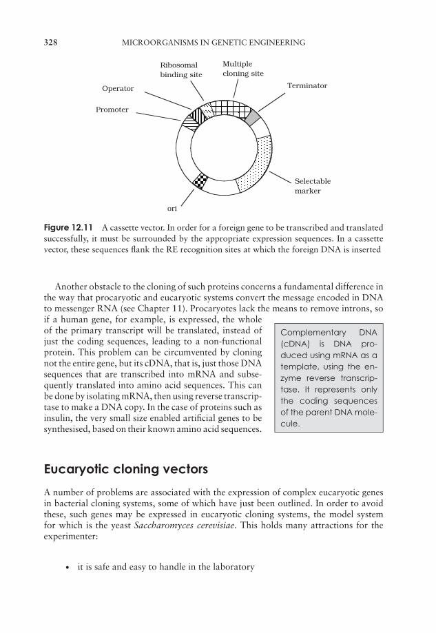
JWBK011-12 JWBK011-Hogg August 12, 2005 20:21 Char Count= 0
328 MICROORGANISMS IN GENETIC ENGINEERING
Multiplecloning site
Terminator
Selectablemarker
ori
Ribosomalbinding site
Operator
Promoter
Figure 12.11 A cassette vector. In order for a foreign gene to be transcribed and translatedsuccessfully, it must be surrounded by the appropriate expression sequences. In a cassettevector, these sequences flank the RE recognition sites at which the foreign DNA is inserted
Another obstacle to the cloning of such proteins concerns a fundamental difference inthe way that procaryotic and eucaryotic systems convert the message encoded in DNAto messenger RNA (see Chapter 11). Procaryotes lack the means to remove introns, so
Complementary DNA(cDNA) is DNA pro-duced using mRNA as atemplate, using the en-zyme reverse transcrip-tase. It represents onlythe coding sequencesof the parent DNA mole-cule.
if a human gene, for example, is expressed, the wholeof the primary transcript will be translated, instead ofjust the coding sequences, leading to a non-functionalprotein. This problem can be circumvented by cloningnot the entire gene, but its cDNA, that is, just those DNAsequences that are transcribed into mRNA and subse-quently translated into amino acid sequences. This canbe done by isolating mRNA, then using reverse transcrip-tase to make a DNA copy. In the case of proteins such asinsulin, the very small size enabled artificial genes to besynthesised, based on their known amino acid sequences.
Eucaryotic cloning vectors
A number of problems are associated with the expression of complex eucaryotic genesin bacterial cloning systems, some of which have just been outlined. In order to avoidthese, such genes may be expressed in eucaryotic cloning systems, the model systemfor which is the yeast Saccharomyces cerevisiae. This holds many attractions for theexperimenter:
� it is safe and easy to handle in the laboratory
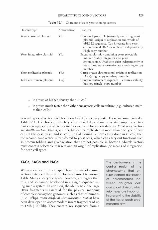
JWBK011-12 JWBK011-Hogg August 12, 2005 20:21 Char Count= 0
EUCARYOTIC CLONING VECTORS 329
Table 12.1 Characteristics of yeast cloning vectors
Plasmid type Abbreviation Features
Yeast episomal plasmid YEp Contain 2 µm circle (naturally occurring yeastplasmid) origin of replication and whole ofpBR322 sequence. Can integrate into yeastchromosomal DNA or replicate independently.High copy number
Yeast integrative plasmid YIp Bacterial plasmid containing yeast selectablemarker. Stably integrates into yeastchromosome. Unable to exist independently inyeast. Low transformation rate and single copynumber
Yeast replicative plasmid YRp Carries yeast chromosomal origin of replication(ARS); high copy number; unstable
Yeast centromere plasmid YCp Contain centromere sequence – ensures stability,but low (single) copy number
� it grows at higher density than E. coli
� it grows much faster than other eucaryotic cells in culture (e.g. cultured mam-malian cells)
Several types of vector have been developed for use in yeasts. These are summarised inTable 12.1. The choice of which type to use will depend on the relative importance to aparticular application of factors such as yield and long-term stability. Most yeast vectorsare shuttle vectors, that is, vectors that can be replicated in more than one type of hostcell (in this case, yeast and E. coli). Initial cloning is more easily done in E. coli, thenthe recombinant vector is transferred to yeast cells, which can carry out functions suchas protein folding and glycosylation that are not possible in bacteria. Shuttle vectorsmust contain selectable markers and an origin of replication (or means of integration)for both cell types.
YACs, BACs and PACs The centromere is thecentral region of thechromosome that en-sures correct distributionof chromosomes be-tween daughter cellsduring cell division, whilsttelomeres are importantin preserving the stabilityof the tips of each chro-mosome arm.
We saw earlier in this chapter how the use of cosmidvectors extended the size of cloneable insert to around45kb. Many eucaryotic genes, however, are bigger thanthis, and so cannot be cloned in a single sequence us-ing such a system. In addition, the ability to clone largeDNA fragments is essential for the physical mappingof complex eucaryotic genomes such as that of humans(3 × 10⁹bp). Yeast artificial chromosomes (YACs) havebeen developed to accommodate insert fragments of upto 1Mb (1000kb). They contain key sequences from a
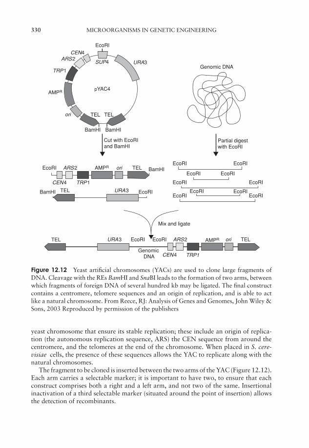
JWBK011-12 JWBK011-Hogg August 12, 2005 20:21 Char Count= 0
330 MICROORGANISMS IN GENETIC ENGINEERING
EcoRI
Genomic DNA
Partial digest with EcoRI
EcoRIEcoRI
EcoRI
EcoRI
EcoRI EcoRIEcoRI
EcoRI EcoRI
GenomicDNA
Mix and ligate
EcoRI
EcoRI
EcoRI
EcoRI
EcoRI
AMPR
AMPR
AMPR
pYAC4
SUP4ARS2
ARS2
ARS2
URA3
URA3
CEN4 TRP1
URA3
TRP1
ori
ori
ori
CEN4
TRP1CEN4
TEL
TEL
TEL
BamHI BamHI
BamHI
BamHI
Cut with EcoRI and BamHI
TEL
TEL
TEL
Figure 12.12 Yeast artificial chromosomes (YACs) are used to clone large fragments ofDNA. Cleavage with the REs BamHI and SnaBI leads to the formation of two arms, betweenwhich fragments of foreign DNA of several hundred kb may be ligated. The final constructcontains a centromere, telomere sequences and an origin of replication, and is able to actlike a natural chromosome. From Reece, RJ: Analysis of Genes and Genomes, John Wiley &Sons, 2003 Reproduced by permission of the publishers
yeast chromosome that ensure its stable replication; these include an origin of replica-tion (the autonomous replication sequence, ARS) the CEN sequence from around thecentromere, and the telomeres at the end of the chromosome. When placed in S. cere-visiae cells, the presence of these sequences allows the YAC to replicate along with thenatural chromosomes.
The fragment to be cloned is inserted between the two arms of the YAC (Figure 12.12).Each arm carries a selectable marker; it is important to have two, to ensure that eachconstruct comprises both a right and a left arm, and not two of the same. Insertionalinactivation of a third selectable marker (situated around the point of insertion) allowsthe detection of recombinants.
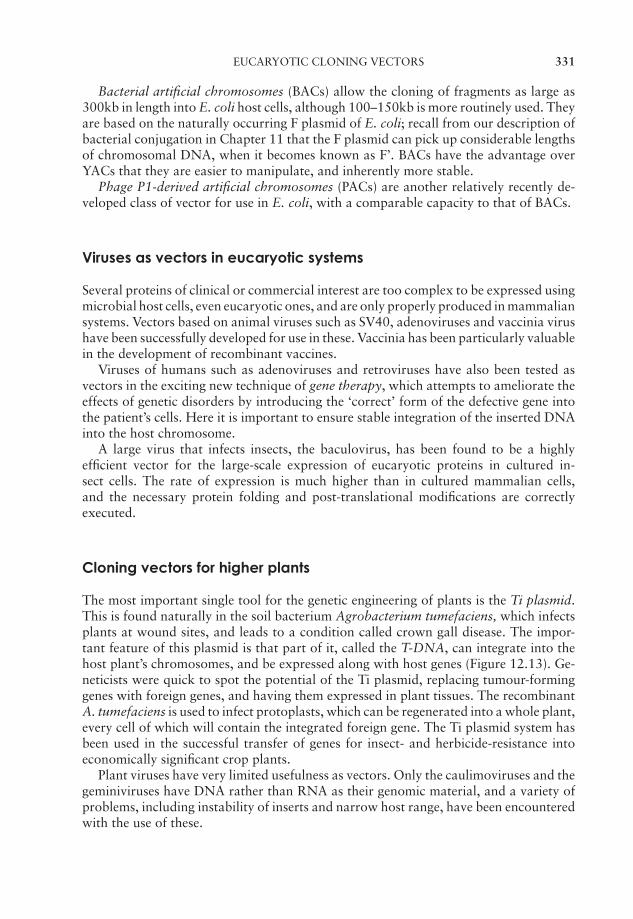
JWBK011-12 JWBK011-Hogg August 12, 2005 20:21 Char Count= 0
EUCARYOTIC CLONING VECTORS 331
Bacterial artificial chromosomes (BACs) allow the cloning of fragments as large as300kb in length into E. coli host cells, although 100–150kb is more routinely used. Theyare based on the naturally occurring F plasmid of E. coli; recall from our description ofbacterial conjugation in Chapter 11 that the F plasmid can pick up considerable lengthsof chromosomal DNA, when it becomes known as F’. BACs have the advantage overYACs that they are easier to manipulate, and inherently more stable.
Phage P1-derived artificial chromosomes (PACs) are another relatively recently de-veloped class of vector for use in E. coli, with a comparable capacity to that of BACs.
Viruses as vectors in eucaryotic systems
Several proteins of clinical or commercial interest are too complex to be expressed usingmicrobial host cells, even eucaryotic ones, and are only properly produced in mammaliansystems. Vectors based on animal viruses such as SV40, adenoviruses and vaccinia virushave been successfully developed for use in these. Vaccinia has been particularly valuablein the development of recombinant vaccines.
Viruses of humans such as adenoviruses and retroviruses have also been tested asvectors in the exciting new technique of gene therapy, which attempts to ameliorate theeffects of genetic disorders by introducing the ‘correct’ form of the defective gene intothe patient’s cells. Here it is important to ensure stable integration of the inserted DNAinto the host chromosome.
A large virus that infects insects, the baculovirus, has been found to be a highlyefficient vector for the large-scale expression of eucaryotic proteins in cultured in-sect cells. The rate of expression is much higher than in cultured mammalian cells,and the necessary protein folding and post-translational modifications are correctlyexecuted.
Cloning vectors for higher plants
The most important single tool for the genetic engineering of plants is the Ti plasmid.This is found naturally in the soil bacterium Agrobacterium tumefaciens, which infectsplants at wound sites, and leads to a condition called crown gall disease. The impor-tant feature of this plasmid is that part of it, called the T-DNA, can integrate into thehost plant’s chromosomes, and be expressed along with host genes (Figure 12.13). Ge-neticists were quick to spot the potential of the Ti plasmid, replacing tumour-forminggenes with foreign genes, and having them expressed in plant tissues. The recombinantA. tumefaciens is used to infect protoplasts, which can be regenerated into a whole plant,every cell of which will contain the integrated foreign gene. The Ti plasmid system hasbeen used in the successful transfer of genes for insect- and herbicide-resistance intoeconomically significant crop plants.
Plant viruses have very limited usefulness as vectors. Only the caulimoviruses and thegeminiviruses have DNA rather than RNA as their genomic material, and a variety ofproblems, including instability of inserts and narrow host range, have been encounteredwith the use of these.
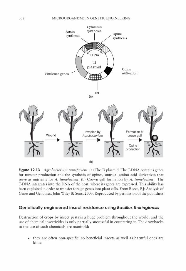
JWBK011-12 JWBK011-Hogg August 12, 2005 20:21 Char Count= 0
332 MICROORGANISMS IN GENETIC ENGINEERING
Auxinsynthesis Opine
synthesis
Opineutilisation
ori
Virulence genes
Cytokininsynthesis
(a)
T DNA
Tiplasmid
WoundInvasion by
AgrobacteriumFormation of
crown gall
Opineproduction
(b)
Figure 12.13 Agrobacterium tumefaciens. (a) The Ti plasmid. The T-DNA contains genesfor tumour production and the synthesis of opines, unusual amino acid derivatives thatserve as nutrients for A. tumefaciens. (b) Crown gall formation by A. tumefaciens. TheT-DNA integrates into the DNA of the host, where its genes are expressed. This ability hasbeen exploited in order to transfer foreign genes into plant cells. From Reece, RJ: Analysis ofGenes and Genomes, John Wiley & Sons, 2003. Reproduced by permission of the publishers
Genetically engineered insect resistance using Bacillus thuringiensis
Destruction of crops by insect pests is a huge problem throughout the world, and theuse of chemical insecticides is only partially successful in countering it. The drawbacksto the use of such chemicals are manifold:
� they are often non-specific, so beneficial insects as well as harmful ones arekilled

JWBK011-12 JWBK011-Hogg August 12, 2005 20:21 Char Count= 0
POLYMERASE CHAIN REACTION (PCR) 333
� they are often non-biodegradable, so they can have lasting environmental ef-fects
� aerial spraying only reaches upper leaf surfaces
� resistance develops with continued use.
A form of natural insecticide does exist; it is a crystalline protein produced duringsporulation by Bacillus thuringiensis. This is highly toxic to insects when convertedto its active form by the enzymes of the gut. This δ-endotoxin is relatively selective;different strains of B. thuringiensis produce different forms of the toxin, which areeffective against the larvae of different insects. Surprising as it may seem, the use of theδ-endotoxin as an insecticide was patented a hundred years ago; however, the successof it has been limited due to a variety of practical considerations.
The development of genetic engineering techniques has meant that attention hasturned in more recent times to introducing the genes for the δ-endotoxin into the cropsthemselves, so that they synthesise their own insecticide. This has been achieved withsome success, but the problem of the insects building up resistance remains.
As a spin-off from the Human Genome Project, the genetic make-up of many microor-ganisms has been elucidated. One of these is Photorhabdus luminescens, which encodesa toxin lethal to the two species of mosquito responsible for the spread of malaria, andit is hoped that determining the sequence of the gene will lead to applications in insectcontrol.
Polymerase chain reaction (PCR)
First described in the mid-1980s by Kary Mullis, PCR is probably the most significantdevelopment in molecular biology since the advent of gene cloning. PCR allows us toamplify a specific section of a genome (for example a particular gene) millions of times
PCR selectively repli-cates a specific DNA se-quence by means of invitro enzymatic amplifi-cation.
from a tiny amount of starting material (theoreticallya single molecule!). The impact of this powerful andhighly specific process has been felt in all areas of biologyand beyond. Medicine, forensic science and evolutionarystudies are but three areas where PCR has opened upnew possibilities over the last 15 years or so. To appre-ciate how PCR works, you will need to understand therole of the enzyme DNA polymerase in DNA replication,as outlined in the previous chapter. This is the enzyme,you’ll recall, that when provided with single-strandedDNA and a short primer, can direct the synthesis of acomplementary second strand. Figure 12.14 illustratesthe three steps in the PCR process:
A primer is a short nu-cleotide sequence thatcan be extended byDNA polymerase. InPCR, synthetic primersare designed to flank ei-ther side of the target se-quence.
� Denaturation: by heating to 95 ◦C, the DNA is sepa-rated into single strands, providing a template for theDNA polymerase.
JWBK011-12 JWBK011-Hogg August 12, 2005 20:21 Char Count= 0
334 MICROORGANISMS IN GENETIC ENGINEERING
3’5’
5’3’
5’
5’
3’
Primers
3’
5’
5’
3’
1. Denature at 95o
2. Anneal primers at 50-60o
3’
5’
5’
3’
5’
5’
3’
3. Extend primers at 72oThermostable DNApolymerase
Figure 12.14 The polymerase chain reaction (PCR). Millions of copies of a specific se-quence of DNA can be made by PCR. Samples are subjected to repeated cycles of denatu-ration, annealing of primers and extension by a thermostable DNA polymerase (for detailssee the text). At the end of the first cycle (shown in the diagram), each of the four strandscan serve as a template for replication in cycle two. After 30 cycles, over a billion copies ofthe target sequence will have been made
� Primer annealing: at a lower temperature (typically 50–60 ◦C, the exact value de-pends on the primer sequence), short single-stranded primers are allowed to attach.Primers are synthetic oligonucleotides with a sequence complementary to the regionsflanking the region we wish to amplify. One primer anneals to each strand, at eitherside of the target sequence.
� Polymerase extension: at around 72 ◦C, the DNA polymerase extends the primer byadding complementary nucleotides and so forms a second strand.
The net result of this process is that instead of one double-stranded molecule, we nowhave two. We now come to the key concept of PCR; if we raise the temperature to 95 ◦Cagain to start another cycle, we will have not two, but four single-stranded templates towork on, each of which can be converted to the double-stranded form as before. After20 such cycles we should have, in theory, over a million copies of our original molecule!Typical PCR protocols run for 30–35 cycles. All this can be achieved in just a coupleof hours; the temperature cycling is carried out by a programmable microprocessor-controlled machine called a thermal cycler.
PCR has widespread applications in microbiology, as in other fields of biology, butin addition, microorganisms play a crucial role in the process itself. If you have read the

JWBK011-12 JWBK011-Hogg August 12, 2005 20:21 Char Count= 0
TEST YOURSELF 335
above description of PCR carefully, something should have struck you as being not quiteright: how can the DNA polymerase (a protein) tolerate being repeatedly heated to over90 ◦C, and what sort of an enzyme can work effectively at 72 ◦C? The answer is thatthe DNA polymerase used in PCR comes from thermophilic bacteria such as Thermusaquaticus, a species found naturally in hot springs. The optimum temperature for Taqpolymerase is 72 ◦C, and it can tolerate being raised to values considerably higher thanthis for short periods. The availability of this enzyme meant that it was not necessaryto add fresh enzyme after each cycle, and the whole process could be automated, a keyfactor in its subsequent phenomenal success.
Test yourself
1 are enzymes that cut DNA at specific se-quences in the backbone.
2 Joining together DNA fragments from different sources produces amolecule.
3 DNA fragments with tend to join together because oftheir complementary single-stranded overhangs.
4 In order to gain access to a host cell and be replicated, a fragment of DNAmust be taken up by a molecule.
5 enable us to detect which host cells have been trans-formed. A commonly used example is resistance to . By insert-ing foreign DNA at this point, recombinants can be detected by
.
6 Another name for a polylinker is a .
7 A short sequence of labelled single-stranded DNA used to locate a specifictarget sequence is known as a .
8 The removable central section of the phage Lambda genome is known as afragment.
9 Bacteriophage is a useful vector if DNA is required in aform.
10 Cloning vectors combining features of plasmids and phage Lambda are called.
11 vectors incorporate foreign DNA at a site that is surrounded byessential signal sequences.
JWBK011-12 JWBK011-Hogg August 12, 2005 20:21 Char Count= 0
336 MICROORGANISMS IN GENETIC ENGINEERING
12 E. coli is not a suitable host for the expression of complex eucaryotic proteinsbecause it lacks the molecular machinery to carry out modificationssuch as .
13 DNA contains only those sequences that are involved in codingfor a polypeptide product.
14 Yeast plasmids are based on a naturally occurring sequence called the.
15 vectors are able to replicate in both and cells.
16 A virus found to be useful in the expression of eucaryotic genes is the. It is grown in cultured cells.
17 The soil bacterium A. tumefaciens contains a plasmid called , whichcauses crown gall disease. Part of it, the , integrates into hostchromosomes.
18 Genes from Bacillus thuringiensis coding for have been introducedinto plant cells, making them resistant.
19 The polymerase chain reaction (PCR) comprises repeated cycles of the follow-ing three steps: , of primers and byDNA polymerase.
20 PCR results in an increase in the number of copies of the targetsequence.

JWBK011-13 JWBK011-Hogg August 12, 2005 16:31 Char Count= 0
Part VControl of Microorganisms
337

JWBK011-13 JWBK011-Hogg August 12, 2005 16:31 Char Count= 0
338
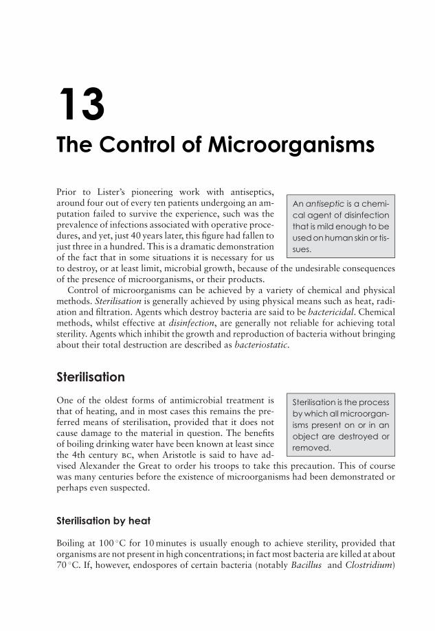
JWBK011-13 JWBK011-Hogg August 12, 2005 16:31 Char Count= 0
13The Control of Microorganisms
An antiseptic is a chemi-cal agent of disinfectionthat is mild enough to beused on human skin or tis-sues.
Prior to Lister’s pioneering work with antiseptics,around four out of every ten patients undergoing an am-putation failed to survive the experience, such was theprevalence of infections associated with operative proce-dures, and yet, just 40 years later, this figure had fallen tojust three in a hundred. This is a dramatic demonstrationof the fact that in some situations it is necessary for usto destroy, or at least limit, microbial growth, because of the undesirable consequencesof the presence of microorganisms, or their products.
Control of microorganisms can be achieved by a variety of chemical and physicalmethods. Sterilisation is generally achieved by using physical means such as heat, radi-ation and filtration. Agents which destroy bacteria are said to be bactericidal. Chemicalmethods, whilst effective at disinfection, are generally not reliable for achieving totalsterility. Agents which inhibit the growth and reproduction of bacteria without bringingabout their total destruction are described as bacteriostatic.
Sterilisation
Sterilisation is the processby which all microorgan-isms present on or in anobject are destroyed orremoved.
One of the oldest forms of antimicrobial treatment isthat of heating, and in most cases this remains the pre-ferred means of sterilisation, provided that it does notcause damage to the material in question. The benefitsof boiling drinking water have been known at least sincethe 4th century bc, when Aristotle is said to have ad-vised Alexander the Great to order his troops to take this precaution. This of coursewas many centuries before the existence of microorganisms had been demonstrated orperhaps even suspected.
Sterilisation by heat
Boiling at 100 ◦C for 10 minutes is usually enough to achieve sterility, provided thatorganisms are not present in high concentrations; in fact most bacteria are killed at about70 ◦C. If, however, endospores of certain bacteria (notably Bacillus and Clostridium)
339
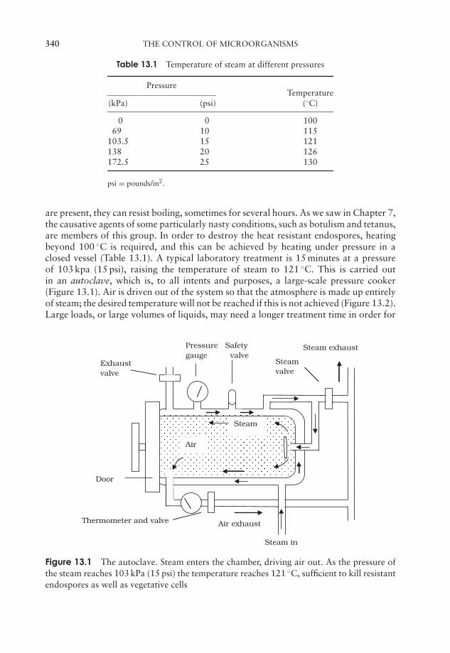
JWBK011-13 JWBK011-Hogg August 12, 2005 16:31 Char Count= 0
340 THE CONTROL OF MICROORGANISMS
Table 13.1 Temperature of steam at different pressures
PressureTemperature
(kPa) (psi) (◦C)
0 0 10069 10 115
103.5 15 121138 20 126172.5 25 130
psi = pounds/in2.
are present, they can resist boiling, sometimes for several hours. As we saw in Chapter 7,the causative agents of some particularly nasty conditions, such as botulism and tetanus,are members of this group. In order to destroy the heat resistant endospores, heatingbeyond 100 ◦C is required, and this can be achieved by heating under pressure in aclosed vessel (Table 13.1). A typical laboratory treatment is 15 minutes at a pressureof 103 kpa (15 psi), raising the temperature of steam to 121 ◦C. This is carried outin an autoclave, which is, to all intents and purposes, a large-scale pressure cooker(Figure 13.1). Air is driven out of the system so that the atmosphere is made up entirelyof steam; the desired temperature will not be reached if this is not achieved (Figure 13.2).Large loads, or large volumes of liquids, may need a longer treatment time in order for
Door
Steam
Air
Air exhaust
Steam exhaust
Thermometer and valve
Steamvalve
Steam in
Exhaustvalve
Pressure Safetygauge valve
Figure 13.1 The autoclave. Steam enters the chamber, driving air out. As the pressure ofthe steam reaches 103 kPa (15 psi) the temperature reaches 121 ◦C, sufficient to kill resistantendospores as well as vegetative cells
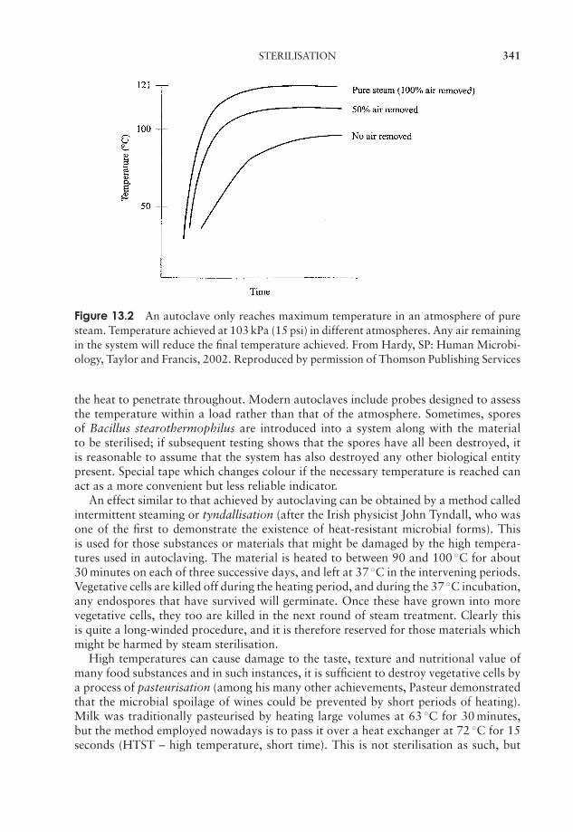
JWBK011-13 JWBK011-Hogg August 12, 2005 16:31 Char Count= 0
STERILISATION 341
Figure 13.2 An autoclave only reaches maximum temperature in an atmosphere of puresteam. Temperature achieved at 103 kPa (15 psi) in different atmospheres. Any air remainingin the system will reduce the final temperature achieved. From Hardy, SP: Human Microbi-ology, Taylor and Francis, 2002. Reproduced by permission of Thomson Publishing Services
the heat to penetrate throughout. Modern autoclaves include probes designed to assessthe temperature within a load rather than that of the atmosphere. Sometimes, sporesof Bacillus stearothermophilus are introduced into a system along with the materialto be sterilised; if subsequent testing shows that the spores have all been destroyed, itis reasonable to assume that the system has also destroyed any other biological entitypresent. Special tape which changes colour if the necessary temperature is reached canact as a more convenient but less reliable indicator.
An effect similar to that achieved by autoclaving can be obtained by a method calledintermittent steaming or tyndallisation (after the Irish physicist John Tyndall, who wasone of the first to demonstrate the existence of heat-resistant microbial forms). Thisis used for those substances or materials that might be damaged by the high tempera-tures used in autoclaving. The material is heated to between 90 and 100 ◦C for about30 minutes on each of three successive days, and left at 37 ◦C in the intervening periods.Vegetative cells are killed off during the heating period, and during the 37 ◦C incubation,any endospores that have survived will germinate. Once these have grown into morevegetative cells, they too are killed in the next round of steam treatment. Clearly thisis quite a long-winded procedure, and it is therefore reserved for those materials whichmight be harmed by steam sterilisation.
High temperatures can cause damage to the taste, texture and nutritional value ofmany food substances and in such instances, it is sufficient to destroy vegetative cells bya process of pasteurisation (among his many other achievements, Pasteur demonstratedthat the microbial spoilage of wines could be prevented by short periods of heating).Milk was traditionally pasteurised by heating large volumes at 63 ◦C for 30 minutes,but the method employed nowadays is to pass it over a heat exchanger at 72 ◦C for 15seconds (HTST – high temperature, short time). This is not sterilisation as such, but
JWBK011-13 JWBK011-Hogg August 12, 2005 16:31 Char Count= 0
342 THE CONTROL OF MICROORGANISMS
it ensures the destruction of disease-causing organisms such as Brucella abortus andMycobacterium tuberculosis, which at one time were frequently found in milk, as wellas significantly reducing the organisms that cause food spoilage, thus prolonging thetime the milk can be kept. Some protocols exceed these minimum values in order toreduce further the microbial content of the milk. One type of milk on sale in the shopsis subjected to more extreme heating regimes; this is ‘UHT’ milk, which can be kept forseveral weeks without refrigeration, though many find that this is achieved at some costto its palatability! It is heated to ultra high temperatures (150 ◦C) for a couple of secondsusing superheated steam. The product is often referred to as being ‘sterilised’, but thisis not true in the strictest sense. Milk is not the only foodstuff to be pasteurised; otherssuch as beer, fruit juices and ice cream each has its own time/temperature combination.
All the above methods employ a combination of heat and moisture to achieve theireffect; the denaturation of proteins, upon which these methods depend, is enhanced inthe presence of water. Heat is more readily transferred through water than through air,and the main reason that endospores are so resistant is because of their very low watercontent. In some situations however, it is possible to employ dry heat, using an oven tosterilise metal instruments or glassware for example. It is a more convenient procedure,but a higher temperature (160–170 ◦C) and longer exposure time (2 hours) are required.Dry heat works by oxidising (‘burning’) the cell’s components. Microorganisms arequite literally burnt to destruction by the most extreme form of dry heat treatment,incineration. Soiled medical dressings and swabs, for example are potentially hazardous,and are destroyed in this way at many hundreds of degrees Celsius. As we saw in Box 4.1,sterilising the loops and needles used to manipulate microorganisms by means of flamingis a routine part of aseptic procedures.
Sterilisation by irradiation
Certain types of irradiation are used to control the growth of microorganisms. Theseinclude both ionising and non-ionising radiation.
The most widely used form of non-ionising radiation is ultraviolet (UV) light. Wave-lengths around 260 nm are used because these are absorbed by the purine and pyrimidinecomponents of nucleic acids, as well as certain aromatic amino acids in proteins. Theabsorbed energy causes a rupture of the chemical bonds, so that normal cellular func-tion is impaired. You will recall from Chapter 11 that UV light causes the formation ofthymine dimers (Figure 11.21), where adjacent thymine nucleotides on the same strandare linked together, inhibiting DNA replication. Although many bacteria are capable of
The world’s biggest UVwastewater treatmentplant was opened inManukau, New Zealandin 2001. Its 8000 UV lampsare able to treat up to50 000 cubic metres ofwater per hour.
repairing this damage by enzyme-mediated photoreac-tivation, viruses are much more susceptible. UV lampsare commonly found in food preparation areas, operat-ing theatres and specialist areas such as tissue culturefacilities, where it is important to prevent contamina-tion. Because they are also harmful to humans (particu-larly the skin and eyes), UV lamps can only be operatedin such areas when people are not present. UV radia-tion has very poor penetrating powers; a thin layer ofglass, paper or fabric is able to impede the passage of
JWBK011-13 JWBK011-Hogg August 12, 2005 16:31 Char Count= 0
STERILISATION 343
the rays. The chief application is therefore in the sterilisation of work surfaces and thesurrounding air, although it is increasingly finding an application in the treatment ofwater supplies.
Ionising radiations have a shorter wavelength and much higher energy, giving themgreater penetrating powers. The effect of ionising radiations is due to the productionof highly reactive free radicals, which disrupt the structure of macromolecules such asDNA and proteins. Surgical supplies such as syringes, catheters and rubber gloves arecommonly sterilised employing gamma (γ ) rays from the isotope cobalt 60 (60Co).
Gamma radiation has been approved for use in over 40 countries for the preservationof food, which it does not only by killing pathogens and spoilage organisms but alsoby inhibiting processes that lead to sprouting and ripening. The practice has arouseda lot of controversy, largely due to concerns about health and safety, although thefirst patent applications for its use date back nearly a hundred years! Although theirradiated product does not become radioactive, there is a general suspicion on the partof the public about anything to do with radiation, which has led to its use on foodbeing only very gradually accepted by consumers. In this respect Europe lags behindthe USA, where during the 1990s a positive attitude towards irradiation of food bothby professional bodies and the media has led to a more widespread acceptance of thetechnology. Gamma radiation is used in situations where heat sterilisation would beinappropriate, because of undesirable effects on the texture, taste or appearance of theproduct. This mainly relates to fresh produce such as meat, poultry, fruit and vegetables.Irradiation is not suitable for some foodstuffs, such as those with a high fat content,where unpleasant tastes and odours result. Ionising radiations have the great advantageover other methods of sterilisation that they can penetrate packaging.
Filtration
Many liquids such as solutions of antibiotics or certain components of culture mediabecome chemically altered at high temperatures, so the use of any of the heat regimesdescribed above is not appropriate. Rather than killing the microorganisms, an alter-native approach is simply to isolate them. This can be done for liquids and gases bypassing them through filters of an appropriate pore size. Filters used to be made frommaterials such as asbestos and sintered glass, but have been largely replaced by mem-brane filters, commonly made of nitrocellulose or polycarbonate (Figure 13.3). Thesecan be purchased ready-sterilised and the liquid passed through by means of pressureor suction. Supplies of air or other gases can also be filter-sterilised in this way. A poresize of 0.22 µm is commonly used; this will remove bacteria plus, of course, anythingbigger, such as yeasts; however, mycoplasma and viruses are able to pass through poresof this size. With a pore size 10 times smaller than this, only the smallest of viruses canpass through, so it is important that an appropriate pore size is chosen for any giventask. A drawback with all filters, but especially those of a small pore size, is that theycan become clogged easily. Filters in general are relatively expensive, and are not thepreferred choice if alternative methods are available.
High efficiency particulate air (HEPA) filters create clean atmospheres in areas suchas operating theatres and laboratory laminar-flow hoods.
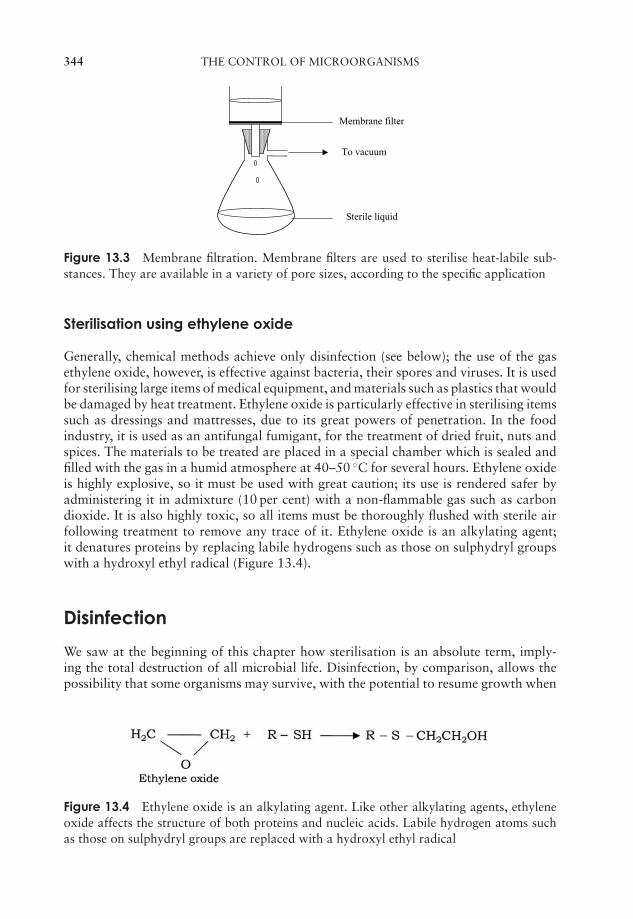
JWBK011-13 JWBK011-Hogg August 12, 2005 16:31 Char Count= 0
344 THE CONTROL OF MICROORGANISMS
To vacuum
Membrane filter
Sterile liquid
Figure 13.3 Membrane filtration. Membrane filters are used to sterilise heat-labile sub-stances. They are available in a variety of pore sizes, according to the specific application
Sterilisation using ethylene oxide
Generally, chemical methods achieve only disinfection (see below); the use of the gasethylene oxide, however, is effective against bacteria, their spores and viruses. It is usedfor sterilising large items of medical equipment, and materials such as plastics that wouldbe damaged by heat treatment. Ethylene oxide is particularly effective in sterilising itemssuch as dressings and mattresses, due to its great powers of penetration. In the foodindustry, it is used as an antifungal fumigant, for the treatment of dried fruit, nuts andspices. The materials to be treated are placed in a special chamber which is sealed andfilled with the gas in a humid atmosphere at 40–50 ◦C for several hours. Ethylene oxideis highly explosive, so it must be used with great caution; its use is rendered safer byadministering it in admixture (10 per cent) with a non-flammable gas such as carbondioxide. It is also highly toxic, so all items must be thoroughly flushed with sterile airfollowing treatment to remove any trace of it. Ethylene oxide is an alkylating agent;it denatures proteins by replacing labile hydrogens such as those on sulphydryl groupswith a hydroxyl ethyl radical (Figure 13.4).
Disinfection
We saw at the beginning of this chapter how sterilisation is an absolute term, imply-ing the total destruction of all microbial life. Disinfection, by comparison, allows thepossibility that some organisms may survive, with the potential to resume growth when
Figure 13.4 Ethylene oxide is an alkylating agent. Like other alkylating agents, ethyleneoxide affects the structure of both proteins and nucleic acids. Labile hydrogen atoms suchas those on sulphydryl groups are replaced with a hydroxyl ethyl radical
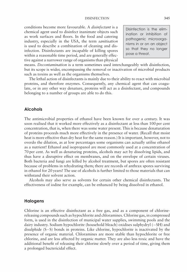
JWBK011-13 JWBK011-Hogg August 12, 2005 16:31 Char Count= 0
DISINFECTION 345
Disinfection is the elim-ination or inhibition ofpathogenic microorga-nisms in or on an objectso that they no longerpose a threat.
conditions become more favourable. A disinfectant is achemical agent used to disinfect inanimate objects suchas work surfaces and floors. In the food and cateringindustry, especially in the USA, the term sanitisationis used to describe a combination of cleaning and dis-infection. Disinfectants are incapable of killing sporeswithin a reasonable time period, and are generally effec-tive against a narrower range of organisms than physicalmeans. Decontamination is a term sometimes used interchangeably with disinfection,but its scope is wider, encompassing the removal or inactivation of microbial productssuch as toxins as well as the organisms themselves.
The lethal action of disinfectants is mainly due to their ability to react with microbialproteins, and therefore enzymes. Consequently, any chemical agent that can coagu-late, or in any other way denature, proteins will act as a disinfectant, and compoundsbelonging to a number of groups are able to do this.
Alcohols
The antimicrobial properties of ethanol have been known for over a century. It wassoon realised that it worked more effectively as a disinfectant at less than 100 per centconcentration, that is, when there was some water present. This is because denaturationof proteins proceeds much more effectively in the presence of water. (Recall that moistheat is more effective than dry heat for the same reason.) It is important, however, not tooverdo the dilution, as at low percentages some organisms can actually utilise ethanolas a nutrient! Ethanol and isopropanol are most commonly used at a concentration of70 per cent. As well as denaturing proteins, alcohols may act by dissolving lipids, andthus have a disruptive effect on membranes, and on the envelope of certain viruses.Both bacteria and fungi are killed by alcohol treatment, but spores are often resistantbecause of problems in rehydrating them; there are records of anthrax spores survivingin ethanol for 20 years! The use of alcohols is further limited to those materials that canwithstand their solvent action.
Alcohols may also serve as solvents for certain other chemical disinfectants. Theeffectiveness of iodine for example, can be enhanced by being dissolved in ethanol.
Halogens
Chlorine is an effective disinfectant as a free gas, and as a component of chlorine-releasing compounds such as hypochlorite and chloramines. Chlorine gas, in compressedform, is used in the disinfection of municipal water supplies, swimming pools and thedairy industry. Sodium hypochlorite (household bleach) oxidises sulphydryl (−SH) anddisulphide (S−S) bonds in proteins. Like chlorine, hypochlorite is inactivated by thepresence of organic material. Chloramines are more stable than hypochlorite or freechlorine, and are less affected by organic matter. They are also less toxic and have theadditional benefit of releasing their chlorine slowly over a period of time, giving thema prolonged bactericidal effect.
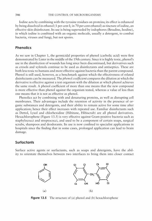
JWBK011-13 JWBK011-Hogg August 12, 2005 16:31 Char Count= 0
346 THE CONTROL OF MICROORGANISMS
Iodine acts by combining with the tyrosine residues on proteins; its effect is enhancedby being dissolved in ethanol (1 per cent I2 in 70 per cent ethanol) as tincture of iodine, aneffective skin disinfectant. Its use is being superseded by iodophores (Betadine, Isodine),in which iodine is combined with an organic molecule, usually a detergent, to combatbacteria, viruses and fungi, but not spores.
Phenolics
As we saw in Chapter 1, the germicidal properties of phenol (carbolic acid) were firstdemonstrated by Lister in the middle of the 19th century. Since it is highly toxic, phenol’suse in the disinfection of wounds has long since been discontinued, but derivatives suchas cresols and xylenols continue to be used as disinfectants and antiseptics. These areboth less toxic to humans and more effective against bacteria than the parent compound.Phenol is still used, however, as a benchmark against which the effectiveness of relateddisinfectants can be measured. The phenol coefficient compares the dilution at which thederivative is effective against a test organism with the dilution at which phenol achievesthe same result. A phenol coefficient of more than one means that the new compoundis more effective than phenol against the organism tested, whereas a value of less thanone means that it is not as effective as phenol.
Phenolics act by combining with and denaturing proteins, as well as disrupting cellmembranes. Their advantages include the retention of activity in the presence of or-ganic substances and detergents, and their ability to remain active for some time afterapplication; hence their effect increases with repeated use. Familiar disinfectants suchas Dettol, Lysol and chlorhexidine (Hibitane, Hibiscrub) are all phenol derivatives.Hexachlorophene (Figure 13.5) is very effective against Gram-positive bacteria such asstaphylococci and streptococci, and used to be a component of certain soaps, surgicalscrubs, shampoos and deodorants. Its use is now confined to specialist applications inhospitals since the finding that in some cases, prolonged application can lead to braindamage.
Surfactants
Surface active agents or surfactants, such as soaps and detergents, have the abil-ity to orientate themselves between two interfaces to bring them into closer contact
Figure 13.5 The structure of (a) phenol and (b) hexachlorophene
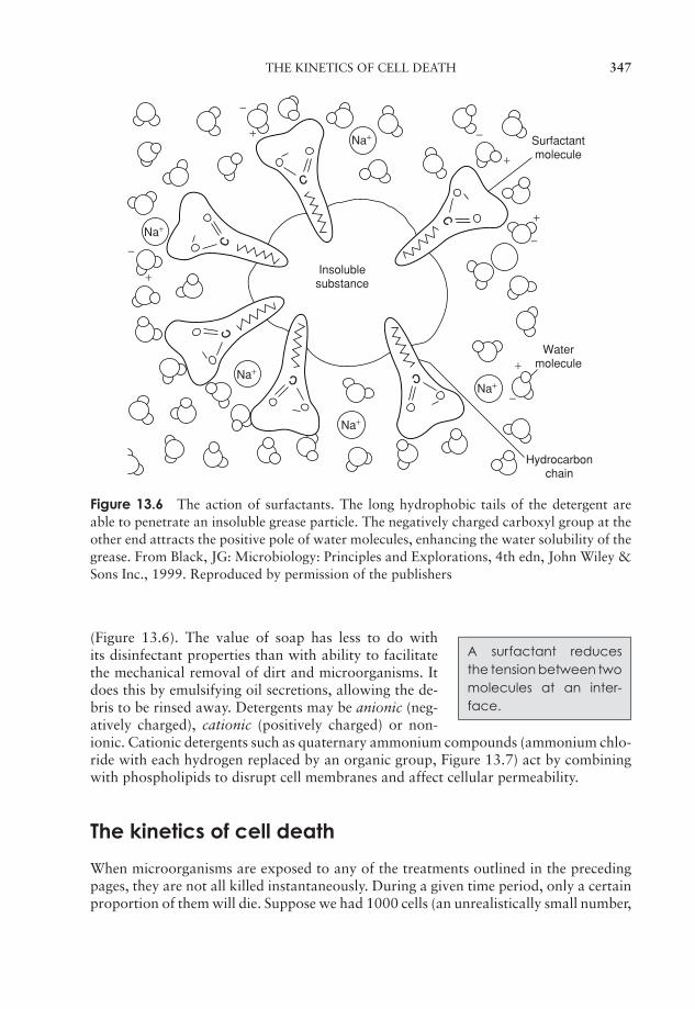
JWBK011-13 JWBK011-Hogg August 12, 2005 16:31 Char Count= 0
THE KINETICS OF CELL DEATH 347
c
cc
c c
cInsolublesubstance
Surfactantmolecule
Watermolecule
Hydrocarbonchain
+
−Na+
Na+
Na+
Na+
Na++
−
−
−
+
+
−
+
Figure 13.6 The action of surfactants. The long hydrophobic tails of the detergent areable to penetrate an insoluble grease particle. The negatively charged carboxyl group at theother end attracts the positive pole of water molecules, enhancing the water solubility of thegrease. From Black, JG: Microbiology: Principles and Explorations, 4th edn, John Wiley &Sons Inc., 1999. Reproduced by permission of the publishers
A surfactant reducesthe tension between twomolecules at an inter-face.
(Figure 13.6). The value of soap has less to do withits disinfectant properties than with ability to facilitatethe mechanical removal of dirt and microorganisms. Itdoes this by emulsifying oil secretions, allowing the de-bris to be rinsed away. Detergents may be anionic (neg-atively charged), cationic (positively charged) or non-ionic. Cationic detergents such as quaternary ammonium compounds (ammonium chlo-ride with each hydrogen replaced by an organic group, Figure 13.7) act by combiningwith phospholipids to disrupt cell membranes and affect cellular permeability.
The kinetics of cell death
When microorganisms are exposed to any of the treatments outlined in the precedingpages, they are not all killed instantaneously. During a given time period, only a certainproportion of them will die. Suppose we had 1000 cells (an unrealistically small number,
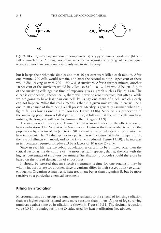
JWBK011-13 JWBK011-Hogg August 12, 2005 16:31 Char Count= 0
348 THE CONTROL OF MICROORGANISMS
Figure 13.7 Quaternary ammonium compounds. (a) cetylpyridinium chloride and (b) ben-zalkonium chloride. Although non-toxic and effective against a wide range of bacteria, qua-ternary ammonium compounds are easily inactivated by soap
but it keeps the arithmetic simple) and that 10 per cent were killed each minute. Afterone minute, 900 cells would remain, and after the second minute 10 per cent of thesewould die, leaving us with 900 − 90 = 810 survivors. After a further minute, another10 per cent of the survivors would be killed, so 810 − 81 = 729 would be left. A plotof the surviving cells against time of exposure gives a graph such as Figure 13.8. Thecurve is exponential; theoretically, there will never be zero survivors, but after a whilewe are going to have less than one cell, let us say one tenth of a cell, which clearlycan not happen. What this really means is that in a given unit volume, there will be aone in 10 chance of there being a cell present. Sterility is generally assumed when thisfigure falls as low as one in a million (see Figure 13.8b). Since only a proportion ofthe surviving population is killed per unit time, it follows that the more cells you haveinitially, the longer it will take to eliminate them (Figure 13.9).
The steepness of the slope in Figure 13.8(b) is an indication of the effectiveness ofheat sterilisation. The decimal reduction time or D value is the time needed to reduce thepopulation by a factor of ten (i.e. to kill 90 per cent of the population) using a particularheat treatment. The D value applies to a particular temperature; at higher temperatures,the rate of killing is enhanced, and so the Dvalue is reduced (Figure 13.10). The increasein temperature required to reduce D by a factor of 10 is the Z value.
Since in real life, the microbial population is certain to be a mixed one, then thecritical factor is the death rate of the most resistant species, that is, the one with thehighest percentage of survivors per minute. Sterilisation protocols should therefore bebased on the rate of destruction of endospores.
It should be stressed that an effective treatment regime for one organism may bewholly inappropriate for another, since organisms differ in their susceptibility to differ-ent agents. Organism A may resist heat treatment better than organism B, but be moresensitive to a particular chemical treatment.
Killing by irradiation
Microorganisms as a group are much more resistant to the effects of ionising radiationthan are higher organisms, and some more resistant than others. A plot of log survivingnumbers against time of irradiation is shown in Figure 13.11. The decimal reductionvalue (D 10) is analogous to the D value used for heat sterilisation (see above).

JWBK011-13 JWBK011-Hogg August 12, 2005 16:31 Char Count= 0
THE KINETICS OF CELL DEATH 349
Figure 13.8 The kinetics of cell death. (a) During heat sterilisation, the number of livingcells decreases by the same proportion per unit time, giving an exponential curve. (b) Whenplotted on a logarithmic scale, the decrease in numbers is seen as a straight line, whoseslope is a reflection of the rate of killing. The time period between A and A′ is the decimalreduction time (D): the time taken to reduce the population to one-tenth of its size. The totalperiod until the point SA is the sterility assurance value, when there is only a one in a millionprobability of any cells having survived. From Hardy, SP: Human Microbiology, Taylor andFrancis, 2002. Reproduced by permission of Thomson Publishing Services
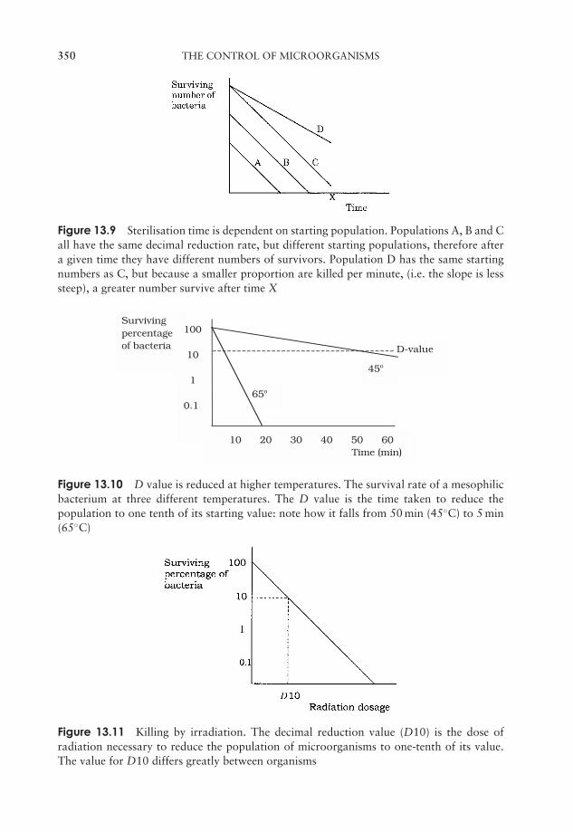
JWBK011-13 JWBK011-Hogg August 12, 2005 16:31 Char Count= 0
350 THE CONTROL OF MICROORGANISMS
Figure 13.9 Sterilisation time is dependent on starting population. Populations A, B and Call have the same decimal reduction rate, but different starting populations, therefore aftera given time they have different numbers of survivors. Population D has the same startingnumbers as C, but because a smaller proportion are killed per minute, (i.e. the slope is lesssteep), a greater number survive after time X
D-value
45o
65o
Survivingpercentageof bacteria
100
10
1
0.1
10 20 30 40 50 60Time (min)
Figure 13.10 D value is reduced at higher temperatures. The survival rate of a mesophilicbacterium at three different temperatures. The D value is the time taken to reduce thepopulation to one tenth of its starting value: note how it falls from 50 min (45◦C) to 5 min(65◦C)
Figure 13.11 Killing by irradiation. The decimal reduction value (D10) is the dose ofradiation necessary to reduce the population of microorganisms to one-tenth of its value.The value for D10 differs greatly between organisms
JWBK011-13 JWBK011-Hogg August 12, 2005 16:31 Char Count= 0
TEST YOURSELF 351
Test yourself
1 An antimicrobial agent that kills microorganisms is termed onethat inhibits their growth is described as .
2 Heat treatment at temperatures in excess of 100◦C is necessary to ensure thedestruction of .
3 Steam heated under a pressure of 103 kPa in an autoclave reaches a temper-ature of ◦C.
4 During tyndallisation, the material being treated is heated to between 90and 100◦C for about on three consecutive days. In the inter-vening periods, it is left at ◦C, to allow any survivingto .
5 The traditional method for the of milk was to heat atfor 30 minutes.
6 Moist heat is more effective than dry heat at the same temperature, becausewater is needed for the of .
7 Ultraviolet light is used mainly in the sterilisation of , because it hasvery poor power.
8 radiation is used in many countries to irradiate foodstuffs.
9 Liquids may be sterilised by the use of made of polycarbonate ornitrocellulose.
10 is a gas used in the sterilisation of hospital equipment.
11 After disinfection, there is a possibility that some microorganisms will.
12 Ethanol is most effective as a disinfectant at a concentration of .
13 A number of chemical disinfectants are more effective when dissolved in.
14 Chlorine and its derivatives are most widely used as disinfectants of.
15 Tincture of is an effective skin disinfectant.
16 The use of carbolic acid in operating theatres, introduced by ,reduced the number of fatalities due to infections.

JWBK011-13 JWBK011-Hogg August 12, 2005 16:31 Char Count= 0
352 THE CONTROL OF MICROORGANISMS
17 The efficacy of a disinfectant can be measured as its coefficient.
18 Soaps and detergents are examples of .
19 is assumed when it is reckoned to be less than a one in a millionchance of a microbial cell being present.
20 The time needed for a bacterial population to be reduced to one tenth at aparticular temperature is called the .
P1: FCG/SPH P2: FCG/SPH QC: FCG/SPH T1: SPH
JWBK011-14 JWBK011-Hogg August 12, 2005 16:32 Char Count= 0
14Antimicrobial Agents
In Chapter 1, we saw how by the end of the 19th century, the germ theory of infectiousdiseases had finally gained widespread acceptance. It is hardly surprising then, thatthe scientific community at that time should turn its collective mind towards ways ofcontrolling infections and the organisms that cause them. In the following pages weshall discover how the development of chemical agents targeted against pathogenicmicroorganisms had a dramatic impact on the treatment of infectious diseases in thetwentieth century.
The term chemotherapy is most closely associated in the minds of most people withthe treatment of cancer. In fact the term was first used by Paul Ehrlich to describe anyuse of a drug or other chemical substance for the treatment of disease; thus, it hasmuch wider terms of reference. In our present discussion, we shall confine ourselves tochemotherapy as it relates to the treatment of infectious diseases. It was Ehrlich who,100 years ago, observed how certain dyes would stain bacteria but not the surroundingtissues, leading him to formulate the idea of selective toxicity, whereby a substancewould selectively target harmful microorganisms but leave human tissues undamaged.He tested hundreds of synthetic compounds in the search for his ‘magic bullet’ beforefinding, in 1910, an arsenic-containing drug, Salvarsan, which was effective againstTreponema pallidum, the causative agent of syphilis (Figure 14.1).
It was another 20 years before another significant antimicrobial drug was developed,when the German chemist Gerhard Domagk showed a synthetic dye, Prontosil, to beactive against a range of Gram-positive bacteria. The active component of prontosilwas shown soon afterwards to be sulphanilamide. In the following decade, numer-ous derivatives of sulphanilamide were synthesised, many of which were more potentantimicrobial agents than the parent molecule. This class of compounds is known col-lectively as the sulphonamides, or sulfa drugs (Box 14.1). In the years leading up to theSecond World War, sulphonamides dramatically improved the mortality rates due topneumonia and puerperal fever.
A side-effect is an unde-sirable and unintendedeffect of a therapeutictreatment on the recipi-ent.
Nowadays, sulphonamides have largely been replacedby antibiotics because of their side-effects, and because,due to wholesale and indiscriminate use in the earlyyears, bacterial resistance to sulphonamides has becomewidespread. Some synthetic compounds are still usefulas antimicrobial agents, however. Isoniazid is one of theprincipal agents used in the treatment of tuberculosis. It
353
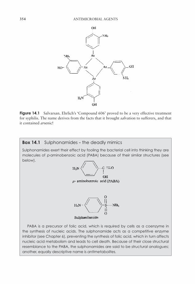
P1: FCG/SPH P2: FCG/SPH QC: FCG/SPH T1: SPH
JWBK011-14 JWBK011-Hogg August 12, 2005 16:32 Char Count= 0
354 ANTIMICROBIAL AGENTS
Figure 14.1 Salvarsan. Ehrlich’s ‘Compound 606’ proved to be a very effective treatmentfor syphilis. The name derives from the facts that it brought salvation to sufferers, and thatit contained arsenic!
Box 14.1 Sulphonamides -- the deadly mimics
Sulphonamides exert their effect by fooling the bacterial cell into thinking they aremolecules of p-aminobenzoic acid (PABA) because of their similar structures (seebelow).
PABA is a precursor of folic acid, which is required by cells as a coenzyme inthe synthesis of nucleic acids. The sulphonamide acts as a competitive enzymeinhibitor (see Chapter 6), preventing the synthesis of folic acid, which in turn affectsnucleic acid metabolism and leads to cell death. Because of their close structuralresemblance to the PABA, the sulphonamides are said to be structural analogues;another, equally descriptive name is antimetabolites.

P1: FCG/SPH P2: FCG/SPH QC: FCG/SPH T1: SPH
JWBK011-14 JWBK011-Hogg August 12, 2005 16:32 Char Count= 0
ANTIBIOTICS 355
is nearly always given in association with another antimicrobial agent because of thehigh incidence of resistant forms of the mycobacteria that cause the disease. Like thesulphonamides, isoniazid is a structural analogue, and is thought to inhibit the produc-tion of mycolic acid in the mycobacterial cell wall (see Chapter 7).
The quinolones are synthetic substances related to nalidixic acid, and includeciprofloxacin and norfloxacin. They interfere with nucleic acid synthesis by inhibitingDNA gyrase, the enzyme responsible for unwinding DNA prior to replication (Chapter11). Quinolones are used in the treatment of urinary infections, as are the nitrofurans.These are active against certain fungi and protozoans as well as a range of bacteria.
Antibiotics
An antibiotic is a mi-crobially produced sub-stance (or a syntheticderivative) that has an-timicrobial properties.
The other major breakthrough in the treatment of infec-tious diseases was of course the discovery of naturallyoccurring antimicrobial agents, or antibiotics. These aremetabolites produced by certain microorganisms, whichinhibit the growth of certain other microorganisms. Aswe shall see, the definition has been extended to in-clude semisynthetic derivatives of these naturally occur-ring molecules. Table 14.1 lists some commonly usedantibiotics.
One of the best known of all stories of scientific discovery is that of how Sir AlexanderFleming discovered penicillin in 1928. Before we consider that, however, it is worthnoting that a number of treatments for infectious diseases practised over the preceding
Table 14.1 Some antibiotics and their microbial source
Antibiotic Microbial Source
Bacteria (Gram-positive)Bacitracin Bacillus subtilisPolymixin Bacillus polymixa
ActinomycetesGentamicin Micromonospora purpureaActinomycin D Streptomyces parvulusErythromycin Streptomyces erythreus
StreptomycesNystatin Streptomyces nourseiRifamycin Streptomyces mediterraneiStreptomycin Streptomyces griseusTetracycline Streptomyces rimosusVancomycin Streptomyces orientalis
FungiCephalosporins Cephalosporium acremoniumGriseofulvin Penicillium griseofulvumPenicillin Penicillium chrysogenum
P1: FCG/SPH P2: FCG/SPH QC: FCG/SPH T1: SPH
JWBK011-14 JWBK011-Hogg August 12, 2005 16:32 Char Count= 0
356 ANTIMICROBIAL AGENTS
Box 14.2 Was Fleming lucky?
In the frequent retelling of the discovery of penicillin, much is made of the roleplayed by chance. It is true that the Penicillium mould in Fleming’spetri dish was anaccidental contaminant, but arguably the real stroke of luck was not so much thefact that the contamination occurred but that it was observed by somebody whoimmediately recognised its significance.
centuries can, with the benefit of hindsight, be regarded as a form of antibiotic therapy.Many hundreds of years ago for example, the Chinese used mouldy soybean curd in thetreatment of boils and South American Indians controlled foot infections by wearingsandals which had become furry with mould! In the late 19th century, Tyndall (seeChapter 13) made the observation that a culture medium cloudy with bacterial growthwould clear when mould grew on the surface. Around the same time Pasteur and Joubertdemonstrated that cultured anthrax bacilli could be inactivated in the presence of certainother microorganisms from the environment. By the early 1920s the search was on forthe isolation of a microbially produced antibacterial agent, and Gratia and Dath isolateda substance from a soil actinomycete which came to be known as actinomycin. However,although potent against a number of pathogens, actinomycin is too toxic to be usefultherapeutically.
Fleming was also looking for a naturally occurring antimicrobial agent. On one occa-sion, he noticed that a plate culture of Staphylococcus aureus had become contaminatedby the growth of a mould; around it were clear areas, where the S. aureus did not grow.The mould was subsequently identified as Penicillium notatum. and the substance thathad diffused through the agar from it, preventing bacterial growth, became knownas penicillin. Further investigation revealed that broth from a culture of the Penicil-lium mould was inhibitory towards the growth of a number of other Gram-positivepathogens, and remained so even when diluted several hundred times. Critically, whentested on mice, it was, for the most part, harmless.
When it came to purifying the active ingredient and using it in vivo however, a num-ber of problems were encountered. The penicillin proved to be impure, only produced inminute amounts, and unstable in the acid conditions of the stomach, thereby limiting itstherapeutic potential. After publishing a few papers on the subject, Fleming ceased workon penicillin and it was left to Howard Florey and Ernst Chain in 1939 to take up thechallenge of producing it in sufficient quantities and in a pure enough form for therapeu-tic use. Early work in Oxford had to continue in the United States because of the Germanair raids in Britain. The American entry into the Second World War in late 1941 meantthat the development of penicillin was awarded war project status, giving it greatlyadded impetus. As a result of their endeavours, the yield of penicillin rose hugely (seeBox 14.3), and in 1945, Fleming, Chain and Florey shared the Nobel Prize for their work.
Other antibiotics were also isolated during this period, most notably streptomycin,isolated by Selman Waksman from Streptomyces griseus, which was to prove so effectiveagainst tuberculosis.
In 1942 there was only enough penicillin in the world to treat a few hundred in-dividuals, but by the end of the Second World War production had grown to such anextent that 7 million people a year could be treated. By the mid-1950s, such well-known

P1: FCG/SPH P2: FCG/SPH QC: FCG/SPH T1: SPH
JWBK011-14 JWBK011-Hogg August 12, 2005 16:32 Char Count= 0
ANTIBIOTICS 357
Box 14.3 How did Florey and Chain improve the yields of penicillin?
The work of Florey and Chain resulted in pure penicillin being produced on a largescale, suitable for therapeutic use. Among their achievements were:
� isolation of a better penicillin-producing species (P. chrysogenum, famously iso-lated from a mouldy cantaloupe melon in Peoria Illinois!) and selection of mutantstrains induced by X-rays and UV irradiation
� development of submerged culture technique, with sterile air being forcedthrough the medium to supply essential oxygen
� improvements in medium composition
� addition of precursors to the medium
� refinements to recovery methods.
antibiotics as tetracycline, chloramphenicol and neomycin had been isolated. The dis-covery of a few naturally occurring compounds had revolutionised the treatment ofinfectious diseases.
New antibiotics are still being sought today; in Chapter 17 we discuss the stages inthe isolation, testing and development of a putative new antibiotic. Of the thousandsisolated so far, only a small proportion have proved to be of any real therapeutic orcommercial value. This is because, like the actinomycin mentioned above, most of thesubstances isolated harm not only bacteria but humans too.
A key prerequisite for any chemotherapeutic agent is selective toxicity. An obviousway of achieving this is for a compound to direct its effect against a metabolic orphysiological function found in microbial cells but not in the host. We shall look at someexamples of this later in this chapter. Those chemotherapeutic agents which inhibit thesame process in host as in pathogen, or which cause harm to the host in some other way,are said to have side-effects. These may include directly toxic effects, hypersensitivity(allergic) reactions or adverse effects on the host’s normal resident microflora.
One of the reasons why penicillin was, and continues to be, so successful, was thatthe target of its action is unique to bacteria, so it its degree of selective toxicity is high.
What other properties should an antibiotic have?
Selective toxicity is the most important single attribute of an antibiotic, but ideally itshould also have as many of the following properties as possible:
� antibiotics, like other chemotherapeutic agents, need to be soluble in body fluids, inorder to exert their effect by penetrating the body tissues. The compound must notbe metabolised so quickly that it is excreted from the body before having a chanceto act.

P1: FCG/SPH P2: FCG/SPH QC: FCG/SPH T1: SPH
JWBK011-14 JWBK011-Hogg August 12, 2005 16:32 Char Count= 0
358 ANTIMICROBIAL AGENTS
� if administered orally, it must not be inactivated by the acid environment of thestomach, and must be capable of being absorbed by the small intestine.
Antibiotics should notproduce a hypersensitiv-ity (allergic) reaction inthe host. This is caused byan extreme response bythe host immune system,and is not the same astoxicity.
� an antibiotic should not have any significant effect onthe resident microflora of the host.
� it should not be easy for the target pathogen to estab-lish resistance against an antibiotic.
� side-effects such as allergic reactions should beminimal.
� it should be sufficiently stable to have a good shelflife, without special storage considerations.
How do antibiotics work?
All antibiotics have the common property of interfering in some way with a normal,critical function of the target bacterial cell. The most commonly used antibiotics exerttheir effect by one of the following methods:
1 Inhibition of cell wall synthesis (group I)
2 Disruption of cell membranes (group II)
3 Interference with protein synthesis (group III)
4 Interference with nucleic acid synthesis (group IV)
Table 14.2 lists examples of each group. Those antibi-otics belonging to groups I and III are better able to dis-criminate between procaryotic and eucaryotic cells, andconsequently show more selective toxicity and a highertherapeutic index.
Group I: Inhibitors of cell wall synthesis
The therapeutic indexprovides a measure ofthe selective toxicityof a chemotherapeu-tic agent. It is the ra-tio between the con-centration at which thesubstance causes harmto its host (toxic dose)and that at which itis required to be clin-ically effective (thera-peutic dose). It is there-fore desirable for anantibiotic to have a hightherapeutic index.
The main group which work in this way are the β-lactamantibiotics, so-called because they contain a β-lactamring in their structure. Included among this group are thepenicillins and the cephalosporins. See also Box 14.4.
You may recall from our discussion of bacterial cellwall structure in Chapter 3 that an important factor inthe strengthening of the peptidoglycan component ofthe bacterial cell walls is the cross-linking of chains bytranspeptidation. It is this process which is acted on bythe β-lactams; they bind irreversibly to the transpep-tidase enzyme, forming covalent bonds with a serine
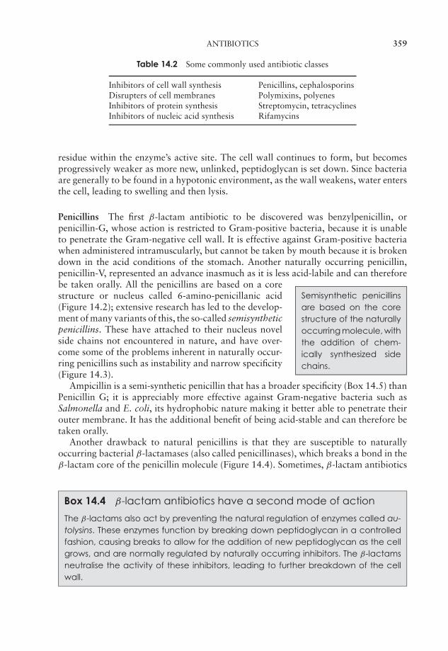
P1: FCG/SPH P2: FCG/SPH QC: FCG/SPH T1: SPH
JWBK011-14 JWBK011-Hogg August 12, 2005 16:32 Char Count= 0
ANTIBIOTICS 359
Table 14.2 Some commonly used antibiotic classes
Inhibitors of cell wall synthesis Penicillins, cephalosporinsDisrupters of cell membranes Polymixins, polyenesInhibitors of protein synthesis Streptomycin, tetracyclinesInhibitors of nucleic acid synthesis Rifamycins
residue within the enzyme’s active site. The cell wall continues to form, but becomesprogressively weaker as more new, unlinked, peptidoglycan is set down. Since bacteriaare generally to be found in a hypotonic environment, as the wall weakens, water entersthe cell, leading to swelling and then lysis.
Penicillins The first β-lactam antibiotic to be discovered was benzylpenicillin, orpenicillin-G, whose action is restricted to Gram-positive bacteria, because it is unableto penetrate the Gram-negative cell wall. It is effective against Gram-positive bacteriawhen administered intramuscularly, but cannot be taken by mouth because it is brokendown in the acid conditions of the stomach. Another naturally occurring penicillin,penicillin-V, represented an advance inasmuch as it is less acid-labile and can therefore
Semisynthetic penicillinsare based on the corestructure of the naturallyoccurring molecule, withthe addition of chem-ically synthesized sidechains.
be taken orally. All the penicillins are based on a corestructure or nucleus called 6-amino-penicillanic acid(Figure 14.2); extensive research has led to the develop-ment of many variants of this, the so-called semisyntheticpenicillins. These have attached to their nucleus novelside chains not encountered in nature, and have over-come some of the problems inherent in naturally occur-ring penicillins such as instability and narrow specificity(Figure 14.3).
Ampicillin is a semi-synthetic penicillin that has a broader specificity (Box 14.5) thanPenicillin G; it is appreciably more effective against Gram-negative bacteria such asSalmonella and E. coli, its hydrophobic nature making it better able to penetrate theirouter membrane. It has the additional benefit of being acid-stable and can therefore betaken orally.
Another drawback to natural penicillins is that they are susceptible to naturallyoccurring bacterial β-lactamases (also called penicillinases), which breaks a bond in theβ-lactam core of the penicillin molecule (Figure 14.4). Sometimes, β-lactam antibiotics
Box 14.4 β-lactam antibiotics have a second mode of action
The β-lactams also act by preventing the natural regulation of enzymes called au-tolysins. These enzymes function by breaking down peptidoglycan in a controlledfashion, causing breaks to allow for the addition of new peptidoglycan as the cellgrows, and are normally regulated by naturally occurring inhibitors. The β-lactamsneutralise the activity of these inhibitors, leading to further breakdown of the cellwall.
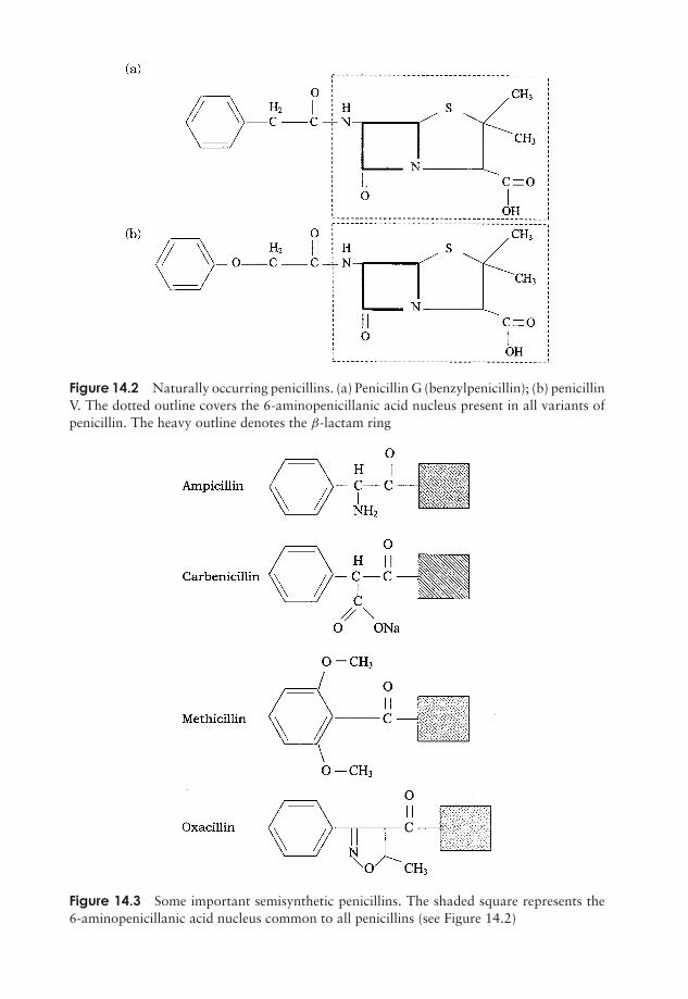
P1: FCG/SPH P2: FCG/SPH QC: FCG/SPH T1: SPH
JWBK011-14 JWBK011-Hogg August 12, 2005 16:32 Char Count= 0
Figure 14.2 Naturally occurring penicillins. (a) Penicillin G (benzylpenicillin); (b) penicillinV. The dotted outline covers the 6-aminopenicillanic acid nucleus present in all variants ofpenicillin. The heavy outline denotes the β-lactam ring
Figure 14.3 Some important semisynthetic penicillins. The shaded square represents the6-aminopenicillanic acid nucleus common to all penicillins (see Figure 14.2)
360
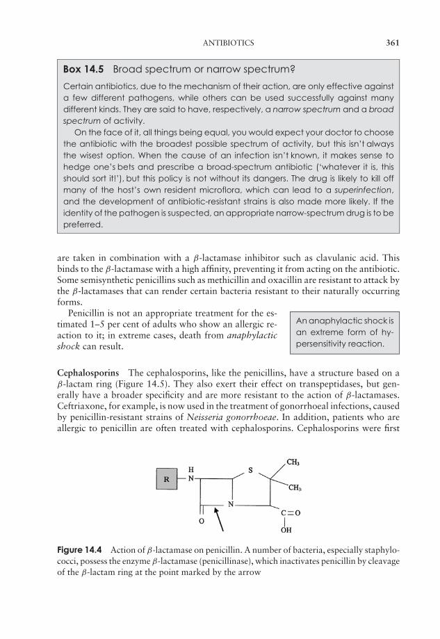
P1: FCG/SPH P2: FCG/SPH QC: FCG/SPH T1: SPH
JWBK011-14 JWBK011-Hogg August 12, 2005 16:32 Char Count= 0
ANTIBIOTICS 361
Box 14.5 Broad spectrum or narrow spectrum?
Certain antibiotics, due to the mechanism of their action, are only effective againsta few different pathogens, while others can be used successfully against manydifferent kinds. They are said to have, respectively, a narrow spectrum and a broadspectrum of activity.
On the face of it, all things being equal, you would expect your doctor to choosethe antibiotic with the broadest possible spectrum of activity, but this isn’t alwaysthe wisest option. When the cause of an infection isn’t known, it makes sense tohedge one’s bets and prescribe a broad-spectrum antibiotic (‘whatever it is, thisshould sort it!’), but this policy is not without its dangers. The drug is likely to kill offmany of the host’s own resident microflora, which can lead to a superinfection,and the development of antibiotic-resistant strains is also made more likely. If theidentity of the pathogen is suspected, an appropriate narrow-spectrum drug is to bepreferred.
are taken in combination with a β-lactamase inhibitor such as clavulanic acid. Thisbinds to the β-lactamase with a high affinity, preventing it from acting on the antibiotic.Some semisynthetic penicillins such as methicillin and oxacillin are resistant to attack bythe β-lactamases that can render certain bacteria resistant to their naturally occurringforms.
An anaphylactic shock isan extreme form of hy-persensitivity reaction.
Penicillin is not an appropriate treatment for the es-timated 1–5 per cent of adults who show an allergic re-action to it; in extreme cases, death from anaphylacticshock can result.
Cephalosporins The cephalosporins, like the penicillins, have a structure based on aβ-lactam ring (Figure 14.5). They also exert their effect on transpeptidases, but gen-erally have a broader specificity and are more resistant to the action of β-lactamases.Ceftriaxone, for example, is now used in the treatment of gonorrhoeal infections, causedby penicillin-resistant strains of Neisseria gonorrhoeae. In addition, patients who areallergic to penicillin are often treated with cephalosporins. Cephalosporins were first
Figure 14.4 Action of β-lactamase on penicillin. A number of bacteria, especially staphylo-cocci, possess the enzyme β-lactamase (penicillinase), which inactivates penicillin by cleavageof the β-lactam ring at the point marked by the arrow
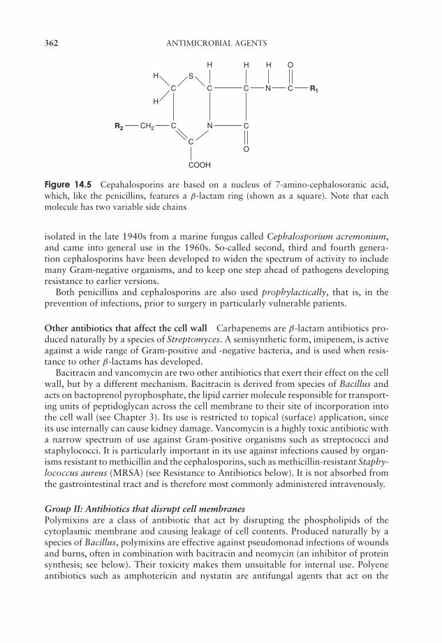
P1: FCG/SPH P2: FCG/SPH QC: FCG/SPH T1: SPH
JWBK011-14 JWBK011-Hogg August 12, 2005 16:32 Char Count= 0
362 ANTIMICROBIAL AGENTS
C C C N C R1
H H OH
C N C
O
S
C
COOH
H
H
CH2R2
Figure 14.5 Cepahalosporins are based on a nucleus of 7-amino-cephalosoranic acid,which, like the penicillins, features a β-lactam ring (shown as a square). Note that eachmolecule has two variable side chains
isolated in the late 1940s from a marine fungus called Cephalosporium acremonium,and came into general use in the 1960s. So-called second, third and fourth genera-tion cephalosporins have been developed to widen the spectrum of activity to includemany Gram-negative organisms, and to keep one step ahead of pathogens developingresistance to earlier versions.
Both penicillins and cephalosporins are also used prophylactically, that is, in theprevention of infections, prior to surgery in particularly vulnerable patients.
Other antibiotics that affect the cell wall Carbapenems are β-lactam antibiotics pro-duced naturally by a species of Streptomyces. A semisynthetic form, imipenem, is activeagainst a wide range of Gram-positive and -negative bacteria, and is used when resis-tance to other β-lactams has developed.
Bacitracin and vancomycin are two other antibiotics that exert their effect on the cellwall, but by a different mechanism. Bacitracin is derived from species of Bacillus andacts on bactoprenol pyrophosphate, the lipid carrier molecule responsible for transport-ing units of peptidoglycan across the cell membrane to their site of incorporation intothe cell wall (see Chapter 3). Its use is restricted to topical (surface) application, sinceits use internally can cause kidney damage. Vancomycin is a highly toxic antibiotic witha narrow spectrum of use against Gram-positive organisms such as streptococci andstaphylococci. It is particularly important in its use against infections caused by organ-isms resistant to methicillin and the cephalosporins, such as methicillin-resistant Staphy-lococcus aureus (MRSA) (see Resistance to Antibiotics below). It is not absorbed fromthe gastrointestinal tract and is therefore most commonly administered intravenously.
Group II: Antibiotics that disrupt cell membranesPolymixins are a class of antibiotic that act by disrupting the phospholipids of thecytoplasmic membrane and causing leakage of cell contents. Produced naturally by aspecies of Bacillus, polymixins are effective against pseudomonad infections of woundsand burns, often in combination with bacitracin and neomycin (an inhibitor of proteinsynthesis; see below). Their toxicity makes them unsuitable for internal use. Polyeneantibiotics such as amphotericin and nystatin are antifungal agents that act on the
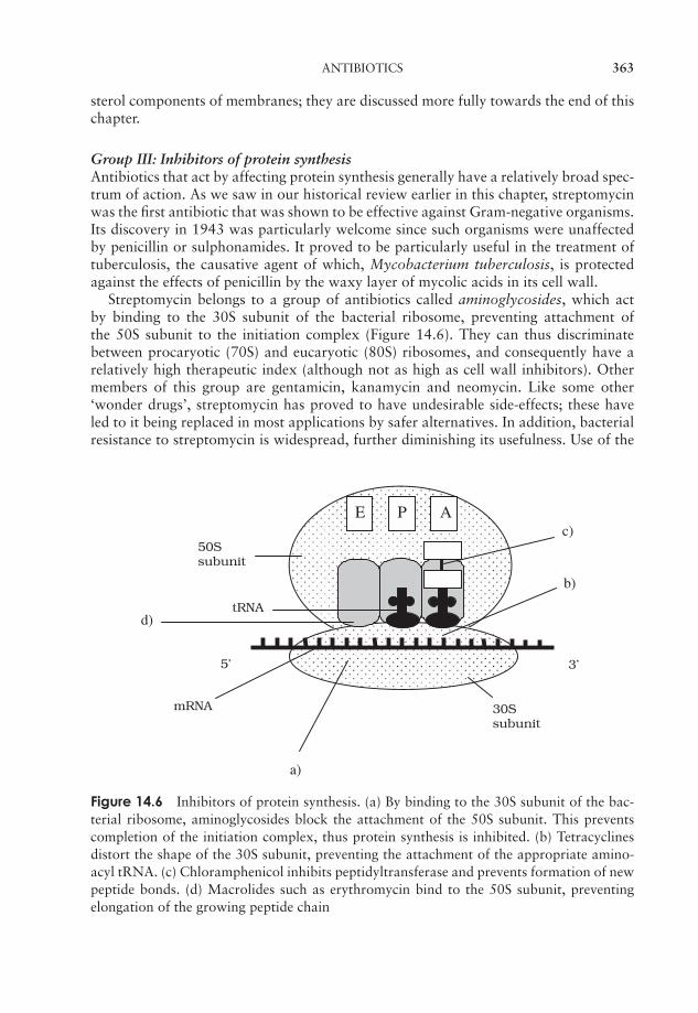
P1: FCG/SPH P2: FCG/SPH QC: FCG/SPH T1: SPH
JWBK011-14 JWBK011-Hogg August 12, 2005 16:32 Char Count= 0
ANTIBIOTICS 363
sterol components of membranes; they are discussed more fully towards the end of thischapter.
Group III: Inhibitors of protein synthesisAntibiotics that act by affecting protein synthesis generally have a relatively broad spec-trum of action. As we saw in our historical review earlier in this chapter, streptomycinwas the first antibiotic that was shown to be effective against Gram-negative organisms.Its discovery in 1943 was particularly welcome since such organisms were unaffectedby penicillin or sulphonamides. It proved to be particularly useful in the treatment oftuberculosis, the causative agent of which, Mycobacterium tuberculosis, is protectedagainst the effects of penicillin by the waxy layer of mycolic acids in its cell wall.
Streptomycin belongs to a group of antibiotics called aminoglycosides, which actby binding to the 30S subunit of the bacterial ribosome, preventing attachment ofthe 50S subunit to the initiation complex (Figure 14.6). They can thus discriminatebetween procaryotic (70S) and eucaryotic (80S) ribosomes, and consequently have arelatively high therapeutic index (although not as high as cell wall inhibitors). Othermembers of this group are gentamicin, kanamycin and neomycin. Like some other‘wonder drugs’, streptomycin has proved to have undesirable side-effects; these haveled to it being replaced in most applications by safer alternatives. In addition, bacterialresistance to streptomycin is widespread, further diminishing its usefulness. Use of the
5’
PE A
3’
50Ssubunit
30Ssubunit
tRNA
mRNA
a)
b)
c)
d)
Figure 14.6 Inhibitors of protein synthesis. (a) By binding to the 30S subunit of the bac-terial ribosome, aminoglycosides block the attachment of the 50S subunit. This preventscompletion of the initiation complex, thus protein synthesis is inhibited. (b) Tetracyclinesdistort the shape of the 30S subunit, preventing the attachment of the appropriate amino-acyl tRNA. (c) Chloramphenicol inhibits peptidyltransferase and prevents formation of newpeptide bonds. (d) Macrolides such as erythromycin bind to the 50S subunit, preventingelongation of the growing peptide chain
P1: FCG/SPH P2: FCG/SPH QC: FCG/SPH T1: SPH
JWBK011-14 JWBK011-Hogg August 12, 2005 16:32 Char Count= 0
364 ANTIMICROBIAL AGENTS
aminoglycosides as a group has diminished since the development of later generationcephalosporins and the tetracyclines.
Tetracyclines also work by binding to the 30S ribosomal subunit, preventing theattachment of aminoacyl tRNA, and therefore extension of the peptide chain (Fig-ure 14.6). They are yet another group of antibiotics produced by Streptomyces spp.Both natural and semisynthetic tetracyclines are easily absorbed from the intestine,allowing them to be taken orally. Coupled with their broad specificity (the broadest ofany antibiotic), this led to inappropriately widespread use in the years following theirdiscovery, sometimes resulting in complications caused by the destruction of the normalresident microflora. Tetracyclines are still used for a number of applications, notably totreat a variety of sexually transmitted diseases.
Two important antibiotics which act on the larger, 50S, subunit of the procaryoticribosome are erythromycin and chloramphenicol. Both combine with the subunit insuch a way as to prevent the assembly of amino acids into a chain (Figure 14.6). Chlo-ramphenicol was the first antibiotic to be discovered with a broad spectrum of activity;it also derives originally from Streptomyces spp., but is nowadays produced synthet-ically. Its use has become severely restricted since it was shown to have some seriousside-effects, notably on the bone marrow, but it remains the agent of choice for thetreatment of typhoid fever.
Erythromycin is the best known of the macrolide group of antibiotics. Unlike chlo-ramphenicol, it has a large hydrophobic molecule and is unable to gain access to mostGram-negative bacteria, thus restricting its spectrum of activity. Erythromycin can betaken orally and has a similar spectrum of activity to penicillin G; it is often used insteadof penicillin in the treatment of staphylococcal and streptococcal infections in children.It is particularly appropriate for this application as it is one of the least toxic of allcommonly used antibiotics.
Group IV: Inhibitors of nucleic acid synthesisRifampin belongs to a group of agents called rifamycins. It acts by inhibiting the enzymeRNA polymerase, thereby preventing the production of mRNA. Rifampin is used againstthe mycobacteria that cause tuberculosis, an application for which its ability to penetratetissues makes it well suited. Unlike most antibiotics, rifampin interacts with other drugs,often reducing or nullifying their effect. When used in high doses, it has the unusual side-effect of turning secretions such as tears, sweat and saliva, as well as the skin, an orange-red colour. As we have already seen, the quinolone group of synthetic antimicrobialdrugs act by disrupting DNA replication.
Resistance to antibiotics
The global increase in resistance to antimicrobial drugs, including the emergence of bacterialstrains that are resistant to all available antibacterial agents, has created a public health problemof potentially crisis proportions.
American Medical Association, 1995

P1: FCG/SPH P2: FCG/SPH QC: FCG/SPH T1: SPH
JWBK011-14 JWBK011-Hogg August 12, 2005 16:32 Char Count= 0
RESISTANCE TO ANTIBIOTICS 365
Box 14.6 Where did antibiotic resistance come from?
Have genes responsible for antibiotic resistance always existed in nature, or havethey arisen since the development and widespread use of antibiotics? The answer,almost certainly, is the former. A sample of an E. coli strain, freeze-dried in 1946, wasrevived many years later, and found to have plasmid-encoded genes for resistanceto streptomycin and tetracycline, neither of which were in clinical use until someyears after the culture was preserved. It seems likely that bacteria possessed thesegenes to protect against naturally occurring antibiotics, an idea supported by thefact that R-plasmids have been found in non-pathogenic soil bacteria. Also, resis-tance to a number of antibiotics has been demonstrated in soil and water bacteriafrom sources sufficiently remote to be free from anthropogenic influence.
As we have already suggested, the impact of certain antibiotics can be greatly reduceddue to the development of resistance by target pathogens (Box 14.6). This represents thegreatest single challenge facing us in the fight against infectious diseases at the start ofthe 21st century. Fleming himself foresaw that the usefulness of penicillin might becomelimited if resistant forms of pathogens arose.
Nosocomial infectionsare ones that are ac-quired in hospitals or sim-ilar locations. Some 5--10percent of hospital pa-tients acquire such an in-fection during their stay.This may prove to befatal, especially amongthe elderly and immuno-compromised. As well asthe human cost, such in-fections extend the av-erage time spent in hos-pital and therefore addgreatly to the costs oftreatment.
Not long after penicillin was put into general use,strains of Staphylococcus aureus were found whichdid not respond to treatment, and by 1950 penicillin-resistant S. aureus was a common cause of infections inhospitals. A decade later, a semi-synthetic form of peni-cillin, methicillin, was introduced; this was not affectedby the β-lactamase enzymes that inactivated PenicillinG, and was used to treat resistant forms. Within years,however, came the first reports of strains of S. aureusthat did not respond to methicillin. The incidence ofmethicillin-resistant S. aureus (MRSA) has increasedgreatly since, and it represents the major source of noso-comial infections. In 1980, synthetic fluoroquinoloneswere introduced to counter the threat of MRSA, butwithin a year 80 per cent of isolated strains had devel-oped resistance to these too. Vancomycin is regarded as alast-resort treatment for MRSA, for a number of reasons;it has a number of serious side-effects, its widespread usewould encourage resistance against it, and it is extremelyexpensive.
A case of vancomycin-resistant Staphylococcus aureus (VRSA) emerged in Japan in1996; a few months later it had reached the USA. This represents a serious threat; someof these strains respond to treatment with a cocktail of antibiotics, but already peoplehave died from untreatable VRSA infections. In 2003, a strain of VRSA was shownto have obtained its vancomycin resistance by cross-species transfer from a strain ofEnterococcus faecalis.
P1: FCG/SPH P2: FCG/SPH QC: FCG/SPH T1: SPH
JWBK011-14 JWBK011-Hogg August 12, 2005 16:32 Char Count= 0
366 ANTIMICROBIAL AGENTS
How does antibiotic resistance work?
We saw earlier in this chapter how antibiotics exert their effects in a variety of ways, soit should come as no surprise that there is no single mechanism of resistance. Resistancemay be natural, that is, intrinsic to the microorganism in question, or it may be acquired.
Some bacteria are able to resist antibiotic action by denying it entry to the cell;penicillin G for example is unable to penetrate the Gram negative cell wall. Otherscan pump the antibiotic back out of the cell before it has had a chance to act, bymeans of enzymes called translocases; this is fairly non-specific, leading to multiple drugresistance. Other bacteria are naturally resistant to a particular antibiotic because theylack the target for its action, for example, mycoplasma do not possess peptidoglycan,the target for penicillin’s action.
To avoid the action of an antibiotic, bacteria may be able to use or develop alter-native biochemical pathways, so that its effect is cancelled out. Many pathogens cansecrete enzymes that modify or degrade antibiotics, causing them to lose their activ-ity; we have already seen that penicillins can be inactivated by enzymatic cleavage oftheir β-lactam ring. Similarly, chloramphenicol can be acetylated, while members of theaminoglycoside family can be acetylated, adenylated or phosphorylated, all leading toloss of antimicrobial activity.
Mutations may occur which modify bacterial proteins in such a way that they arenot affected by antimicrobial agents. You will recall that streptomycin normally acts bybinding to part of the 30S subunit on the bacterial ribosome; the actual binding site isa protein called S12. Mutant forms of the S12 gene can lead to a product which stillfunctions in protein synthesis, but loses its ability to bind to streptomycin. Similarly,mutations in transpeptidase genes in staphylococci means they do not bind to penicillinany more, so cross-linking of the cell wall is not inhibited.
How does resistance arise?
Occasionally, mutations occur spontaneously in bacteria, which render them resistantto one antibiotic or another. Usually the mutation leads to a change in a receptor orbinding site such as those just described, rendering the antibiotic ineffective. The changesare usually brought about by point mutations (see Chapter 11) occurring at very lowfrequency on chromosomal DNA. Bacteria can, however, become resistant much morerapidly by acquiring the mutant resistance-causing gene from another bacterium. Thisis called transmissable antibiotic resistance; it occurs mainly as a result of bacterialconjugation, and is the cause of most of the resistance problems we presently face.Transmissable resistance was first reported in Japan in the late 1950s, when multi-drugresistance in Shigella was shown to have been acquired by conjugation with resistantE. coli in a patient’s large intestine. E. coli is known to transfer R (resistance) plasmidsto several other gut bacteria including Klebsiella, Salmonella and Enterobacter, as wellas Shigella. Whereas chromosomal mutations usually result in a modification to thedrug’s binding site, genes carried on plasmids code for enzymes which inactivate it, (e.g.β-lactamases) or lead to its exclusion from the cell (translocases).
There is a strong link between the use of a particular antibiotic in a locality and theincidence of resistant bacterial strains. This is because of selective pressure favouring
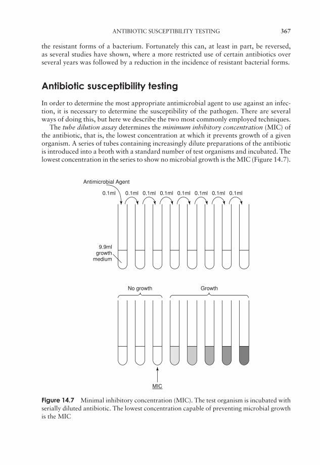
P1: FCG/SPH P2: FCG/SPH QC: FCG/SPH T1: SPH
JWBK011-14 JWBK011-Hogg August 12, 2005 16:32 Char Count= 0
ANTIBIOTIC SUSCEPTIBILITY TESTING 367
the resistant forms of a bacterium. Fortunately this can, at least in part, be reversed,as several studies have shown, where a more restricted use of certain antibiotics overseveral years was followed by a reduction in the incidence of resistant bacterial forms.
Antibiotic susceptibility testing
In order to determine the most appropriate antimicrobial agent to use against an infec-tion, it is necessary to determine the susceptibility of the pathogen. There are severalways of doing this, but here we describe the two most commonly employed techniques.
The tube dilution assay determines the minimum inhibitory concentration (MIC) ofthe antibiotic, that is, the lowest concentration at which it prevents growth of a givenorganism. A series of tubes containing increasingly dilute preparations of the antibioticis introduced into a broth with a standard number of test organisms and incubated. Thelowest concentration in the series to show no microbial growth is the MIC (Figure 14.7).
9.9mlgrowth
medium
Antimicrobial Agent
0.1ml 0.1ml 0.1ml0.1ml0.1ml0.1ml0.1ml0.1ml
MIC
No growth Growth
Figure 14.7 Minimal inhibitory concentration (MIC). The test organism is incubated withserially diluted antibiotic. The lowest concentration capable of preventing microbial growthis the MIC
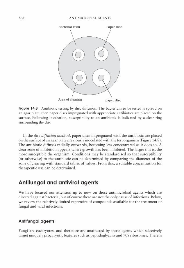
P1: FCG/SPH P2: FCG/SPH QC: FCG/SPH T1: SPH
JWBK011-14 JWBK011-Hogg August 12, 2005 16:32 Char Count= 0
368 ANTIMICROBIAL AGENTS
Bacterial lawn Paper disc
Area of clearing paper disc
Figure 14.8 Antibiotic testing by disc diffusion. The bacterium to be tested is spread onan agar plate, then paper discs impregnated with appropriate antibiotics are placed on thesurface. Following incubation, susceptibility to an antibiotic is indicated by a clear ringsurrounding the disc
In the disc diffusion method, paper discs impregnated with the antibiotic are placedon the surface of an agar plate previously inoculated with the test organism (Figure 14.8).The antibiotic diffuses radially outwards, becoming less concentrated as it does so. Aclear zone of inhibition appears where growth has been inhibited. The larger this is, themore susceptible the organism. Conditions may be standardised so that susceptibility(or otherwise) to the antibiotic can be determined by comparing the diameter of thezone of clearing with standard tables of values. From this, a suitable concentration fortherapeutic use can be determined.
Antifungal and antiviral agents
We have focused our attention up to now on those antimicrobial agents which aredirected against bacteria, but of course these are not the only cause of infections. Below,we review the relatively limited repertoire of compounds available for the treatment offungal and viral infections.
Antifungal agents
Fungi are eucaryotes, and therefore are unaffected by those agents which selectivelytarget uniquely procaryotic features such as peptidoglycans and 70S ribosomes. Therein

P1: FCG/SPH P2: FCG/SPH QC: FCG/SPH T1: SPH
JWBK011-14 JWBK011-Hogg August 12, 2005 16:32 Char Count= 0
ANTIFUNGAL AND ANTIVIRAL AGENTS 369
lies the problem: anything that damages fungal cells is likely to damage human cellstoo.
Polyene antibiotics such as amphotericin and nystatin (both produced by species ofStreptomyces) act on the sterol components of membranes; their use is limited, becausehuman cells can also be affected by their action (to use a term we learnt earlier in thischapter, they have a low therapeutic index). Nystatin is used topically against Candidainfections, while amphotericin B is generally used against systemic infections of fungalorigin. The latter substance can have a wide range of serious side-effects, but in somecases infections are so severe that the physician is faced with no alternative. Syntheticcompounds such as the imidazoles have a similar mode of action to the polyenes; theyare effective against superficial mycoses (fungal infections of the skin, mouth and urino-genital tract). Griseofulvin, a natural antibiotic produced by a species of Penicillium, isanother antifungal agent whose use is restricted; it works by interfering with mitosis andnot surprisingly has a range of side-effects. Although used to treat superficial infections,it is taken orally.
Antiviral agents
In spite of the looming threat of resistant strains, there is no doubt that antibiotics havebeen hugely successful in the control of bacterial diseases. We have, however, been alot less successful when it comes to finding a treatment for diseases caused by viruses;a quick revision of their modus operandi (Chapter 10) should make it clear why this isso. Viruses survive by entering a host cell and hijacking its replicative machinery, thusa substance interfering with the virus is likely to harm the host as well. A number ofcompounds have been developed however, which are able to act selectively on a viraltarget.
All antiviral agents act by interfering with some aspect of the virus’s replication cycle.A number of such compounds have been found, but only a few have been approved foruse in humans.
One of the first antiviral agents to be approved for use was amantidine, which inhibitsuncoating of the influenza A virus by preventing the formation of acid conditions inthe host cell’s endocytotic vesicles (see Chapter 10). Its specificity for the virus is due toselective binding to M2, a matrix protein. Amantidine’s efficacy is dependent on admin-istration within the early stages of an infection. It can be administered prophylactically,but may have side-effects.
Most antiviral agents target nucleic acid synthesis, usually by acting as base ana-logues. These are molecules that are incorporated into viral nucleotides instead of thenormal deoxynucleosides, disrupting synthesis because DNA polymerase is unable toact on them. The majority of viruses encode their own DNA polymerases, and the baseanalogues exert their effect by selectively inhibiting these, thus having little effect onthat of the host cell. An example is acyclovir, which is an analogue of guanosine; it isconverted to the nucleoside triphosphate by the action of thymidine kinase and then inthis form acts as a competitive inhibitor of the ‘correct’ version (Figure 14.9). When theacyclovir nucleotide is incorporated into the viral DNA, there is no attachment pointfor the next nucleotide, so further elongation of the chain is prevented. Acyclovir exerts
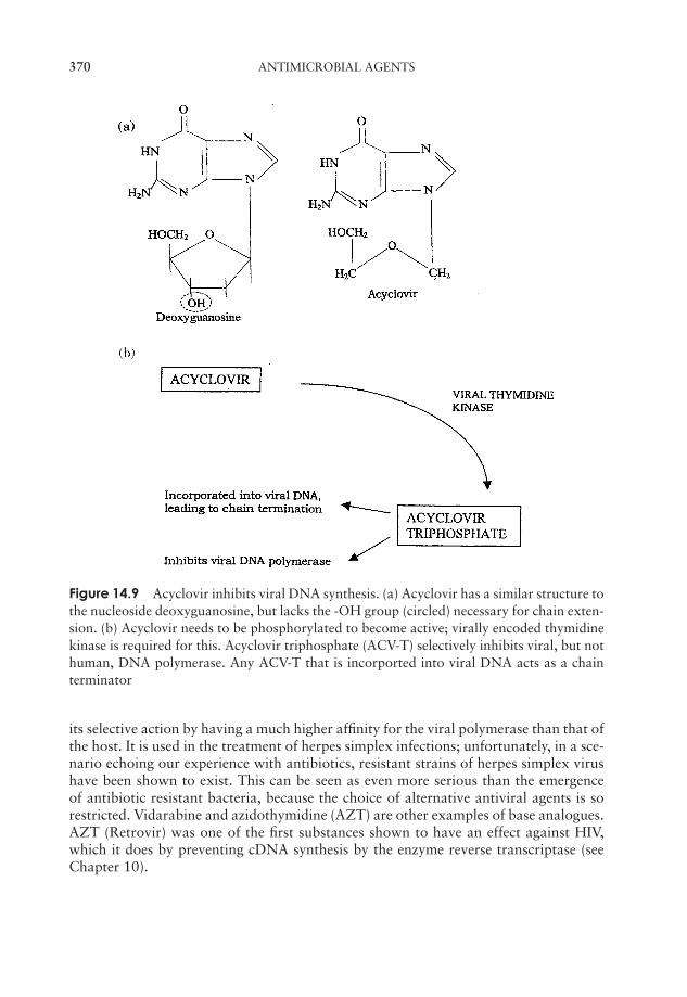
P1: FCG/SPH P2: FCG/SPH QC: FCG/SPH T1: SPH
JWBK011-14 JWBK011-Hogg August 12, 2005 16:32 Char Count= 0
370 ANTIMICROBIAL AGENTS
Figure 14.9 Acyclovir inhibits viral DNA synthesis. (a) Acyclovir has a similar structure tothe nucleoside deoxyguanosine, but lacks the -OH group (circled) necessary for chain exten-sion. (b) Acyclovir needs to be phosphorylated to become active; virally encoded thymidinekinase is required for this. Acyclovir triphosphate (ACV-T) selectively inhibits viral, but nothuman, DNA polymerase. Any ACV-T that is incorported into viral DNA acts as a chainterminator
its selective action by having a much higher affinity for the viral polymerase than that ofthe host. It is used in the treatment of herpes simplex infections; unfortunately, in a sce-nario echoing our experience with antibiotics, resistant strains of herpes simplex virushave been shown to exist. This can be seen as even more serious than the emergenceof antibiotic resistant bacteria, because the choice of alternative antiviral agents is sorestricted. Vidarabine and azidothymidine (AZT) are other examples of base analogues.AZT (Retrovir) was one of the first substances shown to have an effect against HIV,which it does by preventing cDNA synthesis by the enzyme reverse transcriptase (seeChapter 10).
P1: FCG/SPH P2: FCG/SPH QC: FCG/SPH T1: SPH
JWBK011-14 JWBK011-Hogg August 12, 2005 16:32 Char Count= 0
THE FUTURE 371
Zanamivir was approved by the US Food and Drug Administration (FDA) in 1999. Itbelongs to a new class of synthetic compounds called neuraminidase inhibitors, whichact selectively against both influenza A and B viruses. They block the active site of theenzyme neuraminidase, preventing the release of new virus particles from infected cells,hence reducing the spread of the infection. Zanamivir is inhaled as a fine powder directlyinto the lungs of patients who are in the early stages of infection.
The future
The development and widespread use of antibiotics must rank as the most remark-able of all medical advances made in the 20th century. Overconfident assertions thatinfectious diseases would soon be a thing of the past, however, now have a hollow ringto them. Viruses have proved to be more difficult to deal with than bacteria, and manyviral diseases continue to elude effective treatment. Most alarmingly, the threat of re-sistant strains casts a shadow over all the past achievements of antibiotics. The majoraim of scientists now must be to develop new antibiotics or other therapeutic strate-gies at a pace greater than that at which bacteria are developing resistance. In 2000,the FDA approved a new synthetic agent shown to be effective against both MRSAand vancomycin-resistant Enterococcus faecalis. Linezolid (Zyvox), which works byblocking the initiation of protein synthesis, belongs to a new class of antibiotics calledoxazolidinones. It is the first new anti-MRSA compound to be introduced for more than40 years.
Another approach to countering resistant forms is to identify and target the mecha-nism by which they combat antibiotic therapy. A team at Rockefeller University in NewYork have identified two genes that enable resistant forms to rebuild their cell wallsafter antibiotic treatment. By targeting these genes, they hope to restore the potencyof a cell wall inhibitor such as penicillin. Perhaps by the time you read this, other, lessconventional approaches will have yielded promising results (see Boxes 14.7 and 14.8),but you can be just as sure that bacteria will have new tricks up their sleeves, and thatthe battle of Man versus microorganisms will continue well into the new millennium.
Box 14.7 Bugs against bugs?
Scientists are always on the lookout for new weapons in the fight against infectiousdiseases. The emergence of resistant strains of pathogens means that new solutionscontinually need to be found. One novel line of research is hoping to utilise thebactericidal powers of a defence system used by certain insects. A sap-suckingspecies has been found that produces substances that interfere with bacterial pro-tein synthesis. Most of these would harm protein synthesis in humans too, but certainpeptides appear to be more selective in their action, protecting mice from E. coliand Salmonella infections. It may seem an unlikely source for a life saver, but then,so was Penicillium!
P1: FCG/SPH P2: FCG/SPH QC: FCG/SPH T1: SPH
JWBK011-14 JWBK011-Hogg August 12, 2005 16:32 Char Count= 0
372 ANTIMICROBIAL AGENTS
Box 14.8 Bacteriophages: our secret weapon against infections?
If bacteriophages are viruses that infect bacterial cells, why aren’t they used inthe fight against bacteria that cause infectious diseases? The short answer is: insome places they are! In the years following their discovery, there was consider-able enthusiasm in some quarters for the notion that phages might be useful in thetreatment of bacterial diseases. A particular attraction of phages as therapeuticagents is that they are extremely selective in their action, targeting one specificcell type. Early studies and trials had mixed results and before too long, antibioticshad revolutionised the treatment of infectious diseases in the West. This led to phagetherapy being forgotten for several decades, but its use continued in the Soviet bloccountries, however, where even now phage preparations can be bought over thecounter.
Western interest revived in the 1980s in the wake of the upsurge in antibiotic-resistant strains of bacteria, since when phages have attracted attention as possibleallies in the control of infectious diseases, including some not responsive to antibiotictherapy. Several American companies have become involved in phage therapy,some in collaboration with the former Soviet state of Georgia, where research hasbeen carried out over several decades.
Particular attention has been paid to veterinary applications, with a view to re-ducing the amount of antibiotic usage in animals, and it is hoped that one dayphages may prove to be the weapon we need to fight antibiotic resistant strainssuch as MRSA and VRE.
Test yourself
1 Following Fleming’s discovery of penicillin, and werelargely responsible for developing it and bringing it to large scale production.
2 It is essential for an antimicrobial agent to have toxicity, otherwisehost cells will be affected as well as microorganisms.
3 Penicillins and cephalosporins both belong to the group of antibi-otics. They bind to the enzyme, preventing of peptido-glycan.
4 penicillins such as ampicillin have been developed to overcomesome of the problems inherent in naturally occurring forms, such as
and narrow .
5 was the first antibiotic to shown to be effective against Gram-negative bacteria.
6 have a particularly broad range of specificity and act by preventingextension of the peptide chain during protein synthesis.
P1: FCG/SPH P2: FCG/SPH QC: FCG/SPH T1: SPH
JWBK011-14 JWBK011-Hogg August 12, 2005 16:32 Char Count= 0
TEST YOURSELF 373
7 Antibiotics that affect protein synthesis target the differences between pro-caryotic and eucaryotic .
8 Aminoglycoside antibiotics inhibit protein synthesis by preventing attach-ment of the subunit.
9 is a member of the macrolide group of antibiotics, and is oftenused instead of penicillin in the treatment of staphylococcal and streptococcalinfections in children.
10 Rifamycins affect mRNA production by inhibiting the enzyme.
11 One of the antibiotic resistant organisms most frequently found in hospitals isMRSA ( Staphylococcus aureus). Infections originatingin such environments are termed infections.
12 is used against antibiotic-resistant infections; however bacteriahave been found that are also resistant to this agent.
13 Some bacteria are able to pump antibiotics out of the cell before they have hadtime to act. They are able to do this by means of enzymes called .
14 Antibiotic resistance acquired through bacterial conjugation is described asresistance.
15 Genes for enzymes that inactivate antibiotics are usually carried on .
16 The lowest concentration of an antibiotic that prevents growth of a microor-ganism is described as its .
17 In the disc diffusion method, of develop as the antibioticdiffuses outwards.
18 The usefulness of polyene antibiotics in fungal infections is limited becausehuman cells may also be affected. Such compounds are said to have a low
.
19 The antiviral agent acyclovir works by acting as a . Itinhibits viral .
20 has been used as a therapeutic agent in the treatment of AIDS. Itacts by inhibiting viral .

P1: FCG/SPH P2: FCG/SPH QC: FCG/SPH T1: SPH
JWBK011-14 JWBK011-Hogg August 12, 2005 16:32 Char Count= 0
374
JWBK011-15 JWBK011-Hogg August 12, 2005 16:33 Char Count= 0
Part VIMicroorganisms inthe Environment
375

JWBK011-15 JWBK011-Hogg August 12, 2005 16:33 Char Count= 0
376

JWBK011-15 JWBK011-Hogg August 12, 2005 16:33 Char Count= 0
15Microbial Associations
Symbiosis is sometimestaken to mean a re-lationship between dif-ferent organisms fromwhich both participantsderive benefit. We usethe term in its broadersense, as described inthe text.
We have stressed on more than one occasion during thecourse of this book that microorganisms in nature donot exist as pure cultures but alongside numerous otherorganisms, microbial or otherwise, with which they mayhave to compete in the never-ending struggle for survival.In a number of cases, this coexistence may extend be-yond merely sharing the same environmental niche; somemicroorganisms form a close physical association withanother type of organism, from which special benefitsmay accrue for one or both parties. Such associationsare termed collectively symbiosis (‘living together’) (Ta-ble 15.1). Three general forms of symbiotic relationshipmay be defined:
� Parasitism: an association from which one partner derives some or all of itsnutritional requirements by living either in or on the other (the host), whichusually suffers harm as a result.
� Mutualism: an association from which both participants derive benefit. Therelationship is frequently obligatory, that is, both are dependent upon the otherfor survival. Non-obligatory mutualism is sometimes called protocooperation.
� Commensalism: an association from which one participant (the commensal)derives benefit, and the other is neither benefited nor harmed. The relationshipis not usually obligatory.
Microorganisms may be associated with plants, animals or other types of microorganismin any of these types of symbiosis (Tables 15.2–15.4).
Microbial associations with animals
Termites are insects belonging to the order Isoptera that are found particularly in tropicalregions. Their famous ability to destroy trees and wooden structures such as buildingsand furniture is due to a resident population of flagellated protozoans in their hindgut,which are able to break down cellulose. Termites lack the enzymes necessary to do
377
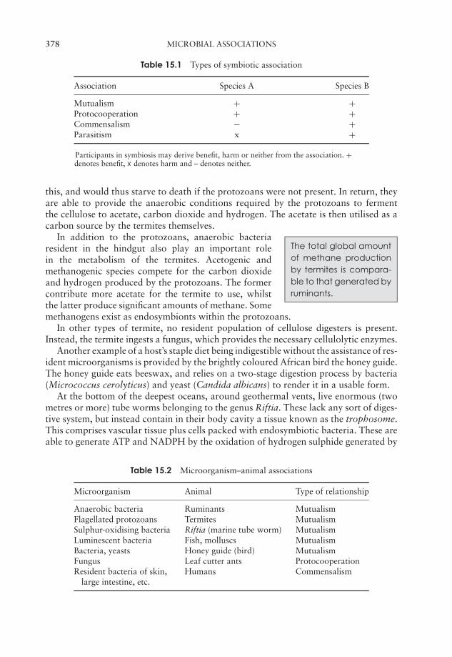
JWBK011-15 JWBK011-Hogg August 12, 2005 16:33 Char Count= 0
378 MICROBIAL ASSOCIATIONS
Table 15.1 Types of symbiotic association
Association Species A Species B
Mutualism + +Protocooperation + +Commensalism − +Parasitism x +
Participants in symbiosis may derive benefit, harm or neither from the association. +denotes benefit, x denotes harm and – denotes neither.
this, and would thus starve to death if the protozoans were not present. In return, theyare able to provide the anaerobic conditions required by the protozoans to fermentthe cellulose to acetate, carbon dioxide and hydrogen. The acetate is then utilised as acarbon source by the termites themselves.
The total global amountof methane productionby termites is compara-ble to that generated byruminants.
In addition to the protozoans, anaerobic bacteriaresident in the hindgut also play an important rolein the metabolism of the termites. Acetogenic andmethanogenic species compete for the carbon dioxideand hydrogen produced by the protozoans. The formercontribute more acetate for the termite to use, whilstthe latter produce significant amounts of methane. Somemethanogens exist as endosymbionts within the protozoans.
In other types of termite, no resident population of cellulose digesters is present.Instead, the termite ingests a fungus, which provides the necessary cellulolytic enzymes.
Another example of a host’s staple diet being indigestible without the assistance of res-ident microorganisms is provided by the brightly coloured African bird the honey guide.The honey guide eats beeswax, and relies on a two-stage digestion process by bacteria(Micrococcus cerolyticus) and yeast (Candida albicans) to render it in a usable form.
At the bottom of the deepest oceans, around geothermal vents, live enormous (twometres or more) tube worms belonging to the genus Riftia. These lack any sort of diges-tive system, but instead contain in their body cavity a tissue known as the trophosome.This comprises vascular tissue plus cells packed with endosymbiotic bacteria. These areable to generate ATP and NADPH by the oxidation of hydrogen sulphide generated by
Table 15.2 Microorganism–animal associations
Microorganism Animal Type of relationship
Anaerobic bacteria Ruminants MutualismFlagellated protozoans Termites MutualismSulphur-oxidising bacteria Riftia (marine tube worm) MutualismLuminescent bacteria Fish, molluscs MutualismBacteria, yeasts Honey guide (bird) MutualismFungus Leaf cutter ants ProtocooperationResident bacteria of skin,
large intestine, etc.Humans Commensalism
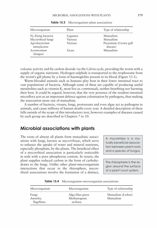
JWBK011-15 JWBK011-Hogg August 12, 2005 16:33 Char Count= 0
MICROBIAL ASSOCIATIONS WITH PLANTS 379
Table 15.3 Microorganism–plant associations
Microorganism Plant Type of relationship
N2-fixing bacteria Legumes MutualismMycorrhizal fungi Various MutualismAgrobacterium
tumefaciensVarious Parasitism (Crown gall
disease)Acremonium
(fungus)Grass Mutualism
volcanic activity and fix carbon dioxide via the Calvin cycle, providing the worm with asupply of organic nutrients. Hydrogen sulphide is transported to the trophosome fromthe worm’s gill plume by a form of haemoglobin present in its blood (Figure 15.1).
Warm-blooded animals such as humans play host in their lower intestinal tract tovast populations of bacteria. Although some of these are capable of producing usefulmetabolites such as vitamin K, most live as commensals, neither benefiting nor harmingtheir host. It could be argued, however, that the very presence of the resident intestinalmicroflora acts as an important defence against colonisation by pathogens, thus makingthe association more one of mutualism.
A number of bacteria, viruses, fungi, protozoans and even algae act as pathogens inanimals, and cause millions of human deaths every year. A detailed description of thesefalls outside of the scope of this introductory text, however examples of diseases causedby each group are described in Chapters 7 to 10.
Microbial associations with plants
A mycorrhiza is a mu-tually beneficial associa-tion between plant rootsand a species of fungus.
The roots of almost all plants form mutualistic associ-ations with fungi, known as mycorrhizae, which serveto enhance the uptake of water and mineral nutrients,especially phosphate, by the plants. The beneficial effectof a mycorrhizal association is particularly noticeablein soils with a poor phosphorus content. In return, the
The rhizosphere is the re-gion around the surfaceof a plant’s root system.
plant supplies reduced carbon in the form of carbohy-drates to the fungi. Unlike other plant–microorganisminteractions that occur in the rhizosphere, mycor-rhizal associations involve the formation of a distinct,
Table 15.4 Microorganism–microorganism associations
Microorganism Microorganism Type of relationship
Fungi Alga/blue green Mutualism (Lichen)Amoeba,
flagellatesMethanogenic
archaeaMutualism
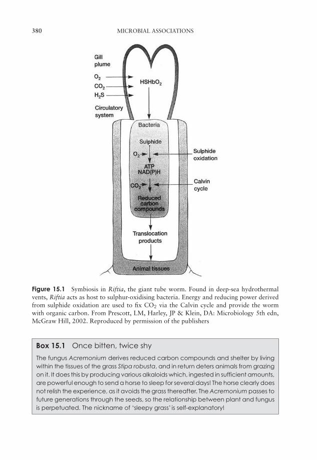
JWBK011-15 JWBK011-Hogg August 12, 2005 16:33 Char Count= 0
380 MICROBIAL ASSOCIATIONS
Figure 15.1 Symbiosis in Riftia, the giant tube worm. Found in deep-sea hydrothermalvents, Riftia acts as host to sulphur-oxidising bacteria. Energy and reducing power derivedfrom sulphide oxidation are used to fix CO2 via the Calvin cycle and provide the wormwith organic carbon. From Prescott, LM, Harley, JP & Klein, DA: Microbiology 5th edn,McGraw Hill, 2002. Reproduced by permission of the publishers
Box 15.1 Once bitten, twice shy
The fungus Acremonium derives reduced carbon compounds and shelter by livingwithin the tissues of the grass Stipa robusta, and in return deters animals from grazingon it. It does this by producing various alkaloids which, ingested in sufficient amounts,are powerful enough to send a horse to sleep for several days! The horse clearly doesnot relish the experience, as it avoids the grass thereafter. The Acremonium passes tofuture generations through the seeds, so the relationship between plant and fungusis perpetuated. The nickname of ‘sleepy grass’ is self-explanatory!
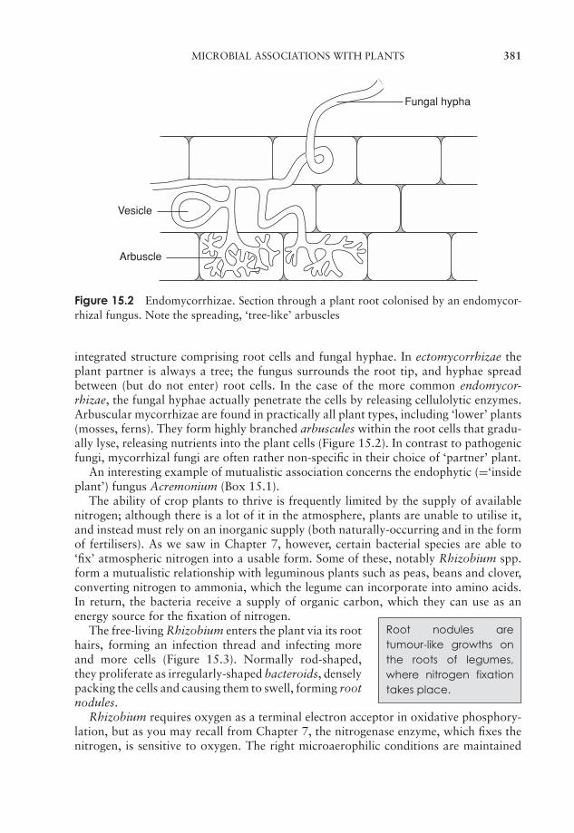
JWBK011-15 JWBK011-Hogg August 12, 2005 16:33 Char Count= 0
MICROBIAL ASSOCIATIONS WITH PLANTS 381
Fungal hypha
Vesicle
Arbuscle
Figure 15.2 Endomycorrhizae. Section through a plant root colonised by an endomycor-rhizal fungus. Note the spreading, ‘tree-like’ arbuscles
integrated structure comprising root cells and fungal hyphae. In ectomycorrhizae theplant partner is always a tree; the fungus surrounds the root tip, and hyphae spreadbetween (but do not enter) root cells. In the case of the more common endomycor-rhizae, the fungal hyphae actually penetrate the cells by releasing cellulolytic enzymes.Arbuscular mycorrhizae are found in practically all plant types, including ‘lower’ plants(mosses, ferns). They form highly branched arbuscules within the root cells that gradu-ally lyse, releasing nutrients into the plant cells (Figure 15.2). In contrast to pathogenicfungi, mycorrhizal fungi are often rather non-specific in their choice of ‘partner’ plant.
An interesting example of mutualistic association concerns the endophytic (=‘insideplant’) fungus Acremonium (Box 15.1).
The ability of crop plants to thrive is frequently limited by the supply of availablenitrogen; although there is a lot of it in the atmosphere, plants are unable to utilise it,and instead must rely on an inorganic supply (both naturally-occurring and in the formof fertilisers). As we saw in Chapter 7, however, certain bacterial species are able to‘fix’ atmospheric nitrogen into a usable form. Some of these, notably Rhizobium spp.form a mutualistic relationship with leguminous plants such as peas, beans and clover,converting nitrogen to ammonia, which the legume can incorporate into amino acids.In return, the bacteria receive a supply of organic carbon, which they can use as anenergy source for the fixation of nitrogen.
Root nodules aretumour-like growths onthe roots of legumes,where nitrogen fixationtakes place.
The free-living Rhizobium enters the plant via its roothairs, forming an infection thread and infecting moreand more cells (Figure 15.3). Normally rod-shaped,they proliferate as irregularly-shaped bacteroids, denselypacking the cells and causing them to swell, forming rootnodules.
Rhizobium requires oxygen as a terminal electron acceptor in oxidative phosphory-lation, but as you may recall from Chapter 7, the nitrogenase enzyme, which fixes thenitrogen, is sensitive to oxygen. The right microaerophilic conditions are maintained
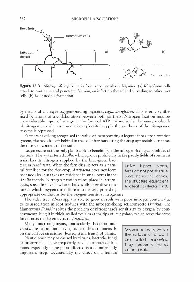
JWBK011-15 JWBK011-Hogg August 12, 2005 16:33 Char Count= 0
382 MICROBIAL ASSOCIATIONS
b)
Root nodules
Root hair
Rhizobium cells
Infectionthread
a)
Figure 15.3 Nitrogen-fixing bacteria form root nodules in legumes. (a) Rhizobium cellsattach to root hairs and penetrate, forming an infection thread and spreading to other rootcells. (b) Root nodule formation.
by means of a unique oxygen-binding pigment, leghaemoglobin. This is only synthe-sised by means of a collaboration between both partners. Nitrogen fixation requiresa considerable input of energy in the form of ATP (16 molecules for every moleculeof nitrogen), so when ammonia is in plentiful supply the synthesis of the nitrogenaseenzyme is repressed.
Farmers have long recognised the value of incorporating a legume into a crop rotationsystem; the nodules left behind in the soil after harvesting the crop appreciably enhancethe nitrogen content of the soil.
Legumes are not the only plants able to benefit from the nitrogen-fixing capabilities ofbacteria. The water fern Azolla, which grows prolifically in the paddy fields of southeast
Unlike higher plants,ferns do not possess trueroots, stems and leaves.The structure equivalentto a leaf is called a frond.
Asia, has its nitrogen supplied by the blue-green bac-terium Anabaena. When the fern dies, it acts as a natu-ral fertiliser for the rice crop. Anabaena does not formroot nodules, but takes up residence in small pores in theAzolla fronds. Nitrogen fixation takes place in hetero-cysts, specialised cells whose thick walls slow down therate at which oxygen can diffuse into the cell, providingappropriate conditions for the oxygen-sensitive nitrogenase.
The alder tree (Alnus spp.) is able to grow in soils with poor nitrogen content dueto its association in root nodules with the nitrogen-fixing actinomycete Frankia. Thefilamentous Frankia solves the problem of nitrogenase’s sensitivity to oxygen by com-partmentalising it in thick-walled vesicles at the tips of its hyphae, which serve the samefunction as the heterocysts of Anabaena.
Organisms that grow onthe surface of a plantare called epiphytes.They frequently live ascommensals.
Many microorganisms, particularly bacteria andyeasts, are to be found living as harmless commensalson the surface structures (leaves, stem, fruits) of plants.
Plant disease may be caused by viruses, bacteria, fungior protozoans. These frequently have an impact on hu-mans, especially if the plant affected is a commerciallyimportant crop. Occasionally the effect on a human
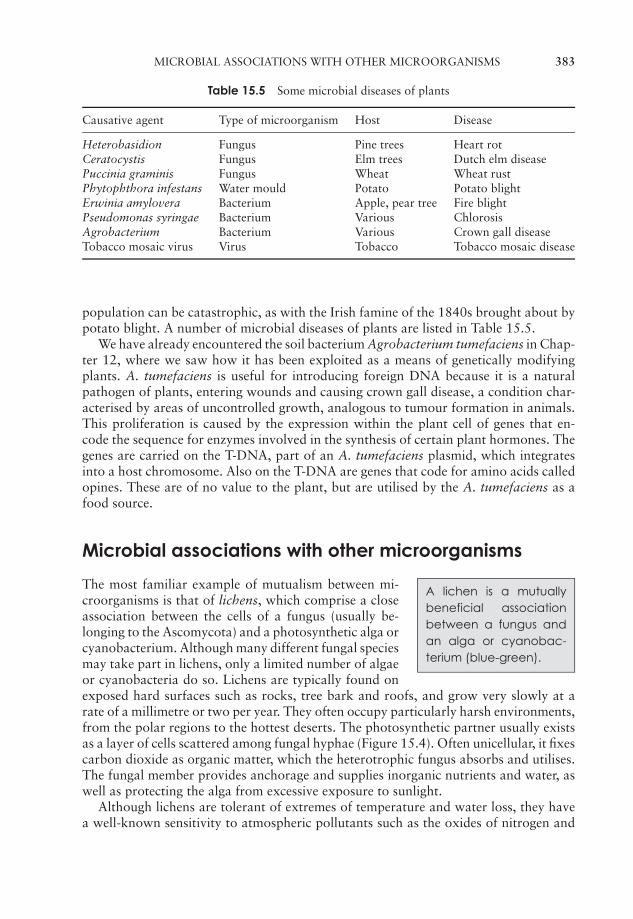
JWBK011-15 JWBK011-Hogg August 12, 2005 16:33 Char Count= 0
MICROBIAL ASSOCIATIONS WITH OTHER MICROORGANISMS 383
Table 15.5 Some microbial diseases of plants
Causative agent Type of microorganism Host Disease
Heterobasidion Fungus Pine trees Heart rotCeratocystis Fungus Elm trees Dutch elm diseasePuccinia graminis Fungus Wheat Wheat rustPhytophthora infestans Water mould Potato Potato blightErwinia amylovera Bacterium Apple, pear tree Fire blightPseudomonas syringae Bacterium Various ChlorosisAgrobacterium Bacterium Various Crown gall diseaseTobacco mosaic virus Virus Tobacco Tobacco mosaic disease
population can be catastrophic, as with the Irish famine of the 1840s brought about bypotato blight. A number of microbial diseases of plants are listed in Table 15.5.
We have already encountered the soil bacterium Agrobacterium tumefaciens in Chap-ter 12, where we saw how it has been exploited as a means of genetically modifyingplants. A. tumefaciens is useful for introducing foreign DNA because it is a naturalpathogen of plants, entering wounds and causing crown gall disease, a condition char-acterised by areas of uncontrolled growth, analogous to tumour formation in animals.This proliferation is caused by the expression within the plant cell of genes that en-code the sequence for enzymes involved in the synthesis of certain plant hormones. Thegenes are carried on the T-DNA, part of an A. tumefaciens plasmid, which integratesinto a host chromosome. Also on the T-DNA are genes that code for amino acids calledopines. These are of no value to the plant, but are utilised by the A. tumefaciens as afood source.
Microbial associations with other microorganisms
A lichen is a mutuallybeneficial associationbetween a fungus andan alga or cyanobac-terium (blue-green).
The most familiar example of mutualism between mi-croorganisms is that of lichens, which comprise a closeassociation between the cells of a fungus (usually be-longing to the Ascomycota) and a photosynthetic alga orcyanobacterium. Although many different fungal speciesmay take part in lichens, only a limited number of algaeor cyanobacteria do so. Lichens are typically found onexposed hard surfaces such as rocks, tree bark and roofs, and grow very slowly at arate of a millimetre or two per year. They often occupy particularly harsh environments,from the polar regions to the hottest deserts. The photosynthetic partner usually existsas a layer of cells scattered among fungal hyphae (Figure 15.4). Often unicellular, it fixescarbon dioxide as organic matter, which the heterotrophic fungus absorbs and utilises.The fungal member provides anchorage and supplies inorganic nutrients and water, aswell as protecting the alga from excessive exposure to sunlight.
Although lichens are tolerant of extremes of temperature and water loss, they havea well-known sensitivity to atmospheric pollutants such as the oxides of nitrogen and
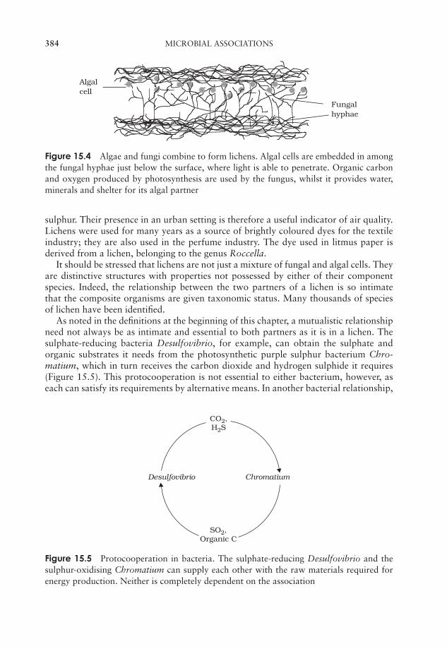
JWBK011-15 JWBK011-Hogg August 12, 2005 16:33 Char Count= 0
384 MICROBIAL ASSOCIATIONS
Algalcell
Fungalhyphae
Figure 15.4 Algae and fungi combine to form lichens. Algal cells are embedded in amongthe fungal hyphae just below the surface, where light is able to penetrate. Organic carbonand oxygen produced by photosynthesis are used by the fungus, whilst it provides water,minerals and shelter for its algal partner
sulphur. Their presence in an urban setting is therefore a useful indicator of air quality.Lichens were used for many years as a source of brightly coloured dyes for the textileindustry; they are also used in the perfume industry. The dye used in litmus paper isderived from a lichen, belonging to the genus Roccella.
It should be stressed that lichens are not just a mixture of fungal and algal cells. Theyare distinctive structures with properties not possessed by either of their componentspecies. Indeed, the relationship between the two partners of a lichen is so intimatethat the composite organisms are given taxonomic status. Many thousands of speciesof lichen have been identified.
As noted in the definitions at the beginning of this chapter, a mutualistic relationshipneed not always be as intimate and essential to both partners as it is in a lichen. Thesulphate-reducing bacteria Desulfovibrio, for example, can obtain the sulphate andorganic substrates it needs from the photosynthetic purple sulphur bacterium Chro-matium, which in turn receives the carbon dioxide and hydrogen sulphide it requires(Figure 15.5). This protocooperation is not essential to either bacterium, however, aseach can satisfy its requirements by alternative means. In another bacterial relationship,
CO2,H2S
Chromatium
SO2,Organic C
Desulfovibrio
Figure 15.5 Protocooperation in bacteria. The sulphate-reducing Desulfovibrio and thesulphur-oxidising Chromatium can supply each other with the raw materials required forenergy production. Neither is completely dependent on the association
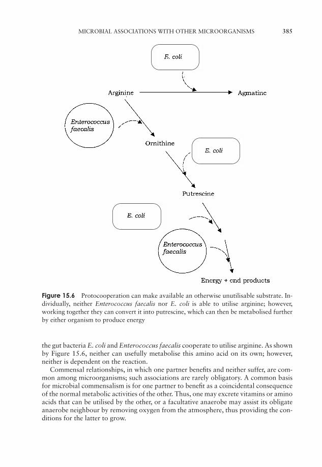
JWBK011-15 JWBK011-Hogg August 12, 2005 16:33 Char Count= 0
MICROBIAL ASSOCIATIONS WITH OTHER MICROORGANISMS 385
Figure 15.6 Protocooperation can make available an otherwise unutilisable substrate. In-dividually, neither Enterococcus faecalis nor E. coli is able to utilise arginine; however,working together they can convert it into putrescine, which can then be metabolised furtherby either organism to produce energy
the gut bacteria E. coli and Enterococcus faecalis cooperate to utilise arginine. As shownby Figure 15.6, neither can usefully metabolise this amino acid on its own; however,neither is dependent on the reaction.
Commensal relationships, in which one partner benefits and neither suffer, are com-mon among microorganisms; such associations are rarely obligatory. A common basisfor microbial commensalism is for one partner to benefit as a coincidental consequenceof the normal metabolic activities of the other. Thus, one may excrete vitamins or aminoacids that can be utilised by the other, or a facultative anaerobe may assist its obligateanaerobe neighbour by removing oxygen from the atmosphere, thus providing the con-ditions for the latter to grow.
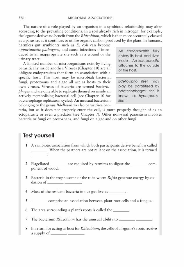
JWBK011-15 JWBK011-Hogg August 12, 2005 16:33 Char Count= 0
386 MICROBIAL ASSOCIATIONS
The nature of a role played by an organism in a symbiotic relationship may alteraccording to the prevailing conditions. In a soil already rich in nitrogen, for example,the legume derives no benefit from the Rhizobium, which is then more accurately classedas a parasite, as it continues to utilise organic carbon produced by the plant. In humans,
An endoparasite fullyenters its host and livesinside it. An ectoparasiteattaches to the outsideof the host.
harmless gut symbionts such as E. coli can becomeopportunistic pathogens, and cause infections if intro-duced to an inappropriate site such as a wound or theurinary tract.
A limited number of microorganisms exist by livingparasitically inside another. Viruses (Chapter 10) are allobligate endoparasites that form an association with a
Bdellovibrio itself mayplay be parasitised bybacteriophages; this isknown as hyperparas-itism!
specific host. This host may be microbial: bacteria,fungi, protozoans and algae all act as hosts to theirown viruses. Viruses of bacteria are termed bacterio-phages and are only able to replicate themselves inside anactively metabolising bacterial cell (see Chapter 10 forbacteriophage replication cycles). An unusual bacteriumbelonging to the genus Bdellovibrio also parasitises bac-teria, but as it does not properly enter the cell, is more properly thought of as anectoparasite or even a predator (see Chapter 7). Other non-viral parasitism involvesbacteria or fungi on protozoans, and fungi on algae and on other fungi.
Test yourself
1 A symbiotic association from which both participants derive benefit is called. When the partners are not reliant on the association, it is termed.
2 Flagellated are required by termites to digest the com-ponent of wood.
3 Bacteria in the trophosome of the tube worm Riftia generate energy by oxi-dation of .
4 Most of the resident bacteria in our gut live as .
5 comprise an association between plant root cells and a fungus.
6 The area surrounding a plant’s roots is called the .
7 The bacterium Rhizobium has the unusual ability to .
8 In return for acting as host for Rhizobium, the cells of a legume’s roots receivea supply of .
JWBK011-15 JWBK011-Hogg August 12, 2005 16:33 Char Count= 0
TEST YOURSELF 387
9 Inside root cells, Rhizobium takes on an irregular, swollen form called a.
10 In a Rhizobium-legume association, the pigment maintainsat a low level, in order that the enzyme can function.
11 Microorganisms that live as commensals on the surface of plants are termed.
12 Potato blight is caused by a fungus belonging to the genus .
13 In a lichen, the fungal partner derives from its photo-synthetic partner.
14 The sensitivity of lichens to gases such as SO2 makes them a good indicatorsof .
15 Escherichia coli and Enterococcus faecalis form a partnership toenable them to metabolise arginine.

JWBK011-15 JWBK011-Hogg August 12, 2005 16:33 Char Count= 0
388
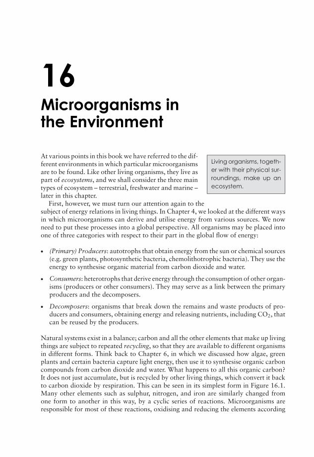
JWBK011-16 JWBK011-Hogg August 12, 2005 16:35 Char Count= 0
16Microorganisms inthe Environment
Living organisms, togeth-er with their physical sur-roundings, make up anecosystem.
At various points in this book we have referred to the dif-ferent environments in which particular microorganismsare to be found. Like other living organisms, they live aspart of ecosystems, and we shall consider the three maintypes of ecosystem – terrestrial, freshwater and marine –later in this chapter.
First, however, we must turn our attention again to thesubject of energy relations in living things. In Chapter 4, we looked at the different waysin which microorganisms can derive and utilise energy from various sources. We nowneed to put these processes into a global perspective. All organisms may be placed intoone of three categories with respect to their part in the global flow of energy:
� (Primary) Producers: autotrophs that obtain energy from the sun or chemical sources(e.g. green plants, photosynthetic bacteria, chemolithotrophic bacteria). They use theenergy to synthesise organic material from carbon dioxide and water.
� Consumers: heterotrophs that derive energy through the consumption of other organ-isms (producers or other consumers). They may serve as a link between the primaryproducers and the decomposers.
� Decomposers: organisms that break down the remains and waste products of pro-ducers and consumers, obtaining energy and releasing nutrients, including CO2, thatcan be reused by the producers.
Natural systems exist in a balance; carbon and all the other elements that make up livingthings are subject to repeated recycling, so that they are available to different organismsin different forms. Think back to Chapter 6, in which we discussed how algae, greenplants and certain bacteria capture light energy, then use it to synthesise organic carboncompounds from carbon dioxide and water. What happens to all this organic carbon?It does not just accumulate, but is recycled by other living things, which convert it backto carbon dioxide by respiration. This can be seen in its simplest form in Figure 16.1.Many other elements such as sulphur, nitrogen, and iron are similarly changed fromone form to another in this way, by a cyclic series of reactions. Microorganisms areresponsible for most of these reactions, oxidising and reducing the elements according
389
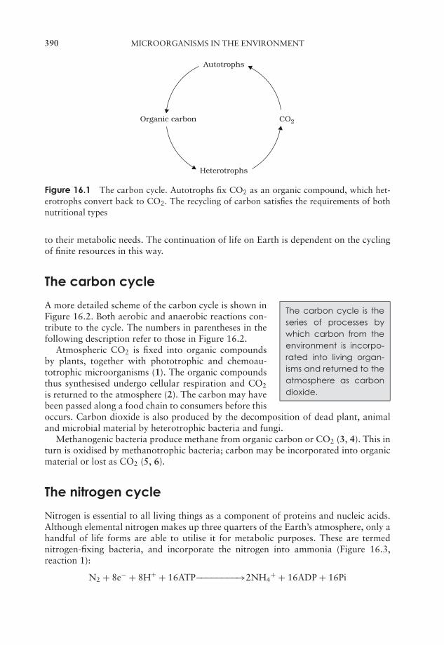
JWBK011-16 JWBK011-Hogg August 12, 2005 16:35 Char Count= 0
390 MICROORGANISMS IN THE ENVIRONMENT
Autotrophs
CO2Organic carbon
Heterotrophs
Figure 16.1 The carbon cycle. Autotrophs fix CO2 as an organic compound, which het-erotrophs convert back to CO2. The recycling of carbon satisfies the requirements of bothnutritional types
to their metabolic needs. The continuation of life on Earth is dependent on the cyclingof finite resources in this way.
The carbon cycle
The carbon cycle is theseries of processes bywhich carbon from theenvironment is incorpo-rated into living organ-isms and returned to theatmosphere as carbondioxide.
A more detailed scheme of the carbon cycle is shown inFigure 16.2. Both aerobic and anaerobic reactions con-tribute to the cycle. The numbers in parentheses in thefollowing description refer to those in Figure 16.2.
Atmospheric CO2 is fixed into organic compoundsby plants, together with phototrophic and chemoau-totrophic microorganisms (1). The organic compoundsthus synthesised undergo cellular respiration and CO2
is returned to the atmosphere (2). The carbon may havebeen passed along a food chain to consumers before thisoccurs. Carbon dioxide is also produced by the decomposition of dead plant, animaland microbial material by heterotrophic bacteria and fungi.
Methanogenic bacteria produce methane from organic carbon or CO2 (3, 4). This inturn is oxidised by methanotrophic bacteria; carbon may be incorporated into organicmaterial or lost as CO2 (5, 6).
The nitrogen cycle
Nitrogen is essential to all living things as a component of proteins and nucleic acids.Although elemental nitrogen makes up three quarters of the Earth’s atmosphere, only ahandful of life forms are able to utilise it for metabolic purposes. These are termednitrogen-fixing bacteria, and incorporate the nitrogen into ammonia (Figure 16.3,reaction 1):
N2 + 8e− + 8H+ + 16ATP−−−−−−−−→2NH4+ + 16ADP + 16Pi
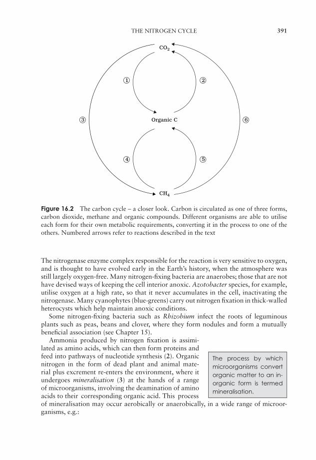
JWBK011-16 JWBK011-Hogg August 12, 2005 16:35 Char Count= 0
THE NITROGEN CYCLE 391
CO2
CH4
Organic C
1 2
4 5
63
Figure 16.2 The carbon cycle – a closer look. Carbon is circulated as one of three forms,carbon dioxide, methane and organic compounds. Different organisms are able to utiliseeach form for their own metabolic requirements, converting it in the process to one of theothers. Numbered arrows refer to reactions described in the text
The nitrogenase enzyme complex responsible for the reaction is very sensitive to oxygen,and is thought to have evolved early in the Earth’s history, when the atmosphere wasstill largely oxygen-free. Many nitrogen-fixing bacteria are anaerobes; those that are nothave devised ways of keeping the cell interior anoxic. Azotobacter species, for example,utilise oxygen at a high rate, so that it never accumulates in the cell, inactivating thenitrogenase. Many cyanophytes (blue-greens) carry out nitrogen fixation in thick-walledheterocysts which help maintain anoxic conditions.
Some nitrogen-fixing bacteria such as Rhizobium infect the roots of leguminousplants such as peas, beans and clover, where they form nodules and form a mutuallybeneficial association (see Chapter 15).
The process by whichmicroorganisms convertorganic matter to an in-organic form is termedmineralisation.
Ammonia produced by nitrogen fixation is assimi-lated as amino acids, which can then form proteins andfeed into pathways of nucleotide synthesis (2). Organicnitrogen in the form of dead plant and animal mate-rial plus excrement re-enters the environment, where itundergoes mineralisation (3) at the hands of a rangeof microorganisms, involving the deamination of aminoacids to their corresponding organic acid. This processof mineralisation may occur aerobically or anaerobically, in a wide range of microor-ganisms, e.g.:
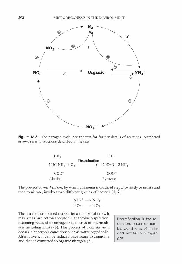
JWBK011-16 JWBK011-Hogg August 12, 2005 16:35 Char Count= 0
392 MICROORGANISMS IN THE ENVIRONMENT
N2
Organic
NO2−
NO2−
NO3− NH4
+
+
6
8
6
7
5
1
8
2
3
4
Figure 16.3 The nitrogen cycle. See the text for further details of reactions. Numberedarrows refer to reactions described in the text
CH3 CH3
COO–
Alanine Pyruvate
2 HC-NH3+ + O2 2 C=O + 2 NH4+
COO–
Deamination
The process of nitrification, by which ammonia is oxidised stepwise firstly to nitrite andthen to nitrate, involves two different groups of bacteria (4, 5).
NH4+ −→ NO2
−
NO2− −→ NO3
−
Denitrification is the re-duction, under anaero-bic conditions, of nitriteand nitrate to nitrogengas.
The nitrate thus formed may suffer a number of fates. Itmay act as an electron acceptor in anaerobic respiration,becoming reduced to nitrogen via a series of intermedi-ates including nitrite (6). This process of denitrificationoccurs in anaerobic conditions such as waterlogged soils.Alternatively, it can be reduced once again to ammoniaand thence converted to organic nitrogen (7).
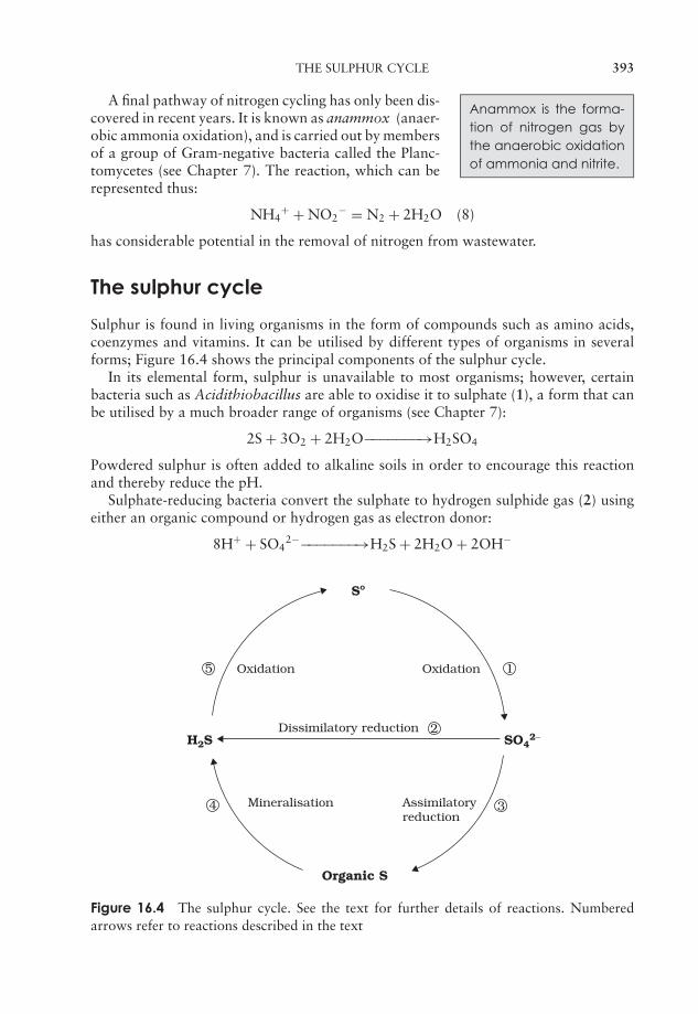
JWBK011-16 JWBK011-Hogg August 12, 2005 16:35 Char Count= 0
THE SULPHUR CYCLE 393
Anammox is the forma-tion of nitrogen gas bythe anaerobic oxidationof ammonia and nitrite.
A final pathway of nitrogen cycling has only been dis-covered in recent years. It is known as anammox (anaer-obic ammonia oxidation), and is carried out by membersof a group of Gram-negative bacteria called the Planc-tomycetes (see Chapter 7). The reaction, which can berepresented thus:
NH4+ + NO2
− = N2 + 2H2O (8)
has considerable potential in the removal of nitrogen from wastewater.
The sulphur cycle
Sulphur is found in living organisms in the form of compounds such as amino acids,coenzymes and vitamins. It can be utilised by different types of organisms in severalforms; Figure 16.4 shows the principal components of the sulphur cycle.
In its elemental form, sulphur is unavailable to most organisms; however, certainbacteria such as Acidithiobacillus are able to oxidise it to sulphate (1), a form that canbe utilised by a much broader range of organisms (see Chapter 7):
2S + 3O2 + 2H2O−−−−−−−→H2SO4
Powdered sulphur is often added to alkaline soils in order to encourage this reactionand thereby reduce the pH.
Sulphate-reducing bacteria convert the sulphate to hydrogen sulphide gas (2) usingeither an organic compound or hydrogen gas as electron donor:
8H+ + SO42−−−−−−−−→H2S + 2H2O + 2OH−
Sº
Organic S
SO42−H2S
Dissimilatory reduction
OxidationOxidation
Mineralisation Assimilatoryreduction
1
2
34
5
Figure 16.4 The sulphur cycle. See the text for further details of reactions. Numberedarrows refer to reactions described in the text
JWBK011-16 JWBK011-Hogg August 12, 2005 16:35 Char Count= 0
394 MICROORGANISMS IN THE ENVIRONMENT
These bacteria are obligate anaerobes, and the process is termed dissimilatory sulphatereduction.
Plants are also able to utilise sulphate, incorporating it into cellular constituents suchas the amino acids methionine and cysteine (3) (assimilatory sulphate reduction).
When the plants die, these compounds are broken down, again with the release ofhydrogen sulphide (4) (see mineralisation, above).
CH2HS CH3
COO–
Cysteine Pyruvate
HC-NH3+ + H2O C=O + NH4+ + H2S
COO–
Green and purple photosynthetic bacteria and some chemoautotrophs use hydrogensulphide as an electron donor in the reduction of carbon dioxide, producing elementalsulphur and thus completing the cycle (5):
H2S + CO2−−−−−−−→(CH2O)n + S0
Phosphorus
Phosphorus exists almost exclusively in nature as phosphate; however, this is cycledbetween soluble and insoluble forms. This conversion is pH-dependent, and if phosphateis only present in an insoluble form, it will act as a limiting nutrient. This explains thesudden surge in the growth of plants, algae and cyanobacteria when a source of solublephosphate (typically fertiliser or detergent) enters a watercourse. Unlike the elementsdiscussed above, phosphorus hardly exists in a gaseous form, so its main ‘reservoir’ isin the sea rather than the atmosphere.
The microbiology of soil
The topsoil is the topfew centimetres of a soil,characterised by its highcontent of organic ma-terial in various stagesof decomposition. It isdistinguished from thesucceeding layers un-derneath it, termed thesubsoil, parent layer andbedrock.
In the following section it will be necessary to generalise,and treat soil as a homogeneous medium. In fact, it is nosuch thing; its precise make-up is dependent upon the un-derlying geology, and the climatic conditions both pastand present. In addition, the microbial population of asoil will vary according to the amount of available wa-ter and organic matter, and different organisms colonisedifferent strata in the soil.
The organic content of a soil derives from the re-mains of dead plants and animals. These are brokendown in the soil by a combination of invertebrates andmicroorganisms (mainly bacteria and fungi) known asthe decomposers. Their action results in the release of
JWBK011-16 JWBK011-Hogg August 12, 2005 16:35 Char Count= 0
THE MICROBIOLOGY OF SOIL 395
Humus is the complexorganic content of asoil, comprising complexmaterials that remainafter micribial degrada-tion.
substances that can be used by plants and by other mi-croorganisms. Much organic material is easily degraded,while the more resistant fraction is referred to as hu-mus, and comprises lignin together with various othermacromolecules. The humus content of a soil, then, isa reflection of how favourable (or otherwise) conditionsare for its decomposition; the value usually falls between2 and 10 per cent by weight. The inorganic fraction ofa soil derives from the weathering of minerals. Microorganisms may be present in soilsin huge numbers, mostly attached to soil particles. Their numbers vary according tothe availability of suitable nutrients. Bacteria (notably actinomycetes) form the largestfraction of the microbial population, together with much smaller numbers of fungi,algae and protozoans. Published values of bacterial numbers range from overestimates(those that do not distinguish between living and dead cells) and underestimates (thosethat depend on colony counts and therefore exclude those organisms we are not yetable to grow in the laboratory – 99 per cent according to some experts!). Suffice tosay that many millions (possibly billions) of bacteria may be present in a single gramof topsoil. In spite of being present in such enormous numbers, microorganisms onlyrepresent a minute percentage of the volume of most soils. Fungi, although present inmuch smaller numbers than bacteria, form a higher proportion of the soil biomass,due to their greater size. The majority of soil microorganisms are aerobic heterotrophs,involved in the decomposition of organic substrates; thus, microbial numbers diminishgreatly the further down into the soil we go, away from organic matter and oxygen.The proportion of anaerobes increases with depth, but unless the soil is waterlogged,they are unlikely to predominate.
Other factors affecting microbial distribution include pH, temperature, and mois-ture. Broadly speaking, neutral conditions favour bacteria, while fungi flourish in mildlyacidic conditions (down to about pH 4), although extremophiles survive well outsidethese limits. Actinomycetes favour slightly alkaline conditions. Bacterial forms occur-ring commonly in soils include Pseudomonas, Bacillus, Clostridium, Nitrobacter andthe nitrogen-fixing Rhizobium and Azotobacter, as well as cyanobacteria such as Nostocand Anabaena. Commonly found actinomycetes include Streptomyces and Nocardia.As we have noted elsewhere, actinomycetes are notable for their secretion of antimicro-bial compounds into their surroundings. This provides an example of how the presenceof one type of microorganism in a soil population can influence the growth of oth-ers, forming a dynamic, interactive ecosystem. In addition, bacteria may serve as preyfor predatory protozoans, and secondary colonisers may depend on a supply of nutri-ents from, for example, cellulose degraders. Important fungal genera common in soilinclude the familiar Penicillium and Aspergillus; these not only recycle nutrients bybreaking down organic material, but also contribute to the fabric of the soil, by bind-ing together microscopic soil particles. Soil protozoans are mostly predators that ingestbacteria or protists such as yeasts or unicellular algae. All the major forms of proto-zoans may be present (flagellates, ciliates and amoebas), moving around the water-linedspaces between soil particles. Algae are of course phototrophic, and are therefore tobe found mostly near the soil surface, although it will be recalled from Chapter 9that some forms are capable of heterotrophic growth, and may thus survive furtherdown.

JWBK011-16 JWBK011-Hogg August 12, 2005 16:35 Char Count= 0
396 MICROORGANISMS IN THE ENVIRONMENT
The surface of soil particles is a good natural habitat for the development of biofilms,complex structures comprising microbial cells held together in a polysaccharide matrix.The microorganisms themselves produce the polysaccharide, which also allows the pas-sage of nutrients from the environment. Biofilms can form on almost any surface, and areoften to be found in rapidly flowing waters. Biofilms may be beneficial (e.g. wastewatertreatment – see below) or harmful (e.g. infections resulting from growth in catheters)to humans.
Although we have emphasised the importance of organic matter in soil ecosystems,microorganisms may also be found growing on or even within rocks. The growth of suchorganisms, together with the action of wind and rainfall, contribute to the weatheringof rocks.
The microbiology of freshwater
The microbial population of freshwater is strongly influenced by the presence or ab-sence of oxygen and light. A body of water such as a pond or lake is stratified intozones (Figure 16.5), each having its own characteristic microflora, determined by the
Figure 16.5 Vertical zonation in a lake or pond. Representative organisms are indicatedfor each zone. From Black, JG: Microbiology: Principles and Explorations, 4th edn, JohnWiley & Sons Inc., 1999. Reproduced by permission of the publishers
JWBK011-16 JWBK011-Hogg August 12, 2005 16:35 Char Count= 0
THE MICROBIOLOGY OF SEAWATER 397
availability of these factors. The littoral zone is the region situated close to land wherethe water is sufficiently shallow for sunlight to penetrate to the bottom. The limneticzone occupies the same depth, but is in open water, away from the shore. The profun-dal zone occupies deeper water, where the sun is unable to penetrate, and finally thebenthic zone comprises the sediment of mud and organic matter at the bottom of thepond or lake.
Oxygen is poorly soluble in water (9 mg/l at 20 ◦C), so its availability is often a limitingfactor in determining the microbial population of a body of water. Oxygen availabilityin lakes and ponds is closely linked to oxygenic photosynthesis and therefore, indirectly,to the penetrability of light. Phototrophs such as algae and blue-greens are limited tothose regions where light is able to penetrate. Oxygen is absent or very limited in thebenthic zone, where anaerobic forms such as the methanogenic bacteria are to be found.Another factor influencing microbial populations is the organic content of the water; ifthis is high, the growth of decomposers will be encouraged, which will in turn depletethe oxygen. This is much less of an issue in rivers and streams, where physical agitationof the water generally ensures its continued oxygenation.
The temperature of freshwater ecosystems ranges between extremes (0–90 ◦C), andmicroorganisms may be found throughout this range.
Microorganisms play a central role in the purification of wastewaters, a topic weshall examine in more later in this chapter.
The microbiology of seawater
The world’s oceans cover some 70 per cent of the Earth’s surface and have a fairlyconstant salt content of 3.5 per cent (w/v). The depth to which light can penetratevaries, but is limited to the first 100 metres or so. A world of permanent darkness existsat greater depths, however in spite of the absence of photosynthesis, oxygen is often stillpresent. This is because the generally low levels of mineral nutrients in seawater limitthe amount of primary production, and therefore heterotrophic activity. At extremedepths, however, anoxic conditions prevail.
Compared to freshwater habitats, marine ecosystems show much less variability inboth temperature and pH, although there are exceptions to this general rule. A morepertinent issue in marine environments is that of pressure; this increases progressivelyin deeper waters, and at 1000 metres reaches around 100 times normal atmosphericpressure. Concomitant with this increase in pressure is a decrease in temperature andnutrients. Surprisingly, however, certain members of the Archaea have been isolatedeven from these extreme conditions.
Phytoplankton is a col-lective term used todescribe the unicellularphotosynthesisers, whichinclude cyanobacteria,dinoflagellates, diatomsand single-celled algae.
In contrast to terrestrial ecosystems, where plants areresponsible for most of the energy fixation via photo-synthesis, marine primary production is largely micro-bial, in the shape of members of the phytoplankton. Aswe have seen, such forms are restricted to those zoneswhere light is able to penetrate. Also found here may beprotozoans and fungi that feed on the phytoplankton.Because of the high salt concentration of seawater, thebacteria that are typically found in such environments
JWBK011-16 JWBK011-Hogg August 12, 2005 16:35 Char Count= 0
398 MICROORGANISMS IN THE ENVIRONMENT
Ultramicrobacteria arebacteria that are muchsmaller than normalforms, and can passthrough a 0.22 µm filter.They may represent aresponse to reduced nu-trient conditions.
differ from those in freshwater. In the last decade or so,the presence of ultramicrobacteria has been detected inmarine ecosystems at relatively high densities; these arearound one-tenth of the size of ‘normal’ bacteria. Ma-rine bacteria are of necessity halophilic. Anaerobic de-composing bacteria inhabit the benthic zone, carryingout reactions similar to those that occur in freshwatersediments, whilst the profundal zone is largely free ofmicrobial life.
Detection and isolation of microorganismsin the environment
As we emphasised in the last chapter, microorganisms rarely, if ever, exist in natureas pure cultures but rather form mixed populations. Methods are required, therefore,for the detection and isolation of specific microbial types from such mixtures. Thetraditional method of isolation is the use of an enrichment culture, as described inChapter 4. As examples, aerobic incubation with a supply of nitrite would assist in theisolation of nitrifying bacteria such as Nitrobacter from mud or sewage, whilst a minimalmedium containing FeSO4 at pH 2 would encourage the isolation of A. ferrooxidansfrom a water sample.
We now know however that there are many types of microorganism in the envi-ronment that have so far resisted all attempts to culture them in the laboratory (oftenreferred to as viable but non-culturable). The use of modern molecular techniques hashelped us to identify the existence of a much broader range of bacteria and archaea thanhad previously been thought to exist. The extreme sensitivity of such methods means thatwe are able to demonstrate the presence of even a single copy of a particular bacteriumin a mixed population. One such technique is called fluorescence in situ hybridisa-tion (FISH). This uses a probe comprising a short sequence of single-stranded DNA orRNA that is unique to a particular microorganism, attached to a fluorescent dye. The
Antibodies are proteinsproduced by the im-mune systems of higheranimals in response to in-fection by a foreign or-ganism; their main char-acteristic is their extremespecificity, thus they canbe used to locate a spe-cific protein.
microorganisms are fixed to a glass slide and incubatedwith the probe. The rules of base pairing in nucleic acidsmean that the probe will seek out its complementarysequence, and cells carrying this sequence can be visu-alised under a fluorescence microscope. The most com-monly used ‘target’ is ribosomal RNA, since this showssequence variation from one microbial type to another,and because there are multiple copies within each cell,providing a stronger response. The polymerase chain re-action (PCR, Chapter 12) is another valuable tool in theidentification of specific nucleic acid sequences. Othermethods, not dependent on DNA, include the use offluorescence-labelled antibodies raised against specificmicroorganisms.
JWBK011-16 JWBK011-Hogg August 12, 2005 16:35 Char Count= 0
BENEFICIAL EFFECTS OF MICROORGANISMS IN THE ENVIRONMENT 399
Beneficial effects of microorganismsin the environment
The central role played by microorganisms in the recycling of essential elements on aglobal scale has already been stressed in this chapter. Many of their natural activities areexploited by humans for their own benefit. Some form the basis of industrial processes
Biodegradation is theterm used to describethe natural processes ofbreakdown of matter bymicroorganisms.
such as those used in the food and drink industries andare considered in Chapter 17, while the application ofothers is essentially environmental. Notable among theseis the harnessing of natural processes of biodegradationto treat the colossal volumes of liquid and solid wastesgenerated by our society. These are reviewed briefly inthe following section.
Solid waste treatment: composting and landfill
We in the modern Western world are often described as living in a ‘throwaway society’.On average, each of us generates around 2 tonnes of solid waste material per year, and allof this must be disposed of in some way! Most of it ends up in landfill sites, huge holes inthe ground where refuse is deposited to prevent it being a hazard. The non-biodegradablecomponents (metals, plastics, rubble, etc.) remain there more or less indefinitely; how-ever, over a period of time biodegradable material (food waste, textiles, paper, etc.)undergoes a decomposition process. The rate at which this happens is dependent on thenature of the waste and the conditions of the landfill, but could take several decades.Aerobic processes give way to anaerobic ones and a significant result of the latter is thegeneration of methane. Modern landfill sites incorporate systems that remove this toprevent it being a fire/explosion hazard, and may put it to good use as a fuel source.
Many householders separate organic waste items such as vegetable peelings andgrass cuttings and use them to make compost. This practice, apart from providing auseful gardening supplement, also substantially reduces the volume of material that hasto be disposed of by other means (see above). We have already mentioned the role ofmicroorganisms in the recycling of carbon in the biosphere; these same processes serve todegrade the organic waste, especially the cellulose, resulting in a considerable reductionof the bulk. Fungi and bacteria, particularly actinomycetes, break down the organicmatter to produce CO2, water and humus, a relatively stable organic end product.Compost is not really a fertiliser, since its nitrogen content is not high, but it neverthelessprovides nutrients to a soil and generally helps to improve its condition. Composting iscarried out on a large scale by local authorities using the waste generated in municipalparks and gardens.
Wastewater treatment
The aim of wastewater treatment is the removal of undesirable substances and haz-ardous microorganisms in order that the water may safely enter a watercourse such as
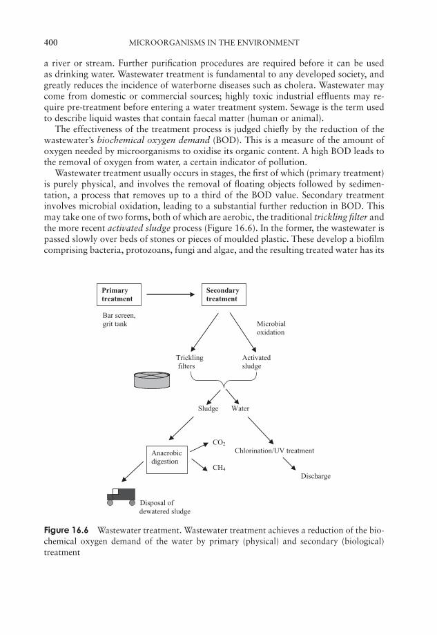
JWBK011-16 JWBK011-Hogg August 12, 2005 16:35 Char Count= 0
400 MICROORGANISMS IN THE ENVIRONMENT
a river or stream. Further purification procedures are required before it can be usedas drinking water. Wastewater treatment is fundamental to any developed society, andgreatly reduces the incidence of waterborne diseases such as cholera. Wastewater maycome from domestic or commercial sources; highly toxic industrial effluents may re-quire pre-treatment before entering a water treatment system. Sewage is the term usedto describe liquid wastes that contain faecal matter (human or animal).
The effectiveness of the treatment process is judged chiefly by the reduction of thewastewater’s biochemical oxygen demand (BOD). This is a measure of the amount ofoxygen needed by microorganisms to oxidise its organic content. A high BOD leads tothe removal of oxygen from water, a certain indicator of pollution.
Wastewater treatment usually occurs in stages, the first of which (primary treatment)is purely physical, and involves the removal of floating objects followed by sedimen-tation, a process that removes up to a third of the BOD value. Secondary treatmentinvolves microbial oxidation, leading to a substantial further reduction in BOD. Thismay take one of two forms, both of which are aerobic, the traditional trickling filter andthe more recent activated sludge process (Figure 16.6). In the former, the wastewater ispassed slowly over beds of stones or pieces of moulded plastic. These develop a biofilmcomprising bacteria, protozoans, fungi and algae, and the resulting treated water has its
Bar screen,grit tank Microbial
oxidation
Trickling Activated filters sludge
Sludge Water
CO2
Chlorination/UV treatment
CH4
Discharge
Disposal of dewatered sludge
Primarytreatment
Secondarytreatment
Anaerobicdigestion
Figure 16.6 Wastewater treatment. Wastewater treatment achieves a reduction of the bio-chemical oxygen demand of the water by primary (physical) and secondary (biological)treatment
JWBK011-16 JWBK011-Hogg August 12, 2005 16:35 Char Count= 0
BENEFICIAL EFFECTS OF MICROORGANISMS IN THE ENVIRONMENT 401
BOD reduced by some 80–85 per cent. Activated sludge plants achieve an even higherdegree of BOD reduction. Here the wastewater is aerated in tanks that have been seededwith a mixed microbial sludge. The main component of this is the bacterium Zoogloea,which secretes slime, forming aggregates called flocs, around which other microorgan-isms such as protozoans attach. Some of the water’s organic content is not immediatelyoxidised, but becomes incorporated into the flocs. After a few hours’ residence in thetank, the sludge is allowed to settle out, and the treated water passes out of the system.Before being discharged to a watercourse, it is treated with chlorine to remove anypathogenic microorganisms that may remain.
The principal operating problem encountered with activated sludge is that of bulking.This is caused by filamentous bacteria such as Sphaerotilus natans; it results in the sludgenot settling properly and consequently passing out with the treated water.
Both secondary treatment processes result in some surplus sludge, which undergoesanaerobic digestion, resulting in the production of methane and CO2. The methane canbe used as a fuel to power the plant, and any remaining sludge is dewatered and usedas a soil conditioner. Care must be taken in this context, however, that the sludge doesnot have a high heavy metal content.
Bioremediation
Perhaps the biggest problem facing the developed world at the start of the twenty firstcentury is that of pollution of the environment. Our dependence on the products of thechemical industries has resulted in the production of vast amounts of toxic waste mate-rial. One way of dealing with such (mostly organic) waste is to encourage the growth of
Bioremediation is the useof biological processesto improve a specific en-vironment, such as bythe removal of a pollu-tant.
bacteria and fungi that are able to oxidise the pollu-tants, a process known as bioremediation. Elsewhere inthis book we have seen how microorganisms are able toutilise an enormous range of organic compounds as car-bon sources; the Gram-negative bacterium Burkholde-ria cepacia can use over 100 such compounds. Manyorganisms can metabolise not only naturally occurringsubstances, but also synthetic ones, making them valu-able allies in the process of bioremediation. Often themost effective microorganisms to use are those found living naturally at the contami-nated site, since they have demonstrated the ability to survive the toxic effects of thepollutant, although in other cases specially adapted or genetically modified bacteria may
Bioaugmentation is thedeliberate introductionof specific microorgan-isms to an environmentin order to assist in biore-mediation.
be introduced (bioaugmentation). Examples of the useof microorganisms include the treatment of toxic wastesites, chemical spills, pesticides in groundwater and oilspills. One of the first successful applications of biore-mediation came in the aftermath of the Exxon Valdezdisaster in 1989 when thousands of tons of crude oilwere released off the coast of Alaska. Depending on thecircumstances, bioremediation procedures may occur
JWBK011-16 JWBK011-Hogg August 12, 2005 16:35 Char Count= 0
402 MICROORGANISMS IN THE ENVIRONMENT
in situ, or the contaminated soil or water may be removed to a specialist facility fortreatment.
Harmful effects of microorganisms in the environment
The natural processes of bioconversion that are so important in the global recycling ofelements may have unwanted consequences for humans. Prominent among these is acidmine drainage, a frequently encountered problem in mining regions. Bacterial oxidationof mineral sulphides, particularly the ubiquitous iron pyrite, leads to the release ofa highly acidic leachate into streams and rivers. This also contains dissolved metals,including ferric iron. When it mixes with stream water, the pH is raised sufficiently forthe iron to precipitate as unsightly orange ferric hydroxides, blanketing the stream bedand wiping out plant and animal life. The main culprits in the formation of acid minedrainage are sulphur-oxidising bacteria, notably Acidithiobacillus ferrooxidans; as weshall see in the final chapter, under controlled conditions this same organism can alsoprovide economic benefits to the mining industry by extracting valuable metals fromlow grade ores.
Biodeterioration is thedamage caused to ma-terials of economic im-portance due to biolog-ical (mainly microbial)processes.
Another area in which environmental microorgan-isms can have detrimental effects is that of biodeterio-ration, whereby economically important materials suchas wood, paper, textiles, petroleum and even metals andconcrete may be subject to damage by a range of mi-croorganisms, mainly fungi and bacteria.
The most important microorganisms in the biodete-rioration of wood are members of the Basidiomycota.Wood is only susceptible to fungal attack when its moisture level reaches around 30 percent. The major component of wood that is subject to microbial attack is cellulose, al-though some forms can also degrade lignin. There are two main forms of rot; white rot,which involves the degradation of lignin as well as cellulose, and brown rot, in whichthe lignin is unaltered. The dry rot fungus Serpula lacrymans produces thick strands ofhyphae called rhizomorphs, which it uses to conduct water and nutrients from damperareas. These are very strong, and able to travel over brickwork and masonry barriers.S. lacrymans is able to generate water as a metabolic end product and thus, once estab-lished, is able to grow even on dry wood. Dry rot flourishes in areas of static dampnesssuch as badly ventilated, uninhabited properties.
Because cellulose is also an important component of paper and textiles, its breakdownis clearly of great economic importance. Degradation by fungi, and, to a lesser extent,bacteria, results in a loss of strength of the material in question. The paper-makingprocess provides warm, wet conditions rich in nutrients, ideal for microbial growth,which can clog up machinery and discolour the finished product. A variety of biocidesare used in an effort to minimise microbial contamination.
The discoloration referred to above raises the point that biodeterioration of a materialneed not necessarily affect its physical or chemical make-up; aesthetic damage can lessenthe economic value of a material by altering its appearance. The blackening of showercurtains by moulds growing on surface detritus, familiar to generations of students, isanother example of this!

JWBK011-16 JWBK011-Hogg August 12, 2005 16:35 Char Count= 0
TEST YOURSELF 403
Test yourself
1 Microorganisms are essential in the of elements in the biosphere.
2 organisms fix carbon dioxide into compounds. Theseare by heterotrophs and carbon dioxide is returned to the
.
3 Nitrogen-fixing bacteria need to keep the interior of their cells oxygen-freebecause of the sensitivity of the towards oxygen.
4 The release of inorganic nutrients from organic compounds as a result ofmicrobial metabolism is termed .
5 Powdered is added to alkaline soils in order to encourage theactivity of bacteria, which leads to a reduction in pH.
6 Certain photosynthetic bacteria are able to use hydrogen sulphide as aof in the reduction of carbon dioxide.
7 often acts as the limiting nutrient in an ecosystem, because it isonly available in an insoluble form.
8 The are the major group of bacteria found in a typical soil.
9 are complex populations of microorganisms held together withina polysaccharide matrix.
10 The microbial population of a aquatic habitat is influenced by the availabilityof and .
11 The profundal zone is the name given to deep waters characterised by theabsence of .
12 In the benthic zone, the microbial population comprises mostlyforms.
13 A high organic content in a body of water encourages the growth of, which may lead to a depletion of available .
14 Unicellular photosynthesisers such as the and are col-lectively termed .
15 Molecular techniques such as FISH () enable the detection of microorganisms it is not possible to cul-
ture by conventional means.

JWBK011-16 JWBK011-Hogg August 12, 2005 16:35 Char Count= 0
404 MICROORGANISMS IN THE ENVIRONMENT
16 The effectiveness of a wastewater treatment regime may be judged by thereduction in the ( ).
17 The two forms of secondary wastewater treatment areand .
18 In anaerobic digestion, organic matter is converted into and.
19 The principal component of wood that is susceptible to biodeterioration is. However the white rot fungi can also degrade .
20 The dry rot fungus Serpula lacrymans is able to conduct water via thickstrands of hyphae called .
JWBK011-17 JWBK011-Hogg August 12, 2005 16:36 Char Count= 0
Part VIIMicroorganisms in Industry
405

JWBK011-17 JWBK011-Hogg August 12, 2005 16:36 Char Count= 0
406
JWBK011-17 JWBK011-Hogg August 12, 2005 16:36 Char Count= 0
17Industrial and FoodMicrobiology
Many aspects of our everyday lives are influenced in some way by microorganisms. Inprevious chapters we have noted their role in our environment, as well as their abilityboth to cause and cure infectious diseases. In addition, they are responsible for theproduction of much of what we eat and drink, synthesise industrially useful chemicals,and can even extract precious metals from the Earth (Table 17.1). In this chapter weshall look at some of the ways in which the activities of microorganisms have beenharnessed for the benefit of humans, and developed on an industrial scale. The firstapplications of biotechnology many thousands of years ago were in the production offood and drink, so it is here that we shall begin our survey.
Microorganisms and food
To the general public, the association of microorganisms and food conjures up negativeimages of rotten fruit or food poisoning. On reflection, many people may rememberthat yeast is involved in bread and beer production, but how many realise that microor-ganisms play a part in the manufacture of soy sauce, pepperoni and even chocolate? Inthe following pages, we shall look at the contribution of microorganisms to the contentsof our shopping baskets before considering one of the negative associations referred toabove, the microbial spoilage of food.
The production of foodstuffs as a result of microbial fermentation reactions pre-dates recorded history. The accidental discovery that such foods were less susceptibleto spoilage than fresh foods must have made them an attractive proposition to peoplein those far-off days. Of course, until relatively recent times, nothing was known of thepart played by microorganisms, so the production of beer, cheese and vinegar wouldnot have been the carefully controlled processes that are used today. Indeed, it was onlywith the development of isolation techniques towards the end of the nineteenth century(recall Chapter 1), that it became possible to use pure cultures in food production forthe first time. The fermentation of foodstuffs, hitherto an art, became a science.
407
JWBK011-17 JWBK011-Hogg August 12, 2005 16:36 Char Count= 0
408 INDUSTRIAL AND FOOD MICROBIOLOGY
Table 17.1 Some applications of microorganisms
Food productsAlcoholic drinksDairy productsBreadVinegarPickled foodsMushroomsSingle-cell protein
Products from microorganismsEnzymesAmino acidsAntibioticsCitric acid
Mining industriesMetal extractionDesulphurisation of coal
AgricultureMicrobial pesticides
EnvironmentBioremediationWastewater treatmentNitrogen fixationComposting
Alcoholic fermentationsRed wine is made fromred (black) grapes only.Both white and redgrapes may be usedin white wine, but redgrapes must first bedeskinned.
There is evidence that alcoholic drinks, including beerand wine, were being produced thousands of years be-fore the Christian era, making them among the earliestknown examples of the exploitation of microorganismsby humans. Ethanol results from the fermentation pro-cess, because the conversion of sugar to carbon dioxideand water is incomplete:
C6H12O6 −−−−−−−→ 2CH3CH2OH + 2CO2
In dry wines, most of thesugar is fermented to al-cohol; in sweet varieties,some sugar remains un-fermented.
Although, in principle, wine can be made from almostany fruit juice with a high sugar content, the vast major-ity of commercially produced wines derive from the fer-mentation of the sugar present in grapes (Figure 17.1).Such fermentation reactions may be initiated by yeastsnaturally found on the grape skin; however the resultsof such fermentations are erratic and may be unpalat-able. In commercial winemaking the must (juice) resulting from the crushed grapes istreated with sulphur dioxide to kill off the natural microflora, and then inoculated withthe yeast Saccharomyces cerevisiae, variety ellipsoideus. Specially developed strains are
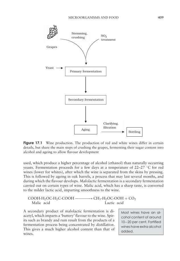
JWBK011-17 JWBK011-Hogg August 12, 2005 16:36 Char Count= 0
MICROORGANISMS AND FOOD 409
Primary fermentationYeast
Secondary fermentation
AgingBottling
Clarifying,filtration
Stemming,crushing
Grapes
SO2treatment
Figure 17.1 Wine production. The production of red and white wines differ in certaindetails, but share the main steps of crushing the grapes, fermenting their sugar content intoalcohol and ageing to allow flavour development
used, which produce a higher percentage of alcohol (ethanol) than naturally occurringyeasts. Fermentation proceeds for a few days at a temperature of 22–27 ◦C for redwines (lower for whites), after which the wine is separated from the skins by pressing.This is followed by ageing in oak barrels, a process that may last several months, andduring which the flavour develops. Malolactic fermentation is a secondary fermentationcarried out on certain types of wine. Malic acid, which has a sharp taste, is convertedto the milder lactic acid, imparting smoothness to the wine.
COOH-H2OC-H2C-COOH −−−−−−−→ CH3-H2OC-OOH + CO2
Malic acid Lactic acid
Most wines have an al-cohol content of around10−20 per cent. Fortifiedwines have extra alcoholadded.
A secondary product of malolactic fermentation is di-acetyl, which imparts a ‘buttery’ flavour to the wine. Spir-its such as brandy and rum result from the products of afermentation process being concentrated by distillation.This gives a much higher alcohol content than that ofwines.
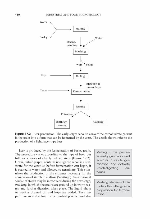
JWBK011-17 JWBK011-Hogg August 12, 2005 16:36 Char Count= 0
410 INDUSTRIAL AND FOOD MICROBIOLOGY
Filtration
Filtration toremove hops
SolidsWort
Drying,grinding
Water
Barley
Hops
Yeast
Malting
Mashing
Water
Boiling
Fermentation
Resting
CaskingBottling/canning
Figure 17.2 Beer production. The early stages serve to convert the carbohydrate presentin the grain into a form that can be fermented by the yeast. The details shown refer to theproduction of a light, lager-type beer
Malting is the processwhereby grain is soakedin water to initiate ger-mination and activatestarch-digesting en-zymes.
Beer is produced by the fermentation of barley grain.The procedure varies according to the type of beer, butfollows a series of clearly defined steps (Figure 17.2).Grain, unlike grapes, contains no sugar to serve as a sub-strate for the yeast, so before fermentation can begin, itis soaked in water and allowed to germinate. This stim-ulates the production of the enzymes necessary for theconversion of starch to maltose (‘malting’). An additional
Mashing releases solublematerial from the grain inpreparation for fermen-tation.
source of starch may be introduced during the next stage,mashing, in which the grains are ground up in warm wa-ter, and further digestion takes place. The liquid phaseor wort is drained off and hops are added. They im-part flavour and colour to the finished product and also
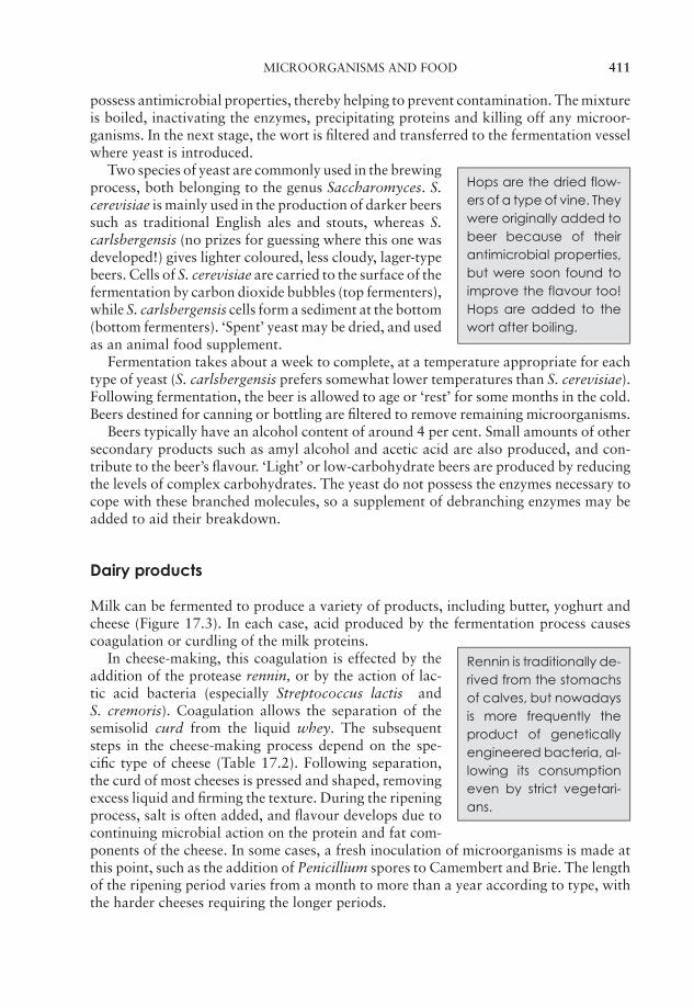
JWBK011-17 JWBK011-Hogg August 12, 2005 16:36 Char Count= 0
MICROORGANISMS AND FOOD 411
possess antimicrobial properties, thereby helping to prevent contamination. The mixtureis boiled, inactivating the enzymes, precipitating proteins and killing off any microor-ganisms. In the next stage, the wort is filtered and transferred to the fermentation vesselwhere yeast is introduced.
Hops are the dried flow-ers of a type of vine. Theywere originally added tobeer because of theirantimicrobial properties,but were soon found toimprove the flavour too!Hops are added to thewort after boiling.
Two species of yeast are commonly used in the brewingprocess, both belonging to the genus Saccharomyces. S.cerevisiae is mainly used in the production of darker beerssuch as traditional English ales and stouts, whereas S.carlsbergensis (no prizes for guessing where this one wasdeveloped!) gives lighter coloured, less cloudy, lager-typebeers. Cells of S. cerevisiae are carried to the surface of thefermentation by carbon dioxide bubbles (top fermenters),while S. carlsbergensis cells form a sediment at the bottom(bottom fermenters). ‘Spent’ yeast may be dried, and usedas an animal food supplement.
Fermentation takes about a week to complete, at a temperature appropriate for eachtype of yeast (S. carlsbergensis prefers somewhat lower temperatures than S. cerevisiae).Following fermentation, the beer is allowed to age or ‘rest’ for some months in the cold.Beers destined for canning or bottling are filtered to remove remaining microorganisms.
Beers typically have an alcohol content of around 4 per cent. Small amounts of othersecondary products such as amyl alcohol and acetic acid are also produced, and con-tribute to the beer’s flavour. ‘Light’ or low-carbohydrate beers are produced by reducingthe levels of complex carbohydrates. The yeast do not possess the enzymes necessary tocope with these branched molecules, so a supplement of debranching enzymes may beadded to aid their breakdown.
Dairy products
Milk can be fermented to produce a variety of products, including butter, yoghurt andcheese (Figure 17.3). In each case, acid produced by the fermentation process causescoagulation or curdling of the milk proteins.
Rennin is traditionally de-rived from the stomachsof calves, but nowadaysis more frequently theproduct of geneticallyengineered bacteria, al-lowing its consumptioneven by strict vegetari-ans.
In cheese-making, this coagulation is effected by theaddition of the protease rennin, or by the action of lac-tic acid bacteria (especially Streptococcus lactis andS. cremoris). Coagulation allows the separation of thesemisolid curd from the liquid whey. The subsequentsteps in the cheese-making process depend on the spe-cific type of cheese (Table 17.2). Following separation,the curd of most cheeses is pressed and shaped, removingexcess liquid and firming the texture. During the ripeningprocess, salt is often added, and flavour develops due tocontinuing microbial action on the protein and fat com-ponents of the cheese. In some cases, a fresh inoculation of microorganisms is made atthis point, such as the addition of Penicillium spores to Camembert and Brie. The lengthof the ripening period varies from a month to more than a year according to type, withthe harder cheeses requiring the longer periods.
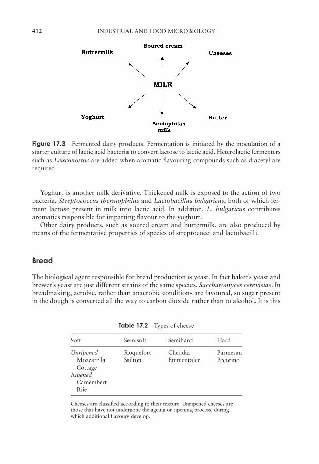
JWBK011-17 JWBK011-Hogg August 12, 2005 16:36 Char Count= 0
412 INDUSTRIAL AND FOOD MICROBIOLOGY
Figure 17.3 Fermented dairy products. Fermentation is initiated by the inoculation of astarter culture of lactic acid bacteria to convert lactose to lactic acid. Heterolactic fermenterssuch as Leuconostoc are added when aromatic flavouring compounds such as diacetyl arerequired
Yoghurt is another milk derivative. Thickened milk is exposed to the action of twobacteria, Streptococcus thermophilus and Lactobacillus bulgaricus, both of which fer-ment lactose present in milk into lactic acid. In addition, L. bulgaricus contributesaromatics responsible for imparting flavour to the yoghurt.
Other dairy products, such as soured cream and buttermilk, are also produced bymeans of the fermentative properties of species of streptococci and lactobacilli.
Bread
The biological agent responsible for bread production is yeast. In fact baker’s yeast andbrewer’s yeast are just different strains of the same species, Saccharomyces cerevisiae. Inbreadmaking, aerobic, rather than anaerobic conditions are favoured, so sugar presentin the dough is converted all the way to carbon dioxide rather than to alcohol. It is this
Table 17.2 Types of cheese
Soft Semisoft Semihard Hard
Unripened Roquefort Cheddar ParmesanMozzarella Stilton Emmentaler PecorinoCottage
RipenedCamembertBrie
Cheeses are classified according to their texture. Unripened cheeses arethose that have not undergone the ageing or ripening process, duringwhich additional flavours develop.
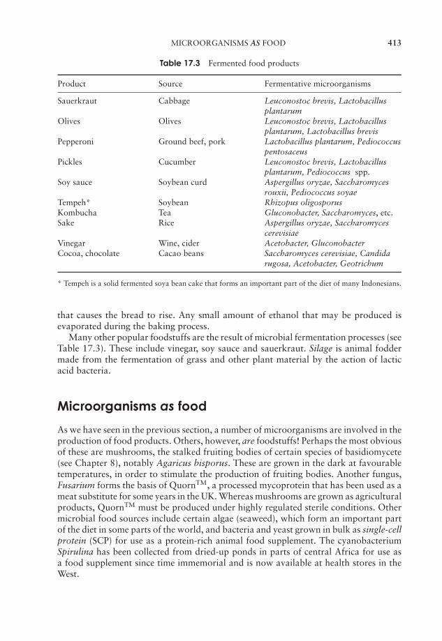
JWBK011-17 JWBK011-Hogg August 12, 2005 16:36 Char Count= 0
MICROORGANISMS AS FOOD 413
Table 17.3 Fermented food products
Product Source Fermentative microorganisms
Sauerkraut Cabbage Leuconostoc brevis, Lactobacillusplantarum
Olives Olives Leuconostoc brevis, Lactobacillusplantarum, Lactobacillus brevis
Pepperoni Ground beef, pork Lactobacillus plantarum, Pediococcuspentosaceus
Pickles Cucumber Leuconostoc brevis, Lactobacillusplantarum, Pediococcus spp.
Soy sauce Soybean curd Aspergillus oryzae, Saccharomycesrouxii, Pediococcus soyae
Tempeh* Soybean Rhizopus oligosporusKombucha Tea Gluconobacter, Saccharomyces, etc.Sake Rice Aspergillus oryzae, Saccharomyces
cerevisiaeVinegar Wine, cider Acetobacter, GluconobacterCocoa, chocolate Cacao beans Saccharomyces cerevisiae, Candida
rugosa, Acetobacter, Geotrichum
* Tempeh is a solid fermented soya bean cake that forms an important part of the diet of many Indonesians.
that causes the bread to rise. Any small amount of ethanol that may be produced isevaporated during the baking process.
Many other popular foodstuffs are the result of microbial fermentation processes (seeTable 17.3). These include vinegar, soy sauce and sauerkraut. Silage is animal foddermade from the fermentation of grass and other plant material by the action of lacticacid bacteria.
Microorganisms as food
As we have seen in the previous section, a number of microorganisms are involved in theproduction of food products. Others, however, are foodstuffs! Perhaps the most obviousof these are mushrooms, the stalked fruiting bodies of certain species of basidiomycete(see Chapter 8), notably Agaricus bisporus. These are grown in the dark at favourabletemperatures, in order to stimulate the production of fruiting bodies. Another fungus,Fusarium forms the basis of QuornTM, a processed mycoprotein that has been used as ameat substitute for some years in the UK. Whereas mushrooms are grown as agriculturalproducts, QuornTM must be produced under highly regulated sterile conditions. Othermicrobial food sources include certain algae (seaweed), which form an important partof the diet in some parts of the world, and bacteria and yeast grown in bulk as single-cellprotein (SCP) for use as a protein-rich animal food supplement. The cyanobacteriumSpirulina has been collected from dried-up ponds in parts of central Africa for use asa food supplement since time immemorial and is now available at health stores in theWest.

JWBK011-17 JWBK011-Hogg August 12, 2005 16:36 Char Count= 0
414 INDUSTRIAL AND FOOD MICROBIOLOGY
The microbial spoilage of food
We have described in previous chapters the nutritional versatility of microorganismsand their role in the global recycling of carbon. Unfortunately for us, fresh foods suchas meats, fruit and vegetables provide a rich source of nutrients, which a wide range ofheterotrophic microorganisms find just as attractive as we do. Certain microbial typesare associated with particular foodstuffs, depending on their chemical composition andphysical factors such as pH and water content. Acidic foods such as fruits, for example,tend to favour the growth of fungi rather than bacteria.
Often, spoilage organisms come from the same source as the food, for examplesoil on vegetables, or meat exposed to intestinal contents following slaughter. Othersare introduced as contaminants during transport, storage or preparation. Among themost commonly found spoilage organisms are a number of human pathogens, includ-ing Pseudomonas, Salmonella, Campylobacter and Listeria. Thus, although microbialspoilage may merely lead to foodstuffs being rendered unpalatable, it can also resultin serious and even fatal illness (‘food poisoning’). Whilst observable changes to food-stuffs are only likely after the microbial population has reached a considerable size,food poisoning can result from the presence of much smaller numbers of contaminants.
Some foodstuffs are more susceptible to spoilage than others: fresh items such as meat,fish, dairy produce and fruit and vegetables are all highly perishable. Foods such as riceand flour, on the other hand, are much more resistant, because having no water contentthey do not provide suitable conditions for microbial growth. Drying is one of a numberof methods of food preservation, all designed to prevent growth of microorganismsby making conditions unfavourable. Other methods include heating/canning, drying,pickling, smoking and, in many countries, irradiation.
Microorganisms in the production of biochemicals
Many products of microbial metabolism find an application in the food and other in-dustries. These include amino acids, steroids, enzymes and antibiotics (Table 17.4).Microbial growth conditions are adjusted so that production of the metabolite in ques-tion takes place at an optimal rate. Often an unnaturally high rate of production isachieved by the use of a mutated or genetically engineered strain of microorganism, orby manipulating culture conditions to favour excess metabolite production.
The development of a microbial means of producing acetone was vital to the alliedeffort in the First World War. Acetone was a crucial precursor in explosives manufactureand the demands of war soon outstripped supply by traditional methods. The problemwas solved when Chaim Weismann isolated a strain of Clostridium acetobutylicum thatcould ferment molasses to acetone and butanol (another industrially useful product).Nowadays, acetone is made more cheaply from petrochemicals.
Microbially produced amino acids are used in the food industry, in medicine andas raw materials in the chemical industry. The one produced in the greatest quantitiesby far is glutamic acid (in excess of half a billion tonnes per year), with most of itending up as the flavour enhancer monosodium glutamate. The amino acids asparticacid and phenylalanine are components of the artificial sweetener aspartame and arealso synthesised on a large scale.

JWBK011-17 JWBK011-Hogg August 12, 2005 16:36 Char Count= 0
MICROORGANISMS IN THE PRODUCTION OF BIOCHEMICALS 415
Table 17.4 Some industrial applications of microbially produced enzymes
Industry Enzyme Application
Food & drink Rennin Cheese manufactureLipasePectinase Fruit juice production
Coffee bean extractionAmylase Improved bread dough quality
Haze removal in beerAmylase Fructose syrup productionGlucoamylaseGlucose isomerase
Animal feed Amylase Improved digestibilityCellulaseProtease
Detergent Protease Stain and grease removalLipaseAmylaseCellulase Fabric softener
Paper Cellulase Pulp production
Textile Cellulase ‘Stone-washed’ jeans
Leather Protease Dehairing, softening, fatLipase removal
Molecular biology Taq polymerase Polymerase chain reaction
A number of organic acids are produced industrially by microbial means, most no-tably citric acid, which has a wide range of applications in the food and pharmaceuticalindustries. This is mostly produced as a secondary metabolite by the large-scale cultureof the mould Aspergillus niger.
Certain microorganisms serve as a ready source of vitamins. In many cases thesecan be synthesised less expensively by chemical means; however, riboflavin (by themould Ashbya gossypii) and vitamin B12 (by the bacteria Propionibacterium shermaniiand Pseudomonas denitrificans) are produced by large-scale microbial fermentation.Microorganisms play a partial role in the production of ascorbic acid (vitamin C).Initially, glucose is reduced chemically to sorbitol, which is then oxidised by a strain ofAcetobacter suboxydans to the hexose sorbose. Chemical modifications convert this toascorbic acid (Figure 17.4).
If you wear contactlenses, you have prob-ably used an enzymepreparation to removeprotein deposits.
Enzymes of fungal and bacterial origin have beenutilised for many centuries in a variety of processes. Itis now possible to isolate and purify the enzymes neededfor a specific process and the worldwide market is cur-rently worth around a billion pounds. The most usefulindustrial enzymes include proteases, amylases, lipases
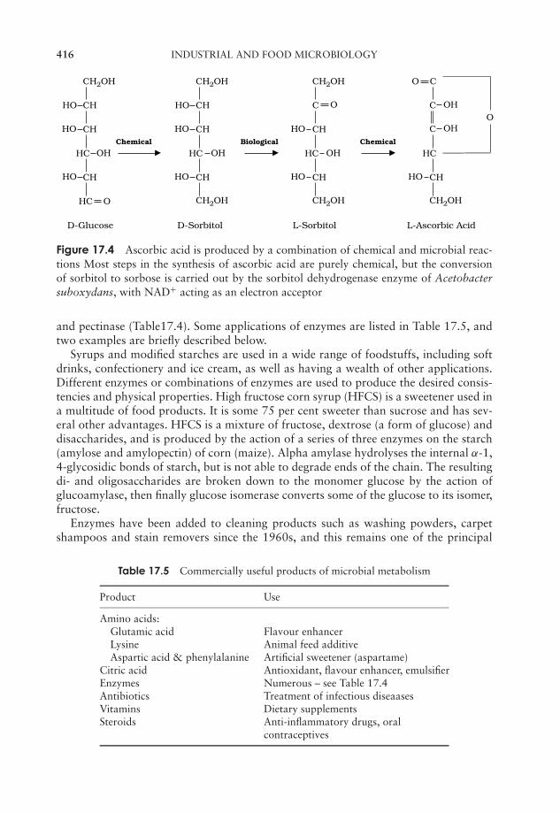
JWBK011-17 JWBK011-Hogg August 12, 2005 16:36 Char Count= 0
416 INDUSTRIAL AND FOOD MICROBIOLOGY
CH2OH CH2OH
CH2OH
CH2OH O C
HO CHO
CHChemical Biological Chemical
HC
CH
HC O
D-Glucose D-Sorbitol L-Sorbitol L-Ascorbic Acid
HO
OH
HO
OHO CH
CH
HC
CH
HO
OH
HO
CH2OH
C
CH
HC
CH
HO
OH
HO
OH
CH2OH
C
HC
CHHO
OHC
Figure 17.4 Ascorbic acid is produced by a combination of chemical and microbial reac-tions Most steps in the synthesis of ascorbic acid are purely chemical, but the conversionof sorbitol to sorbose is carried out by the sorbitol dehydrogenase enzyme of Acetobactersuboxydans, with NAD+ acting as an electron acceptor
and pectinase (Table17.4). Some applications of enzymes are listed in Table 17.5, andtwo examples are briefly described below.
Syrups and modified starches are used in a wide range of foodstuffs, including softdrinks, confectionery and ice cream, as well as having a wealth of other applications.Different enzymes or combinations of enzymes are used to produce the desired consis-tencies and physical properties. High fructose corn syrup (HFCS) is a sweetener used ina multitude of food products. It is some 75 per cent sweeter than sucrose and has sev-eral other advantages. HFCS is a mixture of fructose, dextrose (a form of glucose) anddisaccharides, and is produced by the action of a series of three enzymes on the starch(amylose and amylopectin) of corn (maize). Alpha amylase hydrolyses the internal α-1,4-glycosidic bonds of starch, but is not able to degrade ends of the chain. The resultingdi- and oligosaccharides are broken down to the monomer glucose by the action ofglucoamylase, then finally glucose isomerase converts some of the glucose to its isomer,fructose.
Enzymes have been added to cleaning products such as washing powders, carpetshampoos and stain removers since the 1960s, and this remains one of the principal
Table 17.5 Commercially useful products of microbial metabolism
Product Use
Amino acids:Glutamic acid Flavour enhancerLysine Animal feed additiveAspartic acid & phenylalanine Artificial sweetener (aspartame)
Citric acid Antioxidant, flavour enhancer, emulsifierEnzymes Numerous – see Table 17.4Antibiotics Treatment of infectious diseaasesVitamins Dietary supplementsSteroids Anti-inflammatory drugs, oral
contraceptives
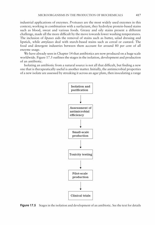
JWBK011-17 JWBK011-Hogg August 12, 2005 16:36 Char Count= 0
MICROORGANISMS IN THE PRODUCTION OF BIOCHEMICALS 417
industrial applications of enzymes. Proteases are the most widely used enzymes in thiscontext; working in combination with a surfactant, they hydrolyse protein-based stainssuch as blood, sweat and various foods. Greasy and oily stains present a differentchallenge, made all the more difficult by the move towards lower washing temperatures.The inclusion of lipases aids the removal of stains such as butter, salad dressing andlipstick, while amylases deal with starch-based stains such as cereal or custard. Thefood and detergent industries between them account for around 80 per cent of allenzyme usage.
We have already seen in Chapter 14 that antibiotics are now produced on a huge scaleworldwide. Figure 17.5 outlines the stages in the isolation, development and productionof an antibiotic.
Isolating an antibiotic from a natural source is not all that difficult, but finding a newone that is therapeutically useful is another matter. Initially, the antimicrobial propertiesof a new isolate are assessed by streaking it across an agar plate, then inoculating a range
Isolation andpurification
Assessment ofantimicrobialefficiency
Small-scaleproduction
Toxicity testing
Pilot-scaleproduction
Clinical trials
Figure 17.5 Stages in the isolation and development of an antibiotic. See the text for details
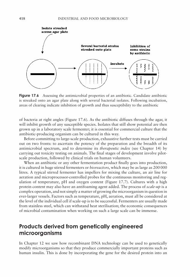
JWBK011-17 JWBK011-Hogg August 12, 2005 16:36 Char Count= 0
418 INDUSTRIAL AND FOOD MICROBIOLOGY
Figure 17.6 Assessing the antimicrobial properties of an antibiotic. Candidate antibioticis streaked onto an agar plate along with several bacterial isolates. Following incubation,areas of clearing indicate inhibition of growth and thus susceptibility to the antibiotic
of bacteria at right angles (Figure 17.6). As the antibiotic diffuses through the agar, itwill inhibit growth of any susceptible species. Isolates that still show potential are thengrown up in a laboratory scale fermenter; it is essential for commercial culture that theantibiotic-producing organism can be cultured in this way.
Before committing to large-scale production, exhaustive further tests must be carriedout on two fronts: to ascertain the potency of the preparation and the breadth of itsantimicrobial spectrum, and to determine its therapeutic index (see Chapter 14) bycarrying out toxicity testing on animals. The final stages of development involve pilot-scale production, followed by clinical trials on human volunteers.
When an antibiotic or any other fermentation product finally goes into production,it is cultured in huge stirred fermenters or bioreactors, which may be as large as 200 000litres. A typical stirred fermenter has impellers for mixing the culture, an air line foraeration and microprocessor-controlled probes for the continuous monitoring and reg-ulation of temperature, pH and oxygen content (Figure 17.7). Cultures with a highprotein content may also have an antifoaming agent added. The process of scale-up is acomplex operation, and not simply a matter of growing the microorganism in question inever-larger vessels. Factors such as temperature, pH, aeration, must all be considered atthe level of the individual cell if scale-up is to be successful. Fermenters are usually madefrom stainless steel, which can withstand heat sterilisation; the economic consequencesof microbial contamination when working on such a large scale can be immense.
Products derived from genetically engineeredmicroorganisms
In Chapter 12 we saw how recombinant DNA technology can be used to geneticallymodify microorganisms so that they produce commercially important proteins such ashuman insulin. This is done by incorporating the gene for the desired protein into an

JWBK011-17 JWBK011-Hogg August 12, 2005 16:36 Char Count= 0
PRODUCTS DERIVED FROM GENETICALLY ENGINEERED MICROORGANISMS 419
Motor
pH probe
Dissolved oxygen probe
Coolingwaterout
Impellers
Coolingjacket
Biosensorunit
Air in
Air filter
Valve
Valve
Coolingwater in
Temperaturesensor and control unit
Harvestline
Valve
Sampleline
Culture or nutrient addition
Figure 17.7 A continuous flow stirred tank reactor. Parameters such as pH and concen-trations of specific metabolites are closely monitored to ensure the maintenance of optimumconditions. Outlets allow for the collection of samples during fermentation as well as thecollection of cells and medium at the conclusion of the reaction. Addition and collectionsare carried out under aseptic conditions. From Prescott, LM, Harley, JP & Klein, DA: Mi-crobiology 5th edn, McGraw Hill, 2002. Reproduced by permission of the publishers
JWBK011-17 JWBK011-Hogg August 12, 2005 16:36 Char Count= 0
420 INDUSTRIAL AND FOOD MICROBIOLOGY
Table 17.6 Medically important proteins made by recombinant microorganisms
Protein Application Produced in
Insulin Treatment of Type 1 diabetes E. coliHuman growthhormone
Treatment of pituitary dwarfism E. coli
Hepatitis Bvaccine
Vaccination of susceptible personnele.g. healthcare workers, drug users
Saccharomycescerevisiae
Epidermalgrowth factor
Treatment of wounds, burns E. coli
Acyltransferase Used in synthesis of ovarian cancerdrug taxol
E. coli
Endostatin Antitumour agent Pichia pastoris(yeast)
appropriate cloning vector, and inserting it into a host cell such as E. coli orSaccharomyces cerevisiae. The initial application of this technology was in the micro-bial production of medically important proteins such as insulin and epidermal growthfactor (Table 17.6), however other proteins may also be produced by these means. Theseinclude enzymes used in diagnostic and analytical applications, where a higher purityof preparation is required than, for example, the enzymes used in detergents. Theseare often derived originally from other microorganisms; for example the thermostableDNA polymerase from Thermus aquaticus used in PCR is now commonly made byrecombinant E. coli cells that have been transformed with the T. aquaticus gene. Manyof the more recent recombinant human proteins to be developed for therapeutic usehave been too complex for expression in a microbial system (e.g. Factor VIII), so it hasbeen necessary to employ cultured mammalian cells.
Microorganisms in wastewater treatment andbioremediation
These applications of microbial processes in an environmental context are discussed inChapter 16.
Microorganisms in the mining industry
An unexpected application for microorganisms is to be found in the mining indus-try. Acidophilic bacteria including Acidithiobacillus ferrooxidans are increasingly beingused to extract valuable metals, notably copper, from low-grade ores that would notbe worth working by conventional technologies. You may recall from Chapter 16 thatA. ferrooxidans is the organism largely responsible for the phenomenon of acid minedrainage; by carrying out the same reactions in a different context, however, it can beput to a beneficial use. Tailings, that is, mineral waste with a low metal content, are
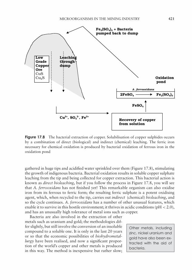
JWBK011-17 JWBK011-Hogg August 12, 2005 16:36 Char Count= 0
MICROORGANISMS IN THE MINING INDUSTRY 421
Fe2(SO4)3 + Bacteriapumped back to dump
Low LeachingGrade throughCopper dumpOreCuSCu2S Oxidation
pond
A . ferrooxidans
2FeSO4 Fe2(SO4)3
FeSO4
Cu2+, SO42-, Fe2+
Recovery of copperfrom solution
Figure 17.8 The bacterial extraction of copper. Solubilisation of copper sulphides occursby a combination of direct (biological) and indirect (chemical) leaching. The ferric ironnecessary for chemical oxidation is produced by bacterial oxidation of ferrous iron in theoxidation pond
gathered in huge tips and acidified water sprinkled over them (Figure 17.8), stimulatingthe growth of indigenous bacteria. Bacterial oxidation results in soluble copper sulphateleaching from the tip and being collected for copper extraction. This bacterial action isknown as direct bioleaching, but if you follow the process in Figure 17.8, you will seethat A. ferrooxidans has not finished yet! This remarkable organism can also oxidiseiron from its ferrous to ferric form; the resulting ferric sulphate is a potent oxidisingagent, which, when recycled to the tip, carries out indirect (chemical) bioleaching, andso the cycle continues. A. ferrooxidans has a number of other unusual features, whichenable it to survive in this hostile environment; it thrives in acidic conditions (pH < 2.0),and has an unusually high tolerance of metal ions such as copper.
Other metals, includingzinc, nickel uranium andgold have also been ex-tracted with the aid ofbacteria.
Bacteria are also involved in the extraction of othermetals such as uranium and gold; the methodologies dif-fer slightly, but still involve the conversion of an insolublecompound to a soluble one. It is only in the last 20 yearsor so that the economic possibilities of biohydrometal-lurgy have been realised, and now a significant propor-tion of the world’s copper and other metals is producedin this way. The method is inexpensive but rather slow;
JWBK011-17 JWBK011-Hogg August 12, 2005 16:36 Char Count= 0
422 INDUSTRIAL AND FOOD MICROBIOLOGY
it may take years to extract the copper from a large tip. However as high-grade copper-bearing ores become increasingly scarce, bioleaching seems likely to play an increasinglyimportant role.
Sulphur-oxidising bacteria also have a role to play in the coal mining industry. In-creased environmental awareness in many countries means that it is no longer acceptableto burn off the sulphur content of coal as sulphur dioxide, so an alternative must befound. One possibility is the biodesulphurisation of coal, using sulphur-oxidising bac-teria to remove the sulphur before combustion. Whilst technically feasible, economicconsiderations mean that this has not yet been widely adopted.
Test yourself
1 In winemaking, juice from the crushed grapes (known as the ) istreated with to remove any naturally occuring microor-ganisms.
2 wines result from converting most of the sugar from the grapes toalcohol.
3 Fermentations in the beer and wine industries are carried out by yeasts ofthe genus .
4 Hops were originally added to beers because of their properties.
5 The alcohol content of an average beer is around per cent.
6 Coagulation of milk using rennet allows separation into and.
7 cheeses, such as Parmesan, require the longest periods of.
8 The bacteria and are involved inyoghurt making, converting lactose to .
9 QuornTM is a meat substitute derived from the fungus .
10 The end-product of sugar fermentation in bread making differs from that inalcoholic fermentations beacuse it takes place in conditions.
11 and are two human pathogens found as spoilage organ-isms in foodstuffs.
12 Foodstuffs with a low content are less susceptible to microbialattack.
JWBK011-17 JWBK011-Hogg August 12, 2005 16:36 Char Count= 0
TEST YOURSELF 423
13 produced microbially from by a species of Clostridium, was im-portant in explosive manufacture during the First World War.
14 The three amino acids produced in greatest quantities for use in the foodindustry are , and .
15 Citric acid is widely used in the food industry; it is produced by the mould.
16 Starch is converted to by the actionof the enzymes α-amylase, glucoamylase and glucose isomerase.
17 The enzymes most widely used in cleaning products are and.
18 Microbial cultures for the commercial production of antibiotics are grownin large-scale .
19 is able to convert copper from its insolubleto its soluble . It can also convert iron from its to its
from.
20 The biggest disadvantage of using biological methods of metal extraction isthat the process is .

JWBK011-17 JWBK011-Hogg August 12, 2005 16:36 Char Count= 0
424

JWBK011-GSY JWBK011-Hogg May 25, 2005 16:33 Char Count= 0
Glossary
Acid–fast stain a procedure for assessing the ability of an organism to retain hotcarbol fuchsin stain when rinsed with acidic alcohol.
Acidophilic (‘acid-loving’):a term applied to organisms that show optimal growth inacid conditions (pH<5.5).
Activated sludge treatment a method of wastewater treatment involving aeration intanks that have been seeded with a mixed microbial sludge.
Activation energy the energy required to initiate a chemical reaction.
Active site the part of an enzyme involved in binding its substrate; the site ofcatalytic action.
Active transport an energy-requiring process in which a substance is transportedagainst an electrochemical gradient.
Adenosine triphosphate (ATP) the principal compound used for the storage andtransfer of energy in cellular systems.
Aerobe an organism that grows in the presence of molecular oxygen, which it usesas a terminal electron acceptor in aerobic respiration.
Aerotolerant anaerobe an anaerobe that is able to tolerate the presence ofoxygen, even though it does not use it.
Aetiology the cause or origin of a disease.
Aldose a sugar molecule that contains an aldehyde group.
Alga a photosynthetic, eucaryotic plant-like organism. It may be unicellular ormulticellular.
425

JWBK011-GSY JWBK011-Hogg May 25, 2005 16:33 Char Count= 0
426 GLOSSARY
Alternation of generations a pattern of sexual reproduction that includes bothhaploid and diploid mature individuals.
Ames test a test to assess the mutagenicity of a substance.
Amino acid an organic acid bearing both amino and carboxyl groups. The buildingblock of proteins.
Amphipathic having a polar region at one end and a nonpolar region at the other.
Anabolism metabolic reactions involved in the synthesis of macromolecules, usuallyrequiring an input of energy.
Anaerobe an organism that grows in the absence of molecular oxygen.
Anammox the formation of nitrogen gas by the anaerobic oxidation of ammoniaand nitrite.
Angstrom unit one ten billionth (10−10) of a metre.
Anisogamy the fusion of unequally sized gametes.
Anoxygenic photosynthesis a form of photosynthesis in which oxygen is notgenerated; found in the purple and green photosynthetic bacteria.
Antibiotic a microbially produced substance (or a synthetic derivative) that hasantimicrobial properties.
Antibody a protein of high binding specificity produced by the immune systems ofhigher animals in response to infection by a foreign organism.
Anticodon the three-nucleotide sequence carried by a tRNA, that base-pairs with itscomplementary mRNA codon.
Antigenic shift a process by which major variations in viral antigens occur.
Antiseptic a chemical agent of disinfection that is mild enough to be used onhuman skin or tissues.
Archaea a group of prokaryotes that diverged from all others (see Bacteria) at anearly stage in evolution, and that show a number of significant differences from them.One of the three domains of life.
Ascospore a haploid spore produced by members of the Ascomycota.
JWBK011-GSY JWBK011-Hogg May 25, 2005 16:33 Char Count= 0
GLOSSARY 427
Ascus a sac-like structure possessed by members of the Ascomycota that containsthe ascospores.
Aseptic technique a set of practical measures designed to prevent the growth ofunwanted contaminants from the environment.
Assimilatory sulphate reduction the incorporation of inorganic sulphate intosulphur-containing amino acids.
Autoclave an appliance that uses steam under pressure to achieve sterilisation.
Atomic mass the average of the mass numbers of an element’sdifferent isotopes,taking into account the proportions in which they occur.
Atomic number the number of protons in the nucleus of an element.
Autotroph an organism that can derive its carbon from carbon dioxide.
Auxotroph a mutant that lacks the ability to synthesise an important nutrient such asan amino acid or vitamin, and must therefore have it provided (c.f. prototroph).
Axenic culture a pure culture containing one type of organism only, and completelyfree from contaminants.
Bacillus (pl. bacilli): a rod-shaped bacterium.
Bacteria procaryotes other than the Archaea (q.v.). Less formally, the term isfrequently used to describe all procaryotes.
Bactericidal causing the death of bacteria.
Bacteriophage a virus whose host is a bacterial cell.
Bacteriostatic inhibiting the growth of bacteria, but not necessarily killing them.
Basidiocarp the fruiting body of members of the Basidiomycota.
Basidiospore a haploid spore produced by members of the Basidiomycota.
Basidium a club-shaped structure carrying the basidiospores in members of theBasidiomycota.
Batch culture a microbial culture grown in a closed vessel with no addition ofnutrients or removal of waste products.

JWBK011-GSY JWBK011-Hogg May 25, 2005 16:33 Char Count= 0
428 GLOSSARY
Benthic zone the sediment of mud and organic matter at the bottom of a pond orlake.
Beta-lactamase an enzyme that breaks a bond in the β-lactam core of the penicillinmolecule.
Bioaugmentation the deliberate introduction of specific microorganisms into anenvironment in order to assist in bioremediation.
Biochemical oxygen demand (BOD) the amount of oxygen needed bymicroorganisms to oxidise the organic content of a water sample.
Biodegradation the natural processes of breakdown of organic matter bymicroorganisms.
Biodeterioration the damage caused to materials of economic importance due tobiological (mainly microbial) processes.
Biofilm a complex system of microorganisms and their surrounding polysaccharidematrix.
Biomass the total amount of cellular material in a system.
Bioreactor a fermentation vessel for the controlled growth of microoganisms.
Bioremediation the use of biological processes to improve a specific environment,such as by the removal of a pollutant.
Calvin cycle a pathway for the fixation of carbon dioxide, used by photosyntheticorganisms and some chemolithotrophs.
Capsid the protein coat of a virus particle.
Capsomer a protein subunit of a viral capsid.
Capsule a clearly defined polysaccharide layer surrounding the cell of certainprocaryotes.
Carcinogen an agent capable of causing cancer.
Catabolism those reactions that break down large molecules, usually coupled to arelease of energy.
Catabolite repression the mechanism by which the presence of a preferred nutrient(generally glucose) has the effect of preventing the synthesis of enzymes thatmetabolise other nutrients.
JWBK011-GSY JWBK011-Hogg May 25, 2005 16:33 Char Count= 0
GLOSSARY 429
Central Dogma of biology the proposal that information (= genetic) flow inorganisms is in one direction only, from DNA to RNA to protein.
Centromere the central region of the chromosome that ensures correct distributionof chromosomes between daughter cells during cell division.
Chemotroph an organism that obtains its energy from chemical compounds.
Chloroplast a chlorophyll-containing organelle found in photosynthetic eucaryotes.
Chromosome a nuclear structure on which most of a eucaryotic cell’sgeneticinformation (DNA) is carried, in association with specialised proteins called histones.The nucleoid of procaryotes (q.v.) is also often referred to as a chromosome.
Cilium (pl. cilia) a short motile hair-like structure found on the surface of someeucaryotic cells.
Citric acid cycle see Tricarboxylic acid cycle.
Clamp connection a mechanism, unique to members of the Basidiomycota, forensuring the maintenance of the dicaryotic state.
Cloning the production of multiple copies of a specific DNA molecule. The term isalso used to describe the production of genetically identical cells or even organisms.
Coccus (pl. cocci) a spherically-shaped bacterium.
Codon a sequence of three nucleotide bases that corresponds to a specific aminoacid.
Coenocytic containing many nuclei within a single plasma membrane.
Coenzyme a loosely-bound organic cofactor that influences the activity of anenzyme.
Cofactor a non-protein component of an enzyme (often a metal ion) essential for itsnormal functioning.
Commensal an organism that lives in or on another organism, deriving some benefitfrom the association but not harming the other party.
Commensalism an association between two species from which one participant(the commensal) derives benefit, and the other is neither benefited nor harmed.
Competence (of a bacterial cell): the state of being able to take up naked DNAfrom outside the cell.
JWBK011-GSY JWBK011-Hogg May 25, 2005 16:33 Char Count= 0
430 GLOSSARY
Compound a substance comprising the atoms of two or more elements.
Conidiophore an aerial hypha that bears conidia.
Conidium (conidiospore) an asexual spore, found in members of the Ascomycotaand Actinomycetes. Often forms chains.
Conjugation a process of genetic transfer involving intimate contact between cellsand direct transfer of DNA across a sex pilus.
Consumer a heterotroph that derives energy from the consumption of otherorganisms.
Continuous culture a microbial culture in which nutrient concentrations and otherconditions are kept constant by the addition of fresh medium and the removal of old.
Contractile vacuole a fluid-filled vacuole involved in the osmoregulation of certainprotists.
Cosmid a hybrid cloning vector capable of accommodating inserts of up to 50kb.
Covalent bond a bond formed by the sharing of a pair of electrons between atoms.
Cyanobacteria a group of mostly unicellular procaryotes that carry out oxygenicphotosynthesis. Commonly known as the blue-greens.
Decimal reduction time (D value) the time needed to reduce a cell population by afactor of ten (i.e. to kill 90% of the population) using a particular heat treatment.
Decomposer an organism that breaks down the remains and waste products ofproducers and consumers.
Defined medium a medium whose precise chemical composition is known.
Denitrification the reduction, under anaerobic conditions, of nitrite and nitrate tonitrogen gas.
Diauxic growth a form of growth that has two distinct phases due to one carbonsource being used preferentially before a second.
Dicaryon a structure formed by two cells whose contents, but not nuclei, havefused.
Differential medium a medium that allows colonies of a particular organism to bedifferentiated from others growing in the same culture.
JWBK011-GSY JWBK011-Hogg May 25, 2005 16:33 Char Count= 0
GLOSSARY 431
Differential stain a stain that employs two or more dyes to distinguish betweendifferent cellular structures or cell types.
Dimorphic existing in two distinct forms.
Dioecious having male and female reproductive structures on separate individuals.
Diploid having two sets of chromosomes.
Disaccharide a carbohydrate formed by the joining of two monosaccharides.
Disc diffusion method a method for assessing the antimicrobial properties of asubstance.
Disinfection the elimination or inhibition of pathogenic microorganisms in or on anobject so that they no longer pose a threat.
Dissimilatory sulphate reduction the reduction of sulphate to hydrogen sulphide byobligate anaerobes, using either an organic compound or hydrogen gas as electrondonor.
DNA library a collection of cloned DNA fragments.
Domain the highest level of taxonomic grouping.
Eclipse period the stage of a viral replication cycle in which no complete viralparticles are present in the infected host cell.
Ecosystem the organisms of a particular habitat, together with their inanimatesurroundings.
Ectoparasite a parasite that attaches to the outside of its host.
Electron a subatomic particle carrying a negatively charge.
Electron transport chain a series of donor/acceptor molecules that transfer electronsfrom donors (e.g. NADH) to a terminal electron acceptor (e.g. O2).
Embden Meyerhof pathway see Glycolysis.
Endoparasite a parasite that fully enters its host and lives inside it.
Endoplasmic reticulum a tubular network found in the cytoplasm of eucaryotic cells.
Endospore a highly resistant spore found within certain bacteria.
JWBK011-GSY JWBK011-Hogg May 25, 2005 16:33 Char Count= 0
432 GLOSSARY
Enrichment culture a culture that uses a selective medium to encourage the growthof an organism present in low numbers.
Entner-Doudoroff Pathway an alternative pathway for the oxidation of glucose,producing a mixture of pyruvate and glyceraldehyde-3-phosphate.
Enzyme a cellular catalyst (usually protein), specific to a particular reaction or groupof reactions.
Epiphyte an organism that grows on the surface of a plant.
Eucaryote an organism whose cells contain a true nucleus and membrane-boundorganelles such as mitochondria and endoplasmic reticulum.
Excision repair a repair mechanism in which damaged sections of DNA are cut outand replaced.
Exon a coding region of a gene. c.f. intron
Expression vector a vector that allows the transcription and translation of a foreigngene inserted into it.
Facilitated diffusion the transport of molecules across a membrane with the help ofcarrier proteins. Transport takes place down a concentration gradient, and energy isnot required.
Facultative anaerobe an organism that can grow in the absence of oxygen, bututilises it when available.
Fastidious (of an organism): unable to synthesise a range of nutrients and thereforehaving complex requirements in culture.
Fatty Acid a long-chain hydrocarbon chain with a carboxyl group at one end.
Feedback inhibition a control mechanism whereby the final product of a metabolicpathway acts as an inhibitor to the enzyme that catalyses an early step (usually thefirst) in the pathway.
Fermentation a microbial process by which an organic substrate (usually acarbohydrate) is broken down without the involvement of oxygen or an electrontransport chain, generating energy by substrate-level phosphorylation
Flagellum a long hair-like extracellular structure associated with locomotion. Foundin both procaryotes and eucaryotes, although each has its own distinctive structure.
JWBK011-GSY JWBK011-Hogg May 25, 2005 16:33 Char Count= 0
GLOSSARY 433
F plasmid a plasmid containing genes that code for the construction of the sex pilus,across which it is transferred to a recipient cell.
Frameshift mutation a mutation that results in a change to the reading frame, andthus an altered sequence of amino acids downstream of the point where it occurs.
Fungi a kingdom of non-photosynthetic eucaryotes characterised by absorptiveheterotrophic nutrition.
Gamete a haploid reproductive cell arising from meiosis. Fuses with another gameteto form a diploid zygote.
Gametophyte the haploid, gamete-forming stage in a life cycle with alternation ofgenerations.
Gene a sequence of DNA that usually encodes a polypeptide.
Generalised transduction the transfer of a gene from one bacterial cell to anotheras a result of being inadvertently packaged into a phage particle.
Generation (doubling) time the time taken for a population of cells to double in timeunder specific conditions.
Genetic code the 64 triplet codons and the corresponding amino acids ortermination sequences.
Genome the complete genetic material of an organism.
Genotype the genetic make-up of an organism.
Gluconeogenesis a series of reactions by which glucose is synthesised fromcompounds such as amino acids and lactate.
Glycolysis a series of reactions by which glucose is oxidised to two molecules ofpyruvate with the synthesis of two molecules of ATP.
Glycosidic linkage a covalent linkage formed between monosaccharides.
Golgi apparatus an organelle of eucaryotes, comprising flattened membranoussacs.
Gram stain a differential stain that divides bacteria into Gram-positive (purple) orGram-negative (pink). The reaction depends on the constitution of the organism’scellwall.
JWBK011-GSY JWBK011-Hogg May 25, 2005 16:33 Char Count= 0
434 GLOSSARY
Group translocation a form of active transport in which a solute is modified toprevent its escape from the cell.
Haploid having only one set of chromosomes.
Haloduric able to tolerate high salt concentrations.
Halophilic showing a requirement for moderate to high salt concentrations.
Heterotroph an organism that must use one or more organic molecules as its sourceof carbon.
Hexose a six-carbon sugar.
Hexose monophosphate shunt see Pentose phosphate pathway
Hfr cell a bacterial cell that has a transferred F plasmid integrated into itschromosome.
Histone a basic protein found associated with DNA in eucaryotic chromosomes.
Horizontal transfer the transfer of genetic material between members of the samegeneration.
Human genome project an international effort to map and sequence all the DNA inthe human genome. The project has also involved sequencing the genomes ofseveral other organisms.
Hydrogen bond a relatively weak bond that forms between covalentlybonded hydrogen and any electronegative atom, most commonly oxygen ornitrogen.
Hydrophilic (‘water-loving’):having an affinity for water.
Hydrophobic (‘water-hating’): repelled by water.
Hypha a thread-like filament of cells characteristic of fungi and actinomycetes.
Immersion oil a viscous oil used to improve the resolution of a light microscope athigh power.
In vitro = ‘in glass’, i.e. outside of the living organism, in test tubes etc.
In vivo = ‘in life’, i.e. within the living organism.
JWBK011-GSY JWBK011-Hogg May 25, 2005 16:33 Char Count= 0
GLOSSARY 435
Inoculum the cells used to ‘seed’a new culture.
Insertion vector a cloning vector based on phage λ that has nonessential genesremoved.
Insertional inactivation the insertion of foreign DNA within the gene sequence of aselectable marker in order to inactivate its expression.
Interrupted mating a technique in which the order of gene transfer duringconjugation is determined by terminating transfer at different time intervals.
Intron a non-coding sequence within a gene. c.f. exon
Ion an atom or group of atoms that carries a charge due to losing or gaining one ormore electrons.
Isogamy the fusion of morphologically identical motile gametes.
Isotope forms of an element, having the same number of protons and electrons butdiffering in the number of neutrons
Karyogamy the fusion of nuclei from two different cells.
Ketose a sugar molecule that contains a ketone group.
Kinetoplast a specialised structure within the mitochondria of certain flagellatedprotozoans.
Koch’s postulates a set of criteria, proposed by Robert Koch, which must be satisfiedin order to link a specific organism to a specific disease.
Krebs cycle see Tricarboxylic acid cycle.
Latent period the period in a viral replication cycle between infection of the hostand release of newly synthesised viral particles.
Latent virus a virus that remains inactive in the host for long periods before beingreactivated.
Lichen a symbiotic association of a fungus (usually an ascomycete) and an alga orcyanophyte (blue-green).
Limnetic zone a zone of a body of water away from the shore, and where light isable to penetrate.
JWBK011-GSY JWBK011-Hogg May 25, 2005 16:33 Char Count= 0
436 GLOSSARY
Lipase an enzyme that digests lipids.
Lithotroph an organism that uses inorganic molecules as a source of electrons.
Littoral zone the region of a body of water situated close to land where the water issufficiently shallow for sunlight to penetrate to the bottom.
Lysogeny a form of bacteriophage replication in which the viral genome isintegrated into that of the host and is replicated along with it. See prophage.
Lytic cycle a process of viral replication involving the bursting of the host cell andrelease of new viral particles.
Magnetosome a particle of magnetite (iron oxide) found in certain bacteria,allowing them to orient themselves in a magnetic field.
Malting the stage in beer-making in which grain is soaked in water to initiategermination and activate starch-digesting enzymes.
Mashing the stage in beer-making in which soluble material is released from thegrain in preparation for fermentation.
Mass number the combined total of protons and neutrons in the nucleus of anelement.
Meiosis a form of nuclear division in diploid eucaryotic cells resulting in haploiddaughter nuclei.
Merodiploid a genome that is partly haploid and partly diploid.
Mesophile an organism that grows optimally at moderate temperatures (20−45◦).
Messenger RNA (mRNA) a form of RNA that is synthesised as a complementary copyof the template strand of DNA.
Methanotroph a bacterium capable of using methane as a carbon and energysource.
Michaelis constant (Km) the concentration of substrate in an enzyme reaction thatresults in a rate of reaction equal to one half of the maximum.
Mineralisation the breakdown of organic matter to carbon dioxide and inorganiccompounds.
JWBK011-GSY JWBK011-Hogg May 25, 2005 16:33 Char Count= 0
GLOSSARY 437
Minimum inhibitory concentration (MIC) the lowest concentration of anantimicrobial substance that prevents growth of a given organism.
Mismatch repair a DNA repair system that replaces incorrectly inserted nucleotides.
Missense mutation a mutation that alters the sense of the message encoded in theDNA, resulting in an incorrect amino acid being incorporated.
Mitochondrion a spherical to ovoid organelle found in eucaryotes. The site of ATPgeneration via TCA cycle and oxidative phosphorylation.
Mitosis a form of nuclear division in eucaryotic cells, resulting in daughter nucleieach with the same chromosome complement as the parent.
Monoecious having reproductive structures of both sexes on the same individual.
Monosaccharide the simplest from of carbohydrate molecule.
Most probable number (MPN) a statistical method of estimating microbial numbersin a liquid sample based upon the highest dilution able to support growth.
mRNA see Messenger RNA
Mutagen a chemical or physical agent capable of inducing mutations.
Mutation any heritable alteration in a DNA sequence. It may or may not have aneffect on the phenotype.
Mutualism an association between two species from which both participants derivebenefit.
Mycelium a tangled mass of branching hyphae.
Mycorrhiza a mutualistic relationship between a fungus and a plant root.
Mycosis a disease caused by a fungus.
Neutron a subatomic particle carrying neither positive nor negative charge.
Nitrification the two-step process by which ammonia is oxidised to nitrite and then tonitrate.
Nonsense codon see stop codon.
JWBK011-GSY JWBK011-Hogg May 25, 2005 16:33 Char Count= 0
438 GLOSSARY
Nonsense mutation a mutation that results in a ‘stop’codon being inserted intothe mRNA at the point where it occurs, and the premature termination of translation.
Nosocomial infection an infection that is acquired in hospital or other healthcaresetting.
Nuclear membrane the double membrane surrounding the nucleus of a eucaryoticcell.
Nucleocapsid the genome of a virus and its surrounding protein coat.
Nucleoid another name for the bacterial chromosome. The site of most of aprocaryotic cell’s DNA.
Nucleolus a discrete region of the eucaryotic nucleus, where ribosomes areassembled.
Nucleotide the building block of nucleic acids, comprising a pentose sugar, anitrogenous base and one or more phosphate groups.
Nucleus the central, membrane-bound structure in eucaryotic cells that containsthe genetic material. Also, the region of an atom that contains the protons andneutrons.
Obligate anaerobe an organism that is incapable of growth in the presence ofoxygen.
Okazaki fragments discontinuous fragments of single-stranded DNA synthesisedcomplementary to the lagging strand during DNA replication.
Oncogene a gene associated with the conversion of a cell to a cancerous form.
Oogamy the fusion of a small, motile sperm cell and a larger, immobile egg cell.
Operon a group of related genes under the control of a single operator sequence.
Organotroph an organism that uses organic molecules as a source of electrons.
Outer membrane the outermost part of the Gram-negative cell wall, comprisingphospholipids and lipopolysacchride.
Oxidation a chemical reaction in which an electron is lost.
Oxygenic photosynthesis a form of photosynthesis in which oxygen is produced;found in algae, cyanobacteria (blue greens) and also green plants.
JWBK011-GSY JWBK011-Hogg May 25, 2005 16:33 Char Count= 0
GLOSSARY 439
Parasitism an association between two species from which one partner derivessome or all of its nutritional requirements by living either in or on the other (the host),which usually suffers harm as a result.
Pasteurisation a mild heating regime used to destroy pathogens and spoilageorganisms present in food and drink, especially milk.
Pathogen an organism with the potential to cause disease.
Pellicle a semi-rigid structure composed of protein strips found surrounding the cellof certain unicellular protozoans and algae.
Pentose a five-carbon sugar.
Pentose phosphate pathway a secondary pathway for the oxidation of glucose,resulting in the production of pentoses that serve as precursors for nucleotides.
Peptide bond the bond formed between the amino group of one amino acid andthe carboxyl group of another.
Peptidoglycan a polymer comprising alternate units of N-acetylmuramic acid andN-acetylglucosamine that forms the major constituent of bacterial cell walls.
Phagemid a hybrid cloning vector, comprising elements of plasmid and phage.
Phagocytosis the ingestion and digestion of particulate matter by a cell, a processunique to eucaryotes.
Phenol coefficient a measure of the efficacy of a disinfectant against a givenorganism, compared to that of phenol.
Phenotype the observable characteristics of an organism.
Phospholipid an important constituent of all membranes, comprising atriacylglycerol in which one fatty acid is replaced by a phosphate group.
Phosphorylation the addition of a phosphate group.
Photophosphorylation the synthesis of ATP using light energy.
Photoreactivation a DNA repair mechanism involving the light-dependent enzymeDNA photolyase.
Photosynthesis a process by which light energy is trapped by chlorophyll andconverted to ATP, which is used to drive the synthesis of carbohydrate by reducing CO2.

JWBK011-GSY JWBK011-Hogg May 25, 2005 16:33 Char Count= 0
440 GLOSSARY
Phototroph an organism that is able to use light as its source of energy.
Phylogenetic relating to the evolutionary relationship between organisms.
Phytoplankton the collective term used to describe the unicellular photosynthesisers,including cyanobacteria, dinoflagellates, diatoms and single-celled algae.
Pilus (pl. pili) a short, hair-like appendage found on surface of procaryotes andassisting with attachment. Specialised sex pili (q.v.) are involved in bacterialconjugation.
Plankton the floating microscopic organisms of aquatic systems.
Plasma membrane the membrane that surrounds a cell.
Plasmid a small, self-replicating loop of extrachromosomal DNA, found in bacteriaand some yeasts. Specially engineered forms are used as vectors in gene cloning.
Plasmodium a mass of protoplasm containing several nuclei and bounded by acytoplasmic membrane.
Plasmogamy the fusion of the cytoplasmic content of two cells.
Plasmolysis the shrinkage of the plasma membrane away from the cell wall, due toosmotic loss of water from thecell.
Point mutation a mutation that involves the substitution of one nucleotide byanother.
Polar having unequal charge distribution, caused by unequal sharing of atoms.
Polymerase chain reaction (PCR) a technique that selectively replicates a specificDNA sequence by means of in vitro enzymatic amplification.
Polypeptide a chain of many amino acids joined together by peptide bonds.
Polyribosome (polysome) a chain of ribosomes attached to the same molecule ofmRNA.
Polysaccharide a carbohydrate polymer of monosaccharide units.
Primary producer an autotroph that obtains energy from the sun or chemicalsources.
JWBK011-GSY JWBK011-Hogg May 25, 2005 16:33 Char Count= 0
GLOSSARY 441
Primer a short sequence of single-stranded DNA or RNA required by DNApolymerase as a starting point for chain extension.
Prion a self-replicating protein responsible for a range of neurodegenerativedisorders in humans and mammals
Procaryote an organism lacking a true nucleus and membrane-bound organelles.
Profundal zone the deepest part of a body of water, where the sun is unable topenetrate
Promoter a sequence upstream of a gene, where RNA polymerase binds to initiatetranscription.
Prophage the DNA of a temperate phage that has integrated into the hostgenome. It remains inactive whilst in this form.
Prostheca a stalked structure formed by an extension of the cell wall and plasmamembrane of certain bacteria.
Prosthetic group a non-polypeptide component of a protein, such as a metal ion ora carbohydrate.
Protease an enzyme that digests proteins.
Protista a eucaryotic kingdom, comprising mostly unicellular organisms.
Protocooperation a form of mutualistic relationship that is not obligatory for eitherpartner.
Proton a positively-charged subatomic particle.
Protoplast a cell that has had its cell wall removed.
Prototroph the normal, nonmutant form of an organism.
Protozoa a group of single celled eucaryotes with certain animal-like characteristics.
Pseudomurein a modified form of peptidoglycan found in some archaean cell walls.
Pseudopodium a projection of the plasma membrane of the amoebas that causesthe cell to change shape and allows movement.
Psychrophile an organism that grows optimally at low temperatures (<15◦).
JWBK011-GSY JWBK011-Hogg May 25, 2005 16:33 Char Count= 0
442 GLOSSARY
Psychrotroph an organism that is able to tolerate low temperatures, but grows betterat more moderate values.
Reading frame the way in which a sequence of nucleotides is read in triplets,depending on the starting point.
Real image an image that can be projected onto a flat surface such as a screen.
Recombinant DNA DNA that comprises material from more than one source.
Recombination any process that results in new combinations of genes.
Redox potential (Eo) the tendency of a compound to lose or gain electrons.
Reduction a chemical reaction in which an electron is gained.
Refractive index the ratio between the velocity of light as it passes through asubstance and its velocity in a vacuum.
Regulatory gene a gene whose protein product has an effect in controlling theexpression of other genes.
Replacement vector a cloning vectors based on phage λ, in which a central sectionof nonessential DNA is replaced by the insert DNA.
Replication fork a Y-shaped structure formed by the separating strands of DNAduring replication.
Repressor protein a protein that prevents transcription of a gene by binding to itsoperator.
Resolution the capacity of an optical instrument to discern detail.
Restriction endonuclease an enzyme of microbial origin that cleaves DNA at aspecific nucleotide sequence.
Reverse transcriptase an enzyme found in retroviruses that can synthesise DNA froman RNA template.
Rhizosphere the region around the surface of a plant’s root system.
Ribosomal RNA (rRNA) a form of RNA that forms part of the structure of ribosomes.
Ribosome an organelle made up of protein and RNA, found in both procaryotesand eucaryotes. The site of protein synthesis.
JWBK011-GSY JWBK011-Hogg May 25, 2005 16:33 Char Count= 0
GLOSSARY 443
rRNA see Ribosomal RNA
Saprobe an organism that feeds on dead and decaying organic materials.Previously termed saprophyte.
Saturated fatty acid a fatty acid that has only single covalent bonds betweenadjacent carbon atoms. (c.f. unsaturated)
Secondary metabolite a substance produced by a microorganism after the phaseof active growth has ceased.
Selectable marker a gene that allows cells containing it to be identified by theexpression of a recognisable characteristic.
Selective medium a medium that favours the growth of a particular organism orgroup of organisms, often by suppressing the growth of others.
Semi-conservative replication the process of DNA replication by which eachstrand acts as a template for the synthesis of a new complementary strand. Eachresultant double stranded molecule thus comprises one original strand and one newone.
Septate separated by septa or cross-walls.
Sex pilus a narrow extension of the bacterial cell, through which genetic material istransferred during conjugation.
Shuttle vector a cloning vector that can replicate in both bacterial and yeast hostcells.
Silent mutation a mutation that has no effect on the amino acid encoded by thetriplet.
Single-cell protein (SCP) bacteria and yeast grown in bulk for use as a protein-richfood supplement.
Slime layer a diffuse and loosely attached polysaccharide layer surrounding the cellof certain procaryotes.
Specialised transduction the transfer of a limited selection of genes due to impreciseexcision of a prophage in a lysogenic infection cycle.
Sporangiophore a specialised aerial hypha that bears the sporangia.
Sporangium a structure inside which spores develop.
JWBK011-GSY JWBK011-Hogg May 25, 2005 16:33 Char Count= 0
444 GLOSSARY
Spore a resistant, non-motile reproductive cell.
Sporophyte the diploid, spore-forming stage in a life cycle with alternation ofgenerations.
Sporozoite a motile infective stage of members of the Sporozoa that gives rise to anasexual stage within the new host.
Sterilisation the process by which all microorganisms present on or in an object aredestroyed or removed.
Steroid a member of a group of lipids based on a four-ring structure.
Stop codon use definition below.
Substrate level phosphorylation the synthesis of ATP by the direct transfer of aphosphate group from a phosphorylated organic compound to ADP.
Symbiosis a close physical association between two species from which specialbenefits may accrue for one or both parties.The term is sometimes used specifically to describe such a relationship in which bothparties derive benefit (see mutualism).
Tautomerism the ability of a molecule such as a nucleotide base to exist in twoalternative forms.
Taxonomy the science of naming and classifying living (and once-living) organisms.
Temperate phage a bacteriophage with a lysogenic replication cycle.
Terminator a sequence of DNA that indicates transcription should stop.
stop codons one of the three triplet sequences (also called nonsense codons) thatindicate translation should stop.
Thallus a simple vegetative plant body showing no differentiation into root, stemand leaf.
Therapeutic index a measure of the selective toxicity of a chemotherapeutic agent.
Thylakoid a photosynthetic membrane found in chloroplasts or free in the cytoplasm(in cyanobacteria). It contains photosynthetic pigments and components of theelectron transport chain.
Total cell count a method that enumerates all cells, living and dead.
JWBK011-GSY JWBK011-Hogg May 25, 2005 16:33 Char Count= 0
GLOSSARY 445
Trace element an element required in minute amounts for growth.
Transamination a reaction involving the transfer of an amino group from onemolecule to another.
Transcription the process by which single-stranded mRNA is synthesised from acomplementary DNA template.
Transduction the bacteriophage-mediated transfer of genetic material betweenbacteria.
Transformation the uptake of naked DNA from the environment and its integrationinto the host genome.
Transposable elements a sequence of DNA that is able to relocate to anotherposition on the genome.
Transition a mutation in which a purine replaces a purine or a pyrimidine replaces apyrimidine.
Translation the process by which the message encoded in mRNA is converted into asequence of amino acids.
Transfer RNA (tRNA) a form of RNA that carries specific amino acids to the site ofprotein synthesis.
Transversion a mutation in which a purine replaces a pyrimidine, or a pyrimidinereplaces a purine.
Triacylglycerol a lipid formed by the joining of three fatty acids to a molecule ofglycerol.
Tricarboxylic acid cycle a series of reactions that oxidise acetate to CO2,generating reducing power in the form of NADH and FADH2 for use in the electrontransport chain. Also known as citric acid cycle or Krebs cycle.
Triose a three-carbon sugar.
tRNA see Transfer RNA
Tyndallisation a form of intermittent steam sterilisation.
Undefined or complex medium a medium whose precise chemical composition isnot known.
JWBK011-GSY JWBK011-Hogg May 25, 2005 16:33 Char Count= 0
446 GLOSSARY
Unsaturated fatty acid a fatty acid that contains one or more double bondsbetween adjacent carbon atoms.
Vaccination inoculation with a vaccine to provide protective immunity.
Vaccine a preparation of dead or inactivated living pathogens or their productsused to provide protective immunity.
Vector a self-replicating DNA molecule used in gene cloning. The sequence to becloned is inserted into the vector, and replicated along with it.
Viable cell count a method that enumerates only those cells capable ofreproducing to form a visible colony.
Virtual image an image that has no physical existence in space, and cannot beprojected onto a screen.
Virion a complete, intact viral particle
Viroid a plant pathogen that comprises only ssRNA and does not code for proteinproduct.
Virulent phage a bacteriophage with a lytic replication cycle.
Virus a submicroscopic, noncellular parasite, comprising protein and RNA or DNA.
Wildtype the normal, nonmutant form of an organism or gene.
Wobble the degree of flexibility allowed in the third base in a codon when pairingwith tRNA. The wobble hypothesis explains how a single tRNA can pair with more thanone codon.
Yeast artificial chromosome (YAC) a cloning vector able to accommodate inserts ofseveral hundred kb in size.
Z value the increase in temperature required to reduce the D value (q.v.) by afactor of ten.
Zoonosis a disease normally found in animals, but transmissible to humans undercertain circumstances.
Zoospore a flagellated, asexual spore.
Zygospore a thick-walled resistant diploid structure formed by members of theZygomycota.

JWBK011-APN JWBK011-Hogg August 12, 2005 16:37 Char Count= 0
Appendix
Solution to Numerical Questions
Question 2.5: 1000 (10 × 10 × 10)
Question 2.15: 61 cytosine residues; 286 hydrogen bonds (61 × 3 for GC and 52 × 2for AT)
447

JWBK011-APN JWBK011-Hogg August 12, 2005 16:37 Char Count= 0
448
JWBK011-18 JWBK011-Hogg May 25, 2005 16:35 Char Count= 0
Further Reading
The declared intention of Essential Microbiology is to serve as an introduction to micro-biology for students who do not intend to specialise in the subject. It would please the au-thor greatly, however, if the book stimulated readers to deepen their knowledge and un-derstanding by referring to other, more specialist texts. Below are listed a number of sug-gested sources for further reading; the list is by no means comprehensive, and the readermay well find other texts to suit his or her individual needs. Although titles are listed un-der the heading of a particular chapter, many will of course also be equally useful in thecontext of other chapters. Journal articles have not been included, with the exceptionof a few review-type articles from readily accessible titles such as Scientific American.
General microbiology
For a general overview of the world of microorganisms and their applications, there canbe no better starting point than John Postgate’s classic Microbes and Man, 4th edition(2000), published by Cambridge University Press. Those wishing to study microbiologyin greater depth may consider one of the many excellent general microbiology textsavailable; the following are well-established examples that embrace the whole field ofmicrobiology, and cover many topics in greater depth than is possible or indeed desirablein the present book.
Black JG (2004) Microbiology: Principles and Explorations, 6th edn. John Wiley & Sons,Chichester.
Lederberg, J (ed) (2000): Encyclopaedia of Microbiology, 2nd edition. Academic Press.Madigan MT, Martinko JM and Parker J (2003) Brock Biology of Microorganisms, 10th edn.
Prentice Hall Inc., Englewood Cliffs, NJ.Prescott LM, Harley JP and Klein DA (2004) Microbiology, 6th edn. McGraw-Hill, New York.Singleton P and Salisbury D (2002): Dictionary of Microbiology and Molecular Biology, 3rd
edition. John Wiley & Sons.Tortora GJ, Funke BR and Case CL (2004) Microbiology: An Introduction, 8th edition. Benjamin
Cummings, San Francisco, CA.
Chapter 1
Beck RW (2000) A Chronology of Microbiology in Historical Context. American Society ofMicrobiology.
449
JWBK011-18 JWBK011-Hogg May 25, 2005 16:35 Char Count= 0
450 FURTHER READING
De Kruif P (2002) Microbe Hunters. Harcourt Publishers, New York.Lagerqvist U (2003) Pioneers of Microbiology and the Nobel Prize. World Scientific Publishing,
London.Murphy DB (2001) Fundamentals of Light Microscopy and Electronic Imaging. John Wiley &
Sons, Chichester.
Chapter 2
Hames DB, Hooper NM and Houghton JD (2000): Instant Notes in Biochemistry, 2nd edn. BiosScientific Publishers, Oxford.
Lewis R and Evans W (2001) Chemistry, 2nd edn. Palgrave, Basingstoke.
Chapter 3
Cooper GM and Hausman RE (2004) The Cell: A Molecular Approach, 3rd edn. Sinauer Asso-ciates Inc, Sunderland, MA.
Singleton P (2004) Bacteria in Biology, Biotechnology and Medicine, 6th edn. John Wiley & Sons,Chichester.
Chapter 4
Atlas, RM (2004) Handbook of Microbiological Media, 3rd edn. CRC Press, Boca Raton, FL.Barrow GI and Feltham RKA (eds) (2004) Cowan and Steel’s Manual for the Identification of
Medical Bacteria, 3rd edn. Cambridge University Press, Cambridge.Cappuccino JG and Sherman N (2005) Microbiology: A Laboratory manual, 7th edn. Benjamin
Cummings, San Francisco, CA.Collins CH, Lyne PM, Grange JM and Falkinham J (2004) Microbiological Methods, 8th edn.
Hodder Arnold, London.
Chapter 5
Isaac S and Jennings D (1995) Microbial Culture. Bios Scientific Publishers, Oxford.Madigan MT and Marrs BL (1997) Extremophiles. Scientific American 276, 82–7.Moat AG, Foster JW and Spector MP (2002) Microbial Physiology, 4th edn. John Wiley & Sons,
Chichester.
Chapter 6
Gilbert HF (2000) Basic Concepts in Biochemistry: A Student’s Survival Guide, 2nd edition.McGraw-Hill, New York.
Matthews HR, Freedland R and Miesfeld RL (1997) Biochemistry: A Short Course. John Wiley& Sons, Chichester.
Zubay G (1998) Biochemistry, 4th edn. McGraw-Hill, New York.
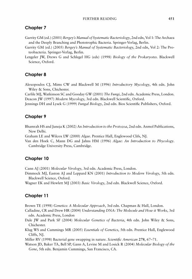
JWBK011-18 JWBK011-Hogg May 25, 2005 16:35 Char Count= 0
FURTHER READING 451
Chapter 7
Garrity GM (ed.) (2001) Bergey’s Manual of Systematic Bacteriology, 2nd edn, Vol 1: The Archaeaand the Deeply Branching and Phototrophic Bacteria. Springer-Verlag, Berlin.
Garrity GM (ed.) (2005) Bergey’s Manual of Systematic Bacteriology, 2nd edn, Vol 2: The Pro-teobacteria. Springer-Verlag, Berlin.
Lengeler JW, Drews G and Schlegel HG (eds) (1998) Biology of the Prokaryotes. BlackwellScience, Oxford.
Chapter 8
Alexopoulos CJ, Mims CW and Blackwell M (1996) Introductory Mycology, 4th edn. JohnWiley & Sons, Chichester.
Carlile MJ, Watkinson SC and Gooday GW (2001) The Fungi, 2nd edn. Academic Press, London.Deacon JW (1997) Modern Mycology, 3rd edn. Blackwell Scientific, Oxford.Jennings DH and Lysek G (1999) Fungal Biology, 2nd edn. Bios Scientific Publishers, Oxford.
Chapter 9
Bhamrah HS and Juneja K (2002) An Introduction to the Protozoa, 2nd edn. Anmol Publications,New Delhi.
Graham LE and Wilcox LW (2000) Algae. Prentice Hall, Englewood Clifs, NJ.Van den Hoek C, Mann DG and Jahns HM (1996) Algae: An Introduction to Phycology.
Cambridge University Press, Cambridge.
Chapter 10
Cann AJ (2001) Molecular Virology, 3rd edn. Academic Press, London.Dimmock MJ, Easton AJ and Leppard KN (2001) Introduction to Modern Virology, 5th edn.
Blackwell Science, Oxford.Wagner EK and Hewlett MJ (2003) Basic Virology, 2nd edn. Blackwell Science, Oxford.
Chapter 11
Brown TE (1998) Genetics: A Molecular Approach, 3rd edn. Chapman & Hall, London.Calladine, CR and Drew HR (2004) Understanding DNA: The Molecule and How it Works, 3rd
edn. Academic Press, LondonDale JW and Park SF (2004) Molecular Genetics of Bacteria, 4th edn. John Wiley & Sons,
Chichester.Klug WS and Cummings MR (2005) Essentials of Genetics, 5th edn. Prentice Hall, Englewood
Cliffs, NJ.Miller RV (1998) Bacterial gene swapping in nature. Scientific American 278, 67–71.Watson JD, Baker TA, Bell SP, Gann A, Levine M and Losick R (2004) Molecular Biology of the
Gene, 5th edn. Benjamin Cummings, San Francisco, CA.
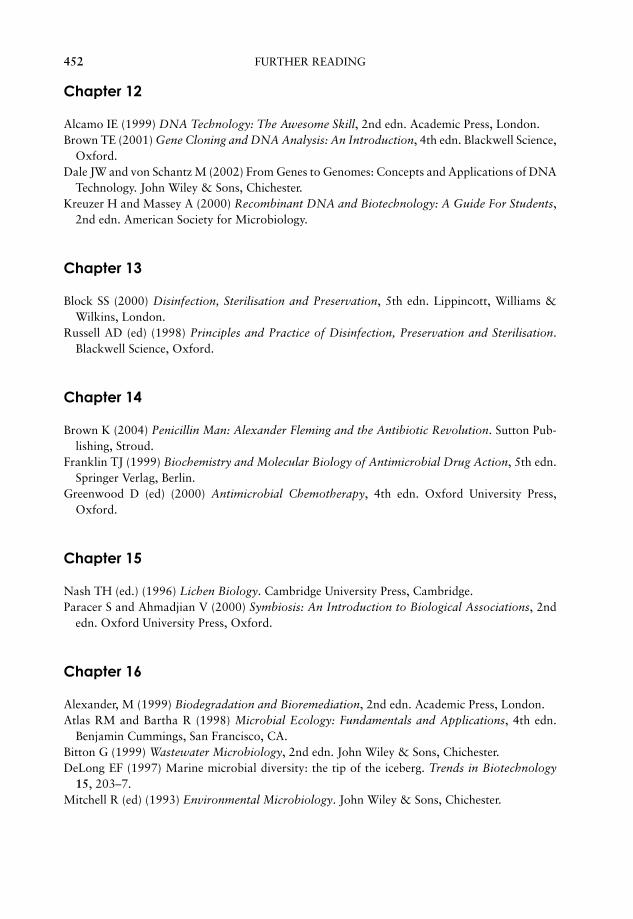
JWBK011-18 JWBK011-Hogg May 25, 2005 16:35 Char Count= 0
452 FURTHER READING
Chapter 12
Alcamo IE (1999) DNA Technology: The Awesome Skill, 2nd edn. Academic Press, London.Brown TE (2001) Gene Cloning and DNA Analysis: An Introduction, 4th edn. Blackwell Science,
Oxford.Dale JW and von Schantz M (2002) From Genes to Genomes: Concepts and Applications of DNA
Technology. John Wiley & Sons, Chichester.Kreuzer H and Massey A (2000) Recombinant DNA and Biotechnology: A Guide For Students,
2nd edn. American Society for Microbiology.
Chapter 13
Block SS (2000) Disinfection, Sterilisation and Preservation, 5th edn. Lippincott, Williams &Wilkins, London.
Russell AD (ed) (1998) Principles and Practice of Disinfection, Preservation and Sterilisation.Blackwell Science, Oxford.
Chapter 14
Brown K (2004) Penicillin Man: Alexander Fleming and the Antibiotic Revolution. Sutton Pub-lishing, Stroud.
Franklin TJ (1999) Biochemistry and Molecular Biology of Antimicrobial Drug Action, 5th edn.Springer Verlag, Berlin.
Greenwood D (ed) (2000) Antimicrobial Chemotherapy, 4th edn. Oxford University Press,Oxford.
Chapter 15
Nash TH (ed.) (1996) Lichen Biology. Cambridge University Press, Cambridge.Paracer S and Ahmadjian V (2000) Symbiosis: An Introduction to Biological Associations, 2nd
edn. Oxford University Press, Oxford.
Chapter 16
Alexander, M (1999) Biodegradation and Bioremediation, 2nd edn. Academic Press, London.Atlas RM and Bartha R (1998) Microbial Ecology: Fundamentals and Applications, 4th edn.
Benjamin Cummings, San Francisco, CA.Bitton G (1999) Wastewater Microbiology, 2nd edn. John Wiley & Sons, Chichester.DeLong EF (1997) Marine microbial diversity: the tip of the iceberg. Trends in Biotechnology
15, 203–7.Mitchell R (ed) (1993) Environmental Microbiology. John Wiley & Sons, Chichester.

JWBK011-18 JWBK011-Hogg May 25, 2005 16:35 Char Count= 0
FURTHER READING 453
Chapter 17
Adams MR and Moss MO (2000) Food Microbiology, 2nd edn. Royal Society of Chemistry,London.
Demain AL (2000) Microbial Technology. Trends in Biotechnology 18, 26–31.Garbutt, J (1997) Essentials of Food Microbiology. Hodder Arnold, London.Hornsey IS (1999) Brewing. Royal Society of Chemistry, London.Ratledge C and Kristiansen B (2001) Basic Biotechnology, 2nd edn. Cambridge University Press,
Cambridge.Smith JE (2004) Biotechnology, 4th edn. Cambridge University Press, Cambridge.
JWBK011-IND JWBK011-Hogg August 12, 2005 16:39 Char Count= 0
Index
The suffix ‘t’ indicates an entry in a table; ‘f’ indicates a footnote
Acetogenic bacteria 378Acetic acid bacteria 178Acetobacter 178, 413tAcetobacter suboxydans 415–16Acetone 414Acetyl-Coenzyme A (Acetyl-CoA) 128–30, 136–7Acid 25Acid mine drainage 191, 402Acid-fast stain 191Acidithiobacillus 393Acidithiobacillus ferrooxidans 88t, 172, 398, 402,
420–2Acidithiobacillus thiooxidans 172Acidophile 98, 166Acne 190, 192tACP see: acyl carrier proteinAcquired immunodeficiency syndrome (AIDS) see:
AIDSAcremonium 380–1Actinobacteria 188, 191–2Actinomyces 191Actinomycetes 188, 191–2, 395Actinomycin 355t, 356Activated sludge 400–1Activation energy see: enzymeActive site see: enzymeActive transport 83Acyclovir 369–70Acyl carrier protein (ACP) 149Adenine 39, 41, 44, 293–6Adenosine diphosphate see: ADPAdenosine triphosphate see: ATPAdenovirus 260tAdenylate cyclase 193ADP 119 see also: Entner Doudoroff pathway;
glycolysis; oxidative phosphorylation; pentosephosphate pathway
Aerobe 99–100Aerobic respiration 123, 128–33Aeromonas 177Aflatoxin 209African sleeping sickness 226Agar 85, 224Agaricus bisporus 413
Agarose 224Agrobacterium tumefaciens 173, 331–2, 383AIDS 208, 238t, 260tAIDS virus see: human immunodeficiency virus
(HIV)Airborne transmission of disease 194Alanine 32–3Alcaligenes 173Alcoholic drinks see: beer; wineAlcoholic fermentation see: fermentationAlcohols, as disinfectants 345Algae 53t, 184, 211–24
carbon sources 82photosynthesis 139
Alkalophile 98, 166Alkylating agents 295Allergic reactions 357–8, 361Alpha (α)-Helix see: protein secondary structureAlternation of generations
heteromorphic 221–2isomorphic 221
Amanita 209Amantidine 369Ames test 298–9Amino acids (see also individual amino acids)
32–3, 81t, 115biosynthesis 152–4shorthand notation 33structure 31–2
p-Aminobenzoic acid 81tAminoglycosides 363–42-Aminopurine 295Ammonia 390–3Amoebae 228–9cAMP see: cyclic AMPAmpicillin 359–60
resistance gene 319, 321Amylase 415t, 416–17Anabaena 184, 382, 395Anabolism 119, 148–54Anaerobe
aerotolerant 99–100, 189facultative 99–100, 138obligate 99–100, 138
454

JWBK011-IND JWBK011-Hogg August 12, 2005 16:39 Char Count= 0
INDEX 455
Anaerobic digestion 401Anaerobic respiration 128, 138Anal pore see: cytoproctAnammox 187, 393Anaphase see: meiosis; mitosisAnaphylactic shock see: allergic reactionsAnion 22Anisogamy 215, 219Anopheles (mosquito) 230Antheridium 204–5, 219–20Anthrax 7, 8t, 192tAntibiotics 355–68
broad- and narrow-spectrum 361desirable properties 357–8isolation & development 417–18mechanism of action 358–364microbial resistance 353, 364–7prophylactic use 362susceptibility testing 367–8
Antibodies 208in detection of microorganisms 398
Antifungal agents 368–9Antigenic shift 259Antiseptic 7Antiviral agent 369–71Apicomplexa 229–30, 233Aquaspirillum 183Aquifaceae 164, 185Aquifex 186Arbuscle 381Archaea 52–3, 158–9, 164–8
methanogenic 168, 378, 390–1Arginine 32–3Arthropod-borne disease 183Ascocarp 204–5Ascogonium 204–5Ascomycota 200, 203–5, 383Ascorbic acid 415–16Ascospore 203–5Ascus 203–5Aseptic technique 84Ashbya gossypii 415Asparagine 32–3Aspartame 414Aspartic acid 32–3, 414Aspergillosis 208Aspergillus 395Aspergillus flavus 209Aspergillus fumigatus 208Aspergillus oryzae 413tAssimilatory sulphate reduction see: sulphate
reduction, assimilatoryAtom 17Atomic mass 18–19Atomic number 17–18Atomic weight see: atomic massATP 80, 84, 119–20, see also: Entner Doudoroff
pathway; glycolysis; oxidativephosphorylation; pentose phosphate pathway
in biosynthesis 149–51in nitrogen fixation 382, 390from photosynthesis 144–8
yield from anaerobic respiration 138yield from glycolysis 122–3yield from oxidative phosphorylation
131–3ATP synthase 132–3Autoclave 8, 58, 340–1Autolysin 359Autotroph 82Auxotroph 303–4Avery, O 269–70Avogadro’s number 20Axenic culture see: pure cultureAzidothymidine (AZT) 370Azotobacter 173, 391, 395AZT see: azidothymidine
BAC see: bacterial artificial chromosomeBacillus 189, 192t, 362, 395
endospores 58, 189, 339Bacillus anthracis 7, 8t, 189Bacillus polymixa 355tBacillus stearothermophilus 341Bacillus subtilis 301–2, 355tBacillus thuringiensis 189, 332–3Bacitracin 355t, 362Bacteria (domain) 53t, 164, 169–95Bacteria see also: procaryotic cell
structure 54–65Bacterial artificial chromosome 330Bacterial cell wall 59–63Bactericidal agents 339Bacteriochlorophyll 147, 170–1Bacteriophaeophytin 148Bacteriophage 238t, 245–9, 257, 386 see also:
cloning vectortemperate 248therapeutic 372
Bacteriophage Lambda 249Bacteriophage Lambda EMBL3 324Bacteriophage Lambda ZAPII 323–4Bacteriophage M13 324–5Bacteriophage T4 245–8Bacteriostatic agents 339Bacteroid 381Bacteroides 188Bacteroidetes 188Baculovirus 331Balantidium coli 228Baltimore, D 244Base analogue 295–6, 369Base pairing in nucleic acids 40, 44Basidium 205–7Basidiocarp see: mushroomBasidiomycota 106, 200, 205–7, 402Basidiospores 206–7Bassi, A 7Batch culture 104Bdellovibrio 181–2, 386Bdellovibrio bacterivorans 181Beadle, G 275–6Beef heart infusion 88tBeer production 410–11

JWBK011-IND JWBK011-Hogg August 12, 2005 16:39 Char Count= 0
456 INDEX
Beggiotoa 172Beijerinck, M 237Benzalkonium chloride 348Bergey’s Manual of Systematic Bacteriology
163–4, 166, 169, 183–4, 186,188Berkeley, M 7Beta (β)-Pleated sheet see: protein secondary
structureBeta (β)-Galactosidase 285–7, 319, 321–2Beta (β)-lactam antibiotics 358–62Beta (β)-lactamase 359, 361, 365
inhibitors 361Bile salts 89Binary fission 72–3, 101, 214–5, 228Bioaugmentation 401Biochemical oxygen demand (BOD) 400–1Biodegradation 399Biodesulphurisation of coal 422Biodeterioration 402Biofilm 64, 396, 400Biohydrometallurgy 421Bioleaching 420–2
direct 421indirect 421
Bioluminescence 177, 215Biomass 91Bioreactors 418–19Bioremediation 401–2Biosynthesis see: amino acids; carbohydrates;
lipids; nucleic acidsBismuth sulphite medium 87Black Death see: plagueBlastomyces dermatitidis 208Blastomycosis 208Blood agar 89Blue greens see: CyanobacteriaBOD see: biochemical oxygen demandBond
covalent 22double 22hydrogen 23–4, 34–6ionic 22
Bordetella 192tBordetella pertussis 8tBotox 189Botulism 8t, 192tBovine spongiform encephalopathy see: BSEBread production 412–35-Bromouracil 295–6Bronchitis 192tBrown rot 402BSE 238t, 256Budding
in yeasts 204in enveloped viruses 251
Budding bacteria see: proteobacteria, buddingBuffer 98Burkholderia cepacia 177, 401Burst size 246Burst time see: latent period2,3-Butanediol fermentation see: fermentationButyryl-ACP 149, 151
Calcium 80tCalf brain infusion 88tCalvin cycle see: photosynthesisCampylobacter 183, 192t, 414Campylobacter jejuni 183Cancer see: virusesCandida 369Candida albicans 208, 378Candida rugosa 413tCandidiasis (‘thrush’) 208Canning of foods 414Capsid 239, 242–3Capsule 64, 300–1Carbapenems 362Carbenicillin 360Carbohydrate 27–30
biosynthesis 149–50Carbon 18t, 79, 80t
isotopes 19sources for microorganisms 81–2structure 18
Carbon cycle 389–91Carbon dioxide 389–91
effect on microbial growth 99Carotenoids 140, 171, 188, 216Caryogamy 204Casein, as protein source in microbial growth
medium 87Cassette vector see: cloning vectorCatabolism (see also specific pathways) 109,
119–48Catabolite activator protein (CAP) 287Catalase 99Cation 22Cauliflower mosaic disease 238tCaulimoviridae 254Caulobacter 178–80, 396cDNA 328Ceftriaxone 361Cell count see: total cell count; viable cell countCell wall, bacterial 59–62
effect of antibiotics 358–62Cell wall, eucaryotic 71Cell wall, fungal 198Cellulase 415tCellulose 52t, 71, 213t, 377–8, 399, 402Central Dogma of biology 276Cephalosporins 355t, 359t, 361–2Cephalosporium acremonium 355t, 362Ceratocystis 383tCetylpyridinium chloride 348CFU see: colony-forming unitChain, E 356–7Chamberland, C 8Chargaff, E 40Charophyta 217, 233Chase, M 270Cheese production 411Chemiosmosis 132–3Chemoautotroph 82Chemoheterotroph 82Chemolithotroph 82, 138, 171–3

JWBK011-IND JWBK011-Hogg August 12, 2005 16:39 Char Count= 0
INDEX 457
Chemoorganotroph 82Chemostat 104–5Chemotroph 82Chicken pox 260tChitin 52t, 71, 198Chlamydia 187, 192tChlamydia pneumoniae 187Chlamydia psittaci 187Chlamydia trachomatis 187Chlamydiae 187Chlamydomonas 218–19Chloramines 345Chloramphenicol 165t, 357, 363–4
resistance 366Chlorine 18t, 80t
as disinfectant 345Chlorobi 185Chlorobium 185Chloroflexi 185Chloroflexus 185Chlorophyll 70, 139–145Chlorophyll a 140–2, 184, 213t, 215–17, 222–3
absorbance spectrum 142structure 141
Chlorophyll b 140, 184, 213t, 217Chlorophyll c 213t, 215–16, 222Chlorophyll d 213t, 223Chlorophyta 213t, 217–221, 233Chloroplast 52t, 69–70, 140, 184, 213, 218,
234Chlorosome 147, 185Choanoflagellata 226–7, 233Chocolate 413tCholera 8t, 192t, 193–4Cholesterol 47–8, 65Chondromyces 182Chromatium 171, 384Chromosome 67, 72–5Chromosome, bacterial see: nucleoidChrysophyta 213t, 215–17, 233Chytridiomycota 199, 202Ciliophora 227–8, 233Cilium 52t, 71–2, 225, 227–8Citrate utilisation 174, 175t, 176tCitric acid cycle see: TCA cycleCitrobacter 176tCJD see: Creutzfeld Jacob diseaseClamp connection 206–7Claviceps 209Cloning see: gene cloningCloning vector 316–32
bacteriophage 323–6cassette 326cosmid 325eucaryotic 328–30expression 326–8phagemid 325plasmid 319–23properties 317–19
Clostridium 189, 192t, 395endospores 58, 189, 339lack of electron transport chain 189
Clostridium acetobutylicum 414Clostridium botulinum 8t, 58, 189Clostridium perfringens 8t, 189Clostridium tetani 8t, 189Club fungi see: BasidiomycotaCobalt 80tCocoa 413tCodon 277Coenzyme Q 131–2Coenzyme 80t, 81tCofactor see: enzymeCold sore 260tColonial algae 219Colony, microbial 86, 91–3Colony-forming unit (CFU) 92Commensal 224Commensalism 377, 378t, 379, 382Common cold 260tCompetence 301–2Complementary DNA see: cDNAComplex medium see: undefined mediumCompost 399Conidiophore 203Conidiospore 191, 203Conjugation 303–9Consumer 389Contact transmission of disease 194Continuous culture 104–5Continuous flow stirred tank reactor
419Contamination, microbial 84Contractile vacuole 213, 225, 228–9Copper 80t, 81
microbial extraction 420–2Coronaviridae 262Corynebacterium 191–2, 192tCorynebacterium diphtheriae 8t, 191Corynebacterium leprae 191Cosmid see: cloning vectorsCovalent bond see: bondCoxiella 183Crenarchaeota 164, 166, 168Creutzfeld Jacob disease 256Crick, F 40, 43, 271, 276Crossing over 75Crown gall disease 331–2Cryptococcosis 208Cryptococcus neoformans 208Cultivation of microorganisms 8–9, 84–9
see also: pour plate; streak plateCulture techniques, microbial 84–89Curd see: cheese productionCyanobacteria 53t, 164, 183–185, 382Cyclic AMP 193Cyclic photophosphorylation see:
photophosphorylation, cyclicCysteine 32–3Cytopathic effects 259Cytoplasm 55Cytoproct 228Cytosine 39, 41, 44, 293–6Cytostome 228
JWBK011-IND JWBK011-Hogg August 12, 2005 16:39 Char Count= 0
458 INDEX
D value 348–50D10 348, 350Dark reactions see: photosynthesisDath, S 356Davis, B 303–4Deamination 295, 392Death phase of microbial growth 102,
104Decimal reduction time see: D valueDecimal reduction value see: D10Decomposer 386, 394Defined medium 87Dehydration 193Deinococcus 186Deinococcus-Thermus phylum 186Delbruck, M 289–90Denaturation see: DNA; enzymes; proteinsDenitrification 392Density gradient centrifugation 272Dental caries 192t see also: tooth decayDeoxyribonucleic acid see: DNADeoxyribose 39–40Dephosphorylation 119Dermatophyte 208Desulfovibrio 174, 384, 396Desulfuromonas 174Detergent see: surfactantsDeuteromycota 200Diamonopimelic acid 59–60, 186Diarrhoea 193Diatom 215Diauxic growth 103, 287Dicaryon formation 201–2, 206Dictyostelida 233–4Diener, T 238t, 255Differential media 89Diffusion, facilitated 83Diffusion, simple 83Dimorphism 198, 208Dinoflagellata 214–15, 233Diphtheria 8t, 192tDiplomonadida 226, 233Disaccharide 28Disc diffusion 368Disinfectant 345–6Disinfection 339, 344–6Dissimilatory sulphate reduction see: sulphate
reduction, dissimilatoryDNA
biosynthesis 151–2lagging strand 273–4leading strand 273–4Okazaki fragments 273–4repair 297–8structure 39–43
DNA gyrase see: DNA topoisomeraseDNA library 317 see also: genomic libraryDNA ligase 273–4DNA polymerase, thermostable 168DNA polymerase I 273–4DNA polymerase III 273–4DNA primase 273–4
DNA replication 271–5in eucaryotes 275
DNA sequencing, dideoxy method 324DNA topoisomerase 273Domagk, G 353Domains of life 53, 165Doubling time 102–4Dutch elm disease 203Dysentery 8t, 192t
Ebola virus 260tEclipse period 247Ectomycorrhizae 381Eggs, in culture of viruses 257–8Ehrlich, P 353–4Electron 17–18Electron microscope:
scanning 15–16transmission 15–16
Electron transport chain 131–2Element 17–18Embden Meyerhof pathway see: glycolysisEmerging and re-emerging infections 262End-product inhibition see: feedback inhibitionEndocarditis 192tEndocytosis 68, 70Endomycorrhizae 381Endoplasmic reticulum 52t, 67Endospore 58, see also: Bacillus; ClostridiumEndostatin 420tEndosymbiotic theory of eucaryotic evolution
70Endotoxin 62Energy
for active transportexpenditure 109–10, 148–54for active transport 83–4generation 82, 109, 118–48inorganic sources 138
Enrichment media 89Entamoeba histolytica 228–9Enteritis 192t, 260tEnterobacter 174, 176t
antibiotic resistance 366Enterococcus 192tEnterococcus faecalis 365, 371Entner-Doudoroff pathway 124, 126, 177Enzyme 110–113
activation energy 113active site 111–12allosteric 117classification and nomenclature 112cofactor 81, 112–13constitutive 285, 287denaturation 96, 114effect of pH 115effect of substrate concentration 115–16effect of temperature 113–14inducible 285, 287kinetics 115–17mode of action 111–13specificity 112
JWBK011-IND JWBK011-Hogg August 12, 2005 16:39 Char Count= 0
INDEX 459
Enzyme inhibitorcompetitive 117–18irreversible 118non-competitive 118–19
Epidermal growth factor 420tEpstein Barr virus 260tErgot 209Erwinia 176tErwinia amylovera 383tErythromycin 191, 194, 355t, 363–4Escherichia 174, 176t, 192tEscherichia coli 174, 359, 365, 420, 420t
antibiotic resistance 366as host for gene cloning 317classification 161genome 56opportunistic pathogen 386protocooperation 385
Ethidium bromide 296Ethylene oxide 344Ethylethanesulphonate (EES) 295Ethylmethanesulphonate (EMS) 295Eucarya 159, 164Eucaryotic cell 51
compared to procaryotic cell 52–3structure 65–72
Euglena 211, 213Euglenophyta 212–14, 233–4Euryarchaeota 164, 166, 168Excision repair 297–8Exon 280–1Exponential phase see: log phase of microbial
growthExpression vector see: cloning vectorExtreme thermophile 97, 166, 168, 185Eyespot 213–14, 218
F+ 304–7, 309F− 304–7F′ 309F (fertility) plasmid 304–7Facilitated diffusion see: diffusion, facilitatedFairy rings 106Fastidious organisms 87Fatty acid 43–46, 165t
biosynthesis 149, 151β oxidation 136–7saturated and unsaturated 45
Feedback inhibition 154–5Fermentation 128, 133–6, 407
alcoholic 134, 408–112,3 butanediol 136, 174lactic acid 134, 189malolactic 409mixed acid 135, 174
Ferredoxin 143Fertility plasmid see: F plasmidFiltration see: sterilisationFirmicutes 188–191FISH see: fluorescence in-situ hybridisationFive kingdom system of classification see:
Whittaker, R
Flagellum 52teucaryotic 52t, 71–2, 212–14, 218–19, 225, 234procaryotic 52t, 63–5
Flavobacterium 188Fleming, A 355–6, 365Florey, H 356–7Fluctuation test 289–90Fluorescence in-situ hybridisation (FISH) 398Folic acid 152Food, microbial spoilage 414Food poisoning 192tFoot and mouth disease 238tForamenifera 229Formylmethionine 281, 284Fracastaro, G 6Frameshift see: mutationsFrankia 382Freeze-drying see: lyophilisationFreshwater microbiology 396–7Fructose 29Frustule 216Fucoxanthin 222Fucus 222Fucus vesiculosus 222–4Functional group 27Fungal disease see also: mycosis
of plants 209Fungi 53t, 158, 160, 197–209, 233 see also:
Ascomycota; Basidiomycota; Chytridiomycota;Zygomycota
characteristics 197classification 199–200morphology 198nutrition 199reproduction 199
Fungi Imperfecti see: Deuteromycota
Galactose 28–9,Gallionella ferruginosa 172, 179Gametangium 201, 219Gametophyte 221–3Gamma radiation see: sterilisationGarrod, A 275Gas gangrene 8t, 192tGastroenteritis 192tGastroenteritis, viral see: viral gastroenteritisGelatin 85, 175t, 176tGene 270Gene cloning 316–32Gene regulation see: regulation of gene expressionGene therapy 238t, 331Generation time see: doubling timeGeneric name 158, 161Genetic code 277–8
degeneracy 278Genital warts 260tGenomic library 325Gentamycin 355t, 363Geosmin 191Geotrichum 413tGerman measles 260tGiardia 69

JWBK011-IND JWBK011-Hogg August 12, 2005 16:39 Char Count= 0
460 INDEX
Giardia lamblia 226Gliding bacteria see: MyxobacteriaGlucoamylase 415t, 416Gluconeogenesis 149–50Gluconobacter 178, 413tGlucose 28–30, 122–3, 147
metabolism see: Entner-Doudoroff pathway;glycolysis; pentose phosphate pathway
Glucose isomerase 415t, 416Glutamate/glutamic acid 32–3, 152–3Glutamine 32–3Glyceraldehyde-3-phosphate 150 see also: Entner
Doudoroff pathway; glycolysis; pentosephosphate pathway; photosynthesis
Glycine 32–3Glycocalyx 52, 64Glycolysis 123–5, 146, 149, 152–3
energy yield 124Glycosidic linkage 29Glyoxylate cycle 130Golgi apparatus 52t, 67–8Gonorrhoea 8t, 183, 192t, 194, 361Goodpasture, E 257Gram stain 14–15Gram, C 59Gram-negative bacteria 59, 61, 63, 165, 169–188Gram-positive bacteria 59, 61–2, 164–5, 188–92Gratia, A 356Green non-sulphur bacteria 82, 164, 185
photosynthesis 148Green sulphur bacteria 185
photosynthesis 148Griffith, F 269, 299–301Griseofulvin 355t, 369Group translocation 84Growth factor 81, 81t, 88tGrowth medium, microbial 85–9 see also: defined
medium; undefined mediumGrowth, microbial see: microbial growthGuanine 39, 41, 44, 293–6, 369–70
H antigen see influenza virusHaeckel, E 158Haemagglutinin 259, 261Haemolysin 190Haemolytic bacteria 190, 194Haemophilus influenzae 259Haemorrhagic fever 260tHaloarcula 165Halobacterium salinarum 168Haloduric bacteria 100Halophile 100, 166Hayes, W 304Heat see: sterilisationHelicase 273–4Helicobacter 192tHelicobacter pylori 9, 183Heliobacteria 189HEPA filter 343Hepadnaviridae 254Hepatitis A 260tHepatitis B vaccine 263, 420t
Hepatovirus A 260tHerpesviridae 240Herpes simplex virus 260t, 370Hershey, A 269Heterobasidion 383tHeterocyst 184–5, 382Heterolactic fermentation see: fermentation,
heterolacticHeterothallism 200Heterotroph 82Hexachlorophene 346Hexose monophosphate shunt see: pentose
phosphate pathwayHFCS see: high fructose corn syrupHfr 305, 307–9High fructose corn syrup (HFCS) 416Higher Fungi 200Histidine 32–3Histone 52t, 67, 165tHistoplasma capsulatum 208Histoplasmosis 208HIV see: human immunodeficincy virusHopanoid 59Hops see: beer productionHuman Genome Project 9Human growth hormone 420tHuman immunodeficiency virus (HIV) 238t, 260tHuman papillomavirus 260tHumus 395Hydrogen 18t, 79, 80tHydrogen bond see: bondHydrogen-oxidising bacteria see: Proteobacteria,
hydrogen-oxidisingHydrogen peroxide 99Hydrogen sulphide 147–8, 171, 175t, 176t, 379,
384, 393Hydroxylamine 296Hypersensitivity reactions see: allergic reactionsHypha 198
coenocytic 198septate 198
Hyphal growth see: microbial growthHypochlorite, as disinfectant 345Hyphomicrobium 179–80
Imidazoles 369Immunosuppressant drugs 208Impetigo 190, 192tInclusion bodies 57Indole production 174, 175t, 176tInduction of gene expression 285–7Infectious mononucleosis 260tInfluenza 259, 260t
1918 pandemic 238t, 259vaccine 263virus 259, 260t, 261, 369, 371
Inosine 282Insertion sequence 310–11Insertional inactivation 321–4Instantaneous growth rate (µ) 103Insulin, recombinant 326–7, 420tIntercalating agent 295–6
JWBK011-IND JWBK011-Hogg August 12, 2005 16:39 Char Count= 0
INDEX 461
International Commission on the Taxonomy ofViruses (ICTV) 244
Interphase see: meiosis; mitosisInterrupted mating 305–8Intron 280–1Iodine, as disinfectant 346Ion 19–22Ionic bond see: bondIPTG 287Irish potato famine see: potato blightIron 18t, 80t, 81Iron-oxidising bacteria see: Proteobacteria,
iron-oxidisingIrradiation of food 343Isogamy 215, 219, 221Isoleucine 32–3Isomer 29Isoniazid 353, 355Isopropyl beta-thiogalactoside see: IPTGIsotope 19Iwanowsky, D 237
Jenner, E 263Joubert, J 356
Kanamycin 363Khorana, H G 277Kinetics of cell death 347–50Kinetics of microbial growth 101–4Kinetoplast 226Kinetoplastida 226, 233–4Klebsiella 176t
antibiotic resistance 366Km see: enzyme kineticsKoch, R 7–9, 85Koch’s postulates 7–8,Kombucha 413tKorarchaeota 164, 166–7Krebs cycle see: TCA cycle
lac operon 285–8Lactate 120–1Lactic acid fermentation see: fermentationLactobacillus 134, 189–90Lactobacillus brevis 413Lactobacillus bulgaricus 412, 413tLactobacillus plantarum 413tLactococcus 189–90Lactose 28
fermentation 89Lag phase of microbial growth 102–4Lagging strand see: DNALambda see: Bacteriophage LambdaLaminaria 222–3Laminarin 213t, 222Landfill 399Lassa fever 260tLassa virus 260tLatent period 247Latent viral infections 261Leading strand see: DNALederberg, J 303–4, 309
Leewenhoek, A van 4–5,Leghaemoglobin 382Legionella 9, 192tLegionnaires’ disease 9, 192tLegumes see: nitrogen fixation, symbioticLeprosy 192tLeptospira interrogans 187Leptospirillum 172Leptospirosis 187Leptothrix 181Leucine 32–3Leuconostoc 134Leuconostoc brevis 413tLeuconostoc mesenteroides 88tLibrary, genomic see: genomic libraryLichen 203, 379t, 383–4
as indicators of air quality 384Light microscope 10–14,Light reactions see: photosynthesisLight requirements of microorganisms 100–1Lignin 395, 402Lineweaver Burk plot see: enzyme kineticsLinezolid 371Linnaeus 158Lipase 415t, 417Lipid 43–48, 215 see also: fatty acid
biosynthesis 149, 151Lister, J 7, 339Listeria 192t, 414Listeriosis 192tLithotroph 82Log phase of microbial growth 102–4Lower Fungi 199–200Luria, S 289–90Lyme disease 9Lymph node 195Lyophilisation 89Lysine 32–3Lysogeny 248–9Lysosome 68Lysozyme 165, 246
MacConkey agar 89Macleod, C 269–70McCarty, M 269–70Macrolide antibiotics 363–4Macronucleus 227–8Mad cow disease see: BSEMagnesium 18t, 80t, 81Magnetosomes 57, 183Magnetospirillum 183Magnetotaxis 183Malaria 8t, 229–30Malolactic fermentation see: fermentationMaltose 28Manganese 80tMannitol salt agar 89Mannose 29Mashing see: beer productionMass number 17–18Mastigophora 225–7Mean growth rate constant (K) 103
JWBK011-IND JWBK011-Hogg August 12, 2005 16:39 Char Count= 0
462 INDEX
Measles 260tMeiosis 73–5Membrane see: phospholipid bilayer; plasma
membraneMembrane filter test 92, 94Meningitis 183, 192tMeselson, M 271–2Mesophile 97Mesosome 62Messenger RNA (mRNA) 276–7
polycistronic 165t, 279Metabolism (see also specific processes) 109–55
regulation 154–5Metaphase see: meiosis; mitosisMethanobacterium 168Methanococcus 168Methanogenic bacteria see: ArchaeaMethanotrophs see: Proteobacteria,
methanotrophicMethicillin 360–2, 365Methicillin-resistant Staphylococcus aureus see:
MRSAMethionine 32–3Methyl red test 175t, 176tMethylation of nucleotide bases 297, 315Methylococcus 174Methylomonas 174Methylotrophs see: Proteobacteria,
methanotrophicMIC see: minimum inhibitory concentrationMichaelis constant see: enzyme kineticsMichaelis-Menton equation see: enzyme kineticsMicroaerophile 99–100Microbial growth 91–106
effect of pH 97–8effect of temperature 96–7hyphal 105phases 102–4
Microbial nutrition see: nutrition, microbialMicrococcus cerolyticus 378Micromonospora purpurea 355tMicronucleus 227–8Micronutrients 79–80Microscope see: electron microscope; light
microscopeMildew 209Mineralisation 391Minimal medium 303–4Minimum inhibitory concentration (MIC) 367Mismatch repair 297Mitchell, P 132Mitochondria 52t, 69–70
DNA 70respiratory enzymes 52t, 69
Mitosis 72–4Mitosome 69Mixed acid fermentation see: fermentationMolecule 17Mollicutes see: MycoplasmaMolybdenum 80tMonera 158–60Monosaccharides 27–9,Monosodium glutamate 414
Morbillivirus 260tMost probable number method (MPN) 92, 94Motility of bacteria 175t, 176tMRSA (methicillin-resistant Staphylococcus aureus)
190, 362, 366, 371–2Mucor 200Mullis, K 333Multiple cloning site 319Mumps 260tMurein see: peptidoglycanMushroom 205–7, 413Mutagens 294–7Mutation 288–99, 366 see also: mutagens;
tautomerismframeshift 292–3missense 289, 291nonsense 291–2reverse 292–3silent 291–2spontaneous 289–90suppressor 293
Mutualism 377, 378tMycelium 198Mycobacterium 191, 192tMycobacterium leprae 191Mycobacterium tuberculosis 8–9, 8t, 191, 363Mycoplasma 70, 188, 190–1, 366Mycoplasma 191, 192tMycorrhizal fungi 379, 381Mycorrhizae 200, 379, 381 see also:
ectomycorrhizae; endomycorrhizaeMycosis 208
cutaneous 208systemic 208
Mycotoxin 209Myxobacteria 182Myxococcus 182Myxomycota 232–3
N antigen see: influenza virusNAD+/NADH 121, 136–7 see also: glycolysis;
Entner Doudoroff pathway; pentose phosphatepathway; TCA cycle
NADP+/NADPH 121, 143–7, 149, 151Nanoarchaeum equitans 167Neisseria 183, 192tNeisseria gonorrhoeae 8t, 87, 194, 361Neomycin 357, 363Neuraminidase 259, 261, 371
inhibitors 371Neurospora 275Neutron 17–18Neutrophile 98Niacin (vitamin B3) 81tNickel 80tNicotinamide adenine dinucleotide see:
NAD+/NADHNicotinamide adenine dinucleotide phosphate see:
NADP+/NADPHNirenberg, M 277Nitrification 171–2, 392Nitrifying bacteria see: Proteobacteria, nitrifyingNitrobacter 172, 395, 398
JWBK011-IND JWBK011-Hogg August 12, 2005 16:39 Char Count= 0
INDEX 463
Nitrofurans 355Nitrogen 18t, 80, 80tNitrogen cycle 390–3Nitrogen fixation 173, 381–2, 390
in cyanobacteria 184, 382symbiotic 173, 381–2
Nitrogenase 173, 381, 391Nitrosomonas 172, 398Nitrous acid 295Nocardia 191, 395Non-cyclic photophosphorylation see:
photophosphorylation, non-cyclicNonsense codon see: stop codonNorwalk virus 260t, 261Nosocomial infections 365Nostoc 395Nucleic acid see: DNA, RNANucleic acid hybridisation 322Nucleocapsid 239Nucleoid 55–6Nucleolus 67Nucleotide see: DNA, RNANucleus
of atom 17–18of eucaryotic cell 67
Nutrition, microbial 79–84Nutritional categories in microorganisms
81–2Nystatin 355t, 369
Oedogonium 219–20Okazaki fragments see: DNA replicationOligosaccharides 29Olives 413tOncogene 262One gene, one enzyme hypothesis 275–6One step growth curve 247Oogamy 217, 219, 223Oogonium 219–20, 223Oomycota 231, 233Operator 285–8Opportunistic pathogen see: pathogen,
opportunisticOrganotroph 82Ornithine 186Ornithine decarboxylase test 175t, 176tOrthomyxoviridae 259Oscillatoria 184Osmotic pressure 100O-specific antigen 62Outer membrane of Gram-negative bacteria
61–2Oxacillin 360–1Oxazolidinones 371Oxidation 119–21Oxidative phosphorylation see: phosphorylation,
oxidativeOxygen 18t, 79, 80t
as terminal electron acceptor in aerobicrespiration 99
effect on microbial growth 99–100solubility in water 397toxicity 99
Paper, microbial degradation 402Parabasalia 226, 233Paramecium 227–8Paramylon 213, 213tParamyxoviridae 240Paramyxovirus 260tParasite 224Parasitism 377, 378tParvoviridae 240Pasteur, L 5–6, 356Pasteurisation 341–2Pathogen 4
opportunistic 208pBR322 319PCR 168, 333–5, 398, 415t, 420Pectinase 415tPediococcus pentosaceus 413tPediococcus soyae 413tPellicle 52t, 71, 212–13Penicillin 59, 103, 165, 194, 355t, 359t,
359–61semisynthetic 359–60
Penicillin G 359–60Penicillin V 359–60Penicillium 395, 411Penicillium chrysogenum 355tPenicillium griseofulvum 355tPenicillium notatum 356Pentose phosphate pathway 124, 126–7Pepperoni 413tPeptic ulcer 192tPeptide bond 31, 33, 281, 284Peptidoglycan (murein) 52t, 59–63, 165, 186,
358–9Peptidyl transferase 281, 284, 363Persistent (slow) viral infection 261–2Petri dish 85Petri, R 85Petroff-Hauser counting chamber 92pH see: enzyme kinetics; growth medium;
microbial growthPhaeophyta 213t, 222–3, 233Phage P1-derived artificial chromosome (PAC)
331Phagemid see: cloning vectorsPharyngitis 192tPhenol 346Phenol coefficient 346Phenolic compounds, as disinfectants 346Phenylalanine 32–33, 414Phosphate buffer 98Phospholipid 45–47, 1151
bilayer 47, 58Phosphorus 18t, 80t, 81, 394Phosphorylation 119
oxidative 128, 132–3, 381substrate level 124
Photoautotroph 82Photobacterium 177Photoheterotroph 82, 185Photophosphorylation
cyclic 144–7non-cyclic 144–6
JWBK011-IND JWBK011-Hogg August 12, 2005 16:39 Char Count= 0
464 INDEX
Photoreactivation 297–8Photorhabdus luminescens 333Photosynthesis 69, 139–148
anoxygenic 139, 147–8Calvin cycle 138, 146–7, 170, 379dark reactions 146–7, 170light reactions 140–146oxygenic 139–147, 183
Photosynthetic unit 141–2Photosystem I 143–5Photosystem II 143–5Phototroph see: photoautotroph;
photoheterotrophPhycobilin 140, 184Phycobilisome 140Phycocyanin 215Phycoerythrin 215Phylogenetic relationships 52Physarum 231Phytophthora infestans 383tPhytoplankton 397Pichia pastoris 420tPickled food 413t, 414Picornaviridae 240Pilus 52t, 64Pirellula 187Plague 8t, 194–5Planctomyces 187Planctomycetes 186–7, 393Plankton 225Plant disease, microbial 209, 382–3Plaque, viral 256–8Plaque-forming units (PFU) 257–8Plasma membrane 58–9
in archaea 165–7effect of antibiotics 362–3passage of nutrients 83–4
Plasmid 56, 165t see also: cloning vector; Fplasmid
and antibiotic resistance 365–6Plasmodium 232Plasmodium 8t, 229–30Plasmogamy 204Plasmolysis 100Plastoquinone 144Pneumocystis carinii 208Pneumocystis pneumonia 208Pneumonia (bacterial) 8t, 190, 192tPolar molecule 22Polio virus 260tPoliomyelitis 260t
vaccine 263Polycistronic mRNA see: messenger RNAPolyene antibiotics 359t, 362–3,
369Polylinker see: multiple cloning sitePolymerase chain reaction see: PCRPolymixin 355t, 359t, 362Polyribosome see: polysomePolysaccharide 29–30Polysome 57Porphyra 224
Post-translational modification see: proteinPotassium 18t, 80tPotato blight 7, 383Pour plate 85–7Poxvirus 249fPressure in marine environments 397Pribnow box 279Primary producer see: producersPrimers for DNA replication 273, 333–4Prion 256Probiotic 189Procaryotic cell 51
compared to eucaryotic cell 52–3structure 54–65
Prochlorococcus 184Prochlorophyta 184Producer 389Proline 32–3Promoter 278–9Prontosil 353Proofreading see: DNA replicationProphage 248Prophase see: meiosis; mitosisPropionibacterium 191–2, 192tPropionibacterium acnes 192Propionibacterium shermanii 415Prosthecate bacteria see: Proteobacteria, stalked;
VerrucomicrobiaProsthecobacter 188Prosthetic group see: proteinProtease 415t, 417Protein 30–9,
denaturation 37, 96, 114post-translational modification 326primary structure 34prosthetic group 38quaternary structure 37secondary structure 34–6tertiary structure 36
Protein synthesis see: transcription; translationeffect of antibiotics 363–4
Proteobacteria 164, 169–83 see also: Vibrio;Pseudomonas; acetic acid bacteria
α, β, γ , δ, ε classes 170budding 178–80enteric 174hydrogen-oxidising 173iron-oxidising 172methanotrophic 173–4, 179nitrifying 171–2, 398nitrogen-fixing 173photosynthetic 170–1predatory 181–2sheathed 180–1stalked 178–9sulphate-reducing 174sulphur-oxidising 172sulphur-reducing 174
Proteose peptone 88tProteus 176tProtista 158–60, 211–34Protocooperation 377, 378t, 384–5

JWBK011-IND JWBK011-Hogg August 12, 2005 16:39 Char Count= 0
INDEX 465
Proton 17–18Proton motive force 132–3Protozoa 53t, 224–30
in soil 395mutualistic relationship with termites 377–8
Provirus 254Prusiner, S 238t, 256Pseudomonas 163, 178, 192t, 395, 414Pseudomonas aeruginosa 177Pseudomonas denitrificans 415Pseudomonas syringae 177, 383tPseudomurein 165–6Pseudoplasmodium 232Pseudopodium 228–9Psittacosis 187Psychrophile 97, 166Psychrotroph 97Puccinia graminis 383tPure culture, microbial 8, 85–7Purine 39, 41, 81t, 152Purple non-sulphur bacteria, photosynthesis 82,
148Purple sulphur bacteria, photosynthesis
148Pyrenoid 217Pyrimidine 39, 41, 81t, 152Pyrococcus furiosus 168Pyrolobus fumari 168Pyridoxine (vitamin B6) 81tPyrrophyta 213t, 214–15Pyruvate/pyruvic acid 120–1, 130, 134–5, 136,
149–50 see also: Entner Doudoroff pathway;glycolysis
Quaternary ammonium compounds 347–8Quinolones 355QuornTM 413
Rabies virus 260tRadioisotope 19Radiolaria 229Ralstonia 173Random coiling see: protein secondary structureReaction centre, photosynthetic 142Reading frame 277Recombinant DNA 315–18, 418, 420Recombination 75Recycling of natural elements 389Redi, F 5Redox potential 121–2Redox reactions 119–21Reduction 119–21Refractive index 10Regulation of gene expression 155, 285–8Regulation of metabolism 154–5Rennin see: cheese productionReoviridae 240Reovirus 260tReplication fork see: DNA replicationRepression of gene expression 287–8Repressor protein 285Resistance to antibiotics see: antibiotic resistance
Respiration see: aerobic respiration; anaerobicrespiration
Restriction endonucleases 315–22Retrovirus 238t, 253–5, 276Reverse electron flow 147–8Reverse transcriptase 253–5, 370Rheumatic fever 190, 194Rhinovirus 260tRhizobium 173, 381–2, 386, 391, 395Rhizopus 199–202Rhizopus oligosporus 413tRhodophyta 213t, 223–4, 233Rhodopseudomonas 171Rhodospirillum 171Riboflavin (vitamin B2) 81tRibonucleic acid see: RNARibose 40, 42Ribose-5-phosphate
biosynthesis of nucleotides 126, 152in pentose phosphate pathway 126–7
Ribosomal binding site see: Shine Dalgarnosequence
Ribosomal RNA (rRNA) 276, 39816S, in phylogenetic studies 158, 163–4, 187
Ribosome 52t,effect of antibiotics 363–470S 52t, 56–7, 70, 165t80S 52t, 57t, 165t
Ribozyme 110Ribulose bisphosphate carboxylase (rubisco)
146Ribulose-5-phosphate 146–7Rickettsia 183Rickettsia 183Rickettsia rickettsii 8tRifampin 364Rifamycins 355t, 359t, 364RNA 42 see also: messenger RNA; ribosomal
RNA; transfer RNAbiosynthesis 151–2
RNA polymerase 278–80effect of antibiotics 364
Roccella 384Rocky Mountain spotted fever 8t, 183Root nodule see: nitrogen fixationRotavirus 260t, 261Rubella virus 260tRust 209
Sabin, A 263Saccharomyces carlsbergensis 411Saccharomyces cerevisiae 411–2, 413t, 420,
420tas host for gene cloning 328–30
Saccharomyces rouxii 413tSake 413tSalmonella 174, 176t, 192t, 359, 414
antibiotic resistance 366Salmonella typhi 8t, 89,Salmonella typhimurium 298–9Salmonellosis 192tSalvarsan 353–4
JWBK011-IND JWBK011-Hogg August 12, 2005 16:39 Char Count= 0
466 INDEX
Sanger, F: see: DNA sequencing, dideoxy methodSaprobic nutrition 199Sarcodina 228–9SARS 238t, 262–3Sauerkraut 413tScarlet fever 192t, 194Scrapie 256Seawater microbiology of 397Secondary metabolite 209Selectable marker see: cloning vector, propertiesSelective media 87Selective toxicity 353, 357Semisynthetic penicillin see: penicillin,
semisyntheticSerial dilution 93Serine 32–3Serpula lacrymans 402Serratia 176tSevere acute respiratory syndrome see: SARSSewage see: wastewater treatmentSex pilus 304–5Sexually transmitted disease see: chlamydia;
gonorrhoea; syphilisSheathed bacteria see: Proteobacteria, sheathedShigella 174, 176t, 192t
antibiotic resistance 366Shigella dysenteriae 8t,Shine Dalgarno sequence 280, 283Shingles 260tSigma factor 278–9Single cell protein 174, 413Single-stranded binding proteins (SSB) 273–4“Sleepy grass” 380S-layer: cell walls of archaea 165Slime layer 64Slime moulds 230–4“Slug” see: pseudoplasmodiumSmallpox 260t, 263Smallpox virus 260tSmut 209Sodium 18t, 80tSoil microbiology 394–6Soy sauce 413tSpecific name 158, 161Sphaerotilus 180–1Spirilla 182Spirochete 187Spirulina 413Spontaneous generation 6Sporangium 200Spore 199Sporophyte 221–3Sporozoite 229–30Stahl, F 271–2Staining, bacteriological 14–15Stalked bacteria see: Proteobacteria, stalked;
VerrucomicrobiaStanley, W 238Staphylococci 190Staphylococcus 192tStaphylococcus aureus 190, 259, 356, 365 see
also: MRSAStarch 213t, 217
Stationary phase of microbial growth 102,104
Sterilisation 339–44boiling 339dry heat 342ethylene oxide 344filtration 343irradiation 342–3under pressure 340–1
Steroid 47–8Stigma see: eyespotStipa robusta (grass) 380Stop codon 277–8, 291–2Streak plate 85–6Strep throat 190, 194Streptococcal pharyngitis see: strep throatStreptococci 190Streptococcus 134, 189–90, 192Streptococcus cremoris 411Streptococcus lactis 411Streptococcus mutans 190Streptococcus pyogenes 190, 194Streptococcus pneumoniae 8t, 190, 300–1Streptococcus thermophilus 412Streptomyces 191, 362, 364, 395Streptomyces erythreus 355tStreptomyces griseus 355t, 356Streptomyces mediterranei 355tStreptomyces noursei 355tStreptomyces orientalis 355tStreptomyces parvulus 355tStreptomyces rimosus 355tStreptomycin 165t, 191, 195, 355t, 356, 359t,
363resistance gene 365
Steroid 47–8Sterol 48, 59, 65, 70Substrate (of enzyme) 111, 115–6Substrate level phosphorylation see:
phosphorylation, substrate levelSulfa drugs see: sulphonamidesSulfolobus 168Sulphate-reducing bacteria see: Proteobacteria,
sulphate-reducingSulphate reduction, assimilatory 394Sulphate reduction, dissimilatory 394Sulphonamides 353–4Sulphur 18t, 80t, 81, 168, 171Sulphur cycle 393–4Sulphur-oxidising bacteria see: Proteobacteria,
oxidisingSulphur-reducing bacteria see: Proteobacteria,
sulphur-reducingSuperoxide free radical 99Superoxide dismutase 99Surfactant 346–7Symbiosis 377–386Syphilis 8t, 187, 192t, 194, 353–4
T4 see: Bacteriophage T4Taq polymerase 415t, 420Tatum, E 275–6, 303–4Tautomerism 293–6
JWBK011-IND JWBK011-Hogg August 12, 2005 16:39 Char Count= 0
INDEX 467
Taxonomy 53, 158TCA cycle 128–9, 131, 152–4T-DNA 331–2Teichoic acid 61–2Telophase see: meiosis; mitosisTempeh 413tTemperature see also: enzyme kinetics; microbial
growtheffect on soil populations 396of freshwater ecosystems 397preservation of microbial cultures 89
Terminator 279Termite 377–8Tetanus 8t, 192tTetracycline 191, 195, 355t, 357, 359t,
363–4resistance gene 319, 365
T-even bacteriophage see: Bacteriophage T4Therapeutic index 358, 418Thermococci 168Thermophile 97, 185 see also: extreme
thermophilesThermoplasmata 168Thermotogae 185–6Thermotoga 186Thermus 186Thermus aquaticus 186, 420Thiospirillum 171Three domain system of classification 159Threonine 32–3Thylakoid 140, 184Thymidine kinase 369–70Thymine 39, 41, 44, 293–6Thymine dimer 297Ti plasmid 331–2Titre, viral 256Toadstool 209Tobacco mosaic disease 237, 238t, 383tTooth decay 190 see also: dental cariesTotal cell count 91Toxic shock syndrome 190, 192tTrachoma 187, 192tTransamination 152Transcription 277, 278–80,
in eucaryotes 280, 289, 291regulation 285–8
Transduction 309–11generalised 309–10specialised 309, 311
Transfer RNA (tRNA) 165t, 276, 280–4Transformation 299–302Transition see: mutationTranslation 277, 280–4, 289, 291Translocase 366Transpeptidase 59Transposable elements 256, 310–11Transversion see: mutationTreponema pallidum 8t, 187, 192t, 194, 353Triacylglycerol (fat) 44, 46Tricarboxylic acid cycle see: TCA cycleTrichocyst 228Trichomonas vaginalis 226Trichophyton 208
Trickling filter 400–1trp operon 287–8Trypanosoma 211, 226Trypanosoma brucei 226Tryptophan 32–3Tube dilution assay 367Tuberculosis 8tTurbidimetry 95Turkey X disease 209Tyndall, J 341Tyndallisation 341Typhoid fever 8tTyphus 183Tyrosine 32–3
Ultramicrobacteria 398Ultraviolet light see: UV lightUlva 220–1Undefined medium 87Undulating membrane 226Uracil 41, 42Urease test 174, 175t, 176tUreoplasma 191Urethritis 192tUV light 194, 297, 342–3
water treatment 343
Vaccines 263Vaginosis, bacterial 192tValine 32–3Van der Waals forces 24Vancomycin 355t, 362, 365Vancomycin-resistant Staphylococcus aureus see
VRSAVaricella-Zoster virus 260tVectors, in gene cloning see: cloning vectorVector-borne transmission of disease 194–5Verrucomicrobia 188Verrucomicrobium 188Viable cell count 91–4Vibrio 177, 192tVibrio cholerae 8t, 177, 193Vidarabine 370Vinegar 413tViral gastroenteritis 261Viral genome 239–41
DNA 239–40, 242RNA 239–41segmented 241
Viroid 238t, 255–6Viruses 3, 161, 237–63 see also: capsid; viral
genomeanimal 249–54bacterial see: bacteriophageand cancer 262classification 243–4cultivation 256–9diseases 259–263enveloped 243, 249–51evolution 239plant 254–5replication 244–255structure 238–43
JWBK011-IND JWBK011-Hogg August 12, 2005 16:39 Char Count= 0
468 INDEX
Vitamin requirements of microorganisms81, 88t
Vitamin B2 see: riboflavinVitamin B3 see: niacinVitamin B6 see: pyridoxineVmax see: enzyme kineticsVoges Proskauer test 175t, 176tVolvox 219VRSA (vancomycin-resistant Staphylococcus
aureus) 365
Waksman, S 356Warts 260tWashing powder 416Wastewater treatment 399–401Water 24Water moulds see: OomycotaWaterborne transmission of disease
193–4Watson, J 40, 43, 271Weismann, C 414Whey see: cheese productionWhite rot 402Whittaker, R 158–60Whooping cough 8t, 192tWine 413tWine production 408–9Wobble hypothesis 280–2Woese, C 164Woodruff, A 257Wort see: beer production
X-rays 295, 297Xanthophyll 213t, 215–16
YAC see: yeast artificial chromosomeYeast 198, 204, 407–11 see also: Candida;
SaccharomycesYeast artificial chromosome 329–30Yeast centromere plasmid 329tYeast episomal plasmid 329tYeast extract 87, 88tYeast integrative plasmid 329tYeast replicative plasmid 329tYellow fever 8t, 238t, 260tYellow fever virus 260tYersinia 174Yersinia pestis 8t, 194–5YM shift 198Yoghurt production 412
Z value 348Zanamivir 371Zinc 80tZinder, N 309Zooflagellate see: MastigophoraZoogloea 401Zoonosis 262Zoospore 218–22Zooxanthella 215Zygomycota 199–202Zygospore 200–2, 219Zygote 217, 219–23