general microbiology appliance
TRANSCRIPT
•
Ministry of Health, Republic of Belarus
Institution of Education
“Grodno State Medical University”
Department of Microbiology, Virology and
Immunology named after S.I.Gelberg
GENERAL MICROBIOLOGY
Training appliance for students of the Department
for International Students
The Subject of Microbiology
Microscopic Method of
Investigation and Staining
Techniques
Theme No1
DEFINITION OF THE TERMS
“MICROBIOLOGY” AND
“MICROORGANISM”
• Science studying microorganisms.
• Organisms, invisible by the unaided eye
(microscopic object = microbe)
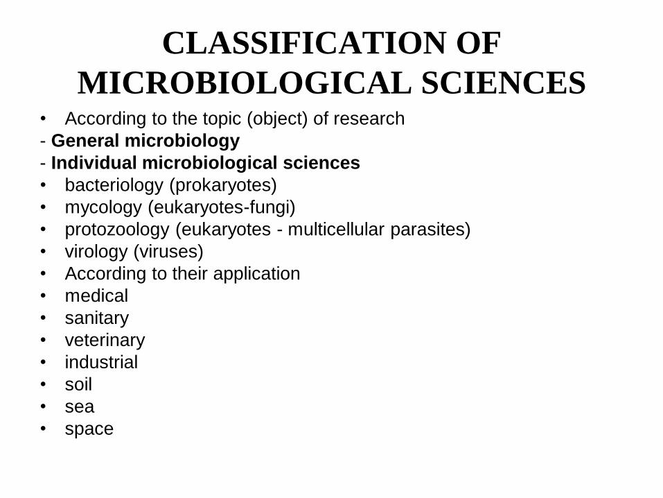
CLASSIFICATION OF
MICROBIOLOGICAL SCIENCES • According to the topic (object) of research
- General microbiology
- Individual microbiological sciences
• bacteriology (prokaryotes)
• mycology (eukaryotes-fungi)
• protozoology (eukaryotes - multicellular parasites)
• virology (viruses)
• According to their application
• medical
• sanitary
• veterinary
• industrial
• soil
• sea
• space
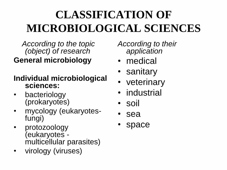
According to the topic (object) of research
General microbiology
Individual microbiological sciences:
• bacteriology (prokaryotes)
• mycology (eukaryotes-fungi)
• protozoology (eukaryotes - multicellular parasites)
• virology (viruses)
According to their application
• medical
• sanitary
• veterinary
• industrial
• soil
• sea
• space
CLASSIFICATION OF
MICROBIOLOGICAL SCIENCES
TASKS OF MEDICAL
MICROBIOLOGY
• Study of structure and biological properties of microorganisms
• Study of cointeraction of microorganism with human organism (i.e. infection), namely:
- pathogenesis
- diagnostics
- treatment
- preventive maintenance
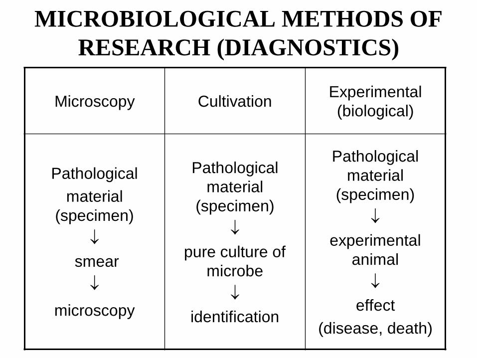
MICROBIOLOGICAL METHODS OF
RESEARCH (DIAGNOSTICS)
Microscopy Cultivation Experimental
(biological)
Pathological
material
(specimen)
smear
microscopy
Pathological
material
(specimen)
pure culture of
microbe
identification
Pathological
material
(specimen)
experimental
animal
effect
(disease, death)
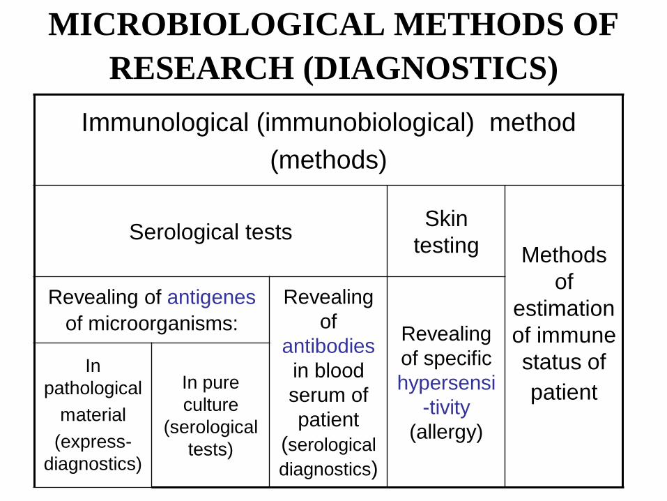
MICROBIOLOGICAL METHODS OF
RESEARCH (DIAGNOSTICS)
Immunological (immunobiological) method
(methods)
Serological tests Skin
testing Methods
of
estimation
of immune
status of
patient
Revealing of antigenes
of microorganisms:
Revealing
of
antibodies
in blood
serum of
patient
(serological
diagnostics)
Revealing
of specific
hypersensi
-tivity
(allergy)
In
pathological
material
(express-
diagnostics)
In pure
culture
(serological
tests)
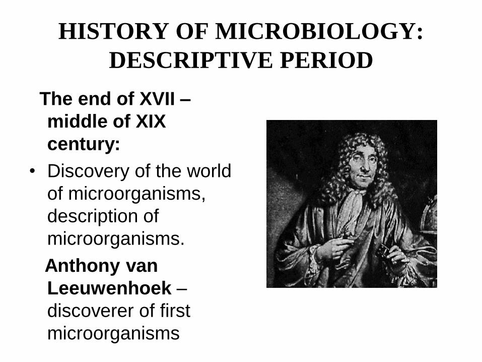
HISTORY OF MICROBIOLOGY:
DESCRIPTIVE PERIOD
The end of XVII –
middle of XIX
century:
• Discovery of the world
of microorganisms,
description of
microorganisms.
Anthony van
Leeuwenhoek –
discoverer of first
microorganisms
HISTORY OF MICROBIOLOGY:
PHYSIOLOGICAL (PASTEUR’S)
PERIOD
Middle of XIX – beginning of ХХ century:
• Study of living activity of microbial cell, discovery
of infectious (causing disease) bacteria,
beginning of scientific microbiology.
Louis Pasteur
Robert Koch
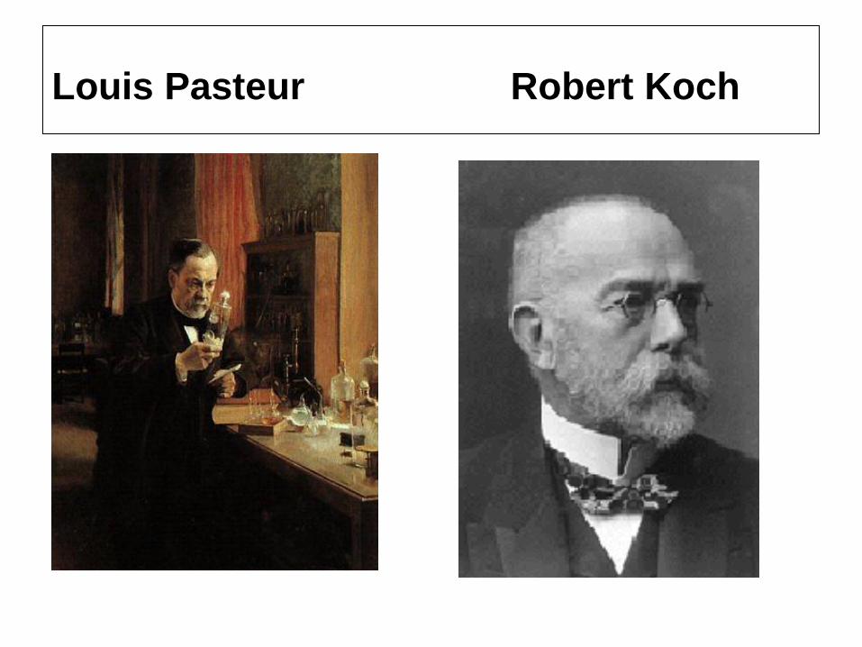
Louis Pasteur Robert Koch
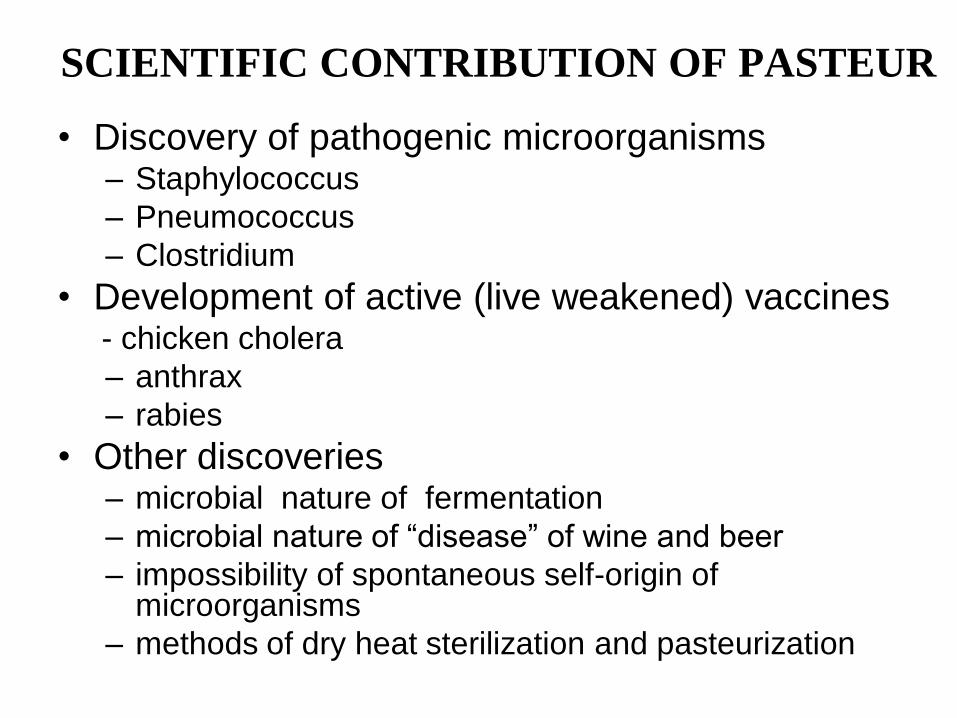
SCIENTIFIC CONTRIBUTION OF PASTEUR
• Discovery of pathogenic microorganisms – Staphylococcus
– Pneumococcus
– Clostridium
• Development of active (live weakened) vaccines - chicken cholera
– anthrax
– rabies
• Other discoveries – microbial nature of fermentation
– microbial nature of “disease” of wine and beer
– impossibility of spontaneous self-origin of microorganisms
– methods of dry heat sterilization and pasteurization
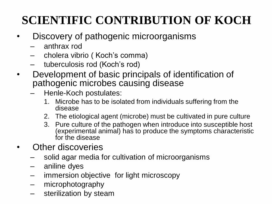
SCIENTIFIC CONTRIBUTION OF KOCH • Discovery of pathogenic microorganisms
– anthrax rod
– cholera vibrio ( Koch’s comma)
– tuberculosis rod (Koch’s rod)
• Development of basic principals of identification of pathogenic microbes causing disease
– Henle-Koch postulates: 1. Microbe has to be isolated from individuals suffering from the
disease
2. The etiological agent (microbe) must be cultivated in pure culture
3. Pure culture of the pathogen when introduce into susceptible host (experimental animal) has to produce the symptoms characteristic for the disease
• Other discoveries – solid agar media for cultivation of microorganisms
– aniline dyes
– immersion objective for light microscopy
– microphotography
– sterilization by steam
HISTORY OF MICROBIOLOGY:
IMMUNOLOGICAL PERIOD
Beginning – middle of ХХ century
• Discovery of immune response
Metchnikoff
Ehrlich
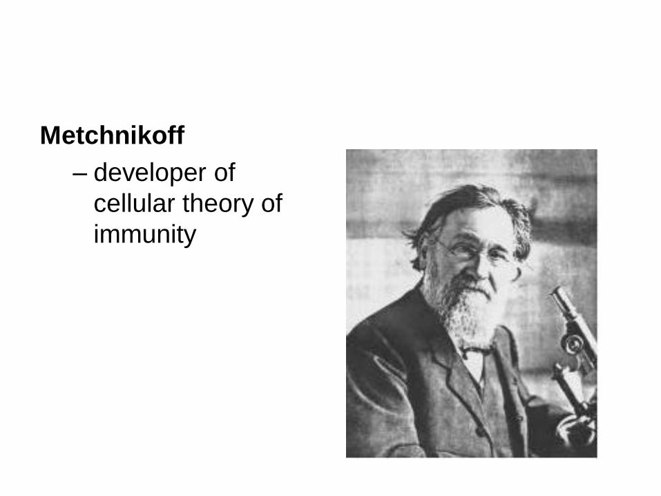
Metchnikoff
– developer of
cellular theory of
immunity
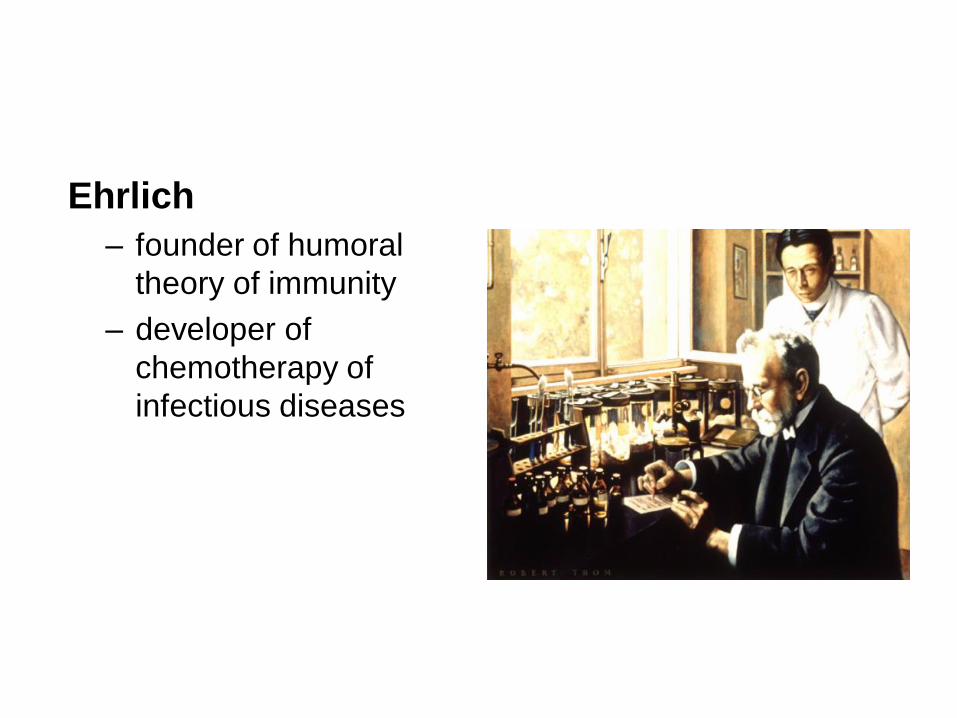
Ehrlich
– founder of humoral
theory of immunity
– developer of
chemotherapy of
infectious diseases
HISTORY OF MICROBIOLOGY:
MODERN PERIOD
Middle of XX century
Molecular biological methods of research
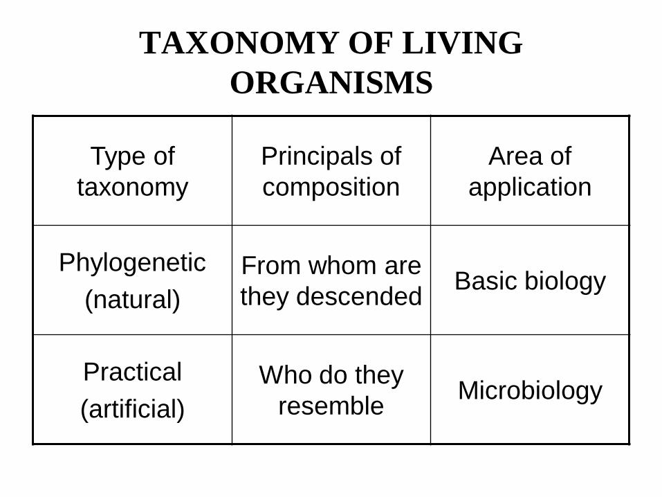
TAXONOMY OF LIVING
ORGANISMS
Type of
taxonomy
Principals of
composition
Area of
application
Phylogenetic
(natural)
From whom are
they descended Basic biology
Practical
(artificial)
Who do they
resemble Microbiology
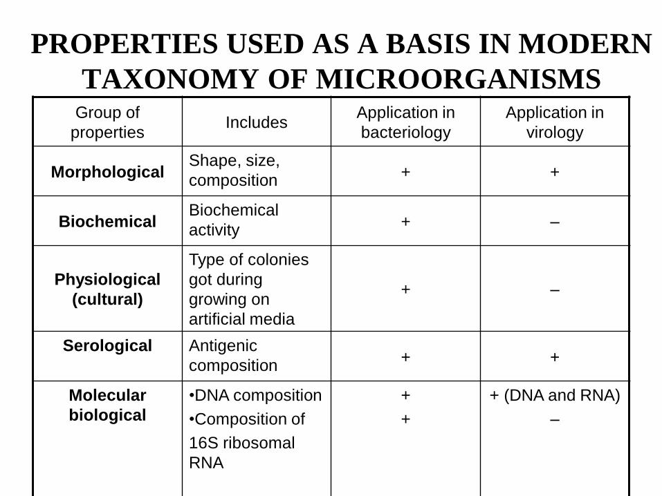
PROPERTIES USED AS A BASIS IN MODERN
TAXONOMY OF MICROORGANISMS Group of
properties Includes
Application in
bacteriology
Application in
virology
Morphological Shape, size,
composition + +
Biochemical Biochemical
activity + –
Physiological
(cultural)
Type of colonies
got during
growing on
artificial media
+ –
Serological Antigenic
composition + +
Molecular
biological
•DNA composition
•Composition of
16S ribosomal
RNA
+
+
+ (DNA and RNA)
–
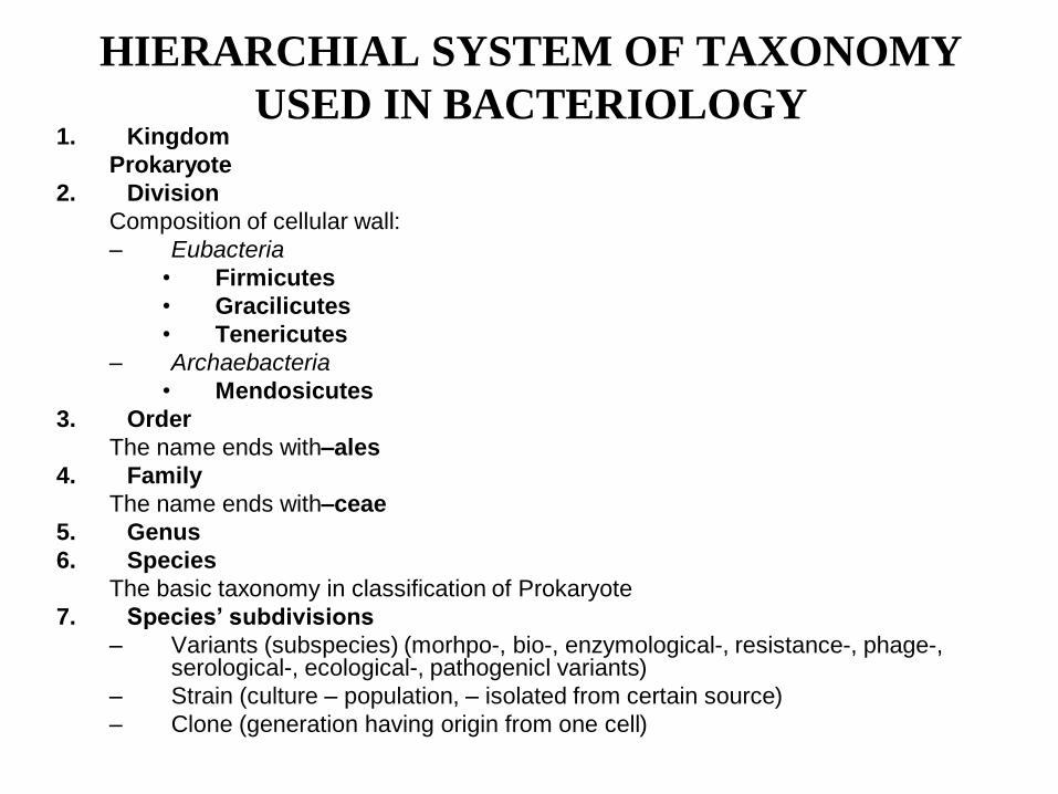
HIERARCHIAL SYSTEM OF TAXONOMY
USED IN BACTERIOLOGY 1. Kingdom
Prokaryote
2. Division
Composition of cellular wall:
– Eubacteria
• Firmicutes
• Gracilicutes
• Tenericutes
– Archaebacteria
• Mendosicutes
3. Order
The name ends with–ales
4. Family
The name ends with–ceae
5. Genus
6. Species
The basic taxonomy in classification of Prokaryote
7. Species’ subdivisions
– Variants (subspecies) (morhpo-, bio-, enzymological-, resistance-, phage-, serological-, ecological-, pathogenicl variants)
– Strain (culture – population, – isolated from certain source)
– Clone (generation having origin from one cell)
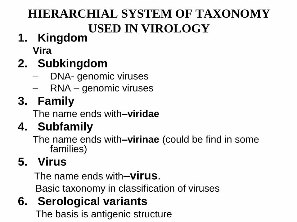
HIERARCHIAL SYSTEM OF TAXONOMY
USED IN VIROLOGY 1. Kingdom
Vira
2. Subkingdom – DNA- genomic viruses
– RNA – genomic viruses
3. Family The name ends with–viridae
4. Subfamily The name ends with–virinae (could be find in some
families)
5. Virus
The name ends with–virus. Basic taxonomy in classification of viruses
6. Serological variants The basis is antigenic structure
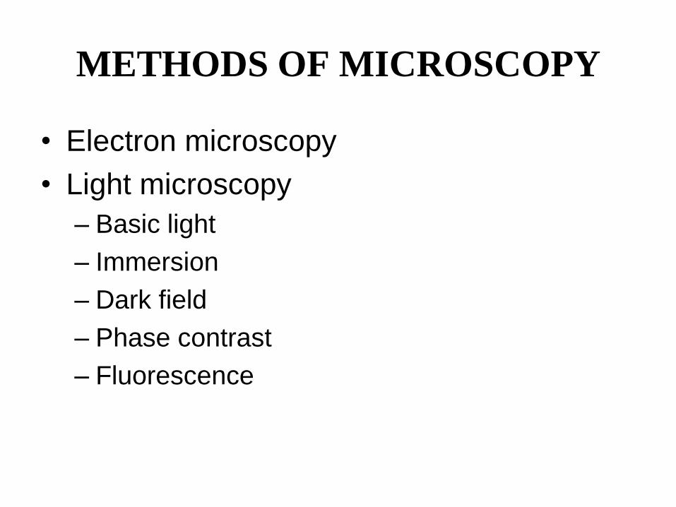
METHODS OF MICROSCOPY
• Electron microscopy
• Light microscopy
– Basic light
– Immersion
– Dark field
– Phase contrast
– Fluorescence
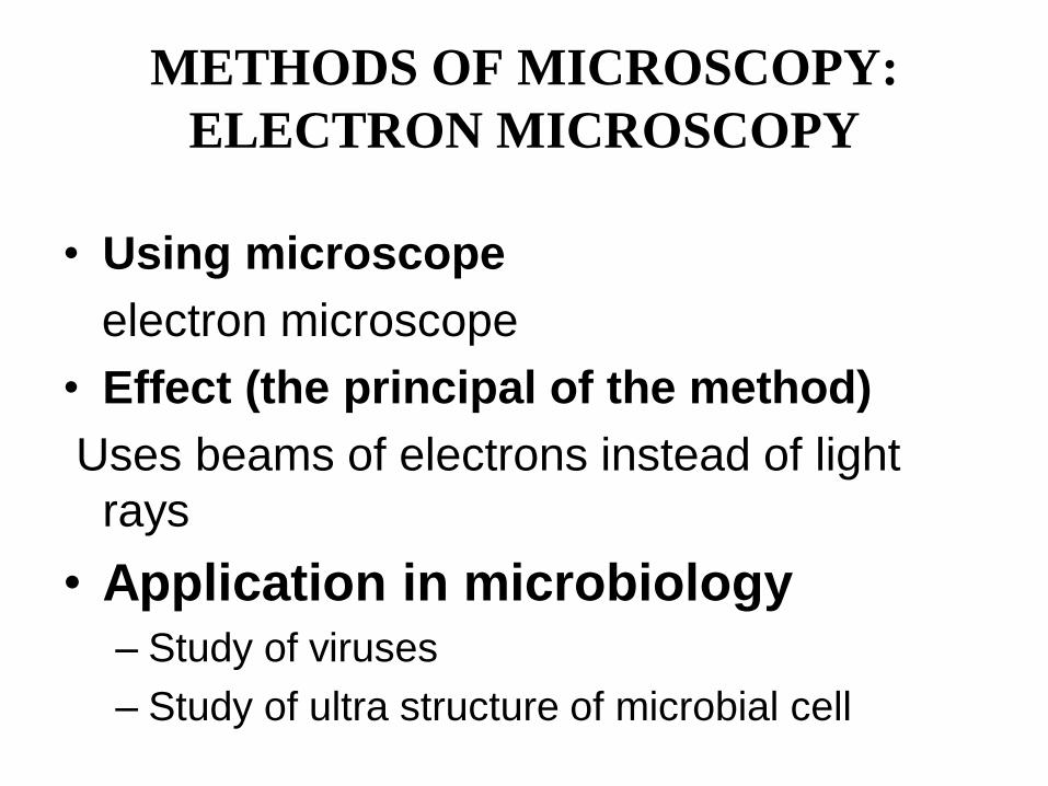
METHODS OF MICROSCOPY:
ELECTRON MICROSCOPY
• Using microscope
electron microscope
• Effect (the principal of the method)
Uses beams of electrons instead of light
rays
• Application in microbiology – Study of viruses
– Study of ultra structure of microbial cell
METHODS OF MICROSCOPY:
USUAL LIGHT MICROSCOPY
• Using microscope
Biological light microscope
• Effect (the principal of the method)
Uses visible light rays (see the course of
Physics)
• Application in microbiology It is not frequently used in microbiology
METHODS OF MICROSCOPY:
IMMERSION MICROSCOPY
• Using microscope
Biological light microscope + immersion objective
• Effect (the principal of the method)
Coefficient of refraction of immersion oil (placed between
glass slide and objective lens) = coefficient of refraction
of glass eliminates losses of light rays getting in
objective lens.
• Application in microbiology
It is most frequently used in bacteriology as a
microscopic method of research.
METHODS OF MICROSCOPY: DARK
FIELD MICROSCOPY
• Using microscope
Biological light microscope + dark field condenser
• Effect (the principal of the method)
Only light rays scattered from the specimen
(object) reach the objective lens (see light object
on a dark background)
• Application in microbiology
It is used for observation of very thin objects, for
example, spirochetes.
METHODS OF MICROSCOPY:
PHASE CONTRAST MICROSCOPY
• Using microscope
Biological light microscope + phase contrast optical
design
• Effect (the principal of the method)
Amplifies small differences in refractive indices (when light is coming through translucent objects we can’t see these changes) to changes of amplitude – we can see these changes and translucent object becomes visible.
• Application in microbiology
It is used for observation of translucent objects, for example, mycoplasmas.
METHODS OF MICROSCOPY:
LUMINESCENT (FLUORESCENT)
MICROSCOPY • Using microscope
Luminescent (fluorescent) microscope
• Effect (the principal of the method)
Luminescence of the object in ultraviolet light is
registered
• Application in microbiology
- microscopy of specimen stained with
fluorescent dyes (auramine, rhodamine, etc.),
- evaluation of serological fluorescence
reactions.
METHODS OF STAINING:
SIMPLE STAINING TECHNIQUES
• Staining by
methylene
blue
• Staining by aqueous
fuchsine
• Revealing of
presence of microbes
in a pathological
material
• Study:
– shape of bacteria
– their arrangement in a
smear
METHODS OF STAINING:
DIFFERENTIAL STAINING
TECHNIQUES • Gram staining (basic method of staining in bacteriology)
– revealing of cell wall structure
• Ziehl-Neelsen staining
– revealing of acid-fast bacteria (mycobacteria)
– revealing of spores
• Neisser staining
– revealing of volutine storage granules and
identification of corynebacteria according the granule
presence
• Burry-Hines staining
– revealing of capsules
METHODS OF STAINING:
DIFFERENTIAL STAINING
TECHNIQUES • Morozov staining
– revealing of flagella
– revealing of treponemas
• Zdradovsky staining
– revealing of viruses causing chickenpox and smallpox in vesicular lesions
– revealing of rickettsia and chlamydia
• Romanovsky-Giеmsa staining
– revealing of rickettsia and chlamydia
– revealing of spirochetes after their preliminary differentiation by colour of staining
– revealing of parasites
Morphology and Structure of
Bacterial Cell
Gram staining technique
Theme No2
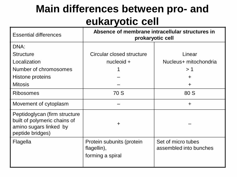
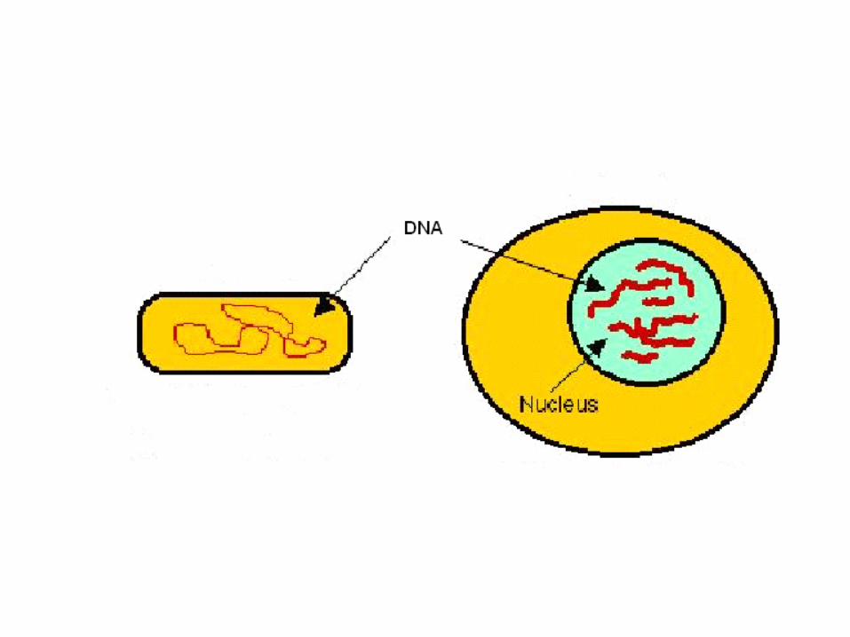
Main differences between pro- and
eukaryotic cell Essential differences
Absence of membrane intracellular structures in
prokaryotic cell
DNA:
Structure
Localization
Number of chromosomes
Histone proteins
Mitosis
Circular closed structure
nucleoid +
1
–
–
Linear
Nucleus+ mitochondria
> 1
+
+
Ribosomes 70 S 80 S
Movement of cytoplasm – +
Peptidoglycan (firm structure
built of polymeric chains of
amino sugars linked by
peptide bridges)
+ –
Flagella Protein subunits (protein
flagellin),
forming a spiral
Set of micro tubes
assembled into bunches
Organelles of bacterial cell: basic • Nucleoid
Circular closed super spiraled double-strand DNA molecule = bacterial chromosome
• Cytoplasm
Similar to cytoplasm of eukaryotic cell
• Ribosomes
Similar to ribosomes of eukaryotic cell but possess lower molecular weight
• Cytoplasmic (plasma) membrane
Similar to cytoplasm (cellular) membrane of eukaryotic cell but without sterols (sterols present only in the membrane of mycoplasmas)
• Mesosomes
Invaginations of cytoplasmic membrane:
– centre of energy producing metabolic reactions
– participation in cell division
• Cell wall
– creates shape of bacterial cell
– preserves cell from osmotic lysis
– possesses two types of a composition (Gram-positive and Gram- negative cell wall)
– lack of cell wall found only in mycoplasmas
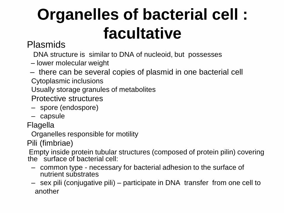
Organelles of bacterial cell :
facultative Plasmids
DNA structure is similar to DNA of nucleoid, but possesses
– lower molecular weight
– there can be several copies of plasmid in one bacterial cell Cytoplasmic inclusions
Usually storage granules of metabolites
Protective structures – spore (endospore)
– capsule
Flagella Organelles responsible for motility
Pili (fimbriae) Empty inside protein tubular structures (composed of protein pilin) covering
the surface of bacterial cell:
– common type - necessary for bacterial adhesion to the surface of nutrient substrates
– sex pili (conjugative pili) – participate in DNA transfer from one cell to
another
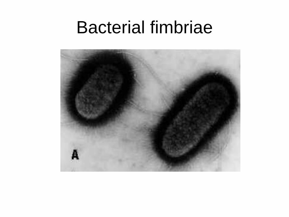
Bacterial fimbriae
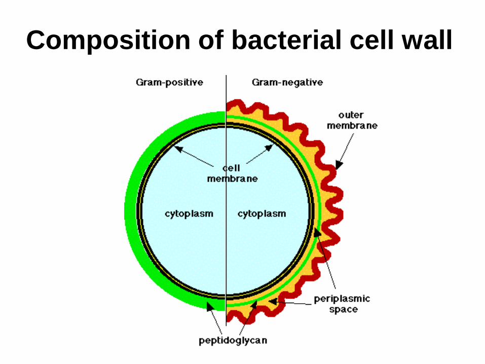
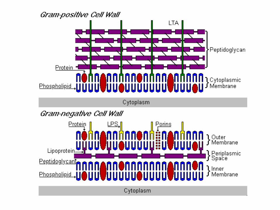
Composition of bacterial cell wall
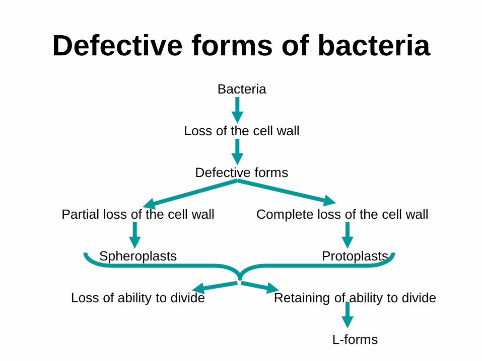
Defective forms of bacteria
Bacteria
Loss of the cell wall
Defective forms
Partial loss of the cell wall Complete loss of the cell wall
Spheroplasts Protoplasts
Loss of ability to divide Retaining of ability to divide
L-forms
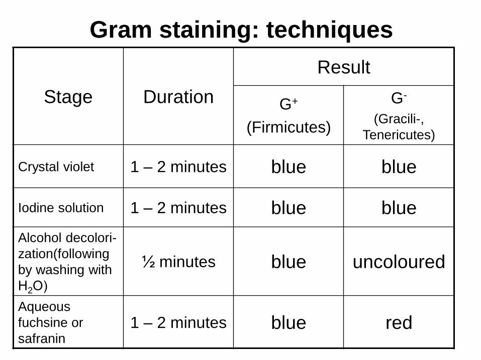
Gram staining: techniques
Stage Duration
Result
G+
(Firmicutes)
G-
(Gracili-,
Tenericutes)
Crystal violet 1 – 2 minutes blue blue
Iodine solution 1 – 2 minutes blue blue
Alcohol decolori-
zation(following
by washing with
Н2О)
½ minutes blue uncoloured
Aqueous
fuchsine or
safranin 1 – 2 minutes blue red
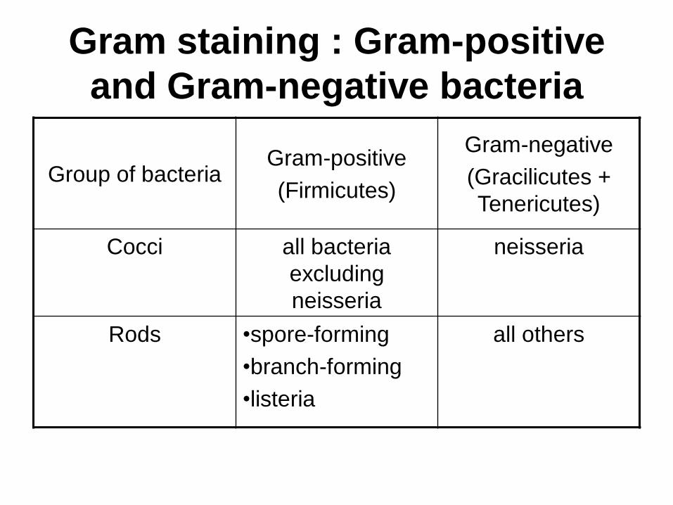
Gram staining : Gram-positive
and Gram-negative bacteria
Group of bacteria Gram-positive
(Firmicutes)
Gram-negative
(Gracilicutes +
Tenericutes)
Cocci all bacteria
excluding
neisseria
neisseria
Rods •spore-forming
•branch-forming
•listeria
all others
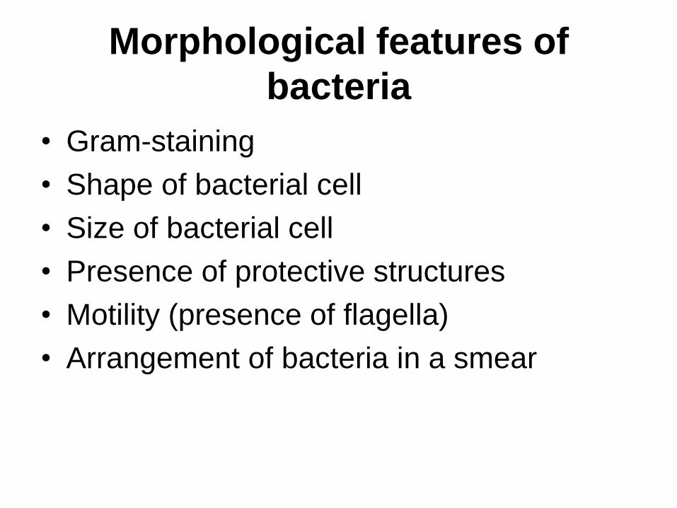
Morphological features of
bacteria
• Gram-staining
• Shape of bacterial cell
• Size of bacterial cell
• Presence of protective structures
• Motility (presence of flagella)
• Arrangement of bacteria in a smear
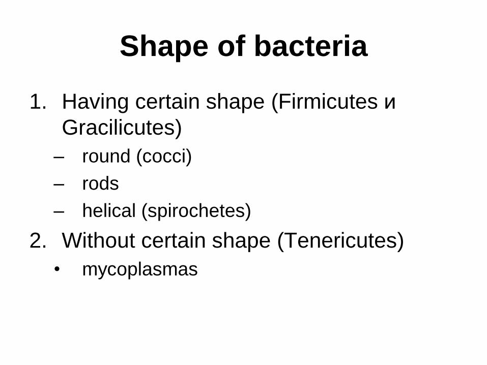
Shape of bacteria
1. Having certain shape (Firmicutes и
Gracilicutes)
– round (cocci)
– rods
– helical (spirochetes)
2. Without certain shape (Tenericutes)
• mycoplasmas
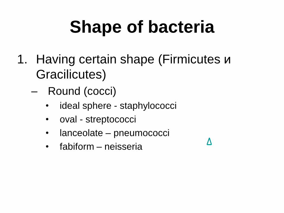
Shape of bacteria
1. Having certain shape (Firmicutes и
Gracilicutes)
– Round (cocci)
• ideal sphere - staphylococci
• oval - streptococci
• lanceolate – pneumococci
• fabiform – neisseria
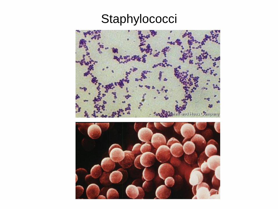
Staphylococci
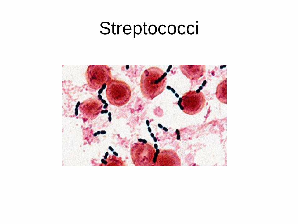
Streptococci
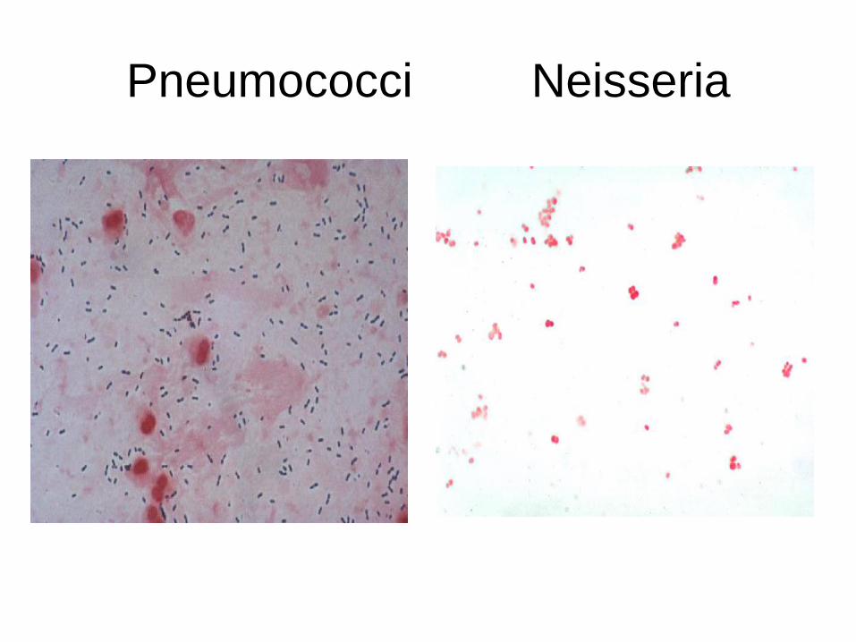
Pneumococci Neisseria
Shape of bacteria
1. Having certain shape (Firmicutes и Gracilicutes)
rods
• cylindrical – most of them
• curved
– one curve - vibrio
– 2-3 curves – campylobacteria and helicobacteria
• branch – forming
– actyninomycetes, mycobacteria, corynebacteria
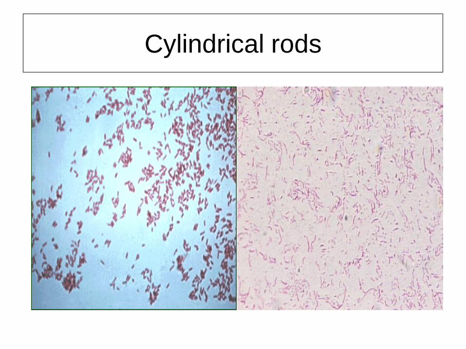
Cylindrical rods
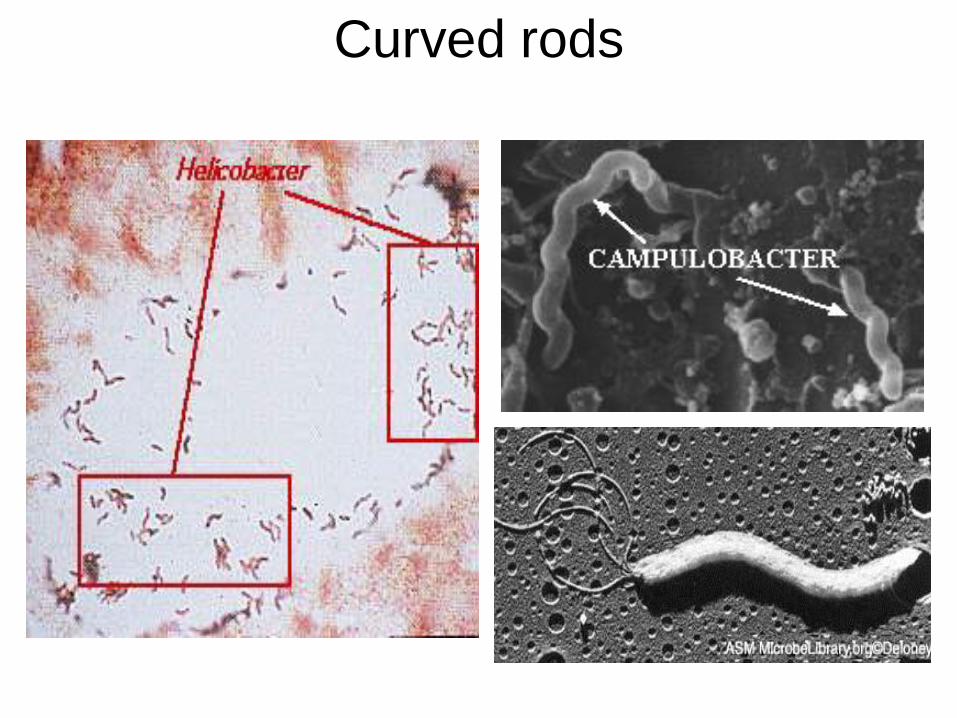
Curved rods
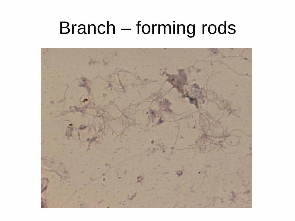
Branch – forming rods
Shape of bacteria
1. Having certain shape(Firmicutes и
Gracilicutes)
– helical
• spirochetes – treponemas, leptospiras and
borrelias
2. Without certain shape (Tenericutes)
• mycoplasmas
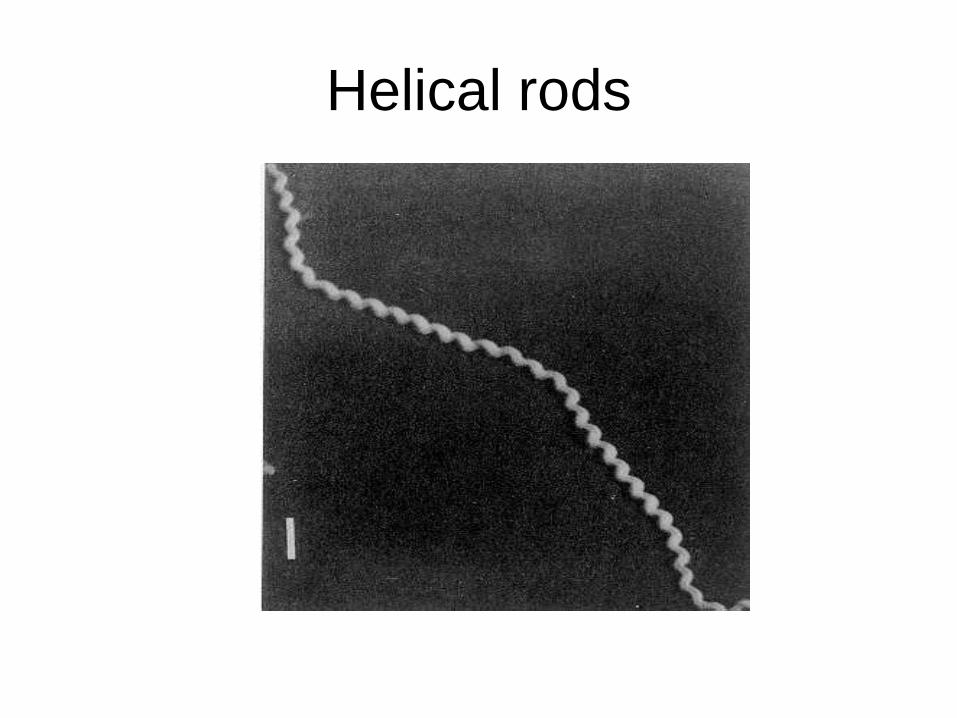
Helical rods
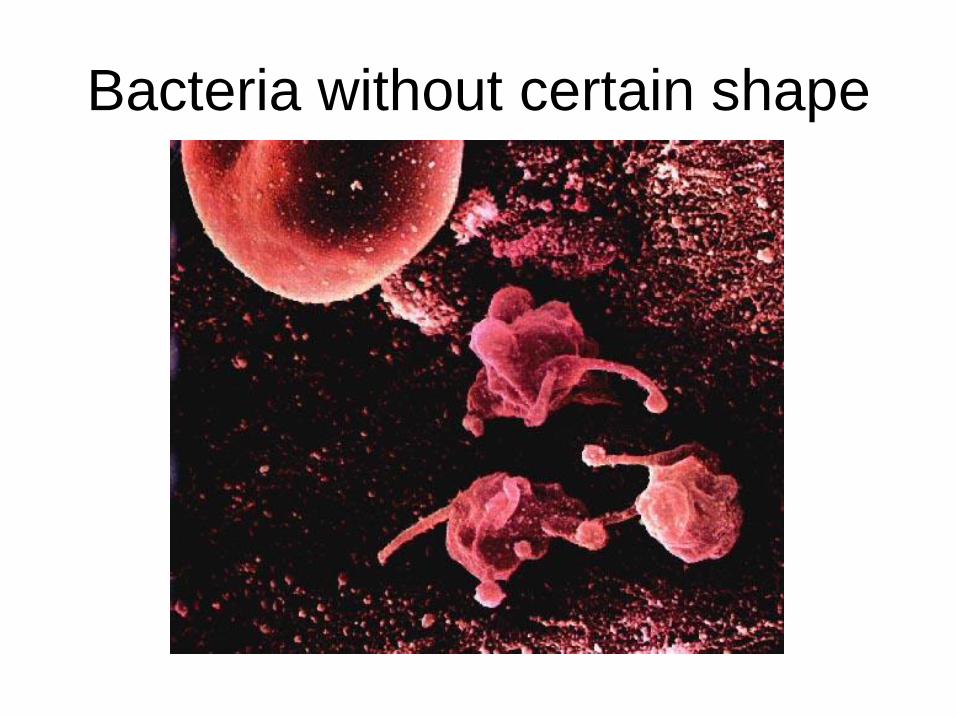
Bacteria without certain shape
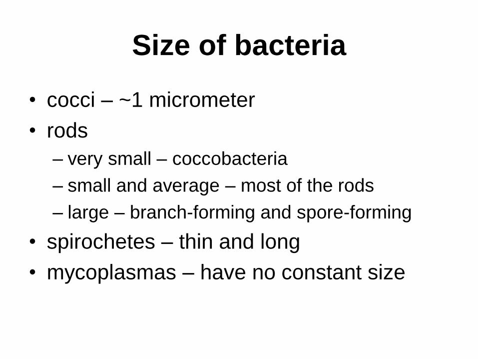
Size of bacteria
• cocci – ~1 micrometer
• rods
– very small – coccobacteria
– small and average – most of the rods
– large – branch-forming and spore-forming
• spirochetes – thin and long
• mycoplasmas – have no constant size
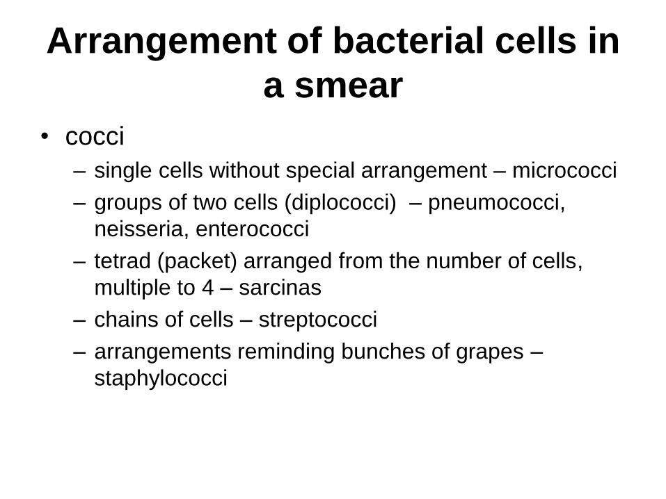
Arrangement of bacterial cells in
a smear
• cocci
– single cells without special arrangement – micrococci
– groups of two cells (diplococci) – pneumococci,
neisseria, enterococci
– tetrad (packet) arranged from the number of cells,
multiple to 4 – sarcinas
– chains of cells – streptococci
– arrangements reminding bunches of grapes –
staphylococci
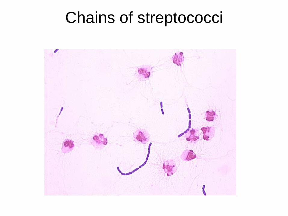
Chains of streptococci
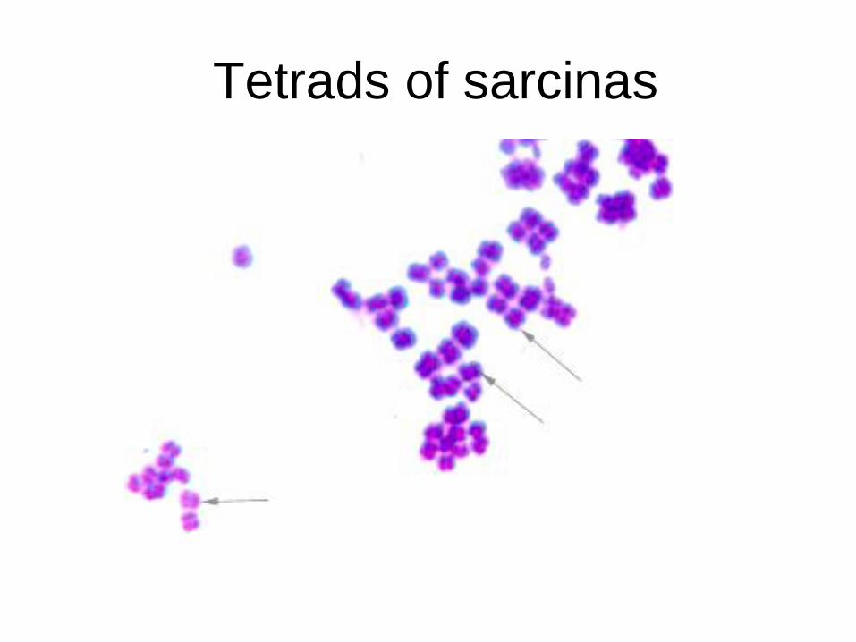
Tetrads of sarcinas
Arrangement of bacterial cells in
a smear
• rods
– without any order – most of the rods
– forming pairs – klebsiellas, corynebacteria
– forming chains – bacilli
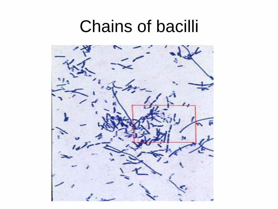
Chains of bacilli
MORPHOLOGY AND STRUCTURE OF
BACTERIAL CELL
(CONTINUATION).
MORPHOLOGICAL AND ULTRA
STRUCTURAL PECULIARITIES OF
ACTINOMYCETES, SPIROCHETES,
RICKETTSIA, CHLAMYDIA, MYCOPLASMAS
AND FUNGI.
Ziehl-Neelsen staining technique
Theme No3

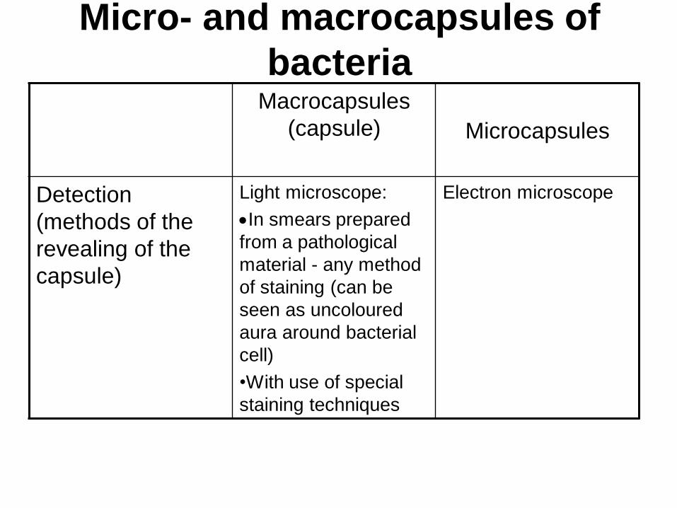
Micro- and macrocapsules of
bacteria Macrocapsules
(capsule) Microcapsules
Definition
The expressed
mucous layer covering
cell wall and having a
fibrillar composition
Mucopolysaccharide
fibrils close-fitting
the cell wall
Composition •Most frequently -
polysaccharides
•rare - polypeptides
Mucopolysaccharides
Function Defense of bacterial cell from:
•phagocytosis
•binding by antibodies
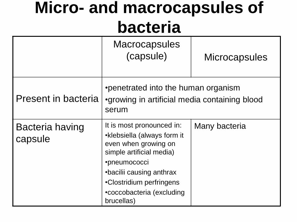
Micro- and macrocapsules of
bacteria Macrocapsules
(capsule)
Microcapsules
Present in bacteria •penetrated into the human organism
•growing in artificial media containing blood
serum
Bacteria having
capsule
It is most pronounced in:
•klebsiella (always form it
even when growing on
simple artificial media)
•pneumococci
•bacilii causing anthrax
•Clostridium perfringens
•coccobacteria (excluding
brucellas)
Many bacteria
Micro- and macrocapsules of
bacteria Macrocapsules
(capsule)
Microcapsules
Detection
(methods of the
revealing of the
capsule)
Light microscope:
In smears prepared
from a pathological
material - any method
of staining (can be
seen as uncoloured
aura around bacterial
cell)
•With use of special
staining techniques
Electron microscope
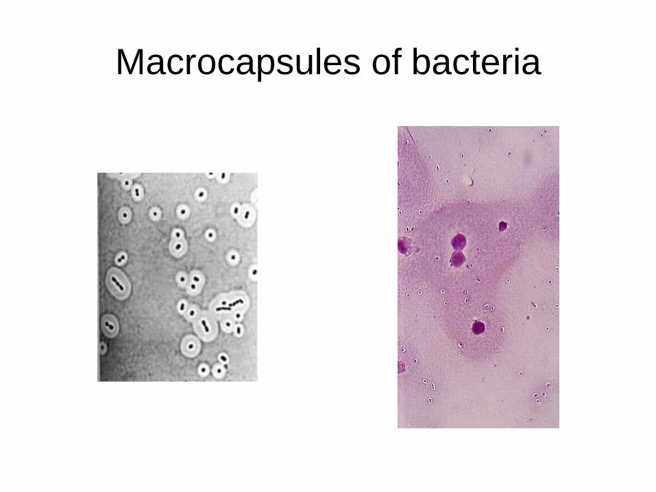
Macrocapsules of bacteria
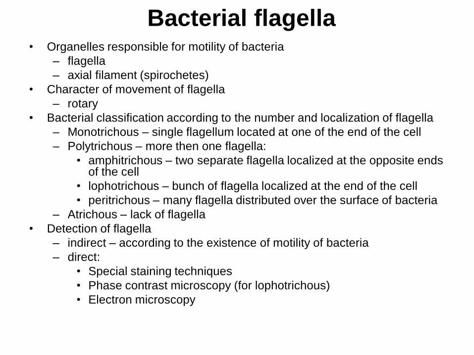
Bacterial flagella • Organelles responsible for motility of bacteria
– flagella
– axial filament (spirochetes)
• Character of movement of flagella
– rotary
• Bacterial classification according to the number and localization of flagella
– Monotrichous – single flagellum located at one of the end of the cell
– Polytrichous – more then one flagella:
• amphitrichous – two separate flagella localized at the opposite ends of the cell
• lophotrichous – bunch of flagella localized at the end of the cell
• peritrichous – many flagella distributed over the surface of bacteria
– Atrichous – lack of flagella
• Detection of flagella
– indirect – according to the existence of motility of bacteria
– direct:
• Special staining techniques
• Phase contrast microscopy (for lophotrichous)
• Electron microscopy
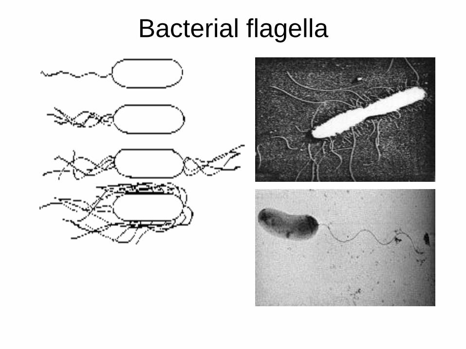
Bacterial flagella
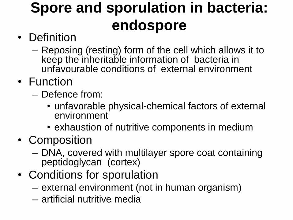
Spore and sporulation in bacteria:
endospore • Definition
– Reposing (resting) form of the cell which allows it to keep the inheritable information of bacteria in unfavourable conditions of external environment
• Function – Defence from:
• unfavorable physical-chemical factors of external environment
• exhaustion of nutritive components in medium
• Composition – DNA, covered with multilayer spore coat containing
peptidoglycan (cortex)
• Conditions for sporulation – external environment (not in human organism)
– artificial nutritive media
Spore and sporulation in bacteria:
endospore
• The factors ensuring thermal resistance
– practically complete absence of unbound
water
– increased calcium concentration
– presence of dipicolinic acid
– especial composition of protein
– especial composition of peptidoglycan of the
cortex
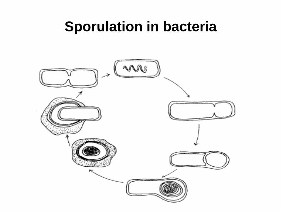
Sporulation in bacteria
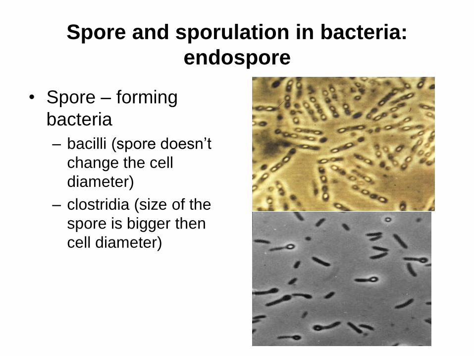
Spore and sporulation in bacteria:
endospore
• Spore – forming
bacteria
– bacilli (spore doesn’t
change the cell
diameter)
– clostridia (size of the
spore is bigger then
cell diameter)
Spore and sporulation in bacteria:
endospore
• Detection
– Ziehl-Neelsen staining technique
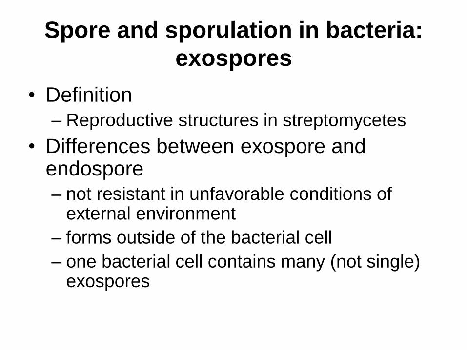
Spore and sporulation in bacteria:
exospores
• Definition
– Reproductive structures in streptomycetes
• Differences between exospore and endospore
– not resistant in unfavorable conditions of external environment
– forms outside of the bacterial cell
– one bacterial cell contains many (not single) exospores
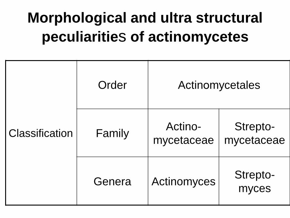
Morphological and ultra structural
peculiarities of actinomycetes
Classification
Order Actinоmycetales
Family Actino-
mycetaceae
Strepto-
mycetaceae
Genera Actinomyces Strepto-
myces
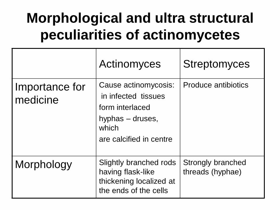
Morphological and ultra structural
peculiarities of actinomycetes
Actinomyces Streptomyces
Importance for
medicine
Cause actinomycosis:
in infected tissues
form interlaced
hyphas – druses,
which
are calcified in centre
Produce antibiotics
Morphology Slightly branched rods
having flask-like
thickening localized at
the ends of the cells
Strongly branched
threads (hyphae)
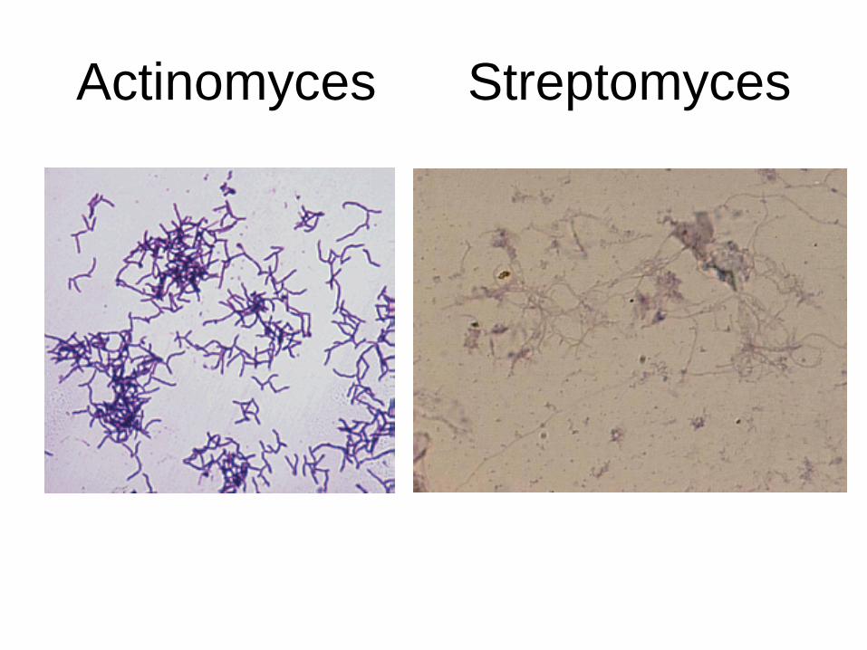
Actinomyces Streptomyces
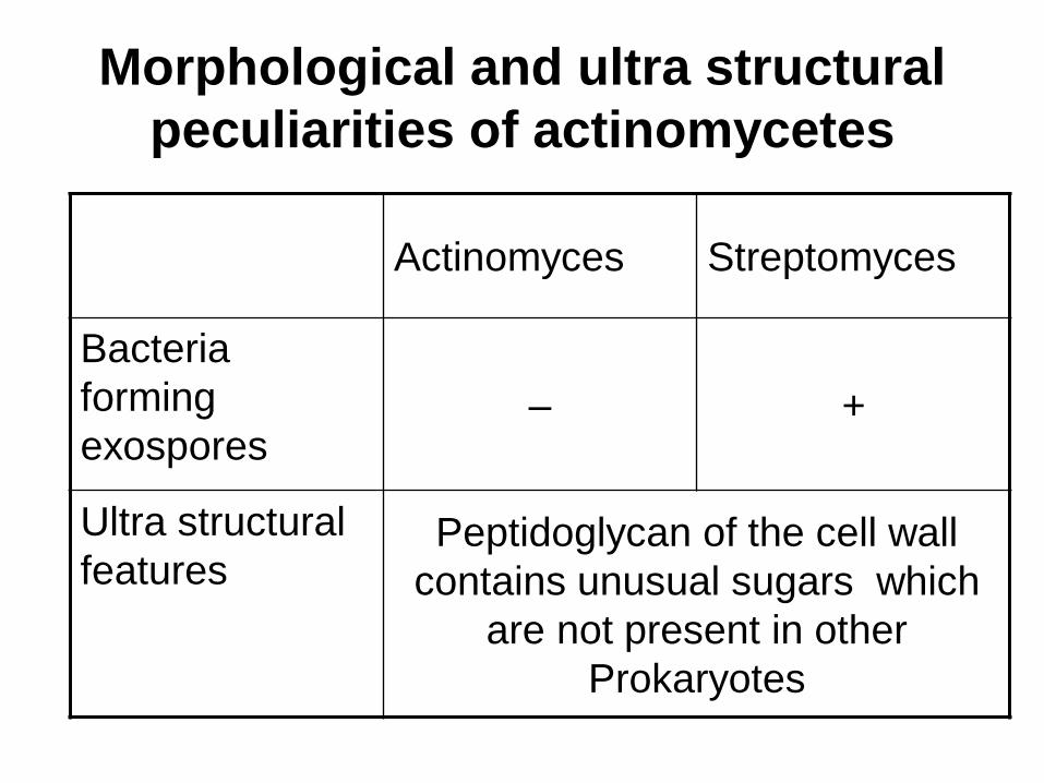
Morphological and ultra structural
peculiarities of actinomycetes
Actinomyces Streptomyces
Bacteria
forming
exospores – +
Ultra structural
features Peptidoglycan of the cell wall
contains unusual sugars which
are not present in other
Prokaryotes
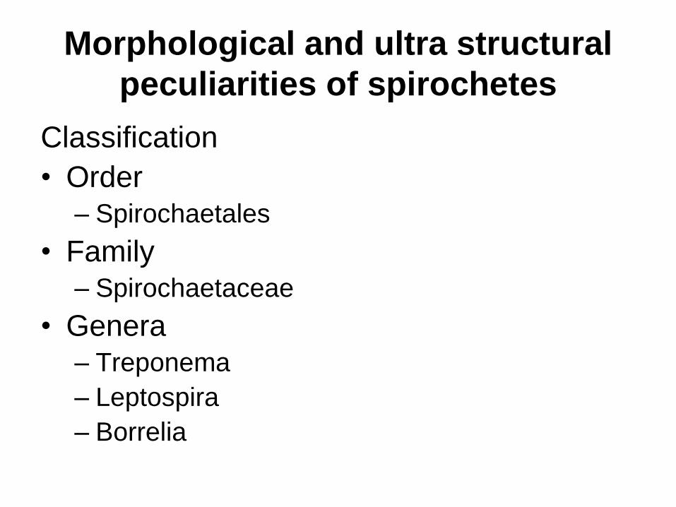
Morphological and ultra structural
peculiarities of spirochetes
Classification
• Order
– Spirochaetales
• Family
– Spirochaetaceae
• Genera
– Treponema
– Leptospira
– Borrelia
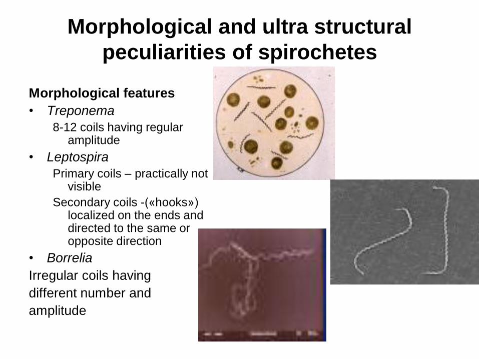
Morphological and ultra structural
peculiarities of spirochetes
Morphological features
• Treponema
8-12 coils having regular amplitude
• Leptospira
Primary coils – practically not visible
Secondary coils -(«hooks») localized on the ends and directed to the same or opposite direction
• Borrelia
Irregular coils having
different number and
amplitude
Morphological and ultra structural
peculiarities of spirochetes
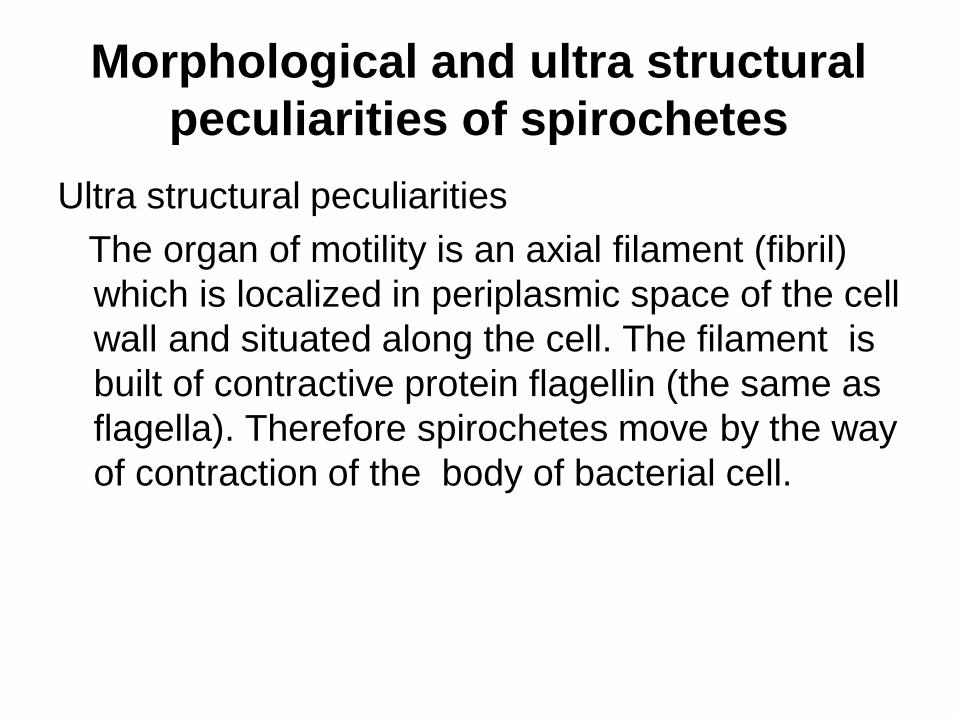
Ultra structural peculiarities
The organ of motility is an axial filament (fibril)
which is localized in periplasmic space of the cell
wall and situated along the cell. The filament is
built of contractive protein flagellin (the same as
flagella). Therefore spirochetes move by the way
of contraction of the body of bacterial cell.
1 — protoplasmic cylinder; 2 — outside cover; 3 — axial
fibrils; 4 — place of anchoring of axial fibrils; 5 —
peptidoglycan layer of the cell wall; 6 — cell membrane.
Morphological and ultra structural
peculiarities of spirochetes
Romanovsky- Giemsa staining technique
• Treponema
pink
• Leptospira
red
• Borrelia
blue
Morphological and ultra structural
peculiarities of spirochetes
Microscopy techniques (methods) frequently
used for detection
• Treponema dark field
• Leptospira microscopy
• Borrelia any microscopy techniques
Morphological and ultra structural
peculiarities of chlamydia
Principal differences between them and
other Prokaryotes
Obligate intracellular parasites
Morphological and ultrastructural
features of rickettsia and chlamydia
Classification
• Order
– Rickettsiales
• Family
– Rickettsiaceae
• Genera • Rickettsia
• Coxiella
• Rochalimaea
• Order
– Chlamydiales
• Family
– Chlamydiaceae
Genus
– Chlamydia
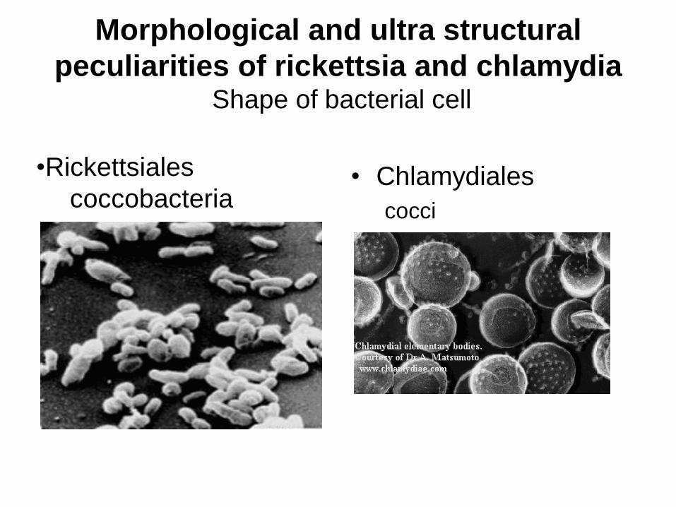
Morphological and ultra structural
peculiarities of rickettsia and chlamydia Shape of bacterial cell
• Chlamydiales
cocci
•Rickettsiales
coccobacteria
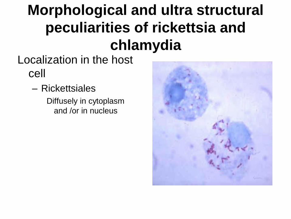
Morphological and ultra structural
peculiarities of rickettsia and
chlamydia Localization in the host
cell
– Rickettsiales
Diffusely in cytoplasm
and /or in nucleus
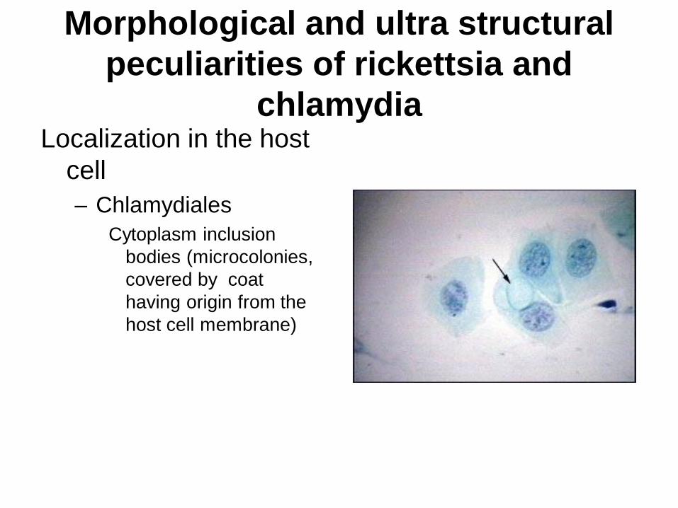
Morphological and ultra structural
peculiarities of rickettsia and
chlamydia Localization in the host
cell
– Chlamydiales
Cytoplasm inclusion
bodies (microcolonies,
covered by coat
having origin from the
host cell membrane)
Morphological and ultra structural
peculiarities of rickettsia and
chlamydia Staining techniques
– Romanovsky-Giemsa staining
Dark blue on the light blue background of the cell
– Zdradovsky staining
Pink on the light blue background of the cell
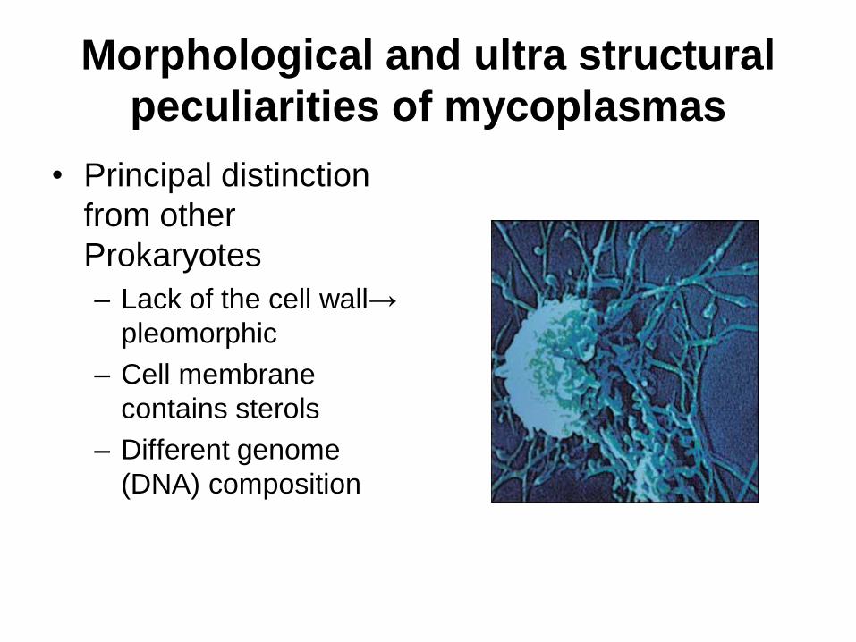
Morphological and ultra structural
peculiarities of mycoplasmas
• Principal distinction
from other
Prokaryotes
– Lack of the cell wall→
pleomorphic
– Cell membrane
contains sterols
– Different genome
(DNA) composition
Morphological and ultra structural
peculiarities of mycoplasmas • Classification
– Division
• Tenericutes
– Class
• Mollicutes
– Order
• Mycoplasmatales
– Family
• Mycoplasmataceae
– Genera
• Mycoplasma
• Ureaplasma
Morphological and ultra structural
peculiarities of mycoplasmas
• Study techniques
– Phase contrast microscopy
– Electron microscopy
Fungi: classification and taxonomy
Eukaryote - kingdom
• Mycota, divisions:
– Myxomycota
– Eumycota (certain fungi).
Pathogenic fungi belong to the next classes:
1. Zygomycetes
2. Ascomycetes fungi which have sexual
3. Basidiomycetes reproduction
4. Deuteromycetes - asexual reproduction - fungi
imperfecti
Structure of fungal cell
• Fungi are eukaryotic organisms
• The fungal cell wall contains polysaccharides
presented by: • chitin (a structural component of fungal cell wall)
which differs from chitin of arthropods (contains
less amounts of N)
• glucans
• mannans
Fungi: reproduction
• Sexual Reproduction
Sexual reproduction occurs by the fusion of two haploid nuclei (karyogamy), followed by meiotic division of the diploid nucleus.
The union of two hyphal protoplasts (plasmogamy) may be followed immediately by karyogamy, or it may be separated in time.
• Asexual Reproduction
Asexual reproduction occurs via division of nuclei by mitosis. With the absence of meiosis, other mechanisms associated with the nuclear cycle result in recombination of hereditary properties and genetic variation.
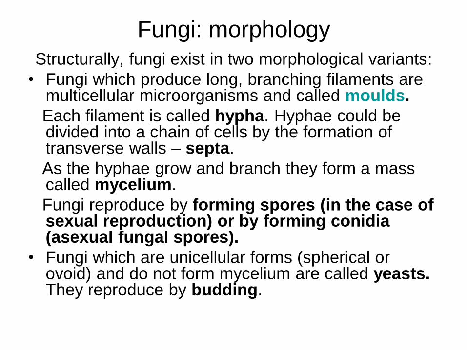
Fungi: morphology
Structurally, fungi exist in two morphological variants:
• Fungi which produce long, branching filaments are multicellular microorganisms and called moulds.
Each filament is called hypha. Hyphae could be divided into a chain of cells by the formation of transverse walls – septa.
As the hyphae grow and branch they form a mass called mycelium.
Fungi reproduce by forming spores (in the case of sexual reproduction) or by forming conidia (asexual fungal spores).
• Fungi which are unicellular forms (spherical or ovoid) and do not form mycelium are called yeasts. They reproduce by budding.
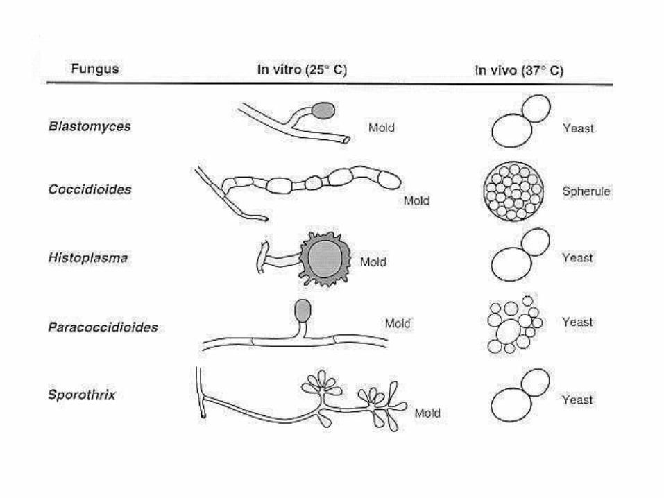
Structures of fungi: dimorphism
Phenomenon called dimorphism is characteristic for fungi. They could be presented by:
• filamentous forms (moulds) – usually when grow in the natural environment or in laboratory culture
• unicellular forms (yeasts) – when they grow in the infected tissue
Some fungi are capable to exist in both forms depending upon the environment, nutrients or other conditions. This is phenomenon of the adaptation of
fungi to changing conditions of environment.
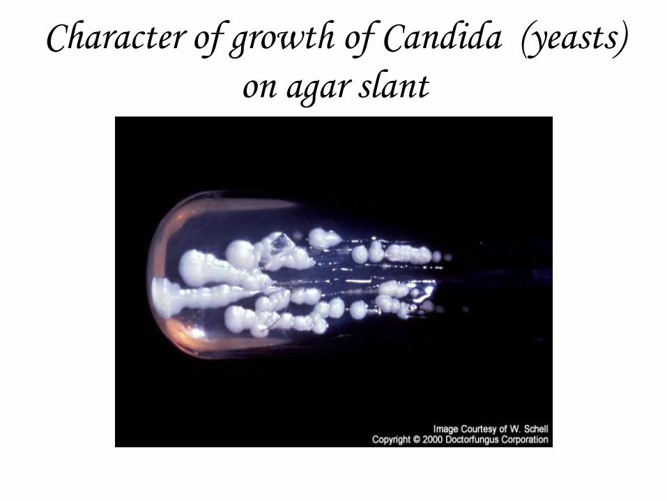
Character of growth of Candida (yeasts)
on agar slant
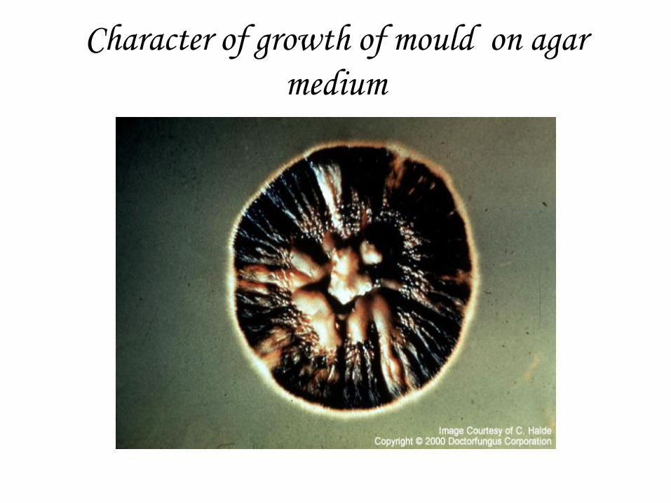
Character of growth of mould on agar
medium
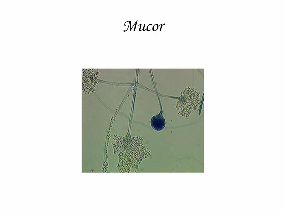
Mucor

Candida albicans in the infected tissue
Ziehl-Neelsen staining
• The purposes of application
– Detection of endospores
– Detection of mycobacteria
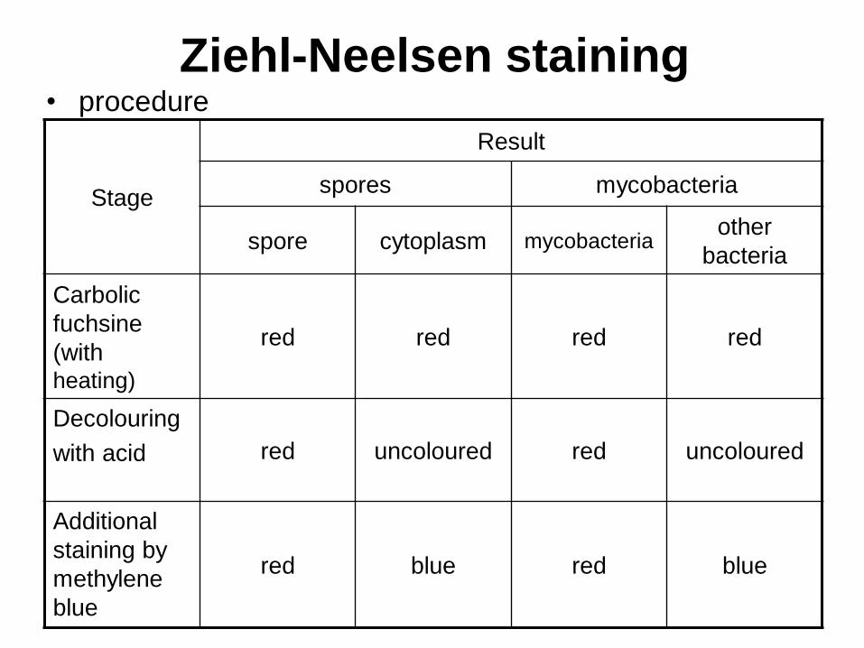
Ziehl-Neelsen staining • procedure
Stage
Result
spores mycobacteria
spore cytoplasm mycobacteria other
bacteria
Carbolic
fuchsine
(with
heating)
red
red
red red
Decolouring
with acid red uncoloured red uncoloured
Additional
staining by
methylene
blue
red blue
red
blue
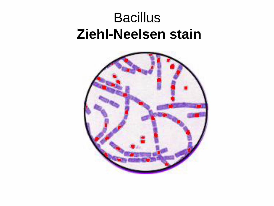
Bacillus
Ziehl-Neelsen stain
Physiology of Bacteria
Theme N4
FEATURES OF METABOLISM IN
MICROORGANISMS
• Bacteria
– can use any source of main chemical
compounds
– possess high speed of metabolic processes
– show high elasticity at the environment
• Viruses
– do not have their own metabolic enzymes
METABOLISM IN
BACTERIA
MAIN ROUTES OF PENETRATION OF
NUTRIENTS INTO BACTERIAL CELL
• Without energy consumption (diffusion) – ordinary
– facilitated (involves activity of enzymes – permeases)
• With energy consumption (involves activity of enzymes – permeases) – without chemical modification of transferred
molecules - active transport
– with chemical modification of transferred molecules - translocation of chemical groups
THE CLASSIFICATION OF BACTERIA BY
THE SOURCE OF CARBON • Inorganic compounds: СО2 or carbonates – autotrophic
bacteria
• Organic compounds – heterotrophic bacteria
– compounds of the environment – saprophytes
– compounds of an alive cell – parasites
Parasites which:
• can also use organic compounds of the
environment – facultative parasites (majority of
pathogenic bacteria)
• can only use organic compounds of an alive cell –
obligate parasites
– Rickettsia
– Chlamydia
CLASSIFICATION OF BACTERIA BY
THEIR GROWTH FACTORS NEEDS
PROTOTROPHIC BACTERIA
• can synthesize growth factors (nitrogen
bases, amino acids, vitamins, lipids, etc) by
themselves
AUXOTROPHIC BACTERIA
• cannot synthesize growth factors and
require their adding to the growth media
CLASSIFICATION OF BACTERIA BY THE
FEATURES OF THEIR ENERGY
METABOLISM
• energy source – the sunlight (phototrophs)
• energy source – oxidation-reduction reactions
with ATP synthesis as a result of the reaction
(chemotrophs)
– electron donor
• inorganic compounds (lithotrophs)
• organic compounds (organotrophs)
– electron acceptor
• external (oxidation)
– oxygen – aerobic respiration
– others (nitrate, fumarate) – anaerobic respiration
• internal– organic compounds of the cell (fermentation)
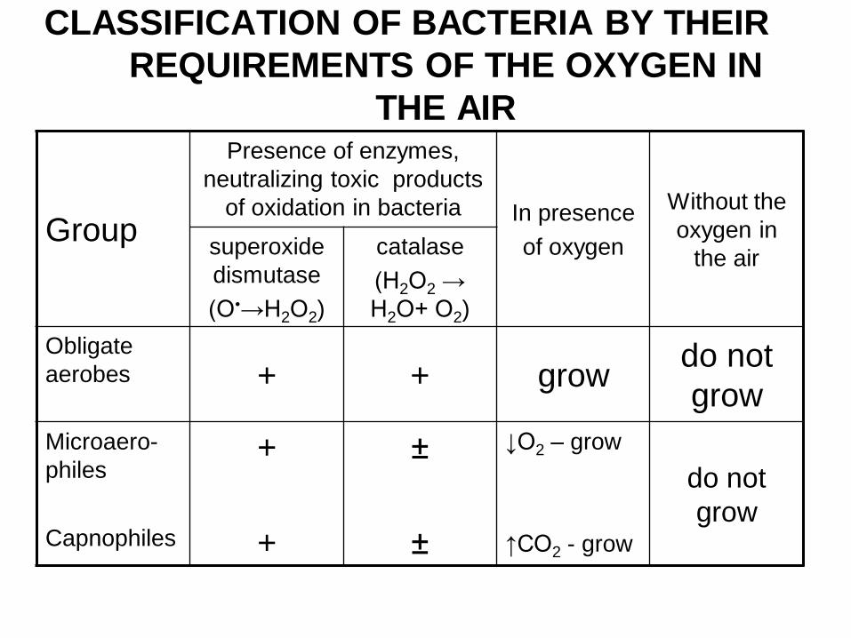
CLASSIFICATION OF BACTERIA BY THEIR
REQUIREMENTS OF THE OXYGEN IN
THE AIR
Group
Presence of enzymes,
neutralizing toxic products
of oxidation in bacteria In presence
of oxygen
Without the
oxygen in
the air superoxide
dismutase
(О•→Н2О2)
catalase
(Н2О2 →
Н2О+ О2)
Obligate
aerobes + + grow do not
grow
Microaero-
philes
Capnophiles
+
+
±
±
↓О2 – grow
↑СО2 - grow
do not
grow
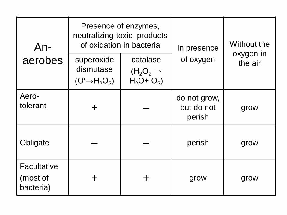
An-
aerobes
Presence of enzymes,
neutralizing toxic products
of oxidation in bacteria In presence
of oxygen
Without the
oxygen in
the air superoxide
dismutase
(О•→Н2О2)
catalase
(Н2О2 →
Н2О+ О2)
Aero-
tolerant + – do not grow,
but do not
perish
grow
Obligate – – perish grow
Facultative
(most of
bacteria) + + grow grow
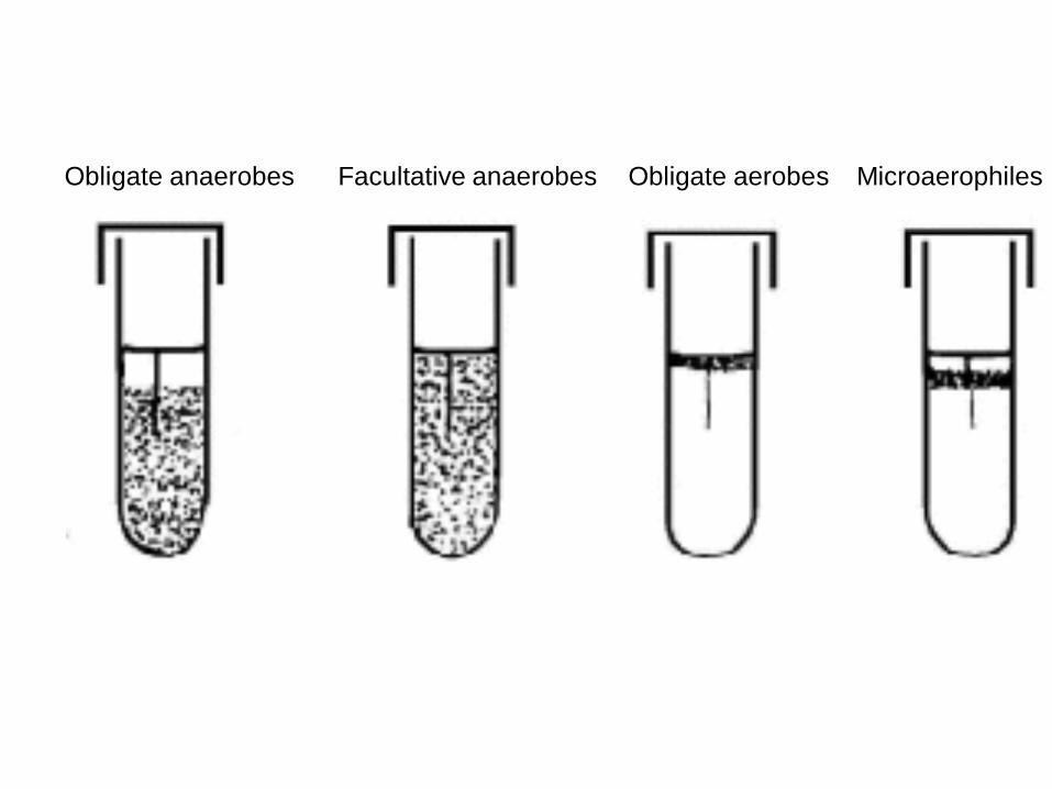
Obligate anaerobes Facultative anaerobes Microaerophiles Obligate aerobes
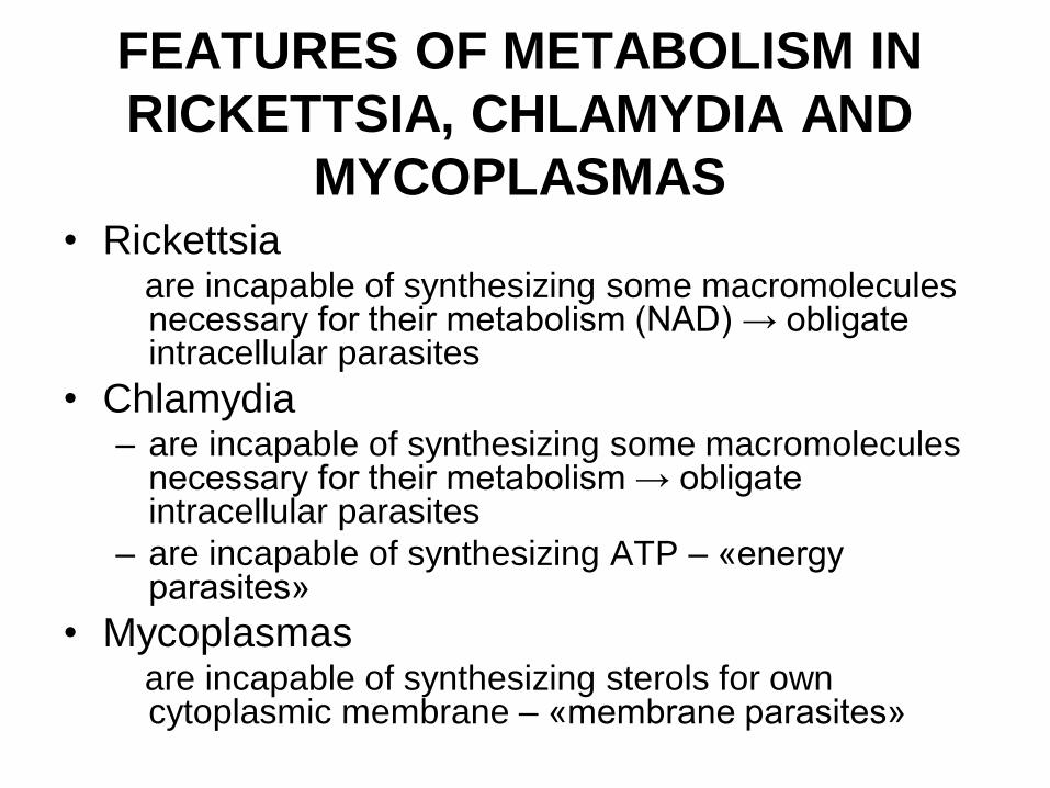
FEATURES OF METABOLISM IN
RICKETTSIA, CHLAMYDIA AND
MYCOPLASMAS • Rickettsia
are incapable of synthesizing some macromolecules necessary for their metabolism (NAD) → obligate intracellular parasites
• Chlamydia – are incapable of synthesizing some macromolecules
necessary for their metabolism → obligate intracellular parasites
– are incapable of synthesizing ATP – «energy parasites»
• Mycoplasmas are incapable of synthesizing sterols for own
cytoplasmic membrane – «membrane parasites»
REPRODUCTION OF
BACTERIA AND MAIN
PRINCIPLES OF THEIR
CULTIVATION
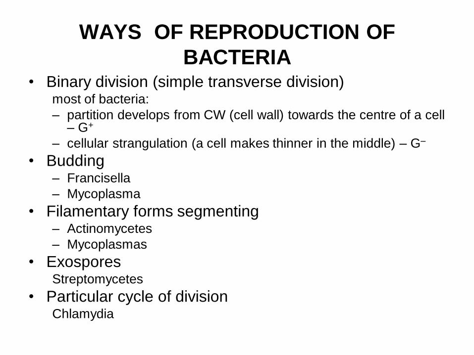
WAYS OF REPRODUCTION OF
BACTERIA • Binary division (simple transverse division)
most of bacteria:
– partition develops from CW (cell wall) towards the centre of a cell – G+
– cellular strangulation (a cell makes thinner in the middle) – G–
• Budding – Francisella
– Mycoplasma
• Filamentary forms segmenting – Actinomycetes
– Mycoplasmas
• Exospores Streptomycetes
• Particular cycle of division Chlamydia
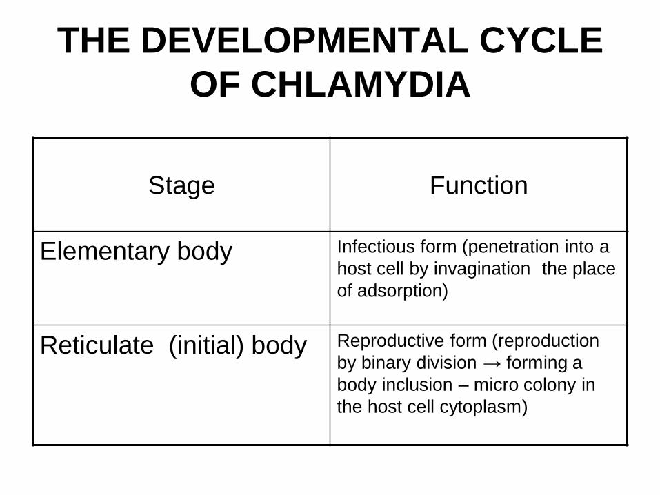
THE DEVELOPMENTAL CYCLE
OF CHLAMYDIA
Stage Function
Elementary body Infectious form (penetration into a
host cell by invagination the place
of adsorption)
Reticulate (initial) body Reproductive form (reproduction
by binary division → forming a
body inclusion – micro colony in
the host cell cytoplasm)
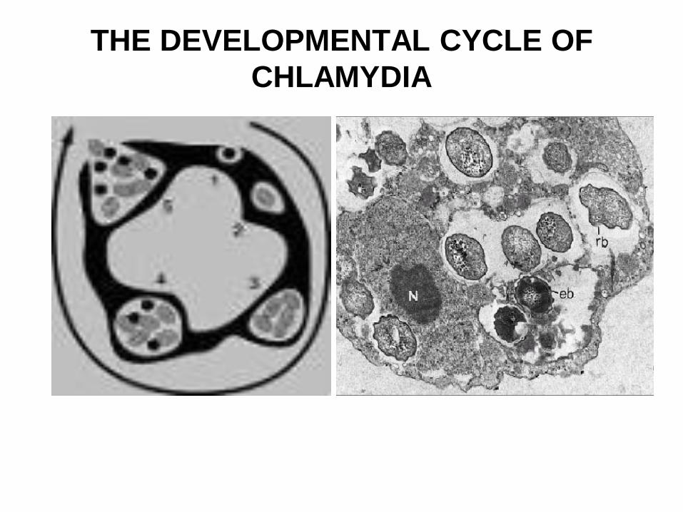
THE DEVELOPMENTAL CYCLE OF
CHLAMYDIA
CLASSIFICATION OF MEDIA
(CULTURE MEDIA)
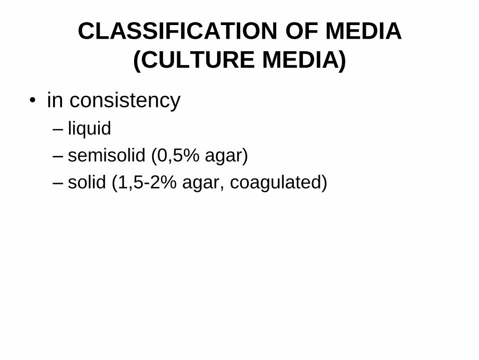
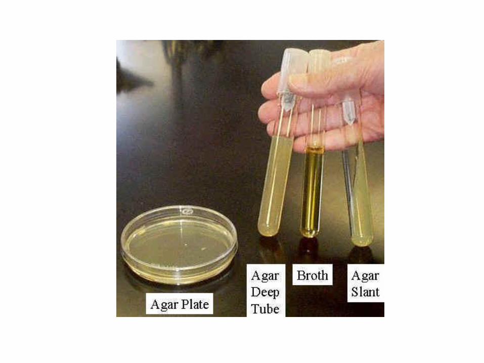
• in consistency
– liquid
– semisolid (0,5% agar)
– solid (1,5-2% agar, coagulated)
CLASSIFICATION OF MEDIA
(CULTURE MEDIA)
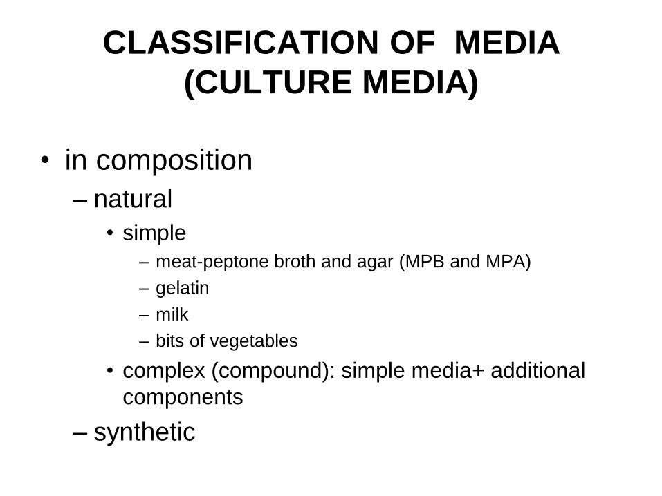
• in composition
– natural
• simple
– meat-peptone broth and agar (MPB and MPA)
– gelatin
– milk
– bits of vegetables
• complex (compound): simple media+ additional
components
– synthetic
CLASSIFICATION OF MEDIA
(CULTURE MEDIA)
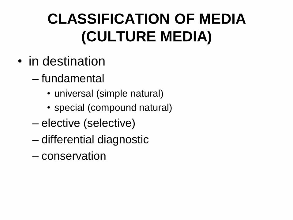
• in destination
– fundamental
• universal (simple natural)
• special (compound natural)
– elective (selective)
– differential diagnostic
– conservation
• Agar – is a complex organic substance received from marine algae. It melts in water at 80-86o C and solidifies at 36-40o C
• Coagulated nutrient media – are solid media with serum or high percentage of albumin (from eggs, e.g.) witch condenses by warming during sterilization
• Natural media are prepared on basis of decoctions, extracts from meat, fish, vegetables and other natural products
• Simple natural media are such decoctions or extracts
• Complex (compound) natural media are prepared by adding any matters to simple natural media (coloring agent, sugar, antibiotic, blood, etc.)
• Synthetic nutrient media are prepared by mixing pure chemical substance (salts, as a rule).
• elective (selective, enriched) media are the ones in which only certain species of bacteria grow well, and other species either grow poorly or do not grow at all. Such media are quite often employed in laboratory practice.
• Differential diagnostic media are used to distinguish among analogous bacteria by their fermentative activity or cultural properties.
• Conservation media are used for primary seeding and transportation of the material for diagnostics (specimens); they prevent the death of microbes, but the bacteria cells do not multiply in them.
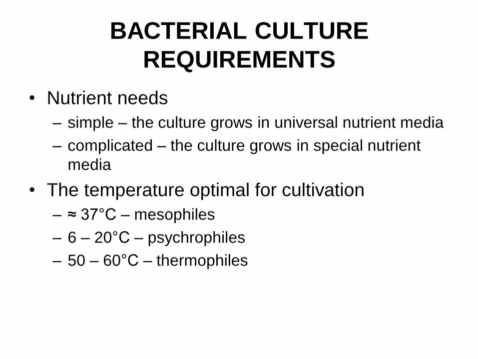
BACTERIAL CULTURE
REQUIREMENTS
• Nutrient needs
– simple – the culture grows in universal nutrient media
– complicated – the culture grows in special nutrient
media
• The temperature optimal for cultivation
– ≈ 37°С – mesophiles
– 6 – 20°С – psychrophiles
– 50 – 60°С – thermophiles
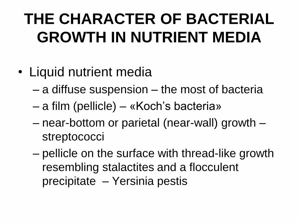
THE CHARACTER OF BACTERIAL
GROWTH IN NUTRIENT MEDIA
• Liquid nutrient media
– a diffuse suspension – the most of bacteria
– a film (pellicle) – «Koch’s bacteria»
– near-bottom or parietal (near-wall) growth –
streptococci
– pellicle on the surface with thread-like growth
resembling stalactites and a flocculent
precipitate – Yersinia pestis
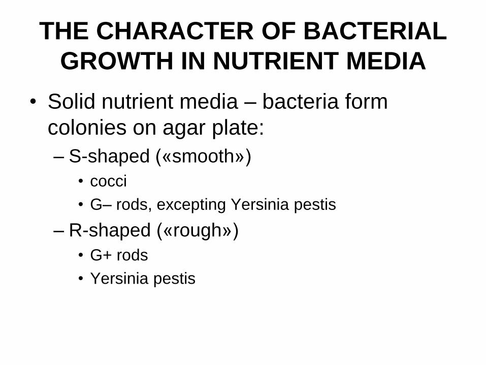
THE CHARACTER OF BACTERIAL
GROWTH IN NUTRIENT MEDIA
• Solid nutrient media – bacteria form
colonies on agar plate:
– S-shaped («smooth»)
• cocci
• G– rods, excepting Yersinia pestis
– R-shaped («rough»)
• G+ rods
• Yersinia pestis
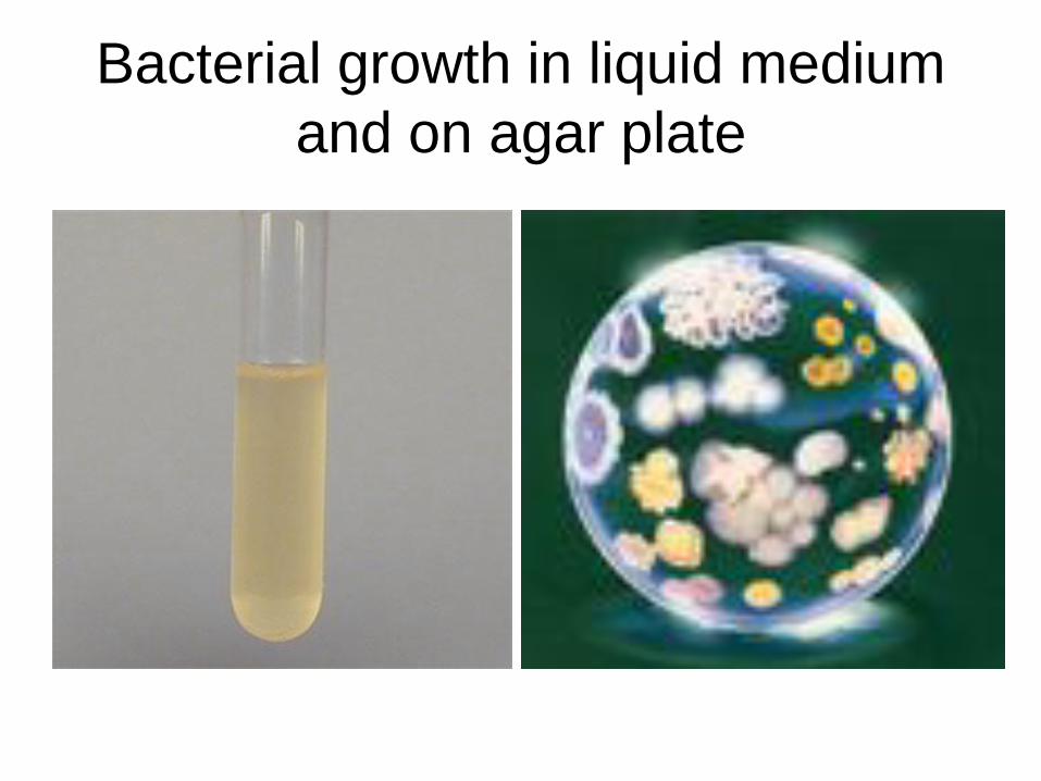
Bacterial growth in liquid medium
and on agar plate
Physiology of Bacteria
(continuation).
Method of Cultivation of
Bacteria.
Bacteriophages
Theme N5
ANAEROBIC
TECHNIQUES USED
FOR BACTERIAL
CULTIVATION
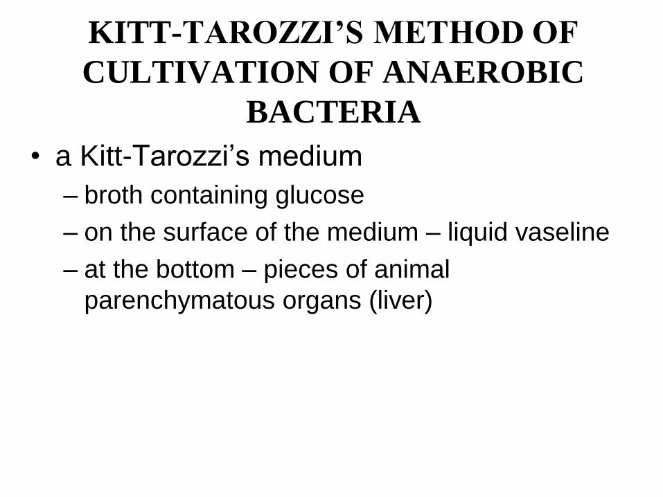
KITT-TAROZZI’S METHOD OF
CULTIVATION OF ANAEROBIC
BACTERIA
• a Kitt-Tarozzi’s medium
– broth containing glucose
– on the surface of the medium – liquid vaseline
– at the bottom – pieces of animal
parenchymatous organs (liver)
METHOD OF GROWING
OF BACTERIAL
CULTURE
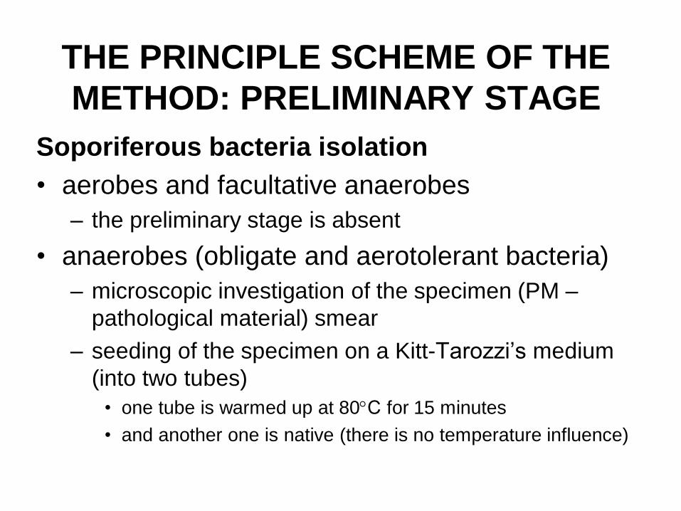
THE PRINCIPLE SCHEME OF THE
METHOD: PRELIMINARY STAGE
Soporiferous bacteria isolation
• aerobes and facultative anaerobes
– the preliminary stage is absent
• anaerobes (obligate and aerotolerant bacteria)
– microscopic investigation of the specimen (PM –
pathological material) smear
– seeding of the specimen on a Kitt-Tarozzi’s medium
(into two tubes)
• one tube is warmed up at 80С for 15 minutes
• and another one is native (there is no temperature influence)
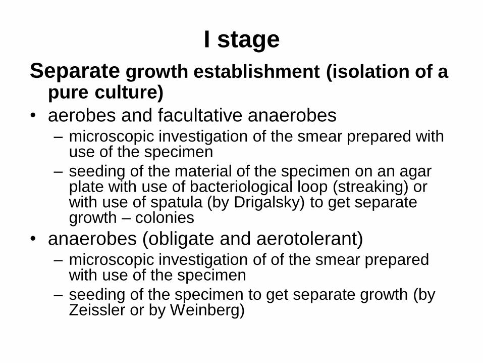
I stage
Separate growth establishment (isolation of a pure culture)
• aerobes and facultative anaerobes – microscopic investigation of the smear prepared with
use of the specimen
– seeding of the material of the specimen on an agar plate with use of bacteriological loop (streaking) or with use of spatula (by Drigalsky) to get separate growth – colonies
• anaerobes (obligate and aerotolerant) – microscopic investigation of of the smear prepared
with use of the specimen
– seeding of the specimen to get separate growth (by Zeissler or by Weinberg)
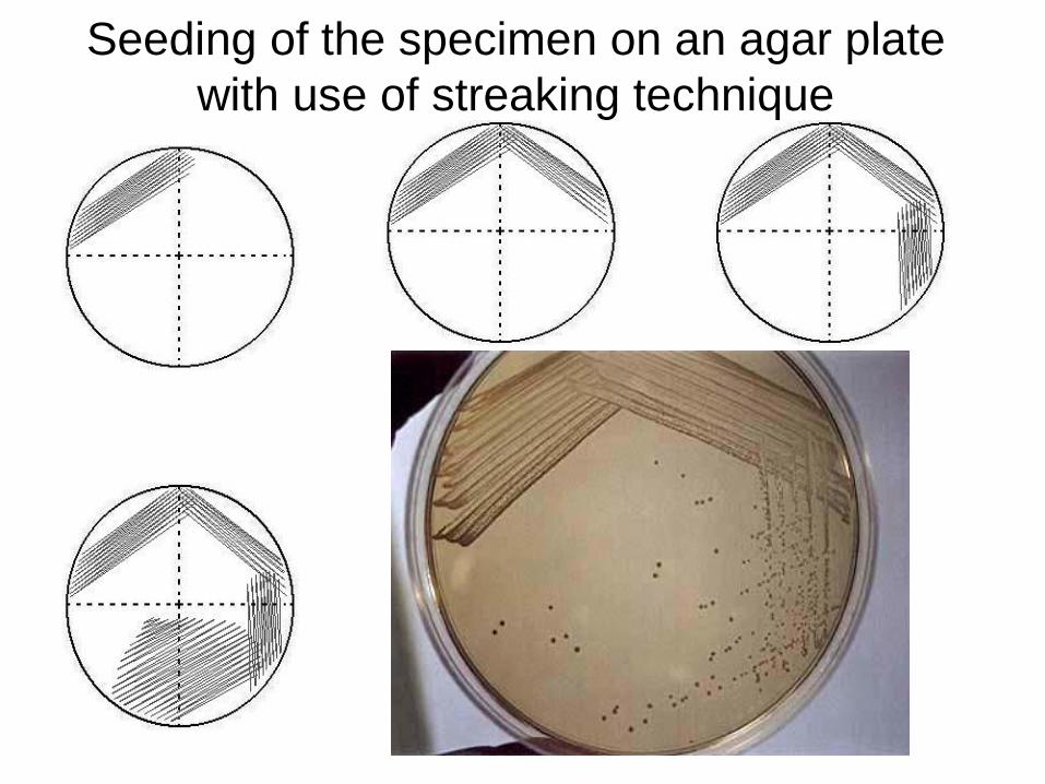
Seeding of the specimen on an agar plate
with use of streaking technique
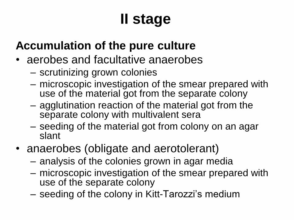
II stage
Accumulation of the pure culture
• aerobes and facultative anaerobes – scrutinizing grown colonies
– microscopic investigation of the smear prepared with use of the material got from the separate colony
– agglutination reaction of the material got from the separate colony with multivalent sera
– seeding of the material got from colony on an agar slant
• anaerobes (obligate and aerotolerant) – analysis of the colonies grown in agar media
– microscopic investigation of the smear prepared with use of the separate colony
– seeding of the colony in Kitt-Tarozzi’s medium
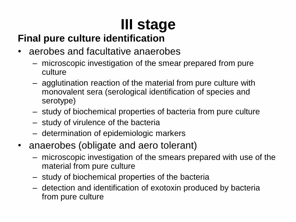
III stage Final pure culture identification
• aerobes and facultative anaerobes – microscopic investigation of the smear prepared from pure
culture
– agglutination reaction of the material from pure culture with monovalent sera (serological identification of species and serotype)
– study of biochemical properties of bacteria from pure culture
– study of virulence of the bacteria
– determination of epidemiologic markers
• anaerobes (obligate and aero tolerant) – microscopic investigation of the smears prepared with use of the
material from pure culture
– study of biochemical properties of the bacteria
– detection and identification of exotoxin produced by bacteria from pure culture
CULTURAL FEATURES OF
BACTERIA
• nutrient needs
• optimal nutrient medium
• temperature conditions optimal for growth
• aeration conditions optimal for growth
• rate of growth of bacteria
• characteristics of bacterial growth in liquid
and solid nutrient media
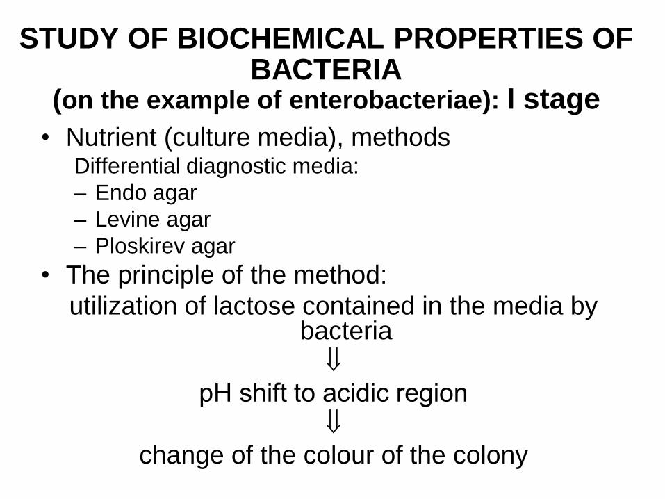
STUDY OF BIOCHEMICAL PROPERTIES OF BACTERIA
(on the example of enterobacteriae): I stage
• Nutrient (culture media), methods Differential diagnostic media:
– Endo agar
– Levine agar
– Ploskirev agar
• The principle of the method:
utilization of lactose contained in the media by bacteria
рН shift to acidic region
change of the colour of the colony
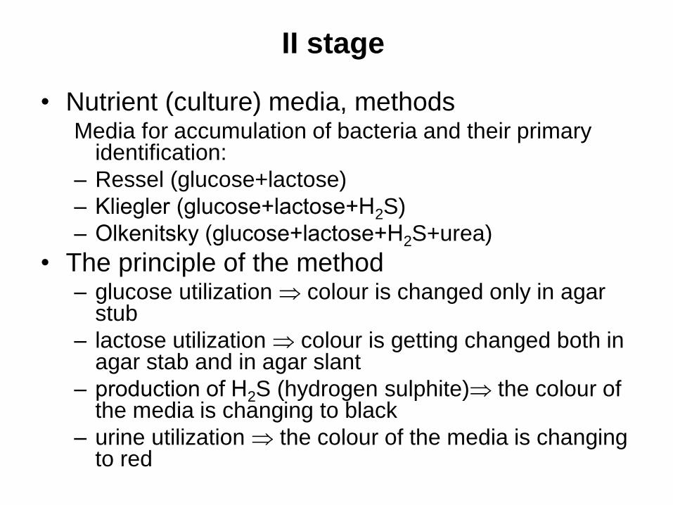
II stage
• Nutrient (culture) media, methods Media for accumulation of bacteria and their primary
identification:
– Ressel (glucose+lactose)
– Kliegler (glucose+lactose+Н2S)
– Olkenitsky (glucose+lactose+Н2S+urea)
• The principle of the method – glucose utilization colour is changed only in agar
stub
– lactose utilization colour is getting changed both in agar stab and in agar slant
– production of Н2S (hydrogen sulphite) the colour of the media is changing to black
– urine utilization the colour of the media is changing to red

Media for accumulation of bacteria and their
primary identification
Ressel Kliegler Olkenitsky
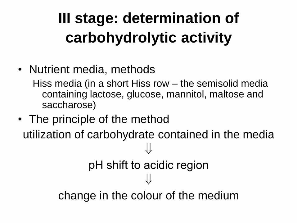
III stage: determination of
carbohydrolytic activity
• Nutrient media, methods Hiss media (in a short Hiss row – the semisolid media
containing lactose, glucose, mannitol, maltose and saccharose)
• The principle of the method
utilization of carbohydrate contained in the media
рН shift to acidic region
change in the colour of the medium
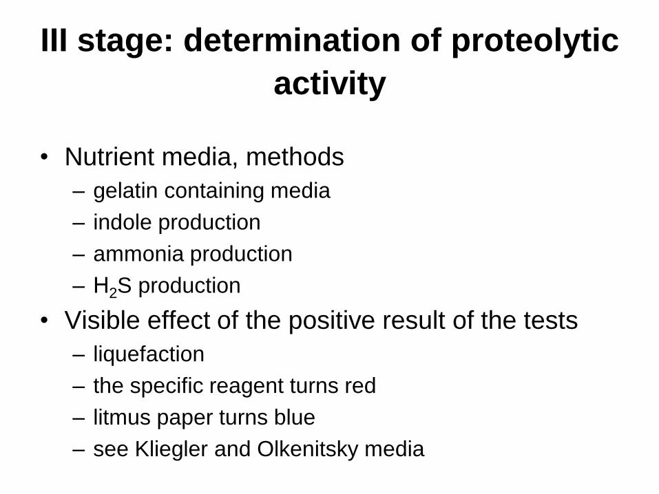
III stage: determination of proteolytic
activity
• Nutrient media, methods
– gelatin containing media
– indole production
– ammonia production
– Н2S production
• Visible effect of the positive result of the tests
– liquefaction
– the specific reagent turns red
– litmus paper turns blue
– see Kliegler and Olkenitsky media
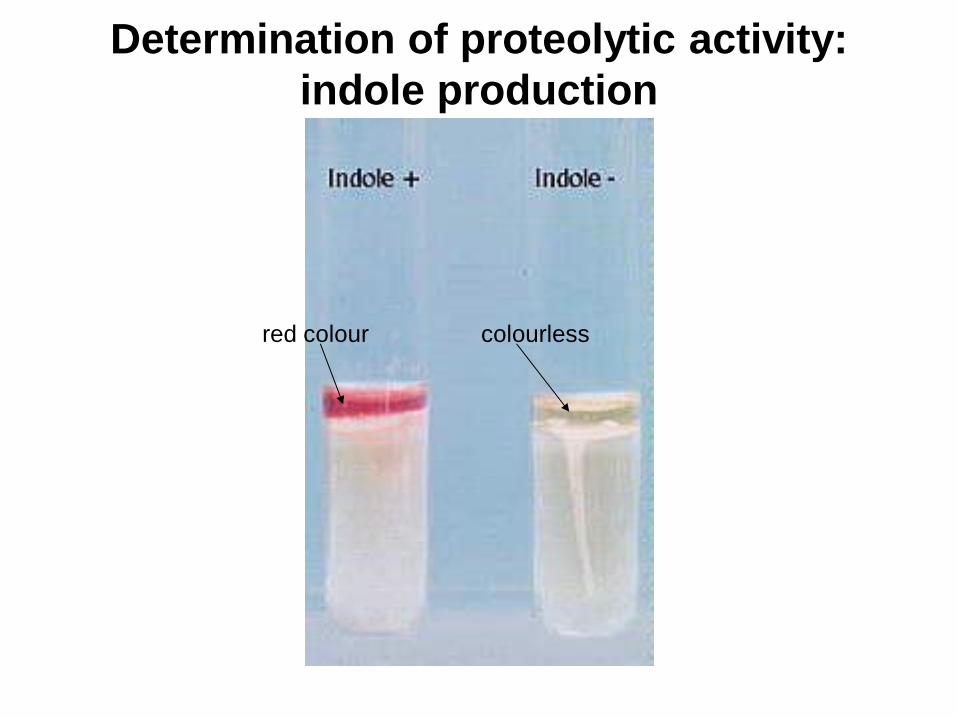
Determination of proteolytic activity:
indole production
red colour colourless
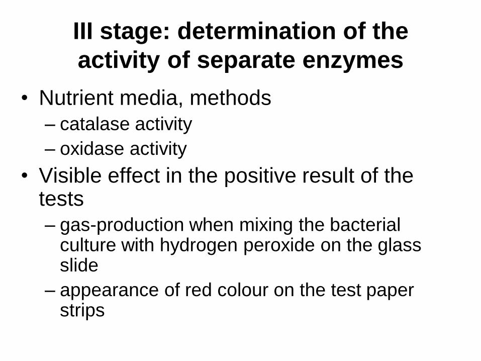
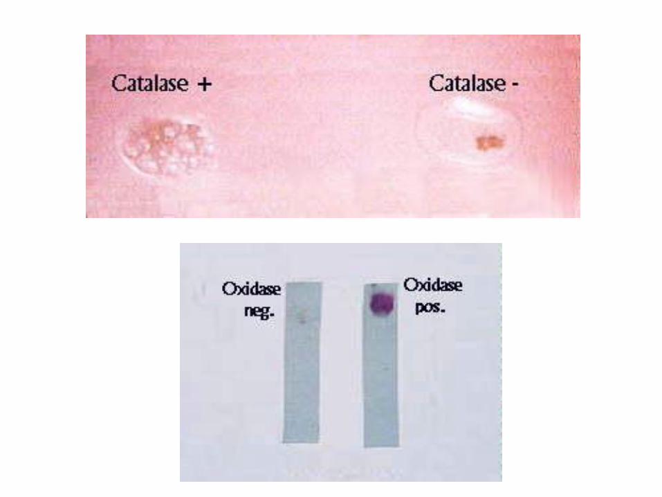
III stage: determination of the
activity of separate enzymes
• Nutrient media, methods
– catalase activity
– oxidase activity
• Visible effect in the positive result of the tests
– gas-production when mixing the bacterial culture with hydrogen peroxide on the glass slide
– appearance of red colour on the test paper strips
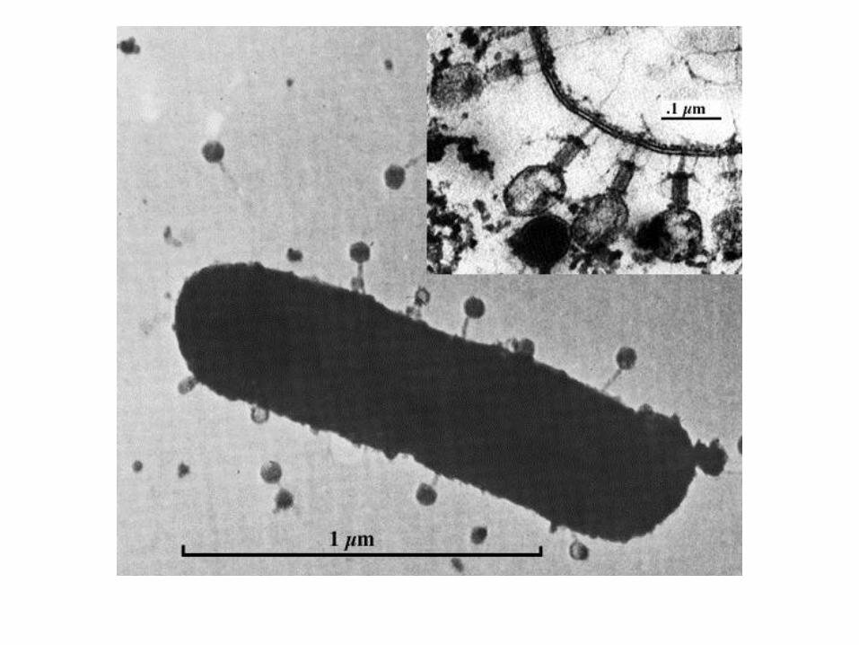
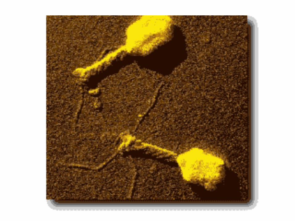
Bacteriophages

The definition of the term
bacteriophage: bacterial viruses.
Discovery of bacteriophage - d’Herelle,
1917.
Nomenclature of phages: it is based on the name of the host which is
sensitive to definite phage plus special
index.
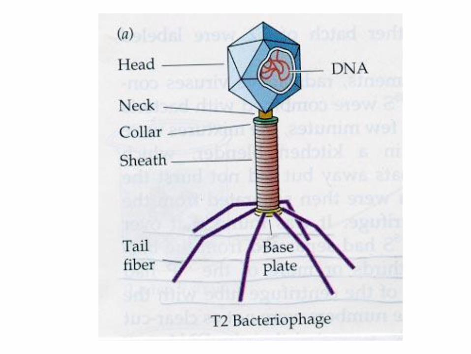
Structure of phages: nucleic acid (DNA
or RNA) + protein
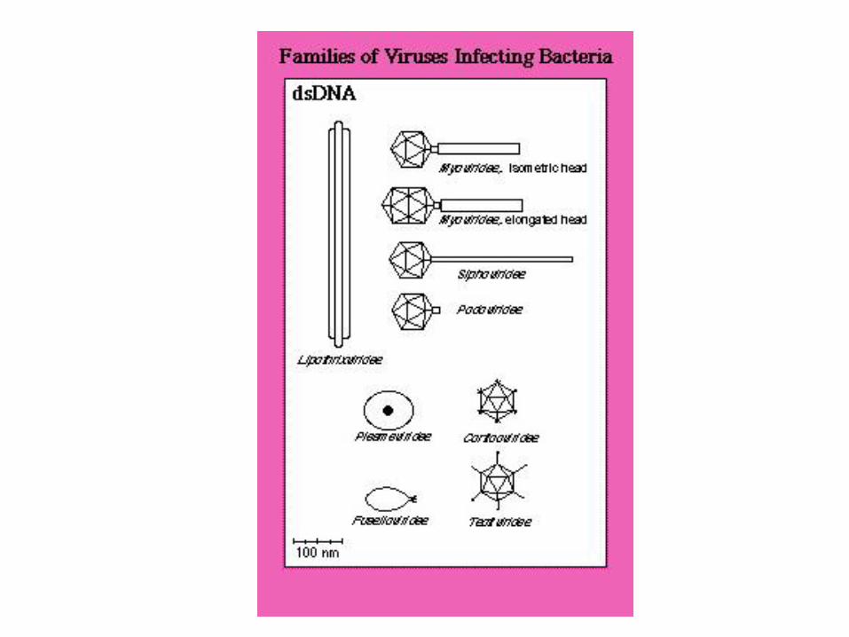
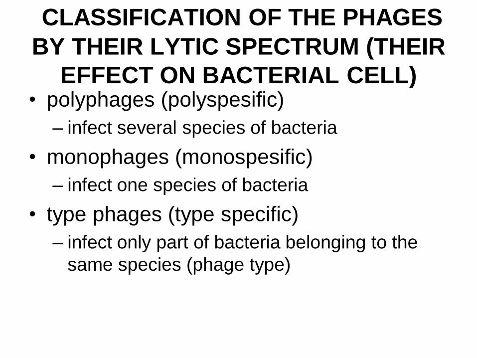
CLASSIFICATION OF THE PHAGES
BY THEIR LYTIC SPECTRUM (THEIR
EFFECT ON BACTERIAL CELL) • polyphages (polyspesific)
– infect several species of bacteria
• monophages (monospesific)
– infect one species of bacteria
• type phages (type specific)
– infect only part of bacteria belonging to the
same species (phage type)
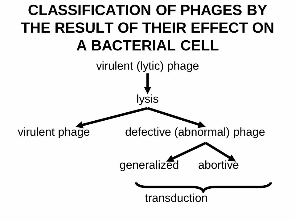
CLASSIFICATION OF PHAGES BY
THE RESULT OF THEIR EFFECT ON
A BACTERIAL CELL
virulent (lytic) phage
lysis
virulent phage defective (abnormal) phage
generalized abortive
transduction
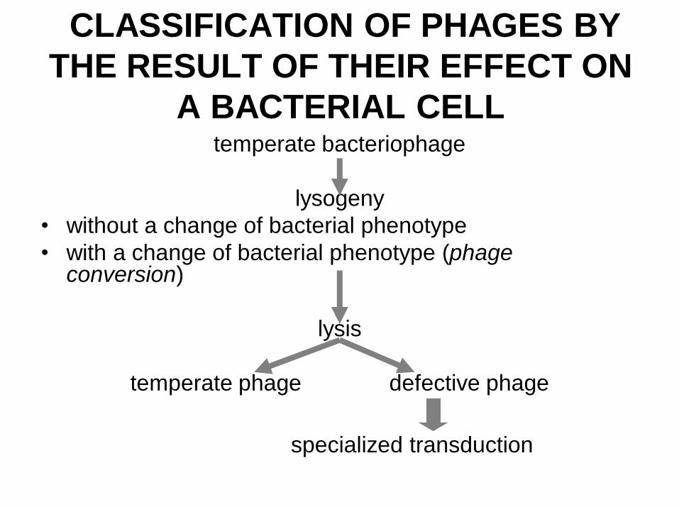
CLASSIFICATION OF PHAGES BY
THE RESULT OF THEIR EFFECT ON
A BACTERIAL CELL temperate bacteriophage
lysogeny
• without a change of bacterial phenotype
• with a change of bacterial phenotype (phage conversion)
lysis
temperate phage defective phage
specialized transduction
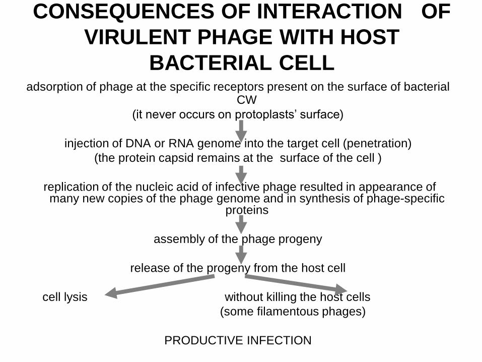
CONSEQUENCES OF INTERACTION OF
VIRULENT PHAGE WITH HOST
BACTERIAL CELL adsorption of phage at the specific receptors present on the surface of bacterial
CW
(it never occurs on protoplasts’ surface)
injection of DNA or RNA genome into the target cell (penetration)
(the protein capsid remains at the surface of the cell )
replication of the nucleic acid of infective phage resulted in appearance of many new copies of the phage genome and in synthesis of phage-specific
proteins
assembly of the phage progeny
release of the progeny from the host cell
cell lysis without killing the host cells
(some filamentous phages)
PRODUCTIVE INFECTION
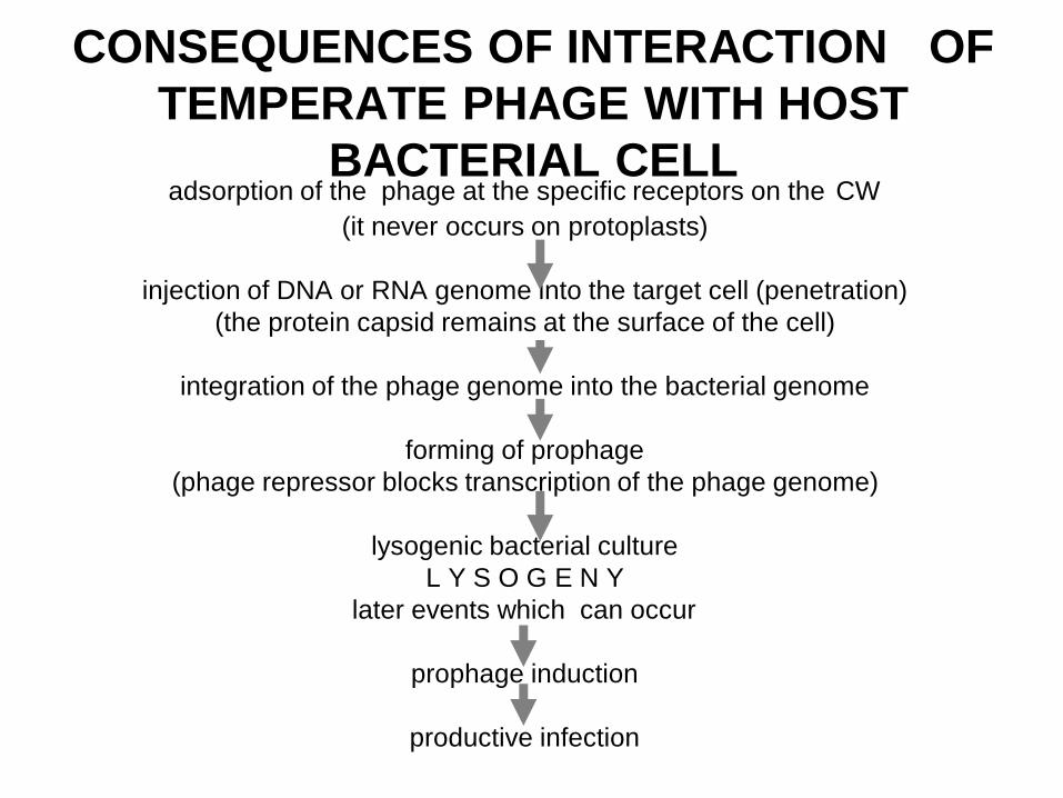
CONSEQUENCES OF INTERACTION OF
TEMPERATE PHAGE WITH HOST
BACTERIAL CELL adsorption of the phage at the specific receptors on the CW
(it never occurs on protoplasts)
injection of DNA or RNA genome into the target cell (penetration)
(the protein capsid remains at the surface of the cell)
integration of the phage genome into the bacterial genome
forming of prophage
(phage repressor blocks transcription of the phage genome)
lysogenic bacterial culture
L Y S O G E N Y
later events which can occur
prophage induction
productive infection
PRACTICAL APPLICATION OF THE
PHAGE IN MEDICINE
Phage diagnostics
1. Detection of the bacterial species in a
pathological material
– the evaluation of increase production of the new
generations of specific phage in the material
2. Pure culture identification
– bacterial species determination
• phage indication
– bacterial phage type determination
• phage typing
PRACTICAL APPLICATION OF THE
PHAGE IN MEDICINE
Phage therapy
Phage could be used locally (at the infected
place)
PRACTICAL APPLICATION OF THE
PHAGE IN MEDICINE
Phage prophylaxis of some bacterial
infections
• enteric fever
• dysentery
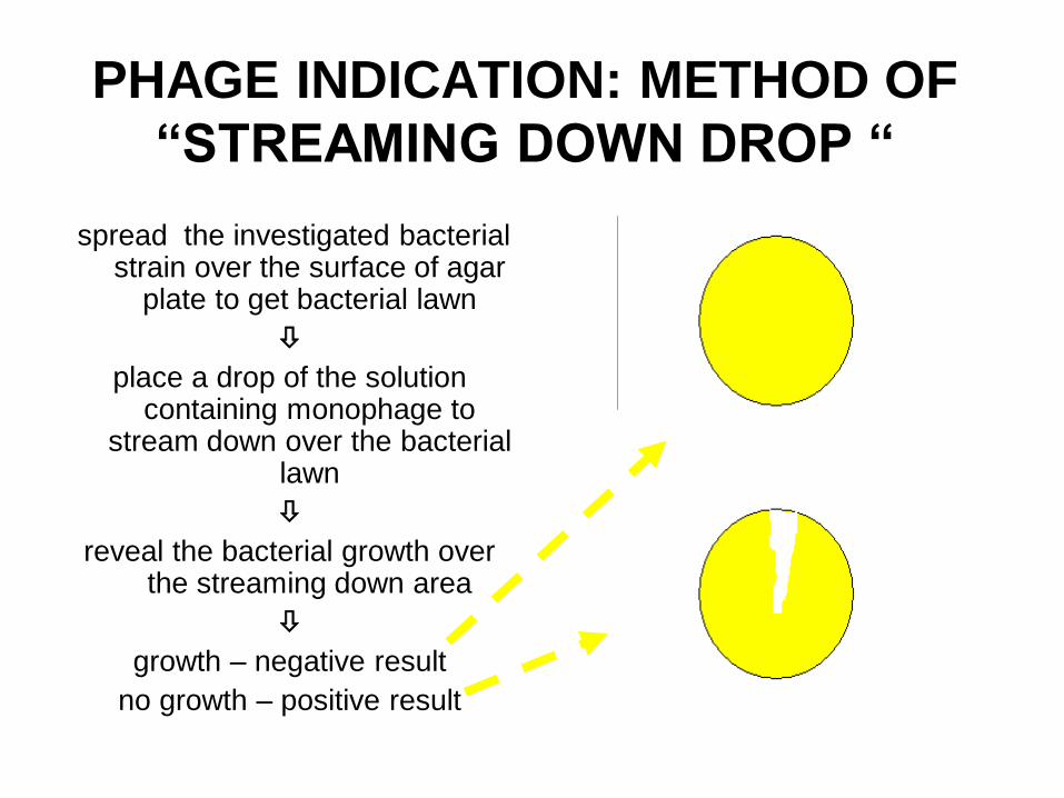
PHAGE INDICATION: METHOD OF
“STREAMING DOWN DROP “
spread the investigated bacterial strain over the surface of agar
plate to get bacterial lawn
place a drop of the solution containing monophage to
stream down over the bacterial lawn
reveal the bacterial growth over the streaming down area
growth – negative result
no growth – positive result
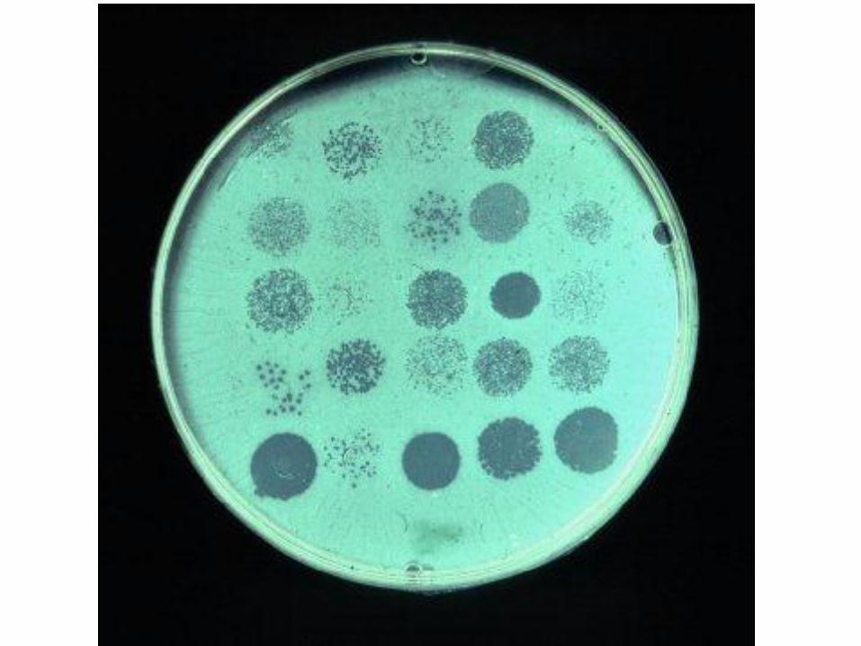
BACTERIA PHAGE TYPING
1. platting of the investigated bacterial strain to get bacterial lawn on an agar plate
2. placing drops of the solution containing type phages
3. incubation
4. revealing of «sterile spots» («plaques») and register phage type of the bacteria = a list of type phages causing lysis of the bacterial strain (appearance of “plaques” on the bacterial lawn)
Genetics of Bacteria
Theme N6
ORGANIZATION OF GENETIC
MATERIAL IN BACTERIA
DNA
• nucleoid (bacterial chromosome)
• it codes information vital for bacterial cell
• extra-chromosomal factors of heredity
– they code the information which is not
important for life of bacteria
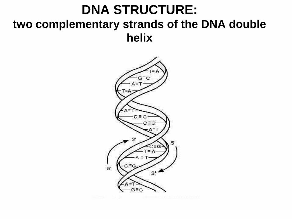
DNA STRUCTURE: two complementary strands of the DNA double
helix
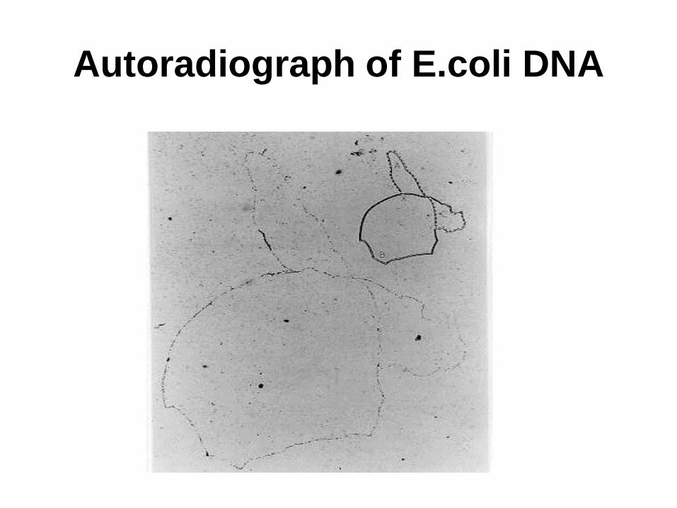
Autoradiograph of E.coli DNA
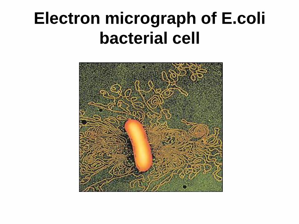
Electron micrograph of E.coli
bacterial cell
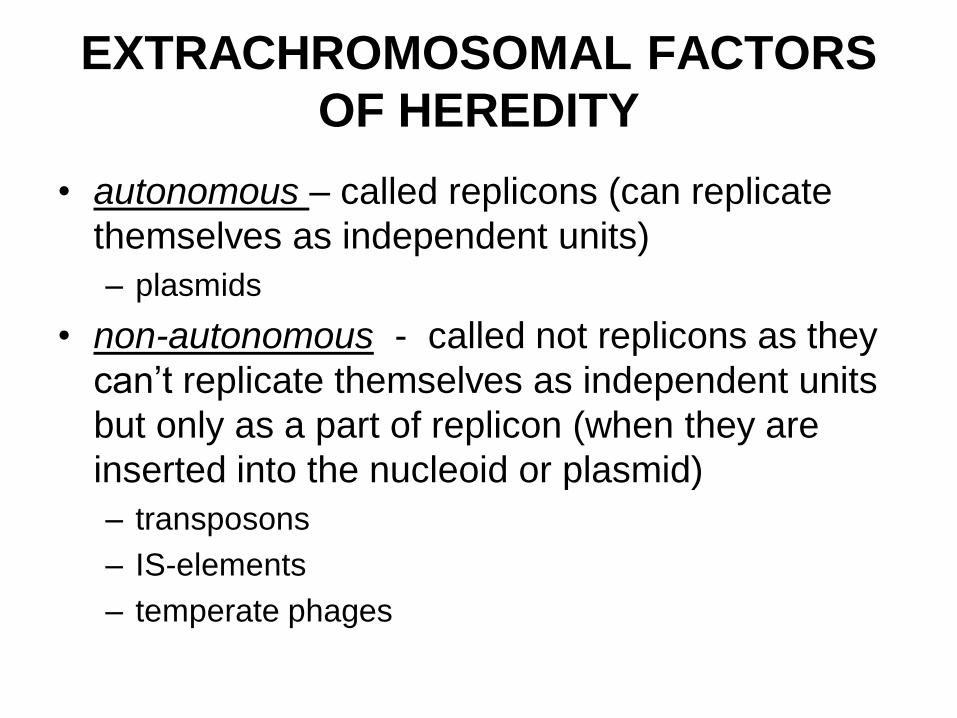
EXTRACHROMOSOMAL FACTORS
OF HEREDITY
• autonomous – called replicons (can replicate
themselves as independent units)
– plasmids
• non-autonomous - called not replicons as they
can’t replicate themselves as independent units
but only as a part of replicon (when they are
inserted into the nucleoid or plasmid)
– transposons
– IS-elements
– temperate phages
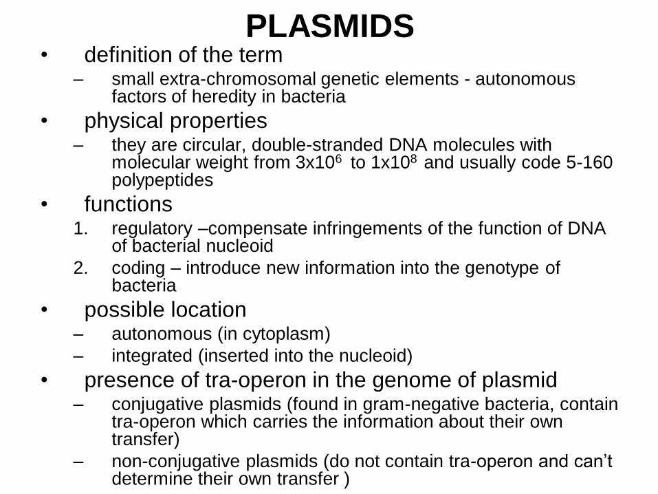
PLASMIDS • definition of the term
– small extra-chromosomal genetic elements - autonomous factors of heredity in bacteria
• physical properties – they are circular, double-stranded DNA molecules with
molecular weight from 3x10⁶ to 1x10⁸ and usually code 5-160 polypeptides
• functions 1. regulatory –compensate infringements of the function of DNA
of bacterial nucleoid
2. coding – introduce new information into the genotype of bacteria
• possible location – autonomous (in cytoplasm)
– integrated (inserted into the nucleoid)
• presence of tra-operon in the genome of plasmid – conjugative plasmids (found in gram-negative bacteria, contain
tra-operon which carries the information about their own transfer)
– non-conjugative plasmids (do not contain tra-operon and can’t determine their own transfer )
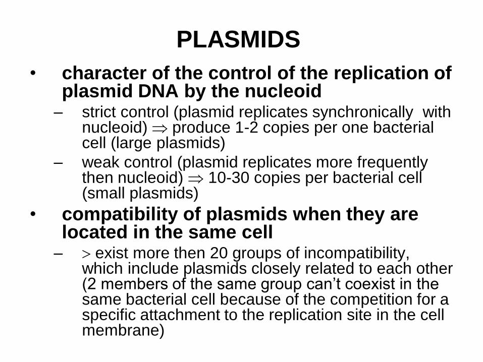
PLASMIDS
• character of the control of the replication of plasmid DNA by the nucleoid
– strict control (plasmid replicates synchronically with nucleoid) produce 1-2 copies per one bacterial cell (large plasmids)
– weak control (plasmid replicates more frequently then nucleoid) 10-30 copies per bacterial cell (small plasmids)
• compatibility of plasmids when they are located in the same cell
– exist more then 20 groups of incompatibility, which include plasmids closely related to each other (2 members of the same group can’t coexist in the same bacterial cell because of the competition for a specific attachment to the replication site in the cell membrane)
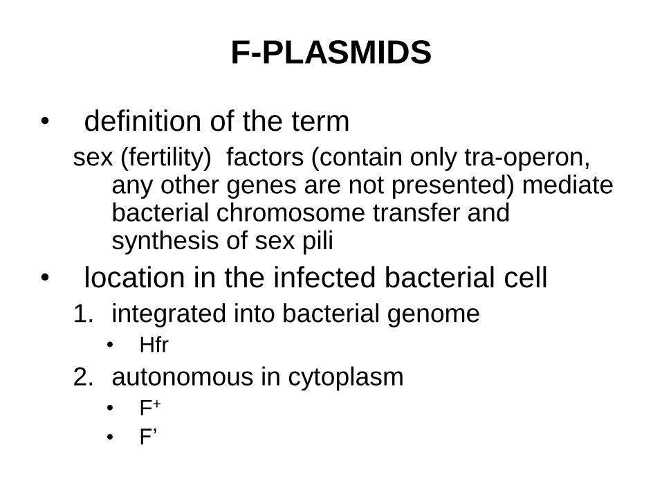
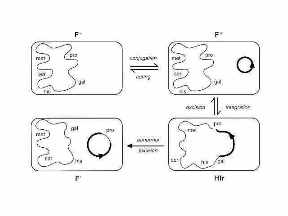
F-PLASMIDS
• definition of the term
sex (fertility) factors (contain only tra-operon, any other genes are not presented) mediate bacterial chromosome transfer and synthesis of sex pili
• location in the infected bacterial cell
1. integrated into bacterial genome • Hfr
2. autonomous in cytoplasm • F+
• F’
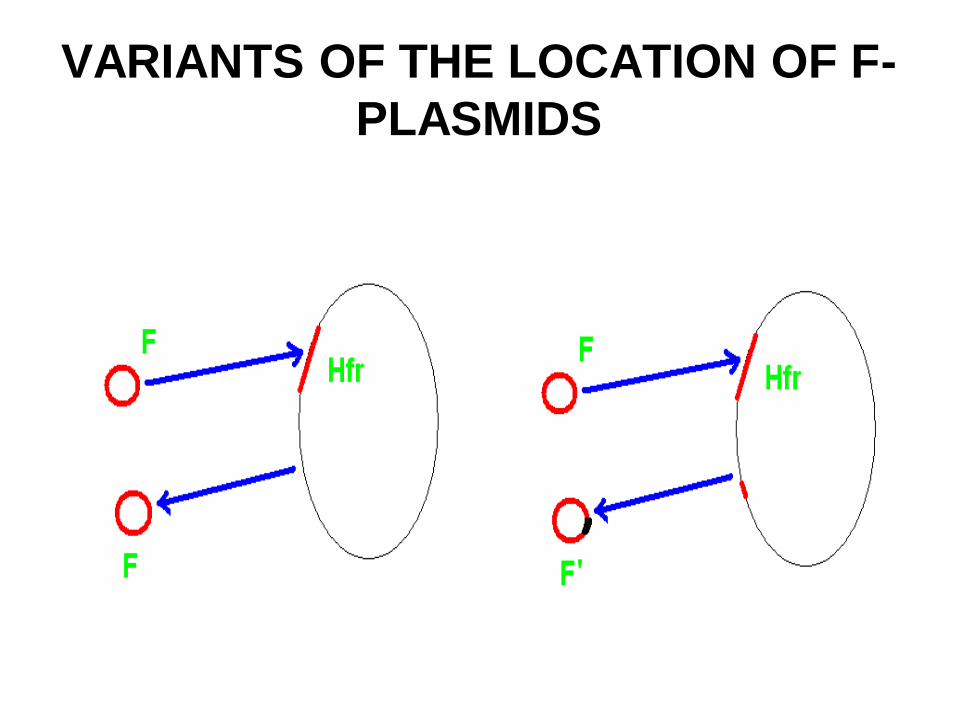
VARIANTS OF THE LOCATION OF F-
PLASMIDS
R-PLASMIDS • definition of the term
resistance factors - plasmids encoding multiple
resistance to various anti-microbial agents, such as
antibiotics
• composition
– r-operon (operone) + tra-operon
– r-operon (operone)
• the ways of transfer of the plasmids from one
bacterial cell to another one
– transduction ( plasmids are transferred by phages in
gram-positive bacteria)
– conjugation (gram-negative bacteria)
PLASMIDS OF BACTERIOCYNOGENITY (example of Col-plasmids of E.coli)
• definition of the term – plasmids encoding synthesis of colicins (antibiotic - like
proteins which are lethal for coliform bacteria)
• composition – genes which cause production of colicins by the bacteria
– tra-operon which determines self-transfer of the plasmid
• peculiarities 1. rarely integrate into the nucleoid
2. usually exist in repressed state
3. after the plasmid derepression bacterial cell synthesizes colicins and dies after that (potentially lethal plasmid)
• biological role – decrease of the density of bacterial population when the
nutrient media got exhausted
• importance for medicine – participate in normalization of natural micro - biocenosis of
intestines
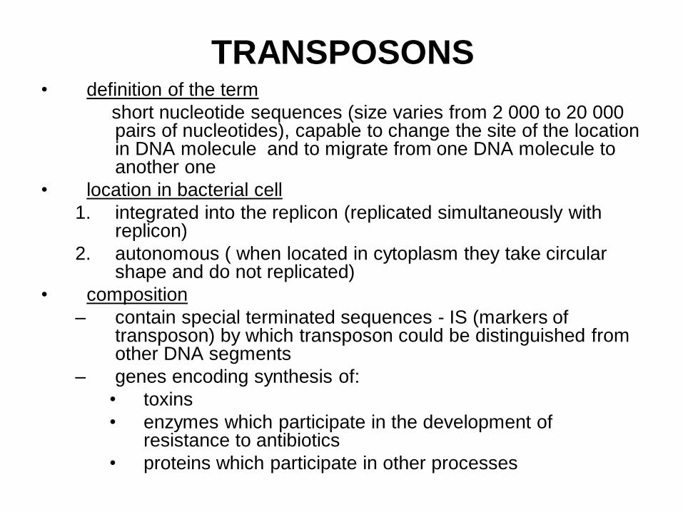
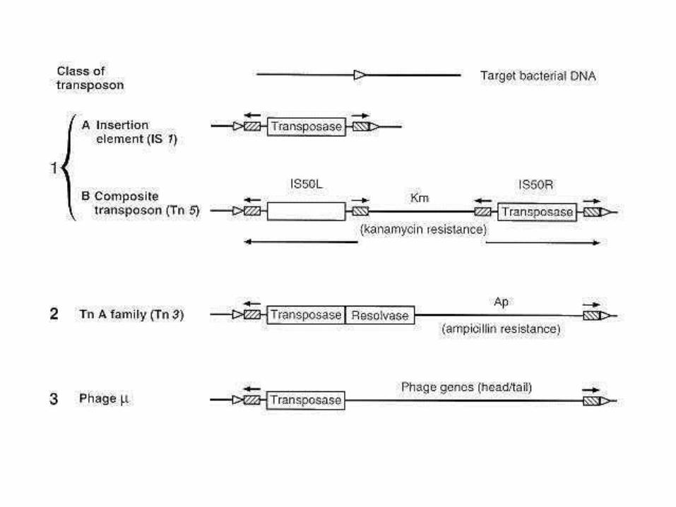
TRANSPOSONS • definition of the term
short nucleotide sequences (size varies from 2 000 to 20 000 pairs of nucleotides), capable to change the site of the location in DNA molecule and to migrate from one DNA molecule to another one
• location in bacterial cell
1. integrated into the replicon (replicated simultaneously with replicon)
2. autonomous ( when located in cytoplasm they take circular shape and do not replicated)
• composition
– contain special terminated sequences - IS (markers of transposon) by which transposon could be distinguished from other DNA segments
– genes encoding synthesis of:
• toxins
• enzymes which participate in the development of resistance to antibiotics
• proteins which participate in other processes
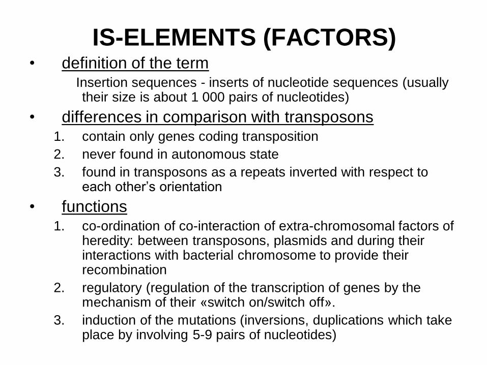
IS-ELEMENTS (FACTORS) • definition of the term
Insertion sequences - inserts of nucleotide sequences (usually their size is about 1 000 pairs of nucleotides)
• differences in comparison with transposons 1. contain only genes coding transposition
2. never found in autonomous state
3. found in transposons as a repeats inverted with respect to each other’s orientation
• functions 1. co-ordination of co-interaction of extra-chromosomal factors of
heredity: between transposons, plasmids and during their interactions with bacterial chromosome to provide their recombination
2. regulatory (regulation of the transcription of genes by the mechanism of their «switch on/switch off».
3. induction of the mutations (inversions, duplications which take place by involving 5-9 pairs of nucleotides)
MODIFICATIONS IN BACTERIA
Changes affecting only the phenotype
(observable properties) of bacteria
– do not accompanied by changes of DNA
structure and thus they are not inherited by
the next generations
– are not stable and usually could be lost very
quickly
MUTATIONS IN BACTERIA
Definition of the term
Any changes which occur in genotype (the set of genetic determinants carried by bacterial genome) so they involve changes of the primary structure of DNA molecule.
The result of the mutation is loss or change of one or several hereditary features which will be inherited by the next generations of bacteria.
MUTATIONS IN BACTERIA
Classification according to the occurring mechanism:
• spontaneous – difficult or not possible to find the effect of curtain factor (mutagen) – mistakes in the function of DNA-polymerase when
replication of DNA takes place
– insertion mutations – occur when extra-chromosomal factors of heredity are inserted into DNA molecule
• induced – produced in the experiment when certain known mutagenic agent (mutagen) is applied
MUTATIONS IN BACTERIA
Classification according to their
direction:
• direct – loss or change of the property
• reverse (reversions) – restoration of the
property
– true – when restoration of genotype and
phenotype takes place
– suppressive – when we see restoration only
of phenotype
SR-DISSOCIATION
• definition of the term appearance of R-shaped colonies in pure bacterial culture which
normally forms S-shape colonies: phenotypic manifestation of the change of some properties of the bacterial cells
• mechanism insertion mutation resulted in the loss of the genes controlling
synthesis of carbohydrate chains which are necessary for the formation of LPS component of the outer membrane of the cell wall
• biological importance – bacteria producing R-shaped colonies are more resistant to
unfavorable physical and chemical factors of the external environment
– bacteria producing S-shaped colonies are more resistant to phagocytosis and to the effect of antibodies
Significantly complicate isolation and identification of pure culture.
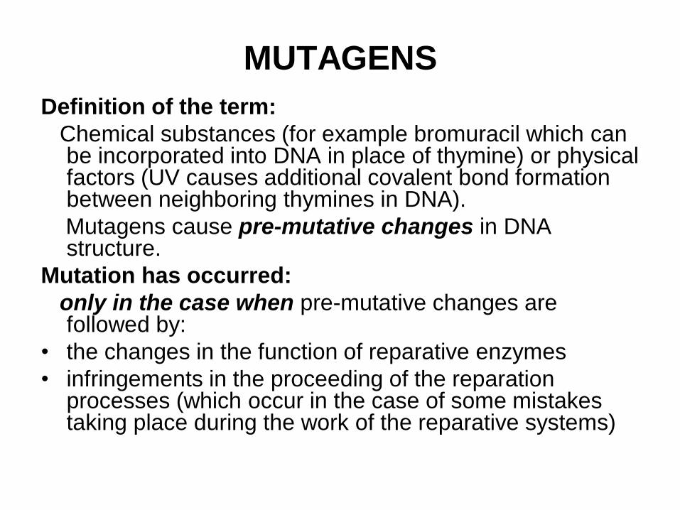
MUTAGENS
Definition of the term:
Chemical substances (for example bromuracil which can be incorporated into DNA in place of thymine) or physical factors (UV causes additional covalent bond formation between neighboring thymines in DNA).
Mutagens cause pre-mutative changes in DNA structure.
Mutation has occurred:
only in the case when pre-mutative changes are followed by:
• the changes in the function of reparative enzymes
• infringements in the proceeding of the reparation processes (which occur in the case of some mistakes taking place during the work of the reparative systems)
RECOMBINATIVE VARIABILITY IN
BACTERIA
Definition of the term
Changes in DNA structure occurring as a
result of integration of the part of DNA of the
recipient cell into the DNA of the donor cell
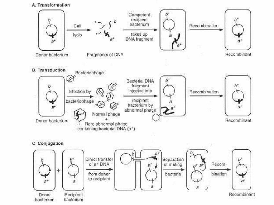
RECOMBINATIVE VARIABILITY IN
BACTERIA
Forms of recombinative variability
1. Transformation– direct transfer of genetic material –soluble DNA segments (could be released spontaneously) from donor cell to the recipient one.
2. Transduction – transfer of genetic material (a fragment of donor chromosome) from donor cell to recipient cell by defective bacteriophages:
– non-specific (generalized transduction) – by virulent phages
– abortive – by virulent phages
– specific (restricted transduction) – by temperate phages
3. Conjugation – transfer of genetic material from donor HFR cell to the recipient cell through conjugative pili in gram-negative bacteria.
4. Lysogeny – bacterial genome carries phage genes after infection of bacteria by temperate phage.
5. Phage conversion – appearance of new properties in bacteria as a result of lysogeny.
GENE ENGEINEERING IN MEDICAL
MICROBIOLOGY
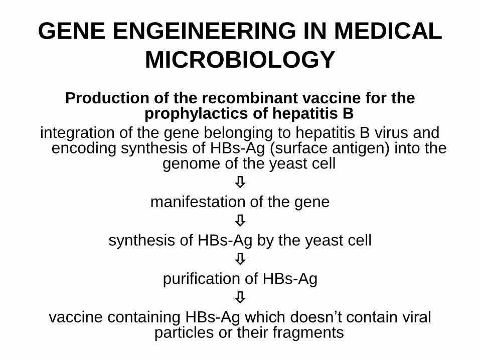
Production of the recombinant vaccine for the prophylactics of hepatitis B
integration of the gene belonging to hepatitis B virus and encoding synthesis of HBs-Ag (surface antigen) into the
genome of the yeast cell
manifestation of the gene
synthesis of HBs-Ag by the yeast cell
purification of HBs-Ag
vaccine containing HBs-Ag which doesn’t contain viral particles or their fragments
METHODS OF GENETICS APPLIED
IN MICROBIOLOGICAL
DIAGNOSTICS • content (in percent) of G+C (guanine +
cytosine) nucleotides in bacterial genome
• method of molecular hybridization
• polymerase chain reaction (PCR)
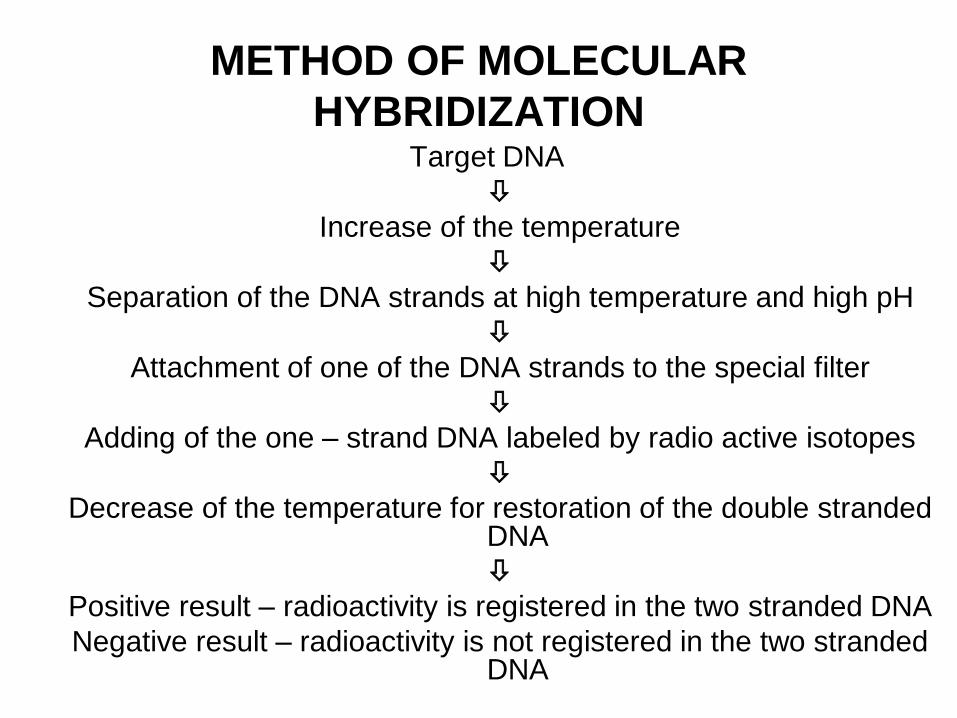
METHOD OF MOLECULAR
HYBRIDIZATION Target DNA
Increase of the temperature
Separation of the DNA strands at high temperature and high pH
Attachment of one of the DNA strands to the special filter
Adding of the one – strand DNA labeled by radio active isotopes
Decrease of the temperature for restoration of the double stranded DNA
Positive result – radioactivity is registered in the two stranded DNA
Negative result – radioactivity is not registered in the two stranded DNA
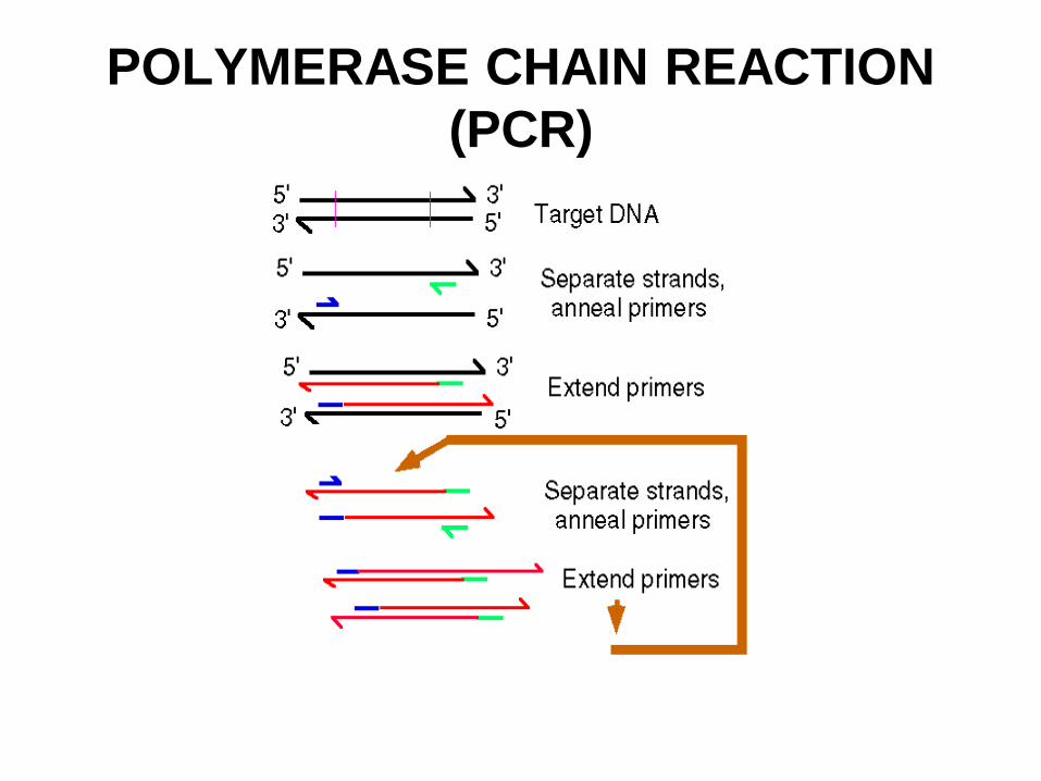
POLYMERASE CHAIN REACTION
(PCR)
ECOLOGY OF
MICROORGANISMS
Theme N7
Definition of the term “ecology of
microorganisms”
• section of general microbiology, studying
the next relationships of microorganisms:
– between each other
– with unanimated objects of environment
– with macro-organism (human organism).
Ecological niches of
microorganisms: soil
Microbial populations (micro-biocenosises) of soil
1. The upper layer of soil is most populated by
microorganisms.
2. Survival of pathogenic microorganisms in soil :
– species of bacteria which don’t form spores and
viruses – from several days to several months,
– spores – for years,
– infectious agents causing botulism, actinomycosis,
deep mycosis and mycotoxicosis – usually live in the
soil.
Ecological niches of
microorganisms: sanitary control of
soil ► For sanitary control the bacteria - indicators of fecal
contamination of soil (representatives of normal micro-flora of human intestines) are usually used.
► Bacteria – indicators live in the soil during the same period of time as pathogenic intestinal microorganisms. Main bacterium – indicator is E.coli.
► The sanitary standards used for evaluation of fecal contamination of soil:
– coli-index – number of E.coli cells in 1 gm of soil,
– coli-titer – mass of the soil (in grams), containing 1 cell of E.coli;
– microbial number – total numbers of all microorganisms in 1 gram of soil.
Ecological niches of
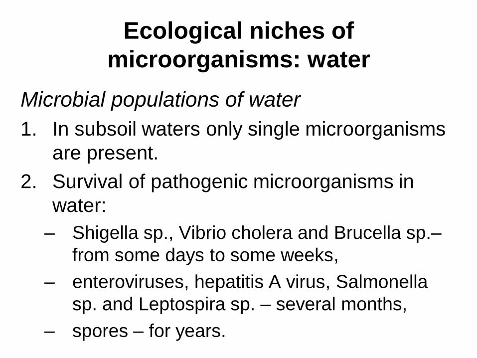
microorganisms: water
Microbial populations of water
1. In subsoil waters only single microorganisms
are present.
2. Survival of pathogenic microorganisms in
water:
– Shigella sp., Vibrio cholera and Brucella sp.–
from some days to some weeks,
– enteroviruses, hepatitis A virus, Salmonella
sp. and Leptospira sp. – several months,
– spores – for years.
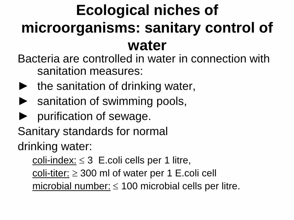
Ecological niches of
microorganisms: sanitary control of
water Bacteria are controlled in water in connection with
sanitation measures:
► the sanitation of drinking water,
► sanitation of swimming pools,
► purification of sewage.
Sanitary standards for normal
drinking water: coli-index: 3 E.coli cells per 1 litre,
coli-titer: 300 ml of water per 1 E.coli cell
microbial number: 100 microbial cells per litre.
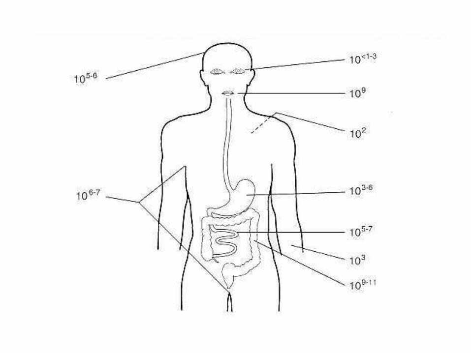
THE MICROFLORA
OF HUMAN BODY
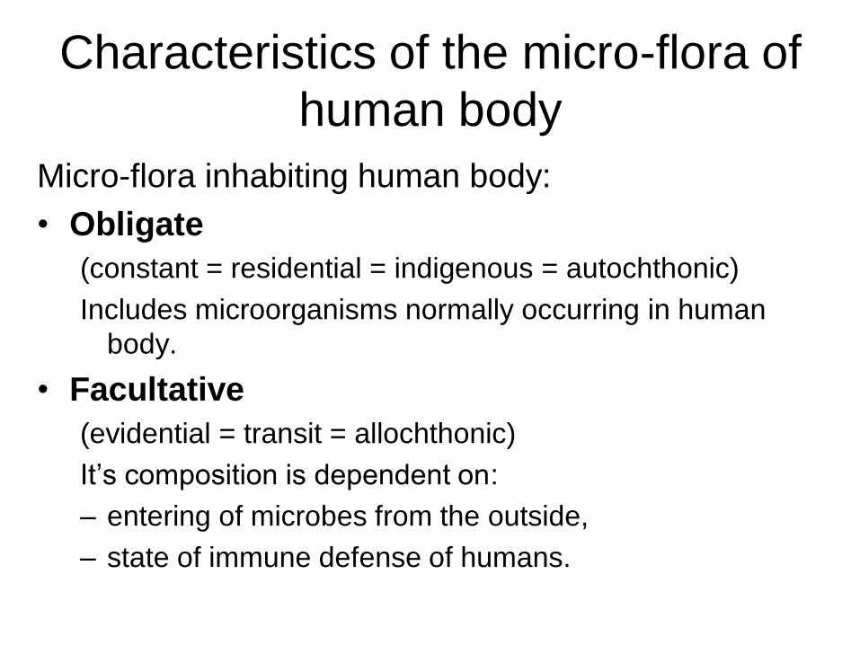
Characteristics of the micro-flora of
human body
Micro-flora inhabiting human body:
• Obligate
(constant = residential = indigenous = autochthonic)
Includes microorganisms normally occurring in human
body.
• Facultative
(evidential = transit = allochthonic)
It’s composition is dependent on:
– entering of microbes from the outside,
– state of immune defense of humans.
Composition of normal micro-
flora of human intestine 1. Predominant:
– bifidobacteria
– lactobacteria
– bacteroides
2. Found in high quantities: – E. coli
– enterococci
3. Found in minor quantities: – other representatives of enteric bacteria
– staphylococci
– fungi Candida
– clostridia
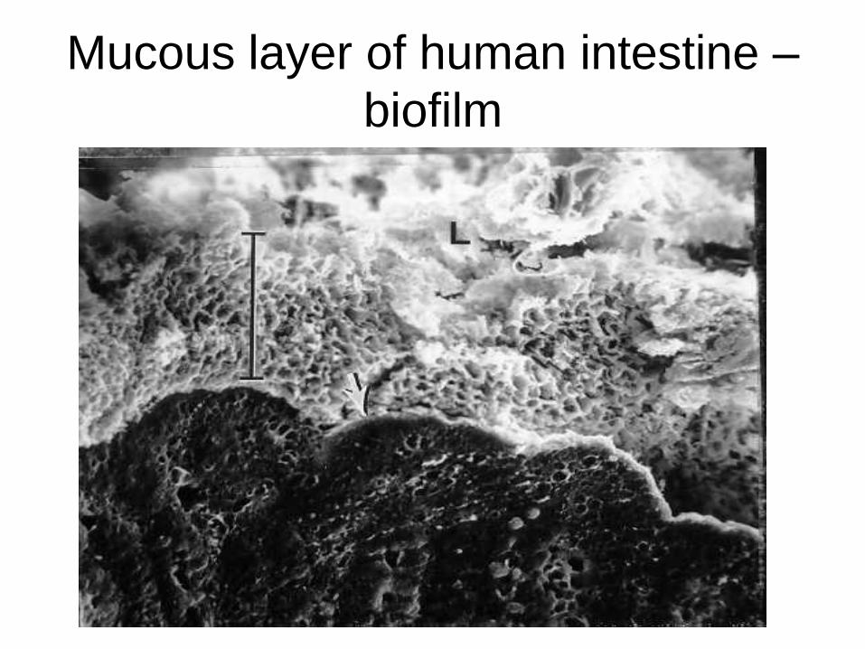
Mucous layer of human intestine –
biofilm
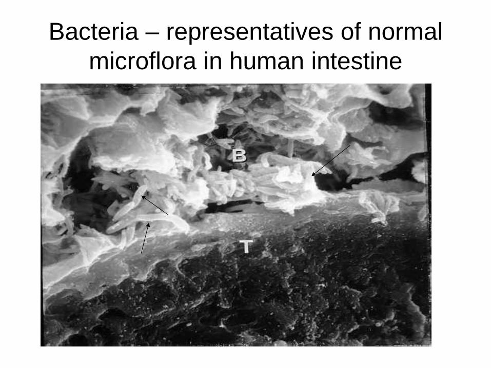
Bacteria – representatives of normal
microflora in human intestine
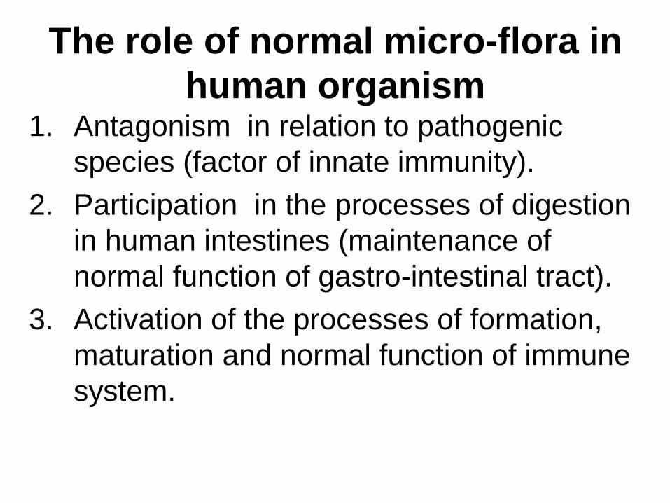
The role of normal micro-flora in
human organism 1. Antagonism in relation to pathogenic
species (factor of innate immunity).
2. Participation in the processes of digestion
in human intestines (maintenance of
normal function of gastro-intestinal tract).
3. Activation of the processes of formation,
maturation and normal function of immune
system.
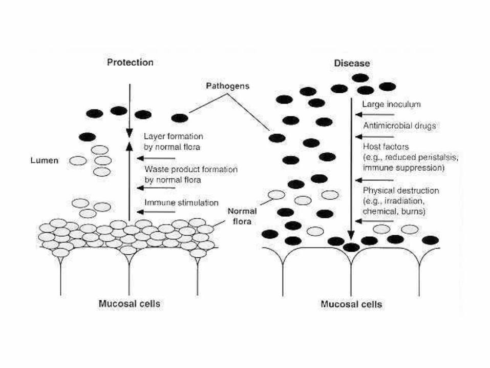
Disturbances in composition of
normal micro-flora Disbacteriosis (disbiosis or dismicrobiosis):
pathological state characterized by qualitative and quantitative disturbances in the composition of microbial populations normally inhabiting human body.
Some factors which can cause disbiosis in
intestine:
• physical destruction (irradiation, burns, chemicals, etc.);
• host factors (e.g. reduced peristalsis, immune suppression);
• antimicrobial drugs (antibiotics).
Some approaches to the normalization of
misbalance of normal micro-flora in the
state of disbiosis • administration of eubiotics (preparations which
contain live bacterial strains – normal inhabitants of human intestines): – bifidobacteria – bifidumbacterin,
– lactobacteria – lactobacterin,
– E.coli – colibacterin,
– bifidobacteria and E.coli – bificol and others.
• administration of probiotics (preparations which stimulate development of normal micro-flora),
• removal of the factors which caused the state of disbiosis.
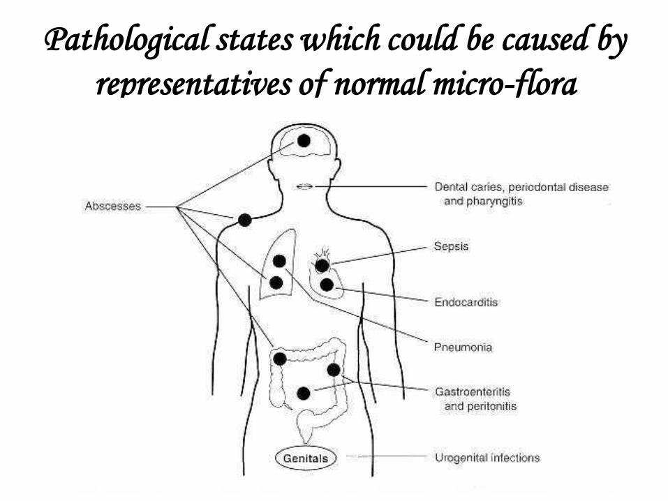
Pathological states which could be caused by
representatives of normal micro-flora
INFLUENCE OF
ECOLOGICAL FACTORS ON
MICROORGANISMS.
MICROBIAL
DECONTAMINATION.
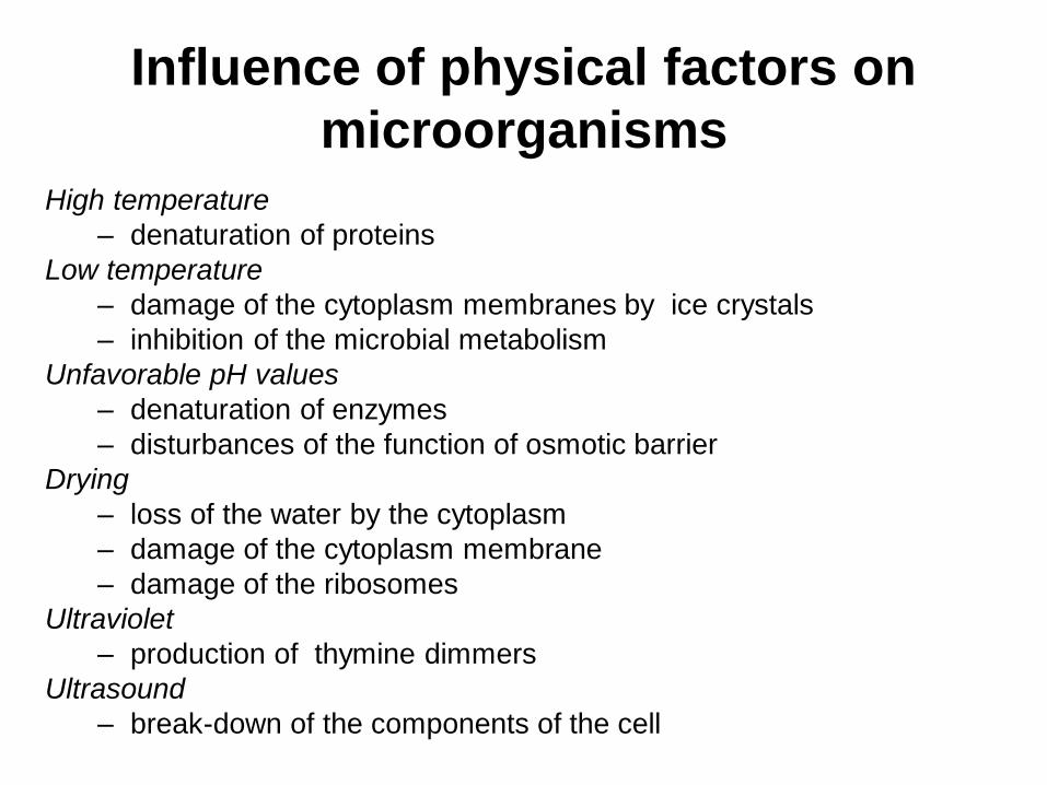
Influence of physical factors on
microorganisms
High temperature
– denaturation of proteins
Low temperature
– damage of the cytoplasm membranes by ice crystals
– inhibition of the microbial metabolism
Unfavorable pH values
– denaturation of enzymes
– disturbances of the function of osmotic barrier
Drying
– loss of the water by the cytoplasm
– damage of the cytoplasm membrane
– damage of the ribosomes
Ultraviolet
– production of thymine dimmers
Ultrasound
– break-down of the components of the cell
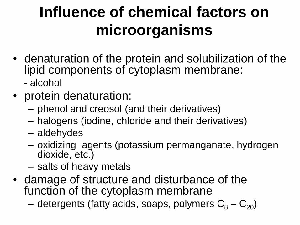
Influence of chemical factors on
microorganisms
• denaturation of the protein and solubilization of the lipid components of cytoplasm membrane:
- alcohol
• protein denaturation: – phenol and creosol (and their derivatives)
– halogens (iodine, chloride and their derivatives)
– aldehydes
– oxidizing agents (potassium permanganate, hydrogen dioxide, etc.)
– salts of heavy metals
• damage of structure and disturbance of the function of the cytoplasm membrane – detergents (fatty acids, soaps, polymers С8 – С20)
MICROBIAL DECONTAMINATION
Definition of the term
• complete or partial removal of
microorganisms from the unanimated
objects of surroundings or from the
human organism with use of the factors
causing direct damage to microorganisms.
Types of microbial
decontamination
Microbial decontamination includes:
• decontamination of the unanimated
objects of surroundings
- sterilization
– disinfection
• decontamination of the live organisms
– antisepsis
– chemotherapy
Sterilization
Definition of the term
• complete removal or killing of all
microorganisms
Sterilization: methods • Use of high temperatures (heat sterilization):
– autoclaving (sterilization by hot water steam with use
of high pressure which help to increase the
temperature of the steam up to 110 - 140С),
– dry heat sterilization - sterilization by hot air (the
temperature reaches 180С) for 1 hour in Pasteur
ovens (dry-heating chambers). Could be used for
sterilization of glass vials and metal equipment
– fractional sterilization by flowing steam (30 minutes
under the temperature of 100С, with several intervals
for one day to cool the material and to enable the
spores to germinate).
Sterilization: methods
• Chemical sterilization: use of formaldehyde, ethylene oxide, chloroform and other chemical substances.
• Sterilization with use of irradiation:
– -rays – usually applied at the factories for sterilization of medical equipment
– UV – applied in practical medicine.
• Filtration (mechanical sterilization) – use of bacterial filters, applied for the decontamination of the heat-labile solutions .
Disinfection of unanimated
environment
Elimination or reduction of the definite group
of pathogenic microorganisms - usually with
use of disinfectants (special chemical
substances).
Antisepsis
• Definition of the term: inhibition of the growth and propagation of microbes on the intact or injured skin and mucous membranes in the next cases: – treatment of the hands of surgeons,
– treatment of places of surgical intervention,
– treatment of wounds and mucous membranes.
In the case if patients have immune deficiency applied methods of antisepsis should give a higher degree of decontamination.
For this aim chemical substances called antiseptics which produce bacteriostatic (or microbiostatic) effect are usually used.
Asepsis
Definition of the term:
• creation of the zone free from any microorganisms (or the zone containing very low numbers of microorganisms) in the next places:
– patient's areas,
– rooms where medical manipulations are carrying out: operating theatre,
– clinical laboratories.

Asepsis
Methods:
• direct
– sterilisation
– disinfection
– antiseptics
• indirect
– separation
Basics of the Infection.
MICROBIOLOGICAL BASICS
OF CHEMOTHERAPY OF
BACTERIAL INFECTIONS
Theme N8
Basic terms and concepts of the
infection
• The infectious process (infection)
– Physiological and pathological reactions of macro-
organism (host), which are initiated and developed as a
result of its interactions with pathogenic microorganism
• Epidemiological process
– The processes of arising and spreading of specific
infectious states among the human population when
intensity of display of the symptoms is varying from
asymptomatic carriage of microbe to manifestation of the
symptoms of disease caused by the circulation of agent in
particular groups of a population
Basic terms and concepts of the
infection
Chain of Infection
The chain of infection includes the three
factors that lead to infection:
• the etiologic agent (microbe),
• the method of transmission,
• the host (susceptible human organism).
Basic terms and concepts of the
infection • The mechanism (method) of transmission of infection:
– is the means by which the infectious agent goes from the source of the infection (could be infected organism) to the host (susceptible organism).
• The stages of the mechanism of transmission of infection:
1. Release of the infectious agent from the host organism (infected) to surroundings
2. Presence of the infectious agent in the abiotic (unanimated) and biotic (animated) objects of surroundings
3. Infection of susceptible organism as a result of penetration of the infectious agent into the organism
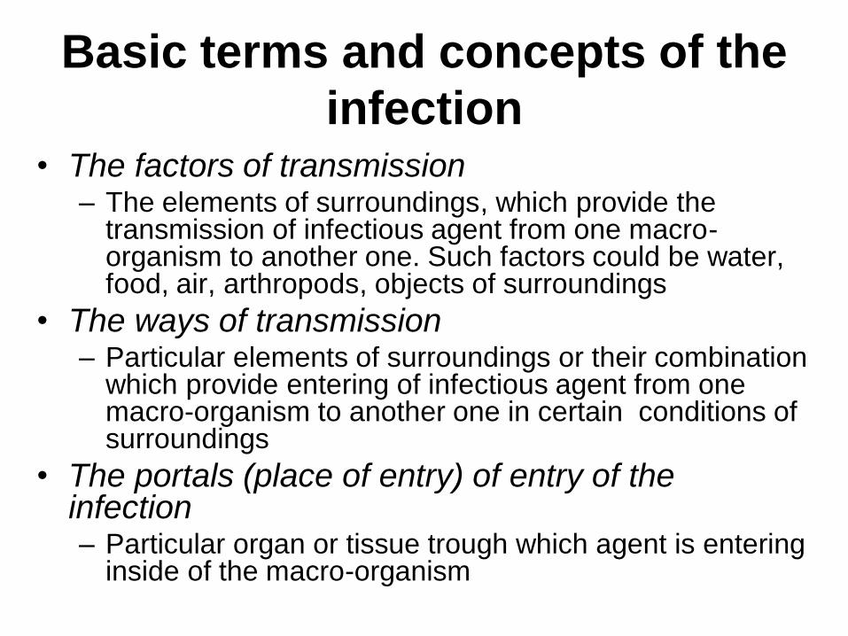
Basic terms and concepts of the
infection • The factors of transmission
– The elements of surroundings, which provide the transmission of infectious agent from one macro-organism to another one. Such factors could be water, food, air, arthropods, objects of surroundings
• The ways of transmission – Particular elements of surroundings or their combination
which provide entering of infectious agent from one macro-organism to another one in certain conditions of surroundings
• The portals (place of entry) of entry of the infection – Particular organ or tissue trough which agent is entering
inside of the macro-organism
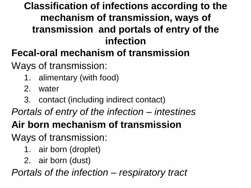
Classification of infections according to the
mechanism of transmission, ways of
transmission and portals of entry of the
infection
Fecal-oral mechanism of transmission
Ways of transmission: 1. alimentary (with food)
2. water
3. contact (including indirect contact)
Portals of entry of the infection – intestines
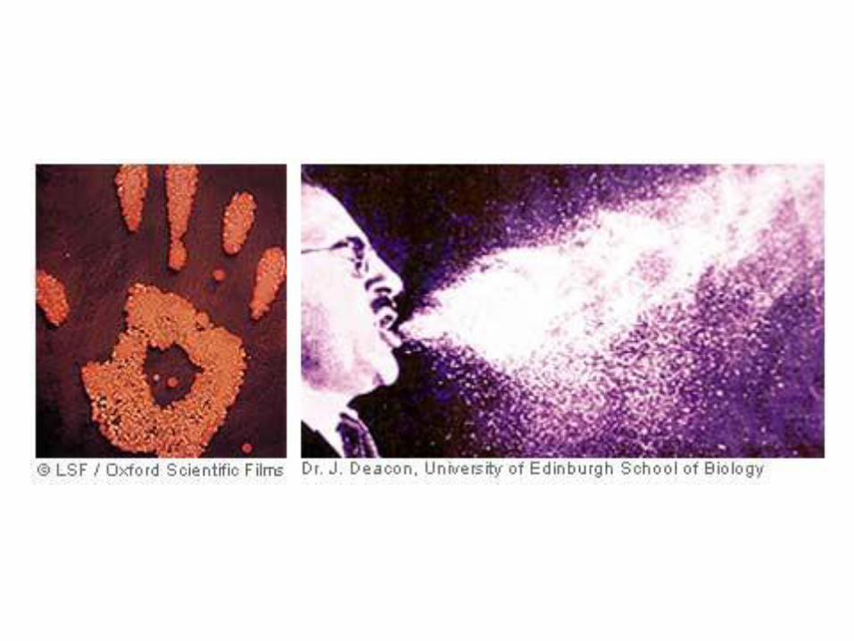
Air born mechanism of transmission
Ways of transmission: 1. air born (droplet)
2. air born (dust)
Portals of the infection – respiratory tract
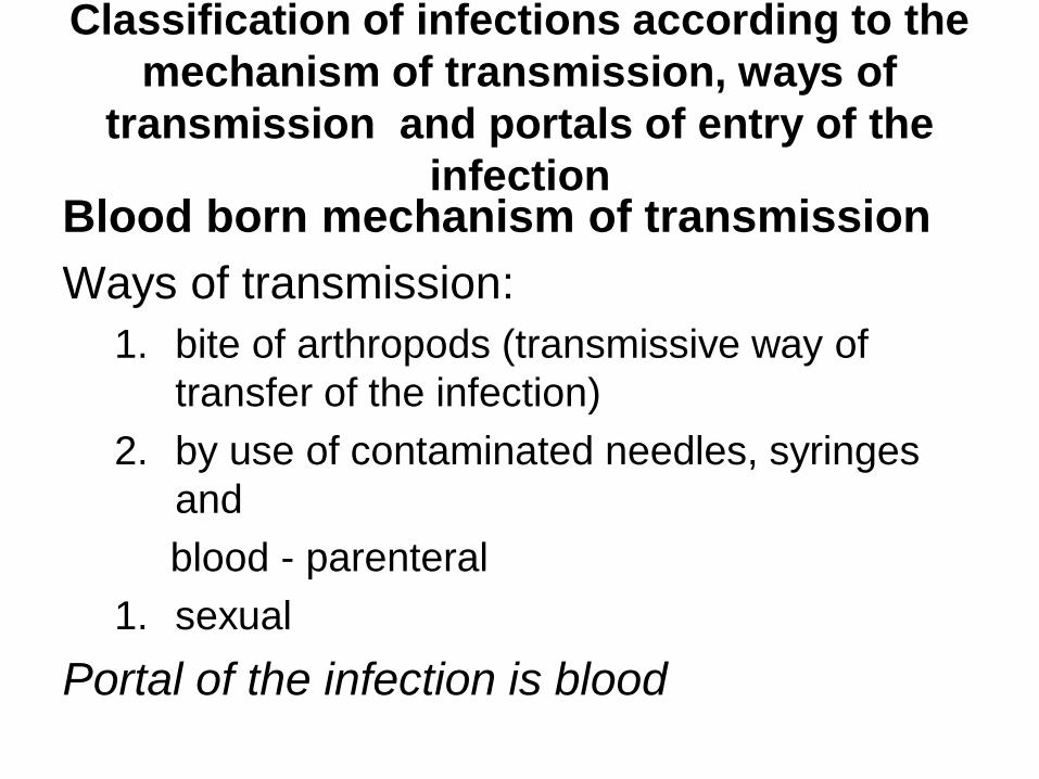
Classification of infections according to the
mechanism of transmission, ways of
transmission and portals of entry of the
infection Blood born mechanism of transmission
Ways of transmission:
1. bite of arthropods (transmissive way of
transfer of the infection)
2. by use of contaminated needles, syringes
and
blood - parenteral
1. sexual
Portal of the infection is blood
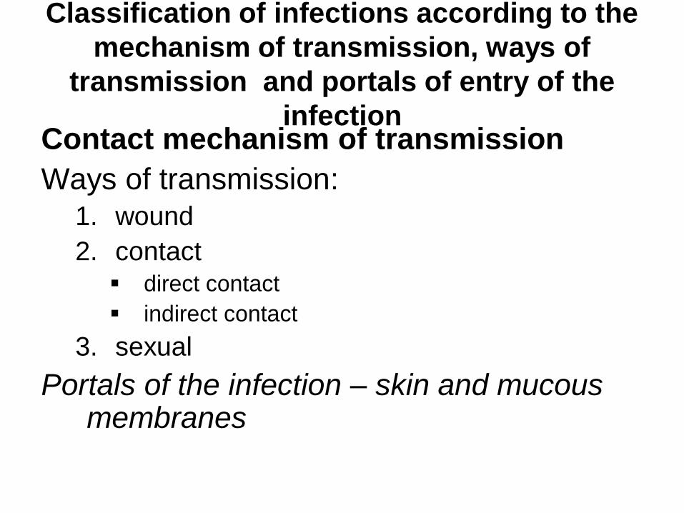
Classification of infections according to the
mechanism of transmission, ways of
transmission and portals of entry of the
infection Contact mechanism of transmission
Ways of transmission:
1. wound
2. contact direct contact
indirect contact
3. sexual
Portals of the infection – skin and mucous membranes
Classification of infections according to
the mechanism of transmission, ways of
transmission and portals of entry of the
infection
Vertical mechanism of transmission
Ways of transmission:
1. transplacental
Portals of the infection – tissues of embryo
Classification of infections
according to the nature of
infectious agent – bacterial
– viral
– fungal (mycosis)
– protozoan (invasion)
Classification of infections
according to their origin and ways of
spreading – exogenous - caused by microbes entering into
human organism from the outside
– endogenous - caused by indigenous (own)
micro-flora of human organism
• autoinfection (variant of endogenous infection – a
result of the infection which develops in human
organism when translocation of the microorganism
from normal (natural) biotope to atypical one takes
place
Classification of the infections according
to redevelopment of disease caused by
the same or different infectious agent – Secondary (the case of addition to the primary infection
another one caused by different microbe).
– Reinfection (the case of repeated infection caused by
the same microbe which occurs after recovery).
– Superinfection (the case of repeated infection caused
by the same microbe which occurs before recovery).
– Relapse (the case of return of clinical manifestations
without additional infection as a result of activation of
the infectious agents which survived in macro-
organism).
Classification of the infection
according to clinical manifestations
– Clinical infection (characterized by marked,
characteristic symptoms).
– Obliterated infection (characterized by feebly
marked symptoms).
– Atypical (characterized by symptoms which
are not typical for the disease).
– Latent or subclinical infection (characterized
by almost complete absence of symptoms).
Classification of the infection according
to the character of the spreading of
the infection and covered territory – Endemic (found only in definite geographic
areas)
– Sporadic (single cases of the disease which are not connected with each other)
– Epidemic (avalanche accumulation of the cases of the disease when all the cases are connected with each other)
– Pandemic (epidemic covering several countries, the whole continent sometimes all human population)
Characteristic properties of the
infectious disease
1. Specificity
2. Contagiousity
3. Recurrence
4. Ability to stimulate specific immune
response
PATHOGENICITY AND
VIRULENCE
Pathogenicity and virulence
• Pathogenicity – ability (inherited genetic
capacity) of microbe to cause infectious
process (disease) in sensitive macro-
organism (human or animal).
• Virulence – phenotypic manifestation of
pathogenicity – the degree in which
microorganism exhibits pathogenic
properties.
Characteristics of pathogenicity
• Potentiality
• Polydeterminancy – Synthesis of biologically active products by infectious
agent such as: • proteins
• carbohydrates
• lipids
– Ability of infectious agent to produce • toxins
• enzymes of invasion and aggression
• Specificity
• Virulence
Factors of virulence
• Adhesion – The ability of bacteria to attach to the specific receptors on the
surface of the cells of macro-organism
• Colonization – Propagation of bacteria on the surface of the cells of macro-
organism after their adhesion
• Penetration – Penetration of bacteria inside of the cells of macro-organism
• Invasion – Getting of bacteria into underlining tissues through mucous
membranes and connective tissues
• Aggression – Withstanding of bacteria against innate and specific immune
defence of macro-organism
Protein toxins: general
characteristics
Protein toxins are exotoxins - metabolites of
Gram-positive and Gram-negative bacteria:
• completely excreting by living bacterial
cells and found in fluid medium,
• partly excreting,
• non-excreting.
Protein toxins: properties 1. Polypeptides having MW 10,000 – 900,000.
2. Relatively unstable: loss toxicity by heat over 600C.
3. High toxic (poisonousness): fatal for laboratory animals in micrograms or less.
4. High immunogenicity - produce strong response of human immune system; stimulate the formation of antitoxin.
5. Specificity of their effect: antitoxin neutralizes toxin.
6. Possess ability to convert into anatoxins (toxoids) – toxin, which lost its poisonousness, but retained its immunogenicity (the property which is typical not for all toxins).
7. Do not produce fever in host.
Protein toxins: classification
• Neurotoxins
– Affect on the nervous system cells
• Enterotoxins
– Affect on the cells of digestive tract
• Cytotoxins
– Block protein synthesis at the subcellular level
• Hemolysins
– Increase permeability of the outer membrane of
erythrocytes resulting in their hemolysis
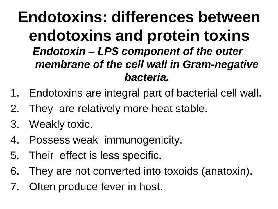
Endotoxins: differences between
endotoxins and protein toxins Endotoxin – LPS component of the outer
membrane of the cell wall in Gram-negative
bacteria.
1. Endotoxins are integral part of bacterial cell wall.
2. They are relatively more heat stable.
3. Weakly toxic.
4. Possess weak immunogenicity.
5. Their effect is less specific.
6. They are not converted into toxoids (anatoxin).
7. Often produce fever in host.
Chemotherapeutic agents:
definition of the term
Medical preparations (drugs) which
selectively inhibit growth of
microorganisms in human organism or kill
them
The basic characteristics of
therapeutic agents
1. Absence of appreciable toxic action on
human organism – selective toxicity
(toxic only for microorganisms).
2. Different antimicrobial spectrum.
3. Constant formation of drug-resistant
forms of microorganisms.
The most important groups of
chemotherapeutic agents and the
mechanism of their effect • Antibiotics
• Sulfanilamide preparations
– antimetabolites of folic acid interfering with its
synthesis
• Organic and inorganic compounds of metals,
sulfur, etc.
– inactivation of enzymes of microorganisms
• Preparations of compounds of nitrofuranum
– infringement of bioenergetic processes in bacterial
cell
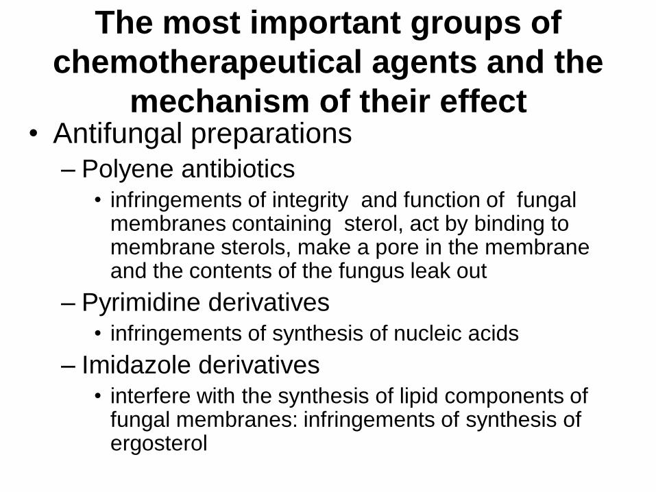
The most important groups of
chemotherapeutical agents and the
mechanism of their effect • Antifungal preparations
– Polyene antibiotics • infringements of integrity and function of fungal
membranes containing sterol, act by binding to membrane sterols, make a pore in the membrane and the contents of the fungus leak out
– Pyrimidine derivatives • infringements of synthesis of nucleic acids
– Imidazole derivatives • interfere with the synthesis of lipid components of
fungal membranes: infringements of synthesis of ergosterol
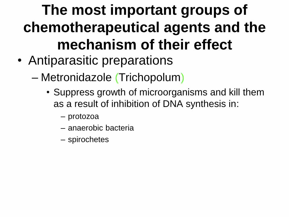
The most important groups of
chemotherapeutical agents and the
mechanism of their effect • Antiparasitic preparations
– Metronidazole (Trichopolum)
• Suppress growth of microorganisms and kill them
as a result of inhibition of DNA synthesis in:
– protozoa
– anaerobic bacteria
– spirochetes

ANTIBIOTICS
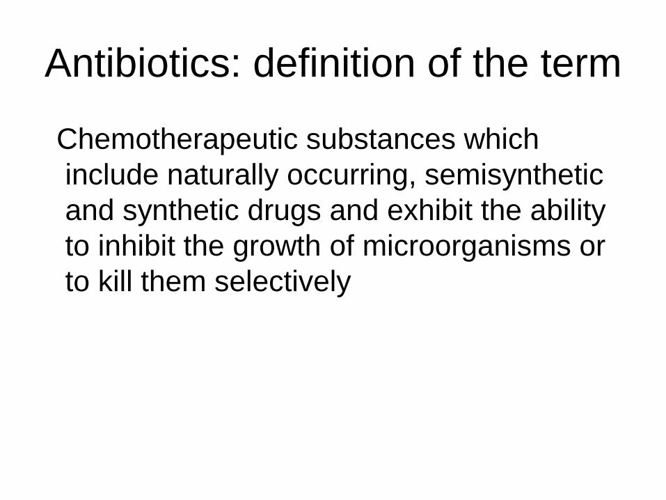
Antibiotics: definition of the term
Chemotherapeutic substances which
include naturally occurring, semisynthetic
and synthetic drugs and exhibit the ability
to inhibit the growth of microorganisms or
to kill them selectively
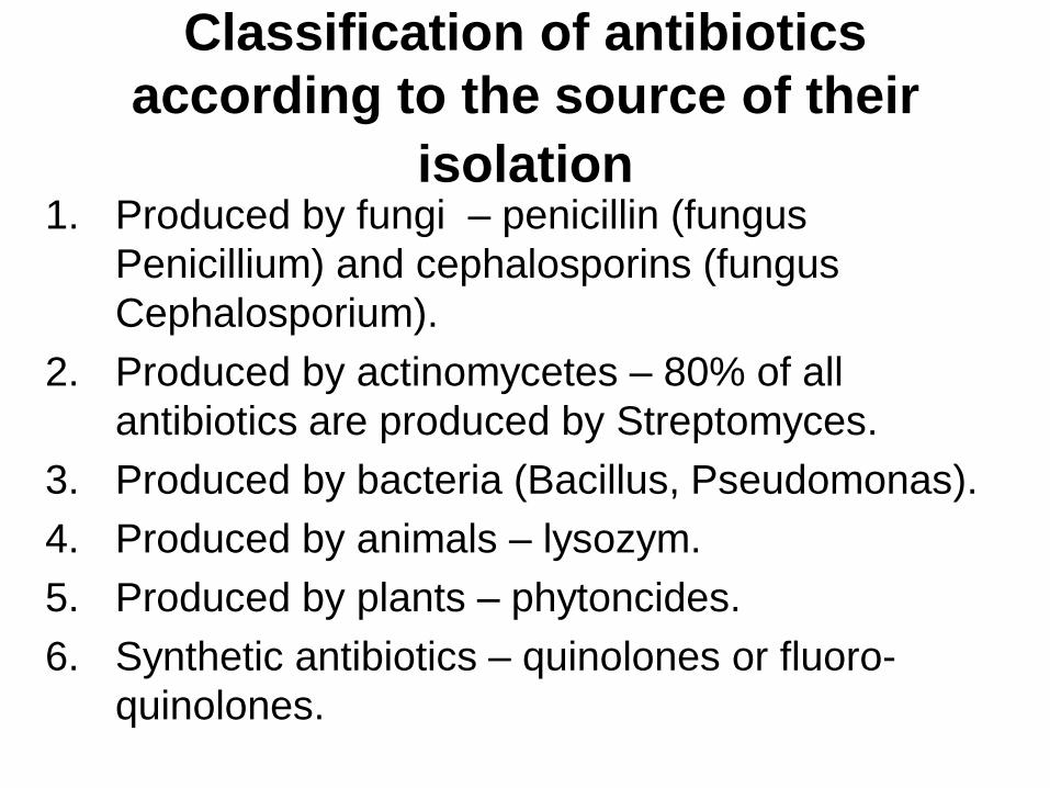
Classification of antibiotics
according to the source of their
isolation 1. Produced by fungi – penicillin (fungus
Penicillium) and cephalosporins (fungus
Cephalosporium).
2. Produced by actinomycetes – 80% of all
antibiotics are produced by Streptomyces.
3. Produced by bacteria (Bacillus, Pseudomonas).
4. Produced by animals – lysozym.
5. Produced by plants – phytoncides.
6. Synthetic antibiotics – quinolones or fluoro-
quinolones.
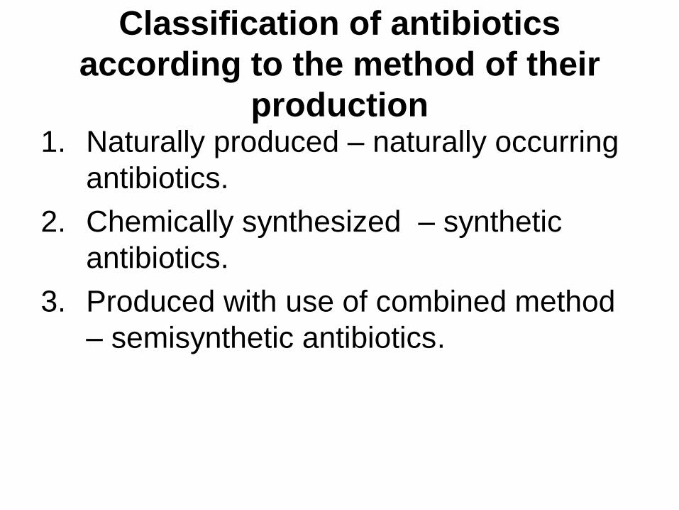
Classification of antibiotics
according to the method of their
production 1. Naturally produced – naturally occurring
antibiotics.
2. Chemically synthesized – synthetic
antibiotics.
3. Produced with use of combined method
– semisynthetic antibiotics.
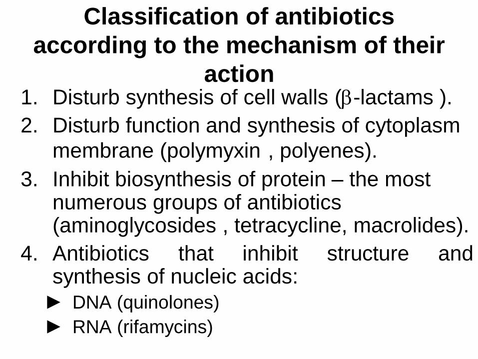
Classification of antibiotics
according to the mechanism of their
action 1. Disturb synthesis of cell walls (-lactams ).
2. Disturb function and synthesis of cytoplasm
membrane (polymyxin , polyenes).
3. Inhibit biosynthesis of protein – the most numerous groups of antibiotics (aminoglycosides , tetracycline, macrolides).
4. Antibiotics that inhibit structure and synthesis of nucleic acids:
► DNA (quinolones)
► RNA (rifamycins)
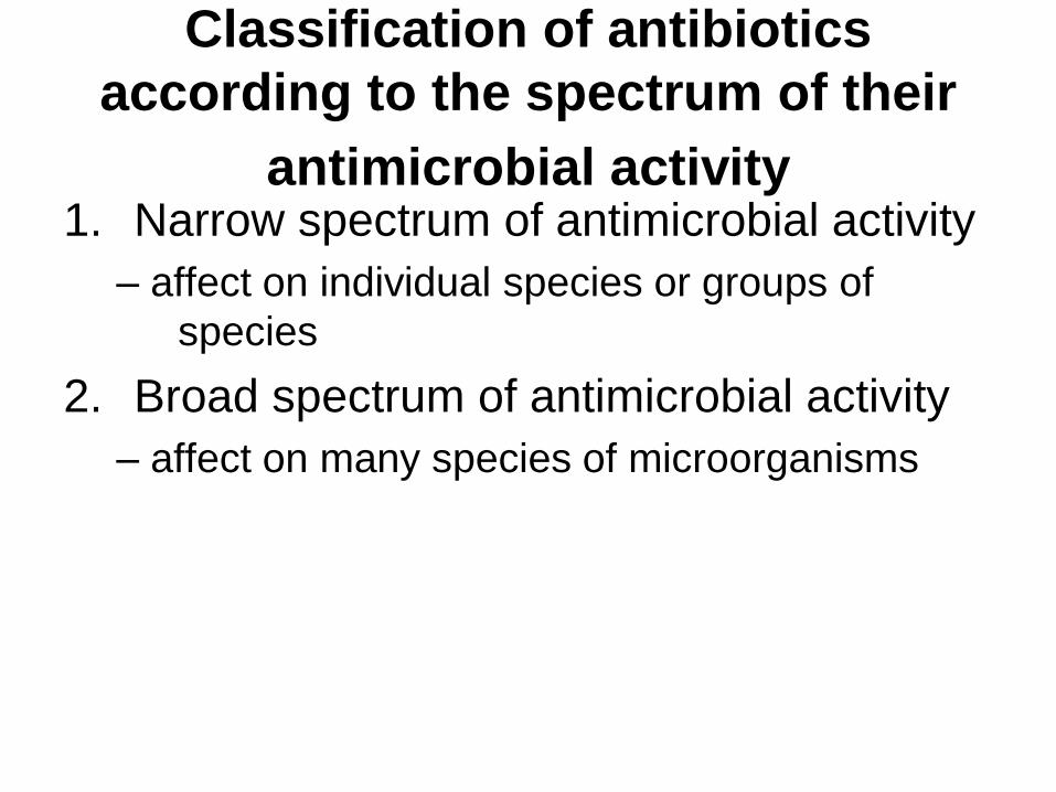
Classification of antibiotics
according to the spectrum of their
antimicrobial activity 1. Narrow spectrum of antimicrobial activity
– affect on individual species or groups of
species
2. Broad spectrum of antimicrobial activity
– affect on many species of microorganisms
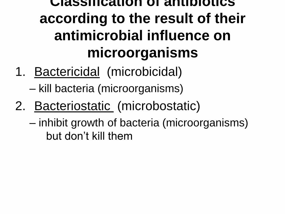
Classification of antibiotics
according to the result of their
antimicrobial influence on
microorganisms
1. Bactericidal (microbicidal)
– kill bacteria (microorganisms)
2. Bacteriostatic (microbostatic)
– inhibit growth of bacteria (microorganisms)
but don’t kill them
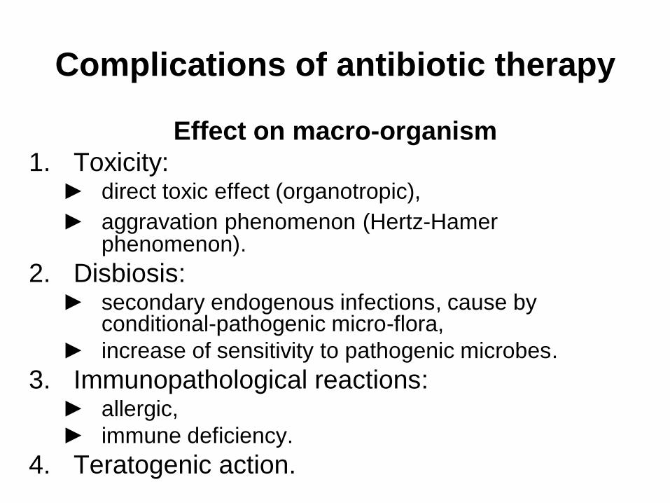
Complications of antibiotic therapy
Effect on macro-organism
1. Toxicity: ► direct toxic effect (organotropic),
► aggravation phenomenon (Hertz-Hamer phenomenon).
2. Disbiosis: ► secondary endogenous infections, cause by
conditional-pathogenic micro-flora,
► increase of sensitivity to pathogenic microbes.
3. Immunopathological reactions: ► allergic,
► immune deficiency.
4. Teratogenic action.
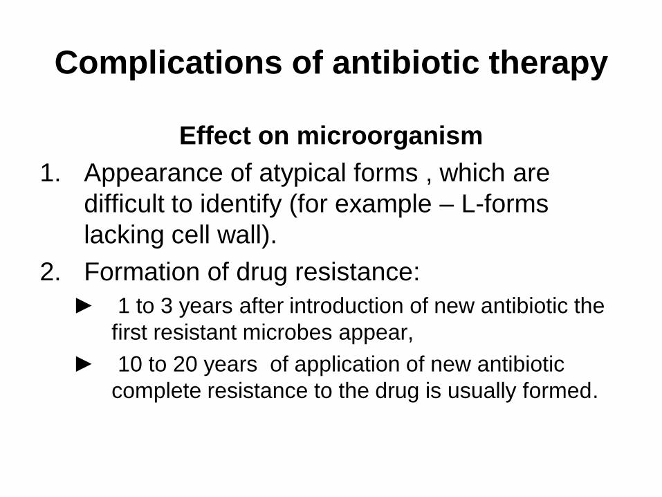
Complications of antibiotic therapy
Effect on microorganism
1. Appearance of atypical forms , which are
difficult to identify (for example – L-forms
lacking cell wall).
2. Formation of drug resistance:
► 1 to 3 years after introduction of new antibiotic the
first resistant microbes appear,
► 10 to 20 years of application of new antibiotic
complete resistance to the drug is usually formed.
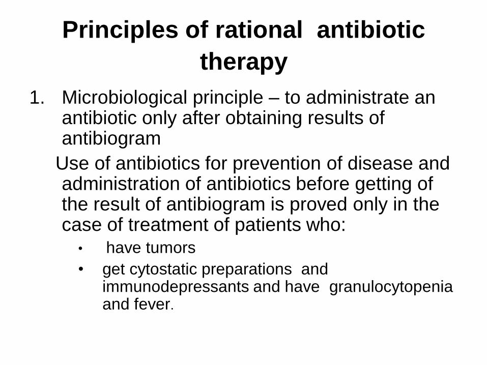
Principles of rational antibiotic
therapy
1. Microbiological principle – to administrate an antibiotic only after obtaining results of antibiogram
Use of antibiotics for prevention of disease and administration of antibiotics before getting of the result of antibiogram is proved only in the case of treatment of patients who:
• have tumors
• get cytostatic preparations and immunodepressants and have granulocytopenia and fever.
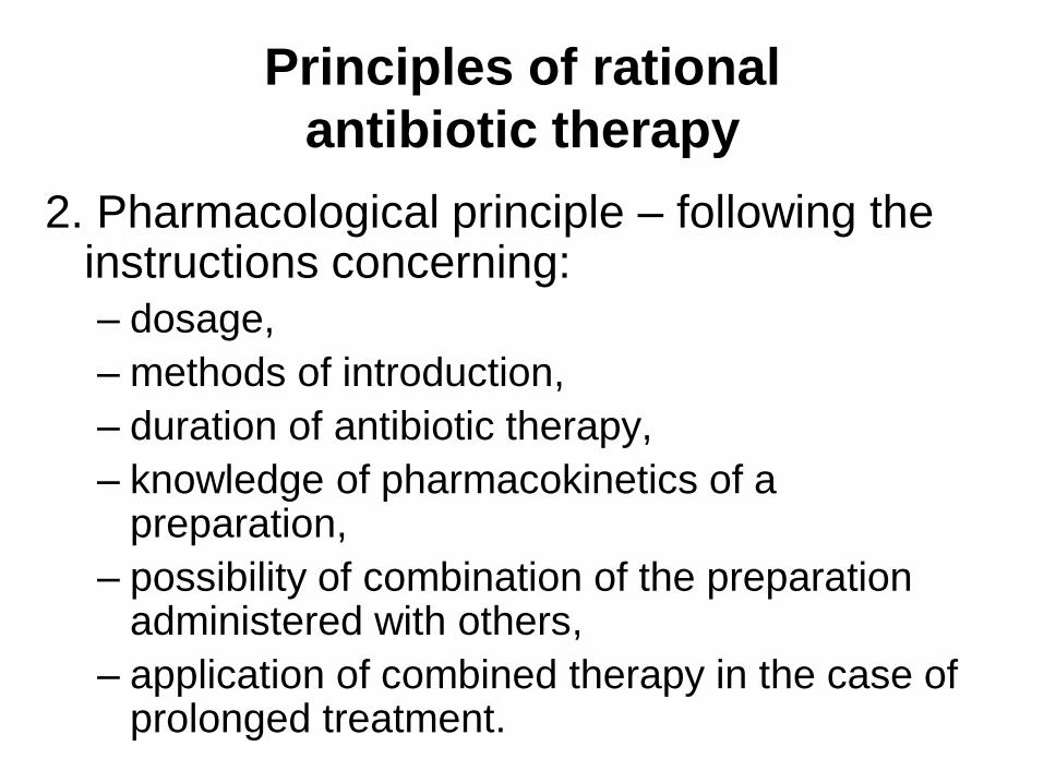
Principles of rational
antibiotic therapy
2. Pharmacological principle – following the instructions concerning:
– dosage,
– methods of introduction,
– duration of antibiotic therapy,
– knowledge of pharmacokinetics of a preparation,
– possibility of combination of the preparation administered with others,
– application of combined therapy in the case of prolonged treatment.
Principles of rational
antibiotic therapy
3. Clinical principle – administration of
antibiotics only when it is very necessary
to improve the condition of the patient.
Principles of rational
antibiotic therapy
4. Epidemiological principle – to take into
account resistance of microorganisms to
antimicrobial agents in:
– given branch of hospital,
– whole hospital,
– geographical region.
Principles of rational
antibiotic therapy
5. Pharmaceutical principle – to take into
account period of validity and instructions
for storage of a drug.
The rules of preference and limitations in use of antibiotics in
clinic ► administration of antibiotic is strongly recommended:
- infections caused by streptococci (sore throat, scarlet fever, etc)
►administration of antibiotic is expedient:
- acute respiratory infections complicated by pneumonia, sinusitis, etc
- acute intestinal infections in the case when the faeces contain blood (resembling dysentery)
►administration of antibiotic is strongly prohibited in the case of:
- acute respiratory infections
- acute intestinal with diarrhoea when the infectious agent is not identified, especially in children of any age
- fevers , leucocytosis when bacterial infections are not proved
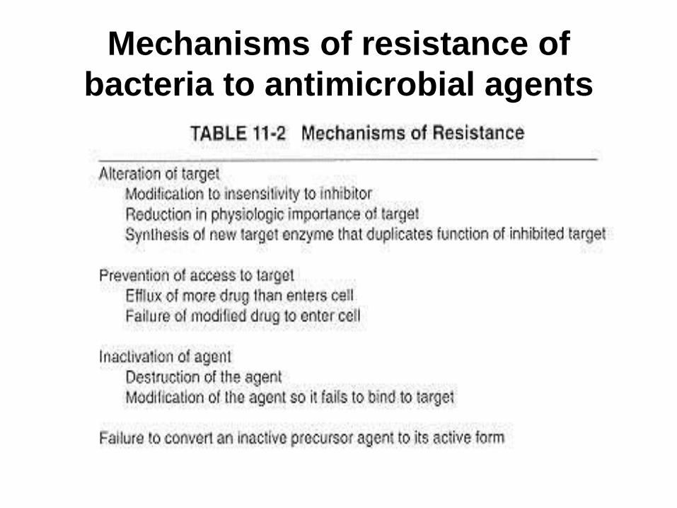
Mechanisms of resistance of
bacteria to antimicrobial agents
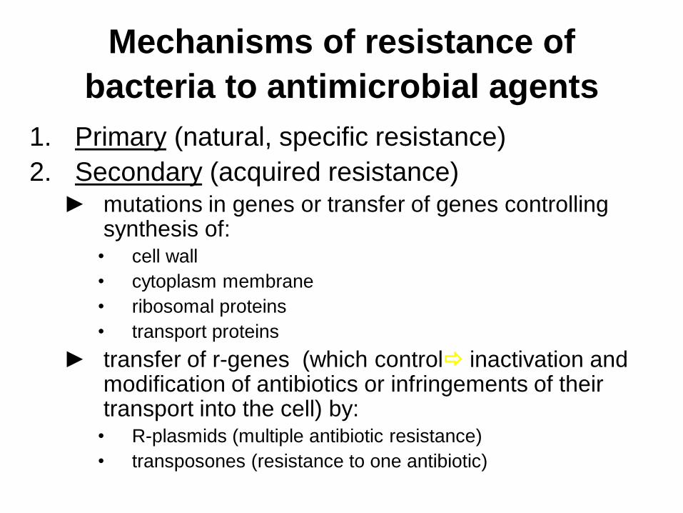
Mechanisms of resistance of
bacteria to antimicrobial agents
1. Primary (natural, specific resistance)
2. Secondary (acquired resistance) ► mutations in genes or transfer of genes controlling
synthesis of: • cell wall
• cytoplasm membrane
• ribosomal proteins
• transport proteins
► transfer of r-genes (which control inactivation and modification of antibiotics or infringements of their transport into the cell) by:
• R-plasmids (multiple antibiotic resistance)
• transposones (resistance to one antibiotic)
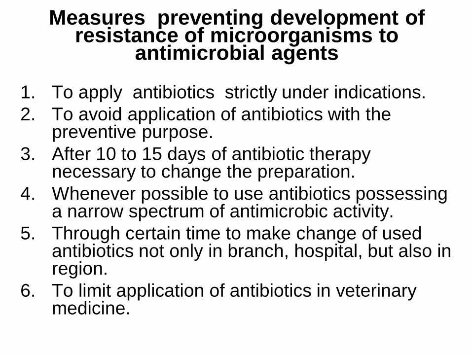
Measures preventing development of resistance of microorganisms to
antimicrobial agents
1. To apply antibiotics strictly under indications.
2. To avoid application of antibiotics with the preventive purpose.
3. After 10 to 15 days of antibiotic therapy necessary to change the preparation.
4. Whenever possible to use antibiotics possessing a narrow spectrum of antimicrobic activity.
5. Through certain time to make change of used antibiotics not only in branch, hospital, but also in region.
6. To limit application of antibiotics in veterinary medicine.
Antimicrobial susceptibility tests
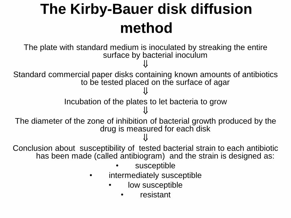
The Kirby-Bauer disk diffusion
method The plate with standard medium is inoculated by streaking the entire
surface by bacterial inoculum
Standard commercial paper disks containing known amounts of antibiotics to be tested placed on the surface of agar
Incubation of the plates to let bacteria to grow
The diameter of the zone of inhibition of bacterial growth produced by the drug is measured for each disk
Conclusion about susceptibility of tested bacterial strain to each antibiotic has been made (called antibiogram) and the strain is designed as:
• susceptible
• intermediately susceptible
• low susceptible
• resistant
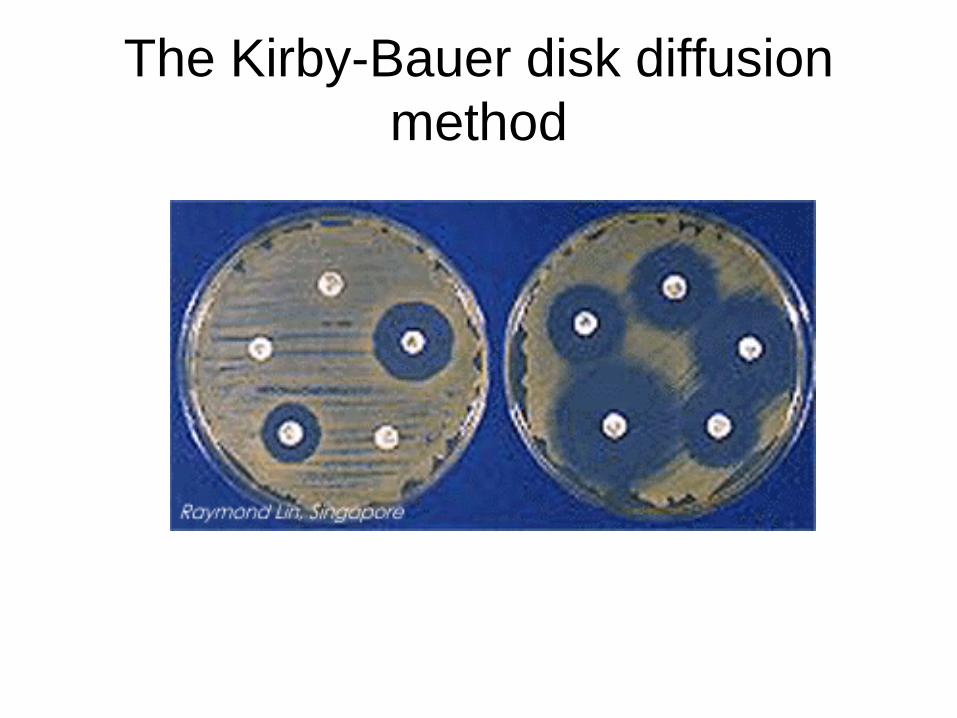
The Kirby-Bauer disk diffusion
method
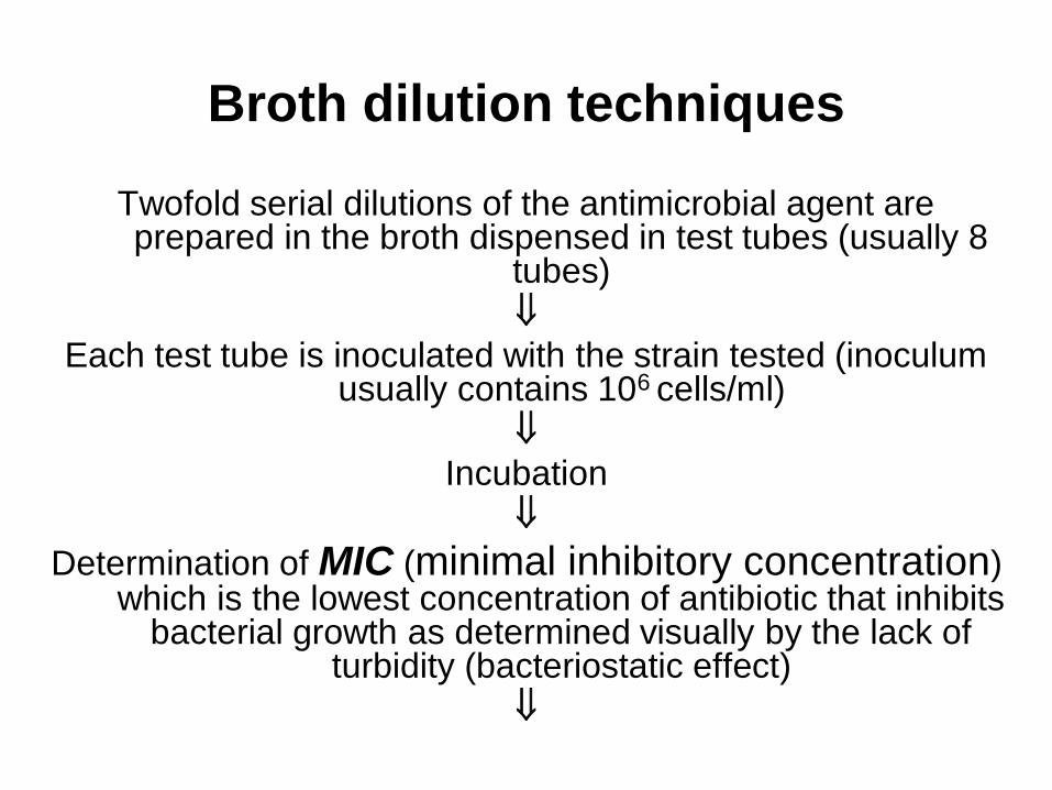
Broth dilution techniques
Twofold serial dilutions of the antimicrobial agent are prepared in the broth dispensed in test tubes (usually 8
tubes)
Each test tube is inoculated with the strain tested (inoculum usually contains 106 cells/ml)
Incubation
Determination of MIC (minimal inhibitory concentration) which is the lowest concentration of antibiotic that inhibits
bacterial growth as determined visually by the lack of turbidity (bacteriostatic effect)
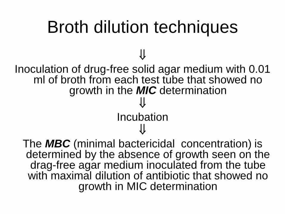
Broth dilution techniques
Inoculation of drug-free solid agar medium with 0.01 ml of broth from each test tube that showed no
growth in the MIC determination
Incubation
The MBC (minimal bactericidal concentration) is determined by the absence of growth seen on the drag-free agar medium inoculated from the tube with maximal dilution of antibiotic that showed no
growth in MIC determination