applied and environmental microbiology applied and environmental microbiology applied and...
TRANSCRIPT
++++
Applied and Environmental MicrobiologyApplied and Environmental MicrobiologyApplied and Environmental MicrobiologyApplied and Environmental Microbiologyaem.asm.org
doi: doi: doi: doi: 10.1128/AEM.70.4.2279-2288.2004 10.1128/AEM.70.4.2279-2288.2004 10.1128/AEM.70.4.2279-2288.2004 10.1128/AEM.70.4.2279-2288.2004
Appl. Environ. Microbiol. April 2004 April 2004 April 2004 April 2004 vol. 70 no. 4 2279-2288 2279-2288 2279-2288 2279-2288
Characterization of Azo Reduction Activity in a Novel
Ascomycete Yeast Strain
Patrícia A. RamalhoPatrícia A. RamalhoPatrícia A. RamalhoPatrícia A. Ramalho1111, , , , M. Helena CardosoM. Helena CardosoM. Helena CardosoM. Helena Cardoso1111,,,,****, , , , A. Cavaco-PauloA. Cavaco-PauloA. Cavaco-PauloA. Cavaco-Paulo2222 and and and and
M. Teresa RamalhoM. Teresa RamalhoM. Teresa RamalhoM. Teresa Ramalho3333
Author Affiliations
ABSTRACT
Several model azo dyes are reductively cleaved by growing cultures of an
ascomycete yeast species, Issatchenkia occidentalis. In liquid media containing 0.2
mM dye and 2% glucose in a mineral salts base, more than 80% of the dyes are
removed in 15 h, essentially under microaerophilic conditions. Under anoxic
conditions, decolorization does not occur, even in the presence of pregrown cells.
Kinetic assays of azo reduction activities in quasi-resting cells demonstrated the
following: (i) while the optimum pH depends on dye structure, the optimum pH
range was observed in the acidic range; (ii) the maximum decolorizing activity
occurs in the late exponential phase; and (iii) the temperature profile approaches
the typical bell-shaped curve. These results indirectly suggest the involvement of
an enzyme activity in azo dye reduction. The decolorizing activity of I. occidentalis
is still observed, although at a lower level, when the cells switch to aerobic
respiration at the expense of ethanol after glucose exhaustion in the culture
medium. Decolorization ceased when all the ethanol was consumed; this
observation, along with other lines of evidence, suggests that azo dye reduction
depends on cell growth. Anthraquinone-2-sulfonate, a redox mediator, enhances
the reduction rates of the N,N-dimethylaniline-based dyes and reduces those of
the 2-naphthol-based dyes, an effect which seems to be compatible with a
thermodynamic factor. The dye reduction products were tested as carbon and
nitrogen sources. 1-Amino-2-naphthol was used as a carbon and nitrogen source,
and N,N-dimethyl-p-phenylenediamine was used only as a nitrogen source.
Sulfanilic and metanilic acids did not support growth either as a carbon or nitrogen
source.
Over the last two decades, considerable work has been done with the goal of using
microorganisms as bioremediation agents in the treatment of wastewater
containing textile dyes. These contaminants contribute a minor fraction to the
usually high load of dissolved organic matter in textile effluents (35353535), but they are
highly visible and must be removed in order to comply with the regulations
concerning effluent discharge.
Azo aromatic dyes are the major group of textile dyestuffs. These structures can
be reductively cleaved into colorless amines by several bacterial species (for
reviews, see references 2222, 9999, 41414141, and 43434343); nevertheless, azo dye reduction
occurring in the presence of living matter can be an abiotic process. An example of
this is the reduction of acid orange 7 (called dye IV in the present study) and
reactive red 2 in anaerobic sludge, where sulfide, produced by sulfate-reducing
microorganisms, can reduce azo bonds (45454545). In most of the reported processes of
azo dye bioreduction, however, the participation of an enzymatic activity is
assumed. Since the products of azo dye reduction, with few exceptions (4444, 11111111, 26262626),
cannot be used by bacteria as carbon and energy sources, the cleavage of azo
bonds is a gratuitous process which can occur when the microorganisms use a
reduced carbon compound as the growth substrate.
Azo dye reduction occurring in the presence of living matter poses additional
problems. Sulfonic azo dyes are impermeant to the cell membranes. NAD(P)H is
also impermeant to the cell membranes and is believed to be the primary source of
electrons. This is why reduction of highly polar azo dyes is usually postulated to
take place outside the cell (43434343). This fact would be compatible either with an
abiotic reduction mediated by some extracellular reductant species (45454545) or with
the involvement of an externally directed reductase activity in the plasma
membrane that was capable of transferring reducing equivalents to acceptor
species in the extracellular medium. So far, several bacterial cytoplasmic
azoreductases have been isolated and characterized (18181818, 33333333, 37373737, 38383838). However, as
shown by two recent studies (5555, 42424242), the putative azoreductases had, in fact,
insignificant in vivo activity.
Independently of the intracellular location of the azoreductase, theoretically, a
redox mediator could facilitate the transfer of reducing equivalents from
intracellular NAD(P)H to the substrate dye. This hypothesis was proposed as early
as 1975 in a study describing the decolorization of azo food dyes by Proteus
vulgaris (15151515). The participation of redox mediators in the decolorization of azo
dyes by bacterial species is now generally accepted, and several mediators (usually
Página 1 de 12Characterization of Azo Reduction Activity in a Novel Ascomycete Yeast Strain
30-01-2015http://aem.asm.org/content/70/4/2279.full
View larger version:
In this pageIn this pageIn this pageIn this page In a new windowIn a new windowIn a new windowIn a new window
Download as PowerPoint SlideDownload as PowerPoint SlideDownload as PowerPoint SlideDownload as PowerPoint Slide
quinonoid compounds) have been described as effectively enhancing
decolorization processes (7777, 22222222, 23232323, 40404040). The redox mediators studied are usually
exogenous compounds added to the culture medium, but in a recent study (22222222),
Keck and coworkers observed that aerobic preincubation of a Sphingomonas sp.
strain with 2-naphthalenesulfonate strongly stimulated the subsequent anaerobic
reduction of azo dyes by the same strain. In a later study (23232323), Keck et al.
identified 4-amino-1,2-naphthoquinone and
4-ethanolamino-1,2-naphthoquinone, formed by spontaneous oxidation of
1,2-dihydroxynaphthalene (an intermediate of the 2-naphthalenesulfonate
degradation), as putative redox mediators in the decolorization reaction. Another
study, observing an autocatalytic effect in the anaerobic reduction of acid orange
7, demonstrated that 1-amino-2-naphthol, one of the dye reduction products,
stimulated decolorization (46464646). The presence of redox mediators in humic
substances naturally found in soils has also been reported to enhance
decolorization and other redox processes (10101010, 16161616, 23232323).
Azo dye reduction occurs preferentially under anoxic or oxygen-limited conditions
(6666, 20202020, 24242424, 28282828, 32323232, 34343434, 40404040, 48484848). Under these circumstances, azo dyes act as
terminal electron acceptors during microbial respiration. Aerobic azo dye
reduction processes in the presence of additional carbon sources were recently
questioned by Stolz (43434343), since the experimental conditions of decolorization have
not unequivocally shown the presence of oxygen in the culture medium, being
more consistent, in fact, with oxygen-limited conditions.
The few reports on bioremediation of colored effluents by yeasts usually mention
biosorption as the major cause for decolorization (14141414, 31313131). Martins et al., however,
isolated a strain of Candida zeylanoides, which efficiently decolorizes several azo
dyes (30303030). Further work demonstrated that a reductive cleavage of the azo bond
was involved in this process and described some characteristics of the
corresponding dye reducing activity (39393939). In this work, we examined the
decolorizing activity of a novel yeast strain, Issatchenkia occidentalis. This species
is even more efficient than C. zeylanoides in decolorizing the previously tested
dyes. Therefore, we decided to investigate, in more detail, the effects of several
parameters on the performance of I. occidentalis as an azo dye reducer. The
evidence obtained from this study is expected to provide a sound basis for the
development of a biotreatment process for azo dye-containing wastewaters.
MATERIALS AND METHODS
Chemicals and culture medium components.Chemicals and culture medium components.Chemicals and culture medium components.Chemicals and culture medium components. Peptone, yeast extract, yeast
nitrogen base (YNB), and yeast carbon base (YCB) were obtained from Difco. Other
chemicals were commercially available, analytical grade reagents.
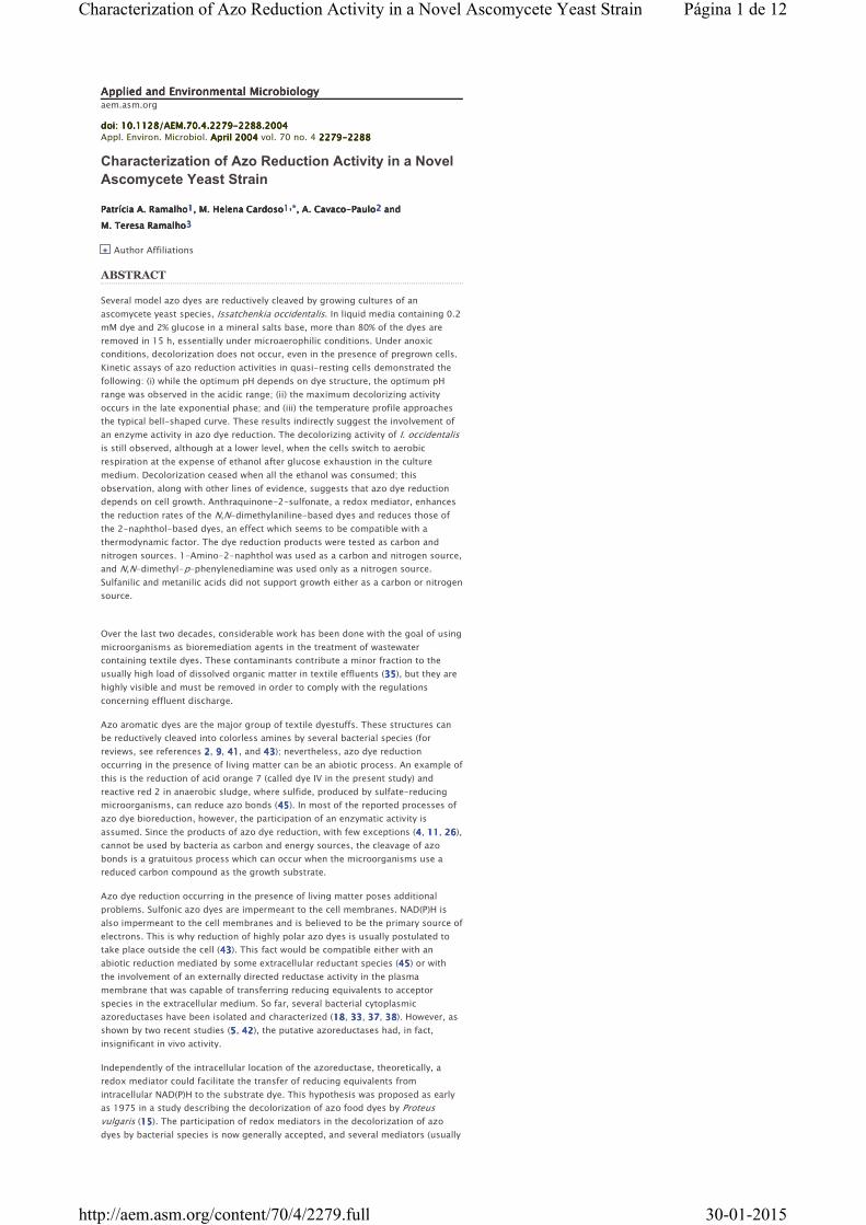
Dyes.Dyes.Dyes.Dyes. The structures of the dyes tested in the present work are depicted in Fig. 1111.
Dye II (methyl orange, CI 13025) and dye IV (orange II, CI 15510), both ca. 85% dye
content, were purchased from Sigma-Aldrich and used without further purification.
The other two dyes (minimum 90% dye content) were synthesized by the
conventional method of coupling the diazonium salt of metanilic acid with either
N ,N -dimethyl-p -phenylenediamine or 1-amino-2-naphthol (17171717). The structures
of the isolated dyes, as sodium salts, were confirmed by 1H nuclear magnetic
resonance spectroscopy in dimethyl sulfoxide.
FIG. 1.FIG. 1.FIG. 1.FIG. 1.
Structures of the azo dyes used
in this work:
m -[(4-dimethylamino)
phenylazo]benzenesulfonic acid
(dye I), p -[(4-dimethylamino)
phenylazo] benzenesulfonic
acid (CI 13025 methyl orange
or orange 52) (dye II),
m -[(2-hydroxy-1-napthyl)azo]
benzenesulfonic acid (dye III), and p -[(2-hydroxy-1-napthyl) azo]
benzenesulfonic acid (CI 15510 orange II or acid orange 7) (dye IV).
Microorganism and maintenance conditions.Microorganism and maintenance conditions.Microorganism and maintenance conditions.Microorganism and maintenance conditions. The ascomycete yeast I.
occidentalis (PYCC 5770) (deposited in the Portuguese Yeast Culture Collection)
was isolated on the basis of its capacity to decolorize YEPD (yeast extract-
peptone-dextrose) agar plates containing 0.5% [wt/vol] yeast extract, 1% [wt/vol]
peptone, 2% [wt/vol] glucose and the azo dye orange II, as described in a previous
publication (30303030). The identification of the yeast strain followed the usual methods
described for yeast taxonomy (44444444): testing of defined carbon compounds as
fermentation substrates or as the sole carbon and energy sources, assimilation
tests of nitrogen compounds, temperature tolerance for growth (30, 35, and 40°C),
growth in the presence of 0.01 and 0.1% (wt/vol) cycloheximide, growth in the
presence of 50 and 60% (wt/vol) D-glucose, and hydrolysis of urea. Morphological
characteristics were also considered, i.e., vegetative reproduction only by budding,
formation of pseudohyphae, and formation of persistent asci containing round
Página 2 de 12Characterization of Azo Reduction Activity in a Novel Ascomycete Yeast Strain
30-01-2015http://aem.asm.org/content/70/4/2279.full
ascospores on yeast malt agar. A commercial software (3333) was used as an aid for
species identification. The strain was routinely maintained on slants of YEPD agar.
Analytical methods.Analytical methods.Analytical methods.Analytical methods. Cell growth in liquid medium was monitored by attenuance
measurements (47474747) at 640 nm (D 640). Blanks for these readings were prepared
from aliquots of centrifuged medium. At high cell densities, both sample and
blank were diluted with distilled water by the same factor. A linear correlation
between attenuance readings (D 640) and cell weight (dry weight) was established
by the standard gravimetric method (cell weight [dry weight] in grams · liter−1
) =
1.1289(D 640 − 0.0487); r2 = 0.996]. Dye concentration was monitored by
absorbance readings of aliquots of centrifuged medium at the dye λmax. The assay
cuvette contained 0.3 ml of 1 M acetate buffer (pH 4.0), sample, and water to a
volume of 3.0 ml; the blank was prepared with the same dilution of buffer in
distilled water. Dissolved oxygen was measured as partial oxygen pressure using a
Clark-type polarographic electrode and an ATI RUSSEL model RL 400 instrument
according to the manufacturer's instructions (detection level, 0.1 mg · liter−1
).
Metanilic and sulfanilic acids, formed by reduction of dye I or III and dye II or IV,
respectively, were detected and quantified by high-pressure liquid
chromatography (HPLC) analysis, using a reversed-phase column (RP-18) and an
eluent containing the ion-pair reagent tetrabutylammonium phosphate (TBAP), as
previously described (39393939). Under these conditions, metanilic and sulfanilic acids
had retention times of 10.0 ± 0.5 and 12.4 ± 0.5 min, respectively. Glucose and
ethanol concentrations in the growth media were also determined by HPLC
(retention times of 11.1 ± 0.2 and 24.5 ± 0.2 min, respectively), using a Polyspher
OAKC (Merck) column, refractive index detection, and arabinose as the internal
standard (27272727).
Decolorization in liquid media.Decolorization in liquid media.Decolorization in liquid media.Decolorization in liquid media. Decolorization experiments by growing cultures
of I. occidentalis were typically performed in 250-ml cotton-plugged Erlenmeyer
flasks with 100 ml of sterile medium (normal decolorization medium [NDM])
containing 2% glucose as a carbon and energy source and the test dye (0.2 mmol ·
liter−1
) in a mineral salts base, as previously described (39393939). The cultures were
inoculated at a low cell density with a cell suspension prepared from a freshly
grown slant. The flasks were incubated under orbital shaking (120 rpm) at 26°C,
except where stated otherwise. Biomass and concentrations of dye, sulfanilic or
metanilic acid, glucose, and ethanol were measured as described above, in
samples of the incubation mixture collected at defined times. Dissolved oxygen
was measured directly in the incubation media.
When testing the effect of adaptation of the strain to the dyes on the
decolorization process, the cells were pregrown in NDM with or without dye, until
the mid-exponential growth phase was reached. The cells were then harvested by
centrifugation at 5,000 rpm in an Eppendorf Centrifuge 584OR, washed twice in
cold sterile distilled water, and used to inoculate fresh sterile NDM containing 0.2
mmol of dye · liter−1
. The cultures were started at high cell densities (1.4 D units;
1.6 g [dry wt] of cells · liter−1
). In control experiments, the inocula consisted of
unadapted cells.
Assimilation of azo dye reduction products.Assimilation of azo dye reduction products.Assimilation of azo dye reduction products.Assimilation of azo dye reduction products. Minimal media with all essential
nutrients and vitamins except a carbon source (YNB) or a nitrogen source (YCB)
were prepared according to the manufacturer's instructions and supplemented
with 0.5 mM metanilic or sulfanilic acid. Appropriate volumes of these media (100
ml) in 250-ml Erlenmeyer flasks were inoculated with freshly grown cells and
incubated at 26°C and 120 rpm. Cell growth was monitored as described above.
Control experiments were performed with 0.5 mM ammonium sulfate as the sole
nitrogen source in YCB or with 0.5 mM glucose as the sole carbon and energy
source in YNB. Using the same method, 1-amino-2-naphthol and
N ,N -dimethyl-p -phenylenediamine, both 0.5 mM, were tested as nitrogen
sources. The initial pH was adjusted to 5.2 ± 0.2.
Activity assays with resting cells.Activity assays with resting cells.Activity assays with resting cells.Activity assays with resting cells. The decolorization activities of nongrowing cells
were determined at various pH values, with cells harvested in different growth
stages and with or without redox by using the redox mediator anthraquinone-2-
sulfonic acid (AQS). To investigate the effect of pH or temperature on
decolorization activity, the cells were grown for ca. 11 h on NDM, harvested by
centrifugation, washed, and resuspended in 0.05 M phosphate buffer solution of
the required pH. The assay mixture contained, in a total volume of 20 ml, 10.4 mM
glucose and 0.047 mM dye; 0.1 mM AQS was included in activity assays of dyes I
and II and omitted in the assays of dyes III and IV. The assay mixtures typically
contained 7.1 ± 0.3 g of cells (dry wt) · liter−1
. The assays were performed in 2 h
in 50-ml cotton-plugged Erlenmeyer flasks shaken at 120 rpm and at 26°C. Within
this period, the dye concentration decreased linearly with time. The same
procedure was followed with cells harvested in different growth phases.
RESULTS
Dye removal by growing cultures.Dye removal by growing cultures.Dye removal by growing cultures.Dye removal by growing cultures. In decolorization assays performed with dyes I
to IV, the exponential growth phase was observed between 5 and 12 h of
incubation, following a lag phase of ca. 4 h. The fastest decrease in dye
concentration occurred during the late exponential growth phase. After 20 h of
Página 3 de 12Characterization of Azo Reduction Activity in a Novel Ascomycete Yeast Strain
30-01-2015http://aem.asm.org/content/70/4/2279.full
View larger version:
In this pageIn this pageIn this pageIn this page In a new windowIn a new windowIn a new windowIn a new window
Download as PowerPoint SlideDownload as PowerPoint SlideDownload as PowerPoint SlideDownload as PowerPoint Slide
View larger version:
In this pageIn this pageIn this pageIn this page In a new windowIn a new windowIn a new windowIn a new window
Download as PowerPoint SlideDownload as PowerPoint SlideDownload as PowerPoint SlideDownload as PowerPoint Slide
incubation, the percentages of dye removal were >95% for dyes I and II and ca.
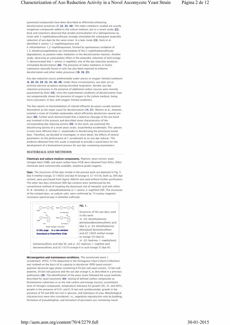
85% for dyes III and IV. Figure 2A2A2A2A shows a typical growth curve, as well as the
evolution of the medium pH and dye II concentration versus time, whereas Fig. 2B2B2B2B
displays the variation in glucose, ethanol, and dissolved oxygen concentrations
within the same period.
FIG. 2.FIG. 2.FIG. 2.FIG. 2.
Typical growth (□), pH (◊), and
decolorization curves (▵) (A)
and concentrations of glucose
(▪), ethanol (•) and oxygen (▴)
(B) during incubation of I.
occidentalis on NDM
containing 0.2 mM dye II. The
initial cell density (D 640) was
0.09.
HPLC analysis of supernatant samples for the presence of dye reduction products
revealed the presence of metanilic acid or sulfanilic acid during decolorization of
dyes I (or III) and II (or IV), respectively. These acids are stable under the
experimental conditions, and as shown below, they are not used by this yeast as a
carbon and energy source or as a nitrogen source within the experiment period.
The maximum concentrations of these sulfonates in decolorized media
approached the stoichiometric levels (0.17 ± 0.02 mM).
Ethanol starts to accumulate in the culture medium at the onset of the exponential
growth phase, indicating that I. occidentalis is a fermentative yeast. Since the
maximum yield of ethanol is 0.51 gg of glucose−1
, the observed concentration of
ethanol in the culture medium (Fig. 2B2B2B2B) approaches the expected value.
Effects of oxygen.Effects of oxygen.Effects of oxygen.Effects of oxygen. In the standard decolorization experiments described above,
the incubation mixtures were contained in cotton-plugged Erlenmeyer flasks and
subjected to agitation (120 rpm). Under these conditions, oxygen (from air) is
admitted in the system, but CO2 evolution, resulting from glucose fermentation,
contributes to decrease its concentration in the medium. In fact, analysis of Fig. 2B2B2B2B
shows that, after 6 h of incubation, dissolved oxygen concentration is 0.2 mg ·
liter−1
or lower. Decolorization, which is faster during the late exponential phase,
i.e., between 8 and 11 h of incubation (Fig. 2A2A2A2A), therefore occurs under low
oxygen concentrations.
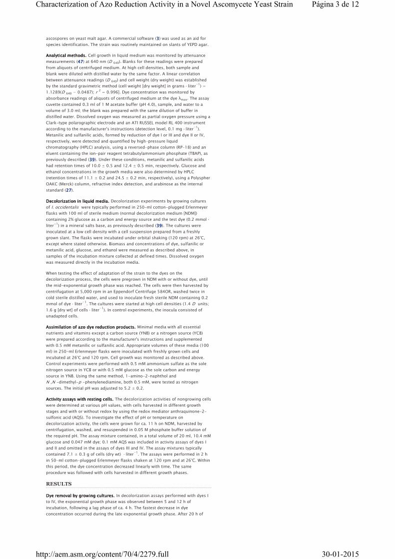
In an attempt to further elucidate the effect of dissolved oxygen in the
decolorization process, additional decolorization experiments with cell
suspensions inoculated at high densities (ca. 3.0 D units) were performed. Cells
were grown for 10 h under the usual conditions and then harvested by
centrifugation and washed. The combined pellets were resuspended in fresh NDM
with 0.2 mM dye II, and 100-ml volumes of the resulting suspension were placed
in separate Erlenmeyer flasks. The aeration conditions were as follows: one flask
was continuously flushed with sterile air and incubated while being stirred
magnetically, and a second flask was flushed with nitrogen for 15 min, tightly
plugged with a rubber stopper, and placed in the orbital incubator. A control
experiment, in our standard conditions, was simultaneously run. In the subsequent
12 h of incubation, a very slight increase in growth was detected (Fig. 3A3A3A3A), but in
the flasks incubated under the standard conditions or with oxygen flushing, a
slight decrease in the pH was observed (Fig. 3B3B3B3B). This was taken as evidence of
metabolic activity, which supported a decolorization of ca. 70%, in both cases (Fig.
3C3C3C3C). The dissolved oxygen concentrations were 3.9 ± 0.4 mg · liter−1
in the
aerated culture and <0.2 mg · liter−1
in the control culture. Under anoxic
conditions, the color loss was only ca. 20%, as seen in Fig. 3333, and oxygen
concentration remained below the detection level.
FIG. 3.FIG. 3.FIG. 3.FIG. 3.
Variation of cell density
measured as D 640 (A), pH (B),
and dye concentration
measured as A 470 (C) in NDM
preadjusted to pH 3.2
containing 0.2 mM dye II under
different conditions: anoxic
conditions (▵), air-flushed cultures (□), and normal incubation at 120
rpm (◊) (see text for details). The initial cell density was ≈3 D units.
The absence of metabolic activity in the anoxic conditions, obtained as described
above, was further confirmed through the observation that a culture started at a
low cell density and incubated for 24 h failed to grow (results not shown).
Página 4 de 12Characterization of Azo Reduction Activity in a Novel Ascomycete Yeast Strain
30-01-2015http://aem.asm.org/content/70/4/2279.full
View larger version:
In this pageIn this pageIn this pageIn this page In a new windowIn a new windowIn a new windowIn a new window
Download as PowerPoint SlideDownload as PowerPoint SlideDownload as PowerPoint SlideDownload as PowerPoint Slide
View larger version:
In this pageIn this pageIn this pageIn this page In a new windowIn a new windowIn a new windowIn a new window
Download as PowerPoint SlideDownload as PowerPoint SlideDownload as PowerPoint SlideDownload as PowerPoint Slide
View larger version:
In this pageIn this pageIn this pageIn this page In a new windowIn a new windowIn a new windowIn a new window
Download as PowerPoint SlideDownload as PowerPoint SlideDownload as PowerPoint SlideDownload as PowerPoint Slide
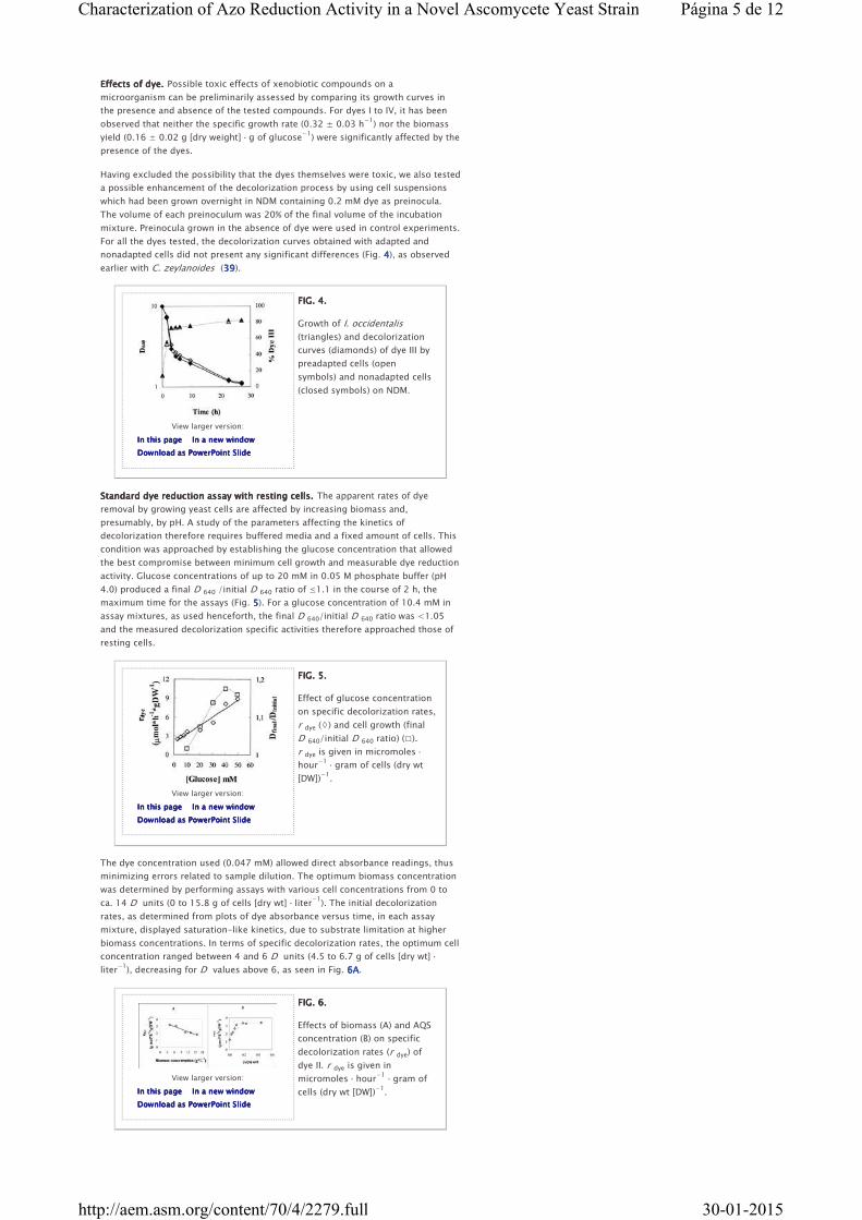
Effects of dye.Effects of dye.Effects of dye.Effects of dye. Possible toxic effects of xenobiotic compounds on a
microorganism can be preliminarily assessed by comparing its growth curves in
the presence and absence of the tested compounds. For dyes I to IV, it has been
observed that neither the specific growth rate (0.32 ± 0.03 h−1
) nor the biomass
yield (0.16 ± 0.02 g [dry weight] · g of glucose−1
) were significantly affected by the
presence of the dyes.
Having excluded the possibility that the dyes themselves were toxic, we also tested
a possible enhancement of the decolorization process by using cell suspensions
which had been grown overnight in NDM containing 0.2 mM dye as preinocula.
The volume of each preinoculum was 20% of the final volume of the incubation
mixture. Preinocula grown in the absence of dye were used in control experiments.
For all the dyes tested, the decolorization curves obtained with adapted and
nonadapted cells did not present any significant differences (Fig. 4444), as observed
earlier with C. zeylanoides (39393939).
FIG. 4.FIG. 4.FIG. 4.FIG. 4.
Growth of I. occidentalis
(triangles) and decolorization
curves (diamonds) of dye III by
preadapted cells (open
symbols) and nonadapted cells
(closed symbols) on NDM.
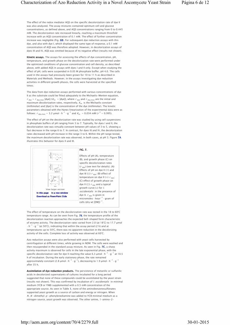
Standard dye reduction assay with resting cells.Standard dye reduction assay with resting cells.Standard dye reduction assay with resting cells.Standard dye reduction assay with resting cells. The apparent rates of dye
removal by growing yeast cells are affected by increasing biomass and,
presumably, by pH. A study of the parameters affecting the kinetics of
decolorization therefore requires buffered media and a fixed amount of cells. This
condition was approached by establishing the glucose concentration that allowed
the best compromise between minimum cell growth and measurable dye reduction
activity. Glucose concentrations of up to 20 mM in 0.05 M phosphate buffer (pH
4.0) produced a final D 640 /initial D 640 ratio of ≤1.1 in the course of 2 h, the
maximum time for the assays (Fig. 5555). For a glucose concentration of 10.4 mM in
assay mixtures, as used henceforth, the final D 640/initial D 640 ratio was <1.05
and the measured decolorization specific activities therefore approached those of
resting cells.
FIG. 5.FIG. 5.FIG. 5.FIG. 5.
Effect of glucose concentration
on specific decolorization rates,
r dye (◊) and cell growth (final
D 640/initial D 640 ratio) (□).
r dye is given in micromoles ·
hour−1
· gram of cells (dry wt
[DW])−1
.
The dye concentration used (0.047 mM) allowed direct absorbance readings, thus
minimizing errors related to sample dilution. The optimum biomass concentration
was determined by performing assays with various cell concentrations from 0 to
ca. 14 D units (0 to 15.8 g of cells [dry wt] · liter−1
). The initial decolorization
rates, as determined from plots of dye absorbance versus time, in each assay
mixture, displayed saturation-like kinetics, due to substrate limitation at higher
biomass concentrations. In terms of specific decolorization rates, the optimum cell
concentration ranged between 4 and 6 D units (4.5 to 6.7 g of cells [dry wt] ·
liter−1
), decreasing for D values above 6, as seen in Fig. 6A6A6A6A.
FIG. 6.FIG. 6.FIG. 6.FIG. 6.
Effects of biomass (A) and AQS
concentration (B) on specific
decolorization rates (r dye) of
dye II. r dye is given in
micromoles · hour−1
· gram of
cells (dry wt [DW])−1
.
Página 5 de 12Characterization of Azo Reduction Activity in a Novel Ascomycete Yeast Strain
30-01-2015http://aem.asm.org/content/70/4/2279.full
View larger version:
In this pageIn this pageIn this pageIn this page In a new windowIn a new windowIn a new windowIn a new window
Download as PowerPoint SlideDownload as PowerPoint SlideDownload as PowerPoint SlideDownload as PowerPoint Slide
The effect of the redox mediator AQS on the specific decolorization rate of dye II
was also analyzed. The assay mixtures contained optimum cell and glucose
concentrations, as defined above, and AQS concentrations ranging from 0 to 0.445
mM. The decolorization rate increased linearly, reaching a maximum threefold
increase with an AQS concentration of 0.1 mM. The effect of further concentration
increase was negligible (Fig. 6B6B6B6B). For subsequent dye reduction assays with this
dye, and also with dye I, which displayed the same type of response, a 0.1 mM
concentration of AQS was therefore adopted. However, in decolorization assays of
dyes III and IV, AQS was omitted because of its negative effect (results not shown).
Kinetic assays.Kinetic assays.Kinetic assays.Kinetic assays. The assays for assessing the effects of dye concentration, pH,
temperature, and growth phase on the decolorization rate were performed under
the optimized conditions of glucose concentration and cell density, as described
above, with added AQS in assays with dyes I and II only. Except when studying the
effect of pH, cells were suspended in 0.05 M phosphate buffer, pH 4.0. The cells
used in the assays had previously been grown for 10 to 11 h as described in
Materials and Methods. However, in the assays investigating dye reduction
activities in different growth phases, the cells were harvested at the specified
times.
The data from dye reduction assays performed with various concentrations of dye
II as the substrate could be fitted adequately to the Michaelis-Menten equation,
r dye = r dye,max [dye]/(Km + [dye]), where r dye and r dye,max are the initial and
maximum decolorization rates, respectively, Km is the Michaelis constant
(millimolar) and [dye] is the concentration of the dye (millimolar). The kinetic
parameters obtained with the Hanes linearization of the experimental data were as
follows: r dye,max = 3.2 μmol · h−1
· g−1
and Km = 0.034 mM (r2 = 0.995).
The effect of pH on the decolorization rate was studied by using cell suspensions
in phosphate buffers of pH ranging from 3 to 7. Typically, for dyes I and II, the
decolorization rate was virtually constant between pH values of 3 to 5, showing a
fast decrease in the range 6 to 7. In contrast, for dyes III and IV, the decolorization
rates decreased with pH increase in the range 3 to 6. Within the pH range tested,
the maximum decolorization rate was observed, in both cases, at pH 3. Figure 7A7A7A7A
illustrates this behavior for dyes II and III.
FIG. 7.FIG. 7.FIG. 7.FIG. 7.
Effects of pH (A), temperature
(B), and growth phase (C) on
specific decolorization rates
(r dye) (see text for details). (A)
Effects of pH on dye II (◊) and
dye III (□) r dye; (B) effect of
temperature on dye II (⋄) r dye;
(C) effect of growth phase on
dye II (□) r dye and a typical
growth curve (▴) for I.
occidentalis in the presence of
dye II. r dye is given in
micromoles · hour−1
· gram of
cells (dry wt [DW])−1
.
The effect of temperature on the decolorization rate was tested in the 18 to 60°C
temperature range. As can be seen from Fig. 7B7B7B7B, the temperature profile of the
decolorization reaction approaches the expected bell-shaped form characteristic
of enzyme activity. The decolorization rates varied from 2.0 (at 18°C) to 17.7 μmol
· h−1
· g−1
(at 50°C), indicating that within the assay period (2 h) and at
temperatures up to 50°C, there was no apparent reduction in the decolorizing
activity of the cells. Complete loss of activity was observed at 60°C.
Azo reduction assays were also performed with yeast cells harvested by
centrifugation at different times, while growing in NDM. The cells were washed and
then resuspended in the standard assay mixture. As seen in Fig. 7C7C7C7C, a sharp
activity maximum is observed for cells in the late exponential phase, with the
specific decolorization rate for dye II reaching the value 6.3 μmol · h−1
· g−1
at 10.5
h of incubation. During the early stationary phase, the rate remained
approximately constant (2.8 μmol · h−1
· g−1
), decreasing to 1.9 μmol · h−1
· g−1
after 35 h.
Assimilation of dye reduction products.Assimilation of dye reduction products.Assimilation of dye reduction products.Assimilation of dye reduction products. The persistence of metanilic or sulfanilic
acids in decolorized supernatants of cultures incubated for a long period
suggested that none of these compounds could be assimilated by the yeast strain
(results not shown). This was confirmed by incubation of I. occidentalis in minimal
medium (YCB or YNB) supplemented with a 0.5 mM concentration of the
appropriate source. As seen in Table 1111, none of the aminobenzenesulfonates
supported yeast growth as a source of carbon and energy or nitrogen. When
N ,N -dimethyl-p -phenylenediamine was added to YCB minimal medium as a
nitrogen source, yeast growth was observed. The other amine, 1-amino-2-
Página 6 de 12Characterization of Azo Reduction Activity in a Novel Ascomycete Yeast Strain
30-01-2015http://aem.asm.org/content/70/4/2279.full

View this table:
In this windowIn this windowIn this windowIn this window In a new windowIn a new windowIn a new windowIn a new window
View larger version:
In this pageIn this pageIn this pageIn this page In a new windowIn a new windowIn a new windowIn a new window
Download as PowerPoint SlideDownload as PowerPoint SlideDownload as PowerPoint SlideDownload as PowerPoint Slide
naphthol, was used as a source of carbon and energy and of nitrogen by this yeast.
Whenever assimilation occurred, an acidification of the medium was observed
(final pH of 3.5 ± 0.5).
TABLE 1.TABLE 1.TABLE 1.TABLE 1.
Biomass yields of I.
occidentalis on sulfanilic and
metanilic acids, 1-amino-2-naphthol, and
N,N -dimethyl-p -phenylenediamine as the carbon or nitrogen source
Sequential batch decolorization of dye II.Sequential batch decolorization of dye II.Sequential batch decolorization of dye II.Sequential batch decolorization of dye II. Standard decolorization experiments
were usually monitored over a period up to 24 h. By this time, glucose was
exhausted from the medium and dyes I and II had been completely removed; for
dyes III and IV, a residual amount of 15 to 20% was still present. To examine the
decolorization activity of the yeast cells after glucose depletion, a decolorized
culture (300 ml), which had been incubated for 24 h under the standard
conditions, was subjected to five successive additions of the required amounts of
dye II stock solution to restore the initial dye concentration. Each dye addition was
done after complete decolorization of the medium. As seen in Fig. 8888,
decolorization was complete during the first five cycles. During this process,
ethanol concentration first increased to a maximum of 10 g · liter−1
in the course
of the first cycle and then gradually decreased, disappearing at 160 h of
incubation. By this time, decolorization also ceased. In the course of successive
cycles, the concentration of sulfanilic acid gradually increased, as expected. Cell
growth also continued, with an increase from 2.7 to 6.1 D 640 units, showing that
the yeast switched from a fermentative metabolism, with glucose, to aerobic
respiration, with ethanol. The dissolved oxygen concentration consistently
remained below the detection level until the end of the experiment. The
decolorizing activity of the yeast cells, measured by the standard assay, slowly
decreased with the decolorization-dye addition cycles but was still present in the
last sample of cells, collected after 200 h of incubation.
FIG. 8.FIG. 8.FIG. 8.FIG. 8.
Monitoring of several
parameters during successive
cycles of addition of dye to a
culture of I. occidentalis on
NDM, incubated at 120 rpm,
without any further nutrient
addition. Wide white arrows
indicate the times of dye
addition (see text for details).
(A) pH (▵) and concentrations of
oxygen (⧫) and sulfanilic acid [Sulf.Acid] (○); (B) cell growth (◊) and
concentrations of glucose •, ethanol (▴), and dye II (□).
DISCUSSION
Decolorization of azo dyes by yeasts is much less studied than the homologous
process mediated by bacterial species. In this work, an attempt was made to
elucidate some basic physiologic aspects associated with azo dye destruction by I.
occidentalis. This yeast, like a previously described ascomycete yeast strain (39393939), is
capable of reducing several monoazo dyes to the corresponding amines and,
therefore, promotes their decolorization through the cleavage of the azo bond.
The decolorizing activity of I. occidentalis was first studied in actively growing
cultures in a glucose-containing culture medium with moderate shaking (Fig. 2222).
Under these conditions, ethanol was detected in the culture supernatant, showing
that the microorganism was fermenting glucose.
The similarities of the decolorization profiles by dye-adapted and nonadapted
cells (Fig. 4444) indicate that the azo dye reduction activity in yeast cultures is a
constitutive property of the cells, independent of their previous exposure to the
dye. So far, we have not been able to detect any decolorizing activity in cell
extracts or in the supernatant broth, even when high [NADH]/[dye] ratios were
used. It thus appears that azo dye reduction activities are dependent on intact,
active cells. Thus far, even in bacteria, the major enzyme responsible for
azoreductase activity in vivo has not been positively identified. A recent report (42424242)
described a high in vitro azoreductase activity of a cytoplasmic flavin reductase,
part of the ribonucleotide reductase complex in Escherichia coli, but its
overexpression in a Sphingomonas sp. strain failed to significantly increase the in
vivo reducing activity of the bacterium. Earlier, Kudlich and coworkers (24242424) had
also detected an azoreductase activity in a membrane fraction of the same
Sphingomonas sp. strain. According to these researchers, the NADH:ubiquinone
oxidoreductase was a likely candidate for the azoreductase activity.
Página 7 de 12Characterization of Azo Reduction Activity in a Novel Ascomycete Yeast Strain
30-01-2015http://aem.asm.org/content/70/4/2279.full
View larger version:
In this pageIn this pageIn this pageIn this page In a new windowIn a new windowIn a new windowIn a new window
Download as PowerPoint SlideDownload as PowerPoint SlideDownload as PowerPoint SlideDownload as PowerPoint Slide
(1)
Dissolved oxygen is repeatedly considered an inhibitor of the dye bioreduction
process (36363636), since both molecules act as electron acceptors and oxygen is a much
stronger oxidant. This is, apparently, the reason why azo dyes are more readily
reduced under anaerobiosis. The effect of oxygen was particularly addressed in
this work with I. occidentalis, because in the preliminary experiments of the
decolorizing potential of the yeast strains that we have studied so far, no attempt
was made to prevent the access of oxygen to the dye-containing incubation
medium. The decolorizing experiments were usually performed in cotton-plugged
flasks, shaken at 120 rpm. Under these standard conditions, faster decolorization
rates were indeed observed at low oxygen concentrations (<0.2 mg · liter−1
), which
can be considered microaerophilic (21212121). However, our results indicate that oxygen
does not significantly interfere with color loss, as seen in air-flushed cultures (Fig.
3333). A likely explanation for this fact is the high kinetic barrier involved in the
reduction of the triplet ground-state dioxygen. In contrast, under anoxic
conditions, i.e., in a nitrogen-flushed culture incubated in rubber-stoppered
flasks, the extent of decolorization was rather low (ca. 20%). Since pH (as well as
cell density) essentially remained constant in the course of the experiment, a lack
of metabolic activity was suspected. In a separate experiment that was also
conducted under anoxic conditions in which the culture was inoculated at a low
cell density, the yeast failed to grow. These observations show that I. occidentalis
has an absolute requirement for oxygen, i.e., places it in the category of the
aerobic-fermenting yeasts (1111). Therefore, even pregrown cells were unable of
performing azo dye reduction in the absence of oxygen. As far as we know, there
is no previous report of a similar situation in decolorization processes mediated by
facultative anaerobic bacteria.
The effects of pH, temperature, dye and redox mediator concentrations, and
growth phase on decolorization rates suggest the participation (direct or indirect)
of an enzyme activity in azo dye reduction, because (i) the temperature profile of
the azo reduction activity cannot easily be interpreted on the grounds of a purely
abiotic process and (ii) the decline of activity at the onset of the stationary phase is
more compatible with a reduced metabolic activity of the cells. Additionally, azo
dye reduction rate, like the intracellular formation of NAD(P)H, is a growth-
dependent process, since it does not occur without a carbon and energy source. In
fact, decolorization of dyes III and IV ceased upon glucose exhaustion in the
culture medium, and decolorization of dye II, after the diauxic shift (Fig. 8888), was
linked to the presence of ethanol.
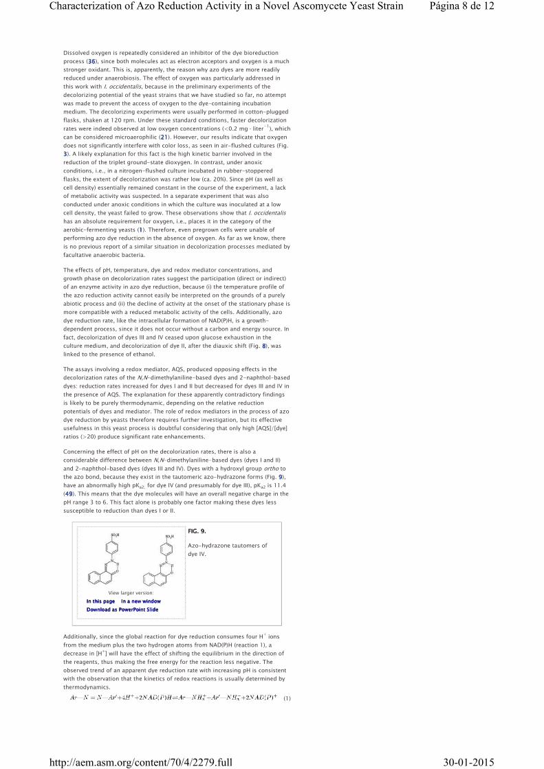
The assays involving a redox mediator, AQS, produced opposing effects in the
decolorization rates of the N,N-dimethylaniline-based dyes and 2-naphthol-based
dyes: reduction rates increased for dyes I and II but decreased for dyes III and IV in
the presence of AQS. The explanation for these apparently contradictory findings
is likely to be purely thermodynamic, depending on the relative reduction
potentials of dyes and mediator. The role of redox mediators in the process of azo
dye reduction by yeasts therefore requires further investigation, but its effective
usefulness in this yeast process is doubtful considering that only high [AQS]/[dye]
ratios (>20) produce significant rate enhancements.
Concerning the effect of pH on the decolorization rates, there is also a
considerable difference between N,N-dimethylaniline-based dyes (dyes I and II)
and 2-naphthol-based dyes (dyes III and IV). Dyes with a hydroxyl group ortho to
the azo bond, because they exist in the tautomeric azo-hydrazone forms (Fig. 9999),
have an abnormally high pKa2; for dye IV (and presumably for dye III), pKa2 is 11.4
(49494949). This means that the dye molecules will have an overall negative charge in the
pH range 3 to 6. This fact alone is probably one factor making these dyes less
susceptible to reduction than dyes I or II.
FIG. 9.FIG. 9.FIG. 9.FIG. 9.
Azo-hydrazone tautomers of
dye IV.
Additionally, since the global reaction for dye reduction consumes four H+ ions
from the medium plus the two hydrogen atoms from NAD(P)H (reaction 1), a
decrease in [H+] will have the effect of shifting the equilibrium in the direction of
the reagents, thus making the free energy for the reaction less negative. The
observed trend of an apparent dye reduction rate with increasing pH is consistent
with the observation that the kinetics of redox reactions is usually determined by
thermodynamics.
Página 8 de 12Characterization of Azo Reduction Activity in a Novel Ascomycete Yeast Strain
30-01-2015http://aem.asm.org/content/70/4/2279.full
View larger version:
In this pageIn this pageIn this pageIn this page In a new windowIn a new windowIn a new windowIn a new window
Download as PowerPoint SlideDownload as PowerPoint SlideDownload as PowerPoint SlideDownload as PowerPoint Slide
Dyes I and II, in contrast, have a pKa2 of ∼3.5, and protonation occurs in an azo
nitrogen (Fig. 10101010). This means that, up to pH ∼5, a still considerable fraction of the
molecules will be protonated in one azo nitrogen, apparently in an amount
allowing a fast decolorization rate.
FIG. 10.FIG. 10.FIG. 10.FIG. 10.
Protonization of
N,N-dimethylamino-based dyes
at an azo nitrogen.
The above conclusions show that, in this respect also, the reduction by a yeast is
considerably different from the reduction by bacteria, which has an optimum pH
range close to neutrality (8888, 33333333).
The decolorizing activity of I. occidentalis is not strictly related to a fermentative
process, since it continued even after glucose exhaustion. Under these conditions,
the yeast switches to aerobic respiration, at the expense of ethanol, which is the
major carbon and energy source in the medium. Ethanol depletion, at 160 h of
incubation, coincided with the disappearance of the decolorizing activity. Because
the shake flasks were moderately agitated (120 rpm), the concentration of
dissolved oxygen remained below the detection level. Therefore, the ethanol-
dependent azo dye reduction equally occurred under microaerophilic conditions,
although at a lower rate.
The assimilation assays with sulfanilic and metanilic acids revealed their inability
to be used as carbon and energy sources and also as nitrogen sources. This fact is
not surprising, because except for a few works describing the bacterial
degradation of sulfanilic acid (12121212, 13131313), 6-aminonaphthalene-2-sulfonate (19191919, 25252525),
and 2-aminobenzenesulfonate (29292929), arenesulfonates are usually described as
xenobiotic compounds. The persistence of the other two amines expected from
dye reduction, N,N-dimethyl-p-phenylenediamine (dyes I and II) and 1-amino-2-
naphthol (dyes III and IV), could not be detected by HPLC analysis, since they are
quickly oxidized in aerated aqueous solutions, forming colored products, and their
elution profiles change considerably within a short period of time. However, the
colors associated with the oxidation products of either
N,N-dimethyl-p-phenylenediamine (deep pink) or 1-amino-2-naphthol (dark
yellow to brown) were not seen in decolorized media. The assimilation tests
revealed that N,N-dimethyl-p-phenylenediamine was used as nitrogen source
whereas 1-amino-2-naphthol was both a source of nitrogen and a source of
carbon and energy, thus accounting for the absence of their oxidation products in
the decolorized supernatants.
This work shows that the ascomycete yeast I. occidentalis displays an effective azo
dye reduction capacity, comparatively unspecific but, nevertheless, structure
dependent in such aspects as glucose requirements for complete decolorization,
pH-activity profiles, and effect of the tested redox mediator. All of these factors
must be taken into account when considering the possible application of the yeast
to a bioremediation process for azo textile dyes. Equally important is the effect of
dissolved oxygen in the decolorization process. This particular strain, because it is
an aerobic-fermentative yeast, has an absolute requirement of oxygen for growth
and hence, for decolorization. While oxygen traces are sufficient for growth and
allow dye reduction, our results also point to a noninhibitory effect of moderate
levels of oxygen in the process. The decolorization process is also growth
dependent, pointing to NAD(P)H availability as a crucial factor. This conclusion
opens further perspectives in the search for more efficient yeast strains in azo dye
decolorization.
ACKNOWLEDGMENTS
P.A.R. gratefully acknowledges a Ph.D. scholarship from the European Project
BIOEFTEX.
FOOTNOTES
Received 10 September 2003.
Accepted 9 January 2004.
↵↵↵↵ ****Corresponding author. Mailing address: Department of Biology, University
of Minho, Campus de Gualtar, 4710-057 Braga, Portugal. Phone: (351) 253
604043. Fax: (351) 253 678983. E-mail: [email protected]@[email protected]@bio.uminho.pt.
REFERENCES
1. Alexander, M. A., and T. W. Jeffries.Alexander, M. A., and T. W. Jeffries.Alexander, M. A., and T. W. Jeffries.Alexander, M. A., and T. W. Jeffries. 1990. Respiratory efficiency and
metabolite partitioning as regulatory phenomena in yeasts. Enzyme Microb.
Technol. 12121212::::2-19. CrossRefCrossRefCrossRefCrossRef
Página 9 de 12Characterization of Azo Reduction Activity in a Novel Ascomycete Yeast Strain
30-01-2015http://aem.asm.org/content/70/4/2279.full
2. Banat, I. M., P. Nigam, D. Singh, and R. Marchant.Banat, I. M., P. Nigam, D. Singh, and R. Marchant.Banat, I. M., P. Nigam, D. Singh, and R. Marchant.Banat, I. M., P. Nigam, D. Singh, and R. Marchant. 1996. Microbial
decolorization of textile dye-containing effluents: a review. Bioresour.
Technol. 58585858::::217-227. CrossRefCrossRefCrossRefCrossRef
3. Barnett, J. A., R. W. Payne, and D. Y. Yarrow.Barnett, J. A., R. W. Payne, and D. Y. Yarrow.Barnett, J. A., R. W. Payne, and D. Y. Yarrow.Barnett, J. A., R. W. Payne, and D. Y. Yarrow. 1990. Yeast identification
program, version 2. Cambridge University Press, Cambridge, United Kingdom.
4. Blümel, S., M. Contzen, M. Lutz, A. Stolz, and H.-J. Knackmuss.Blümel, S., M. Contzen, M. Lutz, A. Stolz, and H.-J. Knackmuss.Blümel, S., M. Contzen, M. Lutz, A. Stolz, and H.-J. Knackmuss.Blümel, S., M. Contzen, M. Lutz, A. Stolz, and H.-J. Knackmuss. 1998.
Isolation of a bacterial strain with the ability to utilize the sulfonated azo
compound 4-carboxy-4′-sulfoazobenzene as the sole source of carbon and
energy. Appl. Environ. Microbiol. 64646464::::2315-2317. AbstractAbstractAbstractAbstract////FREE FREE FREE FREE Full TextFull TextFull TextFull Text
5. Blümel, S., H.-J. Knackmuss, and A. Stolz.Blümel, S., H.-J. Knackmuss, and A. Stolz.Blümel, S., H.-J. Knackmuss, and A. Stolz.Blümel, S., H.-J. Knackmuss, and A. Stolz. 2002. Molecular cloning and
characterization of the gene coding for the aerobic azoreductase from
Xenophilus azovorans KF46F. Appl. Environ. Microbiol. 68686868::::3948-3955.
AbstractAbstractAbstractAbstract////FREE FREE FREE FREE Full TextFull TextFull TextFull Text
6. Bragger, J. L., A. W. Lloyd, S. H. Soozandehfar, S. F. Bloomfield, C. Marriott, Bragger, J. L., A. W. Lloyd, S. H. Soozandehfar, S. F. Bloomfield, C. Marriott, Bragger, J. L., A. W. Lloyd, S. H. Soozandehfar, S. F. Bloomfield, C. Marriott, Bragger, J. L., A. W. Lloyd, S. H. Soozandehfar, S. F. Bloomfield, C. Marriott,
and G. P. Martin.and G. P. Martin.and G. P. Martin.and G. P. Martin. 1997. Investigations into the azo reducing activity of a
common colonic microorganism. Int. J. Pharm. 157157157157::::61-71. CrossRefCrossRefCrossRefCrossRef
7. Cervantes, F. J., F. P. van der Zee, G. Lettinga, and J. A. Field.Cervantes, F. J., F. P. van der Zee, G. Lettinga, and J. A. Field.Cervantes, F. J., F. P. van der Zee, G. Lettinga, and J. A. Field.Cervantes, F. J., F. P. van der Zee, G. Lettinga, and J. A. Field. 2001.
Enhanced decolourisation of Acid Orange 7 in a continuous UASB reactor with
quinones as redox mediators. Water Sci. Technol. 44444444::::123-128.
8. Chang, J.-S., C. Chou, Y.-C. Lin, J.-Y. Ho, and T. L. Hu.Chang, J.-S., C. Chou, Y.-C. Lin, J.-Y. Ho, and T. L. Hu.Chang, J.-S., C. Chou, Y.-C. Lin, J.-Y. Ho, and T. L. Hu.Chang, J.-S., C. Chou, Y.-C. Lin, J.-Y. Ho, and T. L. Hu. 2001. Kinetic
characteristics of bacterial azo-dye decolourisation by Pseudomonas luteola.
Water Res. 35353535::::2841-2850. MedlineMedlineMedlineMedline
9. Chung, K.-T., and S. E. Stevens, Jr.Chung, K.-T., and S. E. Stevens, Jr.Chung, K.-T., and S. E. Stevens, Jr.Chung, K.-T., and S. E. Stevens, Jr. 1993. Degradation of azo dyes by
environmental microorganisms and helminths. Environ. Toxicol. Chem.
12121212::::2121-2132. CrossRefCrossRefCrossRefCrossRef
10. Coates, J. D., K. A. Cole, R. Chakraborty, S. M. O'Connor, and L. A. Coates, J. D., K. A. Cole, R. Chakraborty, S. M. O'Connor, and L. A. Coates, J. D., K. A. Cole, R. Chakraborty, S. M. O'Connor, and L. A. Coates, J. D., K. A. Cole, R. Chakraborty, S. M. O'Connor, and L. A.
Achenbach.Achenbach.Achenbach.Achenbach. 2002. Diversity and ubiquity of bacteria capable of utilizing
humic substances as electron donors for anaerobic respiration. Appl. Environ.
Microbiol. 68686868::::2445-2452. AbstractAbstractAbstractAbstract////FREE FREE FREE FREE Full TextFull TextFull TextFull Text
11. Coughlin, M. F., B. K. Kinkle, and P. L. Bishop.Coughlin, M. F., B. K. Kinkle, and P. L. Bishop.Coughlin, M. F., B. K. Kinkle, and P. L. Bishop.Coughlin, M. F., B. K. Kinkle, and P. L. Bishop. 1999. Degradation of azo
dyes containing aminonaphthol by Sphingomonas sp. strain 1CX. J. Ind.
Microbiol. Biotechnol. 23232323::::341-346.
12. Coughlin, M. F., B. K. Kinkle, and P. L. Bishop.Coughlin, M. F., B. K. Kinkle, and P. L. Bishop.Coughlin, M. F., B. K. Kinkle, and P. L. Bishop.Coughlin, M. F., B. K. Kinkle, and P. L. Bishop. 2002. Degradation of Acid
Orange 7 in an aerobic biofilm. Chemosphere 46464646::::11-19. MedlineMedlineMedlineMedline
13. Coughlin, M. F., B. K. Kinkle, and P. L. Bishop.Coughlin, M. F., B. K. Kinkle, and P. L. Bishop.Coughlin, M. F., B. K. Kinkle, and P. L. Bishop.Coughlin, M. F., B. K. Kinkle, and P. L. Bishop. 2003. High performance
degradation of azo dye Acid Orange 7 and sulfanilic acid in a laboratory scale
reactor after seeding with cultured bacterial strains. Water Res.
37373737::::2757-2763. MedlineMedlineMedlineMedline
14. Donmez, G.Donmez, G.Donmez, G.Donmez, G. 2002. Bioaccumulation of the reactive textile dyes by C.
tropicalis growing in molasses medium. Enzyme Microb. Technol.
30303030::::363-366. CrossRefCrossRefCrossRefCrossRef
15. Dubin, P., and K. L. Wright.Dubin, P., and K. L. Wright.Dubin, P., and K. L. Wright.Dubin, P., and K. L. Wright. 1975. Reduction of azo food dyes in cultures of
Proteus vulgaris. Xenobiotica 5555::::563-571. MedlineMedlineMedlineMedline
16. Field, J. A.Field, J. A.Field, J. A.Field, J. A. 2002. Limits of anaerobic biodegradation. Water Sci. Technol.
45454545::::9-18.
17. Furniss, B. S., A. J. Hannaford, P. W. G. Smith, and A. R. Tatchell.Furniss, B. S., A. J. Hannaford, P. W. G. Smith, and A. R. Tatchell.Furniss, B. S., A. J. Hannaford, P. W. G. Smith, and A. R. Tatchell.Furniss, B. S., A. J. Hannaford, P. W. G. Smith, and A. R. Tatchell. 1989.
Vogel's textbook of practical organic chemistry, 5th ed., p. 951. Longman
Group UK, Ltd., Harlow, Essex, United Kingdom.
18. Ghosh, D. K., A. Mandal, and J. Chaudhuri.Ghosh, D. K., A. Mandal, and J. Chaudhuri.Ghosh, D. K., A. Mandal, and J. Chaudhuri.Ghosh, D. K., A. Mandal, and J. Chaudhuri. 1992. Purification and partial
characterization of two azoreductases from Shigella dysenteriae type 1. FEMS
Microbiol. Lett. 98989898::::229-233. CrossRefCrossRefCrossRefCrossRef
19. Haug, W., A. Schmidt, B. Nörtmann, D. C. Hempel, A. Stolz, and H.-J. Haug, W., A. Schmidt, B. Nörtmann, D. C. Hempel, A. Stolz, and H.-J. Haug, W., A. Schmidt, B. Nörtmann, D. C. Hempel, A. Stolz, and H.-J. Haug, W., A. Schmidt, B. Nörtmann, D. C. Hempel, A. Stolz, and H.-J.
Knackmuss.Knackmuss.Knackmuss.Knackmuss. 1991. Mineralization of the sulfonated azo dye Mordant Yellow 3
by a 6-amino-naphthalene-2-sulfonate-degrading bacterial consortium.
Appl. Environ. Microbiol. 57575757::::3144-3149. AbstractAbstractAbstractAbstract////FREE FREE FREE FREE Full TextFull TextFull TextFull Text
20. Hayase, N., K. Kouno, and K. Ushio.Hayase, N., K. Kouno, and K. Ushio.Hayase, N., K. Kouno, and K. Ushio.Hayase, N., K. Kouno, and K. Ushio. 2000. Isolation and characterization of
Aeromonas sp. B-5 capable of decolorizing various dyes. J. Biosci. Bioeng.
90909090::::570-573. MedlineMedlineMedlineMedline
21. Isik, M., and D. T. Sponza.Isik, M., and D. T. Sponza.Isik, M., and D. T. Sponza.Isik, M., and D. T. Sponza. 2003. Effect of oxygen on the decolorization of
azo dyes by Escherichia coli and Pseudomonas sp. and fate of aromatic
amines. Process Biochem. 38383838::::1183-1192. CrossRefCrossRefCrossRefCrossRef
22. Keck, A., J. Klein, M. Kudlich, A. Stolz, H.-J. Knackmuss, and R. Mattes.Keck, A., J. Klein, M. Kudlich, A. Stolz, H.-J. Knackmuss, and R. Mattes.Keck, A., J. Klein, M. Kudlich, A. Stolz, H.-J. Knackmuss, and R. Mattes.Keck, A., J. Klein, M. Kudlich, A. Stolz, H.-J. Knackmuss, and R. Mattes.
1997. Reduction of azo dyes by redox mediators originating in the
naphthalenesulfonic acid degradation pathway of Sphingomonas sp. strain
BN6. Appl. Environ. Microbiol. 63636363::::3684-3690. AbstractAbstractAbstractAbstract////FREE FREE FREE FREE Full TextFull TextFull TextFull Text
23. Keck, A., J. Rau, T. Reemtsma, R. Mattes, A. Stolz, and J. Klein.Keck, A., J. Rau, T. Reemtsma, R. Mattes, A. Stolz, and J. Klein.Keck, A., J. Rau, T. Reemtsma, R. Mattes, A. Stolz, and J. Klein.Keck, A., J. Rau, T. Reemtsma, R. Mattes, A. Stolz, and J. Klein. 2002.
Identification of quinoide redox mediators that are formed during the
degradation of naphthalene-2-sulfonate by Sphingomonas xenophaga BN6.
Appl. Environ. Microbiol. 68686868::::4341-4349. AbstractAbstractAbstractAbstract////FREE FREE FREE FREE Full TextFull TextFull TextFull Text
24. Kudlich, M., A. Keck, J. Klein, and A. Stolz.Kudlich, M., A. Keck, J. Klein, and A. Stolz.Kudlich, M., A. Keck, J. Klein, and A. Stolz.Kudlich, M., A. Keck, J. Klein, and A. Stolz. 1997. Localization of the enzyme
system involved in anaerobic reduction of azo dyes by Sphingomonas sp.
strain BN6 and effect of artificial redox mediators on the rate of azo dye
reduction. Appl. Environ. Microbiol. 63636363::::3691-3694. AbstractAbstractAbstractAbstract////FREE FREE FREE FREE Full TextFull TextFull TextFull Text
25. Kuhm, A. E., A. Stolz, K. L. Ngai, and H.-J. Knackmuss.Kuhm, A. E., A. Stolz, K. L. Ngai, and H.-J. Knackmuss.Kuhm, A. E., A. Stolz, K. L. Ngai, and H.-J. Knackmuss.Kuhm, A. E., A. Stolz, K. L. Ngai, and H.-J. Knackmuss. 1991. Purification
and characterization of 1,2-dihydroxynaphthalene dioxygenase from a
bacterium that degrades naphthalenesulfonic acids. J. Bacteriol.
173173173173::::3795-3802. AbstractAbstractAbstractAbstract////FREE FREE FREE FREE Full TextFull TextFull TextFull Text
26. Kulla, H. G., R. Krieg, T. Zimmermann, and T. Leisinger.Kulla, H. G., R. Krieg, T. Zimmermann, and T. Leisinger.Kulla, H. G., R. Krieg, T. Zimmermann, and T. Leisinger.Kulla, H. G., R. Krieg, T. Zimmermann, and T. Leisinger. 1984. Experimental
evolution of azo dye-degrading bacteria, p. 663-667. In M. J. Klug and C. A.
Página 10 de 12Characterization of Azo Reduction Activity in a Novel Ascomycete Yeast Strain
30-01-2015http://aem.asm.org/content/70/4/2279.full
Reddy (ed.), Current perspectives in microbial ecology. American Society for
Microbiology, Washington, D.C.
27. Lages, F., and C. Lucas.Lages, F., and C. Lucas.Lages, F., and C. Lucas.Lages, F., and C. Lucas. 1997. Contribution to the physiological
characterization of glycerol active uptake in Saccharomyces cerevisiae.
Biochem. Biophys. Acta 1322132213221322::::8-18. MedlineMedlineMedlineMedline
28. Laszlo, J. A.Laszlo, J. A.Laszlo, J. A.Laszlo, J. A. 2000. Regeneration of azo-dye-saturated cellulosic anion
exchange resin by Burkholderia cepacia anaerobic dye reduction. Environ. Sci.
Technol. 34343434::::167-172. CrossRefCrossRefCrossRefCrossRef
29. Mampel, J., J. Ruff, F. Junker, and A. M. Cook.Mampel, J., J. Ruff, F. Junker, and A. M. Cook.Mampel, J., J. Ruff, F. Junker, and A. M. Cook.Mampel, J., J. Ruff, F. Junker, and A. M. Cook. 1999. The oxygenase
component of the 2-aminobenzenesulfonate dioxygenase system from
Alcaligenes sp. strain O-1. Microbiology 145145145145::::3255-3264.
30. Martins, M. A., M. H. Cardoso, M. J. Queiroz, M. T. Ramalho, and A. M. Martins, M. A., M. H. Cardoso, M. J. Queiroz, M. T. Ramalho, and A. M. Martins, M. A., M. H. Cardoso, M. J. Queiroz, M. T. Ramalho, and A. M. Martins, M. A., M. H. Cardoso, M. J. Queiroz, M. T. Ramalho, and A. M.
Oliveira-Campos.Oliveira-Campos.Oliveira-Campos.Oliveira-Campos. 1999. Biodegradation of azo dyes by the yeast Candida
zeylanoides in batch aerated cultures. Chemosphere 38383838::::2455-2460.
MedlineMedlineMedlineMedline
31. Meehan, C., I. M. Banat, G. McMullan, P. Nigam, F. Smyth, and R. Marchant.Meehan, C., I. M. Banat, G. McMullan, P. Nigam, F. Smyth, and R. Marchant.Meehan, C., I. M. Banat, G. McMullan, P. Nigam, F. Smyth, and R. Marchant.Meehan, C., I. M. Banat, G. McMullan, P. Nigam, F. Smyth, and R. Marchant.
2000. Decolorization of Remazol Black-B using a thermotolerant yeast, K.
marxianus IMB3. Environ. Int. 26262626::::75-79. CrossRefCrossRefCrossRefCrossRef MedlineMedlineMedlineMedline
32. Moir, D., S. Masson, and I. Chu.Moir, D., S. Masson, and I. Chu.Moir, D., S. Masson, and I. Chu.Moir, D., S. Masson, and I. Chu. 2001. Structure-activity relationship study
on the bioreduction of azo dyes by Clostridium paraputrificum. Environ.
Toxicol. Chem. 20202020::::479-484. CrossRefCrossRefCrossRefCrossRef MedlineMedlineMedlineMedline
33. Moutaouakkil, A.,Y. Zeroual, F. Z. Dzayri, M. Talbi, K. Lee, and M. Blaghen.Moutaouakkil, A.,Y. Zeroual, F. Z. Dzayri, M. Talbi, K. Lee, and M. Blaghen.Moutaouakkil, A.,Y. Zeroual, F. Z. Dzayri, M. Talbi, K. Lee, and M. Blaghen.Moutaouakkil, A.,Y. Zeroual, F. Z. Dzayri, M. Talbi, K. Lee, and M. Blaghen.
2003. Purification and partial characterization of azoreductase from
Enterobacter agglomerans. Arch. Biochem. Biophys. 413413413413::::139-146. CrossRefCrossRefCrossRefCrossRef
MedlineMedlineMedlineMedline
34. Nigam, P., I. M. Banat, D. Singh, and R. Marchant.Nigam, P., I. M. Banat, D. Singh, and R. Marchant.Nigam, P., I. M. Banat, D. Singh, and R. Marchant.Nigam, P., I. M. Banat, D. Singh, and R. Marchant. 1996. Microbial process
for the decolorization of textile effluent containing azo, diazo and reactive
dyes. Process Biochem. 31313131::::435-442. CrossRefCrossRefCrossRefCrossRef
35. Park, J., and J. Shore.Park, J., and J. Shore.Park, J., and J. Shore.Park, J., and J. Shore. 1984. Water for the dyehouse—supply, consumption,
recovery and disposal. J. Soc. Dyers Colourists 100100100100::::383-399.
36. Pearce, C. I., J. R. Lloyd, and J. T. Guthrie.Pearce, C. I., J. R. Lloyd, and J. T. Guthrie.Pearce, C. I., J. R. Lloyd, and J. T. Guthrie.Pearce, C. I., J. R. Lloyd, and J. T. Guthrie. 2003. The removal of colour from
textile wastewater using whole bacterial cells: a review. Dyes Pigments
58585858::::179-196. CrossRefCrossRefCrossRefCrossRef
37. Rafii, F., and C. E. Cerniglia.Rafii, F., and C. E. Cerniglia.Rafii, F., and C. E. Cerniglia.Rafii, F., and C. E. Cerniglia. 1990. An anaerobic non denaturing gel assay
for the detection of azoreductase from anaerobic bacteria. J. Microbiol.
Methods 12121212::::139-148. CrossRefCrossRefCrossRefCrossRef
38. Rafii, F., and T. Coleman.Rafii, F., and T. Coleman.Rafii, F., and T. Coleman.Rafii, F., and T. Coleman. 1999. Cloning and expression in Escherichia coli of
an azoreductase gene from Clostridium perfringens and comparison with
azoreductase genes from other bacteria. J. Basic Microbiol. 39393939::::29-35.
CrossRefCrossRefCrossRefCrossRef MedlineMedlineMedlineMedline
39. Ramalho, P. A., H. Scholze, M. H. Cardoso, M. T. Ramalho, and A. M. Ramalho, P. A., H. Scholze, M. H. Cardoso, M. T. Ramalho, and A. M. Ramalho, P. A., H. Scholze, M. H. Cardoso, M. T. Ramalho, and A. M. Ramalho, P. A., H. Scholze, M. H. Cardoso, M. T. Ramalho, and A. M.
Oliveira-Campos.Oliveira-Campos.Oliveira-Campos.Oliveira-Campos. 2002. Improved conditions for the aerobic reductive
decolorisation of azo dyes by Candida zeylanoides. Enzyme Microb. Technol.
31313131::::848-854. CrossRefCrossRefCrossRefCrossRef
40. Rau, J., H. J. Knackmuss, and A. Stolz.Rau, J., H. J. Knackmuss, and A. Stolz.Rau, J., H. J. Knackmuss, and A. Stolz.Rau, J., H. J. Knackmuss, and A. Stolz. 2002. Effects of different quinoid
redox mediators on the anaerobic reduction of azo dyes by bacteria. Environ.
Sci. Technol. 36363636::::1497-1504. MedlineMedlineMedlineMedline
41. Robinson, T., G. McMullan, R. Marchant, and P. Nigam.Robinson, T., G. McMullan, R. Marchant, and P. Nigam.Robinson, T., G. McMullan, R. Marchant, and P. Nigam.Robinson, T., G. McMullan, R. Marchant, and P. Nigam. 2001. Remediation
of dyes in textile effluent: a critical review on current treatment technologies
with a proposed alternative. Bioresour. Technol. 77777777::::247-255. CrossRefCrossRefCrossRefCrossRef
MedlineMedlineMedlineMedline
42. Russ, R., J. Rau, and A. Stolz.Russ, R., J. Rau, and A. Stolz.Russ, R., J. Rau, and A. Stolz.Russ, R., J. Rau, and A. Stolz. 2000. The function of cytoplasmic flavin
reductases in the reduction of azo dyes by bacteria. Appl. Environ. Microbiol.
66666666::::1429-1434. AbstractAbstractAbstractAbstract////FREE FREE FREE FREE Full TextFull TextFull TextFull Text
43. Stolz, A.Stolz, A.Stolz, A.Stolz, A. 2001. Basic and applied aspects in the microbial degradation of azo
dyes. Appl. Microbiol. Biotechnol. 56565656::::69-80. CrossRefCrossRefCrossRefCrossRef MedlineMedlineMedlineMedline
44. van der Walt, J. P., and D. Y. Yarrow.van der Walt, J. P., and D. Y. Yarrow.van der Walt, J. P., and D. Y. Yarrow.van der Walt, J. P., and D. Y. Yarrow. 1984. Methods for the isolation,
maintenance, classification and identification of yeasts, p. 45-104. In N. J.
Kreger-van Rij (ed.), Yeasts—a taxonomic study. Elsevier, Amsterdam, The
Netherlands.
45. van der Zee, F. P., I. A. E. Bisschops, V. G. Blanchard, R. H. M. Bouwman, G. van der Zee, F. P., I. A. E. Bisschops, V. G. Blanchard, R. H. M. Bouwman, G. van der Zee, F. P., I. A. E. Bisschops, V. G. Blanchard, R. H. M. Bouwman, G. van der Zee, F. P., I. A. E. Bisschops, V. G. Blanchard, R. H. M. Bouwman, G.
Lettinga, and J. A. Field.Lettinga, and J. A. Field.Lettinga, and J. A. Field.Lettinga, and J. A. Field. 2003. The contribution of biotic and abiotic
processes during azo dye reduction in anaerobic sludge. Water Res.
37373737::::3098-3109. MedlineMedlineMedlineMedline
46. van der Zee, F. P., G. Lettinga, and J. A. Field.van der Zee, F. P., G. Lettinga, and J. A. Field.van der Zee, F. P., G. Lettinga, and J. A. Field.van der Zee, F. P., G. Lettinga, and J. A. Field. 2000. The role of (auto)
catalysis in the mechanism of an anaerobic azo reduction. Water Sci. Technol.
42424242::::301-308.
47. Verhoven, J. W.Verhoven, J. W.Verhoven, J. W.Verhoven, J. W. 1996. International Union of Pure and Applied Chemistry,
Organic Chemistry Division Commission on Photochemistry: glossary of terms
used in photochemistry (IUPAC recommendations 1996). Pure Appl. Chem.
68686868::::2223-2286.
48. Yu, J., X. Wang, and P. L. Yue.Yu, J., X. Wang, and P. L. Yue.Yu, J., X. Wang, and P. L. Yue.Yu, J., X. Wang, and P. L. Yue. 2001. Optimal decolorization and kinetic
modeling of synthetic dyes by Pseudomonas strains. Water Res.
35353535::::3579-3586. MedlineMedlineMedlineMedline
49. Zollinger, H.Zollinger, H.Zollinger, H.Zollinger, H. 1991. Color chemistry. Syntheses, properties and applications
of organic dyes and pigments, 2nd ed. VCH, Weinheim, Germany.
American Society for Microbiology
Página 11 de 12Characterization of Azo Reduction Activity in a Novel Ascomycete Yeast Strain
30-01-2015http://aem.asm.org/content/70/4/2279.full
Articles citing this article
Azo Reductase Activity of Intact Saccharomyces cerevisiae Cells Is Azo Reductase Activity of Intact Saccharomyces cerevisiae Cells Is Azo Reductase Activity of Intact Saccharomyces cerevisiae Cells Is Azo Reductase Activity of Intact Saccharomyces cerevisiae Cells Is
Dependent on the Fre1p Component of Plasma Membrane Ferric Dependent on the Fre1p Component of Plasma Membrane Ferric Dependent on the Fre1p Component of Plasma Membrane Ferric Dependent on the Fre1p Component of Plasma Membrane Ferric
Reductase Reductase Reductase Reductase
Appl. Environ. Microbiol. July 2005 71:7 3882-3888
AbstractAbstractAbstractAbstract Full TextFull TextFull TextFull Text PDFPDFPDFPDF
Página 12 de 12Characterization of Azo Reduction Activity in a Novel Ascomycete Yeast Strain
30-01-2015http://aem.asm.org/content/70/4/2279.full