epistemology of environmental microbiology
TRANSCRIPT
Critical Review
Epistemology of Environmental MicrobiologyE U G E N E L . M A D S E N
Section of Microbiology Division of Biological Sciences Cornell University,Ithaca, New York 14853-8101
Despite critical geochemical roles of microorganisms inbiosphere maintenance, knowledge of microorganisms asthey function in soils, sediments, and waters is limited.Constraints on knowledge are caused largely by methodolo-gies that do not contend well with the complexity of fieldsites, with the scale differential between microorganismsand humans, and with artifacts that may arise in charac-terizing microorganisms using laboratory-based physiological,biochemical, genetic, and molecular biological assays.A paradigm describing how knowledge is obtained inenvironmental microbiology suggests that the constraintson knowledge will yield to relationships developing betweenmethodological innovations and their iterative applicationto naturally occurring microorganisms in field sites.
IntroductionSince its early foundation in the work of Beijerinck andWinogradsky (1-3), environmental microbiology has beenconcerned with the presence, abundance, interactions, andphysiological activities of microorganisms in terrestrial andaquatic environments. [Note: The term microbiology hastraditionally included five groups of microscopically visual-izable organisms: bacteria, viruses, fungi, protozoa, and algae(4). Based on phylogenetic analysis of the ribosomal RNAmolecule (5-8), the first of these five groups corresponds tothe domains Archaea plus Bacteria, and the latter three groupsare distributed throughout various branches of the Eukarya(8, 9). The physiological, morphological, ecological, andphylogenetic span of these organisms is immense and resistsmost generalizations. Consequently, this paper will addressprimarily a major subset of the microbial world, heterotrophicmicroorganismssespecially bacteria, but also fungi andprotozoa. Because algae and some types of bacteria andprotozoa are photosynthetic, specific comments related tonutrition may not always apply to these organisms. Somecomments also may not apply to viruses because of their uniquenoncellular and parasitic traits.] Environmental microbi-ology is related to, but distinct from, medical (10) andindustrial (11) microbiology. One reason for advancingenvironmental microbiology is that a mechanistic under-standing of microorganisms and their activities improveshuman ability to manage natural systems and expandsbiotechnological products and services (12, 13) essential forindustrial and medical microbiology.
The earth’s habitats present complex gradients of envi-ronmental conditions that include extreme variations intemperature, light, pH, pressure, salinity, and both inorganicand organic compounds (materials ranging from elementalsulfur to ammonia, hydrogen gas, and methane and from
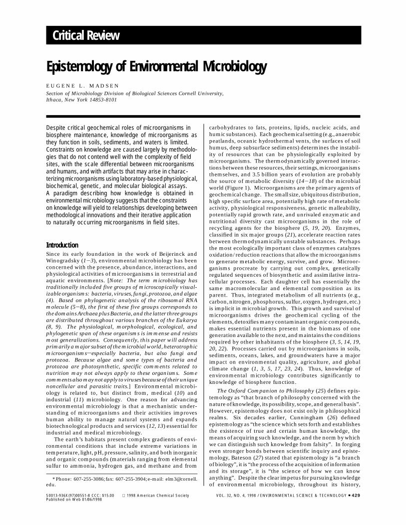
carbohydrates to fats, proteins, lipids, nucleic acids, andhumic substances). Each geochemical setting (e.g., anaerobicpeatlands, oceanic hydrothermal vents, the surfaces of soilhumus, deep subsurface sediments) determines the instabil-ity of resources that can be physiologically exploited bymicroorganisms. The thermodynamically governed interac-tions between these resources, their settings, microorganismsthemselves, and 3.5 billion years of evolution are probablythe source of metabolic diversity (14-18) of the microbialworld (Figure 1). Microorganisms are the primary agents ofgeochemical change. The small size, ubiquitous distribution,high specific surface area, potentially high rate of metabolicactivity, physiological responsiveness, genetic malleability,potentially rapid growth rate, and unrivaled enzymatic andnutritional diversity cast microorganisms in the role ofrecycling agents for the biosphere (5, 19, 20). Enzymes,classified in six major groups (21), accelerate reaction ratesbetween thermodynamically unstable substances. Perhapsthe most ecologically important class of enzymes catalyzesoxidation/reduction reactions that allow the microorganismsto generate metabolic energy, survive, and grow. Microor-ganisms procreate by carrying out complex, geneticallyregulated sequences of biosynthetic and assimilative intra-cellular processes. Each daughter cell has essentially thesame macromolecular and elemental composition as itsparent. Thus, integrated metabolism of all nutrients (e.g.,carbon, nitrogen, phosphorus, sulfur, oxygen, hydrogen, etc.)is implicit in microbial growth. This growth and survival ofmicroorganisms drives the geochemical cycling of theelements, detoxifies many contaminant organic compounds,makes essential nutrients present in the biomass of onegeneration available to the next, and maintains the conditionsrequired by other inhabitants of the biosphere (3, 5, 14, 19,20, 22). Processes carried out by microorganisms in soils,sediments, oceans, lakes, and groundwaters have a majorimpact on environmental quality, agriculture, and globalclimate change (1, 3, 5, 17, 23, 24). Thus, knowledge ofenvironmental microbiology contributes significantly toknowledge of biosphere function.
The Oxford Companion to Philosophy (25) defines epis-temology as “that branch of philosophy concerned with thenature of knowledge, its possibility, scope, and general basis”.However, epistemology does not exist only in philosophicalrealms. Six decades earlier, Cunningham (26) definedepistemology as “the science which sets forth and establishesthe existence of true and certain human knowledge, themeans of acquiring such knowledge, and the norm by whichwe can distinguish such knowledge from falsity”. In forgingeven stronger bonds between scientific inquiry and episte-mology, Bateson (27) stated that epistemology is “a branchof biology”, it is “the process of the acquisition of informationand its storage”, it is “the science of how we can knowanything”. Despite the clear impetus for pursuing knowledgeof environmental microbiology, throughout its history,
* Phone: 607-255-3086; fax: 607-255-3904; e-mail: [email protected].
S0013-936X(97)00551-8 CCC: $15.00 1998 American Chemical Society VOL. 32, NO. 4, 1998 / ENVIRONMENTAL SCIENCE & TECHNOLOGY 9 429Published on Web 01/06/1998
methodological limitations (16, 28-33) have impeded ob-taining answers to fundamental questions such as “who,what, when, where, how, and why of microorganisms in thebiosphere?”. The objectives of this paper are to examineconstraints on knowledge of environmental microbiologyand to describe how an integration and accrual of newmethodologies into a continuum of field and molecularobservations progressively advances the epistemological basisof environmental microbiology.
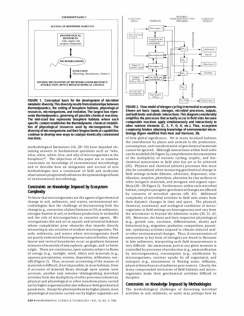
Constraints on Knowledge Imposed by EcosystemComplexityTo know that microorganisms are the agents of geochemicalchange in soil, sediments, and waters, environmental mi-crobiologists face the challenge of documenting both thechange (e.g., conversion of plant biomass to CO2 in sediments,nitrogen fixation in soil, or methane production in wetlands)and the role of microorganisms as causative agents. Mi-croorganisms live and act in aquatic and terrestrial habitatswhose complexities pose obstacles that impede directlymeasuring in situ activities of resident microorganisms. Thesoils, sediments, and waters where microorganisms dwellare poorly understood heterogeneous natural bodies, whoselateral and vertical boundaries occur as gradients betweenmixtures of materials of atmospheric, geologic, and/or bioticorigin. These are continuous, open systems subject to fluxesof energy (e.g., sunlight, wind, tides) and materials (e.g.,aqueous precipitation, erosion, deposition, infiltration, run-off) (Figure 2). Thus, accurate accounting of the masses ofmaterials is difficult, if not impossible, in most habitats. Evenif accounts of material fluxes through open system wereaccurate, another task remainssdistinguishing microbialactivities from the multiplicity of other processes (chemical,physical and physiological or other transformations carriedout by higher organisms) that also influence field geochemicalparameters. Except for photosynthesis by higher plants, mostphysiological reactions carried out by higher organisms are
of little global significance. Yet in many localized habitatsthe contribution by plants and animals to the production,consumption, and transformation of geochemical materialscannot be ignored. Although interactions within food webscan be modeled (34; Figure 2), comprehensive documentationof the multiplicity of nutrient cycling, trophic, and bio-chemical interactions in field sites has yet to be achieved(35). Physical and chemical (abiotic) processes that mustalso be considered when measuring geochemical change infield settings include dilution, advection, dispersion, vola-tilization, sorption, photolysis, alteration by clay surfaces orother inorganic materials, and inorganic and organic equi-libria (36-39; Figure 2). Furthermore, within each microbialhabitat, complex synergistic geochemical changes are effectedby consortia of microbial species (40, 41). Additionalcomplexity of microbial activities in field sites stems fromtheir dynamic changes in time and space. The physical,chemical, nutritional, and ecological conditions of micro-organisms in field settings are heterogeneous and vary fromthe micrometer to beyond the kilometer scales (30, 31, 42,43). Moreover, the biota and their respective physiological(e.g., growth rate, excretion, differentiation, death) andbehavioral (e.g., migration, predation, competition, parasit-ism, symbiosis) activities respond to climate-induced and/or other environmental changes. Thus, if concentrations ofammonium (a key form of nitrogen) are found to fluctuatein lake sediments, interpreting such field measurements isvery difficult: the ammonium pool at any given moment iscontrolled by processes of production (e.g., ammonificationby microorganisms), consumption (e.g., nitrification bymicroorganisms, nutrient uptake by all organisms), andtransport (e.g., entrainment in flowing water, diffusion,physical disturbances of sediment pore waters). Clearly, themany compounded intricacies of field habitats and micro-organisms make their geochemical activities difficult todecipher.
Constraints on Knowledge Imposed by MethodologiesThe methodological challenges of discerning microbialactivities in soil, sediment, or water may perhaps best be
FIGURE 1. Conceptual basis for the development of microbialmetabolic diversity. This diversity results from relationships betweenthermodynamics, the setting of biosphere habitats, physiologicalresources, microorganisms, and evolution. The largest box repre-sents thermodynamics, governing all possible chemical reactions.The mid-sized box represents biosphere habitats where eachspecific context establishes the thermodynamic chemical instabili-ties of physiological resources used by microorganisms. Thediversity of microorganisms and their biogeochemical capabilitiescontinue to develop new ways to catalyze kinetically constrainedreactions.
FIGURE 2. Flow model of nitrogen cycling in terrestrial ecosystems.Shown are basic inputs, storages, microbial processes, outputs,and both biotic and abiotic interactions. This diagram considerablysimplifies the processes that actually occur in field sites becausecomparable reactions apply simultaneously and interactively toother nutrient elements (C, S, P, O, H, etc.). Thus, ecosystemcomplexity hinders obtaining knowledge of environmental micro-biology (figure modified from Heal and Harrison; 35).
430 9 ENVIRONMENTAL SCIENCE & TECHNOLOGY / VOL. 32, NO. 4, 1998
appreciated by considering how we know that higher plantscarry out photosynthesis. In surveying a given landscape,humans can gather evidence for photosynthesis simply bynoting the location of vegetation. Humans and the vegetationare roughly the same scale (≈ meter); therefore, detectingplants and their spatial relationships to one another andtheir habitats is facile. Photosynthesis is the major bio-geochemical function of higher plants; without it there wouldbe no plants nor food chains based thereupon. Thus, thepresence of higher plants provides evidence for conversionof atmospheric CO2 to biomass, and (because rooted plantsare immobile) we simultaneously discern where the pho-tosynthesis has occurred. At a mere glance then, humansgain plant-related biogeochemical knowledge addressing fourkey questions: who? (the plant), what? (photosynthesis),when? (recent history), and where? (the plant’s location). Togain knowledge of the remaining two commonly asked keyquestions, “how?” and “why?”, we rely on reductionisticbiological disciplines that include physiology, biochemistry,genetics, and molecular biologyssome of which can beapplied to field-gathered plant samples or be manifest aschambers deployed to field sites. Now let us contrast howthe six key questions pertinent to plant photosynthesis wereanswered with how the same questions are answered formetabolic activities of microorganisms in field habitats.
Limited Methods for Determining the Position andComposition of Microorganisms in Their Habitats. Mi-croorganisms are small (on the order of micrometers). Themillion-fold discrepancy in size between humans andmicroorganisms ensures that gathering field samples formicroscopic analysis will physically disturb both the mi-croorganisms and their habitats. Microorganisms removedfrom their native environments have been characterizedmicroscopically (44, 45). However, very little is known aboutthe three-dimensional structure of microenvironments thatsurround microorganisms in field sites. New approachessuch as transmission electron microscopy performed on thinsections of embedded samples and environmental scanningelectron microscopy are developing for examining complexenvironments such as soil (46, 47). Yet, complete microscopiccharacterization of soil is a distant possibility because thesoil biomass occupies only 0.001% of the soil volume (46).This means that a multitude of microscopic fields, eachsurveying a very small volume of soil, would need to beprocessed to obtain information accurately representing insitu spatial relationships of soil microorganisms. Thus, unlikeplants in landscapes, detailed knowledge of where micro-organisms dwell is very difficult to obtain because of scale-related and sampling-related physical characteristics ofmicrohabitats and microorganisms therein.
To answer the question “Who is there?”, environmentalmicrobiologists have developed three general types of assays(24, 29, 32, 48-53): (i) viable plate counts of organisms ableto grow on laboratory-incubated selective agar media; (ii)extraction and analysis of nucleic acids, phospholipids, orother cellular biomarkers; and (iii) microscopic examinationof fixed, stained samples. Each of these methodologies hasits own limitations. Common to all three is the highprobability of overlooking members of microbial communi-ties that may be functionally significant but may occur inlow abundances and therefore be undetected. Results ofviable plate count assays provide information about the small(≈1%) proportion of the initially diverse mixture of micro-organisms that are able to grow under physiological condi-tions imposed by limited resources presented to the mi-croorganisms in laboratory-incubated agar media (54, 55).Unmet challenges in designing the proper laboratory condi-tions for growing microorganisms are a major reason forsuch low cultivation efficiencies; but some microorganismsmay also attain an unculturable physiological state (12).
Extraction of cell-specific biomarkers has proven to beeffective for some cellular components (such as phospholipidfatty acids; 56-58) but susceptible to inefficiencies and biasesfor others (such as nucleic acids; 59-61). Nucleic acidextraction followed by cloning and sequencing of phyloge-netically revealing 16S rRNA genes (8, 54, 55, 62-67; discussedfurther below) has recently provided evidence for novelresidents of many habitats. When applied to a given fieldsite, the results of this phenotype-free means of identifyingmicroorganisms usually contrast strikingly with those ofgrowth-based assays. However, physiological inferencesfrom phenotype-free methodologies can be misleadingbecause bacteria that are closely related by molecular criteriacan display widely different biogeochemical capabilities (8).
The microscopic approach for characterizing naturallyoccurring microorganisms typically disperses an environ-mental sample (e.g., soil), preserves it with a chemical fixative,and smears a portion onto a glass slide where the keymicrobial components (especially nucleic acids, antigeniccell surfaces, or unique nucleic acid sequences) can be stained(with general nucleic acid-binding dyes, cell-specific anti-bodies, or with gene-specific oligonucleotide hybridizationprobes, respectively) to distinguish microbial cells from theinorganic and noncellular organic materials (29, 32, 48-52).General nucleic acid staining provides information on totalmicroorganisms but usually falls short of providing informa-tion about the identity of individual cells because few typesof microorganisms are morphologically distinctive. Whenmicroscopy is combined with cell-specific (antibody andnucleic acid) procedures designed to allow particular mi-croorganisms to be recognized (8, 33, 54, 55, 62-70), itremains a challenge to verify the specificity and accuracy ofresults from cells that probe positively in complex naturallyoccurring communities.
Despite substantial sophistication in many of the aboveprocedures assessing “Who is there?” in naturally occurringmicrobial communities, a complete census has yet to besuccessfully accomplished in any environment (32). Fur-thermore, of the 300 000-1 000 000 species of bacteriabelieved to exist globally (24), less than 5000 have beencharacterized in traditional culture collections (8), andapproximately the same number of rRNA genes (from bothcultured microorganisms and field-extracted nucleic acids)have been sequenced. Thus, there is much knowledge yetto be gained.
Limited Methods for Determining in Situ Biogeochemi-cal Activities and When They Occur. Of the millions ofmicroorganisms found in each cubic centimeter of soil,sediment, and water, there are thousands of species (24),each with complex genomes conferring the potential to carryout a variety of biogeochemical processes. Furthermore,many naturally occurring microorganisms exist as spores orother resting, completely dormant, or nonviable forms. Thus,unlike the clear link between the presence of higher plantsand photosynthetic activity, the presence of microorganismsin environmental samples provides few clues about theirspecific physiological functions in situ.
The question “What are microorganisms doing?” can besubdivided into “What is the general physiological status ofthe cells?” and “What specific geochemical activities are thecells engaged in?”. To assess the general physiological statusof microorganisms in field sites, environmental microbiolo-gists again rely on samples that are usually physicallydisturbed by removal from the field. And, similar to pro-cedures inquiring about the composition of microbialcommunities (above), information about physiological statuscan be obtained from measurements conducted on labora-tory-incubated samples, from biomarkers extracted from thesamples, and/or via microscopic techniques (29, 32, 48-53).However, the physiological assays focus on key indicators
VOL. 32, NO. 4, 1998 / ENVIRONMENTAL SCIENCE & TECHNOLOGY 9 431
such as cellular contents of ribosomes (indicative of proteinsynthesis activity; 71); intracellular energy reserves (poly-â-hydroxyalkanoates, ATP; 57, 58, 72); membrane compo-nents reflecting nutritional status (proportions of trans/cisor cyclopropyl phospholipid fatty acids or electron transportcarriers; 56-58); and time-course measurements of cellelongation, uptake of physiological substrates, or the reduc-tion of dyes indicative of respiratory activity (the latter threeassays are performed on laboratory-incubated environmentalsamples; 29, 32, 53). These kinds of assays are insightful, buteach is limited in the information provided and carriesartifactual risks (16, 29, 32; see below).
Methods for inquiring into the specific in situ geochemicalactivities catalyzed by microorganisms seek to documentthe impact of microbial activities on the chemical composi-tion of soils, sediments, waters, and the atmosphere. Forsome microbial activities, the geochemical materials ofinterest or related microbial metabolites are volatile gases;hence, the underlying net microbial processes are measurableusing field chambers placed over the surface of habitats beingstudied (73, 74). However, when neither the geochemicalmaterials nor their metabolic products are volatile, docu-menting field metabolic processes requires more elaboratestrategies that include physiologically guided chemicalanalysis of field samples, seeking stable isotopic fractionationpatterns in field samples (75), field release of stable isoto-pically labeled materials, isolating a portion of the habitatfor hypothesis-driven manipulations, in situ microelectrodemeasurements of chemical gradients (76, 77), probing formRNAs and/or enzymes indicative of gene expression (78-81), and conducting physiological assays indicative of themetabolic activity of interest on laboratory-incubated fieldsamples (16, 29, 32, 48-53). The credibility of such bio-geochemical activity measures varies on a case-by-case basiswith the habitat studied, the means of procedural imple-mentation, and the microbiological process of interest. Mostare influenced by uncertainties discussed below.
Accurate knowledge of temporal aspects of microbialactivity in field sites, addressing the question, “When are themicroorganisms active?”, is difficult to obtain. If field samplesare fixed the moment they are gathered, then informationsubsequently gleaned after analysis completion can beconsidered indicative of the status of the microbial com-munity at the time of sampling (32). This real time-typecharacterization of microorganisms in field sites is implicitin most field-chamber (73) and microelectrode (76) inves-tigations and has recently been applied to biodegradation ofenvironmental contaminants (82). But knowledge of whenmicroorganisms carry out key biogeochemical reactions isoften uncertainsinferred after the fact. Just as net photo-synthesis in higher plants is inferred by the presence of plantbiomass, microbial decay processes in steady-state ecosys-tems (e.g., salt marshes, forests, grasslands) can be inferredfrom the steady-state itself (35). Many ecosystems displayproductive seasons of plant growth that lead to the transferof deceased biomass to soil and sediments. Yet, litter layerson the surface of soils and sediments do not constantly accrue.Thus, environmental microbiologists infer (after testingalternative hypotheses) that in situ microbially mediateddecay processes counterbalance carbon inputs from pho-tosynthesis. This large-scale mass-balance approach toecosystem biogeochemistry has revealed essential insightsinto watershed processes (83), but the scale of resolutiondoes not address the mechanistic biochemical intricaciessought by microbiologists.
Insights and Uncertainties from Model Systems De-signed To Determine “What, Why, and How” of MicrobialBiogeochemical Reactions. Assembling mass balances forgeochemical components in field sites, distinguishing mi-crobiological from other processes, and tracing circuitous
routes of geochemical materials through food chains andoxidation/reduction reactions are formidable tasks (seeabove; Figure 2). Many environmental microbiologists haveconfronted this situation and concluded that such adversitiesare nearly insurmountable in efforts aimed at discerning whatmicroorganisms are doing in field sites (30-32, 84). Thecommon way to contend with uncertainties of microbialactivities is to initiate flask assays in the laboratory thatmonitor the chemical transformation(s) of interest in samplesgathered from field sites. These laboratory assays providedefinitive qualitative evidence for potential microbial meta-bolic reactions because sterilized or poisoned treatmentscan be examined as abiotic controls and mass balances aremade possible by performing the assays in sealed vessels.For example, in the 1870s while examining microbialtransformations of nitrogen, Schloesing and Muntz [cited inWaksman (3)] described a key link in the nitrogen cycle,nitrification, by reporting that nitrate was formed fromammonia in nonsterile but not in poisoned columns of sandinfiltrated by sewage effluent. However, it is critical toacknowledge that measurements performed on laboratory-incubated environmental samples reveal what may be, butnot necessarily what is, actually occurring in field sites.
A Heisenberg uncertainty-type principle is inescapablein environmental microbiology (32, 85) and must be con-fronted both in the examination of field site samples and inexploiting the spectrum of disciplines that contribute to ourmechanistic understanding of microbiological processes.When one begins in a field site or with site-derived samples,the closer microorganisms are examined, the more likely theresultant information is to suffer from artifacts imposed bythe measurement procedures. The basis for such artifactsis habitat disturbance (discussed above) and the responsive-ness of both individual microorganisms and entire microbialcommunities to environmental change implicit in habitatdisturbance (30-32). Environmental microbiologists gener-ally agree that, given sufficient time, the microbial communitypresent in every environmental sample will change accordingto selective pressures (resources and environmental condi-tions) imposed by removal of samples from their originallocation in field sites and by all intentional and unintentionallaboratory incubation conditions (temperature, oxygen ten-sion, physical disturbance, addition of nutrients or growthsubstrates, etc.). Of considerable controversy, however, isthe amount of time required for microorganisms in envi-ronmental samples to respond to sampling and experimen-tally induced environmental changes. Implicit in manypublished investigations is the hypothesis that accuratequalitative and quantitative microbial activity determinationsof in situ processes can be performed in the laboratory withina safe period before artifacts develop (32). This hypothesishas not been adequately tested. Yet, its validity is essentialfor the extrapolation of results from laboratory incubationsto field sites (16, 32, 48-53, 86, 87). The alternative con-servative methodological approach views laboratory incuba-tions of environmental samples, at best, as a means towardestimating field processes. From the conservative viewpoint,quantitative extrapolation from laboratory results to actualfield processes is taboo (16, 32) because the instant anenvironmental sample is removed from a field study site,intricate and tightly regulated genetic-, biochemical-, cel-lular-, and population-level changes may be triggered (32).It is the investigator’s inability to obtain disturbance-freesamples and to fully characterize, understand, and duplicatefield conditions in the laboratory that undermine theacceptance of laboratory measurements performed on fieldsamples as valid surrogates for true in situ field processes.
Controlled model laboratory experiments allow a logicalreductionistic progression to proceed from field sample, tolaboratory incubation, to enrichment cultures, to the isolation
432 9 ENVIRONMENTAL SCIENCE & TECHNOLOGY / VOL. 32, NO. 4, 1998
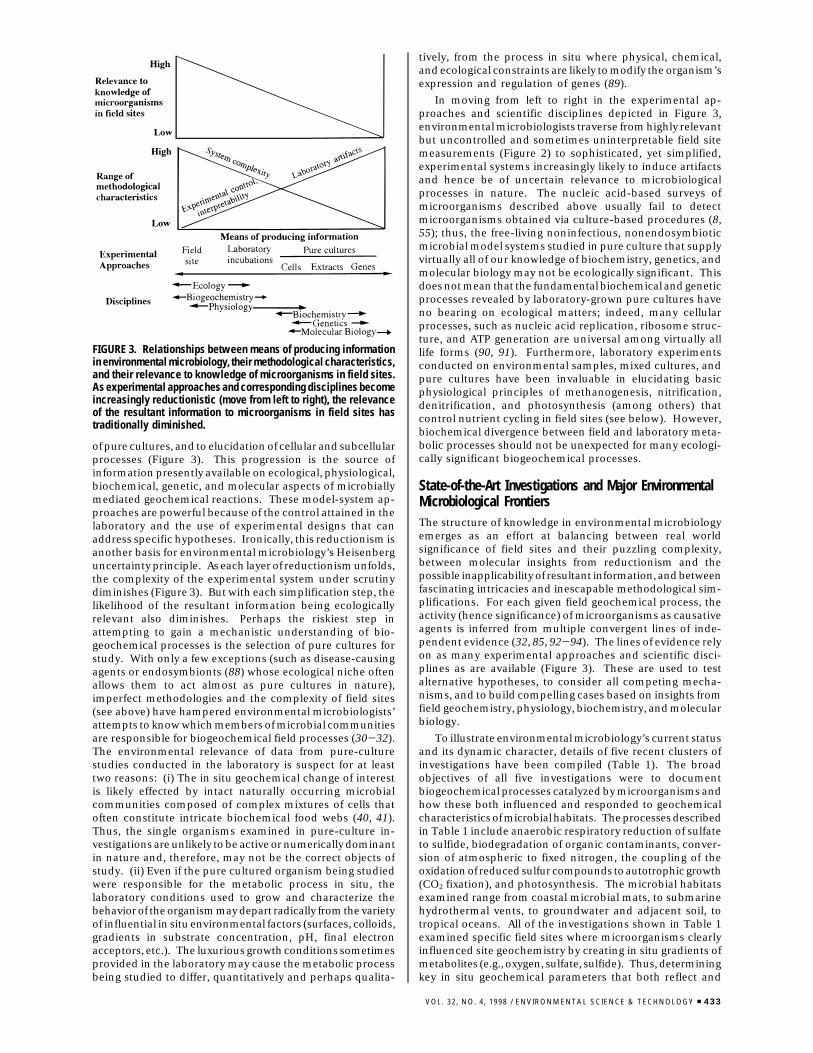
of pure cultures, and to elucidation of cellular and subcellularprocesses (Figure 3). This progression is the source ofinformation presently available on ecological, physiological,biochemical, genetic, and molecular aspects of microbiallymediated geochemical reactions. These model-system ap-proaches are powerful because of the control attained in thelaboratory and the use of experimental designs that canaddress specific hypotheses. Ironically, this reductionism isanother basis for environmental microbiology’s Heisenberguncertainty principle. As each layer of reductionism unfolds,the complexity of the experimental system under scrutinydiminishes (Figure 3). But with each simplification step, thelikelihood of the resultant information being ecologicallyrelevant also diminishes. Perhaps the riskiest step inattempting to gain a mechanistic understanding of bio-geochemical processes is the selection of pure cultures forstudy. With only a few exceptions (such as disease-causingagents or endosymbionts (88) whose ecological niche oftenallows them to act almost as pure cultures in nature),imperfect methodologies and the complexity of field sites(see above) have hampered environmental microbiologists’attempts to know which members of microbial communitiesare responsible for biogeochemical field processes (30-32).The environmental relevance of data from pure-culturestudies conducted in the laboratory is suspect for at leasttwo reasons: (i) The in situ geochemical change of interestis likely effected by intact naturally occurring microbialcommunities composed of complex mixtures of cells thatoften constitute intricate biochemical food webs (40, 41).Thus, the single organisms examined in pure-culture in-vestigations are unlikely to be active or numerically dominantin nature and, therefore, may not be the correct objects ofstudy. (ii) Even if the pure cultured organism being studiedwere responsible for the metabolic process in situ, thelaboratory conditions used to grow and characterize thebehavior of the organism may depart radically from the varietyof influential in situ environmental factors (surfaces, colloids,gradients in substrate concentration, pH, final electronacceptors, etc.). The luxurious growth conditions sometimesprovided in the laboratory may cause the metabolic processbeing studied to differ, quantitatively and perhaps qualita-
tively, from the process in situ where physical, chemical,and ecological constraints are likely to modify the organism’sexpression and regulation of genes (89).
In moving from left to right in the experimental ap-proaches and scientific disciplines depicted in Figure 3,environmental microbiologists traverse from highly relevantbut uncontrolled and sometimes uninterpretable field sitemeasurements (Figure 2) to sophisticated, yet simplified,experimental systems increasingly likely to induce artifactsand hence be of uncertain relevance to microbiologicalprocesses in nature. The nucleic acid-based surveys ofmicroorganisms described above usually fail to detectmicroorganisms obtained via culture-based procedures (8,55); thus, the free-living noninfectious, nonendosymbioticmicrobial model systems studied in pure culture that supplyvirtually all of our knowledge of biochemistry, genetics, andmolecular biology may not be ecologically significant. Thisdoes not mean that the fundamental biochemical and geneticprocesses revealed by laboratory-grown pure cultures haveno bearing on ecological matters; indeed, many cellularprocesses, such as nucleic acid replication, ribosome struc-ture, and ATP generation are universal among virtually alllife forms (90, 91). Furthermore, laboratory experimentsconducted on environmental samples, mixed cultures, andpure cultures have been invaluable in elucidating basicphysiological principles of methanogenesis, nitrification,denitrification, and photosynthesis (among others) thatcontrol nutrient cycling in field sites (see below). However,biochemical divergence between field and laboratory meta-bolic processes should not be unexpected for many ecologi-cally significant biogeochemical processes.
State-of-the-Art Investigations and Major EnvironmentalMicrobiological FrontiersThe structure of knowledge in environmental microbiologyemerges as an effort at balancing between real worldsignificance of field sites and their puzzling complexity,between molecular insights from reductionism and thepossible inapplicability of resultant information, and betweenfascinating intricacies and inescapable methodological sim-plifications. For each given field geochemical process, theactivity (hence significance) of microorganisms as causativeagents is inferred from multiple convergent lines of inde-pendent evidence (32, 85, 92-94). The lines of evidence relyon as many experimental approaches and scientific disci-plines as are available (Figure 3). These are used to testalternative hypotheses, to consider all competing mecha-nisms, and to build compelling cases based on insights fromfield geochemistry, physiology, biochemistry, and molecularbiology.
To illustrate environmental microbiology’s current statusand its dynamic character, details of five recent clusters ofinvestigations have been compiled (Table 1). The broadobjectives of all five investigations were to documentbiogeochemical processes catalyzed by microorganisms andhow these both influenced and responded to geochemicalcharacteristics of microbial habitats. The processes describedin Table 1 include anaerobic respiratory reduction of sulfateto sulfide, biodegradation of organic contaminants, conver-sion of atmospheric to fixed nitrogen, the coupling of theoxidation of reduced sulfur compounds to autotrophic growth(CO2 fixation), and photosynthesis. The microbial habitatsexamined range from coastal microbial mats, to submarinehydrothermal vents, to groundwater and adjacent soil, totropical oceans. All of the investigations shown in Table 1examined specific field sites where microorganisms clearlyinfluenced site geochemistry by creating in situ gradients ofmetabolites (e.g., oxygen, sulfate, sulfide). Thus, determiningkey in situ geochemical parameters that both reflect and
FIGURE 3. Relationships between means of producing informationin environmental microbiology, their methodological characteristics,and their relevance to knowledge of microorganisms in field sites.As experimental approaches and corresponding disciplines becomeincreasingly reductionistic (move from left to right), the relevanceof the resultant information to microorganisms in field sites hastraditionally diminished.
VOL. 32, NO. 4, 1998 / ENVIRONMENTAL SCIENCE & TECHNOLOGY 9 433
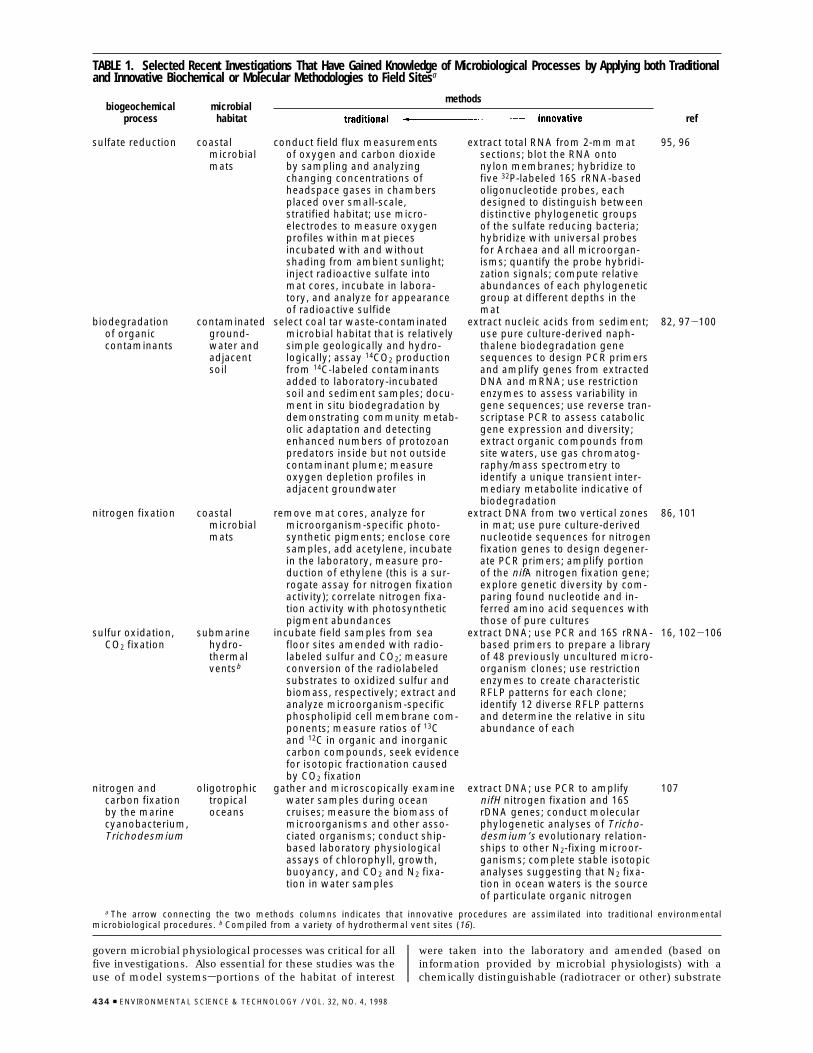
govern microbial physiological processes was critical for allfive investigations. Also essential for these studies was theuse of model systemssportions of the habitat of interest
were taken into the laboratory and amended (based oninformation provided by microbial physiologists) with achemically distinguishable (radiotracer or other) substrate
TABLE 1. Selected Recent Investigations That Have Gained Knowledge of Microbiological Processes by Applying both Traditionaland Innovative Biochemical or Molecular Methodologies to Field Sitesa
methodsbiogeochemical
processmicrobial
habitat ref
sulfate reduction coastalmicrobialmats
conduct field flux measurementsof oxygen and carbon dioxideby sampling and analyzingchanging concentrations ofheadspace gases in chambersplaced over small-scale,stratified habitat; use micro-electrodes to measure oxygenprofiles within mat piecesincubated with and withoutshading from ambient sunlight;inject radioactive sulfate intomat cores, incubate in labora-tory, and analyze for appearanceof radioactive sulfide
extract total RNA from 2-mm matsections; blot the RNA ontonylon membranes; hybridize tofive 32P-labeled 16S rRNA-basedoligonucleotide probes, eachdesigned to distinguish betweendistinctive phylogenetic groupsof the sulfate reducing bacteria;hybridize with universal probesfor Archaea and all microorgan-isms; quantify the probe hybridi-zation signals; compute relativeabundances of each phylogeneticgroup at different depths in themat
95, 96
biodegradationof organiccontaminants
contaminatedground-water andadjacentsoil
select coal tar waste-contaminatedmicrobial habitat that is relativelysimple geologically and hydro-logically; assay 14CO2 productionfrom 14C-labeled contaminantsadded to laboratory-incubatedsoil and sediment samples; docu-ment in situ biodegradation bydemonstrating community metab-olic adaptation and detectingenhanced numbers of protozoanpredators inside but not outsidecontaminant plume; measureoxygen depletion profiles inadjacent groundwater
extract nucleic acids from sediment;use pure culture-derived naph-thalene biodegradation genesequences to design PCR primersand amplify genes from extractedDNA and mRNA; use restrictionenzymes to assess variability ingene sequences; use reverse tran-scriptase PCR to assess catabolicgene expression and diversity;extract organic compounds fromsite waters, use gas chromatog-raphy/mass spectrometry toidentify a unique transient inter-mediary metabolite indicative ofbiodegradation
82, 97-100
nitrogen fixation coastalmicrobialmats
remove mat cores, analyze formicroorganism-specific photo-synthetic pigments; enclose coresamples, add acetylene, incubatein the laboratory, measure pro-duction of ethylene (this is a sur-rogate assay for nitrogen fixationactivity); correlate nitrogen fixa-tion activity with photosyntheticpigment abundances
extract DNA from two vertical zonesin mat; use pure culture-derivednucleotide sequences for nitrogenfixation genes to design degener-ate PCR primers; amplify portionof the nifA nitrogen fixation gene;explore genetic diversity by com-paring found nucleotide and in-ferred amino acid sequences withthose of pure cultures
86, 101
sulfur oxidation,CO2 fixation
submarinehydro-thermalventsb
incubate field samples from seafloor sites amended with radio-labeled sulfur and CO2; measureconversion of the radiolabeledsubstrates to oxidized sulfur andbiomass, respectively; extract andanalyze microorganism-specificphospholipid cell membrane com-ponents; measure ratios of 13Cand 12C in organic and inorganiccarbon compounds, seek evidencefor isotopic fractionation causedby CO2 fixation
extract DNA; use PCR and 16S rRNA-based primers to prepare a libraryof 48 previously uncultured micro-organism clones; use restrictionenzymes to create characteristicRFLP patterns for each clone;identify 12 diverse RFLP patternsand determine the relative in situabundance of each
16, 102-106
nitrogen andcarbon fixationby the marinecyanobacterium,Trichodesmium
oligotrophictropicaloceans
gather and microscopically examinewater samples during oceancruises; measure the biomass ofmicroorganisms and other asso-ciated organisms; conduct ship-based laboratory physiologicalassays of chlorophyll, growth,buoyancy, and CO2 and N2 fixa-tion in water samples
extract DNA; use PCR to amplifynifH nitrogen fixation and 16SrDNA genes; conduct molecularphylogenetic analyses of Tricho-desmium’s evolutionary relation-ships to other N2-fixing microor-ganisms; complete stable isotopicanalyses suggesting that N2 fixa-tion in ocean waters is the sourceof particulate organic nitrogen
107
a The arrow connecting the two methods columns indicates that innovative procedures are assimilated into traditional environmentalmicrobiological procedures. b Compiled from a variety of hydrothermal vent sites (16).
434 9 ENVIRONMENTAL SCIENCE & TECHNOLOGY / VOL. 32, NO. 4, 1998
whose alteration by site microorganisms was indicative ofthe geochemical metabolic processes of interest. Anothercommon trait in the five investigations shown in Table 1 wasthe rapid processing of field samples using biochemicalprocedures to analyze cellular components. The method-ologies described in Table 1 are divided into two columns,labeled “traditional” and “innovative”. The arrow thatconnects these two categories of methods is designed toillustrate the dynamic state of methodologies, hence infor-mation and knowledge in environmental microbiology. Allmethods presently viewed as traditional were once innova-tive. For instance, the early 1970s marked the introductionof epifluorescent microscopic procedures, which providednew knowledge of the abundance and distribution of aquaticmicroorganisms (30, 31, 33). These epifluorescent proce-dures have since become broadly accepted and are consid-ered traditional today. This pattern in which innovativemethodologies are assimilated into traditional ones is theongoing mechanism that ensures that hypotheses will betested in new ways so that the knowledge of environmentalmicrobiology will constantly grow, be refined, and asymp-totically approach truth.
One of the most striking current innovations in environ-mental microbiology (well represented in column 4 of Table1) is the nucleic acid-based approach for inquiring intomicrobial diversity (described above). Proposed in the mid1980s (62), these procedures are founded on an understand-ing of the molecular phylogeny of ribosomal RNA and othergenes that has revolutionized the evolutionary and taxonomicbasis of biology (6-8). Other key developments essential forthis type of inquiry were innovations in molecular biologyand other disciplines that include microscopy, flow cytom-etry, nucleic acid sequencing procedures, the polymerasechain reaction, nucleic acid hybridization procedures, in-strumentation for nucleotide sequence analyses, and elec-tronic storage and retrieval of nucleotide sequences (54, 55,62-67).
Another recent set of innovations elegantly uses gaseousmetabolites found in field sites to infer the in situ physiologicalactivities of native microbial populations. The partialpressure of hydrogen gas (a key transient intermediarymetabolite in anaerobic food chains) has been shown to occurin anaerobic habitats at discrete nanomolar concentrationsthat are diagnostic for methanogenesis, sulfate reduction,iron reduction, and denitrification (108, 109). In addition,knowledge of isotopic fractionation processes can reveal howmicroorganisms control the geochemistry of their habitats(75). For instance, the carbon dioxide found in field siteshas different 13C/12C ratios depending on the 13C/12C signatureof the substrates respired and the 13C-enriching process ofmethanogenesis. When site-specific signatures of bothinorganic and organic carbon reservoirs have been charac-terized, the relative contribution of each to the pool of CO2
can be discerned (110). The radioactive (14C) component ofCO2 is also revealing because the different carbon pools mayhave distinctive ages (111).
The information compiled in Table 2 extends the inves-tigation-specific details presented in Table 1 in a mannerthat addresses the six key questions (raised above) aboutgeochemical reactions catalyzed by microorganisms in theirhabitats. Included in Table 2 are partial compilations of thesignificance of the key questions and associated enduringissues for environmental microbiology. To emphasize thedynamic status of environmental microbiological knowledge,Table 2 also partially compiles traditional methods foranswering the questions, innovative methodological im-provements and their bases, and examples of recent knowl-edge stemming from the innovations.
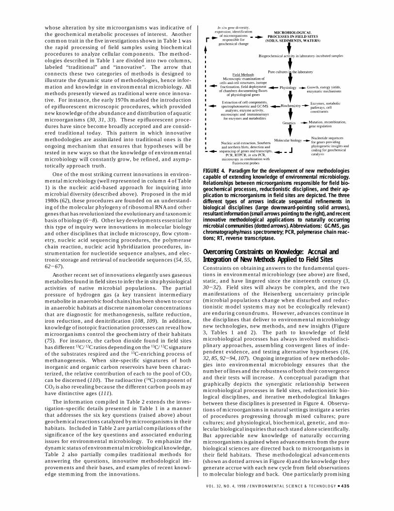
Overcoming Constraints on Knowledge: Accrual andIntegration of New Methods Applied to Field SitesConstraints on obtaining answers to the fundamental ques-tions in environmental microbiology (see above) are fixed,static, and have lingered since the nineteenth century (3,30-32). Field sites will always be complex, and the twomanifestations of the Heisenberg uncertainty principle(microbial populations change when disturbed and reduc-tionistic model systems may not be ecologically relevant)are enduring conundrums. However, advances continue inthe disciplines that deliver to environmental microbiologynew technologies, new methods, and new insights (Figure3, Tables 1 and 2). The path to knowledge of fieldmicrobiological processes has always involved multidisci-plinary approaches, assembling convergent lines of inde-pendent evidence, and testing alternative hypotheses (16,32, 85, 92-94, 107). Ongoing integration of new methodolo-gies into environmental microbiology ensures that thenumber of lines and the robustness of both their convergenceand their tests will increase. A conceptual paradigm thatgraphically depicts the synergistic relationship betweenmicrobiological processes in field sites, reductionistic bio-logical disciplines, and iterative methodological linkagesbetween these disciplines is presented in Figure 4. Observa-tions of microorganisms in natural settings instigate a seriesof procedures progressing through mixed cultures; purecultures; and physiological, biochemical, genetic, and mo-lecular biological inquiries that each stand alone scientifically.But appreciable new knowledge of naturally occurringmicroorganisms is gained when advancements from the purebiological sciences are directed back to microorganisms intheir field habitats. These methodological advancements(shown as dotted arrows in Figure 4) and the knowledge theygenerate accrue with each new cycle from field observationsto molecular biology and back. One particularly promising
FIGURE 4. Paradigm for the development of new methodologiescapable of extending knowledge of environmental microbiology.Relationships between microorganisms responsible for field bio-geochemical processes, reductionistic disciplines, and their ap-plication to microorganisms in field sites are depicted. The threedifferent types of arrows indicate sequential refinements inbiological disciplines (large downward-pointing solid arrows),resultant information (small arrows pointing to the right), and recentinnovative methodological applications to naturally occurringmicrobial communities (dotted arrows). Abbreviations: GC/MS, gaschromatography/mass spectrometry; PCR, polymerase chain reac-tions; RT, reverse transcriptase.
VOL. 32, NO. 4, 1998 / ENVIRONMENTAL SCIENCE & TECHNOLOGY 9 435
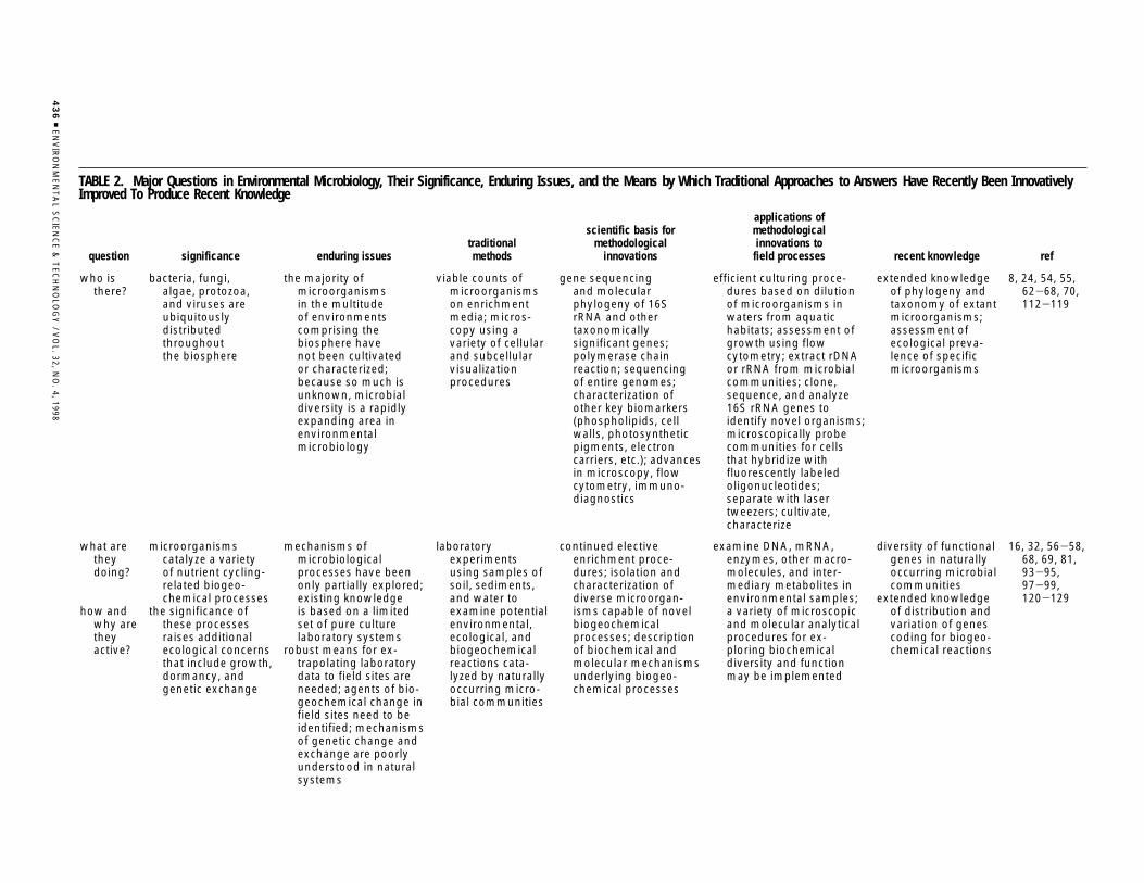
TABLE 2. Major Questions in Environmental Microbiology, Their Significance, Enduring Issues, and the Means by Which Traditional Approaches to Answers Have Recently Been InnovativelyImproved To Produce Recent Knowledge
question significance enduring issuestraditionalmethods
scientific basis formethodological
innovations
applications ofmethodologicalinnovations to
field processes recent knowledge ref
who isthere?
bacteria, fungi,algae, protozoa,and viruses areubiquitouslydistributedthroughoutthe biosphere
the majority ofmicroorganismsin the multitudeof environmentscomprising thebiosphere havenot been cultivatedor characterized;because so much isunknown, microbialdiversity is a rapidlyexpanding area inenvironmentalmicrobiology
viable counts ofmicroorganismson enrichmentmedia; micros-copy using avariety of cellularand subcellularvisualizationprocedures
gene sequencingand molecularphylogeny of 16SrRNA and othertaxonomicallysignificant genes;polymerase chainreaction; sequencingof entire genomes;characterization ofother key biomarkers(phospholipids, cellwalls, photosyntheticpigments, electroncarriers, etc.); advancesin microscopy, flowcytometry, immuno-diagnostics
efficient culturing proce-dures based on dilutionof microorganisms inwaters from aquatichabitats; assessment ofgrowth using flowcytometry; extract rDNAor rRNA from microbialcommunities; clone,sequence, and analyze16S rRNA genes toidentify novel organisms;microscopically probecommunities for cellsthat hybridize withfluorescently labeledoligonucleotides;separate with lasertweezers; cultivate,characterize
extended knowledgeof phylogeny andtaxonomy of extantmicroorganisms;assessment ofecological preva-lence of specificmicroorganisms
8, 24, 54, 55,62-68, 70,112-119
what aretheydoing?
how andwhy aretheyactive?
microorganismscatalyze a varietyof nutrient cycling-related biogeo-chemical processes
the significance ofthese processesraises additionalecological concernsthat include growth,dormancy, andgenetic exchange
mechanisms ofmicrobiologicalprocesses have beenonly partially explored;existing knowledgeis based on a limitedset of pure culturelaboratory systems
robust means for ex-trapolating laboratorydata to field sites areneeded; agents of bio-geochemical change infield sites need to beidentified; mechanismsof genetic change andexchange are poorlyunderstood in naturalsystems
laboratoryexperimentsusing samples ofsoil, sediments,and water toexamine potentialenvironmental,ecological, andbiogeochemicalreactions cata-lyzed by naturallyoccurring micro-bial communities
continued electiveenrichment proce-dures; isolation andcharacterization ofdiverse microorgan-isms capable of novelbiogeochemicalprocesses; descriptionof biochemical andmolecular mechanismsunderlying biogeo-chemical processes
examine DNA, mRNA,enzymes, other macro-molecules, and inter-mediary metabolites inenvironmental samples;a variety of microscopicand molecular analyticalprocedures for ex-ploring biochemicaldiversity and functionmay be implemented
diversity of functionalgenes in naturallyoccurring microbialcommunities
extended knowledgeof distribution andvariation of genescoding for biogeo-chemical reactions
16, 32, 56-58,68, 69, 81,93-95,97-99,120-129
43
69
EN
VIR
ON
ME
NT
AL
SC
IEN
CE
&T
EC
HN
OLO
GY
/V
OL.
32,N
O.
4,1998
strategy for inquiry into the genetic diversity of microbialmetabolic capabilities is to bypass the “pure cultures” stepin Figure 4 by creating genomic libraries directly fromenvironmental samples (130-132). These can then bescreened for a variety of previously undiscovered genes andgene products. The static constraints on knowledge inenvironmental microbiology have been yielding and willcontinue to yield at an accelerating rate to the dynamicaccruing advancements that arise when new methodologiesare applied to field sites. This bodes well for the future ofenvironmental microbiology and its impacts on biospheremanagement and biotechnological products and services.
AcknowledgmentsSupport from the Air Force Office of Scientific Research(Grants AFOSR-91-436, F49620-93-1-0414, and F49620-95-1-0346); by the Cornell Biotechnology Program, which issponsored by the New York State Science and TechnologyFoundation, a consortium of industries, and the NationalScience Foundation; and the NIEHS/Superfund Basic Re-search and Education Program (Grant ES-05950-03) isgratefully acknowledged. I thank W. C. Ghiorse, J. B. Yavitt,and S. H. Zinder for discussions assisting in the developmentof ideas presented here and four anonymous reviewers andthe editor (J. M. Suflita), who contributed many key refine-ments.
Literature Cited(1) Atlas, R. M.; Bartha, R. Microbial Ecology: Fundamentals and
Applications, 4th ed.; Benjamin/Cummings Publishing Co.:Menlo Park, CA, 1997.
(2) Winogradsky, S. Microbiologie du sol, problemes et methodes;cinquante ans de recherches. Oeuvres completes; Masson: Paris,1949.
(3) Waksman, S. A. Principles of Soil Microbiology; Williams &Wilkins, Co.: Baltimore, MD, 1927.
(4) Hurst, C. J. In Manual of Environmental Microbiology; Hurst,C. J., Knudsen, G. R., McInerney, M. J., Stetzenbach, L. D., WalterM. V., Eds.; ASM Press: Washington, DC, 1997; pp 3-4.
(5) Madigan, M. T.; Martinko, J. M.; Parker, J. Biology of Microor-ganisms, 8th ed.; Prentice Hall: Englewood Cliffs, NJ, 1997.
(6) Woese, C. R. Microbiol. Rev. 1987, 51, 221-271.(7) Stackebrandt, E. In The Prokaryotes, 2nd ed.; Balows, A., Truper,
H. G., Dworkin, M., Harder, W., Schleifer, K.-H., Eds.; Springer-Verlag: New York, 1992; pp 19-47.
(8) Pace, N. Science 1997, 276, 734-740.(9) Strickberger, M. W. Evolution, 2nd ed.; Jones and Bartlett
Publishers: Sudbury, MA, 1996.(10) Ryan, K. J., Ed. Sherris Medical Microbiology: An Introduction
to Infectious Diseases, 3rd ed.; Appleton and Lange: Norwalk,CT, 1994.
(11) Demain, A. L., Soloman, N. A., Eds. Biology of IndustrialMicroorganisms; Benjamin-Cummings Publishing Co.: MenloPark, CA, 1985.
(12) Colwell, R. R.; Clayton, R. A.; Ortiz-Conde, B. A.; Jacobs, D.;Russek-Cohen, E. In Microbial Diversity and Ecosystem Function;Allsopp, D., Colwell, R. R., Hawksworth, D. L., Eds.; CABInternational: Wallingford, U.K., 1992; pp 3-15.
(13) Bull, A. T.; Goodfellow, M.; Slater, J. H. Annu. Rev. Microbiol.1992, 46, 219-252.
(14) Dworkin, M. In The Prokaryotes, 2nd ed.; Balows, A., Truper, H.G., Dworkin, M., Harder, W., Schleifer, K.-H., Eds.; Springer-Verlag: New York, 1992; pp 48-74.
(15) Ehrlich, H. L. Geomicrobiology, 3rd ed.; Marcel Dekker, Inc.:New York, 1995.
(16) Karl, D. M. In The Microbiology of Deep Sea Hydrothermal Vents;Karl, D. M., Ed.; CRC Press: New York, 1995; pp 35-124.
(17) Schlegel, H. G.; Jannasch, H. W. In The Prokaryotes, 2nd ed.;Balows, A., Truper, H. G., Dworkin, M., Harder, W., Schleifer,K.-H., Eds.; Springer-Verlag: New York, 1992; pp 75-125.
(18) Leadbetter, E. R. In Manual of Environmental Microbiology;Hurst, C. J., Knudsen, G. R., McInerney, M. J., Stetzenbach, L.D., Walter, M. V., Eds.; ASM Press: Washington, DC, 1997; pp14-24.
(19) Rayner, A. D. M. In Microbial Diversity and Ecosystem Function;Allsopp, D., Colwell, R. R., Hawksworth, D. L., Eds.; CABInternational: Wallingford, U.K., 1995; pp 231-251.w
hen
and
wh
ere
are
they
acti
ve?
spat
iala
nd
tem
po
ral
con
tro
lso
fm
icro
-b
iala
ctiv
ity
are
po
orl
yu
nd
erst
oo
din
virt
ual
lyal
lfie
ldsi
tes
ph
ysic
al,c
hem
ical
,an
db
iolo
gic
alh
eter
og
enei
-ti
esth
atco
ntr
ibu
teto
fiel
dsi
teco
mp
lexi
tyan
dg
rad
ien
tso
fre
sou
rces
uti
lized
by
mic
roo
rgan
ism
sin
thei
rh
abit
ats
are
po
orl
yu
nd
erst
oo
d;f
acto
rsth
atco
ntr
olm
icro
bia
lfie
ldp
roce
sses
nee
dto
be
un
der
sto
od
;co
ntr
ols
incl
ud
em
ole
cula
rsi
gn
als,
ph
ysic
al,c
hem
i-ca
l,an
den
viro
nm
enta
lch
arac
teri
stic
s,an
dce
ll-ce
llin
tera
ctio
ns
(in
clu
din
gsy
mb
iosi
s,p
red
ato
r-p
rey,
par
a-si
tism
,co
mp
etit
ion
,an
dan
tag
on
ism
)
ava
riet
yo
fas
epti
csa
mp
ling
,fix
atio
n,
and
mic
rosc
op
icex
amin
atio
nas
says
;in
stal
lch
amb
ers,
pie
zo-
met
ers
for
mo
ni-
tori
ng
chan
ges
invo
lati
leo
rso
lub
lean
alyt
es;
wh
ole
-wat
ersh
edst
ud
ies
that
com
-p
lete
mas
sb
alan
cean
dfl
ux
mea
sure
-m
ents
sen
sors
and
fiel
dch
arac
teri
zati
on
and
sam
plin
gp
roce
du
res
that
can
be
com
bin
edw
ith
ph
ysio
log
ical
,b
ioch
emic
al,g
enet
ic,
and
mo
lecu
lar
assa
ys
dep
loy
sen
sors
,ch
emic
als
and
/or
mic
roo
rgan
ism
sin
fiel
dsi
tes;
extr
act
key
enzy
mes
or
inte
rmed
iate
met
abo
lites
;mea
sure
and
inte
rpre
tst
able
iso
top
efr
acti
on
atio
np
atte
rns;
use
mic
rose
nso
rsto
do
cum
ent
con
cen
trat
ion
gra
die
nts
insi
tu
inq
uir
ies
into
spec
ific
insi
tub
iog
eoch
emi-
cala
ctiv
itie
sca
ta-
lyze
db
ym
icro
org
an-
ism
sin
real
tim
e
29,3
2,73
-80
,82
,86,
87,9
3,94
,100
,104
,10
8-11
1,12
7
VOL. 32, NO. 4, 1998 / ENVIRONMENTAL SCIENCE & TECHNOLOGY 9 437
(20) Zehnder, A. J. B.; Stumm, W. In Biology of Anaerobic Microor-ganisms; Zehnder, A. J. B., Ed.; John Wiley & Sons, Inc.: NewYork, 1988; pp 1-38.
(21) Devlin, T. M., Ed. Textbook of Biochemistry with ClinicalCorrelations; Wiley-Liss: New York, 1997.
(22) Waksman, S. A. Science 1945, 102, 339-344.(23) Schlesinger, W. H. Biogeochemistry: An analysis of Global
Change; Academic Press: Inc: San Diego, CA, 1991.(24) Tiedje, J. M. In Microbial Diversity and Ecosystem Function;
Allsopp, D., Colwell, R. R., Hawksworth, D. L., Eds.; CABInternational: Wallingford, U.K., 1995; pp 73-97.
(25) Honderich, T., Ed. The Oxford Companion to Philosophy; OxfordUniversity Press: Oxford, 1995.
(26) Cunningham, W. F. Notes on Epistemology; Declan X. McMullenCo., Inc.: New York, 1930.
(27) Bateson, G. In A Sacred Unity; Donaldson, R. E., Ed.; HarperCollins Publishers: New York, 1991.
(28) Brock, T. D. In Ecology of Microbial Communities; Forty-firstSymposium of the Society for General Microbiology; CambridgeUniversity Press: New York, 1987; pp 1-17.
(29) Karl, D. M In Bacteria in Nature; Poindexter, J. S., Leadbetter,E. R., Eds.; Plenum Press: New York, 1986; Vol. 2, pp 85-176.
(30) Hobbie, J. E. In Handbook of Methods in Aquatic MicrobialEcology; Kemp, P. F., Sherr, B. F., Sherr, E. B., Cole, J. J., Eds.;Lewis Publishers: Boca Raton, FL, 1993; pp 1-5.
(31) Hobbie, J. E.; Ford, T. E. In Aquatic Microbiology; Ford, T. E.,Ed.; Blackwell Scientific Publishers: Boston, MA, 1993; pp 1-14.
(32) Madsen, E. L. In Soil Biochemistry, Vol. 9; Stotzky, G., Bollag,J.-M., Eds.; Marcel Dekker, Inc.: New York, 1996; pp 287-370.
(33) Paul, J. H. In Aquatic Microbiology; Ford, T. E., Ed.; BlackwellScientific Publishers: Boston, MA, 1993; pp 15-46.
(34) Parton, W. J.; Stewart, J. W. B.; Cole, C. V. Biogeochemistry 1988,5, 109-131.
(35) Heal, O. W.; Harrison, A. F. In Nutrient Cycling in TerrestrialEcosystems: Field Methods, Application and Interpretation;Harrison, A. F., Ineson, P., Heal, O. W., Eds.; Elsevier AppliedScience: London, 1990; pp 170-178.
(36) Mansour, M.; Feicht, E. A. Chemosphere 1994, 28, 323-332.(37) Wolfe, N. L. Abiotic Transformations of Pesticides in Natural
Waters and Sediments; John Wiley & Sons, Inc.: New York, 1992.(38) Thibodeaux, L. J. Environmental Chemodynamics, 2nd ed.; John
Wiley & Sons: New York, 1995.(39) Stumm, W.; Morgan, J. J. Aquatic Chemistry, 3rd ed.; John Wiley
& Sons: New York, 1996.(40) Gottschal, J. C.; Harder, W.; Prins, R. A. In The Prokaryotes, 2nd
ed.; Balows, A., Truper, H. G., Dworkin, M., Harder, W., Schleifer,K.-H., Eds.; Springer-Verlag: New York, 1992; pp 149-196.
(41) Zinder, S. H. In Methanogenesis: Ecology, Physiology, Biochem-istry, and Genetics; Ferry, J. G., Ed.; Chapman and Hall: NewYork, 1993; pp 128-206.
(42) Groffman, P. M. Agriculture Research in the Northeastern UnitedStates: Critical Review and Future Perspectives; ASA: Madison,WI, 1993; pp 19-26.
(43) Parkin, T. B. J. Environ. Qual. 1993, 22, 409-417.(44) Caldwell, D. E.; Korber, D. R.; Lawrence, J. R. Adv. Microb. Ecol.
1993, 12, 1-67.(45) Sieburth, J. M. Microbial Seascapes: A Pictorial Essay on Marine
Microorganisms and Their Environment; University Park Press:Baltimore, MD, 1975.
(46) Foster, R. C. In Soil Micromorphology: Studies in Managementand Genesis; Ringrose-Voase, A. J., Humphreys, G. S., Eds.;Elsevier: New York, 1993; pp 381-393.
(47) Ladd, J. N.; Foster, R. C.; Nannipieri, P.; Oades, J. M. In SoilBiochemistry, Vol. 9; Stotzky, G., Bollag, J.-M., Eds.; MarcelDekker, Inc.: New York, 1996; pp 23-78.
(48) Hurst, C. J., Knudsen, G. R., McInerney, M. J., Stetzenbach, L.D., Walter M. V., Eds. Manual of Environmental Microbiology;American Society for Microbiology: Washington, DC, 1997.
(49) Kemp, P. F., Sherr, B. F., Sherr, E. B., Cole, J. J., Eds. Handbookof Methods in Aquatic Microbial Ecology; Lewis Publishers: BocaRaton, FL, 1993.
(50) Levin, M. A., Seidler, R. J., Rogul, M., Eds. Microbial Ecology:Principles, Methods, and Applications; McGraw-Hill, Inc.: NewYork, 1992.
(51) Weaver, R. W. et al., Eds. Methods of Soil Analysis Part 2.Microbiological and Biochemical Properties; Soil Science Societyof America: Madison, WI, 1994.
(52) Burlage, R., Ed. Techniques in Microbial Ecology; OxfordUniversity Press: New York, 1997.
(53) Staley, J. T.; Konopka, A. Annu. Rev. Microbiol. 1985, 39, 321-346.
(54) Amann, R. I.; Ludwig, W.; Schleifer, K.-H. Microbiol. Rev. 1995,59, 143-169.
(55) Ward, D. M.; Bateson, M. M.; Weller, R.; Ruff-Roberts, A. L. Adv.Microb. Ecol. 1993, 12, 219-286.
(56) Findlay, R. H.; Dobbs, F. C. In Handbook of Methods in AquaticMicrobial Ecology; Kemp, P. F., Sherr, B. F., Sherr, E. B., Cole,J. J., Eds.; Lewis Publishers: Chelsea, MI, 1993; pp 271-284.
(57) Tunlid, A.; White, D. C. In Soil Biochemistry, Vol. 7; Stotzky, G.,Bollag, J.-M., Eds.; Marcel Dekker: Inc.: New York, 1992; pp229-262.
(58) White, D. C.; Pinkart, H. C.; Ringelberg, D. B. In Manual ofEnvironmental Microbiology; Hurst, C. J., Knudsen, G. R.,McInerney, M. J., Stetzenbach, L. D., Walter M. V., Eds.; ASMPress: Washington, DC, 1997; pp 91-101.
(59) Farrelly, V.; Rainey, F. A.; Stackebrandt, E. Appl. Environ.Microbiol. 1995, 61, 2798-2801.
(60) More, M. I.; Herrick, J. B.; Silva, M. C.; Ghiorse, W. C.; Madsen,E. L. Appl. Environ. Microbiol. 1994, 60, 1572-1580.
(61) Suzuki, M. T.; Giovannoni, S. J. Appl. Environ. Microbiol. 1996,62, 625-630.
(62) Pace, N. R.; Stahl, D. A.; Lane, D. J.; Olsen, G. J. Adv. Microb.Ecol. 1986, 9, 1-55.
(63) Barns, S. M.; Fundyga, R. E.; Jeffries, M. W.; Pace, N. R. Proc.Natl. Acad. Sci. U.S.A. 1994, 91, 1609-1613.
(64) Giovannoni, S. J.; Britschgi, T. B.; Moyer, C. L.; Field, K. G. Nature1990, 345, 60-63.
(65) Stahl, D. A. In Manual of Environmental Microbiology; Hurst,C. J., Knudsen, G. R., McInerney, M. J., Stetzenbach, L. D., WalterM. V., Eds.; ASM Press: Washington, DC, 1997; pp 102-114.
(66) Ward, N.; Rainey, F. A.; Goebel, B.; Stackebrandt, E. In MicrobialDiversity and Ecosystem Function; Allsopp, D., Colwell, R. R.,Hawksworth, D. L., Eds.; CAB International: Wallingford, U.K.,1995; pp 89-110.
(67) Barns, S. M.; Delwiche, C. F.; Palmer, J. C.; Pace, N. R. Proc. Natl.Acad. Sci. U.S.A. 1996, 93, 9188-9193.
(68) Ward, B. B. Limnol. Oceanogr. 1984 29, 402-410.(69) Currin, C. A.; Paerl, H. W.; Suba, G. K.; Alberte, R. S. Limnol.
Oceanogr 1990, 35, 59-71.(70) Siering, P. L.; Ghiorse, W. C. Appl. Environ. Microbiol. 1996, 63,
644-651.(71) Kemp, P. F.; Lee, S.; LaRoche, J. In Handbook of Methods in
Aquatic Microbial Ecology; Kemp, P. F., Sherr, B. F., Sherr, E. B.,Cole, J. J., Eds.; Lewis Publishers: Boca Raton, FL, 1993; pp415-422.
(72) Karl, D. M. In Handbook of Methods in Aquatic Microbial Ecology;Kemp, P. F., Sherr, B. F., Sherr, E. B., Cole, J. J., Eds.; LewisPublishers: Chelsea, MI, 1993; pp 483-494.
(73) Conrad, R. Microbiol. Rev. 1996, 60, 609-640.(74) Yavitt, J. B.; Lang, G. E.; Sexstone, A. J. J. Geophys. Res. 1990, 95,
22463-22474.(75) Grossman, E. L. In Manual of Environmental Microbiology; Hurst,
C. J., Knudsen, G. R., McInerney, M. J., Stetzenbach, L. D., WalterM. V., Eds.; ASM Press: Washington, DC, 1997; pp 565-576.
(76) Glud, R. N.; Gundersen, J. K.; Revsbech, N. P.; Jorgensen, B. B.Limnol. Oceanogr. 1994, 39, 462-467.
(77) Revsbech, N. P.; Jørgensen, B. B. Adv. Microb. Ecol. 1986, 9,293-352.
(78) Pichard, S. L.; Paul, J. H. Appl. Environ. Microbiol. 1993, 59,451-457.
(79) Ogram, A.; Sun, W.; Brockman, F. J.; Fredrickson, J. K. Appl.Environ. Microbiol. 1995, 61, 763-768.
(80) Ogunseitan, O. A. J. Microbiol. Methods 1997, 28, 55-63.(81) Hodson, R. E.; Dustman, W. A.; Garg, R. P.; Moran, M. A. Appl.
Environ. Microbiol. 1995, 61, 4074-4082.(82) Wilson, M. S.; Madsen, E. L. Environ. Sci. Technol. 1996, 30,
2099-2103.(83) Likens, G. E.; Bormann, F. H. Biogeochemistry of a Forested
Ecosystem; Springer-Verlag: New York, 1995.(84) Bull, A. T. In Contemporary Microbial Ecology; Ellwood, D. C.,
Hedger, J. N., Latham, M. J., Lynch, J. M., Slater, J. H., Eds.;Academic Press: New York, 1980; pp 107-136.
(85) Madsen, E. L. Environ. Sci. Technol. 1991, 25, 1662-1673.(86) Pinckney, J.; Paerl, H. W.; Fitzpatrick, M. Mar. Ecol. Prog. Ser.
1995, 123, 207-216.(87) Tiedje, J. M.; Simkins, S.; Groffman, P. M. Plant Soil 1989, 115,
261-284.(88) Ruby, E. G. Annu. Rev. Microbiol. 1996, 50, 591-624.(89) Lindow, S. E. Mol. Ecol. 1995, 4, 555-566.(90) Kluyver, A. J.; van Niel, C. B. The Microbe’s Contribution to
Biology; Harvard University Press: Cambridge, MA, 1954.(91) Neidhardt, F. C.; Ingraham, J. I.; Schaechter, M. Physiology of
the Bacterial Cell; Sinauer Assoc., Inc.: Sunderland, MA, 1990.
438 9 ENVIRONMENTAL SCIENCE & TECHNOLOGY / VOL. 32, NO. 4, 1998
(92) McKay, D. S.; et al. Science 1996, 273, 924-930.(93) Stevens, T. O.; McKinley, J. P. Science 1995, 270, 450-454.(94) Krumholz, L. R.; McKinley, J. P.; Ulrich, G. A.; Suflita, J. M. Nature
1997, 386, 64-66.(95) Canfield, D. E.; Des Marais, D. J. Geochim. Cosmochim. Acta
1993, 57, 3971-3984.(96) Risatti, J. B.; Chapman, W. C.; Stahl, D. A. Proc. Natl. Acad. Sci.
U.S.A. 1994, 91, 10173-10177.(97) Herrick, J. B.; Madsen, E. L.; Batt, C. A.; Ghiorse, W. C. Appl.
Environ. Microbiol. 1993, 59, 687-694.(98) Madsen, E. L.; Sinclair, J. L.; Ghiorse, W. C. Science 1991, 252,
830-833.(99) Murarka, I.; Neuhauser, E.; Sherman, M.; Taylor, B. B.; Mauro,
D. M.; Ripp, J.; Taylor, T. J. Hazard. Mater. 1992, 32, 245-261.(100) Wilson, M. S.; Madsen, E. L. Abstract, American Society for
Microbiology Annual Meeting, Miami, FL, 1997; N-142; p 404.(101) Zehr, J. P.; Mellon, M.; Braun, S.; Litaker, W.; Steppe, T.; Paerl,
H. W. Appl. Environ. Microbiol. 1995, 61, 2527-2532.(102) Hedrick, D. B.; Pledger, R. D.; White, D. C.; Baross, J. A. FEMS
Microbiol. Ecol. 1992, 101, 1-10.(103) Jannasch, H. W. In Autotrophic Bacteria; Schlegel, H. G., Bowien,
B., Eds.; Science Tech Publishers: Madison, WI, 1989; pp 147-166.
(104) Kennicutt, M. C., II; Burke, R. A., Jr. In The Microbiology ofDeep Sea Hydrothermal Vents; Karl, D. M., Ed.; CRC Press:New York, 1995; pp 275-287.
(105) Moyer, C. L.; Dobbs, F. C.; Karl, D. M. Appl. Environ. Microbiol.1994, 60, 871-879.
(106) Tuttle, J. H. Biol. Soc. Wash. Bull. 1985, 6, 335-343.(107) Capone, D. B.; Zehr, J. P.; Paerl, H. W.; Bergman, B.; Carpenter,
E. J. Science 1997, 226, 1221-1229.(108) Lovley, D. R.; Chapelle, F. H.; Woodward, J. C. Environ. Sci.
Technol. 1994, 28, 1205-1210.(109) Chapelle, F. H.; Vroblesky D. A.; Woodward, J. C.; Lovley, D.
R. Environ. Sci. Technol. 1997, 31, 2873-2877.(110) Landemeyer, J. E.; Vroblesky, D. A.; Chapelle, F. H. Environ.
Sci. Technol. 1996, 30, 1120-1128.(111) Conrad, M. E.; Daley, P. F.; Fischer, M. L.; Buchanan, B. B.;
Leighton, T.; Kashgarian, M. Environ. Sci. Technol. 1997, 31,1463-1469.
(112) Button, D. K.; Schut, F.; Quang, P.; Martin, R.; Robertson, B.R. Appl. Environ. Microbiol. 1993, 59, 881-891.
(113) Ferris, M. J.; Muyzer, G.; Ward, D. M. Appl. Environ. Microbiol.1996, 62, 340-346.
(114) Huber, R.; Burggraf, S.; Mayer, T.; Barns, S. M.; Rossnagel, P.;Stetter, K. O. Nature 1995, 376, 57-58.
(115) Holben, W. E.; Harris, D. Mol. Ecol. 1995, 4, 627-631.(116) Holben, W. E. In Manual of Environmental Microbiology; Hurst,
C. J., Knudsen, G. R., McInerney, M. J., Stetzenbach, L. D.,Walter M. V., Eds.; ASM Press: Washington, DC, 1997; pp 431-436.
(117) Ogram, A.; Feng, X. In Manual of Environmental Microbiology;Hurst, C. J., Knudsen, G. R., McInerney, M. J., Stetzenbach, L.D., Walter M. V., Eds.; ASM Press: Washington, DC, 1997; pp422-430.
(118) Wawer, C.; Muyzer, G. Appl. Environ. Microbiol. 1995, 61, 2203-2210.
(119) Liu, W.-T.; Marsh, T. L.; Cheng, H.; Forney, L. J. Appl. Environ.Microbiol. 1997, 63, 4516-4522.
(120) Fleming, J. T.; Sanseverino, J.; Sayler, G. S. Environ. Sci. Technol.1993, 27, 1068-1074.
(121) Sayler, G. S.; Layton, A.; Lajoie, C.; Bowman, J.; Tschantz, M.;Fleming, J. T. Appl. Biochem. Biotechnol. 1995, 54, 277-290.
(122) van der Meer, J. R. Antonie van Leeuwenhoek 1997, 71, 159-178.
(123) Williams, P. A.; Sayers, J. R. Biodegradation 1994, 5, 195-217.(124) Zylstra, G. J. In Molecular Environmental Biology; Garte, S. J.,
Ed.; Lewis Publishers Inc.: Boca Raton, FL, 1983; pp 83-115.(125) Brockman, F. J. Mol. Ecol. 1995, 4, 567-578.(126) Byrne, A. M.; Kukor, J. J.; Olsen, R. H. Gene 1995, 154, 65-70.(127) Harkness, M. R.; et al. Science 1993, 259, 503-507.(128) De Souza, M. L.; Sadowsky, M. J.; Wackett, L. P. J. Bacteriol.
1996, 178, 4894-4900.(129) Herrick, J. B.; Stuart-Keil, K. G.; Ghiorse, W. C.; Madsen, E. L.
Appl. Environ. Microbiol. 1997, 63, 2330-2337.(130) Healy, F. G.; Ray, R. M.; Aldrich, H. C.; Wilkie, A. C.; Ingram,
L. O.; Shanmugam, K. T. Appl. Microbiol. Biotechnol. 1995, 43,667-674.
(131) Bakermans, C.; Madsen, E. L. Abstract, American Society forMicrobiology, Annual Meeting, Miami, FL, 1997; N-120; p 401.
(132) Stein, J. L.; Marsh, T. L.; Wu, K. Y.; Shizuya, H.; DeLong, E. F.J. Bacteriol. 1996, 178, 591-599.
Received for review June 23, 1997. Revised manuscript re-ceived November 4, 1997. Accepted November 19, 1997.
ES970551Y
VOL. 32, NO. 4, 1998 / ENVIRONMENTAL SCIENCE & TECHNOLOGY 9 439