essential amino acid supplementation decreases liver damage induced by chronic ethanol consumption...
TRANSCRIPT
ORIGINAL ARTICLE
Dietary supplementation with essential amino acids booststhe beneficial effects of rosuvastatin on mouse kidney
Giovanni Corsetti • Giuseppe D’Antona • Chiara Ruocco •
Alessandra Stacchiotti • Claudia Romano • Laura Tedesco •
Francesco Dioguardi • Rita Rezzani • Enzo Nisoli
Received: 9 April 2014 / Accepted: 26 May 2014
� The Author(s) 2014. This article is published with open access at Springerlink.com
Abstract The effects of high-potency statins on renal
function are controversial. To address the impact of statins
on renal morpho-functional aspects, normotensive young
mice were treated with rosuvastatin (Rvs). Moreover,
because statins may impair mitochondrial function, mice
received either dietary supplementation with an amino acid
mixture enriched in essential amino acids (EAAm), which
we previously demonstrated to increase mitochondrial bio-
genesis in muscle or an unsupplemented control diet for
1 month. Mitochondrial biogenesis and function, apoptosis,
and insulin signaling pathway events were studied, primarily
in cortical proximal tubules. By electron microscopy ana-
lysis, mitochondria were more abundant and more hetero-
geneous in size, with dense granules in the inner matrix, in
Rvs- and Rvs plus EAAm-treated animals. Rvs administra-
tion increased protein kinase B and endothelial nitric oxide
synthase phosphorylation, but the mammalian target of
rapamycin signaling pathway was not affected. Rvs
increased the expression of sirtuin 1, peroxisome prolifera-
tor-activated receptor c coactivator-1a, cytochrome oxidase
type IV, cytochrome c, and mitochondrial biogenesis
markers. Levels of glucose-regulated protein 75 (Grp75),
B-cell lymphoma 2, and cyclin-dependent kinase inhibitor 1
were increased in cortical proximal tubules, and expression
of the endoplasmic reticulum–mitochondrial chaperone
Grp78 was decreased. EAAm supplementation maintained
or enhanced these changes. Rvs promotes mitochondrial
biogenesis, with a probable anti-apoptotic effect. EAAm
boosts these processes and may contribute to the efficient
control of cellular energetics and survival in the mouse
kidney. This suggests that appropriate nutritional interven-
tions may enhance the beneficial actions of Rvs, and could
potentially prevent chronic renal side effects.
Keywords Rosuvastatin � Essential amino acids �Kidney � Mitochondria � Mice
Abbreviations
p-AKT Phosphorylated protein kinase B
Bcl-2 B-cell lymphoma 2
CDKN1A Cyclin-dependent kinase inhibitor 1 (p21)
COX IV Cytochrome c oxidase
Cyt c Cytochrome c
EAAm Essential amino acid mixture
p-eNOS Phosphorylated endothelial nitric oxide
synthase
ER Endoplasmic reticulum
GAPDH Glyceraldehyde 3-phosphate dehydrogenase
Grp75 Glucose-regulated protein 75
Grp78 Glucose-regulated protein 78
mtDNA Mitochondrial DNA
G. Corsetti, G. D’Antona and C. Ruocco contributed equally to this
work.
G. Corsetti � A. Stacchiotti � C. Romano � R. Rezzani
Division of Human Anatomy, Department of Clinical and
Experimental Sciences, University of Brescia,
25123 Brescia, Italy
G. D’Antona
Department of Molecular Medicine, University of Pavia,
27100 Pavia, Italy
C. Ruocco � L. Tedesco � E. Nisoli (&)
Department of Medical Biotechnology and Translational
Medicine, University of Milan, via Vanvitelli 32,
20129 Milan, Italy
e-mail: [email protected]
F. Dioguardi
Department of Clinical Sciences and Community, University of
Milan, 20122 Milan, Italy
123
Amino Acids
DOI 10.1007/s00726-014-1772-5

NRF-1 Nuclear respiratory factor-1
OXPHOS Oxidative phosphorylation
PGC-1a Peroxisome proliferator-activated receptor ccoactivator-1 a
Tfam Mitochondrial transcription factor A
Rvs Rosuvastatin
Introduction
According to the 2013 American College of Cardiology/
American Heart Association Guideline on the Treatment of
Blood Cholesterol, statins are the only class of drugs
indicated for the prevention and treatment of atheroscle-
rosis and cardiovascular disease (CVD) in subjects with
increased risk (Stone et al. 2013). Statins are also known as
3-hydroxy-3-methylglutaryl-CoA (HMG-CoA) reductase
inhibitors. HMG-CoA reductase is the rate-limiting
enzyme in cholesterol synthesis, and its inhibition reduces
pleiotropically the production of a number of molecules
with inflammatory, endothelial, oxidant, and blood vessel
tone properties (Susic et al. 2003; Bae et al. 2010; Fiore
et al. 2011). Adverse effects and intolerance of statins
depend on the specific prescribed molecule and on patient
characteristics (Mancini et al. 2013). Adverse muscle,
hepatic, and gastrointestinal effects have been extensively
reported in the literature (Mansi et al. 2013), but contro-
versy exists regarding the effects of high-potency statins on
renal function. Statin-induced hematuria and proteinuria
have been well established, and microalbuminuria is
thought to be secondary to statin interference with the
tubular reabsorption of albumin (van der Tol et al. 2012;
Robles et al. 2013). In 36 studies of rosuvastatin (Rvs)
involving more than 40,000 patients, renal impairment or
renal failure was reported in only 536 participants (Stein
et al. 2012). In addition, two meta-analyses that assessed
the benefits and harms of statin use in patients with renal
disease showed no deterioration, and rather a trend toward
improvement or maintenance of renal function, with
decreased CVD and mortality, in patients with chronic
kidney disease (Upadhyay et al. 2012; Palmer et al. 2012).
Different potential mechanisms have been suggested as
causing or contributing to statin side effects (Knauer et al.
2010). Mitochondrial mechanisms have been repeatedly
implicated, particularly in muscle damage (Golomb and
Evans 2008). Statins lead to dose-dependent reductions in
coenzyme Q10 (De Pinieux et al. 1996), a key mitochon-
drial antioxidant and electron transport carrier that can
bypass existing mitochondrial respiratory chain defects
(Rosenfeldt et al. 2002). Several studies demonstrate that
statins predispose to mitochondrial defects (Gambelli et al.
2004; Schick et al. 2007) in all users and, to a greater
degree, in vulnerable individuals. A decreased Q10 content
was accompanied by a decreased maximal oxidative
phosphorylation (OXPHOS) capacity in simvastatin-trea-
ted patients (Larsen et al. 2013). Moreover, dose-dependent
reductions in coenzyme Q10 can reduce cell energy, pro-
mote oxidation or apoptosis, and unmask silent mito-
chondrial defects (Lenaz et al. 2002). It is plausible that
these findings partly explain the muscle pain and exercise
intolerance that many patients experience with their statin
treatment.
Extensive evidence supports the efficacy of dietary
supplementation with a balanced amino acid mixture
enriched in essential amino acids (EAAm) in clinical dis-
orders characterized by deficits of energy production,
including ageing, type 2 diabetes, heart failure, alcohol
consumption, and renal failure (Cano et al. 2006; Valerio
et al. 2011; Corsetti et al. 2012). It is widely accepted that
the kidneys play a major role in amino acid metabolism
and, partially, in the regulation of amino acid plasma lev-
els. About 97 % of the filtered amino acids are actively
reabsorbed by renal tubules, and amino acids affect several
kidney physiological processes depending on their quality
and quantity (Silbernagl 1988). Among others, arginine,
the substrate of nitric oxide synthase (NOS) and thus the
precursor of NO synthesis (Andrew and Myer 1999), plays
key roles in renal vessel and tubular functions (Rajapaskse
and Mattson 2013). Arginine ingestion or systemic
administration increases renal blood flow and glomerular
filtration rate in humans (Kano et al. 1992), possibly by
displacement of asymmetric-di-methyl-arginine (ADMA),
the endogenous inhibitor of endothelial nitric oxide syn-
thase (eNOS) (MacAllister and Vallance 1994). Further-
more, renal eNOS is present not only in endothelial cells
but also in the cells of the collecting ducts and macula
densa, where NO regulates tubulo-glomerular feedback and
renin secretion (MacAllister and Vallance 1994). The
mTOR-AKT-eNOS pathway is known to be the cellular
sensor of amino acid availability (Zoncu et al. 2011).
Signaling via mTOR has been implicated in proliferative
kidney diseases with an increased phosphorylation of
downstream targets, including ribosomal protein S6 (Shil-
lingford et al. 2006).
Notably, the EAAs leucine, lysine and phenylalanine act
similarly to insulin and suppress proteolysis of circulating
and renal peptides. Supplementation with this EAAm was
found to slow renal senescence in rodents (Corsetti, et al.
2010) and in hemodialysis patients (Bolasco et al. 2011).
We have recently demonstrated that this specific amino
acid formula restores mitochondrial function and antioxi-
dant responses through eNOS expression, with reduced
inflammatory processes, in the organs and tissues of aged
rodents (D’Antona et al. 2010; Corsetti et al. 2011).
G. Corsetti et al.
123

Although lacking arginine, the EAAm promotes arginine
synthesis (Rondanelli et al. 2011).
The present work aimed to study firstly the effects of
long-term treatment with Rvs on the morphological and
immunohistochemical aspects of the kidney in young mice,
and secondly the impact of EAAm supplementation on the
renal effects of Rvs, mainly focusing on mitochondrial
adaptive processes.
Materials and methods
Animals and treatments
C57BL/6 male mice (2 months old, n = 36, Charles River,
Calco, Italy) were housed in a quiet room with controlled
temperature and humidity. A 12 h/12 h light/dark cycle
was maintained (lights on from 7 a.m. to 7 p.m.). The
experimental protocol was conducted in accordance with
European Community guidelines and the Italian Ministry
of Health, complied with The National Animal Protection
Guidelines, and was approved by the Institutional Animal
Ethical Committee.
Animals were given unrestricted access to a standard
diet (4.3 % fat, 18.8 % protein, 76.9 % carbohydrate, La-
boratorio Dottori Piccioni; for information about dietary
ingredients and actual composition of the diet see Table 1)
and tap water, and were treated for up to 1 month with Rvs
(20 mg/kg/day, n = 10 animals), Rvs plus EAAm (1.5 mg/
g body weight, n = 10 animals), or EAAm alone (1.5 mg/g
body weight, n = 8 animals) via drinking water. A control
group received drinking water without any drugs or sup-
plement (CTRL; n = 8 animals). EAAm (composition,
relative percentage, and dietary intake of each amino acid
are reported in Table 2) was dissolved in tap water in
quantities determined by calculating average daily drinking
for 2 weeks before starting treatment (approximately 6 ml
as in Bachmanov et al. 2002) and stored at 4 �C before
daily administration. EAAm concentrations were previ-
ously found to be active in rodents and mimic the recom-
mended daily dose for humans (see Pellegrino et al. 2005;
D’Antona et al. 2010). The Rvs dosage selected in this
study is comparable to those adopted in other studies
conducted to prove its ability to lower blood cholesterol
(Enomoto et al. 2009).
Body weight, kidney weight, and average daily food and
water consumption are reported in Table 3. Interestingly,
the dietary EAAm supplementation induced a non-statis-
tically significant reduction of food consumption in Rvs-
treated and Rvs-untreated mice in comparison to the
respective controls. This reduction led to slight changes in
daily protein intake (CTRL: 0.76 ± 0.07 g/day; EAAm:
Table 1 Dietary ingredients, amino acid content and actual compo-
sition of the diet
Macronutrient composition
Nutrient % Amino acid g/100 g
Raw proteins 18.8 Arginine 1.15
Histidine 0.48
Isoleucine 0.80
Leucine 1.50
Lysine 1.05
Methionine ? cysteine 0.75
Phenylalanine 0.90
Threonine 0.75
Tryptophan 0.23
Valine 1.00
Crude fat 4.3
Crude cellulose 3.8
Crude ash 7.7
Humidity 12
Micronutrient composition (mg/kg)
Vitamin A 12,000 UI Folic acid 2
Vitamin D 1,000 UI Choline cloride 1,000
Vitamin E 40 Biotin 0.1
Vitamin B1 8 Iron 100
Vitamin B2 10 Cobalt 0.25
Vitamin B6 10 Copper 3
Pantothenic acid 15 Manganese 55
Vitamin K 1 Iodine 0.8
Vitamin PP 40 Zinc 50
Vitamin B12 0.02 DL-methionine 500
Amino acid contents are reported as g/100 g mice food. Methionine
was supplemented to the basic mixture
Table 2 Composition, relative percentage and dietary intake of
EAAm
Amino acid Molecular mass
(g/mol)
Percentage
(%)
Dietary intake
(g/kg/day)
Leucine 131.2 30.5 0.457
Lysine 142.2 13.2 0.198
Isoleucine 131.2 15.6 0.234
Valine 117.1 19.6 0.294
Threonine 119.1 10.8 0.162
Cysteine 121.2 4.4 0.066
Histidine 155.2 2.7 0.040
Phenylalanine 165.2 1.6 0.024
Methionine 149.2 1 0.015
Tyrosine 181.2 0.4 0.006
Tryptophan 204.2 0.2 0.003
Dietary supplementation with essential amino acids
123
0.65 ± 0.24 g/day; Rvs: 0.76 ± 0.11 g/day; Rvs plus
EAAm: 0.75 ± 0.12 g/day).
At the end of the study treatment, mice were killed
under deep ether anesthesia at 09.00–10.00 a.m., blood
samples were collected, and the kidneys were quickly
removed and placed in an ice-cold saline solution. Samples
used for histochemical analysis were mounted in Tissue-
Tek� OCT (Sakura Finetek Europe, Alphen aan den Rijn,
The Netherlands) embedding medium, before being frozen
in liquid nitrogen and stored at –80 �C. Samples used for
immunohistochemical analysis and morphometry were
fixed in buffered 4% formaldehyde and stored at 4 �C for
24 h, and then paraffin was added. Samples used for
mRNA, mtDNA and protein analysis were quickly frozen
in liquid nitrogen and stored at –80 �C.
Plasma amino acid levels
Amino acid analysis was performed with the method pre-
viously described slightly modified (Aquilani et al. 2000).
Briefly the concentration of free amino acids in plasma was
determined by means of the AminoQuant II amino acid
analyser based on the HP Amino Quant 1090L HPLC
system with fully automated precolumn derivatization
using both ortho-phtalaldehyde (OPA, for amino acids
containing primary amine groups) and 9-fluorenylmethyl-
chloroformate (FMOC, for amino acids containing sec-
ondary amine groups, i.e., proline and hydroxyproline)
reaction chemistries according to the manufacturer’s pro-
tocol. Derivatized amino acids were separated by reverse-
phase HPLC on hypersil ODS 250 9 2.1 mm, 5 lm col-
umn thermostatted at 40 �C and absorbance was recorded
at 338 excitation and 390 nm emission for OPA and
262 nm excitation and 324 nm emission for FMOC, using
a diode array detector. The procedure used was as follows:
500 lL samples of plasma were deproteinized by adding
250 lL of 0.5 N HCI and, after centrifugation at 5,000g for
10 min at 5 �C, the supernatant was concentrated up to
200 lL under a nitrogen stream and further filtered on a
0.22 lm Spin-X filter. Aliquots (1 lL each) were auto-
matically transferred to the reaction coil and derivatized.
The remaining deproteinized serum was stored at 20 �C.
Analyses were performed in duplicate, and the value
reported for each amino acid was the mean of two inde-
pendent determinations. The amino acid plasma concen-
trations were expressed as pmol/ll.
Glomeruli morphometry
All measurements were obtained by a blind observer using
standard morphometric techniques on periodic acid–Schiff
(PAS) stained sections (Corsetti et al. 1998). The number
of glomeruli (Nglo), the mean area of glomeruli (Aglo),
and the total area of the renal parenchyma (Atot) were
measured from thick sections stained with epoxy tissue
stain. The ratio between Aglo and Atot (Aglo/Atot), and
the number of glomeruli per unit area, also called glo-
merular density (Nglo/mm2), were calculated.
Histochemistry and electron microscopy
Collagen deposition and fibrosis were evaluated by sirius
red staining using a modified picro-sirius procedure, as
previously described (Dayan et al. 1989). Briefly, frozen
slices were serially sectioned at 5 lm and stained with
sirius red, and collagen fibers were detected by polarized
light microscopy (Olympus, Hamburg, Germany). Type I
collagen (newly-formed) fibers appear yellow–red, and
type III collagen (constitutive) fibers appear green. For
transmission electron microscopy analysis, renal cortex
pieces were immersed in 2.5 % glutaraldehyde for 3 h at
4 �C, post-fixed in 1 % osmium tetroxide, and embedded
in Epon 812 epoxic mixture as previously reported (Stac-
chiotti et al. 2011).
Immunohistochemistry
Sections were incubated overnight with primary antibodies.
Anti-eNOS and anti-Bcl-2 polyclonal antibodies (both
from Santa Cruz, CA, USA) were diluted 1:100 in PBS.
Anti-Grp75 and anti-Grp78 (both from Stressgen, Vinci-
Biochem, Vinci, Italy) polyclonal antibodies were diluted
1:300 in PBS. The sections were processed in accordance
with the manufacturers’ protocols, visualized with a rabbit
Table 3 Body weight, kidney weight, feeding, and water consumption
Control EAAm Rvs Rvs ? EAAm
Body weight (g) 28.25 ± 1.1 27.95 ± 0.9 28.12 ± 1.4 27.43 ± 1.2
Kidney weight (g) 1.02 ± 0.14 1.11 ± 0.17 1.09 ± 0.1 1.05 ± 0.09
Food intake (g) 4.06 ± 0.4 3.92 ± 0.12 4.1 ± 0.5 3.96 ± 0.7
Water intake (g) 6.1 ± 1.3 5.8 ± 1.4 6.2 ± 0.8 5.9 ± 1.1
Measurements were done in 8–10 animals per group. Values represent mean ± SEM. Statistical analysis did not show any differences between
groups
G. Corsetti et al.
123

ABC-peroxidase staining system kit (Santa Cruz), and
dehydrated and mounted with Distyrene Plasticizer Xylene
(DPX). The reaction product was visualized using 0.3 %
H2O2 and 3,30-diaminobenzidine (DAB) at room tempera-
ture with positive staining appearing as a brownish color.
Some sections were incubated with isotype-matched IgGs
instead of primary antibodies as controls. Similar results
were obtained when experiments were performed using the
peroxidase–anti-peroxidase detection system, confirming
that the presence of endogenous biotin did not lead to
misinterpretation of the immunostaining data. Each set of
experiments was performed in triplicate under the same
experimental conditions. The intensity of histochemical
and immunohistochemical staining was measured using an
optical Olympus BX50 microscope equipped with an
image analysis program (Image Pro PlusTM 4.5.1, Imma-
gini & Computer, Milan, Italy) and was then analyzed
quantitatively. The integrated optical density (IOD) was
calculated for arbitrary areas by observing ten fields in each
sample with a 409 objective. Data were pooled, mean
values were calculated, and the results of each group were
statistically compared.
Gene expression and mtDNA quantification
RNA was isolated from kidneys using the RNA easy Mini
Kit (Qiagen, Milan, Italy) and cDNA was synthesized
using iScript cDNA Synthesis Kit (Bio-Rad Laboratories,
Segrate, Italy), as described by D’Antona et al. (2010). For
the evaluation of mtDNA, total DNA was extracted with
QIAamp DNA extraction kit (Qiagen). The mRNA levels
and mtDNA amount were measured by quantitative Real-
Time PCR in triplicate, with iTaq Universal SYBR Green
SuperMix (Bio-Rad Laboratories) on a CFX Connect Real-
Time PCR System (Bio-Rad Laboratories). Primers were
designed using Primer3 (version 0.4.0) software and are
shown in Table 4. The cycle number at which the various
transcripts were detectable (threshold cycle, CT) was
compared with housekeeping CT, referred to as DCT. The
gene relative level was expressed as 2-(DDCT), in which
DDCT corresponded to the difference between the DCT of
either treatment group and the DCT of the control group.
Immunoblot analysis of mitochondrial markers
Protein extracts were obtained from kidneys using T-PER
Mammalian Protein Extraction Reagent (Pierce, Thermo
Scientific, Rockford, USA), as indicated by the manufac-
turer, in the presence of a cocktail of protease and phos-
phatase inhibitors (Sigma-Aldrich, Milan, Italy). Protein
content was determined with bicinchoninic acid protein
assays (BCA, Pierce, Euroclone, Milan, Italy). An appro-
priate amount of protein was run on SDS-PAGE under
reducing conditions for immunoblotting. The separated
proteins were then semi-dry transferred to a nitrocellulose
membrane (Bio-Rad Laboratories) and proteins of interest
were revealed with specific antibodies: anti-p-S6 (Ser235/
236), anti-S6, anti-p-AKT (Ser473), anti-AKT, anti-p-eNOS
(Ser1177), anti-eNOS, anti-COX IV, anti-Cyt c, and anti-
Grp78 (all from Cell Signaling, Euroclone, Milan, Italy);
anti-SIRT1, anti-Grp75, and anti-Bcl-2 (all from Santa
Cruz); and anti-PGC-1a (Abcam, Cambridge, UK) each at
1:1,000 dilution. Anti-b-Actin (1:10,000; Cell Signaling)
and anti-Vinculin (1:10,000; Sigma-Aldrich) were used as
loading controls. Immunostaining was detected using
horseradish peroxidase conjugated anti-rabbit or anti-mouse
immunoglobulin for 1 h at room temperature (Tedesco et al.
2010). Amounts of each protein were measured using Su-
perSignal Substrate (Pierce) and densitometrically quanti-
fied with an IMAGEJ software image analyzer.
Table 4 Primers used for quantitative PCR analysis
Gene ID Primer sequences PCR product Ta (�C)
PGC1-a NM_008904.2 Sense 50-ACTATGAATCAAGCCACTACAGAC-30 148 bp 60
Antisense 50-TTCATCCCTCTTGAGCCTTTCG-30
Tfam NM_009360.4 Sense 50-AAGACCTCGTTCAGCATATAACATT-30 104 bp 60
Antisense 50-TTTTCCAAGCCTCATTTACAAGC-30
NRF-1 NM_001164226.1 Sense 50-ACAGATAGTCCTGTCTGGGGAAA-30 99 bp 60
Antisense 50-TGGTACATGCTCACAGGGATCT-30
CDKN1A NM_007669.4 Sense 50-TTGCACTCTGGTGTCTGAGC-30 127 bp 60
Antisense 50-GGGCACTTCAGGGTTTTCTC-30
mtDNA NC_005089.1 Sense 50-ACATGCAAACCTCCATAGACCGG-30 131 bp 60
Antisense 50-TCACTGCTGAGTCCCGTGGG-30
GAPDH NM_008084 Sense 50-AACTTTGGCATTGTGGAAGG-30 183 bp 60
Antisense 50-ACACATTGGGGGTAGGAACA-30
Ta annealing temperature
Dietary supplementation with essential amino acids
123

Statistical analysis
Morphometric data were expressed as mean ± standard
deviation (SD) or standard error of the mean (SEM) unless
otherwise stated. Statistical analysis was performed using
two-way ANOVA followed by Bonferroni post hoc test for
multiple comparisons (GraphPad Prism, CA). Statistical
significance was set at p \ 0.05.
Results
Plasma amino acid levels
Table 5 shows the plasma amino acid levels in the different
groups of mice. In particular, glycine, leucine, lysine, and
threonine were higher, in plasma of EAAm-treated mice
compared to untreated animals. Accordingly to the results
obtained by Trupp et al. (2012) in humans treated with
simvastatin, Rvs increased threonine, alanine and valine
levels compared to untreated animals. Finally, changes
were seen in Rvs plus EAAm group, with increased levels
of alanine, glycine, threonine, and valine compared to
untreated animals. Interestingly, the only significant inter-
action between Rvs and EAAm treatment was observed for
alanine. The plasma levels of this amino acid were higher
in comparison with EAAm and Rvs alone thus suggesting
an additive effect of the treatments. Overall, the effects of
EAAm and Rvs on plasma amino acids suggest that
changes in certain circulating amino acids may contribute
to the observed effects in the mouse kidney.
Histology and ultrastructure analysis
Body weight, feeding, and water consumption in each
group of mice are shown in Table 3. No differences were
evident between Rvs- and Rvs plus EAAm-treated mice, or
between these treated animals and the untreated controls.
Kidney weight, a parameter related to renal function, did
not differ between groups. Histological evaluation after
staining with hematoxylin/eosin, sirius red (for collagen
detection), or PAS (for detecting polysaccharides, includ-
ing glycogen, and mucosubstances, including glycopro-
teins, glycolipids and mucins) did not reveal damage,
Table 5 Plasma amino acid
profile after Rvs and/or amino
acid treatment in mice
Plasma amino acid profile after
Rvs and/or amino acid treatment
in mice. Plasma concentrations
of individual amino acids
(pmol/ll) were measured in
control mice fed ad libitum
(CTRL; n = 8), dietary
supplemented with amino acid
mixture (EAAm n = 8), treated
with rosuvastatin (Rvs n = 8)
and rosuvastatin plus EAAm
(Rvs ? EAAm n = 8). Sum
AA, total amino acids.
Statistical significance was
tested by two-way ANOVA
followed by Bonferroni post hoc
test for multiple comparison
* Indicates significantly
different vs Control values
(* = p \ 0.05, ** = p \ 0.01,
*** = p \ 0.001); �, indicates
significantly different vs Rsv
treatment values (� = p \ 0.05,�� = p \ 0.01); §, indicates
significantly different vs
Rvs ? EAAm treatment values,
p \ 0.05; �, indicates significant
interaction p \ 0.001
Control EAAm Rvs Rvs ? EAAm
Mean ± SEM Mean ± SEM Mean ± SEM Mean ± SEM
Alanine 372 ± 16 351 ± 15��, § 453 ± 23* 451 ± 29*, �
Arginine 243 ± 27 220 ± 22 266 ± 46 196 ± 17
Asparagine 21 ± 1 19 ± 1 19 ± 1 21 ± 2
Aspartic acid 16 ± 2 19 ± 3 18 ± 3 23 ± 3
Citrulline 70 ± 6 65 ± 5 69 ± 7 58 ± 7
Cysteine 14 ± 1 16 ± 1 16 ± 1 16 ± 1
Glutamic acid 67 ± 4 78 ± 5 75 ± 4 73 ± 4
Glutamine 627 ± 35 546 ± 24 594 ± 19 600 ± 14
Glycine 203 ± 4 258 ± 13** 224 ± 15 270 ± 12***
Histidine 46 ± 1 47 ± 2 37 ± 5 39 ± 4
1Met-histidine 8 ± 1 8 ± 1 9 ± 1 9 ± 1
Isoleucine 66 ± 10 85 ± 6 72 ± 8 89 ± 5
Leucine 122 ± 9 167 ± 18* 153 ± 10 151 ± 10
Lysine 89 ± 22 162 ± 22*, � 93 ± 15 120 ± 14
Methionine 59 ± 4 55 ± 7 56 ± 9 42 ± 2
Ornitine 54 ± 14 58 ± 12 69 ± 11 80 ± 14
Phenylalanine 53 ± 5 69 ± 9 70 ± 11 73 ± 4
Proline 90 ± 5 100 ± 7 105 ± 9 107 ± 13
Hydroxyproline 102 ± 36 104 ± 41 100 ± 14 87 ± 10
Serine 123 ± 6 118 ± 8 120 ± 8 122 ± 8
Threonine 123 ± 10 159 ± 9* 164 ± 8* 160 ± 9*
Tryptophan 93 ± 7 93 ± 7 95 ± 7 95 ± 7
Tyrosine 86 ± 7 85 ± 8 92 ± 10 92 ± 7
Valine 142 ± 11 155� ± 14 218 ± 17** 215 ± 18**
Sum AA 2,861 ± 82 3,025 ± 120 3,142 ± 63 3,203 ± 79
G. Corsetti et al.
123
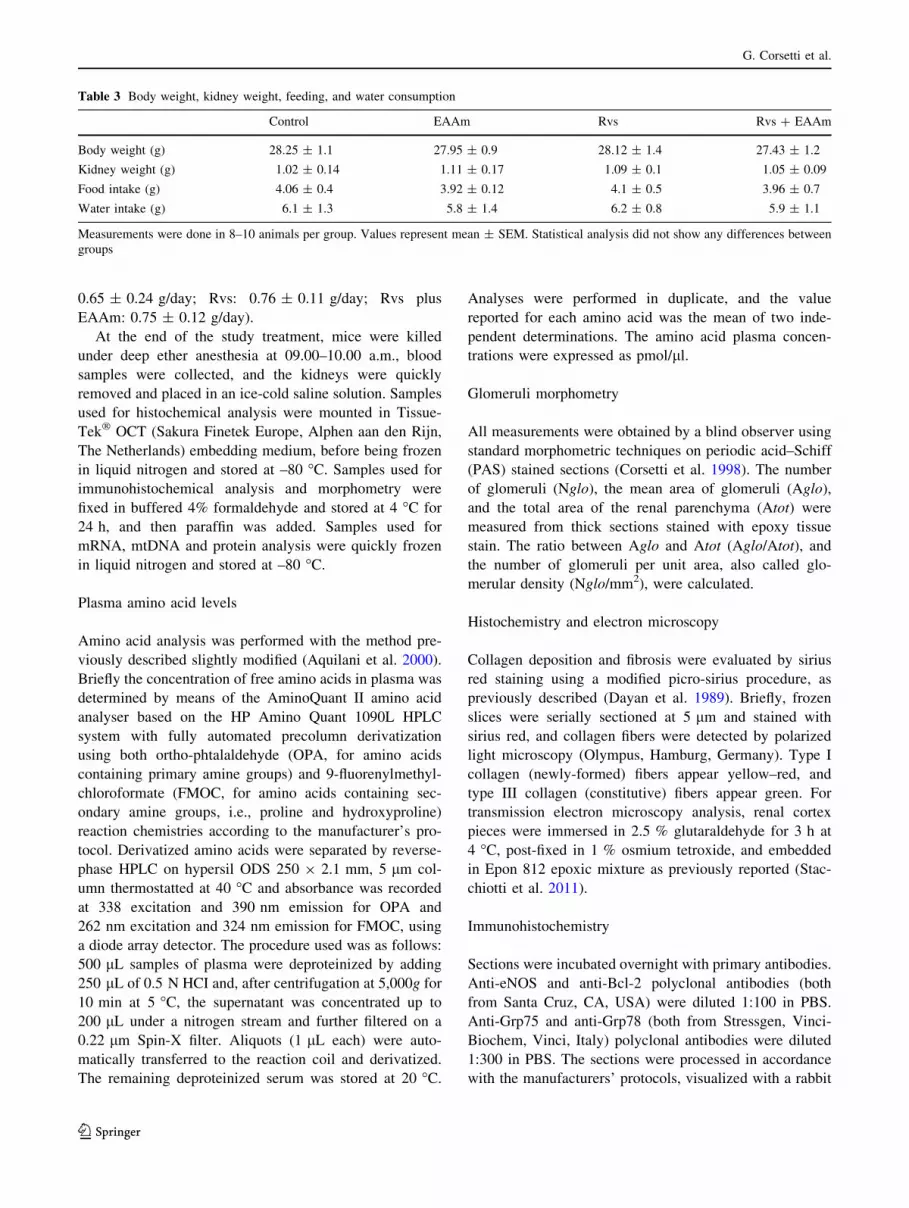
fibrosis, or other evident changes in any renal compartment
(Fig. 1 and data not shown). The number of renal glomeruli
(Nglo/mm2) and the ratios between cross-sectional glo-
merular areas and total area (Aglo/Atot) were not statisti-
cally different between the experimental groups (Table 6).
Ultrastructural analysis of the proximal tubular cells, which
are characterized by a high OXPHOS mitochondrial
activity (Szeto et al. 2011) and which are actively involved
in Rvs excretion (Verhulst et al. 2008), showed abundant,
round-shaped mitochondria, particularly in the EAAm-
supplemented group (Fig. 2). Heterogeneous and elongated
mitochondria, with multiform cristae and electron-dense
granules in the matrix, were more evident around the nuclei
of these cells both in Rvs- and Rvs plus EAAm-treated
mice when compared to untreated ones (Fig. 2).
Rosuvastatin alone or in combination with EAAm
promotes mitochondrial biogenesis and function
in mouse kidneys
The effects of Rvs alone or in combination with EAAm on
mitochondrial biogenesis were investigated in kidney
homogenates from treated and untreated animals.
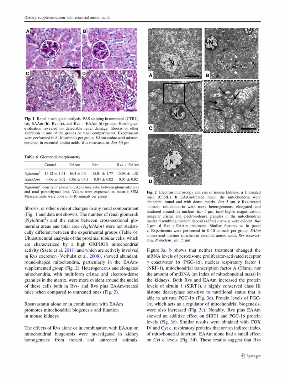
Figure 3a, b shows that neither treatment changed the
mRNA levels of peroxisome proliferator-activated receptor
c coactivator 1a (PGC-1a), nuclear respiratory factor 1
(NRF-1), mitochondrial transcription factor A (Tfam), nor
the amount of mtDNA (an index of mitochondrial mass) in
the kidneys. Both Rvs and EAAm increased the protein
levels of sirtuin 1 (SIRT1), a highly conserved class III
histone deacetylase sensitive to nutritional status that is
able to activate PGC-1a (Fig. 3c). Protein levels of PGC-
1a, which acts as a regulator of mitochondrial biogenesis,
were also increased (Fig. 3c). Notably, Rvs plus EAAm
showed an additive effect on SIRT1 and PGC-1a protein
levels (Fig. 3c). Similar results were obtained with COX
IV and Cyt c, respiratory proteins that are an indirect index
of mitochondrial function. EAAm alone had a small effect
on Cyt c levels (Fig. 3d). These results suggest that Rvs
Fig. 1 Renal histological analysis. PAS staining in untreated (CTRL)
(a), EAAm (b), Rvs (c), and Rvs ? EAAm (d) groups. Histological
evaluation revealed no detectable renal damage, fibrosis or other
alteration in any of the groups or renal compartments. Experiments
were performed in 8–10 animals per group. EAAm amino acid mixture
enriched in essential amino acids, Rvs rosuvastatin. Bar 50 lm
Table 6 Glomeruli morphometry
Control EAAm Rvs Rvs ? EAAm
Nglo/mm2 15.12 ± 1.51 16.4 ± 0.9 15.01 ± 1.77 15.98 ± 1.48
Aglo/Atot 0.06 ± 0.02 0.06 ± 0.01 0.04 ± 0.02 0.05 ± 0.02
Nglo/mm2, density of glomeruli; Aglo/Atot, ratio between glomerular area
and total parenchymal area. Values were expressed as mean ± SEM.
Measurements were done in 8–10 animals per groupFig. 2 Electron microscopy analysis of mouse kidneys. a Untreated
mice (CTRL). b EAAm-treated mice; the mitochondria were
abundant, round and with dense matrix. Bar 5 lm. c Rvs-treated
animals; mitochondria were more heterogeneous, elongated and
scattered around the nucleus. Bar 5 lm. Inset higher magnification;
irregular cristae and electron-dense granules in the mitochondrial
matrix resembling calcium deposits (black arrows) were evident. Bar
2 lm. d Rvs ? EAAm treatment. Similar features as in panel
c. Experiments were performed in 8–10 animals per group. EAAm
amino acid mixture enriched in essential amino acids, Rvs rosuvast-
atin, N nucleus. Bar 5 lm
Dietary supplementation with essential amino acids
123
promotes the expression of some markers of mitochondrial
biogenesis and that EAAm supplementation reinforces this
effect.
Rosuvastatin stimulates AKT and eNOS
phosphorylation in mouse kidneys
To investigate the signaling mechanism(s) involved in the
effects of Rvs and EAAm on renal mitochondria, we ana-
lyzed the mTOR-AKT-eNOS pathway. To determine
whether Rvs and EAAm affected mTOR signaling in mice,
kidney lysates were prepared and the amounts of phos-
phorylated S6 (p-S6) were determined by immunoblot
analysis (Fig. 4a). The level of p-S6, normalized to total S6
(pS6/S6), was unchanged by either Rvs or EAAm, alone or
in combination. In accordance with previous results
(Bussolati et al. 2005; Ito et al. 2010), Rvs activated protein
kinase AKT through increased Ser473-AKT phosphoryla-
tion (Fig. 4a). This effect was not strengthened by EAAm,
which alone was unable to modify AKT phosphorylation
(Fig. 4a). Neither Rvs nor EAAm, or their combination,
promoted phosphorylation of AKT in Thr308 (data not
shown). Activated AKT phosphorylates multiple targets,
including eNOS. Ser117 phosphorylation of eNOS
increases nitric oxide production (Kureishi et al. 2000).
Accordingly, Rvs increased Ser1177 p-eNOS levels in the
kidney, while EAAm did not change either basal or Rvs-
induced eNOS phosphorylation (Fig. 4a). Specific immu-
nohistochemical analysis showed a moderate eNOS
immunostaining uniformly distributed in the cortical
Fig. 3 Molecular markers of mitochondrial biogenesis in mouse
kidneys. a mRNA levels of mitochondrial biogenesis markers and
b mtDNA amounts in the kidneys of untreated (CTRL) and treated
groups analyzed by quantitative RT-PCR. The cycle number at which
the transcripts were detectable was compared to that of GAPDH and
expressed as relative expression versus controls, taken as 1.0. The
experiments were performed in 8–10 animals per group and data are
expressed as mean ± SEM. c, d Effects of treatments with Rvs and
EAAm, either alone or in combination, on SIRT1, PGC-1a, COX IV,
and Cyt c protein levels measured by immunoblot analysis. The
Western blot images are representative of separate experiments done
in 8–10 animals per group. The relative values were detected by
densitometric analysis, relative to either vinculin or b-actin levels,
with untreated (CTRL) mice given a value of 1.0. Data are expressed
as mean ± SEM. *p \ 0.05 vs untreated (CTRL) mice; #p \ 0.05 vs
Rvs-treated mice
G. Corsetti et al.
123

tubular cells of untreated and EAAm-supplemented mice
(Fig. 4b; Table 7). The staining intensity was weakly
decreased in the kidney of Rvs-treated animals, but eNOS
was markedly increased when Rvs was used in combina-
tion with EAAm, particularly in the proximal tubular cells
(Fig. 4b; Table 7). Faint and sparse staining for inducible
and neuronal NOS (iNOS and nNOS) was observed in the
kidneys of each group (data not shown).
Rosuvastatin regulates the physiology of mitochondria
and endoplasmic reticulum in mouse kidneys
S473-phosphorylated AKT was recently localized to the
endoplasmic reticulum (ER) subcompartment, termed the
mitochondria-associated ER membrane (MAM) (Betz et al.
2013). In mammalian MAM, the ER and mitochondria are
physically tethered to each other by the inositol trisphosphate
(IP3) receptor (IP3R)/glucose-regulated protein 75 (Grp75)/
voltage-dependent anion-selective channel 1 (VDAC1) tri-
meric complex (Betz et al. 2013). Grp75 expression was
therefore investigated. Rvs, in contrast to EAAm alone,
markedly increased Grp75 protein levels in the kidneys of
treated mice (Fig. 5a). This increased expression was not
changed by EAAm supplementation. These results were
confirmed by immunohistochemical analysis. Grp75
immunostaining was faint and uniformly distributed in
tubular and glomerular cells of mice in the control and
EAAm alone groups (Fig. 5b), but was strongly increased by
Rvs, mainly in the proximal tubular cells with glomeruli only
occasionally stained (Fig. 5b). Although Grp75 staining was
less intense when Rvs was given with EAAm supplemen-
tation, it remained much higher than in untreated and EAAm-
supplemented mice (Fig. 5b; Table 7).
Statins reduce ER stress in isolated cell systems, and
glucose-regulated protein 78 (Grp78) is involved in the
homeostatic response to cellular redox damage (Breder et al.
2010; Molins et al. 2010). Grp78 expression was thus
examined in the kidneys of treated and untreated mice.
Chronic exposure to Rvs reduced Grp78 protein levels
(Fig. 5c), while EAAm treatment did not change either basal
or Rvs-decreased Grp78 expression. Immunostaining
clearly identified Grp78 in the tubular cells of untreated
animals (Fig. 5d); however, Rvs treatment induced sparse
and moderate cytoplasmic staining of glomerular cells,
while the proximal tubules were intensely stained (Fig. 5d).
EAAm only partly modified these staining patterns (Fig. 5d;
Table 7). These findings suggest that Rvs positively modu-
lates the expression of proteins controlling mitochondrial
and ER physiology, and that EAAm supplementation does
not play a relevant role in favoring these processes.
Rosuvastatin and EAAm in combination promote
an anti-apoptotic mechanism
Mitochondria and ER are involved in the apoptotic process,
as are AKT, Grp75 and Grp78 (Tabas and Ron 2011; Logue
Fig. 4 Rosuvastatin and amino acids affect the AKT-eNOS pathway
in mouse kidneys. a Rvs alone or in combination with EAAm
activates AKT and eNOS, but not mTOR, in kidneys. The phosphor-
ylated S6, AKT and eNOS protein levels were analyzed by
immunoblot, and the relative values were detected by densitometric
analysis, relative to S6, AKT, or eNOS total protein levels, when
control measurement was given a value of 1.0. The Western blot
images are representative of separate experiments performed in 8–10
animals per group. Data are expressed as mean ± SEM. *p \ 0.05 vs
untreated mice (CTRL). b eNOS immunohistochemical analysis.
EAAm amino acid mixture enriched in essential amino acids, Rvs
rosuvastatin. Bar 50 lm
Table 7 Immunohistochemical measurements
Control EAAm Rvs Rvs ? EAAm
eNOS 20.5 ± 2.1 30.1 ± 2.3� 18.3 ± 1.2* 25.9 ± 1.7#
Grp75 32.3 ± 3.1 30.5 ± 2.8 18.2 ± 3.7�, * 21.1 ± 3.2
Grp78 18.6 ± 2.6 20.4 ± 3.1 37 ± 4.4�, * 30.2 ± 4.6
Bcl-2 9.1 ± 0.9 19.4 ± 1.2� 18.3 ± 2.7� 25.2 ± 1.8�, #, *
Integrated optical density (IOD) values (±SEM) of immunohisto-
chemical staining in each group. Measurements were done in 8–10
animals per group
� p \ 0.05 vs Control group * p \ 0.01 vs EAAm group; # p\ 0.05
vs rosuvastatin group, two-way ANOVA–Bonferroni post hoc test
Dietary supplementation with essential amino acids
123
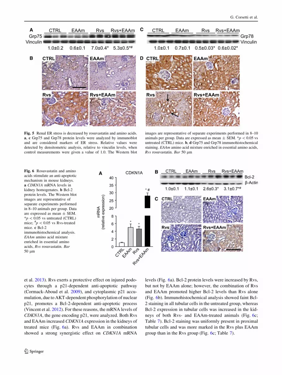
et al. 2013). Rvs exerts a protective effect on injured podo-
cytes through a p21-dependent anti-apoptotic pathway
(Cormack-Aboud et al. 2009), and cytoplasmic p21 accu-
mulation, due to AKT-dependent phosphorylation of nuclear
p21, promotes a Bcl-2-dependent anti-apoptotic process
(Vincent et al. 2012). For these reasons, the mRNA levels of
CDKN1A, the gene encoding p21, were analyzed. Both Rvs
and EAAm increased CDKN1A expression in the kidneys of
treated mice (Fig. 6a). Rvs and EAAm in combination
showed a strong synergistic effect on CDKN1A mRNA
levels (Fig. 6a). Bcl-2 protein levels were increased by Rvs,
but not by EAAm alone; however, the combination of Rvs
and EAAm promoted higher Bcl-2 levels than Rvs alone
(Fig. 6b). Immunohistochemical analysis showed faint Bcl-
2 staining in all tubular cells in the untreated group, whereas
Bcl-2 expression in tubular cells was increased in the kid-
neys of both Rvs- and EAAm-treated animals (Fig. 6c;
Table 7). Bcl-2 staining was uniformly present in proximal
tubular cells and was more marked in the Rvs plus EAAm
group than in the Rvs group (Fig. 6c; Table 7).
Fig. 5 Renal ER stress is decreased by rosuvastatin and amino acids.
a, c Grp75 and Grp78 protein levels were analyzed by immunoblot
and are considered markers of ER stress. Relative values were
detected by densitometric analysis, relative to vinculin levels, when
control measurements were given a value of 1.0. The Western blot
images are representative of separate experiments performed in 8–10
animals per group. Data are expressed as mean ± SEM. *p \ 0.05 vs
untreated (CTRL) mice. b, d Grp75 and Grp78 immunohistochemical
staining. EAAm amino acid mixture enriched in essential amino acids,
Rvs rosuvastatin. Bar 50 lm
Fig. 6 Rosuvastatin and amino
acids stimulate an anti-apoptotic
mechanism in mouse kidneys.
a CDKN1A mRNA levels in
kidney homogenates. b Bcl-2
protein levels. The Western blot
images are representative of
separate experiments performed
in 8–10 animals per group. Data
are expressed as mean ± SEM.
*p \ 0.05 vs untreated (CTRL)
mice; #p \ 0.05 vs Rvs-treated
mice. c Bcl-2
immunohistochemical analysis.
EAAm amino acid mixture
enriched in essential amino
acids, Rvs rosuvastatin. Bar
50 lm
G. Corsetti et al.
123

Discussion
The present results show that long-term treatment with
Rvs, at a dose comparable to that normally used in clinical
trials to lower blood cholesterol levels, promotes the
expression of markers of mitochondrial biogenesis in the
kidneys of young mice. In particular, ultrastructural ana-
lysis identifies abundant and elongated mitochondria
around the nuclei of cortical proximal tubular cells in mice
treated with Rvs. Multiform cristae and electron-dense
granules in mitochondrial matrix are evident, suggesting
either intense functional activity or calcium accumulation
within the organelles (Khraiwesh et al. 2013). The protein
levels of SIRT1 and PGC-1a, two master regulators of
mitochondrial biogenesis, and COX IV, an OXPHOS
respiratory protein, are increased by Rvs in the kidneys.
Dietary supplementation with EAAm, which modifies the
plasma amino acid profile, potentiates these effects. Recent
results have suggested that SIRT1 in proximal tubules
protects against albuminuria in diabetes by maintaining
nicotinamide mononucleotide concentrations around
glomeruli, thus influencing podocyte function (Hasegawa
et al. 2013).
Previously, statins were reported to increase NO bio-
availability in patients with hypercholesterolemia (John
et al. 2001) and to exert beneficial effects on the activation
and expression of eNOS in hypertensive rats (Kishi et al.
2008; Ohkawara et al. 2010). Moreover, Kureishi et al.
(2000) demonstrated that simvastatin induces AKT-regu-
lated eNOS activation (via Ser1177 phosphorylation),
which increases NO production and consequently promotes
endothelial cell survival. Similarly, EAAm was reported to
promote mitochondrial biogenesis in cardiac and skeletal
muscle by promoting eNOS activity (D’Antona et al. 2010)
and AKT phosphorylation (Flati et al. 2010). Accordingly,
the present results demonstrate that Rvs induces AKT
phosphorylation at Ser473 and increases eNOS phosphor-
ylation at Ser1177 in mouse kidney. Moderate eNOS
immunostaining is uniformly distributed in cortical tubular
cells and is evident in untreated and Rvs-treated or EAAm-
supplemented mice. However, simultaneous administration
of Rvs and EAAm markedly increases eNOS staining in
the kidneys, particularly in the proximal tubules.
Controversial recent evidence suggests that statins may
both activate or suppress the mTOR pathway in cancer or
non-cancer cells (Roudier et al. 2006; Finlay et al. 2007).
To this effect, Woodard et al. (2008) showed that fluvast-
atin induced apoptosis potently and limited the prolifera-
tion of renal cell carcinoma (RCC) cells in vitro; these
effects were mediated by the suppression of AKT phos-
phorylation/activation, resulting in inhibition of mTOR and
p70 S6 kinase. In the current study, S6 phosphorylation
was not altered by Rvs and EAAm, alone or in
combination. This result is consistent with a previous study
in which dietary leucine supplementation did not modify
the mTOR signaling in the kidney of neonatal pigs
(Suryawan et al. 2012). Together these findings support a
direct role for statins and amino acids in modulating the
eNOS-dependent NO signaling pathway in renal tubular
and endothelial cells through an AKT-dependent mTOR-
independent pathway. In particular, the EAAs have been
shown to increase synthesis of arginine through rein-
forcement of the anaplerotic export of aspartate, necessary
for recycling of citrulline to arginine by arginino-succinate
synthetase. Complex mechanisms, including the stimula-
tory effect of Rvs on eNOS activity and increased arginine
availability from EAAm supplementation, may contribute
to the processes observed in the present work.
Considering the positive effect of NO on mitochondrial
biogenesis and function (Nisoli et al. 2003, 2004; Nisoli
and Carruba 2006), the present results imply that Rvs,
particularly when combined with EAAm, may have rele-
vant beneficial effects on kidneys, and particularly on the
proximal tubular cells. Notably, these cells do not use
glucose for their energy production (Balaban and Mandel
1988) but rely primarily on fatty acid oxidation. Fatty acid
oxidation is mostly performed by mitochondria, which
therefore have a central role in these cells. Mitochondrial
production of ATP, which is essential for generating the
energy-dependent ion gradients that drive renal tubular
reabsorption, is impaired in acute kidney injury (Hall et al.
2011). This can cause massive and life-threatening losses
of fluids, electrolytes, and low-molecular weight nutrients,
a dysfunction known as renal Fanconi’s syndrome (Che
et al. 2014). Recently, a rare mutation was identified in
autosomal dominant Fanconi’s syndrome that creates a
new mitochondrial targeting motif in the N-terminal por-
tion of enoyl-CoA hydratase/3-hydroxyacyl CoA dehy-
drogenase (EHHADH) (Klootwijk et al. 2014). EHHADH
is involved in peroxisomal oxidation of fatty acids and is
expressed in proximal tubules. Studies of proximal tubular
cells expressing mutant EHHADH revealed that mistar-
geting of a peroxisomal protein to mitochondria impaired
mitochondrial oxidative phosphorylation, with marked
defects in the transport of fluids and glucose across the
epithelium, indicative of the central role of mitochondria in
proximal tubular function (Klootwijk et al. 2014).
Although hyperaminoaciduria is a hallmark of the condi-
tion (Baum 1998), it is tempting to speculate that Rvs and
particularly Rvs in combination with EAAm could have
beneficial effects on mitochondrial function in the renal
tubular cells impaired in Fanconi’s syndrome.
The present findings suggest that Rvs modulates the
physiological relationship between the mitochondria and
the ER. Rvs markedly increased the expression of Grp75 in
the proximal tubular and glomerular cells. Grp75 is a
Dietary supplementation with essential amino acids
123

component of a trimeric complex (IP3-Grp75-VDAC1)
that regulates physical tethering of the ER and mitochon-
dria at MAMs (Betz et al. 2013), together with mitofusins 1
and 2 (Twig et al. 2008). The ER–mitochondria tethering
complex affects mitochondrial dynamics and function. In
particular, association with the ER forms microdomains on
the outer mitochondrial membrane. These sites are enri-
ched in mitochondrial division components, such as dyn-
amin-related guanosine triphosphatases 1 (DRP1) and
membrane protein mitochondrial fission factor (Mff)
(Friedman et al. 2011; Youle and van der Bliek 2012).
Under stress conditions, the microdomains may recruit and
regulate the activation of pro-apoptotic Bcl-2 proteins such
as Bax, which promote permeabilization of the mitochon-
drial outer membrane and the release of death mediators
such as Cyt c (Hoppins and Nunnari 2012). Thus, the ER–
mitochondria contact points regulate apoptosis under par-
ticular conditions, in addition to modulating lipid transfer
and calcium flux between the organelles. These processes
are essential for mitochondrial function and ER homeo-
stasis. Diverse evidence supports the effect of statins in
reducing ER stress, and the role of Grp78, an ER stress
sensor, in the homeostatic response to cellular redox
damage has been explored (Breder et al. 2010; Molins et al.
2010). In the present work, long-term treatment with Rvs
reduces Grp78 protein levels in kidney homogenates. This
occurs specifically in glomerular cells, in contrast to the
proximal tubular cells where Grp78 was accumulated.
These findings suggest that Rvs can communicate diverse
survival messages to different kidney cell types.
Rvs was previously found to protect injured podocytes
through a p21-dependent anti-apoptotic pathway (Cor-
mack-Aboud et al. 2009). Accordingly, the present results
show that the anti-apoptotic Bcl-2 protein levels are
increased by Rvs, alone and in combination with EAAm.
Immunostaining confirms that Bcl-2 is accumulated in the
tubular cells. Vincent et al. (2012) have recently demon-
strated that cytoplasmic p21 accumulation, resulting from
AKT-dependent phosphorylation of nuclear p21, promotes
a Bcl-2-dependent anti-apoptotic process in cancer cells.
Accordingly, both Rvs and EAAm here increase CDKN1A
expression in the kidneys of treated mice, and this effect is
more marked with Rvs and EAAm in combination.
These results are consistent with recent studies showing
that both Rvs treatment and EAAm supplementation had
beneficial effects on kidneys in rodents (Corsetti, et al.
2010) and humans (Vidt et al. 2006; Bolasco et al. 2011).
Although data on the potential renal toxicity of high-
potency statins are controversial (Tiwari 2006; Golomb
and Evans 2008; Dodiya et al. 2011), two meta-analyses
showed that statin therapy is safe and effective in pre-
venting mortality and major cardiovascular events in
patients with non-dialysis-dependent chronic kidney
disease (CKD) (Upadhyay et al. 2012, 2013; Palmer et al.
2012). However, these studies provide very limited evi-
dence to support the use of statins in patients on dialysis,
and statin therapy was not found to be effective in reducing
the risk of kidney failure or decline in kidney function
(Upadhyay 2013). Conversely, a recent pilot study con-
ducted in 15 CKD patients on hemodialysis treatment for at
least 6 months has shown that 3 month EAAm supple-
mentation increased serum albumin and total proteins, with
reduced levels of inflammatory markers and improved
anemia (Bolasco et al. 2011). Although these effects were
studied in a small group of patients and might be due to
ameliorated nutrient intake besides that of the amino acid
mixture, EAAm supplementation might improve the effi-
cacy of statin therapy in dialysis-dependent subjects.
Conclusions
The present results support the favorable effects of a high-
potency statin, like Rvs, on the morpho-functional aspects
of mouse kidney. In particular, long-term treatment with
Rvs supports mitochondrial function, probably through the
AKT-eNOS signaling pathway, and regulates survival of
proximal tubular cells. An original formula of essential
amino acids enhances the statin’s effects. This provides
mechanistic confirmation of recent clinical evidence in
kidney disease and suggests the potential use of essential
amino acid supplementation in conjunction with statins in
patients with renal disorders characterized by tubular cell
dysfunction.
Acknowledgments The authors wish to thank Dr. Antonio Lavazza
for assistance with electron microscopy and Dr. Patrizia Arcidiaco
(Centro Grandi Strumenti, University of Pavia) for assistance with
plasma amino acids determination. This work was supported by the
Ministero dell’Istruzione, dell’Universita e della Ricerca (Italy) grant
no. 2009E48P9M_001 (E.N.).
Conflict of interest The authors declare no conflict of interest.
Open Access This article is distributed under the terms of the
Creative Commons Attribution License which permits any use, dis-
tribution, and reproduction in any medium, provided the original
author(s) and the source are credited.
References
Andrew PJ, Myer B (1999) Enzymatic function of nitric oxide
synthases. Cardiovasc Res 43:521–531
Aquilani R, Viglio S, Iadarola P, Guarnaschelli C, Arrigoni N,
Fugazza G, Catapano M, Boschi F, Dossena M, Pastoris O
(2000) Peripheral plasma amino acid abnormalities in rehabil-
itation patients with severe brain injury. Arch Phys Med Rehabil
81:176–181
G. Corsetti et al.
123

Bachmanov AA, Reed DR, Beauchamp GK, Tordoff MG (2002)
Food intake, water intake, and drinking spout side preference of
28 mouse strains. Behav Genet 32:435–443
Bae E, Kim I, Park J, Ma S, Lee J, Kim S (2010) Renoprotective
effect of rosuvastatin in DOCA-salt hypertensive rats. Nephrol
Dial Transplant 25:1051–1059
Balaban RS, Mandel LJ (1988) Metabolic substrate utilization by
rabbit proximal tubule: an NADH fluorescence study. Am J
Physiol 254:F407–F416
Baum M (1998) The Fanconi syndrome of cystinosis: insights into the
pathophysiology. Pediatr Nephrol 12:492–497
Betz C, Stracka D, Prescianotto-Baschong C, Frieden M, Demaurex
N, Hall MN (2013) mTOR complex 2-AKT signaling at
mitochondria-associated endoplasmic reticulum membranes
(MAM) regulates mitochondrial physiology. Proc Natl Acad
Sci USA 110:12526–12534
Bolasco P, Caria S, Cupisti A, Secci R, Dioguardi FS (2011) A novel
amino acids oral supplementation in hemodialysis patients: a
pilot study. Ren Fail 33:1–5
Breder I, Coope A, Arruda AP, Razolli D, Milanski M, Dorighello
GG, de Oliveira HC, Velloso LA (2010) Reduction of endo-
plasmic reticulum stress-a novel mechanism of action of statins
in the protection against atherosclerosis. Atherosclerosis
212:30–31
Bussolati B, Deregibus MC, Fonsato V, Doublier S, Spatola T,
Procida S, Di Carlo F, Camussi G (2005) Statins prevent
oxidized LDL-induced injury of glomerular podocytes by
activating the phosphatidylinositol 3-kinase/AKT-signaling
pathway. J Am Soc Nephrol 16:1936–1947
Cano NJ, Fouque D, Leverve XM (2006) Application of branched-
chain amino acids in human pathological states: renal failure.
J Nutr 136:299S–307S
Che R, Yuan Y, Huang S, Zhang A (2014) Mitochondrial dysfunction
in the pathophysiology of renal diseases. Am J Physiol Renal
Physiol 306:F367–F378
Cormack-Aboud FC, Brinkkoetter PT, Pippin JW, Shankland SJ,
Durvasula RV (2009) Rosuvastatin protects against podocyte
apoptosis in vitro. Nephrol Dial Transplant 24:404–412
Corsetti G, Rezzani R, Rodella L, Bianchi R (1998) Ultrastructural
study of the alterations in spinal ganglion cells of rats chronically
fed on ethanol. Ultrastruct Pathol 22:309–319
Corsetti G, Stacchiotti A, D’Antona G, Nisoli E, Dioguardi F,
Rezzani R (2010) Supplementation with essential amino acids in
middle age maintains the healthy of rat kidney. Int J Immuno-
pathol Pharmacol 23:523–533
Corsetti G, Stacchiotti A, Tedesco L, D’Antona G, Pasini E,
Dioguardi FS, Nisoli E, Rezzani R (2011) Essential amino acid
supplementation decreases liver damage induced by chronic
ethanol consumption in rats. Int J Immunopathol Pharmacol
24:611–619
Corsetti G, Pasini E, Ferrari-Vivaldi M, Romano C, Bonomini F,
Tasca G, Dioguardi FS, Rezzani R, Assanelli D (2012)
Metabolic syndrome and chronic simvastatin therapy enhanced
human cardiomyocyte stress before and after ischemia-reperfu-
sion in cardio-pulmonary bypass patients. Int J Immunopathol
Pharmacol 25:1063–1074
D’Antona G, Ragni M, Cardile A, Tedesco L, Dossena M, Bruttini F,
Caliaro F, Corsetti G, Bottinelli R, Carruba MO, Valerio A,
Nisoli E (2010) Branched-chain amino acid supplementation
promotes survival and supports cardiac and skeletal muscle
mitochondrial biogenesis in middle-aged mice. Cell Metab
12:362–372
Dayan D, Hiss Y, Hirshberg A, Bubis J, Wolman M (1989) Are the
polarization colors of picrosirius red-stained collagen determined
only by the diameter of the fibers? Histochemistry 93:27–29
De Pinieux G, Chariot P, Ammi-Saıd M, Louarn F, Lejonc JL, Astier
A, Jacotot B, Gherardi R (1996) Lipid-lowering drugs and
mitochondrial function: effects of HMG-CoA reductase inhibi-
tors on serum ubiquinone and blood lactate/pyruvate ratio. Br J
Clin Pharmacol 42:333–337
Dodiya H, Jain M, Goswami S (2011) Renal toxicity of lisinopril and
rosuvastatin, alone and in combination, in Wistar rats. Int J
Toxicol 30:518–527
Enomoto S, Sata M, Fukuda D, Nakamura K, Nagai R (2009)
Rosuvastatin prevents endothelial cell death and reduces
atherosclerotic lesion formation in ApoE-deficient mice. Biomed
Pharmacother 63:19–26
Finlay GA, Malhowski AJ, Liu Y, Fanburg BL, Kwiatkowski DJ,
Toksoz D (2007) Selective inhibition of growth of tuberous
sclerosis complex 2 null cells by atorvastatin is associated with
impaired Rheb and Rho GTPase function and reduced mTOR/S6
kinase activity. Cancer Res 67:9878–9886
Fiore M, Jimenez P, Cremonezzi D, Juncos L, Garcia N (2011) Statins
reverse renal inflammation and endothelial dysfunction induced
by chronic high salt intake. Am J Physiol Renal Physiol
301:F263–F270
Flati V, Caliaro F, Speca S, Corsetti G, Cardile A, Nisoli E, Bottinelli
R, D’ Antona G (2010) Essential amino acids improve insulin
activation of AKT/MTOR signaling in soleus muscle of aged
rats. Int J Immunopathol Pharmacol 23:81–89
Friedman JR, Lackner LL, West M, DiBenedetto JR, Nunnari J,
Voeltz GK (2011) ER tubules mark sites of mitochondrial
division. Science 334:358–362
Gambelli S, Dotti MT, Malandrini A, Mondelli M, Stromillo ML,
Gaudiano C, Federico A (2004) Mitochondrial alterations in
muscle biopsies of patients on statin therapy. J Submicrosc Cytol
Pathol 36:85–89
Golomb BA, Evans MA (2008) Statin adverse effects: a review of the
literature and evidence for a mitochondrial mechanism. Am J
Cardiovasc Drugs 8:373–418
Hall AM, Hendry BM, Nitsch D, Connolly JO (2011) Tenofovir-
associated kidney toxicity in HIV-infected patients: a review of
the evidence. Am J Kidney Dis 57:773–780
Hasegawa K, Wakino S, Simic P, Sakamaki Y, Minakuchi H,
Fujimura K, Hosoya K, Komatsu M, Kaneko Y, Kanda T,
Kubota E, Tokuyama H, Hayashi K, Guarente L, Itoh H (2013)
Renal tubular Sirt1 attenuates diabetic albuminuria by epigenet-
ically suppressing Claudin-1 overexpression in podocytes. Nat
Med 19:1504–1946
Hoppins S, Nunnari J (2012) Mitochondrial dynamics and apopto-
sis—the ER connection. Science 337:1052–1054
Ito D, Ito O, Mori N, Muroya Y, Cao PY, Takashima K, Kanazawa
M, Kohzuki M (2010) Atorvastatin upregulates nitric oxide
synthases with Rho-kinase inhibition and AKT activation in the
kidney of spontaneously hypertensive rats. J Hypertens
28:2278–2288
John S, Delles C, Jacobi J, Schlaich MP, Schneider M, Schmitz G,
Schmieder RE (2001) Rapid improvement of nitric oxide
bioavailability after lipid-lowering therapy with cerivastatin
within two weeks. J Am Coll Cardiol 37:1351–1358
Kano K, Hirata Y, Emory T, Ohta K, Eguci S, Imai T, Marumo F
(1992) L-arginine infusion induces hypotension and diuresis/
natriuresis with concomitant increased urinary excretion of
nitrite/nitrate and cyclic GMP in humans. Clin Exp Pharmacol
Physiol 19:619–625
Khraiwesh H, Lopez-Dominguez J, Lopez-Lluch G, Navas P, de Cabo
R, Ramsey J, Villalba J, Gonzalez-Reyes J (2013) Alterations of
ultrastructural and fission/fusion markers in hepatocyte mito-
chondria from mice following calorie restriction with different
dietary fats. J Gerontology A Biol Sci Med Sci 68:1023–1034
Dietary supplementation with essential amino acids
123

Kishi T, Hirooka Y, Shimokawa H, Takeshita A, Sunagawa K (2008)
Atorvastatin reduces oxidative stress in the rostral ventrolateral
medulla of stroke-prone spontaneously hypertensive rats. Clin
Exp Hypertens 30:3–11
Klootwijk ED, Reichold M, Helip-Wooley A, Tolaymat A, Broeker
C, Robinette SL, Reinders J, Peindl D, Renner K, Eberhart K,
Assmann N, Oefner PJ, Dettmer K, Sterner C, Schroeder J,
Zorger N, Witzgall R, Reinhold SW, Stanescu HC, Bockenhauer
D, Jaureguiberry G, Courtneidge H, Hall AM, Wijeyesekera AD,
Holmes E, Nicholson JK, O’Brien K, Bernardini I, Krasnewich
DM, Arcos-Burgos M, Izumi Y, Nonoguchi H, Jia Y, Reddy JK,
Ilyas M, Unwin RJ, Gahl WA, Warth R, Kleta R (2014)
Mistargeting of Peroxisomal EHHADH and Inherited Renal
Fanconi’s Syndrome. New Engl J Med 370:129–138
Knauer MJ, Urquhart BL, zu Schwabedissen HEM, Schwarz UI,
Lemke CJ, Leake BF, Kim RB, Tirona RG (2010) Human
skeletal muscle drug transporters determine local exposure and
toxicity of statins. Circ Res 106:297–306
Kureishi Y, Luo Z, Shiojima I, Bialik A, Fulton D, Lefer DJ, Sessa
WC, Walsh K (2000) The HMG-CoA reductase inhibitor
simvastatin activates the protein kinase AKT and promotes
angiogenesis in normocholesterolemic animals. Nat Med
6:1004–1010
Larsen S, Stride N, Hey-Mogensen M, Hansen CN, Bang LE,
Bundgaard H, Nielsen LB, Helge JW, Dela F (2013) Simvastatin
effects on skeletal muscle: relation to decreased mitochondrial
function and glucose intolerance. J Am Coll Cardiol 61:44–53
Lenaz G, Bovina C, D’Aurelio M, Fato R, Formiggini G, Genova ML,
Giuliano G, Pich MM, Paolucci U, Castelli GP, Ventura B
(2002) Role of mitochondria in oxidative stress and aging. Ann
N Y Acad Sci 959:199–213
Logue SE, Cleary P, Saveljeva S, Samali A (2013) New directions in
ER stress-induced cell death. Apoptosis 18:537–546
MacAllister R, Vallance P (1994) Nitric oxide in essential and renal
hypertension. J Am Soc Nephrol 5:1057–1065
Mancini GB, Tashakkor AY, Baker S, Bergeron J, Fitchett D,
Frohlich J, Genest J, Gupta M, Hegele RA, Ng DS, Pearson GJ,
Pope J (2013) Diagnosis, prevention, and management of statin
adverse effects and intolerance: canadian working group
consensus update. Can J Cardiol 29:1553–1568
Mansi I, Frei CR, Pugh MJ, Makris U, Mortensen EM (2013) Statins
and musculoskeletal conditions, arthropathies, and injuries.
JAMA Intern Med 173:1–10
Molins B, Pena E, Padro T, Casani L, Mendieta C, Badimon L (2010)
Glucose-regulated protein 78 and platelet deposition: effect of
rosuvastatin. Arterioscler Thromb Vasc Biol 30:1246–1252
Nisoli E, Carruba MO (2006) Nitric oxide and mitochondrial
biogenesis. J Cell Sci 119:2855–2862
Nisoli E, Clementi E, Paolucci C, Cozzi V, Tonello C, Sciorati C,
Bracale R, Valerio A, Francolini M, Moncada S, Carruba MO
(2003) Mitochondrial biogenesis in mammals: the role of
endogenous nitric oxide. Science 299:896–899
Nisoli E, Falcone S, Tonello C, Cozzi V, Palomba L, Fiorani M,
Pisconti A, Brunelli S, Cardile A, Francolini M, Cantoni O,
Carruba MO, Moncada S, Clementi E (2004) Mitochondrial
biogenesis by NO yields functionally active mitochondria in
mammals. Proc Natl Acad Sci USA 101:16507–16512
Ohkawara H, Ishibashi T, Saitoh S, Inoue N, Sugimoto K, Kamioka
M, Uekita H, Kaneshiro T, Ando K, Takuwa Y, Maruyama Y,
Takeishi Y (2010) Preventive effects of pravastatin on thrombin-
triggered vascular responses via AKT/eNOS and RhoA/Rac1
pathways in vivo. Cardiovasc Res 88:492–501
Palmer SC, Craig JC, Navaneethan SD, Tonelli M, Pellegrini F,
Strippoli GF (2012) Benefits and harms of statin therapy for
persons with chronic kidney disease: a systematic review and
meta-analysis. Ann Intern Med 157:263–275
Pellegrino MA, Brocca L, Dioguardi F, Bottinelli R, D’Antona G
(2005) Effects of voluntary wheel running and amino acid
supplementation on skeletal muscle of mice. Eur J Physiol
93:655–664
Rajapaskse NW, Mattson DL (2013) Role of cellular L-arginine and
nitric oxide production on renal blood flow and arterial pressure
regulation. Curr Opin Nephrol Hypertens 22:45–50
Robles NR, Velasco J, Mena C, Polo J, Angulo E, Espinosa J (2013)
Increased frequency of microalbuminuria in patients receiving
statins. Clin Lipidol 51:554–561
Rondanelli M, Opizzi A, Antoniello N, Boschi F, Iadarola P, Pasini E,
Aquilani R, Dioguardi FS (2011) Effect of essential amino acid
supplementation on quality of life, amino acid profile and strength
in institutionalized elderly patients. Clin Nutr 30:571–577
Rosenfeldt FL, Pepe S, Linnane A, Nagley P, Rowland M, Ou R,
Marasco S, Lyon W, Esmore D (2002) Coenzyme Q10 protects
the aging heart against stress: studies in rats, human tissues, and
patients. Ann NY Acad Sci 959:355–359
Roudier E, Mistafa O, Stenius U (2006) Statins induce mammalian
target of rapamycin (mTOR)-mediated inhibition of Akt signal-
ing and sensitize p53-deficient cells to cytostatic drugs. Mol
Cancer Ther 5:2706–2715
Schick BA, Laaksonen R, Frohlich JJ, Paiva H, Lehtimaki T,
Humphries KH, Cote HC (2007) Decreased skeletal muscle
mitochondrial DNA in patients treated with high-dose simva-
statin. Clin Pharmacol Ther 81:650–653
Shillingford JM, Murcia NS, Larson CH, Low SH, Hedgepeth R,
Brown N, Flask CA, Novick AC, Goldfarb DA, Kramer-Zucker
A, Waltz G, Piontek KB, Germino GG, Weimbs T (2006) The
mTOR pathway is regulated by polycystin-1, and its inhibition
reverses renal cystinosis in polycystic kidney disease. Proc Natl
Acad Sci USA 103:5466–5471
Silbernagl S (1988) The renal handling of amino acids and
oligopeptides. Physiol Rev 68:911–1007
Stacchiotti A, Volti GL, Lavazza A, Schena I, Aleo MF, Rodella L,
Rezzani R (2011) Different role of Schisandrin B on mercury-
induced renal damage in vivo and in vitro. Toxicology
286:48–57
Stein EA, Vidt DG, Shepherd J, Cain VA, Anzalone D, Cressman MD
(2012) Renal safety of intensive cholesterol-lowering treatment
with rosuvastatin: a retrospective analysis of renal adverse
events among 40,600 participants in the rosuvastatin clinical
development program. Atherosclerosis 221:471–477
Stone NJ, Robinson J, Lichtenstein AH, Bairey Merz CN, Lloyd-Jones
DM, Blum CB, McBride P, Eckel RH, Schwartz JS, Goldberg
AC, Shero ST, Gordon D, Smith SC Jr, Levy D, Watson K,
Wilson PW. (2013) 2013 ACC/AHA Guideline on the Treatment
of Blood Cholesterol to Reduce Atherosclerotic Cardiovascular
Risk in Adults: A Report of the American College of Cardiology/
American Heart Association Task Force on Practice Guidelines
Circulation. doi:10.1016/j.jacc.2013.11.002
Suryawan A, Torrazza RM, Gazzaneo MC, Orellana RA, Fiorotto
ML, El-Kadi SW, Srivastana N, Nguyen HV, Davis TA (2012)
Enteral leucine supplementation increases protein synthesis in
skeletal and cardiac muscles and visceral tissues of neonatal pigs
through mTORC1-dependent pathways. Pediatr Res 71:324–331
Susic D, Varagic J, Ahn J, Slama M, Frohlich E (2003) Beneficial
pleiotropic vascular effects of rosuvastatin in two hypertensive
models. J Am Coll Cardiol 42:1091–1097
Szeto HH, Liu S, Soong Y, Wu D, Darrah SE, Cheng FY, Zhao Z,
Ganger M, Tow CY, Seshan SV (2011) Mitochondria-targeted
peptide accelerates ATP recovery and reduce ischemic kidney
injury. J Am Soc Nephrol 22:1041–1052
Tabas I, Ron D (2011) Integrating the mechanisms of apoptosis
induced by endoplasmic reticulum stress. Nat Cell Biol
13:184–190
G. Corsetti et al.
123

Tedesco L, Valerio A, Dossena M, Cardile A, Ragni M, Pagano C,
Pagotto U, Carruba MO, Vettor R, Nisoli E (2010) Cannabinoid
receptor stimulation impairs mitochondrial biogenesis in mouse
white adipose tissue, muscle, and liver: the role of eNOS, p38
MAPK, and AMPK pathways. Diabetes 59:2826–2836
Tiwari A (2006) An overview of statin-associated proteinuria. Drug
Discov Today 11:458–464
Trupp M, Zhu H, Wikoff WR, Baillie RA, Zeng Z-B, Karp PD, Fiehn
O, Krauss RM, Kaddurah-Daouk R (2012) Metabolomics reveals
amino acids contribute to variation in response to simvastatin
treatment. PLoS One 7(7):e38386. doi:10.1371/journal.pone.
0038386
Twig G, Elorza A, Molina AJ, Mohamed H, Wikstrom JD, Walzer G,
Stiles L, Haigh SE, Katz S, Las G, Alroy J, Wu M, Py BF, Yuan
J, Deeney JT, Corkey BE, Shirihai OS (2008) Fission and
selective fusion govern mitochondrial segregation and elimina-
tion by autophagy. EMBO J 27:433–446
Upadhyay A (2013) Statins in chronic kidney disease: what do meta-
analysis tell us? Clin Exp Nephrol [Epub ahead of print]
Upadhyay A, Earley A, Lamont JL, Haynes S, Wanner C, Balk EM
(2012) Lipid-lowering therapy in persons with chronic kidney
disease: a systematic review and meta-analysis. Ann Intern Med
157:251–262
Valerio A, D’Antona G, Nisoli E (2011) Branched-chain aminoacids,
mitochondrial biogenesis, and health span: an evolutionary
perspective. Aging 3:464–478
van der Tol A, Van Biesen W, Van Laecke S, Bogaerts K, De
Lombaert K, Warrinnier H, Vanholder R (2012) Statin use and
the presence of microalbuminuria. Results from the ERICABEL
trial: a non-interventional epidemiological cohort study. PLoS
One 7(2):e31639
Verhulst A, Sayer R, De Broe M, D’Haese P, Brown C (2008) Human
proximal tubular epithelium actively secretes but does not retain
rosuvastatin. Mol Pharmacol 74:1084–1091
Vidt DG, Harris S, McTaggart F, Ditmarsch M, Sager PT, Sorof JM
(2006) Effect of short-term rosuvastatin treatment on estimated
glomerular filtration rate. Am J Cardiol 97:1602–1606
Vincent AJ, Ren S, Harris LG, Devine DJ, Samant RS, Fodstad O,
Shevde LA (2012) Cytoplasmic translocation of p21 mediates
NUPR1-induced chemoresistance: NUPR1 and p21 in chemore-
sistance. FEBS Lett 586:3429–3434
Woodard J, Sassano A, Hay N, Platanias LC (2008) Statin-dependent
suppression of the Akt/mammalian target of rapamycin signaling
cascade and programmed cell death 4 up-regulation in renal cell
carcinoma. Clin Cancer Res 14:4640–4649
Youle RJ, van der Bliek AM (2012) Mitochondrial fission, fusion, and
stress. Science 337:1062–1065
Zoncu R, Efeyan A, Sabatini DM (2011) mTOR: from growth signal
integration to cancer, diabetes and ageing. Nat Rev Mol Cell
Biol 12:21–35
Dietary supplementation with essential amino acids
123