environmental barcoding reveals massive dinoflagellate diversity in marine environments
TRANSCRIPT
Seediscussions,stats,andauthorprofilesforthispublicationat:https://www.researchgate.net/publication/47812871
EnvironmentalBarcodingRevealsMassiveDinoflagellateDiversityinMarineEnvironments
ARTICLEinPLOSONE·NOVEMBER2010
ImpactFactor:3.23·DOI:10.1371/journal.pone.0013991·Source:PubMed
CITATIONS
57
READS
152
13AUTHORS,INCLUDING:
RowenaFStern
SirAlisterHardyFoundationforOceanScience
15PUBLICATIONS542CITATIONS
SEEPROFILE
RobertAAndersen
UniversityofWashingtonSeattle
112PUBLICATIONS4,508CITATIONS
SEEPROFILE
FrithjofCKüpper
UniversityofAberdeen
114PUBLICATIONS3,467CITATIONS
SEEPROFILE
BenoîtVéron
UniversitédeCaenNormandie
44PUBLICATIONS795CITATIONS
SEEPROFILE
Availablefrom:BenoîtVéron
Retrievedon:04February2016

Environmental Barcoding Reveals Massive DinoflagellateDiversity in Marine EnvironmentsRowena F. Stern1*, Ales Horak1, Rose L. Andrew1, Mary-Alice Coffroth2, Robert A. Andersen3, Frithjof C.
Kupper4, Ian Jameson5, Mona Hoppenrath6, Benoıt Veron7,8, Fumai Kasai8, Jerry Brand9, Erick R. James1,
Patrick J. Keeling1
1 The Biodiversity Research Centre, University of British Columbia, Vancouver, British Columbia, Canada, 2 Department of Geology, State University of New York at Buffalo,
Buffalo, New York, United States of America, 3 Provasoli-Guillard National Center for Culture of Marine Phytoplankton, Bigelow Laboratory for Ocean Sciences, West
Boothbay Harbor, Maine, United States of America, 4 Culture Collection of Algae and Protozoa, Scottish Association for Marine Science, Scottish Marine Institute, Oban,
United Kingdom, 5 Australian National Algae Culture Collection, CSIRO Marine and Atmospheric Research, Hobart, Australia, 6 Forschungsinstitut Senckenberg, Deutsches
Zentrum fur Marine Biodiversitatsforschung (DZMB), Wilhelmshaven, Germany, 7 Algobank-Caen, Universite de Caen Basse-Normandie, Caen, France, 8 National Institute
for Environmental Studies, Tsukuba, Japan, 9 School of Biological Sciences, University of Texas at Austin, Austin, Texas, United States of America
Abstract
Background: Dinoflagellates are an ecologically important group of protists with important functions as primary producers,coral symbionts and in toxic red tides. Although widely studied, the natural diversity of dinoflagellates is not well known.DNA barcoding has been utilized successfully for many protist groups. We used this approach to systematically sampleknown ‘‘species’’, as a reference to measure the natural diversity in three marine environments.
Methodology/Principal Findings: In this study, we assembled a large cytochrome c oxidase 1 (COI) barcode database from8 public algal culture collections plus 3 private collections worldwide resulting in 336 individual barcodes linked to specificcultures. We demonstrate that COI can identify to the species level in 15 dinoflagellate genera, generally in agreement withexisting species names. Exceptions were found in species belonging to genera that were generally already known to betaxonomically challenging, such as Alexandrium or Symbiodinium. Using this barcode database as a baseline for cultureddinoflagellate diversity, we investigated the natural diversity in three diverse marine environments (Northeast Pacific,Northwest Atlantic, and Caribbean), including an evaluation of single-cell barcoding to identify uncultivated groups. Fromall three environments, the great majority of barcodes were not represented by any known cultured dinoflagellate, and wealso observed an explosion in the diversity of genera that previously contained a modest number of known species,belonging to Kareniaceae. In total, 91.5% of non-identical environmental barcodes represent distinct species, but only 51out of 603 unique environmental barcodes could be linked to cultured species using a conservative cut-off based ondistances between cultured species.
Conclusions/Significance: COI barcoding was successful in identifying species from 70% of cultured genera. When appliedto environmental samples, it revealed a massive amount of natural diversity in dinoflagellates. This highlights the extent towhich we underestimate microbial diversity in the environment.
Citation: Stern RF, Horak A, Andrew RL, Coffroth M-A, Andersen RA, et al. (2010) Environmental Barcoding Reveals Massive Dinoflagellate Diversity in MarineEnvironments. PLoS ONE 5(11): e13991. doi:10.1371/journal.pone.0013991
Editor: Sharyn Jane Goldstien, University of Canterbury, New Zealand
Received May 29, 2010; Accepted October 12, 2010; Published November 15, 2010
Copyright: � 2010 Stern et al. This is an open-access article distributed under the terms of the Creative Commons Attribution License, which permitsunrestricted use, distribution, and reproduction in any medium, provided the original author and source are credited.
Funding: This project was funded by Genome Canada and the Canadian Barcode of Life Network. The funders had no role in study design, data collection andanalysis, decision to publish, or preparation of the manuscript.
Competing Interests: The authors have declared that no competing interests exist.
* E-mail: [email protected]
Introduction
Assessing biodiversity in the microbial world has always been a
difficult problem: not only are microorganisms inherently more
difficult to examine and differentiate by classical methods, but it is
also not clear if the theoretical taxonomic frameworks, applied to
more familiar life forms, even apply to the diversity of microbial
life. Even the validity of the species concept is debatable for some
protist groups. Within microbial eukaryotes, the protists, there is a
persistent debate over how much diversity exists, irrespective of
how we divide it up. On one side of the debate it is argued that
protist diversity typically consists of a relatively few cosmopolitan
species because their small size allows them to live ubiquitously
[1–3]. The alternative argument is that the microscopic size of
protists allows greater opportunity for cosmopolitan existence, but
at the same time factors such as their sheer abundance, short
generation time and ability to reproduce asexually allows for
greater endemism [4]. At the heart of this debate are the
difficulties in estimating diversity. One recent review summarizes
estimates that vary from 90,000 to 300,000 protist species [4].
However, morphology can mask hidden genetic diversity and
morphotypes can easily be misinterpreted [5]. Cosmopolitan
PLoS ONE | www.plosone.org 1 November 2010 | Volume 5 | Issue 11 | e13991

genera can exist as multiple distinct genetic and even reproductive
entities [6,7]. Furthermore, many genetically divergent organisms
can appear identical due to the lack of recognizable characters to
distinguish them [8–11] and, conversely, morphologically distinct
entities have also been shown to be genetically identical [11,12].
With the ongoing decline in taxonomic expertise [13] the
description of new species is also on the decline.
The application of molecular systematics to various protist
lineages has revealed unexpected levels of diversity and a surge in
the documentation of morphologically cryptic species [11,14–20].
DNA barcoding methodology described by Hebert and colleagues
[21] has already revealed novel diversity in protist taxa using the
COI marker including red algae [22,23], brown algae [24,25],
diatoms [26] and the ciliate genus Tetrahymena [27]. Here, we have
used COI barcoding to examine the diversity of cultured and
uncultured dinoflagellates.
Dinoflagellates are an ancient and evolutionarily complex group
of protists, members of which occupy every major ecological niche
from primary producers to parasites (reviewed in [28,29]) and are
famous for forming harmful red tides [30–32]. It is estimated that
there are about 2,000 species of extant dinoflagellates, about half
of which are photosynthetic. Marine strains are especially well
represented in culture collections [28]. Because of their ecological
and evolutionary importance, dinoflagellates have a relatively well
developed taxonomy for certain lineages but many taxa are
uncharacterized or misplaced. There is a strong descriptive bias
toward species that are large with distinct morphological features
(e.g. thecal plates) as well as those that are commercially important
and/or cultivatable. Here we have used the large numbers of
dinoflagellates available in public and private culture collections to
establish a baseline of dinoflagellate DNA barcode diversity. By
doing so, we can address a number of important questions. First,
culture collections rely on depositors for correct species names,
which can be inaccurate [33,34]. Therefore, our baseline survey of
336 barcodes tied to specific cultures from 11 public and private
culture collections allows for an accurate assessment of culture
collection identifications, and provides a means to correctly
identify future accessions. Secondly, by applying species diversity
values from our systematic survey of characterized, cultured
dinoflagellates to environmental samples, we can estimate levels of
natural diversity and gauge how much natural diversity is
represented in culture collections.
Recently, DNA barcoding was used to assess freshwater and
brackish dinoflagellates using two mitochondrial markers, a small
number of COI (cytochrome c oxidase 1) barcodes and a larger
database of mitochondrial cytochrome b (cob) barcodes. This study
revealed a high level of diversity in these environments including
unexpected species, although diversity estimates were hampered
by a limited cob database [35]. As an estimated 77% of recognized
dinoflagellate species are found in marine systems [28,36] there is
potentially an even larger diversity of marine dinoflagellates from
environmental studies of marine alveolates [17,37,38]. One
example is the exclusively marine genus, Symbiodinium, which
displays diversity levels similar to that of orders in other
dinoflagellate taxa [38]. In addition, some studies show that some
freshwater or brackish species are not closely related to their
marine counterparts [39,40] and one recently divergent freshwater
to brackish lineage demonstrated unusually high levels of cob
divergence [41]. Similar results have been found for other protist
groups [42–45]. Therefore it is a concern that genetic distances
may be distorted in some taxa when calculating species-level
genetic distances. To maximize criteria for DNA barcode-based
species identification, COI (the standard barcode marker) was
used as it has substantial representation in sequence databases and
can potentially be compared with other protist species. COI
barcoding was comprehensively applied to previously identified
culture collection strains of marine dinoflagellates. These had been
largely identified morphologically and some with additional
molecular markers, most commonly the small rDNA subunit
(SSU), the large rDNA subunit (LSU), the internal transcribed
spacer regions of rDNA (ITS), and for some Symbiodinium strains, a
hypervariable region within Domain V of the chloroplast 23S
rDNA (23S-rDNA).This was done in order to get as much
accuracy as possible for our database (see for example a
demonstration that the accuracy of species identification falls off
in poorly characterized genera of cowries and leads to an
overestimate of ‘unknown’ diversity). Another barcode marker to
consider is ITS, which performed well in a study by Litiker and
colleagues [46]. However generally, ITS is highly variable, with
indels and paralogues which caused multiple peaks when directly
sequenced (unpublished results). It would be worthwhile, however,
to consider ITS as a potential marker for low-diversity genera such
as Alexandrium.
Our survey successfully identified 101 strains (cultures with
separate identification labels) from species belonging to 15 of 18
genera from culture collections to species level with a good
correlation between named species and its COI sequence in most
sub-groups. Nevertheless, several cases of cryptic diversity within
culture collections were evident, particularly in the genus
Scrippsiella. Variable levels of diversity in COI were observed
between species belonging to different genera, also observed with
the cob marker in dinoflagellates barcodes [35]. A large number of
dinoflagellate strains were identified to species or in the case of
Symbiodinium strains to the clade level. We also characterized 713
environmental barcodes from three marine environments: the
Northeast Pacific, Northwest Atlantic, and Caribbean. The
inferred species diversity in environmental barcodes greatly
exceeded the collective diversity of all public culture collections.
Indeed, from the 603 non-identical environmental barcodes only
51 sequences could be attributed to cultured species, and using the
estimates of within-species diversity from the cultures 91.5% of all
environmental sequences would represent unknown species.
Moreover, significant expansions in several genera were seen in
environmental samples: the most extreme of which were the
barcodes from Northeast Pacific samples, nearly one third of
which clustered with the close relatives Karlodium and Karenia,
which are relatively species-poor genera. Taken together, these
data suggest that we have substantially underestimated dinofla-
gellate diversity in the marine environment, and that our culture
collections, even though they are biased towards photosynthetic,
planktonic dinoflagellates, represent only a small fraction of
natural diversity.
Results
Evaluating COI as a barcode marker for dinoflagellatesOut of 669 culture collection samples from 11 collections, we
retrieved 566 COI amplicons as some taxa failed to amplify (most
commonly, these were Amphidinium sp., Heterocapsa sp., Oxyrrhis sp.
and some unknown gymnodinioid dinoflagellates). 304 amplicons
were successfully direct-sequenced with sufficient quality to act as
barcodes. Of these, 293 were included for barcoding analysis (the
others being determined to be non-dinoflagellate sequences),
together with 62 publicly available dinoflagellate COI sequences
from Genbank. This resulted in a total of 336 sequences,
representing 54 named species and five Symbiodinium clades. Most
culture collections were heavily biased towards photosynthetic,
planktonic and toxic genera such as Alexandrium, Scrippsiella,
Barcoding in Dinoflagellates
PLoS ONE | www.plosone.org 2 November 2010 | Volume 5 | Issue 11 | e13991
Karlodinium, and Karenia. Another well represented taxon was
Symbiodinium, a diverse genus divided into the so-called clades A–H
originally based on small (SSU) ribosomal subunit phylogeny and
later incorporated results from other DNA markers that resulted in
the subdivision of clades into subclades or types, as reviewed in
[47]. Species in the genus Gymnodinium, consist of Gymnodinium sensu
strictu but this genus is also an umbrella term to describe several
distantly related species [39,48,49]. A full list of cultivated taxa is
shown in Table S1.
Average pairwise distances (PWD) were calculated for all strains
with named species within 18 genera to measure variance within
species over the whole dataset in order to account for differing
sample sizes, ranging from 1 to 16 strains per species (average 3.4)
and 6 Symbiodinium strains per clade. In calculating PWD that
defined a species-O.T.U (Operational Taxonomic Unit), we
excluded strains with no species names to retain objective
comparisons with our COI-based findings, although this reduced
our dataset. COI proved to be highly conserved and species names
broadly agreed with COI barcodes across all culture collections.
Nearly 73% of the strains could be assigned to a species at a value
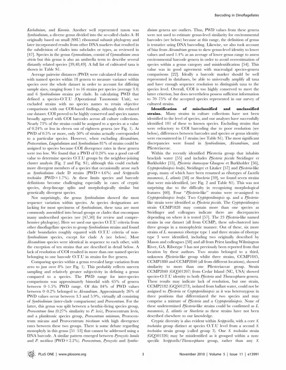
of 0.24% or less in eleven out of eighteen genera (see Fig. 1). At
PWD of 0.5% or more, only 50% of strains actually corresponded
to a particular species. However, when excluding Alexandrium,
Protoceratium, Lingulodinium and Symbiodinium 81% of strains could be
assigned to species because COI divergence rates in these genera
were too low. We found that a PWD of 0.24% was a good cut-off
value to determine species O.T.U groups by the neighbor-joining
cluster analysis (Fig. 2 and Fig. S1), although this could exclude
more divergent members of a species that occasionally arose such
as Symbiodinium clade D strains (PWD = 4.6%) and Scrippsiella
trochoidea (PWD = 1.7%). At these limits species and barcode
definitions become challenging especially in cases of cryptic
species, deep-lineage splits and morphologically similar but
genetically divergent species.
Not surprisingly, the genus Symbiodinium showed the most
sequence variation within species. As species designations are
lacking for most specimens of Symbiodinium, these taxa are most
commonly assembled into broad groups or clades that encompass
many undescribed species (see [47,50] for review and compre-
hensive phylogeny). Here we used our species O.T.U criteria from
other dinoflagellate species to group Symbiodinium strains and found
clade boundaries roughly equated with O.T.U criteria of non-
Symbiodinium species, except for clade A (see below). Most
Alexandrium species were identical in sequence to each other, with
the exception of ten strains that are described in detail below. A
lack of resolution of COI resulted in more than one named species
belonging to one barcode O.T.U in strains for five genera.
Comparing species within a genus revealed large variation from
zero to just over 8% (see Fig. 1). This probably reflects uneven
sampling and relatively greater subjectivity in defining a genus
compared to a species. The PWD range for inter-species
comparisons was approximately bimodal with 65% of genera
between 0–1.5% PWD range. Of this 84% of PWD values
between 0–0.2% belonged to Alexandrium. Approximately 26% of
PWD values occur between 3.3 and 5.9%, virtually all consisting
of Symbiodinium (inter-clade comparisons) and Prorocentrum. For the
latter, this genus was split between a benthic-living species group,
Prorocentrum lima (0.27% similarity to P. levis), Prorocentrum levis,
and a planktonic species group, Prorocentrum minimum, Prorocen-
trum micans and Prorocentrum triestinum with high divergence
rates between these two groups. There is some debate regarding
monophyly in this genus [51–53] that cannot be addressed using a
DNA barcode. A similar pattern emerged between Pyrocystis lunula
and P. noctiluca (PWD = 2.2%). Prorocentrum, Pyrocystis and Symbio-
dinium genera are outliers. Thus, PWD values from these genera
were not used to estimate genus-level similarity for environmental
barcodes (see below) because at this range, the definition of genus
is tentative using DNA barcoding. Likewise, we also took account
of bias from Alexandrium genus to skew genus-level identity to lower
values and used 1.4% as an average of lower genus range to assess
environmental barcode genera in order to avoid overestimation of
species within a genus category and misidentification [54]. This
value was in good agreement with macroalgal species-genera
comparisons [22]. Ideally a barcode marker should be well
represented in databases, be able to universally amplify all taxa
and have enough sequence resolution to distinguish taxa to the
species level. Overall, COI is too highly conserved to meet the
latter criterion, but does nevertheless possess sufficient information
to 70–75% of the accepted species represented in our survey of
cultured strains.
Identification of misclassified and unclassified
strains. Many strains in culture collections have not been
identified to the level of species, and our analyses have successfully
identified 101 of these to known species. Excluding strains that
were refractory to COI barcoding due to poor resolution (see
below), differences between barcodes and species or genus identity
were encountered in 17 strains (see Table S1). The most significant
discrepancies were found in Symbiodinium, Alexandrium, and
Pfiesteriaceae.
Within the recently identified Pfiesteria group that inhabits
brackish water [55] and includes Pfiesteria piscida Steidinger et
Burkholder [55], Pfiesteria shumwayae Glasgow et Burkholder [56],
Cryptoperidiniopsis brodyi, Steidinger et Litaker [57] and Pfiesteria-like
group, many of which have been renamed as ribotypes of Luciella
masanensis, L. atlantis [58] or Stoeckeria [59], we found seven strains
that were mis-identified, (see Fig. 2 and Table S1). This was not
surprising due to the difficulty in recognizing morphological
features [60]. Four ‘‘Pfiesteria-like’’ strains were re-assigned to
Cryptoperidiniopsis brodyi. Two Cryptoperidiniopsis sp. and a Pfiesteria-
like strain were identified as Pfiesteria piscida. The Cryptoperidiniopsis
strain CCMP1828 may contain more than one species as
Steidinger and colleagues indicate there are discrepancies
depending on where it is tested [57]. The 23 Pfiesteria-like named
strains in our dataset (all from CCMP), these sub-clustered into
three groups in a monophyletic manner. Out of these, six more
strains of L. masanensis ribotype type 1 and three strains of ribotype
type 3 were identified, including two originally described by
Mason and colleagues [58] and all from Priest landing Wilmington
River, GA. Ribotype 3 has not previously been reported from that
location by these authors. Two strains belonged to a third,
unknown Pfiesteria-like group whilst three strains, CCMP1845,
CCMP1880 and CCMP2840 (all from different locations), showed
similarity to more than one Pfiesteriaceae group. Strain
CCMP2840 (GQ501207) from Cedar Island (NC, USA) showed
species-O.T.U identity to both Pfiesteria and Thoracosphaera genera.
These results may indicate lack of resolution, but one strain,
CCMP2182 (GQ501273), isolated from ballast water, could not be
assigned to Pfiesteria or Cryptoperidiniopsis as it was heterozygous in
three positions that differentiated the two species and may
comprise a mixture of Pfiesteria and a Cryptoperidiniopsis. None of
these undetermined Pfiesteria-like strains could be confirmed as L.
masanensis, L. atlantis or Stoeckeria as these strains have not been
described elsewhere to our knowledge.
Cryptic diversity is also evident within Scrippsiella, with a core S.
trochoidea group distinct at species O.T.U level from a second S.
trochoidea strain group (called group 3). One S. trochoidea strain
(GQ501326) may be misidentified as it grouped within a non-
specific Scrippsiella/Thoracosphaera group, rather than any S.
Barcoding in Dinoflagellates
PLoS ONE | www.plosone.org 3 November 2010 | Volume 5 | Issue 11 | e13991
trochoidea groups. CCMP2775 (GQ501330) had greater than
genera-level O.T.U distance to any other Scrippsiella or Thoraco-
sphaera species and so was labeled Calciodinellaceae. The
separation of S. trochoidea strains is consistent with the description
of distinct ‘S.trochoidea’ species complexes using ITS sequencing
[61,62]. Four S. precaria strains and one S. cf lachrymosa strain were
identical to each other and had a conserved nucleotide not present
in other Scrippsiella sp. However, Thoracosphaera heimii and Scrippsiella
sp. are represented in one species O.T.U clade in our analysis.
These strains cannot be resolved in species groups using COI
barcodes.
In other cases, taxonomically distinct species were found to have
identical DNA barcodes. In our dataset, eight species showed no
distinction by COI barcode. Karenia mikimotoi and K. brevis; L.
polyedrum and P. reticulatum; some of the aforementioned Scrippsiella
and Thoracosphaera plus Togulla jolla (HM236201) and Spiniferodinium
(GQ501332). The latter two are both of uncertain taxonomic
position but separated at the genus level and unlikely to be the
same, as they possess different morphologies. In addition, nine
different Alexandrium species had identical barcodes. Ten strains
were found to have unusually high levels of divergence compared
to the rest of the genus. Of these, six could be identified as A.
pseudogonyaulax, and the remaining four could not be placed within
any known clade. One identified A. pseudogonlaulax strain, CCAP
1119/1, may be a contamination as it is named A. tamarense and
three of its strain synonyms were different to it. The latter strains
may need taxonomic revision given their unusually high species-
O.T.U divergence levels compared with those of other Alexandrium
species that were identical by COI barcode sequence.
In contrast, other taxonomically related species were found to
be quite distinct. The two Thecadinium species analysed here did
not cluster together: Thecadinium yashimaense (as T. inclinatum)
(HM236199) collected from Canada (held in CCCM) was later
identified as T. mucosum [63] and was subsequently re-classified as
conspecific to T. yashimaense and T. fovealatum [64]. However, our
data showed that T. yashimaense (inclinatum) (HM236199) and T.
yashimaense (HM236200) from Germany were distinct species. As
there are no molecular data for T. yashimaense, these two strains
may be two different species of Thecadinium that may have
originally been misclassified. Small cells interpreted as life cycle
stages were observed in the cultures (unpubl. data), so there is the
possibility of a mixed culture of the two species.
Symbiodinium Identification. Takabayashi and colleagues
[65] found that COI markers corresponded well with Symbiodinium
Figure 1. A Comparison of Uncorrected Pairwise Distances (PWD) of COI Barcodes from Selected Cultured Dinoflagellate Strains.Horizontal axis compares strain isolates within a species (or strains within a clade for Symbiodinium) with species (or Symbiodinium clades) within theirgenus, separated by a red dashed line. Most intra-species comparisons fall between 0 and 0.24%, except for two species, known to have highdivergence or cryptic species. Note higher PWD values and variances obtained when comparing species within a genus, versus isolates within aspecies.doi:10.1371/journal.pone.0013991.g001
Barcoding in Dinoflagellates
PLoS ONE | www.plosone.org 4 November 2010 | Volume 5 | Issue 11 | e13991
clades using a nuclear marker and chloroplast marker (Cp23S-
rDNA) [66]. A total of 81 Symbiodinium strains could be classified to
their correct clade or subclade using shorter COI barcodes and an
additional 23 previously strains that were unclassified or had
ambiguous clade status were successfully attributed to a clade. Of
these, 64 strains had no species/clade identity, two were
completely unidentified and three were misclassified.
Furthermore, our genus-level O.T.U based on these barcodes
are congruent with the authors’ principle groupings (Fig. 3).
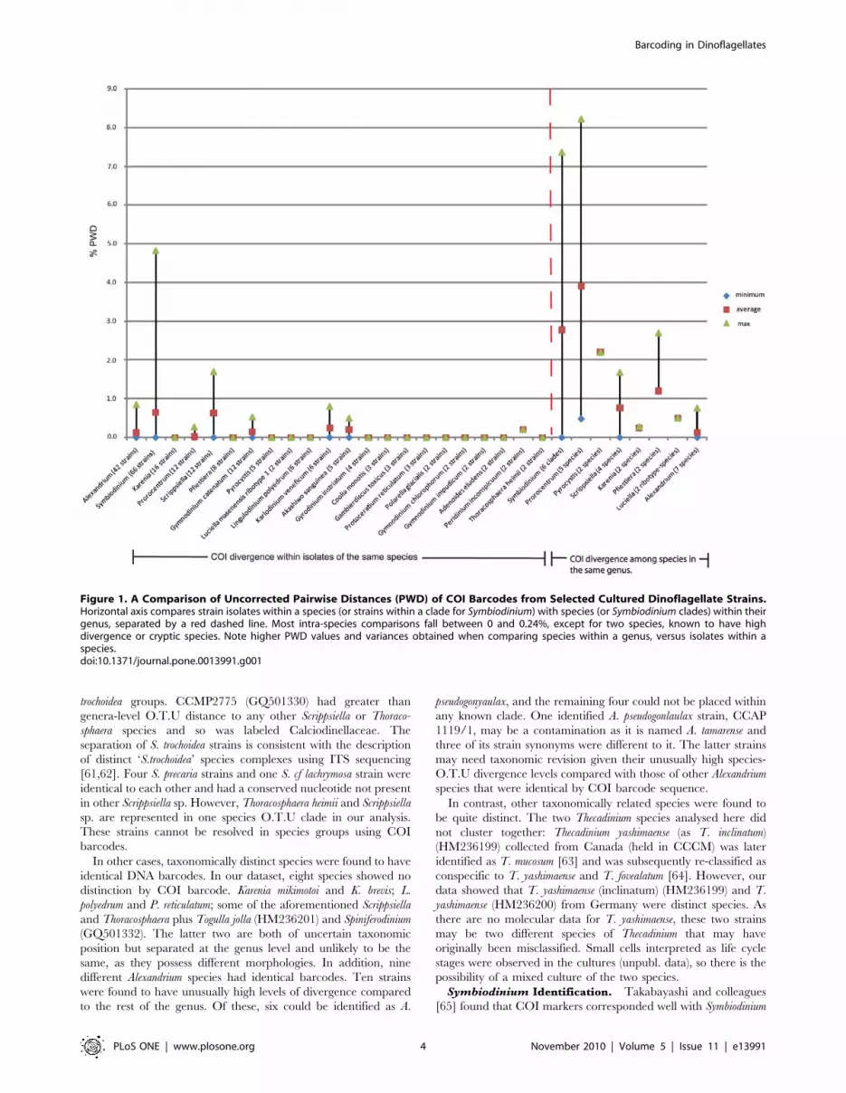
Symbiodinium DNA barcodes formed eight clusters (Fig. 2) that
Figure 2. Neighbor Joining Cluster Analysis of Uncorrected PWD from all Culture Collection COI Barcodes. Each species is colouredaccording to its original species or clade designation. Species of the same genus or clades share a colour theme. Brackets indicate ‘‘barcode’’ speciesgroupings, calculated by uncorrected pairwise distance of 0.24% or less, including those for Symbiodinium clades. Red crosses show strains that falloutside species range. Star symbols show strains that show species level similarity to more than one grouping, also marked similarly. Exclamationmark indicates the group is slightly above species cut-off threshold. Strain names were removed for clarity but are shown in Fig. S2. Species nameabbreviations: A.K.: Antarctic Kareniaceae; Ad.: Adenoides; Al: Alexandrium; Ak: Akashiwo; Cr.: Cryptoperidiniopsis; Gam: Gambierdiscus; Gon: Gonyaulax;Gym: Gymnodinium; Gyr: Gyrodinium; Kar: Karenia; Karl: Karlodinium; Lep: Lepidodinium; Lin.: Lingulodinium; Per: Peridinium; Pf: Pfiesteria; Pol: Polarella;Pro: Prorocentrum; Prot.: Protoceratium; Pyr: Pyrocystis (noct: noctiluca); Sp: Spiniferodinium; Sc: Scrippsiella; Sym: Symbiodinium; T.J. Togulla jolla; Thec:Thecadinium. T. yash (Can) and T. yash (Ger) refer to Canadian and German isolates of T. yashimaense respectively. Thor: Thoracosphaera; W.:Woloszynskia. Unidentified cultured strains are shown in grey shading.doi:10.1371/journal.pone.0013991.g002
Barcoding in Dinoflagellates
PLoS ONE | www.plosone.org 5 November 2010 | Volume 5 | Issue 11 | e13991
corresponded well with clades B, D, and F and within genera-level
O.T.U for clade E (0.3–1.4%). Clade A strains were reliably
identified at the clade level, but the subgroups of clade A only
partially overlapped with subclades as defined by LaJeunesse [59].
Group A3 included two sequences belonging to clade A3, but
also contained two strains that were identified to Cp23S-rDNA
genotype marker A194 (nomenclature based on clade A and size of
the hypervariable region, 194 bp) that generally corresponds to A1
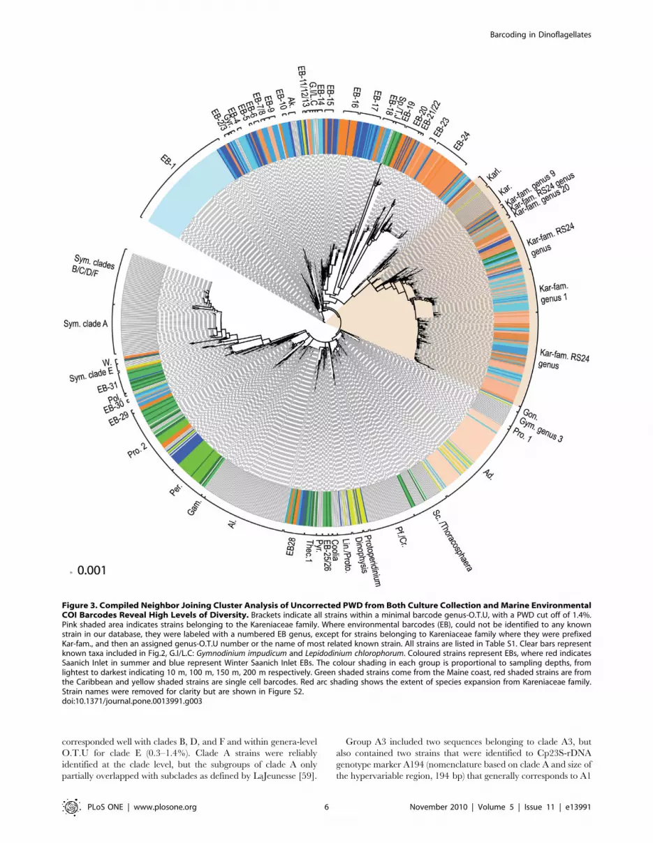
Figure 3. Compiled Neighbor Joining Cluster Analysis of Uncorrected PWD from Both Culture Collection and Marine EnvironmentalCOI Barcodes Reveal High Levels of Diversity. Brackets indicate all strains within a minimal barcode genus-O.T.U, with a PWD cut off of 1.4%.Pink shaded area indicates strains belonging to the Kareniaceae family. Where environmental barcodes (EB), could not be identified to any knownstrain in our database, they were labeled with a numbered EB genus, except for strains belonging to Kareniaceae family where they were prefixedKar-fam., and then an assigned genus-O.T.U number or the name of most related known strain. All strains are listed in Table S1. Clear bars representknown taxa included in Fig.2, G.I/L.C: Gymnodinium impudicum and Lepidodinium chlorophorum. Coloured strains represent EBs, where red indicatesSaanich Inlet in summer and blue represent Winter Saanich Inlet EBs. The colour shading in each group is proportional to sampling depths, fromlightest to darkest indicating 10 m, 100 m, 150 m, 200 m respectively. Green shaded strains come from the Maine coast, red shaded strains are fromthe Caribbean and yellow shaded strains are single cell barcodes. Red arc shading shows the extent of species expansion from Kareniaceae family.Strain names were removed for clarity but are shown in Figure S2.doi:10.1371/journal.pone.0013991.g003
Barcoding in Dinoflagellates
PLoS ONE | www.plosone.org 6 November 2010 | Volume 5 | Issue 11 | e13991

but, may also belong to clade A4. Group Ax contained a mixture
of strains belonging to A194 and a second, uncharacterized
Cp23S-rDNA clade A genotype called A188 and contained all
full-length COI clade A sequences deposited in Genbank. Strain
Zs (AY289692) was identical to CCMP2461 (GQ501337) and had
borderline identity (0.24–0.27%) to CCMP2429 (GQ501395), a
more distal clade A2 strain ([67] and R. Moore, personal
communication).
Clade C/F contained two strains, CCMP2466 (GQ501334
clade C1) and Mv (AY289712) originally identified as F1 [68] but
now designated to clade F5 [50]. This group is given provisional
C/F status and probably grouped together due to lack of COI
resolution. Both these strains were identical to CCMP2434
(GQ501353) and have borderline clade identity (0.24%) to
members of clade B and F, although CCMP2434 (GQ501353) is
identical to just under half of clade B strains. The strains in clade
C/F are clearly distinct but related to either clade F or B so
CCMP2434 (GQ501353) may be a B subtype. Although most
members of clade F and B were easy to distinguish, a previous
study showed AY289712 (strain Mv) belonged to clade F but
shared an identical Cp23S-rDNA genotype allele size with clade B
[66]. Subsequent sequence analysis revealed these to be two
distinct strains, but such homoplasy may be replicated using COI
barcodes in these strains. MAC-Pdiv 45a (GQ501370) had
ambiguous clade identity (either F or B), but was identified as
clade B using COI.
Symbiodinium clade E [68–70] gave unexpected results, especially
for the three strain synonyms of CCMP421 (GQ501340,
GQ501339, GQ501242), belonging to clade E2, and an
unclassified strain attributed to Gymnodinium (AC561, GQ501241)
which were between 0.3–0.9% similar and well within genus level
O.T.U of each other. The identification of AC561 is significant as
there are only two other clade E cultures. In particular, nucleotide
variation was observed in six positions over a 60 bp region in all
CCMP421 strain synonyms barring AC561. CCMP421 was
started as a single cell and was previously misidentified as
Gymnodinium [69]. A fourth strain synonymous to CCMP421
deposited in Genbank (AY289708) did not fall within clade E with
the other synonyms, but instead showed borderline clade level
identity to one of the two clade D1-4 (‘‘S. trenchi’’) strains
(GQ501397) [71–73] which also clustered together with full length
COI gene [65]. The placement of CCMP421 strain AY289708 in
clade D may be because this sequence is a COI paralogue (see
below) or because of phylogenetic uncertainty between clades E
and D using COI in some studies ([65] and reviewed by [74]).
Two previously identified clade D strains, PSP1-05 and HpiH-
showed no identity to any strain in our database. PSP1-05, is
recognized as a highly divergent member of clade D1 [65,69] so
this result is not surprising.
Testing for paralogues and pseudogenesPseudogenes are known to occur in dinoflagellates [66,75], their
organellar genomes are highly unusual and their mitochondria
contain many fragmented gene copies which could be co-amplified
in this instance [76,77]. To determine whether this was the cause
of unusually high variation observed in strains belonging to
Scrippsiella trochoidea, Prorocentrum and Symbiodinium, we sequenced 30
clones from seven strains (average 5 clones per strain) belonging to
Scrippsiella sp. (2 strains), Prorocentrum (3 strains) and Symbiodinium sp.
(2 strains, including CCMP421). Comparing the directly se-
quenced strains with their cloned counterparts (see Table S3)
showed very slight variation, none more than 0.2% PWD for all
but one strain, less than the species-O.T.U cut off of 0.24%
indicating strain diversity has not been overestimated in these
species. No clones of cryptic S. trochoidea strain CCAP1134/9
overlapped with separate S. trochoidea (CS-297). The one exception
to this were clonal DNA barcodes of CCMP421, where the
average distance between all clones plus the direct sequence was
1.3%, a value expected for an average intra-species comparison
and likely to be due to the presence of multiple paralogues. Three
CCMP421 COI barcode clones were identical to that of AC561,
which explains at least some of the variation observed in the
directly sequenced strain synonyms of CCMP421. No stop codons
were present in cloned and directly sequenced barcodes except for
CCAP 1136/16, Prorocentrum minimum (GQ501297) at positions
corresponding to previously reported RNA editing sites for this
species [78].
Comparing culture collections to natural diversityIn order to evaluate how much natural diversity is represented
in culture collections, environmental DNA barcoding of total
planktonic DNA from three different marine environments was
carried out with dinoflagellate-specific primer sets. The deepest
sampling was done from Saanich Inlet - a marine fjord off
Vancouver Island that is hypoxic from 100 m to 200 m depth in
the summer, but which is mixed in the winter. Near-coastal
planktonic samples were also taken from the coast of Maine, USA
and the island of Guadeloupe in the Caribbean. After screening
out poor quality sequences, this resulted in a total of 713
environmental barcode sequences (listed in Table S2): 574
barcodes from Saanich Inlet (Northeast Pacific); 86 from Maine
(Northwest Atlantic), and 29 from Guadeloupe (Caribbean).
Finally, to show that both morphology and DNA barcodes could
be recorded for single cells, especially those refractory to culturing,
we also isolated 24 single dinoflagellate cells from the west coast of
Vancouver Island, photographed them, and produced COI
barcodes from individual cells. Photographed single cells were
also used to corroborate the identity of unknown environmental
barcodes belonging to the same species-O.T.U.
Clustering the combined culture collection and environmental
data resulted in a total of 1049 dinoflagellate barcodes (Fig. 3) that
showed depth and seasonal stratification (Fig. 3) also confirmed
using Principle Coordinates Analysis (PCoA) (Fig. S3). The most
striking first observation from these data was the level of diversity
within the environmental groups with 531 different barcode
species out of 603 unique environmental barcodes, mostly from
Saanich Inlet. Although this study only covered 54% of known
cultured strains, it is still striking that only 91 of those
environmental barcodes, including single cells could be matched
at the species level (261 at genus level) to cultured strains using an
average cut off value of 0.24%. This Figure fell to 51 identified
barcode clones, once identical clones were removed. A further 92
unique environmental barcodes that were related to each other
could not be correlated to any cultured species. In total, 24% of
unique environmental barcodes were related to at least one other
sampled sequence from a known or unknown strain at the species
level. To link environmental barcodes to known phylotypes at a
broader level, each environmental barcode was binned with a
known genus if it was within the PWD boundaries defining that
genus (this varied between genera as each had different levels of
sequence diversity) plus to any other strains also grouped with that
genus. By these criteria, many environmental barcodes could not
be identified to any known taxon at any level, so in these cases an
average genus-level PWD cut-off of 1.4% was used. This was a
conservative estimate, in line with minimum values for most of the
known genera. Based on this genus-level PWD estimate,
environmental barcodes were grouped in the cluster analysis
shown in Fig. 3, except for the highlighted grouping containing
Barcoding in Dinoflagellates
PLoS ONE | www.plosone.org 7 November 2010 | Volume 5 | Issue 11 | e13991

Karlodinium, Karenia and an unnamed Antarctic dinoflagellate
(RS24) belonging to the Kareniaceae family [79,80] which is an
exceptional case discussed below. In addition to the Kareniaceae,
the second major identified group in the environmental samples
are a group of sequences identical or closely related to Adenoides or
Amphidinium cf. semilunatum [81]. These occurred exclusively in
10 m July 2006 Saanich Inlet samples and matched cultured in
single cell isolates, whereas an unknown cluster (shown in pale
blue, Fig. 3, and blue circle in Fig. S3) represented almost the
entire diversity of 10 m winter sample from Saanich Inlet. These
are the only seasonally related clusters. Many of the Caribbean
and Maine samples grouped with Prorocentrum and Peridinium sp.
and a minority to Scrippsiella or Thoracosphaera. Finally a small
number of environmental barcodes, mostly from unidentified
groups EB23 and EB24 (Fig. 3 and Fig. S2) were found exclusively
in deep water, from 100–120 m depth in Saanich Inlet exclusively
in summer, below the hypoxic boundary.
Overall, the majority of environmental barcodes from these
marine samples could not be identified because there was little
overlap in diversity between the environmental and cultured
dinoflagellate barcodes. In addition, there was little overlap of
species assemblages between different environments. Nevertheless
numerous other environmental barcodes could be identified to the
genus level or higher, and many of these represent great
expansions in the known diversity of these groups.
Single-cell barcoding. With the majority of environmental
barcodes not attributable to any taxonomic group represented in
the barcode library of culture collections, a method to connect
environmental barcodes to cells will be required to assess natural
microbial diversity. For a start, barcoding from single cells that
have been photographed prior to isolation would at least allow us
to identify major groups otherwise only made up of environmental
sequences. To test this in principle, almost 70 single cells were
photographed and manually isolated from the west coast of
Vancouver Island, and 24 COI barcodes were generated from
these single cells, although images for four single-cells in this study
(GQ502036, HM236194, GQ502039, HM236195, GQ501405)
were very poor quality and discarded. The single-cell barcodes
proved to be successful in providing benchmarks for the
environmental barcode libraries for the difficult-to-culture
heterotrophic Dinophysis and Protoperidinium genera (Fig. 4), as
well as identifying representatives of other major environmental
groups with poor representation in culture collections such as
Adenoides (GQ501403-GQ501406) which is found in benthic and
seawater environments. These strains, together with cultured
Adenoides eludens strains were used to aid the identification of a large
cluster of uncultured environmental barcodes from Saanich Inlet
(2006), almost exclusively found at 10 m. GQ501404 was
morphologically similar to the taxonomically unresolved
Amphidinium semilunatum, a species that is distinct from Adenoides,
but both previously grouped under the genus Amphidinium.
GQ501404 was identical to cultured A. eludens so it is possible
that the COI barcode lacks resolution to distinguish between these
two potentially separate species. However, GQ501404 had eight
non-ambiguous sequence differences to the COI barcode of a
third Adenoides species, NIES-1402 (HM355857-not included here),
that suggests this group requires taxonomic re-evaluation.
Kareniaceae – a case study of exceptional natural
diversity. Only four species of Kareniaceae are represented in
our barcodes from culture collections (Fig. 2), but the diversity of
this group exploded with the addition of environmental barcodes
from the Northwest Atlantic, and particularly from the Northeast
Pacific.
The Kareniaceae have traditionally been a relatively small
family made up of dinoflagellates that are distinguished by having
a 199 hexanoyloxyfucoxanthin-type plastid derived from a
haptophyte [82]. The Antarctic dinoflagellate RS24 (GQ501253)
is a sister species to Karenia and Karlodinium [80], with a temporary
haptophyte resident identical to Phaeocystis antarctica. [79]. Recent
studies have shown that Kareniaceae are more diverse than
previously thought, with another six new species recently described
[83] but there still only 20 known species in this family, including
the RS24. However, of 713 environmental sequences, 177 clones
fell within the Kareniaceae group as a whole, and 122 clones
representing 88 species showed genus-O.T.U level identity with
RS24 that showed same grouping with PCoA (Fig. 3, Fig. S3 and
Table S2). These included 26 environmental barcode clones
mostly from Saanich Inlet which had species-level identity to RS24
(Fig. 3) and this species was the most common among the
Kareniaceae. One non-photosynthetic single-cell PL9-11
(GQ502034), was found to belong to this family but was
genetically different to RS24, and may also represent another
species Interestingly, virtually all environmental barcodes falling
within this group were from Saanich Inlet and from depths
between 100–120 m, although there were no distinct seasonal or
habitat differences, with barcodes clustering with RS24 at species
level being found in Northwest Atlantic too.
Discussion
Barcoding culture collection strainsCulture collections represent the most accessible and traceable
repositories of living microalgae, but they are also biased towards
the species and strains most amenable to cultivation, and/or of
commercial and medical importance. It has long been known,
especially in prokaryotes, that only a small fraction of natural
diversity is easily cultured using common strategies. Accordingly,
cultured strains do not adequately represent either the depth or
breadth of microbial diversity, but they still make useful
benchmarks for molecular barcode databases of natural diversity.
However, while it is becoming increasingly easy to use molecular
data for taxonomic surveys that avoid culture work, cultures still
remain a primary source for scientists who need cells for
biochemical, biotechnical, cellular and physiological studies. These
scientists will benefit greatly from barcodes that can provide an
easy and quick means for quality control.
Figure 4. Expanded View of Neighbor Joining Cluster Analysisfrom Figure 3. Showing Dinophysis and Protoperidinium SingleCell Barcodes with Their Photomicrographs. Black solid linesshow species level similarity.doi:10.1371/journal.pone.0013991.g004
Barcoding in Dinoflagellates
PLoS ONE | www.plosone.org 8 November 2010 | Volume 5 | Issue 11 | e13991
COI barcoding worked as a means of distinguishing most
dinoflagellate species in 15 of 21 genera where COI sequence
divergence rates were congruent with speciation events. For those
15 genera 81% of strains could be distinguished at species-O.T.U
level using a PWD cut-off value of 0.24%. This criterion would
allow for the majority of COI samples obtained from environ-
mental samples to be binned into genetic clusters which on
average corresponds with distinct species. This approach will
overestimate the number of true species in genera with higher
divergence rates and underestimate those with lower rates of
divergence, but on average it should give a reasonable estimate of
species diversity. A small assessment of species in the most variable
genera demonstrated that genetic differences are unlikely to be due
to pseudogenes or paralogues in all but one strain (discussed
below), at least for those species tested. Other species, such as those
belonging to Kareniaceae, showed low intra-species PWDs and
unlikely to possess paralogues that would artificially inflate
observed diversity in the environmental barcode dataset.
Assessing species diversity in the five genera (Alexandrium,
Lingulodinium, Protoceratium, some Thoracosphaera and Scrippsiella sp.)
where COI sequence divergences were too highly conserved or too
divergent (Symbiodinium) to prove useful in distinguishing species will
require a different barcoding marker. For example, our failure to
find a robust breakdown of Alexandrium strains in common with cob
barcoding study by Lin and colleagues [35] reflects a well-
documented problem with this genus, whose morphology can vary
greatly under different environmental conditions [28,84–87]. Due
to this difficulty, several species have been grouped into species
complexes (reviewed in [28]). However, the correct choice of
marker may allow resolution of taxonomical challenges, such as the
successful use of the variable region of the large ribosomal subunit to
identify cryptic species within the A. tamarense species complex [88].
Despite this lack of complete coverage, the COI can be used to
assess species diversity in a number of important dinoflagellate
groups such as Karenia and Karlodinium genera. Systematic barcoding
of available cultures has revealed instances of out-dated nomencla-
ture, cryptic diversity, and misidentifications, all of which may be
expected in such a diverse and abundant group. The advantages of
DNA barcoding allows for quick, cost effective means of identifying
species that can be carried out using easy-to-use and fast
phylogenetic and bioinformatic tools, which will become easier as
the database expands. The COI marker has the advantages of
circumventing time-consuming and expensive cloning procedures.
It is difficult to compare the effectiveness of COI in comparison
to cob [35], with two different methods of analysis, different
sampling depths and different species studied. However, compar-
ing similar species groups, both COI and cob showed little intra-
specific variation, and inter-specific variation of COI in our studies
were remarkably similar to that of cob, except for Symbiodinium
which had a much larger range in our study, due to the larger
sample size of our dataset. The cob gene was found to resolve about
half of the diversity in a study by Sampayo and colleagues and was
not suitable for ancestral Symbiodinium types [89]. As a whole, these
results were unexpected given that cob was reported to be more
variable than COI but could be explained, in part, by the greater
sampling depth of known species used here.
DNA barcoding of a diverse genus: Symbiodinium. The
genus Symbiodinium has largely been recognized to be incongruent
with other dinoflagellate genera in terms of genetic diversity. COI
barcoding made largely accurate identifications of the major
clades in accordance with Symbiodinium phylogenies [47,50,65].
However, the lack of fine level resolution with COI, and the short
length of a single DNA barcode is unlikely to capture accurate
phylogenetic relationships achievable with longer markers [69,70].
Therefore the subclade groupings did not correspond entirely to
those described by LaJeunesse [68], particularly for subclade A,
and is probably the reason for the ambiguous identity of some
strains in clade F, C and B. COI barcodes would be useful as a
complementary marker for example, resolving the identification of
two strains with ambiguous clade identity. The variation observed
in multiple CCMP421 strain synonyms appears to be caused by
the presence of multiple paralogues, which was not observed in
MAC-579 (clade B). An unusually high number of ITS paralogues
have also been reported for CCMP421 [90] which may have an
evolutionary significance that deserves further investigation.
Whilst monoclonal cultured cells can be a useful source to control
morphological plasticity [68], one caveat of using cultured
Symbiodinium for identification is they may not reflect the true
biological symbiotic strain in the host. Often, cultures can contain
surface contaminants such as free-living forms of Symbiodinium
[68,91]. COI barcodes have uncovered strain misidentifications and
also a rare new clade E culture (AC561) and highlight the usefulness
of DNA barcoding in culture collections. Although COI is not
suitable to identify Symbiodinium strains to subclade and type level, it
could be considered an easily amplifiable validation marker for
Symbiodinium diversity in the same way as other organelle markers
have, namely cob and Cp-23S rDNA (chloroplast large subunit
rDNA gene). The latter were able to identify strains to species level
when combined with nuclear markers [89]. This could reduce the
considerable problems caused by ITS paralogues and pseudogenes
[90]. Furthermore, these classifications could allow more universal
comparisons with other dinoflagellates in future.
Diversity of dinoflagellates in marine environmentsIn common with previous SSU deep amplicon sequencing
surveys of protists in marine environments [17,18], our study
revealed astonishing diversity, although SSU may be too
conserved to distinguish many species of dinoflagellates [92].
However, diversity may not exclude ubiquity as environmental
diversity studies have identified the same species in distant regions
[18,35]. The Saanich Inlet fjord system has many discrete habitats:
the summer thermocline in Saanich Inlet and an additional
halocline from glacial waters mixing at the surface creates
ecological boundaries, partitioning species into ecological niches
that may support genetically stratified populations of dinoflagel-
lates by sexual and/or environmental selection. This dataset
revealed massive diversity in environmental dinoflagellate bar-
codes at 10 m and 100 m in summer and winter, which showed
spatial and temporal species-level separation. Do our results
indicate cosmopolitan or endemic species? The diversity in
Kareniaceae family, the lack of overlap of some environmental
sequences at different depths and seasons give some indication of
endemism. Although very little overlap existed between the three
environments, there was evidence for cosmopolitan species: the
Antarctic Karlodinium-like dinoflagellate, RS24, and some Scripp-
siella sp. were common to NE pacific and NW Atlantic samples.
Relatively few sequences were obtained from of NW Atlantic and
Caribbean environments so with greater sampling depth there
could potentially be more species that overlap between different
environments. The nature of the environment is a major force that
selects for endemic or cosmopolitan species. Deep sequencing of
ecologically similar environments in different locations using DNA
barcoding methodology will be instrumental in addressing this
debate. Without morphology to aid identification, studies should
consider confounding factors such as paralogues, by using more
than one marker. A more difficult question is how to apply cut off
values in order to accurately measure species diversity, given
uneven speciation rates.
Barcoding in Dinoflagellates
PLoS ONE | www.plosone.org 9 November 2010 | Volume 5 | Issue 11 | e13991
Kareniaceae diversity. Recent studies described five new
species of Kareniaceae [83], in addition to RS24 described by
Gast et al. [79], although all at surface waters. There are many
toxic species in this family, yet little is known of them because they
are small, fragile and poorly sampled. A large proportion of these
dinoflagellates existed at 100 m and lower. It is unclear how these
and the two other groups of deep water dinoflagellates survive at
these depths, some below the hypoxic boundary. Zaikova and
colleagues [93,94] analysed the same Saanich Inlet samples for
bacterial diversity and reported a dominant bacteria group,
SUP05 present at 100 m and below in 2006 for both seasons.
SUP05 was originally identified from Suiyo Seamount [95], and a
common hypoxic water species which may be a food source for
these dinoflagellates. Deep water heterotrophic Gymnodinioid
species have also been identified in deep, stratified marine systems
such as the Gulf of Gdansk (over 100 m) [95]. Alternatively they
could be cysts, although cysts have not been described in
Karlodinium or Karenia [96]. Our findings have contributed to
evidence of a diverse and uncharacterized group of deep water
dinoflagellates from stratified marine waters that have adapted to
this habitat, possibly with unique trophic and respiratory
mechanisms.
Seasonal diversity in Saanich Inlet. The summer and
winter species compositions in Saanich Inlet were distinct and
dominated by one genus. Coincidentally the greatest different in
abiotic measurements were found between the February and July
time points [93]. Forty four percent of the summer 10 m samples
contained photosynthetic Adenoides, a genus with only two known
species ([81] and unpublished data) that increased to 35 species-
O.T.Us using our criteria. The February 2006 dinoflagellates at
10 m were unidentifiable but again were diverse,numbering 61
species-O.T.Us. A study of winter phytoplankton from 0–20 m
depth in 1978 by Takahashi and colleagues [97] revealed
dinoflagellates were the second dominant group, mostly
consisting of Katodinium rotundum where light and temperature
were the predominant limiting factors for growth. Given the
dominance of Gymnodinioid dinoflagellates in both our samples
and that of Takahashi et al. [97] perhaps K. rotundum may belong to
the 10 m Saanich Inlet winter surface dinoflagellate assemblage
and deserves further investigation. The use of single-cell barcoding
would be particularly useful in identifying this and other
unculturable protists and could be automated by flow cytometry,
which has already been applied to protists [98] and provide better
estimates of natural diversity in these communities.
Concluding Remarks: natural diversity of microbial lifeOur results show cultured dinoflagellates that are considered to
be different species can be resolved using DNA barcoding,
although robust taxonomy using other DNA markers, morphology
and chemotaxonomic markers such as lipids and pigments is
needed to provide a solid basis for DNA barcoding to work [99].
The vast majority of cultured species studied here could be
identified with a PWD of 0.24% or less, showing that the concept
of DNA barcoding can work for dinoflagellates and could be used
to identify and segregate taxonomic units in environmental studies,
although COI might not be the best single gene with which to
assess and identify dinoflagellates. The true value of barcoding is
its scale and ease to match unknown environmental sequences
with a single cell barcode or a culture collection strain.
The environmental barcodes in this dataset may be a small
fraction of the real diversity of the environments they represent,
since none seemed to be sampled exhaustively. An even tinier
proportion of dinoflagellates are represented by the combined
holdings of culture collections, reflecting human bias in sampling
and cultivation. DNA barcoding studies will be instrumental in
evaluating biogeographical speciation and ecological assemblages
within protist populations. With increasing use of next generation
sequencing technology that can combine multiple markers, deep-
level biodiversity studies will be more able to demonstrate true
estimates of protist diversity and may provide useful information
for the cultivation of a greater proportion of presently unculturable
species. These methods combined with flow cytometry, already in
use [[98,100,101], would provide additional morphological
characters enabling single cells from environment and cultures
to be evaluated at the individual rather than population level.
Materials and Methods
Sample CollectionCultured strains or DNA samples were donated or purchased
from eight public and three private culture collections, summarized
in Table S1. Summary codes for strain donors are thus: AC:
Algobank-Caen, Universite de Caen Basse-Normandie, France;
CCAP: Culture Collection of Algae and Protozoa, Scottish
Association for Marine Science, U.K.; CCMP: Provasoli-Guillard
National Center for Culture of Marine Phytoplankton, Bigelow
Laboratory, USA; CS: Australian National Algae Culture Collec-
tion, CSIRO, Australia; CAWD: Cawthron Institute, Culture
Collection of Micro-algae (CICCM), Nelson, NZ; NEPCC: North-
East Pacific Culture Collection (part of Canadian Centre for
Cultured Microorganisms, CCCM), University of British Colum-
bia, Canada; NIES: National Institute for Environmental Studies,
Japan; BURR: Buffalo Undersea Reef Research Culture Collection,
State University of New York at Buffalo, Buffalo, USA MH: Mona
Hoppenrath, Forschungsinstitut Senckenberg, Germany; UTEX:
The Culture Collection of Algae at the University of Texas, Austin,
TX, USA. Culture RS24 was donated by Rebecca Gast from
Antarctic Protist Culture Collection, Woods Hole Oceanographic
Institution, Woods Hole, MA, USA (private).
For environmental analysis, planktonic samples were collected
through a 20 m plankton net taken from the island of Guadeloupe
in the Caribbean (15.1539N, 61.3475W), from the Bigelow
Laboratory pier, West Boothbay Harbor, ME, USA (38.1904N,
76.2707W) and also from mouth of the Damarascotta River,
Maine, USA approximately 44N, 69.5W) representing two
different coastal environments in Maine, USA (Northwest
Atlantic). DNA from Saanich Inlet, Vancouver Island, BC,
Canada (43.39N, 123.39W) (Northeast Pacific) was kindly donated
by Dr. D. Walsh and Dr. S. Hallam, UBC, from non-filtered
marine water collected by David Walsh in water collector
containers at depths of 10 m, 100 m, 120 m and the bottom at
200 m in equal volumes. These same samples are also described
by [93]. Single cells were collected from surface plankton or sand
in Bamfield, Vancouver Island, BC, Canada; Saanich Inlet, BC,
Canada. Isolation was performed under an inverted microscope.
Cells were picked using a sterile extended pasteur pipette from a
dish, washed in fresh autoclaved seawater to check for single
isolation under a microscope, photographed and picked using a
separate autoclaved, pipette. Benthic samples were filtered from
sand using a method described by Hoppenrath and Leander [102]
and single cell isolated from these samples were processed in a
similar manner to those of planktonic samples. All uncultured,
environmental samples used in this study are listed in Table S2.
DNA extractionTypically between 1.5–15 ml of dinoflagellate cells from culture
were collected by centrifugation at initially 3000 g then at 1150 g,
snap frozen in liquid nitrogen and thawed three times. For one
Barcoding in Dinoflagellates
PLoS ONE | www.plosone.org 10 November 2010 | Volume 5 | Issue 11 | e13991

third of culture collection samples, additional grinding was
performed using plastic pestle and microfuge tube. DNA
extraction was carried out using the DNeasyTM plant purification
DNA kit (Qiagen, Mississauga, ON, Canada), following their
protocol except incubating cells in lysis solution for 30 minutes
instead of 10 minutes. MasterpureTM Complete DNA and RNA
Purification Kit (EpicentreH Biotechnologies, Madison, WI, USA)
was also used in about one third of cultures and for single cells,
using Lysis of Fluid sample protocol followed by Precipitation of
Total DNA protocol. For whole marine extracts DNA extraction
was performed by mixing whole marine sample with an equal
volume of a phenol-chloroform- isoamyl alcohol mixture (25:24:1)
(Sigma-Aldrich, Oakville, ON, Canada), the DNA containing
phase was removed and DNA extracted with the addition of 2.5
volumes of 100% ethanol (Sigma- Aldrich, Oakville, ON, Canada)
and 0.1 volumes of 3M sodium acetate, pH 5.2 (Sigma-Aldrich),
washed twice in 75% Ethanol and resuspended to 300 mg/ml in
sterile water.
PCR, cloning and SequencingHighest amplification rates were achieved using a nested primer
set, consisting of primers DINOCOX1F 59AAAAATTGTAAT-
CATAAACGCTTAGG 39and DINOCOX1R TGTTGAGC-
CACCTATAGTAAACATTA described by [52] and then using
a nested primer set designed by B. Imanian COX1.DINO.F 59
GAATTTGGAGGTGGCACNGGNTGGACNYT 39 and COX1.
DINO.2.R 59-CCCATCGTATACATRTGRTGNCCCCANAC
39. PCR amplification was carried out on 25–100 ng of DNA using
PuReTaq Ready-to-Go beads (GE Lifesciences, NJ, USA) at 94uCfor 3 minutes followed by 35 cycles of 94uC for 30 seconds, 48uC for
30 seconds and 72uC for 45 seconds, ending with a 72uC extension
step for 7 minutes. All culture collection and single-cell cultures were
sequenced directly. Single PCR products were diluted to 30 ng/ml or
purified by gel extraction using the QIAquick Gel Extraction kit
(Qiagen, Mississauga, ON, Canada), according to manufacturer’s
instructions and either sent to Canadian Centre of DNA Barcoding,
Guelph, ON for DNA sequencing or sequenced directly using
BigDye v3.1 reagents on at NAPS unit at University of British
Columbia, BC. COI amplification products from whole marine
extracts, or environmental barcodes, were cloned using TOPO TA
cloning kit (Invitrogen, Burlington, ON, Canada) according to
manufacturer’s directions except 200 ml of transformations were
plated onto LB ampicillin plates. Transformed white colonies were
screened using Amplitaq kitH (Invitrogen, Burlington, ON, Canada)
as per manufacturer’s instructions using 1 mM of M13 forward
59GTAAAACGACGGCCAG 39 and M13 reverse 59CAGGAAA-
CAGCTATGAC 39 primers (synthesized by IDT, BC, Canada),
3.5 mM MgCl2 and 2 ml of colony dissolved in 20 ml dH2O per
reaction. Screening reactions were amplified with an initial
denaturation step at 94uC for 3 minutes followed by 30 cycles of
94uC for 30 seconds, 50uC for 30 seconds and 72uC for 45 seconds,
ending with 72uC extension step for 7 minutes. The screening
amplification reaction produced single products and these were
diluted to 200 ng/ml and sequenced at Canadian Centre of DNA
Barcoding, Guelph, ON, using M13 forward and reverse primers.
Sequences are available at http://www.barcodinglife.org.
Sequence analysisCOI barcodes for cultivated and uncultivated environmental
dinoflagellates used in this study are listed in Tables S1 and S2
respectively, and sequences can be retrieved from the Barcode of
Life Database (BOLD) at http://www.boldsystems.org/views/
login.php. Cultured sequences 1-332 are listed under DACOI, and
all other uncultured sequences under DINOB. Genbank accession
numbers GQ501108-GQ502113 and HM236191-HM236201 are
also available for all barcodes and listed in Tables S1 and S2.
Sequences of cloned products for pseudogene analysis are listed in
BOLD within DACL project. All sequences were manually edited
using SequencherTM v4.2 (Gene Codes Corporation, Ann Harbor,
USA) and BioEditTM 7.09 [103]. Nucleotide sequences were
translated to the amino acids, aligned and translated back to the
nucleotides using BioEdit COI sequences were checked for
reading frame interruptions that might indicate the presence of
pseudogenes, except where they corresponded to known RNA
editing sites in published sequences [78] or had common changes
to multiple strains of the same species. To determine the best tree-
building method, several algorithms were chosen including
maximum likelihood, neighbor-joining using the Kimura-2
distance substitution model (data not shown) and neighbor joining
with uncorrected distances using PAUP* 4.0b10 [104]. There was
virtually no difference in the topologies produced with any of these
methods, so a neighbor joining with uncorrected distance was used
in order to compare with pairwise distances between strains which
was calculated using PAUP* 4.0b10. The resulting tree or
clustergram was visualized by ITOL [105] and Adobe illustra-
torTM CS2 12.0.0 (San Jose, CA, USA). To calculate cut-off values
for a species, only strains identified to species level were used in
order to provide an objective comparison of how well COI
barcodes corresponded with known species. All unique strains with
no matching COI barcode plus Alexandrium, Protoceratium, Lingulo-
dinium were excluded from such calculations, since COI was
ineffective at discriminating species in Alexandrium or genera in the
latter two taxa. Symbiodinium was also excluded from initial species
and genera level- O.T.U calculations, because species definitions
did not apply, this genus being primarily defined by clades.
However, once cut-off values were established, these were used to
group Symbiodinium strains. Out of Gymnodinium, only G. catenatum
and G. impudicum, belonging to Gymnodinium sensu strictu group, were
included for calculations to find species and genera level cut off
values, the rest being paraphyletic and therefore unsuitable.
Whenever the species category did not match COI barcode
O.T.U (such as when more than one named species fell into a
single species-O.T.U), the strains were given an O.T.U name that
represented all members. The names of strains with only one
representative not matching any barcode in this database
remained the same. To detect the presence of pseudogenes, the
sequences derived from cloned barcodes of a strain were grouped
together with that strains directly sequenced product. An average
PWD for each strain group was calculated with MEGA software
[106] using uncorrected p-distance model including transition and
transversion substitutions, homogeneous lineage pattern with
uniform site rate. Within group variation was checked manually
for unusually high genetic divergence values that might point to
presence of a paralogue.
In calculating genus-level O.T.U for environmental barcodes,
we categorized each PWD comparison between species and
ordered them according to increasing PWD. Each strain was
checked and categorized so that every member of a genus-O.T.U
group had no more than 1.4% identity to any other member of the
same group. Although at the lower end of PWD of known genera,
this value also minimized ambiguous identities, where a strain
showed equal identity to more than one genus, or showed identity
to only some but not all members of a genus-level O.T.U group. In
these cases, the corresponding sequences were checked for
sequence quality and excluded from analysis if below quality of
other sequences. Even with sequence quality parsing, unresolved
Barcoding in Dinoflagellates
PLoS ONE | www.plosone.org 11 November 2010 | Volume 5 | Issue 11 | e13991
cases remained. These sequences were removed from their original
group and placed in a separate smaller grouping or on their own,
for single strains, as shown in Fig. 3 and Fig. S2. Environmental
barcodes that did not group with known genera and species were
prefixed with EB and a genus number followed by a species
number e.g. EB1-1. Genera were labeled according to their
groupings in Fig. S2. Some genera were split e.g. Prorocentrum,
genus 1 was assigned to those barcodes showing closest similarity
to P. levis and P. lima whereas genus 2 was used to denote those
barcodes similar to P. micans, P. minimum and P. triestinum. The only
exception were those strains known to belong to Family
Kareniaceae, where species in this group were called RS24- if
they belonged to the same species as cultured dinoflagellate RS24,
and Karenieaceae RS24, if they had genus level identity with that
strain.
For Fig. S3. Principal coordinates analysis (PCoA) was
performed in GenAlEx 6.2 [107] in order to explore further the
relationships between environmental samples and culture collec-
tions. This multivariate ordination technique finds the orthogonal
axes along which the variation among points described by a
distance matrix is greatest. Pairwise distances among all cultured
and environmental samples were imported into GenAlEx and
squared to produce the matrix required for PCoA, which was
performed on covariances and standardized.
Supporting Information
Figure S1 Neighbor Joining Cluster Analysis of Uncorrected
PWD from All Culture Collection COI Barcodes as in Figure 2,
with Strain Names.
Found at: doi:10.1371/journal.pone.0013991.s001 (1.58 MB
TIF)
Figure S2 Neighbor Joining Cluster Analysis of Uncorrected
PWD from Culture Collections and Marine Environmental COI
Barcodes as in Figure 3, with Strain Names.
Found at: doi:10.1371/journal.pone.0013991.s002 (0.75 MB
TIF)
Figure S3 Principle Coordinate separation of DNA Barcodes
from Cultured and Environmental Dinoflagellates. PCoA using
the first three principle coordinates (labeled all-st-1, 2 or 3) of all
environmental barcodes and culture collection strains, colour-
coded in a similar manner to Fig. 3, except that cultured strains
are shown in black. Most cultured strains plus a proportion of
environmental clones are in two large cloud points indicating
similar variance to each other. The diverse Symbiodinium strains
(circled in grey) are the only cultured strains are more distant to
the majority of cultured dinoflagellates. Most of the pale red
barcodes (circled in orange) belong to Adenoides sp. from July 2006
together with known Adenoides sp. The pale blue barcodes (circled
in blue) are an abundant, unidentified genus-level O.T.U. that
represented almost the entire diversity of 10 m winter sample from
Saanich Inlet. These are the only seasonally related clusters. The
small cluster (circled in red) mostly correspond to unidentified
groups EB23 and EB24 from Saanich Inlet and are exclusively
deep water, from 100–120 m. RS-24 (shown by asterix) is labeled
within the Kareniaceae (circled in black). K label indicates position
of cultured members of Kareniaceae.
Found at: doi:10.1371/journal.pone.0013991.s003 (0.75 MB
TIF)
Table S1 Identification of Cultivated and Genbank deposited
Dinoflagellate Strains in this Study Using COI barcodes. PWD
cut-off of Species-O.T.U is 0.24% or less, Genera 0.T.U is 1.4%
or less. Strain Synonyms are indicated in brackets. Identification of
COI barcode are explained in the materials and methods. Cross in
brackets indicates a strain is over the species cut-off value for its
group, explained in main text. Star indicates a strain shows either
partial identity with its barcode group or identity to more than one
barcode group due to insufficient marker resolution. Misidentified
strains are highlighted in italics.
Found at: doi:10.1371/journal.pone.0013991.s004 (0.16 MB
XLS)
Table S2 Uncultivated Environmental Barcodes in this Study
from Dinoflagellate-specific Amplified DNA and from Individual
Dinoflagellate Cells. O.T.Us were defined according to PWD
comparison cut off values, based on values from known species
comparisons (Species is 0.24% or less, genera is 1.4% or less).
Naming of COI barcodes are explained in materials and methods.
Asterix indicates no image is available.
Found at: doi:10.1371/journal.pone.0013991.s005 (0.17 MB
XLS)
Table S3 Within group average calculations for seven species to
determine presence of paralogues in COI barcodes. Five clones
were sequenced per strain, except for CCMP1746 (3 clones)
showing that clonal variation in all but one strain was less than
0.2%. Clones of CCMP421 revealed almost 10 times as much
diversity compared to that of other dinoflagellates, including
another Symbiodinium strain.
Found at: doi:10.1371/journal.pone.0013991.s006 (0.03 MB
XLS)
Acknowledgments
We thank all private and public culture collection for donating and
participating in this study especially Donna Dinh for kindly donating
samples from CCCM, Rebecca Gast and Dawn Moran for donating
Antarctic dinoflagellate samples, Dion Frampton from ANACC as well as
Cecilia Rad-Menendez, Christine N. Campbell and Joanne Field from
CCAP for culture preparation and DNA preparation. Also thanks go to
Julie Sexton from CCMP, Bertrand Le Roy from Algobank-Caen for
culture preparation and assistance. We are grateful to Dr. S. Hallam and
Dr. D. Walsh for donating DNA samples from Saanich Inlet. We thank B.
Imanian for primer design and R.B. Moore for sharing unpublished results,
Todd Harper for comments and review of this manuscript and Daniel
Poland and Jill Mansfield from Professor Coffroth lab, State University of
New York at Buffalo, USA for experimental analysis. We also wish to thank
the Canadian Barcode of Life Network, Chris Grainger, Megan Milton,
Natalia Ivanova and Sujeevan Ratnasingham for sequencing database and
web support. We also thank Paul D. N. Hebert, Robert Hanner and
Mehrdad Hajibabaei for their organizational support. Finally we extend
our gratitude to thoughtful suggestions by the reviewers of this manuscript.
Author Contributions
Conceived and designed the experiments: RFS PJK. Performed the
experiments: RFS MAC ERJ. Analyzed the data: RFS AH RA MAC FCK
IJ MH ERJ PJK. Contributed reagents/materials/analysis tools: AH RA
MAC RAA FCK IJ MH BV FK JB PJK. Wrote the paper: RFS PJK.
Contributed to manuscript preparation: AH RA MAC FCK RAA IJ MH
BV FK JB. Established cultures: MAC MH. Contributed to collective
acquisition of cultures: RAA. Contributed to interpretation of results: RAA
IJ MH. Contributed to preparation of materials: IJ.
References
1. Finlay BJ (2002) Global dispersal of free-living microbial eukaryotic species.
Science 296: 1061–1063.
2. Finlay BJ (2004) Protist taxonomy: an ecological perspective. Philos Trans R Soc
Lond B Biol Sci 359: 599–610.
Barcoding in Dinoflagellates
PLoS ONE | www.plosone.org 12 November 2010 | Volume 5 | Issue 11 | e13991
3. Fenchel T (2005) Cosmopolitan microbes and their ‘‘cryptic’’ species. Aquat
Microb Ecol 41: 49–54.
4. Foisner W (2008) Protist diversity and distribution: some basic considerations.
Biodivers Conserv 17: 235–242.
5. Potter D, Lajeunesse TC, Saunder GW, Andersen RA (1997) Convergent
evolution masks extensive biodiversity among marine coccoid picoplankton.
Biodivers Conserv 6: 99–107.
6. Kooistra WH, Sarno D, Balzano S, Gu H, Andersen RA, et al. (2008) Global
diversity and biogeography of Skeletonema species (Bacillariophyta). Protist 159:
177–193.
7. Amato A, Kooistra WH, Ghiron JH, Mann DG, Proschold T, et al. (2007)
Reproductive isolation among sympatric cryptic species in marine diatoms.
Protist 158: 193–207.
8. Cavalier-Smith T, Lewis R, Chao EE, Oates B, Bass D (2008) Morphology and
phylogeny of Sainouron acronematica sp. n. and the ultrastructural unity of
Cercozoa. Protist 159: 591–620.
9. Schenckenbach F, Wylezich C, Mylnikov AP, Weitere M, Arndt H (2006)
Molecular Comparisons of Freshwater and Marine Isolates of the Same
Morphospecies of Heterotrophic Flagellates. Appl Environ Microbiol 72:
6638–6643.
10. Wilcox TP (1998) Large-subunit ribosomal RNA systematics of symbiotic
dinoflagellates: morphology does not recapitulate phylogeny. Mol Phylogenet
Evol 10: 436–448.
11. Harper J, Gile GH, James ER, Carpenter KJ, Keeling PJ (2009) The
inadequacy of morphology for species and genus delineation in microbial
eukaryotes: an example from the parabasalian termite symbiont Coronympha.
PLoS One 4: e6577.
12. Logares R (2007) Microbial evolution: patterns of diversity in aquatic protists.
Lund: University of Lund. 160 p.
13. Smith VS (2005) DNA Barcoding: Perspectives from a ‘‘Partnerships for
Enhancing Expertise in Taxonomy’’ (PEET) Debate. Syst Biol 54: 841–844.
14. Moon-van der Staay SY, De Wachter R, Vaulot D (2001) Oceanic 18S rDNA
sequences from picoplankton reveal unsuspected eukaryotic diversity. Nature
409: 607–610.
15. Lefevre E, Roussel B, Amblard C, Sime-Ngando T (2008) The molecular
diversity of freshwater picoeukaryotes reveals high occurrence of putative
parasitoids in the plankton. PLoS ONE 3: e2324.
16. Howe AT, Bass D, Vickerman K, Chao EE, Cavalier-Smith T (2009)
Phylogeny, Taxonomy, and Astounding Genetic Diversity of Glissomonadida
ord. nov., The Dominant Gliding Zooflagellates in Soil (Protozoa: Cercozoa).
Protist 160: 159–189.
17. Guillou L, Viprey M, Chambouvet A, Welsh RM, Kirkham AR, et al. (2008)
Widespread occurrence and genetic diversity of marine parasitoids belonging to
Syndiniales (Alveolata). Environ Microbiol 10: 3349–3365.
18. Lopez-Garcia P, Rodriguez-Valera F, Pedros-Alio C, Moreira D (2001)
Unexpected diversity of small eukaryotes in deep-sea Antarctic plankton.
Nature 409: 603–607.
19. Von der Heyden S, Cavalier-Smith T (2005) Culturing and environmental
DNA sequencing uncover hidden kinetoplastid biodiversity and a major marine
clade within ancestrally freshwater Neobodo designis. Int J Syst Evol Microbiol 55:
2605–2621.
20. Hoef-Emden K, Melkonian M (2003) Revision of the genus Cryptomonas
(Cryptophyceae): a combination of molecular phylogeny and morphology
provides insights into a long-hidden dimorphism. Protist 154: 371–409.
21. Hebert PD, Ratnasingham S, deWaard JR (2003) Barcoding animal life:
cytochrome c oxidase subunit 1 divergences among closely related species. Proc
Biol Sci 270 Suppl 1: S96–99.
22. Saunders GW (2005) Applying DNA barcoding to red macroalgae: a
preliminary appraisal holds promise for future applications. Philos
Trans R Soc Lond B Biol Sci 360: 1879–1888.
23. Clarkston B, Saunders GW (2010) A comparison of two DNA barcode markers
for species discrimination in the red algal family Kallymeniaceae (Gigartinales,
Florideophyceae) with a description of Euthora timburtonii sp. nov. Botany 88:
119–131.
24. Kucera H, Saunders GW (2008) Assigning morphological variants of Fucus
(Fucales, Phaeophyceae) in Canadian waters to recognized species using DNA
barcoding. Botany 86: 1065–1079.
25. Lane CE, Lindstrom SC, Saunders GW (2007) A molecular assessment of
northeast Pacific Alaria species (Laminariales, Phaeophyceae) with reference to
the utility of DNA barcoding. Mol Phylogenet Evol 44: 634–648.
26. Evans KM, Wortley AH, Mann DG (2007) An assessment of potential diatom
‘‘barcode’’ genes (cox1, rbcL, 18S and ITS rDNA) and their effectiveness in
determining relationships in Sellaphora (Bacillariophyta). Protist 158: 349–364.
27. Chantangsi C, Lynn DH, Brandl MT, Cole JC, Hetrick N, et al. (2007)
Barcoding ciliates: a comprehensive study of 75 isolates of the genus
Tetrahymena. Int J Syst Evol Microbiol 57: 2412–2425.
28. Taylor FJR, Hoppenrath M, Saldarriaga JF (2008) Dinoflagellate diversity and
distribution. Biodivers Conserv 17: 407–418.
29. Delwiche CF (2007) The Origin and Evolution of Dinoflagellates. In:
Falkowski P, Knoll AH, eds. Evolution of Primary Producers in the Sea.
Maryland Heights, MO, USA: Elsevier. pp 191–205.
30. Andersen D, Keafer BA (1987) An endogenous annual clock in the toxic
marine dinoflagellate Gonyaulax tamarensis. Nature 325: 616–617.
31. Hallegraeff GM (1993) Review of harmful algal blooms and their apparent
global increase. Phycologia 32: 79–99.
32. San Diego-McGlone ML, Azanza RV, Villanoy CL, Jacinto GS (2008)
Eutrophic waters, algal bloom and fish kill in fish farming areas in Bolinao,
Pangasinan, Philippines. Mar Pollut Bull 57: 295–301.
33. Hoef-Emden K, Kupper FC, Andersen RA (2007) Meeting report: Sloan
Foundation Workshop to resolve problems relating to the taxonomy of
microorganisms and to culture collections arising from the barcoding initiatives;
Portland ME, November 6-7, 2006. Protist 158: 135–137.
34. Smirnov AV, Nassonova ES, Cavalier-Smith T (2008) Correct identification of
species makes the amoebozoan rRNA tree congruent with morphology for the
order Leptomyxida Page 1987; with description of Acramoeba dendroida n. g., n.
sp., originally misidentified as ‘Gephyramoeba sp.’. Eur J Protistol 44: 35–44.
35. Lin S, Zhang H, Hou Y, Zhuang Y, Miranda L (2009) High-level diversity of
dinoflagellates in the natural environment, revealed by assessment of
mitochondrial cox1 and cob genes for dinoflagellate DNA barcoding. Appl
Environ Microbiol 75: 1279–1290.
36. Gomez F (2005) A list of dinoflagellates in the world oceans. Acta Bot Croatia
84: 129–212.
37. Gunderson J, John SA, Boman WC, Coats DW (2002) Multiple strains of the
parasitic dinoflagellate Amoebophrya exist in Chesapeake Bay. J Eukaryot
Microbiol 49: 469–474.
38. Rowan R, Powers D (1992) Ribosomal RNA sequences and the diversity of
symbiotic dinoflagellates (zooxanthellae). Proc Natl Acad Sci U S A 89:
3639–3643.
39. Saldarriaga JF, Taylor FJR, Cavalier-Smith T, Menden-Deuer S, Keeling PJ
(2004) Molecular Data and the evolutionary history of dinoflagellates.
Eur J Protistol 40: 85–111.
40. Taylor FJR (1987) General group characteristics, special features of interest,
short history of dinoflagellate study. In: Taylor FJR, ed. The Biology of
Dinoflagellates, Botanical Monographs. Oxford: Blackwell Scientific Publica-
tions. pp 1–23.
41. Logares R, Daugbjerg N, Boltovskoy A, Kremp A, Laybourn-Parry J, et al.
(2008) Recent evolutionary diversification of a protist lineage. Environ
Microbiol 10: 1231–1243.
42. Richards TA, Vepritskiy AA, Gouliamova DE, Nierzwicki-Bauer SA (2005)
The molecular diversity of freshwater picoeukaryotes from an oligotrophic lake
reveals diverse, distinctive and globally dispersed lineages. Environ Microbiol 7:
1413–1425.
43. Hoef-Emden K, Marin B, Melkonian M (2002) Nuclear and nucleomorph SSU
rDNA phylogeny in the Cryptophyta and the evolution of cryptophyte
diversity. J Mol Evol 55: 161–179.
44. Von der Heyden S, Chao EE, Cavalier-Smith T (2004) Genetic diversity of
goniomonads, an ancient divergence between marine and freshwater species.
Eur J Phycol 39: 343–350.
45. Sims TA, Mann DG, Medlin LK (2006) Evolution of the diatoms: insights from
fossil, biological and molecular data. Phycologia 45: 361–402.
46. Litaker RW, Vandersea MW, Kibler SR (2007) Recognising dinoflagellate
species using ITS rDNA sequences. Journal of Phycol 43: 344–355.
47. Coffroth MA, Santos SR (2005) Genetic diversity of symbiotic dinoflagellates in
the genus Symbiodinium. Protist 156: 19–34.
48. Murray S, de Salas MF, Luong-Van J, Hallegraeff GM (2007) Phylogenetic
study of Gymnodinium dorsalisulcum comb. nov. from tropical Australian coastal
waters (Dinophyceae). Phycol Res 55: 176–184.
49. Daugbjerg N, Hansen G, Larsen J, Moestrop O (2000) Phylogeny of some of
the major genera of dinoflagellates based on ultrastructure and partial LSU
rDNA sequence data including the erection of three new genera of unarmoured
dinoflagellates. Phycologia 39: 302–317.
50. Pochon X, Montoya-Burgos JL, Stadelmann B, Pawlowski J (2006) Molecular
Phylogeny, evolutionary rates, and divergence timing of the symbiotic
dinoflagellate genus Symbiodinium. Mol Phylogenet Evol 38: 20–30.
51. Murray S, Ip CL, Moore R, Nagahama Y, Fukuyo Y (2009) Are prorocentroid
dinoflagellates monophyletic? A study of 25 species based on nuclear and
mitochondrial genes. Protist 160: 245–264.
52. Zhang H, Bhattacharya D, Lin S (2007) A three-gene dinoflagellate phylogeny
suggests monophyly of prorocentrales and a basal position for Amphidinium and
Heterocapsa. J Mol Evol 65: 463–474.
53. McLachlin J, Boalch GT, Jahn R (1997) Reinstatement of the genus Exuviaella
(Dinophyceae, Prorocentrophycidae) and an assessment of Prorocentrum lima.
Phycologia 36: 48–46.
54. Meier R, Zhang G, Ali F (2008) The use of mean instead of smallest
interspecific distances exaggerates the size of the ‘‘barcoding gap’’ and leads to
misidentification. Syst Biol 57: 809–813.
55. Steidinger K, Burkholder JM, Glasgow HB, Jr., Hobbs CW, Jr., Garrett JK,
et al. (1996) Pfiesteria piscida gen. et. sp. nov. (Pfiesteriaceae). a new toxic
dinoflagellate with a complex life cycle and behaviour. J Phycol 32: 157–164.
56. Glasgow HB, Burkholder JM, Morton SL, Springer J (2001) A second species of
ichthyotoxic Pfiesteria (Dinamoebales, Dinophyceae). Phycologia 40: 234–245.
57. Steidinger KA, Landsberg JH, Mason PL, Vogelbein WK, Tester PA, et al.
(2006) Cryptoperidiniopsis brodyi gen. et sp. nov. (Dinophyceae), a small lightly
armoreddinoflagellate in the Pfiesteriaceae. J Phycol 42: 951–961.
58. Mason PL, Litaker RW, Jeong HJ, Ha JH, Reece KS, et al. (2007) Description
of a new genus of Pfiesteria-like dinoflagellate, Luciella gen. nov. (dinophyceae),
Barcoding in Dinoflagellates
PLoS ONE | www.plosone.org 13 November 2010 | Volume 5 | Issue 11 | e13991
including two new species:Luciella masanensis sp. nov. and Luciella atlantis sp. nov.
J Phycol 43: 799–810.59. Jeong HJ, Kim JS, Park JY, Kim JH, Kim S, et al. (2005) Stoeckeria algicida n.
gen., n. sp. (Dinophyceae) from the Coastal Waters off Southern Korea:
Morphology and Small Subunit Ribosomal DNA Gene Sequence. J EukaryotMicrobiol 52: 382–390.
60. Marshall HG, Hargraves PE, Burkholder JM, Parrow MW, Elbrachter M,et al. (2006) Taxonomy of Pfiesteria (Dinophyceae) Harmful Algae 5: 481–496.
61. Gottschling M, Keupp H, Plotner J, Knop R, Willems H, et al. (2005)
Phylogeny of calcareous dinoflagellates as inferred from ITS and ribosomalsequence data. Mol Phylogenet Evol 36: 444–455.
62. Montresor M, Sgrosso S, Procaccini G, Kooistra WHCF (2003) Intraspecificdiversity in Scrippsiella trochoidea (Dinophyceae): evidence for cryptic species.
Phycologia 42: 56–70.63. Hoppenrath M, Saldarriaga JF, Schweikert M, Elbrachter M, Taylor FJR
(2004) Description of Thecadinium mucosum sp. nov. (Dinophyceae). A new sand-
dwelling marine dinoflagellate, and an emended description of Thecadinium
inclinatum Balech. J Phycol 40: 946–961.
64. Hoppenrath M, Bolch CJ, Yoshimatsu S, Saldarriaga JF, Schweikert M, et al.(2005) Nomenclatural notes on Thecadinium species (Dinophyceae, Gonyaula-
cales) which was described as new independently three times within two
months. J Phycol 41: 1284–1286.65. Takabayashi M, Santos SR, Cook CB (2004) Mitochondrial DNA phylogeny of
the symbiotic dinoflagellates (Symbiodinium, Dinophyta). J Phycol 40: 160–164.66. Santos S, Gutierrez-Rodriquez C, Coffroth MA (2003) Phylogenetic identifi-
cation of symbiotic dinoflagellates via length heteroplasmy in domain V ofchloroplast large subunit (cp23S)—ribosomal DNA sequences. Mar Biotechnol
(NY) 5: 130–140.
67. Moore RB (2006) Molecular ecology and Phylogeny of Protistan AlgalSymbionts form Corals. Sydney: University of Sydney.
68. LaJeunesse TC (2001) Investigating the biodiversity, ecology, and phylogeny ofendosymbiotic dinoflagellates in the genus Symbiodinium using the ITS region in
search of a ‘‘species’’ level marker. J Phycol 37: 866–880.
69. Santos S, Taylor DJ, Kinzie III RA, Hidaka M, Sakai K, et al. (2002)Molecular phylogeny of symbiotic dinoflagellates inferred from partial
chloroplast large subunit (23S)-rDNA sequences. Mol Phylogenet Evol 23.70. LaJeunesse T, Trench RK (2000) The biogeography of two species of
Symbiodinium (Freudenthal) inhabiting the intertidal anemone, Anthopleura
elegantissima (Brandt). Biol Bull 199: 126–134.
71. LaJeunesse T, Pettay DT, Sampayo EM, Phongsuwan N, Brown B, et al.
(2010) Long-standing environmental conditions, geographic isolation and host–symbiont specificity influence the relative ecological dominance and genetic
diversification of coral endosymbionts in the genus Symbiodinium. J Biogeogr 37:785–800.
72. Carlos A, Baillie BK, Kawachi M, Maruyama T (1999) Phylogenetic position
of Symbiodinium (Dinophyceae) isolates from Tridacnids (Bivalvia), Cardiids(Bivalvia), a sponge (Porifera), a soft coral (Anthozoa), and a free-living strain.
J Phyc 35: 1054–1062.73. LaJeunesse T, Lambert G, Andersen RA, Coffroth MA, Galbraith DW (2005)
Symbiodinium (Pyrrhophyta) genome sizes (DNA content) are smallest amongdinoflagellates. J Phycol 41: 880–886.
74. Santos SR (2004) Phylogenetic analysis of a free-living strain of Symbiodinium
isolated from Jiaozhou Bay, P.R. China. J Phycol 40: 395–397.75. Scholin CA, Herzog M, Sogin M, Anderson DM (1994) Identification of
Group- and Strain-specific genetic markers for globally distributed Alexandrium
(Dinophyceae). II. Sequence analysis of a fragment of the LSU rRNA gene.
J Phycol 30: 999–1011.
76. Zhang Z, Cavalier-Smith T, Green BR (2002) Evolution of dinoflagellateunigenic minicircles and the partially concerted divergence of their putative
replicon origins. Mol Biol Evol 19: 489–500.77. Nash A, Barbrook AC, Edwards-Stuart RK, Bernhardt K, Howe CJ, et al.
(2007) Organization of the mitochondrial genome in the dinoflagellate
Amphidinium carterae. Mol Biol Evol 24: 1528–1536.78. Zhang H, Bhattacharya D, Maranda L, Lin S (2008) Mitochondrial cob and
cox1 genes and editing of the corresponding mRNAs in Dinophysis acuminata
from Narragansett Bay, with special reference to the phylogenetic position of
the genus Dinophysis. Appl Environ Microbiol 74: 1546–1554.79. Gast RJ, Moran DM, Dennett MR, Caron DA (2007) Kleptoplasty in an
Antarctic dinoflagellate: caught in evolutionary transition? Environ Microbiol
9: 39–45.80. Gast R, Moran DM, Beaudoin DJ, Blythe JN, Dennett MR, et al. (2006)
Abundance of a novel dinoflagellate phylotype in the Ross Sea, Antarctica.J Phyc 42: 233–242.
81. Hoppenrath M, Schweikert M, Elbrachter M (2003) Morphological reinves-
tigation and characterisation of the marine, sand-dwelling dinoflagellateAdenoides eludens (Dinophyceae). Phycol Res 51: 61–67.
82. Tengs T, Dahlberg OJ, Shalchian-Tabrizi K, Klaveness D, Rudi K, et al.(2000) Phylogenetic analyses indicate that the 199Hexanoyloxy-fucoxanthin-
containing dinoflagellates have tertiary plastids of haptophyte origin. Mol Biol
Evol 17: 718–729.
83. De Salas MF, Laza-Martınez A, Hallegraeff GM (2009) Novel unarmoureddinoflagellates from the toxigenic family Kareniaceae (Gymnodiniales): five
new species of Karlodinium and one new Takayama from the Australian sector ofthe southern ocean. J Phycol 44: 241–257.
84. Zardoya R, Costas E, Lopez-Rodas V, Garrido-Pertierra A, Baustista JM(1995) Revised dinoflagellate phylogeny inferred from molecular analysis of
large-subunit ribosomal RNA gene sequences. J Mol Evol 41: 637–645.
85. Guillou L, Nezan E, Cueff V, Erard-Le Denn E, Cambon-Bonavita MA, et al.(2002) Genetic diversity and molecular detection of three toxic dinoflagellate
genera (Alexandrium, Dinophysis, and Karenia) from French coasts. Protist 153:
223–238.
86. Masseret E, Grzebyk D, Nagai S, Genovesi B, Lasserre B, et al. (2009)Unexpected genetic diversity among and within populations of the toxic
dinoflagellate Alexandrium catenella as revealed by nuclear microsatellite markers.Appl Environ Microbiol 75: 2037–2045.
87. Touzet N, Franco JM, Raine R (2007) Characterization of nontoxic and toxin-
producing strains of Alexandrium minutum (Dinophyceae) in Irish coastal waters.
Appl Environ Microbiol 73: 3333–3342.
88. Lilly EL, Halanych KM, Anderson DM (2007) Species boundaries and globalbiogeography of the Alexandrium tamarense complex (Dinophyceae). J Phycol 43:
1329–1338.
89. Sampayo E, Dove S, LaJeunesse TC (2009) Cohesive molecular genetic data
delineate species diversity in the dinoflagellate genus Symbiodinium. Mol Ecol 18:500–519.
90. Thornhill DJ, Lajeunesse TC, Santos SR (2007) Measuring rDNA diversity in
eukaryotic microbial systems: how intragenomic variation, pseudogenes, andPCR artifacts confound biodiversity estimates. Mol Ecol 16: 5326–5340.
91. Santos S, Taylor DJ, Coffroth MA (2001) Genetic comparisons of freshlyisolated versus cultured symbiotic dinoflagellates: Implications for extrapolating
to the intact symbiosis. J Phycol 37: 900–912.
92. Murray S, Flo Jorgensen M, Ho SY, Patterson DJ, Jermiin LS (2005)Improving the analysis of dinoflagellate phylogeny based on rDNA. Protist 156:
269–286.
93. Zaikova E, Walsh DA, Stilwell CP, Mohn WW, Tortell PD, et al. (2010)
Microbial community dynamics in a seasonally anoxic fjord: Saanich Inlet,British Columbia. Environ Microbiol. pp 172–191.
94. Sunamura M, Higashi Y, Miyako C, Ishibashi J, Maruyama A (2004) Two
bacteria phylotypes are predominant in the Suiyo seamount hydrothermal
plume. Environ Microbiol 70: 1190–1198.
95. Bralewska J, Witek Z (1995) Heterotrophic dinoflagellates in the ecosystem ofthe Gulf of Gdansk. Marine Ecology Progress Series 117: 241–248.
96. Bergholz T, Duagbjerg N, Moestrup Ø, Fernandez-Tejedor M (2005) On the
identity of Karlodinium veneficum and the description of Karlodinium armiger sp.nov. (Dinophyceae), based on light and electron microscopy, nuclear-encoded
LSU rDNA, and pigment composition. J Phycol 42: 170–193.
97. Takahashi M, Barwell-Clarke J, Whitney F, Koeller P (1978) Winter condition
of marine plankton populations in Saanich Inlet, B.C., Canada. I.Phytoplankton and its surrounding environment. J exp mar Biol Ecol 31:
283–301.
98. Vaulot D EW, Viprey M, Moreau H (2008) The diversity of small eukaryotic
phytoplankton (, or = 3 microm) in marine ecosystems. FEMS Microbiol Rev32: 795–820.
99. Meyer CP, Paulay G (2005) DNA barcoding: error rates based on
comprehensive sampling. PLoS Biol 3: e422.
100. Biegala I, Vaulot D, Simon N (2003) Quantitative assessment of picoeukaryotes
in the natural environment by using taxon-specific oligonucleotide probes inassociation with tyramide signal amplification-fluorescence in situ hybridization
and flow cytometry. Appl Environ Microbiol 69: 5519–5529.
101. Fuller N, Campbell C, Allen DJ, Pitt FD, Zwirlglmaier K, et al. (2006) Analysisof photosynthetic picoeukaryote diversity at open ocean sites in the Arabian Sea
using a PCR biased towards marine algal plastids. Aquat Microb Ecol 43.
102. Hoppenrath M, Leander BS (2008) Morphology and molecular phylogeny of a
new marine sand dwelling Prorocentrum species. P.tswawwassenense (Dinophyceae,Prorocentrales) from British Columbia, Canada. J Phycol 44: 451–466.
103. Hall T (1999) BioEdit: a user-friendly biological sequence alignment editor and
analysis program for Windows 95/98/NT. Nucl Acids Symp Ser 41: 95–98.
104. Swofford DL (2002) PAUP*.Phylogenetic Analysis Using Parsimony (*and
Other Methods). Sunderland MA.: Sinauer Associates.
105. Letunic I, Bork P (2007) Interactive Tree of Life (iTOL): an online tool forphylogenetic tree display and annotation. Bioinformatics 23: 127–128.
106. Tamura K, Dudley J, Nei M, Kumar S (2007) MEGA4: Molecular
Evolutionary Genetics Analysis (MEGA) software version 4.0. Mol Biol Evol
24: 1596–1599.
107. Peakall R SP (2006) GenAlEx 6: Genetic analysis in Excel. Population geneticsoftware for teaching and research. Mol Ecol Notes 6: 288–295.
Barcoding in Dinoflagellates
PLoS ONE | www.plosone.org 14 November 2010 | Volume 5 | Issue 11 | e13991