effects of precipitation conditions on the membrane morphology and permeation characteristics
TRANSCRIPT

Effects of precipitation conditions on the membranemorphology and permeation characteristics
Dong-Tsamn Lina, Liao-Ping Chengb, Yu-Jung Kangc, Leo-Wang Chend, Tai-Horng Youngc,*
aDepartment of Laboratory Medicine, College of Medicine, National Taiwan University, Taipei 10016, Taiwan, ROCbDepartment of Chemical Engineering, Tamkang University, Taipei, Taiwan, ROC
cCenter for Biomedical Engineering, College of Medicine, National Taiwan University, Taipei 10016, Taiwan, ROCdDepartment of Chemical Engineering, National Taiwan University, Taipei, Taiwan, ROC
Received 24 February 1997; received in revised form 13 October 1997; accepted 16 October 1997
Abstract
The permeability and permselectivity of asymmetric and particulate membranes towards glucose and proteins of various
molecular sizes were studied. It was found that the skin layer of asymmetric membranes was permeable to glucose and insulin
but effectively prevent the permeation of immunoglobulins. This result parallels our interest for the development of arti®cial
pancreas. It was also found that skinless particulate membranes exhibited not only high permeation rates with respect to
albumin and immunoglobulins but also good selectivity between these components. Thus, particulate membranes has the
potential to be used in separating albumin from immunoglobulins for treating disorders related to immunoglobulin
abnormalities. # 1998 Elsevier Science B.V.
Keywords: Asymmetric membranes; Particulate membranes; Glucose; Insulin; Albumin; Immunoglobulins
1. Introduction
During the past four decades, membrane separation
process has experienced a notable growth in different
phases of biomedical industry. Various novel mem-
branes have been tailored to ful®ll speci®c purposes
and many of them have been commercialized, e.g.,
hemodialysis, controlled release, sterilization of heat-
sensitive material, etc. The effectiveness of a mem-
brane is often determined by its permeability and
permselectivity with respect to the target components.
For instance, only small molecules (e.g., uric acid and
creatinine) can penetrate hemodialysis membranes. In
the development of arti®cial pancreas [1±10], prolif-
erating investigations have been performed to inte-
grate the islets of Langerhans into synthetic
membranes, which can then be used to treat the type
I diabetes mellitus. In such cases, the membrane is
required to be permeable towards glucose and insulin.
Yet, it must be absolutely impermeable to immuno-
globulins and lymphocytes. In case that a membrane is
to be employed in plasma fractionation for the treat-
ment of immunologically related symptoms (e.g.,
abnormal immunoglobulins G, M (IgG or IgM) or
immune complexes) [11±14], it has to be permselec-
tive toward albumin and immunoglobulins.
Journal of Membrane Science 140 (1998) 185±194
*Corresponding author. Fax: 886-2-3940049, E-mail:
0376-7388/98/$19.00 # 1998 Elsevier Science B.V. All rights reserved.
P I I S 0 3 7 6 - 7 3 8 8 ( 9 7 ) 0 0 2 8 1 - 0

Direct immersion±precipiation [15] is the process
widely used to manufacture porous membranes, in
which a polymer solution is immersed without eva-
poration into a bath of nonsolvent. At some stage,
precipitation (sometimes recognized as liquid±liquid
demixing, phase inversion, crystallization, or some
combinations in the literature) occurs leading to the
formation of a porous solid ®lm. The structures of the
formed membranes are very complex and are depen-
dent upon the composition of coagulation bath [16±
18]. In this study, two types of membranes were
synthesized: particulate and traditional asymmetric
membranes. There are several possible mechanisms
for the formation of particles in the membrane. When
the polymer concentration in the casting solution is
lower than the critical point of binodal, particles are
generated by the nucleation and growth of the poly-
mer-rich phase resulted from liquid±liquid demixing
[18,19]. Another possible origin of nodules is spinodal
demixing [20]. It has also been proposed that crystal-
lization of polymer is responsible for the formation of
particles [21±24]. However, consensus has not yet
been reached among membranologists regarding the
formation mechanism of these particles. The particu-
late membranes were skinless and had open contin-
uous pores between particles whereas the asymmetric
membranes were generally skinned and had indepen-
dent pores. Permeation of glucose and various proteins
through the formed membranes were examined using
plasma obtained from both healthy donor and sys-
temic lupus erythematosus patients. We seek to ®nd
the relation between morphology and permeation
properties of these membranes and ultimately the
possibility of applying these membranes to various
biomedical applications.
2. Materials and methods
2.1. Materials
The membrane materials used in this study were
poly(ethylene-co-vinyl alcohol) (EVAL, E105A con-
taining ca. 56 mole% vinyl alcohol, Kuraray, Japan),
Nylon-610 (Ultramid S3, BASF) and poly(vinylidene
¯uoride) (PVDF, Kynar 740, Elf Ato Chem). All
polymers were obtained from commercial sources
and used as received. Dimethyl sulfoxide (DMSO),
N,N-dimethylformamide (DMF) and 1-octanol were
purchased from Nacalai Tesque (Kyoto, Japan, extra
pure reagent grade) and used as received to prepare
membranes. Water was double distilled and deionized
before use. Glucose (molecular weight � 180 Da,
Sigma), insulin (molecular weight about 5,800 Da,
nominal activity � 28 IU/mg; Sigma), bovine serum
albumin (BSA, molecular weight about 67 kDa, frac-
tion V, containing more than 99% monomeric albu-
min, Sigma), and human immunoglobulin G (IgG,
molecular weight about 150 kDa, Sigma) were dis-
solved in phosphate buffer solution (PBS, pH � 7.4,
Boehringer Ingelheim, Germany) to make feed solu-
tions for permeability measurements.
2.2. Membrane preparation
Both asymmetric and particulate membranes were
made by the immersion±precipitation method. An
appropriate amount of polymer was dissolved in sol-
vent to form a 25 wt% dope solution. Using an auto-
coater, this dope was spread uniformly on a glass plate
to form a 100 mm ®lm, which was immediately
immersed into a coagulation bath maintained at
258C for at least 30 min. During this period of time,
precipitation occurs and the dope becomes a white
solid laminate. Following Young and Chen's method
[25], EVAL were precipitated into various asymmetric
structures using coagulation baths that contain differ-
ent amount of solvent (DMSO) and nonsolvent
(water). The compositions of these coagulation baths
are summarized in Table 1. Particulate membranes
were made from three polymers, namely, EVAL,
Nylon-610 and PVDF. Nylon-610 is made into a
particulate morphology which is readily wetted by
water. Although PVDF membranes are skinless, they
Table 1
Preparation condition of asymmetric membranes(temp. of casting
solution and coagulation bath: 258C)
Membrane Coagulation bath: DMSO/H2O
G1 0/1
G2 1/4
G3 2/3
G4 1/1
G5 2/1
G6 3/1
186 D.-T. Lin et al. / Journal of Membrane Science 140 (1998) 185±194

are in fact hydrophobic and can not be wetted by
water. EVAL contains hydrophilic vinyl alcohol seg-
ments and hydrophobic ethylene segments. Its wet-
tability is intermediate of Nylon-610 and PVDF
membranes. The preparation conditions for these par-
ticulate membranes are shown in Table 2. The non-
solvent for these polymers is 1-octanol. It is very
interesting that even though these polymers have
rather different chemical properties, they all form
membranes with particulate morphology as they are
precipitated from 1-octanol [26±28].
The morphologies of different faces of the mem-
branes were examined using a scanning electron
microscope (SEM). The membranes were freeze-
dried, then frozen in liquid nitrogen and fractured
to expose the cross-sectional areas. The dried sample
were gold coated and viewed with an SEM (S-800,
Hitachi, Japan) at 20 kV.
2.3. Permeability measurements
The permeation by diffusion of various solutes (i.e.,
glucose, insulin, BSA, and human IgG) through the
formed membranes were studied at 378C. The con-
centrations of these solutions were 180 mg/dl for
glucose, 400 mU/ml for insulin, 3.5 g/dl for BSA,
and 2 mg/ml for IgG, respectively. The diffusion
experiments were carried out in a dual-chamber,
well-stirred diffusion cell with a volume of 3.5 ml
for each chamber. The membranes with an effective
permeation area equal to 0.64 cm2 were sandwiched in
between the chambers. Vigorous agitation were
employed, the speed of which was ca. 600 rpm, using
independently controlled magnetic stirrers for both
chambers.
The donor side of the diffusion cell was ®lled with
PBS containing solute molecules and the receptor side
was ®lled only with PBS. At selected intervals of time,
equal amount of samples (either 40 or 100 ml) were
taken from both compartments for subsequent exam-
inations. Glucose and insulin concentrations were
analyzed using a glucose analyzer and an enzyme
immunoassay (Medgenix, Belgium), respectively.
Concentrations of BSA and IgG were measured by
a UV spectrophotometer at 280 nm.
2.4. Determination of mass transfer coefficients
The mass balance equation that describes the evo-
lution of solute concentration in the receptor chamber
is given by
VdCr
dt� A
LD�Cd ÿ Cr� (1)
where Cd and Cr are concentrations of the solute in the
donor and receptor chambers, respectively. A is the
effective transport area for the solute, L is the thick-
ness of the membrane, V is the volume of each
chamber and D is the solute diffusion coef®cient in
membrane. Eq. (1) can be solved analytically to give
ln�Cd ÿ Cr�t�Cd ÿ Cr�0
� �� ÿ2
DA
LVt (2)
where �Cd ÿ Cr�0 and �Cd ÿ Cr�t refers, respectively,
to the initial concentration difference and that at time t.
Linear regression analysis of the above equation gives
the slope 2DA/LV, from which the membrane diffusive
permeability (D/L, mass transfer coef®cient) can be
calculated. In these experiments, the mass transfer
boundary layer resistance near the membrane surface
was found to be much smaller than the membrane
resistance according to the procedure proposed by
Smith et al. [29] and Colton [30], and accounted for
less than 1.5% of total mass transfer resistance. There-
fore, the diffusion resistance at the liquid±membrane
interface can be neglected when the permeation test
was performed with suf®cient mixing.
2.5. Water flux and plasma ultrafiltration
Pure water ¯ux and plasma ultra®ltration were
determined using a 25 mm dia. Amicon Stirred Ultra-
®ltration Cell (Model 8010) at a stirring speed of
600 rpm. The transmembrane pressure was equal to
0.5 kgf/cm2 connected to a compressed nitrogen gas
source. The plasma used in this work was extracted
Table 2
Preparation condition of particulate membranes (temp. of casting
solution and coagulation bath: 258C)
Membrane Polymer/Solvent/Nonsolvent
P1 EVAL/DMSO/1-Octanol
P2 Nylon-610/DMF/1-Octanol
P3 PVDF/DMF/1-Octanol
D.-T. Lin et al. / Journal of Membrane Science 140 (1998) 185±194 187
from human blood of healthy donors and systemic
lupus erythematosus patients. This blood was centri-
fuged (Beckman CS-15R, USA) at 4500 rpm to obtain
the supernatant plasma. All ®ltration experiments
were carried out at room temperature (23�28C). After
the permeate ¯ux reaches a stable constant value (ca.
50 min after operation), the ®ltrate samples were
collected for 3 h for subsequent analysis. The albumin
and the IgG content were measured by using the Array
Protein System (Beckman) and the Nephlometer-Ana-
lyzer (Behring), respectively. It is interesting to notice
that abnormal plasma composition (especially, high
IgG count in the plasma) was observed for all of the
patients with systemic lupus erythematosus. In addi-
tion, a 100 ppm of blue dextran (average molecular
weight � 2,000 kDa, Sigma) solution was ®ltered to
check whether the membrane was defected. All tested
membranes were found to reject the passage of this
blue dextran.
3. Results and discussion
3.1. Morphologies of asymmetric and particulate
membranes
The SEM photomicrographs of membranes G1±G6
were given in a previous report (see Figs. 5±10 of Ref.
[25]). Membranes G1±G4 exhibited a typical asym-
metric structure composed of a thin and dense skin
layer and a porous bulk that contains independent
®nger-like cavities enclosed in a porous solid matrix.
The skin layer is responsible for the permeation or
rejection of solutes whereas the porous bulk acts as a
mechanical support. Membrane structure may be
regulated by a variety of methods, one of which is
to adjust the concentration of the coagulation bath. As
shown by Young and Chen [25], the skin layer became
less dense and the ®nger-like macrovoids became less
evident, as the DMSO (solvent) content in the coa-
gulation bath was increased (i.e., the bath becomes
`̀ softer'' with respect to polymer precipitation). The
macrovoids may sometimes be eliminated, if the bath
contains a signi®cant amount of solvent. Membranes
G5 and G6 are such instances, as can be seen from the
SEM (Figs. 9 and 10 of Ref. [25]) that the macrovoids
of these membranes are absent and that the surface
layers become somewhat porous. Young and Chen
have rationalized the formation mechanism for these
structures [17,25]. Since the skin layer dictates the
permselectivity of a membrane, it is very important to
have a strict control of the membrane formation
conditions, in case that solute molecules of different
sizes are to be fractionated in a ®ltration process.
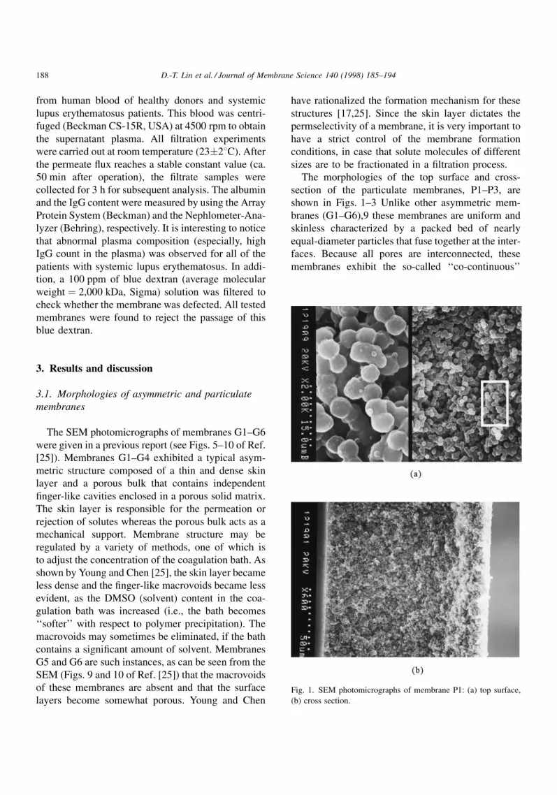
The morphologies of the top surface and cross-
section of the particulate membranes, P1±P3, are
shown in Figs. 1±3 Unlike other asymmetric mem-
branes (G1±G6),9 these membranes are uniform and
skinless characterized by a packed bed of nearly
equal-diameter particles that fuse together at the inter-
faces. Because all pores are interconnected, these
membranes exhibit the so-called `̀ co-continuous''
Fig. 1. SEM photomicrographs of membrane P1: (a) top surface,
(b) cross section.
188 D.-T. Lin et al. / Journal of Membrane Science 140 (1998) 185±194

structure. The pore size and therefore the ®ltration
capability of these membranes is closely related to the
size of the particles. As shown in Figs. 1±3, Nylon-
610 precipitates from 1-octanol into very large parti-
cles (ca. 8 mm dia. in membrane P2) whereas EVAL
and PVDF membranes have much smaller particles
(ca. 1 mm dia. in membranes P1 and P3).
3.2. Permeation studies of single component
Permeation measurements of solutions containing
only one kind of solute molecule were carried out for
various membranes in a dual-chamber diffusion cell.
The concentration of solute in both chambers were
monitored. In Fig. 4, the measured data for glucose
permeation are presented in the form conforming to
Eq. (2). From the slope of each best-®tted line, mass
transfer coef®cient (D/L) was calculated for each
membrane. These results are shown in Table 3. It is
observed that all membranes are permeable to glucose
with a mass transfer coef®cient on the order of
10ÿ4 cm/s. For the permeation of other solutes (insu-
lin, albumin and IgG), likewise, a liner relationship
was obtained complying with Eq. (2). The calculated
mass transfer coef®cients for these cases are summar-
ized in Table 3. For each membrane, as is anticipated,
the mass transfer coef®cient is smaller for larger solute
molecules. Several asymmetric membranes (G2, G4
Fig. 2. SEM photomicrographs of membrane P2: (a) top surface,
(b) cross section.Fig. 3. SEM photomicrographs of membrane P3: (a) top surface,
(b) cross section.
D.-T. Lin et al. / Journal of Membrane Science 140 (1998) 185±194 189

and G6) were found to reject completely IgG for the
period of 24 h (the detection limit of UV spectro-
photometer is calibrated to be 0.005 mg/dl of IgG).
Although it is possible that IgG is able to diffuse
across these membranes in longer operation times
[31], a 24 h experiment is long enough to evaluate
preliminarily whether a membrane has the potential to
separate various proteins before proteins denature.
Table 3 indicates that the mass transfer coef®cients
of the particulate membranes (P1±P3) are roughly
twice larger than those of the asymmetric membranes,
except for membrane G6 which has a porous top
surface and an open cellular structure in the interior.
This suggests that the dense skin of the asymmetric
membranes offers the major resistance against solute
transportation. In addition, because these asymmetric
membranes are permeable to albumin while rejecting
IgG, the pores in the skin layer can be considered
between the dimension of albumin (67 kDa) and IgG
(150 kDa).
If a membrane is to be implanted as a part of the
arti®cial pancreas system, it must allow rapid permea-
Fig. 4. Time dependent permeation of glucose through various membranes.
Table 3
Mass transfer coefficients for various solutes (cm/sec)
Membrane Glucose Insulin Albumin IgG
G1 9.65�10ÿ5 бa б б
G2 9.70�10ÿ5 2.50�10ÿ5 7.11�10ÿ7 NDb
G3 8.48�10ÿ5 б б б
G4 8.77�10ÿ5 2.51�10ÿ5 9.77�10ÿ7 ND
G5 1.19�10ÿ4 б б б
G6 1.64�10ÿ4 3.23�10ÿ5 1.13�10ÿ6 ND
P1 2.06�10ÿ4 3.72�10ÿ5 2.10�10ÿ6 1.19�10ÿ6
P2 1.53�10ÿ4 3.61�10ÿ5 3.28�10ÿ6 1.37�10ÿ6
P3 1.67�10ÿ4 3.55�10ÿ5 1.44�10ÿ5 2.09�10ÿ6
aNot tested. bND�not detectable after 24 h permeation.
190 D.-T. Lin et al. / Journal of Membrane Science 140 (1998) 185±194

tion of glucose and insulin so as to provide therapeutic
bene®ts. Ward et al. reported recently the diffusive
permeability of 5.5�10ÿ5 cm/s for glucose through
polyurethane membranes [6]. For commercial mem-
branes, the glucose mass transfer coef®cient of the
Vita®ber cell was found to be 3.67�10ÿ5 cm/s on the
average and the Amicon and polyacrylonitrile mem-
branes have similar diffusive permeabilities [32]. The
experimental data indicates that membrane G2
appears to have a higher diffusive permeability with
respect to glucose (ca. 9.70�10ÿ5 cm/s) for use in an
arti®cial pancreas. On the other hand, the membrane
has to be permselective to prevent the in¯ux of IgG
and other larger antibodies which cause rejection of
the islets inside the membranes. For the membranes
shown in Table 3, it appears that all of the asymmetric
membranes are suitable choices to be built into the
arti®cial pancreas system; in particular G6, since it is
not only impermeable to IgG but also has the largest
mass transfer coef®cients both with respect to glucose
and insulin.
Table 3 shows that skinless particulate membranes,
in general, exhibit a higher solute permeation rate than
asymmetric ones. For particulate membranes, an
ordinary diffusion path for solutes is the tortuous
but continuous void space between fused particles
(Note: one other path is the nano-pores within the
particle. This point is discussed in a separate publica-
tion [33]). Since the diameter of this path is relatively
large, large solute molecules, such as IgG, can pass it
at a reasonable speed. In contrast, asymmetric mem-
branes have a skin layer as being a region with very
small pore size that provides an effective diffusion
barrier for solutes. In Table 4, water ¯ux data are given
at transmembrane pressure equal to 0.5 kgf/cm2 for
both types of membranes. The data for membrane P3
is not included because its water ¯ux is higher than the
measurable range of our apparatus. As is anticipated,
particulate membranes have water ¯uxes signi®cantly
higher than those of the asymmetric ones. However,
because of their low permselectivity towards IgG,
these particulate membranes should not be used in
an arti®cial pancreas system.
3.3. Solute rejection during filtration of plasma
In order to examine the permeability of one type of
solute in the presence of the others and also to know
the performance of the membranes in realistic condi-
tions, ®ltration experiments were carried out using
human plasma as the feed in a dead-end ®ltration.
Feed and ®ltrate samples were analyzed to yield the
data of total protein concentrations, sieving coef®-
cients, and selectivity of albumin relative to IgG for
various membranes. These results are shown in
Table 5(a) for the normal plasma (from healthy
donors) cases and in Table 5(b) for the abnormal
plasma (from patients) cases. From Table 5(a), it
can be seen that IgG could not penetrate asymmetric
membranes G1±G4, but could pass through mem-
branes G5 and G6 to a signi®cant degree. This is
different from the results obtained from the single-
component diffusion experiments (see Table 3) which
indicate that all asymmetric membranes are imperme-
able to IgG. One possible explanation for this obser-
vation is that in a pressure driven ®ltration process,
molecules are compressed to become more compact
so as to enter the membrane skin layer more easily
[34,35]. Also, because membranes G5 and G6 were
prepared from very soft baths, their skins are not dense
and thus are permeable to large molecules such as IgG.
Table 5(a) shows that albumin are undetectable in the
permeate for membranes G1±G3. This should be
attributed to the resolution of the instrument. Analyz-
ing the plasma proteins is far more dif®cult than that
for single protein. The lower measurable limits in this
study are 22.2 mg/dl and 170 mg/dl for albumin and
IgG, respectively.
As far as particulate membranes (P1 and P2) are
concerned, more albumin and IgG passed through
them than the asymmetric ones, as shown in
Table 5(a). Membrane P2 has a higher albumin siev-
ing coef®cient than membrane P1; its capability to
Table 4
Water flux with transmembrane pressure 0.5 kgf/cm2
Membrane Flux (l/h m2 atm)
G1 18
G2 20
G3 58
G4 62
G5 90
G6 76
P1 140
P2 110
D.-T. Lin et al. / Journal of Membrane Science 140 (1998) 185±194 191

separate albumin from IgG is, however, much lower
than membrane P1. The selectivity (albumin/
IgG�1.820) of membrane P1 is the highest among
all membranes in normal plasma ®ltration. The G5 and
G6 membranes have good selectivity (albumin/IgG) as
well, but their ¯uxes and sieving coef®cients toward
albumin are too low. In order to achieve an effective
plasma fractionation, membranes are required to reject
as much as possible IgG while at the same time
recover most albumin in the plasma. This suggests
that membrane P1 is better than G5 and G6, and even
better than the commercial membranes [11]. There-
fore, membrane P1 is an alternative to patients with
certain classes of autoimmune diseases that are cur-
rently treated by plasma exchange.
Comparison of Table 5(a) and Table 5(b) indicates
that ®ltration of abnormal plasma yields results largely
consistent with those from normal plasma ®ltration.
Again, membrane P1 has the highest albumin/IgG
selectivity (S�1.155) among all membranes. Its value
is, however, signi®cantly lower than that for normal
plasma (S�1.82). This may be attributed to the high
IgG concentration in the abnormal plasma, in which
case some smaller pores of the membrane are likely to
be plugged by IgG aggregates. In addition, the aggre-
gation of IgG, especially for the high IgG concentra-
tion, at the upstream membrane surface that causes
concentration polarization also may reduce the per-
meation of albumin. For these reasons, the albumin
sieving coef®cient is decreased from 0.606 for normal
plasma ®ltration to the current value of 0.476. Similar
situations are observed for asymmetric membranes,
G4 and G6, whose pores are smaller than those of
membrane P1. Table 5 indicates that the albumin
permeation has reduced considerably (to ca. 1/3 of
the value for normal plasma) for these membranes.
Contrary to these cases, the albumin sieving coef®-
cient of membrane P2 is higher in abnormal plasma
®ltration. Because the pores of membrane P2 are very
large (Fig. 2), it is impossible to plug them even in
concentrated IgG solutions. (The increase in albumin
sieving coef®cient is still unexplainable at present.)
This paper places emphasis upon discussing the per-
formances of asymmetric and particulate membranes
towards various proteins. No attempt is made to
describe the mechanism that governs multicomponent
mass transfer in actual plasma ultra®ltration pro-
cesses. However, enthusiastic investigations are cur-
rently undergoing on subjects, such as the effect of
protein concentration, the effect of concentration
Table 5
Filtrate concentrations, sieving coefficient and selectivity for plasma ultrafiltration
Membrane Filtrate analysis Sieving coefficient (S)c Selectivityd
TPa (g/dl) Albb (g/dl) IgG (mg/dl) Alb IgG
(a) Normal plasma
Feed: TP�6.4 g/dl, Alb�3.3 g/dl, IgG�1100 mg/dl
G1 NDe ND ND ND ND Ð-
G2 ND ND ND ND ND Ð-
G3 ND ND ND ND ND Ð-
G4 0.6 0.4 ND 0.121 ND Ð-
G5 2.7 1.5 315 0.455 0.286 1.591
G6 2.6 1.6 316 0.485 0.287 1.670
P1 3.5 2.0 366 0.606 0.333 1.820
P2 4.6 2.3 844 0.670 0.767 0.874
(b) Abnormal plasma
Feed: TP�16.5 g/dl, Alb�2.1 g/dl, IgG�14800 mg/dl
G2 ND ND ND ND ND Ð-
G4 1.1 0.1 900 0.048 0.061 0.787
G6 3.2 0.3 2050 0.143 0.139 1.029
P1 7.5 1.0 6100 0.476 0.412 1.155
P2 14.6 1.9 12400 0.905 0.838 1.080
aTP � Total Protein; bAlb � Albumin; cSieving coefficient (S) � Cout/Cin; dSelectivity � SAlb/SIgG; eND� not detectable.
192 D.-T. Lin et al. / Journal of Membrane Science 140 (1998) 185±194

polarization, the effects of size and packing pattern of
particles in particulate membranes, the effect of trans-
membrane pressure upon permeation of various spe-
cies during ®ltration, etc.
4. Conclusion
The structures of these membranes were found to
affect signi®cantly their permeability and selectivity
towards glucose and various proteins in human
plasma. In order to work as an immunoprotective
barrier for the arti®cial pancreas, the membrane has
to have a skin which is dense enough to prevent inward
diffusion of IgG and other larger antibodies. On the
other hand, the skin has to be somewhat porous to
admit fast transportation of glucose and insulin. The
results of current work indicate that this can be
achieved by adjusting the `̀ softness'' of the coagula-
tion bath during membrane formation. In addition,
experimental evidences point out that some asym-
metric EVAL membranes and particulate membranes
(EVAL and Nylon-610) are potential candidates for
plasma proteins fractionations. Especially, the parti-
culate membranes which exhibit high permeation
rates and good selectivity with respect to various
species in plasma. This encourages us to continue
our pursuing for a membrane capable of separating
effectively albumin from immunoglobulins in fractio-
nation operations.
Acknowledgements
The authors thank the National Science Council of
the Republic of China for their ®nancial support,
project number: NSC 86-2314-B-002-173 and NSC
86-2216-E002-003.
References
[1] F. Lim, A.M. Sun, Microencapsulated islets as bioartificial
endocrine pancreas, Science 210 (1980) 908±910.
[2] J.J. Altman, A. Houlbert, P. Callard, P. McMillan, B.A.
Solomon, J. Rosen, P.M. Galetti, Long term plasma glucose
normalization in experimental diabetic rats with macroen-
capsulated implants of benign human insulinomas, Diabetes
35 (1986) 625±633.
[3] K. Inoue, T. Fujisato, Y.J. Gu, K. Burczak, S. Sumi, M. Kogire, T.
Tobe, K. Uchida, I. Nakai, S. Maetani, Y. Ikada, Experimental
hybrid islet transplantation: application of polyvinyl mem-
brane for entrapment islets, Pancreas 7 (1992) 562±568.
[4] K. Burczak, T. Fujisato, M. Hatadaand, Y. Ikada, Protein
permeation through poly(vinyl alcohol) hydrogel membranes,
Biomaterials 15 (1994) 231±237.
[5] R.P. Lanza, A.M. Beyer, W.L. Chick, Xenogenic humoral
responses to islets transplanted in biohybrid diffusion
chambers, Transplantation 57 (1994) 1371±1375.
[6] R.S. Ward, K.A. White, C.A. Wolcott, A.Y. Wang, R.W.
Kuhn, J.E. Taylor, J.K. John, Development of a hybrid
artificial pancreas with a dense polyurethane membrane,
ASAIO J. 39 (1993) 261±267.
[7] L. Kessler, M. Pinget, M. Aprahamian, D. Poinsot, M. Keipes,
C. Damge, Diffusion properties of an artificial membrane
used for Langerhans islets encapsulation: interest of an in
vitro test, Transplant. Proc. 24 (1992) 953±954.
[8] T. Zekorn, R.G. Bretzel, U. Siebers, W. Doppl, M. Renardy, P.
Zschocke, H. Planck, K. Federlin, Protein coat causes
improved insulin diffusion through membranes for immuno-
isolated islet transplantation. Improved islet survival by
pretreatment of membrane and islets, Transplant. Proc. 22
(1990) 867±869.
[9] L. Kessler, G. Legeay, C. Jesser, C. Damge, M. Pinget,
Influence of corona surface treatment on the properties of an
artificial membrane used for Langerhans islets encapsulation:
permeability and biocompatibility studies, Biomaterials 16
(1995) 185±191.
[10] T.H. Young, N.K. Yao, R.F. Chang, L.W. Chen, Evaluation of
asymmetric poly(vinyl alcohol) membranes for use in the
artificial islets, Biomaterials 17 (1996) 2139±2145.
[11] M. Zborowski, P.S. Malchesky, Pore size and temperature
effects in membrane separation of albumin from immunoglo-
bulins, ASAIO Trans. 36 (1990) 730±733.
[12] C. Charcosset, M.Y. Jaffrin, L. Ding, Time and pressure
dependence of sieving coefficients during membrane plasma
fractionation, ASAIO Trans. 36 (1990) 594±597.
[13] M. Zborowski, P.S. Malchesky, Y. Nose, Temperature
dependent protein removal by large pore membrane filtration,
ASAIO Trans. 35 (1989) 572±575.
[14] T. Horiuchi, P.S. Malchesky, M. Usami, M. Emura, Y. Nose,
Effect of plasma solute±membrane interaction on mean pore
diameter, ASAIO Trans. 32 (1986) 429±434.
[15] R.E. Kesting, Synthetic Polymeric Membranes, Wiley, New
York, 1985.
[16] T.H. Young, L.W. Chen, Roles of bimolecular interaction and
relative diffusion rate in membrane structure control, J.
Membr. Sci. 83 (1993) 153±166.
[17] T.H. Young, L.W. Chen, Pore formation mechanism of
membranes from phase inversion process, Desalination 103
(1995) 233±247.
[18] T.H. Young, L.W. Chen, L.P. Cheng, Membranes with a
microparticulate morphology, Polymer 37 (1996) 1305±1310.
[19] K. Kamide, S. Manabe, in: D.R. Lloyd. (Ed.), Material
Science of Synthetic Membranes, American Chemical
Society, Washington D.C., pp. 197±228, 1985.
D.-T. Lin et al. / Journal of Membrane Science 140 (1998) 185±194 193
[20] S.P. Nunes, T. Inoue, Evidence for spinodal decomposition
and nucleation and growth mechanisms during membrane
formation, J. Membr. Sci. 111 (1996) 93±103.
[21] A.M.W. Bulte, B. Folkers, M.H.V. Mulder, C.A. Smolders,
Membranes of semicrystalline aliphatic polyamide nylon 4,6:
formation by diffusion-induced phase separation, J. Apply.
Polym. Sci. 50 (1993) 13±26.
[22] L.P. Cheng, A.W. Dwan, C.C. Gryte, Membrane formation
by isothermal precipitation in polyamide±formic acid±water
systems I. Description of membrane morphology, J. Polym.
Sci. Polym. Phys. 33 (1995) 211±222.
[23] L.P. Cheng, A.W. Dwan, C.C. Gryte, Membrane formation
by isothermal precipitation in polyamide±formic acid±water
systems II. Precipitation dynamics, J. Polym. Sci. Polym.
Phys. 33 (1995) 223±235.
[24] T.H. Young, J.Y. Lai, W.M. Yu, L.P. Cheng, Equilibrium
phase behavior of the membrane forming water±DMSO±
EVAL copolymer system, J. Membr. Sci. 128 (1997) 55±65.
[25] T.H. Young, L.W. Chen, A two step mechanism of diffusion-
controlled ethylene vinyl alcohol membrane formation, J.
Membr. Sci. 57 (1991) 69±81.
[26] L. Broens, D.M. Koenhen, C.A. Smolders, On the mechanism
of formation of asymmetric ultra- and hyper-filtration
membranes, Desalination 22 (1977) 205±219.
[27] J.G. Wijmans, H.J.J. Rutten, C.A. Smolders, Phase separation
phenomena in solutions of poly(2,6-dimethyl-1,4-phenylene-
oxide) in mixtures of trichloroethylene, 1-octanol, and
methanol: relationship to membrane formation, J. Polym.
Sci. Polym. Phys. 23 (1985) 1941±1955.
[28] W.R. Burghardt, L. Yilmaz, A.J. McHugh, Glass transition,
crystallization and thermoreversible gelation in ternary PPO
solution; relationship to asymmetric formation, Polymer 28
(1987) 2085±2092.
[29] K.A. Smith, C.K. Colton, E.W. Merrill, L.B. Evans,
Convective transport in a batch dialyzer. Determination of
the true membrane permeability from a single measurement,
AIChE Symp. Ser. 64 (1968) 45.
[30] C.K. Colton, Permeability and transport studies in batch and
flow dialyzers with applications to hemodialysis, Ph.D.
Dissertation, M.I.T., Mass., USA, 1969.
[31] C.K. Colton, E.S. Avgoustiniatos, Bioengineering in devel-
opment of the hybrid artificial pancreas, J. Biomech. Eng. 113
(1991) 152±170.
[32] M.Y. Jaffrin, G. Reach, D. Notelet, Analysis of ultrafiltration
and mass transfer in a bioartificial pancreas, J. Biomech. Eng.
110 (1988) 1±10.
[33] L.P. Cheng, H.Y. Lin, L.W. Chen, T.H. Young, Solute
rejection of dextran by EVAL membranes with asymmetric
and particulate morphologies, Polymer, in press.
[34] H. Balmann, R. Nobrega, Deformation of dextran molecules.
Causes and consequences in ultrafiltration, J. Membr. Sci. 40
(1989) 311±327.
[35] G. Schock, A. Miquel, Characterization of ultrafiltration
membranes: Cutoff determination by gel permeation chro-
matography, J. Membr. Sci. 41 (1989) 55±67.
194 D.-T. Lin et al. / Journal of Membrane Science 140 (1998) 185±194