dicalcium phosphate (cahpo4·2h2o) precipitation through ortho- or meta-phosphoric acid-etching:...
TRANSCRIPT

JJOD-2131; No. of Pages 13
Dicalcium phosphate (CaHPO4�2H2O) precipitationthrough ortho- or meta-phosphoric acid-etching:Effects on the durability and nanoleakage/ultra-morphology of resin–dentine interfaces
Victor Pinheiro Feitosa a, Maria Giulia Bazzocchi b, Angelo Putignano c,Giovanna Orsini c, Arlinda Luzi Luzi d, Mario Alexandre Coelho Sinhoreti a,Timothy F. Watson e, Salvatore Sauro e,f,*aDivision of Dental Materials, Piracicaba Dental School, Piracicaba, BrazilbDepartment of Endodontic, Section of Pediatric Dentistry, Dental School, University of Bologna, Bologna, ItalycDepartment of Clinical Sciences and Stomatology, Polytechnic University of Marche, Ancona, ItalydDepartment of Restorative Dentistry and Endodontics, Universidad CEU Cardenal Herrera Valencia, SpaineBiomaterials, Biomimetics and Biophotonics, King’s College London Dental Institute, London, UKfDental Biomaterials Science and Minimally Invasive Dentistry, Universidad CEU Cardenal Herrera Valencia, Spain
j o u r n a l o f d e n t i s t r y x x x ( 2 0 1 3 ) x x x – x x x
a r t i c l e i n f o
Article history:
Received 2 February 2013
Received in revised form
13 August 2013
Accepted 19 August 2013
Available online xxx
Keywords:
Bond durability
Etch-and-rinse adhesives
Meta-phosphoric acid
Resin–dentine interface ultra-
morphology
Confocal microscopy
Calcium-staining technique
FTIR vibration analysis
a b s t r a c t
Objectives: To compare the effects of two etching procedures using meta-phosphoric (MPA)
or ortho-phosphoric acid (OPA) on dentine demineralisation, resin–dentine bonds durability
and interface nanoleakage/ultra-morphology.
Methods: Middle-dentine specimens were etched using 37% OPA (15 s) or 40% MPA (60 s) and
submitted to infrared spectroscopy (FTIR) or ultra-morphology dye-assisted (calcium-stain-
ing) confocal microscopy (Ca-CLSM). A three-step etch-and-rinse adhesive was formulated,
applied onto dentine and light-cured for 30 s before composite build-up. After 24 h, the
dentine-bonded specimens were cut into 1 mm2 beams; half were immediately submitted
to microtensile bond strength (mTBS) and half stored in DW for six months. The mTBS results
were analysed with repeated-measures ANOVA and Tukey’s test ( p < 0.05). Further teeth were
bonded and prepared for interface nanoleakage/ultra-morphology confocal evaluation.
Results: FTIR and Ca-CLSM analyses showed dicalcium phosphate dihydrate (Brushite)
precipitation in MPA-etched dentine and on the bottom (front of demineralisation) of the
OPA-etched dentine. Statistical analysis showed similar mTBS for both etching procedures
after 24 h. The mTBS of specimens in OPA-group dropped significantly ( p < 0.05) after six
month; the specimens in the MPA group showed no statistically difference ( p > 0.05). CLSM
depicted no evident sign of nanoleakage within the resin–dentine interface of the MPA-
treated specimens, while the specimens in OPA-group presented intense nanoleakage and
interface degradation.
Conclusion: The use of MPA (60 s) as an alternative dentine conditioning agent in etch-and-rinse
bonding procedures may be a suitable strategy to create more durable resin–dentine bonds.
# 2013 Elsevier Ltd. All rights reserved.
* Corresponding author at: Dental Biomaterials Science and Minimally Invasive Dentistry, Universidad CEU Cardenal Herrera en Valencia,C/Luis Vives, 1. Alfara del Patriarca, 46115 Valencia, Spain. Tel.: +36 961 36 90 00; fax: +44 020 71881823.
Available online at www.sciencedirect.com
ScienceDirect
journal homepage: www.intl.elsevierhealth.com/journals/jden
E-mail addresses: [email protected], [email protected], [email protected] (S. Sauro).
Please cite this article in press as: Feitosa VP, et al. Dicalcium phosphate (CaHPO4�2H2O) precipitation through ortho- or meta-phosphoric acid-etching: Effects on the durability and nanoleakage/ultra-morphology of resin–dentine interfaces. Journal of Dentistry (2013), http://dx.doi.org/10.1016/j.jdent.2013.08.014
0300-5712/$ – see front matter # 2013 Elsevier Ltd. All rights reserved.http://dx.doi.org/10.1016/j.jdent.2013.08.014
JJOD-2131; No. of Pages 13
j o u r n a l o f d e n t i s t r y x x x ( 2 0 1 3 ) x x x – x x x2
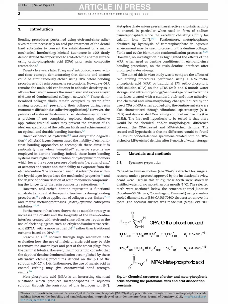
Fig. 1 – Chemical structures of ortho- and meta-phosphoric
acids showing the protonable sites and acid dissociation
constants.
1. Introduction
Bonding procedures performed using etch-and-rinse adhe-
sives require necessarily an acid pre-treatment of the dental
hard substrates to consent the establishment of a micro-
mechanical interlocking; Michael Buonocore in 1955 firstly
demonstrated the importance to acid-etch the enamel surface
using ortho-phosphoric acid (OPA) prior resin composite
restorations.1
Twenty five years later Fusayama2 introduced a new acid-
and-rinse concept, demonstrating that dentine and enamel
could be simultaneously etched using OPA before bonding
procedures and resin composite restorations. Nowadays OPA
remains the main acid-conditioner in adhesive dentistry as it
allows clinicians to remove the smear layer and expose a layer
(6–9 mm) of demineralised collagen network.3,4 These demi-
neralised collagen fibrils remain occupied by water after
rinsing procedures5 preventing their collapse during resin
monomers diffusion (i.e. adhesive application).6 However, the
presence of water in the demineralised dentine may represent
a problem if not completely replaced during adhesive
application; residual water may prevent the creation of an
ideal encapsulation of the collagen fibrils and achievement of
an optimal and durable bonding interface.3,7
Direct evidence of hydrolytic8,9 and enzymatic degrada-
tion5,6 of hybrid layers demonstrated the inability of etch-and-
rinse bonding approaches to accomplish these aims; it is
particularly true when ‘‘simplified’’ adhesive systems are
employed in dentine bonding. Indeed, these latter bonding
systems have higher concentration of hydrophilic monomers
which lower the vapour pressure of solvents (i.e. ethanol and/
or acetone) and water and their ability to evaporate from the
etched-dentine. The presence of residual solvent/water within
the hybrid layer jeopardises the mechanical properties10 and
the degree of polymerisation of resin monomers compromis-
ing the longevity of the resin composite restorations.11,12
However, acid-etched dentine represents a functional
substrate for potential therapeutic treatments during bonding
procedures,13 such as application of collagen cross-linkers14,15
and matrix metalloproteinases (MMPs)/cysteine cathepsins
inhibitors.16,17
Furthermore, it has been shown that a suitable strategy to
increases the quality and the longevity of the resin–dentine
interface created with etch-and-rinse adhesives requires the
use of cheleting agents such as ethylenediaminetetraacetic-
acid (EDTA) with a more neutral pH6,7 rather than traditional
etchants based on OPA17,18;
Breschi et al.19 showed through high resolution SEM
evaluation how the use of maleic or citric acid may be able
to remove the smear layer and part of the smear plugs from
the dentinal tubules. However, it is important to consider that
the depth of dentine demineralisation accomplished by these
alternative etching procedures depend on the pH of the
solution (pH 0.7 < 1.4); furthermore, the use of maleic acid in
enamel etching may give controversial bond strength
results.20
Meta-phosphoric acid (MPA) is an interesting chemical
substance which produces metaphosphates in aqueous
solution through the ionisation of one hydrogen ion [H+].
Please cite this article in press as: Feitosa VP, et al. Dicalcium phosphate (etching: Effects on the durability and nanoleakage/ultra-morphology of r10.1016/j.jdent.2013.08.014
Metaphosphate anions present an effective cariostatic activity
in enamel, in particular when used in form of sodium
trimetaphosphate since the excellent chelating affinity for
calcium ions [Ca+2].20,21 Furthermore, metaphosphates
obtained by hydrolysis of trimetaphosphate in aqueous
environment may be used to cross-link the dentine collagen
fibrils and evoke biomimetic remineralisation processes.22,23
However, no investigation has highlighted the effects of the
MPA, when used as dentine conditioner in etch-and-rinse
bonding procedures, on the resin–dentine interfaces after
prolonged water storage.
The aim of this in vitro study was to compare the effects of
two etching procedures performed using a 40% meta-
phosphoric acid (MPA) or traditional 37% ortho-phosphoric
acid solution (OPA) on the mTBS (24 h and 6-month water
storage) and ultra-morphology/nanoleakage of resin–dentine
interfaces created with a standard etch-and-rinse adhesive.
The chemical and ultra-morphology changes induced by the
use of OPA or MPA when applied onto the dentine surface were
also characterised through vibrational spectroscopy (ATR-
FTIR) and dye-assisted Ca-staining confocal microscopy (Ca-
CLSM). The first null hypothesis to be tested is that there
would be no chemical and/or morphological difference
between the OPA-treated and MPA-etched dentine. The
second null hypothesis is that no difference would be found
in mTBS of bonded-dentine specimens created both on OPA-
etched or MPA-etched dentine after 6-month of water storage.
2. Materials and methods
2.1. Specimen preparation
Caries-free human molars (age 20–40) extracted for surgical
reasons under a protocol approved by the institutional review
board were used in this study; these teeth were stored in
distilled water for no more than one month (4 8C). The selected
teeth were sectioned below the cemento-enamel junction
(Accutom-50; Struers, Copenhagem, Denmark) using a water-
cooled diamond saw (330-CA RS-70300; Struers) to remove the
roots. The occlusal surface was made flat (Meta-Serv 3000
CaHPO4�2H2O) precipitation through ortho- or meta-phosphoric acid-esin–dentine interfaces. Journal of Dentistry (2013), http://dx.doi.org/
Table 1 – Composition of the etch-and-rinse adhesiveused in this study.
Standard resin bonding system
Bond resin Primer resin
(pH �6.5) (pH �6.8)
40 wt% E-BisADM 20 wt% E-BisADM
30 wt% TEGDMA 15 wt% TEGDMA
30 wt% HEMA 15 wt% HEMA
– 50 wt% ethanol
E-BisADM: ethoxylated-Bisphenol A dimethacrylate; TEGDMA:
triethyleneglycoldimethacrylates; HEMA: 2-hydroxyethyl metacry-
late.
CQ, camphoroquinone (0.50 wt%); EDAB, 2-ethyl-dimethyl-4-ami-
nobenzoate (0.50 wt%); DPIHP, diphenyliodonium hexafluoropho-
sphate (0.50 wt%) were added as wt% at the end of the final resin
monomers blend (100 wt%) formulation.
j o u r n a l o f d e n t i s t r y x x x ( 2 0 1 3 ) x x x – x x x 3
JJOD-2131; No. of Pages 13
Grinder-Polisher, Buehler Lake Bluff, IL, USA) using ascending
SiC abrasive papers 180#-to-320#-grit (Versocit; Struers,
Copenhagen, Denmark) under constant water irrigation to
provide a standardised smear-layer in middle dentine surface.
The dentine specimens were divided in two principal
groups and acid-etched for 15 s using 37% ortho-phosphoric
acid (OPA; pH < 1) or 40% meta-phosphoric acid (MPA; pH 2.6)
for 60 s (Fig. 1); these were finally rinsed with deionised water
(DW) for 15 s. The two acids (powder purity > 98%) were
purchased from Sigma–Aldrich (Sigma–Aldrich Chemical,
Gillingham, UK).
2.2. Characterisation of the OPA and MPA-etched dentine
Three middle-dentine specimens were submitted to ATR-FTIR
vibrational analysis (FTIR) before and after acid-etching
treatment using OPA or MPA.24 FTIR analysis was performed
in wet (excess of water removed using absorbent paper points)
or dry (excess of water totally removed by 10 s air-drying)
using the ATR-FTIR Spectrometer (Perkin-Elmer Spectrum
One; Perkin-Elmer, Beaconsfield, UK) in the region of 650–
4000 cm�1 with a resolution of 4 cm�1 and 64 number of scans
for each spectrum. The ATR area had a 2 mm diameter and the
IR radiation penetration was approximately 3–5 mm. In order
to place the specimens in good contact with the ATR crystal, a
moderate pressure was applied (5 psi) during the measure-
ment to achieve high quality results, but at the same time, to
avoid compression of the demineralised collagen to the
underlying mineralised dentine. A control FTIR analysis was
performed for the commercial high purity (>97%) hydroxyap-
atite and brushite (Sigma–Aldrich). To minimise the homoge-
neity variability of the specimens, three spectra were recorded
in different positions of the surface.25
Further four specimens per group were prepared as
previously described and submitted to the dye-assisted
calcium-staining confocal microscopy analysis (CLSM-Ca) in
order to evaluate the morphology features induced by the two
different etching agents (OPA or MPA).25 In details, two
specimens were immediately immersed in a 0.5 wt% calci-
um-chelating dye solution26 (Xylenol Orange; Sigma–Aldrich)
for 24 h at 37 8C (pH 7.2), while two specimens were immersed
for 24 h at 37 8C in a 0.1 wt% dye water/ethanol (50v%/50v%)
solution (FITC – fluorescein isothiocyanate; Sigma–Aldrich)
able to covalently bind collagen.27 Subsequent to the dye
immersion, the specimens were copiously rinsed with water
and treated in an ultrasonic water-bath for 5 min and then
submitted to confocal microscopy evaluation. The confocal
microscopy analysis was performed using a confocal laser
scanning microscope (DM-IRE2 CLSM; Leica, Heidelberg,
Germany) equipped with a 63�/1.4 NA oil immersion lens
and a 488-nm Argon/Krypton ions laser (Fluorescein excita-
tion) or 568-nm Helium/Neon ions laser. The emission
fluorescence was recorded at 512–538 nm and 585–650 nm,
respectively; control dentine (sound – no treatment) speci-
mens were also employed in the Ca-CLSM analysis. CLSM
images were obtained with a 1 mm Z-step (Z-stack) or X-step
(X-stack) to optically section the specimens to a depth up to
25 mm. The Z-stack and X-stack scans performed along the
interface were arbitrarily pseudo-coloured by the same
operator for better exposure and compiled into single
Please cite this article in press as: Feitosa VP, et al. Dicalcium phosphate (etching: Effects on the durability and nanoleakage/ultra-morphology of r10.1016/j.jdent.2013.08.014
projections or 3D images using the Leica image-processing
software (Leica).9 The configuration of the system was
standardised and used throughout the entire investigation.
Each slab was completely investigated and micrographs
representing the most common features observed along the
etched dentine surfaces were captured and recorded.
2.3. Adhesive formulation and bonding procedures
An experimental etch-and-rinse adhesive (primer and bond)
was formulated and used in this study in order to standardise
the chemical composition by having constant concentration
of monomers, solvents, initiators and co-initiators. A neat
resin blend mixture was formulated using 30 wt% 2-hydro-
xyethyl metacrylate (HEMA: Aldrich Chemical Co., Gillingham,
UK), 30 wt% triethylene-glycol-dimethacrylate (TEGDMA),
40 wt% ethoxylated-Bisphenol-A-dimethacrylate (E-BisADM),
(Esstech Essington, PA, USA). The resin blend was made photo-
polymerisable by adding 0.5 wt% camphoroquinone (CQ:
Aldrich Chemicals), 0.5 wt% ethyl-4-dimethylaminobenzoate
(EDMAB: Aldrich Chemicals) and 0.5 wt% diphenyliodonium-
hexafluorophosphate13 (DPIHP: Aldrich Chemicals).
Five-minute sonication (Model QS3; Ultrawave Ltd., Cardiff,
156 UK) and 2-day shaking (Orbital Shakers PSU-20i; Cole
Fisher 157 Scientific Ltd., Loughborough, UK) were required to
yield well-mixed resin mixtures (i.e. primer and bond).13
Subsequently, a resin primer system was created by mixing
50 wt% absolute ethanol (Aldrich) and 50 wt% of neat co-
monomer resin blend; the neat resin (solvent-free) was also
used as a resin bond system. The detailed composition of the
DBA used in this study is shown in Table 1.
The bonding procedures were performed by applying two
consecutive coats of the experimental primer on the etched-
dentine specimens and gentle air-drying for 5 s. Two conse-
cutive coats of the experimental solvent-free resin bond were
immediately applied, spread thin with moisture-free air-blast
and light-cured for 30 s using a halogen light-curing unit
(Translux EC; KulzerGmBh, Bereich Dental, Werheim,
Germany). The output intensity (>600 mW/cm2) was moni-
tored with a Radiometer (Demetron Model 100; Demetron,
Danbury, CT, USA). Composite build-ups (6 mm) were layered
CaHPO4�2H2O) precipitation through ortho- or meta-phosphoric acid-esin–dentine interfaces. Journal of Dentistry (2013), http://dx.doi.org/
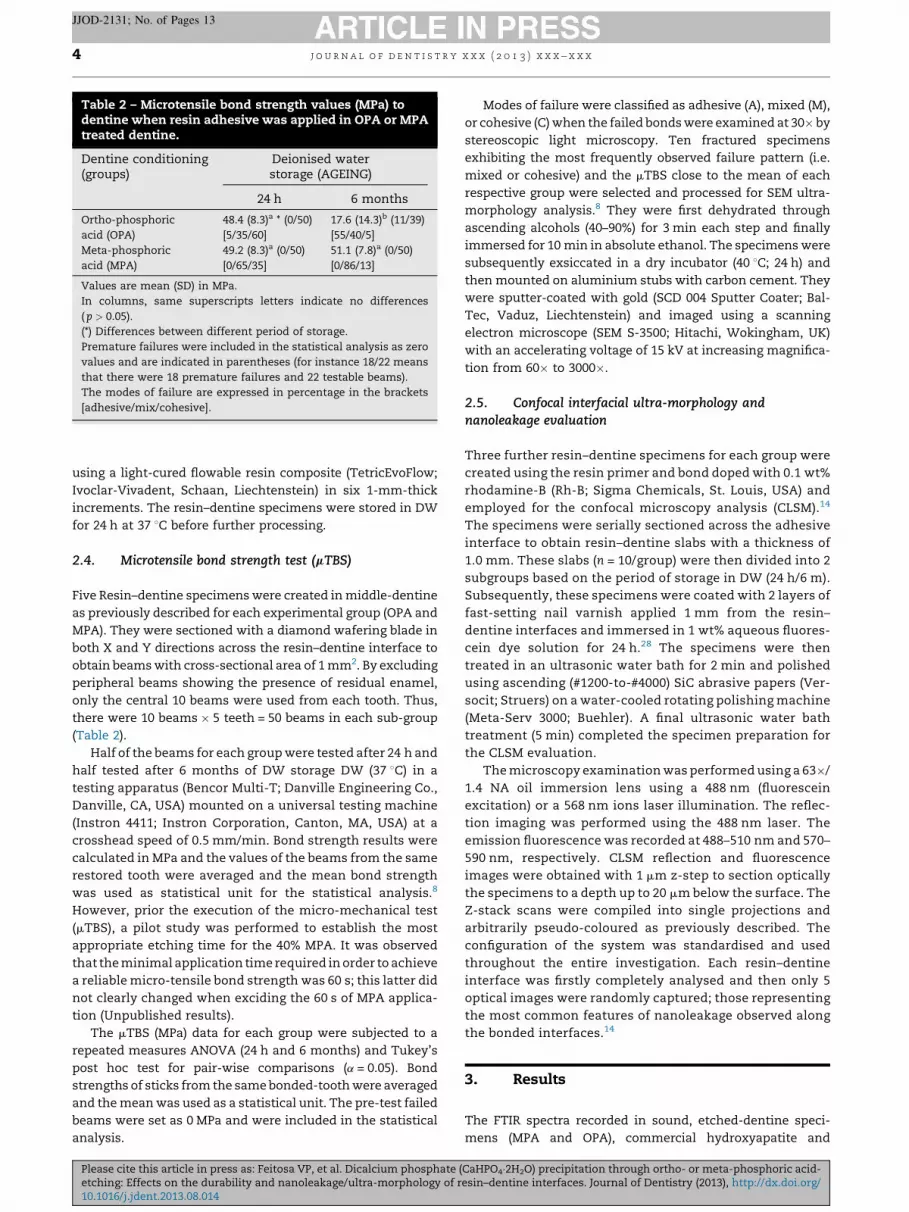
Table 2 – Microtensile bond strength values (MPa) todentine when resin adhesive was applied in OPA or MPAtreated dentine.
Dentine conditioning(groups)
Deionised waterstorage (AGEING)
24 h 6 months
Ortho-phosphoric
acid (OPA)
48.4 (8.3)a * (0/50)
[5/35/60]
17.6 (14.3)b (11/39)
[55/40/5]
Meta-phosphoric
acid (MPA)
49.2 (8.3)a (0/50)
[0/65/35]
51.1 (7.8)a (0/50)
[0/86/13]
Values are mean (SD) in MPa.
In columns, same superscripts letters indicate no differences
( p > 0.05).
(*) Differences between different period of storage.
Premature failures were included in the statistical analysis as zero
values and are indicated in parentheses (for instance 18/22 means
that there were 18 premature failures and 22 testable beams).
The modes of failure are expressed in percentage in the brackets
[adhesive/mix/cohesive].
j o u r n a l o f d e n t i s t r y x x x ( 2 0 1 3 ) x x x – x x x4
JJOD-2131; No. of Pages 13
using a light-cured flowable resin composite (TetricEvoFlow;
Ivoclar-Vivadent, Schaan, Liechtenstein) in six 1-mm-thick
increments. The resin–dentine specimens were stored in DW
for 24 h at 37 8C before further processing.
2.4. Microtensile bond strength test (mTBS)
Five Resin–dentine specimens were created in middle-dentine
as previously described for each experimental group (OPA and
MPA). They were sectioned with a diamond wafering blade in
both X and Y directions across the resin–dentine interface to
obtain beams with cross-sectional area of 1 mm2. By excluding
peripheral beams showing the presence of residual enamel,
only the central 10 beams were used from each tooth. Thus,
there were 10 beams � 5 teeth = 50 beams in each sub-group
(Table 2).
Half of the beams for each group were tested after 24 h and
half tested after 6 months of DW storage DW (37 8C) in a
testing apparatus (Bencor Multi-T; Danville Engineering Co.,
Danville, CA, USA) mounted on a universal testing machine
(Instron 4411; Instron Corporation, Canton, MA, USA) at a
crosshead speed of 0.5 mm/min. Bond strength results were
calculated in MPa and the values of the beams from the same
restored tooth were averaged and the mean bond strength
was used as statistical unit for the statistical analysis.8
However, prior the execution of the micro-mechanical test
(mTBS), a pilot study was performed to establish the most
appropriate etching time for the 40% MPA. It was observed
that the minimal application time required in order to achieve
a reliable micro-tensile bond strength was 60 s; this latter did
not clearly changed when exciding the 60 s of MPA applica-
tion (Unpublished results).
The mTBS (MPa) data for each group were subjected to a
repeated measures ANOVA (24 h and 6 months) and Tukey’s
post hoc test for pair-wise comparisons (a = 0.05). Bond
strengths of sticks from the same bonded-tooth were averaged
and the mean was used as a statistical unit. The pre-test failed
beams were set as 0 MPa and were included in the statistical
analysis.
Please cite this article in press as: Feitosa VP, et al. Dicalcium phosphate (etching: Effects on the durability and nanoleakage/ultra-morphology of r10.1016/j.jdent.2013.08.014
Modes of failure were classified as adhesive (A), mixed (M),
or cohesive (C) when the failed bonds were examined at 30� by
stereoscopic light microscopy. Ten fractured specimens
exhibiting the most frequently observed failure pattern (i.e.
mixed or cohesive) and the mTBS close to the mean of each
respective group were selected and processed for SEM ultra-
morphology analysis.8 They were first dehydrated through
ascending alcohols (40–90%) for 3 min each step and finally
immersed for 10 min in absolute ethanol. The specimens were
subsequently exsiccated in a dry incubator (40 8C; 24 h) and
then mounted on aluminium stubs with carbon cement. They
were sputter-coated with gold (SCD 004 Sputter Coater; Bal-
Tec, Vaduz, Liechtenstein) and imaged using a scanning
electron microscope (SEM S-3500; Hitachi, Wokingham, UK)
with an accelerating voltage of 15 kV at increasing magnifica-
tion from 60� to 3000�.
2.5. Confocal interfacial ultra-morphology andnanoleakage evaluation
Three further resin–dentine specimens for each group were
created using the resin primer and bond doped with 0.1 wt%
rhodamine-B (Rh-B; Sigma Chemicals, St. Louis, USA) and
employed for the confocal microscopy analysis (CLSM).14
The specimens were serially sectioned across the adhesive
interface to obtain resin–dentine slabs with a thickness of
1.0 mm. These slabs (n = 10/group) were then divided into 2
subgroups based on the period of storage in DW (24 h/6 m).
Subsequently, these specimens were coated with 2 layers of
fast-setting nail varnish applied 1 mm from the resin–
dentine interfaces and immersed in 1 wt% aqueous fluores-
cein dye solution for 24 h.28 The specimens were then
treated in an ultrasonic water bath for 2 min and polished
using ascending (#1200-to-#4000) SiC abrasive papers (Ver-
socit; Struers) on a water-cooled rotating polishing machine
(Meta-Serv 3000; Buehler). A final ultrasonic water bath
treatment (5 min) completed the specimen preparation for
the CLSM evaluation.
The microscopy examination was performed using a 63�/
1.4 NA oil immersion lens using a 488 nm (fluorescein
excitation) or a 568 nm ions laser illumination. The reflec-
tion imaging was performed using the 488 nm laser. The
emission fluorescence was recorded at 488–510 nm and 570–
590 nm, respectively. CLSM reflection and fluorescence
images were obtained with 1 mm z-step to section optically
the specimens to a depth up to 20 mm below the surface. The
Z-stack scans were compiled into single projections and
arbitrarily pseudo-coloured as previously described. The
configuration of the system was standardised and used
throughout the entire investigation. Each resin–dentine
interface was firstly completely analysed and then only 5
optical images were randomly captured; those representing
the most common features of nanoleakage observed along
the bonded interfaces.14
3. Results
The FTIR spectra recorded in sound, etched-dentine speci-
mens (MPA and OPA), commercial hydroxyapatite and
CaHPO4�2H2O) precipitation through ortho- or meta-phosphoric acid-esin–dentine interfaces. Journal of Dentistry (2013), http://dx.doi.org/

j o u r n a l o f d e n t i s t r y x x x ( 2 0 1 3 ) x x x – x x x 5
JJOD-2131; No. of Pages 13
brushite (control powders) are shown in Fig. 2.The spectra
generated from the dentine surface before OPA or MPA acid-
etching presented phosphate bands at 885–1180 cm�1 (phos-
phate stretching mode) representative of mineral components
and amide bands from organic components (1200–1725 cm�1).
The commercial brushite and the dentine surface etched
with MPA showed bands at 3544, 3491, 3290, 3163 and 2955 due
to the O–H stretching of water (H2O), while the OPA-treated
dentine showed an higher H2O band at 3200–3400 cm�1; the
H2O band was clearly less intense in the dried specimens. The
commercial brushite also showed the presence of H2O at
1653 cm�1 (bending mode), O–H in-plane bending at 1219 cm�1
and H2O oscillating motion at 791 cm�1. The dentine surface
treated using MPA showed the presence of collagen in the
same region (C O: 1650 cm�1 – amide I) and (CNH: 1540 cm�1 –
amide II). The PO stretching peaks of the commercial brushite
as well as that of MPA-treated the specimens were observed at
1134, 1057, and 987 cm�1. Sound dentine presented the PO
peaks at 961 (n1), 1019 (n3 – asymmetric stretching mode of
hydroxyapatite) and carbonate bands at 1400–1500 cm�1.
The OPA-treated dentine showed low intensity peaks at
1080 and 1030 cm�1 related to the P–OR esters. Furthermore, P–
O(H) stretching was found at 876 cm�1 in all the specimens,
except for the OPA-treated dentine. In summary, the FTIR
survey showed the presence of brushite after etching the
dentine surface using MPA, whereas no signs of superficial
minerals (3–5) mm were detected in OPA-treated dentine.
Fig. 2 – FTIR-ATR characterisation of the dentine surface before a
Bands at 3200–3400 cmS1 represent the O–H stretching of water
hydroxyapatite and acid-etched dentine surfaces; brushite also
in-plane bending at 1219 cmS1 and in oscillating motion (librati
found at 1134, 1057, and 987 cmS1, while hydroxyapatite presen
mode of hydroxyapatite) and carbonate bands at 1400–1500 cm
collagen is visible in at 1650 cmS1 (C O: amide I) and 1540 cmS
Please cite this article in press as: Feitosa VP, et al. Dicalcium phosphate (etching: Effects on the durability and nanoleakage/ultra-morphology of r10.1016/j.jdent.2013.08.014
However, when these specimens were over-dried and the
demineralised collagen collapsed, it was possible to detect the
representative peaks of brushite from the underlying front of
demineralisation (Fig. 2). The CLSM-Ca analysis confirmed the
precipitation of calcium-minerals (Fig. 3A) along 8–10 mm of
the MPA-etched dentine (Fig. 3A1); however, only 5–7 mm of
fluorescein-stained mineral-depleted collagen were detected
(Fig. 3B1 and B2). Conversely, the dentine specimens treated
using OPA (Fig. 3C) showed a thicker layer (12–14 mm) of
fluorescein-stained demineralised collagen and only 3–4 mm
of calcium-minerals deposition (Fig. 3C1) underneath
this layer.
The microtensile bond strength (mTBS) was affected by the
different acid-etching procedure employed in this study
( p < 0.05); means, standard deviations, number of pre-test
failures and the spreading of failure patterns are depicted
in Table 2.
In details, it was observed that subsequent to 24 h of DW
storage, the mTBS values of the specimens in the groups OPA
and MPA were similar ( p > 0.05). After 6 months of ageing in
DW, the mTBS of OPA group statistically dropped (�64%)
compared to the results obtained after 24 h. Conversely, the
mTBS of MPA group remained stable after ageing with no
statistical difference ( p > 0.05) compared to the control group
(24 h in DW).
Different fractures were observed after etching treatments
in both periods of storage. In particular after 24 h, the
nd after acid-etching using ortho- or meta-phosphoric acid.
(H2O) both in commercial (control powders) brushite/
showed the H2O bending mode at 1653 cmS1 and the O–H
on mode) at 791 cmS1. The PO stretching of the brushite is
ts the PO peaks at 961 (n1), 1019 (n3 – asymmetric stretchingS1. P–O(H) stretching is found at 876 cmS1. The dentine1 (CNH: amide II).
CaHPO4�2H2O) precipitation through ortho- or meta-phosphoric acid-esin–dentine interfaces. Journal of Dentistry (2013), http://dx.doi.org/

Fig. 3 – Dye-assisted calcium-staining confocal microscopy characterisation of the OPA and MPA-etched dentine. Confocal
single Z-stack projection (calcium-staining fluorescence) of the dentine surface treated using MPA for 60 s (A) showing a
clear presence of Ca-minerals (8–10 mm) along the demineralised substrate (A1–X-stack projection). However, it is also
possible to observe both in the Z-stack image (B1) and in the 3D reconstructed image (B2) that the thickness of collagen
within this demineralised layer (collagen-staining fluorescence) is in a range of 6–8 mm.
The confocal single Z-stack projection (calcium-staining fluorescence) of the dentine surface treated using OPA (C) shows
only a slight presence of Ca-minerals deposited (3–4 mm) within the demineralised substrate (C1–X-stack projection). In
this case, the OPA-treated dentine surface submitted to fluorescence collagen-staining technique (D) presented both in
the Z-stack image (D1) and in the 3D image (B2) that the thickness of the demineralised collagen was in a range of 12–
14 mm.
j o u r n a l o f d e n t i s t r y x x x ( 2 0 1 3 ) x x x – x x x6
JJOD-2131; No. of Pages 13
Please cite this article in press as: Feitosa VP, et al. Dicalcium phosphate (CaHPO4�2H2O) precipitation through ortho- or meta-phosphoric acid-etching: Effects on the durability and nanoleakage/ultra-morphology of resin–dentine interfaces. Journal of Dentistry (2013), http://dx.doi.org/10.1016/j.jdent.2013.08.014

Fig. 4 – SEM micrographs showing fractures at low (300T) and higher (2500T) magnification captured in dentine side of de-
bonded beams subsequent to mTBS testing. (A) The beams of the OPA group tested after 24 h of storage in DW, which de-
bonded prevalently in mixed mode (arrow), present remnant collagen fibrils (open arrows) and totally or partially
j o u r n a l o f d e n t i s t r y x x x ( 2 0 1 3 ) x x x – x x x 7
JJOD-2131; No. of Pages 13
Please cite this article in press as: Feitosa VP, et al. Dicalcium phosphate (CaHPO4�2H2O) precipitation through ortho- or meta-phosphoric acid-etching: Effects on the durability and nanoleakage/ultra-morphology of resin–dentine interfaces. Journal of Dentistry (2013), http://dx.doi.org/10.1016/j.jdent.2013.08.014

j o u r n a l o f d e n t i s t r y x x x ( 2 0 1 3 ) x x x – x x x8
JJOD-2131; No. of Pages 13
specimens etched using OPA mainly de-bonded in cohesive
(60%) and mixed (35%) mode, while after 6 months of DW
ageing a remarkable number of prematurely failed sticks was
attained and the main failures occurred during the mTBS test
were adhesive (55%) and mixed (40%) mode. Conversely, the
specimens etched with MPA showed no adhesive failure both
after 24 h and 6 months of DW storage. The main failure mode
observed after 24 h during the mTBS test was mixed (65%) and
cohesive (35%). Subsequent to prolonged DW ageing (6
months), the specimens debonded mainly in mixed mode
(86%).
Representative SEM images showing the main failure
features (e.g. Fractography) are displayed in Fig. 4. Briefly,
the specimens of OPA group (24 h) showed the presence of
fractured resin tags inside the dentinal tubules and collagen
fibrils that were possibly detached from tat the bottom of the
hybrid layer during the microtensile test (Fig. 4A). Conversely,
after 6 months of DW storage, most of the fractures occurred
at the bottom of hybrid layers showing a very rough collagen-
free dentine surface due to severe interface degradation and
several patent tubules with residual resin tags (Fig. 4B).
The specimens from the MPA-treated dentine group
showed no clear presence of collagen on the fractured surface
(Fig. 4C). Likewise, the same specimens stored for 6 months in
DW debonded prevalently at the bottom of hybrid layer
leaving a well mineralised surface as well as intratubular
dentine portion; particular remnants of adhesive and resin
tags were also detected (Fig. 4D).
The features of CLSM nanoleakage are presented in
Fig. 5.The hybrid layer of OPA-treated specimens showed
intense fluorescein uptake (Fig. 5A) within the thick resin–
dentine interface (�10–12 mm). Subsequent to the DW ageing
period, it was possible to observe only a partial presence of
nanoleakage within the resin–dentine interface due to the
degeneration of the hybrid layer structure (Fig. 5B). The OPA-
etched specimens created a thicker hybrid layer compared
to that observed in the MPA-treated specimens. The resin–
dentine interface of the MPA group showed high resistance
to fluorescein infiltration; it was only rarely present at the
bottom of the adhesive layer (Fig. 5C and C1). Nevertheless,
an evident fluorescein penetration was detected within the
adhesive layers of the specimens submitted to prolonged
ageing in DW (Fig. 5D and D1).
4. Discussion
The two essential processes involved in dentine etch-and-
rinse bonding procedures are the dissolution of the mineral
obliterated (resin tags) dentinal tubules. (B) In OPA group, the bea
in adhesive mode at the bottom of hybrid layer (open arrow) le
dentinal tubules, probably due to peritubular dentine degradati
The beams of the MPA group tested after 24 h of DW storage d
dentine with no clear presence of exposed collagen fibrils (arrow
fracture is represented by de-bonding along the hybrid layer (op
months of DW storage showed the absence of exposed collage
some fractured resin tags inside the dentinal tubules (arrows) a
(open arrows).
Please cite this article in press as: Feitosa VP, et al. Dicalcium phosphate (etching: Effects on the durability and nanoleakage/ultra-morphology of r10.1016/j.jdent.2013.08.014
phase (i.e. 35–40% phosphoric acid) and the infiltration of the
demineralised collagen matrix with adhesive resin monomers
which polymerise in situ to form the hybrid layer (HL).29,30
However, hybrid layers with a high quality and longevity
characteristics can be only achieved if the acid-etched dentine
is fully resin-infiltrated.31 Unfortunately, complete resin
monomers diffusion within OPA-etched dentine is such a
complex task to achieve during in situ bonding procedures in
vital teeth. Indeed, the resulting poorly-infiltrated phases (i.e.
nano- and micro-porosities) within the resin–dentine inter-
face represent a clear evidence of such a critical condition.32
Such porosities are relatively rich in water content and they
symbolise the main centres for the enzymatic (MMPs 2, 8, 9, 20)
collagen degradation which, in part, contributes to the
reduction of the resin–dentine durability.33–36 Moreover,
hydrolysis of specific resin-monomers (i.e. methacrylates
ester bonds) caused by salivary or bacterial esterases contrib-
utes to the degradation of the hybrid layer jeopardising the
longevity of resin-composite restorations.37–39 Nowadays, it is
of primary importance to find a suitable strategy which may
increase the longevity of adhesive restorations.40–42
The first null hypotheses tested in this study that no
difference would be observed between the OPA-treated and
MPA-etched dentine must be rejected due to the chemical
differences encountered during the FTIR and morphological
CLSM-Ca assessment. The second null hypothesis that no
difference would be found in microtensile bond strength
(mTBS) after 6-month of water storage for the resin–dentine
interfaces created in OPA-etched or MPA-etched dentine
needs to be also rejected as the specimens in OPA-group
showed important differences when compared to those
specimens in MPA-group.
In details, the resin–dentine specimens created in MPA-
etched dentine showed no statistical mTBS drop after 6 months
of DW storage ( p > 0.05), while OPA-etched bonded specimens
showed significant lower mTBS results ( p < 0.05). The speci-
mens of the MPA group showed no adhesive failures after DW
storage; however they mainly de-bonded in mixed (65%) and
cohesive (35%) after 24 h and in mixed mode (86%) after 6
months. Contrariwise, 60% of the specimens in OPA group de-
bonded in cohesive mode, 35% in mixed mode and only 5% of
the specimens failed in cohesive mode after 24 h. The
prolonged DW storage (6 months) caused an evident change
in the failure mode of the specimens in OPA group which de-
bonded prevalently in adhesive mode (55%) and mixed mode
(40%), (Table 2).
The SEM analysis of the de-bonded specimens in the OPA
group (24 h) showed that the fractured surface were char-
acterised by the presence of some collagen fibrils which were
ms tested after 6 months of DW storage de-bonded mainly
aving a rough dentine surface characterised by wide open
on, and very few residual fractured resin tags (arrow). (C)
e-bonded prevalently in mixed mode showing a fractured
) but a uniform presence of remaining resin adhesive; the
en arrows). (D) The specimens in MPA group tested after 6
n fibrils but a fractured dentine surface characterised by
nd small remnants of hybrid and adhesive layers
CaHPO4�2H2O) precipitation through ortho- or meta-phosphoric acid-esin–dentine interfaces. Journal of Dentistry (2013), http://dx.doi.org/

Fig. 5 – Single projections (reflection/fluorescence) obtained during the CLSM ultramorphology/nanoleakage analysis of
the resin–dentine interfaces created in MPA or OPA etched-dentine. (A) Resin–dentine interface created in OPA-etched
dentine showing a thick hybrid layer (HL) completely infiltrated by fluorescein (nanoleakage) due to imperfect resin
infiltration within the demineralised collagen matrix (porous hybrid layer). It is also possible to observe long resin-tags
penetrating several microns into the dentinal tubules (t). (B) Resin–dentine interface created in OPA-etched dentine and
stored in DW for 6 months. This image evidently show degradation of the HL characterised by micro-imperfections
(pointers) located between the dentine and the adhesive-(a)/composite-(c) layers. However, although part of the hybrid
layer (HL) is preserved within the resin–dentine, the entire interface is affected by severe nanoleakage. (C) Resin–dentine
interface created in MPA-etched dentine showing a thick adhesive layer (a) and short resin-tags (rt). Note the limited
presence of fluorescein (nanoleakage) within the resin–dentine interface (C1). (D) Resin–dentine interface created in MPA-
etched dentine aged in DW for 6 months shows limited presence of nanoleakage (fluorescein infiltration) within the resin–
dentine interface. Nevertheless, it is clear to observe the presence of dye penetration throughout the entire thickness of
the adhesive layer (D1).
j o u r n a l o f d e n t i s t r y x x x ( 2 0 1 3 ) x x x – x x x 9
JJOD-2131; No. of Pages 13
probably poorly resin-infiltrated (Fig. 4A). The specimens from
the same group de-bonded prevalently at the bottom of hybrid
layer after 6 months of DW storage leaving a dentine surface
with several patent tubules presenting a large diameter, likely
due to degradation of the intratubular dentine (Fig. 4B).
Conversely, the specimens created on the MPA-etched dentine
showed no presence of collagen fibrils both after 24 h and 6
months of DW storage (Fig. 4C and D) indicating that the
demineralised dentine collagen network may have been better
infiltrated by resin monomers; residual adhesive resin and
Please cite this article in press as: Feitosa VP, et al. Dicalcium phosphate (etching: Effects on the durability and nanoleakage/ultra-morphology of r10.1016/j.jdent.2013.08.014
fractured resin tags were often observed on the de-bonded
specimens (Fig. 4B).
The confocal microscopy analysis performed in wet
condition (oil immersion specimens) supported the features
attained in the mTBS/failure mode analysis by showing intense
fluorescein nanoleakage (Fig. 5A) within the resin–dentine
interfaces of the OPA group submitted to 24 h of DW storage; a
severe degradation of the hybrid layer was observed subse-
quent to prolonged DW immersion (Fig. 5B). On the other
hand, the resin–dentine interface of the MPA-etched
CaHPO4�2H2O) precipitation through ortho- or meta-phosphoric acid-esin–dentine interfaces. Journal of Dentistry (2013), http://dx.doi.org/

j o u r n a l o f d e n t i s t r y x x x ( 2 0 1 3 ) x x x – x x x10
JJOD-2131; No. of Pages 13
specimens showed high resistance to fluorescein infiltration
both after 24 h (Fig. 5C and C1) and 6 months of DW storage
(Fig. 5D and D1) with no sign of degradation at the resin–
dentine interface.
The results obtained in this study may be possibly
attributed to the milder acidity and the particular acid-
ionisation constant (Ka) of MPA (Fig. 1) which contributed to
the creation of a thinner partially demineralised collagen layer
(Fig. 3D) characterised by an essential presence of mineral
precipitation before and after rinsing procedures (Figs. 2 and
3A). The etching time is dependent on the pH, buffer capacity
and number of hydrogen ions available for deprotonisation in
the chemical nature of the etching agent.51 MPA differs in
many aspects from OPA; in particular, it has only one
hydrogen ion available for deprotonisation, a mild pH (2.5
with �40% concentration) and a great buffering ability.
Meanwhile, OPA has three protonable hydrogen ions and a
very low pH (<1.0 with 35–37% concentration).
FTIR analysis performed in wet and dry conditions showed
the precipitation of dicalcium phosphate (Brushite: CaH-
PO4�2H2O) in the MPA-etched dentine specimens. Conversely,
the specimens etched with OPA showed the presence of
brushite only when over-dried and the demineralised collagen
collapsed; it may be hypothesised that the rinsing procedure
removed most of the calcium phosphates from the surface of
the OPA-etched dentine.
It has been reported that the extra-fibrillar and the intra-
fibrillar mineral of dentine surfaces etched with OPA are
totally dissolved; this may increase the risk for demineralised
fibrils to dehydrate and collapse during the bonding proce-
dures.43 Indeed, in case of collagen matrix shrinkage the
individual collagen fibrils touch each other and create
chemical interactions (mainly H-bonds) which then require
a great effort from the solvated co-monomers to diffuse
around the fibrils and break these chemical bonds to allow
optimal resin encapsulation.6,44,45 Moreover, it has been
demonstrated through micro-Raman spectroscopy the impact
of water present within the demineralised dentine on the
development of an ideal resin–dentine interface; the hybrid
layer is mainly characterised by collagen and HEMA, with poor
contribution from the hydrophobic components (i.e. Bis-
GMA).7,46
In this regard, we hypothesise that the thinner demi-
neralised collagen layer created by MPA-etching, along
with the presence of residual brushite crystallites and a
lower presence of water within the collagen fibrils (Fig. 2),
may have prevented the shrinkage of the collagen matrix
during infiltration of co-monomers, thus facilitating the
diffusion of the adhesive within the demineralised den-
tine.18,47 A remarkable feature which may support this
hypothesis can be found in the SEM results of the de-
bonded specimens created in MPA-etched dentine, which
showed the absence of recession in the mineralised
peritubular dentine (Fig. 4C) with the maintenance of the
diameter of the tubules lumen of the tubules (intact
intratubular dentine); these features were not observed
in the de-bonded specimens created in OPA-etched
(Fig. 4D). This correlation with the characteristics of the
intratubular dentine is in accordance with previous
observations reported by Marshall et al.6
Please cite this article in press as: Feitosa VP, et al. Dicalcium phosphate (etching: Effects on the durability and nanoleakage/ultra-morphology of r10.1016/j.jdent.2013.08.014
A further possible explanation for the results attained with
the specimens of the MPA group may be attributed to the
presence of brushite precipitation (Figs. 2 and 3A) within the
partially demineralised dentine which might have protected
sensitive cleavage sites of collagen from the MMP degradation.
This hypothesis seems to be in accordance with the results
obtained by Osorio et al.48 who have recently demonstrated
that the quality and the longevity of the resin–dentine
interface may be increased by using innovative dental
adhesives able to induce proteolytic resistance48 and mineral
precipitations in demineralised dentine explants.49,50
However, future studies are necessary to evaluate if the
brushite precipitation induced by MPA during etching proce-
dures may inhibit MMPs and/or protect the demineralised
dentine collagen degradation. Furthermore, further studies
are already in progress to evaluate the ability of commercial
and experimental etch-and-rinse adhesive systems formulat-
ed with or without the use of functional monomers to bond
MPA-etched enamel as the use of mild acids in enamel etching
may give controversial bond strength results.20
It is important to define that dicalcium phosphates such as
brushite and monetite are stable at low pH (<4.2).52,53 An
interesting paper of Jiang et al.52 has demonstrated that the
transformation from brushite to stable hydroxyapatite is also
possible when immersed in a calcium-ion saturated solution
and through pH increase. Nowadays, this transformation is
used in bio-engineered scaffolds to biomineralise bone
tissues via hydroxyapatite crystallisation.54 In dentistry, this
type of transformation is not new and it has been explored for
some decades55,56; indeed, dentine acid-etching procedures
induce the release of calcium and phosphate which may re-
precipitate as brushite, monetite or octacalcium phosphate
depending on the environmental pH. At pH below 3, such as in
the case of MPA and OPA solutions, brushite is maily
precipitated,53,55 while in very low pH (<1.0) there is the
formation of more ionic forms of calcium and phosphate
rather than brushite, very easily removed by water rinsing.
The low solubility of brushite (created after MPA-etching)
along with the volume of its crystallites57 contributed to the
entrapment of minerals within the demineralised dentine
collagen also after vigorous water rinsing. However, the
possible formation of hydroxyapatite from the brushite
within the demineralised dentine and resin–dentine inter-
face requires further investigation and cannot be confirmed
by our findings.
Although the effectiveness of MPA to create a potential
substrate suitable to create resin–dentine interface with
improved longevity compared to those attained in traditional
OPA-etched dentine was proven in this study (Table 2), it may
be also speculate that metaphosphate anions (MPAa) derived
from the dissociation of the MPA-etchant once applied on
dentine could remain chemically bound to collagen fibrils as
demonstrated by Gu et al.22 and Liu et al.23 when using
hydrolysed sodium trimetaphosphate as a biomimetic analo-
gues of matrix phosphoproteins. This might also favour
dentine collagen remineralisation in presence of super-
saturated solutions containing specific ions (e.g. Ca+2 and
PO43�). However, further investigations are already on going to
confirm this hypothesis of remineralising resin–dentine
interfaces subsequent etching procedures performed using
CaHPO4�2H2O) precipitation through ortho- or meta-phosphoric acid-esin–dentine interfaces. Journal of Dentistry (2013), http://dx.doi.org/

j o u r n a l o f d e n t i s t r y x x x ( 2 0 1 3 ) x x x – x x x 11
JJOD-2131; No. of Pages 13
MPA with or without rinsing step and subsequent application
of ions releasing restorative/adhesive materials.58
It is also important to consider that the experimental
adhesive used in this study was formulated based on a tertiary
photo-initiator/co-initiator system (Table 1) containing cam-
phoroquinone (CQ), 2-ethyl-dimethyl-4-aminobenzoate
(EDAB), and diphenyliodonium hexafluorophosphate (DPIHP;
hydrophilic activator). This latter component (DPIHP) has been
demonstrated to increase the degree of conversion and the
monomer cross-linking ability of adhesive systems containing
hydrophilic monomers such as HEMA12 and/or phosphate
monomers (acidic functional monomer).59 However, DPIHP
seems to offer a greater contribute on the longevity of the
resin–dentine bonds (mTBS results after 6 mouths of DW
storage) only in presence of partially mild-demineralised
dentine collagen (Figs. 2 and 3) or in case self-etch/two-step
adhesive systems were employed.59
We suppose that the differences observed in the mTBS
results attained in this study and in those recently reported by
Leal at al.59 who showed that a model self-etch/two-step
adhesive formulated using a CQ/EDAB/DPIHP system was able
to maintain high mTBS results after one year of DW storage,
may be probably due to the less water content within the
etched-dentine substrate (low nanoleakage expression;
Fig. 5C1 and D1) which facilitated the diffusion and a greater
polymerisation of more hydrophobic resin monomers within
the bonding interface (Fig. 5C and D).
The clinical relevance extrapolated from the results
attained in this study are that alternative etching procedures
using MPA may provide a suitable dentine substrate to create
resin–dentine interfaces with higher durability, despite the
time required for MPA etching is much longer (60 s) than 35–
37% OPA in a clinical scenario (10–15 s).
In conclusion, it possible to affirm that the use of MPA as an
alternative dentine acid etching agent induces reliable
brushite precipitation which benefits the durability and
nanoleakage/ultra-morphology of resin–dentine interfaces.
On-going in vitro studies will highlight the durability and the
ultra-morphology characteristics of the resin–dentine inter-
faces created using commercial etch-and-rinse and self-
etching adhesives applied on MPA-conditioned dentine.
Conflict of interest
There are no known conflicts of interest.
Contribution
Dr. Feitosa performed part of the experimental processes and
wrote the first draft of the manuscript (confocal microscopy
and optical analysis of the fracture).
Dr, Bazzocchi performed part of the experimental process-
es (micro-tensile testing).
Prof. A. Putignano proofread the manuscript and contrib-
uted substantially for the discussion and conclusion of the
manuscript.
Prof. G. Orsini performed part of the experimental
processes (SEM-analysis and proofread the manuscript).
Please cite this article in press as: Feitosa VP, et al. Dicalcium phosphate (etching: Effects on the durability and nanoleakage/ultra-morphology of r10.1016/j.jdent.2013.08.014
Prof. A. Luzi Luzi performed the FTIR analysis and
contributed substantially for the discussion and conclusion
of the manuscript.
Prof. M. Sinhoreti performed the statistical analysis.
Prof. T.F. Watson proofread the manuscript and contribut-
ed substantially for the discussion and conclusion of the
manuscript.
Prof. S. Sauro led and supervised the entire experimental
project, proofread the manuscript and contributed substan-
tially for the discussion and conclusion of the manuscript.
Acknowledgments
This article presents independent research commissioned by
the National Institute for Health Research (NIHR) under the
Comprehensive Biomedical Research Centre at Guy’s & St
Thomas’ Trust. The views expressed in this publication are
those of the author(s) and not necessarily those of the NHS, the
NIHR or the Department of Health. The authors also
acknowledge support from the Centre of Excellence in Medical
Engineering funded by the Wellcome Trust and the Ministry of
Education of Brazil (Capes Grant 6850-12-0).
r e f e r e n c e s
1. Buonocore MG. A simple method of increasing the adhesionof acrylic filling materials to enamel surfaces. The ChineseJournal of Dental Research 1955;34:849–53.
2. Fusayama T. New concepts in operative dentistry. Tokyo:Quintessence Publishing Co., Inc.; 1980: 61–156.
3. Kanca J. Resin bonding to wet substrate. 1. Bonding todentin. Quintessence International 1992;23:39–41.
4. Yousry MM. Effect of re-etching oxalate-occluded dentinand enamel on bonding effectiveness of etch-and-rinseadhesives. The Journal of Adhesive Dentistry 2012;14:31–8.
5. Kinney JH, Marshall SJ, Marshall GW. The mechanicalproperties of human dentin: a critical review and re-evaluation of the dental literature. Critical Reviews in OralBiology and Medicine 2003;14:13–29.
6. Marshall GW, Marshall SJ, Kinney JH, Balooch M. The dentinsubstrate: structure and properties related to bonding.Dental Journal 1997;25:441–58.
7. Wang Y, Spencer P. Hybridization of the adhesive/dentininterface with wet bonding. Journal of Dental Research2003;82:141–5.
8. Feitosa VP, Sauro S, Watson TF, Correr AB, Osorio R,Toledano M, et al. Evaluation of the micro-mechanicalstrength of resin bonded-dentin interfaces submitted toshort-term degradation strategies. Journal of the MechanicalBehavior of Biomedical Materials 2012;15:112–20.
9. Sauro S, Watson TF, Mannocci F, Tay FR, Pashley DH.Prevention of water contamination of ethanol-saturateddentin and hydrophobic hybrid layers. The Journal of AdhesiveDentistry 2009;11:271–8.
10. Cadenaro M, Breschi L, Rueggeberg FA, Suchko M, Grodin E,Agee K, et al. Effects of residual ethanol on the rate anddegree of conversion of five experimental resins. DentalMaterials 2009;25:621–8.
11. Breschi L, Cadenaro M, Antoniolli F, Sauro S, Biasotto M,Prati C, et al. Polymerization kinetics of dental adhesivescured with LED: correlation between extent of conversionand permeability. Dental Materials 2007;23:1066–672.
CaHPO4�2H2O) precipitation through ortho- or meta-phosphoric acid-esin–dentine interfaces. Journal of Dentistry (2013), http://dx.doi.org/

j o u r n a l o f d e n t i s t r y x x x ( 2 0 1 3 ) x x x – x x x12
JJOD-2131; No. of Pages 13
12. Sauro S, Vijay S, Deb S. Development and assessment ofexperimental dental polymers with enhancedpolymerisation, crosslink density and resistance to fluidpermeability based on ethoxylated-Bisphenol-A-dimethacrylates and 2-hydroxy-ethyl-methacrylate.European Polymer Journal 2012;48:1466–74.
13. Sauro S, Osorio R, Watson TF, Toledano M. Therapeuticeffects of novel resin bonding systems containing bioactiveglasses on mineral-depleted areas within the bonded-dentine interface. Journal of Materials Science Materials inMedicine 2012;23:1521–32.
14. Castellan CS, Pereira PN, Grande RH, Bedran-Russo AK.Mechanical characterization of proanthocyanidin-dentinmatrix interaction. Dental Materials 2010;26:968–73.
15. Cova A, Breschi L, Nato F, Ruggeri Jr A, Carrilho M,Tjaderhane L, et al. Effect of UVA-activated riboflavin ondentin bonding. Journal of Dental Research 2011;90:1439–45.
16. Almahdy A, Koller G, Sauro S, Bartsch JW, Sherriff M,Watson TF, et al. Effects of MMP inhibitors incorporatedwithin dental adhesives. Journal of Dental Research2012;91:605–11.
17. Osorio R, Erhardt MC, Pimenta LA, Osorio E, Toledano M.EDTA treatment improves resin–dentin bonds’ resistance todegradation. Journal of Dental Research 2005;84:736–40.
18. Sauro S, Toledano M, Aguilera FS, Mannocci F, Pashley DH,Tay FR, et al. Resin-dentin bonds to EDTA-treated vs. acid-etched dentin using ethanol wet-bonding. Dental Materials2010;26:368–79.
19. Breschi L, Gobbi P, Mazzotti G, Falconi M, Ellis TH, Stangel I.High resolution SEM evaluation of dentin etched withmaleic and citric acid. Dental Materials 2002;18:26–35.
20. Triolo PT, Swift EJ, Mudgil A, Levine A. Effects of etchingtime on enamel bond strengths. American Journal of Dentistry1993;6:302–4.
21. Harris RS, Das SK, Nizel AE. Cariostatic effects of three typesof phosphates when fed singly or in combinations in thediets of rats. Journal of Dental Research 1965;44:549–53.
22. Gu LS, Kim J, Kim YK, Liu Y, Dickens SH, Pashley DH, et al. Achemical phosphorylation-inspired design for Type Icollagen biomimetic remineralization. Dental Materials2010;26:1077–89.
23. Liu Y, Li N, Qi Y, Niu LN, Elshafiy S, Mao J, et al. The use ofsodium trimetaphosphate as a biomimetic analog of matrixphosphoproteins for remineralization of artificial caries-likedentin. Dental Materials 2011;27:465–77.
24. Yoshihara K, Yoshida Y, Hayakawa S, Nagaoka N, Torii Y,Osaka A, et al. Self-etch monomer-calcium salt depositionon dentin. Journal of Dental Research 2011;90:602–6.
25. Sauro S, Osorio R, Fulgencio R, Watson TF, Cama G,Thompson I, et al. Remineralisation properties of innovativelight-curable resin-based dental materials containingbioactive micro-fillers. Journal Material Chemistry B2013;1:2624–38.
26. Rahn BA, Perren SM. Xylenol Orange, a fluorochrome usefulin polychrome sequential labeling of calcifying tissues. StainTechnology 1971;46:125–9.
27. Fujisawa R, Kuboki Y. Affinity of bone sialoprotein andseveral other bone and dentin acidic proteins to collagenfibrils. Calcified Tissue International 1992;51:438–42.
28. Profeta AC, Mannocci F, Foxton RM, Thompson I, WatsonTF, Sauro S. Bioactive effects of a calcium/sodiumphosphosilicate on the resin–dentine interface: amicrotensile bond strength, scanning electron microscopy,and confocal microscopy study. European Journal of OralScience 2012;120:353–62.
29. Spencer P, Ye Q, Park J, Topp EM, Misra A, Marangos O, et al.Adhesive/dentin interface: the weak link in the compositerestoration. Annals of Biomedical Engineering 2010;38:1989–2003.
Please cite this article in press as: Feitosa VP, et al. Dicalcium phosphate (etching: Effects on the durability and nanoleakage/ultra-morphology of r10.1016/j.jdent.2013.08.014
30. Toledano M, Cabello I, Yamauti M, Osorio R. Differentialresin–dentin bonds created after caries removal withpolymer burs. Microscopy and Microanalysis 2012;18:497–508.
31. Breschi L, Mazzoni A, Ruggeri A, Cadenaro M, Di Lenarda R,De Stefano Dorigo E. Dental adhesion review: aging andstability of the bonded interface. Dental Materials 2008;24:90–101.
32. Pashley DH, Tay FR, Breschi L, Tjaderhane L, Carvalho RM,Carrilho MR, et al. State of the art etch-and-rinse adhesives.Dental Materials 2011;27:1–16.
33. Tay FR, Pashley DH. Biomimetic remineralization of resin-bonded acid-etched dentin. Journal of Dental Research2009;88:719–24.
34. Hebling J, Pashley DH, Tjaderhane L, Tay FR. Chlorhexidinearrests subclinical degradation of dentin hybrid layersin vivo. Journal of Dental Research 2005;84:741–6.
35. Boukpessi T, Menashi S, Camoin L, Tencate JM, Goldberg M,Chaussain-Miller C. The effect of stromelysin-1 (MMP-3) onnon-collagenous extracellular matrix proteins ofdemineralized dentin and the adhesive properties ofrestorative resins. Biomaterials 2008;29:4367–73.
36. Carrilho MR, Tay FR, Donnelly AM, Agee KA, Tjaderhane L,Mazzoni A, et al. Host-derived loss of dentin matrix stiffnessassociated with solubilization of collagen. Journal ofBiomedical Materials Research B Applied Biomaterials2009;90:373–80.
37. Perdigao J, Reis A, Loguercio AD. Dentin adhesion andMMPs: a comprehensive review. Journal of Esthetic andRestorative Dentistry 2013;25:219–41.
38. Ferracane JL. Hygroscopic and hydrolytic effects in dentalpolymer networks. Dental Materials 2006;22:211–22.
39. Feitosa VP, Leme AA, Sauro S, Correr-Sobrinho L, WatsonTF, Sinhoreti MA, et al. Hydrolytic degradation of the resin–dentine interface induced by the simulated pulpal pressure,direct and indirect water ageing. Journal of Dentistry2012;40:1134–43.
40. Pashley DH, Tay FR, Yiu C, Hashimoto M, Breschi L,Carvalho RM, et al. Collagen degradation by host-derivedenzymes during aging. Journal of Dental Research 2004;83:216–21.
41. Henn S, de Carvalho RV, Ogliari FA, de Souza AP, Line SR, daSilva AF, et al. Addition of zinc methacrylate in dentalpolymers: MMP-2 inhibition and ultimate tensile strengthevaluation. Clinical Oral Investigations 2012;16:531–6.
42. Jiang LP, Zou C, Yuan X, Luo W, Wen Y, Chen Y. N-terminalmodification increases the stability of the recombinanthuman endostatin in vitro. Biotechnology and AppliedBiochemistry 2009;54:113–20.
43. El Feninat F, Ellis TH, Sacher E, Stangel I. A tapping modeAFM study of collapse and denaturation in dentinalcollagen. Dental Materials 2001;17:284–8.
44. Habelitz S, Balooch M, Marshall SJ, Balooch G, Marshall GW.In situ atomic force microscopy of partially demineralizedhuman dentin collagen fibrils. Journal of Structural Biology2002;138:227–36.
45. Marshall Jr GW, Inai N, Wu-Magidi IC, Balooch M, Kinney JH,Tagami J, et al. Dentin demineralization: effects of dentindepth, pH and different acids. Dental Materials 1997;13:338–43.
46. Spencer P, Wang Y. Adhesive phase separation at the dentininterface under wet bonding conditions. Journal of BiomedicalMaterials Research 2002;62:447–56.
47. Tay FR, Pashley DH. Have dentin adhesives become toohydrophilic? Journal of Canadian Dental Association2003;69:726–31.
48. Osorio R, Yamauti M, Osorio E, Roman JS, Toledano M. Zinc-doped dentin adhesive for collagen protection at the hybridlayer. European Journal of Oral Science 2011;119:401–10.
CaHPO4�2H2O) precipitation through ortho- or meta-phosphoric acid-esin–dentine interfaces. Journal of Dentistry (2013), http://dx.doi.org/
j o u r n a l o f d e n t i s t r y x x x ( 2 0 1 3 ) x x x – x x x 13
JJOD-2131; No. of Pages 13
49. Osorio R, Yamauti M, Osorio E, Ruiz-Requena ME, PashleyDH, Tay FR, et al. Zinc reduces collagen degradation indemineralized human dentin explants. Journal of Dentistry2011;39:148–53.
50. Toledano M, Yamauti M, Ruiz-Requena M, Osorio RA. ZnO-doped adhesive reduced collagen degradation favouringdentine remineralization. Journal of Dentistry 2012;40:756–65.
51. Shellis RP, Barbour ME, Jones SB, Addy M. Effects of pH andacid concentration on erosive dissolution of enamel,dentine, and compressed hydroxyapatite. European Journal ofOral Science 2010;118:475–82.
52. Jiang W, Chu X, Wang B, Pan H, Xu X, Tang R.Biomimetically triggered inorganic crystal transformationby biomolecules: a new understanding of biomineralization.The Journal of Physical Chemistry B 2009;113:10838–44.
53. Pan HB, Darvell BW. Solid titration of octacalciumphosphate. Caries Research 2009;43:322–30.
54. Azami M, Moosavifar MJ, Baheiraei N, Moztarzadeh F, Ai J.Preparation of a biomimetic nanocomposite scaffold forbone tissue engineering via mineralization of gelatin
Please cite this article in press as: Feitosa VP, et al. Dicalcium phosphate (etching: Effects on the durability and nanoleakage/ultra-morphology of r10.1016/j.jdent.2013.08.014
hydrogel and study of mineral transformation in simulatedbody fluid. Journal of Biomedical Materials Research A2012;100:1347–55.
55. Shellis RP, Heywood BR, Wahab FK. Formation of brushite,monetite and whitlockite during equilibration of humanenamel with acid solutions at 37 degrees C. Caries Research1997;31:71–7.
56. Johnsson MS, Nancollas GH. The role of brushite andoctacalcium phosphate in apatite formation. Critical Reviewsin Oral Biology and Medicine 1992;3:61–82.
57. Monma H, Kamiya T. Preparation of hydroxyapatite byhydrolysis of brushite. Journal of Materials Science1987;22:4247–50.
58. Pashley DH, Tay FR, Imazato S. How to increase thedurability of resin–dentin bonds. The Compendium ofContinuing Education in Dentistry 2011;32:60–4.
59. Leal FB, Lima GS, Collares FM, Samuel SM, Petzhold CL, PivaE, et al. Iodonium salt improves the dentin bondingperformance in an experimental dental adhesive resin.International Journal of Adhesion and Adhesives 2012;38:1–4.
CaHPO4�2H2O) precipitation through ortho- or meta-phosphoric acid-esin–dentine interfaces. Journal of Dentistry (2013), http://dx.doi.org/