effects of brain leptin administration in the rat metabolic
TRANSCRIPT
276:R1425-R1433, 1999.Am J Physiol Regul Integr Comp PhysiolG. Baskin, Stephen C. Woods and Michael W. SchwartzCharles W. Wilkinson, Paul Burn, L. Arthur Campfield, Renata Tenenbaum, Denis Gertjan van Dijk, Randy J. Seeley, Todd E. Thiele, Mark I. Friedman, Hong Ji,effects of brain leptin administration in the ratMetabolic, gastrointestinal, and CNS neuropeptide
You might find this additional info useful...
59 articles, 29 of which can be accessed free at:This article cites http://ajpregu.physiology.org/content/276/5/R1425.full.html#ref-list-1
5 other HighWire hosted articlesThis article has been cited by
[PDF] [Full Text] [Abstract]
, October 1, 1999; 277 (4): R1020-R1024.Am J Physiol Regul Integr Comp PhysiolJill E. Schneider and Dan ZhouInteractive effects of central leptin and peripheral fuel oxidation on estrous cyclicity
[PDF] [Full Text] [Abstract], May 15, 2001; 21 (10): 3639-3645.J. Neurosci.
Folkert Kuipers, Roger A. H. Adan, Denis G. Baskin, Michael W. Schwartz and Gertjan van DijkTiziana Adage, Anton J. W. Scheurink, Sietse F. de Boer, Koert de Vries, Jan Pieter Konsman,Melanocortin Signaling in RatsHypothalamic, Metabolic, and Behavioral Responses to Pharmacological Inhibition of CNS
[PDF] [Full Text] [Abstract], November 1, 2002; 283 (5): R1020-R1026.Am J Physiol Regul Integr Comp Physiol
I. Swart, J. W. Jahng, J. M. Overton and T. A. HouptHypothalamic NPY, AGRP, and POMC mRNA responses to leptin and refeeding in mice
[PDF] [Full Text] [Abstract], January 1, 2004; 286 (1): R217-R225.Am J Physiol Regul Integr Comp Physiol
Alfonso Abizaid, Diana Kyriazis and Barbara Woodsidemilk deliveryEffects of leptin administration on lactational infertility in food-restricted rats depend on
[PDF] [Full Text] [Abstract], April 1, 2004; 286 (4): R756-R763.Am J Physiol Regul Integr Comp Physiol
Gertjan van DijkMarie-Pierre Ruffin, Tiziana Adage, Folkert Kuipers, Jan H. Strubbe, Anton J. W. Scheurink andof obesity in ratsFeeding and temperature responses to intravenous leptin infusion are differential predictors
including high resolution figures, can be found at:Updated information and services http://ajpregu.physiology.org/content/276/5/R1425.full.html
can be found at:Comparative PhysiologyAmerican Journal of Physiology - Regulatory, Integrative andabout Additional material and information
http://www.the-aps.org/publications/ajpregu
This infomation is current as of October 21, 2011.
ESSN: 1522-1490. Visit our website at http://www.the-aps.org/.0363-6119,Society, 9650 Rockville Pike, Bethesda MD 20814-3991. Copyright © 1999 by the American Physiological Society. ISSN:
from molecules to humans, including clinical investigations. It is published 12 times a year (monthly) by the American Physiologicalilluminate normal or abnormal regulation and integration of physiological mechanisms at all levels of biological organization, ranging
publishes original investigations thatAmerican Journal of Physiology - Regulatory, Integrative and Comparative Physiology
on October 21, 2011
ajpregu.physiology.orgD
ownloaded from
Metabolic, gastrointestinal, and CNS neuropeptideeffects of brain leptin administration in the rat
GERTJAN VAN DIJK,1 RANDY J. SEELEY,2 TODD E. THIELE,3 MARK I. FRIEDMAN,4HONG JI,4 CHARLES W. WILKINSON,5 PAUL BURN,6 L. ARTHUR CAMPFIELD,6RENATA TENENBAUM,6 DENIS G. BASKIN,7 STEPHEN C. WOODS,2AND MICHAEL W. SCHWARTZ7
1Department of Animal Physiology, University of Groningen, 9750 AA Haren, The Netherlands;2Department of Psychiatry, University of Cincinnati College of Medicine, Cincinnati, Ohio 45267;Departments of 3Psychology, 5Psychiatry and Behavioral Sciences, and 7Medicine, University ofWashington and Veterans Affairs Puget Sound Health Care System, Seattle, Washington 98108;4Monell Chemical Senses Center, Philadelphia, Pennsylvania 19104; and 6Department of MetabolicDiseases, Hoffmann-La Roche, Nutley, New Jersey 07110
Van Dijk, Gertjan, Randy J. Seeley, Todd E. Thiele,Mark I. Friedman, Hong Ji, Charles W. Wilkinson, PaulBurn, L. Arthur Campfield, Renata Tenenbaum, DenisG. Baskin, Stephen C. Woods, and Michael W. Schwartz.Metabolic, gastrointestinal, and CNS neuropeptide effects ofbrain leptin administration in the rat. Am. J. Physiol. 276(Regulatory Integrative Comp. Physiol. 45): R1425–R1433,1999.—To investigate whether brain leptin involves neuropep-tidergic pathways influencing ingestion, metabolism, andgastrointestinal functioning, leptin (3.5 µg) was infused dailyinto the third cerebral ventricular of rats for 3 days. Todistinguish between direct leptin effects and those secondaryto leptin-induced anorexia, we studied vehicle-infused ratswith food available ad libitum and those that were pair-fed toleptin-treated animals. Although body weight was compara-bly reduced (28%) and plasma glycerol was comparablyincreased (142 and 17%, respectively) in leptin-treated andpair-fed animals relative to controls, increases in plasmafatty acids and ketones were only detected (132 and 234%,respectively) in pair-fed rats. Resting energy expenditure(215%) and gastrointestinal fill (250%) were reduced bypair-feeding relative to the ad libitum group, but they werenot reduced by leptin treatment. Relative to controls, leptinincreased hypothalamic mRNA for corticotropin-releasinghormone (CRH; 61%) and for proopiomelanocortin (POMC;31%) but did not reduce mRNA for neuropeptide Y. Theseresults suggest that CNS leptin prevents metabolic/gastroin-testinal responses to caloric restriction by activating hypotha-lamic CRH- and POMC-containing pathways and raise thepossibility that these peripheral responses to CNS leptinadministration contribute to leptin’s anorexigenic action.
OB protein; sympathetic nervous system; corticotropin-releasing hormone; proopiomelanocortin; food intake
EVIDENCE SUGGESTS that food intake and body adiposityare controlled, in part, by hormones that modulateneuropeptides within areas in the central nervoussystem (CNS) that are involved in the regulation ofeating behavior. After its discovery by Zhang et. al. (60)in 1994, attention has focused on leptin (also named OBprotein), the 167-amino acid protein product of the ob
gene that is mainly synthesized in adipose tissue andsecreted in proportion to body adiposity (e.g., 11, 31, 44,52). Leptin gains access to the CNS, apparently via areceptor-mediated transport system (2, 44), where itinteracts with neuronal leptin receptors in brain areasthat are involved in the control of ingestive behavior(e.g., 10, 45, 57). A central site of action is suggested bythe observation that administration of relatively lowdoses of leptin into the CNS causes a reduction of foodintake and body weight (e.g., 9, 13, 49) without produc-ing incapacitation or malaise (55).
Body adiposity is affected not only by changes in foodintake, but also by changes in energy metabolism.Accordingly, leptin also affects fuel metabolism, includ-ing metabolic rate, thermogenesis, cellular fat oxida-tion, and glucose turnover (16, 21, 25, 26, 32, 34, 42, 51,61). Although direct effects of leptin on peripheraltissue have been reported (51, 61), alterations in meta-bolic processes may also result from leptin effects in theCNS (8). Hence, central leptin administration influ-ences the synthesis and/or release of CNS peptides,such as neuropeptide Y (NPY) and corticotropin-releasing hormone (CRH) (4, 23, 24, 37, 43, 45), thatregulate autonomic outflow. However, many studiesinvestigating the effect of central leptin on CNS neuro-peptides involved in regulation of autonomic outflowwere either performed using 1) fasting animals tocircumvent leptin’s effect on food intake or 2) leptin-deficient rodents (e.g., ob/ob mice) that display super-sensitivity to exogenous leptin. To investigate the roleof central leptin in the control of peripheral metabolismand hypothalamic neuropeptides in normal lean (non-fasting and nonleptin deficient) rats, we injected leptininto the third cerebral ventricle (i3vt) over a 3-dayperiod and then measured a variety of metabolic param-eters [oxygen consumption (VO2), carbon dioxide produc-tion (VCO2)], circulating levels of fuels and hormones,hepatic glycogen content, body adiposity, gastrointesti-nal fill, and hypothalamic gene expression of severalneuropeptides that respond to changes in energy bal-ance and that regulate neuroendocrine outflow andmetabolism [NPY, CRH, and proopiomelanocortin(POMC)]. To distinguish between the metabolic, neuraland gastrointestinal effects of intracerebroventricularleptin and those that are secondary to the decrease in
The costs of publication of this article were defrayed in part by thepayment of page charges. The article must therefore be herebymarked ‘‘advertisement’’ in accordance with 18 U.S.C. Section 1734solely to indicate this fact.
0363-6119/99 $5.00 Copyright r 1999 the American Physiological Society R1425
on October 21, 2011
ajpregu.physiology.orgD
ownloaded from
food intake resulting from leptin treatment, rats givencentral leptin were compared with vehicle-treated ratsthat had food available ad libitum and rats that werepair-fed to the leptin-treated animals. The resultsconfirm other studies showing that leptin reducescirculating levels of free fatty acids and ketones, in-creases fat oxidation (Fat-ox), and prevents the fall inresting energy expenditure (EE) that normally occurswith reduced caloric intake. In addition, relative topair-fed animals, leptin increased gastrointestinal fillto the level found in ad libitum-fed controls. Here weshow that these effects can be mediated by leptin actingin the CNS and suggest that they may, in part, beexplained by leptin-mediated increases in hypotha-lamic CRH and/or POMC gene expression.
MATERIAL AND METHODS
Animal preparation. Twenty-seven male Long-Evans ratsobtained from the breeding colony maintained by the Depart-ment of Psychology of the University of Washington werehoused individually in a temperature-controlled environment(22°C) in stainless steel hanging cages and maintained on a12:12-h light-dark cycle. All procedures were performed inaccordance with guidelines for animal use at the University ofWashington. Pelleted chow and water were available adlibitum (except where noted). Under Equithesin anesthesia,the animals were implanted with 21-gauge stainless steelcannulas (Plastic One, Roanoke, VA) aimed at the thirdventricle, according to techniques described elsewhere (57).The cannulas were fitted with removable obturators thatextended 0.5 mm beyond the tip of the guide cannulas. Aftersurgery, each rat was given 0.15 ml Chloromycetin (100mg/ml sc) and Gentamicin (40 mg/ml ip) prophylactically. Oneweek after surgery, cannula placements were confirmed byadministration of 10 ng angiotensin II in 1 µl of saline. Animalsthat did not drink 5 ml of water within 60 min were not used (n 53). Rats were allowed to recover for at least an additional 2wk, during which time they were handled twice daily foradaptation to the experimental procedures. All rats hadreturned to above presurgical weights by the time of testing.
Assessment of food consumption and body weight. On 3consecutive days, freely feeding rats (n 5 8) received aninfusion (over 1 min) of 3.5 µg human leptin dissolved in 3.5 µlsynthetic cerebrospinal fluid (sCSF) i3vt 2 h before the onsetof the dark phase. Human leptin was chosen because thisallowed us to identify unwanted leakage of i3vt-administeredleptin to the periphery (by analysis of human and rat leptinlevels in plasma with specific RIAs sensitive to either form ofleptin). Hence, absence of human leptin in plasma (or at leastnondetectable) argues against the possibility that the periph-eral effects of i3vt leptin are mediated by a direct peripheralaction of the hormone. Leptin (at least 90% pure) washarvested from an expression system in which recombinantDNA of the human gene was overexpressed in Escherichiacoli (for details, see Ref. 8). Food intake and body weight ofthese and all other rats included in the present experimentswere assessed daily starting 1 wk before infusions until theend of the 3-day treatment period. I3vt administration of thevehicle (sCSF alone) was given to rats that either had food adlibitum (ad libitum/sCSF; n 5 8) or that were given the sameamount of food as consumed by the leptin-treated rats on eachday (pair-fed/sCSF; n 5 8). The pair-feeding procedure con-sisted of matching (on the basis of identical body weights,within 2 g, at the start of i3vt treatments) of eight sCSF-treated animals to eight leptin-treated animals. On eachsCSF treatment day, half the amount of food that was
consumed by a leptin-treated rat on that day was provided atthe onset of the dark phase, whereas the other half was givenafter 4 h in the dark phase, except for the final treatment day.On that day, half the food was given after 3 h into the darkphase and the other half 3 h later.
Indirect calorimetry and behavioral assessment. Immedi-ately after i3vt infusion on the last day of the 3-day treatmentperiod, animals were placed in an indirect calorimeter cham-ber (Oxyeco, Columbus Instruments) until 3 h into the darkphase with access to water only. As soon as the lights went off,VO2 and carbon dioxide production (VCO2) were assessed. EEand carbohydrate oxidation (CHO-ox) and Fat-ox were deter-mined from VO2 and VCO2 using the equations of Ferranini(18). During gas exchange measurements, behavior of theanimals was recorded and later assessed by an investigatorwho was blind to the treatments. Each minute, the behaviorof animals was assigned as ‘‘grooming’’ (whenever an animalgroomed during that minute), ‘‘resting’’ (if an animal waslying motionless on the cage floor throughout the wholeminute), or ‘‘alertness’’ (when assigned neither grooming norresting). Assessment of behavior allowed distinction betweenthe indirect calorimetry data under resting and nonrestingconditions. For analyses of indirect calorimetry data, weassumed (on the basis of our unpublished observations of therelationship between behavior and VO2) that animals ap-proached a stable resting metabolic state when they dis-played ongoing resting behavior for at least 15 min aftergrooming and for at least 5 min after alertness. Directly afterthe 3-h period in the calorimeter, animals were transportedback to their home cage, where they were allowed to feed.
Blood and tissue collection and analyses. The followingmorning (1 h after lights on), animals were taken from theirhome cages and, within 2 min after opening their cages, wereanesthetized by brief exposure to CO2 and decapitated.Immediately thereafter, brains and liver samples were takenand frozen (280°C) and trunk blood was collected in cooled(0°C) heparinized borosilicate tubes. Brains, liver tissue, andplasma (after centrifugation for 10 min at 1,500 g, 4°C,separation into different vials) were stored at 280°C untilanalyses. Carcasses were eviscerated and stored at 220°C.Weights of whole livers, intestines (from stomach to the distalrectum), and retroperitoneal fat pads were assessed and alsostored at 220°C. Gastrointestinal fill was assessed from theweight difference of the intestines with and without itscontents. Plasma glucose, triglycerides, and free fatty acidswere assayed using commercial kits (Sigma 510-DA, Sigma337-B, and Waco, respectively). Total plasma ketone bodies(acetoacetate plus hydroxy butyrate) and glycerol were mea-sured using enzymatic procedures with fluorometric detec-tion (38). Sensitive RIAs assessed the plasma level of humanleptin (Linco; containing antibodies to human leptin withoutcross-reactivity to rodent leptin), rat leptin (Linco; containingantibodies to mouse and rat leptin without cross-reactivity tohuman leptin), insulin (47), and corticosterone. Release ofglucose from liver homogenates incubated with amyloglucosi-dase was used to calculate liver glycogen. Carcasses andorgans were dried, and fat was extracted according to themethod of Leshner et al. (28). Percentage fat of carcasses andorgans was determined from weight differences before andafter the fat extraction procedure.
In situ hybridization. Coronal sections (14 µm) of frozen ratbrain were cut on a cryostat, mounted on RNase-free slides,and hybridized using antisense oligonucleotide probes basedon cDNA sequences of rat CRH and NPY or with riboprobescomplementary to mRNA for POMC or the mRNA encodingfor the long-form of the leptin receptor (OB-Rb). The probeswere labeled with [33P]adenosine as described elsewhere (46),
R1426 CENTRAL LEPTIN AND METABOLISM
on October 21, 2011
ajpregu.physiology.orgD
ownloaded from
and after hybridization, slides were rinsed under high-stringency conditions and apposed to X-ray film to generateautoradiographs, which were analyzed by computerized im-age analysis (53). Determination of NPY, POMC, and OB-RbmRNA levels were made on sections from the midregion of therostrocaudal extent of the arcuate hypothalamic nucleus(Arc) selected by an investigator blind to the study conditions.Measurements of CRH mRNA used a similar approach onsections obtained from the paraventricular hypothalamicnucleus (PVN). All densitometry data were collected by atechnician blind to the conditions. The product of hybridiza-tion area and density was used as an index of relative mRNAlevels.
Statistical analyses. ANOVA with repeated measures withthree levels (ad libitum/sCSF vs. ad libitum/leptin vs. pair-fed/sCSF) and four factors (days 0–3) was used for analyses ofbody weight and food intake data. Further data analyses(except hypothalamic mRNA) included standard one-wayANOVA with three levels (ad libitum/sCSF vs. ad libitum/leptin vs. pair-fed/sCSF). Post hoc analyses were conductedonly if the ANOVA was significant at the P , 0.05 (2 sided)level and used Tukey’s highly significant difference test, alsoset at P , 0.05.
It was previously observed that food-deprivation reduceshypothalamic POMC mRNA (46), whereas it increases hypo-thalamic NPY mRNA (43–46). Because leptin treatment inthe same dose used in the present study partially restores theexpression of mRNA levels of POMC and NPY to levels foundin rats under ad libitum feeding conditions (45, 46) andincreases hypothalamic CRH mRNA (45), we ran plannedcomparisons (58) between mRNA levels of ad libitum/sCSF-treated and pair-fed/sCSF-treated animals (effect of negativeenergy balance) and between mRNA levels of leptin-treatedand pair-fed/sCSF-treated animals (effect of leptin).
RESULTS
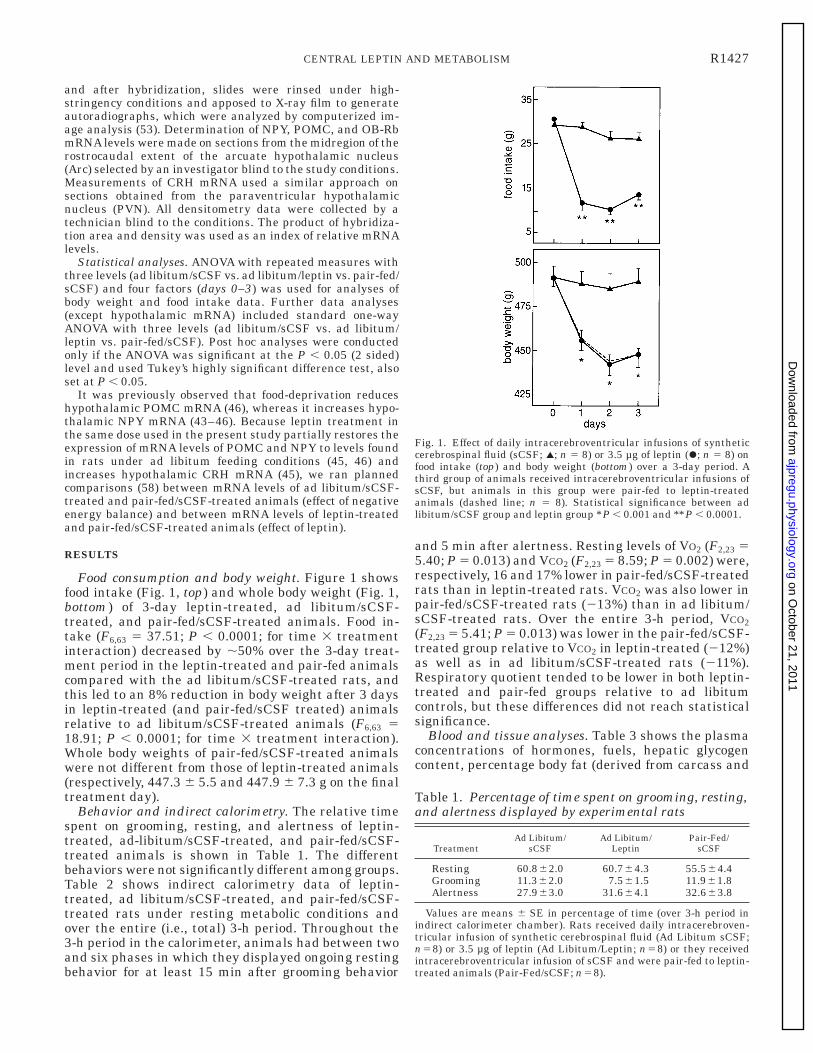
Food consumption and body weight. Figure 1 showsfood intake (Fig. 1, top) and whole body weight (Fig. 1,bottom) of 3-day leptin-treated, ad libitum/sCSF-treated, and pair-fed/sCSF-treated animals. Food in-take (F6,63 5 37.51; P , 0.0001; for time 3 treatmentinteraction) decreased by ,50% over the 3-day treat-ment period in the leptin-treated and pair-fed animalscompared with the ad libitum/sCSF-treated rats, andthis led to an 8% reduction in body weight after 3 daysin leptin-treated (and pair-fed/sCSF treated) animalsrelative to ad libitum/sCSF-treated animals (F6,63 518.91; P , 0.0001; for time 3 treatment interaction).Whole body weights of pair-fed/sCSF-treated animalswere not different from those of leptin-treated animals(respectively, 447.3 6 5.5 and 447.9 6 7.3 g on the finaltreatment day).
Behavior and indirect calorimetry. The relative timespent on grooming, resting, and alertness of leptin-treated, ad-libitum/sCSF-treated, and pair-fed/sCSF-treated animals is shown in Table 1. The differentbehaviors were not significantly different among groups.Table 2 shows indirect calorimetry data of leptin-treated, ad libitum/sCSF-treated, and pair-fed/sCSF-treated rats under resting metabolic conditions andover the entire (i.e., total) 3-h period. Throughout the3-h period in the calorimeter, animals had between twoand six phases in which they displayed ongoing restingbehavior for at least 15 min after grooming behavior
and 5 min after alertness. Resting levels of VO2 (F2,23 55.40; P 5 0.013) and VCO2 (F2,23 5 8.59; P 5 0.002) were,respectively, 16 and 17% lower in pair-fed/sCSF-treatedrats than in leptin-treated rats. VCO2 was also lower inpair-fed/sCSF-treated rats (213%) than in ad libitum/sCSF-treated rats. Over the entire 3-h period, VCO2(F2,23 5 5.41; P 5 0.013) was lower in the pair-fed/sCSF-treated group relative to VCO2 in leptin-treated (212%)as well as in ad libitum/sCSF-treated rats (211%).Respiratory quotient tended to be lower in both leptin-treated and pair-fed groups relative to ad libitumcontrols, but these differences did not reach statisticalsignificance.
Blood and tissue analyses. Table 3 shows the plasmaconcentrations of hormones, fuels, hepatic glycogencontent, percentage body fat (derived from carcass and
Fig. 1. Effect of daily intracerebroventricular infusions of syntheticcerebrospinal fluid (sCSF; l; n 5 8) or 3.5 µg of leptin (r; n 5 8) onfood intake (top) and body weight (bottom) over a 3-day period. Athird group of animals received intracerebroventricular infusions ofsCSF, but animals in this group were pair-fed to leptin-treatedanimals (dashed line; n 5 8). Statistical significance between adlibitum/sCSF group and leptin group *P , 0.001 and **P , 0.0001.
Table 1. Percentage of time spent on grooming, resting,and alertness displayed by experimental rats
TreatmentAd Libitum/
sCSFAd Libitum/
LeptinPair-Fed/
sCSF
Resting 60.862.0 60.764.3 55.564.4Grooming 11.362.0 7.561.5 11.961.8Alertness 27.963.0 31.664.1 32.663.8
Values are means 6 SE in percentage of time (over 3-h period inindirect calorimeter chamber). Rats received daily intracerebroven-tricular infusion of synthetic cerebrospinal fluid (Ad Libitum sCSF;n58) or 3.5 µg of leptin (Ad Libitum/Leptin; n58) or they receivedintracerebroventricular infusion of sCSF and were pair-fed to leptin-treated animals (Pair-Fed/sCSF; n58).
R1427CENTRAL LEPTIN AND METABOLISM
on October 21, 2011
ajpregu.physiology.orgD
ownloaded from
organ fat extraction) of i3vt leptin-treated, ad libitum/sCSF-treated, and pair-fed/sCSF-treated rats. Relativeto the ad libitum/sCSF group, the leptin-treated ani-mals and pair-fed animals had lower levels of plasmaleptin (measured with mouse RIA: F2,23 5 16.12; P ,0.0001; levels of leptin with a human-leptin RIA werenondetectable), hepatic glycogen content (F2,23 5 8.84;P 5 0.002), and percentage body fat (F2,23 5 9.54; P 50.001). The plasma triglyceride level was lower inleptin-treated (240%) and pair-fed (265%) rats rela-tive to the level in ad libitum/sCSF-treated rats (F2,23 56.71; P 5 0.006). There was a tendency of percentagebody fat to be lower in leptin-treated rats than inpair-fed/sCSF-treated rats (P 5 0.08). No treatmentdifferences were observed in plasma levels of glucose,corticosterone, or insulin among groups. The plasmalevels of corticosterone are relatively high comparedwith those in other studies (e.g., 48). Although repeti-tive i3vt administration of leptin and/or vehicle is notnecessarily associated with negative side effects (see
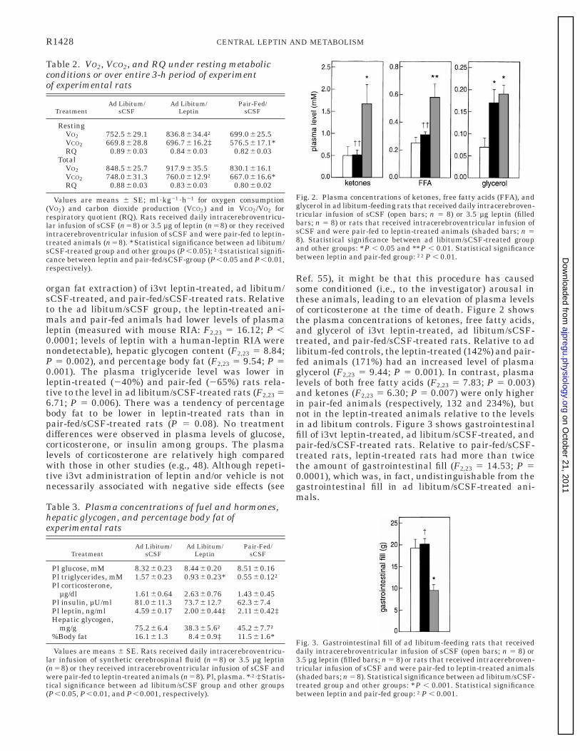
Ref. 55), it might be that this procedure has causedsome conditioned (i.e., to the investigator) arousal inthese animals, leading to an elevation of plasma levelsof corticosterone at the time of death. Figure 2 showsthe plasma concentrations of ketones, free fatty acids,and glycerol of i3vt leptin-treated, ad libitum/sCSF-treated, and pair-fed/sCSF-treated rats. Relative to adlibitum-fed controls, the leptin-treated (142%) and pair-fed animals (171%) had an increased level of plasmaglycerol (F2,23 5 9.44; P 5 0.001). In contrast, plasmalevels of both free fatty acids (F2,23 5 7.83; P 5 0.003)and ketones (F2,23 5 6.30; P 5 0.007) were only higherin pair-fed animals (respectively, 132 and 234%), butnot in the leptin-treated animals relative to the levelsin ad libitum controls. Figure 3 shows gastrointestinalfill of i3vt leptin-treated, ad libitum/sCSF-treated, andpair-fed/sCSF-treated rats. Relative to pair-fed/sCSF-treated rats, leptin-treated rats had more than twicethe amount of gastrointestinal fill (F2,23 5 14.53; P 50.0001), which was, in fact, undistinguishable from thegastrointestinal fill in ad libitum/sCSF-treated ani-mals.
Table 2. VO2, VCO2, and RQ under resting metabolicconditions or over entire 3-h period of experimentof experimental rats
TreatmentAd Libitum/
sCSFAd Libitum/
LeptinPair-Fed/
sCSF
RestingVO2 752.5629.1 836.8634.4† 699.0625.5VCO2 669.8628.8 696.7616.2‡ 576.5617.1*RQ 0.8960.03 0.8460.03 0.8260.03
TotalVO2 848.5625.7 917.9635.5 830.1616.1VCO2 748.0631.3 760.0612.9† 667.0616.6*RQ 0.8860.03 0.8360.03 0.8060.02
Values are means 6 SE; ml·kg21 ·h21 for oxygen consumption(VO2) and carbon dioxide production (VCO2) and in VCO2/VO2 forrespiratory quotient (RQ). Rats received daily intracerebroventricu-lar infusion of sCSF (n58) or 3.5 µg of leptin (n58) or they receivedintracerebroventricular infusion of sCSF and were pair-fed to leptin-treated animals (n58). *Statistical significance between ad libitum/sCSF-treated group and other groups (P,0.05); †,‡statistical signifi-cance between leptin and pair-fed/sCSF-group (P,0.05 and P,0.01,respectively).
Table 3. Plasma concentrations of fuel and hormones,hepatic glycogen, and percentage body fat ofexperimental rats
TreatmentAd Libitum/
sCSFAd Libitum/
LeptinPair-Fed/
sCSF
Pl glucose, mM 8.3260.23 8.4460.20 8.5160.16Pl triglycerides, mM 1.5760.23 0.9360.23* 0.5560.12†Pl corticosterone,
µg/dl 1.6160.64 2.6360.76 1.4360.45Pl insulin, µU/ml 81.0611.3 73.7612.7 62.367.4Pl leptin, ng/ml 4.5960.17 2.0060.44‡ 2.1160.42‡Hepatic glycogen,
mg/g 75.266.4 38.365.6† 45.267.7†%Body fat 16.161.3 8.460.9‡ 11.561.6*
Values are means 6 SE. Rats received daily intracerebroventricu-lar infusion of synthetic cerebrospinal fluid (n58) or 3.5 µg leptin(n58) or they received intracerebroventricular infusion of sCSF andwere pair-fed to leptin-treated animals (n58). Pl, plasma. *,†,‡Statis-tical significance between ad libitum/sCSF group and other groups(P,0.05, P,0.01, and P,0.001, respectively).
Fig. 2. Plasma concentrations of ketones, free fatty acids (FFA), andglycerol in ad libitum-feeding rats that received daily intracerebroven-tricular infusion of sCSF (open bars; n 5 8) or 3.5 µg leptin (filledbars; n 5 8) or rats that received intracerebroventricular infusion ofsCSF and were pair-fed to leptin-treated animals (shaded bars; n 58). Statistical significance between ad libitum/sCSF-treated groupand other groups: *P , 0.05 and **P , 0.01. Statistical significancebetween leptin and pair-fed group: ††P , 0.01.
Fig. 3. Gastrointestinal fill of ad libitum-feeding rats that receiveddaily intracerebroventricular infusion of sCSF (open bars; n 5 8) or3.5 µg leptin (filled bars; n 5 8) or rats that received intracerebroven-tricular infusion of sCSF and were pair-fed to leptin-treated animals(shaded bars; n 5 8). Statistical significance between ad libitum/sCSF-treated group and other groups: *P , 0.001. Statistical significancebetween leptin and pair-fed group: †P , 0.001.
R1428 CENTRAL LEPTIN AND METABOLISM
on October 21, 2011
ajpregu.physiology.orgD
ownloaded from
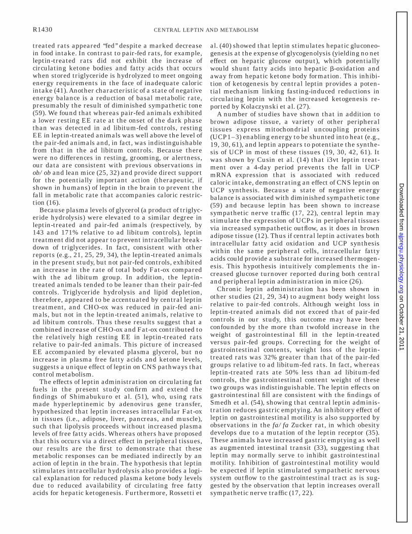
EE. Because stored triglycerides in fat tissue is ametabolically inactive compartment, resting and totalCHO-ox and Fat-ox and resting and total EE of animalswere calculated for lean body masses of animals (bycorrecting for the amount of body fat from Table 3). Therelative CHO-ox and Fat-ox of leptin-treated, ad libitum/sCSF-treated, and pair-fed/sCSF-treated animals arepresented in Table 4. Resting (F2,23 5 3.63; P 5 0.044)and total (F2,23 5 3.83, P 5 0.038) CHO-ox in adlibitum/sCSF-treated animals was 45 and 49% higherthan in pair-fed/sCSF-treated animals, respectively,whereas only resting Fat-ox in the leptin-treated groupwas 53% higher than the level in the ad libitum/sCSFgroup (F2,23 5 3.94; P 5 0.035). Although EE was notdifferent among groups over the total 3-h period (5.76 60.19, 5.62 6 0.14, and 5.25 6 0.15 W/kg lean mass inthe ad libitum/sCSF-, ad libitum/leptin-, and pair-fed/sCSF-treated rats, respectively), resting EE in ad libi-tum/sCSF-treated and leptin-treated animals werehigher (13%) than in pair-fed/sCSF-treated animals(F2,23 5 5.43; P 5 0.013) (Fig. 4).
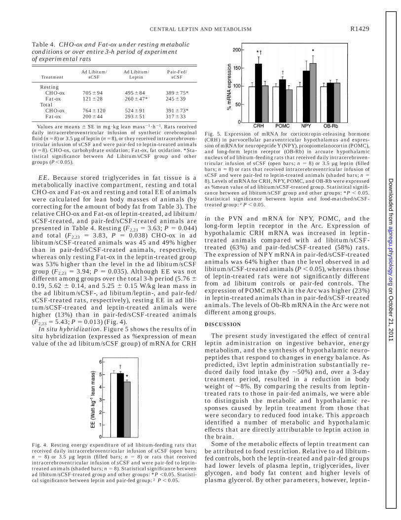
In situ hybridization. Figure 5 shows the results of insitu hybridization (expressed as %expression of meanvalue of the ad libitum/sCSF group) of mRNA for CRH
in the PVN and mRNA for NPY, POMC, and thelong-form leptin receptor in the Arc. Expression ofhypothalamic CRH mRNA was increased in leptin-treated animals compared with ad libitum/sCSF-treated (63%) and pair-fed/sCSF-treated (58%) rats.The expression of NPY mRNA in pair-fed/sCSF-treatedanimals was 64% higher than the level observed in adlibitum/sCSF-treated animals (P , 0.05), whereas thoseof leptin-treated rats were not significantly differentfrom ad libitum controls or pair-fed controls. Theexpression of POMC mRNA in the Arc was higher (23%)in leptin-treated animals than in pair-fed/sCSF-treatedanimals. The levels of Ob-Rb mRNA in the Arc were notdifferent among groups.
DISCUSSION
The present study investigated the effect of centralleptin administration on ingestive behavior, energymetabolism, and the synthesis of hypothalamic neuro-peptides that respond to changes in energy balance. Aspredicted, i3vt leptin administration substantially re-duced daily food intake (by ,50%) and, over a 3-daytreatment period, resulted in a reduction in bodyweight of ,8%. By comparing the results from leptin-treated rats to those in pair-fed animals, we were ableto distinguish the metabolic and hypothalamic re-sponses caused by leptin treatment from those thatwere secondary to reduced food intake. This approachidentified a number of metabolic and hypothalamiceffects that are directly attributable to leptin action inthe brain.
Some of the metabolic effects of leptin treatment canbe attributed to food restriction. Relative to ad libitum-fed controls, both the leptin-treated and pair-fed groupshad lower levels of plasma leptin, triglycerides, liverglycogen, and body fat content and higher levels ofplasma glycerol. By other parameters, however, leptin-
Table 4. CHO-ox and Fat-ox under resting metabolicconditions or over entire 3-h period of experimentof experimental rats
TreatmentAd Libitum/
sCSFAd Libitum/
LeptinPair-Fed/
sCSF
RestingCHO-ox 705694 495684 389675*Fat-ox 121628 260647* 245639
TotalCHO-ox 7646120 524691 391673*Fat-ox 200644 293651 317633
Values are means 6 SE in mg·kg lean mass21 ·h21. Rats receiveddaily intracerebroventricular infusion of synthetic cerebrospinalfluid (n58) or 3.5 µg of leptin (n58), or they received intracerebroven-tricular infusion of sCSF and were pair-fed to leptin-treated animals(n58). CHO-ox, carbohydrate oxidation; Fat-ox, fat oxidation. *Sta-tistical significance between Ad Libitum/sCSF group and othergroups (P,0.05).
Fig. 4. Resting energy expenditure of ad libitum-feeding rats thatreceived daily intracerebroventricular infusion of sCSF (open bars;n 5 8) or 3.5 µg leptin (filled bars; n 5 8) or rats that receivedintracerebroventricular infusion of sCSF and were pair-fed to leptin-treated animals (shaded bars; n 5 8). Statistical significance betweenad libitum/sCSF-treated group and other groups: *P ,0.05. Statisti-cal significance between leptin and pair-fed group: † P , 0.05.
Fig. 5. Expression of mRNA for corticotropin-releasing hormone(CRH) in parvocellular paraventricular hypothalamus and expres-sion of mRNAfor neuropeptide Y (NPY), proopiomelanocortin (POMC),and long-form leptin receptor (OB-Rb) in arcuate hypothalamicnucleus of ad libitum-feeding rats that received daily intracerebroven-tricular infusion of sCSF (open bars; n 5 8) or 3.5 µg leptin (filledbars; n 5 8) or rats that received intracerebroventricular infusion ofsCSF and were pair-fed to leptin-treated animals (shaded bars; n 58). Levels of mRNA for CRH, NPY, POMC, and OB-Rb were expressedas %mean value of ad libitum/sCSF-treated group. Statistical signifi-cance between ad libitum/sCSF group and other groups: *P , 0.05.Statistical significance between leptin and food-matched/sCSF-treated group: †P , 0.05.
R1429CENTRAL LEPTIN AND METABOLISM
on October 21, 2011
ajpregu.physiology.orgD
ownloaded from
treated rats appeared ‘‘fed’’ despite a marked decreasein food intake. In contrast to pair-fed rats, for example,leptin-treated rats did not exhibit the increase ofcirculating ketone bodies and fatty acids that occurswhen stored triglyceride is hydrolyzed to meet ongoingenergy requirements in the face of inadequate caloricintake (41). Another characteristic of a state of negativeenergy balance is a reduction of basal metabolic rate,presumably the result of diminished sympathetic tone(59). We found that whereas pair-fed animals exhibiteda lower resting EE rate at the onset of the dark phasethan was detected in ad libitum-fed controls, restingEE in leptin-treated animals was well above the level ofthe pair-fed animals and, in fact, was indistinguishablefrom that in the ad libitum controls. Because therewere no differences in resting, grooming, or alertness,our data are consistent with previous observations inob/ob and lean mice (25, 32) and provide direct supportfor the potentially important action (therapeutic, ifshown in humans) of leptin in the brain to prevent thefall in metabolic rate that accompanies caloric restric-tion (16).
Because plasma levels of glycerol (a product of triglyc-eride hydrolysis) were elevated to a similar degree inleptin-treated and pair-fed animals (respectively, by143 and 171% relative to ad libitum controls), leptintreatment did not appear to prevent intracellular break-down of triglycerides. In fact, consistent with otherreports (e.g., 21, 25, 29, 34), the leptin-treated animalsin the present study, but not pair-fed controls, exhibitedan increase in the rate of total body Fat-ox comparedwith the ad libitum group. In addition, the leptin-treated animals tended to be leaner than their pair-fedcontrols. Triglyceride hydrolysis and lipid depletion,therefore, appeared to be accentuated by central leptintreatment, and CHO-ox was reduced in pair-fed ani-mals, but not in the leptin-treated animals, relative toad libitum controls. Thus these results suggest that acombined increase of CHO-ox and Fat-ox contributed tothe relatively high resting EE in leptin-treated ratsrelative to pair-fed animals. This picture of increasedEE accompanied by elevated plasma glycerol, but noincrease in plasma free fatty acids and ketone levels,suggests a unique effect of leptin on CNS pathways thatcontrol metabolism.
The effects of leptin administration on circulating fatfuels in the present study confirm and extend thefindings of Shimabukuro et al. (51), who, using ratsmade hyperleptinemic by adenovirus gene transfer,hypothesized that leptin increases intracellular Fat-oxin tissues (i.e., adipose, liver, pancreas, and muscle),such that lipolysis proceeds without increased plasmalevels of free fatty acids. Whereas others have proposedthat this occurs via a direct effect in peripheral tissues,our results are the first to demonstrate that thesemetabolic responses can be mediated indirectly by anaction of leptin in the brain. The hypothesis that leptinstimulates intracellular hydrolysis also provides a logi-cal explanation for reduced plasma ketone body levelsdue to reduced availability of circulating free fattyacids for hepatic ketogenesis. Furthermore, Rossetti et
al. (40) showed that leptin stimulates hepatic gluconeo-genesis at the expense of glycogenolysis (yielding no neteffect on hepatic glucose output), which potentiallywould shunt fatty acids into hepatic b-oxidation andaway from hepatic ketone body formation. This inhibi-tion of ketogenesis by central leptin provides a poten-tial mechanism linking fasting-induced reductions incirculating leptin with the increased ketogenesis re-ported by Kolaczynski et al. (27).
A number of studies have shown that in addition tobrown adipose tissue, a variety of other peripheraltissues express mitochondrial uncoupling proteins(UCP1–3) enabling energy to be shunted into heat (e.g.,19, 30, 61), and leptin appears to potentiate the synthe-sis of UCP in most of these tissues (19, 30, 42, 61). Itwas shown by Cusin et al. (14) that i3vt leptin treat-ment over a 4-day period prevents the fall in UCPmRNA expression that is associated with reducedcaloric intake, demonstrating an effect of CNS leptin onUCP synthesis. Because a state of negative energybalance is associated with diminished sympathetic tone(59) and because leptin has been shown to increasesympathetic nerve traffic (17, 22), central leptin maystimulate the expression of UCPs in peripheral tissuesvia increased sympathetic outflow, as it does in brownadipose tissue (12). Thus if central leptin activates bothintracellular fatty acid oxidation and UCP synthesiswithin the same peripheral cells, intracellular fattyacids could provide a substrate for increased thermogen-esis. This hypothesis intuitively complements the in-creased glucose turnover reported during both centraland peripheral leptin administration in mice (26).
Chronic leptin administration has been shown inother studies (21, 29, 34) to augment body weight lossrelative to pair-fed controls. Although weight loss inleptin-treated animals did not exceed that of pair-fedcontrols in our study, this outcome may have beenconfounded by the more than twofold increase in theweight of gastrointestinal fill in the leptin-treatedversus pair-fed groups. Correcting for the weight ofgastrointestinal contents, weight loss of the leptin-treated rats was 32% greater than that of the pair-fedgroups relative to ad libitum-fed rats. In fact, whereasleptin-treated rats ate 50% less than ad libitum-fedcontrols, the gastrointestinal content weight of thesetwo groups was indistinguishable. The leptin effects ongastrointestinal fill are consistent with the findings ofSmedh et al. (54), showing that central leptin adminis-tration reduces gastric emptying. An inhibitory effect ofleptin on gastrointestinal motility is also supported byobservations in the fa/fa Zucker rat, in which obesitydevelops due to a mutation of the leptin receptor (35).These animals have increased gastric emptying as wellas augmented intestinal transit (33), suggesting thatleptin may normally serve to inhibit gastrointestinalmotility. Inhibition of gastrointestinal motility wouldbe expected if leptin stimulated sympathetic nervoussystem outflow to the gastrointestinal tract as is sug-gested by the observation that leptin increases overallsympathetic nerve traffic (17, 22).
R1430 CENTRAL LEPTIN AND METABOLISM
on October 21, 2011
ajpregu.physiology.orgD
ownloaded from
To investigate the CNS mechanisms that mediateleptin’s behavioral, metabolic, and gastrointestinal ef-fects, we measured hypothalamic mRNA encoding neu-ropeptides implicated in energy homeostasis. Consis-tent with our previous studies, we found that centralleptin administration increased CRH mRNA in thehypothalamic PVN. This effect was not due to reducedfood intake, because pair-fed animals did not show thisresponse. Because central administration of CRH hasanorexigenic and thermogenic effects (7, 39), the datain the present and other studies (37, 45) suggest thatleptin’s effects could be mediated through activation ofhypothalamic CRH neurons. This idea is consistentwith a recent study of Uehara et al. (56) demonstratingthat the anorexigenic efficacy of central leptin wasreduced by central administration of a CRH receptorantagonist. Activation of hypothalamic CRH neuronscould also explain the increased SNS outflow (6) andthermogenesis (39) that is observed with leptin admin-istration. It is noteworthy that these stimulatory ef-fects of leptin on CRH signaling coexist with inhibitoryeffects of leptin on the hypothalamic-pituitary axis (23,24). These paradoxical observations suggest that leptinmay exert distinctly different regulatory effects ondiscrete subpopulations of CRH neurons in the PVN.
In addition to leptin effects on CRH mRNA, leptin-treated animals also had an increased level of POMCmRNA in the arcuate nucleus relative to the levelobserved in pair-fed animals. The increase in hypotha-lamic POMC gene expression is consistent with whatwe observed after central leptin treatment in rats thatwere food deprived (46) and supports the hypothesisthat melanocortins, which act centrally to reduce foodintake, are important mediators of leptin signaling(50).
Previous studies (e.g., Ref. 5) indicate that dramaticweight loss due to caloric restriction or fasting isassociated with reduced CRH mRNA levels in the PVN,and this response has been proposed to contribute tothe stimulated feeding behavior and reduced SNSoutflow that occurs in this setting (6). Although foodrestriction in the present study was therefore expectedto lower hypothalamic CRH gene expression, we foundno evidence of reduced CRH mRNA in pair-fed animals,despite weight loss of ,8% relative to ad libitum-fedcontrols. This observation suggests that weight lossmust exceed that experienced by pair-fed rats beforeCRH mRNA levels are decreased in the PVN (see alsoRef. 48). On the other hand, we did find an increase inNPY gene expression in the arcuate nucleus of pair-fedanimals relative to ad libitum-fed controls, an effectthat is consistent with previous studies (1, 43, 45, 48).However, the leptin-treated animals did not have signifi-cantly reduced levels of NPY mRNA relative to thepair-fed group. The effect of leptin to reduce hypotha-lamic NPY gene expression may therefore be detectableonly in animals in which the NPY system has beenactivated (e.g., when leptin levels are low). Finally, wereport in the present study that neither leptin treat-ment nor food restriction significantly affected long-form leptin receptor mRNA expression in the arcuate
nucleus. Thus, although leptin deficiency in ob/ob miceis associated with a two- to threefold increase ofexpression of leptin receptor mRNA in this brain area(4), our results do not support the hypothesis thatleptin administration downregulates leptin receptorgene expression in nonmutant animals with ad libitumaccess to food.
Perspectives
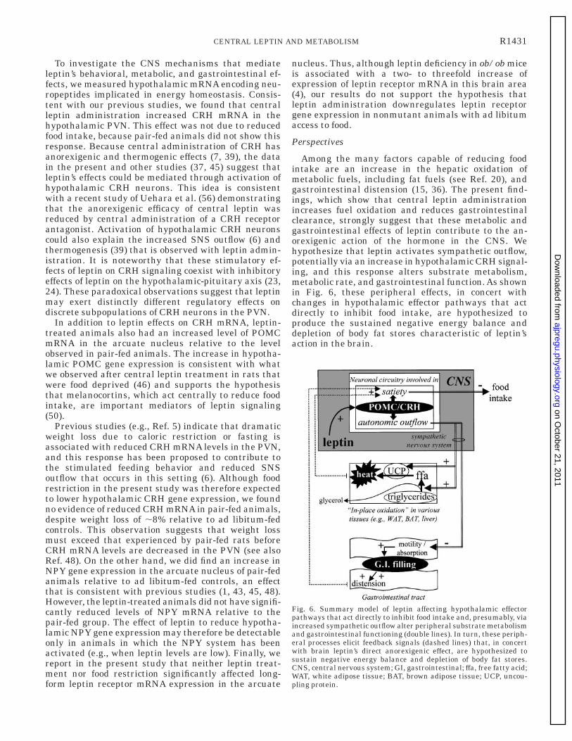
Among the many factors capable of reducing foodintake are an increase in the hepatic oxidation ofmetabolic fuels, including fat fuels (see Ref. 20), andgastrointestinal distension (15, 36). The present find-ings, which show that central leptin administrationincreases fuel oxidation and reduces gastrointestinalclearance, strongly suggest that these metabolic andgastrointestinal effects of leptin contribute to the an-orexigenic action of the hormone in the CNS. Wehypothesize that leptin activates sympathetic outflow,potentially via an increase in hypothalamic CRH signal-ing, and this response alters substrate metabolism,metabolic rate, and gastrointestinal function. As shownin Fig. 6, these peripheral effects, in concert withchanges in hypothalamic effector pathways that actdirectly to inhibit food intake, are hypothesized toproduce the sustained negative energy balance anddepletion of body fat stores characteristic of leptin’saction in the brain.
Fig. 6. Summary model of leptin affecting hypothalamic effectorpathways that act directly to inhibit food intake and, presumably, viaincreased sympathetic outflow alter peripheral substrate metabolismand gastrointestinal functioning (double lines). In turn, these periph-eral processes elicit feedback signals (dashed lines) that, in concertwith brain leptin’s direct anorexigenic effect, are hypothesized tosustain negative energy balance and depletion of body fat stores.CNS, central nervous system; GI, gastrointestinal; ffa, free fatty acid;WAT, white adipose tissue; BAT, brown adipose tissue; UCP, uncou-pling protein.
R1431CENTRAL LEPTIN AND METABOLISM
on October 21, 2011
ajpregu.physiology.orgD
ownloaded from
We thank K. Blake, R. Flower, L. Madden, E. Colasurdo, S. Reed,J. Breininger, Z. Jonak, V. Hoagland, K. Peacock, and T. Herman forexcellent technical contributions.
This study was supported by grants from the Dutch DiabetesAssociation, the Dutch Scientific Organization (NWO), Merit ReviewPrograms of the Department of Veterans Affairs, and the NationalInstitutes of Health (NS-32273 and DK-53109, -17844, -12829, and-6339).
Address for reprint requests and other correspondence: G. vanDijk, Dept. Animal Physiology, Univ. of Groningen, PO Box 14, 9750AA Haren, The Netherlands (E-mail: [email protected]).
Received 25 September 1998; accepted in final form 3 February 1999.
REFERENCES
1. Ahima, R. S., D. Prabakaran, C. Mantzoros, D. Qu, B.Lowell, E. Maratos-Flier, and J. S. Flier. Role of leptin in theneuroendocrine response to fasting. Nature 382: 250–252, 1996.
2. Banks, W. A., A. J. Kastin, W. Huang, J. B. Jaspan, and L. M.Maness. Leptin enters the brain by a saturable system indepen-dent of insulin. Peptides 17: 305–311, 1996.
3. Baskin, D. G., R. J. Seeley, J. L. Kuyper, S. Lok, D. S. Weigle,J. C. Erickson, R. D. Palmiter, and M. W. Schwartz. In-creased expression of mRNA for the long form of the leptinreceptor in the hypothalamus is associated with leptin hypersen-sitivity and fasting. Diabetes 47: 538–543, 1998.
4. Billington, C. J., J. E. Briggs, S. Harker, M. Grace, and A. S.Levine. Neuropeptide Y in the hypothalamic paraventricularnucleus: a center coordinating energy metabolism. Am. J. Physiol.266 (Regulatory Integrative Comp. Physiol. 35): R1765–R1770,1994.
5. Brady, L., M. A. Smith, P. W. Gold, and M. Herkenham.Altered expression of hypothalamic neuropeptide mRNAs infood-restricted and food-deprived rats. Neuroendocrinology 52:441–447, 1990.
6. Bray, G. A. The nutrient balance hypothesis: peptides, sympa-thetic activity, and food intake. Ann. NY Acad. Sci. 676: 223–241,1993.
7. Buwalda, B., S. F. de Boer, A. A. van Kalkeren, and J. M.Koolhaas. Physiological and behavioral effects of chronic intra-cerebroventricular infusion of corticotropin-releasing factor inthe rat. Psychoneuroendocrinology 22: 297–309, 1997.
8. Campfield, L. A., F. J. Smith, and P. Burn. The OB protein(leptin) pathway—a link between adipose tissue mass andcentral neural networks. Horm. Metab. Res. 28: 619–632, 1996.
9. Campfield, L. A., F. J. Smith, Y. Guisez, R. Devos, and P.Burn. Recombinant mouse OB protein: evidence for a peripheralsignal linking adiposity and central neural networks. Science269: 546–549, 1995.
10. Cheung, C. C., D. K. Clifton, and R. A. Steiner. Proopiomela-nocortin neurons are direct targets for leptin in the hypothala-mus. Endocrinology 138: 4489–4492, 1997.
11. Considine, R. V., M. K. Sinha, M. L. Heiman, A. Kriauci-unas, T. W. Stephans, M. R. Nyce, J. P. Ohanessian, C. C.Marco, L. J. McKee, T. L. Bauer, and J. F. Caro. Serumimmunoreactive-leptin concentrations in normal weight andobese humans. N. Engl. J. Med. 334: 292–295, 1996.
12. Collins, S., C. M. Kuhn, A. E. Petro, A. G. Swick, B. A.Chrunyk, and R. S. Surwit. Role of leptin in fat regulation(Abstract). Nature 380: 677, 1996.
13. Cusin, I., F. Rohner-Jeanrenaud, A. Stricker-Krongrad,and B. Jeanrenaud. The weight-reducing effect of an intracere-broventricular bolus injection of leptin in genetically obese fa/farats. Reduced sensitivity compared with lean animals. Diabetes45: 1446–1450, 1996.
14. Cusin, I., K. E. Zakrzewska, O. Boss, P. Muzzin, J. Giaco-bino, D. Ricquier, B. Jeanrenaud, and F. Rohner-Jeanre-naud. Chronic central leptin infusion enhances insulin-stimu-lated glucose metabolism and favors the expression of uncouplingproteins. Diabetes 47: 1014–1019, 1998.
15. Davis, J. D., B. J. Collins, and M. W. Levine. Peripheralcontrol of drinking: gastrointestinal filling as a negative feedbacksignal, a theoretical and experimental analysis. J. Comp. Physiol.Psychol. 89: 985–1002, 1975.
16. Doring, H., K. Schwartzer, B. Nuesslein-Hildesheim, and I.Schmidt. Leptin selectively increases energy expenditure offood-restricted lean mice. Int. J. Obes. Relat. Metab. Disord. 22:83–88, 1998.
17. Dunbar, J. C., Y. Hu, and H. Lu. Intracerebroventricular leptinincreases lumbar and renal sympathetic nerve activity and bloodpressure in normal rats. Diabetes 46: 2024–2043, 1997.
18. Ferranini, E. The theoretical bases of indirect calorimetry: areview. Metabolism 37: 287–301, 1988.
19. Fleury, C., M. Neverova, S. Collins, S. Raimbault, O.Champigny, C. Levi-Meyrueis, F. Bouillaud, M. F. Seldin,R. S. Surwit, D. Ricquier, and C. H. Warden. Uncouplingprotein-2: a novel gene linked to obesity and hyperinsulinemia.Nat. Genet. 15: 269–272, 1997.
20. Friedman, M. I. Control of energy intake by energy metabolism.Am. J. Clin. Nutr. 62: 1096S–1100S, 1995.
21. Halaas, J. L., C. Boozer, J. Blair-West, N. Fidahusein, D. A.Denton, and J. M. Friedman. Physiological response to long-term peripheral and central leptin infusion in lean and obesemice. Proc. Natl. Acad. Sci. USA 94: 8878–8883, 1997.
22. Haynes, W. G., D. A. Morgan, S. A. Walsh, A. L. Mark, andW. I. Siwitz. Receptor-mediated regional sympathetic nerveactivation by leptin. J. Clin. Invest. 100: 270–278, 1997.
23. Heiman, M. L., L. S. Craft, H. M. Hsiung, B. Schoner, andT. W. Stephens. Leptin inhibition of the hypothalamic-pituitary-adrenal axis in response to stress. Endocrinology 138: 3859–3863, 1997.
24. Huang, Q., R. Rivest, and D. Richard. Effects of leptin oncorticotropin-releasing factor (CRF) synthesis and CRF neuronactivation in the paraventricular hypothalamic nucleus of obese(ob/ob) mice. Endocrinology 139: 1524–1532, 1998.
25. Hwa, J. J., A. B. Fawzi, M. P. Graziano, L. Ghibaudi, P.Williams, M. van Heek, H. Davis, M. Rudinsky, E. Sybertz,and C. D. Strader. Leptin increases energy expenditure and selec-tively promotes fat metabolism in ob/ob mice. Am. J. Physiol. 272(Regulatory Integrative Comp. Physiol. 41): R1204–R1209, 1997.
26. Kamohara, S., R. Burcelin, J. L. Halaas, J. M. Friedman,and M. J. Charron. Acute stimulation of glucose metabolism inmice by leptin treatment. Nature 389: 374–377, 1997.
27. Kolaczynski, J. W., R. V. Considine, J. Ohannesian, C.Marco, I. Opentanova, M. C. Nyce, M. Myint, and J. F. Caro.Responses of leptin to short-term fasting and refeeding inhumans: a link with ketogenesis but not ketones themselves.Diabetes 45: 1511–1515, 1996.
28. Leshner, A. I., V. A. Litwin, and R. L. Squibb. A simplemethod for carcass analysis. Physiol. Behav. 9: 281–282, 1972.
29. Levin, N., C. Nelson, A. Gurney, R. Vandlen, and F. DeSauvage. Decreased food intake does not completely account foradiposity reduction after ob protein infusion. Proc. Natl. Acad.Sci. USA 93: 1726–1730, 1996.
30. Liu, Q., C. Bai, F. Chen, R. Wang, T. MacDonald, M. Gu, Q.Zhang, M. Morsy, and T. Caskey. Uncoupling protein-3: amuscle specific gene upregulated by leptin in ob/ob mice. Gene207: 1–7, 1998.
31. Maffei, M., J. Halaas, E. Ravussin, R. E. Pratley, G. H. Lee,Y. Zhang, H. Fei, S. Kim, R. Lallone, S. Ranganathan, P. A.Kern, and J. M. Friedman. Leptin levels in human and rodent:measurement of plasma leptin and ob RNA in obese and weight-reduced subjects. Nat. Med. 1: 1155–1161, 1995.
32. Mistry, A. M., A. G. Swick, and D. R. Romsos. Leptin rapidlylowers food intake and elevates metabolic rates in lean and ob/obmice. J. Nutr. 127: 2065–2072, 1997.
33. Moos, A. B., C. L. McLaughlin, and C. A. Baile. Effects of CCKon gastrointestinal function in lean and obese Zucker rats.Peptides 3: 619–622, 1982.
34. Pelleymounter, M. A., M. J. Cullen, M. B. Baker, R. Hecht,D. Winters, T. Boone, and F. Collins. Effects of the obese geneproduct on body weight regulation in ob/ob mice. Science 269:540–543, 1995.
35. Phillips, M. S., Q. Y. Liu, H. A. Hammond, V. Dugan, P. J.Hey, C. T. Caskey, and J. F. Hess. Leptin receptor missensemutation in the fatty Zucker rat. Nat. Genet. 13: 18–19, 1996.
36. Phillips, R. J., and T. J. Powley. Gastric volume rather thannutrient content inhibits food intake. Am. J. Physiol. 271 (Regu-latory Integrative Comp. Physiol. 40): R766–R779, 1996.
R1432 CENTRAL LEPTIN AND METABOLISM
on October 21, 2011
ajpregu.physiology.orgD
ownloaded from
37. Raber, J., S. Chen, L. Mucke, and L. Feng. Corticotropin-releasing factor and adrenocorticotrophic hormone as potentialcentral mediators of OB effects. J. Biol. Chem. 272: 15057–15060, 1997.
38. Ramirez, I. Physiological and biochemical measurements inrelation to feeding. In: Feeding and Drinking, edited by F. M.Toates and N. E. Rowland. Amsterdam: Elsevier, 1987, p. 151–165.
39. Richard, D. Involvement of corticotropin-releasing factor in thecontrol of food intake and energy expenditure. Ann. NY Acad.Sci. 697: 155–172, 1993.
40. Rossetti, L., D. Massilon, N. Barzilai, P. Vuguin, W. Chen,M. Hawkins, J. Wu, and J. Wang. Short term effects of leptinon hepatic gluconeogenesis and in vivo insulin action. J. Biol.Chem. 272: 27758–27763, 1997.
41. Saudec, C. D., and P. Felig. The metabolic events of starvation.Am. J. Med. 60: 117–126, 1976.
42. Scarpace, P. J., M. Matheny, B. H. Pollock, and N. Tumur.Leptin increases uncoupling protein expression and energyexpenditure. Am. J. Physiol. 273 (Endocrinol. Metab. 36): E226–E230, 1997.
43. Schwartz, M. W., D. G. Baskin, T. R. Bukowski, J. L.Kuijper, D. Foster, G. Lasser, D. E. Prunkard, D. Porte, Jr.,S. C. Woods, R. J. Seeley, and D. S. Weigle. Specificity of leptinaction on elevated blood glucose levels and hypothalamic neuro-peptide Y gene expression in ob/ob mice. Diabetes 45: 531–535,1996.
44. Schwartz, M. W., E. Peskind, M. Raskind, E. J. Boyko, andD. Porte, Jr. Cerebrospinal fluid leptin levels: relationship toplasma levels and to adiposity in humans. Nat. Med. 2: 589–593,1996.
45. Schwartz, M. W., R. J. Seeley, L. A. Campfield, P. Burn, andD. G. Baskin. Identification of targets of leptin action in rathypothalamus. J. Clin. Invest. 98: 1101–1106, 1997.
46. Schwartz, M. W., R. J. Seeley, S. C. Woods, D. S. Weigle, L. A.Campfield, P. Burn, and D. G. Baskin. Leptin increaseshypothalamic pro-opiomelanocortin mRNA expression in therostral arcuate nucleus. Diabetes 46: 2119–2123, 1997.
47. Schwartz, M. W., A. J. Sipols, J. L. Marks, G. Sanacora, J. D.White, A. Scheurink, S. E. Kahn, D. G. Baskin, S. C. Woods,D. P. Figlewicz, and D. Porte, Jr. Inhibition of hypothalamicneuropeptide Y gene expression by insulin. Endocrinology 130:3608–3616, 1992.
48. Seeley, R. J., C. A. Matson, M. Chavez, S. C. Woods, M. F.Dallman, and M. W. Schwartz. Behavioral, endocrine, andhypothalamic responses to involuntary overfeeding. Am. J.Physiol. 271 (Regulatory Integrative Comp. Physiol. 40): R819–R823, 1996.
49. Seeley, R. J., G. van Dijk, L. A. Campfield, F. J. Smith, P.Burn, J. A. Nelligan, S. M. Bell, D. G. Baskin, S. C. Woods,
and M. W. Schwartz. Intraventricular leptin reduces foodintake and body weight of lean rats but not obese Zucker rats.Horm. Metab. Res. 28: 664–668, 1996.
50. Seeley, R. J., K. A. Yagaloff, S. L. Fisher, P. Burn, T. E.Thiele, G. van Dijk, D. G. Baskin, and M. W. Schwartz.Melanocortin receptors in leptin effects (Abstract). Nature 390:349, 1997.
51. Shimabukuro, M., K. Koyama, G. Cheng, M. Y. Wang, F.Trieu, Y. Lee, C. B. Newgard, and R. H. Unger. Directantidiabetic effect of leptin through triglyceride depletion oftissues. Proc. Natl. Acad. Sci. USA 94: 4637–4641, 1997.
52. Shimizu, H., Y. Shimomura, R. Hayashi, K. Ohtani, N. Sato,T. Futawatari, and M. Mori. Serum leptin concentration isassociated with total body mass, but not abdominal fat distribu-tion. Int. J. Obes. Relat. Metab. Disord. 21: 536–541, 1997.
53. Sipols, A. J., D. G. Baskin, and M. W. Schwartz. Effect ofintracerebroventricular insulin infusion on diabetic hyperphagiaand hypothalamic neuropeptide gene expression. Diabetes 44:147–151, 1995.
54. Smedh, U., M. Hakansson, B. Meister, and K. Uvnas-Moberg. Leptin injected into the fourth ventricle inhibits gastricemptying. Neuroreport 9: 297–301, 1998.
55. Thiele, T. E., G. van Dijk, L. A. Campfield, F. J. Smith, P.Burn, S. C. Woods, I. L. Bernstein, and R. J. Seeley. Centralinfusion of GLP-1, but not leptin, produces conditioned tasteaversions in rats. Am. J. Physiol. 272 (Regulatory IntegrativeComp. Physiol. 41): R726–R730, 1997.
56. Uehara, Y., H. Shimizu, K. Ohtani, N. Sato, and M. Mori.Hypothalamic corticotropin-releasing hormone is a mediator ofthe anorexigenic effect of leptin. Diabetes 47: 890–893, 1998.
57. Van Dijk, G., T. E. Thiele, J. C. K. Donahey, L. A. Campfield,F. J. Smith, P. Burn, I. L. Bernstein, S. C. Woods, and R. J.Seeley. Central infusions of leptin and GLP-1(7—36) amidedifferentially stimulate c-FLI in the rat brain. Am. J. Physiol.271 (Regulatory Integrative Comp. Physiol. 40): R1096–R1100,1996.
58. Winer, G. J., D. R. Brown, and K. M. Michaels. StatisticalPrinciples in Experimental Designs (3rd ed.). New York: McGrawHill, 1991.
59. Young, J. B., and L. Landsberg. Suppression of sympatheticnervous system during fasting. Obesity Res. 5: 646–649, 1997.
60. Zhang, Y., R. Proenca, M. Maffei, M. Barone, L. Leopold,and J. M. Friedman. Positional cloning of the mouse obese geneand its human analogue. Nature 372: 425–432, 1994.
61. Zhou, Y. T., M. Shimabukuro, K. Koyama, Y. Lee, M. Y.Wang, F. Trieu, C. B. Newgard, and R. H. Unger. Induction byleptin of uncoupling protein-2 and enzymes of fatty acid oxida-tion. Proc. Natl. Acad. Sci. USA 94: 6386–6390, 1997.
R1433CENTRAL LEPTIN AND METABOLISM
on October 21, 2011
ajpregu.physiology.orgD
ownloaded from