dna microarray technology for biodiversity ... - mediatum
TRANSCRIPT
Alexander Loy
DNAMicroarrayTechnology
forBiodiversityInventories
ofSulfate
ReducingProkaryotes

Lehrstuhl für Mikrobiologie
der Technischen Universität München
DNA Microarray Technology
for Biodiversity Inventories
of Sulfate-Reducing Prokaryotes
Alexander Loy
Vollständiger Abdruck der von der Fakultät Wissenschaftszentrum Weihenstephan für
Ernährung, Landnutzung und Umwelt der Technischen Universität München
zur Erlangung des akademischen Grades eines
Doktors der Naturwissenschaften
genehmigten Dissertation.
Vorsitzender: Univ.-Prof. Dr. Gert Forkmann
Prüfer der Dissertation: 1. Univ.-Prof. Dr. Michael Wagner, Universität Wien/Österreich
2. Univ.-Prof. Dr. Karl-Heinz Schleifer
3. Univ.-Prof. Dr. Rudi F. Vogel
Die Dissertation wurde am 13.03.2003 bei der Technischen Universität München eingereicht
und durch die Fakultät Wissenschaftszentrum Weihenstephan für Ernährung, Landnutzung
und Umwelt am 02.06.2003 angenommen.

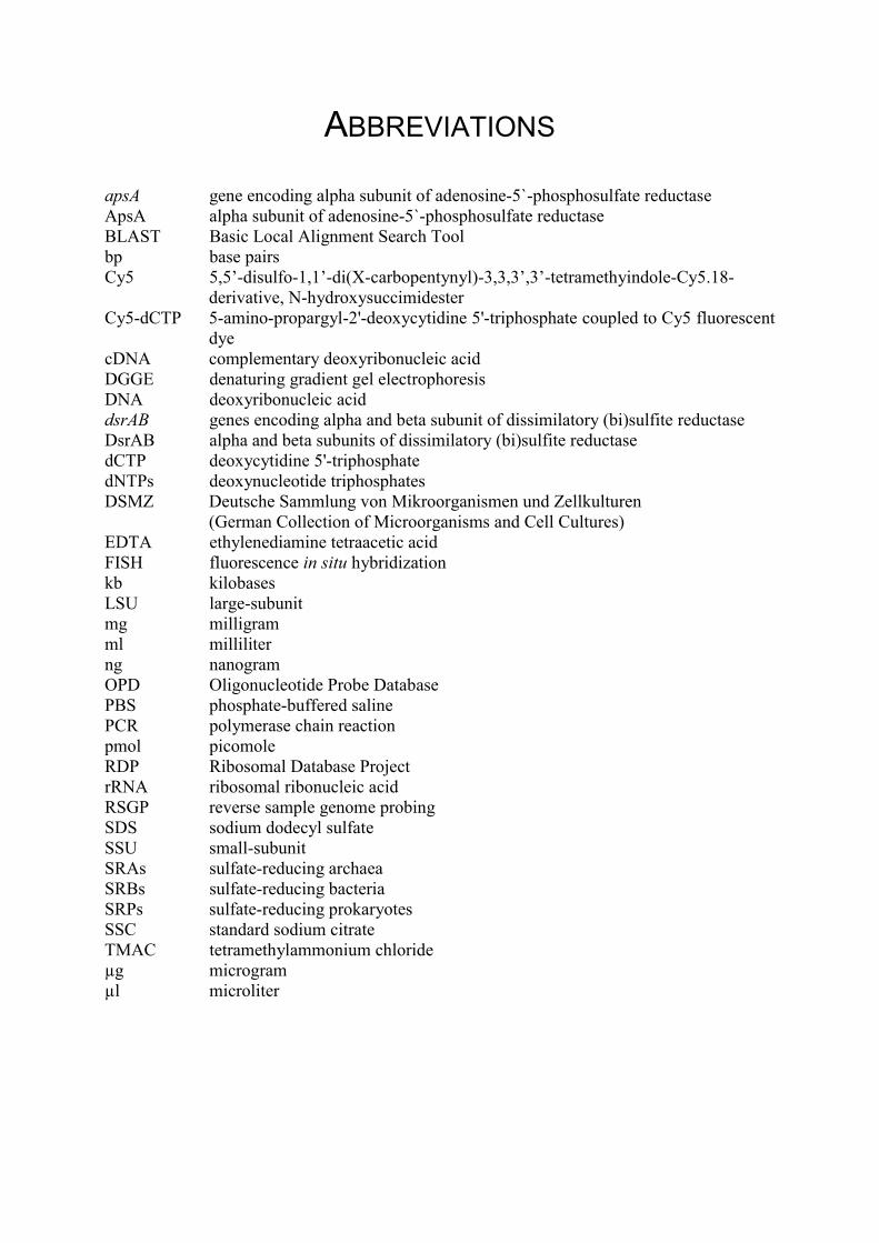
ABBREVIATIONS
apsA gene encoding alpha subunit of adenosine-5`-phosphosulfate reductaseApsA alpha subunit of adenosine-5`-phosphosulfate reductaseBLAST Basic Local Alignment Search Toolbp base pairsCy5 5,5’-disulfo-1,1’-di(X-carbopentynyl)-3,3,3’,3’-tetramethyindole-Cy5.18-
derivative, N-hydroxysuccimidesterCy5-dCTP 5-amino-propargyl-2'-deoxycytidine 5'-triphosphate coupled to Cy5 fluorescent
dyecDNA complementary deoxyribonucleic acidDGGE denaturing gradient gel electrophoresisDNA deoxyribonucleic aciddsrAB genes encoding alpha and beta subunit of dissimilatory (bi)sulfite reductaseDsrAB alpha and beta subunits of dissimilatory (bi)sulfite reductasedCTP deoxycytidine 5'-triphosphatedNTPs deoxynucleotide triphosphatesDSMZ Deutsche Sammlung von Mikroorganismen und Zellkulturen
(German Collection of Microorganisms and Cell Cultures)EDTA ethylenediamine tetraacetic acidFISH fluorescence in situ hybridizationkb kilobasesLSU large-subunitmg milligramml milliliterng nanogramOPD Oligonucleotide Probe DatabasePBS phosphate-buffered salinePCR polymerase chain reactionpmol picomoleRDP Ribosomal Database ProjectrRNA ribosomal ribonucleic acidRSGP reverse sample genome probingSDS sodium dodecyl sulfateSSU small-subunitSRAs sulfate-reducing archaeaSRBs sulfate-reducing bacteriaSRPs sulfate-reducing prokaryotesSSC standard sodium citrateTMAC tetramethylammonium chlorideµg microgramµl microliter

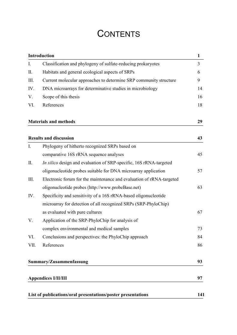
CONTENTS
Introduction 1
I. Classification and phylogeny of sulfate-reducing prokaryotes 3
II. Habitats and general ecological aspects of SRPs 6
III. Current molecular approaches to determine SRP community structure 9
IV. DNA microarrays for determinative studies in microbiology 14
V. Scope of this thesis 16
VI. References 18
Materials and methods 29
Results and discussion 43
I. Phylogeny of hitherto recognized SRPs based on
comparative 16S rRNA sequence analyses 45
II. In silico design and evaluation of SRP-specific, 16S rRNA-targeted
oligonucleotide probes suitable for DNA microarray application 57
III. Electronic forum for the maintenance and evaluation of rRNA-targeted
oligonucleotide probes (http://www.probeBase.net) 63
IV. Specificity and sensitivity of a 16S rRNA-based oligonucleotide
microarray for detection of all recognized SRPs (SRP-PhyloChip)
as evaluated with pure cultures 67
V. Application of the SRP-PhyloChip for analysis of
complex environmental and medical samples 73
VI. Conclusions and perspectives: the PhyloChip approach 84
VII. References 86
Summary/Zusammenfassung 93
Appendices I/II/III 97
List of publications/oral presentations/poster presentations 141


INTRODUCTION
1
INTRODUCTION

INTRODUCTION
3
I. Classification and phylogeny of sulfate-reducing prokaryotes
Taxonomic considerations concerning dissimilatory sulfate-reducing bacteria (SRBs) began in
1895 with Beijerink’s first isolation of a strict anaerobic, sulfate-reducing bacterium, which
he termed Spirillum desulfuricans (Beijerinck 1895). Vibrio (Baars 1930) was a synonymous
genus name for Spirillum desulfuricans for which finally the genus Desulfovibrio (D.
desulfuricans as the type species) was established by Kluyver and van Niel (1936). The early
history on the classification of Desulfovibrio desulfuricans already reflected the problems of
continual reclassifications and amendments that microbial taxonomists faced over years of
research on SRB systematics.
In 1925, Elion was the first to describe the thermophilic sulfate-reducing bacterium Vibrio
thermodesulfuricans (Elion 1925). The capability of some SRBs to form endospores was
initially recognized for the thermophiles Clostridium nigrificans (Werkman and Weaver
1927) and Sporovibrio desulfuricans (Starkey 1938). Later, Campbell et al. demonstrated that
both bacteria were members of the same species (Campbell et al. 1957). The continuous
accumulation of newly described SRBs demanded thorough (re)classification of all existing
strains. As a result, all non-sporulating SRBs were assigned to the vibrio-shaped genus
Desulfovibrio (Postgate and Campbell 1966), whereas the endospore-forming species formed
the new sausage-shaped genus Desulfotomaculum (Campbell and Postgate 1965). At that time
it was thought that SRBs comprise a small and nutritionally limited guild, growing
preferentially on electron donors such as lactate and pyruvate that are incompletely oxidized
to acetate. This point of view changed considerably with the description of new types of SRBs
capable of completely oxidizing acetate, higher fatty acids, or aromatic compounds (Bak and
Widdel 1986a, Bak and Widdel 1986b, Brysch et al. 1987, Pfennig and Widdel 1981, Pfennig
et al. 1981, Widdel 1980, Widdel et al. 1983, Widdel and Pfennig 1977, Widdel and Pfennig
1981a, Widdel and Pfennig 1981b, Widdel and Pfennig 1982). In addition, the novel genus
Thermodesulfobacterium was established for thermophilic SRBs which were isolated from
hot aquatic habitats in the Yellowstone National Park (USA), contained unusual ether lipids,
and were phylogenetically distinct from previously known SRBs (Langworthy et al. 1983,
Zeikus et al. 1983). Studies by Stetter et al. on hyperthermophiles led to first description of
the archaeal genus Archaeoglobus and demonstrated that the capacity for dissimilatory sulfate
reduction is not restricted to the bacterial domain of life (Achenbach-Richter et al. 1987,
Burggraf et al. 1990, Stetter et al. 1987).
INTRODUCTION
4
Traditional classification of sulfate-reducing prokayotes (SRPs) relied on (i) phenotypic
characteristics such as nutrition and morphology and (ii) biochemical properties such as the
presence of desulfoviridin, lipid fatty acids, or menaquinones. The discovery of ribosomal
RNA (rRNA) as the ultimate universal molecular chronometer set the basis for modern
prokaryotic phylogeny and taxonomy (Fox et al. 1980, Woese 1987). Together with the
advent of the nucleic acid sequencing era, comparative 16S rRNA sequence analysis became
decisive for the inference of natural relationships among prokaryotes, consequently for SRPs,
too. Early applications of this novel taxonomic approach demonstrated that the delta
subdivision of the phylum purple bacteria harbored bacteria with different phenotypes like
sulfate-reducing bacteria (represented by Desulfovibrio desulfuricans and Desulfobacter
postgatei), sulfur-reducing bacteria, myxobacteria and relatives, and bdellovibrios (Oyaizu
and Woese 1985, Woese 1987). Later, all members of the phylum purple bacteria were
reclassified into the new class Proteobacteria (Stackebrandt et al. 1988). A more
comprehensive phylogenetic study of 20 nonsporeforming and two endospore-forming SRBs
based on comparison of nearly complete 16S rRNA sequences was performed by Devereux et
al. (1989). This study confirmed the classification of the genus Desulfotomaculum within the
gram-positive bacteria as suggested previously by 16S rRNA oligonucleotide cataloging
(Fowler et al. 1986). Among the nonsporeforming deltaproteobacterial species seven natural
groups could be defined. Although this grouping was generally consistent with the existent
physiology-based classification, the need for taxonomic revision was obvious. A similar
study, focussing on phylogeny of Desulfovibrio species, revealed further misclassified species
and strains (Devereux et al. 1990). Additionally, the monophyletic origin of the genuine
Desulfovibrio group was recognized at a higher taxonomic level, what led to the provisional
proposal of the family “Desulfovibrionaceae”.
In the early nineties, several reviews summarized phylogenetic and taxonomic relationships
among SRPs (Devereux and Stahl 1993, Stackebrandt et al. 1995, Widdel and Bak 1992).
SRPs were roughly classified into four main groups according to cell wall properties and
growth temperature requirements: the mesophilic gram-negative, the thermophilic gram-
negative, the thermophilic gram-positive SRBs and the hyperthermophilic sulfate-reducing
archaea (SRAs). All mesophilic SRBs known at that time could be assigned to the two
provisionally proposed deltaproteobacterial families “Desulfovibrionaceae” (Devereux et al.
1990) and “Desulfobacteriaceae” (Widdel and Bak 1992). A few years later, a third major
line of descent of SRBs within the delta-subclass of Proteobacteria was provisionally
recognized as the family “Desulfobulbusaceae” (Rooney-Varga et al. 1998). Phylogenetically
INTRODUCTION
5
independent from deltaproteobacterial SRBs are the genera Thermodesulfobacterium (phylum
Thermodesulfobacteria) and Thermodesulfovibrio (phylum Nitrospirae) which encompass the
thermophilic gram-negative members of the SRP guild. Use of transversion distances in
phylogenetic tree reconstruction reduced biases introduced by differences in DNA G+C
content of the analyzed microorganisms and demonstrated that these two thermophilic gram-
negative genera were as dissimilar to each other as to mesophilic Desulfovibrio species
(Henry et al. 1994). More recently the thermophilic gram-positive SRBs were subject of
extensive phylogenetic analysis and reclassification (Stackebrandt et al. 1997). Accordingly,
the genera Desulfotomaculum and Desulfosporosinus, which are phylogenetically
independent from each other, were validly recognized to belong to the low DNA G+C content
gram-positive bacterial line of descent (phylum Firmicutes). As aforementioned, the only
SRAs recognized to date are members of the genus Archaeoglobus (phylum Euryarchaeota).
In conclusion, the phylogenetic backbone based on comparative 16S rRNA sequence analysis
was generally supported by classical SRP taxonomy. However, phylogenetic inconsistencies,
namely the poly- or paraphyletic origin of some SRP genera and species, pointed out
particular misclassified SRPs which already were (for example Kuever et al. 2001, Loubinoux
et al. 2002c, Sharak Genthner et al. 1997) or should be subject of further taxonomic revisions.
Although considerable effort is put in the correct (re)classification of SRP genera and species,
a valid hierarchical SRP taxonomy above the genus level is currently lacking. With the
increasing number of newly described SRP species the necessity for higher order
classification arose. Especially the provisional taxonomic trichotomy in
“Desulfovibrionaceae”, “Desulfobacteriaceae”, and “Desulfobulbusaceae” among the
deltaproteobacterial SRBs can not be regarded as sufficient anymore.
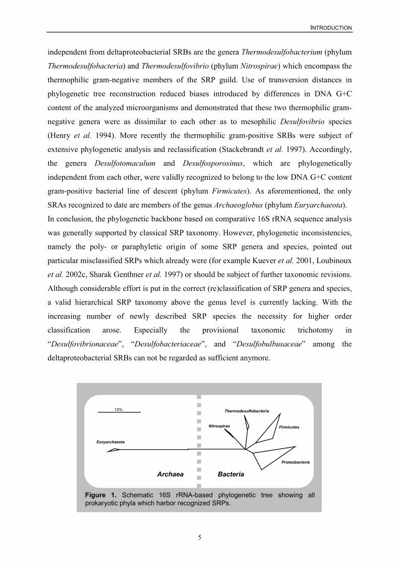
Figure 1. Schematic 16S rRNA-based phylogenetic tree showing allprokaryotic phyla which harbor recognized SRPs.
Euryarchaeota
Firmicutes Nitrospirae
Proteobacteria
Thermodesulfobacteria10%
Archaea Bacteria
INTRODUCTION
6
Although the latest edition of Bergey`s Manual of Systematic Bacteriology already proposed
a substantial taxonomic ranking for all prokaryotes, this outline classification “is a work in
progress” and must await further amendment (Garrity and Holt 2001). Today, 126 sulfate-
reducing species belonging to 35 genera, which can be assigned to four bacterial phyla and
one archaeal phylum (Figure 1), have been validly described (to date 19 september 2002,
http://www.bacterio.cict.fr/) (Euzeby 1997).
II. Habitats and general ecological aspects of SRPs
SRPs constitute an essential biotic component of the global sulfur cycle. As already shown in
the preceding chapter, SRPs form a rather heterogenous group from the viewpoint of modern
rRNA-based taxonomy. However, their general geobiological importance legitimates
grouping in a functional microbial guild. The unifying physiological trait of these
microorganisms is their ability to use sulfate dissimilatively as terminal electron acceptor
coupled with the generation of energy. This unique geobiological process is called
dissimilatory sulfate reduction or anaerobic sulfate respiration and is exclusively restricted to
the prokaryotic domains of life. Dissimilatory sulfate reduction is a very ancient process.
Earliest geological records of microbial sulfate reduction date back in the early Archaean era,
more than ~3.47 billion years ago (Shen et al. 2001).
SRPs are ubiquitous and inhabit mainly anoxic zones but also the oxic/anoxic interface of
various environments. Findings from environmental studies that nonsporeforming SRPs
existed in high numbers in oxic environments (Canfield and Des Marais 1991, Krekeler et al.
1997, Minz et al. 1999a, Ramsing et al. 1993, Teske et al. 1998) and could cope with oxygen
stress came as a surprise, because, for a long time, all SRPs were considered to be obligate
anaerobic microorganisms. Only endospore-forming SRPs of the genus Desulfotomaculum
were thought to survive under long-term oxic conditions (Widdel 1988). Physiological and
biochemical studies on the influence of oxygen on anaerobes revealed that several oxygen-
scavenging mechanisms exist among different SRP species. For instance, some Desulfovibrio
species are able to utilize oxygen directly via periplasmatic reduction. Despite a high
respiration rate and energy coupling, it has been assumed that this process has only a
protective function (Baumgarten et al. 2001, Cypionka 2000). Further protective mechanisms
involve enzymes such as rubredoxin oxidoreductase (desulfoferredoxin) (Lumppio et al.
2001, Silva et al. 2001b) or neelaredoxin (Abreu et al. 2000, Silva et al. 2001a) that catalyze
the removal of toxic superoxide which is formed in the presence of oxygen. In addition to
INTRODUCTION
7
these physiological capacities, some SRPs also show behavioral responses to oxygen
exposure such as flocculation (Sigalevich et al. 2000), simple migration to anoxic regions
(Krekeler et al. 1997, Krekeler et al. 1998), or the formation of bands in oxygen-containing
zones at concentrations of lesser than or equal to 20% air saturation. The latter behavior is
driven by a complex interplay of positive and negative aerotaxis (Eschemann et al. 1999).
Although all these protective mechanisms allow SRPs to survive oxygen stress, substantial
aerobic growth in pure culture has not yet been observed. Thus, SRPs still remain anaerobic
microorganisms but the dogma on their strict dependence on anoxic living conditions had to
be reconsidered.
In terms of microbial abundance and ecoproductivity, anoxic marine environments in general
and sediments in particular represent the most important habitats for SRPs. High sulfate
concentrations in sea water (approximately 28 mM) promote growth and activity of SRPs.
Jørgensen et al. have shown by using the 35SO42- radiotracer method (Sorokin 1972) that up to
50% of the organic carbon in marine sediments is mineralized via dissimilatory sulfate
reduction (Jørgensen 1977, Jørgensen 1982). Because of their profound ecological importance
in these systems, SRPs in marine sediments were subject of many extensive studies
(Devereux and Mundfrom 1994, Knoblauch et al. 1999a, Knoblauch et al. 1999b, Llobet-
Brossa et al. 1998, Ravenschlag et al. 2001). Worth mentioning is the anaerobic oxidation of
methane in marine sediments as it is an extraordinary example for the versatility of the
ecological processes that SRPs are involved in. Anaerobic methane oxidation has been known
for a long time (Reeburgh 1982), but is was only recently demonstrated that microbial
aggregates composed of SRPs and methane-oxidizing archaea catalyze this geobiological
process (Boetius et al. 2000, DeLong 2000, Orphan et al. 2001, Thomsen et al. 2001).
However, besides sediments there is a vast number of other ecological niches in marine
environments that are colonized by SRP.
An example for a highly specialized niche for SRPs is the gutless marine oligochaete Olavius
algarvensis. It harbors sulfate-reducing and sulfide-oxidizing bacterial endosymbionts which
syntrophically catalyze a closed endosymbiotic sulfur cycle in the worm (Dubilier et al.
2001).
Another mutualistic relationship probably gives rise to the high numbers and activities of
SRPs associated with marine macrophytes (Hines et al. 1999, Küsel et al. 1999, Rooney-
Varga et al. 1997). SRPs inhabit the rhizosphere of marsh and sea grasses and it is
hypothesized that they profit from the dissolved organic carbon exuded from the roots in
exchange for fixed nitrogen (Nielsen et al. 2001).
INTRODUCTION
8
Well-studied habitats that encompass a variety of phylogenetically diverse SRP groups are
cyanobacterial microbial mats (Minz et al. 1999a, Risatti et al. 1994, Teske et al. 1998). The
distinct SRP groups are mostly distributed in nonoverlapping depth intervals of the mat what
led to the suggestion that they are responsible for specific interrelated metabolic functions in
the community (Risatti et al. 1994).
In contrast to marine sediments, the main carbon mineralization process in freshwater
sediments is methanogenesis. However, dissimilatory sulfate reduction, carried out by a
diverse assemblage of SRPs (Li et al. 1999, Sass et al. 1998), may contribute with more than
20% to the total anaerobic mineralization (Ingvorson and Brock 1982). Further freshwater
habitats where occurrence of SRPs has been demonstrated are waterlogged rice soils
(Ouattara et al. 1999, Scheid and Stubner 2001, Stubner and Meuser 2000, Wind and Conrad
1995, Wind et al. 1999), groundwater from aquifers (Lehman et al. 2001), and wastewater
treatment systems (De Smul and Verstraete 1999, Ito et al. 2002a, Ito et al. 2002b, Lens et al.
1995, Manz et al. 1998, Oude Elferink et al. 1998, Ramsing et al. 1993, Schramm et al.
1999), to name only a few.
The detection of SRPs in anthropogenically or naturally contaminated habitats (Anderson and
Lovley 2000, Leu et al. 1998, Robertson et al. 2001, Voordouw et al. 1991) has attracted
economic interests to SRPs. Some specialized SRPs have the metabolic capacity to degrade
environmental pollutants such as oil (Harms et al. 1999, Rabus et al. 1996¸ Annweiler, 2001
#1592) and thus, these SRPs are promising candidates for the use in large-scale
bioremediation attempts. In contrast to these beneficial aspects, there are cases where high
metabolic activity of SRPs is undesired from an economical point of view. Such a negative
aspect is that SRPs, among other microorganisms, are the driving force for microbiologically
influenced metal corrosion. SRP growth is responsible for significant modifications of many
physicochemical parameters at metal surfaces, including local changes in pH and redox
potential values, variations in anion and cation concentrations and alteration of the
composition and structure of corrosion products (Javaherdashti 1999, Videla 2000). This
harmful activity of SRPs causes considerable economical damage to e.g. oil pipelines or other
man-made metal constructions (Rao et al. 2000). Therefore, mechanisms to control or
suppress colonization of such environments by SRPs are needed (Billman 1997, Jayaraman et
al. 1999).
Besides their ecological and economical importance, SRPs also attracted attention as potential
opportunistic pathogens in connection with human diseases such as periodontitis (Langendijk
et al. 1999, Langendijk et al. 2000, Langendijk et al. 2001, Loubinoux et al. 2002a), pyogenic
INTRODUCTION
9
liver abscesses (Schoenborn et al. 2001, Tee et al. 1996), inflammatory bowel diseases
(Loubinoux et al. 2002b) or bacteremia (Loubinoux et al. 2000, McDougall et al. 1997).
III. Current molecular approaches to determine SRP community structure
Traditional approaches attempted to identify and quantify microbial biodiversity by means of
cultivation. Especially for the isolation (for review see Widdel and Bak 1992) and
enumeration (Vester and Ingvorsen 1998) of anaerobic SRPs, sophisticated media and
culturing techniques were designed in order to mimic in situ growth conditions as perfectly as
possible in vitro. However, the introduction of cultivation-independent molecular, primarily
nucleic acid-based techniques in microbial ecology research led to the recognition that these
cultivation approaches underlie significant quantitative (Staley and Konopka 1985) and
qualitative biases (Wagner et al. 1993). Although molecular methods are generally not free
from methodological errors (Martin-Laurent et al. 2001, Polz and Cavanaugh 1998,
Speksnijder et al. 2001, Suzuki and Giovannoni 1996, von Wintzingerode et al. 1997), their
vast application in SRP diversity research provided the basis for our today’s view on the
natural distribution and ecophysiological function of these microorganisms in the
environment. The individual molecular methods which found widespread application in
studies on natural SRP communities are summarized in the next paragraph. Because benefits
and potential pitfalls of these methods have already been reviewed in detail elsewhere
(Amann et al. 1995, Friedrich 2002, Klein et al. 2001, Muyzer et al. 1998, Muyzer et al.
1996, Voordouw 1998, Wagner et al. 1998), only general technical aspects in the context of
SRP diversity research are presented.
Comparative sequence analysis
Comparative sequence analysis of rRNA (genes) following nucleic acid extraction from an
environmental sample, the use of universal, bacterial or archaeal primers for PCR
amplification of rRNA genes, and the setup of a rRNA gene library, has become the prime
tool for molecular microbial ecologists to assess prokaryotic species richness independent
from cultivation (Amann et al. 1995, Pace et al. 1986, Stackebrandt and Rainey 1995). Its
numerous application in all kinds of habitats has dramatically improved our knowledge on the
phylogenetic extent of microbial life in general (for example see Barns et al. 1996, Eder et al.
1999, Hugenholtz et al. 1998, Kuske et al. 1997, Ludwig et al. 1997). Nevertheless, the
INTRODUCTION
10
application of this general rRNA gene library approach is limited in environments with a high
prokaryotic diversity like sediments and soils (Torsvik et al. 2002) because hundreds of rRNA
gene sequences must be sequenced to cover the whole microbial richness. A more focussed
view on the diversity of certain microorganisms is possible by using primers that target
phylogenetic groups at higher levels of specificity. A first application of this technique for
SRPs has revealed unique environmental 16S rRNA sequences in a sandy marine sediment
(Devereux and Mundfrom 1994). However, in contrast to phylogenetically and functionally
homogeneous bacterial groups such as the betaproteobacterial ammonium oxidizers (Purkhold
et al. 2000), the polyphyletic origin of SRPs (i) does not allow the design of a single 16S
rRNA-targeted primer pair that is specific for all SRPs and (ii) complicates the unambiguous
assignment of environmentally derived 16S rRNA sequences to this microbial guild.
16S rRNA aside, comparative amino acid sequence analyses of key enzymes of the
dissimilatory sulfate reduction pathway, such as the siroheme dissimilatory (bi)sulfite
reductase (EC 1.8.99.3) or the adenosine-5`-phosphosulfate reductase (EC 1.8.99.2), provide
a bypass for this drawback of rRNA-based approaches. The genetic capacity of sulfate
reduction can be directly deduced from these so-called “functional” phylogenetic marker
molecules. The remarkable sequence conservation of the genes encoding dissimilatory
(bi)sulfite (DsrAB) and adenosine-5`-phosphosulfate reductases (ApsA) (Hipp et al. 1997,
Karkhoff-Schweizer et al. 1995) allowed the design of degenerated PCR primers for their
detection in the environment (Friedrich 2002, Klein et al. 2001, Wagner et al. 1998).
Analogous to the rRNA approach, dsrAB or apsA gene sequences can be PCR-amplified from
environmental DNA, singularized by cloning, and phylogenetically classified by comparative
analyses of nucleic acid and/or deduced amino acid sequences. One downside of the dsrAB
and the apsA approach is that these genes were subject of several lateral transfer events as
indicated by partly inconsistent phylogenetic tree topologies of SRP pure cultures inferred
from 16S rRNA, DsrAB, and ApsA sequence analyses (Friedrich 2002, Klein et al. 2001,
Stahl et al. 2002). This fact hampers exact identification of environmental sequences that are
not closely related to known SRP lineages. Nevertheless, it has been proven for various
environments that it is possible to reveal the presence of heretofore uncultured SRPs beyond
the recognized lineages via dsrAB (Castro et al. 2002, Chang et al. 2001, Cottrell and Cary
1999, Dubilier et al. 2001, Joulian et al. 2001, Minz et al. 1999b, Nakagawa et al. 2002,
Schramm et al. 1999, Thomsen et al. 2001) or apsA gene-based molecular metabolic diversity
surveys (Deplancke et al. 2000).
INTRODUCTION
11
Denaturing gradient gel electrophoresis
A very common PCR-based method, which was introduced in microbial ecology by Muyzer
et al. (1993) and since then was often applied in SRP diversity research, is denaturing
gradient gel electrophoresis (DGGE). The basic principle of this nucleic acid fingerprinting
technique is the analytical resolution of PCR-amplified DNA fragments identical in length but
different in sequence composition. Separation in DGGE is based on decreased electrophoretic
mobility of a partially melted DNA fragment in polyacrylamide gels containing a linearly
increasing gradient of DNA denaturants (for methodological details see Muyzer et al. 1998,
Muyzer et al. 1996). The separated DNA bands can be identified either by comparative
sequence analysis following extraction of the bands from the gel, reamplification by PCR, and
cloning, or by hybridization with nucleic acid probes following blotting of bands on nylon
membranes. The greatest advantage of DGGE is that the genetic diversity of many samples
can be rapidly analyzed in parallel by a single gel run. However, besides general biases that
underlie all analytical methods based on nucleic acid extraction and PCR-amplification
(Bonnet et al. 2002, Martin-Laurent et al. 2001, Polz and Cavanaugh 1998, Speksnijder et al.
2001, Suzuki and Giovannoni 1996, von Wintzingerode et al. 1997), a DGGE-specific caveat
is that only short PCR fragments of up to 500 bp can be well separated which limits
phylogenetic information retrieved after sequencing of the individual bands. Furthermore, it
has been shown that amplification of identical sequences by using degenerated primers leads
to multiple bands in DGGE, which are solely caused by differences in the primer sequence
(Nicolaisen and Ramsing 2002). This potential bias can be avoided if non-degenerated
primers are used for DGGE.
Analyses of complex SRP communities by DGGE mainly used 16S rRNA genes as target
molecules (Kleikemper et al. 2002, Koizumi et al. 2002, Santegoeds et al. 1998, Teske et al.
1998, Teske et al. 1996). Nevertheless, as aforementioned it strongly depends on the
specificity of the rRNA gene-targeted PCR primer pair whether the obtained DGGE
fingerprints can be directly linked to SRPs. For a subgroup of SRPs, an alternative to 16S
rRNA (gene) as target molecule for DGGE is the gene encoding the large subunit of [NiFe]
hydrogenase (Wawer and Muyzer 1995), an enzyme which plays an important role in the
hydrogen metabolism of Desulfovibrio species. The [NiFe] hydrogenase gene is conserved
among all Desulfovibrio spp. investigated so far, making it an ideal target for their PCR-based
detection (Voordouw et al. 1990). The determination of diversity and expression of this
INTRODUCTION
12
functional gene by DGGE in anaerobic bioreactors allowed to differentiate active from
dormant members in natural assemblages of Desulfovibrio spp. (Wawer et al. 1997).
Hybridization with rRNA-targeted oligonucleotide probes
Integral part of the experimental setup of most ecological studies focussing on identification
and abundance of SRPs in their natural habitats, is the application of rRNA-targeted
oligonucleotide probes (for review on rRNA-targeted probes in general see Amann and Kühl
1998, Amann and Ludwig 2000, Amann and Schleifer 2001, Amann 1995, Amann et al.
1995, DeLong et al. 1989, Giovannoni et al. 1988, Stahl and Amann 1991). Today, a whole
suite of empirically pretested probes targeting different taxonomic SRP groups is available for
straightforward use in different hybridization formats (Daly et al. 2000, Devereux et al. 1992,
Hristova et al. 2000, Manz et al. 1998). Thus, the composition of the SRP community can be
analyzed with increasing taxonomic resolution if probes of hierarchical specificity are
hybridized either in parallel with the same sample or separately with parallel subsamples,
depending on the hybridization format chosen. Hybridization of whole cells and hybridization
of extracted nucleic acids on a membrane are the two basic formats to use rRNA-targeted
oligonucleotide probes for quantification.
Fluorescence in situ hybridization (FISH). The essence of FISH is that, using fluorescently
tagged probes, it allows the specific visualization of morphologically intact organisms (hence,
FISH is also referred to as whole-cell hybridization) directly in their natural environment. The
simultaneous hybridization with three nested probes, each probe labeled with a different
fluorophore, permits differentiation of up to seven distinct microbial populations within a
single sample (Amann et al. 1996). Besides identification and spatial localization of
microorganisms, absolute and/or relative numbers of visualized cells can be determined either
by tedious manual counting (Glöckner et al. 1999) or semi-automatic quantification by using
sophisticated digital image analyses (Bouchez et al. 2000, Daims et al. 2001, Juretschko et al.
2002, Schmid et al. 2000). Furthermore, FISH has the great potential that it can be directly
combined with techniques such as microsensors (Ramsing et al. 1993) or
microautoradiography (Ito et al. 2002a) to elucidate the ecophysiology of identified SRPs.
First application of FISH for SRP community analysis identified Desulfovibrio vulgaris-
related bacteria in sulfidogenic biofilms established in anaerobic bioreactors (Amann et al.
1992). Further FISH studies monitored abundance and spatial organization of single
deltaproteobacterial SRB populations in activated sludge (Manz et al. 1998), investigated
INTRODUCTION
13
response of SRPs to oxygen stress under oligotrophic conditions in particle-free systems
(Bade et al. 2000), or revealed the spatial structure of a consortium of Archaea and SRBs
responsible for anaerobic methane oxidation (Boetius et al. 2000).
Quantitative membrane hybridization. In the dot-/slot-blot hybridization format,
environmentally retrieved total rRNA is immobilized on a membrane by using a blotting
apparatus with round (dot) or longitudinal (slot) cavities and subsequently hybridized with
radioactively labeled phylogenetic probes. The amount of rRNA of a certain microbial
population measured by a specific probe (as counts per minute) is normalized against the
amount of rRNA measured by a probe of broader specificity e.g. a universal probe (Raskin et
al. 1994, Stahl et al. 1988). However, this information on relative rRNA abundance can not
be directly extrapolated into total cell numbers because the amount of rRNA per cell may
vary drastically according to the physiological state of the cell (Kemp et al. 1993). This
particular drawback of the membrane format can be partially overcome if total DNA is
immobilized and the number of rRNA genes is quantified (Edgcomb et al. 1999). The number
of target cells in a sample can be estimated by comparing the amount of a specific probe
hybridized to extracted DNA to that obtained with a standard curve of genomic DNA for
reference strains included on the same membrane. Nevertheless, one has to bear in mind that
different species might have different genome and/or rRNA operon copy numbers (Fegatella
et al. 1998, Klappenbach et al. 2000) leading to errors that can be greater than severalfold.
In practice, quantitative dot-/slot-blot hybridization has been preferentially applied in classical
SRP environments such as cyanobacterial mats (Minz et al. 1999a, Risatti et al. 1994) or
sediments (Hines et al. 1999, Li et al. 1999, Ravenschlag et al. 2001, Ravenschlag et al. 2000,
Rooney-Varga et al. 1997, Sahm et al. 1999a, Sahm et al. 1999b), because high background
fluorescence of theses habitats hampers application of FISH techniques.
DNA microarray technology. In a ground-breaking study by Guschin et al. (1997), a new
hybridization format for rRNA-targeted oligonucleotides, the DNA microarray, was
introduced to microbial ecology research. Only recently, this novel technology has been
applied in an proof-of-principal attempt to directly detect rRNA from SRPs in soils (Small et
al. 2001) and in an anaerobic toluene- and ethylbenzene-degrading enrichment (Koizumi et
al. 2002). Further details on DNA microarrays and their application for determinative
microbiological studies are presented in a separate chapter below.
INTRODUCTION
14
Reverse sample genome probing
Another DNA hybridization method that was initially applied for identification of SRPs in oil
field samples is reverse sample genome probing (RSGP) (Voordouw et al. 1993, Voordouw et
al. 1992, Voordouw et al. 1991). In this approach total genomic DNA from cultured reference
strains is denatured and immobilized on a membrane together with an internal control
(concentration series of bacteriophage lambda DNA). The genomes of the different reference
strains on the membrane (the so-called master filter) should generally cross-hybridize less
than 1%. Total environmental DNA is mixed with a standard amount of lambda DNA,
radioactively labeled by random prime labeling with [��32]P, and hybridized under stringent
conditions to the master filter. After hybridization and washing, the amount of bound sample
DNA is quantified. Subsequently, the fraction of environmental DNA composed of individual
component DNA is calculated from the hybridization to the individual genomic pure culture
standards relative to the lambda reference series (Voordouw et al. 1993).
The main advantage of this whole-genome probe array technique is that it provides
information on complex microbial communities in a single assay. Although the actual RSGP
does not require culturing, the most criticized aspect of this technique is that composition of
the microbial community is displayed in terms of its culturable component (Voordouw 1998).
IV. DNA microarrays for determinative studies in microbiology
The DNA microarray (microchip) technology allows parallel analysis of many genes in a
single assay and thus emblematize, as no other methodological means, the (post-) genomic era
of “big science”. Very simply described, DNA microchips consist of up to thousands of
diagnostic nucleic acid sequences (referred to as probes) tethered to a miniaturized solid
support (usually a glass slide) in an arrayed order. Probes can be either oligonucleotides or
PCR amplificates (usually cDNA). The identity (and amount) of labeled nucleic acid
sequences that are subject of analysis (referred to as target) is revealed after hybridization to
the microarray.
Initially microarrays containing probes for every single gene of an organism were developed
to analyze gene expression on a genomic scale (Schena et al. 1995). Since then, these
genomic microarrays have been widely applied in microbiological research to reveal genes
involved in response to stress and environmental change, cellular response to bacterial
infection, the cell-cycle, and dissection of regulatory circuitry. Moreover, it is possible to
INTRODUCTION
15
compare different strains (genomotyping) or to identify potential drug target sites by
microarray hybridization (for a summary of microbiological DNA microarray studies see
Lucchini et al. 2001).
Beyond this genomic level-oriented research, DNA microarrays hold much potential for
determinative studies in environmental and clinical microbiology (Cho and Tiedje 2001, Cho
and Tiedje 2002, Guschin et al. 1997, Kingsley et al. 2002, Koizumi et al. 2002, Liu et al.
2001, Reyes-Lopez et al. 2003, Small et al. 2001, Urakawa et al. 2002, Volokhov et al. 2002,
Wilson et al. 2002, Wu et al. 2001, Zhou and Thompson 2002). The scope of such studies
was often restricted by conventional experimental formats such as FISH or dot-/slot-blot
hybridization, which strongly limit the number of probes that can be applied and the number
of samples that can be analyzed. The invention of DNA microarrays provided the basis for a
hybridization format that allows greater sample throughput and highly parallel detection of
complex microbial communities. Target nucleic acids for DNA microarray probes are
basically the same as in conventional hybridization assays used for microbial identification:
namely the small- and/or large-subunit rRNA genes or functional respectively virulence genes
that are diagnostic for certain microbial groups.
Ribosomal RNA-based oligonucleotide microarrays developed so far can be divided in two
categories according to the strategy of the underlying probe design. One strategy, namely the
so-called “multiple probe concept” (Amann and Schleifer 2001, Behr et al. 2000), takes
advantage of the fact that rRNA genes consist of highly conserved and variable sequence
regions. Thus, it is possible to design multiple oligonucleotide probes to detect target groups
at different (or same) phylogenetic levels by targeting rRNA regions of different (or same)
sequence conservation. Consequently, the simultaneous application of a whole set of nested
and parallel probes enhances the reliability of the detection of target organisms. The “multiple
probe concept” proved to be fruitful for the application of rRNA-targeted oligonucleotide
probes for e.g. whole cell (Amann et al. 1996, Juretschko et al. 2002), membrane (Raskin et
al. 1994), or micro well plate hybridization (Behr et al. 2000) but can probably be utilized
most excessively for DNA microarray hybridization (Liu et al. 2001), owing to the enormous
number of potentially applicable probes. A different probe design strategy was the basis for a
high-density microarray of small-subunit rRNA-targeted oligonucleotides developed by
Wilson et al. (2002). Based on a subalignment of the small-subunit rRNA database (version
5.0) of the Ribosomal Database Project (RDP) almost all possible 20-mer probes for every
single sequence in the database were designed, resulting in a total of 31179 oligonucleotides
on the DNA microarray. After hybridization of a given environmental sample with the DNA
INTRODUCTION
16
microarray, an RDP 16S rRNA sequence was regarded to be present if 22 or more of at least
24 probes specifically targeting this sequence were scored positive. This probe design strategy
is totally devoted to high parallelism, the main feature of DNA microarrays, but it ignores the
potential to specifically design rRNA-targeted probes for phylogenetically defined target
groups.
If diagnostic DNA microarrays target functional genes (Wu et al. 2001) or virulence genes
(Volokhov et al. 2002) a certain physiological property or pathogenic potential can be directly
inferred upon identification as already pointed out in the preceding chapters. In comparison to
rRNA-based microarrays, probe design for functional gene arrays has some general
disadvantages. One is that sequence databases, the basis for adequate probe design, for
functional genes of microbial groups of interest are currently still not as comprehensive as the
respective small-subunit rRNA databases. Furthermore, development of nested
oligonucleotide probes according to the “multiple probe concept” is more difficult due to the
highly variable, third codon (wobble) position.
Another DNA microarray variant for detection and differentiation of microorganisms consists
of random oligonucleotides that allow fingerprinting of microbial strains as shown for closely
related Xanthomonas pathovars (Kingsley et al. 2002). Furthermore, random genome
fragments can be immobilized on microarrays and used analogous to traditional whole
genomic DNA-DNA hybridization for bacterial species determination (Cho and Tiedje 2001).
Although potential fields of application of diagnostic DNA microarrays are numerous, most
microarrays developed so far for microbial identification were mainly used for method
development and optimization. Moreover, with a single exception (Wilson et al. 2002) these
microarrays consisted of a limited number of probes, not making use of the advantageous
capacity of DNA microarrays for highly parallel identification. Consequently, diagnostic
DNA microarrays are not yet routinely implemented in environmental and medical research.
V. Scope of this thesis
The initial part of this thesis should focus on the collection and alignment of all 16S rRNA
sequences from isolated and yet uncultured SRPs available in public databases. Subsequently,
a thorough reevaluation of natural relationships among SRPs based on comparative 16S
rRNA sequence analysis should provide (i) a robust phylogenetic and taxonomic framework
for the assignment of environmentally retrieved SRP sequences and (ii) the basis for the
design of an encompassing oligonucleotide probe set that target SRPs at multiple hierarchical
INTRODUCTION
17
and parallel levels of specificity and is suitable for application on diagnostic DNA
microarrays. After in silico development of a comprehensive 16S rRNA-based
oligonucleotide microarray for identification of SRPs (SRP-PhyloChip), specificity and
sensitivity of this hybridization technique should be evaluated and optimized with suitable
SRP pure cultures. Besides methodological development, a main aspect of this thesis will
focus on the potential applicability of the developed microarray for routine detection of SRPs
in environmental and medical samples. Therefore, results obtained by microarray
hybridization in environmental or clinical studies should be confirmed by well-established
molecular methods for SRP identification based on comparative sequence analyses of 16S
rRNA genes and dissimilatory (bi)sulfite reductase genes (dsrAB).
INTRODUCTION
18
VI. References
Abreu IA, Saraiva LM, Carita J, Huber H,Stetter KO, Cabelli D, and Teixeira M [2000]Oxygen detoxification in the strict anaerobicarchaeon Archaeoglobus fulgidus: superoxidescavenging by neelaredoxin. Mol Microbiol. 38:322-334.
Achenbach-Richter L, Stetter KO, and WoeseCR [1987] A possible biochemical missing linkamong archaebacteria. Nature. 327: 348–349.
Amann R, and Kühl M [1998] In situ methods forassessment of microorganisms and their activities.Curr Opin Microbiol. 1: 352-358.
Amann R, and Ludwig W [2000] RibosomalRNA-targeted nucleic acid probes for studies inmicrobial ecology. FEMS Microbiol Rev. 24: 555-565.
Amann R, and Schleifer K-H [2001] Nucleic acidprobes and their application in environmentalmicrobiology, p. 67-82. In Garrity GM (ed.),Bergey's manual of systematic bacteriology, 2nd ed,vol. 1. Springer, New York.
Amann R, Snaidr J, Wagner M, Ludwig W, andSchleifer KH [1996] In situ visualization of highgenetic diversity in a natural microbial community.J Bacteriol. 178: 3496-3500.
Amann RI [1995] In situ identification of micro-organisms by whole cell hybridization with rRNA-targeted nucleic acid probes, p. 1-15. In AkkermansADL, van Elsas JD and de Bruijn FJ (ed.),Molecular Microbial Ecology Manual, vol. 3.3.6.Kluwer Academic Publishers, Dortrecht.
Amann RI, Ludwig W, and Schleifer K-H [1995]Phylogenetic identification and in situ detection ofindividual microbial cells without cultivation.Microbiol Rev. 59: 143-169.
Amann RI, Stromley J, Devereux R, Key R, andStahl DA [1992] Molecular and microscopicidentification of sulfate-reducing bacteria inmultispecies biofilms. Appl Environ Microbiol. 58:614-623.
Anderson RT, and Lovley DR [2000] Anaerobicbioremediation of benzene under sulfate-reducingconditions in a petroleum-contaminated aquifer.Environ Science Techn. 34: 2261-2266.
Baars JK [1930] PhD thesis. University of Delft,Delft.
Bade K, Manz W, and Szewzyk U [2000]Behaviour of sulfate-reducing bacteria underoligotrophic conditions and oxygen stress inparticle-free systems related to drinking water.FEMS Microbiol Ecol. 32: 215-223.
Bak F, and Widdel F [1986a] Anaerobicdegradation of indolic compounds by sulfate-reducing enrichment cultures, and description ofDesulfobacterium indolicum gen. nov, sp. nov.Arch Microbiol. 146: 170–176.
Bak F, and Widdel F [1986b] Anaerobicdegradation of phenol and phenol derivatives byDesulfobacterium phenolicum sp. nov. ArchMicrobiol. 146: 177–180.
Barns SM, Delwiche CF, Palmer JD, and PaceNR [1996] Perspectives on archaeal diversity,thermophily and monophyly from environmentalrRNA sequences. Proc Natl Acad Sci USA. 93:9188-9193.
Baumgarten A, Redenius I, Kranczoch J, andCypionka H [2001] Periplasmic oxygen reductionby Desulfovibrio species. Arch Microbiol. 176:306-309.
Behr T, Koob C, Schedl M, Mehlen A, Meier H,Knopp D, Frahm E, Obst U, Schleifer K,Niessner R, and Ludwig W [2000] A nested arrayof rRNA targeted probes for the detection andidentification of enterococci by reversehybridization. Syst Appl Microbiol. 23: 563-572.
Beijerinck WM [1895] Über Spirillumdesulfuricans als Ursache von Sulfatreduktion.Zentralb Bakteriol II Abt. 1: 49–59, 104–114.
Billman JA [1997] Antibiofoulants: a practicalmethodology for control of corrosion caused bysulfate-reducing bacteria. Materials Performance.36: 43-48.
Boetius A, Ravenschlag K, Schubert CJ, RickertD, Widdel F, Gieseke A, Amann R, JorgensenBB, Witte U, and Pfannkuche O [2000] A marinemicrobial consortium apparently mediatinganaerobic oxidation of methane. Nature. 407: 623-626.
Bonnet R, Suau A, Dore J, Gibson GR, andCollins MD [2002] Differences in rDNA librariesof faecal bacteria derived from 10- and 25-cyclePCRs. Int J Syst Evol Microbiol. 52: 757-763.
Bouchez T, Patureau D, Dabert P, Juretschko S,Doré J, Delgenès P, Moletta R, and Wagner M
INTRODUCTION
19
[2000] Ecological study of a bioaugmentationfailure. Environ Microbiol. 2: 179-190.
Brysch K, Schneider C, Fuchs G, and Widdel F[1987] Lithoautotrophic growth of sulfate-reducingbacteria, and description of Desulfobacteriumautotrophicum gen. nov., sp. nov. Arch Microbiol.148: 264–274.
Burggraf S, Jannasch HW, Nicolaus B, andStetter KO [1990] Archeoglobus profundus sp.nov., represents a new species within the sulfate-reducing archaebacteria. System Appl Microbiol.13: 24-28.
Campbell LL, Frank HA, and Hall ER [1957]Studies on the thermophilic sulfate-reducingbacteria. I. Identification of Sporovibriodesulfuricans as Clostridium nigrificans. JBacteriol. 73: 516–521.
Campbell LL, and Postgate JR [1965]Classification of the spore-forming sulfate-reducingbacteria. Bacteriol Rev. 29: 359–363.
Canfield DE, and Des Marais DJ [1991] Aerobicsulfate reduction in microbial mats. Science. 251:1471-1473.
Castro H, Reddy KR, and Ogram A [2002]Composition and function of sulfate-reducingprokaryotes in eutrophic and pristine areas of theFlorida Everglades. Appl Environ Microbiol. 68:6129-6137.
Chang YJ, Peacock AD, Long PE, Stephen JR,McKinley JP, Macnaughton SJ, Hussain AK,Saxton AM, and White DC [2001] Diversity andcharacterization of sulfate-reducing bacteria ingroundwater at a uranium mill tailings site. ApplEnviron Microbiol. 67: 3149-3160.
Cho J-C, and Tiedje JM [2001] Bacterial speciesdetermination from DNA-DNA hybridization byusing genome fragments and DNA microarrays.Appl Environ Microbiol. 67: 3677-3682.
Cho J-C, and Tiedje JM [2002] Quantitativedetection of microbial genes by using DNAmicroarrays. Appl Environ Microbiol. 68: 1425-1430.
Cottrell MT, and Cary SC [1999] Diversity ofdissimilatory bisulfite reductase genes of bacteriaassociated with the deep-sea hydrothermal ventpolychaete annelid Alvinella pompejana. ApplEnviron Microbiol. 65: 1127-1132.
Cypionka H [2000] Oxygen respiration byDesulfovibrio species. Annu Rev Microbiol. 54:827-848.
Daims H, Ramsing NB, Schleifer KH, andWagner M [2001] Cultivation-independent,semiautomatic determination of absolute bacterialcell numbers in environmental samples byfluorescence in situ hybridization. Appl EnvironMicrobiol. 67: 5810-5818.
Daly K, Sharp RJ, and McCarthy AJ [2000]Development of oligonucleotide probes and PCRprimers for detecting phylogenetic subgroups ofsulfate-reducing bacteria. Microbiology. 146: 1693-1705.
De Smul A, and Verstraete W [1999] Retention ofsulfate-reducing bacteria in expanded granular-sludge-blanket reactors. Water Environ Res. 71:427-431.
DeLong EF [2000] Resolving a methane mystery.Nature. 407: 577-579.
DeLong EF, Wickham GS, and Pace NR [1989]Phylogenetic stains: ribosomal RNA-based probesfor the identification of single cells. Science. 243:1360-1363.
Deplancke B, Hristova KR, Oakley HA,McCracken VJ, Aminov R, Mackie RI, andGaskins HR [2000] Molecular ecological analysisof the succession and diversity of sulfate-reducingbacteria in the mouse gastrointestinal tract. ApplEnviron Microbiol. 66: 2166-2174.
Devereux R, Delaney M, Widdel F, and StahlDA [1989] Natural relationships among sulfate-reducing eubacteria. J Bacteriol. 171: 6689-6695.
Devereux R, He SH, Doyle CL, Orkland S, StahlDA, LeGall J, and Whitman WB [1990] Diversityand origin of Desulfovibrio species: phylogeneticdefinition of a family. J Bacteriol. 172: 3609-3619.
Devereux R, Kane MD, Winfrey J, and Stahl DA[1992] Genus- and group-specific hybridizationprobes for determinative and environmental studiesof sulfate-reducing bacteria. Syst Appl Microbiol.15: 601-609.
Devereux R, and Mundfrom GW [1994] Aphylogenetic tree of 16S rRNA sequences fromsulfate-reducing bacteria in a sandy marinesediment. Appl Environ Microbiol. 60: 3437-3439.
Devereux R, and Stahl DA [1993] Phylogeny ofsulfate-reducing bacteria and a perspective foranalyzing their natural communities, p. 131-160. InOdom JM and Singleton R (ed.), The sulfate-reducing bacteria: contemporary perspectives.Springer-Verlag, New York.
Dubilier N, Mulders C, Ferdelman T, de Beer D,Pernthaler A, Klein M, Wagner M, Erseus C,
INTRODUCTION
20
Thiermann F, Krieger J, Giere O, and Amann R[2001] Endosymbiotic sulphate-reducing andsulphide-oxidizing bacteria in an oligochaete worm.Nature. 411: 298-302.
Eder W, Ludwig W, and Huber R [1999] Novel16S rRNA gene sequences retrieved from highlysaline brine sediments of kebrit deep, red Sea. ArchMicrobiol. 172: 213-218.
Edgcomb VP, McDonald JH, Devereux R, andSmith DW [1999] Estimation of bacterial cellnumbers in humic acid-rich salt marsh sedimentswith probes directed to 16S ribosomal DNA. ApplEnviron Microbiol. 65: 1516-1523.
Elion L [1925] A thermophilic sulphate-reducingbacterium. Zentralb Bakteriol II Abt. 63: 58–67.
Eschemann A, Kühl M, and Cypionka H [1999]Aerotaxis in Desulfovibrio. Environ Microbiol. 1:489-494.
Euzeby JP [1997] List of Bacterial Names withStanding in Nomenclature: a folder available on theInternet. Int J Syst Bacteriol. 47: 590-592.
Fegatella F, Lim J, Kjelleberg S, and CavicchioliR [1998] Implications of rRNA operon copynumber and ribosome content in the marineoligotrophic ultramicrobacterium Sphingomonas sp.strain RB2256. Appl Environ Microbiol. 64: 4433-4438.
Fowler VJ, Widdel F, Pfennig N, and Woese CR[1986] Phylogenetic relationships of sulfate- andsulfur-reducing eubacteria. Syst Appl Microbiol. 8:32–41.
Fox GE, Stackebrandt E, Hespell RB, Gibson J,Maniloff J, Dyer TA, Wolfe RS, Balch WE,Tanner RS, Magrum LJ, Zablen LB, BlakemoreR, Gupta R, Bonen L, Lewis BJ, Stahl DA,Luehrsen KR, Chen KN, and Woese CR [1980]The phylogeny of prokaryotes. Science. 209: 457-463.
Friedrich MW [2002] Phylogenetic analysisreveals multiple lateral transfers of adenosine-5'-phosphosulfate reductase genes among sulfate-reducing microorganisms. J Bacteriol. 184: 278-289.
Garrity GM, and Holt JG [2001] The road map tothe manual, p. 119-166. In Garrity GM (ed.),Bergey's manual of systematic bacteriology, 2nd ed,vol. 1. Springer, New York.
Giovannoni SJ, DeLong EF, Olsen GJ, and PaceNR [1988] Phylogenetic group-specificoligodeoxynucleotide probes for identification ofsingle microbial cells. J Bacteriol. 170: 720-726.
Glöckner FO, Fuchs BM, and Amann R [1999]Bacterioplankton compositions of lakes and oceans:a first comparison based on fluorescence in situhybridization. Appl Environ Microbiol. 65: 3721-3726.
Guschin DY, Mobarry BK, Proudnikov D, StahlDA, Rittmann BE, and Mirzabekov AD [1997]Oligonucleotide microchips as genosensors fordeterminative and environmental studies inmicrobiology. Appl Environ Microbiol. 63: 2397-2402.
Harms G, Zengler K, Rabus R, Aeckersberg F,Minz D, Rossello-Mora R, and Widdel F [1999]Anaerobic oxidation of o-xylene, m-xylene, andhomologous alkylbenzenes by new types of sulfate-reducing bacteria. Appl Environ Microbiol. 65:999-1004.
Henry EA, Devereux R, Maki JS, Gilmour CC,Woese CR, Mandelco L, Schauder R, RemsenCC, and Mitchell R [1994] Characterization of anew thermophilic sulfate-reducing bacteriumThermodesulfovibrio yellowstonii, gen. nov. and sp.nov.: its phylogenetic relationship toThermodesulfobacterium commune and theirorigins deep within the bacterial domain. ArchMicrobiol. 161: 62-69.
Hines ME, Evans RS, Sharak Genthner BR,Willis SG, Friedman S, Rooney-Varga JN, andDevereux R [1999] Molecular phylogenetic andbiogeochemical studies of sulfate-reducing bacteriain the rhizosphere of Spartina alterniflora. ApplEnviron Microbiol. 65: 2209-2216.
Hipp WM, Pott AS, Thum-Schmitz N, Faath I,Dahl C, and Truper HG [1997] Towards thephylogeny of APS reductases and sirohaem sulfitereductases in sulfate-reducing and sulfur-oxidizingprokaryotes. Microbiology. 143: 2891-2902.
Hristova KR, Mau M, Zheng D, Aminov RI,Mackie RI, Gaskins HR, and Raskin L [2000]Desulfotomaculum genus- and subgenus-specific16S rRNA hybridization probes for environmentalstudies. Environ Microbiol. 2: 143-159.
Hugenholtz P, Goebel BM, and Pace NR [1998]Impact of culture-independent studies on theemerging phylogenetic view of bacterial diversity. JBacteriol. 180: 4765-4774.
Ingvorson K, and Brock TD [1982] Electron flowvia sulfate reduction and methanogenesis in theanaerobic hypolimnion of Lake Mendota. LimnolOceanogr. 27: 559-564.
Ito T, Nielsen JL, Okabe S, Watanabe Y, andNielsen PH [2002a] Phylogenetic identification and
INTRODUCTION
21
substrate uptake patterns of sulfate-reducingbacteria inhabiting an oxic-anoxic sewer biofilmdetermined by combining microautoradiographyand fluorescent in situ hybridization. Appl EnvironMicrobiol. 68: 356-364.
Ito T, Okabe S, Satoh H, and Watanabe Y[2002b] Successional development of sulfate-reducing bacterial populations and their activities ina wastewater biofilm growing undermicroaerophilic conditions. Appl EnvironMicrobiol. 68: 1392-1402.
Javaherdashti R [1999] A review of somecharacteristics of MIC caused by sulfate-reducingbacteria: Past, present and future. Anti-CorrosionMethods and Materials. 46: 173-180.
Jayaraman A, Hallock PJ, Carson RM, Lee C-C,Mansfeld FB, and Wood TK [1999] Inhibitingsulfate-reducing bacteria in biofilms on steel withantimicrobial peptides generated in situ. ApplMicrobiol Biotech. 52: 267-275.
Jørgensen BB [1977] The sulfur cycle of a coastalmarine sediment (Limfjorden, Denmark). LimnolOceanogr. 22: 814–832.
Jørgensen BB [1982] Mineralization of organicmatter in the sea-bed - the role of sulphatereduction. Nature. 296: 643–645.
Joulian C, Ramsing NB, and Ingvorsen K [2001]Congruent phylogenies of most common small-subunit rRNA and dissimilatory sulfite reductasegene sequences retrieved from estuarine sediments.Appl Environ Microbiol. 67: 3314-3318.
Juretschko S, Loy A, Lehner A, and Wagner M[2002] The microbial community composition of anitrifying-denitrifying activated sludge from anindustrial sewage treatment plant analyzed by thefull-cycle rRNA approach. Syst Appl Microbiol. 25:84-99.
Karkhoff-Schweizer RR, Huber DP, andVoordouw G [1995] Conservation of the genes fordissimilatory sulfite reductase from Desulfovibriovulgaris and Archaeoglobus fulgidus allows theirdetection by PCR. Appl Environ Microbiol. 61:290-296.
Kemp PF, Lee S, and LaRoche J [1993]Estimating the growth rate of slowly growingmarine bacteria from RNA content. Appl EnvironMicrobiol. 59: 2594–2601.
Kingsley MT, Straub TM, Call DR, Daly DS,Wunschel SC, and Chandler DP [2002]Fingerprinting closely related Xanthomonaspathovars with random nonamer oligonucleotide
microarrays. Appl Environ Microbiol. 68: 6361-6370.
Klappenbach JA, Dunbar JM, and Schmidt TM[2000] rRNA operon copy number reflectsecological strategies of bacteria. Appl EnvironMicrobiol. 66: 1328-1333.
Kleikemper J, Schroth MH, Sigler WV,Schmucki M, Bernasconi SM, and Zeyer J[2002] Activity and diversity of sulfate-reducingbacteria in a petroleum hydrocarbon-contaminatedaquifer. Appl Environ Microbiol. 68: 1516-1523.
Klein M, Friedrich M, Roger AJ, Hugenholtz P,Fishbain S, Abicht H, Blackall LL, Stahl DA,and Wagner M [2001] Multiple lateral transfers ofdissimilatory sulfite reductase genes between majorlineages of sulfate-reducing prokaryotes. JBacteriol. 183: 6028-6035.
Kluyver AJ, and van Niel CB [1936] Prospectsfor a natural system of classification of bacteria.Zentralb Bakteriol II Abt. 94: 369–403.
Knoblauch C, Jørgensen BB, and Harder J[1999a] Community size and metabolic rates ofpsychrophilic sulfate-reducing bacteria in arcticmarine sediments. Appl Environ Microbiol. 65:4230-4233.
Knoblauch C, Sahm K, and Jørgensen BB[1999b] Psychrophilic sulfate-reducing bacteriaisolated from permanently cold arctic marinesediments: description of Desulfofrigus oceanensegen. nov., sp. nov., Desulfofrigus fragile sp. nov.,Desulfofaba gelida gen. nov., sp. nov., Desulfotaleapsychrophila gen. nov., sp. nov. and Desulfotaleaarctica sp. nov. Int J Syst Bacteriol. 49: 1631-1643.
Koizumi Y, Kelly JJ, Nakagawa T, Urakawa H,El-Fantroussi S, Al-Muzaini S, Fukui M,Urushigawa Y, and Stahl DA [2002] Parallelcharacterization of anaerobic toluene- andethylbenzene-degrading microbial consortia byPCR-denaturing gradient gel electrophoresis, RNA-DNA membrane hybridization, and DNAmicroarray technology. Appl Environ Microbiol.68: 3215-3225.
Krekeler D, Sigalevich P, Teske A, Cypionka H,and Cohen Y [1997] A sulfate-reducing bacteriumfrom the oxic layer of a microbial mat from SolarLake (Sinai), Desulfovibrio oxyclinae sp. nov. ArchMicrobiol. 167: 369-375.
Krekeler D, Teske A, and Cypionka H [1998]Strategies of sulfate-reducing bacteria to escapeoxygen stress in a cyanobacterial mat. FEMSMicrobiol Ecol. 25: 89-96.
INTRODUCTION
22
Kuever J, Konneke M, Galushko A, andDrzyzga O [2001] Reclassification of Desulfo-bacterium phenolicum as Desulfobacula phenolicacomb. nov. and description of strain SaxT asDesulfotignum balticum gen. nov., sp. nov. Int JSyst Evol Microbiol. 51: 171-177.
Küsel K, Pinkart HC, Drake HL, and DevereuxR [1999] Acetogenic and sulfate-reducing bacteriainhabiting the rhizoplane and deep cortex cells ofthe sea grass Halodule wrightii. Appl EnvironMicrobiol. 65: 5117-5123.
Kuske CR, Barns SM, and Busch JD [1997]Diverse uncultivated bacterial groups from soils ofthe arid southwestern United States that are presentin many geographic regions. Appl EnvironMicrobiol. 63: 3614-3621.
Langendijk PS, Hagemann J, and van derHoeven JS [1999] Sulfate-reducing bacteria inperiodontal pockets and in healthy oral sites. J ClinPeriodontol. 26: 596-599.
Langendijk PS, Hanssen JTJ, and van derHoeven JS [2000] Sulfate-reducing bacteria inassociation with human periodontitis. J ClinPeriodontol. 27: 943-950.
Langendijk PS, Kulik EM, Sandmeier H, MeyerJ, and van der Hoeven JS [2001] Isolation ofDesulfomicrobium orale sp. nov. and Desulfovibriostrain NY682, oral sulfate-reducing bacteriainvolved in human periodontal disease. Int J SystEvol Microbiol. 51: 1035-1044.
Langworthy TA, Holzer G, Zeikus G, andTornabene TG [1983] Iso- and anteiso-branchedglycerol diethers of the thermophilic anaerobeThermodesulfobacterium commune. Syst ApplMicrobiol. 4: 1–17.
Lehman RM, Roberto FF, Earley D, Bruhn DF,Brink SE, O'Connell SP, Delwiche ME, andColwell FS [2001] Attached and unattachedbacterial communities in a 120-meter corehole in anacidic, crystalline rock aquifer. Appl EnvironMicrobiol. 67: 2095-2106.
Lens PN, De Poorter M-P, Cronenberg CC, andVerstraete WH [1995] Sulfate reducing andmethane producing bacteria in aerobic wastewatertreatment systems. Water Res. 29: 871-880.
Leu J-Y, McGovern-Traa CP, Porter AJR,Harris WJ, and Hamilton WA [1998]Identification and phylogenetic analysis ofthermophilic sulfate-reducing bacteria in oil fieldsamples by 16S rDNA gene cloning andsequencing. Anaerobe. 4: 165-174.
Li J-H, Purdy KJ, Takii S, and Hayashi H [1999]Seasonal changes in ribosomal RNA of sulfate-reducing bacteria and sulfate reducing activity in afreshwater lake sediment. FEMS Microbiol Ecol.28: 31-39.
Liu WT, Mirzabekov AD, and Stahl DA [2001]Optimization of an oligonucleotide microchip formicrobial identification studies: a non-equilibriumdissociation approach. Environ Microbiol. 3: 619-629.
Llobet-Brossa E, Rossello-Mora R, and AmannR [1998] Microbial community composition ofWadden sea sediments as revealed by fluorescencein situ hybridization. Appl Environ Microbiol. 64:2691-2696.
Loubinoux J, Bisson-Boutelliez C, Miller N, andLe Faou AE [2002a] Isolation of the provisionallynamed Desulfovibrio fairfieldensis from humanperiodontal pockets. Oral Microbiol Immunol. 17:321-323.
Loubinoux J, Bronowicki JP, Pereira IAC,Mougenel JL, and Le Faou AE [2002b] Sulfate-reducing bacteria in human feces and theirassociation with inflammatory bowel diseases.FEMS Microbiol Ecol. 1341: 1-6.
Loubinoux J, Mory F, Pereira IA, and Le FaouAE [2000] Bacteremia caused by a strain ofDesulfovibrio related to the provisionally namedDesulfovibrio fairfieldensis. J Clin Microbiol. 38:931-934.
Loubinoux J, Valente FM, Pereira IA, Costa A,Grimont PA, and Le Faou AE [2002c]Reclassification of the only species of the genusDesulfomonas, Desulfomonas pigra, asDesulfovibrio piger comb. nov. Int J Syst EvolMicrobiol. 52: 1305-1308.
Lucchini S, Thompson A, and Hinton JC [2001]Microarrays for microbiologists. Microbiology.147: 1403-1414.
Ludwig W, Bauer SH, Bauer M, Held I,Kirchhof G, Schulze R, Huber I, Spring S,Hartmann A, and Schleifer KH [1997] Detectionand in situ identification of representatives of awidely distributed new bacterial phylum. FEMSMicrobiol Lett. 153: 181-190.
Lumppio HL, Shenvi NV, Summers AO,Voordouw G, and Kurtz DM, Jr. [2001]Rubrerythrin and Rubredoxin oxidoreductase inDesulfovibrio vulgaris: a novel oxidative stressprotection system. J Bacteriol. 183: 101-108.
Manz W, Eisenbrecher M, Neu TR, andSzewzyk U [1998] Abundance and spatial
INTRODUCTION
23
organization of Gram-negative sulfate-reducingbacteria in activated sludge investigated by in situprobing with specific 16S rRNA targetedoligonucleotides. FEMS Microbiol Ecol. 25: 43-61.
Martin-Laurent F, Philippot L, Hallet S,Chaussod R, Germon JC, Soulas G, and CatrouxG [2001] DNA extraction from soils: old bias fornew microbial diversity analysis methods. ApplEnviron Microbiol. 67: 2354-2359.
McDougall R, Robson J, Paterson D, and Tee W[1997] Bacteremia caused by a recently describednovel Desulfovibrio species. J Clin Microbiol. 35:1805-1808.
Minz D, Fishbain S, Green SJ, Muyzer G, CohenY, Rittmann BE, and Stahl DA [1999a]Unexpected population distribution in a microbialmat community: sulfate-reducing bacteria localizedto the highly oxic chemocline in contrast to aeukaryotic preference for anoxia. Appl EnvironMicrobiol. 65: 4659-4665.
Minz D, Flax JL, Green SJ, Muyzer G, Cohen Y,Wagner M, Rittmann BE, and Stahl DA [1999b]Diversity of sulfate-reducing bacteria in oxic andanoxic regions of a microbial mat characterized bycomparative analysis of dissimilatory sulfitereductase genes. Appl Environ Microbiol. 65: 4666-4671.
Muyzer G, Brinkhoff T, Nübel U, Santegoeds C,Schäfer H, and Wawer C [1998] Denaturinggradient gel electrophoresis (DGGE) in microbialecology, p. 3.4.4.: 1-27. In Akkermans ADL, vanElsas JD and de Bruijn FJ (ed.), MolecularMicrobial Ecology Manual. Kluwer AcademicPublishers, Dordrecht, The Netherlands.
Muyzer G, de Waal EC, and Uitterlinden AG[1993] Profiling of complex microbial populationsby denaturing gradient gel electrophoresis analysisof polymerase chain reaction-amplified genescoding for 16S rRNA. Appl Environ Microbiol. 59:695-700.
Muyzer G, Hottenträger S, Teske A, and WawerC [1996] Denaturing gradient gel electrophoresis ofPCR-amplified 16S rDNA - A new molecularapproach to analyse the genetic diversity of mixedmicrobial communities, p. 3.4.4.: 1-23. InAkkermans ADL, van Elsas JD and de Bruijn FJ(ed.), Molecular Microbial Ecology Manual.Kluwer Academic Publishers, Dordrecht, TheNetherlands.
Nakagawa T, Hanada S, Maruyama A, MarumoK, Urabe T, and Fukui M [2002] Distribution anddiversity of thermophilic sulfate-reducing bacteriawithin a Cu-Pb-Zn mine (Toyoha, Japan). FEMSMicrobiol Ecol. 41: 199-209.
Nicolaisen MH, and Ramsing NB [2002]Denaturing gradient gel electrophoresis (DGGE)approaches to study the diversity of ammonia-oxidizing bacteria. J Microbiol Methods. 50: 189-203.
Nielsen LB, Finster K, Welsh DT, Donelly A,Herbert RA, de Wit R, and Lomstein BA [2001]Sulphate reduction and nitrogen fixation ratesassociated with roots, rhizomes and sediments fromZostera noltii and Spartina maritima meadows.Environ Microbiol. 3: 63-71.
Orphan VJ, Hinrichs KU, Ussler W, 3rd, PaullCK, Taylor LT, Sylva SP, Hayes JM, and DelongEF [2001] Comparative analysis of methane-oxidizing archaea and sulfate-reducing bacteria inanoxic marine sediments. Appl Environ Microbiol.67: 1922-1934.
Ouattara AS, Patel BK, Cayol JL, Cuzin N,Traore AS, and Garcia JL [1999] Isolation andcharacterization of Desulfovibrio burkinensis sp.nov. from an African ricefield, and phylogeny ofDesulfovibrio alcoholivorans. Int J Syst Bacteriol.49: 639-643.
Oude Elferink SJWH, Vorstman WJC, SopjesA, and Stams AJM [1998] Characterization of thesulfate-reducing and syntrophic population ingranular sludge from a full-scale anaerobic reactortreating papermill wastewater. FEMS MicrobiolEcol. 27: 185-194.
Oyaizu H, and Woese CR [1985] Phylogeneticrelationships among the sulfate respiring bacteria,myxobacteria and purple bacteria. Syst ApplMicrobiol. 6: 257–263.
Pace NR, Stahl DA, Lane DL, and Olsen GJ[1986] The analysis of natural microbialpopulations by rRNA sequences. Adv MicrobiolEcol. 9: 1-55.
Pfennig N, and Widdel F [1981] Ecology andphysiology of some anaerobic bacteria from themicrobial sulfur cycle, p. 169–177. In Bothe H andTrebst A (ed.), Biology of inorganic nitrogen andsulfur. Springer-Verlag, Berlin.
Pfennig N, Widdel F, and Trüper HG [1981] Thedissimilatory sulfur-reducing bacteria, p. 926–940.In Starr MP, Stolp H, Trüper HG, Balows A andSchlegel HG (ed.), The Prokaryotes, vol. 1.Springer-Verlag, Berlin.
Polz MF, and Cavanaugh CM [1998] Bias intemplate-to-product ratios in multitemplate PCR.Appl Environ Microbiol. 64: 3724-3730.
INTRODUCTION
24
Postgate JR, and Campbell LL [1966]Classification of Desulfovibrio species, thenonsporulating sulfate-reducing bacteria. BacteriolRev. 30: 732–738.
Purkhold U, Pommering-Röser A, Juretschko S,Schmid MC, Koops H-P, and Wagner M [2000]Phylogeny of all recognized species of ammoniaoxidizers based on comparative 16S rRNA andamoA sequence analysis: implications for moleculardiversity surveys. Appl Environ Microbiol. 66:5368-5382.
Rabus R, Fukui M, Wilkes H, and Widdle F[1996] Degradative capacities and 16S rRNA-targeted whole-cell hybridization of sulfate-reducing bacteria in an anaerobic enrichmentculture utilizing alkylbenzenes from crude oil. ApplEnviron Microbiol. 62: 3605-3613.
Ramsing NB, Kühl M, and Jørgensen BB [1993]Distribution of sulfate-reducing bacteria, O2, andH2S in photosynthetic biofilms determined byoligonucleotide probes and microelectrodes. ApplEnviron Microbiol. 59: 3840-3849.
Rao TS, Sairam TN, Viswanathan B, and NairKVK [2000] Carbon steel corrosion by ironoxidising and sulphate reducing bacteria in afreshwater cooling system. Corrosion Science. 42:1417-1431.
Raskin L, Poulsen LK, Noguera DR, RittmannBE, and Stahl DA [1994] Quantification ofmethanogenic groups in anaerobic biologicalreactors by oligonucleotide probe hybridization.Appl Environ Microbiol. 60: 1241-1248.
Ravenschlag K, Sahm K, and Amann R [2001]Quantitative molecular analysis of the microbialcommunity in marine arctic sediments (Svalbard).Appl Environ Microbiol. 67: 387-395.
Ravenschlag K, Sahm K, Knoblauch C,Jørgensen BB, and Amann R [2000] Communitystructure, cellular rRNA content, and activity ofsulfate-reducing bacteria in marine arcticsediments. Appl Environ Microbiol. 66: 3592-3602.
Reeburgh WS [1982] , p. 203-217. In Fanning Kand Manheim FT (ed.), Dynamik Environment ofthe Ocean Floor. Heath Lexington, Massachusetts.
Reyes-Lopez MA, Mendez-Tenorio A,Maldonado-Rodriguez R, Doktycz MJ, FlemingJT, and Beattie KL [2003] Fingerprinting ofprokaryotic 16S rRNA genes usingoligodeoxyribonucleotide microarrays and virtualhybridization. Nucleic Acids Res. 31: 779-789.
Risatti JB, Capman WC, and Stahl DA [1994]Community structure of a microbial mat: thephylogenetic dimension. Proc Natl Acad Sci. 91:10173-10177.
Robertson WJ, Bowman JP, Franzmann PD,and Mee BJ [2001] Desulfosporosinus meridiei sp.nov., a spore-forming sulfate-reducing bacteriumisolated from gasolene-contaminated groundwater.Int J Syst Evol Microbiol. 51: 133-140.
Rooney-Varga JN, Devereux R, Evans RS, andHines ME [1997] Seasonal changes in the relativeabundance of uncultivated sulfate- reducingbacteria in a salt marsh sediment and in therhizosphere of Spartina alterniflora. Appl EnvironMicrobiol. 63: 3895-3901.
Rooney-Varga JN, Genthner BR, Devereux R,Willis SG, Friedman SD, and Hines ME [1998]Phylogenetic and physiological diversity ofsulphate-reducing bacteria isolated from a saltmarsh sediment. Syst Appl Microbiol. 21: 557-568.
Sahm K, Knoblauch C, and Amann R [1999a]Phylogenetic affiliation and quantification ofpsychrophilic sulfate- reducing isolates in marinearctic sediments. Appl Environ Microbiol. 65:3976-3981.
Sahm K, MacGregor BJ, Jørgensen BB, andStahl DA [1999b] Sulphate reduction and verticaldistribution of sulphate-reducing bacteria quantifiedby rRNA slot-blot hybridization in a coastal marinesediment. Environ Microbiol. 1: 65-74.
Santegoeds CM, Ferdelman TG, Muyzer G, andde Beer D [1998] Structural and functionaldynamics of sulfate-reducing populations inbacterial biofilms. Appl Environ Microbiol. 64:3731-3739.
Sass H, Wieringa E, Cypionka H, Babenzien H-D, and Overmann J [1998] High genetic andphysiological diversity of sulfate-reducing bacteriaisolated from an oligotrophic lake sediment. ArchMicrobiol. 170: 243-251.
Scheid D, and Stubner S [2001] Structure anddiversity of Gram-negative sulfate-reducingbacteria on rice roots. FEMS Microbiol Ecol. 36:175-183.
Schena M, Shalon D, Davis RW, and Brown PO[1995] Quantitative monitoring of gene expressionpatterns with a complementary DNA microarray.Science. 270: 467-470.
Schmid M, Twachtmann U, Klein M, Strous M,Juretschko S, Jetten M, Metzger JW, SchleiferKH, and Wagner M [2000] Molecular evidencefor genus level diversity of bacteria capable of
INTRODUCTION
25
catalyzing anaerobic ammonium oxidation. SystAppl Microbiol. 23: 93-106.
Schoenborn L, Abdollahi H, Tee W, Dyall-SmithM, and Janssen PH [2001] A member of the deltasubgroup of Proteobacteria from a pyogenic liverabscess is a typical sulfate reducer of the genusDesulfovibrio. J Clin Microbiol. 39: 787-790.
Schramm A, Santegoeds CM, Nielsen HK, PlougH, Wagner M, Pribyl M, Wanner J, Amann R,and de Beer D [1999] On the occurrence of anoxicmicroniches, denitrification, and sulfate reductionin aerated activated sludge. Appl EnvironMicrobiol. 65: 4189-4196.
Sharak Genthner BR, Friedman S, andDevereux R [1997] Reclassification ofDesulfovibrio desulfuricans Norway 4 asDesulfomicrobium norvegicum comb. nov. andconfirmation of Desulfomicrobium escambiense(corrig., formerly "escambium") as a new species inthe genus Desulfomicrobium. Int J Syst Bacteriol.47: 889-892.
Shen Y, Buick R, and Canfield DE [2001]Isotopic evidence for microbial sulphate reductionin the early Archaean era. Nature. 410: 77-81.
Sigalevich P, Meshorer E, Helman Y, and CohenY [2000] Transition from anaerobic to aerobicgrowth conditions for the sulfate-reducingbacterium Desulfovibrio oxyclinae results inflocculation. Appl Environ Microbiol. 66: 5005-5012.
Silva G, LeGall J, Xavier AV, Teixeira M, andRodrigues-Pousada C [2001a] Molecularcharacterization of Desulfovibrio gigasneelaredoxin, a protein involved in oxygendetoxification in anaerobes. J Bacteriol. 183: 4413-4420.
Silva G, Oliveira S, LeGall J, Xavier AV, andRodrigues-Pousada C [2001b] Analysis of theDesulfovibrio gigas transcriptional unit containingrubredoxin (rd) and rubredoxin-oxygenoxidoreductase (roo) genes and upstream ORFs.Biochem Biophys Res Commun. 280: 491-502.
Small J, Call DR, Brockman FJ, Straub TM,and Chandler DP [2001] Direct detection of 16SrRNA in soil extracts by using oligonucleotidemicroarrays. Appl Environ Microbiol. 67: 4708-4716.
Sorokin YI [1972] The bacterial population and theprocess of hydrogen sulphide oxidation in the BlackSea. J Conseil Int Explor Mer. 34: 423–455.
Speksnijder AG, Kowalchuk GA, De Jong S,Kline E, Stephen JR, and Laanbroek HJ [2001]
Microvariation artifacts introduced by PCR andcloning of closely related 16S rRNA genesequences. Appl Environ Microbiol. 67: 469-472.
Stackebrandt E, Murray RGE, and Trüper HG[1988] Proteobacteria classis nov., a name for thephylogenetic taxon that includes the "purplebacteria and their relatives". Int J Syst Bacteriol.38: 321-325.
Stackebrandt E, and Rainey FA [1995] Partialand complete 16S rDNA sequences, their use ingeneration of 16S rDNA phylogenetic trees andtheir implications in molecular ecological studies,p. 1-17. In Akkermans ADL, van Elsas JD and deBruijn FJ (ed.), Molecular Microbial EcologyManual, vol. 3.1.1. Kluwer Academic Publishers,Dortrecht.
Stackebrandt E, Sproer C, Rainey FA,Burghardt J, Pauker O, and Hippe H [1997]Phylogenetic analysis of the genusDesulfotomaculum: evidence for themisclassification of Desulfotomaculum guttoideumand description of Desulfotomaculum orientis asDesulfosporosinus orientis gen. nov., comb. nov.Int J Syst Bacteriol. 47: 1134-1139.
Stackebrandt E, Stahl DA, and Devereux R[1995] Taxonomic Relationships, p. 49-87. InBarton LL (ed.), Sulfate-Reducing Bacteria.Plenum Press, New York.
Stahl DA, and Amann R [1991] Development andapplication of nucleic acid probes, p. 205-248. InStackebrandt E and Goodfellow M (ed.), Nucleicacid techniques in bacterial systematics. JohnWiley & Sons Ltd., Chichester, England.
Stahl DA, Fishbain S, Klein M, Baker BJ, andWagner M [2002] Origins and diversification ofsulfate-respiring microorganisms. Antonie vanLeeuwenhoek. 81: 189-195.
Stahl DA, Flesher B, Mansfield HR, andMontgomery L [1988] Use of phylogeneticallybased hybridization probes for studies of ruminalmicrobial ecology. Appl Environ Microbiol. 54:1079-1084.
Staley JT, and Konopka A [1985] Measurementof in situ activities of nonphotosyntheticmicroorganisms in aquatic and terrestrial habitats.Annu Rev Microbiol. 39: 321-346.
Starkey RL [1938] A study of spore formation andother morphological characteristics of Vibriodesulfuricans. Arch Mikrobiol. 9: 268–304.
Stetter KO, Lauerer G, Thomm M, and NeunerA [1987] Isolation of extremely thermophilic
INTRODUCTION
26
sulfate reducers: Evidence for a novel branch ofarchaebacteria. Science. 236: 822–824.
Stubner S, and Meuser K [2000] Detection ofDesulfotomaculum in an Italian rice paddy soil by16S ribosomal nucleic acid analyses. FEMSMicrobiol Ecol. 34: 73-80.
Suzuki MT, and Giovannoni SJ [1996] Biascaused by template annealing in the amplificationof mixtures of 16S rRNA genes by PCR. ApplEnviron Microbiol. 62: 625-630.
Tee W, Dyall-Smith M, Woods W, and Eisen D[1996] Probable new species of Desulfovibrioisolated from a pyogenic liver abscess. J ClinMicrobiol. 34: 1760-1764.
Teske A, Ramsing NB, Habicht K, Fukui M,Kuver J, Jørgensen BB, and Cohen Y [1998]Sulfate-reducing bacteria and their activities incyanobacterial mats of Solar Lake (Sinai, Egypt).Appl Environ Microbiol. 64: 2943-2951.
Teske A, Wawer C, Muyzer G, and Ramsing NB[1996] Distribution of sulfate-reducing bacteria in astratified fjord (Mariager Fjord, Denmark) asevaluated by most-probable-number counts anddenaturing gradient gel electrophoresis of PCR-amplified ribosomal DNA fragments. Appl EnvironMicrobiol. 62: 1405-1415.
Thomsen TR, Finster K, and Ramsing NB [2001]Biogeochemical and molecular signatures ofanaerobic methane oxidation in a marine sediment.Appl Environ Microbiol. 67: 1646-1656.
Torsvik V, Ovreas L, and Thingstad TF [2002]Prokaryotic diversity--magnitude, dynamics, andcontrolling factors. Science. 296: 1064-1066.
Urakawa H, Noble PA, El Fantroussi S, Kelly JJ,and Stahl DA [2002] Single-base-pairdiscrimination of terminal mismatches by usingoligonucleotide microarrays and neural networkanalyses. Appl Environ Microbiol. 68: 235-244.
Vester F, and Ingvorsen K [1998] Improved most-probable-number method to detect sulfate-reducingbacteria with natural media and a radiotracer. ApplEnviron Microbiol. 64: 1700-1707.
Videla HA [2000] An overview of mechanisms bywhich sulphate-reducing bacteria influencecorrosion of steel in marine environments.Biofouling. 15: 37-47.
Volokhov D, Rasooly A, Chumakov K, andChizhikov V [2002] Identification of Listeriaspecies by microarray-based assay. J ClinMicrobiol. 40: 4720-4728.
von Wintzingerode F, Göbel UB, andStackebrandt E [1997] Determination of microbialdiversity in environmental samples: pitfalls of PCR-based rRNA analysis. FEMS Microbiol Rev. 21:213-229.
Voordouw G [1998] Reverse sample genomeprobing of microbial community dynamics. ASMNews. 64: 627-633.
Voordouw G, Niviere V, Ferris FG, FedorakPM, and Westlake DWS [1990] Distribution ofhydrogenase genes in Desulfovibrio spp. and theiruse in identification of species from the oil fieldenvironment. Appl Environ Microbiol. 56: 3748–3754.
Voordouw G, Shen Y, Harrington CS, TelangAJ, Jack TR, and Westlake DWS [1993]Quantitative reverse sample genome probing ofmicrobial communities and its application to oilfield production waters. Appl Environ Microbiol.59: 4101-4114.
Voordouw G, Voordouw JK, Jack TR, Foght J,Fedorak PM, and Westlake DWS [1992]Identification of distinct communities of sulfate-reducing bacteria in oil fields by reverse samplegenome probing. Appl Environ Microbiol. 58:3542-3552.
Voordouw G, Voordouw JK, Karkhoff-Schweizer RR, Fedorak PM, and Westlake DWS[1991] Reverse sample genome probing, a newtechnique for identification of bacteria inenvironmental samples by DNA hybridization, andits application to the identification of sulfate-reducing bacteria in oil field samples. Appl EnvironMicrobiol. 57: 3070-3078.
Wagner M, Amann R, Lemmer H, and SchleiferK-H [1993] Probing activated sludge witholigonucleotides specific for proteobacteria:inadequacy of culture-dependent methods fordescribing microbial community structure. ApplEnviron Microbiol. 59: 1520-1525.
Wagner M, Roger AJ, Flax JL, Brusseau GA,and Stahl DA [1998] Phylogeny of dissimilatorysulfite reductases supports an early origin of sulfaterespiration. J Bacteriol. 180: 2975-2982.
Wawer C, Jetten MS, and Muyzer G [1997]Genetic diversity and expression of the [NiFe]hydrogenase large-subunit gene of Desulfovibriospp. in environmental samples. Appl EnvironMicrobiol. 63: 4360-4369.
Wawer C, and Muyzer G [1995] Genetic diversityof Desulfovibrio spp. in environmental samplesanalyzed by denaturing gradient gel electrophoresis
INTRODUCTION
27
of [NiFe] hydrogenase gene fragments. ApplEnviron Microbiol. 61: 2203-2210.
Werkman CH, and Weaver HJ [1927] Studies inthe bacteriology of sulphur stinker spoilage ofcanned sweet corn. Iowa State Coll J Sci. 2: 57–67.
Widdel F [1980] PhD thesis. Georg-August-Universität, Göttingen.
Widdel F [1988] Microbiology and ecology ofsulfate- and sulfur-reducing bacteria, p. 469–585. InZehnder AJB (ed.), Biology of anaerobicmicroorganisms. John Wiley & Sons, New York.
Widdel F, and Bak F [1992] Gram-negativemesophilic sulfate-reducing bacteria, p. 3352–3378.In Balows A, Trüper HG, Dworkin M, Harder Wand Schleifer K-H (ed.), The prokaryotes, 2nd ed,vol. 3. Springer-Verlag, New York.
Widdel F, Kohring G-W, and Mayer F [1983]Studies on dissimilatory sulfate-reducing bacteriathat decompose fatty acids. III. Characterization ofthe filamentous gliding Desulfonema limicola gen.nov., sp. nov., and Desulfonema magnum sp. nov.Arch Microbiol. 134: 286–294.
Widdel F, and Pfennig N [1977] A new anaerobic,sporing, acetate-oxidizing, sulfate-reducingbacterium, Desulfotomaculum (emend.)acetoxidans. Arch Microbiol. 112: 119–122.
Widdel F, and Pfennig N [1981a] Sporulation andfurther nutritional characteristics ofDesulfotomaculum acetoxidans. Arch Microbiol.129: 401–402.
Widdel F, and Pfennig N [1981b] Studies ondissimilatory sulfate-reducing bacteria thatdecompose fatty acids. I. solation of new sulfate-reducing bacteria enriched with acetate from salineenvironments. Description of Desulfobacter
postgatei gen. nov., sp. nov. Arch Microbiol. 129:395–400.
Widdel F, and Pfennig N [1982] Studies ondissimilatory sulfate-reducing bacteria thatdecompose fatty acids. II. Incomplete oxidation ofpropionate by Desulfobulbus propionicus gen. nov.,sp. nov. Arch Microbiol. 131: 360–365.
Wilson KH, Wilson WJ, Radosevich JL,DeSantis TZ, Viswanathan VS, Kuczmarski TA,and Andersen GL [2002] High-density microarrayof small-subunit ribosomal DNA probes. ApplEnviron Microbiol. 68: 2535-2541.
Wind T, and Conrad R [1995] Sulfur compounds,potential turnover of sulfate and thiosulfate, andnumbers of sulfate-reducing bacteria in planted andunplanted paddy soil. FEMS Microbiol Ecol. 18:257-266.
Wind T, Stubner S, and Conrad R [1999]Sulfate-reducing bacteria in rice field soil and onrice roots. Syst Appl Microbiol. 22: 269-279.
Woese CR [1987] Bacterial Evolution. MicrobiolReviews. 51: 221-271.
Wu L, Thompson DK, Li G, Hurt RA, TiedjeJM, and Zhou J [2001] Development andevaluation of functional gene arrays for detection ofselected genes in the environment. Appl EnvironMicrobiol. 67: 5780-5790.
Zeikus JG, Dawson MA, Thompson TE,Ingvorsen K, and Hatchikian EC [1983]Microbial ecology of volcanic sulphidogenesis:isolation and characterization ofThermodesulfobacterium commune gen. nov. andsp. nov. J Gen Microbiol. 129: 1159-1169.
Zhou J, and Thompson DK [2002] Challenges inapplying microarrays to environmental studies.Curr Opin Biotechnol. 13: 204-207.


MATERIALS AND METHODS
29
MATERIALS AND METHODS

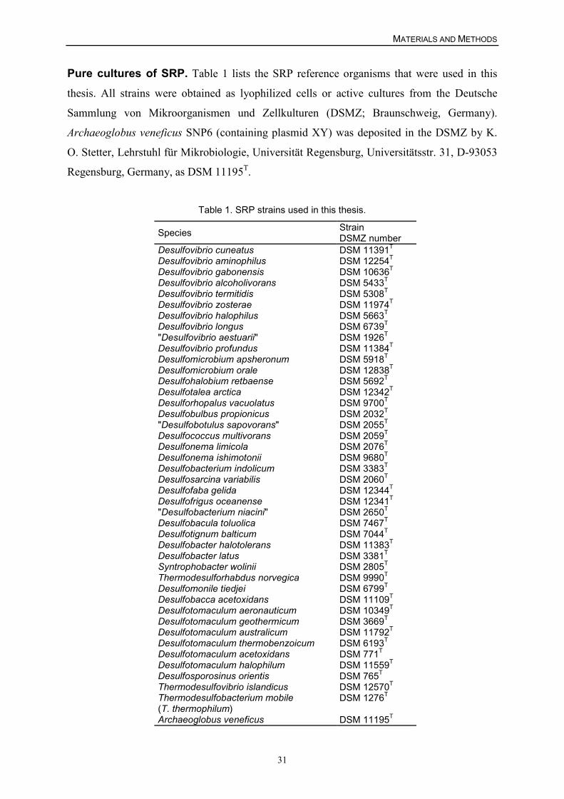
MATERIALS AND METHODS
31
Pure cultures of SRP. Table 1 lists the SRP reference organisms that were used in this
thesis. All strains were obtained as lyophilized cells or active cultures from the Deutsche
Sammlung von Mikroorganismen und Zellkulturen (DSMZ; Braunschweig, Germany).
Archaeoglobus veneficus SNP6 (containing plasmid XY) was deposited in the DSMZ by K.
O. Stetter, Lehrstuhl für Mikrobiologie, Universität Regensburg, Universitätsstr. 31, D-93053
Regensburg, Germany, as DSM 11195T.
Table 1. SRP strains used in this thesis.
Species StrainDSMZ number
Desulfovibrio cuneatus DSM 11391T
Desulfovibrio aminophilus DSM 12254T
Desulfovibrio gabonensis DSM 10636T
Desulfovibrio alcoholivorans DSM 5433T
Desulfovibrio termitidis DSM 5308T
Desulfovibrio zosterae DSM 11974T
Desulfovibrio halophilus DSM 5663T
Desulfovibrio longus DSM 6739T
"Desulfovibrio aestuarii" DSM 1926T
Desulfovibrio profundus DSM 11384T
Desulfomicrobium apsheronum DSM 5918T
Desulfomicrobium orale DSM 12838T
Desulfohalobium retbaense DSM 5692T
Desulfotalea arctica DSM 12342T
Desulforhopalus vacuolatus DSM 9700T
Desulfobulbus propionicus DSM 2032T
"Desulfobotulus sapovorans" DSM 2055T
Desulfococcus multivorans DSM 2059T
Desulfonema limicola DSM 2076T
Desulfonema ishimotonii DSM 9680T
Desulfobacterium indolicum DSM 3383T
Desulfosarcina variabilis DSM 2060T
Desulfofaba gelida DSM 12344T
Desulfofrigus oceanense DSM 12341T
"Desulfobacterium niacini" DSM 2650T
Desulfobacula toluolica DSM 7467T
Desulfotignum balticum DSM 7044T
Desulfobacter halotolerans DSM 11383T
Desulfobacter latus DSM 3381T
Syntrophobacter wolinii DSM 2805T
Thermodesulforhabdus norvegica DSM 9990T
Desulfomonile tiedjei DSM 6799T
Desulfobacca acetoxidans DSM 11109T
Desulfotomaculum aeronauticum DSM 10349T
Desulfotomaculum geothermicum DSM 3669T
Desulfotomaculum australicum DSM 11792T
Desulfotomaculum thermobenzoicum DSM 6193T
Desulfotomaculum acetoxidans DSM 771T
Desulfotomaculum halophilum DSM 11559T
Desulfosporosinus orientis DSM 765T
Thermodesulfovibrio islandicus DSM 12570T
Thermodesulfobacterium mobile(T. thermophilum)
DSM 1276T
Archaeoglobus veneficus DSM 11195T
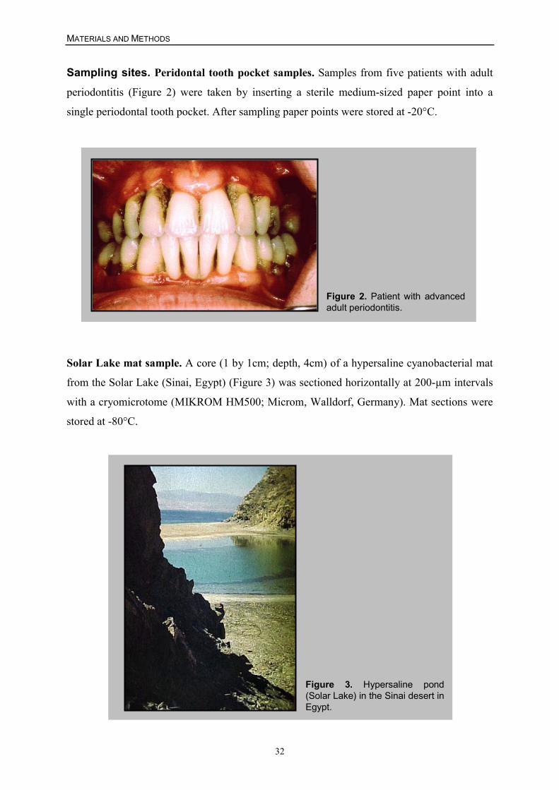
MATERIALS AND METHODS
32
Sampling sites. Peridontal tooth pocket samples. Samples from five patients with adult
periodontitis (Figure 2) were taken by inserting a sterile medium-sized paper point into a
single periodontal tooth pocket. After sampling paper points were stored at -20°C.
Figure 2. Patient with advancedadult periodontitis.
Solar Lake mat sample. A core (1 by 1cm; depth, 4cm) of a hypersaline cyanobacterial mat
from the Solar Lake (Sinai, Egypt) (Figure 3) was sectioned horizontally at 200-µm intervals
with a cryomicrotome (MIKROM HM500; Microm, Walldorf, Germany). Mat sections were
stored at -80°C.
Figure 3. Hypersaline pond(Solar Lake) in the Sinai desert inEgypt.
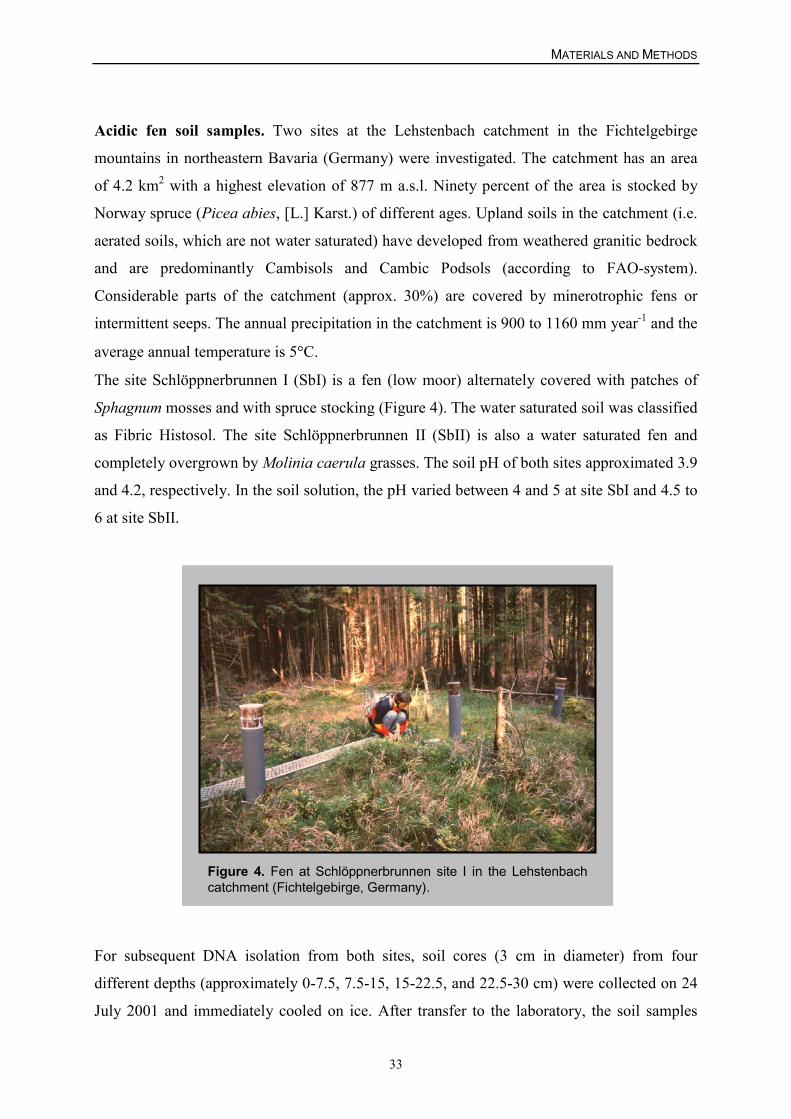
MATERIALS AND METHODS
33
Acidic fen soil samples. Two sites at the Lehstenbach catchment in the Fichtelgebirge
mountains in northeastern Bavaria (Germany) were investigated. The catchment has an area
of 4.2 km2 with a highest elevation of 877 m a.s.l. Ninety percent of the area is stocked by
Norway spruce (Picea abies, [L.] Karst.) of different ages. Upland soils in the catchment (i.e.
aerated soils, which are not water saturated) have developed from weathered granitic bedrock
and are predominantly Cambisols and Cambic Podsols (according to FAO-system).
Considerable parts of the catchment (approx. 30%) are covered by minerotrophic fens or
intermittent seeps. The annual precipitation in the catchment is 900 to 1160 mm year-1 and the
average annual temperature is 5�C.
The site Schlöppnerbrunnen I (SbI) is a fen (low moor) alternately covered with patches of
Sphagnum mosses and with spruce stocking (Figure 4). The water saturated soil was classified
as Fibric Histosol. The site Schlöppnerbrunnen II (SbII) is also a water saturated fen and
completely overgrown by Molinia caerula grasses. The soil pH of both sites approximated 3.9
and 4.2, respectively. In the soil solution, the pH varied between 4 and 5 at site SbI and 4.5 to
6 at site SbII.
Figure 4. Fen at Schlöppnerbrunnen site I in the Lehstenbachcatchment (Fichtelgebirge, Germany).
For subsequent DNA isolation from both sites, soil cores (3 cm in diameter) from four
different depths (approximately 0-7.5, 7.5-15, 15-22.5, and 22.5-30 cm) were collected on 24
July 2001 and immediately cooled on ice. After transfer to the laboratory, the soil samples
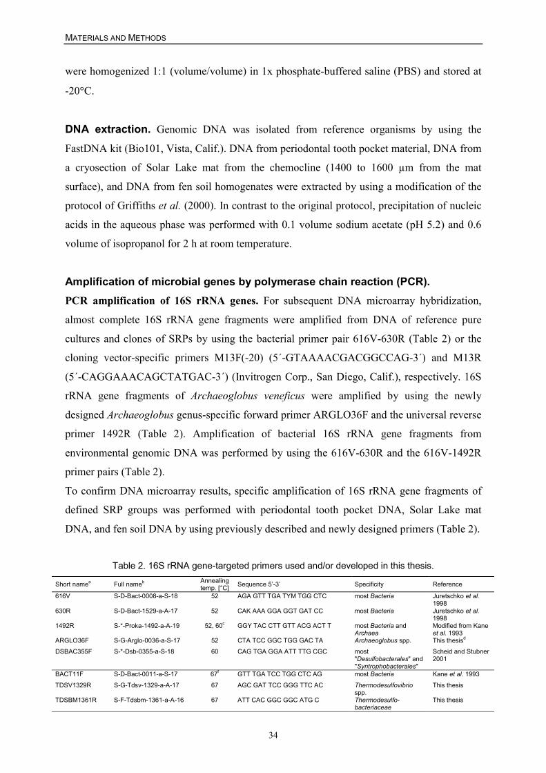
MATERIALS AND METHODS
34
were homogenized 1:1 (volume/volume) in 1x phosphate-buffered saline (PBS) and stored at
-20�C.
DNA extraction. Genomic DNA was isolated from reference organisms by using the
FastDNA kit (Bio101, Vista, Calif.). DNA from periodontal tooth pocket material, DNA from
a cryosection of Solar Lake mat from the chemocline (1400 to 1600 µm from the mat
surface), and DNA from fen soil homogenates were extracted by using a modification of the
protocol of Griffiths et al. (2000). In contrast to the original protocol, precipitation of nucleic
acids in the aqueous phase was performed with 0.1 volume sodium acetate (pH 5.2) and 0.6
volume of isopropanol for 2 h at room temperature.
Amplification of microbial genes by polymerase chain reaction (PCR).PCR amplification of 16S rRNA genes. For subsequent DNA microarray hybridization,
almost complete 16S rRNA gene fragments were amplified from DNA of reference pure
cultures and clones of SRPs by using the bacterial primer pair 616V-630R (Table 2) or the
cloning vector-specific primers M13F(-20) (5´-GTAAAACGACGGCCAG-3´) and M13R
(5´-CAGGAAACAGCTATGAC-3´) (Invitrogen Corp., San Diego, Calif.), respectively. 16S
rRNA gene fragments of Archaeoglobus veneficus were amplified by using the newly
designed Archaeoglobus genus-specific forward primer ARGLO36F and the universal reverse
primer 1492R (Table 2). Amplification of bacterial 16S rRNA gene fragments from
environmental genomic DNA was performed by using the 616V-630R and the 616V-1492R
primer pairs (Table 2).
To confirm DNA microarray results, specific amplification of 16S rRNA gene fragments of
defined SRP groups was performed with periodontal tooth pocket DNA, Solar Lake mat
DNA, and fen soil DNA by using previously described and newly designed primers (Table 2).
Table 2. 16S rRNA gene-targeted primers used and/or developed in this thesis.
Short namea Full nameb Annealingtemp. [°C] Sequence 5’-3’ Specificity Reference
616V S-D-Bact-0008-a-S-18 52 AGA GTT TGA TYM TGG CTC most Bacteria Juretschko et al.1998
630R S-D-Bact-1529-a-A-17 52 CAK AAA GGA GGT GAT CC most Bacteria Juretschko et al.1998
1492R S-*-Proka-1492-a-A-19 52, 60c GGY TAC CTT GTT ACG ACT T most Bacteria andArchaea
Modified from Kaneet al. 1993
ARGLO36F S-G-Arglo-0036-a-S-17 52 CTA TCC GGC TGG GAC TA Archaeoglobus spp. This thesisd
DSBAC355F S-*-Dsb-0355-a-S-18 60 CAG TGA GGA ATT TTG CGC most"Desulfobacterales" and"Syntrophobacterales"
Scheid and Stubner2001
BACT11F S-D-Bact-0011-a-S-17 67f GTT TGA TCC TGG CTC AG most Bacteria Kane et al. 1993
TDSV1329R S-G-Tdsv-1329-a-A-17 67 AGC GAT TCC GGG TTC AC Thermodesulfovibriospp.
This thesis
TDSBM1361R S-F-Tdsbm-1361-a-A-16 67 ATT CAC GGC GGC ATG C Thermodesulfo-bacteriaceae
This thesis
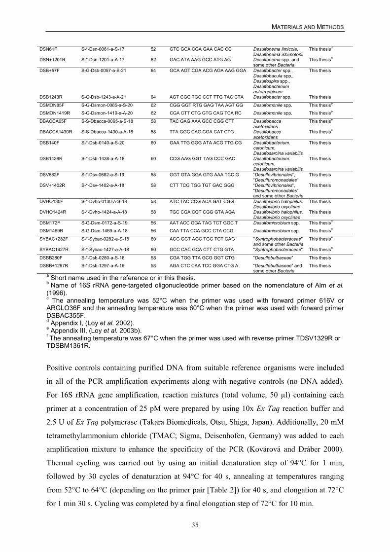
MATERIALS AND METHODS
35
DSN61F S-*-Dsn-0061-a-S-17 52 GTC GCA CGA GAA CAC CC Desulfonema limicola,Desulfonema ishimotonii
This thesisd
DSN+1201R S-*-Dsn-1201-a-A-17 52 GAC ATA AAG GCC ATG AG Desulfonema spp. andsome other Bacteria
This thesisd
DSB+57F S-G-Dsb-0057-a-S-21 64 GCA AGT CGA ACG AGA AAG GGA Desulfobacter spp.,Desulfobacula spp.,Desulfospira spp.,Desulfobacteriumautotrophicum
This thesis
DSB1243R S-G-Dsb-1243-a-A-21 64 AGT CGC TGC CCT TTG TAC CTA Desulfobacter spp. This thesis
DSMON85F S-G-Dsmon-0085-a-S-20 62 CGG GGT RTG GAG TAA AGT GG Desulfomonile spp. This thesise
DSMON1419R S-G-Dsmon-1419-a-A-20 62 CGA CTT CTG GTG CAG TCA RC Desulfomonile spp. This thesise
DBACCA65F S-S-Dbacca-0065-a-S-18 58 TAC GAG AAA GCC CGG CTT Desulfobaccaacetoxidans
This thesise
DBACCA1430R S-S-Dbacca-1430-a-A-18 58 TTA GGC CAG CGA CAT CTG Desulfobaccaacetoxidans
This thesise
DSB140F S-*-Dsb-0140-a-S-20 60 GAA TTG GGG ATA ACG TTG CG Desulfobacterium.cetonicum,Desulfosarcina variabilis
This thesis
DSB1438R S-*-Dsb-1438-a-A-18 60 CCG AAG GGT TAG CCC GAC Desulfobacterium.cetonicum,Desulfosarcina variabilis
This thesis
DSV682F S-*-Dsv-0682-a-S-19 58 GGT GTA GGA GTG AAA TCC G “Desulfovibrionales“,“Desulfuromonadales“
This thesis
DSV+1402R S-*-Dsv-1402-a-A-18 58 CTT TCG TGG TGT GAC GGG “Desulfovibrionales“,“Desulfuromonadales“,and some other Bacteria
This thesis
DVHO130F S-*-Dvho-0130-a-S-18 58 ATC TAC CCG ACA GAT CGG Desulfovibrio halophilus,Desulfovibrio oxyclinae
This thesis
DVHO1424R S-*-Dvho-1424-a-A-18 58 TGC CGA CGT CGG GTA AGA Desulfovibrio halophilus,Desulfovibrio oxyclinae
This thesis
DSM172F S-G-Dsm-0172-a-S-19 56 AAT ACC GGA TAG TCT GGC T Desulfomicrobium spp. This thesisd
DSM1469R S-G-Dsm-1469-a-A-18 56 CAA TTA CCA GCC CTA CCG Desulfomicrobium spp. This thesisd
SYBAC+282F S-*-Sybac-0282-a-S-18 60 ACG GGT AGC TGG TCT GAG "Syntrophobacteraceae"and some other Bacteria
This thesise
SYBAC1427R S-*-Sybac-1427-a-A-18 60 GCC CAC GCA CTT CTG GTA "Syntrophobacteraceae" This thesise
DSBB280F S-*-Dsb-0280-a-S-18 58 CGA TGG TTA GCG GGT CTG “Desulfobulbaceae” This thesis
DSBB+1297R S-*-Dsb-1297-a-A-19 58 AGA CTC CAA TCC GGA CTG A “Desulfobulbaceae” andsome other Bacteria
This thesis
a Short name used in the reference or in this thesis.b Name of 16S rRNA gene-targeted oligonucleotide primer based on the nomenclature of Alm et al.(1996).c The annealing temperature was 52°C when the primer was used with forward primer 616V orARGLO36F and the annealing temperature was 60°C when the primer was used with forward primerDSBAC355F.d Appendix I, (Loy et al. 2002).e Appendix III, (Loy et al. 2003b).f The annealing temperature was 67°C when the primer was used with reverse primer TDSV1329R orTDSBM1361R.
Positive controls containing purified DNA from suitable reference organisms were included
in all of the PCR amplification experiments along with negative controls (no DNA added).
For 16S rRNA gene amplification, reaction mixtures (total volume, 50 µl) containing each
primer at a concentration of 25 pM were prepared by using 10x Ex Taq reaction buffer and
2.5 U of Ex Taq polymerase (Takara Biomedicals, Otsu, Shiga, Japan). Additionally, 20 mM
tetramethylammonium chloride (TMAC; Sigma, Deisenhofen, Germany) was added to each
amplification mixture to enhance the specificity of the PCR (Kovárová and Dráber 2000).
Thermal cycling was carried out by using an initial denaturation step of 94°C for 1 min,
followed by 30 cycles of denaturation at 94°C for 40 s, annealing at temperatures ranging
from 52°C to 64°C (depending on the primer pair [Table 2]) for 40 s, and elongation at 72°C
for 1 min 30 s. Cycling was completed by a final elongation step of 72°C for 10 min.
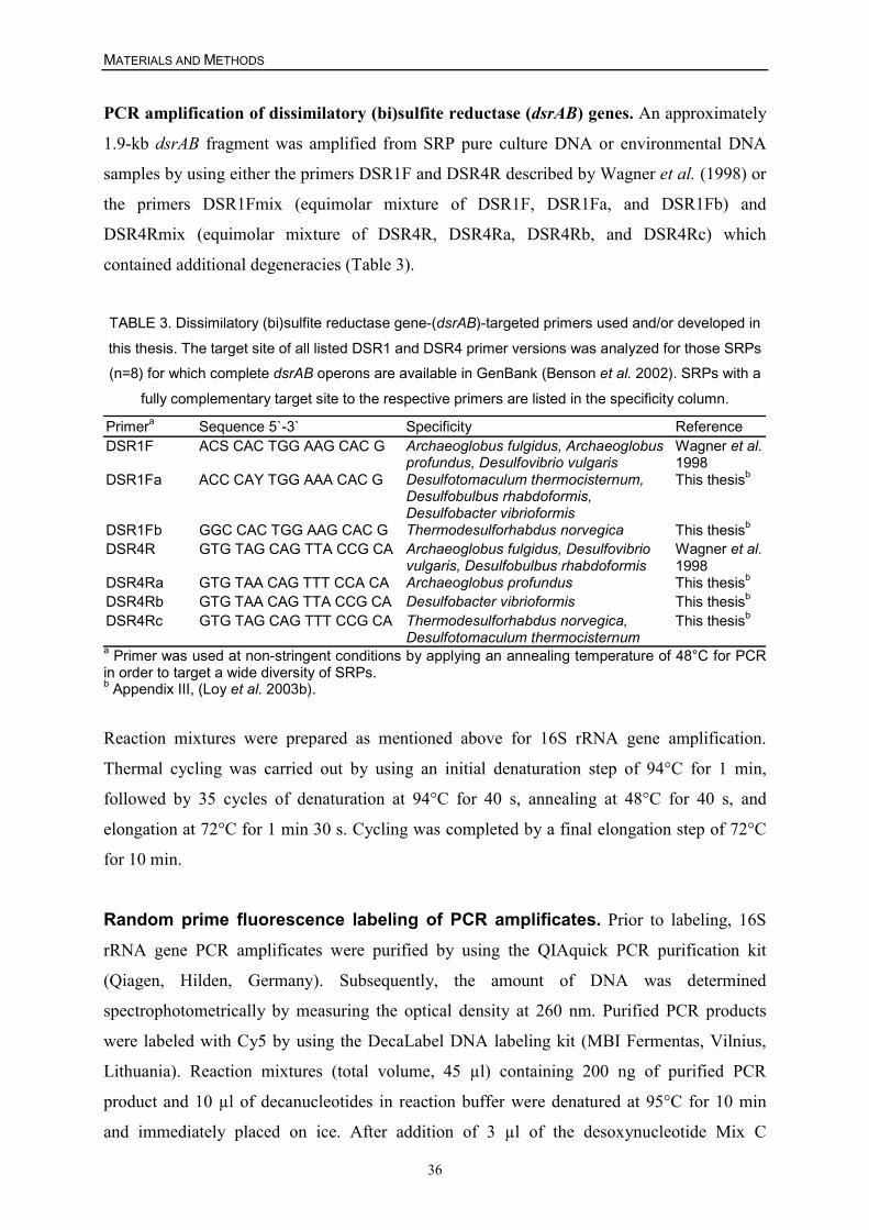
MATERIALS AND METHODS
36
PCR amplification of dissimilatory (bi)sulfite reductase (dsrAB) genes. An approximately
1.9-kb dsrAB fragment was amplified from SRP pure culture DNA or environmental DNA
samples by using either the primers DSR1F and DSR4R described by Wagner et al. (1998) or
the primers DSR1Fmix (equimolar mixture of DSR1F, DSR1Fa, and DSR1Fb) and
DSR4Rmix (equimolar mixture of DSR4R, DSR4Ra, DSR4Rb, and DSR4Rc) which
contained additional degeneracies (Table 3).
TABLE 3. Dissimilatory (bi)sulfite reductase gene-(dsrAB)-targeted primers used and/or developed in
this thesis. The target site of all listed DSR1 and DSR4 primer versions was analyzed for those SRPs(n=8) for which complete dsrAB operons are available in GenBank (Benson et al. 2002). SRPs with a
fully complementary target site to the respective primers are listed in the specificity column.
Primera Sequence 5`-3` Specificity ReferenceDSR1F ACS CAC TGG AAG CAC G Archaeoglobus fulgidus, Archaeoglobus
profundus, Desulfovibrio vulgarisWagner et al.1998
DSR1Fa ACC CAY TGG AAA CAC G Desulfotomaculum thermocisternum,Desulfobulbus rhabdoformis,Desulfobacter vibrioformis
This thesisb
DSR1Fb GGC CAC TGG AAG CAC G Thermodesulforhabdus norvegica This thesisb
DSR4R GTG TAG CAG TTA CCG CA Archaeoglobus fulgidus, Desulfovibriovulgaris, Desulfobulbus rhabdoformis
Wagner et al.1998
DSR4Ra GTG TAA CAG TTT CCA CA Archaeoglobus profundus This thesisb
DSR4Rb GTG TAA CAG TTA CCG CA Desulfobacter vibrioformis This thesisb
DSR4Rc GTG TAG CAG TTT CCG CA Thermodesulforhabdus norvegica,Desulfotomaculum thermocisternum
This thesisb
a Primer was used at non-stringent conditions by applying an annealing temperature of 48°C for PCRin order to target a wide diversity of SRPs.b Appendix III, (Loy et al. 2003b).
Reaction mixtures were prepared as mentioned above for 16S rRNA gene amplification.
Thermal cycling was carried out by using an initial denaturation step of 94°C for 1 min,
followed by 35 cycles of denaturation at 94°C for 40 s, annealing at 48°C for 40 s, and
elongation at 72°C for 1 min 30 s. Cycling was completed by a final elongation step of 72°C
for 10 min.
Random prime fluorescence labeling of PCR amplificates. Prior to labeling, 16S
rRNA gene PCR amplificates were purified by using the QIAquick PCR purification kit
(Qiagen, Hilden, Germany). Subsequently, the amount of DNA was determined
spectrophotometrically by measuring the optical density at 260 nm. Purified PCR products
were labeled with Cy5 by using the DecaLabel DNA labeling kit (MBI Fermentas, Vilnius,
Lithuania). Reaction mixtures (total volume, 45 µl) containing 200 ng of purified PCR
product and 10 µl of decanucleotides in reaction buffer were denatured at 95°C for 10 min
and immediately placed on ice. After addition of 3 µl of the desoxynucleotide Mix C
MATERIALS AND METHODS
37
(containing no dCTP), 1 µl Cy5-dCTP (Amersham Biosciences, Freiburg, Germany) and 1 µl
Klenow fragment (Exo-; 5 U µl-1), the labeling reaction mixtures were incubated at 37°C for
45 min. For more efficient labeling, the addition of Mix C, Cy5-dCTP, and the Klenow
fragment and incubation at 37°C for 45 min were repeated. Labeling was completed by
addition of 4 µl of dNTP-Mix and incubation at 37°C for 60 min. To remove unincorporated
desoxynucleotides and decanucleotides, the labeling mixture was purified with a QIAquick
nucleotide removal kit (Qiagen) by using double-distilled water for DNA elution. Finally, the
eluted DNA was vacuum-dried and stored in the dark at -20°C.
Microarray manufacture and processing. Oligonucleotides for microarray printing
were obtained from MWG Biotech (Ebersberg, Germany). The sequence, specificity, and
microarray position of each oligonucleotide probe are shown in Table 5. In addition,
difference alignments for all probes generated with the latest ARB small-subunit rRNA
database (http://www.arb-home.de) can be viewed at the probeBase website
(http://www.probebase.net) (Appendix II, Loy et al. 2003a). The 5’ end of each
oligonucleotide probe was tailed with 15 dTTP molecules (T-spacer) to increase the on-chip
accessibility of spotted probes to target DNA (Shchepinov et al. 1997, Southern et al. 1999).
In addition, the 5’-terminal nucleotide of each oligonucleotide was aminated to allow covalent
coupling of the oligonucleotides to aldehyde group-coated CSS-100 glass slides (CEL
Associates, Houston, Tex.). The concentration of oligonucleotide probes before printing was
adjusted to 50 pmol µl-1 in 50% dimethyl sulfoxide to prevent evaporation during the printing
procedure. SRP-PhyloChips were printed by using a GMS 417 contact arrayer (Affymetrix,
Santa Clara, Calif.). Spotted DNA microarrays were dried overnight at room temperature to
allow efficient crosslinking. Slides were washed twice at room temperature in 0.2% sodium
dodecyl sulfate (SDS) and then twice with double-distilled water with vigorous agitation to
remove unbound oligonucleotides and the SDS. After air drying, the slides were incubated for
5 min in fresh sodium borohydride solution (1.0 g NaBH4 in 300 ml phosphate-buffered
saline and 100 ml of absolute ethanol) to reduce all remaining reactive aldehyde groups on the
glass. The reaction was stopped by adding ice-cold absolute ethanol. The reduced slides were
washed three times with 0.2% SDS and double-distilled water, air dried, and stored in the
dark at room temperature.
Reverse hybridization on microarrays. Vacuum-dried Cy5-labeled PCR products (400
ng) and 0.5 pmol of the Cy5-labeled control oligonucleotide CONT-COMP (Table 5) were
MATERIALS AND METHODS
38
resuspended in 20 µl of hybridization buffer (5x SSC [1x SSC is 0.15 M NaCl plus 0.015 M
sodium citrate] , 1% blocking reagent [Roche, Mannheim, Germany], 0.1% n-lauryl
sarcosine, 0.02% SDS, 5% formamide), denatured for 10 min at 95°C, and immediately
placed on ice. Then the solution was pipetted onto an SRP-PhyloChip, covered with a cover
slip, and inserted into a tight custom-made hybridization chamber
(http://cmgm.stanford.edu/pbrown/mguide/HybChamber.pdf) containing 50 µl of
hybridization buffer for subsequent equilibration. Hybridization was performed overnight at
42°C in a water bath. After hybridization, the slides were washed immediately under stringent
conditions for 5 min at 55°C in 50 ml washing buffer (containing 3 M TMAC, 50 mM Tris-
HCl, 2 mM EDTA, and 0.1% SDS). To record probe-target melting curves, the temperature of
the washing step was varied from 42 to 80°C. After the stringent washing, the slides were
washed twice with ice-cold double-distilled water, air dried, and stored in the dark at room
temperature.
Scanning of microarrays. Fluorescence images of the SRP-PhyloChips were recorded by
scanning the slides with a GMS 418 array scanner (Affymetrix). The fluorescence signals
were quantified by using the ImaGene 4.0 software (BioDiscovery, Inc., Los Angeles, Calif.).
A grid of individual circles defining the location of each spot on the array was superimposed
on the image to designate each fluorescent spot to be quantified. The mean signal intensity
was determined for each spot. In addition, the mean signal intensity of the local background
area surrounding the spots was determined.
Selective enrichment of nucleic acids by a capture probe approach. Five
microliters of aldehyde group-coated glass beads (diameter, 1 µm; Xenopore, Hawthorne, NJ)
was incubated overnight with 5 µl of the appropriate capture probe (100 pmol µl-1; tailed with
15 dTTP molecules, aminated with 5’-terminal nucleotide) at room temperature.
Subsequently, the beads were washed once with 400 µl of 0.2% SDS and pelleted by
centrifugation (1 min at 14.000 rpm), and the supernatant was decanted. After this step, the
beads were washed twice with 400 µl double-distilled water, dried, and stored at room
temperature prior to hybridization. A vacuum-dried bacterial 16S rRNA gene PCR product
(obtained from DNA from the Solar Lake mat with the 616V-1492R primer pair) was
resuspended with 200 µl of hybridization buffer (see above), denatured for 10 min at 95°C,
and immediately cooled on ice. The hybridization solution and capture probe beads were
mixed in a screw-cap tube and incubated overnight at 42°C on a shaker. Subsequently, the
MATERIALS AND METHODS
39
beads were washed twice with 1.5 ml washing buffer (see above) at 55°C for 2.5 min. After
the stringent washes, the beads were washed with 1.5 ml ice-cold double-distilled water and
then with ice cold 70% ethanol. Beads with captured nucleic acids were vacuum dried and
resuspended in 50 µl EB buffer (part of the QIAquick PCR purification kit; Qiagen) for
storage at –20°C. Reamplification of bacterial 16S rRNA gene fragments from the captured
nucleic acids was performed by using 5 µl of the resuspended beads for PCR performed by
using the 616V-1492R primer pair and the protocols described above.
Cloning and sequencing. Prior to cloning the PCR amplification products were purified
by low-melting-point agarose (1.5%) gel electrophoresis (NuSieve 3:1; FMC Bioproducts,
Biozym Diagnostics GmbH, Oldendorf, Germany) and stained in SYBR Green I solution (10
µl 10.000x SYBR Green I stain in 100 µl TAE buffer [40 mM TRIS, 10 mM sodium acetate,
1 mM EDTA, pH 8.0]; Biozym Diagnostics GmbH) for 45 min. Bands of the expected size
were excised from the agarose gel with a glass capillary and melted with 80 µl double-
distilled water for 10 min at 80°C. Four microliters of each solution were ligated as
recommended by the manufacturer (Invitrogen Corp.) either into the cloning vector pCR2.1 of
the TOPO TA cloning kit (16S rRNA gene amplificates) or into the cloning vector pCR-XL-
TOPO of the TOPO XL cloning kit (dsrAB gene amplificates). Nucleotide sequences were
determined by the dideoxynucleotide method (Sanger et al. 1977) as described by Purkhold et
al. (2000). In addition, internal dsrAB gene-targeted sequencing primers (Table 4) were used
to complete the dsrAB sequences.
TABLE 4. Internal dissimilatory (bi)sulfite reductase gene-(dsrAB)-targeted sequencing primers
Primera Sequence 5`-3` Specificity Referenceb
DSR978Fa GGT CAT CGA CCT TTG TCC Schlöppnerbrunnen I soil OTU 5 This thesisDSR978Fb CGT CGT CGG GAA GTG CCC Schlöppnerbrunnen I soil OTU 8 This thesisDSR978Fc AGT AGT CGA CCT TTG CCC Schlöppnerbrunnen I+II soil OTU 6 This thesisDSR978Fd TGT CAC CGA TCT CTG CCC Schlöppnerbrunnen I soil OTU 1 This thesisDSR978Fe TGT TAC CGA CCT CTG CCC Schlöppnerbrunnen II soil OTU 1
(dsrSbII-20)This thesis
DSR978Ff TGT CAC CGA TCT TTG CCC Schlöppnerbrunnen II soil OTU 4(dsrSbII-15)
This thesis
DSR978Fg CGT CAC CAT TCT CTG CCC Schlöppnerbrunnen II soil OTU 4(dsrSbII-9)
This thesis
DSR978Fh GGT CGT TGA CAT GTG TCC Schlöppnerbrunnen II soil OTU 11 This thesisDSR978Fi GGT CTG CAA TCT CTG YCC Schlöppnerbrunnen I+II soil OTU 2 and 3 This thesisDSR978Fj GGT TGT TGA CCT TTG CCC Schlöppnerbrunnen I soil OTU 9 This thesisDSR978Fk CGT TTG CGA TCT CTG CCC Schlöppnerbrunnen II soil OTU 7 This thesisDSR860F AGA TCC GGC GGG ACG ATG Schlöppnerbrunnen I soil OTU 10 This thesisa Internal sequencing primer used to complete dsrAB gene sequences retrieved from acidic fen sitesSchlöppnerbrunnen I and II.b Appendix III, (Loy et al. 2003b).
MATERIALS AND METHODS
40
Phylogeny inference. All phylogenetic analyses were performed by using the
undermentioned alignment and treeing tools implemented in the ARB program package
(http://www.arb-home.de).
16S rRNA gene-based phylogeny. The new 16S rRNA sequences were added to an ARB
alignment of about 16.000 small-subunit rRNA sequences by using the alignment tool
ARB_EDIT. Alignments were refined manually by visual inspection. 16S rRNA phylogenetic
analyses were exclusively performed with sequences having more than 1.300 bases by
applying distance-matrix, maximum-parsimony, and maximum-likelihood methods and the
aforementioned ARB treeing tools for nucleotide sequences. The composition of the 16S
rRNA data sets varied with respect to the reference sequences and the alignment positions
included. Variability of the individual alignment positions was determined by using the
ARB_SAI tools and used as criterion to remove or include variable positions (50%
conservation filter) for phylogenetic analyses. Parsimony bootstrap analyses based on 100 or
1000 resamplings were performed with PHYLIP. All phylogenetic consensus trees were
drawn according to the recommendations of Ludwig et al. (1998).
dsrAB gene-based phylogeny. New dsrAB sequences were added to an ARB alignment
which contains all dsrAB sequences of recognized (Friedrich 2002, Klein et al. 2001) and
uncultured SRPs available in GenBank (Benson et al. 2002). Deduced amino acid sequences
were manually aligned by using the editor GDE 2.2 (S.W. Smith, C. Wang, P.M. Gillevet and
W. Gilbert (1992) Genetic Data Environment and the Harvard Genome Database. Genome
mapping and Sequencing, Cold Spring Harbor Laboratory). Nucleic acid sequences were
aligned according to the amino acid alignment. During phylogenetic analyses of amino acid
sequences, regions of insertions and deletions were removed from the DsrAB data set by
using an amino acid alignment mask (indel filter) prepared in ARB. A total of 543 amino acid
positions (alpha subunit, 327; beta subunit, 216) were used in DsrAB analyses. Distance-
matrix (using FITCH with global rearrangements and randomized input order of species) and
maximum-parsimony trees were calculated with the PHYLogeny Inference Package
(PHYLIP, version 3.57c, J. Felsenstein, Department of Genetics, University of Washington,
Seattle). In addition, the programs MOLPHY (version 2.3, Computer science monographs,
no. 28.: Programs for molecular phylogenetics based on maximum-likelihood. J. Adachi and
M. Hasegawa, Institute of Statistics and Mathematics, Tokyo, Japan) and TREE-PUZZLE
(Strimmer and von Haeseler 1996) were used to infer maximum-likelihood trees with JTT-f
as the amino acid replacement model. To perform dsrAB phylogenetic analysis on the
nucleotide level filters were constructed which allowed to exclude regions of insertions and
MATERIALS AND METHODS
41
deletions as well as the third codon position for phylogenetic analysis. Nucleic acid level
phylogenetic analyses were performed by applying distance-matrix [using the Jukes-Cantor
model (Jukes and Cantor 1969)], maximum-parsimony (PHYLIP program), and maximum-
likelihood methods [fastDNAml program (Olsen et al. 1994)]. Parsimony bootstrap analyses
for protein (DsrAB) and nucleotide level (dsrAB) phylogenetic analysis were performed with
PHYLIP. For each calculation 100 or 1000 resamplings were performed. All phylogenetic
consensus trees were drawn according to the recommendations of Ludwig et al. (1998).
Nomenclature of prokaryotes. Names of bacterial taxa were used in accordance with the
prokaryotic nomenclature proposed in the taxonomic outline of the second edition of Bergey`s
Manual of Systematic Bacteriology (http://www.cme.msu.edu/bergeys/) (Garrity and Holt
2001) and the Approved Lists of Bacterial Names (http://www.bacterio.cict.fr/) (Euzeby
1997) as recommended by Oren and Stackebrandt (2002).
Nucleotide sequence accession numbers. The sequences determined in this thesis are
available in the GenBank database under accession numbers AY083010 to AY083027 and
AY167444 to AY167462 (16S rRNA gene clones), and AY083028/AY083029 and
AY167464 to AY167483 (dsrAB gene clones). The dsrAB gene sequence of
Desulfomicrobium orale DSM 12838T has been deposited under accession number
AY083030.
References
Alm EW, Oerther DB, Larsen N, Stahl DA, andRaskin L [1996] The oligonucleotide probedatabase. Appl Environ Microbiol. 62: 3557-3559.
Benson DA, Karsch-Mizrachi I, Lipman DJ,Ostell J, Rapp BA, and Wheeler DL [2002]GenBank. Nucleic Acids Res. 30: 17-20.
Euzeby JP [1997] List of Bacterial Names withStanding in Nomenclature: a folder available on theInternet. Int J Syst Bacteriol. 47: 590-592.
Friedrich MW [2002] Phylogenetic analysisreveals multiple lateral transfers of adenosine-5'-phosphosulfate reductase genes among sulfate-reducing microorganisms. J Bacteriol. 184: 278-289.
Garrity GM, and Holt JG [2001] The road map tothe manual, p. 119-166. In Garrity GM (ed.),
Bergey's manual of systematic bacteriology, 2nd ed,vol. 1. Springer, New York.
Griffiths RI, Whiteley AS, O'Donnell AG, andBailey MJ [2000] Rapid method for coextraction ofDNA and RNA from natural environments foranalysis of ribosomal DNA- and rRNA-basedmicrobial community composition. Appl EnvironMicrobiol. 66: 5488-5491.
Jukes TH, and Cantor CR [1969] Evolution ofprotein molecules, p. 21-132. In Munro HN (ed.),Mammalian Protein Metabolism. Academic Press,New York.
Juretschko S, Timmermann G, Schmid M,Schleifer KH, Pommerening-Roser A, KoopsHP, and Wagner M [1998] Combined molecularand conventional analyses of nitrifying bacteriumdiversity in activated sludge: Nitrosococcus mobilis
MATERIALS AND METHODS
42
and Nitrospira- like bacteria as dominantpopulations. Appl Environ Microbiol. 64: 3042-3051.
Kane MD, Poulsen LK, and Stahl DA [1993]Monitoring the enrichment and isolation of sulfate-reducing bacteria by using oligonucleotidehybridization probes designed fromenvironmentally derived 16S rRNA sequences.Appl Environ Microbiol. 59: 682-686.
Klein M, Friedrich M, Roger AJ, Hugenholtz P,Fishbain S, Abicht H, Blackall LL, Stahl DA,and Wagner M [2001] Multiple lateral transfers ofdissimilatory sulfite reductase genes between majorlineages of sulfate-reducing prokaryotes. JBacteriol. 183: 6028-6035.
Kovárová M, and Dráber P [2000] Newspecificity and yield enhancer of polymerase chainreactions. Nucleic Acids Res. 28: E70.
Loy A, Horn M, and Wagner M [2003a]probeBase: an online resource for rRNA-targetedoligonucleotide probes. Nucleic Acids Res. 31: 514-516.
Loy A, Küsel K, Lehner A, Drake HL, andWagner M [2003b] Diversity of sulfate-reducingprokaryotes in an acidic fen: Comparison of 16SrRNA gene-based oligonucleotide microarrayhybridization with dissimilatory (bi)sulfitereductase gene surveys. Appl Environ Microbiol.submitted.
Loy A, Lehner A, Lee N, Adamczyk J, Meier H,Ernst J, Schleifer K-H, and Wagner M [2002]Oligonucleotide microarray for 16S rRNA gene-based detection of all recognized lineages ofsulfate-reducing prokaryotes in the environment.Appl Environ Microbiol. 68: 5064-5081.
Ludwig W, Strunk O, Klugbauer S, KlugbauerN, Weizenegger M, Neumaier J, Bachleitner M,and Schleifer KH [1998] Bacterial phylogenybased on comparative sequence analysis.Electrophoresis. 19: 554-568.
Olsen GJ, Matsuda H, Hagstrom R, andOverbeek R [1994] fastDNAml: A tool forconstruction of phylogenetic trees of DNAsequences using maximum likelihood. Comput ApplBiosci. 10: 41-48.
Oren A, and Stackebrandt E [2002] Prokaryotetaxonomy online: challenges ahead. Nature. 419:15.
Purkhold U, Pommering-Röser A, Juretschko S,Schmid MC, Koops H-P, and Wagner M [2000]Phylogeny of all recognized species of ammoniaoxidizers based on comparative 16S rRNA andamoA sequence analysis: implications for moleculardiversity surveys. Appl Environ Microbiol. 66:5368-5382.
Sanger F, Nicklen S, and Coulson AR [1977]DNA sequencing with chain-terminating inhibitors.Proc Natl Acad Sci. 74: 5463-5467.
Scheid D, and Stubner S [2001] Structure anddiversity of Gram-negative sulfate-reducingbacteria on rice roots. FEMS Microbiol Ecol. 36:175-183.
Shchepinov MS, Case-Green SC, and SouthernEM [1997] Steric factors influencing hybridisationof nucleic acids to oligonucleotide arrays. NucleicAcids Res. 25: 1155-1161.
Southern E, Mir K, and Shchepinov M [1999]Molecular interactions on microarrays. Nat Genet.21: 5-9.
Strimmer K, and von Haeseler A [1996] Quartetpuzzling: A quartet maximum likelihood methodfor reconstructing tree topologies. Mol Biol Evol.13: 964-969.
Wagner M, Roger AJ, Flax JL, Brusseau GA,and Stahl DA [1998] Phylogeny of dissimilatorysulfite reductases supports an early origin of sulfaterespiration. J Bacteriol. 180: 2975-2982.

RESULTS AND DISCUSSION
43
RESULTS AND DISCUSSION

RESULTS AND DISCUSSION
45
I. Phylogeny of hitherto recognized SRPs based on comparative 16S rRNAsequence analyses
The last encompassing study that has revealed natural relationships among all known SRP
genera already dates back eight years (Stackebrandt et al. 1995). Since that time microbial
taxonomy was confronted with a tremendous increase in the description of novel SRPs. In
total, 70 new species and 19 new genera of SRPs have been validly published between 1996
and 2002. The primary aim of the work presented in this chapter was to establish a thorough
and robust phylogenetic framework for SRPs based on comparison of 16S rRNA gene
sequences. Therefore, all 16S rRNA gene sequences from provisionally proposed and validly
recognized SRPs available in GenBank were collected, aligned, and analyzed
phylogenetically by using maximum-likelihood, maximum-parsimony, and distance-matrix
treeing methods, each in combination with appropriate conservation filters (for details see
figure legends in this chapter). As already pointed out in the introduction part of this thesis,
above-genus level classification of SRPs that is currently in use is scarce and not up-to-date.
Thus, latest taxonomic outlines proposed in the second edition of Bergey`s Manual of
Systematic Bacteriology (http://www.cme.msu.edu/bergeys/) (Garrity and Holt 2001c) were
used as guidance to assign SRPs into higher taxonomic ranks and to reveal potentially
misclassified species.
SRBs of the class “Deltaproteobacteria” belonging to the bacterial phylumProteobacteria
Most SRB species isolated so far belong to the orders “Desulfobacterales”,
“Syntrophobacterales”, and “Desulfovibrionales” (Garrity and Holt 2001c) within the delta-
class of Proteobacteria. Figure 5 depicts the 16S rRNA-based phylogeny of
“Deltaproteobacteria” with emphasis on the affiliation of distinct SRB lineages. Independent
from the treeing method applied, all analyzed species of the order “Desulfovibrionales”
formed a stable monophyletic entity which was highly supported by parsimony bootstrap
analysis (92%). In contrast, the phylogenetic position of distinct “Desulfobacterales” and
“Syntrophobacterales” families relative to each other could not be resolved on the basis of
16S rRNA phylogeny as indicated by a polytomic tree topology (Figure 5).
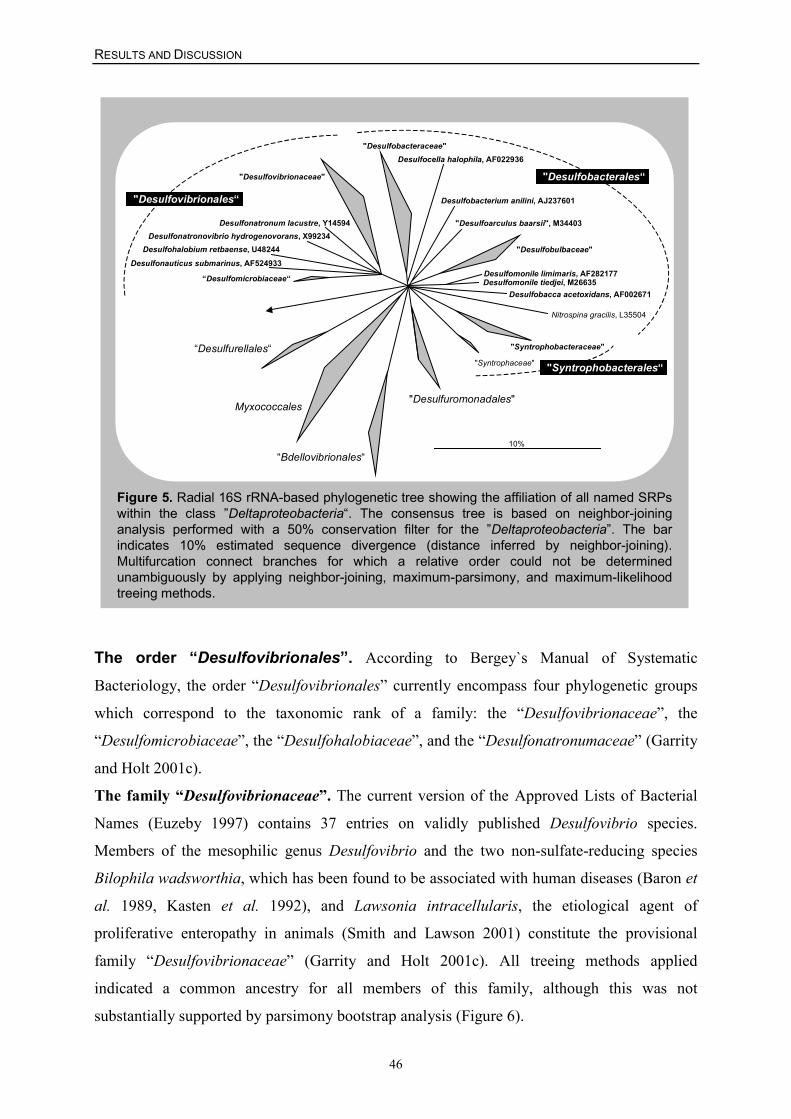
RESULTS AND DISCUSSION
46
"Desulfovibrionaceae"
“Desulfomicrobiaceae“
Desulfonatronovibrio hydrogenovorans, X99234 Desulfonatronum lacustre, Y14594
Desulfohalobium retbaense, U48244
"Desulfuromonadales"
"Syntrophaceae"
"Desulfobulbaceae"
Desulfomonile limimaris, AF282177 Desulfomonile tiedjei, M26635
Desulfocella halophila, AF022936 "Desulfobacteraceae"
Desulfobacca acetoxidans, AF002671
"Desulfoarculus baarsii", M34403
Desulfobacterium anilini, AJ237601
“Bdellovibrionales“
Nitrospina gracilis, L35504
“Desulfurellales“
Myxococcales
"Syntrophobacteraceae"
10%
"Desulfovibrionales“
"Desulfobacterales“
"Syntrophobacterales“
Figure 5. Radial 16S rRNA-based phylogenetic tree showing the affiliation of all named SRPswithin the class ”Deltaproteobacteria“. The consensus tree is based on neighbor-joininganalysis performed with a 50% conservation filter for the ”Deltaproteobacteria”. The barindicates 10% estimated sequence divergence (distance inferred by neighbor-joining).Multifurcation connect branches for which a relative order could not be determinedunambiguously by applying neighbor-joining, maximum-parsimony, and maximum-likelihoodtreeing methods.
Desulfonauticus submarinus, AF524933
The order “Desulfovibrionales”. According to Bergey`s Manual of Systematic
Bacteriology, the order “Desulfovibrionales” currently encompass four phylogenetic groups
which correspond to the taxonomic rank of a family: the “Desulfovibrionaceae”, the
“Desulfomicrobiaceae”, the “Desulfohalobiaceae”, and the “Desulfonatronumaceae” (Garrity
and Holt 2001c).
The family “Desulfovibrionaceae”. The current version of the Approved Lists of Bacterial
Names (Euzeby 1997) contains 37 entries on validly published Desulfovibrio species.
Members of the mesophilic genus Desulfovibrio and the two non-sulfate-reducing species
Bilophila wadsworthia, which has been found to be associated with human diseases (Baron et
al. 1989, Kasten et al. 1992), and Lawsonia intracellularis, the etiological agent of
proliferative enteropathy in animals (Smith and Lawson 2001) constitute the provisional
family “Desulfovibrionaceae” (Garrity and Holt 2001c). All treeing methods applied
indicated a common ancestry for all members of this family, although this was not
substantially supported by parsimony bootstrap analysis (Figure 6).
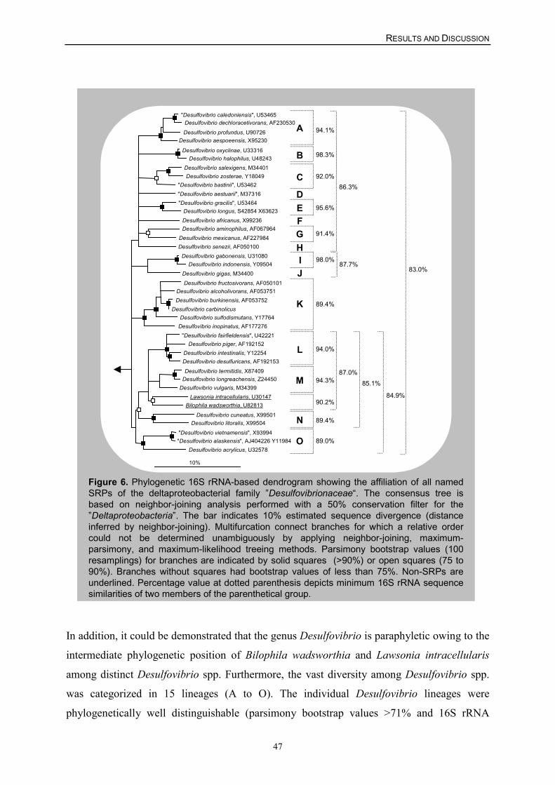
RESULTS AND DISCUSSION
47
Figure 6. Phylogenetic 16S rRNA-based dendrogram showing the affiliation of all namedSRPs of the deltaproteobacterial family ”Desulfovibrionaceae“. The consensus tree isbased on neighbor-joining analysis performed with a 50% conservation filter for the”Deltaproteobacteria”. The bar indicates 10% estimated sequence divergence (distanceinferred by neighbor-joining). Multifurcation connect branches for which a relative ordercould not be determined unambiguously by applying neighbor-joining, maximum-parsimony, and maximum-likelihood treeing methods. Parsimony bootstrap values (100resamplings) for branches are indicated by solid squares (>90%) or open squares (75 to90%). Branches without squares had bootstrap values of less than 75%. Non-SRPs areunderlined. Percentage value at dotted parenthesis depicts minimum 16S rRNA sequencesimilarities of two members of the parenthetical group.
83.0%
86.3%
"Desulfovibrio caledoniensis", U53465 Desulfovibrio dechloracetivorans, AF230530
Desulfovibrio profundus, U90726 Desulfovibrio aespoeensis, X95230
Desulfovibrio oxyclinae, U33316 Desulfovibrio halophilus, U48243
Desulfovibrio salexigens, M34401 Desulfovibrio zosterae, Y18049
"Desulfovibrio bastinii", U53462 "Desulfovibrio aestuarii", M37316 "Desulfovibrio gracilis", U53464
Desulfovibrio longus, S42854 X63623 Desulfovibrio africanus, X99236 Desulfovibrio aminophilus, AF067964
Desulfovibrio mexicanus, AF227984 Desulfovibrio senezii, AF050100
Desulfovibrio gabonensis, U31080 Desulfovibrio indonensis, Y09504
Desulfovibrio gigas, M34400 Desulfovibrio fructosivorans, AF050101
Desulfovibrio alcoholivorans, AF053751 Desulfovibrio burkinensis, AF053752
Desulfovibrio carbinolicus Desulfovibrio sulfodismutans, Y17764
Desulfovibrio inopinatus, AF177276 "Desulfovibrio fairfieldensis", U42221
Desulfovibrio piger, AF192152 Desulfovibrio intestinalis, Y12254 Desulfovibrio desulfuricans, AF192153
Desulfovibrio termitidis, X87409 Desulfovibrio longreachensis, Z24450
Desulfovibrio vulgaris, M34399 Lawsonia intracellularis, U30147
Bilophila wadsworthia, U82813
Desulfovibrio cuneatus, X99501 Desulfovibrio litoralis, X99504
"Desulfovibrio vietnamensis", X93994 "Desulfovibrio alaskensis", AJ404226 Y11984
Desulfovibrio acrylicus, U32578
10%
87.7%
89.4%
85.1%87.0%
94.3%
94.0%
90.2%
89.4%
89.0%
84.9%
94.1%
98.3%
92.0%
95.6%
91.4%
A
B
C
DEFGHIJ
K
L
M
N
O
98.0%
In addition, it could be demonstrated that the genus Desulfovibrio is paraphyletic owing to the
intermediate phylogenetic position of Bilophila wadsworthia and Lawsonia intracellularis
among distinct Desulfovibrio spp. Furthermore, the vast diversity among Desulfovibrio spp.
was categorized in 15 lineages (A to O). The individual Desulfovibrio lineages were
phylogenetically well distinguishable (parsimony bootstrap values >71% and 16S rRNA
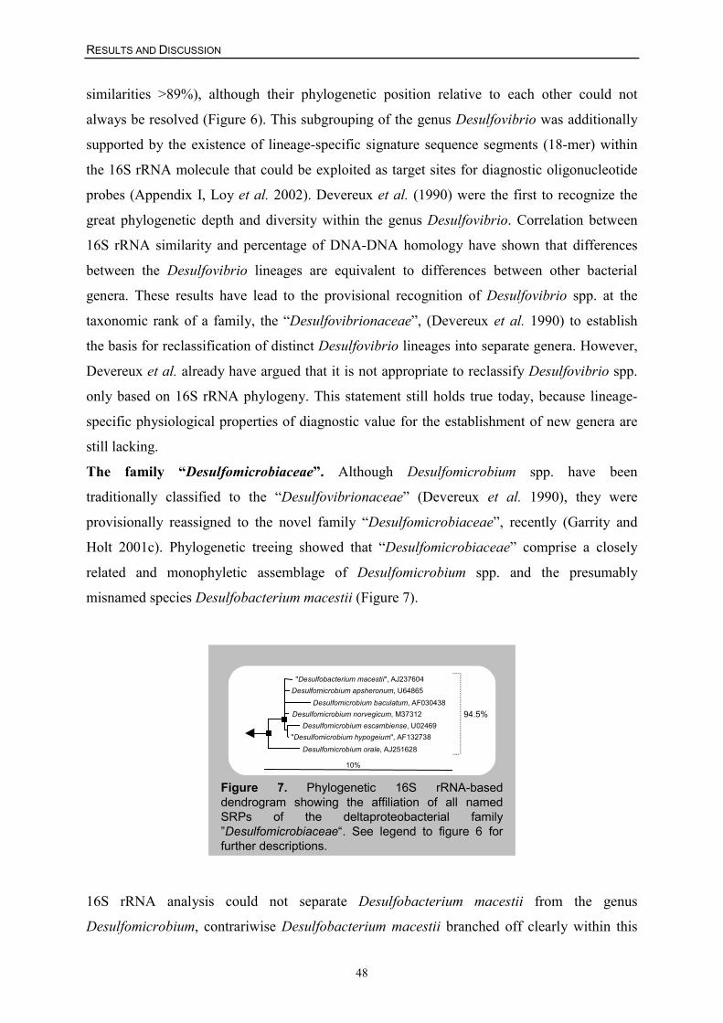
RESULTS AND DISCUSSION
48
similarities >89%), although their phylogenetic position relative to each other could not
always be resolved (Figure 6). This subgrouping of the genus Desulfovibrio was additionally
supported by the existence of lineage-specific signature sequence segments (18-mer) within
the 16S rRNA molecule that could be exploited as target sites for diagnostic oligonucleotide
probes (Appendix I, Loy et al. 2002). Devereux et al. (1990) were the first to recognize the
great phylogenetic depth and diversity within the genus Desulfovibrio. Correlation between
16S rRNA similarity and percentage of DNA-DNA homology have shown that differences
between the Desulfovibrio lineages are equivalent to differences between other bacterial
genera. These results have lead to the provisional recognition of Desulfovibrio spp. at the
taxonomic rank of a family, the “Desulfovibrionaceae”, (Devereux et al. 1990) to establish
the basis for reclassification of distinct Desulfovibrio lineages into separate genera. However,
Devereux et al. already have argued that it is not appropriate to reclassify Desulfovibrio spp.
only based on 16S rRNA phylogeny. This statement still holds true today, because lineage-
specific physiological properties of diagnostic value for the establishment of new genera are
still lacking.
The family “Desulfomicrobiaceae”. Although Desulfomicrobium spp. have been
traditionally classified to the “Desulfovibrionaceae” (Devereux et al. 1990), they were
provisionally reassigned to the novel family “Desulfomicrobiaceae”, recently (Garrity and
Holt 2001c). Phylogenetic treeing showed that “Desulfomicrobiaceae” comprise a closely
related and monophyletic assemblage of Desulfomicrobium spp. and the presumably
misnamed species Desulfobacterium macestii (Figure 7).
Figure 7. Phylogenetic 16S rRNA-baseddendrogram showing the affiliation of all namedSRPs of the deltaproteobacterial family”Desulfomicrobiaceae“. See legend to figure 6 forfurther descriptions.
"Desulfobacterium macestii", AJ237604 Desulfomicrobium apsheronum, U64865
Desulfomicrobium baculatum, AF030438 Desulfomicrobium norvegicum, M37312
Desulfomicrobium escambiense, U02469 "Desulfomicrobium hypogeium", AF132738
Desulfomicrobium orale, AJ251628
10%
94.5%
16S rRNA analysis could not separate Desulfobacterium macestii from the genus
Desulfomicrobium, contrariwise Desulfobacterium macestii branched off clearly within this
RESULTS AND DISCUSSION
49
genus (Figure 7). Based on this phylogenetic evidence, a reclassification of Desulfobacterium
macestii into the genus Desulfomicrobium should be taken into consideration.
The family “Desulfohalobiaceae”. Although the only two species of this family,
Desulfohalobium retbaense and Desulfonatronovibrio hydrogenovorans, share the ability to
tolerate high salinity (Zhilina et al. 1997), they represented two independent lines of descent
within the radiation of the “Desulfovibrionales” (Figure 5). In addition, 16S rRNA
dissimilarity of both species to each other (12.5%) was in the range of 16S rRNA
dissimilarities of both species to other “Desulfovibrionales” (11.1-17.4%). Whether
Desulfonatronovibrio hydrogenovorans is a true member of the “Desulfohalobiaceae” or
represent a novel family must await further taxonomic analysis of yet to describe SRBs which
are most closely related to Desulfohalobium retbaense or Desulfonatronovibrio
hydrogenovorans.
The family “Desulfonatronumaceae”. This family currently comprises only a single species:
the extremely alkaliphilic Desulfonatronum lacustre (Pikuta et al. 1998), which was
phylogenetically well distinguishable from other SRBs of the “Desulfovibrionales” (Figure
5).
Additional family-level diversity within the order “Desulfovibrionales”. The 16S rRNA
sequence of the newly described hydrothermal vent SRB Desulfonauticus submarinus
(Audiffrin et al. 2003) was phylogenetically in the radiation of the “Desulfovibrionales”
(Figure 5). Further assignment of Desulfonauticus submarinus to one of the proposed
“Desulfovibrionales” families was not unambiguously possible due to the low 16S rRNA
similarity of Desulfonauticus submarinus (82.8 to 86.7%) to other SRBs of this order.
According to its 16S rRNA-based genealogy, Desulfonauticus submarinus is likely a member
of a novel SRB family.
The order “Desulfobacterales”. The order “Desulfobacterales” is subdivided at present
into three provisional families, the “Desulfobacteraceae”, the “Desulfobulbaceae”, and the
“Nitrospinaceae” (Garrity and Holt 2001c). The former two families include solely bacteria
capable of anaerobic sulfate respiration.
The family “Desulfobacteraceae” (formerly “Desulfobacteriaceae”). Aside from
Desulfovibrio and Desulfomicrobium, all remaining deltaproteobacterial SRBs have been
traditionally grouped into the “Desulfobacteraceae” (Widdel and Bak 1992). Presently, this
family comprises a phenotypically and phylogenetically diverse collection of meso- and
psychrophilic SRB genera, most of which have a monophyletic origin (Figure 8).
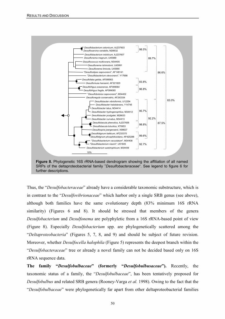
RESULTS AND DISCUSSION
50
Desulfobacterium cetonicum, AJ237603 Desulfosarcina variabilis, M26632
Desulfobacterium indolicum, AJ237607 Desulfonema magnum, U45989
Desulfococcus multivorans, M34405 Desulfonema ishimotonii, U45991 Desulfonema limicola, U45990
"Desulfostipes sapovorans", AF148141 "Desulfobacterium oleovorans", Y17698
Desulfofaba gelida, AF099063 Desulfomusa hansenii, AF321820
Desulfofrigus oceanense, AF099064 Desulfofrigus fragile, AF099065
"Desulfobotulus sapovorans", M34402 Desulforegula conservatrix, AF243334
Desulfobacter vibrioformis, U12254 Desulfobacter halotolerans, Y14745
Desulfobacter latus, M34414 Desulfobacter hydrogenophilus, M34412 Desulfobacter postgatei, M26633 Desulfobacter curvatus, M34413 Desulfobacula phenolica, AJ237606 Desulfobacula toluolica, X70953 Desulfospira joergensenii, X99637
Desulfotignum balticum, AF233370 Desulfotignum phosphitoxidans, AF420288
"Desulfobacterium vacuolatum", M34408 "Desulfobacterium niacini", U51845
Desulfobacterium autotrophicum, M34409
10%
83.0%
87.5%
86.6%
92.2%
92.7%
99.6%
98.8%
95.7%
96.8%
93.8%
98.5%
89.7%
Figure 8. Phylogenetic 16S rRNA-based dendrogram showing the affiliation of all namedSRPs of the deltaproteobacterial family ”Desulfobacteraceae“. See legend to figure 6 forfurther descriptions.
Thus, the “Desulfobacteraceae” already have a considerable taxonomic substructure, which is
in contrast to the “Desulfovibrionaceae” which harbor only a single SRB genus (see above),
although both families have the same evolutionary depth (83% minimum 16S rRNA
similarity) (Figures 6 and 8). It should be stressed that members of the genera
Desulfobacterium and Desulfonema are polyphyletic from a 16S rRNA-based point of view
(Figure 8). Especially Desulfobacterium spp. are phylogenetically scattered among the
“Deltaproteobacteria” (Figures 5, 7, 8, and 9) and should be subject of future revision.
Moreover, whether Desulfocella halophila (Figure 5) represents the deepest branch within the
“Desulfobacteraceae” tree or already a novel family can not be decided based only on 16S
rRNA sequence data.
The family “Desulfobulbaceae” (formerly “Desulfobulbusaceae”). Recently, the
taxonomic status of a family, the “Desulfobulbaceae”, has been tentatively proposed for
Desulfobulbus and related SRB genera (Rooney-Varga et al. 1998). Owing to the fact that the
“Desulfobulbaceae” were phylogenetically far apart from other deltaproteobacterial families
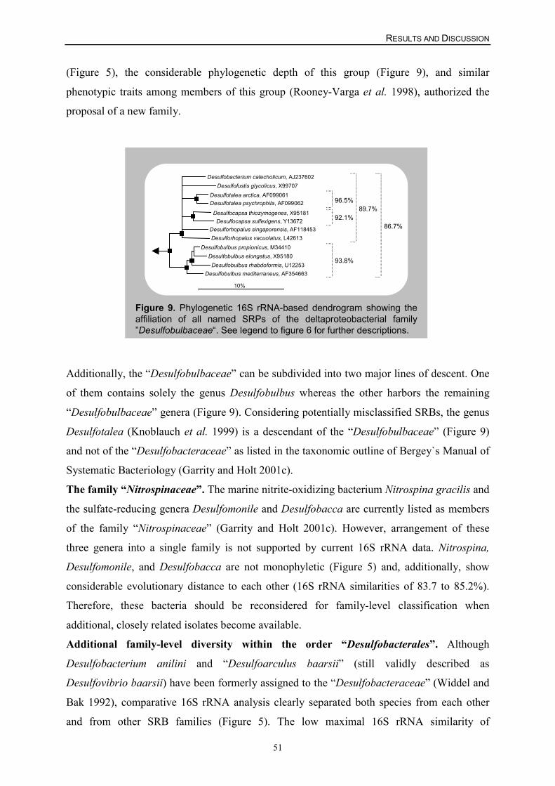
RESULTS AND DISCUSSION
51
(Figure 5), the considerable phylogenetic depth of this group (Figure 9), and similar
phenotypic traits among members of this group (Rooney-Varga et al. 1998), authorized the
proposal of a new family.
Figure 9. Phylogenetic 16S rRNA-based dendrogram showing theaffiliation of all named SRPs of the deltaproteobacterial family”Desulfobulbaceae“. See legend to figure 6 for further descriptions.
Desulfobacterium catecholicum, AJ237602
Desulforhopalus singaporensis, AF118453
Desulfofustis glycolicus, X99707 Desulfotalea arctica, AF099061 Desulfotalea psychrophila, AF099062
Desulfocapsa thiozymogenes, X95181 Desulfocapsa sulfexigens, Y13672
Desulforhopalus vacuolatus, L42613 Desulfobulbus propionicus, M34410
Desulfobulbus elongatus, X95180 Desulfobulbus rhabdoformis, U12253
Desulfobulbus mediterraneus, AF354663
10%
93.8%
86.7%
89.7%92.1%
96.5%
Additionally, the “Desulfobulbaceae” can be subdivided into two major lines of descent. One
of them contains solely the genus Desulfobulbus whereas the other harbors the remaining
“Desulfobulbaceae” genera (Figure 9). Considering potentially misclassified SRBs, the genus
Desulfotalea (Knoblauch et al. 1999) is a descendant of the “Desulfobulbaceae” (Figure 9)
and not of the “Desulfobacteraceae” as listed in the taxonomic outline of Bergey`s Manual of
Systematic Bacteriology (Garrity and Holt 2001c).
The family “Nitrospinaceae”. The marine nitrite-oxidizing bacterium Nitrospina gracilis and
the sulfate-reducing genera Desulfomonile and Desulfobacca are currently listed as members
of the family “Nitrospinaceae” (Garrity and Holt 2001c). However, arrangement of these
three genera into a single family is not supported by current 16S rRNA data. Nitrospina,
Desulfomonile, and Desulfobacca are not monophyletic (Figure 5) and, additionally, show
considerable evolutionary distance to each other (16S rRNA similarities of 83.7 to 85.2%).
Therefore, these bacteria should be reconsidered for family-level classification when
additional, closely related isolates become available.
Additional family-level diversity within the order “Desulfobacterales”. Although
Desulfobacterium anilini and “Desulfoarculus baarsii” (still validly described as
Desulfovibrio baarsii) have been formerly assigned to the “Desulfobacteraceae” (Widdel and
Bak 1992), comparative 16S rRNA analysis clearly separated both species from each other
and from other SRB families (Figure 5). The low maximal 16S rRNA similarity of
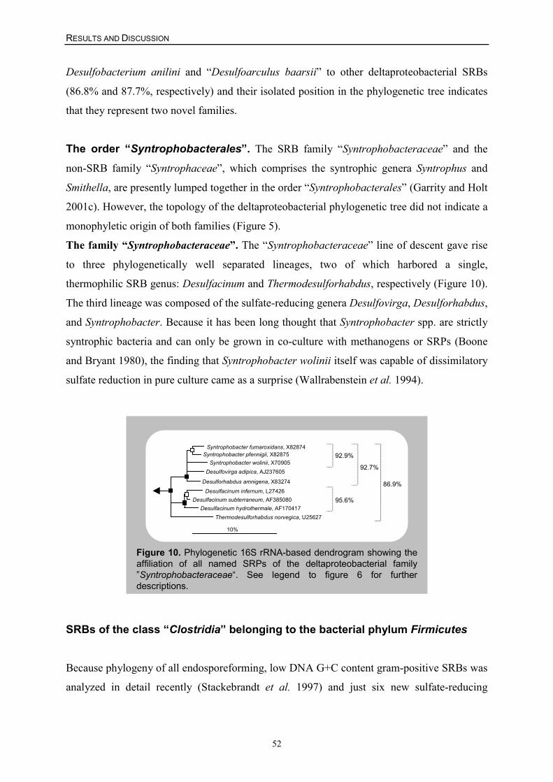
RESULTS AND DISCUSSION
52
Desulfobacterium anilini and “Desulfoarculus baarsii” to other deltaproteobacterial SRBs
(86.8% and 87.7%, respectively) and their isolated position in the phylogenetic tree indicates
that they represent two novel families.
The order “Syntrophobacterales”. The SRB family “Syntrophobacteraceae” and the
non-SRB family “Syntrophaceae”, which comprises the syntrophic genera Syntrophus and
Smithella, are presently lumped together in the order “Syntrophobacterales” (Garrity and Holt
2001c). However, the topology of the deltaproteobacterial phylogenetic tree did not indicate a
monophyletic origin of both families (Figure 5).
The family “Syntrophobacteraceae”. The “Syntrophobacteraceae” line of descent gave rise
to three phylogenetically well separated lineages, two of which harbored a single,
thermophilic SRB genus: Desulfacinum and Thermodesulforhabdus, respectively (Figure 10).
The third lineage was composed of the sulfate-reducing genera Desulfovirga, Desulforhabdus,
and Syntrophobacter. Because it has been long thought that Syntrophobacter spp. are strictly
syntrophic bacteria and can only be grown in co-culture with methanogens or SRPs (Boone
and Bryant 1980), the finding that Syntrophobacter wolinii itself was capable of dissimilatory
sulfate reduction in pure culture came as a surprise (Wallrabenstein et al. 1994).
Figure 10. Phylogenetic 16S rRNA-based dendrogram showing theaffiliation of all named SRPs of the deltaproteobacterial family”Syntrophobacteraceae“. See legend to figure 6 for furtherdescriptions.
Syntrophobacter fumaroxidans, X82874 Syntrophobacter pfennigii, X82875
Desulfovirga adipica, AJ237605 Syntrophobacter wolinii, X70905
Desulforhabdus amnigena, X83274 Desulfacinum infernum, L27426
Desulfacinum subterraneum, AF385080 Desulfacinum hydrothermale, AF170417
Thermodesulforhabdus norvegica, U25627
10%
86.9%
95.6%
92.7%
92.9%
SRBs of the class “Clostridia” belonging to the bacterial phylum Firmicutes
Because phylogeny of all endosporeforming, low DNA G+C content gram-positive SRBs was
analyzed in detail recently (Stackebrandt et al. 1997) and just six new sulfate-reducing
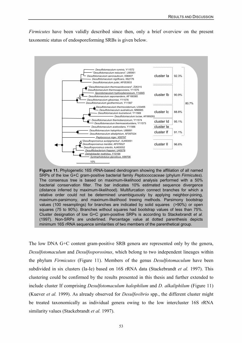
RESULTS AND DISCUSSION
53
Firmicutes have been validly described since then, only a brief overview on the present
taxonomic status of endosporeforming SRBs is given below.
Figure 11. Phylogenetic 16S rRNA-based dendrogram showing the affiliation of all namedSRPs of the low G+C gram-positive bacterial family Peptococcaceae (phylum Firmicutes).The consensus tree is based on maximum-likelihood analysis performed with a 50%bacterial conservation filter. The bar indicates 10% estimated sequence divergence(distance inferred by maximum-likelihood). Multifurcation connect branches for which arelative order could not be determined unambiguously by applying neighbor-joining,maximum-parsimony, and maximum-likelihood treeing methods. Parsimony bootstrapvalues (100 resamplings) for branches are indicated by solid squares (>90%) or opensquares (75 to 90%). Branches without squares had bootstrap values of less than 75%.Cluster designation of low G+C gram-positive SRPs is according to Stackebrandt et al.(1997). Non-SRPs are underlined. Percentage value at dotted parenthesis depictsminimum 16S rRNA sequence similarities of two members of the parenthetical group.
"Desulfotomaculum thermoacidovorans", Z26315 Desulfotomaculum thermosapovorans, Y11575
Sporotomaculum hydroxybenzoicum, Y14845 Desulfotomaculum sapomandens, AF168365
Desulfotomaculum gibsoniae, Y11576 Desulfotomaculum geothermicum, Y11567
Desulfotomaculum thermocisternum, U33455 Desulfotomaculum australicum, M96665
Desulfotomaculum kuznetsovii, Y11569 Desulfotomaculum luciae, AF069293
Desulfotomaculum thermobenzoicum, Y11574 Desulfotomaculum thermoacetoxidans, Y11573
Desulfotomaculum ruminis, Y11572 "Desulfotomaculum reducens", U95951
Desulfotomaculum aeronauticum, X98407 Desulfotomaculum nigrificans, X62176 Desulfotomaculum putei, AF053933
Desulfotomaculum halophilum, U88891 Desulfotomaculum alkaliphilum, AF097024
Desulfotomaculum acetoxidans, Y11566
Peptococcus niger, X55797“Desulfosporosinus auripigmentus“, AJ493051 Desulfosporosinus meridiei, AF076527 Desulfosporosinus orientis, AJ493052
Desulfitobacterium frappieri, U40078 Dehalobacter restrictus, Y10164
Syntrophobotulus glycolicus, X99706
10%
96.6%
91.1%
95.1%
88.8%
90.9%
92.3%
cluster II
cluster If
cluster Id
cluster Ic
cluster Ib
cluster Ia
cluster Ie
80.7%
The low DNA G+C content gram-positive SRB genera are represented only by the genera,
Desulfotomaculum and Desulfosporosinus, which belong to two independent lineages within
the phylum Firmicutes (Figure 11). Members of the genus Desulfotomaculum have been
subdivided in six clusters (Ia-Ie) based on 16S rRNA data (Stackebrandt et al. 1997). This
clustering could be confirmed by the results presented in this thesis and further extended to
include cluster If comprising Desulfotomaculum halophilum and D. alkaliphilum (Figure 11)
(Kuever et al. 1999). As already observed for Desulfovibrio spp., the different cluster might
be treated taxonomically as individual genera owing to the low intercluster 16S rRNA
similarity values (Stackebrandt et al. 1997).
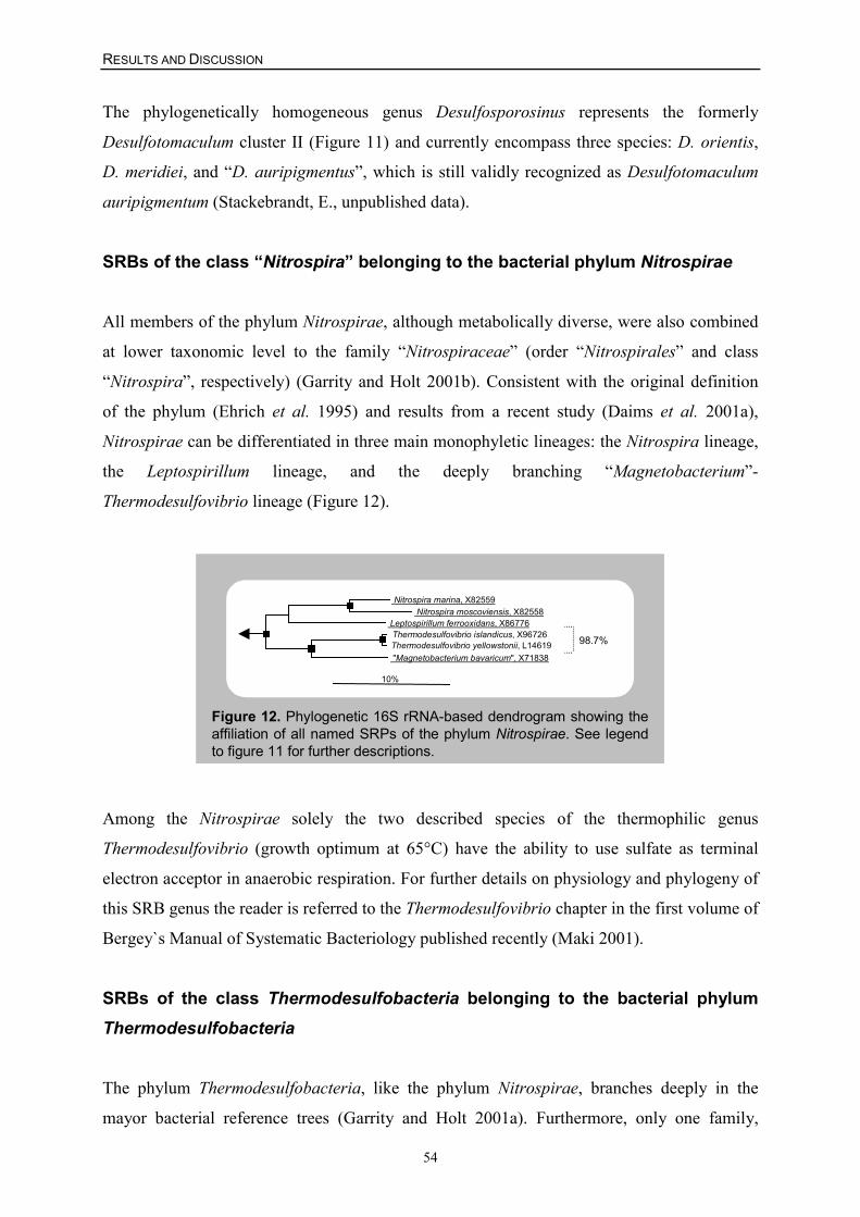
RESULTS AND DISCUSSION
54
The phylogenetically homogeneous genus Desulfosporosinus represents the formerly
Desulfotomaculum cluster II (Figure 11) and currently encompass three species: D. orientis,
D. meridiei, and “D. auripigmentus”, which is still validly recognized as Desulfotomaculum
auripigmentum (Stackebrandt, E., unpublished data).
SRBs of the class “Nitrospira” belonging to the bacterial phylum Nitrospirae
All members of the phylum Nitrospirae, although metabolically diverse, were also combined
at lower taxonomic level to the family “Nitrospiraceae” (order “Nitrospirales” and class
“Nitrospira”, respectively) (Garrity and Holt 2001b). Consistent with the original definition
of the phylum (Ehrich et al. 1995) and results from a recent study (Daims et al. 2001a),
Nitrospirae can be differentiated in three main monophyletic lineages: the Nitrospira lineage,
the Leptospirillum lineage, and the deeply branching “Magnetobacterium”-
Thermodesulfovibrio lineage (Figure 12).
Figure 12. Phylogenetic 16S rRNA-based dendrogram showing theaffiliation of all named SRPs of the phylum Nitrospirae. See legendto figure 11 for further descriptions.
Nitrospira marina, X82559 Nitrospira moscoviensis, X82558
Leptospirillum ferrooxidans, X86776 Thermodesulfovibrio islandicus, X96726 Thermodesulfovibrio yellowstonii, L14619 "Magnetobacterium bavaricum", X71838
10%
98.7%
Among the Nitrospirae solely the two described species of the thermophilic genus
Thermodesulfovibrio (growth optimum at 65°C) have the ability to use sulfate as terminal
electron acceptor in anaerobic respiration. For further details on physiology and phylogeny of
this SRB genus the reader is referred to the Thermodesulfovibrio chapter in the first volume of
Bergey`s Manual of Systematic Bacteriology published recently (Maki 2001).
SRBs of the class Thermodesulfobacteria belonging to the bacterial phylumThermodesulfobacteria
The phylum Thermodesulfobacteria, like the phylum Nitrospirae, branches deeply in the
mayor bacterial reference trees (Garrity and Holt 2001a). Furthermore, only one family,
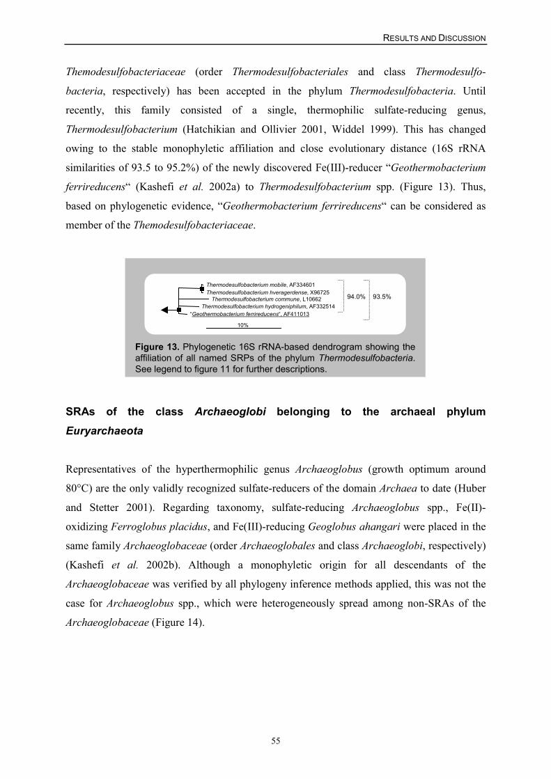
RESULTS AND DISCUSSION
55
Themodesulfobacteriaceae (order Thermodesulfobacteriales and class Thermodesulfo-
bacteria, respectively) has been accepted in the phylum Thermodesulfobacteria. Until
recently, this family consisted of a single, thermophilic sulfate-reducing genus,
Thermodesulfobacterium (Hatchikian and Ollivier 2001, Widdel 1999). This has changed
owing to the stable monophyletic affiliation and close evolutionary distance (16S rRNA
similarities of 93.5 to 95.2%) of the newly discovered Fe(III)-reducer “Geothermobacterium
ferrireducens“ (Kashefi et al. 2002a) to Thermodesulfobacterium spp. (Figure 13). Thus,
based on phylogenetic evidence, “Geothermobacterium ferrireducens“ can be considered as
member of the Themodesulfobacteriaceae.
Figure 13. Phylogenetic 16S rRNA-based dendrogram showing theaffiliation of all named SRPs of the phylum Thermodesulfobacteria.See legend to figure 11 for further descriptions.
Thermodesulfobacterium mobile, AF334601 Thermodesulfobacterium hveragerdense, X96725
Thermodesulfobacterium commune, L10662 Thermodesulfobacterium hydrogeniphilum, AF332514
“Geothermobacterium ferrireducens“, AF411013
10%
94.0% 93.5%
SRAs of the class Archaeoglobi belonging to the archaeal phylumEuryarchaeota
Representatives of the hyperthermophilic genus Archaeoglobus (growth optimum around
80°C) are the only validly recognized sulfate-reducers of the domain Archaea to date (Huber
and Stetter 2001). Regarding taxonomy, sulfate-reducing Archaeoglobus spp., Fe(II)-
oxidizing Ferroglobus placidus, and Fe(III)-reducing Geoglobus ahangari were placed in the
same family Archaeoglobaceae (order Archaeoglobales and class Archaeoglobi, respectively)
(Kashefi et al. 2002b). Although a monophyletic origin for all descendants of the
Archaeoglobaceae was verified by all phylogeny inference methods applied, this was not the
case for Archaeoglobus spp., which were heterogeneously spread among non-SRAs of the
Archaeoglobaceae (Figure 14).
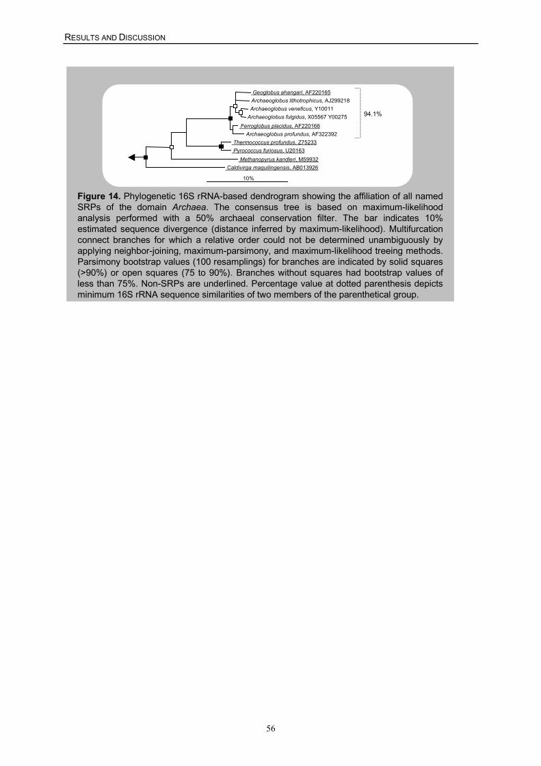
RESULTS AND DISCUSSION
56
Figure 14. Phylogenetic 16S rRNA-based dendrogram showing the affiliation of all namedSRPs of the domain Archaea. The consensus tree is based on maximum-likelihoodanalysis performed with a 50% archaeal conservation filter. The bar indicates 10%estimated sequence divergence (distance inferred by maximum-likelihood). Multifurcationconnect branches for which a relative order could not be determined unambiguously byapplying neighbor-joining, maximum-parsimony, and maximum-likelihood treeing methods.Parsimony bootstrap values (100 resamplings) for branches are indicated by solid squares(>90%) or open squares (75 to 90%). Branches without squares had bootstrap values ofless than 75%. Non-SRPs are underlined. Percentage value at dotted parenthesis depictsminimum 16S rRNA sequence similarities of two members of the parenthetical group.
94.1%
Geoglobus ahangari, AF220165 Archaeoglobus lithotrophicus, AJ299218 Archaeoglobus veneficus, Y10011
Archaeoglobus fulgidus, X05567 Y00275 Ferroglobus placidus, AF220166
Archaeoglobus profundus, AF322392 Thermococcus profundus, Z75233 Pyrococcus furiosus, U20163
Methanopyrus kandleri, M59932 Caldivirga maquilingensis, AB013926
10%
RESULTS AND DISCUSSION
57
II. In silico design and evaluation of SRP-specific, 16S rRNA-targetedoligonucleotide probes suitable for DNA microarray application
Essential for the design of a set of rRNA-targeted oligonucleotides with hierarchical
specificity is the knowledge of the phylogeny of the target groups. Hence, the 16S rRNA-
based phylogenetic framework established for SRPs in the first part of this thesis served as
reference throughout the whole, computer-assisted probe development procedure.
Initially, the specificities of previously described probes and primers for SRPs (Amann et al.
1990, Daims et al. 2000, Daly et al. 2000, Devereux et al. 1992, Fukui et al. 1999, Hristova et
al. 2000, Manz et al. 1998, Rabus et al. 1996, Ravenschlag et al. 2000, Scheid and Stubner
2001, Stubner and Meuser 2000) were reevaluated with the current 16S rRNA data set by
using the ARB PROBE_MATCH tool (Strunk and Ludwig, http://www.arb-home.de). Based
on this analysis, 27 probes were considered to be suitable for inclusion on the DNA
microarray (Table 5). These probes were, if necessary, adjusted to a length of 18 nucleotides
(not including the T-spacer). Twenty-five of these probes exclusively target SRPs. Probes
SRB385 (Amann et al. 1990) and SRB385Db (Rabus et al. 1996) were included on the
microarray because they have been widely used in previous SRP research (Amann et al. 1992,
Edgcomb et al. 1999, Li et al. 1999, Oude Elferink et al. 1998, Santegoeds et al. 1998, Teske
et al. 1996), although both probes do target a considerable number of phylogenetically diverse
non-SRPs. In addition, the existing SRP probe set was significantly extended by designing
111 probes targeting monophyletic groups of SRPs. (Table 5) (Appendix I and III, Loy et al.
2003b, Loy et al. 2002). In silico design of oligonucleotide probes was performed with ARB
tools PROBE_DESIGN and PROBE_MATCH in order to retrieve lists of all potential, SRP-
specific probes from the 16S rRNA database and, subsequently, to determine the most
specific probes (Hugenholtz et al. 2001). The probes were selected to have a minimum G+C
content of 50%, a length of 18 nucleotides (not including the T-spacer), and as many centrally
located mismatches with the target sites on 16S rRNA genes of nontarget organisms as
possible. Consistent with design formats used in previous microarray applications for
identification of other bacterial groups (Guschin et al. 1997, Liu et al. 2001), a hierarchical
set of oligonucleotides complementary to the 16S rRNA genes of the target microorganisms
at multiple levels of specificity was developed according to the “multiple probe concept”
(Amann and Schleifer 2001, Behr et al. 2000). Thus, several of these probes target the same
SRPs, complementing several unique regions of the 16S rRNA gene, while others exhibit
hierarchical specificity. For example, the genus Desulfotalea is specifically detected by five
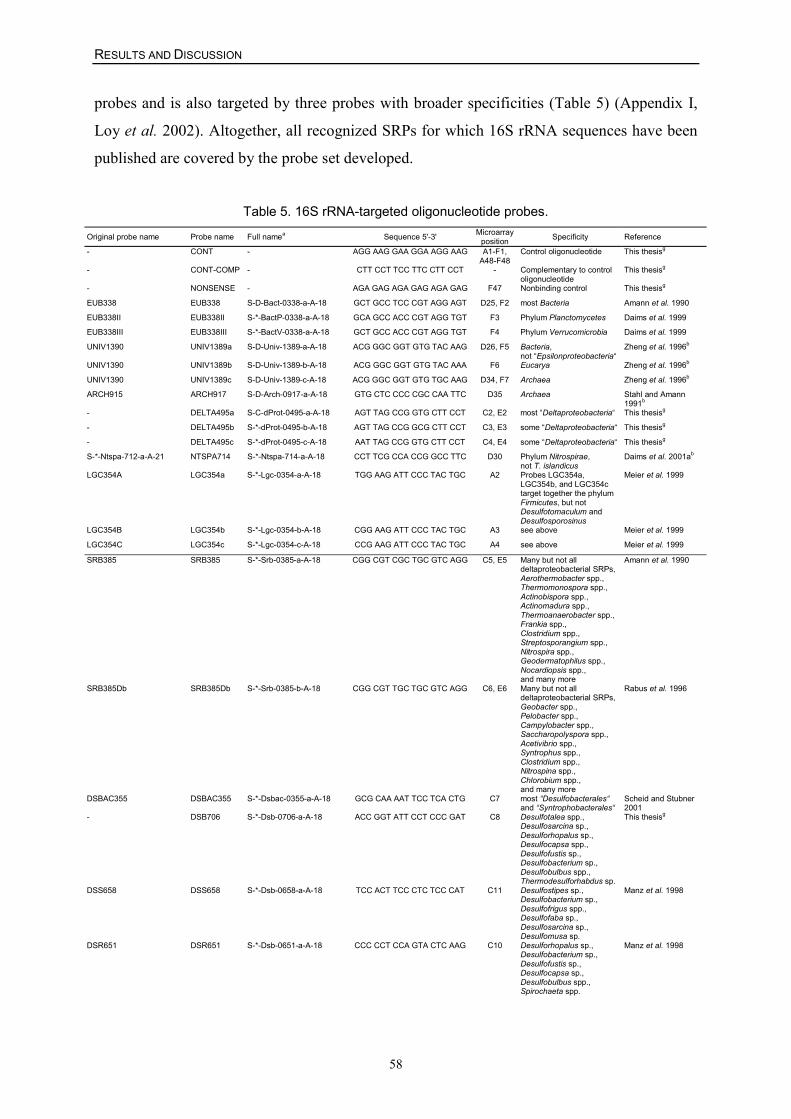
RESULTS AND DISCUSSION
58
probes and is also targeted by three probes with broader specificities (Table 5) (Appendix I,
Loy et al. 2002). Altogether, all recognized SRPs for which 16S rRNA sequences have been
published are covered by the probe set developed.
Table 5. 16S rRNA-targeted oligonucleotide probes.
Original probe name Probe name Full namea Sequence 5'-3' Microarrayposition Specificity Reference
- CONT - AGG AAG GAA GGA AGG AAG A1-F1,A48-F48
Control oligonucleotide This thesisg
- CONT-COMP - CTT CCT TCC TTC CTT CCT - Complementary to controloligonucleotide
This thesisg
- NONSENSE - AGA GAG AGA GAG AGA GAG F47 Nonbinding control This thesisg
EUB338 EUB338 S-D-Bact-0338-a-A-18 GCT GCC TCC CGT AGG AGT D25, F2 most Bacteria Amann et al. 1990
EUB338II EUB338II S-*-BactP-0338-a-A-18 GCA GCC ACC CGT AGG TGT F3 Phylum Planctomycetes Daims et al. 1999
EUB338III EUB338III S-*-BactV-0338-a-A-18 GCT GCC ACC CGT AGG TGT F4 Phylum Verrucomicrobia Daims et al. 1999
UNIV1390 UNIV1389a S-D-Univ-1389-a-A-18 ACG GGC GGT GTG TAC AAG D26, F5 Bacteria,not “Epsilonproteobacteria“
Zheng et al. 1996b
UNIV1390 UNIV1389b S-D-Univ-1389-b-A-18 ACG GGC GGT GTG TAC AAA F6 Eucarya Zheng et al. 1996b
UNIV1390 UNIV1389c S-D-Univ-1389-c-A-18 ACG GGC GGT GTG TGC AAG D34, F7 Archaea Zheng et al. 1996b
ARCH915 ARCH917 S-D-Arch-0917-a-A-18 GTG CTC CCC CGC CAA TTC D35 Archaea Stahl and Amann1991b
- DELTA495a S-C-dProt-0495-a-A-18 AGT TAG CCG GTG CTT CCT C2, E2 most “Deltaproteobacteria“ This thesisg
- DELTA495b S-*-dProt-0495-b-A-18 AGT TAG CCG GCG CTT CCT C3, E3 some “Deltaproteobacteria“ This thesisg
- DELTA495c S-*-dProt-0495-c-A-18 AAT TAG CCG GTG CTT CCT C4, E4 some “Deltaproteobacteria“ This thesisg
S-*-Ntspa-712-a-A-21 NTSPA714 S-*-Ntspa-714-a-A-18 CCT TCG CCA CCG GCC TTC D30 Phylum Nitrospirae,not T. islandicus
Daims et al. 2001ab
LGC354A LGC354a S-*-Lgc-0354-a-A-18 TGG AAG ATT CCC TAC TGC A2 Probes LGC354a,LGC354b, and LGC354ctarget together the phylumFirmicutes, but notDesulfotomaculum andDesulfosporosinus
Meier et al. 1999
LGC354B LGC354b S-*-Lgc-0354-b-A-18 CGG AAG ATT CCC TAC TGC A3 see above Meier et al. 1999
LGC354C LGC354c S-*-Lgc-0354-c-A-18 CCG AAG ATT CCC TAC TGC A4 see above Meier et al. 1999
SRB385 SRB385 S-*-Srb-0385-a-A-18 CGG CGT CGC TGC GTC AGG C5, E5 Many but not alldeltaproteobacterial SRPs,Aerothermobacter spp.,Thermomonospora spp.,Actinobispora spp.,Actinomadura spp.,Thermoanaerobacter spp.,Frankia spp.,Clostridium spp.,Streptosporangium spp.,Nitrospira spp.,Geodermatophilus spp.,Nocardiopsis spp.,and many more
Amann et al. 1990
SRB385Db SRB385Db S-*-Srb-0385-b-A-18 CGG CGT TGC TGC GTC AGG C6, E6 Many but not alldeltaproteobacterial SRPs,Geobacter spp.,Pelobacter spp.,Campylobacter spp.,Saccharopolyspora spp.,Acetivibrio spp.,Syntrophus spp.,Clostridium spp.,Nitrospina spp.,Chlorobium spp.,and many more
Rabus et al. 1996
DSBAC355 DSBAC355 S-*-Dsbac-0355-a-A-18 GCG CAA AAT TCC TCA CTG C7 most “Desulfobacterales“and “Syntrophobacterales“
Scheid and Stubner2001
- DSB706 S-*-Dsb-0706-a-A-18 ACC GGT ATT CCT CCC GAT C8 Desulfotalea spp.,Desulfosarcina sp.,Desulforhopalus sp.,Desulfocapsa spp.,Desulfofustis sp.,Desulfobacterium sp.,Desulfobulbus spp.,Thermodesulforhabdus sp.
This thesisg
DSS658 DSS658 S-*-Dsb-0658-a-A-18 TCC ACT TCC CTC TCC CAT C11 Desulfostipes sp.,Desulfobacterium sp.,Desulfofrigus spp.,Desulfofaba sp.,Desulfosarcina sp.,Desulfomusa sp.
Manz et al. 1998
DSR651 DSR651 S-*-Dsb-0651-a-A-18 CCC CCT CCA GTA CTC AAG C10 Desulforhopalus sp.,Desulfobacterium sp.,Desulfofustis sp.,Desulfocapsa sp.,Desulfobulbus spp.,Spirochaeta spp.
Manz et al. 1998
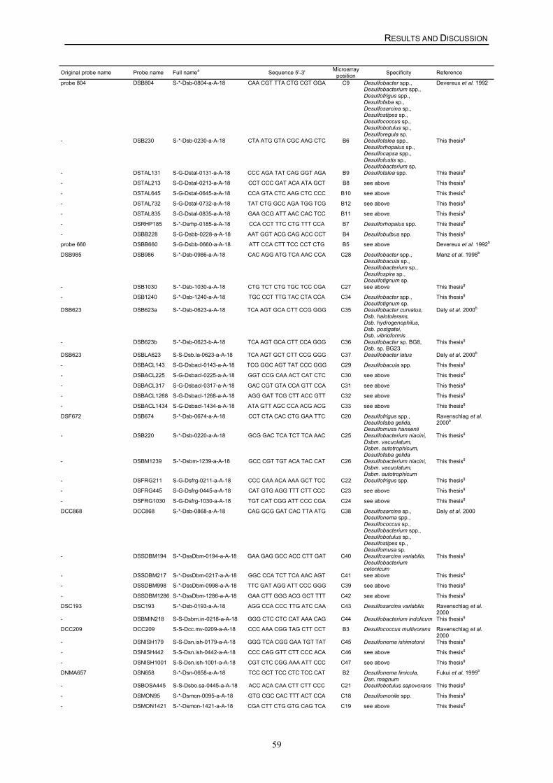
RESULTS AND DISCUSSION
59
Original probe name Probe name Full namea Sequence 5'-3' Microarrayposition Specificity Reference
probe 804 DSB804 S-*-Dsb-0804-a-A-18 CAA CGT TTA CTG CGT GGA C9 Desulfobacter spp.,Desulfobacterium spp.,Desulfofrigus spp.,Desulfofaba sp.,Desulfosarcina sp.,Desulfostipes sp.,Desulfococcus sp.,Desulfobotulus sp.,Desulforegula sp.
Devereux et al. 1992
- DSB230 S-*-Dsb-0230-a-A-18 CTA ATG GTA CGC AAG CTC B6 Desulfotalea spp.,Desulforhopalus sp.,Desulfocapsa spp.,Desulfofustis sp.,Desulfobacterium sp.
This thesisg
- DSTAL131 S-G-Dstal-0131-a-A-18 CCC AGA TAT CAG GGT AGA B9 Desulfotalea spp. This thesisg
- DSTAL213 S-G-Dstal-0213-a-A-18 CCT CCC GAT ACA ATA GCT B8 see above This thesisg
- DSTAL645 S-G-Dstal-0645-a-A-18 CCA GTA CTC AAG CTC CCC B10 see above This thesisg
- DSTAL732 S-G-Dstal-0732-a-A-18 TAT CTG GCC AGA TGG TCG B12 see above This thesisg
- DSTAL835 S-G-Dstal-0835-a-A-18 GAA GCG ATT AAC CAC TCC B11 see above This thesisg
- DSRHP185 S-*-Dsrhp-0185-a-A-18 CCA CCT TTC CTG TTT CCA B7 Desulforhopalus spp. This thesisg
- DSBB228 S-G-Dsbb-0228-a-A-18 AAT GGT ACG CAG ACC CCT B4 Desulfobulbus spp. This thesisg
probe 660 DSBB660 S-G-Dsbb-0660-a-A-18 ATT CCA CTT TCC CCT CTG B5 see above Devereux et al. 1992b
DSB985 DSB986 S-*-Dsb-0986-a-A-18 CAC AGG ATG TCA AAC CCA C28 Desulfobacter spp.,Desulfobacula sp.,Desulfobacterium sp.,Desulfospira sp.,Desulfotignum sp.
Manz et al. 1998b
- DSB1030 S-*-Dsb-1030-a-A-18 CTG TCT CTG TGC TCC CGA C27 see above This thesisg
- DSB1240 S-*-Dsb-1240-a-A-18 TGC CCT TTG TAC CTA CCA C34 Desulfobacter spp.,Desulfotignum sp.
This thesisg
DSB623 DSB623a S-*-Dsb-0623-a-A-18 TCA AGT GCA CTT CCG GGG C35 Desulfobacter curvatus,Dsb. halotolerans,Dsb. hydrogenophilus,Dsb. postgatei,Dsb. vibrioformis
Daly et al. 2000b
- DSB623b S-*-Dsb-0623-b-A-18 TCA AGT GCA CTT CCA GGG C36 Desulfobacter sp. BG8,Dsb. sp. BG23
This thesisg
DSB623 DSBLA623 S-S-Dsb.la-0623-a-A-18 TCA AGT GCT CTT CCG GGG C37 Desulfobacter latus Daly et al. 2000b
- DSBACL143 S-G-Dsbacl-0143-a-A-18 TCG GGC AGT TAT CCC GGG C29 Desulfobacula spp. This thesisg
- DSBACL225 S-G-Dsbacl-0225-a-A-18 GGT CCG CAA ACT CAT CTC C30 see above This thesisg
- DSBACL317 S-G-Dsbacl-0317-a-A-18 GAC CGT GTA CCA GTT CCA C31 see above This thesisg
- DSBACL1268 S-G-Dsbacl-1268-a-A-18 AGG GAT TCG CTT ACC GTT C32 see above This thesisg
- DSBACL1434 S-G-Dsbacl-1434-a-A-18 ATA GTT AGC CCA ACG ACG C33 see above This thesisg
DSF672 DSB674 S-*-Dsb-0674-a-A-18 CCT CTA CAC CTG GAA TTC C20 Desulfofrigus spp.,Desulfofaba gelida,Desulfomusa hansenii
Ravenschlag et al.2000b
- DSB220 S-*-Dsb-0220-a-A-18 GCG GAC TCA TCT TCA AAC C25 Desulfobacterium niacini,Dsbm. vacuolatum,Dsbm. autotrophicum,Desulfofaba gelida
This thesisg
- DSBM1239 S-*-Dsbm-1239-a-A-18 GCC CGT TGT ACA TAC CAT C26 Desulfobacterium niacini,Dsbm. vacuolatum,Dsbm. autotrophicum
This thesisg
- DSFRG211 S-G-Dsfrg-0211-a-A-18 CCC CAA ACA AAA GCT TCC C22 Desulfofrigus spp. This thesisg
- DSFRG445 S-G-Dsfrg-0445-a-A-18 CAT GTG AGG TTT CTT CCC C23 see above This thesisg
- DSFRG1030 S-G-Dsfrg-1030-a-A-18 TGT CAT CGG ATT CCC CGA C24 see above This thesisg
DCC868 DCC868 S-*-Dsb-0868-a-A-18 CAG GCG GAT CAC TTA ATG C38 Desulfosarcina sp.,Desulfonema spp.,Desulfococcus sp.,Desulfobacterium spp.,Desulfobotulus sp.,Desulfostipes sp.,Desulfomusa sp.
Daly et al. 2000
- DSSDBM194 S-*-DssDbm-0194-a-A-18 GAA GAG GCC ACC CTT GAT C40 Desulfosarcina variabilis,Desulfobacteriumcetonicum
This thesisg
- DSSDBM217 S-*-DssDbm-0217-a-A-18 GGC CCA TCT TCA AAC AGT C41 see above This thesisg
- DSSDBM998 S-*-DssDbm-0998-a-A-18 TTC GAT AGG ATT CCC GGG C39 see above This thesisg
- DSSDBM1286 S-*-DssDbm-1286-a-A-18 GAA CTT GGG ACG GCT TTT C42 see above This thesisg
DSC193 DSC193 S-*-Dsb-0193-a-A-18 AGG CCA CCC TTG ATC CAA C43 Desulfosarcina variabilis Ravenschlag et al.2000
- DSBMIN218 S-S-Dsbm.in-0218-a-A-18 GGG CTC CTC CAT AAA CAG C44 Desulfobacterium indolicum This thesisg
DCC209 DCC209 S-S-Dcc.mv-0209-a-A-18 CCC AAA CGG TAG CTT CCT B3 Desulfococcus multivorans Ravenschlag et al.2000
- DSNISH179 S-S-Dsn.ish-0179-a-A-18 GGG TCA CGG GAA TGT TAT C45 Desulfonema ishimotonii This thesisg
- DSNISH442 S-S-Dsn.ish-0442-a-A-18 CCC CAG GTT CTT CCC ACA C46 see above This thesisg
- DSNISH1001 S-S-Dsn.ish-1001-a-A-18 CGT CTC CGG AAA ATT CCC C47 see above This thesisg
DNMA657 DSN658 S-*-Dsn-0658-a-A-18 TCC GCT TCC CTC TCC CAT B2 Desulfonema limicola,Dsn. magnum
Fukui et al. 1999b
- DSBOSA445 S-S-Dsbo.sa-0445-a-A-18 ACC ACA CAA CTT CTT CCC C21 Desulfobotulus sapovorans This thesisg
- DSMON95 S-*-Dsmon-0095-a-A-18 GTG CGC CAC TTT ACT CCA C18 Desulfomonile spp. This thesisg
- DSMON1421 S-*-Dsmon-1421-a-A-18 CGA CTT CTG GTG CAG TCA C19 see above This thesisg
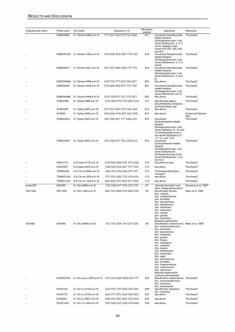
RESULTS AND DISCUSSION
60
Original probe name Probe name Full namea Sequence 5'-3' Microarrayposition Specificity Reference
- DSMON999 S-*-Dsmon-0999-a-A-18 TTT CCA TAG CTG TCC GGG B17 Uncultured Desulfomonile-related bacteria(Schlöppnerbrunnen I soilclones SbIDsmon2, 3, 4, 5,and 8; Cadagno Lakeclones 618, 624, 626, 650,and 651)
This thesish
- DSMON1283 S-*-Dsmon-1283-a-A-18 CTG AGG ACC GAT TTG TGG B18 Uncultured Desulfomonile-related bacteria(Schlöppnerbrunnen I soilclones SbIDsmon2, 3, 4, 5,and 8)
This thesish
- DSMON447 S-*-Dsmon-0447-a-A-18 ACT CAT GGA GGG TTC TTC B19 Uncultured Desulfomonile-related bacteria(Schlöppnerbrunnen I soilclones SbIDsmon3, 5, and8)
This thesish
- DSMON468a S-*-Dsmon-0468-a-A-18 CCG TCA TTT CCA TGA GCT B20 See above This thesish
- DSMON446 S-*-Dsmon-0446-a-A-18 CTA GAA GAG GTT TCT TCC B21 Uncultured Desulfomonile-related bacteria(Schlöppnerbrunnen I soilclones SbIDsmon2 and 4)
This thesish
- DSMON468b S-*-Dsmon-0468-b-A-18 CCG TCA GTT CCT CTA GCT B22 See above This thesish
- SYBAC986 S-*-Sybac-0986-a-A-18 CCG GGG ATG TCA AGC CCA C17 Desulfovirga adipica,Desulforhabdus amnigena,Syntrophobacter spp.
This thesisg
- SYBAC697 S-*-Sybac-0697-a-A-18 CCT CCC GAT CTC TAC GAA B13 See above This thesish
- SYN835 S-*-Sybac-0835-a-A-18 GCA GGA ATG AGT ACC CGC B14 See above Scheid and Stubner2001
- SYBAC587a S-*-Sybac-0587-a-A-18 CAT CAG ACT TTT CGG CCC B15 UnculturedSyntrophobacter-relatedbacteria(Schlöppnerbrunnen I soilclones SbISybac13, 15, and19; Schlöppnerbrunnen IIsoil clones SbIISybac12-1,1-2, 3-2, and 13-2)
This thesish
- SYBAC587b S-*-Sybac-0587-b-A-18 CAT CAG ACT TGC CGG CCC B16 UnculturedSyntrophobacter-relatedbacteria(Schlöppnerbrunnen I soilclones SbISybac16;Schlöppnerbrunnen II soilclones SbIISybac25-1, 6-2,and 8-2)
This thesish
- DSACI175 S-G-Dsaci-0175-a-A-18 CCG AAG GGA CGT ATC CGG C16 Desulfacinum spp. This thesisg
- DSACI207 S-G-Dsaci-0207-a-A-18 CGA ACA CCA GCT TCT TCG C15 see above This thesisg
- TDRNO448 S-S-Tdr.no-0448-a-A-18 AAC CCC ATG AAG GTT CTT C13 Thermodesulforhabdusnorvegica
This thesisg
- TDRNO1030 S-S-Tdr.no-1030-a-A-18 TCT CCC GGC TCC CCA ATA C12 see above This thesisg
- TDRNO1443 S-S-Tdr.no-1443-a-A-18 GAC ACA ATC GCG GTT GGC C14 see above This thesisg
probe 687 DSV686 S-*-Dsv-0686-a-A-18 CTA CGG ATT TCA CTC CTA E7 “Desulfovibrionales“ andother “Deltaproteobacteria“
Devereux et al. 1992b
DSV1292 DSV1292 S-*-Dsv-1292-a-A-18 CAA TCC GGA CTG GGA CGC E9 Desulfovibrio litoralis,Dsv. vulgaris,Dsv. longreachensis,Dsv. termitidis,Dsv. desulfuricans,Dsv. fairfieldensis,Dsv. intestinalis,Dsv. inopinatus,Dsv. senezii,Dsv. gracilis,Dsv. halophilus,Bilophila wadsworthia
Manz et al. 1998
DSV698 DSV698 S-*-Dsv-0698-a-A-18 TCC TCC AGA TAT CTA CGG E8 Desulfovibrio caledoniensis,Dsv. dechloracetivorans,Dsv. profundus,Dsv. aespoeensis,Dsv. halophilus,Dsv. gracilis,Dsv. longus,Dsv. salexigens,Dsv. zosterae,Dsv. bastinii,Dsv. fairfieldensis,Dsv. intestinalis,Dsv. piger,Dsv. desulfuricans,Dsv. termitidis,Dsv. longreachensis,Dsv. vietnamensis,Dsv. alaskensis,Bilophila wadsworthia,Lawsonia intracellularis
Manz et al. 1998b
- DVDAPC872 S-*-Dv.d.a.p.c-0872-a-A-18 TCC CCA GGC GGG ATA TTT E33 Desulfovibrio caledoniensis,Dsv. dechloracetivorans,Dsv. profundus,Dsv. aespoeensis
This thesisg
- DVHO130 S-*-Dv.h.o-0130-a-A-18 CCG ATC TGT CGG GTA GAT E36 Desulfovibrio halophilus,Dsv. oxyclinae
This thesisg
- DVHO733 S-*-Dv.h.o-0733-a-A-18 GAA CTT GTC CAG CAG GCC E37 see above This thesisg
- DVHO831 S-*-Dv.h.o-0831-a-A-18 GAA CCC AAC GGC CCG ACA E35 see above This thesisg
- DVHO1424 S-*-Dv.h.o-1424-a-A-18 TGC CGA CGT CGG GTA AGA E38 see above This thesisg
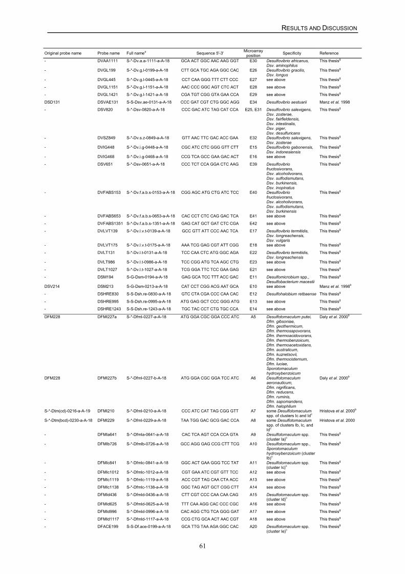
RESULTS AND DISCUSSION
61
Original probe name Probe name Full namea Sequence 5'-3' Microarrayposition Specificity Reference
- DVAA1111 S-*-Dv.a.a-1111-a-A-18 GCA ACT GGC AAC AAG GGT E30 Desulfovibrio africanus,Dsv. aminophilus
This thesisg
- DVGL199 S-*-Dv.g.l-0199-a-A-18 CTT GCA TGC AGA GGC CAC E26 Desulfovibrio gracilis,Dsv. longus
This thesisg
- DVGL445 S-*-Dv.g.l-0445-a-A-18 CCT CAA GGG TTT CTT CCC E27 see above This thesisg
- DVGL1151 S-*-Dv.g.l-1151-a-A-18 AAC CCC GGC AGT CTC ACT E28 see above This thesisg
- DVGL1421 S-*-Dv.g.l-1421-a-A-18 CGA TGT CGG GTA GAA CCA E29 see above This thesisg
DSD131 DSVAE131 S-S-Dsv.ae-0131-a-A-18 CCC GAT CGT CTG GGC AGG E34 Desulfovibrio aestuarii Manz et al. 1998
- DSV820 S-*-Dsv-0820-a-A-18 CCC GAC ATC TAG CAT CCA E25, E31 Desulfovibrio salexigens,Dsv. zosterae,Dsv. fairfieldensis,Dsv. intestinalis,Dsv. piger,Dsv. desulfuricans
This thesisg
- DVSZ849 S-*-Dv.s.z-0849-a-A-18 GTT AAC TTC GAC ACC GAA E32 Desulfovibrio salexigens,Dsv. zosterae
This thesisg
- DVIG448 S-*-Dv.i.g-0448-a-A-18 CGC ATC CTC GGG GTT CTT E15 Desulfovibrio gabonensis,Dsv. indonesiensis
This thesisg
- DVIG468 S-*-Dv.i.g-0468-a-A-18 CCG TCA GCC GAA GAC ACT E16 see above This thesisg
- DSV651 S-*-Dsv-0651-a-A-18 CCC TCT CCA GGA CTC AAG E39 Desulfovibriofructosivorans,Dsv. alcoholivorans,Dsv. sulfodismutans,Dsv. burkinensis,Dsv. inopinatus
This thesisg
- DVFABS153 S-*-Dv.f.a.b.s-0153-a-A-18 CGG AGC ATG CTG ATC TCC E40 Desulfovibriofructosivorans,Dsv. alcoholivorans,Dsv. sulfodismutans,Dsv. burkinensis
This thesisg
- DVFABS653 S-*-Dv.f.a.b.s-0653-a-A-18 CAC CCT CTC CAG GAC TCA E41 see above This thesisg
- DVFABS1351 S-*-Dv.f.a.b.s-1351-a-A-18 GAG CAT GCT GAT CTC CGA E42 see above This thesisg
- DVLVT139 S-*-Dv.l.v.t-0139-a-A-18 GCC GTT ATT CCC AAC TCA E17 Desulfovibrio termitidis,Dsv. longreachensis,Dsv. vulgaris
This thesisg
- DVLVT175 S-*-Dv.l.v.t-0175-a-A-18 AAA TCG GAG CGT ATT CGG E18 see above This thesisg
- DVLT131 S-*-Dv.l.t-0131-a-A-18 TCC CAA CTC ATG GGC AGA E22 Desulfovibrio termitidis,Dsv. longreachensis
This thesisg
- DVLT986 S-*-Dv.l.t-0986-a-A-18 TCC CGG ATG TCA AGC CTG E23 see above This thesisg
- DVLT1027 S-*-Dv.l.t-1027-a-A-18 TCG GGA TTC TCC GAA GAG E21 see above This thesisg
- DSM194 S-G-Dsm-0194-a-A-18 GAG GCA TCC TTT ACC GAC E11 Desulfomicrobium spp.,Desulfobacterium macestii
This thesisg
DSV214 DSM213 S-G-Dsm-0213-a-A-18 CAT CCT CGG ACG AAT GCA E10 see above Manz et al. 1998b
- DSHRE830 S-S-Dsh.re-0830-a-A-18 GTC CTA CGA CCC CAA CAC E12 Desulfohalobium retbaense This thesisg
- DSHRE995 S-S-Dsh.re-0995-a-A-18 ATG GAG GCT CCC GGG ATG E13 see above This thesisg
- DSHRE1243 S-S-Dsh.re-1243-a-A-18 TGC TAC CCT CTG TGC CCA E14 see above This thesisg
DFM228 DFMI227a S-*-DfmI-0227-a-A-18 ATG GGA CGC GGA CCC ATC A5 Desulfotomaculum putei,Dfm. gibsoniae,Dfm. geothermicum,Dfm. thermosapovorans,Dfm. thermoacidovorans,Dfm. thermobenzoicum,Dfm. thermoacetoxidans,Dfm. australicum,Dfm. kuznetsovii,Dfm. thermocisternum,Dfm. luciae,Sporotomaculumhydroxybenzoicum
Daly et al. 2000b
DFM228 DFMI227b S-*-DfmI-0227-b-A-18 ATG GGA CGC GGA TCC ATC A6 Desulfotomaculumaeronauticum,Dfm. nigrificans,Dfm. reducens,Dfm. ruminis,Dfm. sapomandens,Dfm. halophilum
Daly et al. 2000b
S-*-Dtm(cd)-0216-a-A-19 DFMI210 S-*-DfmI-0210-a-A-18 CCC ATC CAT TAG CGG GTT A7 some Desulfotomaculumspp. of clusters Ic and Idc
Hristova et al. 2000b
S-*-Dtm(bcd)-0230-a-A-18 DFMI229 S-*-DfmI-0229-a-A-18 TAA TGG GAC GCG GAC CCA A8 some Desulfotomaculumspp. of clusters Ib, Ic, andIdc
Hristova et al. 2000
- DFMIa641 S-*-DfmIa-0641-a-A-18 CAC TCA AGT CCA CCA GTA A9 Desulfotomaculum spp.(cluster Ia)c
This thesisg
- DFMIb726 S-*-DfmIb-0726-a-A-18 GCC AGG GAG CCG CTT TCG A10 Desulfotomaculum spp.,Sporotomaculumhydroxybenzoicum (clusterIb)c
This thesisg
- DFMIc841 S-*-DfmIc-0841-a-A-18 GGC ACT GAA GGG TCC TAT A11 Desulfotomaculum spp.(cluster Ic)c
This thesisg
- DFMIc1012 S-*-DfmIc-1012-a-A-18 CGT GAA ATC CGT GTT TCC A12 see above This thesisg
- DFMIc1119 S-*-DfmIc-1119-a-A-18 ACC CGT TAG CAA CTA ACC A13 see above This thesisg
- DFMIc1138 S-*-DfmIc-1138-a-A-18 GGC TAG AGT GCT CGG CTT A14 see above This thesisg
- DFMId436 S-*-DfmId-0436-a-A-18 CTT CGT CCC CAA CAA CAG A15 Desulfotomaculum spp.(cluster Id)c
This thesisg
- DFMId625 S-*-DfmId-0625-a-A-18 TTT CAA AGG CAC CCC CGC A16 see above This thesisg
- DFMId996 S-*-DfmId-0996-a-A-18 CAC AGG CTG TCA GGG GAT A17 see above This thesisg
- DFMId1117 S-*-DfmId-1117-a-A-18 CCG CTG GCA ACT AAC CGT A18 see above This thesisg
- DFACE199 S-S-Df.ace-0199-a-A-18 GCA TTG TAA AGA GGC CAC A20 Desulfotomaculum spp.(cluster Ie)c
This thesisg
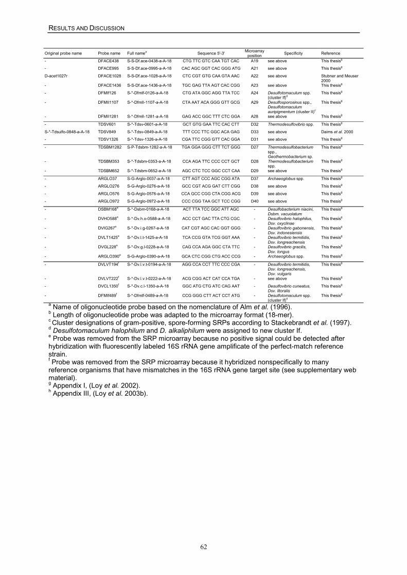
RESULTS AND DISCUSSION
62
Original probe name Probe name Full namea Sequence 5'-3' Microarrayposition Specificity Reference
- DFACE438 S-S-Df.ace-0438-a-A-18 CTG TTC GTC CAA TGT CAC A19 see above This thesisg
- DFACE995 S-S-Df.ace-0995-a-A-18 CAC AGC GGT CAC GGG ATG A21 see above This thesisg
D-acet1027r DFACE1028 S-S-Df.ace-1028-a-A-18 CTC CGT GTG CAA GTA AAC A22 see above Stubner and Meuser2000
- DFACE1436 S-S-Df.ace-1436-a-A-18 TGC GAG TTA AGT CAC CGG A23 see above This thesisg
- DFMIf126 S-*-DfmIf-0126-a-A-18 CTG ATA GGC AGG TTA TCC A24 Desulfotomaculum spp.(cluster If)d
This thesisg
- DFMII1107 S-*-DfmII-1107-a-A-18 CTA AAT ACA GGG GTT GCG A29 Desulfosporosinus spp.,Desulfotomaculumauripigmentum (cluster II)c
This thesisg
- DFMII1281 S-*-DfmII-1281-a-A-18 GAG ACC GGC TTT CTC GGA A28 see above This thesisg
- TDSV601 S-*-Tdsv-0601-a-A-18 GCT GTG GAA TTC CAC CTT D32 Thermodesulfovibrio spp. This thesisg
S-*-Tdsulfo-0848-a-A-18 TDSV849 S-*-Tdsv-0849-a-A-18 TTT CCC TTC GGC ACA GAG D33 see above Daims et al. 2000
- TDSV1326 S-*-Tdsv-1326-a-A-18 CGA TTC CGG GTT CAC GGA D31 see above This thesisg
- TDSBM1282 S-P-Tdsbm-1282-a-A-18 TGA GGA GGG CTT TCT GGG D27 Thermodesulfobacteriumspp.,Geothermobacterium sp.
This thesisg
- TDSBM353 S-*-Tdsbm-0353-a-A-18 CCA AGA TTC CCC CCT GCT D28 Thermodesulfobacteriumspp.
This thesisg
- TDSBM652 S-*-Tdsbm-0652-a-A-18 AGC CTC TCC GGC CCT CAA D29 see above This thesisg
- ARGLO37 S-G-Arglo-0037-a-A-18 CTT AGT CCC AGC CGG ATA D37 Archaeoglobus spp. This thesisg
- ARGLO276 S-G-Arglo-0276-a-A-18 GCC CGT ACG GAT CTT CGG D38 see above This thesisg
- ARGLO576 S-G-Arglo-0576-a-A-18 CCA GCC CGG CTA CGG ACG D39 see above This thesisg
- ARGLO972 S-G-Arglo-0972-a-A-18 CCC CGG TAA GCT TCC CGG D40 see above This thesisg
- DSBM168e S-*-Dsbm-0168-a-A-18 ACT TTA TCC GGC ATT AGC - Desulfobacterium niacini,Dsbm. vacuolatum
This thesisg
- DVHO588e S-*-Dv.h.o-0588-a-A-18 ACC CCT GAC TTA CTG CGC - Desulfovibrio halophilus,Dsv. oxyclinae
This thesisg
- DVIG267e S-*-Dv.i.g-0267-a-A-18 CAT CGT AGC CAC GGT GGG - Desulfovibrio gabonensis,Dsv. indonesiensis
This thesisg
- DVLT1425e S-*-Dv.l.t-1425-a-A-18 TCA CCG GTA TCG GGT AAA - Desulfovibrio termitidis,Dsv. longreachensis
This thesisg
- DVGL228e S-*-Dv.g.l-0228-a-A-18 CAG CCA AGA GGC CTA TTC - Desulfovibrio gracilis,Dsv. longus
This thesisg
- ARGLO390e S-G-Arglo-0390-a-A-18 GCA CTC CGG CTG ACC CCG - Archaeoglobus spp. This thesisg
- DVLVT194f S-*-Dv.l.v.t-0194-a-A-18 AGG CCA CCT TTC CCC CGA - Desulfovibrio termitidis,Dsv. longreachensis,Dsv. vulgaris
This thesisg
- DVLVT222f S-*-Dv.l.v.t-0222-a-A-18 ACG CGG ACT CAT CCA TGA - see above This thesisg
- DVCL1350f S-*-Dv.c.l-1350-a-A-18 GGC ATG CTG ATC CAG AAT - Desulfovibrio cuneatus,Dsv. litoralis
This thesisg
- DFMIf489f S-*-DfmIf-0489-a-A-18 CCG GGG CTT ACT CCT ATG - Desulfotomaculum spp.(cluster If)d
This thesisg
a Name of oligonucleotide probe based on the nomenclature of Alm et al. (1996).b Length of oligonucleotide probe was adapted to the microarray format (18-mer).c Cluster designations of gram-positive, spore-forming SRPs according to Stackebrandt et al. (1997).d Desulfotomaculum halophilum and D. alkaliphilum were assigned to new cluster If.e Probe was removed from the SRP microarray because no positive signal could be detected afterhybridization with fluorescently labeled 16S rRNA gene amplificate of the perfect-match referencestrain.f Probe was removed from the SRP microarray because it hybridized nonspecifically to manyreference organisms that have mismatches in the 16S rRNA gene target site (see supplementary webmaterial).g Appendix I, (Loy et al. 2002).h Appendix III, (Loy et al. 2003b).
RESULTS AND DISCUSSION
63
III. Electronic forum for the maintenance and evaluation of rRNA-targetedoligonucleotide probes (http://www.probeBase.net)
All previously published and newly developed SRP-specific, rRNA-targeted oligonucleotide
probes were compiled in a database that can be freely accessed via a world wide web-
interface (http://www.probeBase.net) (Appendix II, Loy et al. 2003a). Each probe entry
contains information on the probe sequence, target organisms, target molecule (16S or 23S
rRNA), target site, G+C content, melting temperature, molecular weight, and the reference
that originally described the oligonucleotide probe plus a direct link to the respective
reference abstract at PubMed (Wheeler et al. 2002). In order to facilitate database searches,
each probe entry includes additional hidden information on the taxonomic context of the
probe target organisms. If a probe has been successfully applied for FISH, the probe name is
highlighted, and the recommended formamide concentration in the hybridization buffer
required for specific hybridization is provided.
Additionally, one has to keep in mind that with the increasing amounts of rRNA sequence
data stored in public databases (Cole et al. 2003, Maidak et al. 2001, Wuyts et al. 2002)
(Strunk and Ludwig, http://www.arb-home.de) the recognized specificity range for a probe
might change. A good example is probe SRB385 which was designed to be specific for
deltaproteobacterial SRBs (Amann et al. 1990), but nowadays with many more 16S rRNA
sequence data available has been recognized to target various, phylogenetically unrelated
bacterial groups (Table 5). Thus, prior to the application of a rRNA-targeted oligonucleotide
probe, researchers are obliged to ascertain that the specificity proposed for this probe in the
original publication is still valid. Probe match tools as implemented in the ARB program
package or provided by the Ribosomal Database Project II (RDP-II) (Cole et al. 2003, Maidak
et al. 2001) offer an option to check for up-to-date specificity of a probe when used in
combination with the latest rRNA databases. Consequently, for each probe, probeBase offers
direct links to the probe match tool at the RDP-II web site. Difference alignments for 16S
rRNA-targeted probes are available that were generated using the PROBE_MATCH tool of
the ARB software and the ARB database (release June 2002).
Microbiological research in general was confronted with an increase in interest in rRNA-
targeted oligonucleotide probes during the past years. This is reflected in the widespread use
of rRNA-targeted oligonucleotide probes in hybridization formats like dot-/slot-blot (Raskin
et al. 1994) and FISH (Juretschko et al. 2002) to identify uncultured prokaryotes and to
RESULTS AND DISCUSSION
64
quantitatively determine the composition of complex microbial communities (Daims et al.
2001b). Several recent studies also demonstrated the applicability of FISH for routine
diagnostic purposes in the clinical laboratory (Hu et al. 2002, Jansen et al. 2000, Poppert et
al. 2002). In addition, a suite of new techniques circling around rRNA-targeted probes has
been developed. For example, the combination of FISH and microautoradiography can be
used to determine the ecophysiology of microorganisms by visualizing in situ uptake and
subsequent incorporation of a radioactively labeled substrate into individual microbial cells
(Gray et al. 2000, Lee et al. 1999, Ouverney and Fuhrman 1999). As a consequence of the
increased development and application of rRNA-targeted oligonucleotide probes during the
past years, several hundred of ready-to-use domain-, phylum-, genus-, and species-specific
probes are already available. Furthermore, it is obvious that this number of already available
rRNA-targeted oligonucleotide probes will dramatically increase in the near future because of
the continuous exploitation of highly parallel hybridization formats such as DNA microarray
technology (Guschin et al. 1997, Liu et al. 2001, Small et al. 2001, Urakawa et al. 2002,
Wilson et al. 2002). However, an overview over published probe sequences can only be
obtained by a time-consuming, tedious literature search.
In order to account for the pronounced interest in rRNA-targeted oligonucleotide probes of
the scientific community, the aim behind the setup of probeBase was not only to compile
SRP-specific but all published rRNA-targeted probes. A similar probe database project started
in 1996, when Alm et al. compiled the Oligonucleotide Probe Database (OPD) that listed 96
PCR primers and probes mainly targeting small-subunit (SSU) and large-subunit (LSU)
rRNA (Alm et al. 1996). However, OPD has not been updated since 1997 and is now no
longer available through the internet. ProbeBase (Appendix II, Loy et al. 2003a) closes this
gap by providing a user-friendly web-interface to search for more than 850 published
oligonucleotide probe sequences (to date February 2003) and annotated bibliographic and
biological information. In addition, probeBase offers two main functions, <search probeBase>
and <find probe set>, to search for suitable probes. Figure 15 gives a schematic overview of
the basic structure and functions of probeBase.
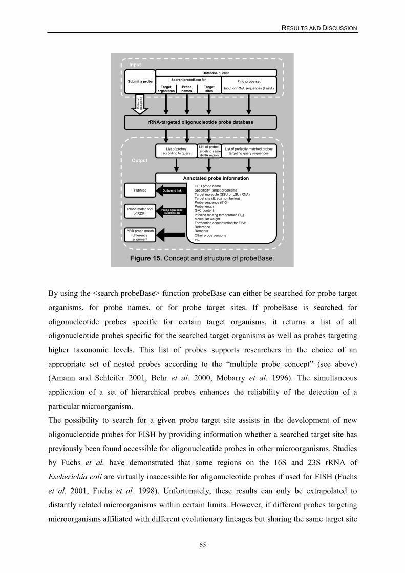
RESULTS AND DISCUSSION
65
Figure 15. Concept and structure of probeBase.
Search probeBase for
Targetorganisms
Probenames
Targetsites
List of perfectly matched probestargeting query sequences
rRNA-targeted oligonucleotide probe database
List of probesaccording to query
Submit a probe
List of probes targeting same
rRNA region
OPD probe nameSpecificity (target organisms)Target molecule (SSU or LSU rRNA)Target site (E. coli numbering)Probe sequence (5‘-3‘)Probe lengthG+C contentInferred melting temperature (Tm)Molecular weightFormamide concentration for FISHReferenceRemarksOther probe versionsetc.
PubMed
Probe match tool of RDP-II
ARB probe matchdifferencealignment
Probe sequencesubmission
Edito
rial
proc
essi
ng
Find probe set
Input of rRNA sequences (FastA)
Outbound link
Input
Annotated probe information
Output
Database queries
By using the <search probeBase> function probeBase can either be searched for probe target
organisms, for probe names, or for probe target sites. If probeBase is searched for
oligonucleotide probes specific for certain target organisms, it returns a list of all
oligonucleotide probes specific for the searched target organisms as well as probes targeting
higher taxonomic levels. This list of probes supports researchers in the choice of an
appropriate set of nested probes according to the “multiple probe concept” (see above)
(Amann and Schleifer 2001, Behr et al. 2000, Mobarry et al. 1996). The simultaneous
application of a set of hierarchical probes enhances the reliability of the detection of a
particular microorganism.
The possibility to search for a given probe target site assists in the development of new
oligonucleotide probes for FISH by providing information whether a searched target site has
previously been found accessible for oligonucleotide probes in other microorganisms. Studies
by Fuchs et al. have demonstrated that some regions on the 16S and 23S rRNA of
Escherichia coli are virtually inaccessible for oligonucleotide probes if used for FISH (Fuchs
et al. 2001, Fuchs et al. 1998). Unfortunately, these results can only be extrapolated to
distantly related microorganisms within certain limits. However, if different probes targeting
microorganisms affiliated with different evolutionary lineages but sharing the same target site
RESULTS AND DISCUSSION
66
on the respective rRNA molecule have been successfully applied for FISH, it is very likely
that the respective target site is generally accessible for oligonucleotide probes.
The second function, the <find probe set> tool of probeBase can be used to rapidly retrieve all
published probes targeting one or several query rRNA gene sequences without prior
comparative sequence analysis. A set of up to 150 sequences, provided by the researcher as
rRNA or DNA sense strand sequence in 5'-3' orientation (FastA format) can be searched
simultaneously for the presence of the perfect match target sites of all probes deposited at
probeBase. The output is a table sequentially listing (i) each single query sequence with all
perfectly matching probes found in probeBase and (ii) each possible probe with all perfectly
matching query sequences. Using this probeBase feature researchers will, for example, easily
be able to determine a set of already published probes that target the microbial sequences in a
certain environmental rRNA gene clone library. This probe set might then be used in
subsequent hybridization experiments to confirm the presence of the organisms detected in
the rRNA gene clone library in situ and to gain insight in the actual abundance of these
microorganisms in the investigated environment.
RESULTS AND DISCUSSION
67
IV. Specificity and sensitivity of a 16S rRNA-based oligonucleotide microarrayfor detection of all recognized SRPs (SRP-PhyloChip) as evaluated with purecultures
Prior to hybridization, the SRP-specific probes were spotted onto glass slides by using a
pattern roughly reflecting the phylogeny of the SRPs (Table 5). In addition, universal,
bacterial, and archaeal probes as well as a nonsense probe (NONSENSE, with a sequence
having at least four mismatches to every known 16S rRNA sequence) were immobilized on
the microarray for hybridization control purposes (Table 5). Furthermore, another nonsense
probe (CONT) (Table 5) was spotted at the beginning and the end of each probe row of the
microarray. During hybridization, a fluorescently labeled oligonucleotide fully
complementary to this probe was added for control of hybridization efficiency and for
straightforward localization of the probe spot rows in the microarray readout. The number of
16S rRNA-targeted oligonucleotide probes used in this thesis is significantly higher than the
number of probes used in previous applications of microarrays for bacterial identification
(Guschin et al. 1997, Koizumi et al. 2002, Liu et al. 2001, Reyes-Lopez et al. 2003, Small et
al. 2001). This difference had important implications for the strategy which was selected for
optimizing the hybridization conditions to ensure maximum specificity of the probes.
In the first step, temperature-dependent dissociation of several probe-target duplexes with
perfect matches or mismatches was measured by hybridizing the SRP-PhyloChip with
fluorescently labeled 16S rRNA gene amplificates of Desulfovibrio halophilus,
Desulfomicrobium apsheronum, and Desulfohalobium retbaense under increasingly stringent
conditions. For each data point, a separate microarray with nine replicate spots of each probe
was hybridized, washed, and analyzed. Figure 16 shows representative melting curves of
probe-target duplexes for two of the SRP-specific probes and for the bacterial probe EUB338
with the labeled 16S rRNA gene amplificates of the three reference organisms. Positive
hybridization signals were recorded with probe EUB338 for the three SRPs when wash
temperatures between 42°C and 60°C were used. However, the EUB338 hybridization signal
intensities varied significantly for the three reference organisms, indicating that there were
variations in the efficiency of the fluorescence labeling of the PCR amplificates (Figure 16C).
Clear discrimination between perfectly matched and mismatched duplexes was achieved for
most but not all of the probes investigated (Figure 16 A and B and Figure 17). When a wash
temperature of 42°C was used, the fluorescence intensity of probe-target hybrids with
mismatches was almost always lower than the fluorescence intensity of completely matched
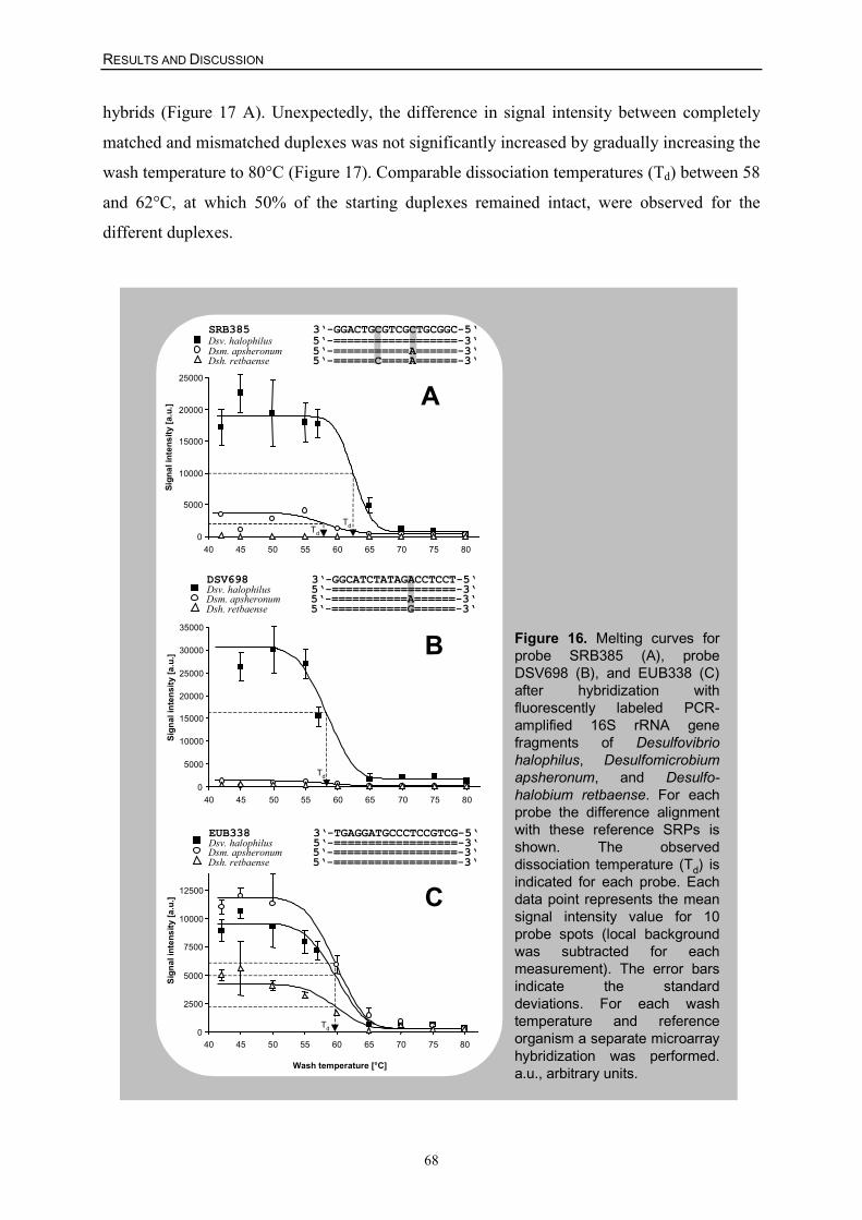
RESULTS AND DISCUSSION
68
hybrids (Figure 17 A). Unexpectedly, the difference in signal intensity between completely
matched and mismatched duplexes was not significantly increased by gradually increasing the
wash temperature to 80°C (Figure 17). Comparable dissociation temperatures (Td) between 58
and 62°C, at which 50% of the starting duplexes remained intact, were observed for the
different duplexes.
Figure 16. Melting curves forprobe SRB385 (A), probeDSV698 (B), and EUB338 (C)after hybridization withfluorescently labeled PCR-amplified 16S rRNA genefragments of Desulfovibriohalophilus, Desulfomicrobiumapsheronum, and Desulfo-halobium retbaense. For eachprobe the difference alignmentwith these reference SRPs isshown. The observeddissociation temperature (Td) isindicated for each probe. Eachdata point represents the meansignal intensity value for 10probe spots (local backgroundwas subtracted for eachmeasurement). The error barsindicate the standarddeviations. For each washtemperature and referenceorganism a separate microarrayhybridization was performed.a.u., arbitrary units.
Dsv. halophilus 5‘-==================-3‘Dsm. apsheronum 5‘-===========A======-3‘Dsh. retbaense 5‘-======C====A======-3‘
SRB385 3‘-GGACTGCGTCGCTGCGGC-5‘
A
0
5000
10000
15000
20000
25000
40 45 50 55 60 65 70 75 80
Sign
al in
tens
ity [a
.u.]
Td
Td
Dsv. halophilus 5‘-==================-3‘
Dsh. retbaense 5‘-===========G======-3‘
DSV698 3‘-GGCATCTATAGACCTCCT-5‘
Dsm. apsheronum 5‘-===========A======-3‘
B
0
5000
10000
15000
20000
25000
30000
35000
40 45 50 55 60 65 70 75 80
Sign
al in
tens
ity [a
.u.]
Td
Dsv. halophilus 5‘-==================-3‘
Dsh. retbaense 5‘-==================-3‘
EUB338 3‘-TGAGGATGCCCTCCGTCG-5‘
Dsm. apsheronum 5‘-==================-3‘
C
0
2500
5000
7500
10000
12500
40 45 50 55 60 65 70 75 80
Wash temperature [°C]
Sign
al in
tens
ity [a
.u.]
Td
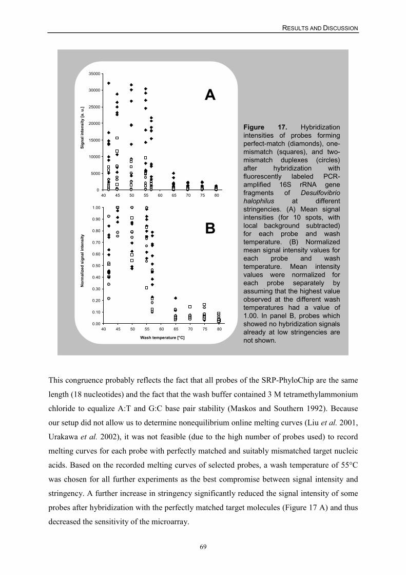
RESULTS AND DISCUSSION
69
Figure 17. Hybridizationintensities of probes formingperfect-match (diamonds), one-mismatch (squares), and two-mismatch duplexes (circles)after hybridization withfluorescently labeled PCR-amplified 16S rRNA genefragments of Desulfovibriohalophilus at differentstringencies. (A) Mean signalintensities (for 10 spots, withlocal background subtracted)for each probe and washtemperature. (B) Normalizedmean signal intensity values foreach probe and washtemperature. Mean intensityvalues were normalized foreach probe separately byassuming that the highest valueobserved at the different washtemperatures had a value of1.00. In panel B, probes whichshowed no hybridization signalsalready at low stringencies arenot shown.
0.00
0.10
0.20
0.30
0.40
0.50
0.60
0.70
0.80
0.90
1.00
40 45 50 55 60 65 70 75 80
Wash temperature [°C]
Nor
mal
ized
sig
nal i
nten
sity
B
0
5000
10000
15000
20000
25000
30000
35000
40 45 50 55 60 65 70 75 80
Sign
al in
tens
ity [a
. u.]
A
This congruence probably reflects the fact that all probes of the SRP-PhyloChip are the same
length (18 nucleotides) and the fact that the wash buffer contained 3 M tetramethylammonium
chloride to equalize A:T and G:C base pair stability (Maskos and Southern 1992). Because
our setup did not allow us to determine nonequilibrium online melting curves (Liu et al. 2001,
Urakawa et al. 2002), it was not feasible (due to the high number of probes used) to record
melting curves for each probe with perfectly matched and suitably mismatched target nucleic
acids. Based on the recorded melting curves of selected probes, a wash temperature of 55°C
was chosen for all further experiments as the best compromise between signal intensity and
stringency. A further increase in stringency significantly reduced the signal intensity of some
probes after hybridization with the perfectly matched target molecules (Figure 17 A) and thus
decreased the sensitivity of the microarray.
RESULTS AND DISCUSSION
70
In the next step, an SRP-PhyloChip with duplicate spots for each probe was evaluated by
using 42 SRP reference organisms to determine a threshold value above which a probe
hybridization signal was considered positive. For each SRP-specific probe, this set of
reference organisms contained an SRP which has a 16S rRNA gene with a perfectly matched
target site. For each reference organism, fluorescently labeled, PCR-amplified 16S rRNA
gene fragments were hybridized separately with the microarray by using 55°C as the wash
temperature. The array readout was quantitatively analyzed by digital image analysis to
determine a signal-to-noise ratio for each probe according to the following formula:
T = [IP – (IN-INLB)] x IPLB-1
where T is the signal-to-noise ratio of the probe, IP is the mean pixel intensity of both specific
probe spots, IN is the mean pixel intensity of both NONSENSE probe spots (note that IN-INLB
must always have a lower value than IP), INLB is the mean pixel intensity of the local
background area around both NONSENSE probe spots, and IPLB is the mean pixel intensity of
the local background area around both specific probe spots.
Spots for which the signal-to-noise ratio was equal to or greater than 2.0 were considered
positive in the pure culture evaluation experiments and all subsequent analyses. Furthermore,
the signal-to-noise ratio of each probe was divided by the signal-to-noise ratio of the bacterial
EUB338 probe recorded on the same microarray in order to compare the duplex yields of the
different SRP-specific probes. To do this, the following formula was used:
R = T x {[IEUB – (IN-INLB)] x IEUBLB-1}-1
where R is the normalized signal-to-noise ratio of the probe, IEUB is the mean pixel intensity
of all EUB338 probe spots, and IEUBLB is the mean pixel intensity of the local background area
around all EUB338 probe spots.
The individual hybridization results for each of the 142 probes with each of the reference
organisms can be downloaded via the internet (supplementary material at
http://www.microbial-ecology.net/) (Appendix I and III, Loy et al. 2003b, Loy et al. 2002).
Consistent with data from quantitative FISH experiments performed with different 16S
rRNA-targeted oligonucleotide probes for Escherichia coli (Fuchs et al. 1998), the
normalized signal-to-noise ratios of the probes ranged from 0.3 for probe DFACE1028 with
Desulfotomaculum acetoxidans to 17.6 for probe DSBAC355 with Syntrophobacter wolinii,
RESULTS AND DISCUSSION
71
demonstrating that different probes exhibit very different signal intensities after hybridization
with their perfectly matched target sequences (factor up to 59). In addition, six of the probes
evaluated (listed separately in Table 5) did not show a positive hybridization signal with any
of the reference organisms, including the perfect-match target SRP, and thus were excluded
from the microarray in subsequent experiments. Dramatic differences in duplex yield arising
from different regions of the target were also observed in other microarray applications
(Milner et al. 1997, Southern et al. 1994) and probably reflect either accessibility differences
for the different probe target sites due to secondary structures of the target DNA or different
steric hindrances of the different nucleic acid hybrids formed on the microarrays after
hybridization. Furthermore, four probes (listed separately in Table 5) were found to be not
suitable for SRP diversity surveys due to their nonspecific binding to many nontarget
organisms under stringent hybridization conditions (see supplementary web material).
Under the conditions used, 75 of the probes found to be suitable for the SRP-PhyloChip
hybridized exclusively to their target organisms. The other probes hybridized to rRNA gene
amplificates with perfectly matched target sites, as well as to some rRNA genes with target
sites having between one and six mismatches. In summary, of the 5248 individual probe-
target hybridization reactions performed (by hybridizing the reference organisms with the
final SRP-PhyloChip), 5050 (96%) gave the expected results by either showing a detectable
signal with the appropriate perfect-match target or showing no signal with target sequences
containing mismatches (Appendix I, Loy et al. 2002).
Finally, the SRP-PhyloChip was hybridized in independent experiments with different
amounts (1, 5, 10, 25, 50, 100, 200, and 400 ng) of PCR amplified, labeled 16S rRNA gene
fragments of Desulfovibrio halophilus. The same hybridization pattern was observed when 50
to 400 ng of labeled nucleic acids was used. When less than 50 ng of added nucleic acid was
used, the signal-to-noise ratios of the hybridization signals were less than 2.0.
The evaluation of the microarray with SRP pure cultures demonstrated (i) that false-negative
hybridization never occurred (within the detection limit of the microarray method) but (ii) that
some of the probes still hybridized to nontarget organisms under the hybridization and
washing conditions used, leading to false-positive results (see supplementary web material).
As expected, the nucleotide composition of the mismatch, the mismatch position (Stahl and
Amann 1991, Urakawa et al. 2002), and possibly other variables, such as the influence of an
adjacent nucleotide stacking interaction (Fotin et al. 1998), were the major factors
determining the duplex yields of probes with mismatched target nucleic acids. Most of the
mismatched duplexes with signal intensities above the threshold value (used to differentiate
RESULTS AND DISCUSSION
72
between positive and negative hybridization results) had a signal intensity (and normalized
signal-to-noise ratio) lower than that of the corresponding perfect-match duplex (Figure 17
A). However, this difference cannot be exploited for interpretation of microarray
hybridization results for environmental samples because a low hybridization signal of a probe
can be caused not only by mismatched duplex formation but also by low abundance of the
perfect-match target nucleic acid.
Misinterpretation of microarray hybridization patterns caused by the nonperfect specificity of
some of the probes could be avoided at least partially by using the “multiple probe concept”.
While hybridization patterns consistent with the hierarchical or parallel specificity of the
probes increase the reliability of detection, inconsistent probe hybridization patterns must be
interpreted with caution. In complex samples, inconsistent hybridization patterns can be
caused either by nonspecific binding of one or several probes or by previously unrecognized
prokaryotes with unusual combinations of perfect-match probe target sites in their 16S rRNA
gene sequences.
RESULTS AND DISCUSSION
73
V. Application of the SRP-PhyloChip for analysis of complex environmentaland medical samples
To evaluate the applicability of the SRP-PhyloChip for clinical and environmental
determinative studies, three different habitats each containing a diverse assemblage of
microorganisms were analyzed.
In the first experiment, tooth pocket samples from five patients suffering from adult
periodontitis (Loesche and Grossman 2001) were investigated (Appendix I, Loy et al. 2002).
Previous culturing studies have suggested that SRPs, among other well-known, anaerobic
bacteria (Loesche and Grossman 2001), may be etiologically involved in destructive
periodontal diseases (Langendijk et al. 1999, Langendijk et al. 2000). Hydrogen sulfide
(H2S), main end product of metabolically active SRPs, is a biologically active reagent with a
strongly cytotoxic effect (Ratcliff and Johnson 1999, Rizzo 1967). Elevated concentration of
H2S do correlate with increased periodontal pocket depth (Persson 1992), reaching H2S
concentrations far beyond the toxic level (Ratcliff and Johnson 1999). Sulfate, that serves as
terminal electron acceptor and promotes growth of SRPs, might theoretically derive from
serum and/or be liberated from glycosaminoglycans in the connective tissue (van der Hoeven
et al. 1995).
While for three of the five patients analyzed none of the SRP-specific probes on the
microarray showed a positive signal (data not shown), probe hybridization patterns indicative
of the presence of members of the genus Desulfomicrobium were obtained for the other two
patients (Figure 18 A). This result was confirmed independently by PCR analysis of the DNA
obtained from the tooth pockets of the five patients by using primers specific for the 16S
rRNA gene of members of the genus Desulfomicrobium (primer pair DSM172F-DSM1469R
[Table 2]). Consistent with the microarray results, specific PCR amplificates were obtained
for two of the five patients. Amplificates from both of these patients were cloned and
sequenced. Comparative analysis of six clones demonstrated that the amplified sequences
were almost identical to each other and to the corresponding 16S rRNA gene fragment of
Desulfomicrobium orale (99.6 to 99.9% sequence similarity) (Figure 19).
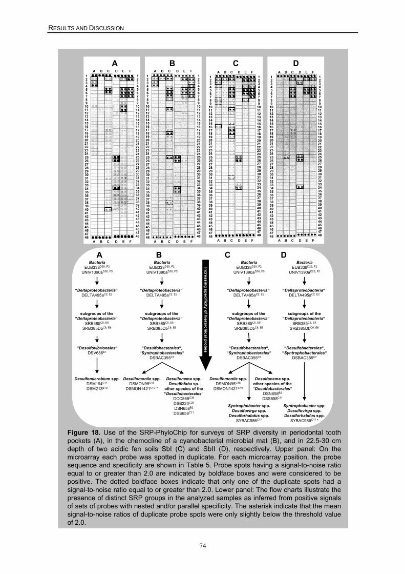
RESULTS AND DISCUSSION
74
Figure 18. Use of the SRP-PhyloChip for surveys of SRP diversity in periodontal toothpockets (A), in the chemocline of a cyanobacterial microbial mat (B), and in 22.5-30 cmdepth of two acidic fen soils SbI (C) and SbII (D), respectively. Upper panel: On themicroarray each probe was spotted in duplicate. For each microarray position, the probesequence and specificity are shown in Table 5. Probe spots having a signal-to-noise ratioequal to or greater than 2.0 are indicated by boldface boxes and were considered to bepositive. The dotted boldface boxes indicate that only one of the duplicate spots had asignal-to-noise ratio equal to or greater than 2.0. Lower panel: The flow charts illustrate thepresence of distinct SRP groups in the analyzed samples as inferred from positive signalsof sets of probes with nested and/or parallel specificity. The asterisk indicate that the meansignal-to-noise ratios of duplicate probe spots were only slightly below the threshold valueof 2.0.
Desulfonema spp.Desulfofaba sp.
other species of the“Desulfobacterales“
DCC868C38
DSB220C25
DSN658B2
DSS658C11
BacteriaEUB338D25, F2
UNIV1390aD26, F5
“Deltaproteobacteria“ DELTA495aC2, E2
“Desulfobacterales“,“Syntrophobacterales“
DSBAC355C7
subgroups of the“Deltaproteobacteria“
SRB385C5, E5
SRB385DbC6, E6
Desulfomonile spp.DSMON95C18
DSMON1421C19 *
Increasing specificity of hierarchical probes
BacteriaEUB338D25, F2
UNIV1390aD26, F5
“Deltaproteobacteria“ DELTA495aC2, E2
“Desulfobacterales“,“Syntrophobacterales“
DSBAC355C7
Syntrophobacter spp.Desulfovirga spp.
Desulforhabdus spp.SYBAC986C17 *
subgroups of the“Deltaproteobacteria“
SRB385C5, E5
SRB385DbC6, E6
C
234
1
678
5
101112
9
141516
13
181920
17
222324
21
234
1
678
5
101112
9
141516
13
181920
17
222324
21
262728
25
303132
29
343536
33
383940
37
424344
41
464748
45
A B C D E F
262728
25
303132
29
343536
33
383940
37
424344
41
464748
45
A B C D E F
234
1
678
5
101112
9
141516
13
181920
17
222324
21
262728
25
303132
29
343536
33
383940
37
424344
41
464748
45
A B C D E F
234
1
678
5
101112
9
141516
13
181920
17
222324
21
262728
25
303132
29
343536
33
383940
37
424344
41
464748
45
A B C D E F
D234
1
678
5
101112
9
141516
13
181920
17
222324
21
262728
25
303132
29
343536
33
383940
37
424344
41
464748
45
A B C D E F
234
1
678
5
101112
9
141516
13
181920
17
222324
21
262728
25
303132
29
343536
33
383940
37
424344
41
464748
45
A B C D E F
A234
1
678
5
101112
9
141516
13
181920
17
222324
21
262728
25
303132
29
343536
33
383940
37
424344
41
464748
45
A B C D E F
234
1
678
5
101112
9
141516
13
181920
17
222324
21
262728
25
303132
29
343536
33
383940
37
424344
41
464748
45
A B C D E F
B
BacteriaEUB338D25, F2
UNIV1390aD26, F5
“Deltaproteobacteria“ DELTA495aC2, E2
“Desulfovibrionales“DSV686E7
Desulfomicrobium spp.DSM194E11
DSM213E10
subgroups of the“Deltaproteobacteria“
SRB385C5, E5
SRB385DbC6, E6
Desulfonema spp.other species of the“Desulfobacterales“
DSN658B2
DSS658C11
BacteriaEUB338D25, F2
UNIV1390aD26, F5
“Deltaproteobacteria“ DELTA495aC2, E2
“Desulfobacterales“,“Syntrophobacterales“
DSBAC355C7
Syntrophobacter spp.Desulfovirga spp.
Desulforhabdus spp.SYBAC986C17
subgroups of the“Deltaproteobacteria“
SRB385C5, E5
SRB385DbC6, E6
Desulfomonile spp.DSMON95C18
DSMON1421C19
C DA B
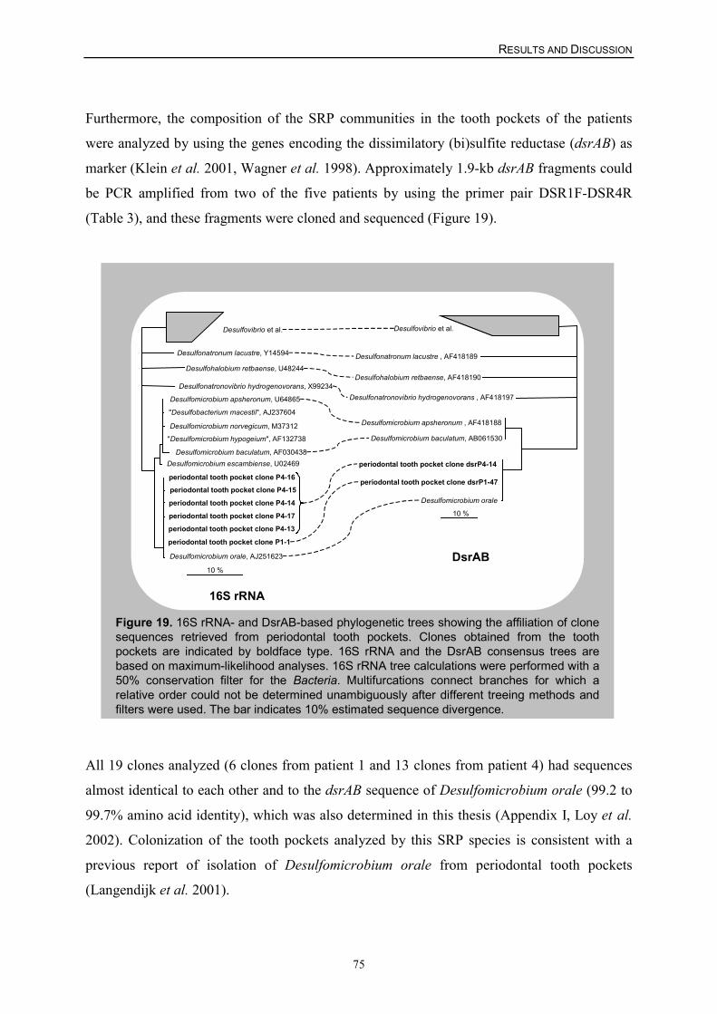
RESULTS AND DISCUSSION
75
Furthermore, the composition of the SRP communities in the tooth pockets of the patients
were analyzed by using the genes encoding the dissimilatory (bi)sulfite reductase (dsrAB) as
marker (Klein et al. 2001, Wagner et al. 1998). Approximately 1.9-kb dsrAB fragments could
be PCR amplified from two of the five patients by using the primer pair DSR1F-DSR4R
(Table 3), and these fragments were cloned and sequenced (Figure 19).
Figure 19. 16S rRNA- and DsrAB-based phylogenetic trees showing the affiliation of clonesequences retrieved from periodontal tooth pockets. Clones obtained from the toothpockets are indicated by boldface type. 16S rRNA and the DsrAB consensus trees arebased on maximum-likelihood analyses. 16S rRNA tree calculations were performed with a50% conservation filter for the Bacteria. Multifurcations connect branches for which arelative order could not be determined unambiguously after different treeing methods andfilters were used. The bar indicates 10% estimated sequence divergence.
Desulfonatronum lacustre, Y14594
Desulfonatronovibrio hydrogenovorans, X99234
Desulfohalobium retbaense, U48244
Desulfomicrobium apsheronum, U64865
"Desulfobacterium macestii", AJ237604
Desulfomicrobium baculatum, AF030438
Desulfomicrobium norvegicum, M37312
"Desulfomicrobium hypogeium", AF132738
Desulfomicrobium escambiense, U02469
periodontal tooth pocket clone P4-16periodontal tooth pocket clone P4-15
periodontal tooth pocket clone P4-14
periodontal tooth pocket clone P4-17periodontal tooth pocket clone P4-13
Desulfomicrobium orale, AJ251623
periodontal tooth pocket clone P1-1
10 %
Desulfovibrio et al.
10 %
Desulfonatronum lacustre , AF418189
Desulfohalobium retbaense, AF418190
Desulfonatronovibrio hydrogenovorans , AF418197
Desulfomicrobium apsheronum , AF418188
Desulfomicrobium baculatum, AB061530
periodontal tooth pocket clone dsrP4-14
periodontal tooth pocket clone dsrP1-47
Desulfomicrobium orale
Desulfovibrio et al.
16S rRNA
DsrAB
All 19 clones analyzed (6 clones from patient 1 and 13 clones from patient 4) had sequences
almost identical to each other and to the dsrAB sequence of Desulfomicrobium orale (99.2 to
99.7% amino acid identity), which was also determined in this thesis (Appendix I, Loy et al.
2002). Colonization of the tooth pockets analyzed by this SRP species is consistent with a
previous report of isolation of Desulfomicrobium orale from periodontal tooth pockets
(Langendijk et al. 2001).
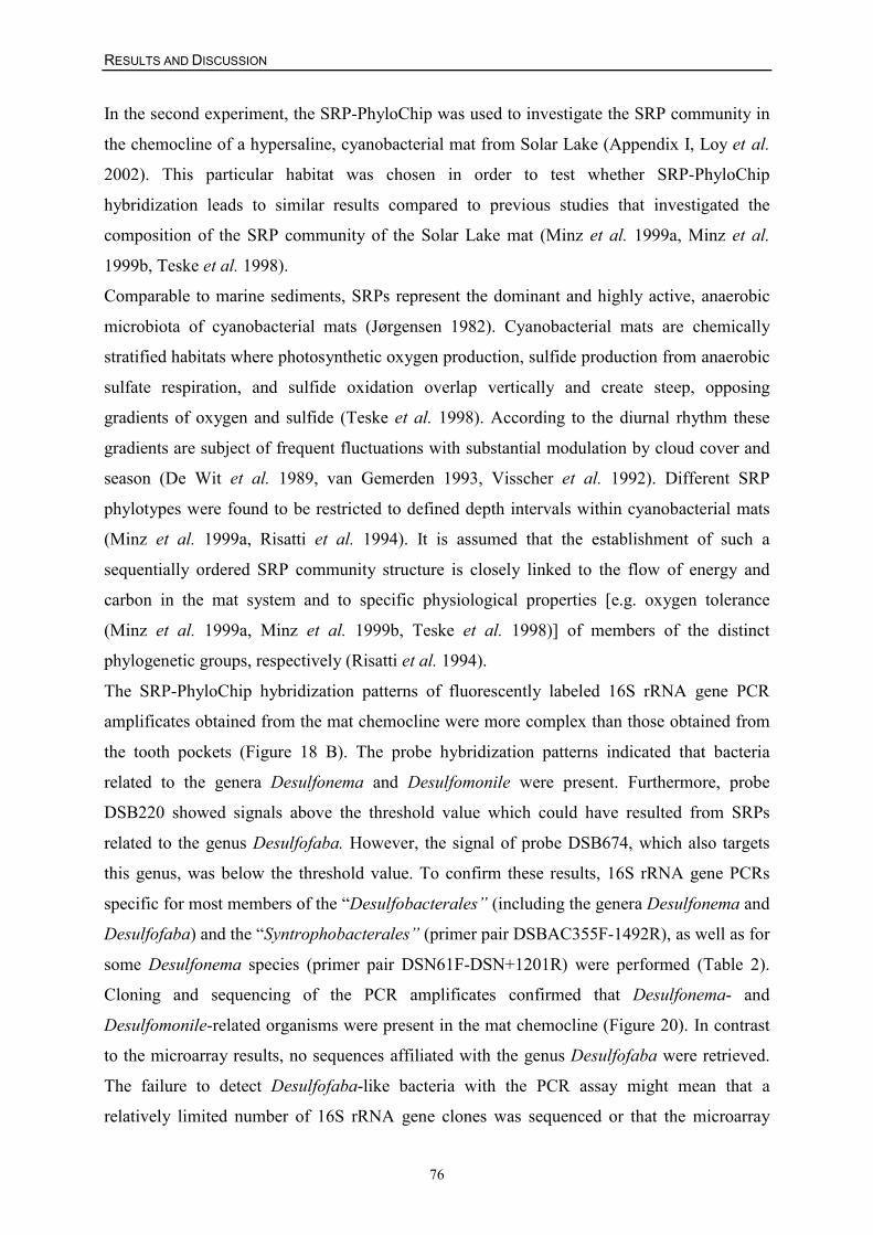
RESULTS AND DISCUSSION
76
In the second experiment, the SRP-PhyloChip was used to investigate the SRP community in
the chemocline of a hypersaline, cyanobacterial mat from Solar Lake (Appendix I, Loy et al.
2002). This particular habitat was chosen in order to test whether SRP-PhyloChip
hybridization leads to similar results compared to previous studies that investigated the
composition of the SRP community of the Solar Lake mat (Minz et al. 1999a, Minz et al.
1999b, Teske et al. 1998).
Comparable to marine sediments, SRPs represent the dominant and highly active, anaerobic
microbiota of cyanobacterial mats (Jørgensen 1982). Cyanobacterial mats are chemically
stratified habitats where photosynthetic oxygen production, sulfide production from anaerobic
sulfate respiration, and sulfide oxidation overlap vertically and create steep, opposing
gradients of oxygen and sulfide (Teske et al. 1998). According to the diurnal rhythm these
gradients are subject of frequent fluctuations with substantial modulation by cloud cover and
season (De Wit et al. 1989, van Gemerden 1993, Visscher et al. 1992). Different SRP
phylotypes were found to be restricted to defined depth intervals within cyanobacterial mats
(Minz et al. 1999a, Risatti et al. 1994). It is assumed that the establishment of such a
sequentially ordered SRP community structure is closely linked to the flow of energy and
carbon in the mat system and to specific physiological properties [e.g. oxygen tolerance
(Minz et al. 1999a, Minz et al. 1999b, Teske et al. 1998)] of members of the distinct
phylogenetic groups, respectively (Risatti et al. 1994).
The SRP-PhyloChip hybridization patterns of fluorescently labeled 16S rRNA gene PCR
amplificates obtained from the mat chemocline were more complex than those obtained from
the tooth pockets (Figure 18 B). The probe hybridization patterns indicated that bacteria
related to the genera Desulfonema and Desulfomonile were present. Furthermore, probe
DSB220 showed signals above the threshold value which could have resulted from SRPs
related to the genus Desulfofaba. However, the signal of probe DSB674, which also targets
this genus, was below the threshold value. To confirm these results, 16S rRNA gene PCRs
specific for most members of the “Desulfobacterales” (including the genera Desulfonema and
Desulfofaba) and the “Syntrophobacterales” (primer pair DSBAC355F-1492R), as well as for
some Desulfonema species (primer pair DSN61F-DSN+1201R) were performed (Table 2).
Cloning and sequencing of the PCR amplificates confirmed that Desulfonema- and
Desulfomonile-related organisms were present in the mat chemocline (Figure 20). In contrast
to the microarray results, no sequences affiliated with the genus Desulfofaba were retrieved.
The failure to detect Desulfofaba-like bacteria with the PCR assay might mean that a
relatively limited number of 16S rRNA gene clones was sequenced or that the microarray
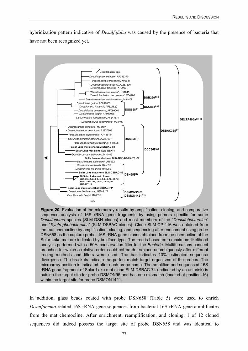
RESULTS AND DISCUSSION
77
hybridization pattern indicative of Desulfofaba was caused by the presence of bacteria that
have not been recognized yet.
Figure 20. Evaluation of the microarray results by amplification, cloning, and comparativesequence analysis of 16S rRNA gene fragments by using primers specific for someDesulfonema species (SLM-DSN clones) and most members of the ”Desulfobacterales”and ”Syntrophobacterales” (SLM-DSBAC clones). Clone SLM-CP-116 was obtained fromthe mat chemocline by amplification, cloning, and sequencing after enrichment using probeDSN658 as the capture probe. 16S rRNA gene clones obtained from the chemocline of theSolar Lake mat are indicated by boldface type. The tree is based on a maximum-likelihoodanalysis performed with a 50% conservation filter for the Bacteria. Multifurcations connectbranches for which a relative order could not be determined unambiguously after differenttreeing methods and filters were used. The bar indicates 10% estimated sequencedivergence. The brackets indicate the perfect-match target organisms of the probes. Themicroarray position is indicated after each probe name. The amplified and sequenced 16SrRNA gene fragment of Solar Lake mat clone SLM-DSBAC-74 (indicated by an asterisk) isoutside the target site for probe DSMON95 and has one mismatch (located at position 16)within the target site for probe DSMON1421.
Desulfobacter spp.
Desulfospira joergensenii, X99637 Desulfotignum balticum, AF233370
Desulfobacula phenolica, AJ237606 Desulfobacula toluolica, X70953
"Desulfobacterium niacini", U51845 "Desulfobacterium vacuolatum", M34408
Desulfobacterium autotrophicum, M34409
"Desulfobotulus sapovorans", M34402
Desulfofaba gelida, AF099063 Desulfomusa hansenii, AF321820
Desulfofrigus oceanense, AF099064 Desulfofrigus fragile, AF099065
Desulforegula conservatrix, AF243334
"Desulfobacterium oleovorans", Y17698
Desulfosarcina variabilis , M34407 Desulfobacterium cetonicum, AJ237603
"Desulfostipes sapovorans", AF148141
Solar Lake mat clone SLM-DSBAC-81
Desulfobacterium indolicum, AJ237607
10%
Solar Lake mat clone SLM-DSBAC-74* Desulfomonile limimaris, AF282177
Desulfomonile tiedjei, M26635
18 Solar Lake mat clonesSLM-DSN-1,-2,-3,-5,-6,-7,-8,-9,-10,-11,-12SLM-DSBAC-65,-70,-73,-78,-79,-84SLM-CP-116
Desulfonema limicola, U45990 Desulfonema magnum, U45989
Desulfococcus multivorans, M34405 Solar Lake mat clones SLM-DSBAC-75,-76,-77
Solar Lake mat clone SLM-DSBAC-82
Solar Lake mat clone SLM-DSN-4
Desulfonema ishimotonii, U45992
DSN658B2
DCC868C38
DELTA495aC2, E2
DSBAC355C7
DSS658C11
DSS658C11DCC868C38
DSB220C25
DSMON95C18
DSMON1421C19
In addition, glass beads coated with probe DSN658 (Table 5) were used to enrich
Desulfonema-related 16S rRNA gene sequences from bacterial 16S rRNA gene amplificates
from the mat chemocline. After enrichment, reamplification, and cloning, 1 of 12 cloned
sequences did indeed possess the target site of probe DSN658 and was identical to
RESULTS AND DISCUSSION
78
Desulfonema-related sequences obtained by the specific PCR assay described above (Figure
20). The remaining 11 cloned sequences did not possess the probe DSN658 target site and
were unrelated to recognized SRPs (data not shown). The detection of Desulfonema-like
bacteria in the chemocline of the Solar Lake mat is consistent with findings of previous
studies based on comparative sequence analysis of dsrAB genes (Minz et al. 1999b),
quantitative membrane hybridization (Minz et al. 1999a), and DGGE (Teske et al. 1998), and
further supports the importance of these filamentous SRPs in the oxic/anoxic interface of
hypersaline mat ecosystems.
The third experiment aimed to identify SRPs in two acidic fen soils in a forested catchment
(Lehstenbach, Bavaria) (Appendix III, Loy et al. 2003b), where the SRP community structure
has not yet been investigated.
SO2
SO42-
SO42- desorption
S mineralization
-C-S -C-O-SO3
O2
boundorganic S
organic sulfate ester
H2SO4H2SO3
Acidic rainuntil the 90s
Acidic fenacidification
dissimilatoryFe(III) reduction
dissimilatorysulfate reduction
Fe(III)
reduced organic SFe(II) S2-
� alkalinity of soil solution
Oxidation
CH4 emission
Pool of FeS,FeS2, organic S
Fe(II)solubleH2S
Fixation
Figure 21. Biogeochemical processes in the Lehstenbach catchment(Fichtelgebirge, Germany).
In contrast to the well-studied sulfate-reducing communities in marine (Llobet-Brossa et al.
1998, Ravenschlag et al. 2001, Ravenschlag et al. 2000, Sahm et al. 1999a, Sahm et al.
1999b) and freshwater habitats (Li et al. 1999), knowledge of the distribution, diversity, and
RESULTS AND DISCUSSION
79
function of SRPs in terrestrial ecosystems is generally scarce. Biogeochemical analyses based
on �34S values and 35S radiolabeling have shown that dissimilatory sulfate reduction is an
ongoing process at the fen sites in the Lehstenbach catchment (Alewell and Gehre 1999,
Alewell and Novak 2001). Owing to air pollution this catchment has seen a major deposition
of sulfate through acidic rainfall (in form of sulfuric and sulfurous acid) until the 1990s, when
efficient air filtration systems have been introduced in factories in former East Germany. With
each rainfall, sulfate is leached out of the upland aerated soils into the lower situated fens. It
has been hypothesized that dissimilatory sulfate reduction in these mainly anaerobic and
waterlogged soils contributes to the retention of sulfur in this ecosystem (Alewell and Gehre
1999, Alewell and Giesemann 1996, Novak et al. 1994). Sulfate reduction in these fens is a
sink for sulfate and protons, thus decreasing acidity of soil solution and the adjacent
groundwater (Figure 21).
Initially, the SRP-PhyloChip was used to screen for members of already recognized SRP
lineages at the sites SbI and SbII. Bacterial 16S rRNA genes were separately amplified from
each DNA extract retrieved from the four soil depths, fluorescently labeled with Cy5, and
hybridized with the SRP-PhyloChip. At site SbI highly similar microarray hybridization
patterns were observed from the four different soil depths indicating low changes in SRP
richness over depth (Appendix III, Loy et al. 2003b). Positive signals of nested probes
indicated the presence of (i) Desulfomonile spp., (ii) Desulfonema or related species of the
order “Desulfobacterales” and (iii) bacteria belonging to the Syntrophobacter-Desulfovirga-
Desulforhabdus line of descent of the family “Syntrophobacteraceae” (order
“Syntrophobacterales”) (Figure 18 C). For confirmation of the microarray results, 16S rRNA
gene PCRs specific for Desulfomonile spp. (primer pair DSMON85F-DSMON1419R), for
some Desulfonema species (primer pair DSN61F-DSN+1201R) as well as for members of the
“Syntrophobacteraceae” (primer pair SYBAC+282F-SYBAC1427R) were performed with
DNA from each soil depth (Table 2). With each of the Desulfomonile- and
“Syntrophobacteraceae”-specific primer pairs increasing amounts of PCR products of the
expected size were retrieved with increasing soil depth (data not shown). Cloning and
comparative sequence analysis of the PCR amplificates from 22.5-30 cm depth confirmed that
Desulfomonile spp. and Syntrophobacter wolinii-related bacteria were present at site SbI
(Figure 22).
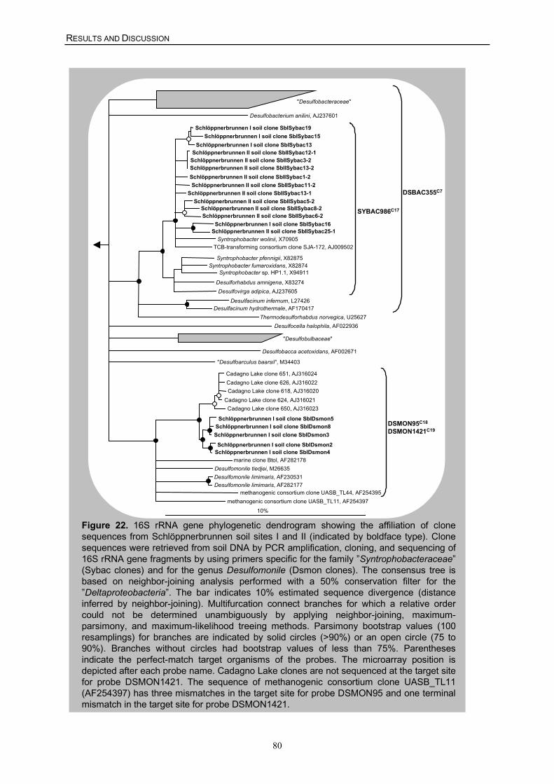
RESULTS AND DISCUSSION
80
Figure 22. 16S rRNA gene phylogenetic dendrogram showing the affiliation of clonesequences from Schlöppnerbrunnen soil sites I and II (indicated by boldface type). Clonesequences were retrieved from soil DNA by PCR amplification, cloning, and sequencing of16S rRNA gene fragments by using primers specific for the family ”Syntrophobacteraceae”(Sybac clones) and for the genus Desulfomonile (Dsmon clones). The consensus tree isbased on neighbor-joining analysis performed with a 50% conservation filter for the”Deltaproteobacteria”. The bar indicates 10% estimated sequence divergence (distanceinferred by neighbor-joining). Multifurcation connect branches for which a relative ordercould not be determined unambiguously by applying neighbor-joining, maximum-parsimony, and maximum-likelihood treeing methods. Parsimony bootstrap values (100resamplings) for branches are indicated by solid circles (>90%) or an open circle (75 to90%). Branches without circles had bootstrap values of less than 75%. Parenthesesindicate the perfect-match target organisms of the probes. The microarray position isdepicted after each probe name. Cadagno Lake clones are not sequenced at the target sitefor probe DSMON1421. The sequence of methanogenic consortium clone UASB_TL11(AF254397) has three mismatches in the target site for probe DSMON95 and one terminalmismatch in the target site for probe DSMON1421.
"Desulfobacteraceae"
"Desulfobulbaceae"
Desulfobacca acetoxidans, AF002671
Desulfobacterium anilini, AJ237601
"Desulfoarculus baarsii", M34403
10%methanogenic consortium clone UASB_TL11, AF254397
Schlöppnerbrunnen I soil clone SbIDsmon5 Schlöppnerbrunnen I soil clone SbIDsmon8
Schlöppnerbrunnen I soil clone SbIDsmon3 Schlöppnerbrunnen I soil clone SbIDsmon2
Schlöppnerbrunnen I soil clone SbIDsmon4
Cadagno Lake clone 651, AJ316024
Cadagno Lake clone 618, AJ316020
Cadagno Lake clone 650, AJ316023 Cadagno Lake clone 624, AJ316021
Cadagno Lake clone 626, AJ316022
methanogenic consortium clone UASB_TL44, AF254395
Desulfocella halophila, AF022936
Schlöppnerbrunnen I soil clone SbISybac19 Schlöppnerbrunnen I soil clone SbISybac15
Schlöppnerbrunnen I soil clone SbISybac13 Schlöppnerbrunnen II soil clone SbIISybac12-1
Schlöppnerbrunnen II soil clone SbIISybac3-2 Schlöppnerbrunnen II soil clone SbIISybac13-2 Schlöppnerbrunnen II soil clone SbIISybac1-2
Syntrophobacter wolinii, X70905
Schlöppnerbrunnen II soil clone SbIISybac11-2 Schlöppnerbrunnen II soil clone SbIISybac13-1
Schlöppnerbrunnen I soil clone SbISybac16 Schlöppnerbrunnen II soil clone SbIISybac25-1
Syntrophobacter pfennigii, X82875 Syntrophobacter fumaroxidans, X82874
Syntrophobacter sp. HP1.1, X94911
Desulforhabdus amnigena, X83274 Desulfovirga adipica, AJ237605
Desulfacinum infernum, L27426 Desulfacinum hydrothermale, AF170417
Thermodesulforhabdus norvegica, U25627
TCB-transforming consortium clone SJA-172, AJ009502
Schlöppnerbrunnen II soil clone SbIISybac5-2 Schlöppnerbrunnen II soil clone SbIISybac8-2 Schlöppnerbrunnen II soil clone SbIISybac6-2
SYBAC986C17
DSMON95C18
DSMON1421C19
Desulfomonile tiedjei, M26635 Desulfomonile limimaris, AF230531 Desulfomonile limimaris, AF282177
marine clone Btol, AF282178
DSBAC355C7
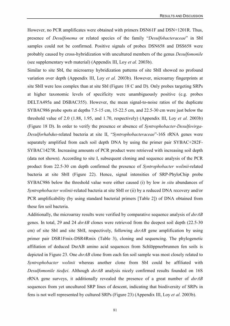
RESULTS AND DISCUSSION
81
However, no PCR amplificates were obtained with primers DSN61F and DSN+1201R. Thus,
presence of Desulfonema or related species of the family “Desulfobacteraceae” in SbI
samples could not be confirmed. Positive signals of probes DSN658 and DSS658 were
probably caused by cross-hybridization with uncultured members of the genus Desulfomonile
(see supplementary web material) (Appendix III, Loy et al. 2003b).
Similar to site SbI, the microarray hybridization patterns of site SbII showed no profound
variation over depth (Appendix III, Loy et al. 2003b). However, microarray fingerprints at
site SbII were less complex than at site SbI (Figure 18 C and D). Only probes targeting SRPs
at higher taxonomic levels of specificity were unambiguously positive (e.g. probes
DELTA495a and DSBAC355). However, the mean signal-to-noise ratios of the duplicate
SYBAC986 probe spots at depths 7.5-15 cm, 15-22.5 cm, and 22.5-30 cm were just below the
threshold value of 2.0 (1.88, 1.95, and 1.70, respectively) (Appendix III, Loy et al. 2003b)
(Figure 18 D). In order to verify the presence or absence of Syntrophobacter-Desulfovirga-
Desulforhabdus-related bacteria at site II, “Syntrophobacteraceae”-16S rRNA genes were
separately amplified from each soil depth DNA by using the primer pair SYBAC+282F-
SYBAC1427R. Increasing amounts of PCR product were retrieved with increasing soil depth
(data not shown). According to site I, subsequent cloning and sequence analysis of the PCR
product from 22.5-30 cm depth confirmed the presence of Syntrophobacter wolinii-related
bacteria at site SbII (Figure 22). Hence, signal intensities of SRP-PhyloChip probe
SYBAC986 below the threshold value were either caused (i) by low in situ abundances of
Syntrophobacter wolinii-related bacteria at site SbII or (ii) by a reduced DNA recovery and/or
PCR amplificability (by using standard bacterial primers [Table 2]) of DNA obtained from
these fen soil bacteria.
Additionally, the microarray results were verified by comparative sequence analysis of dsrAB
genes. In total, 29 and 24 dsrAB clones were retrieved from the deepest soil depth (22.5-30
cm) of site SbI and site SbII, respectively, following dsrAB gene amplification by using
primer pair DSR1Fmix-DSR4Rmix (Table 3), cloning and sequencing. The phylogenetic
affiliation of deduced DsrAB amino acid sequences from Schlöppnerbrunnen fen soils is
depicted in Figure 23. One dsrAB clone from each fen soil sample was most closely related to
Syntrophobacter wolinii whereas another clone from SbI could be affiliated with
Desulfomonile tiedjei. Although dsrAB analysis nicely confirmed results founded on 16S
rRNA gene surveys, it additionally revealed the presence of a great number of dsrAB
sequences from yet uncultured SRP lines of descent, indicating that biodiversity of SRPs in
fens is not well represented by cultured SRPs (Figure 23) (Appendix III, Loy et al. 2003b).
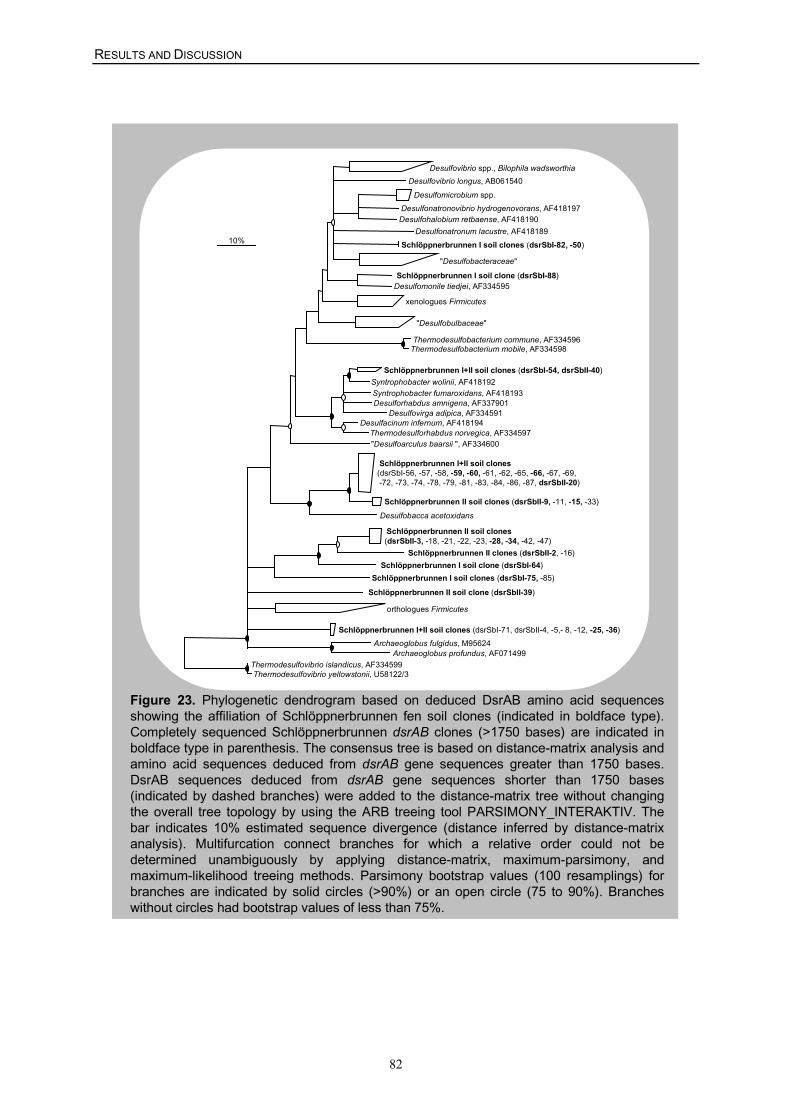
RESULTS AND DISCUSSION
82
Figure 23. Phylogenetic dendrogram based on deduced DsrAB amino acid sequencesshowing the affiliation of Schlöppnerbrunnen fen soil clones (indicated in boldface type).Completely sequenced Schlöppnerbrunnen dsrAB clones (>1750 bases) are indicated inboldface type in parenthesis. The consensus tree is based on distance-matrix analysis andamino acid sequences deduced from dsrAB gene sequences greater than 1750 bases.DsrAB sequences deduced from dsrAB gene sequences shorter than 1750 bases(indicated by dashed branches) were added to the distance-matrix tree without changingthe overall tree topology by using the ARB treeing tool PARSIMONY_INTERAKTIV. Thebar indicates 10% estimated sequence divergence (distance inferred by distance-matrixanalysis). Multifurcation connect branches for which a relative order could not bedetermined unambiguously by applying distance-matrix, maximum-parsimony, andmaximum-likelihood treeing methods. Parsimony bootstrap values (100 resamplings) forbranches are indicated by solid circles (>90%) or an open circle (75 to 90%). Brancheswithout circles had bootstrap values of less than 75%.
"Desulfobacteraceae"
Desulfomicrobium spp. Desulfonatronovibrio hydrogenovorans, AF418197 Desulfohalobium retbaense, AF418190
Desulfovibrio longus, AB061540 Desulfovibrio spp., Bilophila wadsworthia
Desulfonatronum lacustre, AF418189
Desulfomonile tiedjei, AF334595
xenologues Firmicutes
"Desulfobulbaceae"
Thermodesulfobacterium commune, AF334596 Thermodesulfobacterium mobile, AF334598
Syntrophobacter fumaroxidans, AF418193 Desulforhabdus amnigena, AF337901
Desulfovirga adipica, AF334591
Syntrophobacter wolinii, AF418192
Desulfacinum infernum, AF418194 Thermodesulforhabdus norvegica, AF334597"Desulfoarculus baarsii ", AF334600
Desulfobacca acetoxidans
Archaeoglobus fulgidus, M95624 Archaeoglobus profundus, AF071499
orthologues Firmicutes
Thermodesulfovibrio islandicus, AF334599 Thermodesulfovibrio yellowstonii, U58122/3
10%
Schlöppnerbrunnen I+II soil clones (dsrSbI-71, dsrSbII-4, -5,- 8, -12, -25, -36)
Schlöppnerbrunnen I soil clones (dsrSbI-82, -50)
Schlöppnerbrunnen I soil clone (dsrSbI-88)
Schlöppnerbrunnen I+II soil clones (dsrSbI-54, dsrSbII-40)
Schlöppnerbrunnen I+II soil clones(dsrSbI-56, -57, -58, -59, -60, -61, -62, -65, -66, -67, -69, -72, -73, -74, -78, -79, -81, -83, -84, -86, -87, dsrSbII-20)
Schlöppnerbrunnen II soil clones (dsrSbII-9, -11, -15, -33)
Schlöppnerbrunnen II soil clones(dsrSbII-3, -18, -21, -22, -23, -28, -34, -42, -47)
Schlöppnerbrunnen II clones (dsrSbII-2, -16) Schlöppnerbrunnen I soil clone (dsrSbI-64)
Schlöppnerbrunnen I soil clones (dsrSbI-75, -85)
Schlöppnerbrunnen II soil clone (dsrSbII-39)
RESULTS AND DISCUSSION
83
In summary, an encompassing 16S rRNA gene-targeting oligonucleotide microarray suitable
for SRP diversity analyses of complex environmental and clinical samples was developed.
The microarray was used to screen samples in order to rapidly obtain indications of the
presence of distinct lineages of SRPs. However, keeping in mind that (i) most environmental
microbial communities contain a high percentage of bacteria not yet sequenced on the 16S
rRNA level and (ii) not all probes on the microarray are absolutely specific under the
monostringent conditions used (see preceding chapter), the SRP-PhyloChip experiments
should always be supplemented with microarray-independent techniques to confirm the
phylogenetic affiliations of the SRPs detected. Subsequently, the information obtained after
microarray hybridization was used to select or develop appropriate PCR-based techniques
(Tables 2 and 3) for verification of the microarray results and for retrieval of sequence
information for phylogenetic analysis.
It should be emphasized that the SRP-PhyloChip developed in this thesis allows to obtain a
phylogenetically informative, high-resolution fingerprint of the entire SRP diversity in a given
sample within 48 h (including all experimental work from DNA extraction to hybridization
pattern interpretation) and, thus, nicely complements the molecular tool box of previously
available methods for cultivation-independent identification of SRPs (Devereux et al. 1992,
Friedrich 2002, Manz et al. 1998, Risatti et al. 1994, Voordouw et al. 1991, Wagner et al.
1998).
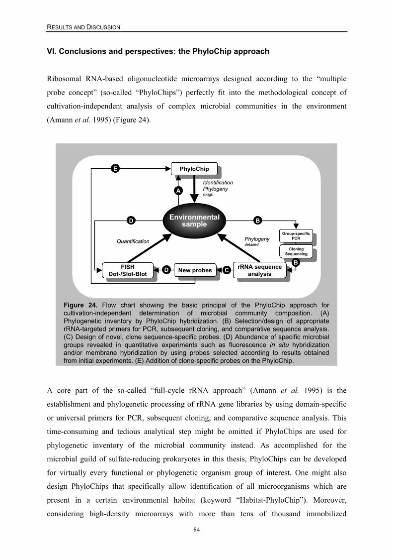
RESULTS AND DISCUSSION
84
VI. Conclusions and perspectives: the PhyloChip approach
Ribosomal RNA-based oligonucleotide microarrays designed according to the “multiple
probe concept” (so-called “PhyloChips”) perfectly fit into the methodological concept of
cultivation-independent analysis of complex microbial communities in the environment
(Amann et al. 1995) (Figure 24).
Figure 24. Flow chart showing the basic principal of the PhyloChip approach forcultivation-independent determination of microbial community composition. (A)Phylogenetic inventory by PhyloChip hybridization. (B) Selection/design of appropriaterRNA-targeted primers for PCR, subsequent cloning, and comparative sequence analysis.(C) Design of novel, clone sequence-specific probes. (D) Abundance of specific microbialgroups revealed in quantitative experiments such as fluorescence in situ hybridizationand/or membrane hybridization by using probes selected according to results obtainedfrom initial experiments. (E) Addition of clone-specific probes on the PhyloChip.
PhyloChipPhyloChip
New probesNew probes
Group-specificPCR
Group-specificPCR
FISHDot-/Slot-Blot
FISHDot-/Slot-Blot
IdentificationIdentificationPhylogenyPhylogenyroughrough
QuantificationQuantification
Environmentalsample
PhylogenyPhylogenydetaileddetailed
rRNA sequenceanalysis
rRNA sequenceanalysis
CloningSequencing
CloningSequencing
B
C
D
DB
E
A
A core part of the so-called “full-cycle rRNA approach” (Amann et al. 1995) is the
establishment and phylogenetic processing of rRNA gene libraries by using domain-specific
or universal primers for PCR, subsequent cloning, and comparative sequence analysis. This
time-consuming and tedious analytical step might be omitted if PhyloChips are used for
phylogenetic inventory of the microbial community instead. As accomplished for the
microbial guild of sulfate-reducing prokaryotes in this thesis, PhyloChips can be developed
for virtually every functional or phylogenetic organism group of interest. One might also
design PhyloChips that specifically allow identification of all microorganisms which are
present in a certain environmental habitat (keyword “Habitat-PhyloChip”). Moreover,
considering high-density microarrays with more than tens of thousand immobilized
RESULTS AND DISCUSSION
85
oligonucleotides, it would theoretically be possible to develop a single PhyloChip for
detection of all members of the entire bacterial and/or archaeal domain.
PhyloChip hybridization of an environmental sample reveals a phylogenetically informative,
diversity fingerprint of the microbial community (Figure 24 A). The degree of phylogenetic
resolution of this hybridization pattern depends on the specificity range of the respective
rRNA-targeted probes on the microarray. The diversity microarray fingerprint allows
researchers to preselect the relevant microbial groups whose phylogeny, abundance, and
ecophysiological function should be further characterized. Consequently, the genealogy of the
identified microbial groups can be analyzed in greater detail by designing/applying group-
specific PCR primers for retrieval of rRNA gene sequences from the analyzed sample
(Figure 24 B). Furthermore, based on the obtained rRNA sequence data, clone-specific
probes can be designed (Figure 24 C) and applied in subsequent, more precise hybridization
experiments (Figure 24 D and E). It should be pointed out that the microarray technique as
described in this thesis does not allow to obtain quantitative data on the composition of
microbial communities due to the recognized biases introduced by using PCR for rRNA gene
amplification (Polz and Cavanaugh 1998, Suzuki and Giovannoni 1996, von Wintzingerode et
al. 1997). In addition, the signal intensity respectively duplex yield of a probe on the
microarray is dependent not only on the actual abundance of its perfect-match target nucleic
acid in the PCR amplificate mixture but also on a variety of other factors, including the
labeling efficiency of the specific target nucleic acid, the secondary structure of the target
region, and the inherent variations associated with microarray fabrication. Nevertheless, in the
course of this integrated and encompassing PhyloChip-based approach, probes specific for the
microorganisms identified in the initial experiments can be applied in quantitative
hybridization experiments such as FISH (Daims et al. 2001b, Juretschko et al. 2002) and/or
membrane hybridization (Raskin et al. 1994) (Figure 24 D).
Although quantitative data can not be readily collected by using the microarray technology
developed in this thesis, PhyloChips have great potential for rapid screening of microbial
diversity in complex samples. This methodological option should be integrated in the pool of
rRNA-based techniques and is suggested to be of particular value if large numbers of samples
are to be analyzed to study temporal or spatial variations in microbial diversity.
RESULTS AND DISCUSSION
86
VII. References
Alewell C, and Gehre M [1999] Patterns of stableS isotopes in a forested catchment as indicators forbiological S turnover. Biogeochemistry. 47: 319-333.
Alewell C, and Giesemann A [1996] Sulfatereduction in a forested catchment as indicated byd34S values of sulfate in soil solutions and runoff.Isotopes Environm Health Studies. 32: 203-210.
Alewell C, and Novak M [2001] Spotting zones ofdissimilatory sulfate reduction in a forestedcatchment: the 34S-35S approach. Environ Pollut.112: 369-377.
Alm EW, Oerther DB, Larsen N, Stahl DA, andRaskin L [1996] The oligonucleotide probedatabase. Appl Environ Microbiol. 62: 3557-3559.
Amann R, and Schleifer K-H [2001] Nucleic acidprobes and their application in environmentalmicrobiology, p. 67-82. In Garrity GM (ed.),Bergey's manual of systematic bacteriology, 2nd ed,vol. 1. Springer, New York.
Amann RI, Binder BJ, Olson RJ, Chisholm SW,Devereux R, and Stahl DA [1990] Combination of16S rRNA-targeted oligonucleotide probes withflow cytometry for analyzing mixed microbialpopulations. Appl Environ Microbiol. 56: 1919-1925.
Amann RI, Ludwig W, and Schleifer K-H [1995]Phylogenetic identification and in situ detection ofindividual microbial cells without cultivation.Microbiol Rev. 59: 143-169.
Amann RI, Stromley J, Devereux R, Key R, andStahl DA [1992] Molecular and microscopicidentification of sulfate-reducing bacteria inmultispecies biofilms. Appl Environ Microbiol. 58:614-623.
Audiffrin C, Cayol J-L, Joulian C, Casalot L,Garcia J-L, and Ollivier B [2003]Characterization of Desulfonauticus submarinusgen. nov., sp. nov., a new sulfate-reducingbacterium isolated from a deep-sea hydrothermalvent. Int J Syst Evol Microbiol. In preparation.
Baron EJ, Summanen P, Downes J, RobertsMC, Wexler H, and Finegold SM [1989]Bilophila wadsworthia, gen. nov. and sp. nov., aunique gram-negative anaerobic rod recovered fromappendicitis specimens and human faeces. J GenMicrobiol. 135: 3405-3411.
Behr T, Koob C, Schedl M, Mehlen A, Meier H,Knopp D, Frahm E, Obst U, Schleifer K,Niessner R, and Ludwig W [2000] A nested arrayof rRNA targeted probes for the detection andidentification of enterococci by reversehybridization. Syst Appl Microbiol. 23: 563-572.
Boone DR, and Bryant MP [1980] Propionate-degrading bacterium, Syntrophobacter wolinii sp.nov. gen. nov., from methanogenic ecosystems.Appl Environ Microbiol. 40: 626-632.
Cole JR, Chai B, Marsh TL, Farris RJ, Wang Q,Kulam SA, Chandra S, McGarrell DM, SchmidtTM, Garrity GM, and Tiedje JM [2003] TheRibosomal Database Project (RDP-II): previewinga new autoaligner that allows regular updates andthe new prokaryotic taxonomy. Nucleic Acids Res.31: 442-443.
Daims H, Brühl A, Amann R, Schleifer K-H, andWagner M [1999] The domain-specific probeEUB338 is insufficient for the detection of allBacteria: Development and evaluation of a morecomprehensive probe set. Syst Appl Microbiol. 22:434-444.
Daims H, Nielsen JL, Nielsen PH, Schleifer KH,and Wagner M [2001a] In situ characterization ofNitrospira-like nitrite-oxidizing bacteria active inwastewater treatment plants. Appl EnvironMicrobiol. 67: 5273-5284.
Daims H, Nielsen PH, Nielsen JL, Juretschko S,and Wagner M [2000] Novel Nitrospira-likebacteria as dominant nitrite-oxidizers in biofilmsfrom wastewater treatment plants: diversity and insitu physiology. Wat Sci Tech. 41: 85-90.
Daims H, Ramsing NB, Schleifer KH, andWagner M [2001b] Cultivation-independent,semiautomatic determination of absolute bacterialcell numbers in environmental samples byfluorescence in situ hybridization. Appl EnvironMicrobiol. 67: 5810-5818.
Daly K, Sharp RJ, and McCarthy AJ [2000]Development of oligonucleotide probes and PCRprimers for detecting phylogenetic subgroups ofsulfate-reducing bacteria. Microbiology. 146: 1693-1705.
De Wit R, Jonkers HM, van den Ende FP, andvan Gemerden H [1989] In situ fluctuations ofoxygen and sulphide in marine microbial sedimentecosystems. Neth J Sea Res. 23: 271–281.
RESULTS AND DISCUSSION
87
Devereux R, He SH, Doyle CL, Orkland S, StahlDA, LeGall J, and Whitman WB [1990] Diversityand origin of Desulfovibrio species: phylogeneticdefinition of a family. J Bacteriol. 172: 3609-3619.
Devereux R, Kane MD, Winfrey J, and Stahl DA[1992] Genus- and group-specific hybridizationprobes for determinative and environmental studiesof sulfate-reducing bacteria. Syst Appl Microbiol.15: 601-609.
Edgcomb VP, McDonald JH, Devereux R, andSmith DW [1999] Estimation of bacterial cellnumbers in humic acid-rich salt marsh sedimentswith probes directed to 16S ribosomal DNA. ApplEnviron Microbiol. 65: 1516-1523.
Ehrich S, Behrens D, Lebedeva E, Ludwig W,and Bock E [1995] A new obligatelychemolithoautotrophic, nitrite-oxidizing bacterium,Nitrospira moscoviensis sp. nov. and itsphylogenetic relationship. Arch Microbiol. 164: 16-23.
Euzeby JP [1997] List of Bacterial Names withStanding in Nomenclature: a folder available on theInternet. Int J Syst Bacteriol. 47: 590-592.
Fotin AV, Drobyshev AL, Proudnikov DY,Perov AN, and Mirzabekov AD [1998] Parallelthermodynamic analysis of duplexes onoligodeoxyribonucleotide microchips. NucleicAcids Res. 26: 1515-1521.
Friedrich MW [2002] Phylogenetic analysisreveals multiple lateral transfers of adenosine-5'-phosphosulfate reductase genes among sulfate-reducing microorganisms. J Bacteriol. 184: 278-289.
Fuchs BM, Syutsubo K, Ludwig W, and AmannR [2001] In situ accessibility of Escherichia coli23S rRNA to fluorescently labeled oligonucleotideprobes. Appl Environ Microbiol. 67: 961-968.
Fuchs BM, Wallner G, Beisker W, Schwippl I,Ludwig W, and Amann R [1998] Flow cytometricanalysis of the in situ accessibility of Escherichiacoli 16S rRNA for fluorescently labeledoligonucleotide probes. Appl Environ Microbiol.64: 4973-4982.
Fukui M, Teske A, Aßmus B, Muyzer G, andWiddel F [1999] Physiology, phylogeneticrelationships, and ecology of filamentous sulfate-reducing bacteria (genus Desulfonema). ArchMicrobiol. 172: 193-203.
Garrity GM, and Holt JG [2001a] Phylum BIII.Thermodesulfobacteria phy. nov., p. 389. In GarrityGM (ed.), Bergey's manual of systematicbacteriology, 2nd ed, vol. 1. Springer, New York.
Garrity GM, and Holt JG [2001b] Phylum BVIII.Nitrospirae phy. nov., p. 451. In Garrity GM (ed.),Bergey's manual of systematic bacteriology, 2nd ed,vol. 1. Springer, New York.
Garrity GM, and Holt JG [2001c] The road mapto the manual, p. 119-166. In Garrity GM (ed.),Bergey's manual of systematic bacteriology, 2nd ed,vol. 1. Springer, New York.
Gray ND, Howarth R, Pickup RW, Jones JG,and Head IM [2000] Use of combinedmicroautoradiography and fluorescence in situhybridization to determine carbon metabolism inmixed natural communities of uncultured bacteriafrom the genus Achromatium. Appl EnvironMicrobiol. 66: 4518-4522.
Guschin DY, Mobarry BK, Proudnikov D, StahlDA, Rittmann BE, and Mirzabekov AD [1997]Oligonucleotide microchips as genosensors fordeterminative and environmental studies inmicrobiology. Appl Environ Microbiol. 63: 2397-2402.
Hatchikian EC, and Ollivier B [2001] Genus I.Thermodesulfobacterium, p. 390-393. In GarrityGM (ed.), Bergey's manual of systematicbacteriology, 2nd ed, vol. 1. Springer, New York.
Hristova KR, Mau M, Zheng D, Aminov RI,Mackie RI, Gaskins HR, and Raskin L [2000]Desulfotomaculum genus- and subgenus-specific16S rRNA hybridization probes for environmentalstudies. Environ Microbiol. 2: 143-159.
Hu J, Limaye AP, Fritsche TR, Horn M,Juretschko S, and Gautom R [2002] Directdetection of Legionellae in respiratory tractspecimens using fluorescence in situ hybridization,p. 221-224. In Marre R (ed.), Legionella. Amer.Soc. Microbiol. Press, Washington, D.C.
Huber H, and Stetter KO [2001] Genus I.Archaeoglobus, p. 349-352. In Garrity GM (ed.),Bergey's manual of systematic bacteriology, 2nd ed,vol. 1. Springer, New York.
Hugenholtz P, Tyson GW, and Blackall LL[2001] Design and evaluation of 16S rRNA-targeted oligonucleotide probes for fluorescence insitu hybridization, p. 29-42. In Lieberman BA (ed.),Methods in Molecular Biology, vol. 176. HumanaPress Inc., Totowa, NJ.
Jansen GJ, Mooibroek M, Idema J, HarmsenHJ, Welling GW, and Degener JE [2000] Rapididentification of bacteria in blood cultures by usingfluorescently labeled oligonucleotide probes. J ClinMicrobiol. 38: 814-817.
RESULTS AND DISCUSSION
88
Jørgensen BB [1982] Mineralization of organicmatter in the sea-bed - the role of sulphatereduction. Nature. 296: 643–645.
Juretschko S, Loy A, Lehner A, and Wagner M[2002] The microbial community composition of anitrifying-denitrifying activated sludge from anindustrial sewage treatment plant analyzed by thefull-cycle rRNA approach. Syst Appl Microbiol. 25:84-99.
Kashefi K, Holmes DE, Reysenbach AL, andLovley DR [2002a] Use of Fe(III) as an electronacceptor to recover previously unculturedhyperthermophiles: isolation and characterization ofGeothermobacterium ferrireducens gen. nov., sp.nov. Appl Environ Microbiol. 68: 1735-1742.
Kashefi K, Tor JM, Holmes DE, Gaw VanPraagh CV, Reysenbach AL, and Lovley DR[2002b] Geoglobus ahangari gen. nov., sp. nov., anovel hyperthermophilic archaeon capable ofoxidizing organic acids and growingautotrophically on hydrogen with Fe(III) serving asthe sole electron acceptor. Int J Syst EvolMicrobiol. 52: 719-728.
Kasten MJ, Rosenblatt JE, and R. GD [1992]Bilophila wadsworthia bacteremia in two patientswith hepatic abscesses. J Clin Microbiol. 30: 2502-2503.
Klein M, Friedrich M, Roger AJ, Hugenholtz P,Fishbain S, Abicht H, Blackall LL, Stahl DA,and Wagner M [2001] Multiple lateral transfers ofdissimilatory sulfite reductase genes between majorlineages of sulfate-reducing prokaryotes. JBacteriol. 183: 6028-6035.
Knoblauch C, Sahm K, and Jørgensen BB [1999]Psychrophilic sulfate-reducing bacteria isolatedfrom permanently cold arctic marine sediments:description of Desulfofrigus oceanense gen. nov.,sp. nov., Desulfofrigus fragile sp. nov., Desulfofabagelida gen. nov., sp. nov., Desulfotaleapsychrophila gen. nov., sp. nov. and Desulfotaleaarctica sp. nov. Int J Syst Bacteriol. 49: 1631-1643.
Koizumi Y, Kelly JJ, Nakagawa T, Urakawa H,El-Fantroussi S, Al-Muzaini S, Fukui M,Urushigawa Y, and Stahl DA [2002] Parallelcharacterization of anaerobic toluene- andethylbenzene-degrading microbial consortia byPCR-denaturing gradient gel electrophoresis, RNA-DNA membrane hybridization, and DNAmicroarray technology. Appl Environ Microbiol.68: 3215-3225.
Kuever J, Rainey FA, and Hippe H [1999]Description of Desulfotomaculum sp. Groll asDesulfotomaculum gibsoniae sp. nov. Int J SystBacteriol. 49: 1801-1808.
Langendijk PS, Hagemann J, and van derHoeven JS [1999] Sulfate-reducing bacteria inperiodontal pockets and in healthy oral sites. J ClinPeriodontol. 26: 596-599.
Langendijk PS, Hanssen JTJ, and van derHoeven JS [2000] Sulfate-reducing bacteria inassociation with human periodontitis. J ClinPeriodontol. 27: 943-950.
Langendijk PS, Kulik EM, Sandmeier H, MeyerJ, and van der Hoeven JS [2001] Isolation ofDesulfomicrobium orale sp. nov. and Desulfovibriostrain NY682, oral sulfate-reducing bacteriainvolved in human periodontal disease. Int J SystEvol Microbiol. 51: 1035-1044.
Lee N, Nielsen PH, Andreasen KH, JuretschkoS, Nielsen JL, Schleifer K-H, and Wagner M[1999] Combination of fluorescent in situhybridization and microautoradiography - a newtool for structure-function analyses in microbialecology. Appl Environ Microbiol. 65: 1289-1297.
Li J-H, Purdy KJ, Takii S, and Hayashi H [1999]Seasonal changes in ribosomal RNA of sulfate-reducing bacteria and sulfate reducing activity in afreshwater lake sediment. FEMS Microbiol Ecol.28: 31-39.
Liu WT, Mirzabekov AD, and Stahl DA [2001]Optimization of an oligonucleotide microchip formicrobial identification studies: a non-equilibriumdissociation approach. Environ Microbiol. 3: 619-629.
Llobet-Brossa E, Rossello-Mora R, and AmannR [1998] Microbial community composition ofWadden sea sediments as revealed by fluorescencein situ hybridization. Appl Environ Microbiol. 64:2691-2696.
Loesche WJ, and Grossman NS [2001]Periodontal disease as a specific, albeit chronic,infection: diagnosis and treatment. Clin MicrobiolRev. 14: 727-752.
Loy A, Horn M, and Wagner M [2003a]probeBase: an online resource for rRNA-targetedoligonucleotide probes. Nucleic Acids Res. 31: 514-516.
Loy A, Küsel K, Lehner A, Drake HL, andWagner M [2003b] Diversity of sulfate-reducingprokaryotes in an acidic fen: Comparison of 16SrRNA gene-based oligonucleotide microarrayhybridization with dissimilatory (bi)sulfitereductase gene surveys. Appl Environ Microbiol.submitted.
RESULTS AND DISCUSSION
89
Loy A, Lehner A, Lee N, Adamczyk J, Meier H,Ernst J, Schleifer K-H, and Wagner M [2002]Oligonucleotide microarray for 16S rRNA gene-based detection of all recognized lineages ofsulfate-reducing prokaryotes in the environment.Appl Environ Microbiol. 68: 5064-5081.
Maidak BL, Cole JR, Lilburn TG, Parker CT,Jr., Saxman PR, Farris RJ, Garrity GM, OlsenGJ, Schmidt TM, and Tiedje JM [2001] TheRDP-II (Ribosomal Database Project). NucleicAcids Res. 29: 173-174.
Maki JS [2001] Genus IV. Thermodesulfovibrio, p.460-464. In Garrity GM (ed.), Bergey's manual ofsystematic bacteriology, 2nd ed, vol. 1. Springer,New York.
Manz W, Eisenbrecher M, Neu TR, andSzewzyk U [1998] Abundance and spatialorganization of Gram-negative sulfate-reducingbacteria in activated sludge investigated by in situprobing with specific 16S rRNA targetedoligonucleotides. FEMS Microbiol Ecol. 25: 43-61.
Maskos U, and Southern EM [1992] Parallelanalysis of oligodeoxyribonucleotide(oligonucleotide) interactions. I. Analysis of factorsinfluencing oligonucleotide duplex formation.Nucleic Acids Res. 20: 1675-1678.
Meier H, Amann R, Ludwig W, and Schleifer K-H [1999] Specific oligonucleotide probes for in situdetection of a major group of gram-positivebacteria with low DNA G+C content. Syst ApplMicrobiol. 22: 186-196.
Milner N, Mir KU, and Southern EM [1997]Selecting effective antisense reagents oncombinatorial oligonucleotide arrays. NatBiotechnol. 15: 537-541.
Minz D, Fishbain S, Green SJ, Muyzer G, CohenY, Rittmann BE, and Stahl DA [1999a]Unexpected population distribution in a microbialmat community: sulfate-reducing bacteria localizedto the highly oxic chemocline in contrast to aeukaryotic preference for anoxia. Appl EnvironMicrobiol. 65: 4659-4665.
Minz D, Flax JL, Green SJ, Muyzer G, Cohen Y,Wagner M, Rittmann BE, and Stahl DA [1999b]Diversity of sulfate-reducing bacteria in oxic andanoxic regions of a microbial mat characterized bycomparative analysis of dissimilatory sulfitereductase genes. Appl Environ Microbiol. 65: 4666-4671.
Mobarry BK, Wagner M, Urbain V, RittmannBE, and Stahl DA [1996] Phylogenetic probes foranalyzing abundance and spatial organization of
nitrifying bacteria. Appl Environ Microbiol. 62:2156-2162.
Novak M, Wieder RK, and Schell WR [1994]Sulfur during early diagenesis in Sphagnumpeat:Insights from delta34S ratio profiles in 210Pb-dated peat cores. Limnol Oceanogr. 39: 1172-1185.
Oude Elferink SJWH, Vorstman WJC, SopjesA, and Stams AJM [1998] Characterization of thesulfate-reducing and syntrophic population ingranular sludge from a full-scale anaerobic reactortreating papermill wastewater. FEMS MicrobiolEcol. 27: 185-194.
Ouverney CC, and Fuhrman JA [1999]Combined microautoradiography-16S rRNA probetechnique for determination of radioisotope uptakeby specific microbial cell types in situ. ApplEnviron Microbiol. 65: 1746-1752.
Persson S [1992] Hydrogen sulfide and methylmercaptan in periodontal pockets. Oral MicrobiolImmunol. 7: 378-379.
Pikuta EV, Zhilina TN, Zavarzin GA,Kostrikina NA, Osipov GA, and Rainey FA[1998] Desulfonatronum lacustre gen. nov., sp.nov.: a new alkaliphilic sulfate-reducing bacteriumutilizing ethanol. Mikrobiologiya. 67: 123-131.
Polz MF, and Cavanaugh CM [1998] Bias intemplate-to-product ratios in multitemplate PCR.Appl Environ Microbiol. 64: 3724-3730.
Poppert S, Essig A, Marre R, Wagner M, andHorn M [2002] Detection and differentiation ofchlamydiae by fluorescence in situ hybridization.Appl Environ Microbiol. 68: 4081-4089.
Rabus R, Fukui M, Wilkes H, and Widdle F[1996] Degradative capacities and 16S rRNA-targeted whole-cell hybridization of sulfate-reducing bacteria in an anaerobic enrichmentculture utilizing alkylbenzenes from crude oil. ApplEnviron Microbiol. 62: 3605-3613.
Raskin L, Poulsen LK, Noguera DR, RittmannBE, and Stahl DA [1994] Quantification ofmethanogenic groups in anaerobic biologicalreactors by oligonucleotide probe hybridization.Appl Environ Microbiol. 60: 1241-1248.
Ratcliff PA, and Johnson PW [1999] Therelationship between oral malodor, gingivitis, andperiodontitis. A review. J Periodontol. 70: 485-489.
Ravenschlag K, Sahm K, and Amann R [2001]Quantitative molecular analysis of the microbialcommunity in marine arctic sediments (Svalbard).Appl Environ Microbiol. 67: 387-395.
RESULTS AND DISCUSSION
90
Ravenschlag K, Sahm K, Knoblauch C,Jørgensen BB, and Amann R [2000] Communitystructure, cellular rRNA content, and activity ofsulfate-reducing bacteria in marine arcticsediments. Appl Environ Microbiol. 66: 3592-3602.
Reyes-Lopez MA, Mendez-Tenorio A,Maldonado-Rodriguez R, Doktycz MJ, FlemingJT, and Beattie KL [2003] Fingerprinting ofprokaryotic 16S rRNA genes usingoligodeoxyribonucleotide microarrays and virtualhybridization. Nucleic Acids Res. 31: 779-789.
Risatti JB, Capman WC, and Stahl DA [1994]Community structure of a microbial mat: thephylogenetic dimension. Proc Natl Acad Sci. 91:10173-10177.
Rizzo AA [1967] The possible role of hydrogensulfide in human periodontal disease. I. Hydrogensulfide production in periodontal pockets.Periodontics. 5: 233-236.
Rooney-Varga JN, Genthner BR, Devereux R,Willis SG, Friedman SD, and Hines ME [1998]Phylogenetic and physiological diversity ofsulphate-reducing bacteria isolated from a saltmarsh sediment. Syst Appl Microbiol. 21: 557-568.
Sahm K, Knoblauch C, and Amann R [1999a]Phylogenetic affiliation and quantification ofpsychrophilic sulfate- reducing isolates in marinearctic sediments. Appl Environ Microbiol. 65:3976-3981.
Sahm K, MacGregor BJ, Jørgensen BB, andStahl DA [1999b] Sulphate reduction and verticaldistribution of sulphate-reducing bacteria quantifiedby rRNA slot-blot hybridization in a coastal marinesediment. Environ Microbiol. 1: 65-74.
Santegoeds CM, Ferdelman TG, Muyzer G, andde Beer D [1998] Structural and functionaldynamics of sulfate-reducing populations inbacterial biofilms. Appl Environ Microbiol. 64:3731-3739.
Scheid D, and Stubner S [2001] Structure anddiversity of Gram-negative sulfate-reducingbacteria on rice roots. FEMS Microbiol Ecol. 36:175-183.
Small J, Call DR, Brockman FJ, Straub TM,and Chandler DP [2001] Direct detection of 16SrRNA in soil extracts by using oligonucleotidemicroarrays. Appl Environ Microbiol. 67: 4708-4716.
Smith DG, and Lawson GH [2001] Lawsoniaintracellularis: getting inside the pathogenesis ofproliferative enteropathy. Vet Microbiol. 82: 331-345.
Southern EM, Case-Green SC, Elder JK,Johnson M, Mir KU, Wang L, and Williams JC[1994] Arrays of complementary oligonucleotidesfor analysing the hybridisation behaviour of nucleicacids. Nucleic Acids Res. 22: 1368-1373.
Stackebrandt E, Sproer C, Rainey FA,Burghardt J, Pauker O, and Hippe H [1997]Phylogenetic analysis of the genusDesulfotomaculum: evidence for themisclassification of Desulfotomaculum guttoideumand description of Desulfotomaculum orientis asDesulfosporosinus orientis gen. nov., comb. nov.Int J Syst Bacteriol. 47: 1134-1139.
Stackebrandt E, Stahl DA, and Devereux R[1995] Taxonomic Relationships, p. 49-87. InBarton LL (ed.), Sulfate-Reducing Bacteria.Plenum Press, New York.
Stahl DA, and Amann R [1991] Development andapplication of nucleic acid probes, p. 205-248. InStackebrandt E and Goodfellow M (ed.), Nucleicacid techniques in bacterial systematics. JohnWiley & Sons Ltd., Chichester, England.
Stubner S, and Meuser K [2000] Detection ofDesulfotomaculum in an Italian rice paddy soil by16S ribosomal nucleic acid analyses. FEMSMicrobiol Ecol. 34: 73-80.
Suzuki MT, and Giovannoni SJ [1996] Biascaused by template annealing in the amplificationof mixtures of 16S rRNA genes by PCR. ApplEnviron Microbiol. 62: 625-630.
Teske A, Ramsing NB, Habicht K, Fukui M,Kuver J, Jørgensen BB, and Cohen Y [1998]Sulfate-reducing bacteria and their activities incyanobacterial mats of Solar Lake (Sinai, Egypt).Appl Environ Microbiol. 64: 2943-2951.
Teske A, Wawer C, Muyzer G, and Ramsing NB[1996] Distribution of sulfate-reducing bacteria in astratified fjord (Mariager Fjord, Denmark) asevaluated by most-probable-number counts anddenaturing gradient gel electrophoresis of PCR-amplified ribosomal DNA fragments. Appl EnvironMicrobiol. 62: 1405-1415.
Urakawa H, Noble PA, El Fantroussi S, Kelly JJ,and Stahl DA [2002] Single-base-pairdiscrimination of terminal mismatches by usingoligonucleotide microarrays and neural networkanalyses. Appl Environ Microbiol. 68: 235-244.
van der Hoeven JS, van den Kieboom CW, andSchaeken MJ [1995] Sulfate-reducing bacteria inthe periodontal pocket. Oral Microbiol Immunol.10: 288-290.
RESULTS AND DISCUSSION
91
van Gemerden H [1993] Microbial mats: a jointventure. Mar Geol. 113: 3-25.
Visscher PT, Prins RA, and van Gemerden H[1992] Rates of sulfate reduction and thiosulfateconsumption in a marine microbial mat. FEMSMicrobiol Ecol. 86: 283–294.
von Wintzingerode F, Göbel UB, andStackebrandt E [1997] Determination of microbialdiversity in environmental samples: pitfalls of PCR-based rRNA analysis. FEMS Microbiol Rev. 21:213-229.
Voordouw G, Voordouw JK, Karkhoff-Schweizer RR, Fedorak PM, and Westlake DWS[1991] Reverse sample genome probing, a newtechnique for identification of bacteria inenvironmental samples by DNA hybridization, andits application to the identification of sulfate-reducing bacteria in oil field samples. Appl EnvironMicrobiol. 57: 3070-3078.
Wagner M, Roger AJ, Flax JL, Brusseau GA,and Stahl DA [1998] Phylogeny of dissimilatorysulfite reductases supports an early origin of sulfaterespiration. J Bacteriol. 180: 2975-2982.
Wallrabenstein C, Hauschild E, and Schink B[1994] Pure culture and cytological properties ofSyntrophobacter wolinii. FEMS Microbiol Lett.123: 249-254.
Wheeler DL, Church DM, Lash AE, Leipe DD,Madden TL, Pontius JU, Schuler GD, SchrimlLM, Tatusova TA, Wagner L, and Rapp BA[2002] Database resources of the National Center
for Biotechnology Information: 2002 update.Nucleic Acids Res. 30: 13-16.
Widdel F [1999] The genus Thermodesulfo-bacterium. In Dworkin M, Falkow S, Rosenberg E,Schleifer K-H and Stackebrandt E (ed.), TheProkaryotes: an evolving electronic resource forthe microbiological community, 3rd ed. Springer-Verlag, New York.
Widdel F, and Bak F [1992] Gram-negativemesophilic sulfate-reducing bacteria, p. 3352–3378.In Balows A, Trüper HG, Dworkin M, Harder Wand Schleifer K-H (ed.), The prokaryotes, 2nd ed,vol. 3. Springer-Verlag, New York.
Wilson KH, Wilson WJ, Radosevich JL,DeSantis TZ, Viswanathan VS, Kuczmarski TA,and Andersen GL [2002] High-density microarrayof small-subunit ribosomal DNA probes. ApplEnviron Microbiol. 68: 2535-2541.
Wuyts J, Van de Peer Y, Winkelmans T, and DeWachter R [2002] The European database on smallsubunit ribosomal RNA. Nucleic Acids Res. 30:183-185.
Zheng D, Alm EW, Stahl DA, and Raskin L[1996] Characterization of universal small-subunitrRNA hybridization probes for quantitativemolecular microbial ecology studies. Appl EnvironMicrobiol. 62: 4504-4513.
Zhilina TN, Zavarzin GA, Rainey FA, PikutaEN, Osipov GA, and Kostrikina NA [1997]Desulfonatronovibrio hydrogenovorans gen. nov.,sp. nov., an alkaliphilic, sulfate-reducing bacterium.Int J Syst Bacteriol. 47: 144-149.

SUMMARY/ZUSAMMENFASSUNG
93
SUMMARY
The ability of some prokaryotes to use sulfate as terminal electron acceptor for energy
generation purposes (dissimilatory sulfate reduction) is essential for a central part of the
biogeochemical cycling of sulfur on planet earth. Hence, the community composition and the
ecophysiology of sulfate-reducing prokaryotes (SRPs) in the environment raises great
scientific interest. However, the polyphyletic origin and the vast biodiversity of SRPs
recognized so far hampers their encompassing, cultivation-independent identification by well-
established rRNA-based techniques.
In this thesis, the natural genealogy of all provisionally proposed and validly recognized SRPs
was thoroughly determined based on comparative analyses of 16S rRNA sequences. SRP
phylogeny generally confirmed the hierarchical classification of taxa above the genus level as
proposed by the second edition of Bergey`s Manual of Systematic Bacteriology but also
indicated individual, potentially misnamed SRP species. Furthermore, the updated 16S rRNA
database and the emended phylogenetic information for SRPs was used to develop and
evaluate an encompassing set of 142 phylogenetic oligonucleotide probes in silico. 16S
rRNA-targeted probes were designed in accordance with the “multiple probe concept” to
target SRPs at multiple hierarchical (and identical) phylogenetic levels of specificity. The
applicability of the developed probe set for DNA microarray hybridization was evaluated
according to its specificity and sensitivity with more than 40 SRP pure cultures.
Subsequently, the tested SRP microarray was successfully applied to reveal the SRP diversity
in periodontal tooth pockets, a hypersaline cyanobacterial mat, and acidic fen soils. In
parallel, microarray results were independently verified by comparative analyses of 16S
rRNA and dissimilatory (bi)sulfite reductase (dsrAB) gene sequences retrieved from these
habitats. The results of this thesis demonstrated for the first time that DNA microarrays for
microbial identification can be routinely applied to rapidly screen for the prokaryotic
biodiversity in any given environmental or clinical sample.
In addition, all rRNA-targeted oligonucleotide probes published so far, including the SRP-
specific probes from this thesis, were compiled in a database which is freely accessible
through the world wide web at http://www.probebase.net. Besides additional biological and
bibliographical information available for each probe entry, probeBase offers online tools
assisting in database search and in successful design of new probes for fluorescence in situ
hybridization.

94
SUMMARY/ZUSAMMENFASSUNG
95
ZUSAMMENFASSUNG
Ein zentraler Teil des Schwefelkreislaufes der Erde beruht ausschließlich auf der Aktivität
bestimmter Prokaryonten, die die Fähigkeit besitzen Sulfat als terminalen Elektronenakzeptor
zur Energiegewinnung zu nutzen (dissimilatorische Sulfatreduktion). Folglich erweckt die
Zusammensetzung der Gemeinschaft sulfatreduzierender Prokaryonten (SRPs) in der Umwelt
und ihre ökologische Funktion großes wissenschaftliches Interesse. Die polyphyletische
Herkunft der SRPs und ihre enorme Biodiversität erschwert allerdings eine umfassende,
kultivierungsunabhängige Identifizierung mit Hilfe etablierter rRNS-basierender Techniken.
Die vorliegende Arbeit liefert eine umfassende Bestimmung der natürlichen Abstammung
aller sowohl vorläufig und als auch bereits valide beschriebener SRPs auf der Basis
vergleichender 16S rRNS-Sequenzanalyse. Im Allgemeinen wurde die hierarchische
Klassifizierung von SRP Taxa, wie sie in der zweiten Ausgabe des Bergey’s Manual of
Systematic Bacteriology vorgeschlagen wurde, durch die SRP Phylogenie bestätigte. Im
Speziellen wies sie allerdings auf einzelne, potentiell falsch benannte SRP Arten hin. Des
Weiteren fungierte die aktualisierte 16S rRNS-Datenbank und die erweiterte phylogenetische
Information bezüglich SRPs als Grundlage für die in silico Entwicklung und Evaluierung
eines umfassenden Sondensatzes, bestehend aus 142 so genannten phylogenetischen
Oligonukleotidsonden. Die 16S rRNS-gerichteten Sonden wurden entsprechend des
„Mehrfachsondenkonzepts“ entworfen, um SRPs auf mehreren hierarchischen (und
identischen), phylogenetischen (taxonomischen) Ebenen spezifisch nachweisen zu können.
Die Eignung des entwickelten Sondensatzes für die DNS-Mikroarray-Hybridisierung wurde
mit mehr als 40 SRP Reinkulturen in Bezug auf Sensitivität und Spezifität überprüft.
Anschließend wurde der ausgetestete SRP-Mikroarray erfolgreich zur Aufklärung der SRP-
Diversität in parodontalen Zahntaschen, einer hypersalinen, cyanobakteriellen Matte und
sauren Moorböden eingesetzt. Anhand vergleichender Analyse von 16S rRNS- und
dissimilatorische (Bi)Sulfitreduktase (dsrAB)-Gensequenzen, die aus diesen Habitaten
gewonnen wurden, konnten die Mikroarray-Ergebnisse unabhängig voneinander bestätigt
werden. Die Ergebnisse der vorliegenden Arbeit demonstrieren zum ersten Mal, dass DNS-
Mikroarrays, die zur mikrobiellen Identifizierung entwickelt wurden, routinemässig zur
schnellen Aufklärung prokaryontischer Biodiversität in beliebigen Umwelt- oder klinischen
Proben eingesetzt werden können.
96
Darüber hinaus wurden alle bis zum heutigen Zeitpunkt publizierten, rRNS-gerichteten
Oligonukleotidsonden, u.a. die SRP-spezifischen Sonden dieser Arbeit, in einer über das
Internet frei zugänglichen Datenbank (http://www.probebase.net) zusammengetragen. Neben
zusätzlicher biologischer und bibliographischer Information zu jeder Sonde bietet probeBase
über das Internet abrufbare Funktionen an, die bei der Suche in der Datenbank und bei der
erfolgreichen Entwicklung neuer Sonden für die Fluoreszenz in situ Hybridisierung
assistieren.
97
APPENDIX I
Oligonucleotide Microarray for16S rRNA Gene-Based Detection
of All Recognized Lineages of Sulfate-Reducing Prokaryotes
in the Environment
Published in APPLIED AND ENVIRONMENTAL MICROBIOLOGY,
Oct. 2002, Vol. 68, No. 10, p. 5064–5081

APPLIED AND ENVIRONMENTAL MICROBIOLOGY, Oct. 2002, p. 5064–5081 Vol. 68, No. 100099-2240/02/$04.00�0 DOI: 10.1128/AEM.68.10.5064–5081.2002Copyright © 2002, American Society for Microbiology. All Rights Reserved.
Oligonucleotide Microarray for 16S rRNA Gene-Based Detection ofAll Recognized Lineages of Sulfate-Reducing Prokaryotes
in the EnvironmentAlexander Loy,1 Angelika Lehner,1 Natuschka Lee,1 Justyna Adamczyk,1 Harald Meier,2
Jens Ernst,3 Karl-Heinz Schleifer,1 and Michael Wagner1*Lehrstuhl fur Mikrobiologie, Technische Universitat Munchen, D-85350 Freising,1 and Lehrstuhl fur Rechnertechnik und
Rechnerorganisation,2 and Lehrstuhl fur Effiziente Algorithmen,3 Technische Universitat Munchen,D-80290 Munich, Germany
Received 13 March 2002/Accepted 5 July 2002
For cultivation-independent detection of sulfate-reducing prokaryotes (SRPs) an oligonucleotide microarrayconsisting of 132 16S rRNA gene-targeted oligonucleotide probes (18-mers) having hierarchical and parallel(identical) specificity for the detection of all known lineages of sulfate-reducing prokaryotes (SRP-PhyloChip)was designed and subsequently evaluated with 41 suitable pure cultures of SRPs. The applicability of SRP-PhyloChip for diversity screening of SRPs in environmental and clinical samples was tested by using samplesfrom periodontal tooth pockets and from the chemocline of a hypersaline cyanobacterial mat from Solar Lake(Sinai, Egypt). Consistent with previous studies, SRP-PhyloChip indicated the occurrence of Desulfomicrobiumspp. in the tooth pockets and the presence of Desulfonema- and Desulfomonile-like SRPs (together with otherSRPs) in the chemocline of the mat. The SRP-PhyloChip results were confirmed by several DNA microarray-independent techniques, including specific PCR amplification, cloning, and sequencing of SRP 16S rRNA genesand the genes encoding the dissimilatory (bi)sulfite reductase (dsrAB).
Anaerobic respiration with sulfate is a central component ofthe global sulfur cycle and is exhibited exclusively by pro-karyotes (53). Sulfate-reducing prokaryotes (SRPs) are thus ofmajor numerical and functional importance in many ecosys-tems, including marine sediments (14, 29, 30, 38, 54) and cya-nobacterial microbial mats (46, 56, 70). Recently, SRPs werealso identified as unculturable symbionts of gutless marineoligochetes (15) and as uncultured components of microbialaggregates catalyzing anaerobic methane oxidation (4, 10, 48,72). In addition, some SRPs have been implicated in humandisease (32, 35, 39, 43, 60, 69). More than 130 species of SRPshave been described so far, and they comprise a phylogeneti-cally diverse assemblage of organisms consisting of members ofat least four bacterial phyla and one archaeal phylum (11, 12,66). The polyphyletic affiliation of SRPs and the fact thatseveral SRPs are closely related to microorganisms which can-not perform anaerobic sulfate reduction for energy generationhamper cultivation-independent detection of these organismsby established 16S rRNA-based methods because many differ-ent PCR primer sets or probes would be required to target allmembers of this microbial guild. Consequently, previous envi-ronmental microbiology research on the composition of SRPcommunities performed by using specific 16S rRNA gene-targeting PCR systems or probes has focused on a few selectedgenera or groups (16, 24, 36, 41, 49, 50, 56, 59, 68, 71).
Nucleic acid microarrays, which have recently been intro-duced for bacterial identification in microbial ecology (5, 23,37, 62, 73, 77), provide a powerful tool for parallel detection of
16S rRNA genes (23, 37, 62, 73) and thus might be particularlyuseful for environmental studies of phylogenetically diversemicrobial groups. However, most microarrays developed so farfor bacterial identification consist of a limited number ofprobes and are mainly used for method development and op-timization. In this study, we developed and successfully used amicroarray consisting of 132 16S rRNA-targeted oligonucleo-tide probes covering all recognized lineages of SRPs for high-resolution screening of clinical and environmental samples.For periodontal tooth pockets and a hypersaline microbialmat, microarray SRP diversity fingerprints were found to beconsistent with results obtained by using well-established mo-lecular methods for SRP community composition analysis.
MATERIALS AND METHODS
Pure cultures of SRPs. Table 1 lists the 42 reference organisms that wereobtained as lyophilized cells or active cultures from the Deutsche Sammlung vonMikroorganismen und Zellkulturen (Braunschweig, Germany) and were used toevaluate our microarray (SRP-PhyloChip). Archaeoglobus veneficus SNP6T (con-taining plasmid XY) was deposited in the Deutsche Sammlung von Mikroorgan-ismen und Zellkulturen by K. O. Stetter, Lehrstuhl fur Mikrobiologie, Univer-sitat Regensburg, Regensburg, Germany, as DSM 11195T.
Solar Lake mat sample. A core (1 by 1cm; depth, 4 cm) of a hypersalinecyanobacterial mat from Solar Lake (Sinai, Egypt) was sectioned horizontally at200-�m intervals with a cryomicrotome (MIKROM HM500; Microm, Walldorf,Germany). The mat sections were stored at �80°C.
Peridontal tooth pocket samples. Samples from five patients with adult peri-odontitis were taken by inserting a sterile medium-sized paper point into a singleperiodontal tooth pocket. After sampling the paper points were stored at �20°C.
DNA extraction. Genomic DNA was isolated from reference organisms with aFastDNA kit (Bio 101, Vista, Calif.). DNA from periodontal tooth pocket ma-terial and DNA from a cryosection of Solar Lake mat from the chemocline (1,400to 1,600 �m from the mat surface) were extracted by using a modification of theprotocol of Griffiths et al. (22). In contrast to the original protocol, precipitationof nucleic acids in the aqueous phase was performed with 0.1 volume of sodiumacetate (pH 5.2) and 0.6 volume of isopropanol for 2 h at room temperature.
* Corresponding author. Mailing address: Lehrstuhl fur Mikrobiolo-gie, Technische Universitat Munchen, Am Hochanger 4, D-85350 Fre-ising, Germany. Phone: 49 8161 71 5444. Fax: 49 8161 71 5475. E-mail:[email protected].
5064
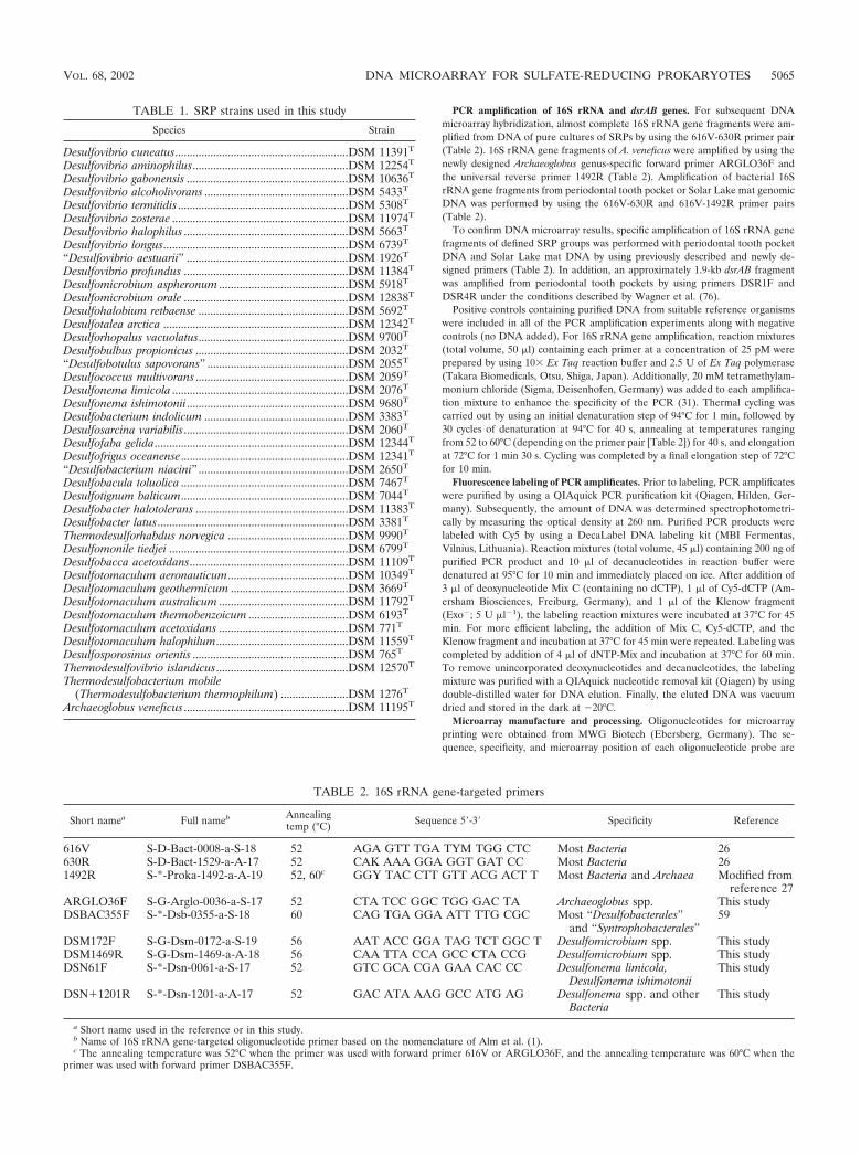
PCR amplification of 16S rRNA and dsrAB genes. For subsequent DNAmicroarray hybridization, almost complete 16S rRNA gene fragments were am-plified from DNA of pure cultures of SRPs by using the 616V-630R primer pair(Table 2). 16S rRNA gene fragments of A. veneficus were amplified by using thenewly designed Archaeoglobus genus-specific forward primer ARGLO36F andthe universal reverse primer 1492R (Table 2). Amplification of bacterial 16SrRNA gene fragments from periodontal tooth pocket or Solar Lake mat genomicDNA was performed by using the 616V-630R and 616V-1492R primer pairs(Table 2).
To confirm DNA microarray results, specific amplification of 16S rRNA genefragments of defined SRP groups was performed with periodontal tooth pocketDNA and Solar Lake mat DNA by using previously described and newly de-signed primers (Table 2). In addition, an approximately 1.9-kb dsrAB fragmentwas amplified from periodontal tooth pockets by using primers DSR1F andDSR4R under the conditions described by Wagner et al. (76).
Positive controls containing purified DNA from suitable reference organismswere included in all of the PCR amplification experiments along with negativecontrols (no DNA added). For 16S rRNA gene amplification, reaction mixtures(total volume, 50 �l) containing each primer at a concentration of 25 pM wereprepared by using 10� Ex Taq reaction buffer and 2.5 U of Ex Taq polymerase(Takara Biomedicals, Otsu, Shiga, Japan). Additionally, 20 mM tetramethylam-monium chloride (Sigma, Deisenhofen, Germany) was added to each amplifica-tion mixture to enhance the specificity of the PCR (31). Thermal cycling wascarried out by using an initial denaturation step of 94°C for 1 min, followed by30 cycles of denaturation at 94°C for 40 s, annealing at temperatures rangingfrom 52 to 60°C (depending on the primer pair [Table 2]) for 40 s, and elongationat 72°C for 1 min 30 s. Cycling was completed by a final elongation step of 72°Cfor 10 min.
Fluorescence labeling of PCR amplificates. Prior to labeling, PCR amplificateswere purified by using a QIAquick PCR purification kit (Qiagen, Hilden, Ger-many). Subsequently, the amount of DNA was determined spectrophotometri-cally by measuring the optical density at 260 nm. Purified PCR products werelabeled with Cy5 by using a DecaLabel DNA labeling kit (MBI Fermentas,Vilnius, Lithuania). Reaction mixtures (total volume, 45 �l) containing 200 ng ofpurified PCR product and 10 �l of decanucleotides in reaction buffer weredenatured at 95°C for 10 min and immediately placed on ice. After addition of3 �l of deoxynucleotide Mix C (containing no dCTP), 1 �l of Cy5-dCTP (Am-ersham Biosciences, Freiburg, Germany), and 1 �l of the Klenow fragment(Exo�; 5 U �l�1), the labeling reaction mixtures were incubated at 37°C for 45min. For more efficient labeling, the addition of Mix C, Cy5-dCTP, and theKlenow fragment and incubation at 37°C for 45 min were repeated. Labeling wascompleted by addition of 4 �l of dNTP-Mix and incubation at 37°C for 60 min.To remove unincorporated deoxynucleotides and decanucleotides, the labelingmixture was purified with a QIAquick nucleotide removal kit (Qiagen) by usingdouble-distilled water for DNA elution. Finally, the eluted DNA was vacuumdried and stored in the dark at �20°C.
Microarray manufacture and processing. Oligonucleotides for microarrayprinting were obtained from MWG Biotech (Ebersberg, Germany). The se-quence, specificity, and microarray position of each oligonucleotide probe are
TABLE 1. SRP strains used in this study
Species Strain
Desulfovibrio cuneatus...........................................................DSM 11391T
Desulfovibrio aminophilus.....................................................DSM 12254T
Desulfovibrio gabonensis .......................................................DSM 10636T
Desulfovibrio alcoholivorans .................................................DSM 5433T
Desulfovibrio termitidis ..........................................................DSM 5308T
Desulfovibrio zosterae ............................................................DSM 11974T
Desulfovibrio halophilus ........................................................DSM 5663T
Desulfovibrio longus...............................................................DSM 6739T
“Desulfovibrio aestuarii” .......................................................DSM 1926T
Desulfovibrio profundus ........................................................DSM 11384T
Desulfomicrobium aspheronum ............................................DSM 5918T
Desulfomicrobium orale ........................................................DSM 12838T
Desulfohalobium retbaense ...................................................DSM 5692T
Desulfotalea arctica ...............................................................DSM 12342T
Desulforhopalus vacuolatus...................................................DSM 9700T
Desulfobulbus propionicus ....................................................DSM 2032T
“Desulfobotulus sapovorans” ................................................DSM 2055T
Desulfococcus multivorans ....................................................DSM 2059T
Desulfonema limicola ............................................................DSM 2076T
Desulfonema ishimotonii.......................................................DSM 9680T
Desulfobacterium indolicum .................................................DSM 3383T
Desulfosarcina variabilis........................................................DSM 2060T
Desulfofaba gelida..................................................................DSM 12344T
Desulfofrigus oceanense.........................................................DSM 12341T
“Desulfobacterium niacini” ...................................................DSM 2650T
Desulfobacula toluolica .........................................................DSM 7467T
Desulfotignum balticum.........................................................DSM 7044T
Desulfobacter halotolerans ....................................................DSM 11383T
Desulfobacter latus.................................................................DSM 3381T
Thermodesulforhabdus norvegica .........................................DSM 9990T
Desulfomonile tiedjei .............................................................DSM 6799T
Desulfobacca acetoxidans......................................................DSM 11109T
Desulfotomaculum aeronauticum.........................................DSM 10349T
Desulfotomaculum geothermicum ........................................DSM 3669T
Desulfotomaculum australicum ............................................DSM 11792T
Desulfotomaculum thermobenzoicum ..................................DSM 6193T
Desulfotomaculum acetoxidans ............................................DSM 771T
Desulfotomaculum halophilum.............................................DSM 11559T
Desulfosporosinus orientis .....................................................DSM 765T
Thermodesulfovibrio islandicus.............................................DSM 12570T
Thermodesulfobacterium mobile(Thermodesulfobacterium thermophilum) .......................DSM 1276T
Archaeoglobus veneficus ........................................................DSM 11195T
TABLE 2. 16S rRNA gene-targeted primers
Short namea Full nameb Annealingtemp (°C) Sequence 5�-3� Specificity Reference
616V S-D-Bact-0008-a-S-18 52 AGA GTT TGA TYM TGG CTC Most Bacteria 26630R S-D-Bact-1529-a-A-17 52 CAK AAA GGA GGT GAT CC Most Bacteria 261492R S-*-Proka-1492-a-A-19 52, 60c GGY TAC CTT GTT ACG ACT T Most Bacteria and Archaea Modified from
reference 27ARGLO36F S-G-Arglo-0036-a-S-17 52 CTA TCC GGC TGG GAC TA Archaeoglobus spp. This studyDSBAC355F S-*-Dsb-0355-a-S-18 60 CAG TGA GGA ATT TTG CGC Most “Desulfobacterales”
and “Syntrophobacterales”59
DSM172F S-G-Dsm-0172-a-S-19 56 AAT ACC GGA TAG TCT GGC T Desulfomicrobium spp. This studyDSM1469R S-G-Dsm-1469-a-A-18 56 CAA TTA CCA GCC CTA CCG Desulfomicrobium spp. This studyDSN61F S-*-Dsn-0061-a-S-17 52 GTC GCA CGA GAA CAC CC Desulfonema limicola,
Desulfonema ishimotoniiThis study
DSN�1201R S-*-Dsn-1201-a-A-17 52 GAC ATA AAG GCC ATG AG Desulfonema spp. and otherBacteria
This study
a Short name used in the reference or in this study.b Name of 16S rRNA gene-targeted oligonucleotide primer based on the nomenclature of Alm et al. (1).c The annealing temperature was 52°C when the primer was used with forward primer 616V or ARGLO36F, and the annealing temperature was 60°C when the
primer was used with forward primer DSBAC355F.
VOL. 68, 2002 DNA MICROARRAY FOR SULFATE-REDUCING PROKARYOTES 5065
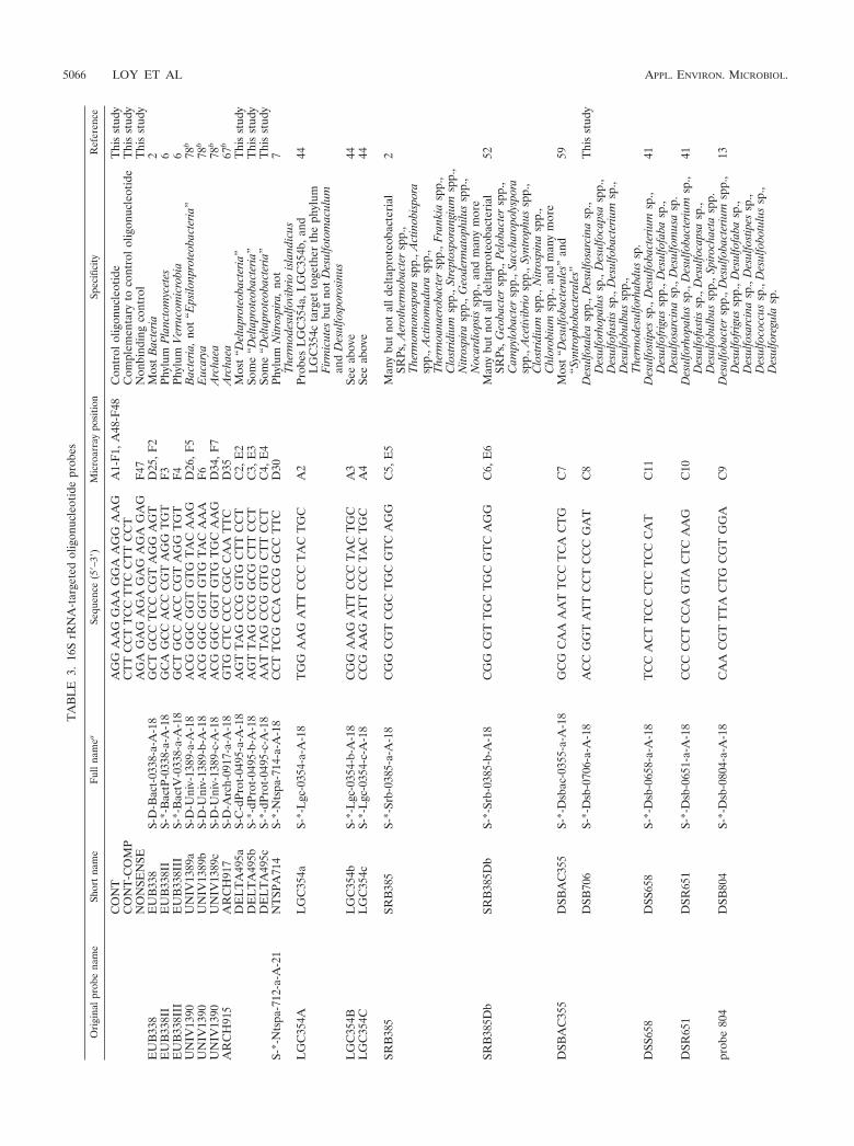
TA
BL
E3.
16S
rRN
A-t
arge
ted
olig
onuc
leot
ide
prob
es
Ori
gina
lpro
bena
me
Shor
tna
me
Ful
lnam
eaSe
quen
ce(5
�–3�
)M
icro
arra
ypo
sitio
nSp
ecifi
city
Ref
eren
ce
CO
NT
AG
GA
AG
GA
AG
GA
AG
GA
AG
A1-
F1,
A48
-F48
Con
trol
olig
onuc
leot
ide
Thi
sst
udy
CO
NT
-CO
MP
CT
TC
CT
TC
CT
TC
CT
TC
CT
Com
plem
enta
ryto
cont
rolo
ligon
ucle
otid
eT
his
stud
yN
ON
SEN
SEA
GA
GA
GA
GA
GA
GA
GA
GA
GF
47N
onbi
ndin
gco
ntro
lT
his
stud
yE
UB
338
EU
B33
8S-
D-B
act-
0338
-a-A
-18
GC
TG
CC
TC
CC
GT
AG
GA
GT
D25
,F2
Mos
tB
acte
ria2
EU
B33
8II
EU
B33
8II
S-*-
Bac
tP-0
338-
a-A
-18
GC
AG
CC
AC
CC
GT
AG
GT
GT
F3
Phyl
umP
lanc
tom
ycet
es6
EU
B33
8III
EU
B33
8III
S-*-
Bac
tV-0
338-
a-A
-18
GC
TG
CC
AC
CC
GT
AG
GT
GT
F4
Phyl
umV
erru
com
icro
bia
6U
NIV
1390
UN
IV13
89a
S-D
-Uni
v-13
89-a
-A-1
8A
CG
GG
CG
GT
GT
GT
AC
AA
GD
26,F
5B
acte
ria,n
ot“E
psilo
npro
teob
acte
ria”
78b
UN
IV13
90U
NIV
1389
bS-
D-U
niv-
1389
-b-A
-18
AC
GG
GC
GG
TG
TG
TA
CA
AA
F6
Euc
arya
78b
UN
IV13
90U
NIV
1389
cS-
D-U
niv-
1389
-c-A
-18
AC
GG
GC
GG
TG
TG
TG
CA
AG
D34
,F7
Arc
haea
78b
AR
CH
915
AR
CH
917
S-D
-Arc
h-09
17-a
-A-1
8G
TG
CT
CC
CC
CG
CC
AA
TT
CD
35A
rcha
ea67
b
DE
LT
A49
5aS-
C-d
Prot
-049
5-a-
A-1
8A
GT
TA
GC
CG
GT
GC
TT
CC
TC
2,E
2M
ost
“Del
tapr
oteo
bact
eria
”T
his
stud
yD
EL
TA
495b
S-*-
dPro
t-04
95-b
-A-1
8A
GT
TA
GC
CG
GC
GC
TT
CC
TC
3,E
3So
me
“Del
tapr
oteo
bact
eria
”T
his
stud
yD
EL
TA
495c
S-*-
dPro
t-04
95-c
-A-1
8A
AT
TA
GC
CG
GT
GC
TT
CC
TC
4,E
4So
me
“Del
tapr
oteo
bact
eria
”T
his
stud
yS-
*-N
tspa
-712
-a-A
-21
NT
SPA
714
S-*-
Nts
pa-7
14-a
-A-1
8C
CT
TC
GC
CA
CC
GG
CC
TT
CD
30Ph
ylum
Nitr
ospi
ra,n
otT
herm
odes
ulfo
vibr
iois
land
icus
7
LG
C35
4AL
GC
354a
S-*-
Lgc
-035
4-a-
A-1
8T
GG
AA
GA
TT
CC
CT
AC
TG
CA
2Pr
obes
LG
C35
4a,L
GC
354b
,and
LG
C35
4cta
rget
toge
ther
the
phyl
umF
irmic
utes
but
not
Des
ulfo
tom
acul
uman
dD
esul
fosp
oros
inus
44
LG
C35
4BL
GC
354b
S-*-
Lgc
-035
4-b-
A-1
8C
GG
AA
GA
TT
CC
CT
AC
TG
CA
3Se
eab
ove
44L
GC
354C
LG
C35
4cS-
*-L
gc-0
354-
c-A
-18
CC
GA
AG
AT
TC
CC
TA
CT
GC
A4
See
abov
e44
SRB
385
SRB
385
S-*-
Srb-
0385
-a-A
-18
CG
GC
GT
CG
CT
GC
GT
CA
GG
C5,
E5
Man
ybu
tno
tal
ldel
tapr
oteo
bact
eria
lSR
Ps,A
erot
herm
obac
ter
spp.
,T
herm
omon
ospo
rasp
p.,A
ctin
obis
pora
spp.
,Act
inom
adur
asp
p.,
The
rmoa
naer
obac
ter
spp.
,Fra
nkia
spp.
,C
lost
ridiu
msp
p.,S
trep
tosp
oran
gium
spp.
,N
itros
pira
spp.
,Geo
derm
atop
hilu
ssp
p.,
Noc
ardi
opsi
ssp
p.,a
ndm
any
mor
e
2
SRB
385D
bSR
B38
5Db
S-*-
Srb-
0385
-b-A
-18
CG
GC
GT
TG
CT
GC
GT
CA
GG
C6,
E6
Man
ybu
tno
tal
ldel
tapr
oteo
bact
eria
lSR
Ps,G
eoba
cter
spp.
,Pel
obac
ter
spp.
,C
ampy
loba
cter
spp.
,Sac
char
opol
yspo
rasp
p.,A
cetiv
ibrio
spp.
,Syn
trop
hus
spp.
,C
lost
ridiu
msp
p.,N
itros
pina
spp.
,C
hlor
obiu
msp
p.,a
ndm
any
mor
e
52
DSB
AC
355
DSB
AC
355
S-*-
Dsb
ac-0
355-
a-A
-18
GC
GC
AA
AA
TT
CC
TC
AC
TG
C7
Mos
t“D
esul
foba
cter
ales
”an
d“S
yntr
opho
bact
eral
es”
59
DSB
706
S-*-
Dsb
-070
6-a-
A-1
8A
CC
GG
TA
TT
CC
TC
CC
GA
TC
8D
esul
fota
lea
spp.
,Des
ulfo
sarc
ina
sp.,
Des
ulfo
rhop
alus
sp.,
Des
ulfo
caps
asp
p.,
Des
ulfo
fust
issp
.,D
esul
foba
cter
ium
sp.,
Des
ulfo
bulb
ussp
p.,
The
rmod
esul
forh
abdu
ssp
.
Thi
sst
udy
DSS
658
DSS
658
S-*-
Dsb
-065
8-a-
A-1
8T
CC
AC
TT
CC
CT
CT
CC
CA
TC
11D
esul
fost
ipes
sp.,
Des
ulfo
bact
eriu
msp
.,D
esul
fofr
igus
spp.
,Des
ulfo
faba
sp.,
Des
ulfo
sarc
ina
sp.,
Des
ulfo
mus
asp
.
41
DSR
651
DSR
651
S-*-
Dsb
-065
1-a-
A-1
8C
CC
CC
TC
CA
GT
AC
TC
AA
GC
10D
esul
forh
opal
ussp
.,D
esul
foba
cter
ium
sp.,
Des
ulfo
fust
issp
.,D
esul
foca
psa
sp.,
Des
ulfo
bulb
ussp
p.,S
piro
chae
tasp
p.
41
prob
e80
4D
SB80
4S-
*-D
sb-0
804-
a-A
-18
CA
AC
GT
TT
AC
TG
CG
TG
GA
C9
Des
ulfo
bact
ersp
p.,D
esul
foba
cter
ium
spp.
,D
esul
fofr
igus
spp.
,Des
ulfo
faba
sp.,
Des
ulfo
sarc
ina
sp.,
Des
ulfo
stip
essp
.,D
esul
foco
ccus
sp.,
Des
ulfo
botu
lus
sp.,
Des
ulfo
regu
lasp
.
13
5066 LOY ET AL APPL. ENVIRON. MICROBIOL.
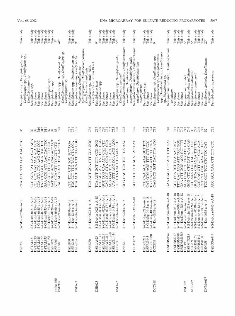
DSB
230
S-*-
Dsb
-023
0-a-
A-1
8C
TA
AT
GG
TA
CG
CA
AG
CT
CB
6D
esul
fota
lea
spp.
,Des
ulfo
rhop
alus
sp.,
Des
ulfo
caps
asp
p.,D
esul
fofu
stis
sp.,
Des
ulfo
bact
eriu
msp
.
Thi
sst
udy
DST
AL
131
S-G
-Dst
al-0
131-
a-A
-18
CC
CA
GA
TA
TC
AG
GG
TA
GA
B9
Des
ulfo
tale
asp
p.T
his
stud
yD
STA
L21
3S-
G-D
stal
-021
3-a-
A-1
8C
CT
CC
CG
AT
AC
AA
TA
GC
TB
8Se
eab
ove
Thi
sst
udy
DST
AL
645
S-G
-Dst
al-0
645-
a-A
-18
CC
AG
TA
CT
CA
AG
CT
CC
CC
B10
See
abov
eT
his
stud
yD
STA
L73
2S-
G-D
stal
-073
2-a-
A-1
8T
AT
CT
GG
CC
AG
AT
GG
TC
GB
12Se
eab
ove
Thi
sst
udy
DST
AL
835
S-G
-Dst
al-0
835-
a-A
-18
GA
AG
CG
AT
TA
AC
CA
CT
CC
B11
See
abov
eT
his
stud
yD
SRH
P185
S-*-
Dsr
hp-0
185-
a-A
-18
CC
AC
CT
TT
CC
TG
TT
TC
CA
B7
Des
ulfo
rhop
alus
spp.
Thi
sst
udy
DSB
B22
8S-
G-D
sbb-
0228
-a-A
-18
AA
TG
GT
AC
GC
AG
AC
CC
CT
B4
Des
ulfo
bulb
ussp
p.T
his
stud
ypr
obe
660
DSB
B66
0S-
G-D
sbb-
0660
-a-A
-18
AT
TC
CA
CT
TT
CC
CC
TC
TG
B5
See
abov
e13
b
DSB
985
DSB
986
S-*-
Dsb
-098
6-a-
A-1
8C
AC
AG
GA
TG
TC
AA
AC
CC
AC
28D
esul
foba
cter
spp.
,Des
ulfo
bacu
lasp
.,D
esul
foba
cter
ium
sp.,
Des
ulfo
spira
sp.,
Des
ulfo
tignu
msp
.
41b
DSB
1030
S-*-
Dsb
-103
0-a-
A-1
8C
TG
TC
TC
TG
TG
CT
CC
CG
AC
27Se
eab
ove
Thi
sst
udy
DSB
1240
S-*-
Dsb
-124
0-a-
A-1
8T
GC
CC
TT
TG
TA
CC
TA
CC
AC
34D
esul
foba
cter
spp.
,Des
ulfo
tignu
msp
.T
his
stud
yD
SB62
3D
SB62
3aS-
*-D
sb-0
623-
a-A
-18
TC
AA
GT
GC
AC
TT
CC
GG
GG
C35
Des
ulfo
bact
ercu
rvat
us,D
esul
foba
cter
halo
tole
rans
,Des
ulfo
bact
erhy
drog
enop
hilu
s,D
esul
foba
cter
post
gate
i,D
esul
foba
cter
vibr
iofo
rmis
9b
DSB
623b
S-*-
Dsb
-062
3-b-
A-1
8T
CA
AG
TG
CA
CT
TC
CA
GG
GC
36D
esul
foba
cter
sp.,
stra
inB
G8,
Des
ulfo
bact
ersp
.str
ain
BG
23T
his
stud
y
DSB
623
DSB
LA
623
S-S-
Dsb
.la-0
623-
a-A
-18
TC
AA
GT
GC
TC
TT
CC
GG
GG
C37
Des
ulfo
bact
erla
tus
9b
DSB
AC
L14
3S-
G-D
sbac
l-014
3-a-
A-1
8T
CG
GG
CA
GT
TA
TC
CC
GG
GC
29D
esul
foba
cula
spp.
Thi
sst
udy
DSB
AC
L22
5S-
G-D
sbac
l-022
5-a-
A-1
8G
GT
CC
GC
AA
AC
TC
AT
CT
CC
30Se
eab
ove
Thi
sst
udy
DSB
AC
L31
7S-
G-D
sbac
l-031
7-a-
A-1
8G
AC
CG
TG
TA
CC
AG
TT
CC
AC
31Se
eab
ove
Thi
sst
udy
DSB
AC
L12
68S-
G-D
sbac
l-126
8-a-
A-1
8A
GG
GA
TT
CG
CT
TA
CC
GT
TC
32Se
eab
ove
Thi
sst
udy
DSB
AC
L14
34S-
G-D
sbac
l-143
4-a-
A-1
8A
TA
GT
TA
GC
CC
AA
CG
AC
GC
33Se
eab
ove
Thi
sst
udy
DSF
672
DSB
674
S-*-
Dsb
-067
4-a-
A-1
8C
CT
CT
AC
AC
CT
GG
AA
TT
CC
20D
esul
fofr
igus
spp.
,Des
ulfo
faba
gelid
a,D
esul
fom
usa
hans
enii
55b
DSB
220
S-*-
Dsb
-022
0-a-
A-1
8G
CG
GA
CT
CA
TC
TT
CA
AA
CC
25D
esul
foba
cter
ium
niac
ini,
Des
ulfo
bact
eriu
mva
cuol
atum
,Des
ulfo
bact
eriu
mau
totr
ophi
cum
,Des
ulfo
faba
gelid
a
Thi
sst
udy
DSB
M12
39S-
*-D
sbm
-123
9-a-
A-1
8G
CC
CG
TT
GT
AC
AT
AC
CA
TC
26D
esul
foba
cter
ium
niac
ini,
Des
ulfo
bact
eriu
mva
cuol
atum
,Des
ulfo
bact
eriu
mau
totr
ophi
cum
Thi
sst
udy
DSF
RG
211
S-G
-Dsf
rg-0
211-
a-A
-18
CC
CC
AA
AC
AA
AA
GC
TT
CC
C22
Des
ulfo
frig
ussp
p.T
his
stud
yD
SFR
G44
5S-
G-D
sfrg
-044
5-a-
A-1
8C
AT
GT
GA
GG
TT
TC
TT
CC
CC
23Se
eab
ove
Thi
sst
udy
DSF
RG
1030
S-G
-Dsf
rg-1
030-
a-A
-18
TG
TC
AT
CG
GA
TT
CC
CC
GA
C24
See
abov
eT
his
stud
yD
CC
868
DC
C86
8S-
*-D
sb-0
868-
a-A
-18
CA
GG
CG
GA
TC
AC
TT
AA
TG
C38
Des
ulfo
sarc
ina
sp.,
Des
ulfo
nem
asp
p.,
Des
ulfo
cocc
ussp
.,D
esul
foba
cter
ium
spp.
,Des
ulfo
botu
lus
sp.,
Des
ulfo
stip
essp
.,D
esul
fom
usa
sp.
9
DSS
DB
M19
4S-
*-D
ssD
bm-0
194-
a-A
-18
GA
AG
AG
GC
CA
CC
CT
TG
AT
C40
Des
ulfo
sarc
ina
varia
bilis
,Des
ulfo
bact
eriu
mce
toni
cum
Thi
sst
udy
DSS
DB
M21
7S-
*-D
ssD
bm-0
217-
a-A
-18
GG
CC
CA
TC
TT
CA
AA
CA
GT
C41
See
abov
eT
his
stud
yD
SSD
BM
998
S-*-
Dss
Dbm
-099
8-a-
A-1
8T
TC
GA
TA
GG
AT
TC
CC
GG
GC
39Se
eab
ove
Thi
sst
udy
DSS
DB
M12
86S-
*-D
ssD
bm-1
286-
a-A
-18
GA
AC
TT
GG
GA
CG
GC
TT
TT
C42
See
abov
eT
his
stud
yD
SC19
3D
SC19
3S-
*-D
sb-0
193-
a-A
-18
AG
GC
CA
CC
CT
TG
AT
CC
AA
C43
Des
ulfo
sarc
ina
varia
bilis
55D
SBM
IN21
8S-
S-D
sbm
.in-0
218-
a-A
-18
GG
GC
TC
CT
CC
AT
AA
AC
AG
C44
Des
ulfo
bact
eriu
min
dolic
umT
his
stud
yD
CC
209
DC
C20
9S-
S-D
cc.m
v-02
09-a
-A-1
8C
CC
AA
AC
GG
TA
GC
TT
CC
TB
3D
esul
foco
ccus
mul
tivor
ans
55D
SNIS
H17
9S-
S-D
sn.is
h-01
79-a
-A-1
8G
GG
TC
AC
GG
GA
AT
GT
TA
TC
45D
esul
fone
ma
ishi
mot
onii
Thi
sst
udy
DSN
ISH
442
S-S-
Dsn
.ish-
0442
-a-A
-18
CC
CC
AG
GT
TC
TT
CC
CA
CA
C46
See
abov
eT
his
stud
yD
SNIS
H10
01S-
S-D
sn.is
h-10
01-a
-A-1
8C
GT
CT
CC
GG
AA
AA
TT
CC
CC
47Se
eab
ove
Thi
sst
udy
DN
MA
657
DSN
658
S-*-
Dsn
-065
8-a-
A-1
8T
CC
GC
TT
CC
CT
CT
CC
CA
TB
2D
esul
fone
ma
limic
ola,
Des
ulfo
nem
am
agnu
m20
b
DSB
OSA
445
S-S-
Dsb
o.sa
-044
5-a-
A-1
8A
CC
AC
AC
AA
CT
TC
TT
CC
CC
21D
esul
fobo
tulu
ssa
povo
rans
Thi
sst
udy
VOL. 68, 2002 DNA MICROARRAY FOR SULFATE-REDUCING PROKARYOTES 5067
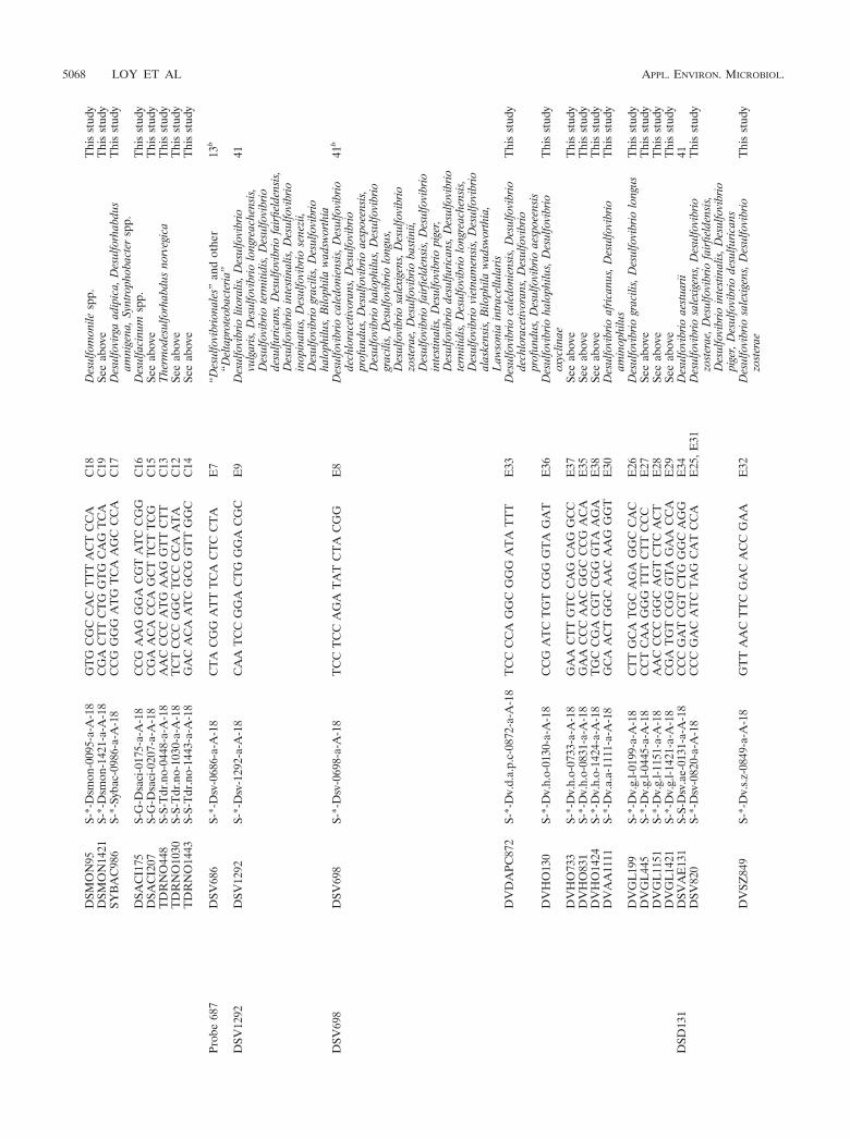
DSM
ON
95S-
*-D
smon
-009
5-a-
A-1
8G
TG
CG
CC
AC
TT
TA
CT
CC
AC
18D
esul
fom
onile
spp.
Thi
sst
udy
DSM
ON
1421
S-*-
Dsm
on-1
421-
a-A
-18
CG
AC
TT
CT
GG
TG
CA
GT
CA
C19
See
abov
eT
his
stud
ySY
BA
C98
6S-
*-Sy
bac-
0986
-a-A
-18
CC
GG
GG
AT
GT
CA
AG
CC
CA
C17
Des
ulfo
virg
aad
ipic
a,D
esul
forh
abdu
sam
nige
na,S
yntr
opho
bact
ersp
p.T
his
stud
y
DSA
CI1
75S-
G-D
saci
-017
5-a-
A-1
8C
CG
AA
GG
GA
CG
TA
TC
CG
GC
16D
esul
faci
num
spp.
Thi
sst
udy
DSA
CI2
07S-
G-D
saci
-020
7-a-
A-1
8C
GA
AC
AC
CA
GC
TT
CT
TC
GC
15Se
eab
ove
Thi
sst
udy
TD
RN
O44
8S-
S-T
dr.n
o-04
48-a
-A-1
8A
AC
CC
CA
TG
AA
GG
TT
CT
TC
13T
herm
odes
ulfo
rhab
dus
norv
egic
aT
his
stud
yT
DR
NO
1030
S-S-
Tdr
.no-
1030
-a-A
-18
TC
TC
CC
GG
CT
CC
CC
AA
TA
C12
See
abov
eT
his
stud
yT
DR
NO
1443
S-S-
Tdr
.no-
1443
-a-A
-18
GA
CA
CA
AT
CG
CG
GT
TG
GC
C14
See
abov
eT
his
stud
y
Prob
e68
7D
SV68
6S-
*-D
sv-0
686-
a-A
-18
CT
AC
GG
AT
TT
CA
CT
CC
TA
E7
“Des
ulfo
vibr
iona
les”
and
othe
r“D
elta
prot
eoba
cter
ia”
13b
DSV
1292
DSV
1292
S-*-
Dsv
-129
2-a-
A-1
8C
AA
TC
CG
GA
CT
GG
GA
CG
CE
9D
esul
fovi
brio
litor
alis
,Des
ulfo
vibr
iovu
lgar
is,D
esul
fovi
brio
long
reac
hens
is,
Des
ulfo
vibr
iote
rmiti
dis,
Des
ulfo
vibr
iode
sulfu
rican
s,D
esul
fovi
brio
fairfi
elde
nsis
,D
esul
fovi
brio
inte
stin
alis
,Des
ulfo
vibr
ioin
opin
atus
,Des
ulfo
vibr
iose
nezi
i,D
esul
fovi
brio
grac
ilis,
Des
ulfo
vibr
ioha
loph
ilus,
Bilo
phila
wad
swor
thia
41
DSV
698
DSV
698
S-*-
Dsv
-069
8-a-
A-1
8T
CC
TC
CA
GA
TA
TC
TA
CG
GE
8D
esul
fovi
brio
cale
doni
ensi
s,D
esul
fovi
brio
dech
lora
cetiv
oran
s,D
esul
fovi
brio
prof
undu
s,D
esul
fovi
brio
aesp
oeen
sis,
Des
ulfo
vibr
ioha
loph
ilus,
Des
ulfo
vibr
iogr
acili
s,D
esul
fovi
brio
long
us,
Des
ulfo
vibr
iosa
lexi
gens
,Des
ulfo
vibr
iozo
ster
ae,D
esul
fovi
brio
bast
inii,
Des
ulfo
vibr
iofa
irfiel
dens
is,D
esul
fovi
brio
inte
stin
alis
,Des
ulfo
vibr
iopi
ger,
Des
ulfo
vibr
iode
sulfu
rican
s,D
esul
fovi
brio
term
itidi
s,D
esul
fovi
brio
long
reac
hens
is,
Des
ulfo
vibr
iovi
etna
men
sis,
Des
ulfo
vibr
ioal
aske
nsis
,Bilo
phila
wad
swor
thia
,L
awso
nia
intr
acel
lula
ris
41b
DV
DA
PC87
2S-
*-D
v.d.
a.p.
c-08
72-a
-A-1
8T
CC
CC
AG
GC
GG
GA
TA
TT
TE
33D
esul
fovi
brio
cale
doni
ensi
s,D
esul
fovi
brio
dech
lora
cetiv
oran
s,D
esul
fovi
brio
prof
undu
s,D
esul
fovi
brio
aesp
oeen
sis
Thi
sst
udy
DV
HO
130
S-*-
Dv.
h.o-
0130
-a-A
-18
CC
GA
TC
TG
TC
GG
GT
AG
AT
E36
Des
ulfo
vibr
ioha
loph
ilus,
Des
ulfo
vibr
ioox
yclin
aeT
his
stud
y
DV
HO
733
S-*-
Dv.
h.o-
0733
-a-A
-18
GA
AC
TT
GT
CC
AG
CA
GG
CC
E37
See
abov
eT
his
stud
yD
VH
O83
1S-
*-D
v.h.
o-08
31-a
-A-1
8G
AA
CC
CA
AC
GG
CC
CG
AC
AE
35Se
eab
ove
Thi
sst
udy
DV
HO
1424
S-*-
Dv.
h.o-
1424
-a-A
-18
TG
CC
GA
CG
TC
GG
GT
AA
GA
E38
See
abov
eT
his
stud
yD
VA
A11
11S-
*-D
v.a.
a-11
11-a
-A-1
8G
CA
AC
TG
GC
AA
CA
AG
GG
TE
30D
esul
fovi
brio
afric
anus
,Des
ulfo
vibr
ioam
inop
hilu
sT
his
stud
y
DV
GL
199
S-*-
Dv.
g.l-0
199-
a-A
-18
CT
TG
CA
TG
CA
GA
GG
CC
AC
E26
Des
ulfo
vibr
iogr
acili
s,D
esul
fovi
brio
long
usT
his
stud
yD
VG
L44
5S-
*-D
v.g.
l-044
5-a-
A-1
8C
CT
CA
AG
GG
TT
TC
TT
CC
CE
27Se
eab
ove
Thi
sst
udy
DV
GL
1151
S-*-
Dv.
g.l-1
151-
a-A
-18
AA
CC
CC
GG
CA
GT
CT
CA
CT
E28
See
abov
eT
his
stud
yD
VG
L14
21S-
*-D
v.g.
l-142
1-a-
A-1
8C
GA
TG
TC
GG
GT
AG
AA
CC
AE
29Se
eab
ove
Thi
sst
udy
DSD
131
DSV
AE
131
S-S-
Dsv
.ae-
0131
-a-A
-18
CC
CG
AT
CG
TC
TG
GG
CA
GG
E34
Des
ulfo
vibr
ioae
stua
rii41
DSV
820
S-*-
Dsv
-082
0-a-
A-1
8C
CC
GA
CA
TC
TA
GC
AT
CC
AE
25,E
31D
esul
fovi
brio
sale
xige
ns,D
esul
fovi
brio
zost
erae
,Des
ulfo
vibr
iofa
irfiel
dens
is,
Des
ulfo
vibr
ioin
test
inal
is,D
esul
fovi
brio
pige
r,D
esul
fovi
brio
desu
lfuric
ans
Thi
sst
udy
DV
SZ84
9S-
*-D
v.s.
z-08
49-a
-A-1
8G
TT
AA
CT
TC
GA
CA
CC
GA
AE
32D
esul
fovi
brio
sale
xige
ns,D
esul
fovi
brio
zost
erae
Thi
sst
udy
5068 LOY ET AL APPL. ENVIRON. MICROBIOL.
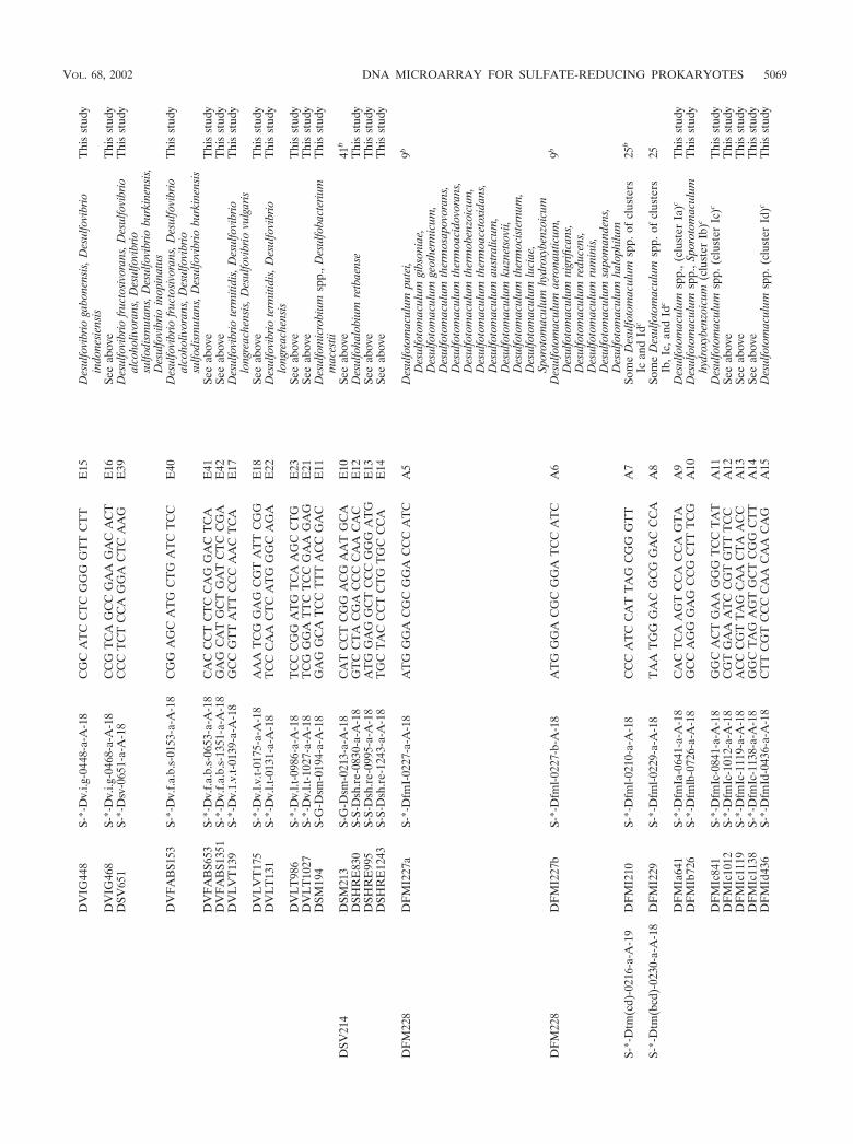
DV
IG44
8S-
*-D
v.i.g
-044
8-a-
A-1
8C
GC
AT
CC
TC
GG
GG
TT
CT
TE
15D
esul
fovi
brio
gabo
nens
is,D
esul
fovi
brio
indo
nesi
ensi
sT
his
stud
y
DV
IG46
8S-
*-D
v.i.g
-046
8-a-
A-1
8C
CG
TC
AG
CC
GA
AG
AC
AC
TE
16Se
eab
ove
Thi
sst
udy
DSV
651
S-*-
Dsv
-065
1-a-
A-1
8C
CC
TC
TC
CA
GG
AC
TC
AA
GE
39D
esul
fovi
brio
fruc
tosi
vora
ns,D
esul
fovi
brio
alco
holiv
oran
s,D
esul
fovi
brio
sulfo
dism
utan
s,D
esul
fovi
brio
burk
inen
sis,
Des
ulfo
vibr
ioin
opin
atus
Thi
sst
udy
DV
FA
BS1
53S-
*-D
v.f.a
.b.s
-015
3-a-
A-1
8C
GG
AG
CA
TG
CT
GA
TC
TC
CE
40D
esul
fovi
brio
fruc
tosi
vora
ns,D
esul
fovi
brio
alco
holiv
oran
s,D
esul
fovi
brio
sulfo
dism
utan
s,D
esul
fovi
brio
burk
inen
sis
Thi
sst
udy
DV
FA
BS6
53S-
*-D
v.f.a
.b.s
-065
3-a-
A-1
8C
AC
CC
TC
TC
CA
GG
AC
TC
AE
41Se
eab
ove
Thi
sst
udy
DV
FA
BS1
351
S-*-
Dv.
f.a.b
.s-1
351-
a-A
-18
GA
GC
AT
GC
TG
AT
CT
CC
GA
E42
See
abov
eT
his
stud
yD
VL
VT
139
S-*-
Dv.
1.v.
t-01
39-a
-A-1
8G
CC
GT
TA
TT
CC
CA
AC
TC
AE
17D
esul
fovi
brio
term
itidi
s,D
esul
fovi
brio
long
reac
hens
is,D
esul
fovi
brio
vulg
aris
Thi
sst
udy
DV
LV
T17
5S-
*-D
v.l.v
.t-01
75-a
-A-1
8A
AA
TC
GG
AG
CG
TA
TT
CG
GE
18Se
eab
ove
Thi
sst
udy
DV
LT
131
S-*-
Dv.
l.t-0
131-
a-A
-18
TC
CC
AA
CT
CA
TG
GG
CA
GA
E22
Des
ulfo
vibr
iote
rmiti
dis,
Des
ulfo
vibr
iolo
ngre
ache
nsis
Thi
sst
udy
DV
LT
986
S-*-
Dv.
l.t-0
986-
a-A
-18
TC
CC
GG
AT
GT
CA
AG
CC
TG
E23
See
abov
eT
his
stud
yD
VL
T10
27S-
*-D
v.l.t
-102
7-a-
A-1
8T
CG
GG
AT
TC
TC
CG
AA
GA
GE
21Se
eab
ove
Thi
sst
udy
DSM
194
S-G
-Dsm
-019
4-a-
A-1
8G
AG
GC
AT
CC
TT
TA
CC
GA
CE
11D
esul
fom
icro
bium
spp.
,Des
ulfo
bact
eriu
mm
aces
tiiT
his
stud
y
DSV
214
DSM
213
S-G
-Dsm
-021
3-a-
A-1
8C
AT
CC
TC
GG
AC
GA
AT
GC
AE
10Se
eab
ove
41b
DSH
RE
830
S-S-
Dsh
.re-
0830
-a-A
-18
GT
CC
TA
CG
AC
CC
CA
AC
AC
E12
Des
ulfo
halo
bium
retb
aens
eT
his
stud
yD
SHR
E99
5S-
S-D
sh.r
e-09
95-a
-A-1
8A
TG
GA
GG
CT
CC
CG
GG
AT
GE
13Se
eab
ove
Thi
sst
udy
DSH
RE
1243
S-S-
Dsh
.re-
1243
-a-A
-18
TG
CT
AC
CC
TC
TG
TG
CC
CA
E14
See
abov
eT
his
stud
y
DF
M22
8D
FM
I227
aS-
*-D
fmI-
0227
-a-A
-18
AT
GG
GA
CG
CG
GA
CC
CA
TC
A5
Des
ulfo
tom
acul
umpu
tei,
Des
ulfo
tom
acul
umgi
bson
iae,
Des
ulfo
tom
acul
umge
othe
rmic
um,
Des
ulfo
tom
acul
umth
erm
osap
ovor
ans,
Des
ulfo
tom
acul
umth
erm
oaci
dovo
rans
,D
esul
foto
mac
ulum
ther
mob
enzo
icum
,D
esul
foto
mac
ulum
ther
moa
ceto
xida
ns,
Des
ulfo
tom
acul
umau
stra
licum
,D
esul
foto
mac
ulum
kuzn
etso
vii,
Des
ulfo
tom
acul
umth
erm
ocis
tern
um,
Des
ulfo
tom
acul
umlu
ciae
,Sp
orot
omac
ulum
hydr
oxyb
enzo
icum
9b
DF
M22
8D
FM
I227
bS-
*-D
fml-0
227-
b-A
-18
AT
GG
GA
CG
CG
GA
TC
CA
TC
A6
Des
ulfo
tom
acul
umae
rona
utic
um,
Des
ulfo
tom
acul
umni
grifi
cans
,D
esul
foto
mac
ulum
redu
cens
,D
esul
foto
mac
ulum
rum
inis
,D
esul
foto
mac
ulum
sapo
man
dens
,D
esul
foto
mac
ulum
halo
philu
m
9b
S-*-
Dtm
(cd)
-021
6-a-
A-1
9D
FM
I210
S-*-
Dfm
l-021
0-a-
A-1
8C
CC
AT
CC
AT
TA
GC
GG
GT
TA
7So
me
Des
ulfo
tom
acul
umsp
p.of
clus
ters
Ican
dId
c25
b
S-*-
Dtm
(bcd
)-02
30-a
-A-1
8D
FM
I229
S-*-
Dfm
l-022
9-a-
A-1
8T
AA
TG
GG
AC
GC
GG
AC
CC
AA
8So
me
Des
ulfo
tom
acul
umsp
p.of
clus
ters
Ib,I
c,an
dId
c25
DF
MIa
641
S-*-
Dfm
Ia-0
641-
a-A
-18
CA
CT
CA
AG
TC
CA
CC
AG
TA
A9
Des
ulfo
tom
acul
umsp
p.,(
clus
ter
Ia)c
Thi
sst
udy
DF
MIb
726
S-*-
Dfm
lb-0
726-
a-A
-18
GC
CA
GG
GA
GC
CG
CT
TT
CG
A10
Des
ulfo
tom
acul
umsp
p.,S
poro
tom
acul
umhy
drox
yben
zoic
um(c
lust
erIb
)cT
his
stud
y
DF
MIc
841
S-*-
Dfm
Ic-0
841-
a-A
-18
GG
CA
CT
GA
AG
GG
TC
CT
AT
A11
Des
ulfo
tom
acul
umsp
p.(c
lust
erIc
)cT
his
stud
yD
FM
Ic10
12S-
*-D
fmIc
-101
2-a-
A-1
8C
GT
GA
AA
TC
CG
TG
TT
TC
CA
12Se
eab
ove
Thi
sst
udy
DF
MIc
1119
S-*-
Dfm
Ic-1
119-
a-A
-18
AC
CC
GT
TA
GC
AA
CT
AA
CC
A13
See
abov
eT
his
stud
yD
FM
Ic11
38S-
*-D
fmIc
-113
8-a-
A-1
8G
GC
TA
GA
GT
GC
TC
GG
CT
TA
14Se
eab
ove
Thi
sst
udy
DF
MId
436
S-*-
Dfm
Id-0
436-
a-A
-18
CT
TC
GT
CC
CC
AA
CA
AC
AG
A15
Des
ulfo
tom
acul
umsp
p.(c
lust
erId
)cT
his
stud
y
VOL. 68, 2002 DNA MICROARRAY FOR SULFATE-REDUCING PROKARYOTES 5069
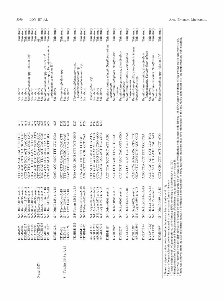
DF
MId
625
S-*-
Dfm
Id-0
625-
a-A
-18
TT
TC
AA
AG
GC
AC
CC
CC
GC
A16
See
abov
eT
his
stud
yD
FM
Id99
6S-
*-D
fmId
-099
6-a-
A-1
8C
AC
AG
GC
TG
TC
AG
GG
GA
TA
17Se
eab
ove
Thi
sst
udy
DF
MId
1117
S-*-
Dfm
Id-1
117-
a-A
-18
CC
GC
TG
GC
AA
CT
AA
CC
GT
A18
See
abov
eT
his
stud
yD
FA
CE
199
S-S-
Df.a
ce-0
199-
a-A
-18
GC
AT
TG
TA
AA
GA
GG
CC
AC
A20
Des
ulfo
tom
acul
umsp
p.(c
lust
erIe
)cT
his
stud
yD
FA
CE
438
S-S-
Df.a
ce-0
438-
a-A
-18
CT
GT
TC
GT
CC
AA
TG
TC
AC
A19
See
abov
eT
his
stud
yD
FA
CE
995
S-S-
Df.a
ce-0
995-
a-A
-18
CA
CA
GC
GG
TC
AC
GG
GA
TG
A21
See
abov
eT
his
stud
yD
-ace
t102
7rD
FA
CE
1028
S-S-
Df.a
ce-1
028-
a-A
-18
CT
CC
GT
GT
GC
AA
GT
AA
AC
A22
See
abov
e68
DF
AC
E14
36S-
S-D
f.ace
-143
6-a-
A-1
8T
GC
GA
GT
TA
AG
TC
AC
CG
GA
23Se
eab
ove
Thi
sst
udy
DF
MIf
126
S-*-
Dfm
If-0
126-
a-A
-18
CT
GA
TA
GG
CA
GG
TT
AT
CC
A24
Des
ulfo
tom
acul
umsp
p.(c
lust
erIf
)dT
his
stud
yD
FM
II11
07S-
*-D
fmII
-110
7-a-
A-1
8C
TA
AA
TA
CA
GG
GG
TT
GC
GA
29D
esul
fosp
oros
inus
spp.
,Des
ulfo
tom
acul
umau
ripig
men
tum
(clu
ster
II)c
Thi
sst
udy
DF
MII
1281
S-*-
Dfm
II-1
281-
a-A
-18
GA
GA
CC
GG
CT
TT
CT
CG
GA
A28
See
abov
eT
his
stud
y
TD
SV60
1S-
*-T
dsv-
0601
-a-A
-18
GC
TG
TG
GA
AT
TC
CA
CC
TT
D32
The
rmod
esul
fovi
brio
spp.
Thi
sst
udy
S-*-
Tds
ulfo
-084
8-a-
A-1
8T
DSV
849
S-*-
Tds
v-08
49-a
-A-1
8T
TT
CC
CT
TC
GG
CA
CA
GA
GD
33Se
eab
ove
8T
DSV
1326
S-*-
Tds
v-13
26-a
-A-1
8C
GA
TT
CC
GG
GT
TC
AC
GG
AD
31Se
eab
ove
Thi
sst
udy
TD
SBM
1282
S-P-
Tds
bm-1
282-
a-A
-18
TG
AG
GA
GG
GC
TT
TC
TG
GG
D27
The
rmod
esul
foba
cter
ium
spp.
,G
eoth
erm
obac
teriu
msp
.T
his
stud
y
TD
SBM
353
S-*-
Tds
bm-0
353-
a-A
-18
CC
AA
GA
TT
CC
CC
CC
TG
CT
D28
The
rmod
esul
foba
cter
ium
spp.
Thi
sst
udy
TD
SBM
652
S-*-
Tds
bm-0
652-
a-A
-18
AG
CC
TC
TC
CG
GC
CC
TC
AA
D29
See
abov
eT
his
stud
y
AR
GL
O37
S-G
-Arg
lo-0
037-
a-A
-18
CT
TA
GT
CC
CA
GC
CG
GA
TA
D37
Arc
haeo
glob
ussp
p.T
his
stud
yA
RG
LO
276
S-G
-Arg
lo-0
276-
a-A
-18
GC
CC
GT
AC
GG
AT
CT
TC
GG
D38
See
abov
eT
his
stud
yA
RG
LO
576
S-G
-Arg
lo-0
576-
a-A
-18
CC
AG
CC
CG
GC
TA
CG
GA
CG
D39
See
abov
eT
his
stud
yA
RG
LO
972
S-G
-Arg
lo-0
972-
a-A
-18
CC
CC
GG
TA
AG
CT
TC
CC
GG
D40
See
abov
eT
his
stud
y
DSB
M16
8eS-
*-D
sbm
-016
8-a-
A-1
8A
CT
TT
AT
CC
GG
CA
TT
AG
CD
esul
foba
cter
ium
niac
ini,
Des
ulfo
bact
eriu
mva
cuol
atum
Thi
sst
udy
DV
HO
588e
S-*-
Dv.
h.o-
0588
-a-A
-18
AC
CC
CT
GA
CT
TA
CT
GC
GC
Des
ulfo
vibr
ioha
loph
ilus,
Des
ulfo
vibr
ioox
yclin
aeT
his
stud
y
DV
IG26
7eS-
*-D
v.i.g
-026
7-a-
A-1
8C
AT
CG
TA
GC
CA
CG
GT
GG
GD
esul
fovi
brio
gabo
nens
is,D
esul
fovi
brio
indo
nesi
ensi
sT
his
stud
y
DV
LT
1425
eS-
*-D
v.l.t
-142
5-a-
A-1
8T
CA
CC
GG
TA
TC
GG
GT
AA
AD
esul
fovi
brio
term
itidi
s,D
esul
fovi
brio
long
reac
hens
isT
his
stud
y
DV
GL
228e
S-*-
Dv.
g.l-0
228-
a-A
-18
CA
GC
CA
AG
AG
GC
CT
AT
TC
Des
ulfo
vibr
iogr
acili
s,D
esul
fovi
brio
long
usT
his
stud
yA
RG
LO
390e
S-G
-Arg
lo-0
390-
a-A
-18
GC
AC
TC
CG
GC
TG
AC
CC
CG
Arc
haeo
glob
ussp
p.T
his
stud
y
DV
LV
T19
4fS-
*-D
v.l.v
.t-01
94-a
-A-1
8A
GG
CC
AC
CT
TT
CC
CC
CG
AD
esul
fovi
brio
term
itidi
s,D
esul
fovi
brio
long
reac
hens
is,D
esul
fovi
brio
vulg
aris
Thi
sst
udy
DV
LV
T22
2fS-
*-D
v.l.v
.t-02
22-a
-A-1
8A
CG
CG
GA
CT
CA
TC
CA
TG
ASe
eab
ove
Thi
sst
udy
DV
CL
1350
fS-
*-D
v.c.
l-135
0-a-
A-1
8G
GC
AT
GC
TG
AT
CC
AG
AA
TD
esul
fovi
brio
cune
atus
,Des
ulfo
vibr
iolit
oral
isT
his
stud
y
DF
MIf
489f
S-*-
Dfm
If-0
489-
a-A
-18
CC
GG
GG
CT
TA
CT
CC
TA
TG
Des
ulfo
tom
acul
umsp
p.(c
lust
erIf
)dT
his
stud
y
aN
ame
ofol
igon
ucle
otid
epr
obe
base
don
the
nom
encl
atur
eof
Alm
etal
.(1)
.b
Len
gth
ofol
igon
ucle
otid
epr
obe
was
adap
ted
toth
em
icro
arra
yfo
rmat
(18-
mer
).c
Clu
ster
desi
gnat
ion(
s)of
gram
-pos
itive
,spo
re-f
orm
ing
SRPs
acco
rdin
gto
Stac
kebr
andt
etal
.(65
).d
Des
ulfo
tom
acul
umha
loph
ilum
and
Des
ulfo
tom
acul
umal
kalip
hilu
mw
ere
assi
gned
tone
wcl
uste
rIf
.e
Prob
ew
asre
mov
edfr
omth
eSR
Pm
icro
arra
ybe
caus
eno
posi
tive
sign
alco
uld
bede
tect
edaf
ter
hybr
idiz
atio
nw
ithflu
ores
cent
lyla
bele
d16
SrR
NA
gene
ampl
ifica
teof
the
perf
ect-
mat
chre
fere
nce
stra
in.
fPr
obe
was
rem
oved
from
the
SRP
mic
roar
ray
beca
use
ithy
brid
ized
nons
peci
fical
lyto
man
yre
fere
nce
orga
nism
sth
atha
vem
ism
atch
esin
the
16S
rRN
Age
neta
rget
site
(see
supp
lem
enta
ryw
ebm
ater
ial)
.
5070 LOY ET AL APPL. ENVIRON. MICROBIOL.
shown in Table 3. In addition, difference alignments for all probes generated withthe latest ARB small-subunit rRNA database (http://www.arb-home.de) can beviewed at the probeBase website (http://www.probebase.net). The 5� end of eacholigonucleotide probe was tailed with 15 dTTP molecules (T-spacer) to increasethe on-chip accessibility of spotted probes to target DNA (61, 63). In addition,the 5�-terminal nucleotide of each oligonucleotide was aminated to allow cova-lent coupling of the oligonucleotides to aldehyde group-coated CSS-100 glassslides (CEL Associates, Houston, Tex.). The concentration of oligonucleotideprobes before printing was adjusted to 50 pmol �l�1 in 50% dimethyl sulfoxideto prevent evaporation during the printing procedure. SRP-PhyloChips wereprinted by using a GMS 417 contact arrayer (Affymetrix, Santa Clara, Calif.).Spotted DNA microarrays were dried overnight at room temperature to allowefficient cross-linking. Slides were washed twice at room temperature in 0.2%sodium dodecyl sulfate (SDS) and then twice with double-distilled water withvigorous agitation to remove unbound oligonucleotides and the SDS. After airdrying, the slides were incubated for 5 min in a fresh sodium borohydridesolution (1.0 g of NaBH4 in 300 ml of phosphate-buffered saline and 100 ml ofabsolute ethanol) to reduce all remaining reactive aldehyde groups on the glass.The reaction was stopped by adding ice-cold absolute ethanol. The reducedslides were washed three times (with 0.2% SDS and double-distilled water), airdried, and stored in the dark at room temperature.
Reverse hybridization on microarrays. Vacuum-dried Cy5-labeled PCR prod-ucts (400 ng) and 0.5 pmol of the Cy5-labeled control oligonucleotide CONT-COMP (Table 3) were resuspended in 20 �l of hybridization buffer (5� SSC, 1%blocking reagent [Roche, Mannheim, Germany], 0.1% n-lauryl sarcosine, 0.02%SDS, 5% formamide [1� SSC is 0.15 M NaCl plus 0.015 M sodium citrate]),denatured for 10 min at 95°C, and immediately placed on ice. Then the solutionwas pipetted onto an SRP-PhyloChip, covered with a coverslip, and inserted intoa tight custom-made hybridization chamber (http://cmgm.stanford.edu/pbrown/mguide/HybChamber.pdf) containing 50 �l of hybridization buffer for subse-quent equilibration. Hybridization was performed overnight at 42°C in a waterbath. After hybridization, the slides were washed immediately under stringentconditions for 5 min at 55°C in 50 ml of washing buffer (containing 3 M tetra-methylammonium chloride, 50 mM Tris-HCl, 2 mM EDTA, and 0.1% SDS). Torecord probe-target melting curves, the temperature of the washing step wasvaried from 42 to 80°C. After the stringent washing, the slides were washed twicewith ice-cold double-distilled water, air dried, and stored in the dark at roomtemperature.
Scanning of microarrays. Fluorescence images of the SRP PhyloChips wererecorded by scanning the slides with a GMS 418 array scanner (Affymetrix). Thefluorescence signals were quantified by using the ImaGene 4.0 software (Bio-Discovery, Inc., Los Angeles, Calif.). A grid of individual circles defining thelocation of each spot on the array was superimposed on the image to designateeach fluorescent spot to be quantified. The mean signal intensity was determinedfor each spot. In addition, the mean signal intensity of the local background areasurrounding the spots was determined.
Selective enrichment of nucleic acids by a capture probe approach. Fivemicroliters of aldehyde group-coated glass beads (diameter, 1 �m; Xenopore,Hawthorne, N.J.) was incubated overnight with 5 �l of the appropriate captureprobe (100 pmol �l�1; tailed with 15 dTTP molecules; aminated with 5�-terminalnucleotide) at room temperature. Subsequently, the beads were washed oncewith 400 �l of 0.2% SDS and pelleted by centrifugation (1 min at 14,000 rpm;Hettich Zentrifuge type 1000, Tuttlingen, Germany), and the supernatant wasdecanted. After this step, the beads were washed twice with 400 �l of double-distilled water, dried, and stored at room temperature prior to hybridization. Avacuum-dried bacterial 16S rRNA gene PCR product (obtained from DNA fromthe Solar Lake mat with the 616V-1492R primer pair) was resuspended in 200 �lof hybridization buffer (see above), denatured for 10 min at 95°C, and immedi-ately cooled on ice. The hybridization solution and capture probe beads weremixed in a screw-cap tube and incubated overnight at 42°C on a shaker. Subse-quently, the beads were washed twice with 1.5 ml of washing buffer (see above)at 55°C for 2.5 min. After the stringent washes, the beads were washed with 1.5ml of ice-cold double-distilled water and then with ice-cold 70% ethanol. Beadswith captured nucleic acids were vacuum dried and resuspended in 50 �l of EBbuffer (part of the QIAquick PCR purification kit; Qiagen) for storage at �20°C.Reamplification of bacterial 16S rRNA gene fragments from the captured nu-cleic acids was performed by using 5 �l of the resuspended beads for PCRperformed by using the 616V-1492R primer pair and the protocols describedabove.
Cloning, sequencing, and phylogeny inference. Prior to cloning, the PCRamplification products were purified by low-melting-point agarose (1.5%) gelelectrophoresis (NuSieve 3:1; FMC Bioproducts, Biozym Diagnostics GmbH,Oldendorf, Germany) and stained in a SYBR Green I solution (10 �l of 1,0000�
SYBR Green I stain in 100 �l of TAE buffer [40 mM Tris, 10 mM sodiumacetate, 1 mM EDTA; pH 8.0]; Biozym Diagnostics GmbH) for 45 min. Bandsof the expected size were excised from the agarose gel with a glass capillary andmelted with 50 �l of double-distilled water for 10 min at 80°C. Four microlitersof each solution was ligated as recommended by the manufacturer into thecloning vector pCR2.1 supplied with a TOPO TA cloning kit (Invitrogen Corp.,San Diego, Calif.). Nucleotide sequences were determined by the dideoxynucle-otide method (57) as described by Purkhold et al. (51). The new 16S rRNAsequences were added to an alignment of about 16,000 small-subunit rRNAsequences by using the alignment tool of the ARB program package (O. Strunkand W. Ludwig, http://www.arb-home.de). Alignments were refined by visualinspection. Phylogenetic analyses were performed by using distance matrix, max-imum-parsimony, and maximum-likelihood methods and the appropriate tools ofthe ARB program package and the fastDNAml program (34). The compositionsof the data sets varied with respect to the reference sequences and the alignmentpositions included. Variability in the individual alignment positions was deter-mined by using the appropriate tool of the ARB package and was used as acriterion to remove or include variable positions for phylogenetic analyses. Phy-logenetic consensus trees were drawn by following the recommendations ofLudwig et al. (40). The new dsrAB sequences were translated into amino acidsand added to an alignment of 62 DsrAB sequences of SRPs (18, 28). Phyloge-netic analyses were performed by using the procedures described by Klein et al.(28).
Nucleotide sequence accession numbers. The sequences determined in thisstudy are available in the GenBank database under accession numbersAY083010 to AY083027 (16S rRNA gene clones) and AY083028 to AY083029(dsrAB gene clones). The dsrAB gene sequence of Desulfomicrobium orale DSM12838T has been deposited under accession number AY083030.
RESULTS
SRP phylogeny. As the basis for development of the SRP-PhyloChip, a thorough reevaluation of the phylogeny of SRPswas performed. All 16S rRNA sequences of SRPs which areavailable in public databases (as of October 2001) were col-lected, aligned, and analyzed phylogenetically by using maxi-mum-parsimony, maximum-likelihood, and neighbor-joiningmethods. Figures 1 and 2 illustrate the phylogeny of the delta-proteobacterial SRPs. Figure 3 shows the phylogeny of SRPsaffiliated with the Firmicutes, Nitrospira, Thermodesulfobacte-ria, and Euryarchaeota phyla (phylum names according to thetaxonomic outline in the second edition of Bergey’s Manual ofSystematic Bacteriology, 2nd ed. [21]).
Probe design. Initially, the specificities of previously de-scribed probes and primers for SRPs (2, 8, 9, 13, 20, 25, 41, 52,55, 59, 68) were reevaluated with the current 16S rRNA dataset containing more than 16,000 entries. Based on this analysis,26 probes were considered to be suitable for inclusion on theSRP-PhyloChip (Table 3). These probes were, if necessary,adjusted to a length of 18 nucleotides (not including the T-spacer). Twenty-four of these probes exclusively target SRPs.Probes SRB385 (2) and SRB385Db (52) were included on thechip because they have been widely used in previous SRPresearch (3, 16, 36, 49, 58, 71), although both of these probestarget a considerable number of phylogenetically diverse non-SRPs. In addition, we significantly extended the SRP probe setby designing 102 probes targeting monophyletic groups ofSRPs (Fig. 1 to 3 and Table 3). These probes were designed tohave a minimum G�C content of 50%, a length of 18 nucle-otides (not including the T-spacer), and as many centrallylocated mismatches with the target sites on 16S rRNA genes ofnontarget organisms as possible. Several of these probes targetthe same SRPs, complementing several unique regions of the16S rRNA gene, while others exhibit hierarchical specificity.
VOL. 68, 2002 DNA MICROARRAY FOR SULFATE-REDUCING PROKARYOTES 5071
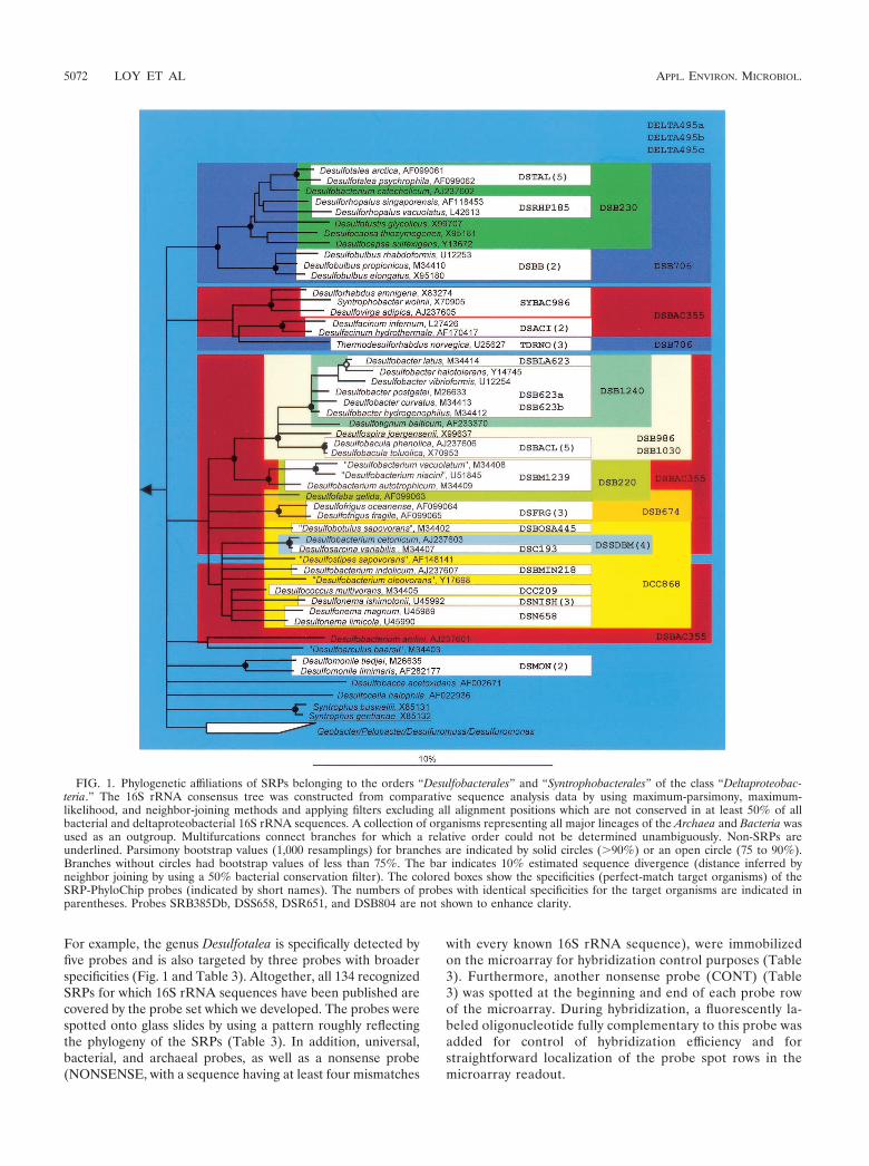
For example, the genus Desulfotalea is specifically detected byfive probes and is also targeted by three probes with broaderspecificities (Fig. 1 and Table 3). Altogether, all 134 recognizedSRPs for which 16S rRNA sequences have been published arecovered by the probe set which we developed. The probes werespotted onto glass slides by using a pattern roughly reflectingthe phylogeny of the SRPs (Table 3). In addition, universal,bacterial, and archaeal probes, as well as a nonsense probe(NONSENSE, with a sequence having at least four mismatches
with every known 16S rRNA sequence), were immobilizedon the microarray for hybridization control purposes (Table3). Furthermore, another nonsense probe (CONT) (Table3) was spotted at the beginning and end of each probe rowof the microarray. During hybridization, a fluorescently la-beled oligonucleotide fully complementary to this probe wasadded for control of hybridization efficiency and forstraightforward localization of the probe spot rows in themicroarray readout.
FIG. 1. Phylogenetic affiliations of SRPs belonging to the orders “Desulfobacterales” and “Syntrophobacterales” of the class “Deltaproteobac-teria.” The 16S rRNA consensus tree was constructed from comparative sequence analysis data by using maximum-parsimony, maximum-likelihood, and neighbor-joining methods and applying filters excluding all alignment positions which are not conserved in at least 50% of allbacterial and deltaproteobacterial 16S rRNA sequences. A collection of organisms representing all major lineages of the Archaea and Bacteria wasused as an outgroup. Multifurcations connect branches for which a relative order could not be determined unambiguously. Non-SRPs areunderlined. Parsimony bootstrap values (1,000 resamplings) for branches are indicated by solid circles (�90%) or an open circle (75 to 90%).Branches without circles had bootstrap values of less than 75%. The bar indicates 10% estimated sequence divergence (distance inferred byneighbor joining by using a 50% bacterial conservation filter). The colored boxes show the specificities (perfect-match target organisms) of theSRP-PhyloChip probes (indicated by short names). The numbers of probes with identical specificities for the target organisms are indicated inparentheses. Probes SRB385Db, DSS658, DSR651, and DSB804 are not shown to enhance clarity.
5072 LOY ET AL APPL. ENVIRON. MICROBIOL.
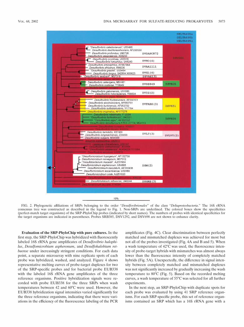
Evaluation of the SRP-PhyloChip with pure cultures. In thefirst step, the SRP-PhyloChip was hybridized with fluorescentlylabeled 16S rRNA gene amplificates of Desulfovibrio halophi-lus, Desulfomicrobium aspheronum, and Desulfohalobium ret-baense under increasingly stringent conditions. For each datapoint, a separate microarray with nine replicate spots of eachprobe was hybridized, washed, and analyzed. Figure 4 showsrepresentative melting curves of probe-target duplexes for twoof the SRP-specific probes and for bacterial probe EUB338with the labeled 16S rRNA gene amplificates of the threereference organisms. Positive hybridization signals were re-corded with probe EUB338 for the three SRPs when washtemperatures between 42 and 60°C were used. However, theEUB338 hybridization signal intensities varied significantly forthe three reference organisms, indicating that there were vari-ations in the efficiency of the fluorescence labeling of the PCR
amplificates (Fig. 4C). Clear discrimination between perfectlymatched and mismatched duplexes was achieved for most butnot all of the probes investigated (Fig. 4A and B and 5). Whena wash temperature of 42°C was used, the fluorescence inten-sity of probe-target hybrids with mismatches was almost alwayslower than the fluorescence intensity of completely matchedhybrids (Fig. 5A). Unexpectedly, the difference in signal inten-sity between completely matched and mismatched duplexeswas not significantly increased by gradually increasing the washtemperature to 80°C (Fig. 5). Based on the recorded meltingcurves, a wash temperature of 55°C was selected for all furtherexperiments.
In the next step, an SRP-PhyloChip with duplicate spots foreach probe was evaluated by using 41 SRP reference organ-isms. For each SRP-specific probe, this set of reference organ-isms contained an SRP which has a 16S rRNA gene with a
FIG. 2. Phylogenetic affiliations of SRPs belonging to the order “Desulfovibrionales” of the class “Deltaproteobacteria.” The 16S rRNAconsensus tree was constructed as described in the legend to Fig. 1. Non-SRPs are underlined. The colored boxes show the specificities(perfect-match target organisms) of the SRP-PhyloChip probes (indicated by short names). The numbers of probes with identical specificities forthe target organisms are indicated in parentheses. Probes SRB385, DSV1292, and DSV698 are not shown to enhance clarity.
VOL. 68, 2002 DNA MICROARRAY FOR SULFATE-REDUCING PROKARYOTES 5073
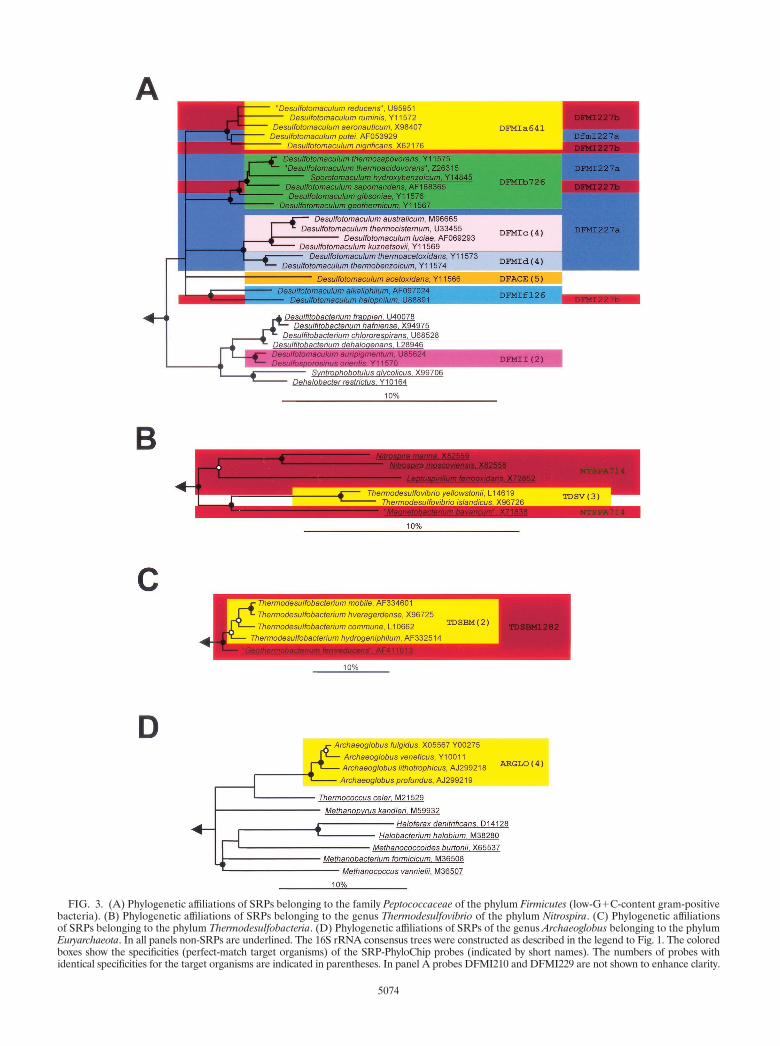
FIG. 3. (A) Phylogenetic affiliations of SRPs belonging to the family Peptococcaceae of the phylum Firmicutes (low-G�C-content gram-positivebacteria). (B) Phylogenetic affiliations of SRPs belonging to the genus Thermodesulfovibrio of the phylum Nitrospira. (C) Phylogenetic affiliationsof SRPs belonging to the phylum Thermodesulfobacteria. (D) Phylogenetic affiliations of SRPs of the genus Archaeoglobus belonging to the phylumEuryarchaeota. In all panels non-SRPs are underlined. The 16S rRNA consensus trees were constructed as described in the legend to Fig. 1. The coloredboxes show the specificities (perfect-match target organisms) of the SRP-PhyloChip probes (indicated by short names). The numbers of probes withidentical specificities for the target organisms are indicated in parentheses. In panel A probes DFMI210 and DFMI229 are not shown to enhance clarity.
5074
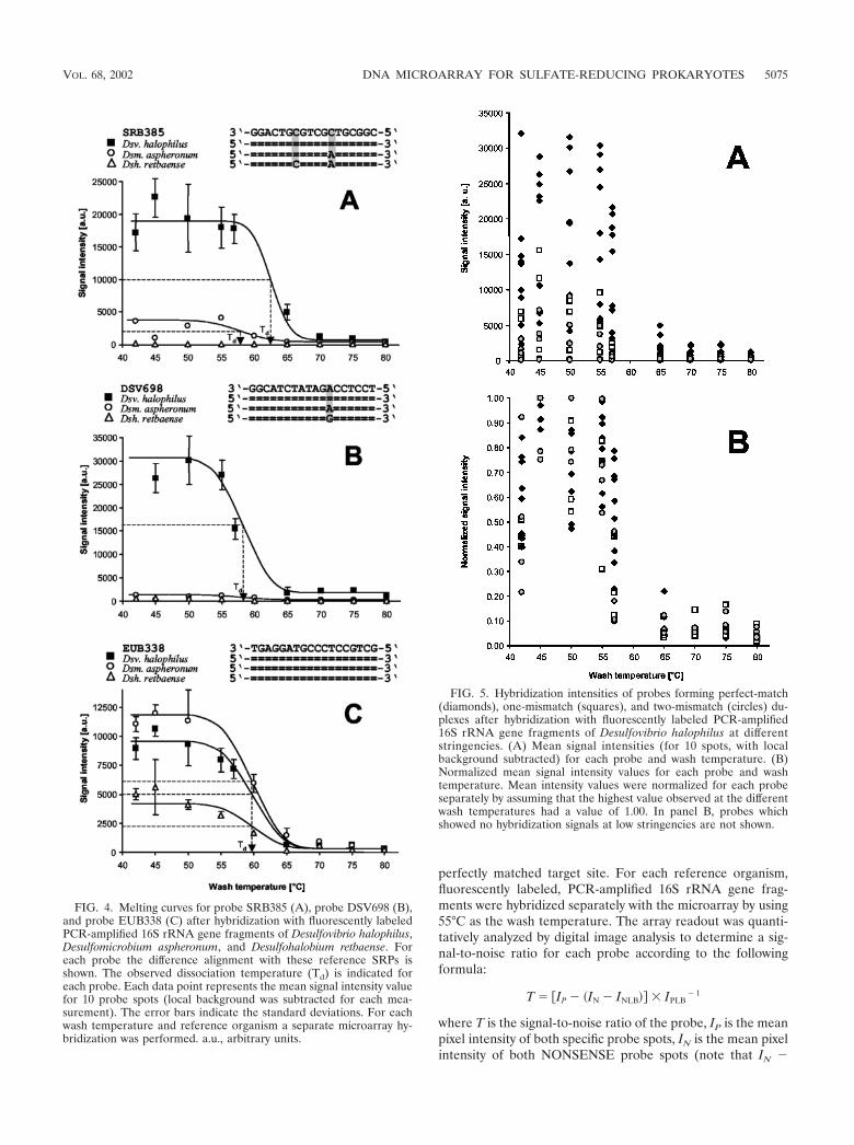
perfectly matched target site. For each reference organism,fluorescently labeled, PCR-amplified 16S rRNA gene frag-ments were hybridized separately with the microarray by using55°C as the wash temperature. The array readout was quanti-tatively analyzed by digital image analysis to determine a sig-nal-to-noise ratio for each probe according to the followingformula:
T � �IP � �IN � INLB � IPLB� 1
where T is the signal-to-noise ratio of the probe, IP is the meanpixel intensity of both specific probe spots, IN is the mean pixelintensity of both NONSENSE probe spots (note that IN �
FIG. 4. Melting curves for probe SRB385 (A), probe DSV698 (B),and probe EUB338 (C) after hybridization with fluorescently labeledPCR-amplified 16S rRNA gene fragments of Desulfovibrio halophilus,Desulfomicrobium aspheronum, and Desulfohalobium retbaense. Foreach probe the difference alignment with these reference SRPs isshown. The observed dissociation temperature (Td) is indicated foreach probe. Each data point represents the mean signal intensity valuefor 10 probe spots (local background was subtracted for each mea-surement). The error bars indicate the standard deviations. For eachwash temperature and reference organism a separate microarray hy-bridization was performed. a.u., arbitrary units.
FIG. 5. Hybridization intensities of probes forming perfect-match(diamonds), one-mismatch (squares), and two-mismatch (circles) du-plexes after hybridization with fluorescently labeled PCR-amplified16S rRNA gene fragments of Desulfovibrio halophilus at differentstringencies. (A) Mean signal intensities (for 10 spots, with localbackground subtracted) for each probe and wash temperature. (B)Normalized mean signal intensity values for each probe and washtemperature. Mean intensity values were normalized for each probeseparately by assuming that the highest value observed at the differentwash temperatures had a value of 1.00. In panel B, probes whichshowed no hybridization signals at low stringencies are not shown.
VOL. 68, 2002 DNA MICROARRAY FOR SULFATE-REDUCING PROKARYOTES 5075
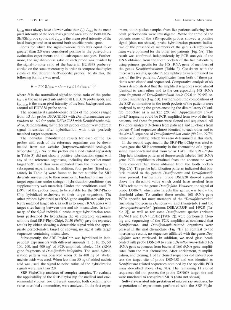
INLB must always have a lower value than IP), INLB is the meanpixel intensity of the local background area around both NON-SENSE probe spots, and IPLB is the mean pixel intensity of thelocal background area around both specific probe spots.
Spots for which the signal-to-noise ratio was equal to orgreater than 2.0 were considered positive in the pure-cultureevaluation experiments and all subsequent analyses. Further-more, the signal-to-noise ratio of each probe was divided bythe signal-to-noise ratio of the bacterial EUB338 probe re-corded on the same microarray in order to compare the duplexyields of the different SRP-specific probes. To do this, thefollowing formula was used:
R � T � ��IEUB � �IN � INLB � IEUBLB� 1��1
where R is the normalized signal-to-noise ratio of the probe,IEUB is the mean pixel intensity of all EUB338 probe spots, andIEUBLB is the mean pixel intensity of the local background areaaround all EUB338 probe spots.
The normalized signal-to-noise ratios of the probes rangedfrom 0.3 for probe DFACE1028 with Desulfotomaculum ace-toxidans to 16.9 for probe DSBAC355 with Desulfobacula tolu-olica, demonstrating that different probes exhibit very differentsignal intensities after hybridization with their perfectlymatched target sequences.
The individual hybridization results for each of the 132probes with each of the reference organisms can be down-loaded from our website (http://www.microbial-ecology.de/srpphylochip/). Six of the probes evaluated (listed separatelyin Table 3) did not show a positive hybridization signal withany of the reference organisms, including the perfect-matchtarget SRP, and thus were excluded from the microarray insubsequent experiments. In addition, four probes (listed sep-arately in Table 3) were found to be not suitable for SRPdiversity surveys due to their nonspecific binding to many non-target organisms under stringent hybridization conditions (seesupplementary web material). Under the conditions used, 75(59%) of the probes found to be suitable for the SRP-Phylo-Chip hybridized exclusively to their target organisms. Theother probes hybridized to rRNA gene amplificates with per-fectly matched target sites, as well as to some rRNA genes withtarget sites having between one and six mismatches. In sum-mary, of the 5,248 individual probe-target hybridization reac-tions performed (by hybridizing the 41 reference organismswith the final SRP-PhyloChip), 5,050 (96%) gave the expectedresults by either showing a detectable signal with the appro-priate perfect-match target or showing no signal with targetsequences containing mismatches.
Subsequently, the SRP-PhyloChip was hybridized in inde-pendent experiments with different amounts (1, 5, 10, 25, 50,100, 200, and 400 ng) of PCR-amplified, labeled 16S rRNAgene fragments of Desulfovibrio halophilus. The same hybrid-ization pattern was observed when 50 to 400 ng of labelednucleic acids was used. When less than 50 ng of added nucleicacid was used, the signal-to-noise ratios of the hybridizationsignals were less than 2.0.
SRP-PhyloChip analyses of complex samples. To evaluatethe applicability of the SRP-PhyloChip for medical and envi-ronmental studies, two different samples, both containing di-verse microbial communities, were analyzed. In the first exper-
iment, tooth pocket samples from five patients suffering fromadult periodontitis were investigated. While for three of thepatients none of the SRP-specific probes showed a positivesignal (data not shown), probe hybridization patterns indica-tive of the presence of members of the genus Desulfomicro-bium were obtained for the other two patients (Fig. 6A). Thisresult was confirmed independently by PCR analysis of theDNA obtained from the tooth pockets of the five patients byusing primers specific for the 16S rRNA gene of members ofthe genus Desulfomicrobium (Table 2). Consistent with themicroarray results, specific PCR amplificates were obtained fortwo of the five patients. Amplificates from both of these pa-tients were cloned and sequenced. Comparative analysis of sixclones demonstrated that the amplified sequences were almostidentical to each other and to the corresponding 16S rRNAgene fragment of Desulfomicrobium orale (99.6 to 99.9% se-quence similarity) (Fig. 6B). Furthermore, the compositions ofthe SRP communities in the tooth pockets of the patients wereanalyzed by using the genes encoding the dissimilatory (bi)sul-fite reductase as a marker (28, 76). Approximately 1.9-kbdsrAB fragments could be PCR amplified from two of the fivepatients, and these fragments were cloned and sequenced. All19 clones analyzed (6 clones from patient 1 and 13 clones frompatient 4) had sequences almost identical to each other and tothe dsrAB sequence of Desulfomicrobium orale (99.2 to 99.7%amino acid identity), which was also determined in this study.
In the second experiment, the SRP-PhyloChip was used toinvestigate the SRP community in the chemocline of a hyper-saline cyanobacterial mat from Solar Lake. The SRP-Phylo-Chip hybridization patterns of fluorescently labeled 16S rRNAgene PCR amplificates obtained from the chemocline weremore complex than those obtained from the tooth pockets(Fig. 7A). The probe hybridization patterns indicated that bac-teria related to the genera Desulfonema and Desulfomonilewere present. Furthermore, probe DSB220 showed signalsabove the threshold value which could have resulted fromSRPs related to the genus Desulfofaba. However, the signal ofprobe DSB674, which also targets this genus, was below thethreshold value. To confirm these results, 16S rRNA genePCRs specific for most members of the “Desulfobacterales”(including the genera Desulfonema and Desulfofaba) and the“Syntrophobacterales” (primers DSBAC355F and 1492R [Ta-ble 2]), as well as for some Desulfonema species (primersDSN61F and DSN�1201R [Table 2]), were performed. Clon-ing and sequencing of the PCR amplificates confirmed thatDesulfonema- and Desulfomonile-related organisms werepresent in the mat chemocline (Fig. 7B). In contrast to themicroarray results, no sequences affiliated with the genus Des-ulfofaba were retrieved. In addition, we used glass beadscoated with probe DSN658 to enrich Desulfonema-related 16SrRNA gene sequences from bacterial 16S rRNA gene amplifi-cates from the mat chemocline. After enrichment, reamplifi-cation, and cloning, 1 of 12 cloned sequences did indeed pos-sess the target site of probe DSN658 and was identical toDesulfonema-related sequences obtained by the specific PCRassay described above (Fig. 7B). The remaining 11 clonedsequences did not possess the probe DSN658 target site andwere unrelated to recognized SRPs (data not shown).
Software-assisted interpretation of microarray readouts. In-terpretation of experiments performed with the SRP-Phylo-
5076 LOY ET AL APPL. ENVIRON. MICROBIOL.
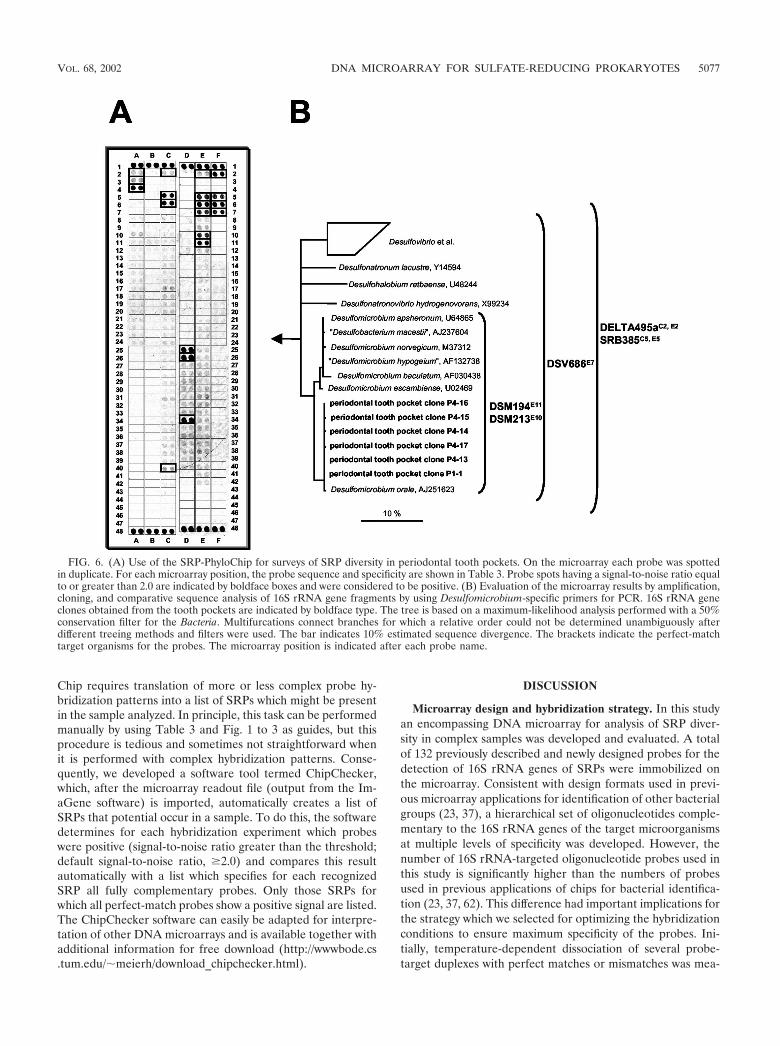
Chip requires translation of more or less complex probe hy-bridization patterns into a list of SRPs which might be presentin the sample analyzed. In principle, this task can be performedmanually by using Table 3 and Fig. 1 to 3 as guides, but thisprocedure is tedious and sometimes not straightforward whenit is performed with complex hybridization patterns. Conse-quently, we developed a software tool termed ChipChecker,which, after the microarray readout file (output from the Im-aGene software) is imported, automatically creates a list ofSRPs that potential occur in a sample. To do this, the softwaredetermines for each hybridization experiment which probeswere positive (signal-to-noise ratio greater than the threshold;default signal-to-noise ratio, �2.0) and compares this resultautomatically with a list which specifies for each recognizedSRP all fully complementary probes. Only those SRPs forwhich all perfect-match probes show a positive signal are listed.The ChipChecker software can easily be adapted for interpre-tation of other DNA microarrays and is available together withadditional information for free download (http://wwwbode.cs.tum.edu/ meierh/download_chipchecker.html).
DISCUSSION
Microarray design and hybridization strategy. In this studyan encompassing DNA microarray for analysis of SRP diver-sity in complex samples was developed and evaluated. A totalof 132 previously described and newly designed probes for thedetection of 16S rRNA genes of SRPs were immobilized onthe microarray. Consistent with design formats used in previ-ous microarray applications for identification of other bacterialgroups (23, 37), a hierarchical set of oligonucleotides comple-mentary to the 16S rRNA genes of the target microorganismsat multiple levels of specificity was developed. However, thenumber of 16S rRNA-targeted oligonucleotide probes used inthis study is significantly higher than the numbers of probesused in previous applications of chips for bacterial identifica-tion (23, 37, 62). This difference had important implications forthe strategy which we selected for optimizing the hybridizationconditions to ensure maximum specificity of the probes. Ini-tially, temperature-dependent dissociation of several probe-target duplexes with perfect matches or mismatches was mea-
FIG. 6. (A) Use of the SRP-PhyloChip for surveys of SRP diversity in periodontal tooth pockets. On the microarray each probe was spottedin duplicate. For each microarray position, the probe sequence and specificity are shown in Table 3. Probe spots having a signal-to-noise ratio equalto or greater than 2.0 are indicated by boldface boxes and were considered to be positive. (B) Evaluation of the microarray results by amplification,cloning, and comparative sequence analysis of 16S rRNA gene fragments by using Desulfomicrobium-specific primers for PCR. 16S rRNA geneclones obtained from the tooth pockets are indicated by boldface type. The tree is based on a maximum-likelihood analysis performed with a 50%conservation filter for the Bacteria. Multifurcations connect branches for which a relative order could not be determined unambiguously afterdifferent treeing methods and filters were used. The bar indicates 10% estimated sequence divergence. The brackets indicate the perfect-matchtarget organisms for the probes. The microarray position is indicated after each probe name.
VOL. 68, 2002 DNA MICROARRAY FOR SULFATE-REDUCING PROKARYOTES 5077
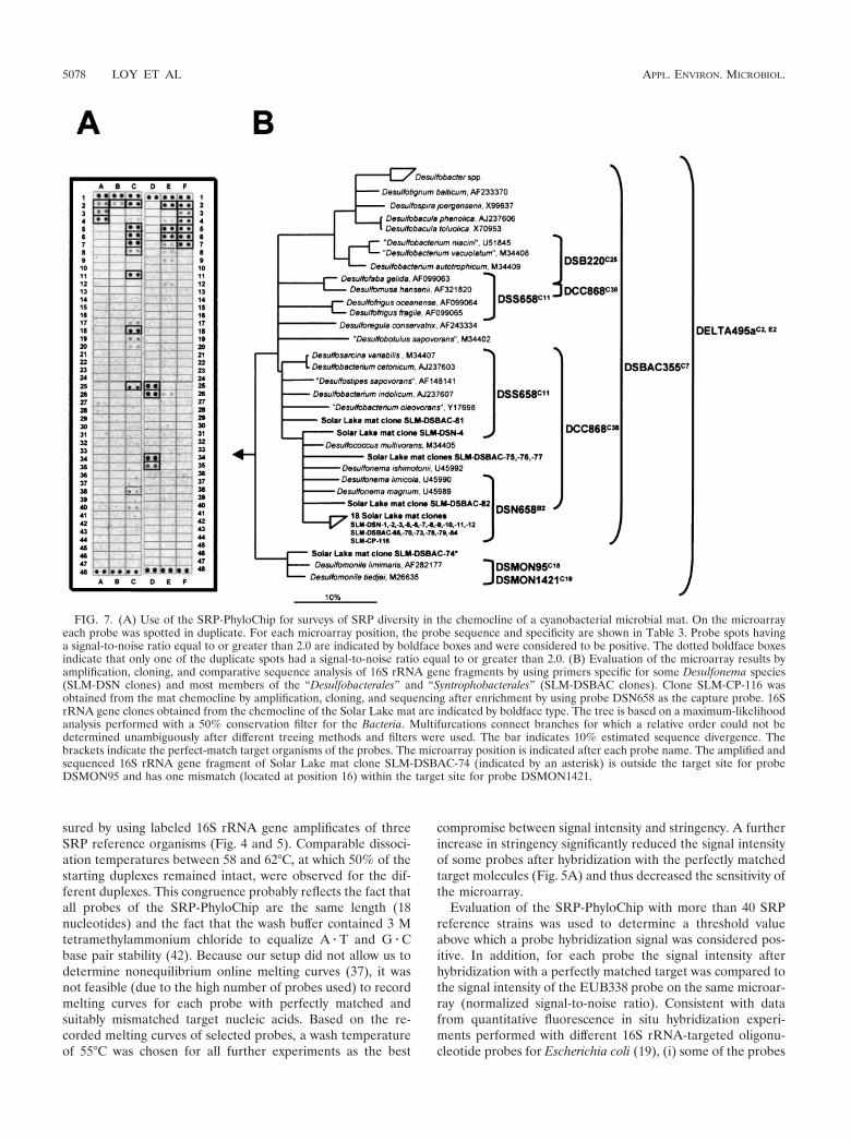
sured by using labeled 16S rRNA gene amplificates of threeSRP reference organisms (Fig. 4 and 5). Comparable dissoci-ation temperatures between 58 and 62°C, at which 50% of thestarting duplexes remained intact, were observed for the dif-ferent duplexes. This congruence probably reflects the fact thatall probes of the SRP-PhyloChip are the same length (18nucleotides) and the fact that the wash buffer contained 3 Mtetramethylammonium chloride to equalize A � T and G � Cbase pair stability (42). Because our setup did not allow us todetermine nonequilibrium online melting curves (37), it wasnot feasible (due to the high number of probes used) to recordmelting curves for each probe with perfectly matched andsuitably mismatched target nucleic acids. Based on the re-corded melting curves of selected probes, a wash temperatureof 55°C was chosen for all further experiments as the best
compromise between signal intensity and stringency. A furtherincrease in stringency significantly reduced the signal intensityof some probes after hybridization with the perfectly matchedtarget molecules (Fig. 5A) and thus decreased the sensitivity ofthe microarray.
Evaluation of the SRP-PhyloChip with more than 40 SRPreference strains was used to determine a threshold valueabove which a probe hybridization signal was considered pos-itive. In addition, for each probe the signal intensity afterhybridization with a perfectly matched target was compared tothe signal intensity of the EUB338 probe on the same microar-ray (normalized signal-to-noise ratio). Consistent with datafrom quantitative fluorescence in situ hybridization experi-ments performed with different 16S rRNA-targeted oligonu-cleotide probes for Escherichia coli (19), (i) some of the probes
FIG. 7. (A) Use of the SRP-PhyloChip for surveys of SRP diversity in the chemocline of a cyanobacterial microbial mat. On the microarrayeach probe was spotted in duplicate. For each microarray position, the probe sequence and specificity are shown in Table 3. Probe spots havinga signal-to-noise ratio equal to or greater than 2.0 are indicated by boldface boxes and were considered to be positive. The dotted boldface boxesindicate that only one of the duplicate spots had a signal-to-noise ratio equal to or greater than 2.0. (B) Evaluation of the microarray results byamplification, cloning, and comparative sequence analysis of 16S rRNA gene fragments by using primers specific for some Desulfonema species(SLM-DSN clones) and most members of the “Desulfobacterales” and “Syntrophobacterales” (SLM-DSBAC clones). Clone SLM-CP-116 wasobtained from the mat chemocline by amplification, cloning, and sequencing after enrichment by using probe DSN658 as the capture probe. 16SrRNA gene clones obtained from the chemocline of the Solar Lake mat are indicated by boldface type. The tree is based on a maximum-likelihoodanalysis performed with a 50% conservation filter for the Bacteria. Multifurcations connect branches for which a relative order could not bedetermined unambiguously after different treeing methods and filters were used. The bar indicates 10% estimated sequence divergence. Thebrackets indicate the perfect-match target organisms of the probes. The microarray position is indicated after each probe name. The amplified andsequenced 16S rRNA gene fragment of Solar Lake mat clone SLM-DSBAC-74 (indicated by an asterisk) is outside the target site for probeDSMON95 and has one mismatch (located at position 16) within the target site for probe DSMON1421.
5078 LOY ET AL APPL. ENVIRON. MICROBIOL.

used in the first version of the SRP-PhyloChip did not hybrid-ize to their perfect-match targets and (ii) the signal intensitiesmeasured for the other probes on the SRP-PhyloChip variedsignificantly, by factors of up to 56. Dramatic differences induplex yield arising from different regions of the target werealso observed in other microarray applications (45, 64) andprobably reflect either accessibility differences for the differentprobe target sites due to secondary structures of the targetDNA or different steric hindrances of the different nucleic acidhybrids formed on the microarrays after hybridization.
The evaluation of the microarray with SRP pure culturesdemonstrated (i) that false-negative hybridization never oc-curred (within the detection limit of the microarray method)but (ii) that some of the probes still hybridized to nontargetorganisms under the hybridization and washing conditionsused, leading to false-positive results (see supplementary webmaterial). As expected, the nucleotide composition of the mis-match, the mismatch position (67, 73), and possibly other vari-ables, such as the influence of an adjacent nucleotide stackinginteraction (17), were the major factors determining the duplexyields of probes with mismatched target nucleic acids. Most ofthe mismatched duplexes with signal intensities above thethreshold value (used to differentiate between positive andnegative hybridization results) had a signal intensity (and nor-malized signal-to-noise ratio) lower than that of the corre-sponding perfect-match duplex (Fig. 5). However, this differ-ence cannot be exploited for interpretation of microarrayhybridization results for environmental samples because a lowhybridization signal of a probe can be caused not only bymismatched duplex formation but also by low abundance ofthe perfect-match target nucleic acid.
Misinterpretation of microarray hybridization patternscaused by the nonperfect specificity of some of the probescould be avoided at least partially by using the multiple-probeconcept. While hybridization patterns consistent with the hier-archical or parallel specificity of the probes increase the reli-ability of detection, inconsistent probe hybridization patternsmust be interpreted with caution. In complex samples, incon-sistent hybridization patterns can be caused either by nonspe-cific binding of one or several probes or by previously unrec-ognized prokaryotes with unusual combinations of perfect-match probe target sites in their 16S rRNA gene sequences.
Microarray-based SRP diversity surveys of complex sam-ples. In this study, periodontal tooth pocket material and acyanobacterial microbial mat were used to demonstrate thesuitability of using the microarray developed for SRP diversityanalysis of medical and environmental samples. For the toothpocket material of two patients suffering from adult periodon-titis the SRP-PhyloChip hybridization pattern indicated thepresence of members of the genus Desulfomicrobium. Coloni-zation of the tooth pockets analyzed by these SRPs, which isconsistent with a previous report of isolation of Desulfomicro-bium orale from periodontal tooth pockets (33), was indepen-dently confirmed by retrieval of 16S rRNA and dsrAB genesequences of Desulfomicrobium orale, demonstrating the reli-ability of the microarray results.
The microarray hybridization patterns obtained by reversehybridization of 16S rRNA gene fragments amplified from thechemocline of a Solar Lake microbial mat suggested that sev-eral phylogenetically different SRPs, including bacteria related
to the genera Desulfonema, Desulfomonile, and Desulfofaba,were present. By using specific PCR assays, 16S rRNA genesequences related to sequences of members of the generaDesulfonema and Desulfomonile were obtained from the matmaterial analyzed, while the presence of Desulfofaba-like or-ganisms could not be confirmed. The failure to detect Desul-fofaba-like bacteria with the PCR assay might mean that arelatively limited number of 16S rRNA gene clones was se-quenced or that the microarray hybridization pattern indicativeof Desulfofaba was caused by the presence of bacteria that havenot been recognized yet. The detection of Desulfonema-likebacteria in the chemocline of the Solar Lake mat is consistentwith findings of previous studies (46, 47, 70) and further sup-ports the importance of these SRPs in hypersaline mat ecosys-tems.
In conclusion, we developed an encompassing 16S rRNAgene-targeting oligonucleotide microarray suitable for SRP di-versity analyses of complex environmental and clinical samples.The microarray was used to screen samples in order to rapidlyobtain indications of the presence of distinct lineages of SRPs.Subsequently, this information was used to select appropriatePCR-based techniques for confirmation of the microarray re-sults and for retrieval of sequence information for phylogeneticanalysis. In contrast to previously available tools for cultiva-tion-independent SRP identification (13, 18, 41, 56, 75, 76), theSRP-PhyloChip allowed us to obtain a phylogenetically infor-mative, high-resolution fingerprint of the SRP diversity in agiven sample within 48 h (including all experimental work fromDNA extraction to hybridization pattern interpretation). How-ever, keeping in mind that (i) most environmental microbialcommunities contain a high percentage of bacteria not yetsequenced on the 16S rRNA level and (ii) not all probes on themicroarray are absolutely specific under the conditions used,the SRP-PhyloChip experiments should always be supplement-ed with microarray-independent techniques to confirm thephylogenetic affiliations of the SRPs detected. Furthermore, itshould be noted that the microarray approach described heredid not allow us to obtain quantitative data on the composi-tions of SRP communities because of the recognized biasesintroduced by using PCR for 16S rRNA gene amplification(74). In addition, the duplex yield of a probe on the microarrayis dependent not only on the actual abundance of its perfect-match target nucleic acid in the PCR amplificate mixture butalso on a variety of other factors, including the labeling effi-ciency of the specific target nucleic acid, the secondary struc-ture of the target region, and the inherent variations associatedwith microarray fabrication. Despite these limitations, the mi-croarray which we developed has great potential for rapidscreening of SRP diversity in complex samples. The SRP di-versity microarray fingerprint technique should allow workersto identify the probes which have relevance for further char-acterization of a sample by PCR or quantitative hybridizationexperiments. This option should be particularly valuable iflarge numbers of samples are to be analyzed to study temporalor spatial variations in SRP diversity.
ACKNOWLEDGMENTS
Yehuda Cohen is acknowledged for kindly providing cyanobacterialmat material from Solar Lake. The excellent technical assistance ofClaudia Schulz, Helga Gaenge, Susanne Thiemann, and Sibylle Schad-
VOL. 68, 2002 DNA MICROARRAY FOR SULFATE-REDUCING PROKARYOTES 5079
hauser is acknowledged. We also thank Bernhard Loy for providingthe tooth pocket samples and Josef Reischenbeck for fabrication ofhybridization chambers.
This research was supported by grants from bmb�f (01 LC 0021subproject 2, in the framework of the BIOLOG program) (to M.W.),DFG (trilateral cyanobacterial mat project RU458/18-4) (to M.W.),and Bayerischen Forschungsstiftung (Development of OligonucleotideDNA Chips, in cooperation with MWG Biotech; project 368/99) (toM.W. and K.-H.S.).
REFERENCES
1. Alm, E. W., D. B. Oerther, N. Larsen, D. A. Stahl, and L. Raskin. 1996. Theoligonucleotide probe database. Appl. Environ. Microbiol. 62:3557–3559.
2. Amann, R. I., B. J. Binder, R. J. Olson, S. W. Chisholm, R. Devereux, andD. A. Stahl. 1990. Combination of 16S rRNA-targeted oligonucleotideprobes with flow cytometry for analyzing mixed microbial populations. Appl.Environ. Microbiol. 56:1919–1925.
3. Amann, R. I., J. Stromley, R. Devereux, R. Key, and D. A. Stahl. 1992.Molecular and microscopic identification of sulfate-reducing bacteria in mul-tispecies biofilms. Appl. Environ. Microbiol. 58:614–623.
4. Boetius, A., K. Ravenschlag, C. J. Schubert, D. Rickert, F. Widdel, A.Gieseke, R. Amann, B. B. Jorgensen, U. Witte, and O. Pfannkuche. 2000. Amarine microbial consortium apparently mediating anaerobic oxidation ofmethane. Nature 407:623–626.
5. Cho, J.-C., and J. M. Tiedje. 2002. Quantitative detection of microbial genesby using DNA microarrays. Appl. Environ. Microbiol. 68:1425–1430.
6. Daims, H., A. Bruhl, R. Amann, K.-H. Schleifer, and M. Wagner. 1999. Thedomain-specific probe EUB338 is insufficient for the detection of all Bacte-ria: development and evaluation of a more comprehensive probe set. Syst.Appl. Microbiol. 22:434–444.
7. Daims, H., J. L. Nielsen, P. H. Nielsen, K. H. Schleifer, and M. Wagner.2001. In situ characterization of Nitrospira-like nitrite-oxidizing bacteria ac-tive in wastewater treatment plants. Appl. Environ. Microbiol. 67:5273–5284.
8. Daims, H., P. H. Nielsen, J. L. Nielsen, S. Juretschko, and M. Wagner. 2000.Novel Nitrospira-like bacteria as dominant nitrite-oxidizers in biofilms fromwastewater treatment plants: diversity and in situ physiology. Water Sci.Technol. 41:85–90.
9. Daly, K., R. J. Sharp, and A. J. McCarthy. 2000. Development of oligonu-cleotide probes and PCR primers for detecting phylogenetic subgroups ofsulfate-reducing bacteria. Microbiology 146:1693–1705.
10. DeLong, E. F. 2000. Resolving a methane mystery. Nature 407:577–579.11. Devereux, R., M. Delaney, F. Widdel, and D. A. Stahl. 1989. Natural rela-
tionships among sulfate-reducing eubacteria. J. Bacteriol. 171:6689–6695.12. Devereux, R., S. H. He, C. L. Doyle, S. Orkland, D. A. Stahl, J. LeGall, and
W. B. Whitman. 1990. Diversity and origin of Desulfovibrio species: phylo-genetic definition of a family. J. Bacteriol. 172:3609–3619.
13. Devereux, R., M. D. Kane, J. Winfrey, and D. A. Stahl. 1992. Genus- andgroup-specific hybridization probes for determinative and environmentalstudies of sulfate-reducing bacteria. Syst. Appl. Microbiol. 15:601–609.
14. Devereux, R., and G. W. Mundfrom. 1994. A phylogenetic tree of 16S rRNAsequences from sulfate-reducing bacteria in a sandy marine sediment. Appl.Environ. Microbiol. 60:3437–3439.
15. Dubilier, N., C. Mulders, T. Ferdelman, D. de Beer, A. Pernthaler, M. Klein,M. Wagner, C. Erseus, F. Thiermann, J. Krieger, O. Giere, and R. Amann.2001. Endosymbiotic sulphate-reducing and sulphide-oxidizing bacteria in anoligochaete worm. Nature 411:298–302.
16. Edgcomb, V. P., J. H. McDonald, R. Devereux, and D. W. Smith. 1999.Estimation of bacterial cell numbers in humic acid-rich salt marsh sedimentswith probes directed to 16S ribosomal DNA. Appl. Environ. Microbiol.65:1516–1523.
17. Fotin, A. V., A. L. Drobyshev, D. Y. Proudnikov, A. N. Perov, and A. D.Mirzabekov. 1998. Parallel thermodynamic analysis of duplexes on oligode-oxyribonucleotide microchips. Nucleic Acids Res. 26:1515–1521.
18. Friedrich, M. W. 2002. Phylogenetic analysis reveals multiple lateral trans-fers of adenosine-5�-phosphosulfate reductase genes among sulfate-reducingmicroorganisms. J. Bacteriol. 184:278–289.
19. Fuchs, B. M., G. Wallner, W. Beisker, I. Schwippl, W. Ludwig, and R.Amann. 1998. Flow cytometric analysis of the in situ accessibility of Esche-richia coli 16S rRNA for fluorescently labeled oligonucleotide probes. Appl.Environ. Microbiol. 64:4973–4982.
20. Fukui, M., A. Teske, B. Aßmus, G. Muyzer, and F. Widdel. 1999. Physiology,phylogenetic relationships, and ecology of filamentous sulfate-reducing bac-teria (genus Desulfonema). Arch. Microbiol. 172:193–203.
21. Garrity, G. M., and J. G. Holt. 2001. The road map to the manual, p.119–166. In G. M. Garrity (ed.), Bergey’s manual of systematic bacteriology,2nd ed., vol. 1. Springer, New York, N.Y.
22. Griffiths, R. I., A. S. Whiteley, A. G. O’Donnell, and M. J. Bailey. 2000. Rapidmethod for coextraction of DNA and RNA from natural environments foranalysis of ribosomal DNA- and rRNA-based microbial community compo-sition. Appl. Environ. Microbiol. 66:5488–5491.
23. Guschin, D. Y., B. K. Mobarry, D. Proudnikov, D. A. Stahl, B. E. Rittmann,
and A. D. Mirzabekov. 1997. Oligonucleotide microchips as genosensors fordeterminative and environmental studies in microbiology. Appl. Environ.Microbiol. 63:2397–2402.
24. Hines, M. E., R. S. Evans, B. R. Sharak Genthner, S. G. Willis, S. Friedman,J. N. Rooney-Varga, and R. Devereux. 1999. Molecular phylogenetic andbiogeochemical studies of sulfate-reducing bacteria in the rhizosphere ofSpartina alterniflora. Appl. Environ. Microbiol. 65:2209–2216.
25. Hristova, K. R., M. Mau, D. Zheng, R. I. Aminov, R. I. Mackie, H. R.Gaskins, and L. Raskin. 2000. Desulfotomaculum genus- and subgenus-specific 16S rRNA hybridization probes for environmental studies. Environ.Microbiol. 2:143–159.
26. Juretschko, S., G. Timmermann, M. Schmid, K. H. Schleifer, A. Pomme-rening-Roser, H. P. Koops, and M. Wagner. 1998. Combined molecular andconventional analyses of nitrifying bacterium diversity in activated sludge:Nitrosococcus mobilis and Nitrospira-like bacteria as dominant populations.Appl. Environ. Microbiol. 64:3042–3051.
27. Kane, M. D., L. K. Poulsen, and D. A. Stahl. 1993. Monitoring the enrich-ment and isolation of sulfate-reducing bacteria by using oligonucleotidehybridization probes designed from environmentally derived 16S rRNA se-quences. Appl. Environ. Microbiol. 59:682–686.
28. Klein, M., M. Friedrich, A. J. Roger, P. Hugenholtz, S. Fishbain, H. Abicht,L. L. Blackall, D. A. Stahl, and M. Wagner. 2001. Multiple lateral transfersof dissimilatory sulfite reductase genes between major lineages of sulfate-reducing prokaryotes. J. Bacteriol. 183:6028–6035.
29. Knoblauch, C., B. B. Jorgensen, and J. Harder. 1999. Community size andmetabolic rates of psychrophilic sulfate-reducing bacteria in arctic marinesediments. Appl. Environ. Microbiol. 65:4230–4233.
30. Knoblauch, C., K. Sahm, and B. B. Jorgensen. 1999. Psychrophilic sulfate-reducing bacteria isolated from permanently cold arctic marine sediments:description of Desulfofrigus oceanense gen. nov., sp. nov., Desulfofrigus fragilesp. nov., Desulfofaba gelida gen. nov., sp. nov., Desulfotalea psychrophila gen.nov., sp. nov. and Desulfotalea arctica sp. nov. Int. J. Syst. Bacteriol. 49:1631–1643.
31. Kovarova, M., and P. Draber. 2000. New specificity and yield enhancer ofpolymerase chain reactions. Nucleic Acids Res. 28:E70.
32. Langendijk, P. S., J. T. J. Hanssen, and J. S. van der Hoeven. 2000. Sulfate-reducing bacteria in association with human periodontitis. J. Clin. Periodon-tol. 27:943–950.
33. Langendijk, P. S., E. M. Kulik, H. Sandmeier, J. Meyer, and J. S. van derHoeven. 2001. Isolation of Desulfomicrobium orale sp. nov. and Desulfovibriostrain NY682, oral sulfate-reducing bacteria involved in human periodontaldisease. Int. J. Syst. Evol. Microbiol. 51:1035–1044.
34. Larsen, N., G. J. Olsen, B. L. Maidak, M. J. McCaughey, R. Overbeek, T. J.Macke, T. L. Marsh, and C. R. Woese. 1993. The Ribosomal DatabaseProject. Nucleic Acids Res. 21:3021–3023.
35. La Scola, B., and D. Raoult. 1999. Third human isolate of a Desulfovibrio sp.identical to the provisionally named Desulfovibrio fairfieldensis. J. Clin. Mi-crobiol. 37:3076–3077.
36. Li, J.-H., K. J. Purdy, S. Takii, and H. Hayashi. 1999. Seasonal changes inribosomal RNA of sulfate-reducing bacteria and sulfate reducing activity ina freshwater lake sediment. FEMS Microbiol. Ecol. 28:31–39.
37. Liu, W. T., A. D. Mirzabekov, and D. A. Stahl. 2001. Optimization of anoligonucleotide microchip for microbial identification studies: a non-equi-librium dissociation approach. Environ. Microbiol. 3:619–629.
38. Llobet-Brossa, E., R. Rossello-Mora, and R. Amann. 1998. Microbial com-munity composition of Wadden Sea sediments as revealed by fluorescence insitu hybridization. Appl. Environ. Microbiol. 64:2691–2696.
39. Loubinoux, J., F. Mory, I. A. Pereira, and A. E. Le Faou. 2000. Bacteremiacaused by a strain of Desulfovibrio related to the provisionally named Desul-fovibrio fairfieldensis. J. Clin. Microbiol. 38:931–934.
40. Ludwig, W., O. Strunk, S. Klugbauer, N. Klugbauer, M. Weizenegger,J. Neumaier, M. Bachleitner, and K. H. Schleifer. 1998. Bacterial phylogenybased on comparative sequence analysis. Electrophoresis 19:554–568.
41. Manz, W., M. Eisenbrecher, T. R. Neu, and U. Szewzyk. 1998. Abundanceand spatial organization of Gram-negative sulfate-reducing bacteria in acti-vated sludge investigated by in situ probing with specific 16S rRNA targetedoligonucleotides. FEMS Microbiol. Ecol. 25:43–61.
42. Maskos, U., and E. M. Southern. 1992. Parallel analysis of oligodeoxyribo-nucleotide (oligonucleotide) interactions. I. Analysis of factors influencingoligonucleotide duplex formation. Nucleic Acids Res. 20:1675–1678.
43. McDougall, R., J. Robson, D. Paterson, and W. Tee. 1997. Bacteremiacaused by a recently described novel Desulfovibrio species. J. Clin. Microbiol.35:1805–1808.
44. Meier, H., R. Amann, W. Ludwig, and K.-H. Schleifer. 1999. Specific oligo-nucleotide probes for in situ detection of a major group of gram-positivebacteria with low DNA G�C content. Syst. Appl. Microbiol. 22:186–196.
45. Milner, N., K. U. Mir, and E. M. Southern. 1997. Selecting effective anti-sense reagents on combinatorial oligonucleotide arrays. Nat. Biotechnol.15:537–541.
46. Minz, D., S. Fishbain, S. J. Green, G. Muyzer, Y. Cohen, B. E. Rittmann, andD. A. Stahl. 1999. Unexpected population distribution in a microbial matcommunity: sulfate-reducing bacteria localized to the highly oxic chemocline
5080 LOY ET AL APPL. ENVIRON. MICROBIOL.
in contrast to a eukaryotic preference for anoxia. Appl. Environ. Microbiol.65:4659–4665.
47. Minz, D., J. L. Flax, S. J. Green, G. Muyzer, Y. Cohen, M. Wagner, B. E.Rittmann, and D. A. Stahl. 1999. Diversity of sulfate-reducing bacteria inoxic and anoxic regions of a microbial mat characterized by comparativeanalysis of dissimilatory sulfite reductase genes. Appl. Environ. Microbiol.65:4666–4671.
48. Orphan, V. J., K. U. Hinrichs, W. Ussler 3rd, C. K. Paull, L. T. Taylor, S. P.Sylva, J. M. Hayes, and E. F. Delong. 2001. Comparative analysis of meth-ane-oxidizing archaea and sulfate-reducing bacteria in anoxic marine sedi-ments. Appl. Environ. Microbiol. 67:1922–1934.
49. Oude Elferink, S. J. W. H., W. J. C. Vorstman, A. Sopjes, and A. J. M. Stams.1998. Characterization of the sulfate-reducing and syntrophic population ingranular sludge from a full-scale anaerobic reactor treating papermill waste-water. FEMS Microbiol. Ecol. 27:185–194.
50. Purdy, K. J., D. B. Nedwell, T. M. Embley, and S. Takii. 1997. Use of 16SrRNA-targeted oligonucleotide probes to investigate the occurrence andselection of sulfate-reducing bacteria in response to nutrient addition tosediment slurry microcosms from a Japanese estuary. FEMS Microbiol.Ecol. 24:221–234.
51. Purkhold, U., A. Pommering-Roser, S. Juretschko, M. C. Schmid, H.-P.Koops, and M. Wagner. 2000. Phylogeny of all recognized species of ammo-nia oxidizers based on comparative 16S rRNA and amoA sequence analysis:implications for molecular diversity surveys. Appl. Environ. Microbiol. 66:5368–5382.
52. Rabus, R., M. Fukui, H. Wilkes, and F. Widdle. 1996. Degradative capacitiesand 16S rRNA-targeted whole-cell hybridization of sulfate-reducing bacteriain an anaerobic enrichment culture utilizing alkylbenzenes from crude oil.Appl. Environ. Microbiol. 62:3605–3613.
53. Rabus, R., T. Hansen, and F. Widdel. 2000. Dissimilatory sulfate- and sulfur-reducing prokaryotes. In M. Dworkin, S. Falkow, E. Rosenberg, K.-H. Schlei-fer, and E. Stackebrandt (ed.), The prokaryotes: an evolving electronicresource for the microbiological community, 3rd ed. Springer-Verlag, NewYork, N.Y.
54. Ravenschlag, K., K. Sahm, and R. Amann. 2001. Quantitative molecularanalysis of the microbial community in marine arctic sediments (Svalbard).Appl. Environ. Microbiol. 67:387–395.
55. Ravenschlag, K., K. Sahm, C. Knoblauch, B. B. Jorgensen, and R. Amann.2000. Community structure, cellular rRNA content, and activity of sulfate-reducing bacteria in marine arctic sediments. Appl. Environ. Microbiol.66:3592–3602.
56. Risatti, J. B., W. C. Capman, and D. A. Stahl. 1994. Community structure ofa microbial mat: the phylogenetic dimension. Proc. Natl. Acad. Sci. USA91:10173–10177.
57. Sanger, F., S. Nicklen, and A. R. Coulson. 1977. DNA sequencing withchain-terminating inhibitors. Proc. Natl. Acad. Sci. USA 74:5463–5467.
58. Santegoeds, C. M., T. G. Ferdelman, G. Muyzer, and D. de Beer. 1998.Structural and functional dynamics of sulfate-reducing populations in bac-terial biofilms. Appl. Environ. Microbiol. 64:3731–3739.
59. Scheid, D., and S. Stubner. 2001. Structure and diversity of Gram-negativesulfate-reducing bacteria on rice roots. FEMS Microbiol. Ecol. 36:175–183.
60. Schoenborn, L., H. Abdollahi, W. Tee, M. Dyall-Smith, and P. H. Janssen.2001. A member of the delta subgroup of Proteobacteria from a pyogenicliver abscess is a typical sulfate reducer of the genus Desulfovibrio. J. Clin.Microbiol. 39:787–790.
61. Shchepinov, M. S., S. C. Case-Green, and E. M. Southern. 1997. Stericfactors influencing hybridisation of nucleic acids to oligonucleotide arrays.Nucleic Acids Res. 25:1155–1161.
62. Small, J., D. R. Call, F. J. Brockman, T. M. Straub, and D. P. Chandler.
2001. Direct detection of 16S rRNA in soil extracts by using oligonucleotidemicroarrays. Appl. Environ. Microbiol. 67:4708–4716.
63. Southern, E., K. Mir, and M. Shchepinov. 1999. Molecular interactions onmicroarrays. Nat. Genet. 21:5–9.
64. Southern, E. M., S. C. Case-Green, J. K. Elder, M. Johnson, K. U. Mir, L.Wang, and J. C. Williams. 1994. Arrays of complementary oligonucleotidesfor analysing the hybridisation behaviour of nucleic acids. Nucleic Acids Res.22:1368–1373.
65. Stackebrandt, E., C. Sproer, F. A. Rainey, J. Burghardt, O. Pauker, and H.Hippe. 1997. Phylogenetic analysis of the genus Desulfotomaculum: evidencefor the misclassification of Desulfotomaculum guttoideum and description ofDesulfotomaculum orientis as Desulfosporosinus orientis gen. nov., comb. nov.Int. J. Syst. Bacteriol. 47:1134–1139.
66. Stackebrandt, E., D. A. Stahl, and R. Devereux. 1995. Taxonomic relation-ships, p. 49–87. In L. L. Barton (ed.), Sulfate-reducing bacteria. PlenumPress, New York, N.Y.
67. Stahl, D. A., and R. Amann. 1991. Development and application of nucleicacid probes, p. 205–248. In E. Stackebrandt and M. Goodfellow (ed.), Nu-cleic acid techniques in bacterial systematics. John Wiley & Sons Ltd., Chich-ester, England.
68. Stubner, S., and K. Meuser. 2000. Detection of Desulfotomaculum in anItalian rice paddy soil by 16S ribosomal nucleic acid analyses. FEMS Micro-biol. Ecol. 34:73–80.
69. Tee, W., M. Dyall-Smith, W. Woods, and D. Eisen. 1996. Probable newspecies of Desulfovibrio isolated from a pyogenic liver abscess. J. Clin. Mi-crobiol. 34:1760–1764.
70. Teske, A., N. B. Ramsing, K. Habicht, M. Fukui, J. Kuver, B. B. Jorgensen,and Y. Cohen. 1998. Sulfate-reducing bacteria and their activities in cya-nobacterial mats of Solar Lake (Sinai, Egypt). Appl. Environ. Microbiol.64:2943–2951.
71. Teske, A., C. Wawer, G. Muyzer, and N. B. Ramsing. 1996. Distribution ofsulfate-reducing bacteria in a stratified fjord (Mariager Fjord, Denmark) asevaluated by most-probable-number counts and denaturing gradient gelelectrophoresis of PCR-amplified ribosomal DNA fragments. Appl. Environ.Microbiol. 62:1405–1415.
72. Thomsen, T. R., K. Finster, and N. B. Ramsing. 2001. Biogeochemical andmolecular signatures of anaerobic methane oxidation in a marine sediment.Appl. Environ. Microbiol. 67:1646–1656.
73. Urakawa, H., P. A. Noble, S. El Fantroussi, J. J. Kelly, and D. A. Stahl. 2002.Single-base-pair discrimination of terminal mismatches by using oligonucle-otide microarrays and neural network analyses. Appl. Environ. Microbiol.68:235–244.
74. von Wintzingerode, F., U. B. Gobel, and E. Stackebrandt. 1997. Determina-tion of microbial diversity in environmental samples: pitfalls of PCR-basedrRNA analysis. FEMS Microbiol. Rev. 21:213–229.
75. Voordouw, G., J. K. Voordouw, R. R. Karkhoff-Schweizer, P. M. Fedorak,and D. W. S. Westlake. 1991. Reverse sample genome probing, a new tech-nique for identification of bacteria in environmental samples by DNA hy-bridization, and its application to the identification of sulfate-reducing bac-teria in oil field samples. Appl. Environ. Microbiol. 57:3070–3078.
76. Wagner, M., A. J. Roger, J. L. Flax, G. A. Brusseau, and D. A. Stahl. 1998.Phylogeny of dissimilatory sulfite reductases supports an early origin ofsulfate respiration. J. Bacteriol. 180:2975–2982.
77. Wu, L., D. K. Thompson, G. Li, R. A. Hurt, J. M. Tiedje, and J. Zhou. 2001.Development and evaluation of functional gene arrays for detection of se-lected genes in the environment. Appl. Environ. Microbiol. 67:5780–5790.
78. Zheng, D., E. W. Alm, D. A. Stahl, and L. Raskin. 1996. Characterization ofuniversal small-subunit rRNA hybridization probes for quantitative molec-ular microbial ecology studies. Appl. Environ. Microbiol. 62:4504–4513.
VOL. 68, 2002 DNA MICROARRAY FOR SULFATE-REDUCING PROKARYOTES 5081
117
APPENDIX II
probeBase: An Online Resourcefor rRNA-Targeted Oligonucleotide Probes
Published in NUCLEIC ACIDS RESEARCH,
Jan. 2003, Vol. 31, No. 1, p. 514–516

probeBase: an online resource for rRNA-targetedoligonucleotide probes
Alexander Loy, Matthias Horn* and Michael Wagner
Lehrstuhl fur Mikrobiologie, Technische Universitat Munchen, Am Hochanger 4, D-85350 Freising, Germany
Received July 8, 2002; Revised and Accepted August 29, 2002
ABSTRACT
Ribosomal RNA-(rRNA)-targeted oligonucleotideprobes are widely used for culture-independentidentification of microorganisms in environmentaland clinical samples. ProbeBase is a comprehensivedatabase containing more than 700 published rRNA-targeted oligonucleotide probe sequences (statusAugust 2002) with supporting bibliographic andbiological annotation that can be accessed throughthe internet at http://www.probebase.net. Each oli-gonucleotide probe entry contains information ontarget organisms, target molecule (small- or large-subunit rRNA) and position, G þ C content, pre-dicted melting temperature, molecular weight,necessity of competitor probes, and the referencethat originally described the oligonucleotide probe,including a link to the respective abstract at PubMed.In addition, probes successfully used for fluores-cence in situ hybridization (FISH) are highlightedand the recommended hybridization conditions arelisted. ProbeBase also offers difference alignmentsfor 16S rRNA-targeted probes by using the probematch tool of the ARB software and the latest small-subunit rRNA ARB database (release June 2002). Theoption to directly submit probe sequences tothe probe match tool of the Ribosomal DatabaseProject II (RDP-II) further allows one to extractsupplementary information on probe specificities.The two main features of probeBase, ‘searchprobeBase’ and ‘find probe set’, help researchersto find suitable, published oligonucleotide probesfor microorganisms of interest or for rRNA genesequences submitted by the user. Furthermore, the‘search target site’ option provides guidance for thedevelopment of new FISH probes.
INTRODUCTION
Comparative sequence analysis of ribosomal RNA (rRNA)gene sequences has become the gold standard to inferprokaryotic phylogeny and is widely used in microbial
ecology. For example, the application of rRNA-targetedoligonucleotide probes in different hybridization formats suchas dot blot (1) and fluorescence in situ hybridization (FISH)(2) allows one to identify uncultured prokaryotes and toquantitatively determine the composition of complex microbialcommunities (3). Several recent studies also demonstrated theapplicability of FISH to routine diagnostic purposes in theclinical laboratory (4–6). In addition, a suite of new techniquescircling around rRNA-targeted probes has been developed.rRNA-based phylogenetic DNA microarrays (so-called‘PhyloChips’) (7–11) consisting of collections of oligonucleo-tide probes that detect the target microorganisms at multipletaxonomic levels of specificity are now increasingly beingdeveloped and applied for diagnostics and environmentalmicrobiology. Furthermore, the combination of FISH andmicroautoradiography can be used to determine the ecophy-siology of microorganisms by visualizing in situ uptake andsubsequent incorporation of a radioactively labelled substrateinto individual microbial cells (12). As a consequence of theapparent increase in interest in rRNA-targeted oligonucleotideprobes during the past years, several hundred ready-to-usedomain-, phylum-, genus-, and species-specific probes arealready available. However, an overview over published probesequences can only be obtained by a time-consuming, tediousliterature search. Additionally, one has to keep in mind thatwith the increasing amounts of rRNA sequence data stored inpublic databases (13,14) (Strunk,O. and Ludwig,W., 1993–2002, ARB—a software environment for sequence data, http://www.arb-home.de) the recognized specificity range for a probemight change. Thus, prior to the application of a rRNA-targeted oligonucleotide probe, researchers are obliged toascertain that the specificity proposed for this probe in theoriginal publication is still valid. Probe match tools asimplemented in the ARB program package (Strunk,O. andLudwig,W., 1993–2002, ARB—a software environment forsequence data, http://www.arb-home.de) or provided by theRibosomal Database Project II (RDP-II) (13) offer an option tocheck for up-to-date specificity of a probe when used incombination with the latest rRNA databases. The pronouncedinterest of the scientific community in rRNA-targeted oligo-nucleotide probes is documented by the average 742 usersessions (347 different users) per month recorded for theprobe match tool of RDP-II in 2001, making this tool one ofthe most frequently used software features of the RDP-IIwebsite (James R. Cole, personal communication). While
*To whom correspondence should be addressed. Tel: þ49 8161715457; Fax: þ49 8161715475; Email: [email protected]
514–516 Nucleic Acids Research, 2003, Vol. 31, No. 1 # 2003 Oxford University PressDOI: 10.1093/nar/gkg015
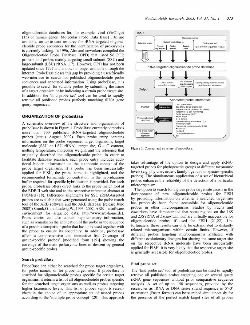
oligonucleotide databases for, for example, viral (VirOligo)(15) or human genes (Molecular Probe Data Base) (16) areavailable, an up-to-date resource for rRNA-targeted oligonu-cleotide probe sequences for the identification of prokaryotesis currently lacking. In 1996, Alm and coworkers compiled theOligonucleotide Probe Database (OPD) that listed 96 PCRprimers and probes mainly targeting small-subunit (SSU) andlarge-subunit (LSU) rRNA (17). However, OPD has not beenupdated since 1997 and is now no longer available through theinternet. ProbeBase closes this gap by providing a user-friendlyweb-interface to search for published oligonucleotide probesequences and annotated information. Using probeBase, it ispossible to search for suitable probes by submitting the nameof a target organism or by indicating a certain probe target site.In addition, the ‘find probe set’ tool can be used to rapidlyretrieve all published probes perfectly matching rRNA genequery sequences.
ORGANIZATION OF probeBase
A schematic overview of the structure and organization ofprobeBase is shown in Figure 1. ProbeBase currently comprisesmore than 700 published rRNA-targeted oligonucleotideprobes (status August 2002). Each probe entry containsinformation on the probe sequence, target organisms, targetmolecule (SSU or LSU rRNA), target site, G þ C content,melting temperature, molecular weight, and the reference thatoriginally described the oligonucleotide probe. In order tofacilitate database searches, each probe entry includes addi-tional hidden information on the taxonomic context of theprobe target organisms. If a probe has been successfullyapplied for FISH, the probe name is highlighted, and therecommended formamide concentration in the hybridizationbuffer required for specific hybridization is provided. For eachprobe, probeBase offers direct links to the probe match tool atthe RDP-II web site and to the respective reference abstract atPubMed (18). Difference alignments for SSU rRNA-targetedprobes are available that were generated using the probe matchtool of the ARB software and the ARB database (release June2002) (Strunk,O. and Ludwig,W., 1993–2002, ARB—a softwareenvironment for sequence data, http://www.arb-home.de).Probe entries can also contain supplementary information,such as remarks on the application of the probe or the sequenceof a possible competitor probe that has to be used together withthe probe to ensure its specificity. In addition, probeBaseoffers a comprehensive and interactive list ‘Coverage ofgroup-specific probes’ [modified from (19)] showing thecoverage of the main prokaryotic lines of descent by generalgroup-specific probes.
Search probeBase
ProbeBase can either be searched for probe target organisms,for probe names, or for probe target sites. If probeBase issearched for oligonucleotide probes specific for certain targetorganisms, it returns a list of all oligonucleotide probes specificfor the searched target organisms as well as probes targetinghigher taxonomic levels. This list of probes supports resear-chers in the choice of an appropriate set of nested probesaccording to the ‘multiple probe concept’ (20). This approach
takes advantage of the option to design and apply rRNA-targeted probes for phylogenetic groups at different taxonomiclevels (e.g. phylum-, order-, family-, genus-, or species-specificprobes). The simultaneous application of a set of hierarchicalprobes enhances the reliability of the detection of a particularmicroorganism.
The option to search for a given probe target site assists in thedevelopment of new oligonucleotide probes for FISHby providing information on whether a searched target sitehas previously been found accessible for oligonucleotideprobes in other microorganisms. Studies by Fuchs andcoworkers have demonstrated that some regions on the 16Sand 23S rRNA of Escherichia coli are virtually inaccessible foroligonucleotide probes if used for FISH (21,22). Un-fortunately, these results can only be extrapolated to distantlyrelated microorganisms within certain limits. However, ifdifferent probes targeting microorganisms affiliated withdifferent evolutionary lineages but sharing the same target siteon the respective rRNA molecule have been successfullyapplied for FISH, it is very likely that the respective target siteis generally accessible for oligonucleotide probes.
Find probe set
The ‘find probe set’ tool of probeBase can be used to rapidlyretrieve all published probes targeting one or several queryrRNA gene sequences without prior comparative sequenceanalysis. A set of up to 150 sequences, provided by theresearcher as rRNA or DNA sense strand sequence in 50–30
orientation (FastA format) can be searched simultaneously forthe presence of the perfect match target sites of all probes
Figure 1. Concept and structure of probeBase.
Nucleic Acids Research, 2003, Vol. 31, No. 1 515
deposited at probeBase. The output is a table sequentiallylisting (i) each single query sequence with all perfectlymatching probes found in probeBase and (ii) each possibleprobe with all perfectly matching query sequences. Using thisprobeBase feature researchers will, for example, easily be ableto determine a set of already published probes that targetthe microbial sequences in a certain environmental rRNA geneclone library. This probe set might then be used in subsequenthybridization experiments to confirm the presence of theorganisms detected in the rRNA gene clone library in situ andto gain insight into the actual abundance of these microorgan-isms in the investigated environment.
AVAILABILITY
ProbeBase is maintained and updated by the Microbial EcologyGroup staff at the Lehrstuhl fur Mikrobiologie of the TechnischeUniversitat Munchen, Bavaria, Germany. Free access toprobeBase is provided via the world wide web at http://www.probebase.net. Researchers are kindly invited and encour-aged to deposit their newly designed probe sequences andsupplementary information at probeBase. Submission mighteither be performed through the probe submission form ‘submita probe’ accessible at the main page or by Email. For queriesconcerning probeBase and for alternative probe submissioncontact [email protected].
CITING probeBase
If you use probeBase as a tool in your published research or ifyou have deposited your newly designed rRNA-targetedoligonucleotide probes at probeBase, we ask that this paperbe cited.
ACKNOWLEDGEMENTS
We gratefully acknowledge Wolfgang Ludwig for providingthe ARB software and the ARB database to the scientificcommunity—without ARB, research on microbial phylogeny,diversity, and ecology would have been less efficient. We thankJames R. Cole from RDP-II for allowing us to directly submitoligonucleotide sequences to the probe match tool of RDP-IIand for giving us insight into the probe match user statistics.We also thank Manuela Hartmann and Silvia Weber for theirhelp with database administration, and Klaus Ungerer for hisgenerous support.
REFERENCES
1. Raskin,L., Poulsen,L.K., Noguera,D.R., Rittmann,B.E. and Stahl,D.A.(1994) Quantification of methanogenic groups in anaerobic biologicalreactors by oligonucleotide probe hybridization. Appl. Environ. Microbiol.,60, 1241–1248.
2. Juretschko,S., Loy,A., Lehner,A. and Wagner,M. (2002) The microbialcommunity composition of a nitrifying–denitrifying activated sludgefrom an industrial sewage treatment plant analyzed by thefull-cycle rRNA approach. Syst. Appl. Microbiol.,25, 84–99.
3. Daims,H., Ramsing,N.B., Schleifer,K.-H. and Wagner,M. (2001) Cultiva-tion-independent, semiautomatic determination of absolute bacterial cellnumbers in environmental samples by fluorescence in situ hybridization.Appl. Environ. Microbiol., 67, 5810–5818.
4. Jansen,G.J., Mooibroek,M., Idema,J., Harmsen,H.J., Welling,G.W. andDegener,J.E. (2000) Rapid identification of bacteria in blood cultures byusing fluorescently labeled oligonucleotide probes. J. Clin. Microbiol., 38,814–817.
5. Hu,J., Limaye,A.P., Fritsche,T.R., Horn,M., Juretschko,S. and Gautom,R.(2002) Direct detection of Legionellae in respiratory tract specimens usingfluorescence in situ hybridization. In Marre,R. (ed.), Legionella. AmericanSociety of Microbiology Press, Washington, DC, pp. 221–224.
6. Poppert,S., Essig,A., Marre,R., Wagner,M. and Horn,M. (2002) Detectionand differentiation of chlamydiae by fluorescence in situ hybridization(FISH). Appl. Environ. Microbiol., 68, 4081–4089.
7. Guschin,D.Y., Mobarry,B.K., Proudnikov,D., Stahl,D.A., Rittmann,B.E.and Mirzabekov,A.D. (1997) Oligonucleotide microchips as genosensorsfor determinative and environmental studies in microbiology. Appl.Environ. Microbiol., 63, 2397–2402.
8. Wilson,K.H., Wilson,W.J., Radosevich,J.L., De Santis,T.Z.,Viswanathan,V.S., Kuczmarski,T.A. and Andersen,G.L. (2002)High-density microarray of small-subunit ribosomal DNA probes.Appl. Environ. Microbiol., 68, 2535–2541.
9. Liu,W.T., Mirzabekov,A.D. and Stahl,D.A. (2001) Optimization of anoligonucleotide microchip for microbial identification studies: a non-equilibrium dissociation approach. Environ. Microbiol., 3, 619–629.
10. Wu,L., Thompson,D.K., Li,G., Hurt,R.A., Tiedje,J.M. and Zhou,J. (2001)Development and evaluation of functional gene arrays for detection ofselected genes in the environment. Appl. Environ. Microbiol., 67,5780–5790.
11. Loy,A., Lehner,A., Lee,N., Adamczyk,J., Meier,H., Ernst,J.,Schleifer,K.-H. and Wagner,M. (2002) Oligonucleotide microarray for 16SrRNA gene-based detection of all recognized lineages of sulfate-reducingprokaryotes in the environment. Appl. Environ. Microbiol., 68, 5064–5081.
12. Lee,N., Nielsen,P.H., Andreasen,K.H., Juretschko,S., Nielsen,J.L.,Schleifer,K.-H. and Wagner,M. (1999) Combination of fluorescent in situhybridization and microautoradiography—a new tool for structure-functionanalyses in microbial ecology. Appl. Environ. Microbiol., 65, 1289–1297.
13. Maidak,B.L., Cole,J.R., Lilburn,T.G., Parker,C.T., Jr., Saxman,P.R.,Farris,R.J., Garrity,G.M., Olsen,G.J., Schmidt,T.M. and Tiedje,J.M. (2001)The RDP-II (Ribosomal Database Project). Nucleic Acids Res., 29,173–174.
14. Wuyts,J., Van de Peer,Y., Winkelmans,T. and De Wachter,R. (2002) TheEuropean database on small subunit ribosomal RNA. Nucleic Acids Res.,30, 183–185.
15. Onodera,K. and Melcher,U. (2002) VirOligo: a database of virus-specificoligonucleotides. Nucleic Acids Res., 30, 203–204.
16. Campi,M.G., Romano,P., Milanesi,L., Marra,D., Manniello,M.A.,Iannotta,B., Rondanina,G., Grasso,E., Ruzzon,T. and Santi,L. (1998)Molecular Probe Data Base (MPDB). Nucleic Acids Res., 26, 145–147.
17. Alm,E.W., Oerther,D.B., Larsen,N., Stahl,D.A. and Raskin,L. (1996)The oligonucleotide probe database. Appl. Environ. Microbiol., 62,3557–3559.
18. Wheeler,D.L., Church,D.M., Lash,A.E., Leipe,D.D., Madden,T.L.,Pontius,J.U., Schuler,G.D., Schriml,L.M., Tatusova,T.A., Wagner,L. andRapp,B.A. (2002) Database resources of the National Center forBiotechnology Information: 2002 update. Nucleic Acids Res., 30, 13–16.
19. Loy,A., Daims,H. and Wagner,M. (2002) Activated sludge-Moleculartechniques for determining community composition. In Bitton,G. (ed.),The Encyclopedia of Environmental Microbiology. John Wiley & Sons,Inc., New York, pp. 26–43.
20. Behr,T., Koob,C., Schedl,M., Mehlen,A., Meier,H., Knopp,D., Frahm,E.,Obst,U., Schleifer,K., Niessner,R. and Ludwig,W. (2000) A nested array ofrRNA targeted probes for the detection and identification of enterococci byreverse hybridization. Syst. Appl. Microbiol., 23, 563–572.
21. Fuchs,B.M., Syutsubo,K., Ludwig,W. and Amann,R. (2001) In situaccessibility of Escherichia coli 23S rRNA to fluorescently labeledoligonucleotide probes. Appl. Environ. Microbiol., 67, 961–968.
22. Fuchs,B.M., Wallner,G., Beisker,W., Schwippl,I., Ludwig,W. andAmann,R. (1998) Flow cytometric analysis of the in situ accessibility ofEscherichia coli 16S rRNA for fluorescently labeled oligonucleotideprobes. Appl. Environ. Microbiol., 64, 4973–4982.
516 Nucleic Acids Research, 2003, Vol. 31, No. 1

123
APPENDIX III
Diversity of Sulfate-Reducing Prokaryotes in anAcidic Fen: Comparison of 16S rRNA Gene-
Based Oligonucleotide Microarray Hybridizationwith Dissimilatory (Bi)Sulfite Reductase Gene
Surveys
Submitted to APPLIED AND ENVIRONMENTAL MICROBIOLOGY

Diversity of Sulfate-Reducing Prokaryotes in an Acidic Fen:Comparison of 16S rRNA Gene-Based Oligonucleotide Microarray
Hybridization with Dissimilatory (Bi)Sulfite Reductase Gene Surveys
Alexander Loy1, Kirsten Küsel2, Angelika Lehner3, Harold L. Drake2,and Michael Wagner1*
1 Lehrstuhl für Mikrobielle Ökologie, Institut für Ökologie und Naturschutz (IECB), Universität Wien, A-1090 Wien, Austria;2 Lehrstuhl für Ökologische Mikrobiologie, BITÖK, Universität Bayreuth, D-95440 Bayreuth, Germany; 3 Lehrstuhl für
Mikrobiologie, Technische Universität München, D-85350 Freising, Germany
The richness of sulfate-reducing prokaryotes (SRPs) in two acidic fen soils (Schlöppnerbrunnen I and II,Lehstenbach, Fichtelgebirge, Germany) was determined cultivation-independently by combination of 16SrRNA gene-based oligonucleotide microarray analyses and phylogenetic surveys of dissimilatory(bi)sulfite reductase genes (dsrAB). Microarray hybridization and subsequent evaluation by SRP group-specific 16S rRNA gene amplification, cloning, and comparative sequence analysis uncovered theoccurrence of bacteria affiliated with the deltaproteobacterial genera Syntrophobacter and Desulfomonile.Detailed sequence analysis of dsrAB genes identified in total eleven distinct operational taxonomic units(OTUs) thereby reconfirming the presence of Syntrophobacter wolinii- and Desulfomonile-related species.Five of these eleven OTUs were of deltaproteobacterial origin (two OTUs most closely related toDesulfobacca acetoxidans) whereas six deeply branching OTUs could not be unambiguously affiliated toany DsrAB amino acid sequence from yet recognized SRPs. Both investigated Schlöppnerbrunnen soilsamples showed exactly the same total SRP richness (seven dsrAB gene OTUs). However, the same threeOTUs were identified at both sampling sites whereas four were exclusively found at one site, respectively.This molecular study further proves the power of the 16S rRNA gene-based microarray technology inidentifying prokaryotes of known SRP lineages but simultaneously substantiates the necessity ofalternative identification approaches that allow the diversity assessment of yet unknown SRPs.
Dissimilatory sulfate reduction, which is carried outexclusively by prokaryotic organisms, is one of themost important organic carbon mineralizationprocesses in anaerobic aquatic environmentsespecially in marine sediments (26, 27). In contrastto the well-studied sulfate-reducing communities inmarine (39, 56-59) and freshwater habitats (38),little is known about the distribution and diversity ofsulfate-reducing prokaryotes (SRPs) in terrestrialecosystems. The contribution of terrestrial SRPs,compared to aquatic SRPs, to sulfur cycling in termsof substrate turnover might be of minor importanceon a global scale. Nevertheless, their ecologicalimportance for the biodegradation of environmentalpollutants such as oil (21, 37, 55, 73) or for theoperativeness of a particular terrestrial ecosystemcan be profound. Based on �34S values and 35Sradiolabeling, it has been shown that dissimilatorysulfate reduction is an ongoing process at some fensites in a forested catchment in Bavaria, Germany(Lehstenbach, Fichtelgebirge) (1, 3). Owing to airpollution the catchment has seen a major depositionof sulfate through acidic rainfall (in form of sulfuric
and sulfurous acid) until the 90s, when efficient airfiltration systems have been introduced in factoriesin former East Germany. With each rainfall, sulfateis leached out of the upland aerated soils in thelower situated fens. It is hypothesized thatdissimilatory sulfate reduction in these mainlyanaerobic and waterlogged soils contributes to theretention of sulfur in this ecosystem (1, 2, 51).Sulfate reduction in these fens is a sink for sulfateand protons, thus decreasing acidity of soil solutionand the adjacent groundwater. The primary aim ofthis study is to establish a detailed inventory of allSRPs inhabiting two selected fen sites at theLehstenbach catchment at a single point in time, toget insights into the phylogenetic origin of thesemicroorganisms, and to provide a robust basis forfurther characterization of their ecophysiologicalrole in time and space.Recently, an 16S rRNA-based oligonucleotide DNAmicroarray for SRPs (SRP-PhyloChip) has beenintroduced in determinative and environmental SRPresearch (43). We used the SRP-PhyloChip torapidly screen for members of all known SRPlineages at the Lehstenbach catchment. However,SRPs constitute a polyphyletic microbial guild andthus, 16S rRNA-based approaches do not easilyallow do directly link the sulfate reducing ability to acertain phylogenetic position. Molecular metabolicdiversity surveys based on key enzymes for certain
* Corresponding author. Mailing address: Lehrstuhl fürMikrobielle Ökologie, Institut für Ökologie und Naturschutz,Universität Wien, Althanstr. 14, A-1090 Wien, Austria. Phone:0043 1 4277 54201. Fax: 0043 1 4277 9542. E-mail:[email protected].
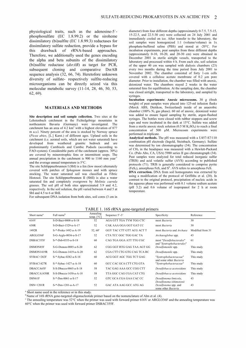
SULFATE-REDUCING PROKARYOTES IN AN ACIDIC FEN 2
physiological traits, such as the adenosine-5`-phosphosulfate (EC 1.8.99.2) or the sirohemedissimilatory (bi)sulfite (EC 1.8.99.3) reductases fordissimilatory sulfate reduction, provide a bypass forthis drawback of rRNA-based approaches.Therefore, we additionally used the genes encodingthe alpha and beta subunits of the dissimilatory(bi)sulfite reductase (dsrAB) as target for PCR,subsequent cloning and comparative DsrABsequence analysis (32, 66, 74). Heretofore unknowndiversity of sulfate- respectively sulfite-reducingmicroorganisms can be directly seized via thismolecular metabolic survey (11-14, 28, 46, 50, 53,62, 69).
MATERIALS AND METHODS
Site description and soil sample collection. Two sites at theLehstenbach catchment in the Fichtelgebirge mountains innortheastern Bavaria (Germany) were investigated. Thecatchment has an area of 4.2 km2 with a highest elevation of 877m a.s.l. Ninety percent of the area is stocked by Norway spruce(Picea abies, [L.] Karst.) of different ages. Upland soils in thecatchment (i.e. aerated soils, which are not water saturated) havedeveloped from weathered granitic bedrock and arepredominantly Cambisols and Cambic Podsols (according toFAO-system). Considerable parts of the catchment (approx. 30%)are covered by minerotrophic fens or intermittent seeps. Theannual precipitation in the catchment is 900 to 1160 mm year-1
and the average annual temperature is 5�C.The site Schlöppnerbrunnen I (SbI) is a fen (low moor) alternatelycovered with patches of Sphagnum mosses and with sprucestocking. The water saturated soil was classified as FibricHistosol. The site Schlöppnerbrunnen II (SbII) is also a watersaturated fen and completely overgrown by Molinia caerulagrasses. The soil pH of both sites approximated 3.9 and 4.2,respectively. In the soil solution, the pH varied between 4 and 5 atSbI and 4.5 to 6 at SbII.For subsequent DNA isolation from both sites, soil cores (3 cm in
diameter) from four different depths (approximately 0-7.5, 7.5-15,15-22.5, and 22.5-30 cm) were collected on 24 July 2001 andimmediately cooled on ice. After transfer to the laboratory, thesoil samples were homogenized 1:1 (volume/volume) in 1xphosphate-buffered saline (PBS) and stored at -20�C. Forincubation experiments, peat samples from three different depths(approximately 0-10, 10-20, and 20-30 cm) were obtained inDecember 2001 in sterile airtight vessels, transported to thelaboratory and processed within 4 h. From each site, soil solutionof the upper 40 cm was sampled with dialysis chambers (23)every two months during the time period from July 2001 toNovember 2002. The chamber consisted of forty 1-cm cellscovered with a cellulose acetate membrane of 0.2 µm porediameter. Prior to installation, the chamber was filled with anoxic,deionized water. The chambers stayed 2 weeks in the watersaturated fens for equilibration. At the sampling date, the chamberwas closed airtight, transported to the laboratory, and sampled bysyringes.Incubation experiments: Anoxic microcosms. 30 g (freshweight) of peat samples were placed into 125-ml infusion flasks(Merck ABS, Dietikon, Switzerland) inside of an anaerobicchamber (100% N2 gas phase). 60 ml of anoxic, deionized waterwas added to ensure liquid sampling by sterile, argon-flushedsyringes. The bottles were closed with rubber stoppers and screwcaps and were incubated in the dark at 15°C. Sulfate was addedfrom a sterile anoxic stock solution (0.5 M K2SO4) to reach a finalconcentration of 500 µM. Microcosm experiments wereperformed in triplicate.Analytical methods. The pH was measured with a U457-S7/110combination pH electrode (Ingold, Steinbach, Germany). Sulfatewas determined by ion chromatography (34). The concentrationof CH4 in the headspace was measured with a Hewlett-PackardCo. (Palo Alto, CA, USA) 5980 series II gas chromatograph (34).Peat samples were analyzed for total reduced inorganic sulfur(TRIS) and acid volatile sulfur (AVS) according to publishedprotocols (71). TRIS is generally considered to comprise pyrite(FeS2), amorphous FeS, and S0. AVS refers to amorphous FeS.DNA extraction. DNA from soil homogenates was extracted byusing a modification of the protocol of Griffiths et al. (20). Incontrast to the original protocol, precipitation of nucleic acids inthe aqueous phase was performed with 0.1 volume sodium acetate(pH 5.2) and 0.6 volume of isopropanol for 2 h at roomtemperature.
TABLE 1. 16S rRNA gene-targeted primersShort namea Full nameb Annealing
temp. [°C] Sequence 5’-3’ Specificity Reference
616V S-D-Bact-0008-a-S-18 52 AGA GTT TGA TYM TGG CTC most Bacteria 30
630R S-D-Bact-1529-a-A-17 52 CAK AAA GGA GGT GAT CC most Bacteria 30
1492R S-*-Proka-1492-a-A-19 52, 60c GGY TAC CTT GTT ACG ACT T most Bacteria and Archaea Modified from 31
ARGLO36F S-G-Arglo-0036-a-S-17 52 CTA TCC GGC TGG GAC TA Archaeoglobus spp. 43
DSBAC355F S-*-Dsb-0355-a-S-18 60 CAG TGA GGA ATT TTG CGC most "Desulfobacterales"and "Syntrophobacterales"
61
DSMON85F S-G-Dsmon-0085-a-S-20 62 CGG GGT RTG GAG TAA AGT GG Desulfomonile spp. This study
DSMON1419R S-G-Dsmon-1419-a-A-20 62 CGA CTT CTG GTG CAG TCA RC Desulfomonile spp. This study
SYBAC+282F S-*-Sybac-0282-a-S-18 60 ACG GGT AGC TGG TCT GAG "Syntrophobacteraceae"and some other Bacteria
This study
SYBAC1427R S-*-Sybac-1427-a-A-18 60 GCC CAC GCA CTT CTG GTA "Syntrophobacteraceae" This study
DBACCA65F S-S-Dbacca-0065-a-S-18 58 TAC GAG AAA GCC CGG CTT Desulfobacca acetoxidans This study
DBACCA1430R S-S-Dbacca-1430-a-A-18 58 TTA GGC CAG CGA CAT CTG Desulfobacca acetoxidans This study
DSN61F S-*-Dsn-0061-a-S-17 52 GTC GCA CGA GAA CAC CC Desulfonema limicola,Desulfonema ishimotonii
43
DSN+1201R S-*-Dsn-1201-a-A-17 52 GAC ATA AAG GCC ATG AG Desulfonema spp. andsome other Bacteria
43
a Short name used in the reference or in this study.b Name of 16S rRNA gene-targeted oligonucleotide primer based on the nomenclature of Alm et al. (4).c The annealing temperature was 52°C when the primer was used with forward primer 616V or ARGLO36F and the annealing temperature was60°C when the primer was used with forward primer DSBAC355F.
SULFATE-REDUCING PROKARYOTES IN AN ACIDIC FEN3
PCR amplification of 16S rRNA and dsrAB genes. All PCRswere performed with five nanograms of DNA. For subsequentDNA microarray hybridization, amplification of bacterial 16SrRNA gene fragments from fen soil genomic DNA wasperformed by using primer pairs 616V-630R and 616V-1492R(Table 1). 16S rRNA gene fragments from reference pure culturesand clones were amplified by using the bacterial primer pair616V-630R or the cloning vector-specific primers M13F(-20) (5´-GTAAAACGACGGCCAG-3´) and M13R (5´-CAGGAAACAGCTATGAC-3´) (Invitrogen Corp., San Diego,Calif.), respectively. For confirmation of microarray results, 16SrRNA gene fragments of defined SRP groups were directlyamplified from soil DNA by using previously published andnewly designed primers (Table 1). In addition, an approximately1.9-kb dsrAB gene fragment was amplified from fen soil DNA byusing the degenerated primers DSR1Fmix (equimolar mixture ofDSR1F, DSR1Fa, and DSR1Fb) and DSR4Rmix (equimolarmixture of DSR4R, DSR4Ra, DSR4Rb, and DSR4Rc) (Table 2).Positive controls containing purified DNA from suitable referenceorganisms were included in all of the PCR amplificationexperiments along with negative controls (no DNA added). For16S rRNA and dsrAB gene amplification, reaction mixturescontaining 25 pM of each primer were prepared in a total volumeof 50 µl by using 10x Ex TaqTM reaction buffer and 2.5 U of ExTaq polymerase (Takara Biomedicals, Japan). Additionally, 20mM tetramethylammonium chloride (Sigma, Deisenhofen,Germany) was added to each amplification mixture to enhancethe specificity of the PCR reaction (33). Thermal cycling wascarried out by an initial denaturation step at 94°C for 1 min,followed by 30 (16S rRNA genes) or 35 cycles (dsrAB genes) ofdenaturation at 94°C for 40 s, annealing from 48°C to 62°C(depending on the primer pair; Table 1 and 2) for 40 s, and
elongation at 72°C for 1 min 30 s. Cycling was completed by afinal elongation step at 72°C for 10 min.DNA microarray technology. Fluorescence labeling of PCRamplificates, manufacturing and processing of SRP-PhyloChips,reverse hybridization on microarrays, scanning and imageanalysis of microarrays were performed as described by Loy et al.(43). Oligonucleotides for printing of the SRP-PhyloChips wereobtained from MWG Biotech (Ebersberg, Germany). Thesequence, specificity, and microarray position of all newlydesigned oligonucleotide probes are depicted in Table 3. Inaddition, the novel SRP-PhyloChip probes were deposited atprobeBase (http://www.probebase.net) (42), where probe-targetdifference alignments, generated with the PROBE_MATCH toolof the ARB program package (Strunk and Ludwig,http://www.arb-home.de), can be viewed for each probe.Cloning and sequencing. Prior to cloning the PCR amplificationproducts were purified by low-melting-point agarose (1.5%) gelelectrophoresis (NuSieve 3:1; FMC Bioproducts, BiozymDiagnostics GmbH, Oldendorf, Germany) and stained in SYBRGreen I solution (10 µl 10.000x SYBR Green I stain in 100 µlTAE buffer [40 mM TRIS, 10 mM sodium acetate, 1 mM EDTA,pH 8.0]; Biozym Diagnostics GmbH) for 45 min. Bands of theexpected size were excised from the agarose gel with a glasscapillary and melted with 80 µl double-distilled water for 10 minat 80°C. Four microliters of each solution were ligated asrecommended by the manufacturer (Invitrogen Corp.) either intothe cloning vector pCR2.1 of the TOPO TA cloning kit (16SrRNA gene amplificates) or into the cloning vector pCR-XL-TOPO of the TOPO XL cloning kit (dsrAB gene amplificates).Nucleotide sequences were determined by the dideoxynucleotidemethod (60) as described by Purkhold et al. (54). In addition,internal dsrAB gene-targeted sequencing primers (Table 2) wereused to complete the dsrAB sequences.
TABLE 2. Dissimilatory (bi)sulfite reductase gene-(dsrAB)-targeted primers. The target site of all listedDSR1 and DSR4 primer versions was analyzed for those SRPs (n=8) for which complete dsrAB operons areavailable in GenBank (8). SRPs with a fully complementary target site to the respective primers are listed in
the specificity column.Primer Sequence 5`-3` Specificity ReferenceDSR1Fa ACS CAC TGG AAG CAC G Archaeoglobus fulgidus, Archaeoglobus profundus,
Desulfovibrio vulgaris74
DSR1Faa ACC CAY TGG AAA CAC G Desulfotomaculum thermocisternum, Desulfobulbusrhabdoformis, Desulfobacter vibrioformis
This study
DSR1Fba GGC CAC TGG AAG CAC G Thermodesulforhabdus norvegica This studyDSR4Ra GTG TAG CAG TTA CCG CA Archaeoglobus fulgidus, Desulfovibrio vulgaris,
Desulfobulbus rhabdoformis74
DSR4Raa GTG TAA CAG TTT CCA CA Archaeoglobus profundus This studyDSR4Rba GTG TAA CAG TTA CCG CA Desulfobacter vibrioformis This studyDSR4Rca GTG TAG CAG TTT CCG CA Thermodesulforhabdus norvegica, Desulfotomaculum
thermocisternumThis study
DSR978Fab GGT CAT CGA CCT TTG TCC Schlöppnerbrunnen I soil OTU 5 This studyDSR978Fbb CGT CGT CGG GAA GTG CCC Schlöppnerbrunnen I soil OTU 8 This studyDSR978Fcb AGT AGT CGA CCT TTG CCC Schlöppnerbrunnen I+II soil OTU 6 This studyDSR978Fdb TGT CAC CGA TCT CTG CCC Schlöppnerbrunnen I soil OTU 1 This studyDSR978Feb TGT TAC CGA CCT CTG CCC Schlöppnerbrunnen II soil OTU 1
(dsrSbII-20)This study
DSR978Ffb TGT CAC CGA TCT TTG CCC Schlöppnerbrunnen II soil OTU 4(dsrSbII-15)
This study
DSR978Fgb CGT CAC CAT TCT CTG CCC Schlöppnerbrunnen II soil OTU 4(dsrSbII-9)
This study
DSR978Fhb GGT CGT TGA CAT GTG TCC Schlöppnerbrunnen II soil OTU 11 This studyDSR978Fib GGT CTG CAA TCT CTG YCC Schlöppnerbrunnen I+II soil OTU 2 and 3 This studyDSR978Fjb GGT TGT TGA CCT TTG CCC Schlöppnerbrunnen I soil OTU 9 This studyDSR978Fkb CGT TTG CGA TCT CTG CCC Schlöppnerbrunnen II soil OTU 7 This studyDSR860Fb AGA TCC GGC GGG ACG ATG Schlöppnerbrunnen I soil OTU 10 This study
a Primer was used at non-stringent conditions by applying an annealing temperature of 48°C for PCR in order to target a wide diversityof SRPs.b Internal sequencing primer used to complete dsrAB gene sequences retrieved from acidic fen sites Schlöppnerbrunnen I and II.
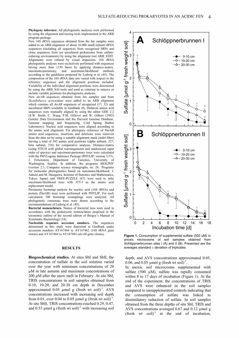
SULFATE-REDUCING PROKARYOTES IN AN ACIDIC FEN 4
Phylogeny inference. All phylogenetic analyses were performedby using the alignment and treeing tools implemented in the ARBprogram package.New 16S rRNA sequences obtained from the fen samples wereadded to an ARB alignment of about 16.000 small-subunit rRNAsequences (including all sequences from recognized SRPs andclone sequences from yet uncultured prokaryotes from sulfate-reducing environments) by using the alignment tool ARB_EDIT.Alignments were refined by visual inspection. 16S rRNAphylogenetic analyses were exclusively performed with sequenceshaving more than 1150 bases by applying distance-matrix,maximum-parsimony, and maximum-likelihood methodsaccording to the guidelines proposed by Ludwig et al. (45). Thecomposition of the 16S rRNA data sets varied with respect to thereference sequences and the alignment positions included.Variability of the individual alignment positions were determinedby using the ARB_SAI tools and used as criterion to remove orinclude variable positions for phylogenetic analyses.New dsrAB sequences obtained from fen samples and fromDesulfobacca acetoxidans were added to an ARB alignmentwhich contains all dsrAB sequences of recognized (17, 32) anduncultured SRPs available in GenBank (8). Deduced amino acidsequences were manually aligned by using the editor GDE 2.2(S.W. Smith, C. Wang, P.M. Gillevet and W. Gilbert (1992)Genetic Data Environment and the Harvard Genome Database.Genome mapping and Sequencing, Cold Spring HarborLaboratory). Nucleic acid sequences were aligned according tothe amino acid alignment. For phylogeny inference of DsrABamino acid sequences, insertions and deletions were removedfrom the data set by using a suitable alignment mask (indel filter)leaving a total of 543 amino acid positions (alpha subunit, 327;beta subunit, 216) for comparative analyses. Distance-matrix(using FITCH with global rearrangements and randomized inputorder of species) and maximum-parsimony trees were calculatedwith the PHYLogeny Inference Package (PHYLIP, version 3.57c,J. Felsenstein, Department of Genetics, University ofWashington, Seattle). In addition, the programs MOLPHY(version 2.3, Computer science monographs, no. 28.: Programsfor molecular phylogenetics based on maximum-likelihood. J.Adachi and M. Hasegawa, Institute of Statistics and Mathematics,Tokyo, Japan) and TREE-PUZZLE (67) were used to infermaximum-likelihood trees with JTT-f as the amino acidreplacement model.Parsimony bootstrap analysis for nucleic acid (16S rRNA) andprotein (DsrAB) trees were performed with PHYLIP. For eachcalculation 100 bootstrap resamplings were analyzed. Allphylogenetic consensus trees were drawn according to therecommendations of Ludwig et al. (45).Bacterial nomenclature. Names of bacterial taxa were used inaccordance with the prokaryotic nomenclature proposed in thetaxonomic outline of the second edition of Bergey`s Manual ofSystematic Bacteriology (18).Nucleotide sequence accession numbers. The sequencesdetermined in this study were deposited at GenBank underaccession numbers AY167444 to AY167462 (16S rRNA geneclones) and AY167464 to AY167483 (dsrAB gene clones).
RESULTS
Biogeochemical studies. At sites SbI and SbII, theconcentration of sulfate in the soil solution variedover the year with minimum concentrations of 20µM in late autumn and maximum concentrations of200 µM after the snow melt in February. At site SbI,TRIS concentrations in soil samples obtained from0-10, 10-20, and 20-30 cm depth in Decemberapproximated 0.05 µmol g (fresh wt soil)-1. AVSconcentrations increased with increasing soil depthfrom 0.01, over 0.04 to 0.05 µmol g (fresh wt soil)-1.At site SbII, TRIS concentrations reached 0.29, 0.47,and 0.53 µmol g (fresh wt soil)-1 with increasing soil
depth, and AVS concentrations approximated 0.05,0.06, and 0.05 µmol g (fresh wt soil)-1.In anoxic soil microcosms supplemented withsulfate (500 µM), sulfate was rapidly consumedwithin 8 to 17 days of incubation (Figure 1). At theend of the experiment, the concentrations of TRISand AVS were enhanced in the soil samplescompared to unsupplemented controls indicating thatthe consumption of sulfate was linked todissimilatory reduction of sulfate. In soil samplesobtained from the three depths of site SbI, TRIS andAVS concentrations averaged 0.67 and 0.12 µmol g(fresh wt soil)-1 at the end of incubation,
Schlöppnerbrunnen II
Incubation time [d]0 2 4 6 8 10 12 14 16 18
Sulfa
te [µ
mol
g-1
fres
h w
eigh
t soi
l]
0
1
2
3
Schlöppnerbrunnen I
Sulfa
te [µ
mol
g-1
fres
h w
eigh
t soi
l]0
1
2
3
4
0-10 cm10-20 cm20-30 cm
A
B0-10 cm
10-20 cm20-30 cm
Figure 1. Consumption of supplemental sulfate (500 µM) inanoxic microcosms of soil samples obtained fromSchlöppnerbrunnen sites I (A) and II (B). Presented are theaverages standard � deviation of triplicates.
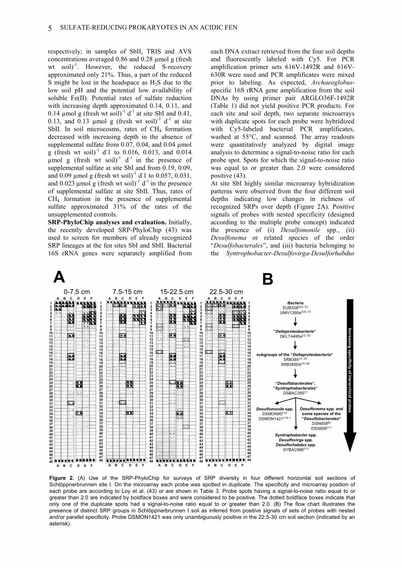
SULFATE-REDUCING PROKARYOTES IN AN ACIDIC FEN5
respectively; in samples of SbII, TRIS and AVSconcentrations averaged 0.86 and 0.28 µmol g (freshwt soil)-1. However, the reduced S-recoveryapproximated only 21%. Thus, a part of the reducedS might be lost in the headspace as H2S due to thelow soil pH and the potential low availability ofsoluble Fe(II). Potential rates of sulfate reductionwith increasing depth approximated 0.14, 0.11, and0.14 µmol g (fresh wt soil)-1 d-1 at site SbI and 0.41,0.13, and 0.13 µmol g (fresh wt soil)-1 d-1 at siteSbII. In soil microcosms, rates of CH4 formationdecreased with increasing depth in the absence ofsupplemental sulfate from 0.07, 0.04, and 0.04 µmolg (fresh wt soil)-1 d-1 to 0.016, 0.013, and 0.014µmol g (fresh wt soil)-1 d-1 in the presence ofsupplemental sulfate at site SbI and from 0.19, 0.09,and 0.09 µmol g (fresh wt soil)-1 d-1 to 0.057, 0.031,and 0.023 µmol g (fresh wt soil)-1 d-1 in the presenceof supplemental sulfate at site SbII. Thus, rates ofCH4 formation in the presence of supplementalsulfate approximated 31% of the rates of theunsupplemented controls.SRP-PhyloChip analyses and evaluation. Initially,the recently developed SRP-PhyloChip (43) wasused to screen for members of already recognizedSRP lineages at the fen sites SbI and SbII. Bacterial16S rRNA genes were separately amplified from
each DNA extract retrieved from the four soil depthsand fluorescently labeled with Cy5. For PCRamplification primer sets 616V-1492R and 616V-630R were used and PCR amplificates were mixedprior to labeling. As expected, Archaeoglobus-specific 16S rRNA gene amplification from the soilDNAs by using primer pair ARGLO36F-1492R(Table 1) did not yield positive PCR products. Foreach site and soil depth, two separate microarrayswith duplicate spots for each probe were hybridizedwith Cy5-labeled bacterial PCR amplificates,washed at 55°C, and scanned. The array readoutswere quantitatively analyzed by digital imageanalysis to determine a signal-to-noise ratio for eachprobe spot. Spots for which the signal-to-noise ratiowas equal to or greater than 2.0 were consideredpositive (43).At site SbI highly similar microarray hybridizationpatterns were observed from the four different soildepths indicating low changes in richness ofrecognized SRPs over depth (Figure 2A). Positivesignals of probes with nested specificity (designedaccording to the multiple probe concept) indicatedthe presence of (i) Desulfomonile spp., (ii)Desulfonema or related species of the order“Desulfobacterales”, and (iii) bacteria belonging tothe Syntrophobacter-Desulfovirga-Desulforhabdus
0-7.5 cm 7.5-15 cm 15-22.5 cm 22.5-30 cm
234
1
678
5
101112
9
141516
13
181920
17
222324
21
262728
25
303132
29
343536
33
383940
37
424344
41
464748
45
A B C D E F
234
1
678
5
101112
9
141516
13
181920
17
222324
21
262728
25
303132
29
343536
33
383940
37
424344
41
464748
45
A B C D E F
A B C D E F
A B C D E F
234
1
678
5
101112
9
141516
13
181920
17
222324
21
262728
25
303132
29
343536
33
383940
37
424344
41
464748
45
A B C D E F
A B C D E F
234
1
678
5
101112
9
141516
13
181920
17
222324
21
234
1
678
5
101112
9
141516
13
181920
17
222324
21
262728
25
303132
29
343536
33
383940
37
424344
41
464748
45
A B C D E F
262728
25
303132
29
343536
33
383940
37
424344
41
464748
45
A B C D E F
Multiple Probe C
oncept
Desulfonema spp. andsome species of the“Desulfobacterales“
DSN658B2
DSS658C11
BacteriaEUB338D25, F2
UNIV1390aD26, F5
“Deltaproteobacteria“ DELTA495aC2, E2
“Desulfobacterales“,“Syntrophobacterales“
DSBAC355C7
Syntrophobacter spp.Desulfovirga spp.
Desulforhabdus spp.SYBAC986C17
subgroups of the “Deltaproteobacteria“ SRB385C5, E5
SRB385DbC6, E6
Desulfomonile spp.DSMON95C18
DSMON1421C19 *
A B
Increasing specificity of hierarchical probes
Figure 2. (A) Use of the SRP-PhyloChip for surveys of SRP diversity in four different horizontal soil sections ofSchlöppnerbrunnen site I. On the microarray each probe was spotted in duplicate. The specificity and microarray position ofeach probe are according to Loy et al. (43) or are shown in Table 3. Probe spots having a signal-to-noise ratio equal to orgreater than 2.0 are indicated by boldface boxes and were considered to be positive. The dotted boldface boxes indicate thatonly one of the duplicate spots had a signal-to-noise ratio equal to or greater than 2.0. (B) The flow chart illustrates thepresence of distinct SRP groups in Schlöppnerbrunnen I soil as inferred from positive signals of sets of probes with nestedand/or parallel specificity. Probe DSMON1421 was only unambiguously positive in the 22.5-30 cm soil section (indicated by anasterisk).
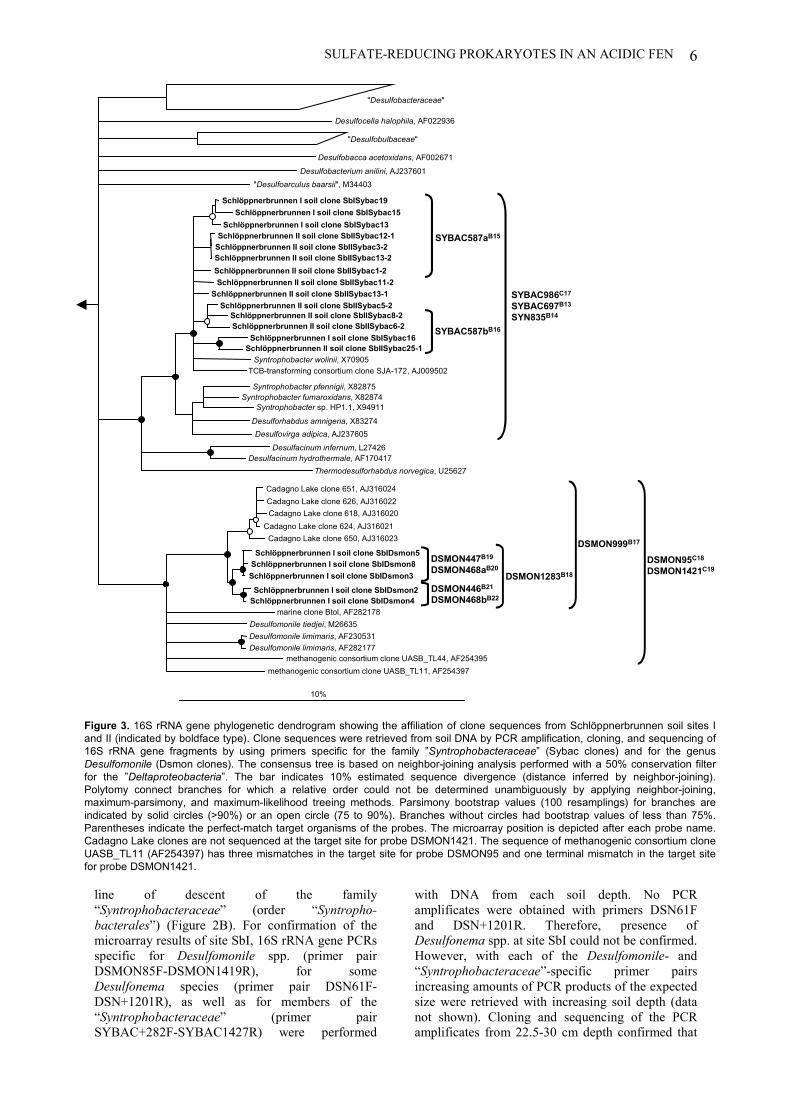
SULFATE-REDUCING PROKARYOTES IN AN ACIDIC FEN 6
line of descent of the family“Syntrophobacteraceae” (order “Syntropho-bacterales”) (Figure 2B). For confirmation of themicroarray results of site SbI, 16S rRNA gene PCRsspecific for Desulfomonile spp. (primer pairDSMON85F-DSMON1419R), for someDesulfonema species (primer pair DSN61F-DSN+1201R), as well as for members of the“Syntrophobacteraceae” (primer pairSYBAC+282F-SYBAC1427R) were performed
with DNA from each soil depth. No PCRamplificates were obtained with primers DSN61Fand DSN+1201R. Therefore, presence ofDesulfonema spp. at site SbI could not be confirmed.However, with each of the Desulfomonile- and“Syntrophobacteraceae”-specific primer pairsincreasing amounts of PCR products of the expectedsize were retrieved with increasing soil depth (datanot shown). Cloning and sequencing of the PCRamplificates from 22.5-30 cm depth confirmed that
"Desulfobacteraceae"
"Desulfobulbaceae"
Desulfobacca acetoxidans, AF002671
Desulfobacterium anilini, AJ237601 "Desulfoarculus baarsii", M34403
10%
methanogenic consortium clone UASB_TL11, AF254397
Schlöppnerbrunnen I soil clone SbIDsmon5 Schlöppnerbrunnen I soil clone SbIDsmon8
Schlöppnerbrunnen I soil clone SbIDsmon3 Schlöppnerbrunnen I soil clone SbIDsmon2
Schlöppnerbrunnen I soil clone SbIDsmon4
Cadagno Lake clone 651, AJ316024
Cadagno Lake clone 618, AJ316020
Cadagno Lake clone 650, AJ316023 Cadagno Lake clone 624, AJ316021
Cadagno Lake clone 626, AJ316022
methanogenic consortium clone UASB_TL44, AF254395
Desulfocella halophila, AF022936
Schlöppnerbrunnen I soil clone SbISybac19 Schlöppnerbrunnen I soil clone SbISybac15
Schlöppnerbrunnen I soil clone SbISybac13 Schlöppnerbrunnen II soil clone SbIISybac12-1
Schlöppnerbrunnen II soil clone SbIISybac3-2 Schlöppnerbrunnen II soil clone SbIISybac13-2 Schlöppnerbrunnen II soil clone SbIISybac1-2
Syntrophobacter wolinii, X70905
Schlöppnerbrunnen II soil clone SbIISybac11-2 Schlöppnerbrunnen II soil clone SbIISybac13-1
Schlöppnerbrunnen I soil clone SbISybac16 Schlöppnerbrunnen II soil clone SbIISybac25-1
Syntrophobacter pfennigii, X82875 Syntrophobacter fumaroxidans, X82874
Syntrophobacter sp. HP1.1, X94911
Desulforhabdus amnigena, X83274 Desulfovirga adipica, AJ237605
Desulfacinum infernum, L27426 Desulfacinum hydrothermale, AF170417
Thermodesulforhabdus norvegica, U25627
TCB-transforming consortium clone SJA-172, AJ009502
Schlöppnerbrunnen II soil clone SbIISybac5-2 Schlöppnerbrunnen II soil clone SbIISybac8-2 Schlöppnerbrunnen II soil clone SbIISybac6-2 SYBAC587bB16
SYBAC986C17
SYBAC697B13
SYN835B14
SYBAC587aB15
DSMON447B19
DSMON468aB20
DSMON446B21
DSMON468bB22
DSMON999B17
DSMON1283B18
DSMON95C18
DSMON1421C19
Desulfomonile tiedjei, M26635 Desulfomonile limimaris, AF230531 Desulfomonile limimaris, AF282177
marine clone Btol, AF282178
Figure 3. 16S rRNA gene phylogenetic dendrogram showing the affiliation of clone sequences from Schlöppnerbrunnen soil sites Iand II (indicated by boldface type). Clone sequences were retrieved from soil DNA by PCR amplification, cloning, and sequencing of16S rRNA gene fragments by using primers specific for the family ”Syntrophobacteraceae” (Sybac clones) and for the genusDesulfomonile (Dsmon clones). The consensus tree is based on neighbor-joining analysis performed with a 50% conservation filterfor the ”Deltaproteobacteria”. The bar indicates 10% estimated sequence divergence (distance inferred by neighbor-joining).Polytomy connect branches for which a relative order could not be determined unambiguously by applying neighbor-joining,maximum-parsimony, and maximum-likelihood treeing methods. Parsimony bootstrap values (100 resamplings) for branches areindicated by solid circles (>90%) or an open circle (75 to 90%). Branches without circles had bootstrap values of less than 75%.Parentheses indicate the perfect-match target organisms of the probes. The microarray position is depicted after each probe name.Cadagno Lake clones are not sequenced at the target site for probe DSMON1421. The sequence of methanogenic consortium cloneUASB_TL11 (AF254397) has three mismatches in the target site for probe DSMON95 and one terminal mismatch in the target sitefor probe DSMON1421.
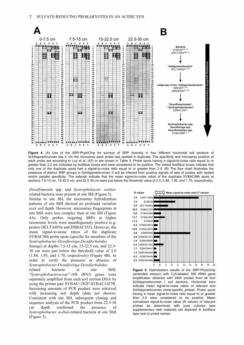
SULFATE-REDUCING PROKARYOTES IN AN ACIDIC FEN7
Desulfomonile spp. and Syntrophobacter wolinii-related bacteria were present at site SbI (Figure 3).Similar to site SbI, the microarray hybridizationpatterns of site SbII showed no profound variationover soil depth. However, microarray fingerprints atsite SbII were less complex than at site SbI (Figure4A). Only probes targeting SRPs at highertaxonomic levels were unambiguously positive (e.g.probes DELTA495a and DSBAC355). However, themean signal-to-noise ratios of the duplicateSYBAC986 probe spots (specific for members of theSyntrophobacter-Desulfovirga-Desulforhabduslineage) at depths 7.5-15 cm, 15-22.5 cm, and 22.5-30 cm were just below the threshold value of 2.0(1.88, 1.95, and 1.70, respectively) (Figure 4B). Inorder to verify the presence or absence ofSyntrophobacter-Desulfovirga-Desulforhabdus-related bacteria at site SbII,“Syntrophobacteraceae”-16S rRNA genes wereseparately amplified from each soil section DNA byusing the primer pair SYBAC+282F-SYBAC1427R.Increasing amounts of PCR product were retrievedwith increasing soil depth (data not shown).Consistent with site SbI, subsequent cloning andsequence analysis of the PCR product from 22.5-30cm depth confirmed the presence ofSyntrophobacter wolinii-related bacteria at site SbII(Figure 3).
Figure 5. Hybridization results of the SRP-PhyloChip(amended version) with Cy5-labeled 16S rRNA geneamplificates obtained with DNA pooled from all fourSchlöppnerbrunnen I soil sections. Horizontal barsindicate mean signal-to-noise ratios of relevant andSchlöppnerbrunnen clone-specific probes. Probe spotshaving a mean signal-to-noise ratio equal to or greaterthan 2.0 were considered to be positive. Meannormalized signal-to-noise ratios (R values) of relevantprobes as determined with pure cultures (seesupplementary web material) are depicted in boldfacetype next to probe names.
0 2 4 6 8 10 12 14 16 18 20
DSMON468bDSMON446
DSMON468aDSMON447
DSMON1283DSMON999
DSMON1421DSMON95
SYBAC587bSYBAC587a
SYN835SYBAC697SYBAC986DSBAC355
DELTA495aEUB338
UNIV1389a
Mean signal-to-noise ratio (T values)+-
4.30.31.21.74.31.02.65.6
10.69.4
11.211.7
9.916.9
2.91.01.8
R values
AIncreasing specificity of hierarchical probes
BacteriaEUB338D25, F2
UNIV1390aD26, F5
“Deltaproteobacteria“ DELTA495aC2, E2
“Desulfobacterales“,“Syntrophobacterales“
DSBAC355C7
Syntrophobacter spp.Desulfovirga spp.
Desulforhabdus spp.SYBAC986C17 *
subgroups of the “Deltaproteobacteria“ SRB385C5, E5
SRB385DbC6, E6
234
1
678
5
101112
9
141516
13
181920
17
222324
21
262728
25
303132
29
343536
33
383940
37
424344
41
464748
45
A B C D E F
A B C D E F
234
1
678
5
101112
9
141516
13
181920
17
222324
21
262728
25
303132
29
343536
33
383940
37
424344
41
464748
45
A B C D E F
A B C D E F
234
1
678
5
101112
9
141516
13
181920
17
222324
21
262728
25
303132
29
343536
33
383940
37
424344
41
464748
45
A B C D E F
A B C D E F
234
1
678
5
101112
9
141516
13
181920
17
222324
21
262728
25
303132
29
343536
33
383940
37
424344
41
464748
45
A B C D E F
234
1
678
5
101112
9
141516
13
181920
17
222324
21
262728
25
303132
29
343536
33
383940
37
424344
41
464748
45
A B C D E F
0-7.5 cm 7.5-15 cm 15-22.5 cm 22.5-30 cmB
Figure 4. (A) Use of the SRP-PhyloChip for surveys of SRP diversity in four different horizontal soil sections ofSchlöppnerbrunnen site II. On the microarray each probe was spotted in duplicate. The specificity and microarray position ofeach probe are according to Loy et al. (43) or are shown in Table 3. Probe spots having a signal-to-noise ratio equal to orgreater than 2.0 are indicated by boldface boxes and were considered to be positive. The dotted boldface boxes indicate thatonly one of the duplicate spots had a signal-to-noise ratio equal to or greater than 2.0. (B) The flow chart illustrates thepresence of distinct SRP groups in Schlöppnerbrunnen II soil as inferred from positive signals of sets of probes with nestedand/or parallel specificity. The asterisk indicate that the mean signal-to-noise ratios of the duplicate SYBAC986 spots atsections 7.5-15 cm, 15-22.5 cm, and 22.5-30 cm were just below the threshold value of 2.0 (1.88, 1.95, and 1.70, respectively).
SULFATE-REDUCING PROKARYOTES IN AN ACIDIC FEN 8
Additionally, 16S rRNA gene PCRs were conductedfor both Schlöppnerbrunnen sites with primer pairDSBAC355F-1492R (Table 1), specific for mostmembers of the “Desulfobacterales” and the“Syntrophobacterales” in order to screen for SRPsof these orders which are not covered by the specificprimer pairs described above. For the deepest soilsection from each site, twelve clone sequences weredetermined, but none was closely related torecognized SRP-16S rRNA sequences (data notshown).Clone-specific probe design and microarrayapplication. Specific 18-mer oligonucleotide probestargeting the 16S rRNA gene Schlöppnerbrunnensoil clones at different taxonomic levels ofspecificity were designed in silico by using the ARBPROBE_DESIGN and PROBE_MATCH tools(Figure 3, Table 3). Altogether, one previouslypublished and nine newly designedSchlöppnerbrunnen soil clone-specific 16S rRNA-targeted probes were included on the SRP-PhyloChip for increasing resolution of detection. Forsubsequent evaluation of the new probes formicroarray application, the extended SRP-PhyloChip was hybridized with fluorescently labeled16S rRNA gene amplificates from four selectedSchlöppnerbrunnen soil clones (SbISybac13,SbISybac16, SbIDsmon2, and SbIDsmon3) andfrom Syntrophobacter wolinii DSM 2805T. For eachreference clone or organism a separate microarraywith triplicate spots for each probe was used. The
mean signal-to-noise ratio (T values) and the meannormalized signal-to-noise ratio (R values) of allreplicate spots for each probe were determined byusing the hybridization conditions and formulasdescribed before (43).The individual hybridization results for each of the142 SRP-PhyloChip probes with the five reference16S rRNA genes are available as supplementarymaterial on our website (http://www.microbial-ecology.net). Mean normalized signal-to-noise ratiosof all probes showing a positive signal (mean signal-to-noise ratios � 2.0) varied from 0.3 to 17.6 forperfect-match duplexes and from 0.1 to 4.3 for notfully complementary probe-target hybrids. In theseexperiments, the ten novel probes on the extendedSRP-PhyloChip showed a positive signal with theirperfectly matched target reference thereby provingtheir suitability for application on microarrays. Theprobe duos DSMON468a/DSMON447 andDSMON468b/DSMON446 each target one of thetwo Desulfomonile-affiliated SbI clone subclusters(Figure 3, Table 3). Under the hybridizationconditions applied, each probe duo highlydiscriminated against the non perfectly matched 16SrRNA gene amplificate of the reference clone fromthe other subcluster (see supplementary material athttp://www.microbial-ecology.net). Highdiscriminatory capacity was also observed forprobes SYBAC587a and SYBAC587b eachtargeting different subgroups of the Syntrophobacterwolinii-affiliated Schlöppnerbrunnen soil clones.
TABLE 3. Habitat specific 16S rRNA-targeted oligonucleotide probes added to the SRP-PhyloChip.Probe name Full namea Sequence 5'-3' Microarray
position Specificityb Reference
SYBAC697 S-*-Sybac-0697-a-A-18 CCT CCC GAT CTC TAC GAA B13 Genera Syntrophobacter, Desulforhabdus, andDesulfovirga
This study
SYN835 S-*-Sybac-0835-a-A-18 GCA GGA ATG AGT ACC CGC B14 See above 61SYBAC587a S-*-Sybac-0587-a-A-18 CAT CAG ACT TTT CGG CCC B15 Uncultured Syntrophobacter-related bacteria
(Schlöppnerbrunnen I soil clones SbISybac13,15, and 19; Schlöppnerbrunnen II soil clonesSbIISybac12-1, 1-2, 3-2, and 13-2)
This study
SYBAC587b S-*-Sybac-0587-b-A-18 CAT CAG ACT TGC CGG CCC B16 Uncultured Syntrophobacter-related bacteria(Schlöppnerbrunnen I soil clones SbISybac16;Schlöppnerbrunnen II soil clones SbIISybac25-1,6-2, and 8-2)
This study
DSMON999 S-*-Dsmon-0999-a-A-18 TTT CCA TAG CTG TCC GGG B17 Uncultured Desulfomonile-related bacteria(Schlöppnerbrunnen I soil clones SbIDsmon2, 3,4, 5, and 8; Cadagno Lake clones 618, 624, 626,650, and 651)
This study
DSMON1283 S-*-Dsmon-1283-a-A-18 CTG AGG ACC GAT TTG TGG B18 Uncultured Desulfomonile-related bacteria(Schlöppnerbrunnen I soil clones SbIDsmon2, 3,4, 5, and 8)
This study
DSMON447 S-*-Dsmon-0447-a-A-18 ACT CAT GGA GGG TTC TTC B19 Uncultured Desulfomonile-related bacteria(Schlöppnerbrunnen I soil clones SbIDsmon3, 5,and 8)
This study
DSMON468a S-*-Dsmon-0468-a-A-18 CCG TCA TTT CCA TGA GCT B20 See above This studyDSMON446 S-*-Dsmon-0446-a-A-18 CTA GAA GAG GTT TCT TCC B21 Uncultured Desulfomonile-related bacteria
(Schlöppnerbrunnen I soil clones SbIDsmon2and 4)
This study
DSMON468b S-*-Dsmon-0468-b-A-18 CCG TCA GTT CCT CTA GCT B22 See above This studya Name of oligonucleotide probe based on the nomenclature of Alm et al. (4).b Oligonucleotide probe difference alignments are available at probeBase (http://www.probeBase.net) (42).
SULFATE-REDUCING PROKARYOTES IN AN ACIDIC FEN9
Finally, fluorescently labeled 16S rRNA gene PCRproduct, amplified from an equimolar DNA mixturefrom all four SbI depths, was hybridized with theextended SRP-PhyloChip containing the habitatspecific probes. Mean signal-to-noise ratios of allrelevant probes are depicted in Figure 5. Only threeof the ten newly added Schlöppnerbrunnen soilclone-specific probes showed positive signalsprobably reflecting (i) low abundance of amplified16S rRNA genes from the Desulfomonile- and theSyntrophobacter wolinii-related SRPs among thetotal bacterial 16S rRNA gene amplificate and/or (ii)differences in the duplex yield of the individualprobes as indicated by their different meannormalized signal-to-noise ratios with fully matchedtargets (see supplementary web material).dsrAB gene diversity survey. In order toindependently verify the results of the 16S rRNA-based SRP diversity survey (microarray and 16SrRNA comparative sequence analysis) and to revealwhether yet unrecognized SRPs also contributed tothe SRP richness at both Schlöppnerbrunnen sites,the genes encoding the alpha and beta subunits ofthe dissimilatory (bi)sulfite reductase (dsrAB) wereused as target molecules for diversity analysis. The
dsrAB genes were separately amplified from eachsoil depth DNA from both Schlöppnerbrunnen sitesby using the primer pair DSR1Fmix-DSR4Rmix. Asobserved for SRP-16S rRNA gene amplification,dsrAB gene PCR yielded higher amounts ofamplificate with increasing soil depth (data notshown). Subsequently, for both sites the PCRproducts retrieved from the deepest soil core (22.5-30 cm depth) were used for construction of dsrABgene clone libraries. 41 and 35 clones of 42 (librarydsrSbII) and 48 (library dsrSbII) randomly pickedclones, respectively, had an insert of the expectedsize (1.9-kb). However, partial sequencing followedby BLAST search (5) revealed that only 29 clonesfrom library dsrSbI and 24 clones from librarydsrSbII contained dsrAB gene sequences.Preliminary phylogeny inference based on the partialDsrAB amino acid sequences grouped the 53Schlöppnerbrunnen clones in eleven clusters.Subsequently, at least one dsrAB clone sequence percluster was fully determined (in total 20 clones). Inthe next step, all dsrAB clones having a deducedDsrAB amino acid sequence identity equal to orgreater than 90% with each other were grouped intoan operational taxonomic unit (OTU) leading to a
TABLE 4. Operational taxonomic units (OTUs) of sulfate- respectively sulfite-reducing prokaryotesbased on comparative sequence analyses of dsrAB genes retrieved from acidic fen soil at the samplingsites Schlöppnerbrunnen I and II. OTUs are listed and sequentially numbered according to the number
of clones retrieved.Number of
clonesOTUa
dsrSbI dsrSbIIdsrAB clonesb
Next related dsrAB sequence in GenBankas determined by BLAST search
(accession number/ amino acid identity)Inferred phylogenyc
1 21 1
dsrSbI-56, -57, -58, -59, -60, -61, -62, -65, -66, -67, -69, -72, -73, -74, -78, -79, -81, -83, -84, -86, -87, dsrSbII-20
uranium mill tailings cloneUMTRAdsr828-17 (AY015508,AY015597/ 85.9-88.6%)
Desulfobacca acetoxidans-related, “Deltaproteobacteria”
2 - 9 dsrSbII-3, -18, -21, -22, -23, -28, -34, -42, -47
Everglades clone F1SU-12 (AY096051/82.5-84.0%) Unaffiliated to known SRPs
3 1 6 dsrSbI-71, dsrSbII-4, -5,- 8, -12,-25, -36
uranium mill tailings clone UMTRA826-5(AY015548, AY015614/ 87.5-87.8%) Unaffiliated to known SRPs
4 - 4 dsrSbII-9, -11, -15, -33uranium mill tailings cloneUMTRAdsr828-17 (AY015508,AY015597/ 87.4-88.8%)
Desulfobacca acetoxidans-related, “Deltaproteobacteria”
5 2 - dsrSbI-82, dsrSbI-50 only distantly related sequences inGenBank “Deltaproteobacteria”
6 1 1 dsrSbI-54, dsrSbII-40 Syntrophobacter wolinii (AF418192/ 86.8-87.5%)
Syntrophobacter wolinii-related, “Deltaproteobacteria”
7 - 2 dsrSbII-2, -16 Everglades clone F1SU-12 (AY096051/83.1%) Unaffiliated to known SRPs
8 2 - dsrSbI-75, -85 only distantly related sequences inGenBank Unaffiliated to known SRPs
9 1 - dsrSbI-88 Desulfomonile tiedjei (AF334595/ 84.9%) Desulfomonile,“Deltaproteobacteria”
10 1 - dsrSbI-64uranium mill tailings cloneUMTRAdsr626-20 (AY015569,AY015611/ 89.9%)
Unaffiliated to known SRPs
11 - 1 dsrSbII-39uranium mill tailings cloneUMTRAdsr624-8 (AY015519,AY015596/ 92.4%)
Unaffiliated to known SRPs
Sum ∑ 29 24Coverage Cd 86% 88%
a dsrAB clones sharing a deduced DsrAB sequence identity equal to or greater than 90% were grouped in an OTU.b Completely sequenced dsrAB clones (>1750 bases) are indicated in boldface type.c Phylogeny of dsrAB clones as inferred from Figure 6.d Homologous coverage C was calculated according to C = [1 - (n1 x N-1)] x 100 %, with n1 as number of OTUs containingonly one sequence, and N as total number of dsrAB gene clones analyzed (19, 29, 63).
SULFATE-REDUCING PROKARYOTES IN AN ACIDIC FEN 10
total of eleven OTUs for both libraries. Table 4 liststhe OTUs with the respective clones and the mostclosely related dsrAB sequences available inGenBank. Three OTUs contained dsrAB clones fromboth fen sites while eight OTUs consistedexclusively of clones from site SBI or SbII (foureach) (Table 4).The affiliation of deduced DsrAB sequences fromSchlöppnerbrunnen fen soils is depicted in Figure 6.OTU 1, which comprised besides one dsrSbII clonemost of the dsrSbI clones, and the dsrSbII-specificOTU 4 each displayed highest sequence identity to agroundwater clone from an uranium mill tailings site(Table 4). These clones formed a stablemonophyletic group with Desulfobacca acetoxidans.Consistent with the DNA microarray data, onedsrAB clone from each fen sites represented OTU 6,which was most closely related to Syntrophobacterwolinii within the deltaprotebacterial family“Syntrophobacteraceae”. A further deltaproteo-bacterial lineage was OTU 9, which consisted ofonly a single clone from site SbI and could beaffiliated with Desulfomonile tiedjei (Figure 6).Another two clones from site SbI, representing OTU5, formed an independent branch within amonophyletic deltaproteobacterial cluster consistingof the family “Desulfobacteraceae” and differentgroups of the order “Desulfovibrionales”. Theremaining six OTUs formed three deeply branchingevolutionary lines of descent clearly different fromany cultured SRP lineage (Figure 6). One of thesethree deeply branching lines of descent housedOTUs 2, 7, 8, and 10, whereas OTUs 3 and 11represented the other two lines of descent. OTUs 2and 7 consisted exclusively of dsrSbII clones. Incontrast, only dsrSbI clones were present in OTUs 8and 10. Interestingly, each of these three deepbranching lines of descent contain also at least onedsrAB clone retrieved from an uranium mill tailingsgroundwater (12). Furthermore, a dsrAB clonerelated to OTU 10 was recently retrieved from anEverglades soil sample (11) (Figure 6). In summary,five of the eleven OTUs identified were affiliatedwith deltaproteobacterial SRPs, whereas theremaining six OTUs branched off deeply in thephylogenetic DsrAB tree and represented yetunknown SRPs.Desulfobacca acetoxidans-related SRPs. Asmentioned above, DsrAB phylogeny surveysidentified Desulfobacca acetoxidans-related SRPs atboth Schlöppnerbrunnen sites. A specific 16SrRNA-based PCR assay was used to retrieve 16SrRNA sequences of Desulfobacca acetoxidans-related bacteria at the fen sites. DNA from each soildepth of both sites was separately amplified with thenewly designed Desulfobacca acetoxidans-specificprimer pair DBACCA65F-DBACCA1430R. PCRswere carried out at low stringency (annealingtemperature 58°C) to allow for amplification of 16SrRNA genes of Desulfobacca acetoxidans-relatedSRPs even if they would have mismatches in theprimer target sites. However, none of the PCRs
yielded amplificates of the expected size (1.4-kb)(data not shown).
DISCUSSION
Biogeochemical studies. The occurrence of TRISand AVS in peat samples of both sites and �34Svalues and 35S tracer studies in peat samplesperformed from site SbI (1, 3) indicated that thedissimilatory reduction of sulfate is an on-goingprocess in both acidic fens of the Lehstenbachcatchment. However, minimum concentration ofsulfate in the soil solution obtained in autumnapproximated limit concentration sufficient fordissimilatory sulfate reduction (40), and maximumconcentrations never exceeded 200 µM. Thus, thelow concentrations of sulfate seem to be seasonally alimiting factor for the activity of sulfate-reducingprokaryotes in these fens. In general, fens of theLehstenbach catchment emit CH4 with rates thatapproximate 0.02 to 15 mmol CH4 m-2 d-1 (24). Inanoxic microcosms supplemented with sulfate,methanogenesis was inhibited in the presence ofsulfate confirming that sulfate-reducing prokayrotesmight outcompete methanogens in peatlands (9, 76).Potential rates of sulfate consumption were twice ashigh as those of methane formation in the absence ofsulfate indicating a rapid microbial turnover ofsulfate if sulfate is available and that the reduction ofsulfate is not a marginal process in these fens.However, the ecophysiological role of sulfatereducers in acidic fens has to be further resolved.H2 appears to be an important substrate for acid-tolerant methanogens, and hydrogenotrophicmethanogens may be symbiotically associated withhydrogen-producing anaerobes (24).Assessing SRP diversity: 16S rRNA and dsrABgene approaches. The basic rRNA (gene) approach,which is characterized by the use of universal,bacterial or archaeal primers for PCR amplification,the setup of a rRNA gene library, and comparativesequence analysis, is the gold standard tool formolecular microbial ecologists to assess prokaryoticspecies richness independent from cultivation (6,65). Its various application in all kinds of habitatshas dramatically improved our knowledge on thephylogenetic extent of microbial life in general {forexample see (7, 15, 25, 35, 44)}. A more focussedview on the diversity of certain microorganisms ispossible when primers are used that targetmonophyletic groups at higher specificity. However,the rRNA (gene) approach has its limits when amicrobial group of interest is defined by a uniquephysiological property but is not monophyletic likeit is the case for SRPs. At first, because of thepolyphyletic nature of the sulfate-reducing microbialguild many different primers would be needed tospecifically target monophyletic groups andsubgroups of known SRPs, thereby making atraditional rRNA approach tedious and time-consuming. Furthermore, if an organism, exclusively
SULFATE-REDUCING PROKARYOTES IN AN ACIDIC FEN11
characterized by an environmental rRNA sequence,belongs to a new lineage it remains unknownwhether it has a certain ecophysiological ability suchas e.g. dissimilatory sulfate reduction. Only if theenvironmental rRNA sequence is closely andmonophyletically related to rRNA sequences fromalready cultured organisms, which all exhibit thisecophysiological function, it is likely that this yetuncultured organism shows similar properties. In thepresented study, we specifically addressed andavoided this major disadvantages of traditionalrRNA-based diversity approaches by combining (i) a16S rRNA-targeted oligonucleotide microarray forhighly parallel pre-screening for SRPs (43) and (ii)the dsrAB gene approach which is directly linked tothe process of dissimilatory sulfate reduction (32,66, 74).The results obtained from SRP-PhyloChiphybridizations were decisive for the selection ofappropriate 16S rRNA gene-targeted primer sets toconfirm the microarray results. The confirmation isessential because under the monostringenthybridization and washing conditions applied notevery single probe does hybridize only with itsperfectly matched target sequence (43). Forexample, presence of Desulfonema or related speciesof the family “Desulfobacteraceae” in SbI samplesindicated by positive signals of probes DSN658 andDSS658 could not be confirmed by specific PCRassays and were probably caused by cross-hybridization with uncultured members of the genusDesulfomonile (see supplementary material athttp://www.microbial-ecology.net). The 16S rRNAsequences retrieved from Schlöppnerbrunnen soilafter SRP group-specific amplification with theselected primer pairs not only confirmed themicroarray results but, as pointed out above, alsoallowed to establish the phylogenetic fine structurewithin these Schlöppnerbrunnen-specific SRPgroups (Figure 3).The applicability of the dsrAB gene approach forSRP diversity establishment has been previouslyproven by various studies which detected SRPs in ahypersaline cyanobacterial mat (46), marine andestuarine sediments (28, 69), groundwater at auranium mill tailings site (12), a Cu-Pb-Zn mine(50), wetland soils (11), activated sludge (62), anddeep-sea hydrothermal vent worms (13, 14).However, it has to be noted that the report of newSRP lineages by some of these studies was partly inconsequence of the incompleteness of dsrAB genereference sequences from recognized SRP purecultures used for comparative sequence analysis.Lately, the framework of dsrAB sequences fromreference cultures has been extensively increased to
include numerous representatives from all majorSRP lineages (17, 32). In addition, wecomplemented this sequence collection with thedsrAB gene sequence of Desulfobacca acetoxidanswhich is the only cultured member of an individualSRP lineage in the 16S rRNA tree apart from otherrecognized deltaproteobacterial SRPs (Figure 6) (43,52).Some of the previous dsrAB gene-basedenvironmental studies inferred SRP phylogeny onlyfrom partial sequence data (either part of the dsrA ordsrB gene). Ludwig et al. (45) emphasized that theinformation content of phylogenetic markermolecules is limited and different parts of theprimary structure provide information on differentphylogenetic levels. Thus, tree calculations shouldalways be based on complete sequence data to allowreliable phylogenetic positioning, especially whenno closely related sequences are present.Nevertheless, it is practicable to pre-screen dsrABgene clone libraries for highly similar clones e.g. byrestriction digest with endonucleases such as MspI(12, 50), HaeII (53), HhaI (11), or MboI (13) toavoid redundant sequencing. Our strategy tocircumvent extensive sequencing was to initiallycluster all partially sequenced dsrAB gene clonesinto OTUs (according to a deduced DsrAB sequenceidentity equal to or greater than 90%) andsubsequently, determine the full sequence of at leastone representative clone per OTU. After sequencing,simple statistical evaluation of the clone library suchas rarefaction analysis (22, 41, 70) or calculation ofthe homologues coverage (19, 29, 63) helps todetermine if enough clones have been sequenced tocover most of the expected diversity in the clonelibrary. As almost 90% of the expected OTUdiversity in each Schlöppnerbrunnen dsrAB geneclone library was harvested, it was unlikely thatcontinued sequencing of additional clones wouldhave revealed the presence of many novel OTUs.Finally, the full dsrAB sequences fromSchlöppnerbrunnen fens and from the SRP pureculture backbone allowed a solid phylogeneticreconstruction based on deduced DsrAB sequences(Figure 6).SRP richness and phylogeny in acidic fen soils. Incontrast to other studies that dealt with SRPs inacidic habitats (16, 36), the polyphasic molecularapproach applied in our study enabled us to identifySRPs at high resolution. According to thebiogeochemical characteristics of the investigatedfen sites, the SRP communities inhabiting those sitesare presumably adapted to acidic conditions and lowsulfate concentrations.
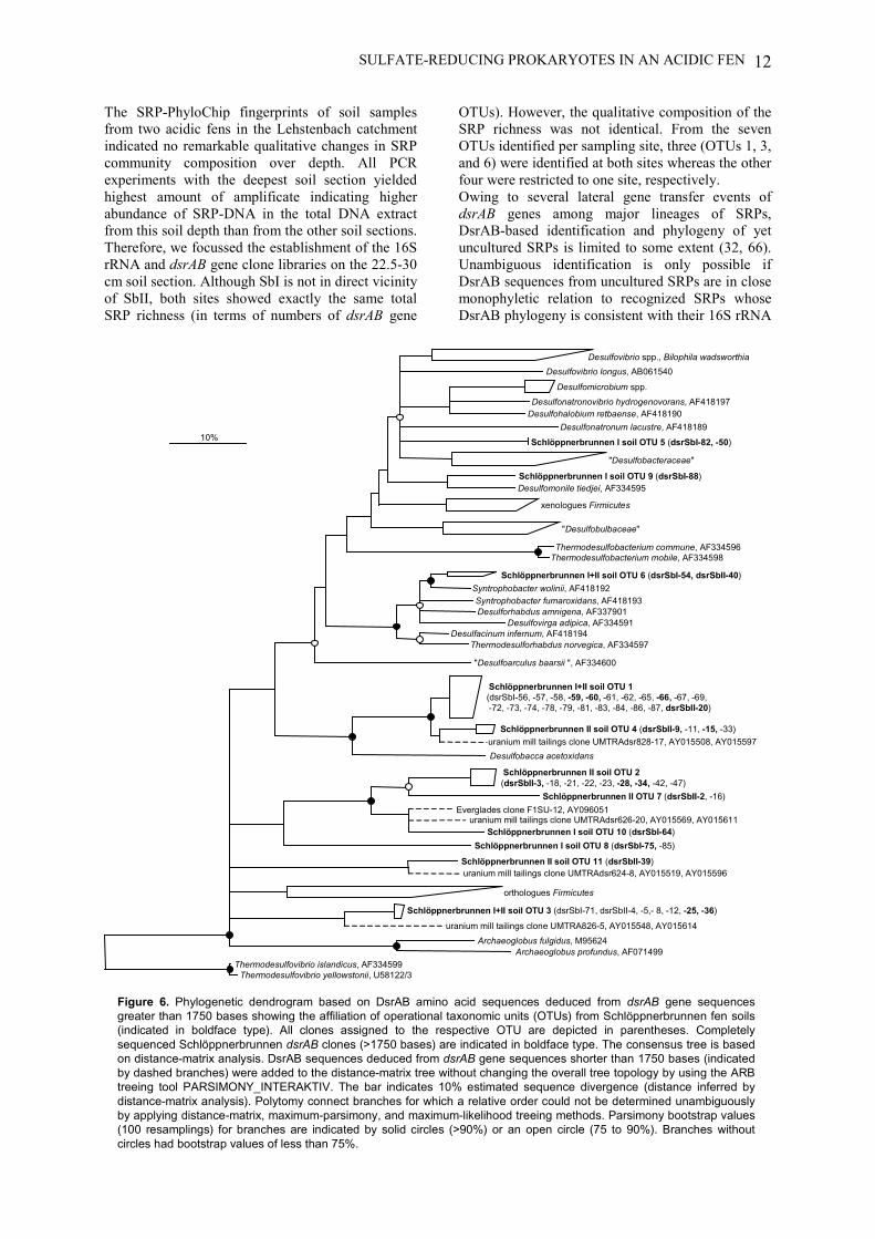
SULFATE-REDUCING PROKARYOTES IN AN ACIDIC FEN 12
The SRP-PhyloChip fingerprints of soil samplesfrom two acidic fens in the Lehstenbach catchmentindicated no remarkable qualitative changes in SRPcommunity composition over depth. All PCRexperiments with the deepest soil section yieldedhighest amount of amplificate indicating higherabundance of SRP-DNA in the total DNA extractfrom this soil depth than from the other soil sections.Therefore, we focussed the establishment of the 16SrRNA and dsrAB gene clone libraries on the 22.5-30cm soil section. Although SbI is not in direct vicinityof SbII, both sites showed exactly the same totalSRP richness (in terms of numbers of dsrAB gene
OTUs). However, the qualitative composition of theSRP richness was not identical. From the sevenOTUs identified per sampling site, three (OTUs 1, 3,and 6) were identified at both sites whereas the otherfour were restricted to one site, respectively.Owing to several lateral gene transfer events ofdsrAB genes among major lineages of SRPs,DsrAB-based identification and phylogeny of yetuncultured SRPs is limited to some extent (32, 66).Unambiguous identification is only possible ifDsrAB sequences from uncultured SRPs are in closemonophyletic relation to recognized SRPs whoseDsrAB phylogeny is consistent with their 16S rRNA
"Desulfobacteraceae"
Desulfomicrobium spp. Desulfonatronovibrio hydrogenovorans, AF418197
Desulfohalobium retbaense, AF418190
Desulfovibrio longus, AB061540 Desulfovibrio spp., Bilophila wadsworthia
Desulfonatronum lacustre, AF418189
Desulfomonile tiedjei, AF334595
xenologues Firmicutes
"Desulfobulbaceae"
Thermodesulfobacterium commune, AF334596 Thermodesulfobacterium mobile, AF334598
Syntrophobacter fumaroxidans, AF418193 Desulforhabdus amnigena, AF337901
Desulfovirga adipica, AF334591
Syntrophobacter wolinii, AF418192
Desulfacinum infernum, AF418194 Thermodesulforhabdus norvegica, AF334597
"Desulfoarculus baarsii ", AF334600
Desulfobacca acetoxidans
Archaeoglobus fulgidus, M95624 Archaeoglobus profundus, AF071499
orthologues Firmicutes
Thermodesulfovibrio islandicus, AF334599 Thermodesulfovibrio yellowstonii, U58122/3
10%
Schlöppnerbrunnen I+II soil OTU 3 (dsrSbI-71, dsrSbII-4, -5,- 8, -12, -25, -36)
Schlöppnerbrunnen I soil OTU 5 (dsrSbI-82, -50)
Schlöppnerbrunnen I soil OTU 9 (dsrSbI-88)
Schlöppnerbrunnen I+II soil OTU 6 (dsrSbI-54, dsrSbII-40)
Schlöppnerbrunnen I+II soil OTU 1(dsrSbI-56, -57, -58, -59, -60, -61, -62, -65, -66, -67, -69, -72, -73, -74, -78, -79, -81, -83, -84, -86, -87, dsrSbII-20)
Schlöppnerbrunnen II soil OTU 4 (dsrSbII-9, -11, -15, -33)
Schlöppnerbrunnen II soil OTU 2(dsrSbII-3, -18, -21, -22, -23, -28, -34, -42, -47)
Schlöppnerbrunnen II OTU 7 (dsrSbII-2, -16)
Schlöppnerbrunnen I soil OTU 10 (dsrSbI-64) Schlöppnerbrunnen I soil OTU 8 (dsrSbI-75, -85)
Schlöppnerbrunnen II soil OTU 11 (dsrSbII-39)
uranium mill tailings clone UMTRA826-5, AY015548, AY015614
uranium mill tailings clone UMTRAdsr624-8, AY015519, AY015596
Everglades clone F1SU-12, AY096051uranium mill tailings clone UMTRAdsr626-20, AY015569, AY015611
uranium mill tailings clone UMTRAdsr828-17, AY015508, AY015597
Figure 6. Phylogenetic dendrogram based on DsrAB amino acid sequences deduced from dsrAB gene sequencesgreater than 1750 bases showing the affiliation of operational taxonomic units (OTUs) from Schlöppnerbrunnen fen soils(indicated in boldface type). All clones assigned to the respective OTU are depicted in parentheses. Completelysequenced Schlöppnerbrunnen dsrAB clones (>1750 bases) are indicated in boldface type. The consensus tree is basedon distance-matrix analysis. DsrAB sequences deduced from dsrAB gene sequences shorter than 1750 bases (indicatedby dashed branches) were added to the distance-matrix tree without changing the overall tree topology by using the ARBtreeing tool PARSIMONY_INTERAKTIV. The bar indicates 10% estimated sequence divergence (distance inferred bydistance-matrix analysis). Polytomy connect branches for which a relative order could not be determined unambiguouslyby applying distance-matrix, maximum-parsimony, and maximum-likelihood treeing methods. Parsimony bootstrap values(100 resamplings) for branches are indicated by solid circles (>90%) or an open circle (75 to 90%). Branches withoutcircles had bootstrap values of less than 75%.
SULFATE-REDUCING PROKARYOTES IN AN ACIDIC FEN13
phylogeny. This held true for fiveSchlöppnerbrunnen OTUs (OTUs 1, 4, 5, 6, and 9)that could be assigned to the class“Deltaproteobacteria”. In contrast, in consequenceof their isolated position within the DsrAB tree theremaining six Schlöppnerbrunnen OTUs (OTUs 2,3, 7, 8, 10, and 11) neither could be identified norcould their exact phylogeny be unambiguouslyinferred. Even more, it could not be clarifiedwhether these six OTUs were of bacterial orarchaeal origin because they displayed similarevolutionary distances to Archaeoglobus spp. and toThermodesulfovibrio spp. (Figure 6). Additionally,some prokaryotes that possess a siroheme sulfitereductase can reduce sulfite but not sulfate (32, 49).Therefore these unknown dsrAB gene sequences donot have to originate coercively from SRPs.Among those OTUs which belonged to the“Deltaproteobacteria” were the Desulfobaccaacetoxidans-related OTUs 1 and 4. The sulfate-reducing bacterium D. acetoxidans has been isolatedfrom granular sludge of a laboratory-scale upflowanaerobic sludge bed reactor fed with acetate and anexcess of sulfate (52). It was the most abundant SRPin this sludge, seemed to be specialized in acetateconsumption, and was able to outcompete acetate-degrading methanogens.As accomplished before for SRPs from estuarinesediments (28), we were able to retrieve congruentphylogenies of DsrAB and 16S rRNA sequencesfrom Syntrophobacter wolinii- and Desulfomonile-related SRPs from the fens. This parallelidentification fortified the phylogenetic position andmetabolic potential of these unculturedSchlöppnerbrunnen bacteria. Stackebrandt andGoebel (64) have proposed that prokaryotes having16S rRNA dissimilarities greater than 3% to eachother represent distinct genomospecies. Thedissimilarities of Syntrophobacter-related 16S rRNAclones retrieved from both sampling sites to the 16SrRNA sequence of Syntrophobacter wolinii rangedfrom 3.6 to 5.5% and most likely represent morethan one different genomospecies becausedissimilarities among those clones ranged from 0.1to 5.2%. The propionate-degrading bacteriumSyntrophobacter wolinii was first isolated insyntrophic anaerobic co-cultures with methanogensor SRPs (10). Syntrophobacter wolinii oxidizespropionate to acetate and CO2. The generatedelectrons are then transferred via hydrogen and/orformate to methanogens that form methane by CO2reduction. This syntrophic lifestyle is reflected bythe fact that propionate oxidation is exergonic onlywhen hydrogen and/or formate are continuouslyremoved by the methanogens. The finding thatSyntrophobacter wolinii is capable of dissimilatorysulfate reduction, in other words to grow slowly onpropionate with sulfate as terminal electron acceptor,led to the first isolation of this bacterium in pureculture (75) and disproved the obligate character ofsyntrophy originally proposed for this bacterium(10).
The Desulfomonile-related 16S rRNA gene clones,solely found in SbI soil, had 16S rRNA sequencedissimilarities to Desulfomonile tiedjei and D.limimaris that varied from 5.2 to 7.5%. Therefore,these sequences at least represent a newgenomospecies within the genus Desulfomonile.Whether the two clone subcluster of Desulfomonile-related 16S rRNA gene sequences from SbI soil(Figure 3) reflect the presence of (i) two strains ofthe same species or (ii) two different species couldnot be elucidated. The most intriguing metabolicfeature of cultivated members of the genusDesulfomonile is their capability of reductivedehalogenation (47, 68). Thus, they play animportant role in the biodegradation of certainenvironmental pollutants such as highly chlorinatedpolychlorinated biphenyls, perchloroethene, andchlorobenzenes (48). Although the PCR-basedmethods used in this study suffer from wellrecognized biases (72) and generally do not allowany quantitative statements about the actual SRPcommunity composition in situ, it should be notedthat Syntrophobacter wolinii- and Desulfomonile-related sequences were among the least abundantonce in the dsrAB gene libraries.
In summary, an extensive phylogenetic SRPinventory was performed for two acidic fen siteswithin the Lehstenbach catchment. The globaldiversity of SRPs known up to now isphylogenetically restricted to four bacterial phylaand one archaeal phylum. Despite this fact, theextent of yet unknown SRP diversity revealed by thepresent study further indicates that the distribution ofthe ability to anaerobically respire sulfate or sulfitefor energy generation purposes within the bacterialand archaeal domains is not yet entirely recognized.Furthermore, the raw data collected here shouldmake it easier for future studies on the microbialecology of the Lehstenbach catchment to focus onquestions such as: What is the influence of seasonalchange on the SRP community composition orwhich SRPs are really active and responsible for thesulfate respectively sulfite turnover in those fens?Additionally, for monitoring successful enrichmentof yet unknown Schlöppnerbrunnen-SRPs, specificdsrAB gene-targeted primers can be designed or theclone-specific 16S rRNA-targeted probes developedin this study can be used in fluorescence in situhybridization assays.
ACKNOWLEDGMENTS
We would like to kindly thank Helga Gaenge,Sibylle Schadhauser, Silvia Weber, Sandra Praßl,and Sonja Trenz for their excellent technicalassistance. Michael Friedrich is acknowledged forproviding the dsrAB gene sequence of Desulfobaccaacetoxidans DSM 11109T (GenBank accessionnumber AY167463). The authors further thankMatthias Horn for critical reading of the manuscript
SULFATE-REDUCING PROKARYOTES IN AN ACIDIC FEN 14
and for helpful discussions and Michael Klein forhelp in dsrAB primer design. This research wassupported by grants of the bmb+f (01 LC 0021subproject 2 in the framework of the BIOLOGprogram to MW and PT BEO 51-0339476 D) andthe Bayerische Forschungsstiftung (Development ofOligonucleotide-DNA-Chips in cooperation withMWG BIOTECH, Ebersberg; project 368/99 toMW).
REFERENCES1. Alewell, C., and M. Gehre. 1999. Patterns of stable S
isotopes in a forested catchment as indicators forbiological S turnover. Biogeochemistry. 47:319-333.
2. Alewell, C., and A. Giesemann. 1996. Sulfatereduction in a forested catchment as indicated by d34Svalues of sulfate in soil solutions and runoff. IsotopesEnvironm. Health Studies. 32:203-210.
3. Alewell, C., and M. Novak. 2001. Spotting zones ofdissimilatory sulfate reduction in a forested catchment:the 34S-35S approach. Environ. Pollut. 112:369-377.
4. Alm, E. W., D. B. Oerther, N. Larsen, D. A. Stahl,and L. Raskin. 1996. The oligonucleotide probedatabase. Appl. Environ. Microbiol. 62:3557-3559.
5. Altschul, S. F., W. Gish, W. Miller, E. W. Myers,and D. J. Lipman. 1990. Basic local alignment searchtool. J. Mol. Biol. 215:403-410.
6. Amann, R. I., W. Ludwig, and K.-H. Schleifer.1995. Phylogenetic identification and in situ detectionof individual microbial cells without cultivation.Microbiol. Rev. 59:143-169.
7. Barns, S. M., C. F. Delwiche, J. D. Palmer, and N.R. Pace. 1996. Perspectives on archaeal diversity,thermophily and monophyly from environmentalrRNA sequences. Proc. Natl. Acad. Sci. USA.93:9188-9193.
8. Benson, D. A., I. Karsch-Mizrachi, D. J. Lipman, J.Ostell, B. A. Rapp, and D. L. Wheeler. 2002.GenBank. Nucleic Acids Res. 30:17-20.
9. Blodau, C., C. L. Roehm, and T. R. Moore. 2002.Iron, sulfur, and dissolved carbon dynamics in anorthern peatland. Arch. Hydrobiol. 154:561-583.
10. Boone, D. R., and M. P. Bryant. 1980. Propionate-degrading bacterium, Syntrophobacter wolinii sp. nov.gen. nov., from methanogenic ecosystems. Appl.Environ. Microbiol. 40:626-632.
11. Castro, H., K. R. Reddy, and A. Ogram. 2002.Composition and function of sulfate-reducingprokaryotes in eutrophic and pristine areas of theFlorida Everglades. Appl. Environ. Microbiol.68:6129-6137.
12. Chang, Y. J., A. D. Peacock, P. E. Long, J. R.Stephen, J. P. McKinley, S. J. Macnaughton, A. K.Hussain, A. M. Saxton, and D. C. White. 2001.Diversity and characterization of sulfate-reducingbacteria in groundwater at a uranium mill tailings site.Appl. Environ. Microbiol. 67:3149-3160.
13. Cottrell, M. T., and S. C. Cary. 1999. Diversity ofdissimilatory bisulfite reductase genes of bacteriaassociated with the deep-sea hydrothermal ventpolychaete annelid Alvinella pompejana. Appl.Environ. Microbiol. 65:1127-1132.
14. Dubilier, N., C. Mulders, T. Ferdelman, D. de Beer,A. Pernthaler, M. Klein, M. Wagner, C. Erseus, F.Thiermann, J. Krieger, O. Giere, and R. Amann.2001. Endosymbiotic sulphate-reducing and sulphide-oxidizing bacteria in an oligochaete worm. Nature.411:298-302.
15. Eder, W., W. Ludwig, and R. Huber. 1999. Novel16S rRNA gene sequences retrieved from highly salinebrine sediments of kebrit deep, red Sea. Arch.Microbiol. 172:213-218.
16. Fortin, D., M. Roy, J.-P. Rioux, and P.-J. Thibault.2000. Occurence of sulfate-reducing bacteria under a
wide range of physico-chemical conditions in Au andCu-Zn mine tailings. FEMS Microbiol. Ecol. 33:197-208.
17. Friedrich, M. W. 2002. Phylogenetic analysis revealsmultiple lateral transfers of adenosine-5'-phosphosulfate reductase genes among sulfate-reducing microorganisms. J. Bacteriol. 184:278-289.
18. Garrity, G. M., and J. G. Holt. 2001. The road mapto the manual, p. 119-166. In G. M. Garrity (ed.),Bergey's manual of systematic bacteriology, 2nd ed,vol. 1. Springer, New York.
19. Giovannoni, S. J., T. D. Mullins, and K. G. Field.1995. Microbial diversity in oceanic systems: rRNAapproaches to the study of unculturable microbes. In J.Joint (ed.), Molecular Ecology of Aquatic Microbes,vol. G38. Springer-Verlag, Berlin, Heidelberg.
20. Griffiths, R. I., A. S. Whiteley, A. G. O'Donnell,and M. J. Bailey. 2000. Rapid method forcoextraction of DNA and RNA from naturalenvironments for analysis of ribosomal DNA- andrRNA-based microbial community composition. Appl.Environ. Microbiol. 66:5488-5491.
21. Harms, G., K. Zengler, R. Rabus, F. Aeckersberg,D. Minz, R. Rossello-Mora, and F. Widdel. 1999.Anaerobic oxidation of o-xylene, m-xylene, andhomologous alkylbenzenes by new types of sulfate-reducing bacteria. Appl. Environ. Microbiol. 65:999-1004.
22. Heck, K. L., G. van Belle, and D. Simberloff. 1975.Explicit calculation of the rarefaction diversitymeasurement and the determination of sufficientsample size. Ecology. 56:1459-1461.
23. Höpner, T. 1981. Design and use of a diffusionsampler for interstitial water from fine grained sample.Environ. Technol. Lett. 2:187-196.
24. Horn, M. A., C. Matthies, K. Küsel, A. Schramm,and H. L. Drake. 2003. Hydrogenotrophicmethanogenesis by moderately acid-tolerantmethanogens of a methane-emitting acidic peat. Appl.Environ. Microbiol. in press.
25. Hugenholtz, P., B. M. Goebel, and N. R. Pace. 1998.Impact of culture-independent studies on the emergingphylogenetic view of bacterial diversity. J. Bacteriol.180:4765-4774.
26. Jørgensen, B. B. 1982. Mineralization of organicmatter in the sea-bed - the role of sulphate reduction.Nature. 296:643–645.
27. Jørgensen, B. B. 1977. The sulfur cycle of a coastalmarine sediment (Limfjorden, Denmark). Limnol.Oceanogr. 22:814–832.
28. Joulian, C., N. B. Ramsing, and K. Ingvorsen. 2001.Congruent phylogenies of most common small-subunitrRNA and dissimilatory sulfite reductase genesequences retrieved from estuarine sediments. Appl.Environ. Microbiol. 67:3314-3318.
29. Juretschko, S., A. Loy, A. Lehner, and M. Wagner.2002. The microbial community composition of anitrifying-denitrifying activated sludge from anindustrial sewage treatment plant analyzed by the full-cycle rRNA approach. Syst. Appl. Microbiol. 25:84-99.
30. Juretschko, S., G. Timmermann, M. Schmid, K. H.Schleifer, A. Pommerening-Roser, H. P. Koops, andM. Wagner. 1998. Combined molecular andconventional analyses of nitrifying bacterium diversityin activated sludge: Nitrosococcus mobilis andNitrospira- like bacteria as dominant populations.Appl. Environ. Microbiol. 64:3042-3051.
31. Kane, M. D., L. K. Poulsen, and D. A. Stahl. 1993.Monitoring the enrichment and isolation of sulfate-reducing bacteria by using oligonucleotidehybridization probes designed from environmentallyderived 16S rRNA sequences. Appl. Environ.Microbiol. 59:682-686.
32. Klein, M., M. Friedrich, A. J. Roger, P.Hugenholtz, S. Fishbain, H. Abicht, L. L. Blackall,D. A. Stahl, and M. Wagner. 2001. Multiple lateral
SULFATE-REDUCING PROKARYOTES IN AN ACIDIC FEN15
transfers of dissimilatory sulfite reductase genesbetween major lineages of sulfate-reducingprokaryotes. J. Bacteriol. 183:6028-6035.
33. Kovárová, M., and P. Dráber. 2000. New specificityand yield enhancer of polymerase chain reactions.Nucleic Acids Res. 28:E70.
34. Küsel, K., and H. L. Drake. 1995. Effect ofenvironmental parameters on the formation andturnover of acetate in forest soils. Appl. Environ.Microbiol. 61:3667-3675.
35. Kuske, C. R., S. M. Barns, and J. D. Busch. 1997.Diverse uncultivated bacterial groups from soils of thearid southwestern United States that are present inmany geographic regions. Appl. Environ. Microbiol.63:3614-3621.
36. Lehman, R. M., F. F. Roberto, D. Earley, D. F.Bruhn, S. E. Brink, S. P. O'Connell, M. E.Delwiche, and F. S. Colwell. 2001. Attached andunattached bacterial communities in a 120-metercorehole in an acidic, crystalline rock aquifer. Appl.Environ. Microbiol. 67:2095-2106.
37. Leu, J.-Y., C. P. McGovern-Traa, A. J. R. Porter,W. J. Harris, and W. A. Hamilton. 1998.Identification and phylogenetic analysis ofthermophilic sulfate-reducing bacteria in oil fieldsamples by 16S rDNA gene cloning and sequencing.Anaerobe. 4:165-174.
38. Li, J.-H., K. J. Purdy, S. Takii, and H. Hayashi.1999. Seasonal changes in ribosomal RNA of sulfate-reducing bacteria and sulfate reducing activity in afreshwater lake sediment. FEMS Microbiol. Ecol.28:31-39.
39. Llobet-Brossa, E., R. Rossello-Mora, and R.Amann. 1998. Microbial community composition ofWadden sea sediments as revealed by fluorescence insitu hybridization. Appl. Environ. Microbiol. 64:2691-2696.
40. Lovley, D. R., and M. J. Klug. 1983. Sulfate reducerscan outcompete methanogens at freshwater sulfateconcentrations. Appl. Environ. Microbiol. 45:187-192.
41. Loy, A., H. Daims, and M. Wagner. 2002. Activatedsludge-Molecular techniques for determiningcommunity composition, p. 26-43. In G. Bitton (ed.),The Encyclopedia of Environmental Microbiology.John Wiley & Sons, Inc., New York.
42. Loy, A., M. Horn, and M. Wagner. 2003. probeBase:an online resource for rRNA-targeted oligonucleotideprobes. Nucleic Acids Res. 31:514-516.
43. Loy, A., A. Lehner, N. Lee, J. Adamczyk, H. Meier,J. Ernst, K.-H. Schleifer, and M. Wagner. 2002.Oligonucleotide microarray for 16S rRNA gene-baseddetection of all recognized lineages of sulfate-reducingprokaryotes in the environment. Appl. Environ.Microbiol. 68:5064-5081.
44. Ludwig, W., S. H. Bauer, M. Bauer, I. Held, G.Kirchhof, R. Schulze, I. Huber, S. Spring, A.Hartmann, and K. H. Schleifer. 1997. Detection andin situ identification of representatives of a widelydistributed new bacterial phylum. FEMS Microbiol.Lett. 153:181-190.
45. Ludwig, W., O. Strunk, S. Klugbauer, N.Klugbauer, M. Weizenegger, J. Neumaier, M.Bachleitner, and K. H. Schleifer. 1998. Bacterialphylogeny based on comparative sequence analysis.Electrophoresis. 19:554-568.
46. Minz, D., J. L. Flax, S. J. Green, G. Muyzer, Y.Cohen, M. Wagner, B. E. Rittmann, and D. A.Stahl. 1999. Diversity of sulfate-reducing bacteria inoxic and anoxic regions of a microbial matcharacterized by comparative analysis of dissimilatorysulfite reductase genes. Appl. Environ. Microbiol.65:4666-4671.
47. Mohn, W. W., and K. J. Kennedy. 1992. Reductivedehalogenation of chlorophenols by Desulfomoniletiedjei DCB-1. Appl. Environ. Microbiol. 58:1367-1370.
48. Mohn, W. W., and J. M. Tiedje. 1992. Microbialreductive dehalogenation. Microbiol. Rev . 56:482-507.
49. Molitor, M., C. Dahl, I. Molitor, U. Schäfer, N.Speich, R. Huber, R. Deutzmann, and H. G.Trüper. 1998. A dissimilatory sirohaem-sulfite-reductase-type protein from the hyperthermophilicarchaeon Pyrobaculum islandicum. Microbiology.144:529-541.
50. Nakagawa, T., S. Hanada, A. Maruyama, K.Marumo, T. Urabe, and M. Fukui. 2002.Distribution and diversity of thermophilic sulfate-reducing bacteria within a Cu-Pb-Zn mine (Toyoha,Japan). FEMS Microbiol. Ecol. 41:199-209.
51. Novak, M., R. K. Wieder, and W. R. Schell. 1994.Sulfur during early diagenesis in Sphagnumpeat:Insights from delta34S ratio profiles in 210Pb-dated peat cores. Limnol. Oceanogr. 39:1172-1185.
52. Oude Elferink, S. J., W. M. Akkermans-van Vliet,J. J. Bogte, and A. J. Stams. 1999. Desulfobaccaacetoxidans gen. nov., sp. nov., a novel acetate-degrading sulfate reducer isolated from sulfidogenicgranular sludge. Int. J. Syst. Bacteriol. 49:345-350.
53. Perez-Jimenez, J. R., L. Y. Young, and L. J.Kerkhof. 2001. Molecular characterization of sulfate-reducing bacteria in anaerobic hydrocarbon-degradingconsortia and pure cultures using the dissimilatorysulfite reductase (dsrAB) genes. FEMS Microbiol.Ecol. 35:145-150.
54. Purkhold, U., A. Pommering-Röser, S. Juretschko,M. C. Schmid, H.-P. Koops, and M. Wagner. 2000.Phylogeny of all recognized species of ammoniaoxidizers based on comparative 16S rRNA and amoAsequence analysis: implications for molecular diversitysurveys. Appl. Environ. Microbiol. 66:5368-5382.
55. Rabus, R., M. Fukui, H. Wilkes, and F. Widdle.1996. Degradative capacities and 16S rRNA-targetedwhole-cell hybridization of sulfate-reducing bacteria inan anaerobic enrichment culture utilizingalkylbenzenes from crude oil. Appl. Environ.Microbiol. 62:3605-3613.
56. Ravenschlag, K., K. Sahm, and R. Amann. 2001.Quantitative molecular analysis of the microbialcommunity in marine arctic sediments (Svalbard).Appl. Environ. Microbiol. 67:387-395.
57. Ravenschlag, K., K. Sahm, C. Knoblauch, B. B.Jørgensen, and R. Amann. 2000. Communitystructure, cellular rRNA content, and activity ofsulfate-reducing bacteria in marine arctic sediments.Appl. Environ. Microbiol. 66:3592-3602.
58. Sahm, K., C. Knoblauch, and R. Amann. 1999.Phylogenetic affiliation and quantification ofpsychrophilic sulfate- reducing isolates in marinearctic sediments. Appl. Environ. Microbiol. 65:3976-3981.
59. Sahm, K., B. J. MacGregor, B. B. Jørgensen, and D.A. Stahl. 1999. Sulphate reduction and verticaldistribution of sulphate-reducing bacteria quantified byrRNA slot-blot hybridization in a coastal marinesediment. Environ. Microbiol. 1:65-74.
60. Sanger, F., S. Nicklen, and A. R. Coulson. 1977.DNA sequencing with chain-terminating inhibitors.Proc. Natl. Acad. Sci. 74:5463-5467.
61. Scheid, D., and S. Stubner. 2001. Structure anddiversity of Gram-negative sulfate-reducing bacteriaon rice roots. FEMS Microbiol. Ecol. 36:175-183.
62. Schramm, A., C. M. Santegoeds, H. K. Nielsen, H.Ploug, M. Wagner, M. Pribyl, J. Wanner, R.Amann, and D. de Beer. 1999. On the occurrence ofanoxic microniches, denitrification, and sulfatereduction in aerated activated sludge. Appl. Environ.Microbiol. 65:4189-4196.
63. Singleton, D. R., M. A. Furlong, S. L. Rathbun, andW. B. Whitman. 2001. Quantitative comparisons of16S rRNA gene sequence libraries from environmentalsamples. Appl. Environ. Microbiol. 67:4374-4376.
SULFATE-REDUCING PROKARYOTES IN AN ACIDIC FEN 16
64. Stackebrandt, E., and B. M. Goebel. 1994.Taxonomic note: a place for DNA-DNA reassociationand 16S rRNA sequence analysis in the present speciesdefinition in bacteriology. Int. J. Syst. Bacteriol.44:846-849.
65. Stackebrandt, E., and F. A. Rainey. 1995. Partialand complete 16S rDNA sequences, their use ingeneration of 16S rDNA phylogenetic trees and theirimplications in molecular ecological studies, p. 1-17.In A. D. L. Akkermans, J. D. van Elsas, and F. J. deBruijn (ed.), Molecular Microbial Ecology Manual,vol. 3.1.1. Kluwer Academic Publishers, Dortrecht.
66. Stahl, D. A., S. Fishbain, M. Klein, B. J. Baker, andM. Wagner. 2002. Origins and diversification ofsulfate-respiring microorganisms. Antonie vanLeeuwenhoek. 81:189-195.
67. Strimmer, K., and A. von Haeseler. 1996. Quartetpuzzling: A quartet maximum likelihood method forreconstructing tree topologies. Mol. Biol. Evol.13:964-969.
68. Sun, B., J. R. Cole, and J. M. Tiedje. 2001.Desulfomonile limimaris sp. nov., an anaerobicdehalogenating bacterium from marine sediments. Int.J. Syst. Evol. Microbiol. 51:365-371.
69. Thomsen, T. R., K. Finster, and N. B. Ramsing.2001. Biogeochemical and molecular signatures ofanaerobic methane oxidation in a marine sediment.Appl. Environ. Microbiol. 67:1646-1656.
70. Tipper, J. C. 1979. Rarefaction and rarefiction - theuse and abuse of a method in paleontology.Paleobiology. 5:423-434.
71. Ulrich, G. A., L. R. Krumholz, and J. M. Suflita.1997. A rapid and simple method for estimating sulfatereduction activity and quantifying inorganic sulfides.Appl. Environ. Microbiol. 63:1627-1630.
72. von Wintzingerode, F., U. B. Göbel, and E.Stackebrandt. 1997. Determination of microbialdiversity in environmental samples: pitfalls of PCR-based rRNA analysis. FEMS Microbiol. Rev. 21:213-229.
73. Voordouw, G., J. K. Voordouw, R. R. Karkhoff-Schweizer, P. M. Fedorak, and D. W. S. Westlake.1991. Reverse sample genome probing, a newtechnique for identification of bacteria inenvironmental samples by DNA hybridization, and itsapplication to the identification of sulfate-reducingbacteria in oil field samples. Appl. Environ. Microbiol.57:3070-3078.
74. Wagner, M., A. J. Roger, J. L. Flax, G. A.Brusseau, and D. A. Stahl. 1998. Phylogeny ofdissimilatory sulfite reductases supports an early originof sulfate respiration. J. Bacteriol. 180:2975-2982.
75. Wallrabenstein, C., E. Hauschild, and B. Schink.1994. Pure culture and cytological properties ofSyntrophobacter wolinii. FEMS Microbiol. Lett.123:249-254.
76. Yavitt, J. B., G. E. Lang, and R. K. Wieder. 1987.Control of carbon mineralization to CH4 and CO2 inanaerobic, Sphagnum derived peat from Big Run, WestVirginia. Biogeochem. 4:141-157.
LIST OF PUBLICATIONS/ORAL AND POSTER PRESENTATIONS
141
LIST OF PUBLICATIONSContributions to the manuscripts presented in this dissertation are listed in parentheses
1. Juretschko S, Loy A, Lehner A, and Wagner M [2002] The microbial community
composition of a nitrifying-denitrifying activated sludge from an industrial sewage
treatment plant analyzed by the full-cycle rRNA approach. Syst Appl Microbiol. 25: 84-
99.
2. Loy A, Daims H, and Wagner M [2002] Activated sludge - Molecular techniques for
determining community composition, p. 26-43. In Bitton G (ed.), The Encyclopedia of
Environmental Microbiology. John Wiley & Sons, Inc., New York.
3. Loy A, Lehner A, Lee N, Adamczyk J, Meier H, Ernst J, Schleifer K-H, and Wagner
M [2002] Oligonucleotide microarray for 16S rRNA gene-based detection of all
recognized lineages of sulfate-reducing prokaryotes in the environment. Appl Environ
Microbiol. 68: 5064-5081.
[concept by M.W. and A.Loy, probe design and experimental work by A.Loy, technical
advice by A.L., J.A., and N.L., programming by H.M. and J.E., writing by M.W. and A.Loy
with editorial help of the co-authors]
4. Wagner M, and Loy A [2002] Bacterial community composition and function in sewage
treatment systems. Curr Opin Biotechnol. 13: 218-227.
5. Wagner M, Loy A, Nogueira R, Purkhold U, Lee N, and Daims H [2002] Microbial
community composition and function in wastewater treatment plants. Antonie Van
Leeuwenhoek. 81: 665-680.
6. Loy A, Horn M, and Wagner M [2003] probeBase: an online resource for rRNA-
targeted oligonucleotide probes. Nucleic Acids Res. 31: 514-516.
[concept by M.H. and A.Loy, programming by M.H., acquisition of data by A.Loy and
M.H., writing by A.Loy and M.H. with editorial help of M.W.]
LIST OF PUBLICATIONS/ORAL AND POSTER PRESENTATIONS
142
7. Loy A, Küsel K, Lehner A, Drake HL, and Wagner M [2003] Diversity of sulfate-
reducing prokaryotes in an acidic fen: Comparison of 16S rRNA gene-based
oligonucleotide microarray hybridization with dissimilatory (bi)sulfite reductase gene
surveys. Submitted to Appl Environ Microbiol.
[concept A.Loy, K.K., and M.W., microarray experiments and comparative sequence
analyses of 16S rRNA and dsrAB genes by A.Loy, biogeochemical analyses by K.K.,
writing A.Loy with editorial help of the co-authors]
Parts of this thesis have been presented at national and international meetings
ORAL PRESENTATIONSSpeaker is underlined
Lehner A, Loy A, Adamczyk J, Lee N, Schöpfer A, and Wagner M [2001] Development
of phylogenetic DNA Micro Arrays (PhyloChips) - Rapid tools for microbial community
profiling and functional analysis. Jahrestagung der Vereinigung für Allgemeine und
Angewandte Mikrobiologie e.V. (VAAM). Oldenburg, Germany, 25.-28. March 2001.
Lehner A, Loy A, Adamczyk J, Lee N, Schöpfer A, Wagner M [2001] Development and
application of phylogenetic DNA-Microarrays (PhyloChips) for microbial community
profiling and functional analysis. Ninth International Conference on Microbial Ecology
(ISME-9). Amsterdam, Netherlands. 26.-31. August 2001.
Loy A, Adamczyk J, Lehner A, Lee N, Stubner S, Küsel K, Drake H, and Wagner M
[2002] The SRP-PhyloChip - a 16S rRNA gene-based oligonucleotide microarray as
biodiversity screening tool for sulfate-reducing prokaryotes in the environment. Jahrestagung
der Vereinigung für Allgemeine und Angewandte Mikrobiologie e.V. (VAAM). Göttingen,
Germany. 24.-27. March 2002.
Loy A, Lehner A, Lee N, and Wagner M [2002] Assessing diversity of sulfate-reducing
prokaryotes - combined 16S rRNA gene and dsrAB approach. 4th International meeting on
Novel Techniques in Microbial Ecology (INTIME). Blokhus, Denmark, 14.-16. May 2002.
LIST OF PUBLICATIONS/ORAL AND POSTER PRESENTATIONS
143
Loy A, Lehner A, Lee N, Stubner S, Küsel K, Drake H, and Wagner M [2002] The SRP-
PhyloChip – a 16S rRNA gene-based oligonucleotide microarray as biodiversity screening
tool for sulfate-reducing prokaryotes in the environment. Joint meeting of the three divisions
of International Union of Microbiological Societies (IUMS) - The world of microbes. Paris,
France. 27. July - 01. August 2002.
POSTER PRESENTATIONS
Adamczyk J, Beier C, Daims H, Horn M, Juretschko S, Klein M, Lee N, Lehner A, Loy
A, Purkhold U, Schlatter B, Schmid M, Schmid M, Walcher M, and Wagner M [2000]
Molecular microbial ecology - exploring the unseen majority on planet earth. BioLog 2000, 2.
Münchner Kongress für Biowissenschaften. München, Germany, November 2000.
Loy A, Adamczyk J, Lehner A, and Wagner M [2001] Biodiversity of nitrifying and
sulfate-reducing prokaryotes. BIOLOG status report 2001 - German programme on
biodiversity and global change. Bonn, Germany. 05.-07. December 2002.
Horn M, Loy A, Ludwig W, and Wagner M [2002] ProbeBase.net - an online resource for
rRNA-targeted oligonucleotide probes. Joint meeting of the three divisions of International
Union of Microbiological Societies (IUMS) - The world of microbes. Paris, France. 27. July -
01. August 2002.
Loy A, Lehner A, Adamczyk J, Schulz C, and Wagner M [2002] rRNA-targeted
oligonucleotide microarrays for identification of microorganisms in environmental and
clinical samples. Late Summer Workshop, Pathogenomics - from Genes to Function.
Würzburg, Germany. 29. September - 02. October 2002.

DANKSAGUNG
Diese Arbeit entstand im Zeitraum Februar 2000 bis März 2003 am Lehrstuhl für
Mikrobiologie der Technischen Universität München. An dieser Stelle möchte ich mich, wie
üblich, bei all denjenigen Personen bedanken, die durch ihr aktives Zutun und/oder ihre
indirekte Unterstützung an diesem Unterfangen beteiligt waren.
Herrn Prof. Dr. Karl-Heinz Schleifer gilt mein herzlicher Dank für die Möglichkeit diese
Arbeit an seinem Lehrstuhl durchzuführen, die vielseitige Unterstützung und die stets
freundliche Arbeitsatmosphäre.
Mehr als nur ein einfaches Dankeschön möchte ich Prof. Dr. Michael Wagner aussprechen.
Unkonventionell, anspruchsvoll, menschlich, verständnisvoll sind nur einige wenige
Attribute, die seine Begleitung meiner Arbeit nur unzureichend beschreiben.
Desweiteren gilt mein besonderer Dank Dr. Kirsten Küsel, Kerstin Bischof und Prof. Dr.
Harold Drake vom BITÖK in Bayreuth für die hervorragende Zusammenarbeit am Projekt
„Schlöppnerbrunnen”.
Dr. Harald Meier gilt immer noch mein Dank für die Einführung in die Welt der Wissenschaft
und der Mikroben und desweiteren für die Zusammenarbeit an der Entwicklung des
„ChipChecker“ Programms. Über den Horizont der Wissenschaft hinaus werden mir die
gemeinsamen, zuletzt leider nicht mehr so zahlreichen Abende und Unterhaltungen in
Erinnerung bleiben.
Meinem langjährigen Studien- und Doktorarbeitskollegen André Mehlen sage ich ein
einfaches Merci für die gemeinsame, ziemlich unkomplizierte Wohnzeit und wünsche ihm,
dass er sich seine Unbeschwertheit auch in Zukunft bewahrt.
Gesondert bedanken möchte ich mich für die Zusammenarbeit mit Andreas Schöpfer und
Claudia Schulz, die im Rahmen ihrer Diplomarbeiten massgeblich zur Entwicklung des
„Rhodocyclales“-Mikroarrays beigetragen haben.
Ein herzliches Dankeschön auch an Josef Reischenbeck für die fachmännische Fabrikation
der Mikroarrayhybridisierungskammern und an Dr. Vladimir Zverlov für stete Versorgung
mit aktuellsten Audiogenüssen.
Der Spass an der Freude und an der Wissenschaft blieb mir aufgrund der einzigartigen
Atmosphäre in unserer Arbeitsgruppe erhalten. Deren jetzige und ehemalige Mitglieder
sollten an dieser Stelle nicht unerwähnt bleiben: Justyna Adamczyk, Cora Beier, Lotte
Bjerrum, Andreas Brühl, Astrid Collingro, Dr. Holger Daims, Dr. Stefan Juretschko, Michael
Klein, Dr. Natuschka Lee, Dr. Angelika Lehner, Sebastian Lücker, Frank Maixner, Regina
Nogueira, Dr. Uli Purkhold, Dr. Markus Schmid, Dr. Michael Schmid, Stephan Schmitz-
Esser, Tobias Schweinitzer, Kilian Stöcker und Dr. Marion Walcher, sowie Sibylle
Schadhauser, Jutta Elgner und Helga Gänge.
Gefühle in Worte zu packen liegt mir nicht. Deshalb lasse ich das auch an dieser Stelle.
Trotzdem will ich erwähnen, dass ich die Unterstützung durch meine Eltern in jederlei
Hinsicht nicht hoch genug bewerten kann.
Das Gleiche gilt für meine Freundin Sabine Grünaug. Die Tatsache, gemeinsam schwere
Phasen gemeistert zu haben lässt mich unbeschwert in die Zukunft blicken. Ich freu‘ mich
d‘rauf!
Wien, den 25. April 2003