dengue: a continuing global threat
TRANSCRIPT
Dengue is the most important arthropod-borne viral
infection of humans. Worldwide, an estimated 2.5 bil-
lion people are at risk of infection, approximately 975 million of whom live in urban areas in tropical and
sub-tropical countries in Southeast Asia, the Pacific and
the Americas 1 . Transmission also occurs in Africa and
the Eastern Mediterranean, and rural communities are
increasingly being affected. It is estimated that more than 50 million infections occur each year, including 500,000
hospitalizations for dengue haemorrhagic fever, mainly
among children, with the case fatality rate exceeding 5%
in some areas 1–4 .
The annual average number of dengue fever/den-gue haemorrhagic fever (DF/DHF) cases reported to
the World Health Organization (WHO) has increased
dramatically in recent years. For the period 2000–2004,
the annual average was 925,896 cases, almost dou-
ble the figure of 479,848 cases that was reported for the period 1990–1999. In 2001, a record 69 countries
reported dengue activity to WHO and in 2002, the
Region of the Americas alone reported more than
1 million cases. Although there is poor surveillance and
no official reporting of dengue to WHO from coun-tries in the African and Eastern Mediterranean regions,
in 2005–2006 outbreaks of suspected dengue were
recorded in Pakistan, Saudi Arabia, Yemen, Sudan and
Madagascar 1–4 , and a large outbreak of dengue involving
>17,000 cases was documented in the Cape Verde islands in 2009 5 .Travellers from endemic areas might serve as
vehicles for further spread 6–9 . Dengue epidemics can
have a significant economic and health toll. In endemic
countries in Asia and the Americas, the burden of den-
gue is approximately 1,300 disability-adjusted life years
(DALYs) per million population, which is similar to the disease burden of other childhood and tropical diseases,
including tuberculosis, in these regions 10 .
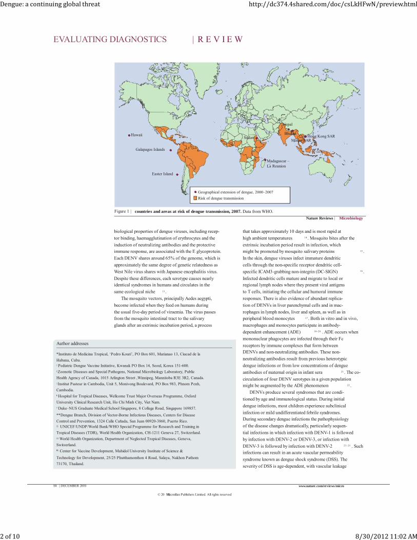
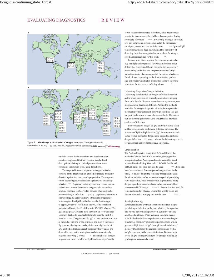
The geographical areas in which dengue transmission
occurs have expanded in recent years (FIG. 1) , and all four
dengue virus serotypes (DENV-1–4) are now circulating in Asia, Africa and the Americas, a dramatically differ-
ent scenario from that which prevailed 20 or 30 years
ago (FIG. 2) . The molecular epidemiology of these sero-
types has been studied in an attempt to understand their
evolutionary relationships 11 . This Review will provide an update on our under-
standing of the pathogenesis of this successful pathogen,
how we diagnose and control infection and the progress
that has been made in vaccine development.
Dengue virus pathogenesisDengue viruses belong to the genus flavivirus within the
Flaviviridae family. DENV-1–4 evolved in non-human
primates from a common ancestor and each entered
the urban cycle independently an estimated 500–1,000 years ago 12 . The virion comprises a spherical particle,
40–50 nm in diameter, with a lipopolysaccharide enve-
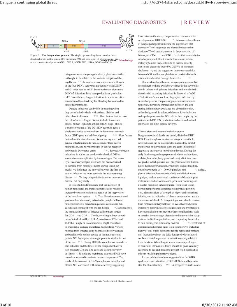
lope. The positive single-strand RNA genome (FIG. 3) ,
which is approximately 11 kb in length, has a single open
reading frame that encodes three structural proteins — the capsid (C), membrane (M) and envelope (E) glyco-
proteins — and seven non-structural proteins (NS1,
NS2A, NS2B, NS3, NS4A, NS4B and NS5). Important
‡‡ UNICEF/UNDP/World Bank/
WHO Special Programme for
Research and Training in
Tropical Diseases (TDR),
World Health Organization,
CH-1211 Geneva 27,
Switzerland.## London School of Hygiene
and Tropical Medicine,
Dept of Clinical Research,
Keppel Street,
London WC1E 7HT, UK.
Correspondence to R.W.P.
e-mail: Rosanna.Peeling@
lshtm.ac.uk
Copyright © WHO, on behalf
of TDR (WHO/TDR) 2010.
doi:10.1038/nrmicro2460
Maria G. Guzman*, Scott B. Halstead ‡ , Harvey Artsob § , Philippe Buchy || ,
Jeremy Farrar ¶ , Duane J. Gubler # , Elizabeth Hunsperger**, Axel Kroeger ‡‡ ,
Harold S. Margolis**, Eric Martínez*, Michael B. Nathan §§ , Jose Luis Pelegrino*,
Cameron Simmons ¶ , Sutee Yoksan ¶¶ and Rosanna W. Peeling ‡‡,##
Abstract | Dengue fever and dengue haemorrhagic fever are important arthropod-borne
viral diseases. Each year, there are ~50 million dengue infections and ~500,000 individuals are
hospitalized with dengue haemorrhagic fever, mainly in Southeast Asia, the Pacific and the
Americas. Illness is produced by any of the four dengue virus serotypes. A global strategy
aimed at increasing the capacity for surveillance and outbreak response, changing
behaviours and reducing the disease burden using integrated vector management in
conjunction with early and accurate diagnosis has been advocated. Antiviral drugs and
vaccines that are currently under development could also make an important contribution to
dengue control in the future.
NATuRE REVIEWS | microbiology DECEMBER 2010 | S7
© 20 Macmillan Publishers Limited. All rights reserved10
Dengue: a continuing global threat http://dc374.4shared.com/doc/csLkHFwN/preview.html
1 of 10 8/30/2012 11:02 AM
Nature Reviews | Microbiology
Galapagos Islands
Easter Island
Hawaii
Madagascar – La Reunion
Hong Kong SARMacao SAR
BhutanSudan
Nepal
Geographical extension of dengue, 2000–2007
Risk of dengue transmission
biological properties of dengue viruses, including recep-
tor binding, haemagglutination of erythrocytes and the
induction of neutralizing antibodies and the protective immune response, are associated with the E glycoprotein.
Each DENV shares around 65% of the genome, which is
approximately the same degree of genetic relatedness as
West Nile virus shares with Japanese encephalitis virus.
Despite these differences, each serotype causes nearly identical syndromes in humans and circulates in the
same ecological niche 13 .
The mosquito vectors, principally Aedes aegypti,
become infected when they feed on humans during
the usual five-day period of viraemia. The virus passes from the mosquito intestinal tract to the salivary
glands after an extrinsic incubation period, a process
that takes approximately 10 days and is most rapid at
high ambient temperatures 14 . Mosquito bites after the
extrinsic incubation period result in infection, which might be promoted by mosquito salivary proteins 15 .
In the skin, dengue viruses infect immature dendritic
cells through the non-specific receptor dendritic cell-
specific ICAM3-grabbing non-integrin (DC-SIGN) 16 .
Infected dendritic cells mature and migrate to local or regional lymph nodes where they present viral antigens
to T cells, initiating the cellular and humoral immune
responses. There is also evidence of abundant replica-
tion of DENVs in liver parenchymal cells and in mac-
rophages in lymph nodes, liver and spleen, as well as in peripheral blood monocytes 17 . Both in vitro and in vivo,
macrophages and monocytes participate in antibody-
dependent enhancement (ADE) 18–20 . ADE occurs when
mononuclear phagocytes are infected through their Fc
receptors by immune complexes that form between DENVs and non-neutralizing antibodies. These non-
neutralizing antibodies result from previous heterotypic
dengue infections or from low concentrations of dengue
antibodies of maternal origin in infant sera 21 . The co-
circulation of four DENV serotypes in a given population might be augmented by the ADE phenomenon 22 .
DENVs produce several syndromes that are condi-
tioned by age and immunological status. During initial
dengue infections, most children experience subclinical
infection or mild undifferentiated febrile syndromes. During secondary dengue infections the pathophysiology
of the disease changes dramatically, particularly sequen-
tial infections in which infection with DENV-1 is followed
by infection with DENV-2 or DENV-3, or infection with
DENV-3 is followed by infection with DENV-2 23–25 . Such infections can result in an acute vascular permeability
syndrome known as dengue shock syndrome (DSS). The
severity of DSS is age-dependent, with vascular leakage
Figure 1 | countries and areas at risk of dengue transmission, 2007. Data from WHO.
Author addresses
*Instituto de Medicina Tropical, ‘Pedro Kouri’, PO Box 601, Marianao 13, Ciucad de la
Habana, Cuba.‡ Pediatric Dengue Vaccine Initiative, Kwanak PO Box 14, Seoul, Korea 151-600.§Zoonotic Diseases and Special Pathogens, National Microbiology Laboratory, Public
Health Agency of Canada, 1015 Arlington Street ,Winnipeg, Mannitoba R3E 3R2, Canada.|| Institut Pasteur in Cambodia, Unit 5, Monivong Boulevard, PO Box 983, Phnom Penh,
Cambodia.¶ Hospital for Tropical Diseases, Wellcome Trust Major Overseas Programme, Oxford
University Clinical Research Unit, Ho Chi Minh City, Viet Nam.# Duke–NUS Graduate Medical School Singapore, 8 College Road, Singapore 169857.
**Dengue Branch, Division of Vector-Borne Infectious Diseases, Centers for Disease
Control and Prevention, 1324 Calle Cañada, San Juan 00920-3860, Puerto Rico.‡‡ UNICEF/UNDP/World Bank/WHO Special Programme for Research and Training in
Tropical Diseases (TDR), World Health Organization, CH-1211 Geneva 27, Switzerland.§§ World Health Organization, Department of Neglected Tropical Diseases, Geneva,
Switzerland.¶¶ Center for Vaccine Development, Mahidol University Institute of Science &
Technology for Development, 25/25 Phutthamonthon 4 Road, Salaya, Nakhon Pathom
73170, Thailand.
EVALUATING DIAGNOSTICS | R E V I E W
S8 | DECEMBER 2010 www.nature.com/reviews/micro
© 20 Macmillan Publishers Limited. All rights reserved10
Dengue: a continuing global threat http://dc374.4shared.com/doc/csLkHFwN/preview.html
2 of 10 8/30/2012 11:02 AM
NS4B
NS4A
Nature Reviews | Microbiology
C M E NS1 NS3 NS5
3′ UTR
5′ UTR
NS2B
NS2A
being most severe in young children, a phenomenon that
is thought to be related to the intrinsic integrity of the
capillaries 26,27 . In adults, primary infections with each of the four DENV serotypes, particularly with DENV-1
and -3, often results in DF. Some outbreaks of primary
DENV-2 infections have been predominantly subclini-
cal 24 . Nonetheless, dengue infections in adults are often
accompanied by a tendency for bleeding that can lead to severe haemorrhages.
Dengue infections can be life-threatening when
they occur in individuals with asthma, diabetes and
other chronic diseases 28–30 . Host factors that increase
the risk of severe dengue disease include female sex, several human leukocyte antigen (HLA) class I alleles,
a promoter variant of the DC-SIGN receptor gene, a
single-nucleotide polymorphism in the tumour necrosis
factor (TNF) gene and AB blood group 31–36 . Host factors
that reduce the risk of severe disease during a second dengue infection include race, second or third degree
malnutrition, and polymorphisms in the Fcγ receptor
and vitamin D receptor genes 37–42 . Secondary dengue
infections in adults can produce the classical DSS or
severe disease complicated by haemorrhages. The sever-ity of secondary dengue infections has been observed
to increase from month-to-month during island out-
breaks 43 ; the longer the interval between the first and
second infection the more severe is the accompanying
disease 44,45 . Tertiary dengue infections can cause severe disease, but only rarely 25 .
In vitro studies demonstrate that the infection of
human monocytes and mature dendritic cells results in
increased virus replication as a result of the suppression
of the interferon system 45 . Type I interferon-associated genes are less abundantly activated in peripheral blood
mononuclear cells taken from patients with severe den-
gue disease compared with milder disease 46 . Subsequently,
the increased number of infected cells present targets
for CD4 + and CD8 + T cells, resulting in large quanti-ties of interleukin (IL)-10, IL-2, interferon (IFN)-γ and
TNF that, singly or in combination, might contribute
to endothelial damage and altered haemostasis. Virions
released from infected cells might also directly damage
endothelial cells and the uptake of the non-structural protein NS1 by hepatocytes might promote viral infection
of the liver 47–49 . During DHF, the complement cascade is
also activated and the levels of the complement activa-
tion products C3a and C5a correlate with the severity
of illness 49 . Soluble and membrane-associated NS1 have been demonstrated to activate human complement. The
levels of the terminal SC5b–9 complement complex and
plasma NS1 correlated with disease severity, suggesting
links between the virus, complement activation and the
development of DHF/DSS 50 . Alternative hypotheses
of dengue pathogenesis include the suggestions that secondary T-cell responses are blunted because stim-
ulation of T-cell memory results in the production of
heterotypic CD4 + and CD8 + cells that have a dimin-
ished capacity to kill but nonetheless release inflam-
matory cytokines that contribute to disease severity 51 ; that severe disease is caused by DENVs of increased
virulence 52 ; and the suggestion that cross-reactivity
between NS1 and human platelets and endothelial cells
raises antibodies that damage these cells 53 .
One working hypothesis of dengue pathogenesis that is consistent with the available evidence is that severe dis-
ease in infants with primary infections and in older indi-
viduals with secondary infections is the result of ADE
of infection of mononuclear phagocytes. Infection by
an antibody–virus complex suppresses innate immune responses, increasing intracellular infection and gen-
erating inflammatory cytokines and chemokines that,
collectively, result in enhanced disease. Liver infection
and a pathogenic role for NS1 add to the complexity. In
patients with DF, IFN production and activated natural killer cells can limit disease severity.
Clinical signs and immunological response
Dengue-associated deaths are usually linked to DHF/
DSS. Even though no vaccines or drugs are available, severe disease can be successfully managed by careful
monitoring of the warning signs and early initiation of
aggressive intravenous rehydration therapy. During the
early febrile stage (the symptoms of which include fever,
malaise, headache, body pains and rash), clinicians can-not predict which patients will progress to severe disease.
Later, during defervescence, symptoms such as bleeding,
thrombocytopenia of <100,000 platelets mm −3 , ascites,
pleural effusion, haematocrit >20% and clinical warn-
ing signs, such as severe and continuous abdominal pain, restlessness and/or somnolence, persistent vomiting and
a sudden reduction in temperature (from fever to sub-
normal temperature) associated with profuse perspira-
tion, adynamia (loss of strength or vigor) and sometimes
fainting, can be indicative of plasma extravasation and the imminence of shock. At this point, patients should receive
fluid replacement (crystalloids) to avoid haemodynamic
instability, narrowness of blood pressure and hypotension.
Early resuscitation can prevent other complications, such
as massive haemorrhage, disseminated intravascular coag-ulation, multiple organ failure, and respiratory failure due
to non-cardiogenic pulmonary oedema 54–57 . Treatment of
uncomplicated dengue cases is only supportive, including
plenty of oral fluids during the febrile period and paraceta-
mol (acetaminophen), the daily dosage of which should not be exceeded to prevent intoxication mainly related to
liver function. When dengue shock becomes prolonged
or recurrent, intravenous fluids should be given carefully
according to age and dosage to prevent fluid overload as
this can result in pulmonary oedema.Recent publications have suggested that the WHO
syndromic case definition of DHF/DSS should be evalu-
ated for clinical utility 58–62 . A prospective multi-centre
Figure 2 | The dengue virus genome. The single open reading frame encodes three
structural proteins (the capsid (C), membrane (M) and envelope (E) glycoproteins) and seven non-structural proteins (NS1, NS2A, NS2B, NS3, NS4A, NS4B and N55).
EVALUATING DIAGNOSTICS | R E V I E W
NATuRE REVIEWS | microbiology DECEMBER 2010 | S9
© 20 Macmillan Publishers Limited. All rights reserved10
Dengue: a continuing global threat http://dc374.4shared.com/doc/csLkHFwN/preview.html
3 of 10 8/30/2012 11:02 AM
Nature Reviews | Microbiology
DEN-1DEN-2
DEN-1DEN-2
DEN-1DEN-2
DEN-1DEN-2DEN-3DEN-4
DEN-1DEN-2DEN-3DEN-4 DEN-1
DEN-2DEN-3DEN-4
DEN-1DEN-2DEN-3DEN-4
DEN-1DEN-2DEN-3DEN-4 DEN-1
DEN-2DEN-3DEN-4
DEN-1DEN-2DEN-3DEN-4
DEN-1DEN-2DEN-3DEN-4
DEN-1DEN-2DEN-3DEN-4
a
b
study in several Latin-American and Southeast asian
countries is planned that will provide standardized descriptions of dengue clinical presentations in the
context of the current WHO case definitions.
The acquired immune response to dengue infection
consists of the production of antibodies that are primarily
directed against the virus envelope proteins. The response varies depending on whether it is a primary or secondary
infection 63,64 . A primary antibody response is seen in indi-
viduals who are not immune to dengue and a secondary
immune response is observed in patients who have had a
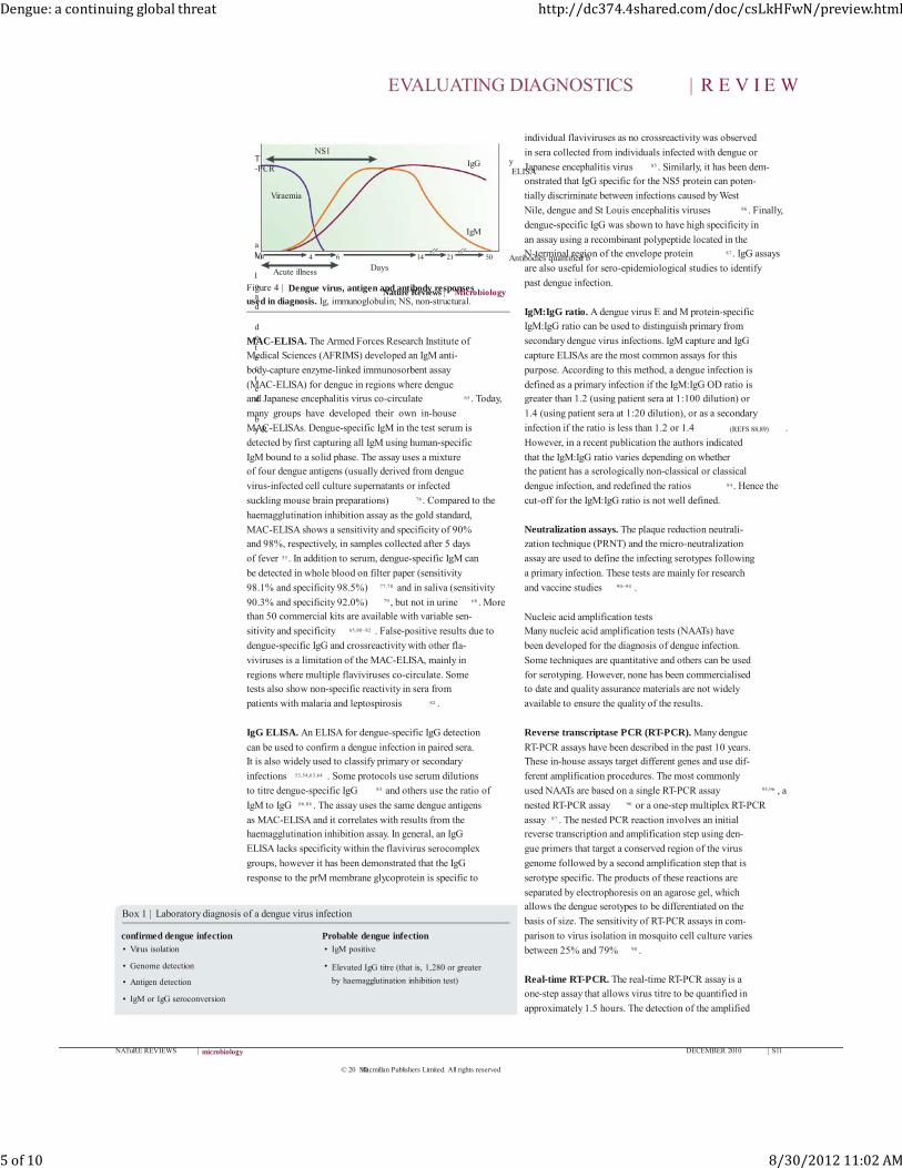
previous dengue infection (FIG. 4) . A primary infection is characterized by a slow and low-titre antibody response.
Immunoglobulin (Ig)M antibodies are the first isotype
to appear, by day 3–5 of illness in 50% of hospitalized
patients and by day 6–10 of illness in 93–99% of cases. The
IgM levels peak ~2 weeks after the onset of fever and then generally decline to undetectable levels over the next 2–3
months 54,55,65 . Dengue-specific IgG is detectable at low titre
at the end of the first week of illness and slowly increases.
By contrast, during a secondary infection, high levels of
IgG antibodies that crossreact with many flaviviruses are detectable even in the acute phase and rise dramatically
over the following 2 weeks 65 . The kinetics of the IgM
response are more variable; as IgM levels are significantly
lower in secondary dengue infections, false-negative test
results for dengue-specific IgM have been reported during
secondary infections 55,66,67 . Following a dengue infection, IgG can be lifelong, which complicates the serodiagno-
sis of past, recent and current infections 65,67 . IgA and IgE
responses have also been documented but the utility of
detecting these immunoglobulins as markers for dengue
serodiagnosis requires further study 68 .In areas where two or more flaviviruses are circulat-
ing, multiple and sequential flavivirus infections make
differential diagnosis difficult owing to the presence of
pre-existing antibodies and the phenomenon of origi-
nal antigenic sin (during sequential flavivirus infections, B-cell clones responding to the first infection synthe-
size antibodies with higher affinity for the first infecting
virus than for the second infecting virus) 69 .
Laboratory diagnosis of dengue infectionLaboratory confirmation of dengue infection is crucial
as the broad spectrum of clinical presentations, ranging
from mild febrile illness to several severe syndromes, can
make accurate diagnosis difficult. Among the methods
available for dengue diagnosis, virus isolation provides the most specific test result. However, facilities that can
support viral culture are not always available. The detec-
tion of the viral genome or viral antigens also provides
evidence of infection.
Seroconversion of IgM or IgG antibodies is the stand-ard for serologically confirming a dengue infection. The
presence of IgM or high levels of IgG in acute serum col-
lected from a suspected dengue case suggests a probable
dengue infection 54,55 . BOX 1 shows the laboratory criteria
for confirmed and probable dengue infections.
Virus isolationThe Aedes albopictus mosquito C6/36 cell line is the
method of choice for DENV isolation, although other
mosquito (such as Aedes pseudoscutellaris AP61) and mammalian (including Vero cells, LLC-MK2 cells and
BHK21 cells) cell lines can also be used 70,71 . Sera that
have been collected from suspected dengue cases in the
first 3–5 days of fever (the viraemic phase) can be used
for virus isolation. After an incubation period permitting virus replication, viral identification is performed using
dengue-specific monoclonal antibodies in immunofluo-
rescence and PCR assays 63,64,72,73 . Serum is often used for
virus isolation but plasma, leukocytes, whole blood and
tissues obtained at autopsy can also be used 63,74,75 .
Serological testingSerological assays are most commonly used for diagno-
sis of dengue infection as they are relatively inexpensive
and easy to perform compared with culture or nucleic acid-based methods. When a dengue infection occurs
in individuals who have experienced a previous dengue
infection, a secondary immune response occurs, which
generates high levels of IgG through the stimulation of
memory B cells from the previous infection as well as an IgM response to the current infection. Because high
levels of IgG compete with IgM for antigen binding, an
IgM capture assay can be used.
Figure 3 | The change in distribution of dengue serotypes. The figure shows the distribution in 1970 ( a) and 2004 (b). Reproduced with permission from REF. 141 .
EVALUATING DIAGNOSTICS | R E V I E W
S10 | DECEMBER 2010 www.nature.com/reviews/micro
© 20 Macmillan Publishers Limited. All rights reserved10
Dengue: a continuing global threat http://dc374.4shared.com/doc/csLkHFwN/preview.html
4 of 10 8/30/2012 11:02 AM
Nature Reviews | Microbiology
IgM
IgG
Viral load detected by R
T-PCR
Antibodies quantified b
y ELISA
Viraemia
Acute illness Days0 4 6 14 21 50
NS1
MAC-ELISA. The Armed Forces Research Institute of Medical Sciences (AFRIMS) developed an IgM anti-
body-capture enzyme-linked immunosorbent assay
(MAC-ELISA) for dengue in regions where dengue and Japanese encephalitis virus co-circulate 65 . Today,
many groups have developed their own in-house
MAC-ELISAs. Dengue-specific IgM in the test serum is
detected by first capturing all IgM using human-specific
IgM bound to a solid phase. The assay uses a mixture of four dengue antigens (usually derived from dengue
virus-infected cell culture supernatants or infected
suckling mouse brain preparations) 76 . Compared to the
haemagglutination inhibition assay as the gold standard,
MAC-ELISA shows a sensitivity and specificity of 90% and 98%, respectively, in samples collected after 5 days
of fever 55 . In addition to serum, dengue-specific IgM can
be detected in whole blood on filter paper (sensitivity
98.1% and specificity 98.5%) 77,78 and in saliva (sensitivity
90.3% and specificity 92.0%) 79 , but not in urine 68 . More than 50 commercial kits are available with variable sen-
sitivity and specificity 65,80–82 . False-positive results due to
dengue-specific IgG and crossreactivity with other fla-
viviruses is a limitation of the MAC-ELISA, mainly in
regions where multiple flaviviruses co-circulate. Some tests also show non-specific reactivity in sera from
patients with malaria and leptospirosis 82 .
IgG ELISA. An ELISA for dengue-specific IgG detection
can be used to confirm a dengue infection in paired sera. It is also widely used to classify primary or secondary
infections 53,54,63,64 . Some protocols use serum dilutions
to titre dengue-specific IgG 83 and others use the ratio of
IgM to IgG 66,84 . The assay uses the same dengue antigens
as MAC-ELISA and it correlates with results from the haemagglutination inhibition assay. In general, an IgG
ELISA lacks specificity within the flavivirus serocomplex
groups, however it has been demonstrated that the IgG
response to the prM membrane glycoprotein is specific to
individual flaviviruses as no crossreactivity was observed
in sera collected from individuals infected with dengue or
Japanese encephalitis virus 85 . Similarly, it has been dem-onstrated that IgG specific for the NS5 protein can poten-
tially discriminate between infections caused by West
Nile, dengue and St Louis encephalitis viruses 86 . Finally,
dengue-specific IgG was shown to have high specificity in
an assay using a recombinant polypeptide located in the N-terminal region of the envelope protein 87 . IgG assays
are also useful for sero-epidemiological studies to identify
past dengue infection.
IgM:IgG ratio. A dengue virus E and M protein-specific IgM:IgG ratio can be used to distinguish primary from
secondary dengue virus infections. IgM capture and IgG
capture ELISAs are the most common assays for this
purpose. According to this method, a dengue infection is
defined as a primary infection if the IgM:IgG OD ratio is greater than 1.2 (using patient sera at 1:100 dilution) or
1.4 (using patient sera at 1:20 dilution), or as a secondary
infection if the ratio is less than 1.2 or 1.4 (REFS 88,89) .
However, in a recent publication the authors indicated
that the IgM:IgG ratio varies depending on whether the patient has a serologically non-classical or classical
dengue infection, and redefined the ratios 84 . Hence the
cut-off for the IgM:IgG ratio is not well defined.
Neutralization assays. The plaque reduction neutrali-
zation technique (PRNT) and the micro-neutralization
assay are used to define the infecting serotypes following
a primary infection. These tests are mainly for research
and vaccine studies 90–94 .
Nucleic acid amplification testsMany nucleic acid amplification tests (NAATs) have
been developed for the diagnosis of dengue infection.
Some techniques are quantitative and others can be used
for serotyping. However, none has been commercialised to date and quality assurance materials are not widely
available to ensure the quality of the results.
Reverse transcriptase PCR (RT-PCR). Many dengue
RT-PCR assays have been described in the past 10 years. These in-house assays target different genes and use dif-
ferent amplification procedures. The most commonly
used NAATs are based on a single RT-PCR assay 95,96 , a
nested RT-PCR assay 96 or a one-step multiplex RT-PCR
assay 97 . The nested PCR reaction involves an initial reverse transcription and amplification step using den-
gue primers that target a conserved region of the virus
genome followed by a second amplification step that is
serotype specific. The products of these reactions are
separated by electrophoresis on an agarose gel, which allows the dengue serotypes to be differentiated on the
basis of size. The sensitivity of RT-PCR assays in com-
parison to virus isolation in mosquito cell culture varies
between 25% and 79% 98 .
Real-time RT-PCR. The real-time RT-PCR assay is a
one-step assay that allows virus titre to be quantified in
approximately 1.5 hours. The detection of the amplified
Figure 4 | Dengue virus, antigen and antibody responses used in diagnosis. Ig, immunoglobulin; NS, non-structural.
Box 1 | Laboratory diagnosis of a dengue virus infection
confirmed dengue infection• Virus isolation
• Genome detection
• Antigen detection
• IgM or IgG seroconversion
Probable dengue infection• IgM positive
• Elevated IgG titre (that is, 1,280 or greater
by haemagglutination inhibition test)
EVALUATING DIAGNOSTICS | R E V I E W
NATuRE REVIEWS | microbiology DECEMBER 2010 | S11
© 20 Macmillan Publishers Limited. All rights reserved10
Dengue: a continuing global threat http://dc374.4shared.com/doc/csLkHFwN/preview.html
5 of 10 8/30/2012 11:02 AM
target by fluorescent probes replaces the need for post-
amplification electrophoresis. Many real-time RT-PCR
assays have been developed that are either ‘singleplex’, detecting one single serotype per reaction, or ‘multiplex’,
identifying all four serotypes from a single sample 99–101 .
One advantage of this assay is the ability to determine
viral titre early in dengue illness, which is believed to be
an important predictor of disease severity 102 .
Nucleic acid-sequence based amplification assay (NASBA). The NASBA assay is an isothermal RNA-
specific amplification assay that has been adapted for
dengue virus. Its performance is comparable to that of other NAATs 103 .
Antigen detection
Dengue antigens can be detected in tissues such as liver,
spleen and lymph nodes as well as tissues from fatal cases (slides from paraffin-embedded, fresh or frozen tissues)
using an enzyme and a colorimetric substrate with
antibodies that target dengue-specific antigens 104–106 .
NS1 antigen and antibody detection. NS1 is a glyco-protein produced by all flaviviruses and is essential for
viral replication and viability. Because this protein is
secreted into the bloodstream, many tests have been
developed to diagnose DENV infections using NS1.
These tests include antigen-capture ELISA, lateral flow antigen detection and measurement of NS1-specific
IgM and IgG responses. NS1 antigen detection kits are
now commercially available. As yet, these kits do not
differentiate between the different DENV serotypes. Additional independent studies are needed to confirm
the performance of these kits and to further validate
the diagnostic and prognostic significance of NS1 and
NS1-specific antibody detection 107–109 .
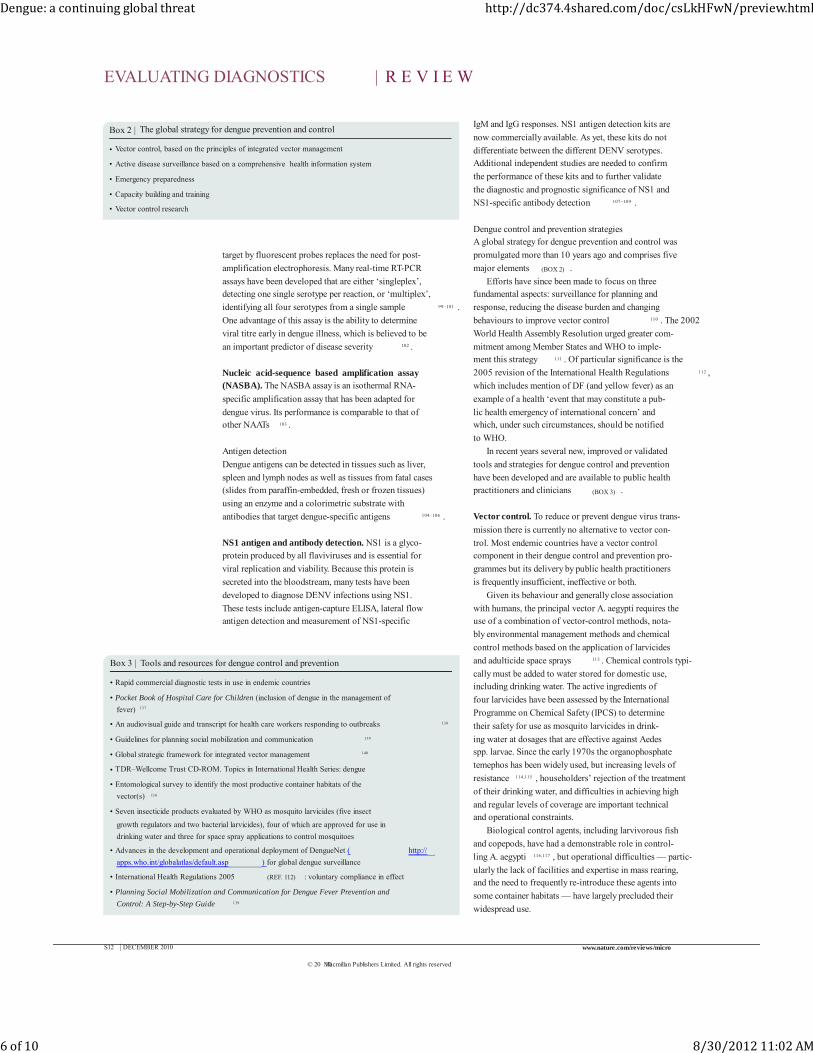
Dengue control and prevention strategiesA global strategy for dengue prevention and control was
promulgated more than 10 years ago and comprises five
major elements (BOX 2) .
Efforts have since been made to focus on three fundamental aspects: surveillance for planning and
response, reducing the disease burden and changing
behaviours to improve vector control 110 . The 2002
World Health Assembly Resolution urged greater com-
mitment among Member States and WHO to imple-ment this strategy 111 . Of particular significance is the
2005 revision of the International Health Regulations 112 ,
which includes mention of DF (and yellow fever) as an
example of a health ‘event that may constitute a pub-
lic health emergency of international concern’ and which, under such circumstances, should be notified
to WHO.
In recent years several new, improved or validated
tools and strategies for dengue control and prevention
have been developed and are available to public health practitioners and clinicians (BOX 3) .
Vector control. To reduce or prevent dengue virus trans-
mission there is currently no alternative to vector con-
trol. Most endemic countries have a vector control component in their dengue control and prevention pro-
grammes but its delivery by public health practitioners
is frequently insufficient, ineffective or both.
Given its behaviour and generally close association
with humans, the principal vector A. aegypti requires the use of a combination of vector-control methods, nota-
bly environmental management methods and chemical
control methods based on the application of larvicides
and adulticide space sprays 113 . Chemical controls typi-
cally must be added to water stored for domestic use, including drinking water. The active ingredients of
four larvicides have been assessed by the International
Programme on Chemical Safety (IPCS) to determine
their safety for use as mosquito larvicides in drink-
ing water at dosages that are effective against Aedes spp. larvae. Since the early 1970s the organophosphate
temephos has been widely used, but increasing levels of
resistance 114,115 , householders’ rejection of the treatment
of their drinking water, and difficulties in achieving high
and regular levels of coverage are important technical and operational constraints.
Biological control agents, including larvivorous fish
and copepods, have had a demonstrable role in control-
ling A. aegypti 116,117 , but operational difficulties — partic-
ularly the lack of facilities and expertise in mass rearing, and the need to frequently re-introduce these agents into
some container habitats — have largely precluded their
widespread use.
Box 2 | The global strategy for dengue prevention and control
• Vector control, based on the principles of integrated vector management
• Active disease surveillance based on a comprehensive health information system
• Emergency preparedness
• Capacity building and training
• Vector control research
Box 3 | Tools and resources for dengue control and prevention
• Rapid commercial diagnostic tests in use in endemic countries
• Pocket Book of Hospital Care for Children (inclusion of dengue in the management of
fever) 137
• An audiovisual guide and transcript for health care workers responding to outbreaks 138
• Guidelines for planning social mobilization and communication 139
• Global strategic framework for integrated vector management 140
• TDR–Wellcome Trust CD-ROM. Topics in International Health Series: dengue
• Entomological survey to identify the most productive container habitats of the
vector(s) 116
• Seven insecticide products evaluated by WHO as mosquito larvicides (five insect
growth regulators and two bacterial larvicides), four of which are approved for use in
drinking water and three for space spray applications to control mosquitoes
• Advances in the development and operational deployment of DengueNet ( http://
apps.who.int/globalatlas/default.asp ) for global dengue surveillance
• International Health Regulations 2005 (REF. 112) : voluntary compliance in effect
• Planning Social Mobilization and Communication for Dengue Fever Prevention and
Control: A Step-by-Step Guide 139
EVALUATING DIAGNOSTICS | R E V I E W
S12 | DECEMBER 2010 www.nature.com/reviews/micro
© 20 Macmillan Publishers Limited. All rights reserved10
Dengue: a continuing global threat http://dc374.4shared.com/doc/csLkHFwN/preview.html
6 of 10 8/30/2012 11:02 AM
Environmental management is generally considered
to be an essential component of dengue prevention and
control, particularly when targeting the most productive container habitats of the vector 118 . Source reduction, ‘clean-
up’ campaigns, regular container emptying and cleaning
(targeting not only households but also public spaces
such as cemeteries, green areas and schools), installation
of water supply systems, solid waste management and urban planning all fall under the rubric of environmental
management. However, huge investments in infrastruc-
ture are needed to increase access to safe and reliable water
supplies and solid waste disposal systems. In addition to
overall health gains, such provision would clearly have a major impact on vector ecology, although the relation-
ship is complex . For instance, cost recovery mechanisms,
such as the introduction of metered water, might actually
encourage the household collection and storage of roof
catchment rainwater, which can be harvested at no cost. Although not studied carefully, the construction of com-
munity water distribution services to rural townships and
villages might be contributing to the rural spread of den-
gue in Southeast Asia and elsewhere by facilitating domes-
tic water storage. When decisions on such infrastructure development are being made, the views of Ministers of
Public Health and municipal health departments are sel-
dom voiced loudly, even when the economic and public
health burden of diseases linked to water and sanitation
are recognized, including those associated with dengue. Most efforts in vector control are centred at the house-
hold and community levels, but with few exceptions, the
achievements to date have been largely unspectacular
and there have been difficulties in scaling up from the
project level 119 . Nevertheless, such community-based interventions are widely seen as the most promising way
of improving delivery and achieving long-term control of
the vector through behaviour change. Towards this end,
a TDR/WHO guide for planning social mobilization
and communication for dengue fever prevention and control has been developed 113 . Additionally, new ‘con-
sumer-friendly’ tools such as window curtains and water
container covers treated with long-lasting insecticide
are being tested 120 as well as controlled release larvicides
that provide several months of control following a single
application to targeted containers.Products for personal and household protection have
a huge potential for household pest control. Generally
speaking, these commercial products tend to be used
by consumers not so much in response to any perceived
public health concerns, but to alleviate the nuisance of biting mosquitoes and in some settings households are
prepared to spend substantial amounts of money on
these products 121 .
With the increased political recognition of dengue as
a public health problem and commitment to prevention and control, better organized control services using new
tools and partnership strategies, based on the principles
of integrated vector management, are likely to have a
major impact on dengue transmission 2 .
Vaccine development. As a result of the failure of vector control, the continuing spread and increasing intensity
of dengue has renewed interest and investment in den-
gue vaccine development, making a safe, effective and
affordable tetravalent dengue vaccine a global public health priority 122 . Dengue vaccine development has been
in progress for several decades, however the complex
pathology of the illness, the need to control four virus
serotypes simultaneously and insufficient investment by
vaccine developers have hampered progress 122 . The observation that DHF/DSS is associated with
DENV secondary infection poses a special challenge
to the development of a dengue vaccine, leading to a
requirement that such vaccines should induce a robust
immune response against the four serotypes in naive as well as previously immune individuals. Animal models
are only partially useful for vaccine evaluation. The poor
understanding of the mechanisms involved in inducing
protective immunity against dengue infection poses
additional challenges 123 . Finally, cases of DHF/DSS have recently been documented 20 or more years after pri-
mary dengue infection, which adds a new dimension to
the problem 25,44 .
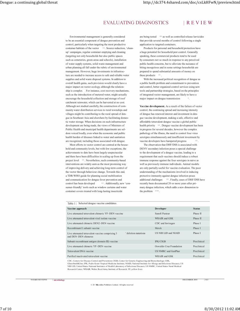
Table 1 | Selected dengue vaccine candidates
Vaccine approach Developer Status
Live attenuated tetravalent chimeric YF–DEN vaccine Sanofi Pasteur Phase II
Live attenuated tetravalent viral isolate vaccine WRAIR and GSK Phase II
Live attenuated chimeric DEN2–DEN vaccine CDC and Inviragen Phase I
Recombinant E subunit vaccine Merck Phase I
Live attenuated tetravalent vaccine comprising 3 ′ deletion mutations and DEN–DEN chimeras
US NIH LID and NIAID Phase I
Subunit recombinant antigen (domain III) vaccine IPK/CIGB Preclinical
Live attenuated chimeric YF–DEN vaccine Oswaldo Cruz Foundation Preclinical
Tetravalent DNA vaccine US NMRC and GenPhar Preclinical
Purified inactivated tetravalent vaccine WRAIR and GSK Preclinical
CDC, Centers for Disease Control and Prevention; CIGB, Center for Genetic Engineering and Biotechnology; GSK, GlaxoSmithKline; IPK, Pedro Kouri Tropical Medicine Institute; NIAID, National Institute for Allergy and Infectious Diseases; US NIH LID, United States National Institutes of Health Laboratory of Infectious Diseases; US NMRC, United States Naval Medical Research Center; WRAIR, Walter Reed Army Institute of Research; YF, yellow fever.
EVALUATING DIAGNOSTICS | R E V I E W
NATuRE REVIEWS | microbiology DECEMBER 2010 | S13
© 20 Macmillan Publishers Limited. All rights reserved10
Dengue: a continuing global threat http://dc374.4shared.com/doc/csLkHFwN/preview.html
7 of 10 8/30/2012 11:02 AM
The available data suggest that neutralizing antibodies
are the major contributors to protective immunity 124,125 ,
however the role of the cellular immune response requires further study 123 . In this context, clinical tri-
als are crucial for vaccine development owing to the
unique information they provide on immune responses
and reactogenicity. Also, long-term observations of vac-
cinated populations will be required to demonstrate the absence of ADE or severe disease.
The ideal dengue vaccine should be free of impor-
tant reactogenicity, induce life-long protection against
infection with any of the four DENV serotypes and be
affordable 126,127 . Vaccine candidates should be evaluated in population-based efficacy trials in several at-risk
populations in different geographical settings includ-
ing Asia and the Americas, which experience different
patterns of dengue transmission intensity and dengue
virus circulation 122 . Vaccine developers are working with the Pediatric Dengue Vaccine Initiative (PDVI) to
establish suitable field sites. Developers are also working
with the WHO Initiative for Vaccine Research (WHO/
IVR) to define the immunological correlates for protec-
tion and clinical trial design. Because of the important role of neutralizing antibodies as surrogates of protec-
tion, the validation of neutralization tests is a priority 128 .
Current approaches to vaccine development involve
using live attenuated viruses, inactivated viruses, subu-
nit vaccines, DNA vaccines, cloned engineered viruses and chimeric viruses using yellow fever vaccine and
attenuated dengue viruses as backbones 129–134 . TABLE 1
summarizes the most advanced vaccine candidates.
Significant progress in the development of dengue vaccine candidates has been achieved lately 135,136 . An
Acambis/Sanofi Pasteur yellow fever–dengue chimeric
vaccine is in advanced Phase II testing in children in
Thailand and others are in Phase 1 or advanced preclini-
cal evaluation. It is expected that a licensed vaccine will be available in less than 10 years.
Conclusions
Dengue is now a global threat and is endemic or epi-
demic in almost every country located in the tropics. While we wait for new tools such as vaccines, antiviral
drugs and improved diagnostics, better use should be
made of the interventions that are currently available.
The challenge that awaits us in the near future will be
how to scale up to deploy these new tools. In recent years, several partnerships such as the
PDVI, the Innovative Vector Control Consortium,
the Asia-Pacific Dengue Prevention Partnership and
the European union’s DENFRAME and DENCO
projects have come into existence, receiving funding from the Bill and Melinda Gates Foundation, regional
Development Banks and the private sector. These part-
nerships are working with WHO and national govern-
ments to develop new tools and strategies to improve
diagnostics and clinical treatments and to achieve a successful vaccine.
1. WHO. Scientific Working Group Report on Dengue
[online]< http://apps.who.int/tdr/publications/
tdr‑research‑publications/swg‑report‑dengue/pdf/swg_
dengue_2.pdf > (WHO, Geneva, Switzerland, 2007).
2. TDR/WHO. Dengue: Guidelines for Diagnosis,
Treatment, Prevention and Control (TDR/WHO,
Geneva, Switzerland, 2009).
3. Guzman, M . G. & Kouri, G. Dengue: an update. Lancet
Infect. Dis. 2, 33–42 (2002).
4. Gubler, D. J. The changing epidemiology of yellow
fever and dengue, 1900 to 2003: full circle? Comp.
Immunol. Microbiol. Infect. Dis. 27, 319–330
(2004).
5. Franco, L. et al. Recent expansion of dengue virus
serotype 3 in West Africa. Euro Surveill. 15,
9ii=19490 (2010).
6. Wilder‑Smith, A. Dengue in travelers. New Engl. J.
Med. 353, 924–932 (2005).
7. Freedman, D. O. et al. Spectrum of disease and
relation to place of exposure among ill returned
travelers. New Engl. J. Med. 354, 119–130 (2006).
8. Wichmann, O. Dengue antibody prevalence in German
travelers. Emerg. Infect. Dis. 11, 762–765 (2005).
9. Jelinek, T. Dengue fever in international travelers.
Clin. Infect. Dis. 31, 144–147 (2000).
10. Gubler, D. J. & Meltzer, M. Impact of dengue/dengue
hemorrhagic fever on the developing world. Adv. Virus
Res. 53, 35–70 (1999).
11. Rodriguez‑Roche, R. et al. Virus evolution during a
severe dengue epidemic in Cuba, 1997. Virology 334,
154–159 (2005).
12. Wang, E. et al. Evolutionary relationships of endemic/
epidemic and sylvatic dengue viruses. J. Virol. 74,
3227–3234 (2000).
13. Halstead, S. B. Dengue virus–mosquito interactions.
Annu. Rev. Entomol. 53, 273–291 (2008).
14. Watts, D. M., Burke, D. S., Harrison, B. A., Whitmire,
R. E. & Nisalak, A. Effect of temperature on the vector
efficiency of Aedes aegypti for dengue 2 virus. Am. J.
Trop. Med. Hyg. 36, 143–152 (1987).
15. Schneider, B. S., Soong, L., Zeidner, N. S. & Higgs, S.
Aedes aegypti salivary gland extracts modulate anti‑viral and TH1/TH2 cytokine responses to Sindbis virus
infection. Viral Immunol. 17, 565–573 (2004).
16. Wu, S. J. et al. Human skin Langerhans cells are
targets of dengue virus infection. Nature Med. 6,
816–820 (2000).
17. Jessie, K., Fong, M. Y., Devi, S., Lam, S. K. &
Wong, K. T. Localization of dengue virus in naturally
infected human tissues, by immunohistochemistry
and in situ hybridization. J. Infect. Dis. 189,
1411–1418 (2004).
18. Halstead, S. B. & O’Rourke, E. J. Dengue viruses and
mononuclear phagocytes. I. Infection enhancement
by non‑neutralizing antibody. J. Exp. Med. 146,
201–217 (1977).
19. Halstead, S. B. In vivo enhancement of dengue virus
infection in rhesus monkeys by passively transferred
antibody. J. Infect. Dis. 140, 527–533 (1979).
20. Goncalvez, A. P., Engle, R. E., St. Claire, M .,
Purcell, R. H. & Lai, C. J. Monoclonal antibody‑mediated enhancement of dengue virus infection
in vitro and in vivo and strategies for prevention.
Proc. Natl Acad. Sci. USA 104, 9422–9427
(2007).
21. Kliks, S. C., Nimmanitya, S., Nisalak, A. & Burke, D. S.
Evidence that maternal dengue antibodies are
important in the development of dengue hemorrhagic
fever in infants. Am. J. Trop. Med. Hyg. 38, 411–419
(1988).
22. Cummings, D. A. T., Schwartz, I. B., Billings, L.,
Shaw, L. B. & Burke, D. S. Dynamic effects of
antibody‑dependent enhancement on the fitness
of viruses. Proc. Natl Acad. Sci. USA 102,
15259–15264 (2005).
23. Guzman, M. G. et al. Dengue hemorrhagic fever in
Cuba, 1981: a retrospective seroepidemiologic study.
Am. J. Trop. Med. Hyg. 42, 179–184 (1990).
24. Guzman, M. G. et al. Epidemiologic studies on Dengue
in Santiago de Cuba, 1997. Am. J. Epidemiol. 152,
793–799; discussion 804 (2000).
25. Alvarez, M. et al. Dengue hemorrhagic fever caused
by sequential dengue 1–3 virus infections over a long
time interval: Havana epidemic, 2001–2002 Am. J.
Trop. Med. Hyg. 75, 1113–1117 (2006).
26. Guzman, M. G. et al. Effect of age on outcome of
secondary dengue 2 infections. Int. J. Infect. Dis. 6,
118–124 (2002).
27. Gamble, J. et al. Age‑related changes in
microvascular permeability: a significant factor in the
susceptibility of children to shock? Clin. Sci. 98,
211–216 (2000).
28. Kouri, G. P., Guzman, M. G. & Bravo, J. R. Why
dengue haemorrhagic fever in Cuba? 2. An integral
analysis. Trans. R. Soc. Trop. Med. Hyg. 81, 821–823
(1987).
29. Halstead, S. B., Nimmannitya, S. & Cohen, S. N.
Observations related to pathogenesis of dengue
hemorrhagic fever. IV. Relation of disease severity to
antibody response and virus recovered. Yale J. Biol.
Med. 42, 311–328 (1970).
30. Lee, M. S., Hwang, K. P., Chen, T. C., Lu, P. L. &
Chen, T. P. Clinical characteristics of dengue and
dengue hemorrhagic fever in a medical center of
southern Taiwan during the 2002 epidemic.
J. Microbiol. Immunol. Infect. 39, 121–129 (2006).
31. Stephens, H. A. et al. HLA‑A and ‑B allele
associations with secondary dengue virus infections
correlate with disease severity and the infecting
viral serotype in ethnic Thais. Tissue Antigens 60,
309–318 (2002).
32. LaFleur, C. et al. HLA‑DR antigen frequencies in
M exican patients with dengue virus infection:
HLA‑DR4 as a possible genetic resistance factor
for dengue hemorrhagic fever. Hum. Immunol. 63,
1039–1044 (2002).
33. Loke, H. et al. Strong HLA class I‑restricted T cell
responses in dengue hemorrhagic fever: a double‑edged sword? J. Infect. Dis. 184, 1369–1373
(2001).
34. Sakuntabhai, A. et al. A variant in the CD209
promoter is associated with severity of dengue
disease. Nature Genet. 37, 507–513 (2005).
35. Fernandez‑M estre, M. T., Gendzekhadze, K.,
Rivas‑Vetencourt, P. & Layrisse, Z. TNF‑α‑308A allele,
a possible severity risk factor of hemorrhagic
manifestation in dengue fever patients. Tissue
Antigens 64, 469–472 (2004).
36. Kalayanarooj, S. et al. Blood group AB is associated
with increased risk for severe dengue disease in
secondary infections. J. Infect. Dis. 195, 1014–1017
(2007).
EVALUATING DIAGNOSTICS | R E V I E W
S14 | DECEMBER 2010 www.nature.com/reviews/micro
© 20 Macmillan Publishers Limited. All rights reserved10
Dengue: a continuing global threat http://dc374.4shared.com/doc/csLkHFwN/preview.html
8 of 10 8/30/2012 11:02 AM
37. Bravo, J. R., Guzman, M. G. & Kouri, G. P. Why dengue
haemorrhagic fever in Cuba? 1. Individual risk factors
for dengue haemorrhagic fever/dengue shock
syndrome (DHF/DSS). Trans. R. Soc. Trop. Med. Hyg.
81, 816–820 (1987).
38. Thisyakorn, U. & Nimmannitya, S. Nutritional status of
children with dengue hemorrhagic fever. Clin. Infect.
Dis. 16, 295–297 (1993).
39. Loke, H. et al. Susceptibility to dengue hemorrhagic
fever in vietnam: evidence of an association with
variation in the vitamin D receptor and Fc gamma
receptor IIa genes. Am. J. Trop. Med. Hyg. 67,
102–106 (2002).
40. Sierra, B. D. et al. Ethnicity and difference in dengue
virus‑specific memory T cell responses in cuban
individuals. Viral Immunol. 19, 662–668 (2006).
41. Sierra, B. D., Kouri, G. & Guzman, M. G. Race: a risk
factor for dengue hemorrhagic fever. Arch. Virol. 152,
533–534 (2007).
42. Halstead, S. B. et al. Haiti: absence of dengue
hemorrhagic fever despite hyperendemic dengue virus
transmission. Am. J. Trop. Med. Hyg. 65, 180–183
(2001).
43. Guzman, M . G., Kouri, G. & Halstead, S. B. Do escape
mutants explain rapid increases in dengue case‑fatality rates within epidemics? Lancet 355,
1902–1903 (2000).
44. Guzman, M . G. et al. Enhanced severity of secondary
dengue‑2 infections: death rates in 1981 and 1997
Cuban outbreaks. Rev. Panam. Salud Pública. 11,
223–227 (2002).
45. Chareonsirisuthigul, T., Kalayanarooj, S. & Ubol, S.
Dengue virus (DENV) antibody‑dependent
enhancement of infection upregulates the production
of anti‑inflammatory cytokines, but suppresses
anti‑DENV free radical and pro‑inflammatory cytokine
production, in THP‑1 cells. J. Gen. Virol. 88, 365–375
(2007).
46. Simmons, C. P. et al. Patterns of host genome‑wide
gene transcript abundance in the peripheral blood of
patients with acute dengue hemorrhagic fever.
J. Infect. Dis. 195, 1097–1107 (2007).
47. Couvelard, A. et al. Report of a fatal case of dengue
infection with hepatitis: demonstration of dengue
antigens in hepatocytes and liver apoptosis. Hum.
Pathol. 30, 1106–1110 (1999).
48. Alcon‑LePoder, S. et al. The secreted form of dengue
virus nonstructural protein NS1 is endocytosed by
hepatocytes and accumulates in late endosomes:
implications for viral infectivity. J. Virol. 79,
11403–11411 (2005).
49. Malasit, P. Complement and dengue haemorrhagic
fever/shock syndrome. Southeast Asian J. Trop. Med.
Pub. Health 18, 316–320 (1987).
50. Avirutnan, P. et al. Vascular leakage in severe dengue
virus infections: a potential role for the nonstructural
viral protein NS1 and complement. J. Infect. Dis. 193,
1078–1088 (2006).
51. Mongkolsapaya, J. et al. Original antigenic sin and
apoptosis in the pathogenesis of dengue
hemorrhagic fever. Nature Med. 9, 921–927
(2003).
52. Rico‑Hesse, R. Microevolution and virulence of dengue
viruses. Adv. Virus Res. 59, 315–341 (2003).
53. Lin, C. F., Wan, S. W., Cheng, H. J., Lei, H. Y. &
Lin, Y. S. Autoimmune pathogenesis in dengue virus
infection. Viral Immunol. 19, 127–132 (2006).
54. TDR/WHO. In Dengue: Guidelines for Diagnosis,
Treatment, Prevention and Control. 23–55
(TDR/WHO, Geneva, Switzerland, 2009).
55. PAHO. Dengue and Dengue Hemorrhagic Fever in the
Americas: Guidelines for Prevention and Control. (Pan
American Health Organization, Washington, DC, USA
1994).
56. Nimmannitya, S. Clinical spectrum and management
of dengue haemorrhagic fever. Southeast Asian
J. Trop. Med. Pub. Health 18, 392–397 (1987).
57. Martinez Torres, E. Preventing deaths from dengue:
a space and challenge for primary health care.
Rev. Panam. Salud Pública 20, 60–74 (2006).
58. Bandyopadhyay, S., Lum, L. C. & Kroeger, A.
Classifying dengue: a review of the difficulties in
using the WHO case classification for dengue
haemorrhagic fever. Trop. Med. Int. Health 11,
1238–1255 (2006).
59. Rigau‑Perez, J. G. Severe dengue: the need for new
case definitions. Lancet Infect. Dis. 6, 297–302
(2006).
60. Deen, J. L. et al. The WHO dengue classification and
case definitions: time for a reassessment. Lancet 368,
170–173 (2006).
61. Balmaseda, A. Assessment of the World Health
Organization scheme for classification of dengue
severity in Nicaragua. Am. J. Trop. Med. Hyg. 73,
1059–1062 (2005).
62. Thangaratham, P. S. & Tyagi, B. K. Indian perspective
on the need for new case definitions of severe dengue.
Lancet Infect. Dis. 7, 81–82 (2007).
63. Vorndam, V. & Kuno, G. In Dengue and Dengue
Hemorrhagic Fever (eds Gubler, D. J. & Kuno, G.)
313–333 (CAB International, New York, USA, 1997).
64. Guzman, M. G. & Kouri, G. Dengue diagnosis,
advances and challenges. Int. J. Infect. Dis. 8, 69–80
(2004).
65. Innis, B. L. et al. An enzyme‑linked immunosorbent
assay to characterize dengue infections where dengue
and Japanese encephalitis co‑circulate. Am. J. Trop.
Med. Hyg. 40, 418–427 (1989).
66. Chanama, S. et al. Analysis of specific IgM responses
in secondary dengue virus infections: levels and
positive rates in comparison with primary infections.
J. Clin. Virol. 31, 185–189 (2004).
67. Gubler, D. J. Serologic diagnosis of dengue/dengue
haemorrhagic fever. Dengue Bull. 20, 20–23
(1996).
68. Vazquez, S. et al. Kinetics of antibodies in sera, saliva,
and urine samples from adult patients with primary or
secondary dengue 3 virus infections. Int. J. Infect. Dis.
11, 256–262 (2007).
69. Halstead, S. B., Rojanasuphot, S. & Sangkawibha, N.
Original antigenic sin in dengue. Am. J. Trop. Med.
Hyg. 32, 154–156 (1983).
70. Singh, K. R. P. & Paul, S. D. M ultiplication of
arboviruses in cell lines from Aedes albopictus and
Aedes aegypti . Curr. Sci. 37, 65–67 (1968).
71. Race, M. W., Williams, M. C. & Agostini, C. F.
Dengue in the Caribbean: virus isolation in a mosquito
(Aedes pseudoscutellaris) cell line. Trans. R. Soc. Trop.
Med. Hyg. 73, 18–22 (1979).
72. Henchal, E. A., McCown, J. M., Seguin, M . C.,
Gentry, M. K. & Brandt, W. E. Rapid identification of
dengue virus isolates by using monoclonal antibodies
in an indirect immunofluorescence assay. Am. J. Trop.
Med. Hyg. 32, 164–169 (1983).
73. Kao, C. L. et al. Flow cytometry compared with indirect
immunofluorescence for rapid detection of dengue
virus type 1 after amplification in tissue culture.
Clin. Microbiol. 39, 3672–3677 (2001).
74. Guzman, M. G. et al. Fatal dengue hemorrhagic fever
in Cuba, 1997. Int. J. Infect. Dis. 3, 130–135 (1999).
75. Rosen, L., Drouet, M. T. & Deubel, V. Detection of
dengue virus RNA by reverse transcription‑polymerase
chain reaction in the liver and lymphoid organs but
not in the brain in fatal human infection. Am. J. Trop.
Med. Hyg. 61, 720–724 (1999).
76. Cardosa, M . J., Tio, P. H., Nimmannitya, S.,
Nisalak, A. & Innis, B. IgM capture ELISA for detection
of IgM antibodies to dengue virus: comparison of
2 formats using hemagglutinins and cell culture
derived antigens. Southeast Asian J. Trop. Med. Pub.
Health 23, 726–729 (1992).
77. Vazquez, S. et al. Detection of IgM against the dengue
virus in whole blood absorbed on filter paper. Rev.
Panam. Salud Pública 3, 174–178 (1998).
78. Herrera, R. D., Cabrera, M. V., Garcia, S. & Gilart, M.
IgM antibodies to dengue virus in dried blood on filter
paper. Clin. Chim. Acta 367, 204–206 (2006).
79. Balmaseda, A. et al. Diagnosis of dengue virus
infection by detection of specific immunoglobulin M
(IgM) and IgA antibodies in serum and saliva. Clin.
Diagn. Lab. Immunol. 10, 317–322 (2003).
80. Blacksell, S. D. et al. The comparative accuracy of
8 commercial rapid immunochromatographic assays
for the diagnosis of acute dengue virus infection.
Clin. Infect. Dis. 42, 1127–1134 (2006).
81. Blacksell, S. D. et al. Prospective study to determine
accuracy of rapid serological assays for diagnosis of
acute dengue virus infection in Laos. Clin. Vaccine
Immunol. 14, 1458–1464 (2007).
82. Hunsperger, E. A. et al. Evaluation of commercially
available anti‑dengue virus immunoglobulin M tests.
Emerg. Infect. Dis. 15, 436–440 (2009).
83. Vazquez, S., Bravo, J. R., Perez, A. B. & Guzman, M. G.
Inhibition ELISA. Its utility for classifying a case of
dengue. Rev. Cubana Med. Trop. 49, 108–112 (1997).
84. Falconar, A. K., de Plata, E. & Romero‑Vivas, C. M.
Altered enzyme‑linked immunosorbent assay
immunoglobulin M (IgM)/IgG optical density ratios can
correctly classify all primary or secondary dengue
virus infections 1 day after the onset of symptoms,
when all of the viruses can be isolated. Clin. Vaccine
Immunol. 13, 1044–1051 (2006).
85. Cardosa, M. J., Wang, S. M ., Sum, M . S. & Tio, P. H.
Antibodies against prM protein distinguish between
previous infection with dengue and Japanese
encephalitis viruses. BMC Microbiol. 2, 9 (2002).
86. Wong, S. J. et al. Immunoassay targeting
nonstructural protein 5 to differentiate west nile virus
infection from dengue and St. Louis encephalitis virus
infections and from flavivirus vaccination. J. Clin.
Microbiol. 41 , 4217–4223 (2003).
87. Baretto dos Santos, F. et al. Analysis of recombinant
dengue virus polypeptides for dengue diagnosis and
evaluation of the humoral immune response. Am. J.
Trop. Med. Hyg. 71, 144–152 (2004).
88. Kuno, G., Gomez, I. & Gubler, D. J. An ELISA
procedure for the diagnosis of dengue infections.
J. Virol. Methods 33, 101–113 (1991).
89. Shu, P. Y. et al. Comparison of capture immunoglobulin
M (IgM) and IgG enzyme‑linked immunosorbent assay
(ELISA) and nonstructural protein NS1 serotype‑specific IgG ELISA for differentiation of primary and
secondary dengue virus infections. Clin. Diagn. Lab.
Immunol. 10, 622–630 (2003).
90. Calisher, C. H. et al. Antigenic relationships between
flaviviruses as determined by cross‑neutralization tests
with polyclonal antisera. J. Gen. Virol. 70, 37–43
(1989).
91. Russell, P. K. & Nisalak, A. Dengue virus identification
by the plaque reduction neutralization test.
J. Immunol. 99, 291–296 (1967).
92. M orens, D. M., Halstead, S. B., Repik, P. M.,
Putvatana, R. & Raybourne, N. Simplified plaque
reduction neutralization assay for dengue viruses by
semimicro methods in BHK‑21 cells: comparison of
the BHK suspension test with standard plaque
reduction neutralization. J. Clin. Microbiol. 22,
250–254 (1985).
93. Thomas, S. J. et al. Dengue plaque reduction
neutralization test (PRNT) in primary and secondary
dengue virus infections: how alterations in assay
conditions impact performance. Am. J. Trop. Med.
Hyg. 81 , 825–833 (2009).
94. Roehrig, J. T. et al. Guidelines for plaque‑reduction
neutralization testing of human antibodies to dengue
viruses. Viral Immunol. 21, 123–132 (2008).
95. M orita, K., Tanaka, M. & Igarashi, A. Rapid
identification of dengue virus serotypes by using
polymerase chain reaction. J. Clin. Microbiol. 29,
2107–2110 (1991).
96. Lanciotti, R. S., Calisher, C. H., Gubler, D. J.,
Chang, G. J. & Vorndam, A. V. Rapid detection and
typing of dengue viruses from clinical samples by
using reverse transcriptase‑polymerase chain reaction.
J. Clin. Microbiol. 30, 545–551 (1992).
97. Harris, E. et al. Typing of dengue viruses in clinical
specimens and mosquitoes by single‑tube multiplex
reverse transcriptase PCR. J. Clin. Microbiol. 36,
2634–2639 (1998).
98. Raengsakulrach, B. et al. Comparison of four reverse
transcription‑polymerase chain reaction procedures
for the detection of dengue virus in clinical specimens.
J. Virol. Methods 105, 219–232 (2002).
99. Johnson, B. W., Russell, B. J. & Lanciotti, R. S.
Serotype‑specific detection of dengue viruses in a
fourplex real‑time reverse transcriptase PCR assay.
J. Clin. Microbiol. 43, 4977–4983 (2005).
100. Chien, L. J. et al. Development of real‑time reverse
transcriptase PCR assays to detect and serotype
dengue viruses. J. Clin. Microbiol. 44, 1295–1304
(2006).
101. Kong, Y. Y., Thay, C. H., Tin, T. C. & Devi, S. Rapid
detection, serotyping and quantification of dengue
viruses by TaqMan real‑time one‑step RT‑PCR. J. Virol.
Methods 138, 123–130 (2006).
102. Vaughn, D. W. et al. Dengue viremia titer, antibody
response pattern, and virus serotype correlate with
disease severity. J. Infect. Dis. 181, 2–9 (2000).
103. Wu, S. J. et al. Detection of dengue viral RNA using a
nucleic acid sequence‑based amplification assay.
J. Clin. Microbiol. 39, 2794–2798 (2001).
104. Hall, W. C. et al. Demonstration of yellow fever and
dengue antigens in formalin‑fixed paraffin‑embedded
human liver by immunohistochemical analysis. Am. J.
Trop. Med. Hyg. 45, 408–417 (1991).
105. Pelegrino, J. L. et al. Standardization of
immunohistochemical techniques for detecting
dengue virus antigens in paraffin‑embedded tissues.
Rev. Cubana Med. Trop. 49, 100–107 (1997).
106. Limonta, D., Capo, V., Torres, G., Perez, A. B. &
Guzman, M. G. Apoptosis in tissues from fatal
dengue shock syndrome. J. Clin. Virol. 40, 50–54
(2007).
EVALUATING DIAGNOSTICS | R E V I E W
NATuRE REVIEWS | microbiology DECEMBER 2010 | S15
© 20 Macmillan Publishers Limited. All rights reserved10
Dengue: a continuing global threat http://dc374.4shared.com/doc/csLkHFwN/preview.html
9 of 10 8/30/2012 11:02 AM
107. Shu, P. Y. et al. Potential application of nonstructural
protein NS1 serotype‑specific immunoglobulin G.
enzyme‑linked immunosorbent assay in the
seroepidemiologic study of dengue virus infection:
correlation of results with those of the plaque
reduction neutralization test. J. Clin. Microbiol. 40,
1840–1844 (2002).
108. Xu, H. et al. Serotype 1‑specific monoclonal antibody‑based antigen capture immunoassay for detection of
circulating nonstructural protein NS1: implications for
early diagnosis and serotyping of dengue virus
infections. J. Clin. Microbiol. 44, 2872–2878 (2006).
109. Young, P. R., Hilditch, P. A., Bletchly, C. & Halloran, W.
An antigen capture enzyme‑linked immunosorbent
assay reveals high levels of the dengue virus protein
NS1 in the sera of infected patients. J. Clin. Microbiol.
38, 1053–1057 (2000).
110. WHO. Scientific working group on dengue. M eeting
Report 3–5 April, 2000 [online] < http://apps.who.int/
tdr/publications/tdr‑research‑publications/swg‑dengue/
pdf/dengue‑swg.pdf > (WHO, Geneva, Switzerland,
2000).
111. WHO. Dengue fever and dengue haemorrhagic fever
prevention and control. World Health Assembly
Resolution WHA55.17, adopted by the 55th World
Health Assembly [online] < http://www.who.int/gb/
ebwha/pdf_files/WHA55/ewha5517.pdf > (WHO,
Geneva, Switzerland, 2002).
112. WHO. Revision of the International Health Regulations.
World Health Assembly Resolution WHA58.3, adopted
by the 58th World Health Assembly [online] < http://
www.who.int/gb/ebwha/pdf_files/WHA58/WHA58_3‑en.pdf > (WHO, Geneva, Switzerland, 2005).
113. TDR/WHO. In Dengue Guidelines for Diagnosis,
Treatment, Prevention and Control. 111–133
(TDR/WHO, Geneva, Switzerland, 2009).
114. Rodriguez, M . M., Bisset, J. A. & Fernandez, D. Levels
of insecticide resistance and resistance mechanisms in
Aedes aegypti from some Latin American countries.
J. Am. Mosq. Control Assoc. 23, 420–429 (2007).
115. Brengues, C. et al. Pyrethroid and DDT cross‑resistance in Aedes aegypti is correlated with novel
mutations in the voltage‑gated sodium channel gene.
Med. Vet. Entomol. 17, 87–94 (2003).
116. Nam, V. S., Yen, N. T., Holynska, M., Reid, J. W. &
Kay, B. H. National progress in dengue vector control
in Vietnam: survey for Mesocyclops (Copepoda),
Micronecta (Corixidae), and fish as biological control
agents. Am. J. Trop. Med. Hyg. 62, 5–10 (2000).
117. Kay, B. H. et al. Control of Aedes vectors of dengue in
three provinces of Vietnam by use of Mesocyclops
(Copepoda) and community‑based methods validated
by entomologic, clinical, and serological surveillance.
Am. J. Trop. Med. Hyg. 66, 40–48 (2002).
118. Focks, D. A. & Alexander, N. Multicountry study of
Aedes aegypti pupal productivity survey methodology:
findings and recommendations [online] < http://apps.
who.int/tdr/publications/tdr‑research‑publications/
multicountry‑study‑aedes‑aegypti/pdf/aedes_aegypti.
pdf > (WHO/TDR, Geneva, Switzerland, 2006).
119. Heintze, C., Garrido, M. V. & Kroeger, A. What do
community‑based dengue control programmes
achieve? A systematic review of published
evaluations. Trans. R. Soc. Trop. Med. Hyg. 101,
317–325 (2007).
120. Kroeger, A. et al. Effective control of dengue vectors
with curtains and water container covers treated with
insecticide in Mexico and Venezuela: cluster randomised
trials. Br. Med. J. 332, 1247–1252 (2006).
121. Mulla, M . S., Thavara, U., Tawatsin, A., Kong‑Ngamsuk, W. & Chompoosri, J. Mosquito burden and
impact on the poor: measures and costs for personal
protection in some communities. J. Am. Mosq. Control
Assoc. 17, 153–159 (2001).
122. Hombach, J. Vaccines against dengue: a review of
current candidate vaccines at advanced development
stages. Rev. Panam. Salud Pública 21, 254–260
(2007).
123. Hombach J., Cardosa J. M ., Sabchareon, A.,
Vaughn, D. W. & Barrett, A. D. T. Scientific consultation
on immunological correlates of protection induced by
dengue vaccines. Report from a meeting held at the
World Health Organization 17–18 November 2005.
Vaccine 25, 4130–4139 (2007).
124. Bettramello, M . et al. The human immune response
to dengue virus is dominated by highly cross‑reactive
antibodies endowed with neutralizing and enhancing
activity. Cell Host Microbe 8, 271–283 (2010).
125. Dejnirattisai, W. et al. Cross‑reacting antibodies
enhance dengue virus infection in humans. Science
328, 745–748 (2010).
126. Whitehead, S. S., Blaney, J. E., Durbin, A. P. &
Murphy, B. R. Prospects for a dengue virus vaccine.
Nature Rev. Microbiol. 5, 518–528 (2007).
127. Rothman, A. L. Dengue: defining protective versus
pathologic immunity. J. Clin. Invest. 113, 946–951
(2004).
128. Halstead, S. B., Heinz, F. X., Barrett, A. D. & Roehrig,
J. T. Dengue virus: molecular basis of cell entry and
pathogenesis. Vaccine 23, 849–856 (2005).
129. Guirakhoo, F. et al. Live attenuated chimeric yellow
fever dengue type 2 (ChimeriVax‑DEN2) vaccine:
Phase I clinical trial for safety and immunogenicity:
effect of yellow fever pre‑immunity in induction of
cross neutralizing antibody responses to all 4 dengue
serotypes. Hum. Vaccine 2, 60–67 (2006).
130. Durbin, A. P. et al. rDEN4 Delta 30, a live attenuated
dengue virus type 4 vaccine candidate, is safe,
immunogenic, and highly infectious in healthy adult
volunteers. J. Infect. Dis. 191, 710–718 (2005).
131. Raviprakash, K. et al. A chimeric tetravalent dengue
DNA vaccine elicits neutralizing antibody to all four
virus serotypes in rhesus macaques. Virology 353,
166–173 (2006).
132. Hermida, L. et al. A recombinant fusion protein
containing the domain III of the dengue‑2 envelope
protein is immunogenic and protective in nonhuman
primates. Vaccine 24, 3165–3171 (2006).
133. Whitehead, S. S. et al. A live, attenuated dengue virus
type 1 vaccine candidate with a 30‑nucleotide deletion
in the 3′ untranslated region is highly attenuated and
immunogenic in monkeys. J. Virol. 77, 1653–1657
(2003).
134. Edelman, R. et al. Phase I trial of 16 formulations of a
tetravalent live‑attenuated dengue vaccine. Am. J.
Trop. Med. Hyg. 69, 48–60 (2003).
135. Wright, P. F. et al. Phase 1 trial of the dengue virus
type 4 vaccine candidate rDEN4∆30‑4995 in healthy
adult volunteers. Am. J. Trop. Med. Hyg. 81,
834–841 (2009).
136. M orrison, D. et al. A novel tetravalent dengue vaccine
is well tolerated and immunogenic against all
4 serotypes in flavivirus‑naive adults. J. Infect. Dis.
201, 370–377 (2010).
137. WHO. Pocket book of hospital care for children.
Guidelines for the management of common illnesses
with limited resources [online] < http://whqlibdoc.who.
int/publications/2005/9241546700.pdf > (WHO,
Geneva, Switzerland, 2005).
138. WHO. Dengue haemorrhagic fever: early recognition,
diagnosis and hospital management. An audiovisual
guide for health‑care workers responding to outbreaks
[online] < http://www.who.int/csr/don/archive/disease/
dengue_fever/dengue.pdf > WHO, Geneva,
Switzerland, 2006).
139. Parks, W. & Lloyd, L. Planning social mobilization and
communication for dengue fever prevention and
control: a step‑by‑step guide [online] < http://apps.
who.int/tdr/publications/training‑guideline‑publications/
planning‑social‑mobilization‑dengue‑fever/pdf/
planning_dengue.pdf > (WHO, Geneva, Switzerland,
2004).
140. WHO. Global strategic framework for integrated vector
management [online] < http://whqlibdoc.who.int/
hq/2004/WHO_CDS_CPE_PVC_2004_10.pdf >
(WHO, Geneva, Switzerland, 2004).
141. Gubler, D. J. Dengue and dengue haemorrhagic fever.
Clin. Microbiol. Rev. 11, 480–496 (1998).
Acknowledgements
We thank Izabela Suder‑Dayao for excellent secretarial
assistance and Martine Guillerm for support.
EVALUATING DIAGNOSTICS | R E V I E W
S16 | DECEMBER 2010 www.nature.com/reviews/micro
© 20 Macmillan Publishers Limited. All rights reserved10
Dengue: a continuing global threat http://dc374.4shared.com/doc/csLkHFwN/preview.html
10 of 10 8/30/2012 11:02 AM